ORIGINAL RESEARCH ARTICLE published: 06 December 2012 doi: 10.3389/fphys.2012.00459 Double-negative feedback between S-phase cyclin-CDK and CKI generates abruptness in the G1/S switch Rainis Venta , Ervin Valk , Mardo Kõivomägi and Mart Loog* Institute of Technology, University of Tartu, Tartu, Estonia Edited by: Matteo Barberis, Humboldt Max Planck Institute for Molecular Genetics, Berlin, Germany Reviewed by: Matteo Barberis, Humboldt University Berlin, Germany Max Planck Institute for Molecular Genetics, Germany Joseph Pomerening, Indiana University, USA *Correspondence: Mart Loog, Institute of Technology, University of Tartu, Nooruse 1, Tartu 50411, Estonia. e-mail: [email protected] The G1/S transition is a crucial decision point in the cell cycle. At G1/S, there is an abrupt switch from a state of high cyclin-dependent kinases (CDK) inhibitor (CKI) levels and low S-phase CDK activity to a state of high S-phase CDK activity and degraded CKI. In budding yeast, this transition is triggered by phosphorylation of the Cdk1 inhibitor Sic1 at multiple sites by G1-phase CDK (Cln1,2-Cdk1) and S-phase CDK (Clb5,6-Cdk1) complexes. Using mathematical modeling we demonstrate that the mechanistic basis for the abruptness of the G1/S transition is the highly specific phosphorylation of Sic1 by S-phase CDK complex. This switch is generated by a double-negative feedback loop in which S-CDK1 phosphorylates Sic1, thus targeting it for destruction, and thereby liberating further S-CDK1 from the inhibitory Sic1-S-CDK1 complex. Our model predicts that the abruptness of the switch depends upon a strong binding affinity within the Sic1-S-CDK inhibitory complex. In vitro phosphorylation analysis using purified yeast proteins revealed that free Clb5-Cdk1 can create positive feedback by phosphorylating Sic1 that is bound in the inhibitory complex, and that Sic1 inhibits Clb5-Cdk1 with a sub-nanomolar inhibition constant. Our model also predicts that if the G1-phase CDK complex is too efficient at targeting Sic1 for destruction, then G1/S becomes a smooth and readily reversible transition. We propose that the optimal role for the G1-phase CDK in the switch would not be to act as a kinase activity directly responsible for abrupt degradation of CKI, but rather to act as a priming signal that initiates a positive feedback loop driven by emerging free S-phase CDK. Keywords: Cdk1, CDK, CKI, cyclin-dependent kinases, Sic1, G1/S, switch, cell cycle INTRODUCTION The major cell cycle transitions are triggered by phosphorylation or dephosphorylation of the targets of cyclin-dependent kinases (CDKs)(Morgan, 2007). At the transition from G1- to S-phase, the S-phase promoting signal is generated by downregulation of CDK inhibitors (the CKIs) that suppress the activity of the S-phase CDK (Peter, 1997). In budding yeast, the CKI acting at the G1/S transition, the protein Sic1, is simultaneously both an inhibitor and a substrate of Cdk1. At the onset of S-phase Sic1 is phosphorylated by Cdk1, thereby generating two “diphos- phodegrons” containing two properly spaced phosphates that are recognized by the Cdc4-SCF ubiquitin ligase (Hao et al., 2007). Ubiquitination by Cdc4-SCF targets Sic1 for proteolysis via the proteasome pathway (Verma et al., 1997b). Sic1 was first identified as a substrate of Cdk1 by immunopre- cipitation (Reed et al., 1985; Wittenberg and Reed, 1988). Later, it was shown to be an inhibitor of Clb-Cdk1 complexes that reg- ulates cell cycle progression at the M/G1 and G1/S transitions (Mendenhall, 1993; Donovan et al., 1994). Transcription of SIC1 starts in late mitosis and its protein levels increase until the end of G1 phase, followed by a rapid turnover at the G1/S transi- tion (Schwob et al., 1994; Verma et al., 1997b). The signal for the degradation was proposed to be the multisite phosphorylation of Sic1 by G1-specific Cln1,2-Cdk1 complexes. This is possi- ble because, in contrast to S- and M-phase specific Clb-Cdk1s, Cln-Cdk1s are not inhibited by Sic1 (Verma et al., 1997a,b; Nash et al., 2001). The destruction of Sic1 was shown to release the Clb5,6-Cdk1 complexes required to initiate DNA replication (Schwob and Nasmyth, 1993; Schwob et al., 1994; Schneider et al., 1996). Recently, we showed that the Cdk1-dependent phosphoryla- tion of Sic1 at multiple sites occurs in a specific sequence that forms a semi-processive phosphorylation cascade. This cascade is mediated by Cks1, the phosphate binding adaptor subunit of Cdk1 (Koivomagi et al., 2011a). The S-phase promoting com- plex Clb5-Cdk1 was shown to be able to efficiently phosphorylate all the crucial sites on Sic1 that were required for generation of phosphodegrons. On the other hand, the G1-specific Cln2-Cdk1 complex, while still capable of phosphorylating Sic1, was much less efficient at transferring phosphates to the critical phosphode- gron residues at the end of the cascade. Importantly, Cln-Cdk1 complexes alone were not able to cause the degradation of Sic1 in vivo. This led us to propose that positive feedback, arising from the emerging free Clb5,6-Cdk1 complexes, is responsible for the degradation of Sic1, while the Cln1,2-Cdk1 complexes play an auxiliary role as a primer of the phosphorylation cascade. The relative effectiveness of either Clns or Clbs as the driv- ing force of the G1/S switch depends on several factors, including relative rates of Sic1 phosphorylation and the strength of inhi- bition of Clb complexes by Sic1. Therefore, to understand the www.frontiersin.org December 2012 | Volume 3 | Article 459 | 1 University Berlin, Germany;

Welcome message from author
This document is posted to help you gain knowledge. Please leave a comment to let me know what you think about it! Share it to your friends and learn new things together.
Transcript

ORIGINAL RESEARCH ARTICLEpublished: 06 December 2012
doi: 10.3389/fphys.2012.00459
Double-negative feedback between S-phase cyclin-CDKand CKI generates abruptness in the G1/S switchRainis Venta , Ervin Valk , Mardo Kõivomägi and Mart Loog*
Institute of Technology, University of Tartu, Tartu, Estonia
Edited by:
Matteo Barberis, Humboldt
Max Planck Institute for MolecularGenetics, Berlin, Germany
Reviewed by:
Matteo Barberis, HumboldtUniversity Berlin, GermanyMax Planck Institute for MolecularGenetics, GermanyJoseph Pomerening, IndianaUniversity, USA
*Correspondence:
Mart Loog, Institute of Technology,University of Tartu, Nooruse 1,Tartu 50411, Estonia.e-mail: [email protected]
The G1/S transition is a crucial decision point in the cell cycle. At G1/S, there is an abruptswitch from a state of high cyclin-dependent kinases (CDK) inhibitor (CKI) levels andlow S-phase CDK activity to a state of high S-phase CDK activity and degraded CKI.In budding yeast, this transition is triggered by phosphorylation of the Cdk1 inhibitorSic1 at multiple sites by G1-phase CDK (Cln1,2-Cdk1) and S-phase CDK (Clb5,6-Cdk1)complexes. Using mathematical modeling we demonstrate that the mechanistic basisfor the abruptness of the G1/S transition is the highly specific phosphorylation of Sic1by S-phase CDK complex. This switch is generated by a double-negative feedback loop inwhich S-CDK1 phosphorylates Sic1, thus targeting it for destruction, and thereby liberatingfurther S-CDK1 from the inhibitory Sic1-S-CDK1 complex. Our model predicts that theabruptness of the switch depends upon a strong binding affinity within the Sic1-S-CDKinhibitory complex. In vitro phosphorylation analysis using purified yeast proteins revealedthat free Clb5-Cdk1 can create positive feedback by phosphorylating Sic1 that is bound inthe inhibitory complex, and that Sic1 inhibits Clb5-Cdk1 with a sub-nanomolar inhibitionconstant. Our model also predicts that if the G1-phase CDK complex is too efficientat targeting Sic1 for destruction, then G1/S becomes a smooth and readily reversibletransition. We propose that the optimal role for the G1-phase CDK in the switch wouldnot be to act as a kinase activity directly responsible for abrupt degradation of CKI, butrather to act as a priming signal that initiates a positive feedback loop driven by emergingfree S-phase CDK.
Keywords: Cdk1, CDK, CKI, cyclin-dependent kinases, Sic1, G1/S, switch, cell cycle
INTRODUCTIONThe major cell cycle transitions are triggered by phosphorylationor dephosphorylation of the targets of cyclin-dependent kinases(CDKs)(Morgan, 2007). At the transition from G1- to S-phase,the S-phase promoting signal is generated by downregulationof CDK inhibitors (the CKIs) that suppress the activity of theS-phase CDK (Peter, 1997). In budding yeast, the CKI actingat the G1/S transition, the protein Sic1, is simultaneously bothan inhibitor and a substrate of Cdk1. At the onset of S-phaseSic1 is phosphorylated by Cdk1, thereby generating two “diphos-phodegrons” containing two properly spaced phosphates that arerecognized by the Cdc4-SCF ubiquitin ligase (Hao et al., 2007).Ubiquitination by Cdc4-SCF targets Sic1 for proteolysis via theproteasome pathway (Verma et al., 1997b).
Sic1 was first identified as a substrate of Cdk1 by immunopre-cipitation (Reed et al., 1985; Wittenberg and Reed, 1988). Later, itwas shown to be an inhibitor of Clb-Cdk1 complexes that reg-ulates cell cycle progression at the M/G1 and G1/S transitions(Mendenhall, 1993; Donovan et al., 1994). Transcription of SIC1starts in late mitosis and its protein levels increase until the endof G1 phase, followed by a rapid turnover at the G1/S transi-tion (Schwob et al., 1994; Verma et al., 1997b). The signal for thedegradation was proposed to be the multisite phosphorylationof Sic1 by G1-specific Cln1,2-Cdk1 complexes. This is possi-ble because, in contrast to S- and M-phase specific Clb-Cdk1s,
Cln-Cdk1s are not inhibited by Sic1 (Verma et al., 1997a,b;Nash et al., 2001). The destruction of Sic1 was shown to releasethe Clb5,6-Cdk1 complexes required to initiate DNA replication(Schwob and Nasmyth, 1993; Schwob et al., 1994; Schneider et al.,1996).
Recently, we showed that the Cdk1-dependent phosphoryla-tion of Sic1 at multiple sites occurs in a specific sequence thatforms a semi-processive phosphorylation cascade. This cascadeis mediated by Cks1, the phosphate binding adaptor subunit ofCdk1 (Koivomagi et al., 2011a). The S-phase promoting com-plex Clb5-Cdk1 was shown to be able to efficiently phosphorylateall the crucial sites on Sic1 that were required for generation ofphosphodegrons. On the other hand, the G1-specific Cln2-Cdk1complex, while still capable of phosphorylating Sic1, was muchless efficient at transferring phosphates to the critical phosphode-gron residues at the end of the cascade. Importantly, Cln-Cdk1complexes alone were not able to cause the degradation of Sic1in vivo. This led us to propose that positive feedback, arising fromthe emerging free Clb5,6-Cdk1 complexes, is responsible for thedegradation of Sic1, while the Cln1,2-Cdk1 complexes play anauxiliary role as a primer of the phosphorylation cascade.
The relative effectiveness of either Clns or Clbs as the driv-ing force of the G1/S switch depends on several factors, includingrelative rates of Sic1 phosphorylation and the strength of inhi-bition of Clb complexes by Sic1. Therefore, to understand the
www.frontiersin.org December 2012 | Volume 3 | Article 459 | 1
University Berlin, Germany;
Venta et al. The G1/S switch
switch, it is crucial to combine quantitative biochemical analy-sis of these parameters with a modeling approach to evaluate theoptimal parameters with respect to the dynamic qualities of theswitch. To function as a sharply defined transition point betweendifferent cellular states, the CKI- and CDK-controlled G1/S tran-sition should possess both ultrasensitivity and high degree ofirreversibility. An ultrasensitive signal-response filters noise bysetting a sharp threshold for switch activation and being non-responsive to small signal fluctuations. An abrupt transition isalso important for coherence in the activation of S-phase pro-cesses and regulatory loops (Skotheim et al., 2008). For example,a gradual transition could create a window in which enough Cdk1activity exists to fire some origins of replication but not enoughactivity to fully inactivate all the origins and thereby preventgenotoxic reinitiation. A high degree of irreversibility would alsoensure that transient decreases of stimulus would not reverse theswitch and interrupt processes that have already initiated.
A sharp transition threshold and irreversibility can be achievedby creating bistability in the switching system (Novak et al.,2007; Domingo-Sananes and Novak, 2010; Zhang et al., 2011).Bistable switches have been described for cell cycle transitions(Ferrell et al., 2009; Trunnell et al., 2011), and the double-negativefeedback system of the CKI/CDK module governing the G1/Stransition is a potential source of bistability. Alternatively, delayedaccumulation of multiply phosphorylated forms of Sic1 has beenproposed as a distinct mechanism to create ultrasensitivity at theG1/S transition (Nash et al., 2001), and also for other switches(Kim and Ferrell, 2007; Takahashi and Pryciak, 2008; Harveyet al., 2011; Trunnell et al., 2011; Lu et al., 2012). Again, it isnot clear which of the alternative mechanisms provides the opti-mal solution in the CKI/CDK system involving two differentcyclin-CDK complexes and a CKI.
In the present study we analyzed the optimal range and com-binations of some of the kinetic parameters governing the G1/Sswitch. Although a number of parameters may have an impacton the switch, including protein synthesis rates, ubiquitinyla-tion rates, proteolysis rates, and transcription factor (SBF/MBF)activation rates, in this study we focus on the analysis of param-eters that control the mutual counteraction of CDK and CKI.We used a minimal model containing both G1- and S-phaseCDK complexes together with the CKI, which is an inhibitorof the S-phase-CDK. For the model simulations we used ourexperimentally validated range of inhibition and phosphorylationparameters and realistic concentrations based on experimentaldata obtained from the literature. The results provide severalimportant predictions about the parameter ranges that allow forbistability of the system, as well as the realistic dynamic pathsof the G1/S switch. The conclusions based on these simulationslay out a general framework for understanding the CDK/CKIswitches in eukaryotes.
RESULTSSic1 INHIBITS Clb5-Cdk1 STOICHIOMETRICALLY, WITHSUBNANOMOLAR Ki
One of the important parameters controlling the G1/S switch isthe strength of the inhibition of Clb5/6-Cdk1 complexes by Sic1.To quantitatively measure this inhibition we performed an in vitro
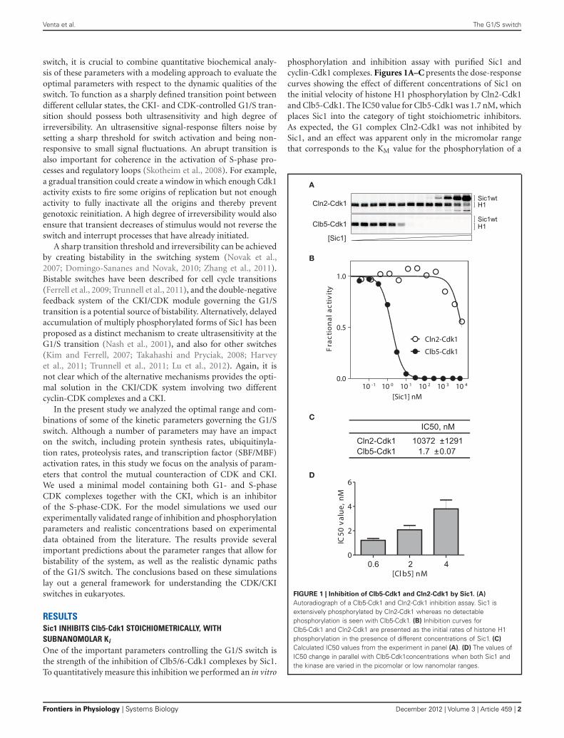
phosphorylation and inhibition assay with purified Sic1 andcyclin-Cdk1 complexes. Figures 1A–C presents the dose-responsecurves showing the effect of different concentrations of Sic1 onthe initial velocity of histone H1 phosphorylation by Cln2-Cdk1and Clb5-Cdk1. The IC50 value for Clb5-Cdk1 was 1.7 nM, whichplaces Sic1 into the category of tight stoichiometric inhibitors.As expected, the G1 complex Cln2-Cdk1 was not inhibited bySic1, and an effect was apparent only in the micromolar rangethat corresponds to the KM value for the phosphorylation of a
A
B
C
D
FIGURE 1 | Inhibition of Clb5-Cdk1 and Cln2-Cdk1 by Sic1. (A)
Autoradiograph of a Clb5-Cdk1 and Cln2-Cdk1 inhibition assay. Sic1 isextensively phosphorylated by Cln2-Cdk1 whereas no detectablephosphorylation is seen with Clb5-Cdk1. (B) Inhibition curves forClb5-Cdk1 and Cln2-Cdk1 are presented as the initial rates of histone H1phosphorylation in the presence of different concentrations of Sic1. (C)
Calculated IC50 values from the experiment in panel (A). (D) The values ofIC50 change in parallel with Clb5-Cdk1concentrations when both Sic1 andthe kinase are varied in the picomolar or low nanomolar ranges.
Frontiers in Physiology | Systems Biology December 2012 | Volume 3 | Article 459 | 2

Venta et al. The G1/S switch
non-inhibitory version of Sic1 (data not shown). This suggeststhat Sic1 only inhibits Cln2-Cdk1 as a competitive substrate.
The experiment presented in Figures 1A–C contained 500 μMATP and 2.5 μM Histone H1. The KM,ATP = 70 μM, and the valuefor KM,H1 is above 10 μM (our unpublished data). Therefore, theobtained value of IC50 is a valid estimate of the apparent inhi-bition constant for substrate phosphorylation at saturating andnearly physiological ATP concentration. However, the experimentpresented in Figures 1A–C was performed at low Clb5-Cdk1 lev-els (1.5–2 nM) that are used in our standard assay conditions.Since the estimated IC50 value was in the same concentrationrange as the enzyme, the inhibition constant may prove to be evenlower. When the enzyme concentration was lowered or raised, theIC50 value changed as well (Figure 1D). This suggests that wecan give an estimate for the apparent Ki value to be in the rangeof 100 pM to 1 nM. The exact value of such a constant is diffi-cult to determine using available methods. This extremely tightinhibition complex should exhibit a very low off-rate, which isan important factor in modeling the Sic1 phosphorylation anddegradation switch, as discussed below.
Sic1 IS EFFICIENTLY PHOSPHORYLATED WITHIN THE Sic1/Clb5-Cdk1COMPLEXThe dynamics of the switch are also shaped by the rates ofSic1 phosphorylation by Cln1,2-Cdk1 and Clb5,6-Cdk1. Wehave previously shown that both Clb5-Cdk1 and Cln2-Cdk1efficiently phosphorylate the N-terminal part of the truncated
non-inhibitory version of Sic1 (Sic1�C) (Koivomagi et al.,2011a). Substrate specificity was shown to be gained by an inter-action between a hydrophobic docking site in Clb5 and RXLmotifs in Sic1, and in the case of Cln2-Cdk1 via a novel hydropho-bic LLPP-docking motif in Sic1 (Koivomagi et al., 2011b; Bhaduriand Pryciak, 2011; Koivomagi and Loog, 2011). Furthermore, bydetailed mapping of the paths of the multisite phosphorylationcascades we found that the Clb5 specificity toward different sites,including the pair S76/S80, which form the critical diphospho-degrons at the end of the cascade, was much more pronouncedcompared to Cln2. These observations led to the conclusion thatalthough Cln2-Cdk1 can phosphorylate Sic1, it cannot triggerSic1 degradation without the help of Clb5-Cdk1. This conclu-sion was supported by in vivo experiments showing that, in theabsence of B-type cyclin (Clb) activity, the Clns alone were notable to cause the degradation of Sic1. Thus, Clb-Cdk1 activity isthe driving force of Sic1 degradation at the G1/S transition. Thisunderscores the likely importance of a double-negative feedbackmechanism whereby Clb5-Cdk1 phosphorylates Sic1, leading toits destruction and thereby releasing further active Clb5-Cdk1from the inhibitory Sic1-Clb5-Cdk1 complex.
This model requires that Clb5-Cdk1 can efficiently phospho-rylate Sic1 bound to the inhibited Clb5-Cdk1 complex. To testthis, we performed a Sic1 phosphorylation assay using partlyinhibited Clb5-Cdk1—that is, we tested the ability of Clb5to phosphorylate a Sic1-Clb5-Cdk1 complex (Figures 2A–D).Additionally, the phosphorylation of non-inhibitory Sic1�C was
Clb5
Cln2
A B
C D
FIGURE 2 | Sic1 is efficiently phosphorylated within the
Sic1/Clb5-Cdk1 complex, when an excess of Clb5-Cdk1 is added.
Assays with Cln2-Cdk1 complexes are included for comparison. (A)
Autoradiography showing the phosphorylation of purified Sic1wt andSic1�C by Clb5-Cdk1 and Cln2-Cdk1 complexes using 15 nM Sic1 and
30 nM cyclin-Cdk1 complexes. (B) The activity of Clb5-Cdk1 wasdetermined using 2.5 μM histone H1, with and without 15 nM Sic1wt inthe assay mixture. (C,D) Quantification of the experiment presented inpanel (A). For comparison, phosphorylation rates were normalized usingcyclin-Cdk1 activity units obtained in panel (B).
www.frontiersin.org December 2012 | Volume 3 | Article 459 | 3

Venta et al. The G1/S switch
assayed in the same conditions. In these assays, 30 nM Clb5-Cdk1 or Cln2-Cdk1 was mixed with 15 nM Sic1. To quantify theexact fraction of uninhibited Clb5-Cdk1 activity, a control sub-strate histone H1 was included in a separate assay (Figure 2B).Clb5-Cdk1 phosphorylated both forms of Sic1 efficiently and ata considerably higher rate than Cln2-Cdk1. The rate of phospho-rylation of the full length Sic1 was similar to that of Sic1�C forboth Cln2 and Clb5. These data indicate that the N-terminus ofSic1 is freely accessible for phosphorylation when Sic1 is boundin the inhibitory complex, and that emerging free Clb5-Cdk1,released from the inhibitory complex as Sic1 is degraded, couldaccelerate Sic1 phosphorylation and degradation, thus creating adouble-negative feedback.
We have also observed that a low level of intramolecularSic1 phosphorylation takes place within the inhibitory Sic1-Clb5-Cdk1 complex during its formation (data not shown). It is par-tially also the likely reason why the full length Sic1 showed slightlyfaster phosphorylation rate compared to Sic1�C (Figures 2C,D).Indeed, structural modeling studies do not exclude the possibil-ity that the active site of Cdk1 may be partly accessible duringthe inhibitor binding (Barberis et al., 2005a,b; Barberis, 2012a,b).The possible role of these phosphorylation events in the switch iscurrently under investigation.
THE MODEL OF THE CDK/CKI-CONTROLLED G1/S SWITCHTo quantitatively analyze the impact of the inhibition strengthand the phosphorylation rates on the abruptness and the degreeof irreversibility of the G1/S switch, we constructed a mini-mal model of Sic1 phosphorylation and degradation (Figure 3,Table 1). As a first step, we analyzed the steady state output ofthe model. This is important to understand the system in gen-eral and to analyze the transition thresholds for the conditions
FIGURE 3 | Minimal model of the G1/S switch. Parameters used fordiagrams and simulations presented in the figures below are listed inTables 1 and 2. Black crosses designate the phospho-dependentdegradation of Sic1 via the SCF-proteasome pathway. Gray crossesdesignate basal degradation of Sic1.
(e.g., slow growth, the arrest, or stress) when the degree of irre-versibility of the switch may become especially important. As asecond step we analyze the behavior of the switch in the normaldynamic circumstances of the cell cycle.
To simplify our model and avoid equation systems that lacksteady state solutions, we did not include the multisite phos-phorylation steps, but considered the net phosphorylation anddegradation process as a single step. This was based on ourexperimental data showing that accumulation of multiply phos-phorylated species is a rapid, semi-processive process in vitro, andthat multiply phosphorylated forms of Sic1 do not accumulatein vivo after cells are released from an α-factor arrest (Koivomagiet al., 2011a). The latter observation suggests that Sic1 degrada-tion is rapid compared to phosphorylation. Therefore, we canconsider the rate of phosphorylation and degradation to be asingle value that is mostly determined by phosphorylation rates.Also, due to the negligible accumulation of multi-phosphorylatedforms of Sic1 and prompt degradation in response to increasingCdk1 activity in vivo, we omitted the counteracting phosphatasefrom the model. This does not mean that the phosphatase doesnot take part in the process but that, for simplicity, its effectwas included in the net rate constant of the whole process. Thefirst of the three ordinary differential equations of the modeldescribe free Sic1 levels, including the association and dissocia-tion of the inhibitory complex and Cln2-dependent phosphory-lation/degradation (Table 1). The second equation describes thedynamics of free Clb5 concentration, which is controlled by the
Table 1 | Equations describing the minimal model of the G1/S switch.
dy1
dt= k5 − k2y1y2 + k1y3 − k6y1 − k4y1y4
dy2
dt= k3y2y3 − k2y1y2 + k1y3 + k6y3 + k4y3y4
dy3
dt= k2y1y2 − k3y2y3 − k1y3 − k6y3 − k4y3y4
y1 = [Sic1FREE
]
y2 = [Clb5 − Cdk1FREE
]
y3 = [Sic1/Clb5 − Cdk1
]
y4 = [Cln2 − Cdk1
] = constant at 200 nM
Conservation equations:
y2 = [Clb5 − Cdk1TOTAL
] − y3
y1 = [Sic1TOTAL
] − y3
Kinetic constants (the values presented below are those used inFigure 4. Parameter values that are varied in other figures are given inthe legends or on the figure panels):
k1 = 0.01 min−1; dissociation rate of the inhibitory complex
k2 = 0.1 nM−1 × min−1; association rate of the inhibitory complex
k3 = 0.001 nM−1 × min−1; the net rate constant for Clb5-dependentphosphorylation and degradation of Sic1
k4 = 0.00001 nM−1 × min−1; the net rate constant for Cln2-dependentphosphorylation and degradation of Sic1
k5 = 10 nM × min−1; the basal synthesis rate of Sic1
k6 = 0.01 min−1; the basal degradation rate of Sic1
The description of the system is presented in Figure 3. For the phase dia-
grams presented in Figures 5–7, the steady-state solutions for the depen-
dencies between Sic1TOTAL and Clb5-Cdk1TOTAL and Clb5-Clb5-Cdk1FREE and
Clb5-Cdk1TOTAL were derived.
Frontiers in Physiology | Systems Biology December 2012 | Volume 3 | Article 459 | 4
Venta et al. The G1/S switch
association-dissociation rates of the inhibitory complex and alsoby the fast release of Clb5-Cdk1 from the inhibitory complexupon Sic1 destruction (Verma et al., 2001). The third equationdescribes the concentration changes of the inhibitory complex.The conservation equations define the total levels of Clb5 andSic1. Finally, the model includes slow basal synthesis and degrada-tion of Sic1. This was based on the finding that although the majorSwi5-induced transcription of SIC1 peaks at the exit from mitosis(Schwob et al., 1994), SIC1 transcription is still observed through-out the entire cell cycle (Knapp et al., 1996; Aerne et al., 1998).Taken together, although considerably simplified, this minimalmodel contains the basic and universal core elements of a switchcontaining a double-negative feedback between a CKI and a CDK.Additionally, the system contains a second CDK complex (theequivalent of Cln2-Cdk1) that is not inhibited by CKI but actsas an initiator of the switch.
The values for the phosphorylation rate constants were derivedfrom our experimentally determined rates of the phosphoryla-tion of Sic1 and other specific model substrates by cyclin-Cdk1complexes (Loog and Morgan, 2005; Koivomagi et al., 2011a,b).A KM value of 3.3 μM and a kcat value of 73 min−1 were deter-mined for Clb5-Cdk1-dependent phosphorylation for Sic1 (datanot shown). Considering that there are about six to seven phos-phorylation steps, and only a slight phosphatase counteraction,the net rate constant for the process of Clb5-dependent phospho-rylation (kClb5) and degradation of Sic1 was taken to be 0.001nM−1 × min−1. Although Cln2-Cdk1 showed only a 3–4-foldlower rate for net phosphorylation of Sic1 in the Sic1-Clb5-Cdk1complex (Figure 2), it was shown to have a considerably weakerability to phosphorylate the priming site T33, and also, to finalizethe phosphorylation cascade, thus creating a di-phosphodegronpS76/pS80 (Koivomagi et al., 2011a). Therefore, the net rate con-stant for Cln2-dependent phosphorylation (kCln2) was initiallytaken to be 0.00001 nM−1 × min−1, two orders of magnitudelower than the corresponding value for Clb5-Cdk1. The impor-tance of this difference on the properties of the switch will be
further analyzed below. Note, as explained above, that these rateconstant are the net constants for both phosphorylation anddegradation.
The parameters defining the inhibitory interaction betweenSic1 and Clb5-Cdk1 were derived from the inhibition experimentpresented in Figure 1. Assuming that the diffusion-limited associ-ation rate of an average protein–protein interaction that involvesan intrinsically disordered binding partner is in the range of 107
to 108 M−1 × min−1, then a KD of a few hundred pM wouldyield a dissociation rate for the Sic1-Clb5-Cdk1 inhibitory com-plex in the range of 10−3 to 10−2 min−1. The effect of varyingthis parameter will be studied in the context of the switch. Thebasal synthesis and degradation rates were taken to be relativelyslow, and for the phase diagrams below these values were chosento give a steady-state concentration of Sic1 of 1 μM in the absenceof cyclin-Cdk1 complexes.
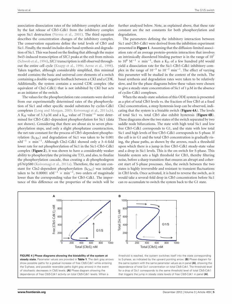
When the steady-state solution of this ODE system is presentedas a plot of total Clb5 levels vs. the fraction of free Clb5 at a fixedCln2 concentration, a steep hysteresis loop can be observed, indi-cating that the system is a bistable switch (Figure 4A). The levelsof total Sic1 vs. total Clb5 also exhibit hysteresis (Figure 4B).These diagrams show the two states of the switch separated by twosaddle node bifurcations. The state with high total Sic1 and lowfree Clb5-Cdk1 corresponds to G1, and the state with low totalSic1 and high levels of free Clb5-Cdk1 corresponds to S-phase. Ifthe cell is in G1 and the total Clb5 concentration is gradually ris-ing, the phase paths, as shown by the arrows, reach a thresholdupon which there is a jump in free Clb5-Cdk1 steady-state valueand a drop in Sic1 levels. This is the on-switch for S-phase. Thisbistable system sets a high threshold for Clb5, thereby filteringnoise, before a sharp transition that ensures an abrupt and coher-ent start of S-phase processes. Also, the switch between the twostates is highly irreversible and resistant to transient fluctuationsin Clb5 levels. Once activated, it is hard to reverse the switch, as itwould take a several-fold drop in Clb5 concentration before Sic1can re-accumulate to switch the system back to the G1 state.
0 200 400 600 800 10000
200
400
600
800
1000
Total [Clb5]; nM
Mn ;]1kdC- 5bl C[ eer F
0 200 400 600 800 10000
200
400
600
800
1000
Total [Clb5]; nM
Mn ;]1ciS [ l at oTG1/S
G1/S
A B
FIGURE 4 | Phase diagrams showing the bistability of the system at
steady state. Parameter values are provided in Table 1. The dark gray arrowsshow possible paths for a gradual increase of free Clb5-Cdk1 while enteringthe S-phase, and possible reversible paths (light gray arrows) in the eventof stochastic decreases in Clb5 levels. (A) Phase diagram showing thedependence of free Clb5-Cdk1 activity on total Clb5-Cdk1 levels. When a
threshold is reached, the system switches itself into the state correspondingto S-phase, as indicated by the upward pointing arrow. (B) Phase diagram forthe same system with the same parameter values as in panel (A), showing thedependence of total Sic1 concentration on total Clb5-Cdk1. The threshold levelfor a drop of Sic1 corresponds to the same threshold level of total Clb5-Cdk1that triggers the jump in steady state levels of free Clb5-Cdk1 in panel (A).
www.frontiersin.org December 2012 | Volume 3 | Article 459 | 5

Venta et al. The G1/S switch
TIGHT INHIBITION OF Clb5-Cdk1 BY Sic1 AND EFFICIENTPHOSPHORYLATION OF Sic1-Clb5-Cdk1 BY FREE Clb5-Cdk1 ARECRUCIAL FOR SETTING A DISCREET THRESHOLD FOR THE G1/STRANSITIONTo understand the basic features of the switch and to determinethe optimal balance of crucial parameters that are required tomaintain the bistable nature of the system, we studied the effect ofinhibition strength and phosphorylation rates on the behavior ofthe phase diagrams. In the diagrams presented in Figures 5A–D,inhibition strength was varied by introducing different values forthe dissociation rate of the inhibitory complex. As the dissocia-tion rate increased, the bistable behavior of the system graduallyweakened and the switch evolved to a smooth transition that caneasily slide back and forth (Figure 5D). This kind of a behaviorwould potentially be very harmful for a transition like G1/S: onlya slight backward deviation of the total Clb5 levels would reversethe cell to G1, interrupting processes like the initiation of repli-cation, and removing the re-replication block. Thus, our modelpredicts that between a Ki of 10–100 nM the on-threshold con-tinues to decrease, and eventually the system loses its hysteresis, aswell as its switch-like response (Figures 5C,D). In this range, Sic1becomes a readily overcome barrier for the accumulating Clb5.
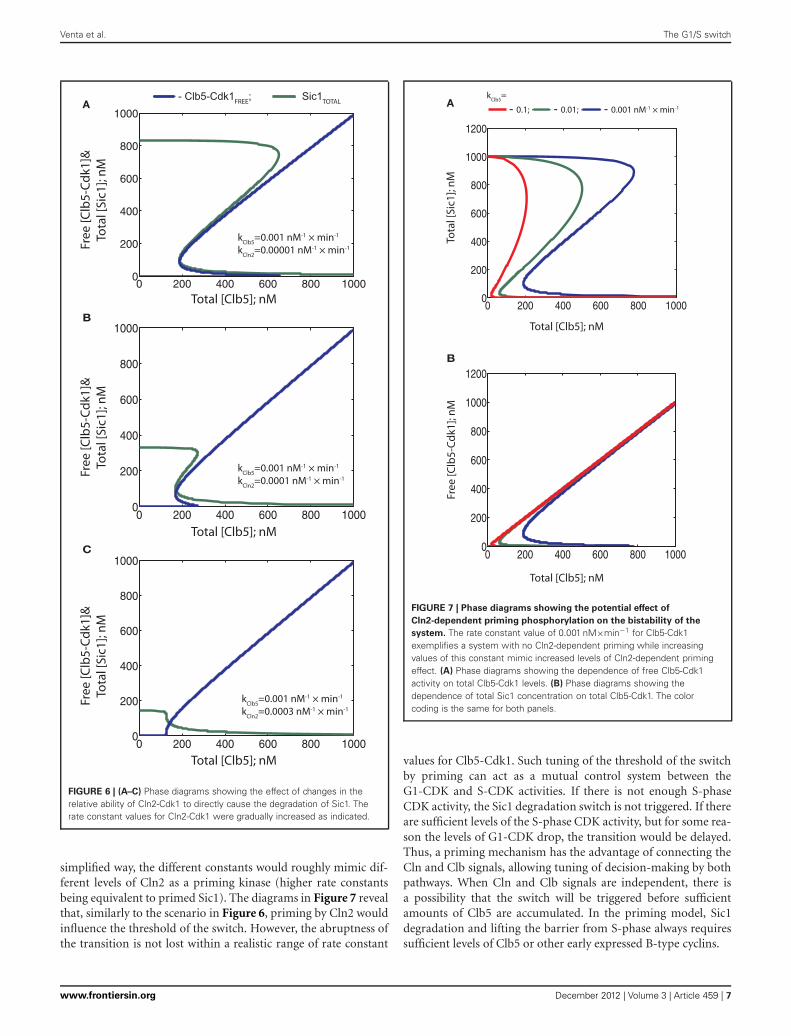
Next, in the diagrams presented in Figure 6, we exploredthe effect of relative phosphorylation rates. To understand theimpact of the experimentally determined strikingly weaker abilityof Cln2-Cdk1 to phosphorylate the critical output degron sitesin Sic1, compared to Clb5-Cdk1 (Koivomagi et al., 2011a), wefirst varied the Cln2-dependent phosphorylation rate relative to afixed Clb5-dependent rate (Figures 6A–C). Importantly, steeperhysteresis was observed when the Cln2-dependent phosphoryla-tion rate was considerably lower compared to the rate of Clb5.Increasing the relative Cln2-dependent rate resulted in less hys-teresis, lowered initial Sic1 levels, and lowered the Clb5 thresholdfor the transition (Figures 6A,B). Thus, the timing of the transi-tion could potentially be tuned by Cln2 activity. However, whenCln2 specificity toward Sic1 approaches the specificity of Clb5, therequirement for Clb5 signal almost ceases to exist (Figure 6C).Similarly, as in the case of a weak Ki (above), the threshold ofTotal (Clb5) decreased and the discontinuity of the Clb5-Cdk1switch was lost. Thus, both tight inhibition and cyclin specificityare important factors for an abrupt transition and a high degree ofirreversibility. Interestingly, the effect of Cln2 specificity on bista-bility was more pronounced compared to the effect of an increasein Ki of the same order.
Thus, in the system described in Figure 3, where both Cln2and Clb5 are able to send Sic1 for degradation, the signal strengthof Cln2 can potentially modulate timing by lowering the thresh-old of Clb5 required to trigger the switch. However, our recentresults indicate that Cln2-Cdk1 cannot phosphorylate the lastphospho-degron sites in Sic1 at a sufficient rate to cause degra-dation in vivo. On the other hand, Cln2-Cdk1 can efficientlyphosphorylate the initial residues in the Sic1 cascade, and thisprimes Sic1 for efficient subsequent phosphorylation by Clb5-Cdk1, which will finalize the cascade to send Sic1 to degradation(Koivomagi et al., 2011a). To study how such a priming effectcould influence the switch, we gradually raised the phosphoryla-tion rate constant for Clb5 in the absence of Cln2 activity. In this
B
C
0 200 400 600 800 10000
200
400
600
800
1000
&] 1kd C-5bl C [ e erFM n ;] 1ci S[ l ato T
Total [Clb5]; nM
0 200 400 600 800 10000
200
400
600
800
1000
Total [Clb5]; nM
&]1kdC-5bl C[ eer FMn ;] 1ci S[ l at oT
0 200 400 600 800 10000
200
400
600
800
1000
&] 1kdC- 5bl C[ eer FMn ;] 1ci S[ l at oT
Total [Clb5]; nM
D
0 200 400 600 800 10000
200
400
600
800
1000
&] 1k dC-5 bl C[ eerFMn ;]1 ciS[ l at oT
Total [Clb5]; nM
A
kDissoc.=0.01 min-1
- Clb5-Cdk1FREE; - Sic1TOTAL
kDissoc.=0.1 min-1
kDissoc.= 1 min-1
kDissoc.= 10 min-1
FIGURE 5 | (A–D) The steady state phase diagrams showing the effect ofthe inhibition strength on the bistability of the G1/S switch. The dissociationrates of the inhibitory complex were increased in 10-fold increments from0.01 to 10 min−1 as indicated. In the model, these values correspond to Ki
values of 0.1–100 nM. Arrows indicate the paths of the system at the G1/Stransition.
Frontiers in Physiology | Systems Biology December 2012 | Volume 3 | Article 459 | 6
Venta et al. The G1/S switch
B
C
0 200 400 600 800 10000
200
400
600
800
1000
Total [Clb5]; nM
0 200 400 600 800 10000
200
400
600
800
1000
Total [Clb5]; nM
0 200 400 600 800 10000
200
400
600
800
1000
A
Total [Clb5]; nM
&]1kdC-5bl C[ eer FMn ;] 1ci S[ l atoT
&] 1kdC-5b l C[ e erF] 1ci S [ l a to T
M n ;&] 1k dC- 5bl C[ eer F
Mn ;] 1ci S[ lat o T - Clb5-Cdk1FREE; - Sic1TOTAL
kClb5=0.001 nM-1 × min-1
kCln2=0.00001 nM-1 × min-1
kClb5=0.001 nM-1 × min-1
kCln2=0.0003 nM-1 × min-1
kClb5=0.001 nM-1 × min-1
kCln2=0.0001 nM-1 × min-1
FIGURE 6 | (A–C) Phase diagrams showing the effect of changes in therelative ability of Cln2-Cdk1 to directly cause the degradation of Sic1. Therate constant values for Cln2-Cdk1 were gradually increased as indicated.
simplified way, the different constants would roughly mimic dif-ferent levels of Cln2 as a priming kinase (higher rate constantsbeing equivalent to primed Sic1). The diagrams in Figure 7 revealthat, similarly to the scenario in Figure 6, priming by Cln2 wouldinfluence the threshold of the switch. However, the abruptness ofthe transition is not lost within a realistic range of rate constant
B
Total [Clb5]; nM
Total [Clb5]; nM
0 200 400 600 800 10000
200
400
600
800
1000
1200
0 200 400 600 800 10000
200
400
600
800
1000
1200
Mn ;]1 ci S [ l atoT
A
Mn ;] 1 kdC- 5 bl C[ eer F
- 0.1; - 0.01; - 0.001 nM-1 × min-1
kClb5=
FIGURE 7 | Phase diagrams showing the potential effect of
Cln2-dependent priming phosphorylation on the bistability of the
system. The rate constant value of 0.001 nM×min−1 for Clb5-Cdk1exemplifies a system with no Cln2-dependent priming while increasingvalues of this constant mimic increased levels of Cln2-dependent primingeffect. (A) Phase diagrams showing the dependence of free Clb5-Cdk1activity on total Clb5-Cdk1 levels. (B) Phase diagrams showing thedependence of total Sic1 concentration on total Clb5-Cdk1. The colorcoding is the same for both panels.
values for Clb5-Cdk1. Such tuning of the threshold of the switchby priming can act as a mutual control system between theG1-CDK and S-CDK activities. If there is not enough S-phaseCDK activity, the Sic1 degradation switch is not triggered. If thereare sufficient levels of the S-phase CDK activity, but for some rea-son the levels of G1-CDK drop, the transition would be delayed.Thus, a priming mechanism has the advantage of connecting theCln and Clb signals, allowing tuning of decision-making by bothpathways. When Cln and Clb signals are independent, there isa possibility that the switch will be triggered before sufficientamounts of Clb5 are accumulated. In the priming model, Sic1degradation and lifting the barrier from S-phase always requiressufficient levels of Clb5 or other early expressed B-type cyclins.
www.frontiersin.org December 2012 | Volume 3 | Article 459 | 7

Venta et al. The G1/S switch
CYCLIN CONCENTRATIONS AND SYNTHESIS RATESAlthough the phase diagrams provide a good general charac-terization of an ODE model describing a bistable system, theydon’t necessarily fully reflect the way the system behaves in reality.Some rates could be too slow relative to the others, in which casesteady states would not be established. In the present case, thesynthesis of Clb5 is fast, with a peak accumulation time of onlyaround 10–20 min (Koivomagi et al., 2011b). Therefore, thereis a possibility that relatively slow phosphorylation rates or dis-sociation rates of the inhibitory complex would not allow theformation of the steady states described by the phase diagrams. Toaddress this concern, we investigated the dynamics of our model.We numerically simulated a modified form of the ODE system(Table 2). We searched the literature to supply realistic synthe-sis rates and peak concentrations of the cyclins and Sic1. Weobtained experimentally estimated endogenous concentrations ofCln1, Cln2, Cln3, Clb5, Clb6, and Sic1. The most detailed studieshave been performed by western blotting of ProtA-tagged pro-teins and by using quantitative calibration of the blotting signalswith ProtA standards (Cross et al., 2002). Second, a study pre-senting a global analysis of protein abundances in yeast providesquantifications based on the western blotting of TAP-tagged pro-teins (Ghaemmaghami et al., 2003). Using the ProtA-tag method,the estimated average number of Sic1 molecules per diploid cell
Table 2 | Equation system used for numerical simulation of the time
courses at G1/S in Figures 8A–C, 9A–D, and 10A.
dy1
dt= −k2y1y2 − k4y1y4 + k1y3
dy2
dt= k8 − k2y1y2 + k3y2y3 + k4y3y4 + k1y3
dy3
dt= k2y1(y6 − y3) − k3y2y3 − k4y3y4 − k1y3
dy4
dt= k7
dy5
dt= −k4y1y4 − k3y2y3 − k4y3y4
dy6
dt= k8
y1 = [Sic1FREE
]
y2 = [Clb5 − Cdk1FREE
]
y3 = [Sic1/Clb5 − Cdk1
]
y4 = [Cln2 − Cdk1
]
y5 = [Sic1TOTAL
]
y6 = [Clb5 − Cdk1TOTAL
]
The kinetic constants (values presented below are those used inFigure 8A. Parameter values for other figures are given in the legends oron the figure panels):
k1 = 0.01 min−1; dissociation rate of the inhibitory complex
k2 = 0.1 nM−1 × min−1; association rate of the inhibitory complex
k3 = 0.001 nM−1 × min−1; the net rate constant for Clb5-dependentphosphorylation and degradation of Sic1
k4 = 0.00001 nM−1 × min−1; the net rate constant for Cln2-dependentphosphorylation and degradation of Sic1
k7 = 100 nM × min−1; Cln2 synthesis rate
k8 = 30 nM × min−1; Clb5 synthesis rate
The initial values for y1 (Sic1FREE ) and y5 (Sic1TOTAL) in two simulations
were 1 μM.
in asynchronous cultures was reported to be 214. The study usingthe TAP-tag in haploid cells provided an estimate of 768 copies ofSic1 per cell. To calculate the realistic initial concentration valuefor Sic1 for the ODE simulations, one needs to take into accountthe predominantly nuclear localization of Sic1 (Huh et al., 2003).The nuclear volume of haploid and diploid cells is thought tobe 2 and 4 fl, respectively (Cross et al., 2002). Thus, the averageestimated nuclear concentration of Sic1 in asynchronous cultureswould be 363 nM. Since Sic1 is present during only a fractionof the cell cycle, we considered that a reasonable peak concen-tration would be three times that of the average abundance inasynchronous cells. Therefore, a realistic concentration of Sic1 atits peak in G1 would be around 1 μM. In fact, this value falls intothe same range as the steady state value of Sic1 in the absence ofClb5 and Cln2 in the phase diagrams presented in Figure 7.
Analogous calculations for Clb5, whose localization is alsopredominantly nuclear (Shirayama et al., 1999), give peak con-centration of 1138 nM (115 nM for Clb6). The cyclins Cln1 andCln2 are localized both in cytoplasm and in nucleus (Miller andCross, 2000; Edgington and Futcher, 2001; Landry et al., 2012),and therefore the cell volume for calculating their concentrationwas taken to be 100 fl for the ProtA study (diploid cells) and 50 flfor the TAP study (haploid cells). Interestingly, the obtained peakvalues for Cln1 and Cln2 were about 164 and 33 nM, respectively,which is much lower than the peak values for Clb5 and Clb6. Thisfact would additionally support the model that Cln1,2 may have aless critical role in catalyzing Sic1 degradation compared to rela-tively abundant Clb5. Cln3 levels were recorded only by the studyusing ProtA, and their concentration in G1, taking into accountthe nuclear localization, was in the range of 269 nM.
Based on these calculations and the half-times of accumula-tion of Cln2 and Clb5, the concentrations of Cln2 and Clb5 in thismodel were set to rise at 30 and 100 nM per minute, respectively.To obtain a clear and formal picture of the effect of accumulationrates on the dynamics of the switch, a constant linear accumu-lation was introduced (Table 2). During the short time windowof cyclin accumulation, the basal synthesis and degradation ratesof Sic1 used in the ODE system for the phase diagrams would betoo slow to significantly affect the dynamics of Sic1 degradation.Therefore, these rates were excluded to lower the complexity ofthe model.
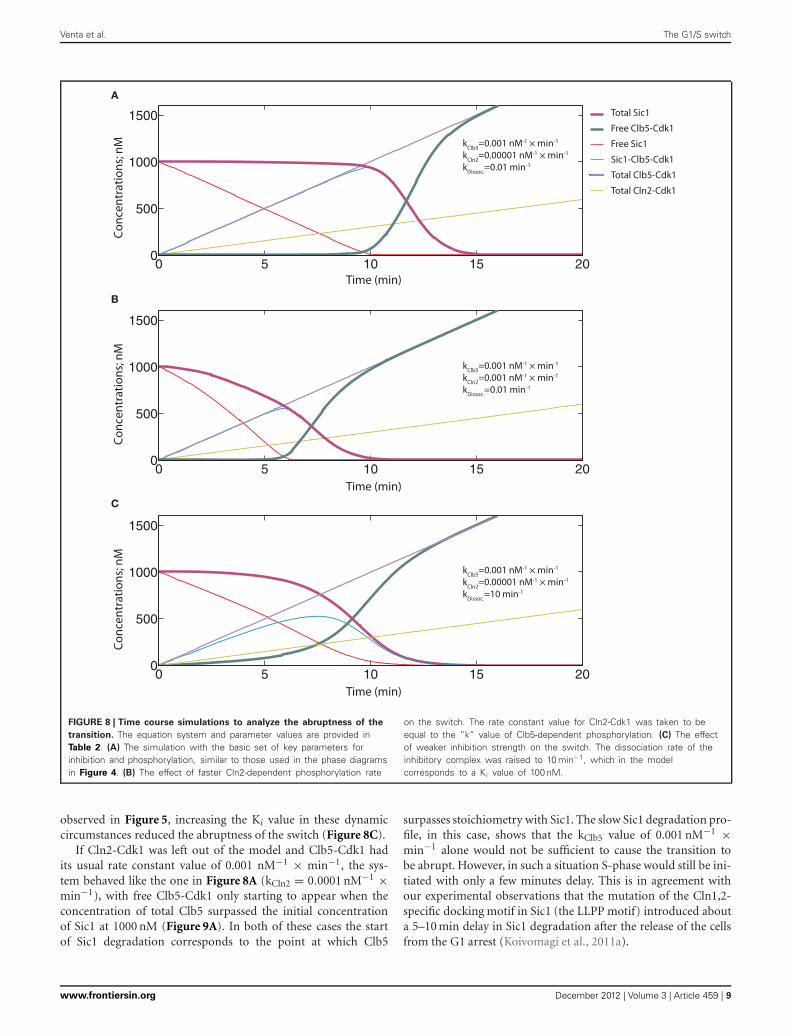
TIGHT INHIBITION AND EFFICIENT Clb5-DEPENDENT Sic1PHOSPHORYLATION IS IMPORTANT FOR THE ABRUPTNESSOF THE SWITCH UNDER REALISTIC DYNAMIC CIRCUMSTANCESThe simulation of the ODE system presented in Figure 8A repre-sents a situation with tight inhibition and high kClb5/kCln2 ratio,analogous to the one presented in Figure 4. Although exhibit-ing a time course with a sigmoidal shape, a fairly slow transitionfrom low to high Clb5-Cdk1 activity level is observed. Thus,although the phase diagrams can provide a general descriptionof the system, the transitions between the alternative states mayhappen less abruptly. We also investigated the effect of loweringthe kClb5/kCln2 ratio. Similarly to the conclusion made based onthe phase diagrams above, a relatively faster Cln2-dependent ratedecreased the ultrasensitivity of the signal response and slowedthe transition (Figure 8B). Also, analogously to the tendency
Frontiers in Physiology | Systems Biology December 2012 | Volume 3 | Article 459 | 8
Venta et al. The G1/S switch
0 5 10 15 200
500
1000
1500
B
C
0 5 10 15 200
500
1000
1500 data1datadatadatadatadata
0 5 10 15 200
500
1000
1500 data1datadatadatadatadata
Total Sic1
Free Clb5-Cdk1
Free Sic1
Sic1-Clb5-Cdk1
Total Clb5-Cdk1
Total Cln2-Cdk1
kClb5=0.001 nM-1 × min-1
kCln2=0.00001 nM-1 × min-1
kDissoc.=0.01 min-1
Time (min)
Conc
entr
atio
ns; n
M
Time (min)
Conc
entr
atio
ns; n
M
Time (min)
Conc
entr
atio
ns; n
M
A
kClb5=0.001 nM-1 × min-1
kCln2=0.001 nM-1 × min-1
kDissoc.=0.01 min-1
kClb5=0.001 nM-1 × min-1
kCln2=0.00001 nM-1 × min-1
kDissoc.=10 min-1
FIGURE 8 | Time course simulations to analyze the abruptness of the
transition. The equation system and parameter values are provided inTable 2. (A) The simulation with the basic set of key parameters forinhibition and phosphorylation, similar to those used in the phase diagramsin Figure 4. (B) The effect of faster Cln2-dependent phosphorylation rate
on the switch. The rate constant value for Cln2-Cdk1 was taken to beequal to the “k” value of Clb5-dependent phosphorylation. (C) The effectof weaker inhibition strength on the switch. The dissociation rate of theinhibitory complex was raised to 10 min−1, which in the modelcorresponds to a Ki value of 100 nM.
observed in Figure 5, increasing the Ki value in these dynamiccircumstances reduced the abruptness of the switch (Figure 8C).
If Cln2-Cdk1 was left out of the model and Clb5-Cdk1 hadits usual rate constant value of 0.001 nM−1 × min−1, the sys-tem behaved like the one in Figure 8A (kCln2 = 0.0001 nM−1 ×min−1), with free Clb5-Cdk1 only starting to appear when theconcentration of total Clb5 surpassed the initial concentrationof Sic1 at 1000 nM (Figure 9A). In both of these cases the startof Sic1 degradation corresponds to the point at which Clb5
surpasses stoichiometry with Sic1. The slow Sic1 degradation pro-file, in this case, shows that the kClb5 value of 0.001 nM−1 ×min−1 alone would not be sufficient to cause the transition tobe abrupt. However, in such a situation S-phase would still be ini-tiated with only a few minutes delay. This is in agreement withour experimental observations that the mutation of the Cln1,2-specific docking motif in Sic1 (the LLPP motif) introduced abouta 5–10 min delay in Sic1 degradation after the release of the cellsfrom the G1 arrest (Koivomagi et al., 2011a).
www.frontiersin.org December 2012 | Volume 3 | Article 459 | 9

Venta et al. The G1/S switch
0 5 10 15 200
500
1000
1500
data1datadatadatadatadata
0 5 10 15 200
500
1000
1500
datadatadatadatadatadata
0 5 10 15 200
500
1000
1500
data1data2data3data4data5data6
C
B
D
0 5 10 15 200
500
1000
1500
Total Sic1
Free Clb5-Cdk1
Free Sic1
Sic1-Clb5-Cdk1
Total Clb5-Cdk1
Time (min)
Conc
entr
atio
ns; n
M
Time (min)
Time (min)
Time (min)
Conc
entr
atio
ns; n
MCo
ncen
trat
ions
; nM
Conc
entr
atio
ns; n
M
A
kClb5=0.01 nM-1 × min-1
kClb5=0.03 nM-1 × min-1
kClb5=0.1 nM-1 × min-1
kClb5=0.001 nM-1 × min-1
FIGURE 9 | Time course simulations showing the potential effect of
Cln2-dependent priming phosphorylation on the abruptness of the
switch. The equation system and parameter values are provided in Table 2.(A) A system with no priming effect in the absence of Cln2. The basal rate
constant value of 0.001 nM−1 × min−1 for Clb5-Cdk1 was used. (B–D)
Different priming effects were mimicked by using the following “k” values forClb5-Cdk1: 0.01 nM−1 × min−1 (B), 0.03 nM−1 × min−1 (C), and 0.1 nM−1 ×min−1 (D).
Frontiers in Physiology | Systems Biology December 2012 | Volume 3 | Article 459 | 10
Venta et al. The G1/S switch
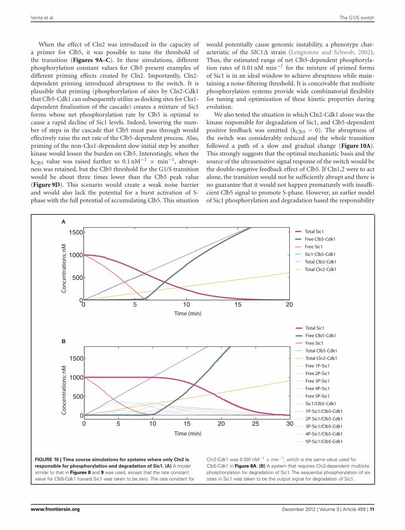
When the effect of Cln2 was introduced in the capacity ofa primer for Clb5, it was possible to tune the threshold ofthe transition (Figures 9A–C). In these simulations, differentphosphorylation constant values for Clb5 present examples ofdifferent priming effects created by Cln2. Importantly, Cln2-dependent priming introduced abruptness to the switch. It isplausible that priming (phosphorylation of sites by Cln2-Cdk1that Clb5-Cdk1 can subsequently utilize as docking sites for Cks1-dependent finalization of the cascade) creates a mixture of Sic1forms whose net phosphorylation rate by Clb5 is optimal tocause a rapid decline of Sic1 levels. Indeed, lowering the num-ber of steps in the cascade that Clb5 must pass through wouldeffectively raise the net rate of the Clb5-dependent process. Also,priming of the non-Cks1-dependent slow initial step by anotherkinase would lessen the burden on Clb5. Interestingly, when thekClb5 value was raised further to 0.1 nM−1 × min−1, abrupt-ness was retained, but the Clb5 threshold for the G1/S transitionwould be about three times lower than the Clb5 peak value(Figure 9D). This scenario would create a weak noise barrierand would also lack the potential for a burst activation of S-phase with the full potential of accumulating Clb5. This situation
would potentially cause genomic instability, a phenotype char-acteristic of the SIC1� strain (Lengronne and Schwob, 2002).Thus, the estimated range of net Clb5-dependent phosphoryla-tion rates of 0.01 nM min−1 for the mixture of primed formsof Sic1 is in an ideal window to achieve abruptness while main-taining a noise-filtering threshold. It is conceivable that multisitephosphorylation systems provide wide combinatorial flexibilityfor tuning and optimization of these kinetic properties duringevolution.
We also tested the situation in which Cln2-Cdk1 alone was thekinase responsible for degradation of Sic1, and Clb5-dependentpositive feedback was omitted (kClb5 = 0). The abruptness ofthe switch was considerably reduced and the whole transitionfollowed a path of a slow and gradual change (Figure 10A).This strongly suggests that the optimal mechanistic basis and thesource of the ultrasensitive signal response of the switch would bethe double-negative feedback effect of Clb5. If Cln1,2 were to actalone, the transition would not be sufficiently abrupt and there isno guarantee that it would not happen prematurely with insuffi-cient Clb5 signal to promote S-phase. However, an earlier modelof Sic1 phosphorylation and degradation based the responsibility
0 5 10 15 200
500
1000
1500
data1datadatadatadatadata
A
Total Sic1
Free Clb5-Cdk1
Free Sic1
Sic1-Clb5-Cdk1
Total Clb5-Cdk1
Total Cln2-Cdk1
Time (min)
Conc
entr
atio
ns; n
M
0 5 10 15 20 25 300
500
1000
1500
B
Time (min)
Conc
entr
atio
ns; n
M
Total Sic1
Free Clb5-Cdk1
Free Sic1
Total Clb5-Cdk1
Total Cln2-Cdk1
Free 1P-Sic1
Free 2P-Sic1
Free 3P-Sic1
Free 4P-Sic1
Free 5P-Sic1
Sic1/Clb5-Cdk1
1P-Sic1/Clb5-Cdk1
2P-Sic1/Clb5-Cdk1
3P-Sic1/Clb5-Cdk1
4P-Sic1/Clb5-Cdk1
5P-Sic1/Clb5-Cdk1
FIGURE 10 | Time course simulations for systems where only Cln2 is
responsible for phosphorylation and degradation of Sic1. (A) A modelsimilar to that in Figures 8 and 9 was used, except that the rate constantvalue for Clb5-Cdk1 toward Sic1 was taken to be zero. The rate constant for
Cln2-Cdk1 was 0.001 nM−1 × min−1, which is the same value used forClb5-Cdk1 in Figure 8A. (B) A system that requires Cln2-dependent multisitephosphorylation for degradation of Sic1. The sequential phosphorylation of sixsites in Sic1 was taken to be the output signal for degradation of Sic1.
www.frontiersin.org December 2012 | Volume 3 | Article 459 | 11

Venta et al. The G1/S switch
of Sic1 phosphorylation entirely on Cln2 (Nash et al., 2001; Vermaet al., 1997a). The ultrasensitive feature of the switch in thismodel was later built on a theoretical presumption of equal ratesof distributive phosphorylation and the delayed accumulationof multiply phosphorylated forms of Sic1 (Deshaies and Ferrell,2001). To test if this scheme would improve the abruptness ofthe switch, we introduced six equally Cln2-specific phosphory-lation steps into the model, with phosphorylation at all six beingrequired for degradation. Clb5-dependent positive feedback effectwas excluded as in Figure 10A (for other details of this modelsee Table 3). The simulation of this system did indeed create alonger lag for the onset of the transition, but it did not improve
the abruptness of the response (Figure 10B). In reality, the pathof the transition would look even less switch-like because the N-terminus of Sic1 contains several crucial phosphorylation sitesbearing suboptimal CDK consensus motifs and exhibits low indi-vidual site specificity with Cln2-Cdk1 (Koivomagi et al., 2011a).These simulations predict that although the delayed accumula-tion of multiply phosphorylated forms could create a thresholdfor Sic1 degradation, multisite phosphorylation would not addconsiderably to the abruptness of the switch. This conclusion isin agreement with the results of a theoretical analysis predict-ing that multisite phosphorylation may provide a threshold fora transition but is generally a poor switch (Gunawardena, 2005).
Table 3 | Equation system used for simulation of the time course for Cln2-dependent multisite phosphorylation of Sic1presented in Figure 10B.
Scheme
The kinetic constants
kon = 0.1;—the association rate of the inhibitory complex. To lower the complexity, the dissociation rate was considered very slow and not included inthis version of the model.
k1 = 0.001; k2 = 0.001; k3 = 0.001; k4 = 0.001; and k5 = 0.001;—the rate constants for Cln2-dependent phosphorylation steps.
k6 = 0.001;—the rate constant for Cln2-dependent phosphorylation of 5P-Sic1 and 5P-Sic1/Clb5-Cdk1 including the net rate for the subsequent fastSCF-mediated degradation.
kN1 = 30; kB1 = 100; the synthesis rates of Cln2 and Clb5, respectively.
The variables
y(1); y(2); y(3); y(4); y(5); y(6);—Sic1 forms bearing 0, 1, 2, . . . , or 5 phosphates, respectively. The form with 6 phosphates was degraded by SCF and dueto the fast rate of degradation this form was not expressed as a separate variable. To lower the complexity, only a sequential 6-step process wasconsidered in the model.
y(7); y(8); y(9); y(10) ; y(11); y(12);—different Sic1/Clb5-Cdk1 inhibitory complexes involving Sic1 with 0, 1, 2, . . . , or 5 phosphates, respectively.
y(13); y(14); y(15);—concentrations of free Clb5-Cdk1, Cln2-Cdk1, and total Clb5-Cdk1, respectively.
The initial value for 0P-Sic1 (y(7)) was taken to be 1, the initial values for the rest of the variables were taken to be zero.
The ODE system
dy(1)/dt = −k1 × y(14) × y(1) − kon × (y(15) − y(7) − y(8) − y(9) − y(10) − y(11) − y(12)) × y(1)
dy(2)/dt = k1 × y(14) × y(1) − k2 × y(14) × y(2) − kon × (y(15) − y(7) − y(8) − y(9) − y(10) − y(11) − y(12)) × y(2)
dy(3)/dt = k2 × y(14) × y(2) − k3 × y(14) × y(3) − kon × (y(15) − y(7) − y(8) − y(9) − y(10) − y(11) − y(12)) × y(3)
dy(4)/dt = k3 × y(14) × y(3) − k4 × y(14) × y(4) − kon × (y(15) − y(7) − y(8) − y(9) − y(10) − y(11) − y(12)) × y(4)
dy(5)/dt = k4 × y(14) × y(4) − k5 × y(14) × y(5) − kon × (y(15) − y(7) − y(8) − y(9) − y(10) − y(11) − y(12)) × y(5)
dy(6)/dt = k5 × y(14) × y(5) − k6 × y(14) × y(6) − kon × (y(15) − y(7) − y(8) − y(9) − y(10) − y(11) − y(12)) × y(6)
dy(7)/dt = kon × (y(15) − y(7) − y(8) − y(9) − y(10) − y(11) − y(12)) × y(1) − k1 × y(14) × (y(15) − y(13) − y(8) − y(9) − y(10) − y(11) − y(12))
dy(8)/dt = kon × (y(15) − y(7) − y(8) − y(9) − y(10) − y(11) − y(12)) × y(2) + (k1 × y(14)) × (y(15) − y(13) − y(8) − y(9) − y(10) − y(11) − y(12)) − (k2 × y(14)) ×(y(15) − y(13) − y(7) − y(9) − y(10) − y(11) − y(12))
dy(9)/dt = kon × (y(15) − y(7) − y(8) − y(9) − y(10) − y(11) − y(12)) × y(3) + (k2 × y(14)) × (y(15) − y(13) − y(7) − y(9) − y(10) − y(11) − y(12)) − (k3 × y(14)) ×(y(15) − y(13) − y(8) − y(7) − y(10) − y(11) − y(12))
dy(10)/dt = kon × (y(15) − y(7) − y(8) − y(9) − y(10) − y(11) − y(12)) × y(4) + (k3 × y(14) × (y(15) − y(13) − y(8) − y(7) − y(10) − y(11) − y(12)) − (k4 × y(14)) ×(y(15) − y(13) − y(8) − y(9) − y(7) − y(11) − y(12))
dy(11)/dt = kon × (y(15) − y(7) − y(8) − y(9) − y(10) − y(11) − y(12)) × y(5) + (k4 × y(14)) × (y(15) − y(13) − y(8) − y(9) − y(7) − y(11) − y(12)) − (k5 × y(14)) ×(y(15) − y(13) − y(8) − y(9) − y(10) − y(7) − y(12))
dy(12)/dt = kon × (y(15) − y(7) − y(8) − y(9) − y(10) − y(11) − y(12)) × y(6) + (k5 × y(14)) × (y(15) − y(13) − y(8) − y(9) − y(10) − y(7) − y(12)) − (k6 × y(14)+) ×(y(15) − y(13) − y(8) − y(9) − y(10) − y(11) − y(7))
dy(13)/dt = kB1 − kon × (y(15) − y(7) − y(8) − y(9) − y(10) − y(11) − y(12)) × (y(1) + y(2) + y(3) + y(4) + y(5) + y(6)) + k6 × y(14) × y(12)
dy(14)/dt = kN1
dy(15)/dt = kB1
Frontiers in Physiology | Systems Biology December 2012 | Volume 3 | Article 459 | 12

Venta et al. The G1/S switch
DISCUSSIONThe double-negative feedback model of the G1/S switch in bud-ding yeast involves two CDK complexes and a CDK inhibitorSic1 that, at the same time, is the substrate of CDK. We aimedto explore which properties such a mutual antagonism wouldrequire to generate a useful switch. Based on mathematical mod-eling and phosphorylation assays we found that, for the transitionto exhibit the best switch-like qualities, it is important to havetight inhibition of Clb5-Cdk1 by Sic1 and efficient phosphory-lation of the Sic1-Clb5-Cdk1 inhibitory complex. The low Ki
ensures that a tight and nearly stoichiometric barrier sets athreshold for the Clb5 signal. The efficient phosphorylation anddegradation rates ensure that the double-negative feedback wouldgain the momentum required to create a switch-like increase infree Clb5-Cdk1 in response to total Clb5. We also studied the rel-ative impact and different roles of Cln2 and Clb5 in the switch.In this respect we analyzed several possible scenarios: first, a sce-nario in which both Cln2 and Clb5 are capable of independentlytriggering the degradation of Sic1; second, a case in which Cln2phosphorylates Sic1 to prime further Cks1-dependent phospho-rylation by Clb5 but is not able to cause the degradation ofSic1 by itself; third, we modeled the situations in which eitherCln2 or Clb5 alone was responsible for phosphorylation anddegradation of Sic1; finally, we explored the impact of multisitephosphorylation on the switch. The general conclusion derivedfrom these case studies was that the optimal role for Cln2-Cdk1in the switch would not be to act as a kinase activity responsiblefor abrupt degradation of CKI, but rather to act as a prim-ing signal that triggers the feedback loop for the emerging freeClb5-Cdk1.
Clb5-dependent positive feedback would be predicted to playa role in Sic1 degradation at the G1/S transition, since there isexpected to be a point when the accumulating fraction of freeClb5-Cdk1 will overcome and phosphorylate Sic1 molecules thatare bound to the inhibitory complex. This feedback is clearlyexpected to have some contribution to the abruptness of theswitch. However, until recently, the extent of this effect had notbeen quantitatively estimated. In addition to our recent find-ings, Tang and colleagues used live cell fluorescent microscopyto show that Clb5,6-dependent positive feedback promotes theswitch-like properties of the G1/S transition (Tang, pers. commu-nication). Additionally, recent work by Barberis and colleagueshas shown the involvement of Clb5 and also later B-type cyclinsin degradation of Sic1 (Barberis et al., 2012). The authors of thiswork proposed a model where a surviving fraction of Sic1 couldbe involved even post-G1/S in the regulation of transcriptionalwaves of later cyclin genes. In general, these findings further argueagainst a solely Cln2-dependent mechanism of Sic1 degradationand emphasize the relatively major importance of B-type cyclinsin this process.
The mutual control between Clb5 and Cln2 in tuning thethreshold point and preventing a premature G1/S transitionemphasizes the advantage of Cln2 as a primer rather than as aqualitatively similar kinase signal relative to Clb5. In addition,we have found that there could be other kinases besides Cln1,2involved in priming of Sic1 for Clb5, since some phosphorylationof an N-terminal site is seen in G1-arrested cells (Koivomagi et al.,
2011a). The inability of Cln1,2 to degrade Sic1 in vivo would likelyalso be partly caused by the much lower nuclear concentration ofCln2 compared to Clb5. Thus, it is possible that Cln1,2-Cdk1splay only a minor role in the switch.
The initial idea that Cln1,2-Cdk1 alone could be responsiblefor degradation of Sic1 was mainly based on an experiment inwhich about 2-fold overexpression of the non-degradable formof Sic1, relative to the endogenous levels of Sic1, did not inhibitendogenous Sic1 degradation but was able to block the start ofreplication (Verma et al., 1997a). However, our recent experimentshowed that about 4-fold overexpression (our unpublished quan-tifications) of the non-degradable inhibitory domain of Sic1 fullystabilized endogenous Sic1 (Koivomagi et al., 2011a). Thus, itseems that at relatively low levels of the inhibitor the active Clb5-Cdk1 is able to accumulate to the levels that exceed the thresholdcapable of degrading Sic1, but not to the levels required to ini-tiate S-phase. Another study reported that when non-degradableSic1 accumulates to about the same levels as endogenous wild-type Sic1, cells are still capable of initiating DNA replication withonly a minor delay (Cross et al., 2007). It seems that if the bar-rier set by the non-degradable inhibitor is at the same levels aswild-type Sic1, then Clb5 will accumulate and can still triggerS-phase. If the barrier is about 2-fold, it can degrade Sic1 butcannot start S-phase. This raises the interesting possibility thatthe Clb5-Cdk1 threshold level for initiation of Sic1 degradationis lower than the threshold for triggering S-phase by phospho-rylation of Sld2 and Sld3. This would ensure that the transitionalways starts before the initiation of replication. Interestingly,a similar, highly resolved substrate phosphorylation order hasbeen demonstrated for the accumulation of mitotic cyclin-CDKactivity in both budding yeast (Oikonomou and Cross, 2011)and human cells (Deibler and Kirschner, 2010; Gavet and Pines,2010).
The minimal model introduced in this study could serveas a core system that could help to understand G1/S controlmodules in other organisms. The crucial CKI at the G1/S tran-sition in human cells, the protein p27Kip1 (Hengst et al., 1994;Polyak et al., 1994; Toyoshima and Hunter, 1994), must bephosphorylated by CDK to be recognized by the SCF ubiqui-tin ligase for subsequent degradation by the proteasome pathway(Pagano et al., 1995). It is believed that a positive feedbackeffect is created by CyclinE-Cdk2-dependent phosphorylationof the phosphodegron residue at Thr187 (Sheaff et al., 1997).Furthermore, the phosphorylation of Thr187 is facilitated bypriming phosphorylation at Tyr88 by tyrosine kinases of theextracellular mitogen initiated signaling pathways (Chu et al.,2007; Grimmler et al., 2007; Jakel et al., 2012). The priminggrants CDK the access to Thr187 and partly activates the inhib-ited CyclinE/Cdk2 by loosening the inhibitory interaction. Infact, human tyrosine kinases, by transmitting growth factor orcytokine signals, and Cln1,2-Cdk1 in yeast, by transmitting thesignal of the cell-size control system, both play similar roles in thecell cycle by initiating S-phase. In both of these cases, the primingactivity is not inhibited by CKI. Thus, it is possible that the varia-tions of the proposed priming mechanism coupled to the mutualnegative feedback of the CKI/CDK system could be function-ally conserved across species. Therefore, the modeling approach
www.frontiersin.org December 2012 | Volume 3 | Article 459 | 13
Venta et al. The G1/S switch
presented here to estimate kinetic parameters that influence theabruptness and irreversibility of the G1/S switch may providegeneral insight into G1/S control in humans. This knowledge isespecially important since perturbations in the regulatory mod-ule governing the G1/S is widely observed in cancer (Chu et al.,2008).
METHODSPROTEIN PURIFICATIONThe TAP method was applied for purification of cyclin-Cdk1complexes as described previously for Clb5-TAP-Cdk1 (Puiget al., 2001; Ubersax et al., 2003). 3HA-Cln2-Cdk1 was puri-fied according to published protocols (McCusker et al., 2007)using rabbit polyclonal antibody against the haemagglutinin epi-tope (Labas). N-terminal 6His-tagged recombinant Sic1wt andSic1�C proteins were purified by cobalt affinity chromatogra-phy. Cks1 was purified as described in Reynard et al. (2000).Protein concentrations were measured by colloidal coomassieG-250 using BSA as a standard.
KINASE ASSAYSThe general composition of the kinase assay mixture was as fol-lows: 50 mM HEPES pH 7.4, 5 mM MgCl2, 150 mM NaCl, 0.1%NP-40, 20 mM imidazole, 2% glycerol, 2 mM EGTA, 0.2 mg/ml−1
BSA, 500 nM Cks1, and 500 μM ATP [with added [γ−32P]ATP(Perkin Elmer)]. For the inhibition assay twelve reactions withSic1wt concentrations ranging from 10 to 0.04 nM and two reac-tions containing no inhibitor were used. As a reference substrate,bovine histone H1 (Upstate) with final concentration 2.5 μMwas used. The concentration of kinase complex in the assay was1.5–2 nM. Before starting the reaction enzyme-inhibitor com-plex was formed (2 min). Reaction was started by adding H1.Two timepoints, 7 and 14 min, were collected and analyzed. Forthe phosphorylation assays presented in Figure 2, 15 nM Sic1wt
or Sic1�C and 30 nM kinase complex were used. Four time-points were collected: 20s, 40s, 60s, and 80s. For demonstratingenzyme activity and Sic1 inhibition ratios 2.5 uM H1 was used asa substrate. For data analysis GraphPad Prism 5.0 software wasused.
MODELINGThe models were constructed based on mass action kineticsusing values of the kinetic constants derived from biochemi-cal experiments. Because Sic1 concentrations in the model wereseveral times below the KM values for Cdk1-dependent phos-phorylation of Sic1, velocities were expressed using kcat/KM as asecond order rate constant (v0 = kcat/KM[S][E]) instead of theMichaelis–Menten equation. The kinetic schemes of the modelsare presented in Figure 3 (for model simulations in Figures 4–7)and Table 3 (for model simulations in Figure 10B). The timecourse simulations in Figures 8, 9, and 10A were based on thescheme in Figure 3 with the exceptions that the synthesis andthe basal degradation rates of Sic1 were omitted and linear syn-thesis rates for Cln2 and Clb5 were added. For the steady statediagrams we solved the equation system using MATLAB and thesteady state values for plotting the diagrams were calculated witha step of 0.01 of the total Clb5 levels. The time course simula-tions for Figures 4–10 were performed using the MATLAB ode45solver.
ACKNOWLEDGMENTSWe would like to thank Liam Holt, Jan Skotheim, and DavidMorgan for valuable comments on the manuscript. This workwas supported by International Senior Research Fellowship No.079014/Z/06/Z from the Wellcome Trust, an installation grantfrom EMBO and HHMI, No. 1253, a targeted financing schemefrom the Estonian government, EMP grant No. 08071N from theNorwegian government.
REFERENCESAerne, B. L., Johnson, A. L., Toyn, J.
H., and Johnston, L. H. (1998). Swi5controls a novel wave of cyclin syn-thesis in late mitosis. Mol. Biol. Cell9, 945–956.
Barberis, M. (2012a). Molecular sys-tems biology of Sic1 in yeast cellcycle regulation through multiscalemodeling. Adv. Exp. Med. Biol. 736,135–167.
Barberis, M. (2012b). Sic1 as a timerof Clb cyclin waves in the yeastcell cycle – design principle ofnot just an inhibitor. FEBS J. 279,3386–3410.
Barberis, M., De Gioia, L., Ruzzene,M., Sarno, S., Coccetti, P., Fantucci,P., et al. (2005a). The yeast cyclin-dependent kinase inhibitor Sic1 andmammalian p27Kip1 are functionalhomologues with a structurally con-served inhibitory domain. Biochem.J. 387, 639–647.
Barberis, M., Pagano, M. A., Gioia,L. D., Marin, O., Vanoni, M.,
Pinna, L. A., et al. (2005b). CK2regulates in vitro the activity ofthe yeast cyclin-dependent kinaseinhibitor Sic1. Biochem. Biophys.Res. Commun. 336, 1040–1048.
Barberis, M., Linke, C., Adrover, M.A., Gonzalez-Novo, A., Lehrach, H.,Krobitsch, S., et al. (2012). Sic1plays a role in timing and oscil-latory behaviour of B-type cyclins.Biotechnol. Adv. 30, 108–130.
Bhaduri, S., and Pryciak, P. M. (2011).Cyclin-specific docking motifs pro-mote phosphorylation of yeast sig-naling proteins by G1/S Cdk com-plexes. Curr. Biol. 21, 1615–1623.
Chu, I., Sun, J., Arnaout, A., Kahn, H.,Hanna, W., Narod, S., et al. (2007).p27 phosphorylation by Src regu-lates inhibition of cyclin E-Cdk2.Cell 128, 281–294.
Chu, I. M., Hengst, L., and Slingerland,J. M. (2008). The Cdk inhibitor p27in human cancer: prognostic poten-tial and relevance to anticancer ther-apy. Nat. Rev. Cancer 8, 253–267.
Cross, F. R., Archambault, V., Miller,M., and Klovstad, M. (2002).Testing a mathematical model ofthe yeast cell cycle. Mol. Biol. Cell13, 52–70.
Cross, F. R., Schroeder, L., and Bean,J. M. (2007). Phosphorylation ofthe Sic1 inhibitor of B-type cyclinsin Saccharomyces cerevisiae is notessential but contributes to cellcycle robustness. Genetics 176,1541–1555.
Deibler, R. W., and Kirschner, M.W. (2010). Quantitative reconstitu-tion of mitotic CDK1 activation insomatic cell extracts. Mol. Cell 37,753–767.
Deshaies, R. J., and Ferrell, J. E. Jr.(2001). Multisite phosphorylationand the countdown to S phase. Cell107, 819–822.
Domingo-Sananes, M. R., and Novak,B. (2010). Different effects ofredundant feedback loops ona bistable switch. Chaos 20,045120.
Donovan, J. D., Toyn, J. H., Johnson,A. L., and Johnston, L. H.(1994). P40SDB25, a putativeCDK inhibitor, has a role in theM/G1 transition in Saccharomycescerevisiae. Genes Dev. 8, 1640–1653.
Edgington, N. P., and Futcher, B.(2001). Relationship between thefunction and the location of G1cyclins in S. cerevisiae. J. Cell Sci.114, 4599–4611.
Ferrell, J. E. Jr., Pomerening, J. R., Kim,S. Y., Trunnell, N. B., Xiong, W.,Huang, C. Y., et al. (2009). Simple,realistic models of complex biologi-cal processes: positive feedback andbistability in a cell fate switch and acell cycle oscillator. FEBS Lett. 583,3999–4005.
Gavet, O., and Pines, J. (2010).Progressive activation of CyclinB1-Cdk1 coordinates entry to mitosis.Dev. Cell 18, 533–543.
Ghaemmaghami, S., Huh, W. K.,Bower, K., Howson, R. W., Belle, A.,Dephoure, N., et al. (2003). Global
Frontiers in Physiology | Systems Biology December 2012 | Volume 3 | Article 459 | 14
Venta et al. The G1/S switch
analysis of protein expression inyeast. Nature 425, 737–741.
Grimmler, M., Wang, Y., Mund, T.,Cilensek, Z., Keidel, E. M., Waddell,M. B., et al. (2007). Cdk-inhibitoryactivity and stability of p27Kip1are directly regulated by oncogenictyrosine kinases. Cell 128, 269–280.
Gunawardena, J. (2005). Multisite pro-tein phosphorylation makes a goodthreshold but can be a poor switch.Proc. Natl. Acad. Sci. U.S.A. 102,14617–14622.
Hao, B., Oehlmann, S., Sowa, M. E.,Harper, J. W., and Pavletich, N.P. (2007). Structure of a Fbw7-Skp1-cyclin E complex: multisite-phosphorylated substrate recogni-tion by SCF ubiquitin ligases. Mol.Cell 26, 131–143.
Harvey, S. L., Enciso, G., Dephoure,N., Gygi, S. P., Gunawardena, J.,and Kellogg, D. R. (2011). A phos-phatase threshold sets the level ofCdk1 activity in early mitosis inbudding yeast. Mol. Biol. Cell 22,3595–3608.
Hengst, L., Dulic, V., Slingerland, J. M.,Lees, E., and Reed, S. I. (1994). A cellcycle-regulated inhibitor of cyclin-dependent kinases. Proc. Natl. Acad.Sci. U.S.A. 91, 5291–5295.
Huh, W. K., Falvo, J. V., Gerke, L.C., Carroll, A. S., Howson, R. W.,Weissman, J. S., et al. (2003). Globalanalysis of protein localization inbudding yeast. Nature 425, 686–691.
Jakel, H., Peschel, I., Kunze, C.,Weinl, C., and Hengst, L. (2012).Regulation of p27 (Kip1) bymitogen-induced tyrosine pho-sphorylation. Cell Cycle 11,1910–1917.
Kim, S. Y., and Ferrell, J. E. Jr. (2007).Substrate competition as a source ofultrasensitivity in the inactivation ofWee1. Cell 128, 1133–1145.
Knapp, D., Bhoite, L., Stillman, D.J., and Nasmyth, K. (1996). Thetranscription factor Swi5 regulatesexpression of the cyclin kinaseinhibitor p40SIC1. Mol. Cell Biol.16, 5701–5707.
Koivomagi, M., and Loog, M. (2011).Cdk1: a kinase with changing sub-strate specificity. Cell Cycle 10,3625–3626.
Koivomagi, M., Valk, E., Venta, R.,Iofik, A., Lepiku, M., Balog, E. R.,et al. (2011a). Cascades of mul-tisite phosphorylation control Sic1destruction at the onset of S phase.Nature 480, 128–131.
Koivomagi, M., Valk, E., Venta, R.,Iofik, A., Lepiku, M., Morgan, D. O.,et al. (2011b). Dynamics of Cdk1substrate specificity during the cellcycle. Mol. Cell 42, 610–623.
Landry, B. D., Doyle, J. P., Toczyski, D.P., and Benanti, J. A. (2012). F-boxprotein specificity for g1 cyclinsis dictated by subcellular localiza-tion. PLoS Genet. 8:e1002851. doi:10.1371/journal.pgen.1002851
Lengronne, A., and Schwob, E. (2002).The yeast CDK inhibitor Sic1 pre-vents genomic instability by pro-moting replication origin licensingin late G(1). Mol. Cell 9, 1067–1078.
Loog, M., and Morgan, D. O. (2005).Cyclin specificity in the phospho-rylation of cyclin-dependent kinasesubstrates. Nature 434, 104–108.
Lu, L. X., Domingo-Sananes, M. R.,Huzarska, M., Novak, B., andGould, K. L. (2012). Multisite phos-phoregulation of Cdc25 activityrefines the mitotic entrance andexit switches. Proc. Natl. Acad. Sci.U.S.A. 109, 9899–9904.
McCusker, D., Denison, C., Anderson,S., Egelhofer, T. A., Yates, J. R. 3rd.,Gygi, S. P., et al. (2007). Cdk1 coor-dinates cell-surface growth with thecell cycle. Nat. Cell Biol. 9, 506–515.
Mendenhall, M. D. (1993). An inhibitorof p34CDC28 protein kinase activ-ity from Saccharomyces cerevisiae.Science 259, 216–219.
Miller, M. E., and Cross, F. R. (2000).Distinct subcellular localization pat-terns contribute to functional speci-ficity of the Cln2 and Cln3 cyclins ofSaccharomyces cerevisiae. Mol. CellBiol. 20, 542–555.
Morgan, D. O., (2007). The Cell Cycle:Principles of Control. London, UK:New Science Press Ltd.
Nash, P., Tang, X., Orlicky, S., Chen,Q., Gertler, F. B., Mendenhall, M.D., et al. (2001). Multisite phos-phorylation of a CDK inhibitorsets a threshold for the onsetof DNA replication. Nature 414,514–521.
Novak, B., Tyson, J. J., Gyorffy, B.,and Csikasz-Nagy, A. (2007).Irreversible cell-cycle transitions aredue to systems-level feedback. Nat.Cell Biol. 9, 724–728.
Oikonomou, C., and Cross, F. R.(2011). Rising cyclin-CDKlevels order cell cycle events.PLoS ONE 6:e20788. doi:10.1371/journal.pone.0020788
Pagano, M., Tam, S. W., Theodoras,A. M., Beer-Romero, P., Del Sal,G., Chau, V., et al. (1995). Roleof the ubiquitin-proteasome path-way in regulating abundance of thecyclin-dependent kinase inhibitorp27. Science 269, 682–685.
Peter, M. (1997). The regulation ofcyclin-dependent kinase inhibitors(CKIs). Prog. Cell Cycle Res. 3,99–108.
Polyak, K., Kato, J. Y., Solomon, M. J.,Sherr, C. J., Massague, J., Roberts, J.M., et al. (1994). p27Kip1, a cyclin-Cdk inhibitor, links transforminggrowth factor-beta and contact inhi-bition to cell cycle arrest. Genes Dev.8, 9–22.
Puig, O., Caspary, F., Rigaut, G., Rutz,B., Bouveret, E., Bragado-Nilsson,E., et al. (2001). The tandem affinitypurification (TAP) method: a gen-eral procedure of protein complexpurification. Methods 24, 218–229.
Reed, S. I., Hadwiger, J. A., and Lorincz,A. T. (1985). Protein kinase activ-ity associated with the product ofthe yeast cell division cycle geneCDC28. Proc. Natl. Acad. Sci. U.S.A.82, 4055–4059.
Reynard, G. J., Reynolds, W., Verma,R., and Deshaies, R. J. (2000).Cks1 is required for G(1) cyclin-cyclin-dependent kinase activity inbudding yeast. Mol. Cell Biol. 20,5858–5864.
Schneider, B. L., Yang, Q. H., andFutcher, A. B. (1996). Linkage ofreplication to start by the Cdkinhibitor Sic1. Science 272, 560–562.
Schwob, E., Bohm, T., Mendenhall,M. D., and Nasmyth, K. (1994).The B-type cyclin kinase inhibitorp40SIC1 controls the G1 to Stransition in S. cerevisiae. Cell 79,233–244.
Schwob, E., and Nasmyth, K. (1993).CLB5 and CLB6, a new pair of Bcyclins involved in DNA replicationin Saccharomyces cerevisiae. GenesDev. 7, 1160–1175.
Sheaff, R. J., Groudine, M., Gordon,M., Roberts, J. M., and Clurman, B.E. (1997). Cyclin E-CDK2 is a reg-ulator of p27Kip1. Genes Dev. 11,1464–1478.
Shirayama, M., Toth, A., Galova,M., and Nasmyth, K. (1999).APC(Cdc20) promotes exit frommitosis by destroying the anaphaseinhibitor Pds1 and cyclin Clb5.Nature 402, 203–207.
Skotheim, J. M., Di Talia, S., Siggia, E.D., and Cross, F. R. (2008). Positivefeedback of G1 cyclins ensurescoherent cell cycle entry. Nature 454,291–296.
Takahashi, S., and Pryciak, P. M. (2008).Membrane localization of scaffoldproteins promotes graded signalingin the yeast MAP kinase cascade.Curr. Biol. 18, 1184–1191.
Toyoshima, H., and Hunter, T. (1994).p27, a novel inhibitor of G1 cyclin-Cdk protein kinase activity, isrelated to p21. Cell 78, 67–74.
Trunnell, N. B., Poon, A. C., Kim,S. Y., and Ferrell, J. E. Jr. (2011).Ultrasensitivity in the regulation of
Cdc25C by Cdk1. Mol. Cell 41,263–274.
Ubersax, J. A., Woodbury, E. L., Quang,P. N., Paraz, M., Blethrow, J. D.,Shah, K., et al. (2003). Targets ofthe cyclin-dependent kinase Cdk1.Nature 425, 859–864.
Verma, R., Annan, R. S., Huddleston,M. J., Carr, S. A., Reynard, G.,and Deshaies, R. J. (1997a).Phosphorylation of Sic1p by G1Cdk required for its degradationand entry into S phase. Science 278,455–460.
Verma, R., Feldman, R. M., andDeshaies, R. J. (1997b). SIC1 isubiquitinated in vitro by a pathwaythat requires CDC4, CDC34, andcyclin/CDK activities. Mol. Biol. Cell8, 1427–1437.
Verma, R., McDonald, H., Yates,J. R. 3rd., and Deshaies, R. J.(2001). Selective degradation ofubiquitinated Sic1 by purified26S proteasome yields active Sphase cyclin-Cdk. Mol. Cell 8,439–448.
Wittenberg, C., and Reed, S. I. (1988).Control of the yeast cell cycle is asso-ciated with assembly/disassembly ofthe Cdc28 protein kinase complex.Cell 54, 1061–1072.
Zhang, T., Schmierer, B., and Novak,B. (2011). Cell cycle commitmentin budding yeast emerges from thecooperation of multiple bistableswitches. Open Biol. 1, 110009.
Conflict of Interest Statement: Theauthors declare that the researchwas conducted in the absence of anycommercial or financial relationshipsthat could be construed as a potentialconflict of interest.
Received: 04 October 2012; paperpending published: 05 November 2012;accepted: 19 November 2012; publishedonline: 06 December 2012.Citation: Venta R, Valk E, KõivomägiM and Loog M (2012) Double-negativefeedback between S-phase cyclin-CDKand CKI generates abruptness in theG1/S switch. Front. Physio. 3:459. doi:10.3389/fphys.2012.00459This article was submitted to Frontiers inSystems Biology, a specialty of Frontiersin Physiology.Copyright © 2012 Venta, Valk,Kõivomägi and Loog. This is an open-access article distributed under the termsof the Creative Commons AttributionLicense, which permits use, distributionand reproduction in other forums,provided the original authors and sourceare credited and subject to any copyrightnotices concerning any third-partygraphics etc.
www.frontiersin.org December 2012 | Volume 3 | Article 459 | 15
Related Documents


![I] }igZ CVi^dcVa Bd]VbbZY K GVWVi &. Cdk# '+ Cdk - Europa · i] }igz cvi^dcva bd]vbbzy k " gvwvi &. cdk# " '+ cdk# '%&' | &.] &mppixxivmi ir zirxi herw piw weppiw wqergi (, teww wiqemri](https://static.cupdf.com/doc/110x72/5f11fdaed039a00e5170006f/i-igz-cvidcva-bdvbbzy-k-gvwvi-cdk-cdk-i-igz-cvidcva-bdvbbzy.jpg)