Double dissociation of N1 and P3 abnormalities in deficit and nondeficit schizophrenia Armida Mucci a, ⁎ , Silvana Galderisi a , Brian Kirkpatrick b , Paola Bucci a , Umberto Volpe a , Eleonora Merlotti a , Fausto Centanaro a , Francesco Catapano a , Mario Maj a a Department of Psychiatry, University of Naples SUN, Largo Madonna delle Grazie, 80138 Naples, Italy b Department of Psychiatry and Health Behavior, Medical College of Georgia, Augusta, Georgia, USA Received 3 January 2006; received in revised form 10 January 2007; accepted 12 January 2007 Abstract It has been proposed that the presence of enduring, idiopathic negative symptoms define a group of patients with a disease (deficit schizophrenia, DS) that is separate from other forms of schizophrenia (nondeficit schizophrenia, NDS). Although several findings support this hypothesis, the possibility that DS represents the severe end of a single schizophrenia continuum cannot be excluded yet. We tested the hypothesis that DS and NDS differ relative to event-related potentials (ERPs). Amplitude, scalp topography and cortical sources of the ERP components were assessed in clinically stable DS and NDS outpatients and in matched healthy subjects (HCS). Twenty subjects per group were recruited. Among the subjects who completed the target detection task, there were no group difference in accuracy. For N1, only patients with DS, as compared with HCS, showed an amplitude reduction over the scalp central leads and a reduced current source density in cingulate and parahippocampal gyrus. For P3, only patients with NDS, as compared with HCS, showed a lateralized amplitude reduction over the left posterior regions and reduced current source density in left temporal and bilateral frontal, cingulate and parietal areas. The DS and NDS groups differed significantly from each other with regard to N1 amplitude and topography, as well as P3 amplitude and cortical sources. The N1 was affected in DS but not in NDS patients, whereas P3 was affected in NDS only. This double dissociation is consistent with the hypothesis that DS represents a separate disease entity within schizophrenia. © 2007 Elsevier B.V. All rights reserved. Keywords: Negative symptoms; Deficit schizophrenia; Event-related potentials; LORETA; N1; P3 1. Introduction It has been proposed that the presence of enduring, idiopathic negative symptoms defines a group of patients with a disease (deficit schizophrenia, DS) that is separate from other forms of schizophrenia (nondeficit schizo- phrenia, NDS) (Kirkpatrick et al., 2001). Historical, clin- ical, neuropsychological and brain imaging differences Schizophrenia Research xx (2007) xxx – xxx + MODEL SCHRES-03060; No of Pages 10 www.elsevier.com/locate/schres ⁎ Corresponding author. Tel.: +39 081 5666504; fax: +39 081 5666523. E-mail addresses: [email protected] (A. Mucci), [email protected] (S. Galderisi), [email protected] (B. Kirkpatrick), [email protected] (P. Bucci), [email protected] (U. Volpe), [email protected] (E. Merlotti), [email protected] (F. Centanaro), [email protected] (F. Catapano), [email protected] (M. Maj). 0920-9964/$ - see front matter © 2007 Elsevier B.V. All rights reserved. doi:10.1016/j.schres.2007.01.026 ARTICLE IN PRESS Please cite this article as: Mucci, A. et al. Double dissociation of N1 and P3 abnormalities in deficit and nondeficit schizophrenia. Schizophr. Res. (2007), doi:10.1016/j.schres.2007.01.026

Welcome message from author
This document is posted to help you gain knowledge. Please leave a comment to let me know what you think about it! Share it to your friends and learn new things together.
Transcript
xx (2007) xxx–xxx
+ MODEL
SCHRES-03060; No of Pages 10
www.elsevier.com/locate/schres
ARTICLE IN PRESS
Schizophrenia Research
Double dissociation of N1 and P3 abnormalities in deficitand nondeficit schizophrenia
Armida Mucci a,⁎, Silvana Galderisi a, Brian Kirkpatrick b, Paola Bucci a,Umberto Volpe a, Eleonora Merlotti a, Fausto Centanaro a,
Francesco Catapano a, Mario Maj a
a Department of Psychiatry, University of Naples SUN, Largo Madonna delle Grazie, 80138 Naples, Italyb Department of Psychiatry and Health Behavior, Medical College of Georgia, Augusta, Georgia, USA
Received 3 January 2006; received in revised form 10 January 2007; accepted 12 January 2007
Abstract
It has been proposed that the presence of enduring, idiopathic negative symptoms define a group of patients with a disease(deficit schizophrenia, DS) that is separate from other forms of schizophrenia (nondeficit schizophrenia, NDS). Although severalfindings support this hypothesis, the possibility that DS represents the severe end of a single schizophrenia continuum cannot beexcluded yet. We tested the hypothesis that DS and NDS differ relative to event-related potentials (ERPs).
Amplitude, scalp topography and cortical sources of the ERP components were assessed in clinically stable DS and NDSoutpatients and in matched healthy subjects (HCS). Twenty subjects per group were recruited.
Among the subjects who completed the target detection task, there were no group difference in accuracy. For N1, only patientswith DS, as compared with HCS, showed an amplitude reduction over the scalp central leads and a reduced current source densityin cingulate and parahippocampal gyrus. For P3, only patients with NDS, as compared with HCS, showed a lateralized amplitudereduction over the left posterior regions and reduced current source density in left temporal and bilateral frontal, cingulate andparietal areas. The DS and NDS groups differed significantly from each other with regard to N1 amplitude and topography, as wellas P3 amplitude and cortical sources.
The N1 was affected in DS but not in NDS patients, whereas P3 was affected in NDS only. This double dissociation isconsistent with the hypothesis that DS represents a separate disease entity within schizophrenia.© 2007 Elsevier B.V. All rights reserved.
Keywords: Negative symptoms; Deficit schizophrenia; Event-related potentials; LORETA; N1; P3
⁎ Corresponding author. Tel.: +39 081 5666504; fax: +39 0815666523.
E-mail addresses: [email protected] (A. Mucci), [email protected](S. Galderisi), [email protected] (B. Kirkpatrick),[email protected] (P. Bucci), [email protected] (U. Volpe),[email protected] (E. Merlotti), [email protected] (F. Centanaro),[email protected] (F. Catapano), [email protected](M. Maj).
0920-9964/$ - see front matter © 2007 Elsevier B.V. All rights reserved.doi:10.1016/j.schres.2007.01.026
Please cite this article as: Mucci, A. et al. Double dissociation of N1 andRes. (2007), doi:10.1016/j.schres.2007.01.026
1. Introduction
It has been proposed that the presence of enduring,idiopathic negative symptoms defines a group of patientswith a disease (deficit schizophrenia, DS) that is separatefrom other forms of schizophrenia (nondeficit schizo-phrenia, NDS) (Kirkpatrick et al., 2001). Historical, clin-ical, neuropsychological and brain imaging differences
P3 abnormalities in deficit and nondeficit schizophrenia. Schizophr.
2 A. Mucci et al. / Schizophrenia Research xx (2007) xxx–xxx
ARTICLE IN PRESS
have been reported between deficit and nondeficit cohorts(Arango et al., 2000; Buchanan et al., 1994, 1997;Delamillieure et al., 2000; Galderisi et al., 2002; Gonulet al., 2003; Heckers et al., 1999; Kirkpatrick et al., 2001;Lahti et al., 2001; Seckinger et al., 2004; Tamminga et al.,1992; Vaiva et al., 2002). Although some of the datasupport the hypothesis that DS represents a disease entityseparate from other forms of schizophrenia, most of theavailable findings are compatible with the alternativepossibility that it is the severe end of a single schizophreniacontinuum. Replication of previous findings compatiblewith the hypothesis of the single disease entity or evidenceof a double dissociation (DS patients impaired onmeasureA but not B; NDS patients impaired on measure B but notA) would greatly help to clarify the issue.
Event-Related Potentials (ERPs) have been widelyused as putative biologicalmarkers or indicators of liabilityto schizophrenia. They provide a functional measure ofelectrical brain activity time-locked to an internal orexternal event and identify discrete stages of informationprocessing (Pfefferbaum et al., 1995). The P3 componentof ERPs is a widespread potential with maximumpositivity at centro-parietal brain areas, and a peak latencyof about 300ms, when using a standard oddball paradigm.An amplitude reduction of the auditory ERP componentP3 has been one of the most replicated findings ofelectrophysiological research in schizophrenia (Ford,1999). It has been associated with several clinical featuresof schizophrenia, including negative symptoms, thoughtdisorder, illness duration and age of onset (Iwanami et al.,2002;Mathalon et al., 2000; Pfefferbaumet al., 1989; Striket al., 1994). Asymmetrical auditory P3 reduction has beenfrequently reported, with smaller amplitudes over the lefttemporal regions (O'Donnell et al., 1999; McCarley et al.,1991, 1993; Salisbury et al., 1994). P3 amplitude reductionor left lateralized deficit has been reported in untreatedschizophrenic patients, in first degree relatives of schizo-phrenic patients, in high risk subjects and psychosis-pronegroups (Nuchpongsai et al., 1999). Not all studies,however, have found auditory P3 alterations in schizo-phrenia (Kathmann et al., 1995; Mathalon et al., 2000;Strik et al., 1997; Weisbrod et al., 2000).
Findings concerning abnormalities of ERP compo-nents preceding the P3 (N1, N2, P2) in patients withschizophrenia and high risk subjects have repeatedly beenreported (Boutros et al., 1999, 2004; Brown et al., 2002;Frangou et al., 1997; Frodl et al., 1998; Laurent et al.,1999), but less consistently than those concerning P3(Brenner et al., 2003; Niznikiewicz et al., 1997; Wintereret al., 2001).
In the present study we tested the hypothesis that DSand NDS patients have different auditory ERP abnor-
Please cite this article as: Mucci, A. et al. Double dissociation of N1 andRes. (2007), doi:10.1016/j.schres.2007.01.026
malities. To our knowledge, only one study, so far,investigated ERPs in DS and NDS patients and foundthat the asymmetrical P3 amplitude reduction waspresent in the nondeficit group, while in patients withDS the component was mainly reduced at the rightparietal site (Turetsky et al., 1998).
2. Materials and methods
2.1. Subjects
Twenty deficit/nondeficit patient pairs and 20 healthycomparison subjects were recruited for the study. Patientswere recruited among those who were regularly attendingthe outpatient units and day-care programs of theUniversity Psychiatric Department of Naples. Thosewho had a clinical diagnosis of schizophrenia and werereported to be clinically stable were tested to verify thatthey met the following inclusion criteria: 1) a DSM-IVdiagnosis of schizophrenia, confirmed by the StructuredClinical Interview for DSM-IV (SCID-I); 2) age between16 and 55 years; 3) no history of severe mental retardation,alcoholism, or drug abuse or dependence in the last12 months, and no previous electroconvulsive therapy; 4)no significant change in the clinical state or in drugtreatment during the preceding 3months. Patients meetingthese criteriawere then classified as having either deficit ornondeficit schizophrenia after being interviewed with theSchedule for the Deficit Syndrome (SDS, Kirkpatricket al., 1989). Healthy comparison subjects–matched topatients for age (within 3 years), education (within3 years), handedness, and sex–were recruited throughflyers from the general population. These comparisonsubjects had no personal or family history of majorpsychiatric disorders–as ascertained by the SCID-NP andthe FamilyHistory Questionnaire and Relative PsychiatricHistory Questionnaires (DeLisi et al., 2000)–and nohistory of severe head trauma or substance-relateddisorders. All subjects expressed their willingness toparticipate in the study procedures, by providing writteninformed consent after complete description of the study.
The two patient groups and their controls werematched for sex (males/females, 17/4 for each group),age (±3 years) and education (±3 years).
There were no significant differences between thedeficit and nondeficit patients in terms of age (mean=37.2 years [SD=8.2] and 35.3 years [SD=7.9], respec-tively) and education (mean=11.7 years [SD=2.6] and11.5 years [SD=3.7]). The mean age of the healthycomparison subjects (35.7 years, SD=8.6) and their meaneducation (12.3 years, SD=2.7) did not significantlydiffer from those of patients.
P3 abnormalities in deficit and nondeficit schizophrenia. Schizophr.
3A. Mucci et al. / Schizophrenia Research xx (2007) xxx–xxx
ARTICLE IN PRESS
All subjects were right-handed as assessed by theEdinburgh inventory (Oldfield, 1971).
The clinical characteristics of the patient subgroupsare reported in Table 1. The two subgroups werecomparable for age at illness onset and duration of theillness, as well as for the negative, positive and dis-organizative symptomatology, as assessed by Andrea-sen's scales for positive and negative symptoms, and forthe general psychopathology, as measured by the totalscore on the Extended Brief Psychiatric Rating Scale.DS patients were more impaired than NDS on thesubscale “Social Relationships” of the Strauss-Carpen-ter Outcome Scale (F=11.5, df=1,38, pb0.002).
Two patients in the deficit and one patient in thenondeficit group were not receiving antipsychotic drugs.Among patients treated with antipsychotics, the meancurrent antipsychotic dose was higher for the nondeficitsubgroup (mean=624 mg/day [SD=207] in chlorprom-azine equivalents) than for the deficit one (mean=457 mg/day [SD=325]), but the difference was notstatistically significant. There was no difference be-tween the deficit and nondeficit patient groups as to thetype of antipsychotic treatment they were receiving(novel antipsychotics: 55.6% [10 out of 18] and 52.6%[N=10 out of 19], respectively; standard neuroleptics:
Table 1Clinical characteristics of the patient groups
DeficitschizophreniaN=20
NondeficitschizophreniaN=20
Age at illness onset (years) 21.6±4.9 20.6±4.4Duration of illness (years) 15.8±8.4 15.0±6.7SANS-SAPS
Negative dimension 12.27±1.98 10.93±3.33Reality distortion 4.07±2.49 3.73±2.91Disorganization 2.27±1.83 2.60±1.88
EBPRSThought disturbance 6.53±2.77 5.64±2.82Activation 4.40±1.88 3.93±1.73Anxiety/depression 5.53±2.53 5.07±1.59Hostility/suspiciousness 5.00±1.81 4.71±1.77Anergia 8.20±2.14 5.57±2.95Total 41.47±6.32 39.07±10.99
Strauss–Carpenter scaleHospitalization 3.80±0.41 3.69±0.63Role functioning 0.80±1.21 1.23±1.17Interpersonal relationships 0.60±0.83 2.23±1.64Symptoms 2.13±1.30 2.69±1.03Total 7.33±2.35 a 9.85±2.70
All data represent mean±SD. EBPRS=Extended Brief PsychiatricRating Scale; SANS/SAPS=Scale for the Assessment of Negative/Positive Symptoms.a Significant difference between patients with deficit schizophrenia
and those with nondeficit schizophrenia, pb0.002.
Please cite this article as: Mucci, A. et al. Double dissociation of N1 andRes. (2007), doi:10.1016/j.schres.2007.01.026
27.8% [5 out of 18] and 31.6% [6 out of 19]; combination:16.7% [3 out of 18] and 15.8% [3 out of 19]).
2.2. Study procedures
ERPs were recorded during a three-tone auditoryoddball paradigm. Three types of stimuli were binau-rally presented through headphones: 1000-Hz, 3000-Hzand 6000-Hz tones (60 dB SL, 10 ms rising/falling time,200 ms plateau). The 1000-Hz tone was designated asthe target stimulus, which required the subject to press abutton at its occurrence; the 3000-Hz and 6000-Hz toneswere designated as frequent standard and rare-nontargettones, respectively. 200 stimuli were used (52 target,104 standard, 44 rare-nontarget tones). The interstimu-lus interval varied randomly between 1500 and2000 ms; tones were presented in a randomized order,within the constraints that there would be no more than 5successive and no less than 2 standard and rare-nontarget tones before a target stimulus.
Scalp electrical activity was recorded from 25unipolar leads (Fpz, Fz, Cz, Pz, Oz, F3, F4, C3, C4,P3, P4, O1, O2, Fp1, Fp2, F7, F8, T3, T4, T5, T6, AF3,AF4, PO7, PO8), following the 10-10 System (Amer-ican Electroencephalographic Society, 1994), referred tothe linked earlobes (a resistor of 10 kΩ was interposedbetween the earlobe leads). The forehead was used forgrounding. Two electrooculogram leads (Fp1-A1 forvertical and Pg1-Pg2 for horizontal eye movements)were added for artifact monitoring. The impedance ofthe electrodes was maintained below 5 kΩ. Light gauzepads were placed over the closed eyelids to reduce blinkartifacts. Subjects were instructed to relax and to avoidmovements throughout the recording session. Scalpelectrical activity was recorded using the 32 channeldigital EEG system Brainscope (M&I, Prague), with atime constant of 0.3 (high-pass filter of 0.50 Hz) and alow-pass filter of 70 Hz; the amplification of the signalwas 10,000 times and the sampling rate was 256 Hz. Acalibration was performed for all channels, using a50 μV sine wave, before each recording session.
The computer software identified the stimuli off-lineand selected epochs of electrical activity starting 120 msbefore stimulus presentation and ending 1200 ms afterit. The 120 ms activity preceding the stimuli was aver-aged, for each channel, across all time points andepochs, to calculate a single baseline value that wassubtracted from the post-stimulus average. ERPs wereobtained by averaging 800 ms activity followingstimulus presentation, after the exclusion of epochscontaminated by artefacts, for each stimulus type andchannel. Epochs were also excluded from the average
P3 abnormalities in deficit and nondeficit schizophrenia. Schizophr.

4 A. Mucci et al. / Schizophrenia Research xx (2007) xxx–xxx
ARTICLE IN PRESS
when omission (no answer to a target tone) or com-mission (answer to a standard or rare-nontarget tone)errors occurred.
2.3. Data analysis
ERP data were digitally filtered with a 0.5–15 Hzbandpass (FIR filter) and recomputed for averagereference to allow a reference-independent estimate ofscalp topography.
Topographic analysis of the ERP data was carried outaccording to the procedure developed by Koenig andLehman (1996), which allows a significant data re-duction. Briefly, it involves the adaptive segmentation ofthe ERP data to identify topographically stable periods ofthe electrical field configuration at the scalp, the so-called “brain microstates”, which correspond to the ERPcomponents. Microstate parameters used in subsequentanalysis included the spatial coordinates of the centroids,which describe the topography of the ERP component,and the global field power (GFP), a measure of the fieldstrength, which is related to the intensity of the neural
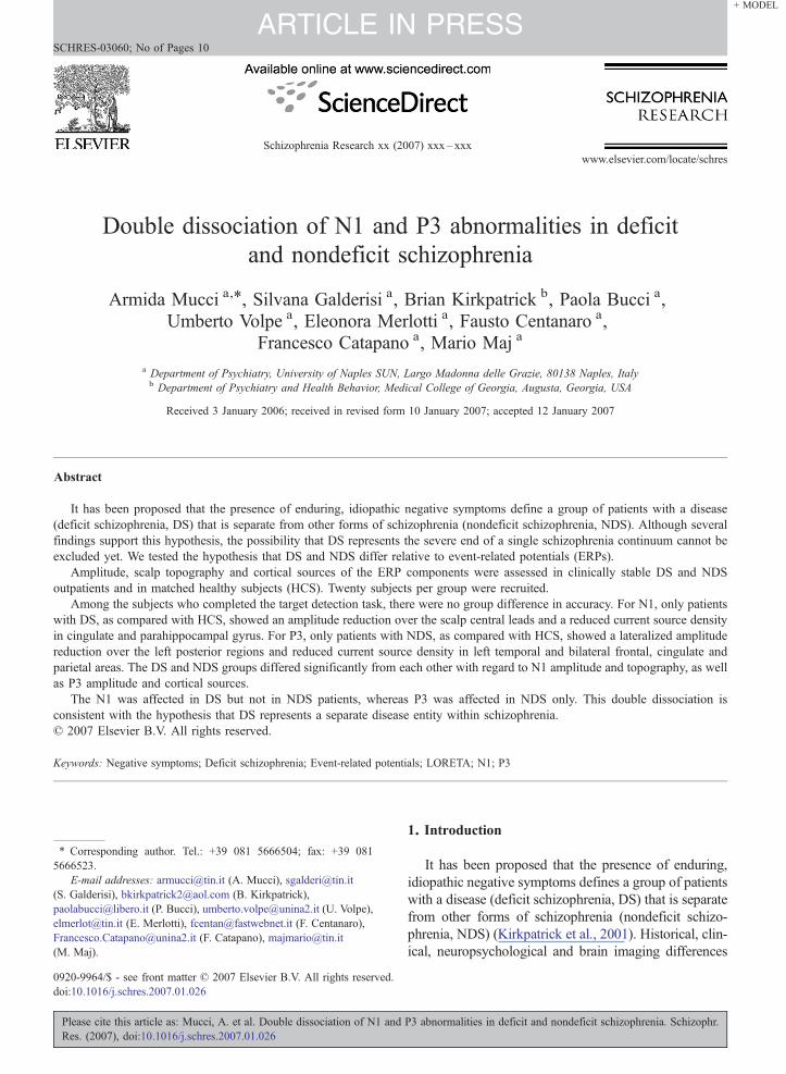
Fig. 1. Mean amplitude maps for N1 elicited by target tones for each group (lcolumn). For the amplitude maps the scale units are μV; light blue correspstatistical maps the units are independent samples t values; light blue indicatesfirst vs the second mentioned group above each map, after Holmes' correctiogroups are illustrated in the schematic insets (right column). NDS=NComparison Subjects. Significant topographic difference between patientsbetween deficit and nondeficit schizophrenia: #pb0.0002.
Please cite this article as: Mucci, A. et al. Double dissociation of N1 andRes. (2007), doi:10.1016/j.schres.2007.01.026
networks activation (Lehmann, 1987). The microstatemean amplitude was also measured at each lead.
The latency of each ERP component was measured atthe GFP peak of each microstate (Skrandies, 1993,1995).
2.3.1. Evaluation of cortical sources of the ERPcomponents by the Low-ResolutionBrainElectromagneticTomography (LORETA)
LORETA represents a minimum-norm estimate ofthe scalp field generators, which selects the smoothestsolution, i.e., the solution in which brain regions closetogether have similar values of source activity. Thissolution is considered more plausible based on theelectrophysiological rule that any scalp field, hence anymental process, is the product of the coordinated andsynchronous activation of distributed neuronal networks(Pascual-Marqui et al., 1994, 1999).
In the present study, the three-dimensional distribu-tion of cortical current density of the average microstatefor N1 and P3 were analyzed for each subject andcondition using LORETA in the version providing
eft column); statistical probability maps for group comparisons (middleonds to most negative and light red to most positive values. For thestatistically significant reduced and light red increased amplitude in then for multiple comparisons. Topographic characteristics in the differentondeficit Schizophrenia; DS=deficit Schizophrenia; HCS=Healthyand healthy subjects: ⁎pb0.002; Significant topographic difference
P3 abnormalities in deficit and nondeficit schizophrenia. Schizophr.

5A. Mucci et al. / Schizophrenia Research xx (2007) xxx–xxx
ARTICLE IN PRESS
current density values of 2394 voxels in the corticalareas, identified by the digitized Talairach atlas (Pascual-Marqui et al., 1999).
2.4. Statistical analyses
Data distributions were examined for normality andhomogeneity of variance. When these assumptions wereviolated, data were log transformed. Two-way ANO-VAs, with diagnosis as the between factor and stimulustype as the within factor, were carried out on the N1 andP3 latency measures and microstate parameters. Onlywhen a significant main effect or interaction of thediagnosis was found Bonferroni post-hoc tests werecarried out (DS vs NDS patients, and each patient groupvs HCS, for each stimulus type). The Greenhouse–Geisser correction for the inflation of degrees offreedom was used when appropriate.
Comparisons among groups on the N1 and P3 meanamplitude were carried out using the Statistical non-Parametric Mapping (SnPM), provided by LORETA, inwhich Holmes' correction for multiple comparisons isused (Holmes et al., 1996).
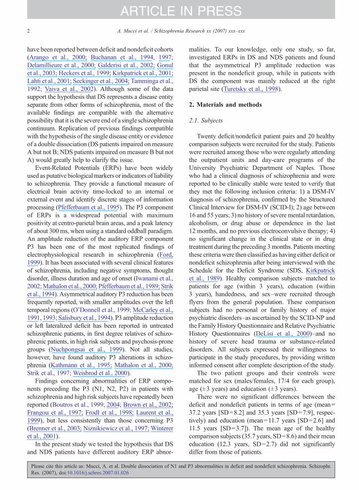
Fig. 2. Mean amplitude maps for P3 elicited by target tones for each group(middle column). For the amplitude maps the scale units are μV; light blue corstatistical maps the units are independent samples t values; light blue indicatesfirst vs the second mentioned group above each map, after Holmes' correctgroups is illustrated in the graphs (right column). NDS=Nondeficit Schizophr⁎Significant difference with respect to healthy subjects: pb0.003; -Significa
Please cite this article as: Mucci, A. et al. Double dissociation of N1 andRes. (2007), doi:10.1016/j.schres.2007.01.026
Comparisons among groups on LORETA currentsource density were conducted using voxel-by-voxelt-statistics, after logarithmic transformation of the data.SnPM with Holmes' correction for multiple comparisonswas applied.
Significance level for all statistical tests was set atp≤0.05 (two-tailed), after correction for multiplecomparisons, when deemed appropriate, as reportedabove.
3. Results
Five subjects with DS, two with NDS and no HCSwere excluded because they unable to perform the targetidentification task during ERP recording. Four HCS and3 NDS were excluded for the presence of artifacts intheir ERP recordings. The final sample for ERP analysisconsisted of 15 DS (13 males and 2 females), 15 NDSpatients (13 males and 2 females) and 16 HCS (13 malesand 3 females). These groups did not differ from theoriginally recruited samples for any relevant variable.
The two patient groups and their controls did notsignificantly differ for accuracy on the target detection
(left column) and statistical probability maps for group comparisonsresponds to most negative and light red to most positive values. For thestatistically significant reduced and light red increased amplitude in theion for multiple comparisons. The global field power in the differentenia; DS=Deficit Schizophrenia; HCS=Healthy Comparison Subjects.nt difference with respect to deficit schizophrenia: pb0.01.
P3 abnormalities in deficit and nondeficit schizophrenia. Schizophr.

6 A. Mucci et al. / Schizophrenia Research xx (2007) xxx–xxx
ARTICLE IN PRESS
task, as measured by the number of errors (mean±SD,1.3±2.6 for DS, 0.5±0.7 for NDS and 0.3±1.0 forHCS) and of omissions (mean±SD, 4.5±5.1 for DS,1.9±2.4 for NDS and 1.3±2.1 for HCS).
The latency of each ERP component did not differamong groups.
The mean amplitude of the N1 was significantly largerfor target than standard tones in both healthy controls andNDS patients, but not in DS patients. N1 amplitude fortarget stimuli was reduced in patients with DS, with
Table 2Stereotaxic coordinates and significance level (t scores) of regions showing
Region (Broadman area/s) N1 current source density forstandard stimuli DS vs HCSa
P3tar
x/y
Superior frontal gyrus (11, 6)Right 11Right 4/−Left
Middle frontal gyrus (6, 11, 10)Right 32Right 4/−Left −2
Medial frontal gyrus (6)Right 4/−Left
Precentral gyrus (6, 4)Left −3Left −4Right 53
Posterior cingulate (29, 31, 23, 24)Right 11Right 11/−45/5 −3.42 4/−Left −10/−46/6 −3.45 −1Left
Parahippocampal gyrus (30)Right 18/−53/1 −3.81Left −10/−46/1 −3.81
Postcentral gyrus (3, 1)Right 46Right 53
Inferior parietal lobule (40)Right 39Left −3
Supramarginal gyrus (40)Left −5
Superior temporal gyrus (22)Left −5
Middle temporal gyrus (21)Left −5Left −6
Inferior temporal gyrus (20)Left −5Left −4Left −5
DS=Patients with Deficit Schizophrenia; NDS=Patients with Nondeficit ScThreshold t for statistical significance after Holmes' correction for multiple
Please cite this article as: Mucci, A. et al. Double dissociation of N1 andRes. (2007), doi:10.1016/j.schres.2007.01.026
respect to both healthy subjects and patients with NDS(Fig. 1). For the N1 microstate, ANOVA showed asignificant interactionDiagnosis×Stimulus type (F=2.73,df=4,84, pb0.007) for the latero-lateral coordinate of thenegative centroid. Follow-up analyses indicated a right-ward shift of the centroid in DS patients, as compared toboth healthy controls and NDS patients (Bonferroni post-hoc pb0.02 and pb0.01, respectively) (Fig. 1).
There were no amplitude or topographic differencesamong groups for the P2 microstate.
group differences on LORETA current source density
current source density forget stimuli NDS vs HCSb
P3 current source density fortarget stimuli NDS vs DSc
/z t x/y/z t
/17/64 −2.8946/57 −2.87
−24/−11/71 −3.18
/3/64 −2.9811/64 −2.984/45/22 −2.84
11/57 −2.98−3/−32/64 −4.62
1/−11/57 −2.785/−18/50 −2.69/−4/50 −2.72
/−46/43 −2.8925/29 −3.64 11/−18/43 −4.930/−46/43 −2.95 −3/−18/43 3.96
−10/−18/43 −3.96
/−25/43 −2.38/−25/57 −2.38
/−39/43 −2.328/−46/43 −3.32
2/−53/36 −2.98
9/−11/1 −2.95
9/−4/−13 −2.756/−18/−13 −2.98
2/−18/−27 −2.815/−18/−27 −2.802/−32/−20 −2.98
hizophrenia; HCS=Healthy Comparison Subjects.comparisons: a3.04; b2.00; c3.12.
P3 abnormalities in deficit and nondeficit schizophrenia. Schizophr.

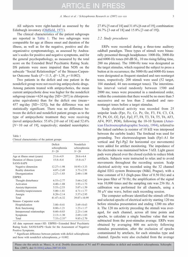
Fig. 3. Low-resolution electromagnetic tomography (LORETA) images of voxel-by-voxel independent sample t statistics for P3 elicited by targettones: group comparisons. Mid-sagittal slices are shown, structural anatomy is shown in gray scale (white-to-black). The scale shows negative (blue)and positive (red) t values for which the alpha is significant after Holmes' correction for multiple comparisons. The decrease of activity in the first vsthe second mentioned group on the left of each row is represented in blue.
7A. Mucci et al. / Schizophrenia Research xx (2007) xxx–xxx
ARTICLE IN PRESS
The mean amplitude of the P3 microstate for targetstimuli was reduced in patients with NDS, with respectto both HCS, over the left temporo-occipital leads, andpatients with DS over bilateral anterior regions (Fig. 2).
ANOVA showed a significant main effect of theDiagnosis (F=3.71; df=2,42; pb0.03) and a significantinteraction Diagnosis×Stimulus type for the GFP(F=4.0 4; df=4,84; pb0.005). Follow-up analysisindicated a significant reduction of the GFP for targetstimuli in patients with NDS, with respect to bothhealthy controls and DS patients (Bonferroni post-hocpb0.02 and 0.009, respectively) (Fig. 2).
As to LORETA findings, DS patients showedreduced N1 cortical current source density in posteriorcingulate and parahippocampal gyrus, while no currentsource density reduction for the N1 was observed forNDS patients (Table 2). In DS patients no alteration ofthe P3 brain sources was observed, with respect tohealthy subjects, while in NDS patients a significantreduction was found in the left temporal, right superiorfrontal, bilateral posterior cingulate, inferior parietal,
Please cite this article as: Mucci, A. et al. Double dissociation of N1 andRes. (2007), doi:10.1016/j.schres.2007.01.026
and supplementary motor areas (Fig. 3). Patients withNDS had reduced source activity also with respect topatients with DS, in bilateral cingulate, left superior andleft middle frontal areas (Fig. 3).
Table 2 presents the Talairach coordinates, Broadmanareas and t values for the regions showing significantgroup differences for N1 and P3 cortical sources.
4. Discussion
In our comparison of deficit, nondeficit, and controlsubjects, we found group differences in both the P3 andN1 components. For the P3 ERP component, onlypatients with NDS showed a reduced global field powerand a lateralized amplitude reduction over the leftposterior regions, with respect to healthy subjects.NDS also presented reduced P3 global field power andlower amplitude over bilateral anterior regions whencompared to DS patients. No difference between DSpatients and healthy controls was observed for the P3. Incontrast, patients with DS, in comparison to both healthy
P3 abnormalities in deficit and nondeficit schizophrenia. Schizophr.

8 A. Mucci et al. / Schizophrenia Research xx (2007) xxx–xxx
ARTICLE IN PRESS
subjects and NDS patients, showed a reduced amplitudeof N1 for target tones and topographic abnormalities ofthe same component for standard tones. They also failedto increase the amplitude of N1 for target stimuli. For thisERP component, no amplitude or topographic alterationwas observed in patients with NDS.
Our findings cannot be accounted for by differences indemographic characteristics, chronicity, clinical symp-tomatology (other than idiopathic, negative symptoms)and type or dose of antipsychotic treatment as the twopatient groups were comparable for all these variables.
According to our results, the lateralized amplitudereduction of P3, often reported in patients with chronicschizophrenia, is present in subjects with NDS only. P3is thought to be involved in the integration of theincoming information with its cognitive and emotionalcontext, indexing working memory functions andconsciously controlled aspects of information proces-sing (Barcelo et al., 2000; Donchin et al., 1986; Gevinsand Smith, 2000; Halgreen and Marinkovic, 1996). Ourfindings suggest that patients with NDS, who havepreserved early sensory and attentional processes, haveabnormalities of the late stages of information proces-sing concerning the updating of working memory andcontrolled aspects of attentional processes.
According to our results, the N1 for standard tonespresents an altered topography in DS patients, revealingdysfunction of auditory sensory processing in this patientgroup. The N1 provides indices of auditory sensoryprocessing, and is influenced by voluntary attention: itsamplitude is larger for target with respect to standardstimuli and, in divided-attention paradigms, for all stimulidelivered to the attended ear with respect to thosedelivered to the unattended ear, reflecting the allocationof perceptual resources (Hillyard, 1999; Mangun andHillyard, 1991). The failure to increase the N1 amplitudefor target stimuli also suggests that early attentionalprocesses are dysfunctional in DS patients. The N1amplitude reduction for target stimuli, in the absence ofchanges in scalp distribution and topographic indices,might suggest that the same neuronal network underliesN1 generation in the three subject groups, but is lessactivated in DS patients. A reduced phase-synchroniza-tion within the network might account for this finding.
LORETA imaging findings added further evidence ofa double dissociation of alterations in DS and NDSpatients. For the P3 component, a widespread reductionof the current source density in left temporal, rightsuperior frontal, bilateral posterior cingulate, inferiorparietal, and supplementary motor areas in NDSpatients, while no alteration of the P3 sources wasfound in DS patients. Significant differences were also
Please cite this article as: Mucci, A. et al. Double dissociation of N1 andRes. (2007), doi:10.1016/j.schres.2007.01.026
observed between the two patient groups, with NDSsubjects showing a reduced current source density inbilateral cingulate, left superior and left middle frontalareas, in comparison with DS patients. The brain areasshowing reduced activation in NDS patients areinvolved in context updating and working memory(Cairo et al., 2004; Pasternak and Greenlee, 2005; Yooet al., 2004).
For the N1 component, LORETA analysis demon-strated a reduced source activity in bilateral retrosplenialareas in DS patients, while no alteration of the N1 sourcewas observed in subjects with NDS. Brain areasshowing reduced current source density in DS patientsare involved in auditory discrimination, evaluation ofmotivational relevance of events, memory retrieval andemotionally relevant stimuli processing (Maddock,et al.,2003; Mesulam et al., 2001; Zatorre et al., 1999).
Our findings are generally consistent with severalprevious reports. Our findings are consistent with thoseof Turetsky et al. (1998), who found that an amplitudereduction of P3 over the left temporal regions waspresent only in the nondeficit group, and are inagreement with recent findings showing that patientswith pronounced negative symptoms do not present P3abnormalities (Meisenzahl et al., 2004). In a group ofpatients with chronic schizophrenia Kayser et al. (2001)reported a pattern of ERP abnormalities akin to thatobserved in our DS patients (normal P3 and reduced N1)and suggested that it reflects the patients' cognitiveeffort to compensate for early perceptual and attentionaldeficits. Our results suggest that inconsistent findings onERP abnormalities in schizophrenia might instead berelated to the heterogeneity of the syndrome. In line withour findings, another study found structural abnormal-ities of posterior cingulate in poor-outcome patients withschizophrenia and a negative correlation between thevolume of this structure and negative symptoms(Mitelman et al., 2005). Our results add to this evidencesuggesting that abnormalities of the posterior cingulatemight be related to primary, enduring negativesymptoms.
If the deficit subtype represented the severe end ofthe continuum, a more severe degree of the sameabnormalities should be observed. However, the patternof ERP abnormalities we observed in patients with DSand NDS is not compatible with that hypothesis. Inconclusion, we could demonstrate a double dissociationof ERP abnormalities in patients with deficit andnondeficit schizophrenia. If confirmed in larger samplesof patients, these findings might support the hypothesisthat deficit and nondeficit schizophrenia representseparate disease entities.
P3 abnormalities in deficit and nondeficit schizophrenia. Schizophr.
9A. Mucci et al. / Schizophrenia Research xx (2007) xxx–xxx
ARTICLE IN PRESS
Acknowledgments
Supported by grant 9806499118 from the ItalianMinistry of University and Scientific Research. Theauthors thank Drs. Dietrich Lehmann and RobertoPascual-Marqui for their thoughtful comments andassistance in the implementation of the advancedelectrophysiological data analysis.
References
American Electroencephalographic Society, 1994. Guideline thirteen:guidelines for standard electrode position nomenclature. J. Clin.Neurophysiol. 11, 111–113.
Arango, C., Kirkpatrick, B., Buchanan, R.W., 2000. Neurologicalsigns and the heterogeneity of schizophrenia. Am. J. Psychiatry157, 560–565.
Barcelo, F., Munoz-Cespedes, J.M., Pozo, M.A., Rubia, F.J., 2000.Attentional set shifting modulates the target P3b response in theWisconsin card sorting test. Neuropsychology 38, 1342–1355.
Boutros, N.N., Belger, A., Campbell, D., D'Souza, C., Krystal, J.,1999. Comparison of four components of sensory gating inschizophrenia and normal subjects: a preliminary report. Psychi-atry Res. 88, 119–130.
Boutros, N.N., Korzyukov, O., Jansen, B., Feingold, A., Bell, M.,2004. Sensory gating deficits during the mid-latency phase ofinformation processing in medicated schizophrenia patients.Psychiatry Res. 126, 203–215.
Brenner, C.A., Sporns, O., Lysaker, P.H., O'Donnell, B.F., 2003. EEGsynchronization to modulated auditory tones in schizophrenia,schizoaffective disorder, and schizotypal personality disorder. Am.J. Psychiatry 160, 2238–2240.
Brown, K.J., Gonsalvez, C.J., Harris, A.W., Williams, L.M., Gordon, E.,2002. Target and non-target ERP disturbances in first episode vs.chronic schizophrenia. Clin. Neurophysiol. 113, 1754–1763.
Buchanan, R.W., Strauss, M.E., Kirkpatrick, B., Holstein, C., Breier,A., Carpenter Jr., W.T., 1994. Neuropsychological impairments indeficit vs nondeficit forms of schizophrenia. Arch. Gen. Psychiatry51, 804–811.
Buchanan, R.W., Strauss, M.E., Breier, A., Kirkpatrick, B., CarpenterJr., W.T., 1997. Attentional impairments in deficit and nondeficitforms of schizophrenia. Am. J. Psychiatry 154, 363–370.
Cairo, T.A., Liddle, P.F., Woodward, T.S., Ngan, E.T., 2004. Theinfluence of working memory load on phase specific patterns ofcortical activity. Brain Res. Cogn. Brain Res. 21, 377–387.
Delamillieure, P., Fernandez, J., Constans, J.M., Brazo, P., Benali,K., Abadie, P., Vasse, T., Thibaut, F., Courtheoux, P., Petit, M.,Dollfus, S., 2000. Proton magnetic resonance spectroscopy of themedial prefrontal cortex in patients with deficit schizophrenia:preliminary report. Am. J. Psychiatry 157, 641–643.
DeLisi, L.E., Razi, K., Stewart, J., Relja, M., Shields, G., Smith, A.B.,Wellman, N., Larach, V.W., Loftus, J., Vita, A., Comazzi, M., Crow,T.J., 2000. No evidence for a parent-of-origin effect detected in thepattern of inheritance of schizophrenia. Biol. Psychiatry 48, 706–709.
Donchin, E., Karis, D., Bashore, T.R., Coles, M.G., Gratton, G., 1986.Cognitive psychophysiology and human information processing.In: Coles, M.G.H., Donchin, E., Porges, S.W. (Eds.), Psychophys-iology: Systems, Processes, and Applications. The Guilford Press,New York, pp. 244–267.
Please cite this article as: Mucci, A. et al. Double dissociation of N1 andRes. (2007), doi:10.1016/j.schres.2007.01.026
Ford, J.M., 1999. Schizophrenia: the broken P300 and beyond.Psychophysiology 36, 667–682.
Frangou, S., Sharma, T., Alarcon, G., Sigmudsson, T., Takei, N.,Binnie, C., Murray, R.M., 1997. The Maudsley Family Study, II:Endogenous event-related potentials in familial schizophrenia.Schizophr. Res. 23, 45–53.
Frodl, T., Meisenzahl, E.M., Gallinat, J., Hegerl, U., Moller, H.J.,1998. Markers from event-related potential subcomponents andreaction time for information processing dysfunction in schizo-phrenia. Eur. Arch. Psychiatry Clin. Neurosci. 248, 307–313.
Galderisi, S., Maj, M., Mucci, A., Cassano, G.B., Invernizzi, G., Rossi,A., Vita, A., Dell'Osso, L., Daneluzzo, E., Pini, S., 2002.Historical, psychopathological, neurological, and neuropsycho-logical aspects of deficit schizophrenia: a multicenter study. Am.J. Psychiatry 159, 983–990.
Gevins, A., Smith, M.E., 2000. Neurophysiological measures ofworking memory and individual differences in cognitive abilityand cognitive style. Cereb. Cortex 10, 829–839.
Gonul, A.S., Kula, M., Esel, E., Tutus, A., Sofuoglu, S.A., 2003. Tc-99m HMPAO SPECT study of regional cerebral blood flow indrug-free schizophrenic patients with deficit and non deficitsyndrome. Psychiatry Res. 123, 199–205.
Halgreen, E., Marinkovic, K., 1996. Neurophysiological networksintegrating human emotions. In: Gazzaniga, M.S. (Ed.), TheCognitive Neurosciences. Mit Press, Cambridge, pp. 217–244.
Heckers, S., Goff, D., Schacter, D.L., Savage, C.R., Fischman, A.J.,Alpert, N.M., Rauch, S.L., 1999. Functional imaging of memoryretrieval in deficit vs nondeficit schizophrenia. Arch. Gen.Psychiatry 58, 165–171.
Hillyard, S.A., 1999. Event-related potentials and human informationprocessing, In: Adelman, G., Smith, B.H. (Eds.), Encyclopedia ofNeuroscience, 2nd ed. Elsevier Science, Amsterdam, pp. 679–682.
Holmes, A.P., Blair, R.C., Watson, J.D., Ford, I., 1996. Non-parametric analysis of statistic images from functional mappingexperiments. J. Cereb. Blood Flow Metab. 16, 7–22.
Iwanami, A., Kato, N., Kasai, K., Kamio, S., Furukawa, S., Fukuda,M., Nakagome, K., Araki, T., Okajima, Y., Isono, H., Kamijima,K., 2002. P300 amplitude over temporal regions in schizophrenia.Eur. Arch. Psychiatry Clin. Neurosci. 252, 1–7.
Kathmann, N., Wagner, M., Rendtorff, N., Schochlin, C., Engel, R.R.,1995. Information processing during eye tracking as revealed byevent-related potentials in schizophrenics, alcoholics, and healthycontrols. Schizophr. Res. 16, 145–156.
Kayser, J., Bruder, G.E., Tenke, C.E., Stuart, B.K., Amador, X.F.,Gorman, J.M., 2001. Event related brain potentials (ERPs) inschizophrenia for tonal and phonetic oddball tasks. Biol.Psychiatry 49, 832–847.
Kirkpatrick, B., Buchanan, R.W., McKenny, P.D., Alphs, L.D.,Carpenter Jr., W.T., 1989. The Schedule for the Deficit Syndrome:an instrument for research in schizophrenia. Psychiatry Res. 30,119–124.
Kirkpatrick, B., Buchanan, R.W., Ross, D.E., Carpenter Jr., W.T.,2001. A separate disease within the syndrome of schizophrenia.Arch. Gen. Psychiatry 58, 165–171.
Koenig, T., Lehman, D., 1996. Microstates in languages-related brainpotential maps show noun–verb differences. Brain Lang. 53,169–182.
Lahti, A.C., Holcomb, H.H., Medoff, D.R., Weiler, M.A., Tamminga,C.A., Carpenter Jr., W.T., 2001. Abnormal patterns of regionalcerebral blood flow in schizophrenia with primary negativesymptoms during an effortful auditory recognition task. Am.J. Psychiatry 158, 1797–1808.
P3 abnormalities in deficit and nondeficit schizophrenia. Schizophr.
10 A. Mucci et al. / Schizophrenia Research xx (2007) xxx–xxx
ARTICLE IN PRESS
Laurent, A., Garcia-Larrea, L., d'Amato, T., Bosson, J.L., Saoud, M.,Marie-Cardine, M., Maugiere, F., Dalery, J., 1999. Auditory event-related potentials and clinical scores in unmedicated schizophrenicpatients. Psychiatry Res. 86, 229–238.
Lehmann, D., 1987. Principles of spatial analysis. In: Gevins, A.S.,Remond, A. (Eds.), Methods of Analysis of Brain Electrical andMagnetic Signals. Handbook of Electroencephalography andClinical Neurophysiology. Elsevier, Amsterdam, pp. 309–354.
Maddock, R.J., Buonocore, M.H., Kile, S.J., Garrett, A.S., 2003. Brainregions showing increased activation by threat-related words inpanic disorder. Neuroreport 14, 325–328.
Mangun, G.R., Hillyard, S.A., 1991. Modulations of sensory-evokedbrain potentials indicate changes in perceptual processing duringvisual–spatial priming. J. Exp. Psychol. Hum. Percept. Perform.17, 1057–1074.
Mathalon, D.H., Ford, J.M., Rosenbloom, M., Pfefferbaum, A., 2000.P300 reduction and prolongation with illness duration inschizophrenia. Biol. Psychiatry 47, 413–427.
McCarley, R.W., Faux, S.F., Shenton, M.E., Nestor, P.G., Adams, J.,1991. Event-related potentials in schizophrenia: their biologicaland clinical correlates and a new model of schizophrenicpathophysiology. Schizophr. Res. 4, 209–231.
McCarley, R.W., Shenton, M.E., O'Donnel, B.F., Faux, S.F., Kikinis,R., Nestor, P.G., Jolesz, F.A., 1993. Auditory P300 abnormalitiesand left posterior superior temporal gyrus volume reduction inschizophrenia. Arch. Gen. Psychiatry 50, 190–197.
Meisenzahl, E.M., Frodl, T., Muller, D., Schimitt, G., Gallinat, J.,Zetzsche, T., Marcuse, A., Juchkel, G., Leinsinger, G., Hahn, K.,Moller, H.J., Hegerl, U., 2004. Superior temporal gyrus and P300in schizophrenia: a combined ERP/structural magnetic resonanceimaging investigation. J. Psychiatr. Res. 38, 153–162.
Mesulam, M.M., Nobre, A.C., Kim, Y.H., Parrish, T.B., Gitelman,D.R., 2001. Heterogeneity of cingulated contributions to spatialattention. NeuroImage 13, 1065–1072.
Mitelman, S.A., Shihabuddin, L., Brickman, A.M., Hazlett, E.A.,Buchsbaum, M.S., 2005. Volume of the cingulate and outcome inschizophrenia. Schizophr. Res. 72, 91–108.
Niznikiewicz, M.A., O'Donnell, B.F., Nestor, P.G., Smith, L., Law, S.,Karapelou, M., Shenton, M.E., McCarley, R.W., 1997. ERPassessment of visual and auditory language processing inschizophrenia. J. Abnorm. Psychology 106, 85–94.
Nuchpongsai, P., Arakaki, H., Langman, P., Ogura, C., 1999. N2 andP3b components of the event-related potential in students at riskfor psychosis. Psychiatry Res. 88, 131–141.
O'Donnell, B.F., McCarley, R.W., Potts, G.F., Salisbury, D.F., Nestor,P.G., Hirayasu, Y., Niznikiewicz, M.A., Barnard, J., Shen, Z.J.,Weinstein, D.M., Bookstein, F.L., Shenton, M.E., 1999. Identifi-cation of neural circuits underlying P300 abnormalities inschizophrenia. Psychophysiology 36, 388–398.
Oldfield, R.C., 1971. The assessment and analysis of handedness: theEdinburgh inventory. Neuropsychology 9, 97–113.
Pascual-Marqui, R.D., Michel, C.M., Lehmann, D., 1994. Lowresolution electromagnetic tomography: a newmethod for localizingelectrical activity in the brain. Int. J. Psychophysiol. 18, 49–65.
Pascual-Marqui, R.D., Lehmann,D., Koenig, T., Kochi, K.,Merlo,M.C.,Hell, D., Koukkou, M., 1999. Low resolution brain electromagnetictomography (LORETA) functional imaging in acute, neuroleptic-
Please cite this article as: Mucci, A. et al. Double dissociation of N1 andRes. (2007), doi:10.1016/j.schres.2007.01.026
naive, first-episode, productive schizophrenia. Psychiatry Res. 90,169–179.
Pasternak, T., Greenlee, M.W., 2005. Working memory in primatesensory systems. Nat. Rev., Neurosci. 6, 97–107.
Pfefferbaum, A., Ford, J.M., White, P.M., Roth, W.T., 1989. P3 inschizophrenia is affected by stimulus modality, response require-ments, medication status, and negative symptoms. Arch. Gen.Psychiatry 46, 1035–1044.
Pfefferbaum, A., Roth, W.T., Ford, J.M., 1995. Event-relatedpotentials in the study of psychiatric disorders. Arch. Gen.Psychiatry 52, 559–563.
Salisbury, D.F., O'Donnell, B.F., McCarley, R.W., Nestor, P.G., Faux,S.F., Smith, R.S., 1994. Parametric manipulations of auditorystimuli differentially affect P3 amplitude in schizophrenics andcontrols. Psychophysiology 31, 29–36.
Seckinger, R.A., Goudsmit, N., Coleman, E., Harkavy-Friedman, J.,Yale, S., Rosenfield, P.J., Malaspina, D., 2004. Olfactoryidentification on WAIS-R performance in deficit and nondeficitschizophrenia. Schizophr. Res. 69, 55–65.
Skrandies, W., 1993. EEG/EP: new techniques. Brain Topogr. 5,347–350.
Skrandies, W., 1995. Visual information processing: topography ofbrain electrical activity. Biol. Psychol. 40, 1–15.
Strik, W.K., Dierks, T., Franzek, E., Stober, G., Maurer, K., 1994. P300asymmetries in schizophrenia revisited with reference-independentmethods. Psychiatry Res. 55, 153–166.
Strik, W.K., Fallgatter, A.J., Stoeber, G., Franzek, E., Beckmann, H.,1997. Specific P300 features in patients with cycloid psychosis.Acta Psychiatr. Scand. 95, 67–72.
Tamminga, C.A., Tharker, G.K., Buchanan, R., Kirkpatrik, B.,Alphs, L.D., Chase, T.N., Carpenter, W.T., 1992. Limbic systemabnormalities identified in schizophrenia using positron emis-sion tomography with fluorodeosyglucose and neocorticalalterations with deficit syndrome. Arch. Gen. Psychiatry 49,522–530.
Turetsky, B., Colbalth, E.A., Gur, R.E., 1998. P300 subcomponentabnormalities in schizophrenia: I Physiological evidence forgender and subtype specific differences in regional pathology.Biol. Psychiatry 43, 84–96.
Vaiva, G., Cottencin, O., Llorca, P.M., Devos, P., Dupont, S., Mazas,O., Rascle, C., Thomas, P., Steinling, M., Goudemand, M., 2002.Regional cerebral blood flow in deficit/nondeficit types ofschizophrenia according to SDS criteria. Prog. Neuro-Psycho-pharmacol. Biol. Psychiatry 26, 481–485.
Weisbrod, M., Kiefer, M., Marzinzik, F., Spitzer, M., 2000. Executivecontrol is disturbed in schizophrenia: evidence from event-relatedpotentials in a Go/NoGo task. Biol. Psychiatry 47, 51–60.
Winterer, G., Egan, M.F., Radler, T., Coppola, R., Weinberger, D.R.,2001. Event-related potentials and genetic risk for schizophrenia.Biol. Psychiatry 50, 407–417.
Yoo, S.S., Paralkar, G., Panych, L.P., 2004. Neural substratesassociated with the concurrent performance of dual workingmemory tasks. Int. J. Neurosci. 114, 613–631.
Zatorre, R.J., Mondor, T.A., Evans, A.C., 1999. Auditory attention tospace and frequency activates similar cerebral systems. Neuro-Image 10, 544–554.
P3 abnormalities in deficit and nondeficit schizophrenia. Schizophr.
Related Documents