HAL Id: hal-03086787 https://hal.archives-ouvertes.fr/hal-03086787 Submitted on 11 Oct 2021 HAL is a multi-disciplinary open access archive for the deposit and dissemination of sci- entific research documents, whether they are pub- lished or not. The documents may come from teaching and research institutions in France or abroad, or from public or private research centers. L’archive ouverte pluridisciplinaire HAL, est destinée au dépôt et à la diffusion de documents scientifiques de niveau recherche, publiés ou non, émanant des établissements d’enseignement et de recherche français ou étrangers, des laboratoires publics ou privés. Dosimetric characterisation and application to radiation biology of a kHz laser-driven electron beam Marco Cavallone, Lucas Rovige, Julius Huijts, Émilie Bayart, Rachel Delorme, Aline Vernier, Patrik Gonçalves Jorge, Raphaël Moeckli, Eric Deutsch, Jérôme Faure, et al. To cite this version: Marco Cavallone, Lucas Rovige, Julius Huijts, Émilie Bayart, Rachel Delorme, et al.. Dosimetric char- acterisation and application to radiation biology of a kHz laser-driven electron beam. Appl.Phys.B, 2021, 127 (4), pp.57. 10.1007/s00340-021-07610-z. hal-03086787

Welcome message from author
This document is posted to help you gain knowledge. Please leave a comment to let me know what you think about it! Share it to your friends and learn new things together.
Transcript
HAL Id: hal-03086787https://hal.archives-ouvertes.fr/hal-03086787
Submitted on 11 Oct 2021
HAL is a multi-disciplinary open accessarchive for the deposit and dissemination of sci-entific research documents, whether they are pub-lished or not. The documents may come fromteaching and research institutions in France orabroad, or from public or private research centers.
L’archive ouverte pluridisciplinaire HAL, estdestinée au dépôt et à la diffusion de documentsscientifiques de niveau recherche, publiés ou non,émanant des établissements d’enseignement et derecherche français ou étrangers, des laboratoirespublics ou privés.
Dosimetric characterisation and application to radiationbiology of a kHz laser-driven electron beam
Marco Cavallone, Lucas Rovige, Julius Huijts, Émilie Bayart, Rachel Delorme,Aline Vernier, Patrik Gonçalves Jorge, Raphaël Moeckli, Eric Deutsch, Jérôme
Faure, et al.
To cite this version:Marco Cavallone, Lucas Rovige, Julius Huijts, Émilie Bayart, Rachel Delorme, et al.. Dosimetric char-acterisation and application to radiation biology of a kHz laser-driven electron beam. Appl.Phys.B,2021, 127 (4), pp.57. �10.1007/s00340-021-07610-z�. �hal-03086787�
myjournal manuscript No.(will be inserted by the editor)
Dosimetric characterisation and application to radiation biology of akHz laser-driven electron beam
Marco Cavallone1, Lucas Rovige1, Julius Huijts1, Emilie Bayart1,2, Rachel Delorme4, Aline Vernier1,Patrik Goncalves Jorge5, Raphael Moeckli5, Eric Deutsch3, Jerome Faure1, Alessandro Flacco1?
1 Laboratoire d’Optique Appliquee, ENSTA Paris, Ecole Polytechnique, CNRS-UMR7639, Institut Polytechnique de Paris,91762 Palaiseau cedex, France
2 SFRO - RadioTransNet, Centre Antoine Beclere, 47 rue de la Colonie, 75013 Paris3 INSERM 1030, Univ Paris-Sud, Univ Paris-Saclay, Department of Radiation Oncology, Gustave Roussy Cancer Campus,
Villejuif, France4 Univ. Grenoble Alpes, CNRS, Grenoble INP, LPSC-IN2P3, 38000 Grenoble, France5 Institute of Radiation Physics, Lausanne University Hospital, Lausanne, Switzerland
Received: date / Revised version: date
Abstract Laser-plasma accelerators can produce ultrashort electron bunches in the femtosecond to picosecondduration range, resulting in high peak dose rates in com-parison with clinical accelerators. This peculiar charac-teristic motivates their application to radiation biologystudies to elucidate the effect of the high peak dose rateon the biological response of living cells, which is stillbeing debated. Electron beams driven by kHz laser sys-tems may represent an attractive option for such applica-tions, since the high repetition rate can boost the meandose rate and improve the stability of the delivered dosein comparison with J-class laser accelerators running at10 Hz. In this work, we present the dosimetric character-isation of a kHz, low energy laser-driven electron sourceand preliminary results on in-vitro irradiation of cancercells. A shot-to-shot dosimetry protocol enabled to mon-itor the beam stability and the irradiation conditions foreach cell sample. Results of survival assays on HCT116colorectal cancer cells are in good agreement with pre-vious findings reported in literature and validate the ro-bustness of the dosimetry and irradiation protocol.
1 Introduction
Laser-plasma accelerators can produce ultra short elec-tron bunches with duration in the range from fem-tosecond to picosecond. Both quasi-monoenergetic high-energy electrons (hundreds of MeV) [1,2,3] and high-charge, low-energy (few MeV) electrons with broadbandspectrum [4] have been obtained with 100 TW class
Send offprint requests to:? Corresponding author: [email protected]
lasers. The peculiarity of these sources is the ultra-high peak dose rate in the pulse (up to 1010 Gy/s)that is orders of magnitude higher than conventionalLinac (∼ 102 Gy/s) [5]. The impact of electrons deliv-ered in such short pulses of ultra-high dose rates onliving cells or tissues still need to be extensively ex-plored. Recent results obtained with FLASH-RT [6,7,8] and laser-accelerated protons [9,10] indicate that thetemporal structure of high dose rate, pulsed irradiationmay have an impact on the radiobiological response andask for a deeper understanding of the phenomena trig-gered by high dose rate irradiation.To date, Laser-Accelerated Electrons (LAEs) producedby commercially available J-class lasers running at 10 Hzhave been used for radiation biology studies [11,12,13].The main limit of these beams is the shot-to-shot point-ing fluctuation in the order of the beam divergence, thathinders the reach of the stability standards required inclinics. In this context, low-energy electrons (few MeV)driven by kHz lasers may represent an interesting alter-native for radiobiology applications. Such laser-plasmaaccelerators have been developed in recent years [14,15,16,17] and, to our knowledge, no radiobiology study inthese conditions is reported in literature at the time ofwriting. The key asset of these beams is the high rep-etition rate, which boosts the mean beam current andallows averaging of the shot-to-shot fluctuations by in-tegration of a large number of shots. From a dosimetricpoint of view, this translates into higher mean dose rates(Gy/s), compared to those obtained with J-class lasers(Gy/min), and into a higher stability of the dose distri-bution at the sample.In this article we present the dosimetric characterisa-tion of a low-energy kHz laser-driven electron beam forradiation biology applications and report preliminary re-
arX
iv:2
012.
0040
8v1
[ph
ysic
s.ac
c-ph
] 1
Dec
202
0
2 Marco Cavallone et al.
SSD
(Vacuum) (Air)
Electron beam
Mylarwindow
EBT3 film Cells RazorIC
Gasjet
Plas8cholder(800μm)
Laser
Motorizedbeamimagingandspectrometer
SSD=40cm
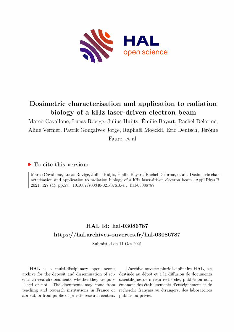
Fig. 1 Schematic draft (horizontal cut, top view) of the ex-perimental set-up.
sults on in-vitro irradiation of HCT116 colorectal cancercells. The dose distribution at the irradiated sample wasmeasured at each irradiation with absolutely calibratedradiochromic films, which have been shown to be dose-rate independent over a wide range of dose rate up to1012 Gy/s [18,19,20]. The use of an IBA Razor NanoChamber provided a real time dose monitoring duringirradiations and evaluation of the dose uncertainty inthe post-processing analysis.
2 Electron source and set-up
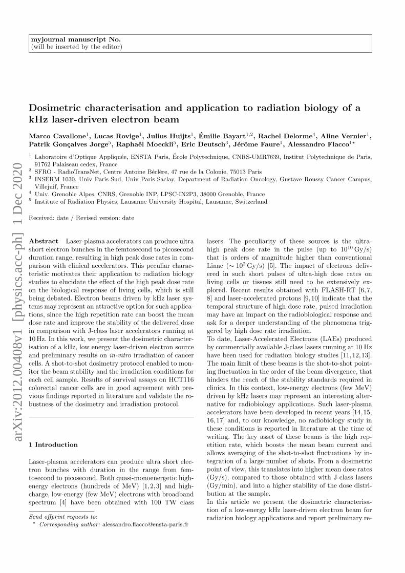
The experiment was performed with the Salle Noirelaser system of the Laboratoire d’Optique Appliquee, de-livering 3.5 fs pulses at 1 kHz repetition rate with 3 mJon target. The laser can generate low energy electronsin a reproducible way in a range from sub-MeV up tofew MeV with charges of ∼10 pC and few pC respec-tively, depending on the gas density and profile [21].In the experiment, the laser pulse was focused with aparabola (50 mm focal length) onto an N2 gas-jet. Thegas-jet density was optimised so as to maximise the ac-celerated charge per bunch, which corresponds to theproduction of an electron beam featuring a low-energyquasi-thermal spectrum up to ∼2 MeV and a divergenceof 70 mrad. Fig. 2 shows the electron spectrum taken infour different days. As shown, the spectral shape can bereproduced from one day to another with good preci-sion. The measured charge (total charge at the source)was 9.8 pC/shot with an instability of ±0.7 pC/shot(standard deviation).A draft of the experimental setup is shown in Fig. 1.A tube was inserted in the vacuum chamber to placethe irradiation site (in air) closer to the electron source,compatibly with the beam diagnostics. A 100 µm thickvacuum-air Mylar window was placed 38 cm far from thesource. At this location, the transverse dimension of thebeam was 3 cm (FWHM), which allowed to irradiate acircular spot of 1 cm diameter with a sufficiently homo-geneous dose. The cell sample was positioned verticallyin air, 2 cm after the Mylar window, with a 800 µm thickplastic holder aligned to the beam-axis. A customisedmetallic support allowed to hold in place the cell holderand to place it at the same depth in the tube at each
0 0.5 1 1.5 2 2.5 3Energy (MeV)
0
0.2
0.4
0.6
0.8
1
Rel
ativ
e co
unts
(a. u
.)
Fig. 2 Spectrum of the electron beam taken in four differentdays, obtained by integrating 15 shots. The lower limit of thespectrometer is 400 keV.
irradiation. The support was also designed to house anIBA Razor Nano Chamber (RNC), which was insertedbehind the cell sample. The chamber (2 mm outer elec-trode diameter, 0.003 cm2 active volume) was used as on-line dose monitor during irradiations and as day-to-daydose reference during the optimisation of the interactionconditions between the laser and the gas-jet prior to anexperimental run. A radiochromic EBT3 film was placedon the front side (in beam’s view) of the cell holder ateach irradiation for dosimetry. A motorised set-up, lo-cated in the vacuum chamber between the source andthe Mylar window, was used for beam imaging and spec-troscopy and was removed during irradiations. It con-tains a removable dipole (0.058 T) and a phosphor screencoupled with a CCD camera. Imaging of the beam trans-verse section, performed by removing the dipole magnetfrom the beam axis, allowed to align the electron beamto the cell sample and to measure the total acceleratedcharge per bunch. The details of these diagnostics canbe found in D. Gustas (2019) [21].
3 Dosimetry
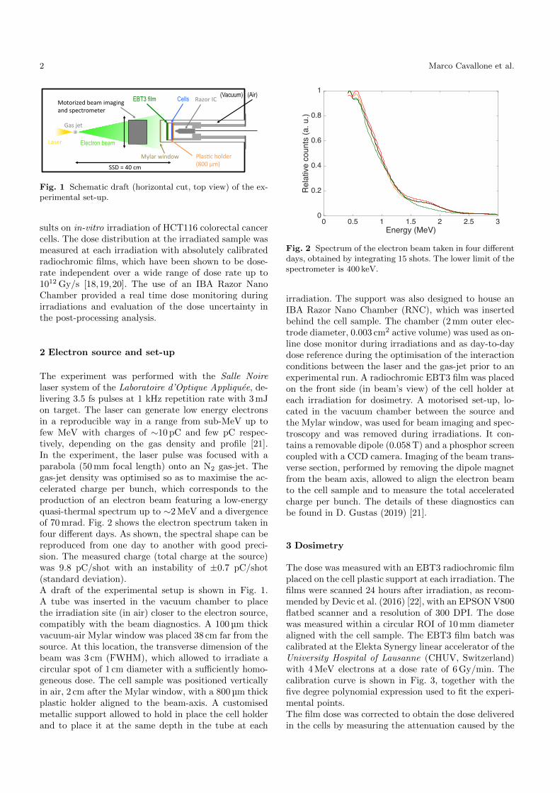
The dose was measured with an EBT3 radiochromic filmplaced on the cell plastic support at each irradiation. Thefilms were scanned 24 hours after irradiation, as recom-mended by Devic et al. (2016) [22], with an EPSON V800flatbed scanner and a resolution of 300 DPI. The dosewas measured within a circular ROI of 10 mm diameteraligned with the cell sample. The EBT3 film batch wascalibrated at the Elekta Synergy linear accelerator of theUniversity Hospital of Lausanne (CHUV, Switzerland)with 4 MeV electrons at a dose rate of 6 Gy/min. Thecalibration curve is shown in Fig. 3, together with thefive degree polynomial expression used to fit the experi-mental points.The film dose was corrected to obtain the dose deliveredin the cells by measuring the attenuation caused by the
Dosimetric characterisation and application to radiation biology of a kHz laser-driven electron beam 3
0 0.1 0.2 0.3 0.4OD
0
2
4
6
8
10
12
Dose
(Gy)
D = -595.48 ⋅OD5+ 638.37 ⋅OD4- 162.34 ⋅OD3+ 38.438 ⋅OD2 + 6.081⋅ODR2 =1
Fig. 3 Calibration of the EBT3 film batch (red channel)with the 4 MeV electron beam generated by the SynergyElekta LINAC available at the University Hospital of Lau-sanne (CHUV, Switzerland). The experimental values (bluepoints) are fitted with a five degree polynomial (black curve),whose expression is reported in the image.
800 µm cell plastic holder and the air gap. Precisely, thedose delivered to the cell is related to the dose absorbedby the film according to:
Dcells = DEBT3 ·Rcells/EBT3 (1)
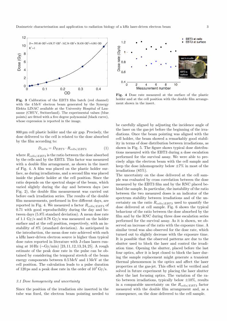
where Rcells/EBT3 is the ratio between the dose absorbedby the cells and by the EBT3. This factor was measuredwith a double film arrangement, as shown in the insertof Fig. 4. A film was placed on the plastic holder sur-face, as during irradiations, and a second film was placedinside the plastic holder at the cell position. Since theratio depends on the spectral shape of the beam, whichvaried slightly during the day and between days (seeFig. 2), the double film measurement was carried outbefore each irradiation series. The results of the doublefilm measurements, performed in five different days, arereported in Fig. 4. We measured a factor Rcells/EBT3 of0.71 with good reproducibility during the day and be-tween days (5.8% standard deviation). A mean dose rateof 1.1 Gy/s and 0.78 Gy/s was measured on the holdersurface and at the cell position, respectively, with an in-stability of 8% (standard deviation). As anticipated inthe introduction, the mean dose rate achieved with sucha kHz laser-driven electron source is higher than typicaldose rates reported in literature with J-class lasers run-ning at 10 Hz (∼Gy/min) [23,11,12,13,24,25]. A roughestimate of the peak dose rate in the pulse can be ob-tained by considering the temporal stretch of the beamenergy components between 0.5 MeV and 1 MeV at thecell position. The calculation leads to a pulse durationof 120 ps and a peak dose rate in the order of 107 Gy/s.
3.1 Dose homogeneity and uncertainty
Since the position of the irradiation site inserted in thetube was fixed, the electron beam pointing needed to
Fig. 4 Dose rate measured at the surface of the plasticholder and at the cell position with the double film arrange-ment shown in the insert.
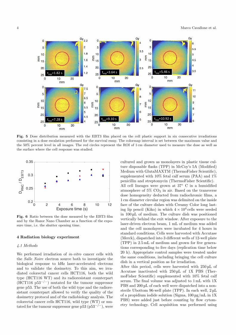
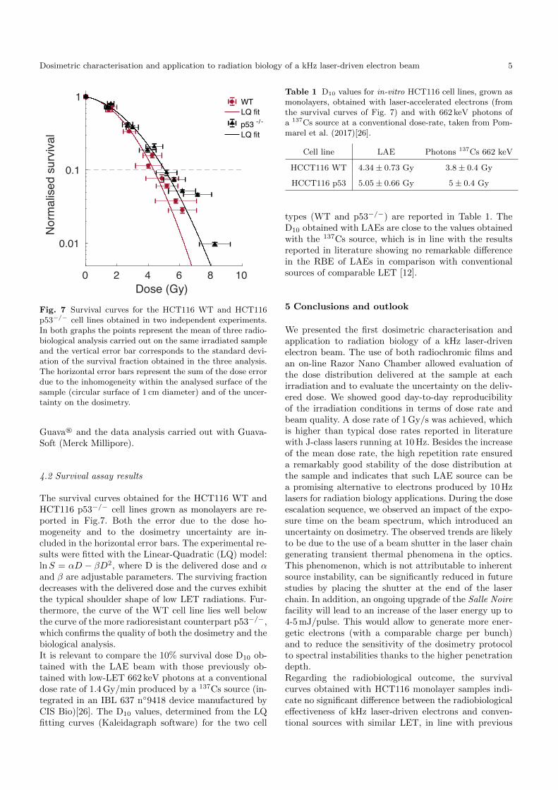
be carefully aligned by adjusting the incidence angle ofthe laser on the gas-jet before the beginning of the irra-diations. Once the beam pointing was aligned with thecell holder, the beam showed a remarkably good stabil-ity in terms of dose distribution between irradiations, asshown in Fig. 5. The figure shows typical dose distribu-tions measured with the EBT3 during a dose escalationperformed for the survival assay. We were able to pre-cisely align the electron beam with the cell sample andkeep the dose inhomogeneity below ±7% in most of theirradiations (85%).The uncertainty on the dose delivered at the cell sam-ple was evaluated by cross correlation between the dosemeasured by the EBT3 film and by the RNC placed be-hind the sample. In particular, the instability of the ratiobetween the two measured doses is an indicator of thespectrum stability between irradiations and of the un-certainty on the ratio Rcells/EBT3 used to quantify thedose delivered at cell sample. Fig. 6 shows the typicalbehaviour of the ratio between the dose absorbed by thefilm and by the RNC during three dose escalation seriesperformed for the survival assay. As it is shown, we ob-served an increase of the ratio with the exposure time. Asimilar trend was also observed for the dose rate, whichturned out to slightly decrease with the exposure time.It is possible that the observed patterns are due to theshutter used to block the laser and control the irradi-ation time. Opening the shutter, placed before the lastfour optics, after it is kept closed to block the laser dur-ing the sample replacement might generate a transientthermal phenomenon in the optics and affect the laserproperties at the gas-jet. This effect will be verified andsolved in future experiment by placing the laser shutterafter the last focusing optics. The variation of the ra-tio between irradiations, typically below ±10%, resultsin a comparable uncertainty on the Rcells/EBT3 factormeasured with the double film arrangement and, as aconsequence, on the dose delivered to the cell sample.
4 Marco Cavallone et al.
texp=1.82s texp=5.46stexp=3.64s
texp=7.28s texp=9.10s texp=10.92s
Fig. 5 Dose distribution measured with the EBT3 film placed on the cell plastic support in six consecutive irradiationsconsisting in a dose escalation performed for the survival essay. The colormap interval is set between the maximum value andthe 50% percent level in all images. The red circles represent the ROI of 1 cm diameter used to measure the dose as well asthe surface where the cell response was studied.
0 2 4 6 8 10 12Exposure time (s)
0.2
0.25
0.3
0.35
DR
NC
/ D
EB
T3
Fig. 6 Ratio between the dose meaured by the EBT3 filmand by the Razor Nano Chamber as a function of the expo-sure time, i.e. the shutter opening time.
4 Radiation biology experiment
4.1 Methods
We performed irradiation of in-vitro cancer cells withthe Salle Noire electron source both to investigate thebiological response to kHz laser-accelerated electronsand to validate the dosimetry. To this aim, we irra-diated colorectal cancer cells HCT116, both the wildtype (HCT116 WT) and its radioresistant counterpart(HCT116 p53−/−) mutated for the tumour suppressorgene p53. The use of both the wild type and the radiore-sistant counterpart allowed to verify the quality of thedosimetry protocol and of the radiobiology analysis. Thecolorectal cancer cells HCT116, wild type (WT) or mu-tated for the tumour suppressor gene p53 (p53−/−), were
cultured and grown as monolayers in plastic tissue cul-ture disposable flasks (TPP) in McCoy’s 5A (Modified)Medium with GlutaMAXTM (ThermoFisher Scientific),supplemented with 10% fetal calf serum (PAA) and 1%penicillin and streptomycin (ThermoFisher Scientific).All cell lineages were grown at 37◦ C in a humidifiedatmosphere of 5% CO2 in air. Based on the transversedose homogeneity deducted from radiochromic films, a1 cm diameter circular region was delimited on the insideface of the culture dishes with Creamy Color long last-ing lip pencil (Kiko) in which 4 × 10⁴ cells were seededin 100 µL of medium. The culture dish was positionedvertically behind the exit window. After exposure to thelaser-driven electron beam, 1 mL of medium was addedand the cell monolayers were incubated for 4 hours instandard conditions. Cells were harvested with Accutase(Merck), dispatched into 3 different wells of 12-well plate(TPP) in 2.5 mL of medium and grown for five genera-tions corresponding to five days (replication time below20 h). Appropriate control samples were treated underthe same conditions, including bringing the cell culturedish in a vertical position as for irradiation.After this period, cells were harvested with 250 µL ofAccutase inactivated with 250 µL of 1X PBS (Ther-moFisher Scientific) supplemented with 10% fetal calfserum. The final volume was adjusted to 1 mL with 1XPBS and 200 µL of each well were dispatched into a non-sterile Ubottom 96-well plate (TPP). In each well, 2 µLof a propidium iodide solution (Sigma, 100 µg/mL in 1XPBS) were added just before counting by flow cytom-etry technology. Cell acquisition was performed using
Dosimetric characterisation and application to radiation biology of a kHz laser-driven electron beam 5
0 2 4 6 8 10Dose (Gy)
0.01
0.1
1
Nor
mal
ised
sur
viva
l
WTLQ fitp53 -/ -
LQ fit
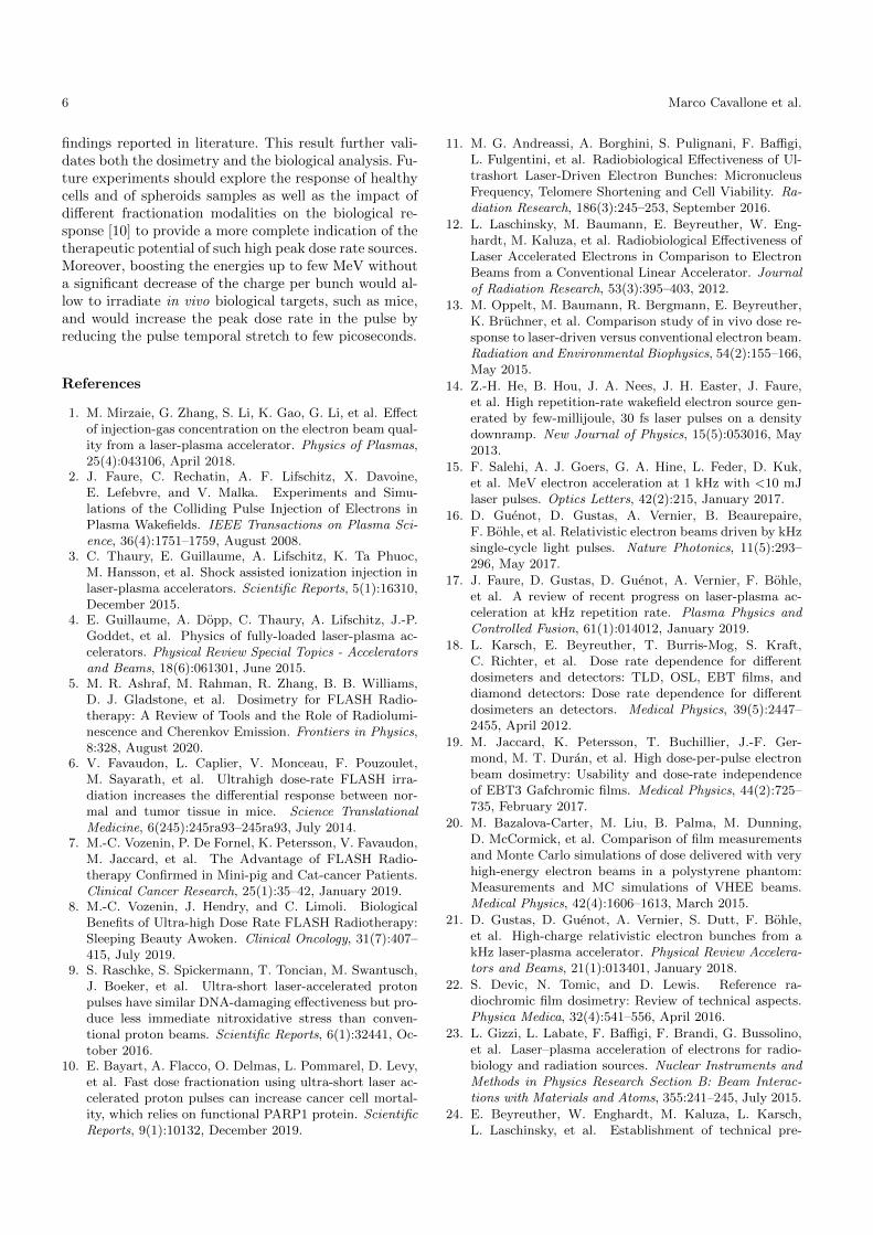
Fig. 7 Survival curves for the HCT116 WT and HCT116p53−/− cell lines obtained in two independent experiments.In both graphs the points represent the mean of three radio-biological analysis carried out on the same irradiated sampleand the vertical error bar corresponds to the standard devi-ation of the survival fraction obtained in the three analysis.The horizontal error bars represent the sum of the dose errordue to the inhomogeneity within the analysed surface of thesample (circular surface of 1 cm diameter) and of the uncer-tainty on the dosimetry.
Guava® and the data analysis carried out with Guava-Soft (Merck Millipore).
4.2 Survival assay results
The survival curves obtained for the HCT116 WT andHCT116 p53−/− cell lines grown as monolayers are re-ported in Fig.7. Both the error due to the dose ho-mogeneity and to the dosimetry uncertainty are in-cluded in the horizontal error bars. The experimental re-sults were fitted with the Linear-Quadratic (LQ) model:lnS = αD − βD2, where D is the delivered dose and αand β are adjustable parameters. The surviving fractiondecreases with the delivered dose and the curves exhibitthe typical shoulder shape of low LET radiations. Fur-thermore, the curve of the WT cell line lies well belowthe curve of the more radioresistant counterpart p53−/−,which confirms the quality of both the dosimetry and thebiological analysis.It is relevant to compare the 10% survival dose D10 ob-tained with the LAE beam with those previously ob-tained with low-LET 662 keV photons at a conventionaldose rate of 1.4 Gy/min produced by a 137Cs source (in-tegrated in an IBL 637 n◦9418 device manufactured byCIS Bio)[26]. The D10 values, determined from the LQfitting curves (Kaleidagraph software) for the two cell
Table 1 D10 values for in-vitro HCT116 cell lines, grown asmonolayers, obtained with laser-accelerated electrons (fromthe survival curves of Fig. 7) and with 662 keV photons ofa 137Cs source at a conventional dose-rate, taken from Pom-marel et al. (2017)[26].
Cell line LAE Photons 137Cs 662 keV
HCCT116 WT 4.34 ± 0.73 Gy 3.8 ± 0.4 Gy
HCCT116 p53 5.05 ± 0.66 Gy 5 ± 0.4 Gy
types (WT and p53−/−) are reported in Table 1. TheD10 obtained with LAEs are close to the values obtainedwith the 137Cs source, which is in line with the resultsreported in literature showing no remarkable differencein the RBE of LAEs in comparison with conventionalsources of comparable LET [12].
5 Conclusions and outlook
We presented the first dosimetric characterisation andapplication to radiation biology of a kHz laser-drivenelectron beam. The use of both radiochromic films andan on-line Razor Nano Chamber allowed evaluation ofthe dose distribution delivered at the sample at eachirradiation and to evaluate the uncertainty on the deliv-ered dose. We showed good day-to-day reproducibilityof the irradiation conditions in terms of dose rate andbeam quality. A dose rate of 1 Gy/s was achieved, whichis higher than typical dose rates reported in literaturewith J-class lasers running at 10 Hz. Besides the increaseof the mean dose rate, the high repetition rate ensureda remarkably good stability of the dose distribution atthe sample and indicates that such LAE source can bea promising alternative to electrons produced by 10 Hzlasers for radiation biology applications. During the doseescalation sequence, we observed an impact of the expo-sure time on the beam spectrum, which introduced anuncertainty on dosimetry. The observed trends are likelyto be due to the use of a beam shutter in the laser chaingenerating transient thermal phenomena in the optics.This phenomenon, which is not attributable to inherentsource instability, can be significantly reduced in futurestudies by placing the shutter at the end of the laserchain. In addition, an ongoing upgrade of the Salle Noirefacility will lead to an increase of the laser energy up to4-5 mJ/pulse. This would allow to generate more ener-getic electrons (with a comparable charge per bunch)and to reduce the sensitivity of the dosimetry protocolto spectral instabilities thanks to the higher penetrationdepth.Regarding the radiobiological outcome, the survivalcurves obtained with HCT116 monolayer samples indi-cate no significant difference between the radiobiologicaleffectiveness of kHz laser-driven electrons and conven-tional sources with similar LET, in line with previous
6 Marco Cavallone et al.
findings reported in literature. This result further vali-dates both the dosimetry and the biological analysis. Fu-ture experiments should explore the response of healthycells and of spheroids samples as well as the impact ofdifferent fractionation modalities on the biological re-sponse [10] to provide a more complete indication of thetherapeutic potential of such high peak dose rate sources.Moreover, boosting the energies up to few MeV withouta significant decrease of the charge per bunch would al-low to irradiate in vivo biological targets, such as mice,and would increase the peak dose rate in the pulse byreducing the pulse temporal stretch to few picoseconds.
References
1. M. Mirzaie, G. Zhang, S. Li, K. Gao, G. Li, et al. Effectof injection-gas concentration on the electron beam qual-ity from a laser-plasma accelerator. Physics of Plasmas,25(4):043106, April 2018.
2. J. Faure, C. Rechatin, A. F. Lifschitz, X. Davoine,E. Lefebvre, and V. Malka. Experiments and Simu-lations of the Colliding Pulse Injection of Electrons inPlasma Wakefields. IEEE Transactions on Plasma Sci-ence, 36(4):1751–1759, August 2008.
3. C. Thaury, E. Guillaume, A. Lifschitz, K. Ta Phuoc,M. Hansson, et al. Shock assisted ionization injection inlaser-plasma accelerators. Scientific Reports, 5(1):16310,December 2015.
4. E. Guillaume, A. Dopp, C. Thaury, A. Lifschitz, J.-P.Goddet, et al. Physics of fully-loaded laser-plasma ac-celerators. Physical Review Special Topics - Acceleratorsand Beams, 18(6):061301, June 2015.
5. M. R. Ashraf, M. Rahman, R. Zhang, B. B. Williams,D. J. Gladstone, et al. Dosimetry for FLASH Radio-therapy: A Review of Tools and the Role of Radiolumi-nescence and Cherenkov Emission. Frontiers in Physics,8:328, August 2020.
6. V. Favaudon, L. Caplier, V. Monceau, F. Pouzoulet,M. Sayarath, et al. Ultrahigh dose-rate FLASH irra-diation increases the differential response between nor-mal and tumor tissue in mice. Science TranslationalMedicine, 6(245):245ra93–245ra93, July 2014.
7. M.-C. Vozenin, P. De Fornel, K. Petersson, V. Favaudon,M. Jaccard, et al. The Advantage of FLASH Radio-therapy Confirmed in Mini-pig and Cat-cancer Patients.Clinical Cancer Research, 25(1):35–42, January 2019.
8. M.-C. Vozenin, J. Hendry, and C. Limoli. BiologicalBenefits of Ultra-high Dose Rate FLASH Radiotherapy:Sleeping Beauty Awoken. Clinical Oncology, 31(7):407–415, July 2019.
9. S. Raschke, S. Spickermann, T. Toncian, M. Swantusch,J. Boeker, et al. Ultra-short laser-accelerated protonpulses have similar DNA-damaging effectiveness but pro-duce less immediate nitroxidative stress than conven-tional proton beams. Scientific Reports, 6(1):32441, Oc-tober 2016.
10. E. Bayart, A. Flacco, O. Delmas, L. Pommarel, D. Levy,et al. Fast dose fractionation using ultra-short laser ac-celerated proton pulses can increase cancer cell mortal-ity, which relies on functional PARP1 protein. ScientificReports, 9(1):10132, December 2019.
11. M. G. Andreassi, A. Borghini, S. Pulignani, F. Baffigi,L. Fulgentini, et al. Radiobiological Effectiveness of Ul-trashort Laser-Driven Electron Bunches: MicronucleusFrequency, Telomere Shortening and Cell Viability. Ra-diation Research, 186(3):245–253, September 2016.
12. L. Laschinsky, M. Baumann, E. Beyreuther, W. Eng-hardt, M. Kaluza, et al. Radiobiological Effectiveness ofLaser Accelerated Electrons in Comparison to ElectronBeams from a Conventional Linear Accelerator. Journalof Radiation Research, 53(3):395–403, 2012.
13. M. Oppelt, M. Baumann, R. Bergmann, E. Beyreuther,K. Bruchner, et al. Comparison study of in vivo dose re-sponse to laser-driven versus conventional electron beam.Radiation and Environmental Biophysics, 54(2):155–166,May 2015.
14. Z.-H. He, B. Hou, J. A. Nees, J. H. Easter, J. Faure,et al. High repetition-rate wakefield electron source gen-erated by few-millijoule, 30 fs laser pulses on a densitydownramp. New Journal of Physics, 15(5):053016, May2013.
15. F. Salehi, A. J. Goers, G. A. Hine, L. Feder, D. Kuk,et al. MeV electron acceleration at 1 kHz with <10 mJlaser pulses. Optics Letters, 42(2):215, January 2017.
16. D. Guenot, D. Gustas, A. Vernier, B. Beaurepaire,F. Bohle, et al. Relativistic electron beams driven by kHzsingle-cycle light pulses. Nature Photonics, 11(5):293–296, May 2017.
17. J. Faure, D. Gustas, D. Guenot, A. Vernier, F. Bohle,et al. A review of recent progress on laser-plasma ac-celeration at kHz repetition rate. Plasma Physics andControlled Fusion, 61(1):014012, January 2019.
18. L. Karsch, E. Beyreuther, T. Burris-Mog, S. Kraft,C. Richter, et al. Dose rate dependence for differentdosimeters and detectors: TLD, OSL, EBT films, anddiamond detectors: Dose rate dependence for differentdosimeters an detectors. Medical Physics, 39(5):2447–2455, April 2012.
19. M. Jaccard, K. Petersson, T. Buchillier, J.-F. Ger-mond, M. T. Duran, et al. High dose-per-pulse electronbeam dosimetry: Usability and dose-rate independenceof EBT3 Gafchromic films. Medical Physics, 44(2):725–735, February 2017.
20. M. Bazalova-Carter, M. Liu, B. Palma, M. Dunning,D. McCormick, et al. Comparison of film measurementsand Monte Carlo simulations of dose delivered with veryhigh-energy electron beams in a polystyrene phantom:Measurements and MC simulations of VHEE beams.Medical Physics, 42(4):1606–1613, March 2015.
21. D. Gustas, D. Guenot, A. Vernier, S. Dutt, F. Bohle,et al. High-charge relativistic electron bunches from akHz laser-plasma accelerator. Physical Review Accelera-tors and Beams, 21(1):013401, January 2018.
22. S. Devic, N. Tomic, and D. Lewis. Reference ra-diochromic film dosimetry: Review of technical aspects.Physica Medica, 32(4):541–556, April 2016.
23. L. Gizzi, L. Labate, F. Baffigi, F. Brandi, G. Bussolino,et al. Laser–plasma acceleration of electrons for radio-biology and radiation sources. Nuclear Instruments andMethods in Physics Research Section B: Beam Interac-tions with Materials and Atoms, 355:241–245, July 2015.
24. E. Beyreuther, W. Enghardt, M. Kaluza, L. Karsch,L. Laschinsky, et al. Establishment of technical pre-
Dosimetric characterisation and application to radiation biology of a kHz laser-driven electron beam 7
requisites for cell irradiation experiments with laser-accelerated electrons: Laser-accelerated electrons for cellirradiation experiments. Medical Physics, 37(4):1392–1400, March 2010.
25. M. Nicolai, A. Savert, M. Reuter, M. Schnell, J. Polz,et al. Realizing a laser-driven electron source applicablefor radiobiological tumor irradiation. Applied Physics B,116(3):643–651, September 2014.
26. L. Pommarel, B. Vauzour, F. Megnin-Chanet, E. Bayart,O. Delmas, et al. Spectral and spatial shaping of a laser-produced ion beam for radiation-biology experiments.Physical Review Accelerators and Beams, 20(3), March2017.
Related Documents


