Dose–response effects of customised foot orthoses on lower limb muscle activity and plantar pressures in pronated foot type Scott Telfer *, Mandy Abbott, Martijn Steultjens, Daniel Rafferty, James Woodburn Institute for Applied Health Research, Glasgow Caledonian University, UK 1. Introduction Customised foot orthoses (FOs) are regularly prescribed for the treatment of symptomatic pronated foot type, with proposed modes of action including reduced calcaneal eversion [1] and muscle tuning [2]. A variety of FOs are available, and these vary primarily in terms of level of customisation to the individual patient. While the evidence base suggests that in general this type of intervention does provide at least some benefit for a number of conditions, the level of customisation required to optimise outcomes for different foot conditions and how to achieve this remains a matter of some debate [3]. To achieve the desired biomechanical effect there are a number of FO design features which can be added or modified [4]. In this article we focus on investigating the dose response effect of one key variable in the prescription of FOs: the extrinsic rearfoot post, which is intended to help control the movement of the rearfoot in the stance phase of gait [5]. The post can be angled medially or laterally, and by varying this angle it is purported that a range of biomechanical effects can be achieved, however these have yet to be fully characterised. Previous research using surface electromyography (EMG) has provided some evidence that FOs can alter muscle activity during gait [6]. Limited evidence exists for alterations in activity of the peroneus longus [7,8] and tibialis posterior muscles [7–10] however the consistency of the effects is unclear. Contradictory findings have also been reported for a range of other leg muscles [7,9]. There are a number of factors such as foot type and type of device used which may potentially confound these measurements, and a recent review of the literature emphasised the need for research of higher methodological quality in this area [6]. Similarly, plantar pressure distribution may also be affected by the use and dose of FOs [11]. Material choice [12] and modifications such as medial heel skives [13] have been demonstrated to have an effect on pressures and some steps have been taken towards integrating plantar pressure measurements into the FO and footwear design process [14,15]. Again however, the literature is limited in terms of quantifying the dose response effects of FOs on these parameters and in describing how foot type influences the response to the intervention. Gait & Posture 38 (2013) 443–449 A R T I C L E I N F O Article history: Received 22 November 2012 Received in revised form 3 January 2013 Accepted 12 January 2013 Keywords: Foot Orthoses Gait analysis Electromyography Plantar pressure measurement 3D printing A B S T R A C T Customised foot orthoses (FOs) featuring extrinsic rearfoot posting are commonly prescribed for individuals with a symptomatic pronated foot type. By altering the angle of the posting it is purported that a controlled dose–response effect during the stance phase of gait can be achieved, however these biomechanical changes have yet to be characterised. Customised FOs were administered to participant groups with symptomatic pronated foot types and asymptomatic normal foot types. The electromyo- graphic (EMG) and plantar pressure effects of varying the dose were measured. Dose was varied by changing the angle of posting from 68 lateral to 108 medial in 28 steps on customised devices produced using computer aided orthoses design software. No effects due to posting level were found for EMG variables. Significant group effects were seen with customised FOs reducing above knee muscle activity in pronated foot types compared to normal foot types (biceps femoris p = 0.022; vastus lateralis p < 0.001; vastus medialis p = 0.001). Interaction effects were seen for gastrocnemius medialis and soleus. Significant linear effects of posting level were seen for plantar pressure at the lateral rearfoot (p = 0.001), midfoot (p < 0.001) and lateral forefoot (p = 0.002). A group effect was also seen for plantar pressure at the medial heel (p = 0.009). This study provides evidence that a customised FOs can provide a dose response effect for selected plantar pressure variables, but no such effect could be identified for muscle activity. Foot type may play an important role in the effect of customised orthoses on activity of muscles above the knee. ß 2013 Elsevier B.V. All rights reserved. * Corresponding author at: Institute for Applied Health Research, Glasgow Caledonian University, Cowcaddens Road, Glasgow G4 0BA, UK. Tel.: +44 0 141 331 8475. E-mail addresses: [email protected], [email protected] (S. Telfer). Contents lists available at SciVerse ScienceDirect Gait & Posture jo u rn al h om ep age: ww w.els evier.c o m/lo c ate/g aitp os t 0966-6362/$ – see front matter ß 2013 Elsevier B.V. All rights reserved. http://dx.doi.org/10.1016/j.gaitpost.2013.01.012

Welcome message from author
This document is posted to help you gain knowledge. Please leave a comment to let me know what you think about it! Share it to your friends and learn new things together.
Transcript

Gait & Posture 38 (2013) 443–449
Dose–response effects of customised foot orthoses on lower limb muscle activityand plantar pressures in pronated foot type
Scott Telfer *, Mandy Abbott, Martijn Steultjens, Daniel Rafferty, James Woodburn
Institute for Applied Health Research, Glasgow Caledonian University, UK
A R T I C L E I N F O
Article history:
Received 22 November 2012
Received in revised form 3 January 2013
Accepted 12 January 2013
Keywords:
Foot
Orthoses
Gait analysis
Electromyography
Plantar pressure measurement
3D printing
A B S T R A C T
Customised foot orthoses (FOs) featuring extrinsic rearfoot posting are commonly prescribed for
individuals with a symptomatic pronated foot type. By altering the angle of the posting it is purported
that a controlled dose–response effect during the stance phase of gait can be achieved, however these
biomechanical changes have yet to be characterised. Customised FOs were administered to participant
groups with symptomatic pronated foot types and asymptomatic normal foot types. The electromyo-
graphic (EMG) and plantar pressure effects of varying the dose were measured. Dose was varied by
changing the angle of posting from 68 lateral to 108 medial in 28 steps on customised devices produced
using computer aided orthoses design software. No effects due to posting level were found for EMG
variables. Significant group effects were seen with customised FOs reducing above knee muscle activity
in pronated foot types compared to normal foot types (biceps femoris p = 0.022; vastus lateralis
p < 0.001; vastus medialis p = 0.001). Interaction effects were seen for gastrocnemius medialis and
soleus. Significant linear effects of posting level were seen for plantar pressure at the lateral rearfoot
(p = 0.001), midfoot (p < 0.001) and lateral forefoot (p = 0.002). A group effect was also seen for plantar
pressure at the medial heel (p = 0.009). This study provides evidence that a customised FOs can provide a
dose response effect for selected plantar pressure variables, but no such effect could be identified for
muscle activity. Foot type may play an important role in the effect of customised orthoses on activity of
muscles above the knee.
� 2013 Elsevier B.V. All rights reserved.
Contents lists available at SciVerse ScienceDirect
Gait & Posture
jo u rn al h om ep age: ww w.els evier .c o m/lo c ate /g ai tp os t
1. Introduction
Customised foot orthoses (FOs) are regularly prescribed for thetreatment of symptomatic pronated foot type, with proposedmodes of action including reduced calcaneal eversion [1] andmuscle tuning [2]. A variety of FOs are available, and these varyprimarily in terms of level of customisation to the individualpatient. While the evidence base suggests that in general this typeof intervention does provide at least some benefit for a number ofconditions, the level of customisation required to optimiseoutcomes for different foot conditions and how to achieve thisremains a matter of some debate [3].
To achieve the desired biomechanical effect there are a numberof FO design features which can be added or modified [4]. In thisarticle we focus on investigating the dose response effect of onekey variable in the prescription of FOs: the extrinsic rearfoot post,which is intended to help control the movement of the rearfoot in
* Corresponding author at: Institute for Applied Health Research, Glasgow
Caledonian University, Cowcaddens Road, Glasgow G4 0BA, UK.
Tel.: +44 0 141 331 8475.
E-mail addresses: [email protected], [email protected] (S. Telfer).
0966-6362/$ – see front matter � 2013 Elsevier B.V. All rights reserved.
http://dx.doi.org/10.1016/j.gaitpost.2013.01.012
the stance phase of gait [5]. The post can be angled medially orlaterally, and by varying this angle it is purported that a range ofbiomechanical effects can be achieved, however these have yet tobe fully characterised.
Previous research using surface electromyography (EMG) hasprovided some evidence that FOs can alter muscle activity duringgait [6]. Limited evidence exists for alterations in activity of theperoneus longus [7,8] and tibialis posterior muscles [7–10]however the consistency of the effects is unclear. Contradictoryfindings have also been reported for a range of other leg muscles[7,9]. There are a number of factors such as foot type and type ofdevice used which may potentially confound these measurements,and a recent review of the literature emphasised the need forresearch of higher methodological quality in this area [6].
Similarly, plantar pressure distribution may also be affected bythe use and dose of FOs [11]. Material choice [12] andmodifications such as medial heel skives [13] have beendemonstrated to have an effect on pressures and some steps havebeen taken towards integrating plantar pressure measurementsinto the FO and footwear design process [14,15]. Again however,the literature is limited in terms of quantifying the dose responseeffects of FOs on these parameters and in describing how foot typeinfluences the response to the intervention.

Table 1Measurement variables.
Measurement type Variable
EMG BF peak (100%)
BF mean (1st 50%)
GL peak (100%)
GL mean (2nd 50%)
GM peak (100%)
GM mean (2nd 50%)
PL peak (100%)
PL mean (1st 50%)
PL mean (2nd 50%)
S peak (100%)
S mean (2nd 50%)
TA peak (100%)
TA mean (1st 50%)
VL peak (100%)
VL mean (1st 50%)
VM peak (100%)
VM mean (1st 50%)
Plantar pressure MRF peak
MRF mean
LRF peak
LRF mean
MF peak
MF mean
1st ray peak
1st ray mean
LFF peak
LFF mean
Percentages refer to stance phase. BF: bicep femoris; GL: gastrocnemius lateralis;
GM: gastronemius medialis; PL: peroneus longus; S: soleus; TA: tibialis anterior;
VL: vastus lateralis; VM: vastus medialis; MRF: medial rearfoot; LRF: rearfoot; MF:
S. Telfer et al. / Gait & Posture 38 (2013) 443–449444
This study aims to investigate the dose response effect ofcustomised FOs on EMG activity of selected lower limb musclesand on in-shoe plantar pressures in participants with normal andpronated foot types. The study exploits the latest CAD–CAMtechnologies to test a range of FOs with varying levels of extrinsicrearfoot posting. Our primary hypothesis is that by progressivelyaltering the angle of the rearfoot post on a personalised FO design,there will be a significant and linear effect on the EMG activity andplantar pressures during stance phase. In addition, we hypothesisethat there will be significant differences between symptomaticpronated foot types and asymptomatic normal foot types.
2. Methods and materials
2.1. Participants
The study protocol was reviewed and ethical approval granted by the local
National Health Service Ethics Committee (reference: 10/S0703/73). The study was
conducted in accordance with the Declaration of Helsinki. Twelve participants were
enrolled in the patient group along with 12 age and gender matched controls. The
exploratory nature of this study combined with the complexity of the protocol
meant that the sample size was limited to this number of participants. Participants
were equally split in terms of gender, and had a mean age of 29.9 years (SD 8.7),
weight 71.6 kg (SD 10.7), height 1.71 m (SD 0.08). All participants provided
informed, written consent upon enrolment into the study.
Potential participants for the patient group were recruited from local podiatry
centres. Inclusion criteria were: pronated foot type as defined by the foot posture
index (FPI) [16]; relaxed calcaneal stance position (RCSP) >48 everted; a current
history of self-reported foot and ankle pain; and a foot or ankle condition which
indicated custom FO treatment. Control participants were recruited from the staff and
student bodies at Glasgow Caledonian University and were eligible for inclusion if
they had: a normal foot type as defined by the FPI; a RCSP �48 everted; and no current
or significant past-history of lower limb pain or dysfunction deemed by a UK Health
and Care Professions Council registered podiatrist (MA) as likely to affect gait.
2.2. Foot orthoses
FOs for this study were 3/4 length semi rigid devices which were designed using
OrthoModel software (Delcam, Birmingham, UK) from a three dimensional (3D)
surface scan of the foot. The protocol used to design these devices has previously
been described [17], and additional information has been provided in the
supplementary materials. FOs were fabricated using a 3D printing system (RapMan;
Bits from Bytes, Clevedon, UK) in polylactide (PLA).
2.3. Protocol
Weight-bearing 3D surface scans of the participant’s feet were taken with the
foot held in subtalar joint neutral position. One pair of participant-specific FOs,
henceforth ‘‘acclimatisation FOs’’, were designed and manufactured as described
above. These devices were checked for fit and, if satisfactory, were taken away with
the participant along with instructions on how to standardise device accommoda-
tion and use over a 14-day period.
After successful fitting of the acclimatisation FOs, nine variations on the design
for one foot per participant – either the symptomatic side or randomly chosen for
those with bilateral pain and controls – were produced and manufactured. The level
of the external rearfoot posting was modified in these nine designs from 68 lateral to
108 medial in 28 increments using the CAD software. This range was chosen as it
encompasses the majority of posting levels routinely prescribed in clinic.
Approximately two weeks after fitting and having successfully introduced the
acclimatisation FOs into daily wear, the participant attended the motion analysis
laboratory at Glasgow Caledonian University for the main evaluation where the
nine posting variations were tested. Footwear was standardised for the testing, with
all participants wearing a modified pair of neutrally posted training shoes (see
supplementary materials).
EMG data were recorded from biceps femoris (BF), lateral gastrocnemius (GL),
medial gastrocnemius (GM), peroneus longus (PL), soleus (S) tibialis anterior (TA),
vastus lateralis (VL), and vastus medialis (VM) muscles during gait. A wireless surface
EMG system was used to collect data (Trigno; Delsys Inc., Boston, USA), with skin
preparation and electrode positioning carried out in accordance with SENIAM
guidelines [18]. Electrodes were 99% sliver contact material in single differential
configuration, inter-electrode distance 10 mm, 4-bar formation, and bandwidth 20–
450 Hz. Data were recorded at 2.4 kHz using Qualisys Track Manager (Qualisys AB,
Gothenburg, Sweden) simultaneously with kinetic data from a force plate embedded in
the walkway (9286B; Kistler Winterthur, Switzerland) to facilitate event identification
and allow stance phase to be defined. In-shoe plantar pressures were measured using
the Pedar-X1 system (Novel GmbH, Munich, Germany) recording at 50 Hz.
Walking trials were carried out for shod only and the nine FO conditions. A
randomised test order was used to avoid order effects, and participants were
blinded to the posting level of the FO during testing. Researchers were also blinded
to the FO test condition during both testing and data processing.
After being given a few moments to acclimatise to each new condition,
participants were asked to walk along the motion capture walkway until at least
seven clean strikes on the force plate with the foot of interest were recorded.
Walking trials exceeding � 5% of the participant’s predetermined self selected speed
were rejected. A rest period of approximately 2 min was given after testing of each
condition to reduce potential fatigue effects.
2.4. Analysis
A core set of EMG and plantar pressures variables (summarised in Table 1) were
identified from the literature and our pilot work for this study as those which are
clinically meaningful and may form mechanical therapeutic targets for FO
interventions, and the analysis was limited to these variables.
Processing of EMG data was carried out using Visual 3D software (C-Motion Inc.;
Rockville, MD, USA). Data were high pass filtered with a cut off frequency of 20 Hz
and a 25 ms root mean squared (RMS) moving average applied. Stance was divided
into three periods: total stance, and first and second half of stance as identified by
the anterior/posterior sheer force changing polarity. For each individual, EMG data
for the FO conditions were normalised to peak activity in the muscle during the
shod condition.
For the plantar pressure data, Automask software (Novel GmbH, Munich,
Germany) was used to divide the foot into five regions: medial rearfoot, lateral
rearfoot, midfoot, 1st ray, and lateral forefoot (see Supplementary materials for
further description). Twelve steps were processed for each condition and the mean
of these used for the statistical analysis. Results were normalised to the shod
condition for all variables.
For all test variables, two-way mixed effect ANOVAs were carried out to
determine if the effects of posting level and foot type were significant (a = 0.05).
Linear, quadratic and cubic contrasts were tested when significant effects of posting
level were found.
3. Results
The results for all tested EMG variables are presented in Table 2.Interaction effects were found for GM peak (p = 0.034) and S peak(p = 0.015). No significant main effects were seen for posting levelfor any of the EMG variables. Significant group effects were foundfor above knee muscle groups (BF mean p = 0.022; VL peak
midfoot; LFF: lateral forefoot.

Table 2Two way mixed effects ANOVA for EMG variables.
Wilks’ lambda F p-Value Partial eta
squared
BF peak (100%)Posting 0.734 0.802 0.54 0.4
Group – 1.713 0.206 0.083
Posting � group 0.496 4.496 0.699 0.35
BF mean (1st 50%)Posting 0.672 1.168 0.331 0.058
Group – 6.239 0.022a 0.247
Posting � group 0.565 1.490 0.223 0.073
GL peak (100%)Posting 0.654 1.337 0.26 0.057
Group – 0.052 0.822 0.002
Posting � group 0.424 1.587 0.18 0.067
GL mean (2nd 50%)Posting 0.609 1.08 0.375 0.047
Group – 1.534 0.229 0.065
Posting � group 0.747 1.03 0.321 0.45
GM peak (100%)Posting 0.381 1.201 3.16 0.052
Group – 0.003 0.958 >0.001
Posting � group 0.503 2.672 0.034a 0.108
GM mean (2nd 50%)Posting 0.443 1.431 0.23 0.061
Group – 0.272 0.607 0.012
Posting � group 0.528 1.865 0.123 0.078
PL peak (100%)Posting 0.732 1.288 0.269 0.055
Group – 0.045 0.834 0.002
Posting � group 0.685 0.962 0.451 0.042
PL mean (1st 50%)Posting 0.647 0.719 0.625 0.032
Group – 0.301 0.589 0.013
Posting � group 0.475 0.632 0.692 0.028
PL mean (2nd 50%)Posting 0.58 0.777 0.563 0.034
Group – 0.334 0.569 0.015
Posting � group 0.61 1.125 0.351 0.049
S peak (100%)Posting 0.417 1.134 0.347 0.051
Group – 1.565 0.225 0.069
Posting � group 0.508 3.101 0.015a 0.129
S mean (2nd 50%)Posting 0.705 0.864 0.51 0.04
Group – 0.011 0.917 0.001
Posting � group 0.514 2.24 0.54 0.096
TA peak (100%)Posting 0.726 0.567 0.681 0.025
Group – 1.904 0.181 0.08
Posting � group 0.621 1.257 0.294 0.054
TA mean (1st 50%)Posting 0.76 0.534 0.693 0.024
Group – 0.376 0.546 0.017
Posting � group 0.567 1.129 0.347 0.049
VL peak (100%)Posting 0.839 0.459 0.787 0.02
Group – 7.284 0.013a 0.249
Posting � group 0.725 0.777 0.556 0.034
VL mean (1st 50%)Posting 0.749 0.604 0.671 0.027
Group – 17.266 <0.001a 0.44
Posting � group 0.56 1.779 0.136 0.075
VM peak (100%)Posting 0.673 0.844 0.531 0.039
Group – 8.165 0.009a 0.28
Posting � group 0.582 2.057 0.068 0.089
VM mean (1st 50%)Posting 0.832 0.741 0.592 0.034
Group – 13.594 0.001a 0.393
Posting � group 0.383 2.255 0.055 0.097
a Statistically significant.
S. Telfer et al. / Gait & Posture 38 (2013) 443–449 445
p = 0.001; VL mean p < 0.001; VM peak p = 0.009; VM peakp = 0.001) with the patient group showing reductions in activityrelative to the shod condition in all cases when compared to thecontrols (Fig. 1).
Technical problems with the plantar pressure measurementsfor one participant from the patient group led to this dataset beingremoved from the plantar pressure analysis. The results for alltested plantar pressure variables are presented in Table 3 andsignificant results presented graphically in Fig. 2. No interactioneffects were found. Significant (p = 0.002) linear effects (p = 0.001)for posting level were seen for LRF mean (Fig. 2), with a meanreduction in pressure of 0.58% per 28 of posting in the medialdirection. At the midfoot, both variables showed a significantlinear effect (MF peak (p < 0.001), MF mean (p < 0.001)), withreductions of 2.48% and 1.59% per 28 of medial posting,respectively. Quadratic contrasts were also significant for thesevariables at p = 0.002 for MF peak and p = 0.008 for MF mean.Significant main effects were seen for LFF peak and LFF mean(p = 0.042 and p = 0.002, respectively) with results showing anincrease of 0.74% and 0.71% per 28 of medial posting, respectively.A significant group effect was also found for the MRF mean(p = 0.009), with the control group showing consistently higherpressures at this region.
Graphical results for all variables tested are provided in thesupplementary materials.
4. Discussion
This study aimed to determine the dose response effects ofcustomised FOs on the EMG activity of selected lower limb musclesand in-shoe plantar pressures, and the effect of foot type on thesevariables. For EMG activity, the results showed no clear effect ofposting level; however a significant group effect was seen formuscles above the knee. Both soleus and gastrocnemius medialisshowed interaction effects. For plantar pressures, our findingsdemonstrate that when the level of rearfoot posting is altered thereis a linear response in terms of plantar pressures at the lateralforefoot, midfoot and rearfoot. These effects appear not to bedependent on foot type. These results increase our understandingof the mechanical effects of FOs for both pronated and normal foottypes.
Our results support previous findings from Murley and Bird [8]who did not find any significant effects on muscle activity byvarying the level of wedging in inverted custom made FOs. Theydid find significant differences between barefoot walking and FOconditions in asymptomatic participants with a pronated foot type,but no differences between different types of FO. As with our study,responses between conditions for FO conditions were highlyvariable. A muscle that was not tested in this study and may beaffected by posting level is tibialis posterior. This muscle acts as astabiliser of the rearfoot and is particularly affected in the pronatedfoot type [19], however it is deep lying and cannot be assessedusing surface EMG. Murley et al. previously used intramuscularelectrodes to determine that this muscle is affected by the use ofboth customised and prefabricated FOs [20]. Future studies of FOposting which include intramuscular EMG measurement of thismuscle may further elucidate the frontal plane biomechanicaleffects of FOs.
This study has demonstrated that foot type played a significantrole in the muscle activity of biceps femoris, vastus lateralis andvastus medialis. A marked change in muscle activity was foundwithin the pronated foot type when comparing shod to FOconditions, a change which is not visible in asymptomatic foottype. This was consistent for all proximal muscles tested andappears to be independent of the posting level of the device. Themechanism for this reduction in activity is unclear. However, as themuscles affected included flexors and extensors, and act bothmedially and laterally to the knee centre, it suggests that correctingthe position of the pronated foot may have an effect on the activestabilisation and neuromuscular control of the knee joint through
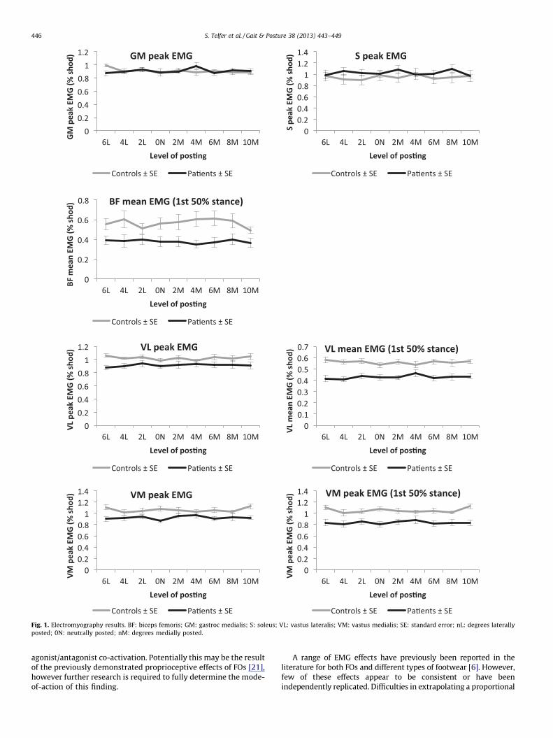
Fig. 1. Electromyography results. BF: biceps femoris; GM: gastroc medialis; S: soleus; VL: vastus lateralis; VM: vastus medialis; SE: standard error; nL: degrees laterally
posted; 0N: neutrally posted; nM: degrees medially posted.
S. Telfer et al. / Gait & Posture 38 (2013) 443–449446
agonist/antagonist co-activation. Potentially this may be the resultof the previously demonstrated proprioceptive effects of FOs [21],however further research is required to fully determine the mode-of-action of this finding.
A range of EMG effects have previously been reported in theliterature for both FOs and different types of footwear [6]. However,few of these effects appear to be consistent or have beenindependently replicated. Difficulties in extrapolating a proportional

Table 3Two way mixed effects ANOVA for plantar pressure variables.
Wilks’ lambda F p-Value Partial eta squared Best contrast Trend per 28 posting
lateral to medial
Peak medial RFPosting 0.584 1.393 0.231 0.062
Group – 1.757 0.199 0.077
Posting � group 0.558 1.387 0.233 0.062
Mean medial RFPosting 0.556 1.204 0.313 0.054
Group – 8.157 0.009a 0.28
Posting � group 0.626 0.835 0.522 0.038
Peak lateral RFPosting 0.488 1.836 0.179 0.07
Group – 0.746 0.397 0.034
Posting � group 0.540 1.493 0.345 0.051
Mean lateral RFPosting 0.29 4.038 0.002a 0.161 Linear (p = 0.001) �0.58%
Group – 1.973 0.175 0.086
Posting � group 0.385 1.355 0.245 0.061
Peak midfootPosting 0.287 7.517 <0.001a 0.264 Linear (p < 0.001) �2.48%
Group – 0.011 0.918 0.001
Posting � group 0.521 1.612 0.164 0.071
Mean midfootPosting 0.361 5.471 <0.001a 0.207 Linear (p < 0.001) �1.59%
Group – 0.799 0.382 0.037
Posting � group 0.536 1.241 0.296 0.056
Peak 1st rayPosting 0.768 0.519 0.796 0.024
Group – 0.041 0.842 0.002
Posting � group 0.57 1.211 0.304 0.055
Mean 1st rayPosting 0.517 1.152 0.337 0.052
Group – 1.217 0.283 0.055
Posting � group 0.636 0.782 0.58 0.036
Peak lateral FFPosting 0.448 2.413 0.042a 0.103 Linear (p < 0.001) 0.74%
Group – 2.593 0.122 0.11
Posting � group 0.632 0.833 0.527 0.038
Mean lateral FFPosting 0.256 3.83 0.002a 0.154 Linear (p < 0.001) 0.71%
Group – 0.001 0.975 0.001
Posting � group 0.619 0.834 0.537 0.038
a Statistically significant.
S. Telfer et al. / Gait & Posture 38 (2013) 443–449 447
relationship from EMG data to muscle force generation are widelyacknowledged [22], and more advanced techniques such as waveletanalysis have been proposed for investigating FO effects on muscleactivity [7]. Future work should investigate these methods whenmeasuring dose response effects of FOs on muscle activity.
Customised FOs have been shown to affect pressure distribu-tion in a number of patient groups and foot types [13,23,24].Bonanno et al. [13] recently demonstrated that altering the depthof a medial heel skive resulted in increased peak pressures at themedial rearfoot, with increases in skive magnitude resulting inincreases in pressure, however no changes were seen at the moredistal portions of the foot. Our results found significant effects atthe lateral rearfoot as well as additional effects at the midfoot andthe forefoot, suggesting that the extrinsic post used in our studymay have a greater effect on full foot biomechanics than the medialskive. The changes in pressure, particularly at the forefoot wererelatively small (<1% per 28 of posting relative to shod) however itis beyond the scope of the current study to determine if thesechanges are clinically relevant.
At the midfoot, although linear contrasts proved to be the mostsignificant, there is an indication that a more complex relationshipexists in terms of the dose response to the devices, with medialposting beyond 48 not seeming to produce any further decrease inpeak or mean pressure. This potentially suggests that there may be athreshold as to how far the centre of pressure can be shifted laterally,however further research is required to test this hypothesis.
Plantar pressure measurement systems have several limita-tions which are relevant to FO research [25]. Most relevant to thisstudy is the fact that the sensors embedded in the insole onlymeasure forces acting perpendicularly to them, and this combinedwith the geometrically complex shape of an FO means thatpotentially relevant non-perpendicular components of the forcesapplied will not be quantified. Despite this limitation, when takenin context with our existing knowledge of the effects of FOs webelieve the plantar pressure results described here to be anaccurate representation of the changes occurring.
The use of 3D printing to manufacture FOs is a relatively newapproach [26], and long term testing of the performance of thedevices used here is required, in particular to understand safetyand tolerance. However this study has demonstrated that low cost3D printing of FOs is feasible and a useful research tool for short-term FO studies. These technological advances give the opportu-nity to understand more fully the mechanical response to smallerincremental changes in functional design features and enablerobust parametric CAD design rules to be developed.
A number of limitations should be noted for this study. Standardclinical measures were used to define foot type and these mayrepresent a source of error. In addition, although the individualswere acclimatised to wearing FOs it is possible that the variationstested may have had different effects given a longer period of wear,thus only the acute biomechanical effects of varying the dose of theFOs were measured. A wide range of FOs are available, varying in

Fig. 2. Plantar pressure results. RF: rearfoot; FF: forefoot; SE: standard error; nL: degrees laterally posted; 0N: neutrally posted; nM: degrees medially posted.
S. Telfer et al. / Gait & Posture 38 (2013) 443–449448
materials, features and level of customisation. The devices used inthis study were 3/4 length semi rigid FOs produced using CADsoftware, and care is advised if extrapolating these results to othertypes of FO. The type of footwear worn may play a large role in theeffect of the FO, and standardised footwear was used in this study toallow the effects of the posting to be determined without thispotentially confounding variable. Again the authors advise cautionin extrapolating these results across all types of footwear.
A number of statistical comparisons were carried out for thisstudy and it is possible that some significant findings may havebeen down to chance. The authors feel the exploratory nature ofthe study justifies this type of analysis. This is supported by themajority of effects found being consistent with each other and/orwith our existing knowledge of FO effects.
In conclusion, this study provides evidence that there is asignificant and linear dose response effect of FOs on plantarpressure variables at the rearfoot, midfoot and forefoot. Foot typeappears to play a significant role in the effect of FOs on above-kneemuscle activity, with the devices producing a reduction in activityin pronated foot type.
Acknowledgments
This work was funded through the European CommissionFramework Seven Program (Grant number NMP2-SE-2009-228893) as part of the A-FOOTPRINT project (www.afootprint.eu).The funders had no input into the study design, collection, analysisand interpretation of data; the writing of the manuscript; or thedecision to submit for publication.
Conflict of interest statementThe authors declare that they have no conflict of interest
relating to the material presented in this article.
Appendix A. Supplementary data
Supplementary data associated with this article can be found, inthe online version, at http://dx.doi.org/10.1016/j.gaitpost.2013.01.012.

S. Telfer et al. / Gait & Posture 38 (2013) 443–449 449
References
[1] Cheung RTH, Chung RCK, Ng GYF. Efficacies of different external controls forexcessive foot pronation: a meta-analysis. British Journal of Sports Medicine2011;45:743–51.
[2] Boyer KA, Nigg BM. Muscle activity in the leg is tuned in response to impactforce characteristics. Journal of Biomechanics 2004;37:1583–8.
[3] Menz HB. Foot orthoses: how much customisation is necessary? Journal ofFoot and Ankle Research 2009;2:23.
[4] Hunter S, Dolan MG, Davis JM. Introduction to orthotic therapy. In: Frey R,editor. Foot orthotics in therapy and sport. Champaign: Human Kinetics; 1995.p. 1–9.
[5] Donatelli R, Wooden M. Biomechanical orthotics. In: Donatelli R, editor. Thebiomechanics of the foot and ankle. Philadelphia: Davis; 1995. p. 193–216.
[6] Murley GS, Landorf KB, Menz HB, Bird AR. Effect of foot posture, foot orthosesand footwear on lower limb muscle activity during walking and running: asystematic review. Gait and Posture 2009;29:172–87.
[7] Mundermann A, Wakeling JM, Nigg BM, Humble RN, Stefanyshyn DJ. Footorthoses affect frequency components of muscle activity in the lower extrem-ity. Gait and Posture 2006;23:295–302.
[8] Murley GS, Bird AR. The effect of three levels of foot orthotic wedging on thesurface electromyography activity of selected lower limb muscles during gait.Clinical Biomechanics 2006;21:1074–80.
[9] Nawoczenski DA, Ludewig PM. Electromyographic effects of foot orthotics onselected lower extremity muscles during running. Archives of Physical Medi-cine and Rehabilitation 1999;80:540–4.
[10] Tomaro J, Burdett RG. The effects of foot orthotics on the EMG activity ofselected leg muscles during gait. Journal of Orthopaedic and Sports PhysicalTherapy 1993;18:532–6.
[11] Hodge MC, Bach TM, Carter GM. Novel award first prize paper, orthoticmanagement of plantar pressure and pain in rheumatoid arthritis. ClinicalBiomechanics 1999;14:567–75.
[12] Healy A, Dunning DN, Chockalingam N. Effect of insole material on lower limbkinematics and plantar pressures during treadmill walking. Prosthetics andOrthotics International 2012;36:53–62.
[13] Bonanno DR, Zhang CY, Farrugia RC, Bull MG, Raspovic AM, Bird AR, et al. Theeffect of different depths of medial heel skive on plantar pressures. Journal ofFoot and Ankle Research 2012;5:1.
[14] Owings TM, Woerner JL, Frampton JD, Cavanagh PR, Botek G. Custom thera-peutic insoles based on both foot shape and plantar pressure measurementprovide enhanced pressure relief. Diabetes Care 2008;31:839–44.
[15] Bus SA, Haspels R, Busch-Westbroek TE. Evaluation and optimization oftherapeutic footwear for neuropathic diabetic foot patients using in-shoeplantar pressure analysis. Diabetes Care 2011;34:1595–600.
[16] Redmond AC, Crosbie J, Ouvrier RA. Development and validation of a novelrating system for scoring standing foot posture: the foot posture index. ClinicalBiomechanics 2006;21:89–98.
[17] Telfer S, Gibson KS, Hennessy K, Steultjens MP, Woodburn J. Computer-aideddesign of customized foot orthoses: reproducibility and effect of method usedto obtain foot shape. Archives of Physical Medicine and Rehabilitation2012;93:863–70.
[18] Hermens HJ, Freriks B, Disselhorst-Klug C, Rau G. Development of recommen-dations for SEMG sensors and sensor placement procedures. Journal of Elec-tromyography and Kinesiology 2000;10:361–74.
[19] Barn R, Turner DE, Rafferty D, Sturrock RD, Woodburn J. Tibialis posteriortenosynovitis and associated pes plano valgus in rheumatoid arthritis: EMG,multi-segment foot kinematics and ultrasound features. Arthritis Care andResearch (Hoboken), in press, http://dx.doi.org/10.1002/acr.21859.
[20] Murley GS, Landorf KB, Menz HB. Do foot orthoses change lower limb muscleactivity in flat-arched feet towards a pattern observed in normal-arched feet?Clinical Biomechanics 2010;25:728–36.
[21] Perry SD, Radtke A, McIlroy WE, Fernie GR, Maki BE. Efficacy and effectivenessof a balance-enhancing insole. Journals of Gerontology Series A BiologicalSciences and Medical Sciences 2008;63:595–602.
[22] Lawrence JH, De Luca CJ. Myoelectric signal versus force relationship indifferent muscles. Journal of Applied Physiology 1983;54:1653–9.
[23] Albert S, Rinoie C. Effect of custom orthotics on plantar pressure distributionin the pronated diabetic foot. Journal of Foot and Ankle Surgery 1994;33:598–604.
[24] Chang BC, Wang JY, Huang BS, Lin HY, Lee WC. Dynamic impression insolein rheumatoid foot with metatarsal pain. Clinical Biomechanics 2012;27:196–201.
[25] Urry SR, Wearing SC. The accuracy of footprint contact measurements: relevanceto the design and performance of pressure platforms. Foot 2001;11:151–7.
[26] Telfer S, Pallari J, Munguia J, Dalgarno K, McGeough M, Woodburn J. Embracingadditive manufacture: implications for foot and ankle orthosis design. BMCMusculoskeletal Disorders 2012;13:84.
Related Documents