CLINICAL INVESTIGATION DOSE TO THE CONTRALATERAL BREAST FROM RADIOTHERAPY AND RISK OF SECOND PRIMARY BREAST CANCER IN THE WECARE STUDY MARILYN STOVALL,PH.D.,* SUSAN A. SMITH, M.P.H.,* BRYAN M. LANGHOLZ,PH.D., y JOHN D. BOICE,JR., SC.D., zx ROY E. SHORE,PH.D., D.P.H., { MICHAEL ANDERSSON, M.D., D.M.SC., k THOMAS A. BUCHHOLZ, M.D.,** MARINELA CAPANU,PH.D., yy LESLIE BERNSTEIN,PH.D., zz CHARLES F. LYNCH, M.D., PH.D., xx KATHLEEN E. MALONE,PH.D., {{ HODA ANTON-CULVER,PH.D., kk ROBERT W. HAILE, D.P.H., y BARRY S. ROSENSTEIN,PH.D.,*** yyy ANNE S. REINER, M.P.H., yy DUNCAN C. THOMAS,PH.D., y JONINE L. BERNSTEIN,PH.D., zzz AND WECARE STUDY COLLABORATIVE GROUP 1 Departments of *Radiation Physics and ** Radiation Oncology, The University of Texas M.D. Anderson Cancer Center, Houston, TX; y Department of Preventive Medicine, University of Southern California, Los Angeles, CA; z International Epidemiology Institute, Rockville, MD, and Department of Medicine, x Vanderbilt University Medical Center, Nashville, TN; { Radiation Effects Research Foundation (RERF), Hiroshima, Japan; k Institute of Cancer Epidemiology, Danish Cancer Society, Copenhagen, Denmark; Departments of yy Statistics and zzz Epidemiology, Memorial Sloan-Kettering Cancer Center, New York, NY; zz Department of Cancer Etiology, City of Hope National Medical Center, Duarte, CA; xx Department of Epidemiology, University of Iowa, Iowa City, IA; {{ Division of Public Health Sciences, Fred Hutchinson Cancer Research Center, Seattle, WA; kk Department of Epidemiology, University of California, Irvine, CA; *** Department of Radiation Oncology, Mount Sinai School of Medicine, New York, NY; and yyy Department of Radiation Oncology, New York School of Medicine, New York, NY Purpose: To quantify the risk of second primary breast cancer in the contralateral breast (CB) after radiotherapy (RT) for first breast cancer. Methods and Materials: The study population included participants in the Women’s Environmental, Cancer, and Radiation Epidemiology study: 708 cases (women with asynchronous bilateral breast cancer) and 1399 controls (women with unilateral breast cancer) counter-matched on radiation treatment. Participants were <55 years of age at first breast cancer. Absorbed doses to quadrants of the CB were estimated. Rate ratios (RR) and 95% con- fidence intervals (CI) were calculated using multivariable-adjusted conditional logistic regression models. Results: Across all patients, the mean radiation dose to the specific quadrant of the CB tumor was 1.1 Gy. Women <40 years of age who received >1.0 Gy of absorbed dose to the specific quadrant of the CB had a 2.5-fold greater risk for CB cancer than unexposed women (RR = 2.5, 95% CI 1.4–4.5). No excess risk was observed in women >40 years of age. Women <40 years of age with follow-up periods >5 years had a RR of 3.0 (95% CI 1.1–8.1), and the dose response was significant (excess RR per Gy of 1.0, 95% CI 0.1–3.0). Conclusions: Women <40 years of age who received a radiation dose >1.0 Gy to the CB had an elevated, long-term risk of developing a second primary CB cancer. The risk is inversely related to age at exposure and is dose dependent. Ó 2008 Elsevier Inc. Contralateral breast, Radiation risk, Secondary breast cancer. INTRODUCTION Improvements in breast cancer treatment over the past few decades have resulted in longer survival and increased use of radiotherapy (RT) (1, 2). However, RT for breast cancer inevitably results in scattered radiation dose to the contralat- eral breast (CB). Radiation is a well-known breast carcinogen (3–10), but it is unclear to what extent radiation to the CB in- creases the risk for a second primary tumor. Most studies do not report significant increases in CB cancer after RT. When increases are found they are seen generally in large studies in which risk is apparent among young women (age <45 years) treated with RT and followed for more than 5 years (11, 12). Reprint requests to: Marilyn Stovall, Ph.D., Department of Radia- tion Physics, Unit 544, The University of Texas M. D. Anderson Can- cer Center, 1515 Holcombe Blvd., Houston, TX 77030. Tel: (713) 745-8999; Fax: (713) 794-1371; E-mail: [email protected] Supported in part by the National Institutes of Health, Grant Nos. U01-CA83178, R01-CA97397, and R01-CA42949. Conflict of interest: none. 1 Members of the WECARE Study Collaborative Group are listed in the Appendix. Acknowledgments—The authors thank Rita E. Weathers, M.S., for her assistance in computer programming analysis; and Barbara Pylate for her help with the manuscript. Received Jan 28, 2008, and in revised form Jan 28, 2008. Accepted for publication Feb 26, 2008. 1 Int. J. Radiation Oncology Biol. Phys., Vol. -, No. -, pp. 1–10, 2008 Copyright Ó 2008 Elsevier Inc. Printed in the USA. All rights reserved 0360-3016/08/$–see front matter doi:10.1016/j.ijrobp.2008.02.040 ARTICLE IN PRESS

Welcome message from author
This document is posted to help you gain knowledge. Please leave a comment to let me know what you think about it! Share it to your friends and learn new things together.
Transcript

CLINICAL INVESTIGATION
DOSE TO THE CONTRALATERAL BREAST FROM RADIOTHERAPYAND RISKOF SECOND PRIMARY BREAST CANCER IN THE WECARE STUDY
MARILYN STOVALL, PH.D.,* SUSAN A. SMITH, M.P.H.,* BRYAN M. LANGHOLZ, PH.D.,y
JOHN D. BOICE, JR., SC.D.,zx ROY E. SHORE, PH.D., D.P.H.,{ MICHAEL ANDERSSON, M.D., D.M.SC.,k
THOMAS A. BUCHHOLZ, M.D.,** MARINELA CAPANU, PH.D.,yy LESLIE BERNSTEIN, PH.D.,zz
CHARLES F. LYNCH, M.D., PH.D.,xx KATHLEEN E. MALONE, PH.D.,{{ HODA ANTON-CULVER, PH.D.,kk
ROBERT W. HAILE, D.P.H.,y BARRY S. ROSENSTEIN, PH.D.,***yyy ANNE S. REINER, M.P.H.,yy
DUNCAN C. THOMAS, PH.D.,y JONINE L. BERNSTEIN, PH.D.,zzz AND
WECARE STUDY COLLABORATIVE GROUP1
Departments of *Radiation Physics and **Radiation Oncology, The University of Texas M.D. Anderson Cancer Center, Houston, TX;yDepartment of Preventive Medicine, University of Southern California, Los Angeles, CA; z International Epidemiology Institute,Rockville, MD, and Department of Medicine, xVanderbilt University Medical Center, Nashville, TN; {Radiation Effects Research
Foundation (RERF), Hiroshima, Japan; k Institute of Cancer Epidemiology, Danish Cancer Society, Copenhagen, Denmark;Departments of yyStatistics and zzzEpidemiology, Memorial Sloan-Kettering Cancer Center, New York, NY; zzDepartment of CancerEtiology, City of Hope National Medical Center, Duarte, CA; xxDepartment of Epidemiology, University of Iowa, Iowa City, IA;{{Division of Public Health Sciences, Fred Hutchinson Cancer Research Center, Seattle, WA; kkDepartment of Epidemiology,
University of California, Irvine, CA; ***Department of Radiation Oncology, Mount Sinai School of Medicine, New York, NY; andyyyDepartment of Radiation Oncology, New York School of Medicine, New York, NY
Purpose: To quantify the risk of second primary breast cancer in the contralateral breast (CB) after radiotherapy(RT) for first breast cancer.Methods and Materials: The study population included participants in the Women’s Environmental, Cancer, andRadiation Epidemiology study: 708 cases (women with asynchronous bilateral breast cancer) and 1399 controls(women with unilateral breast cancer) counter-matched on radiation treatment. Participants were <55 years ofage at first breast cancer. Absorbed doses to quadrants of the CB were estimated. Rate ratios (RR) and 95% con-fidence intervals (CI) were calculated using multivariable-adjusted conditional logistic regression models.Results: Across all patients, the mean radiation dose to the specific quadrant of the CB tumor was 1.1 Gy. Women<40 years of age who received >1.0 Gy of absorbed dose to the specific quadrant of the CB had a 2.5-fold greaterrisk for CB cancer than unexposed women (RR = 2.5, 95% CI 1.4–4.5). No excess risk was observed in women>40 years of age. Women <40 years of age with follow-up periods >5 years had a RR of 3.0 (95% CI 1.1–8.1),and the dose response was significant (excess RR per Gy of 1.0, 95% CI 0.1–3.0).Conclusions: Women <40 years of age who received a radiation dose >1.0 Gy to the CB had an elevated, long-termrisk of developing a second primary CB cancer. The risk is inversely related to age at exposure and is dosedependent. � 2008 Elsevier Inc.
Contralateral breast, Radiation risk, Secondary breast cancer.
INTRODUCTION
Improvements in breast cancer treatment over the past few
decades have resulted in longer survival and increased use
of radiotherapy (RT) (1, 2). However, RT for breast cancer
inevitably results in scattered radiation dose to the contralat-
eral breast (CB). Radiation is a well-known breast carcinogen
(3–10), but it is unclear to what extent radiation to the CB in-
creases the risk for a second primary tumor. Most studies do
not report significant increases in CB cancer after RT. When
increases are found they are seen generally in large studies in
which risk is apparent among young women (age <45 years)
treated with RT and followed for more than 5 years (11, 12).
Reprint requests to: Marilyn Stovall, Ph.D., Department of Radia-tion Physics, Unit 544, The University of TexasM. D.Anderson Can-cer Center, 1515 Holcombe Blvd., Houston, TX 77030. Tel: (713)745-8999; Fax: (713) 794-1371; E-mail: [email protected] in part by the National Institutes of Health, Grant Nos.
U01-CA83178, R01-CA97397, and R01-CA42949.Conflict of interest: none.
1Members of the WECARE Study Collaborative Group are listed inthe Appendix.Acknowledgments—The authors thank Rita E. Weathers, M.S., forher assistance in computer programming analysis; and BarbaraPylate for her help with the manuscript.Received Jan 28, 2008, and in revised form Jan 28, 2008.
Accepted for publication Feb 26, 2008.
1
Int. J. Radiation Oncology Biol. Phys., Vol. -, No. -, pp. 1–10, 2008Copyright � 2008 Elsevier Inc.
Printed in the USA. All rights reserved0360-3016/08/$–see front matter
doi:10.1016/j.ijrobp.2008.02.040
ARTICLE IN PRESS

Several randomized trials (13), registry-based studies (12,
14–17), and hospital-based studies (3, 18–20) have evaluated
the risk of second breast cancer in the CB after RT (21). Few
studies, however, have estimated risk in terms of radiation
dose to the CB (11, 22, 23), and only one study reported a sig-
nificant dose–response relationship (11).
The goal of this investigation was to quantify the radiation
dose to the CB after RT for primary breast cancer and to es-
timate the associated risk of a primary CB tumor. The study
population was composed of participants in the Women’s
Environmental, Cancer, and Radiation Epidemiology
(WECARE) study, a multicenter, population-based, case–
control study designed to examine the interaction of radiation
and genetic factors and their effect on development of breast
cancer (24).
METHODS AND MATERIALS
Study populationTheWECARE study included 708 women with asynchronous bi-
lateral breast cancer (cases) and 1399 women with unilateral breast
cancer (controls). Participants were identified, recruited, and inter-
viewed through five population-based cancer registries: four in the
United States (Iowa; Los Angeles County, CA: Orange and San
Diego Counties, CA; and Seattle, WA) and one in Denmark. Of
the eligible cases (998) and controls (2112), 71% of the cases and
66% percent of the controls agreed to participate. The study design
is described in detail elsewhere (24, 25).
Cases met the following criteria: (1) diagnosis of first primary
breast cancer between January 1, 1985, and December 31, 1999, ini-
tially staged ‘‘local only’’ or ‘‘local plus regional lymph node dis-
ease’’ and diagnosis of a second primary tumor (in situ or
invasive disease) diagnosed in the CB at least 1 year after the diag-
nosis of the first primary breast tumor; (2) a resident of the same
study reporting area during both diagnoses; (3) no previous or inter-vening cancer diagnoses; (4) age <55 years at diagnosis of the first
primary tumor; and (5) alive at the time of contact and able to pro-
vide informed consent to complete the interview.
Controls were individually matched to cases in a 2:1 ratio by
age at first breast cancer diagnosis (5-year strata), year of first can-
cer diagnosis (4-year strata), registry region, and race. Controls
met the following criteria: (1) diagnosed since January 1, 1985,
with first primary invasive breast cancer occurring while residing
in one of the study reporting areas; (2) residing in the same study
reporting area on the reference date (date of first diagnosis plus
the time period between the first and second diagnosis [‘‘at-risk
interval’’] of the matched case); (3) alive at time of contact; (4)never diagnosed (by reference date) as having had a second pri-
mary breast cancer or any other cancer diagnosis; and (5) no prior
prophylactic mastectomy of the CB. Controls were counter-
matched to the cases on registry-reported radiation exposure to
improve statistical efficiency: each case and two matched controls
formed a triplet, with two members of each triplet exposed to RT
and one member unexposed. Counter-matching decreased the
chance that a triplet was all RT-exposed or all non-exposed and,
therefore, uninformative regarding radiation-induced breast cancer
risks (25).
Data collectionThe data collection protocol was approved by the institutional re-
view board at each site, and each patient signed an informed consent
form. Participants were interviewed by telephone with use of a pre-
tested, structured questionnaire. The questionnaire was designed to
obtain information about events occurring before the diagnosis of
the first primary cancer, events occurring within the at-risk period,
and known and suspected risk factors for breast cancer.
Radiation treatment information was sought for each patient
whom the cancer registry, questionnaire, or medical record indi-
cated had received RT for initial primary breast cancer, metastases,
recurrences, or benign conditions. Radiation treatment details were
obtained from the basic RT record, RT summary, RT notes, med-
ical record notes, surgery reports (for brachytherapy), and physi-
cian correspondence. Radiotherapy records were retrieved for
1497 women: 1479 with RT for breast cancer and 18 who received
RT for other conditions (metastases, benign conditions, and/or
ovarian ablation). Ninety-one percent of the records received
were complete for radiation details, 8.5% were missing some radi-
ation details but doses could be estimated, and 0.5% were inade-
quate for dosimetry.
For cases, we collected all available information concerning the
CB tumor, using pathology reports, medical records, physician
notes, and mammography reports. The tumors were classified ac-
cording to their quadrant location, areolar region (1-cm radius),
and/or a clock position. For 609 of the 708 cases, the location
of the CB tumor was known. Of the case sets with known CB lo-
cation, 3 cases and 3 controls were missing dose information and
were eliminated from the dose–response analysis. Analysis of the
quadrant-specific doses for this study was based on 606 cases and
1200 matched controls for whom RT and CB tumor information
were available. The final analytic set of 1806 patients consisted
of 591 complete triplet sets, 12 case–control pairs, 3 control-
only pairs (matched to cases with missing dose information),
and 3 cases only. Of the 1806 patients, 1277 were treated with
RT: 1174 (92%) with external-beam therapy, 101 (8%) with exter-
nal-beam therapy plus brachytherapy, and 2 with an unknown
technique. Of the 1277 patients who received RT, 1266 received
breast irradiation.
Quantification of radiation doseFor external-beam therapy, dose estimates to the CB were mea-
sured with thermoluminescent dosimeters in tissue-equivalent
phantoms. The phantoms were molded with a plastic shell placed
on patients in the treatment position. The dosimeters were lithium
fluoride powder in tissue-equivalent flat packs. Dosimeters (ap-
proximately 300) were placed on a three-dimensional grid
throughout the CB. Measurements were made with and without
medial wedge compensators for tangential fields. Contralateral
breast dose was also measured from supraclavicular, axilla, and in-
ternal mammary chain fields. For brachytherapy, dose estimates to
the CB were derived using standard treatment-planning techniques
(26). The dosimetry techniques are described in detail elsewhere
(24, 26).
Doses were estimated to the four quadrants and the central portion
(areola) of the CB (Fig. 1). Dose to each quadrant was calculated as
the average of doses to points on a three-dimensional grid. Because
some tumors were located at the boundary between quadrants, dose
was estimated also to four clock positions (3, 6, 9, and 12), which
define the dividing lines between the quadrants. For cases with mul-
tifocal tumors (n = 67), the dose was calculated to the tumor of or-
igin, assuming the largest tumor or the invasive tumor came first. All
radiation dosimetry was performed without knowledge of whether
the patient was a case or a control.
2 I. J. Radiation Oncology d Biology d Physics Volume -, Number -, 2008
ARTICLE IN PRESS
Statistical methodsRate ratios (RR) and corresponding 95% confidence intervals
(CI) were estimated by fitting univariate and multivariable-adjusted
conditional logistic regression models, accounting for the counter-
matched sampling (25). The multivariable models were adjusted
for factors found to be statistically significant in the univariate
models and those known to be associated with breast cancer, includ-
ing exact age at diagnosis of first primary breast cancer, age at men-
arche (<13/13+ years), menopausal status (premenopausal/age at
menopause <45/45+ years), number of full-term pregnancies (0, 1,
2, 3, 4+), family history of breast cancer among first-degree relatives
(yes/no), lobular histology (yes/no) and stage (local/regional) of the
first primary, and treatment history (chemotherapy [yes/no] and/or
hormonal therapy [yes/no]).
Counter-matching on RT status allows modeling of main effects
of radiation as well as interactions and is more efficient than simple
random sampling of controls for these analyses (24). Furthermore,
because controls are independently sampled from the failure time
risks sets, the estimated parameters are rate ratios in the proportional
hazards model for cohort data (27) and standard likelihood methods
apply (28). Proportions were estimated by computing the factor dis-
tribution in controls by RT status, then taking a weighted average of
the within-RT status values where the weights are the overall pro-
portions in the risk sets (Table 1).
For the analyses, we used three different measures of radiation:
the location-specific dose, average dose to the breast, and ever/
never RT to the breast. The ‘‘location-specific dose’’ was calcu-
lated as the dose to the quadrant where the second primary for
the cases developed; for the controls it was calculated as the
dose received to the same location as the case. The average dose
was calculated, for both cases and controls, as the average of the
quadrant and central region dose estimates. Dose effects were es-
timated for categories 0, >0–1 Gy, and 1+ Gy and as an excess rel-
ative risk (ERR) trend. A missing data indicator was used to
account for missing covariate data. Analyses were conducted using
SAS (SAS Institute, Cary, NC) and Epicure (Hirosoft Interna-
tional, Seattle, WA) (29).
RESULTS
Reported in Table 1 are the patient characteristics of all
2107 WECARE study participants and of the subset of
1806 cases and controls included in the analyses. There
were no systematic differences between the overall WE-
CARE study population and the subset of participants in
our analyses. For both groups, cases and controls were simi-
lar with regard to race, age at diagnosis of the first primary,
and length of the at-risk period.
The most common RT technique was external-beam radi-
ation to the whole breast with opposing tangential fields.
Most of the tangential fields were delivered with wedge com-
pensators using angles ranging from 15� to 60�; 91% of the
patients were treated with both medial and lateral wedges.
Ninety-four percent of the treatments were delivered with
high-energy photons (87% of these were 4–8 MV). Some
patients also were treated with peripheral lymphatic fields
(supraclavicular, axilla, and/or internal mammary chain).
All external-beam treatments were delivered at a tumor
dose of 1.5–2.0 Gy per day for 5 to 6 weeks. These treatments
are typical of the patterns of care provided for breast cancer
patients from 1985 through 1999.
For all cases, the most common site for the second cancer
was the upper-outer quadrant of the breast (43%) (Fig.1). The
distribution of CB tumors across the breast (inner, central,
and outer) was compared between irradiated and unirradiated
women. For irradiated women, a significantly higher propor-
tionofCB tumorswas located in the inner and central quadrants
(p= 0.03). Therewas a two- to threefold variation inmean dose
across the CB quadrants, with the inner quadrants receiving the
highest doses (Fig. 2). Themeandose and rangeof doses for the
inner, central, and outer regions of the CB for the various treat-
ment regimens are shown in Table 2. Across all patients under-
going RT, the mean dose for the quadrant location in the CB
was 1.1 Gy (range, 0.02–6.2 Gy). There was no difference in
the mean quadrant location CB dose by year of treatment.
The average dose for the entire breast (i.e. mean of all breast
quadrants and central region) was 1.3 Gy for our population.
Comparisons of the threemethods of estimating dose (ever/
never RT, average dose across breast, and location-specific
dose) were conducted. Among all women, nomethod showed
a statistically significant elevation in risk associatedwith radi-
ation (Table 3). However, certain subgroups of women had
statistically significant risks. In women aged <40 years, those
who received >1 Gy of radiation had a 2.5-fold greater risk
than unexposed women (95% CI 1.4–4.5), whereas no excess
risk was observed for women aged >40 years treated with RT
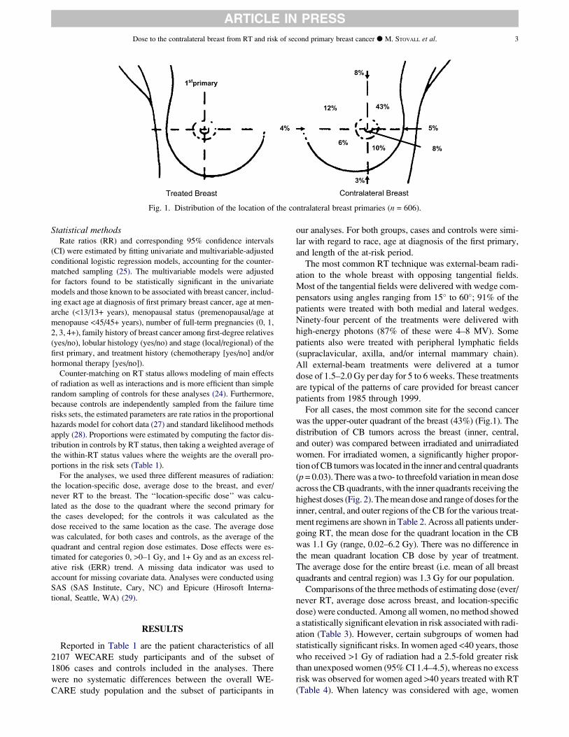
(Table 4). When latency was considered with age, women
Fig. 1. Distribution of the location of the contralateral breast primaries (n = 606).
Dose to the contralateral breast from RT and risk of second primary breast cancer d M. STOVALL et al. 3
ARTICLE IN PRESS

Table 1. Characteristics of study participants
(Continued)
4 I. J. Radiation Oncology d Biology d Physics Volume -, Number -, 2008
ARTICLE IN PRESS

aged <40 years, treated with RT, with 5+ years latency had an
RR of 3.0 (95% CI 1.1–8.1). Women aged <40 years, treated
with RT, with <5 years latency had a lower but significantly
elevated risk (RR = 2.3, 95% CI 1.1–4.6). There was a signif-
icant trend with dose among all women aged <40 years (ERR/
Gy = 0.6, 95% CI 0.1–1.5) and for the subgroup of women
aged <40 years who had at least a 5-year latency (ERR/Gy
= 1.0, 95% CI 0.1–3.0). Confidence intervals in all of these
subgroups were wide, reflecting the small numbers of partic-
ipants.
DISCUSSION
Overall, in our study, cancer of the CBwas not significantly
associated with RT for first breast cancer in our large, interna-
tional, case–control study of women. However, among
women <40 years of age who received RT and were followed
for$5 years, a significant dose–response relationshipwas ob-
served. Radiation dose was estimated to the quadrant where
the second breast cancer occurred, which reduced the uncer-
tainty of analyses based on average dose to the entire CB.
Several studies have assessed the relationship between risk
of subsequent primary cancer in the CB and radiation dose
(11, 22, 23). The study by Basco et al. (22) involved more
than 14,000 women treated for breast cancer in British
Columbia (mean dose to CB approximately 1.5 Gy), but the
number of CB cancers was small (n= 194, with only 30 occur-ring inwomen aged <40 years). In a nested case–control study
conducted by Boice et al. (11) of 41,109 women in Connect-
icut treated for breast cancer between 1935 and 1982, 655
Table 1. Characteristics of study participants (Continued)
Abbreviation: WECARE = Women’s Environmental, Cancer, and Radiation Epidemiology study.Values are number (percentage) or mean (range).*Cases for whom information on location of the tumor and dose were available and their matched controls.yProportions estimated accounting for counter-matched sampling.zReference date for the cases is defined as the diagnosis date of the second primary; for the controls it is defined as the date of the first breast
cancer diagnosis plus the ‘‘at-risk interval’’ of matched case.xActual radiotherapy (RT) as determined by medical records and interviews. Counter-matching was based only on registry-recorded RT in-
formation, which may not be the same as actual RT information.
Dose to the contralateral breast from RT and risk of second primary breast cancer d M. STOVALL et al. 5
ARTICLE IN PRESS

women developed cancer in the CB $5 years after initial
treatment; no significant radiation risk was observed (RR
1.2; mean dose to CB, 2.8 Gy). However, a significant risk
and dose–response relationship were apparent among women
<45 years of age at first treatment (RR 1.6; ERR/Gy = 0.2).
Storm et al. (23) conducted a nested case–control study of
56,540 women in Denmark with breast cancer treated at any
age between 1943 and 1978. Overall, 529 CB cancers oc-
curred $8 years after initial treatment, but no radiation risk
was observed (RR 1.0; mean dose to CB, 2.5 Gy). The Danish
study had fewer women <45 years of age than did the Con-
necticut study (19% vs. 31%, respectively).
A similarity between our study and that of Boice et al. (11)is that both included a large number of women with early-
onset breast cancer, amongwhom a significant dose–response
relationship was seen. However, the Boice study differs
from ours in several ways: (1) Boice et al. used average
dose to the whole breast, whereas we used dose to the quad-
rant of the CB tumor, (2) the patients in the Boice study weretreated with orthovoltage X-rays and 60Co teletherapy,
which resulted in higher doses to the CB than from the
high-energy photons in our study, (3) the Boice study had
a longer latent period (5 to >15 years) than did our study
(1–10 years), (4) the Boice study included women of all
ages, including those >55 years of age, whereas the women
in our study were all <55 years of age at first breast cancer
diagnosis. Despite these differences, the results of the two
studies are consistent. In neither study was radiation risk ob-
served for women >45 years of age at time of treatment; for
women <45 years of age who were followed for $5 years,
the estimates of risk in the two studies were the same
(ERR/Gy = 0.2 [30]).
A meta-analysis of 78 randomized treatment compari-
sons (13) showed a higher risk of CB cancer among
Table 2. Mean radiation dose (Gy) to the inner, central, and outer regions of the contralateral breast
n Dose to inner region Dose to central region Dose to outer region
Method of external treatment Case Control 10%ile Mean 90%ile 10%ile Mean 90%ile 10%ile Mean 90%ile
Tangential breast fields withoutperipheral lymphatics
178 542 1.2 1.8 2.5 0.8 1.3 1.8 0.5 0.8 1.1
Without wedges 11 26 1.0 1.2 1.6 0.7 0.8 1.0 0.4 0.5 0.6With wedges* 167 516 1.3 1.8 2.5 0.9 1.3 1.8 0.5 0.8 1.1
Tangential breast fields with peripherallymphaticsy
71 228 1.7 2.5 3.8 1.0 1.6 2.3 0.7 1.1 1.6
Without wedges 9 23 1.4 2.2 4.6 0.9 1.3 2.4 0.6 0.9 1.7With wedges* 62 205 1.8 2.6 3.7 1.1 1.7 2.3 0.7 1.1 1.6
Direct breast field without peripherallymphatics
11 34 0.3 0.4 0.4 0.1 0.1 0.1 0.1 0.1 0.1
Direct breast field with peripherallymphaticsy
31 111 1.1 1.5 1.8 0.4 0.5 0.6 0.4 0.4 0.5
Other fields 13 47 1.1 1.7 2.1 0.7 1.2 1.4 0.4 0.7 0.9All Patients 304 962 1.1 1.9 2.8 0.6 1.2 1.9 0.4 0.8 1.2
* The majority of patients were treated with medial and lateral wedges using angles ranging from 15� to 60�.y Peripheral lymphatic fields included supraclavicular fields, axilla fields, and internal mammary chain fields.
Fig. 2. Dose (in grays) to the contralateral breast. Mean and range of doses among patients treated with breast radiotherapy(n = 1266).
6 I. J. Radiation Oncology d Biology d Physics Volume -, Number -, 2008
ARTICLE IN PRESS

women >50 years of age; however, no radiation doses were
reconstructed for the CB, and the wide range of treatment
techniques may have confounded the actual radiation risk
to the CB.
Our study adds to the body of knowledge that the risk of de-
veloping radiation-induced CB cancer is stronger among
young women and that the women >45 years of age when
treated with RT for first breast cancer are not at significant
risk (11, 22, 23). This finding is reassuring given that most
women are >45 years of age when breast cancer is first de-
tected. Among younger women (<45 years of age), our data
provide evidence that the lower dose of radiation scatter
(mean 1.3 Gy) to the CB from treatments delivered from
1985 through 1999 remains sufficiently high to result in a de-
tectable increase in the risk of radiation-induced breast cancer.
The cases and controls in our study were diagnosed with
breast cancer as early as 1985. Cases and controls were re-
stricted to patients who were alive so that they could be in-
terviewed. Thus, there is a potential for survival bias if
survival depends on both case–control status and radiation
dose. We explored this possibility in the Los Angeles regis-
try using the RT information available in the registry re-
cords. When all participants were analyzed, the RR
contrasting RT with no RT was estimated to be 1.2 (95%
CI 1.0–1.4). Restricting the analysis to women known to
be alive in 2000 yielded the same result (RR = 1.2, 95%
CI 1.0–1.5). Among younger patients (<45 years of age)
with longer latency periods ($5 years), similar results
were found (RR for all subjects = 1.8 [95% CI 1.2–2.6];
RR restricted to alive in 2000 = 1.9 [95% CI 1.3–2.8]).
Thus, it is unlikely that there is significant survival bias in
our estimates of radiation effect.
A strength of our study was that we standardized the pro-
cedures used to collect information and implemented strin-
gent quality control. There may have been differences in
data quality between the registries, but because subjects
were matched by registry and date of first cancer, cases and
controls should have data of similar quality. There was no
evidence of large variations in radiation effect over registries
or over the calendar years of first cancer diagnosis.
The quadrant of the breast tumor was not available for 99
of the 708 cases, so these patients were omitted from the
main dose–response analyses. Although the tumor charac-
teristics (stage, histology, ER/PR status) of the excluded
cases did not differ statistically from those of patients in-
cluded in the analyses, there was a difference in the percent-
age of patients exposed to radiation between the cases with
known tumor locations (50%) and those with unknown tu-
mor locations (36%). Therefore, although it seems implau-
sible that the radiation dose response for tumors with
unknown location would differ from tumors that could
be assigned a location, we can not entirely rule out the
possibility of a systematic bias from excluding the cases
with unknown tumor location.
A major strength of our study is the large sample size, with
1806 cases and controls. The counter-matched design (24)
further enhanced our statistical power to evaluate risks by en-
suring that each triplet had two radiation-exposed members
and one unexposed. The nested case–control design involv-
ing patients obtained from cancer registries ensured that con-
trols were representative of the population, so the likelihood
of selection bias was minimized. Another strength of our
study is the collection of complete RT records for 91% of pa-
tients who received RT, which allowed detailed dosimetry in-
formation for each quadrant of the CB. The radiation
exposure characterization for each patient was determined
using the patient’s RT record rather than information from
cancer registries, which are not necessarily complete or cor-
rect. Finally, dosimetry was performed without regard to
a subject’s case–control status, so there was little likelihood
of information bias.
Our dosimetry results are compatible with those of prior
studies of patients undergoing RT that estimated the CB
dose to range between 0.5 and 4 Gy, corresponding to ap-
proximately 1–8% of a typical treatment dose of 50 Gy
(13, 31, 32). A novel feature of our study, however, was
Table 3. Risk of developing cancer in the contralateral breast
Method of estimating dose Dose to CB* (Gy) Cases, n (%) Controls, n (%) Rate ratioy 95% confidence interval
Yes/ No RTNever RT — 362 (51) 266 (50) 1.0Ever RT — 346 (49) 1133 (50) 1.1 0.9, 1.3
Average dose to the CB 0 353 (50) 259 (49) 1.00 < 1.0 100 (14) 276 (13) 1.2 0.9, 1.6$1.0 251 (36) 860 (38) 1.0 0.9, 1.3
Trend ERR/Gy 0.0 -0.1, 0.2Location-specific dose to the CB 0 297 (49) 232 (50) 1.0
0 < 1.0 169 (28) 536 (28) 1.2 0.9, 1.5$1.0 140 (23) 432 (23) 1.2 1.0, 1.6
Trend ERR/Gy 0.1 -0.1, 0.3
Abbreviations: CB = contralateral breast; RT = radiotherapy; ERR = excess relative risk.* Scatter radiation dose to the CB.y Adjusted for age at first diagnosis of breast cancer, age of menarche, family history of breast cancer, total number of full-term pregnancies,
age at menopause, chemo/hormonal treatment, histology, and stage.
Dose to the contralateral breast from RT and risk of second primary breast cancer d M. STOVALL et al. 7
ARTICLE IN PRESS
that we estimated radiation dose to the CB quadrant where
the second primary breast cancer developed. The mean
quadrant-specific dose for all patients was found to be lower
than the mean breast dose, which has important implica-
tions with regard to estimating dose-specific risk, because
second primary tumors, similar to first primary tumors,
are more common in the outer quadrants of the breast
(33, 34). Basing analyses on average breast dose rather
than quadrant-specific dose, accordingly, would underesti-
mate the true dose effect. Thus our dose–response analyses
based on quadrant tumor location provide a more valid as-
sessment of radiation risk than has been possible in previ-
ous investigations of the CB.
CONCLUSIONS
Our results show that for the majority of women treated for
breast cancer with recent RT techniques, RT did not play
Table 4. Risk of developing a contralateral breast cancer after RT according to age at time of exposure, time since exposure, andestimated radiation dose delivered to the site of CB cancer (cases = 606, controls = 1200)
Variable Dose to CB (Gy) Cases, n (%) Controls, n (%)* Rate ratioy 95% confidence interval
Time (y) since first breast cancer<5 0 167 (49) 134 (46) 1.0
0 < 1.0 94 (27) 313 (31) 0.9 0.7, 1.3$ 1.0 82 (24) 238 (23) 1.2 0.9, 1.7
Trend ERR/Gy 0.1 -0.1, 0.35-9 0 101 (49) 78 (54) 1.0
0 < 1.0 57 (27) 176 (23) 1.3 0.8, 2.0$ 1.0 50 (24) 154 (23) 1.4 0.9, 2.3
Trend ERR/Gy 0.2 -0.1, 0.610-14 0 29 (53) 20 (64) 1.0
0 < 1.0 18 (33) 47 (23) 1.8 0.8, 4.1$ 1.0 8 (15) 40 (12) 0.7 0.3, 2.0
Trend ERR/Gy �0.2 <<0, 0.5z
Age (y) at first breast cancer<40 0 47 (41) 43 (45) 1.0
0 < 1.0 31 (27) 112 (36) 0.9 0.5, 1.6$ 1.0 36 (32) 68 (19) 2.5 1.4, 4.5
Trend ERR/Gy 0.6 0.1, 1.540-44 0 73 (50) 53 (49) 1.0
0 < 1.0 46 (32) 140 (29) 1.3 0.8, 2.0$ 1.0 26 (18) 89 (22) 1.0 0.6, 1.8
Trend ERR/Gy �0.2 <<0, 0.3z
45-54 0 177 (51) 136 (52) 1.00 < 1.0 92 (27) 284 (25) 1.2 0.9, 1.7$ 1.0 78 (22) 275 (24) 1.0 0.7, 1.4
Trend ERR/Gy 0.0 -0.1, 0.3Age (y) at and time (y) since first
breast cancer<40 and <5 0 27 (42) 26 (41) 1.0
0 < 1.0 17 (26) 61 (37) 0.8 0.4, 1.8$ 1.0 21 (32) 43 (22) 2.3 1.1, 4.6
Trend ERR/Gy 0.4 -0.1, 1.4<40 and 5+ 0 20 (41) 17 (51) 1.0
0 < 1.0 14 (29) 51 (32) 1.0 0.4, 2.4$ 1.0 15 (31) 25 (17) 3.0 1.1, 8.1
Trend ERR/Gy 1.0 0.1, 3.040+ and <5 0 140 (50) 108 (47) 1.0
0 < 1.0 77 (28) 252 (29) 1.0 0.7, 1.4$ 1.0 61 (22) 195 (24) 1.0 0.7, 1.5
Trend ERR/Gy 0.0 -0.2, 0.340+ and 5+ 0 110 (51) 81 (57) 1.0
0 < 1.0 61 (29) 172 (21) 1.7 1.1, 2.5$ 1.0 43 (20) 169 (22) 1.0 0.6, 1.6
Trend ERR/Gy 0.0 <<0, 0.4z
Abbreviations as in Table 3.* Proportions estimated accounting for counter-matched sampling.y Adjusted for age at first diagnosis of breast cancer, age of menarche, family history of breast cancer, total number of full-term pregnancies,
age at menopause, chemo/hormonal treatment, histology, and stage.z << Indicates a very large negative number that could not be estimated by the method.
8 I. J. Radiation Oncology d Biology d Physics Volume -, Number -, 2008
ARTICLE IN PRESS

a significant role in the development of a second breast pri-
mary; however, young women with breast cancer had an el-
evated long-term risk of developing a CB cancer. In addition,
this radiation risk was inversely related to age at exposure and
was dose dependent. Despite the overall reassuring results
and the lower CB doses resulting from recent treatments
(35), the newer treatment techniques, such as intensity-mod-
ulated RT and field-in-field irradiation, should be investi-
gated with regard to their roles as risk factors for secondary
breast cancer in the CB (36).
REFERENCES
1. Ries LAG, Eisner MP, Kosary CL, et al. SEER cancer statisticsreview, 1975–2002. 2005. Available at: http://seer.cancer.gov/csr/1975_2002/.
2. American Cancer Society. Cancer facts & figures 2007.Available at: http://www.cancer.org/docroot/STT/content/STT_1x_Cancer_Facts__Figures_2007.asp.
3. Obedian E, Fischer DB, Haffty BG. Second malignancies aftertreatment of early-stage breast cancer: Lumpectomy and radiationtherapy versus mastectomy. J Clin Oncol 2000;18:2406–2412.
4. Haffty BG, Harrold E, Khan AJ, et al. Outcome of conserva-tively managed early-onset breast cancer by BRCA1/2 status.Lancet 2002;359:1471–1477.
5. Boice JD Jr., Preston D, Davis FG, et al. Frequent chest X-rayfluoroscopy and breast cancer incidence among tuberculosispatients in Massachusetts. Radiat Res 1991;125:214–222.
6. Boice JD Jr. Radiation and breast carcinogenesis. Med PediatrOncol 2001;36:508–513.
7. Land CE, Tokunaga M, Koyama K, et al. Incidence of femalebreast cancer among atomic bomb survivors, Hiroshima andNagasaki, 1950-1990. Radiat Res 2003;160:707–717.
8. Travis LB, Hill DA, Dores GM, et al. Breast cancer followingradiotherapy and chemotherapy among young women withHodgkin disease. JAMA 2003;290:465–475.
9. Ronckers CM, Erdmann CA, Land CE. Radiation and breastcancer: A review of current evidence. Breast Cancer Res2005;7:21–32.
10. Preston DL, Mattsson A, Holmberg E, et al. Radiation effectson breast cancer risk: A pooled analysis of eight cohorts. RadiatRes 2002;158:220–235.
11. Boice JD Jr., Harvey EB, Blettner M, et al. Cancer in the con-tralateral breast after radiotherapy for breast cancer. N EnglJ Med 1992;326:781–785.
12. Gao X, Fisher SG, Emami B. Risk of second primary cancer inthe contralateral breast in women treated for early-stage breastcancer: A population-based study. Int J Radiat Oncol BiolPhys 2003;56:1038–1045.
13. Clarke M, Collins R, Darby S, et al. Effects of radiotherapy andof differences in the extent of surgery for early breast cancer onlocal recurrence and 15-year survival: An overview of the rand-omised trials. Lancet 2005;366:2087–2106.
14. Harvey EB, Brinton LA. Second cancer following cancer of thebreast in Connecticut, 1935-82. Natl Cancer Inst Monogr 1985;68:99–112.
15. Horn PL, Thompson WD, Schwartz SM. Factors associatedwith the risk of second primary breast cancer: An analysis ofdata from the Connecticut Tumor Registry. J Chronic Dis1987;40:1003–1011.
16. Bernstein JL, Thompson WD, Risch N, et al. Risk factors pre-dicting the incidence of second primary breast cancer amongwomen diagnosed with a first primary breast cancer. Am JEpidemiol 1992;136:925–936.
17. Curtis RE, Freedman DM, Ron E, et al., editors. Newmalignan-cies among cancer survivors. SEER cancer registries, 1973-2000. NIH publication no. 05-5302. Bethesda; MD: NationalCancer Institute; 2006.
18. Kurtz JM, Amalric R, Brandone H, et al. Contralateral breastcancer and other second malignancies in patients treated bybreast-conserving therapy with radiation. Int J Radiat OncolBiol Phys 1988;15:277–284.
19. Parker RG, Grimm P, Enstrom JE. Contralateral breast cancersfollowing treatment for initial breast cancers in women. AmJ Clin Oncol 1989;12:213–216.
20. Fowble B, Hanlon A, Freedman G, et al. Second cancers afterconservative surgery and radiation for stages I-II breast cancer:Identifying a subset of women at increased risk. Int J RadiatOncol Biol Phys 2001;51:679–690.
21. Chen Y, Thompson W, Semenciw R, et al. Epidemiology ofcontralateral breast cancer. Cancer Epidemiol BiomarkersPrev 1999;8:855–861.
22. Basco VE, Coldman AJ, Elwood JM, et al. Radiation dose andsecond breast cancer. Br J Cancer 1985;52:319–325.
23. Storm HH, Andersson M, Boice JD Jr., et al. Adjuvant radio-therapy and risk of contralateral breast cancer. J Natl CancerInst 1992;84:1245–1250.
24. Bernstein JL, Langholz B, Haile RW, et al. Study design: Eval-uating gene-environment interactions in the etiology of breastcancer—The WECARE study. Breast Cancer Res 2004;6:199–214.
25. Langholz B, Borgan O. Counter-matching: a stratifiednested case-control sampling method. Biometrika 1995;82:69–79.
26. Stovall M, Weathers R, Kasper C, et al. Dose reconstruction fortherapeutic and diagnostic radiation exposures: Use in epidemi-ological studies. Radiat Res 2006;166:141–157.
27. Cox DR. Regression models and life tables. J Roy Statist Soc B1972;34:187–220.
28. Borgan O, Goldstein L, Langholz B.Methods for the analysis ofsampled cohort data in the Cox proportional hazards model. AnnStat 1995;12:1749–1778.
29. Preston DL, Lubin JH, Pierce DA, et al. Epicure users guide.Seattle, WA: Hirosoft International; 1993.
30. Langholz B, Thomas DC, Zhang X, et al. Statistical methods foranalyzing radiation dose-response with tumor and/or dose loca-tion-specific information with application to the WECAREstudy of asynchronous contralateral breast cancer. Technicalreport 177. Los Angeles, CA: University of Southern Califor-nia, Department of Preventive Medicine, Biostatistics Division;2007.
31. Fraass BA, Roberson PL, Lichter AS. Dose to the contralateralbreast due to primary breast irradiation. Int J Radiat Oncol BiolPhys 1985;11:485–497.
32. Kelly CA,Wang XY, Chu JC, et al. Dose to contralateral breast:A comparison of four primary breast irradiation techniques. IntJ Radiat Oncol Biol Phys 1996;34:727–732.
33. Khan AJ, Haffty BG. The location of contralateral breastcancers after radiation therapy. Breast J 2001;7:331–336.
34. Hill-Kayser CE, Harris EE, Hwang WT, et al. Twenty-year in-cidence and patterns of contralateral breast cancer after breastconservation treatment with radiation. Int J Radiat Oncol BiolPhys 2006;66:1313–1319.
35. Haffty BG. Radiation therapy and the risk of contralateralbreast cancer. Int J Radiat Oncol Biol Phys 2003;56:920–921.
36. Borghero YO, Salehpour M, McNeese MD, et al. Multileaffield-in-field forward-planned intensity-modulated dose com-pensation for whole-breast irradiation is associated with re-duced contralateral breast dose: a phantom model comparison.Radiother Oncol 2007;82:324–328.
Dose to the contralateral breast from RT and risk of second primary breast cancer d M. STOVALL et al. 9
ARTICLE IN PRESS
APPENDIX
CORPORATE AUTHORSHIP LIST WECARE STUDY COLLABORATIVE GROUP
Principal Investigator (PI): Jonine L. Bernstein, Ph.D.; Co-
investigators named on grant: Hoda Anton-Culver, Ph.D.,
Colin Begg, Ph.D., Leslie Bernstein, Ph.D., John Boice, Jr.,
Ph.D., Anne-Lise Børresen-Dale, Ph.D., Marinela Capanu,
Ph.D., Patrick Concannon, Ph.D., Richard A. Gatti, Ph.D.,
Robert W. Haile, Dr.P.H., Ph.D., Bryan M. Langholz, Ph.D.,
Charles F. Lynch, M.D., Ph.D., Kathleen E. Malone,
Ph.D., Jørgen H. Olsen, M.D., DMSc., Barry Rosenstein,
Ph.D., Roy E. Shore, Ph.D., D.P.H., Marilyn Stovall, Ph.D.,
Duncan C. Thomas, Ph.D., W. Douglas Thompson, Ph.D.
Coordinating Center: Memorial Sloan-Kettering Cancer
Center (New York, NY): Jonine L. Bernstein, Ph.D.
(WECARE Study PI.), Xiaolin Liang, M.A. (Informatics
Specialist), Abigail Wolitzer, M.S.P.H. (Project Director);
National Cancer Institute (Bethesda, MD): Daniela Semi-
nara, Ph.D., M.P.H. (Program Officer).
Data Collection Centers: University of Southern California
(Los Angeles, CA): Leslie Bernstein, Ph.D. (PI), Laura Don-
nelly-Allen (Project Manager); Danish Cancer Society (Co-
penhagen, Denmark): Jørgen H. Olsen, M.D., DMSc. (PI),
LeneMellemkjær, Ph.D.,MSc. (ProjectManager);University
of Iowa (Iowa City, IA): Charles F. Lynch, M.D., Ph.D. (PI),
Jeanne DeWall, M.A. (Project Manager); Fred Hutchinson
Cancer Research Center (Seattle, WA): Kathleen E. Malone,
Ph.D. (PI), Noemi Epstein (Project Manager); University of
California at Irvine (Irvine, CA): Hoda Anton-Culver, Ph.D.
(PI), Joan Largent, Ph.D., M.P.H. (Project Manager).
Radiation Measurement: The University of Texas, M.D.
Anderson Cancer Center (Houston, TX): Marilyn Stovall,
Ph.D. (PI), Susan A. Smith, M.P.H. (Quality Assurance Do-
simetry Supervisor); New York University (New York, NY):
Roy E. Shore, Ph.D., D.P.H. (Epidemiologist); International
Epidemiology Institute (Rockville, MD) and Vanderbilt
University (Nashville, TN): John D. Boice, Jr., Sc.D. (Con-
sultant).
Biostatistics Core: University of Southern California (Los
Angeles, CA): Bryan M. Langholz, Ph.D., Duncan C.
Thomas, Ph.D.; Memorial Sloan-Kettering Cancer Center
(New York, NY): Colin Begg, Ph.D., Marinela Capanu,
Ph.D.; University of Southern Maine (Portland, ME): W.
Douglas Thompson, Ph.D. (PI).
External Advisor: Stanford University (Palo Alto, CA):
Alice Whittemore, Ph.D.
10 I. J. Radiation Oncology d Biology d Physics Volume -, Number -, 2008
ARTICLE IN PRESS
Related Documents