Research report Dopamine reverses reward insensitivity in apathy following globus pallidus lesions Robert Adam a,b , Alexander Leff a,b , Nihal Sinha a,b, *, Christopher Turner b , Paul Bays b , Bogdan Draganski c,d and Masud Husain a,b a UCL Institute of Cognitive Neuroscience, London, UK b UCL Institute of Neurology, London, UK c LREN, Department des neurosciences cliniques - CHUV, Universite de Lausanne, Switzerland d Max-Planck Institute for Human Cognitive and Brain Sciences article info Article history: Received 17 December 2011 Reviewed 3 February 2012 Revised 28 February 2012 Accepted 19 April 2012 Action editor Angela Sirigu Published online 17 May 2012 Keywords: Basal ganglia Motivation Effort Stroke abstract Apathy is a complex, behavioural disorder associated with reduced spontaneous initiation of actions. Although present in mild forms in some healthy people, it is a pathological state in conditions such as Alzheimer’s and Parkinson’s disease where it can have profoundly devastating effects. Understanding the mechanisms underlying apathy is therefore of urgent concern but this has proven difficult because widespread brain changes in neuro- degenerative diseases make interpretation difficult and there is no good animal model. Here we present a very rare case with profound apathy following bilateral, focal lesions of the basal ganglia, with globus pallidus regions that connect with orbitofrontal (OFC) and ventromedial prefrontal cortex (VMPFC) particularly affected. Using two measures of oculo- motor decision-making we show that apathy in this individual was associated with reward insensitivity. However, reward sensitivity could be established partially with levodopa and more effectively with a dopamine receptor agonist. Concomitantly, there was an improvement in the patient’s clinical state, with reduced apathy, greater motivation and increased social interactions. These findings provide a model system to study a key neuropsychiatric disorder. They demonstrate that reward insensitivity associated with basal ganglia dysfunction might be an important component of apathy that can be reversed by dopaminergic modulation. ª 2012 Elsevier Ltd. All rights reserved. 1. Introduction Apathy is widespread in mild forms in many people. Recently it has become clear that it can be a severe behavioural condition in disorders such as Alzheimer’s and Parkinson’s disease (Marin, 1991; Starkstein and Leentjens, 2008). Defined as a state of impassivity associated with a lack of interest, concern or enthusiasm, apathy is dissociable from depres- sion (Marin, 1991). But despite increasing awareness of the condition, we lack a good biological model. This is partly because attempts to understand underlying mechanisms in neurodegenerative diseases are difficult because of wide- spread brain changes. In addition it is now appreciated that apathy is unlikely to be a unitary construct but is more likely to be a syndrome that might result from dysfunction in several different component decision-making mechanisms (Levy and Dubois, 2006). Here, we investigate the possibility that one component of apathy might be relative insensitivity * Corresponding author. UCL Institute of Cognitive Neuroscience, 17 Queen Square, London WC1N 3AR, UK. E-mail address: [email protected] (N. Sinha). Available online at www.sciencedirect.com Journal homepage: www.elsevier.com/locate/cortex cortex 49 (2013) 1292 e1303 0010-9452/$ e see front matter ª 2012 Elsevier Ltd. All rights reserved. doi:10.1016/j.cortex.2012.04.013

Welcome message from author
This document is posted to help you gain knowledge. Please leave a comment to let me know what you think about it! Share it to your friends and learn new things together.
Transcript

www.sciencedirect.com
c o r t e x 4 9 ( 2 0 1 3 ) 1 2 9 2e1 3 0 3
Available online at
Journal homepage: www.elsevier.com/locate/cortex
Research report
Dopamine reverses reward insensitivity in apathy followingglobus pallidus lesions
Robert Adama,b, Alexander Leff a,b, Nihal Sinha a,b,*, Christopher Turner b, Paul Bays b,Bogdan Draganski c,d and Masud Husain a,b
aUCL Institute of Cognitive Neuroscience, London, UKbUCL Institute of Neurology, London, UKc LREN, Department des neurosciences cliniques - CHUV, Universite de Lausanne, SwitzerlanddMax-Planck Institute for Human Cognitive and Brain Sciences
a r t i c l e i n f o
Article history:
Received 17 December 2011
Reviewed 3 February 2012
Revised 28 February 2012
Accepted 19 April 2012
Action editor Angela Sirigu
Published online 17 May 2012
Keywords:
Basal ganglia
Motivation
Effort
Stroke
* Corresponding author. UCL Institute of CogE-mail address: [email protected] (N
0010-9452/$ e see front matter ª 2012 Elsevdoi:10.1016/j.cortex.2012.04.013
a b s t r a c t
Apathy is a complex, behavioural disorder associated with reduced spontaneous initiation
of actions. Although present in mild forms in some healthy people, it is a pathological state
in conditions such as Alzheimer’s and Parkinson’s disease where it can have profoundly
devastating effects. Understanding the mechanisms underlying apathy is therefore of
urgent concern but this has proven difficult because widespread brain changes in neuro-
degenerative diseases make interpretation difficult and there is no good animal model.
Here we present a very rare case with profound apathy following bilateral, focal lesions of
the basal ganglia, with globus pallidus regions that connect with orbitofrontal (OFC) and
ventromedial prefrontal cortex (VMPFC) particularly affected. Using two measures of oculo-
motor decision-making we show that apathy in this individual was associated with reward
insensitivity. However, reward sensitivity could be established partially with levodopa and
moreeffectivelywith adopaminereceptoragonist. Concomitantly, therewasan improvement
in the patient’s clinical state, with reduced apathy, greater motivation and increased social
interactions. These findings provide amodel system to study a key neuropsychiatric disorder.
They demonstrate that reward insensitivity associated with basal ganglia dysfunction might
be an important component of apathy that can be reversed by dopaminergic modulation.
ª 2012 Elsevier Ltd. All rights reserved.
1. Introduction condition, we lack a good biological model. This is partly
Apathy is widespread inmild forms inmany people. Recently
it has become clear that it can be a severe behavioural
condition in disorders such as Alzheimer’s and Parkinson’s
disease (Marin, 1991; Starkstein and Leentjens, 2008). Defined
as a state of impassivity associated with a lack of interest,
concern or enthusiasm, apathy is dissociable from depres-
sion (Marin, 1991). But despite increasing awareness of the
nitive Neuroscience, 17 Q. Sinha).ier Ltd. All rights reserve
because attempts to understand underlying mechanisms in
neurodegenerative diseases are difficult because of wide-
spread brain changes. In addition it is now appreciated that
apathy is unlikely to be a unitary construct but is more likely
to be a syndrome that might result from dysfunction in
several different component decision-making mechanisms
(Levy and Dubois, 2006). Here, we investigate the possibility
that one component of apathy might be relative insensitivity
ueen Square, London WC1N 3AR, UK.
d.
c o r t e x 4 9 ( 2 0 1 3 ) 1 2 9 2e1 3 0 3 1293
to rewards mediated by dysfunction in frontostriatal
systems.
It has long been known that damage to medial frontal
cortex can lead to an apathetic state, with patients demon-
strating what has been termed ‘abulia’: reduced initiation of
behaviour, lack of interest in their surroundings and loss of
spontaneous emotional expression (Starkstein and Leentjens,
2008). A similar condition can also occur after focal lesions of
the basal ganglia (Bhatia and Marsden, 1994), with the most
severe presentations associated with bilateral damage
(Laplane and Dubois, 2001; Schmidt et al., 2008). Such cases
are relatively rare, however, and although many aspects of
their behaviour have been reported, there has been very little
experimental study (but see Schmidt et al., 2008).
Here we report one such individual with profound apathy
following focal, bilateral lesions largely involving the globus
pallidus (GPi) of the basal ganglia who provides a rare oppor-
tunity to understand both the neurobiology and pharmacolog-
icalmodulationof the condition.Weused twooculomotor tasks
designed to probe reward-based decision-making. In non-
human primates, such behaviour has frequently been studied
using eye movements, with internal globus pallidus (GPi)
neurons demonstrating reward-related activity on such oculo-
motor tasks (HongandHikosaka, 2008; ShinandSommer, 2010).
Although many brain regions, including parietal and
temporal cortex, are activated by reward, a wide range of
studies has now demonstrated that the basal ganglia, orbito-
frontal cortex (OFC) and ventromedial prefrontal cortex
(VMPFC) make a particularly important contribution to value-
based decision-making (Haber and Knutson, 2010), with dopa-
mine playing a critical role in modulating behavioural
sensitivity to reward (Schultz, 2007). Emerging studies suggest
that dopamine alsomakes a crucial contribution to effort-based
decision-making, overcoming the cost of making efforts to
obtain desired goals (Niv et al., 2007; Kurniawan et al., 2011).
Lesions of themedial frontal cortex affect howmuch effort
rats are willing to invest for rewards (Walton et al., 2002, 2003;
Rudebeck et al., 2006; Schweimer and Hauber, 2005). Rats are
also rendered ‘anergic’ e employing less effortful feeding
behaviour e by disruption of dopaminergic transmission in
the nucleus accumbens (Font et al., 2008) or the GABA-ergic
system in ventral pallidum (Farrar et al., 2008). Moreover,
recent functional imaging in healthy humans implicates
medial frontal and striatal regions in effort-based decision-
making (Croxson et al., 2009). Taken together, these findings
are consistent with the view that frontostriatal dysfunction
might be a key component of apathy in human diseases
(Cummings, 1993; Levy and Dubois, 2006), specifically by
rendering patients unwilling tomake efforts for rewards. They
also point to the possibility that apathy might be amenable to
modulation by dopamine, an hypothesis we were able to test
in our rare case with bilateral GPi lesions.
2. Materials and methods
2.1. Participants
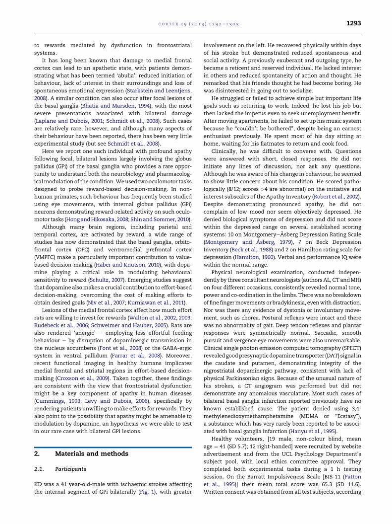
KD was a 41 year-old-male with ischaemic strokes affecting
the internal segment of GPi bilaterally (Fig. 1), with greater
involvement on the left. He recovered physically within days
of his stroke but demonstrated reduced spontaneous and
social activity. A previously exuberant and outgoing type, he
became a reticent and reserved individual. He lacked interest
in others and reduced spontaneity of action and thought. He
remarked that his friends thought he had become boring. He
was disinterested in going out to socialize.
He struggled or failed to achieve simple but important life
goals such as returning to work. Indeed, he lost his job but
then lacked the impetus even to seek unemployment benefit.
After moving apartments, he failed to set up his music system
because he “couldn’t be bothered”, despite being an earnest
enthusiast previously. He spent most of his day sitting at
home, waiting for his flatmates to return and cook food.
Clinically, he was difficult to converse with. Questions
were answered with short, closed responses. He did not
initiate any lines of discussion, nor ask any questions.
Although hewas aware of his change in behaviour, he seemed
to show little concern about his condition. He scored patho-
logically (8/12; scores >4 are abnormal) on the initiative and
interest subscales of the Apathy Inventory (Robert et al., 2002).
Despite demonstrating pronounced apathy, he did not
complain of low mood nor seem objectively depressed. He
denied biological symptoms of depression and did not score
within the depressed range on several established scoring
systems: 10 on MontgomeryeAsberg Depression Rating Scale
(Montgomery and Asberg, 1979), 7 on Beck Depression
Inventory (Beck et al., 1988) and 2 on Hamilton rating scale for
depression (Hamilton, 1960). Verbal and performance IQ were
within the normal range.
Physical neurological examination, conducted indepen-
dentlyby threeconsultantneurologists (authorsAL,CTandMH)
on four different occasions, consistently revealed normal tone,
power and co-ordination in the limbs. Therewas no breakdown
of finefingermovements orbradykinesia, evenwithdistraction.
Nor was there any evidence of dystonia or involuntary move-
ment, such as chorea. Postural reflexes were intact and there
was no abnormality of gait. Deep tendon reflexes and plantar
responses were symmetrically normal. Saccadic, smooth
pursuit and vergence eye movements were also unremarkable.
Clinical single photon emission computed tomography (SPECT)
revealedgoodpresynapticdopamine transporter (DAT) signal in
the caudate and putamen, demonstrating integrity of the
nigrostriatal dopaminergic pathway, consistent with lack of
physical Parkinsonian signs. Because of the unusual nature of
his strokes, a CT angiogram was performed but did not
demonstrate any anomalous vasculature. Most such cases of
bilateral basal ganglia infarction reported previously have no
known established cause. The patient denied using 3,4-
methylenedioxymethamphetamine (MDMA or “Ecstasy”),
a substance which has very rarely been reported to be associ-
ated with basal ganglia infarction (Hanyu et al., 1995).
Healthy volunteers, [19 male, non-colour blind, mean
age ¼ 41 (SD 5.7); 12 right-handed] were recruited by website
advertisement and from the UCL Psychology Department’s
subject pool, with local ethics committee approval. They
completed both experimental tasks during a 1 h testing
session. On the Barratt Impulsiveness Scale [BIS-11 (Patton
et al., 1995)] their mean total score was 65.3 (SD 11.6).
Written consentwas obtained from all test subjects, according
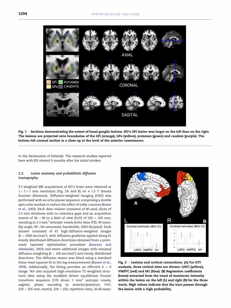
Fig. 1 e Sections demonstrating the extent of basal ganglia lesions. KD’s GPi lesion was larger on the left than on the right.
The lesions are projected onto boundaries of the GPi (orange), GPe (yellow), putamen (green) and caudate (purple). The
bottom left coronal section is a close up at the level of the anterior commissure.
c o r t e x 4 9 ( 2 0 1 3 ) 1 2 9 2e1 3 0 31294
to the Declaration of Helsinki. The research studies reported
here with KD started 9 months after his initial strokes.
Fig. 2 e Lesions and cortical connections. (A) For DTI
analysis, three cortical sites are shown: LOFC (yellow),
VMPFC (red) and M1 (blue). (B) Regression coefficients
(betas) extracted from the voxel of maximum intensity
within the lesion on the left (L) and right (R) for the three
tracts. High values indicate that the tract passes through
the lesion with a high probability.
2.2. Lesion anatomy and probabilistic diffusiontractography
T1-weighted MR acquisitions of KD’s brain were obtained at
1� 1� 1 mm resolution (Fig. 2A and B) on a 1.5 T Sonata
Scanner (Siemens). Diffusion-weighted imaging (DWI) was
performed with an echo planar sequence comprising a double
spin-echo module to reduce the effect of eddy currents (Reese
et al., 2003). Each data volume consisted of 40 axial slices of
2.3 mm thickness with no interslice gaps and an acquisition
matrix of 96 � 96 in a field of view (FoV) of 220 � 220 mm,
resulting in 2.3 mm3 isotropic voxels [echo time (TE), 90 msec;
flip angle, 90�; fat saturation; bandwidth, 2003 Hz/pixel]. Each
dataset consisted of 61 high-diffusion-weighted images
(b ¼ 1000 sec/mm2), with diffusion gradients applied along 61
evenly distributed diffusion directions obtained from a previ-
ously reported optimization procedure (Jansons and
Alexander, 2003) and seven additional images with minimal
diffusion weighting (b ¼ 100 sec/mm2) and evenly distributed
directions. The diffusion tensor was fitted using a standard
linear least squares fit to the log measurements (Basser et al.,
1994). Additionally, the fitting provides an effective b ¼ 0
image. We also acquired high-resolution T1-weighted struc-
tural data using the modified driven equilibrium Fourier
transform sequence [176 slices; 1 mm3 isotropic voxels;
sagittal, phase encoding in anterior/posterior; FoV,
224 � 256 mm; matrix, 224 � 256; repetition time, 20.66 msec;

c o r t e x 4 9 ( 2 0 1 3 ) 1 2 9 2e1 3 0 3 1295
TE, 8.42 msec; inversion time, 640 msec; flip angle, 25�; fat
saturation; bandwidth, 178 Hz/pixel] (Deichmann, 2006).
Several recent human atlases were used to establish the
extent of KD’s lesions. Note that atrophy secondary to
neuronal degeneration means that there is distortion of
normal anatomy, in addition to the lesions themselves. It is
therefore important to be familiar with such changes when
interpreting these images. KD’s lesions largely involved the
GPi, more prominently on the left. There was no clear
involvement of the habenula, subthalamic nucleus (STN),
septum, medial hypothalamus, midline thalamic nuclei, and
bed nucleus of stria terminalis, verified using a MR adapted
version (Krauth et al., 2010) of the Morel histologically-based
probabilistic atlas (Morel, 2007). Although part of the GPe
may have been affected on the left, the lesions are largely
within the GPi as shown in Fig. 1 of the text. Both the patient’s
MRI scan and the atlas were registered to the standardised
Montreal Neurological Institute (MNI) space.We use a recently
validated atlas of the pallidum (Prodoehl et al., 2008) and
found lack of extensive involvement of the GPe.
In addition, to establish which cortical regions were most
likely to be deafferented, diffusion-weighted data from 12
healthy aged-matched male subjects following the algorithm
of Draganski et al. (2008). After automated cortical and
subcortical parcellation using FreeSurfer (http://surfer.nmr.
mgh.harvard.edu) we performed probabilistic diffusion trac-
tography in subject-specific native space using a probabilistic
index of connectivity (PICo) algorithm (Parker and Alexander,
2003, 2005) implemented in Camino software (http://www.cs.
ucl.ac.uk/research/medic/camino/). To delineate the projec-
tion sites of specific cortical areas on the pallidum (Fig. 2A) we
implemented a two stage probabilistic tractography approach:
(i) probabilistic tractography from caudate to cortical targets
as defined in FreeSurfer (LOFC e lateral orbitofrontal cortex,
M1 e precentral and paracentral gyrus) and (ii) probabilistic
tractography from pallidum to caudate after definition of the
specific cortical projection sites. We calculated voxel-based
PICo maps for the pallidum seed structures to each target
area and transformed the individual maps to standard MNI
space using parameter estimates from each individual’s
T1-weighted data.
Statistical analysis was performed within the SPM8 frame-
work. After automated lesion detection using SPM8, we used
KD’s bipallidal lesionmap in standard space to test the pattern
of connectivity profiles of these lesion locations in 12 healthy
subjects. The search volume was restricted to the internal and
external pallidum as defined in the Basal Ganglia Human Area
Template (Prodoehl et al., 2008). We tested the significance of
the probability of the tracts passing through the lesion using an
F-test: regression coefficientswith 90% confidence intervals are
presented in Fig. 2B. Post-hoc t-tests were used to identify
differences in PICo between the three tracts to LOFC, VMPFC
and M1. Data was thresholded at the level of p< .0001 uncor-
rected for multiple comparisons within the described search
volume.
2.3. Experiment 1jtraffic lights task (TLT)
We investigated rapid decision-making under risk for reward
using a ‘traffic lights task’ (TLT) (Adam et al., 2012). Participants
fixated a red light (3� diameter) for 1000msec that successively
turned amber and then green (Fig. 3) which was the signal to
make a saccade to a target at 20� horizontal eccentricity. Amber
duration was drawn probabilistically from a Gaussian distri-
bution (mean 750 msec, SD 125 msec; Fig. 2B). Rewards
depended upon saccadic reaction time (SRT), according to an
exponential discounting function; Fig. 3C). Saccades made
before green onset were penalized with a small, flat penalty.
Because saccades take w200 msec to initiate, any highly
rewarded responses (latencies< 200 msec) have to be pro-
grammed before green onset. Thus to maximize outcome,
subjects needed to make a decision about whether to initiate
a response before the green light e and potentially obtain
a high reward, but risk a penaltye or simplywait for the green
light when they will receive a low reward. Participants were
instructed to make as much money as possible. They per-
formed ten blocks of fifty trials.
Reward (in pence) was calculated from acquiring the target
using a decay function:
R ¼ a e�
�t� t0k1
�
a ¼ 150, k1 ¼ 100 and t � t0 represents RT from green onset
(msec).
Saccades made in advance of “GO!” were punished by
a fixed fine of 10p. Rewardswere displayed at the target site on
each trial and a cumulative total was shown below this. Aural
feedback was also given with a ‘ping’ for rewards of 0e19p,
and a ‘ker-ching’ for rewards of 20p ormore. An error trial was
accompanied by a low pitched ‘beep’ in addition to a visual
cue: “STOP Police! Fine £0.10”. Eye positionwas recorded using
an EyeLink 1000 Hz eye tracker (SR Research Ltd, Ontario,
Canada). Stimuli were displayed on a 22ʺ CRTmonitor (150 Hz)
at 60 cm.
2.3.1. Linear rise-to-threshold modellingIt is not possible to establish definitively for any individual
saccade whether it arose from an anticipatory or a reactive
process. Because humans takew200 msec to plan and execute
saccades, ‘reactive’ saccades e those made in response to
green onset e are expected to have latencies of this order.
Very early saccades (say< 50 msec after green onset) are likely
to have been ‘anticipatory’, planned prior to green onset.
However, there is a grey zone between these extremes.
We used an establishedmethod to decide howmany of the
saccades were statistically most likely to arise from each
distribution, modelled by a linear rise-to-threshold process
(Carpenter and Williams, 1995). We assumed two processes,
one triggered by the amber light and the other by the green.
Thus, the distribution of reactive saccades is described by
a rapid rise-to-threshold process elicited by green onset.
Whereas anticipatory saccades are described by a slower and
independent rise-to-threshold process triggered by amber
onset. A saccade is generated by whichever process reaches
threshold first (Adam et al., 2012).
Maximum likelihood estimation provided best-fitting
mean and variance parameters for each distribution. For
controls, the model estimated a mean for the reactive distri-
bution of 299 msec, SD 31 msec. We used a ‘cut off’ maximum

Fig. 3 e Traffic lights task (TLT). (A) Subjects fixated a circle which successively turned red, amber and green. They were
required not to move their eyes until the onset of the green light, otherwise they receive a small (constant) fine or
punishment. To maximize reward, participants had to make a saccade to the contralateral target as quickly as possible after
green light onset. (B) Amber durations were selected at random from a normal distribution (mean [ 750 msec,
SD [ 125 msec). (C) Reward was calculated with a hyperbolically decaying function with a maximum value of 150 pence
(£1.50) when SRT was zero. Thus to maximize reward subjects should program an eye movement to coincide with green
light onset. However, amber durations were not constant and therefore they either had to take a risk (high reward or
punishment) or wait for the green light before programming a saccade (low reward).
c o r t e x 4 9 ( 2 0 1 3 ) 1 2 9 2e1 3 0 31296
saccadic RT of 200 msec, >3 SDs from this mean, to delineate
anticipatory saccades.
2.4. Experiment 2jdirectional reward-sensitivitysaccade task
We also employed a second paradigm (Fig. 4) to investigate
reward-dependent modulation of behaviour: speeding of
saccades to rewarded targets (Hong and Hikosaka, 2008).
Participants fixated a central cross (3� diameter) for 1000 msec
andmade saccades as quickly as possible to a target, 10� to the
left or right (50% probability). Saccades to targets on only one
side were rewarded depending upon reaction time (with
a discounting function as for the TLT), and the rewarded side
(RS) was altered, without warning, after a series of trials.
Rewards were acknowledged by the display of a pound coin
and a number representing the reward magnitude in pence.
Reward value was dependent on latency using a function
similar to that in the TLT. The RS changed every 10e14 trials.
Participants performed two blocks of 120 trials. The difference
in SRTs to the RS and unrewarded sides (US) was the measure
of reward-sensitivity.
2.5. Dopaminergic drug challenges
KD received a single dose of Madopar 125 mg (100 mg L-dopa
with a peripheral dopa-decarboxylase inhibitor, benserazide
25 mg), directly after the baseline tests. He was reassessed an
hour later when peak L-dopa levels are reached. To assess
whether any effects on L-dopa were due to simply more
experience on the tasks, six controls were also tested an hour
after performing their first session. A second group of controls
(N¼ 12) also received the same dose of L-dopa but in double-
blind randomized fashion, receiving placebo/drug one week
apart.
KD was then given slowly increasing doses, reaching
Madopar CR (long-acting preparation) 125 mg three times
daily after eight weeks. Although there was moderate
improvement in apathy, it was decided that there might be
better response with a direct dopamine receptor agonist.

Fig. 4 e Directional saccadic reward task. Participants attended a central fixation spot which was extinguished after
1000 msec of fixation. They then made a saccade as fast as possible to a target presented either to the left or right (50% each
side). One side was rewarded while the other received no reward. The rewarded side (RS) remained constant for an
unpredictable number of trials before switching to the other side.
c o r t e x 4 9 ( 2 0 1 3 ) 1 2 9 2e1 3 0 3 1297
L-dopa was therefore slowly discontinued and KD was off
medication for 4 weeks (‘drug holiday’) before starting on the
dopamine agonist ropinirole, initially .25mg three times a day
for 1 week, then increasing by .25mg every week eventually to
reach 1 mg thrice daily after three weeks. After a further four
weeks he was established on 4 mg once daily of the long-
acting formulation of ropinirole (Requip XL).
3. Results
3.1. Lesion anatomy and probabilistic tractography data
KD’s lesions (Fig. 1) involved the GPi bilaterally, with greater
involvement on the left. These lesionswere not complete and it
is important to note that part of the GPi was spared. Using
a recently validated atlas of the pallidum (Prodoehl et al., 2008)
we found only modest damage to GPe (external segment of the
GPi) on the left. Therewasno involvementof thehabenula, STN,
septum, medial hypothalamus, midline thalamic nuclei, and
bed nucleus of stria terminalis, verified using a MR adapted
version (Krauth et al., 2010) of a histological atlas (Morel, 2007).
Probabilistic diffusion tractography (Fig. 2)wasused to examine
the topography of pallidal connections to three cortical regions
(Draganski et al., 2008). The region of GPiwhich ismost strongly
connected to LOFC and VMPFC was particularly affected,
compared with projections to primary motor cortex (M1), more
so on the left: VMFC>M1 left Z¼ 5.41, right Z¼ 3.51; LOFC>M1
Z ¼ 5.33, right Z ¼ 3.52 (all p < .001, uncorrected).
3.2. Experiments 1 and 2jbaseline performance
On the TLT (Fig. 3) SRTs in controls demonstrated a bimodal
distribution (Fig. 5A). One population peaked w280 msec after
green onset, consistent with saccades made ‘reactively’
following the GO signal. In addition, there was an early pop-
ulation with a peaking 63 msec after green onset. To demar-
cate these two distributions we used linear rise-to-threshold
modelling, assuming two independent processes, the first
triggered by amber light onset and the second by the green
light (Adam et al., 2012). The early, anticipatory responses
were further divided into errors (saccades before green onset)
and correct anticipations (saccades after green onset, but
planned in advance of it). ‘Reactive’ saccades were classified
as those after 200 msec (see Methods).
Controls demonstrated a high proportion of early responses
(mean 42% saccades, SD 18.95). Half were correct anticipations
(21%, SD 8.64). The rest were errors (21%, SD 14.35). Overall
mean Correct Anticipations: Errors Ratio (CAjER) ratio was 1.53
(SD .87), withmean reward 18p/trial (SD 4.6p). CAjER correlated
well with mean reward obtained (R2¼ .77; p< .0001).
In contrast, KD’s distribution of saccades was unimodal,
with most made after green onset (Fig. 5B). Nearly all his eye
movements were reactive, with only 8.0% early responses,
significantly different from controls (Z¼ 2.8, p¼ .003). Further-
more, the majority of these were errors; correct anticipations
formed only 2.2% of saccades (Z¼ 2.8, p¼ .003). His CAjER was
.4 and he obtained only 14p/trial.
Within the first session, controls gradually increased the
proportion of early responses (Fig. 6A), with a significant
difference between the first 100 trials (30.5% early responses,
SD 25.20) and the third (44.6%, 21.24; p < .05). There was also
a trend for CAjER to increase from the beginning to the end of
the session ( p¼ .08). In contrast to controls, KD showed no
evidence of learning with 8% early responses in the first 100
trials to 7% in the last (Fig. 6A).
On the directional reward-sensitivity saccade task (Fig. 4)
controls showed a small, but significant SRT advantage to the
RS (mean RS 206 msec vs US 219 msec; p¼ .03) (Fig. 7). This
sensitivity to reward did not change significantly over the first
session [analysis of three forty-trial epochs F(5,66)¼ .24,
p> .9]. By contrast, KD showed no significant difference
between rewarded versus unrewarded saccades (mean
US ¼ 236 msec vs RS ¼ 235 msec; p> .5; Fig. 7), and there was
no significant change across epochs. His SRTs were longer
than control means but within normal range.
3.3. Experiments 1 and 2jdopaminergic modulation
On the TLT, KD’s performance altered dramatically 1 h after
a single dose of L-dopa 100 mg (Figs. 5C and 6B). His early
responses increased, with a CAjER of 4.20 (6.67 SD > control

Fig. 5 e Traffic lights task (TLT): saccadic distributions. (A) Saccades for age-matched controls (n[ 13) performing the TLT
two distinct distributions: an early, anticipatory distribution and a later, reactive one made in response to green light onset.
Early responses were divided into errors (saccades before the green light came on) and correct anticipations (saccades
with< 200 msec latency after the green light). The plot here is for a total of 6500 saccades. (B) Pre-treatment, KD made
mostly reactive saccades (461/500 trials [92.2%]) with a median latency of 248 msec. He made very few anticipatory
saccades. (C) After treatment with L-DOPA 100mg (Madopar CR 125mg) three times a day for 12 weeks, there was a dramatic
increase in early responding in KD. (D) After 12 weeks treatment with a dopamine agonist (ropinirole XL, 4 mg once a day),
KD’s distribution of saccades looks most similar to that of control subjects.
c o r t e x 4 9 ( 2 0 1 3 ) 1 2 9 2e1 3 0 31298
mean of 2.20, SD .30) and overall increase in reward. Over the
session, his early responses increased (14% in first 100 trials to
43% in the last; Fig. 6B).
Six controls also performed 500 trials an hour after the first
session, butwithout L-dopa. Their proportionof early responses
did not change significantly from the end of the first session
(45%) to the end of the second (48%; p> .1; Fig. 6A and B). The
same dose of L-dopa in 12 controls, tested in double-blind
fashion, had no significant effect on SRTs (drug mean
306 msec, SD 121 vs 298 msec, SD 95 on placebo) or reward ob-
tained (drug mean 23p/trial vs 24p/trial placebo). Thus L-dopa
increasedanticipatory saccades inKDbutnot inhealthypeople.
The effect in KD was the largest increase in early responses
from baseline of any subject who was tested twice, with or
without L-dopa.
On the directional reward-sensitivity task (Fig. 7), following
L-dopa KD now showed a markedly significant preference for
the RS, apparent within the first epoch of forty trials (RS
211 msec vs US 238 msec; p¼ .002). Six subjects similarly
performed a repeat session 1 h after the first, but without
L-dopa. They demonstrated no further change in behaviour
[F(11,60) ¼ .7, p > .5]. In addition, eight controls tested in
double-blind fashion on the same dose of L-dopa/placebo
demonstrated reward-sensitivity, as previously. However,
there was no further significant modulation by L-dopa (mean
RS ¼ 209 msec vs US ¼ 219 msec placebo, p< .001; 214 msec
and 219 msec on L-dopa, p< .01). Thus L-dopa speeded
saccades to rewarded targets in KD but not in healthy people.
After eight weeks on L-dopa, KD showed moderate
improvement in apathy. Concomitantly, the difference in SRT
to US and RS was much larger than in controls, a consistent
finding across all testing sessions (Fig. 7). Twelve weeks after
initiating therapy, the difference between US and RS saccades
was 36msec (RS ¼ 206 msec vs US ¼ 242 msec; p< .0001). In

Fig. 6 e Percentage early responses on traffic lights task (TLT) over time. (A) Over the course of the first session, healthy
controls showed increased early responses but KD did not. (B) In the second session, an hour later, controls showed no
further change but KD 1 h after receiving L-dopa showed escalating early responses. (C) During the drug holiday period (off L-
dopa), KD’s early responses reverted to pre-treatment levels.
c o r t e x 4 9 ( 2 0 1 3 ) 1 2 9 2e1 3 0 3 1299
isolation, these findings might be attributed to practice.
However, SRTs to unrewarded targets actually increased while
those to rewarded ones decreased, so the effects cannot be
attributed to a simple generalized motor facilitation with
practice and/or L-dopa.
Fig. 7 e Results from the directional saccadic reward task.
The control group (n[ 12, arrows to side) showed
a preference for the rewarded target locations, with
significantly shorter SRTs. KD showed no reward
preference at baseline, before treatment (Session 1). In
Session 2 he was given a single dose (100 mg) of levodopa
which led to a significant reward preference. This was
maintained throughout chronic dopaminergic therapy
(Sessions 3 Madopar 125 mg three times daily for 4 weeks,
Session 4 Madopar CR 125 mg three times daily for
12 weeks). Following a treatment holiday (4 weeks), this
reward preference was absent (Session 5). However, with
subsequent treatment on the dopamine agonist ropinirole
(1 mg three times a day), there was both a re-establishment
of reward preference and significant decrease in latency to
both rewarded and unrewarded targets. Error bars are D/L
1 SEM (standard error of the mean).
On the TLT, performance reached a peak by 24weeks L-dopa
therapy when 33.4% of KD’s saccades were now early
responses, with 23.6% correct and 9.8% errors (CAjER¼ 2.41 and
mean reward now 23.2p/trial). However, a clinical decision was
made to stop L-dopa and assess instead the effects of a dopa-
mine agonist which acts directly at dopaminergic receptors.
Off medication, the difference in SRTs to RS and US targets
became non-significant (Fig. 7), providing further evidence
that reward-sensitivity observed in the previous sessions
could not simply be attributed to practice. However, saccades
were generally faster than before treatment, suggesting that
there was some general practice effect that might have
contributed non-specifically to speeding responses to both US
and RS targets. On the TLT, off medication, the effects on
L-dopa were also partly reversed with early responses strik-
ingly reduced (Fig. 6C) and overall reward dipping to 13.7p/trial
and CAjER ¼ .79.
KD started on an increasing dose of ropinirole, an agonist
acting largely D2 and D3 dopamine receptors. By contrast,
L-dopa would have a balanced effect across all these receptors
by increasing synaptic dopamine. On 4 mg ropinirole daily
there was marked improvement in KD’s apathy. He was far
more spontaneous in conversation, reported better social
interactions and was more interested in events around him.
He managed to secure a job and now scored in the normal
range (4/12) on the initiative and interest subscales of the
Apathy Inventory (Robert et al., 2002).
On the directional reward-sensitivity task, saccades were
generally faster, but those to the RS were significantly faster
(RS ¼ 183 msec vs US ¼ 208 msec; p< .001), far larger than in
controls (Fig. 7). On the TLT by week four (on 4 mg ropinirole
daily) KD demonstrated much greater early responding
(45.2%). However, this was at the expense of greater numbers
of errors (17.8% vs control mean ¼ 24.2%) so the CAjER (1.54)
was not as high as on L-dopa. Despite this, mean reward
(27.3p/trial) exceeded that achieved on L-dopa, matching the
highest performing individual healthy control. Thus KD
showing increased willingness to anticipate frequently and
take risks, an effect that persisted over 12 weeks on ropinirole
(Fig. 5D).
c o r t e x 4 9 ( 2 0 1 3 ) 1 2 9 2e1 3 0 31300
4. Discussion
We used novel probes of oculomotor decision-making to
demonstrate relative insensitivity to reward in an individual
with apathy following bilateral GPi lesions. Our TLT (Adam
et al., 2012) requires reward sensitivity and motivation or
effort to succeed, combined with fast reaction times and the
ability to update behaviour in response to positive and nega-
tive feedback. A reactive response e simply waiting for the
green light e is less well rewarded than an anticipatory
response prepared in advance of the green signal. KD initially
made very few anticipatory responses compared with age-
matched controls. However, dopaminergic therapy, first with
levodopa and then with ropinirole, increased anticipatory
responses to within the normal range.
The directional saccade reward-sensitivity task, originally
developed for the study of reward sensitivity in macaque
monkeys (Hong and Hikosaka, 2008), demonstrated that KD
had SRTs within the normal range but showed no speeding to
the rewarded side (RS), unlike healthy volunteers. Treatment
with levodopa led to reward sensitivity, with speeding of
responses to the RS and slowing to the unrewarded side (US)
compared to baseline. Off medication, the difference in SRTs
to rewarded and unrewarded targets became non-significant,
while subsequently on ropinirole, a direct dopamine D2/D3
receptor agonist, KD again demonstrated reward sensitivity,
as well as generalized speeding.
These effects on dopaminergic medication were associated
with clinical improvemente reductionof apathy and increased
motivation to find work and in social interactions e most
prominently while on the dopamine agonist. The findings
demonstrate a causal relationship between basal ganglia
function and motivation or willingness to make an effort for
reward. They provide proof-of-concept data for the treatment
of apathy which is increasingly recognized to be a key compo-
nent of several neurological disorders (Bonelli and Cummings,
2008; Marin, 1991; Chow et al., 2009; Starkstein, 2009).
Unlike other tasks involving risk, such as the Iowa
Gambling Task (Bechara et al., 1994) or the Cambridge Gamble
Task (Clark et al., 2004), our TLT requires participants to take
risks by making anticipatory responses. Many other para-
digms place certain and risky options on an equal footingwith
the same amount of effort required for both choices. This has
the benefit of establishing risk preferences independently of
effort but tends to favour a careful, deliberative response
strategy. The traffic lights paradigm imposes time constraints
on decisions and rewards behaviour that might be considered
‘functionally impulsive’ (Dickman, 1990): on this task, it can be
functionally useful to make anticipatory responses because
these can lead to greater rewards, analogous to many situa-
tions in real life. It is possible that KD’s lack of anticipatory
responses on this task reflects risk aversion, rather than lack of
motivation or unwillingness to make an effort for rewards.
However, it is less easy to explain how such a mechanism
might account for behaviour on the directional saccadic task,
where there was no risk of incurring a penalty.
How did dopamine reverse apathy and reward insensi-
tivity? Substantial evidence links dopamine to reinforcement
learning (Schultz, 2007). However a growing body of research
also implicates dopamine in effort-based decision-making,
generating the motivation and vigour to overcome costs of
initiating actions (Niv et al., 2007; Kurniawan et al., 2011). The
progressive improvement of KD’s performance on the TLT
immediately post L-dopa (Fig. 6B) is suggestive of dopami-
nergic enhancement of learning. However, during the drug
holiday period such learning was radically reversed (Fig. 6C),
suggesting that if this effect was solely due to a reinforcement
learning effect of L-dopa it had not been completely consoli-
dated. Dopamine was still required to maintain it.
On the directional reward-sensitivity task, L-dopa also had
a dramatic effect after its introduction, speeding saccades to
the RS (Fig. 7). During the drug holiday, however, there was no
longer any significant reward-sensitivity but saccades were
generally faster than before treatment, suggesting there were
some general, non-specific effects of practice on the task. The
time course of action on reward-sensitivity and its reversal
during the drug holiday makes it unlikely that dopaminergic
effects on synaptic plasticity and learning were the only
mechanism of action. Instead, it might also have had an effect
on response vigour or overcoming costs of effort (Niv et al.,
2007; Kurniawan et al., 2011).
Dopamine could act directly on brain systems left intact
after stroke, but perhaps disconnected because the major
outflow from the basal ganglia is via the GP. Alternatively,
because the GPi lesions were not complete in KD, it is possible
that his lesions led to imbalance in cross-talk between striatal
regions which could be ameliorated by dopamine therapy. It
has been demonstrated that parallel corticostriatal loops
through the basal ganglia need not operate in isolation but can
instead communicate with each other, e.g., via spiralling
striato-nigro-striatal connections (Haber et al., 2000) which
allow ventral striatal regions to influence more dorsal striatal
areas. Moreover, the nigrostriatal system is not the only
dopaminergic modulator of basal ganglia function; the intra-
striatal dopaminergic system is complex and can alter with
denervation (Smith and Kieval, 2000). Finally, it is important
also to consider the possibility that the effects of dopamine
observed in KDmight arise from indirect, knock-on effects on
other neurotransmitter systems, e.g., there is evidence of
interactions between dopaminergic and noradrenergic
systems (Hara et al., 2010) as well as several other neuro-
transmitters (see Steiner and Tseng, 2010, for reviews).
In macaques, using the directional reward saccade task,
Hong and Hikosaka (2008) found that saccades to the RS with
shorter latency than to the US, with reward-related speeding
being associated with activity in GPi neurons which project to
the lateral habenula. If a homologous circuit operates in the
human brain, it is likely to have been partially disrupted in KD
in whom both GPi were damaged. However, the lateral habe-
nula remained intact, togetherwith the caudate and putamen.
Furthermore, SPECT imaging of the DAT demonstrated that
the nigrostriatal dopaminergic pathway was intact as there
was good signal bilaterally in the caudate and putamen of KD.
Thus one locus of dopaminergic drug action is potentially the
intact caudate, putamen or even surviving parts of the GP
complex.
Another potential site of action of dopamine is prefrontal
cortex. The OFC, in concert with basal ganglia structures, is
considered to have a special role in the processing of reward
c o r t e x 4 9 ( 2 0 1 3 ) 1 2 9 2e1 3 0 3 1301
signals (Schultz, 2000; Kringelbach and Rolls, 2004; Wallis,
2007). Projection of KD’s lesion onto the known topography
of the pallidal trans-thalamic connections to the cortex,
determined using diffusion-weighted tractography (Draganski
et al., 2008), suggests that the connections to the VMPFC and
OFC have most likely been disrupted (Fig. 2). OFC neurons not
only respond selectively to reward or aversive stimuli, but also
signal relative preference for rewards and may integrate
different types of information to compute a representation of
value (Thorpe et al., 1983; Tremblay and Schultz, 1999; Padoa-
Schioppa and Assad, 2006; Wallis and Kennerley, 2010).
Consistent with these neurophysiological findings in
macaque monkeys, imaging studies in humans have
described activations in OFC and VMPFC which correlate with
behavioural measures of stimulus value (O’Doherty, 2004;
Plassmann et al., 2007; Rangel and Hare, 2010; Haber and
Knutson, 2010; Glascher et al., 2009; Blair et al., 2006).
Lesions of the OFC in humans lead to impaired decision-
making about the expected outcome of choices (Bechara
et al., 1998) while alterations in striatal dopamine binding in
drug addicts is associated with hypoactivity in OFC (Volkow
et al., 2009). Dopaminergic neurons are known to innervate
prefrontal cortex, includingOFC (Williams and Goldman-Rakic,
1993). Although these arise from midbrain dopaminergic pop-
ulations, partial disconnection of OFC neurons from trans-
thalamic pallidal inputs e as is likely in KD e might disrupt
dopaminergic reward signals within OFC. This view is
compatible with recent functional imaging evidence that
dopamine agonists might alter decision-making and risk-
taking in susceptible individuals with Parkinson’s disease via
actions on OFC (van Eimeren et al., 2009).
Intriguingly, previous work also suggests that a dopami-
nergic deficit might be an important contributory factor to
apathy in Parkinson’s disease, which occurs in up to 60% of
cases (Oguru et al., 2010). Patients who undergo STN deep
brain stimulation (DBS) often require reduction or withdrawal
of dopaminergic therapy because of improvements in motor
control following surgery. Czernecki et al. (2008) reported that
apathy occurred after dopamine withdrawal in some of these
cases, but importantly it could be reversed with ropinirole.
More recently, a PET study has demonstrated greater meso-
corticolimbic dopaminergic denervation involving the OFC in
Parkinson’s disease patients who develop postoperative
apathy compared to those who do not (Thobois et al., 2010).
Regardless of the precise locus of drug action in KD, it is
clear that his lesions rendered him apathetic but this could be
ameliorated by dopaminergic modulation. Alteration in
reward-sensitivity mirrored clinical changes, suggesting
apathy in this case is associated with lack of motivation to
obtain rewards. Animal learning theory has proposed that
rewards might in fact constitute the basic goals of voluntary
behaviour (Dickinson and Balleine, 1994). From this perspec-
tive, the absence of sensitivity to rewards would be expected to
have devastating consequences for goal-directed action, just as
one observes in apathy. But note that although this viewmight
account for behaviour in our particular case, apathy is most
likely to be a syndrome that is multidimensional (Cummings,
1993; Levy and Dubois, 2006). In different clinical contexts, it
could potentially result from deficits in other cognitive
components of the decision-making process. Further studies
are required to delineate these components and which specific
deficits occur in different clinical conditions. Our study repre-
sents progress towards understanding one component of
apathy e namely, relative reward insensitivity.
Although cases such as KD with bilateral GPi lesions are
rare, apathy is common in Parkinson’s disease (Oguru et al.,
2010; Pedersen, et al., 2009; Starkstein, 2009), as well as in
other neurodegenerative disorders, including Huntington’s
and Alzheimer’s disease (Bonelli and Cummings, 2008; Chow
et al., 2009; Starkstein et al., 2006; Marin, 1991). These condi-
tions often involve disruption of cortico-striato-thalamo-
cortical loops (Alexander et al., 1986) but the mechanisms
underlying apathy when there is widespread neuro-
degeneration has been difficult to study. Focal lesion cases
such as KD provide important information about the neural
substrates underlying apathy and modulation of this behav-
ioural state with neuropharmacological intervention.
Acknowledgements
This research was supported by The Wellcome Trust and
NIHR BRC at UCLH/UCL. We thank KD for his participation in
these studies.
r e f e r e n c e s
Adam R, Bays PM, and Husain M. Rapid decision-making underrisk. Cognitive Neuroscience, 3(1): 52e61, 2012.
Alexander GE, DeLong MR, and Strick PL. Parallel organization offunctionally segregated circuits linking basal ganglia andcortex. Annual Review of Neuroscience, 9: 357e381, 1986.
Basser PJ, Mattiello J, and LeBihan D. MR diffusion tensorspectroscopy and imaging. Biophysical Journal, 66(1): 259e267,1994.
Bechara A, Damasio A, Damasio H, and Anderson S. Insensitivityto future consequences following damage to humanprefrontal cortex. Cognition, 50(1e3): 7e15, 1994.
Bechara A, Domasio H, Tranel D, and Anderson SW. Dissociationof working memory from decision making within the humanprefrontal cortex. The Journal of Neuroscience, 18(1): 428e437,1998.
Beck A, Steer R, and Garbin M. Psychometric properties of theBeck Depression Inventory e 25 years of evaluation. ClinicalPsychology Review, 8(1): 77e100, 1988.
Bhatia KP and Marsden CD. The behavioural and motorconsequences of focal lesions of the basal ganglia in man.Brain: A Journal of Neurology, 117(Pt 4): 859e876, 1994.
Blair K, Marsh AA, Morton J, Vythilingam M, Jones M, Mondillo K,et al. Choosing the lesser of two evils, the better of two goods:Specifying the roles of ventromedial prefrontal cortex anddorsal anterior cingulate in object choice. The Journal ofNeuroscience, 26(44): 11379e11386, 2006.
Bonelli RM and Cummings JL. Frontalesubcortical dementias.The Neurologist, 14(2): 100e107, 2008.
Carpenter RH and Williams ML. Neural computation of loglikelihood in control of saccadic eye movements. Nature,377(6544): 59e62, 1995.
Chow TW, Binns MA, Cummings JL, Lam I, Black SE, Miller BL,et al. Apathy symptom profile and behavioral associations infrontotemporal dementia vs dementia of Alzheimer type.Archives of Neurology, 66(7): 888e893, 2009.

c o r t e x 4 9 ( 2 0 1 3 ) 1 2 9 2e1 3 0 31302
Clark L, Cools R, and Robbins TW. The neuropsychology of ventralprefrontal cortex: Decision-making and reversal learning.Brain and Cognition, 55(1): 41e53, 2004.
Croxson PL, Walton ME, O’Reilly JX, Behrens TE, andRushworth MF. Effort-based cost-benefit valuation and thehuman brain. The Journal of Neuroscience: The Official Journal ofthe Society for Neuroscience, 29(14): 4531e4541, 2009.
Cummings JL. Frontalesubcortical circuits and human behavior.Archives of Neurology, 50(8): 873e880, 1993.
Czernecki V, Schupbach M, Yaici S, Levy R, Bardinet E, Yelnik J,et al. Apathy following subthalamic stimulation in ParkinsonDisease: A dopamine responsive symptom. MovementDisorders, 23(7): 964e969, 2008.
Deichmann R. Fast structural brain imaging using an MDEFTsequence with a FLASH-EPI hybrid readout. NeuroImage, 33(4):1066e1071, 2006.
Dickinson A and Balleine B. Motivational control of goal-directedaction. Animal Learning Behaviour, 22: 1e18, 1994.
Dickman SJ. Functional and dysfunctional impulsivity:Personality and cognitive correlates. Journal of Personality andSocial Psychology, 58(1): 95e102, 1990.
Draganski B, Kherif F, Kloppel S, Cook PA, Alexander DC,Parker GJM, et al. Evidence for segregated and integrativeconnectivity patterns in the human basal ganglia. Journal ofNeuroscience, 28(28): 7143e7152, 2008.
Farrar AM, Font L, Pereira M, Mingote S, Bunce JG, Chrobak JJ, et al.Forebrain circuitry involved in effort-related choice: Injectionsof the GABAA agonist muscimol into ventral pallidum alterresponse allocation in food-seeking behavior. Neuroscience,152(2): 321e330, 2008.
Font L, Mingote S, Farrar AM, Pereira M, Worden L, Stopper C,et al. Intra-accumbens injections of the adenosine A2Aagonist CGS 21680 affect effort-related choice behavior in rats.Psychopharmacology, 199(4): 515e526, 2008.
Glascher J, Hampton AN, and O’Doherty JP. Determining a role forventromedial prefrontal cortex in encoding action-basedvalue signals during reward-related decision making. CerebralCortex, 19(2): 483e495, 2009.
Haber SN and Knutson B. The reward circuit: Linking primateanatomy and human imaging. Neuropsychopharmacology, 35(1):4e26, 2010.
Haber SN, Fudge JL, and McFarland NR. Striatonigrostriatalpathways in primates form an ascending spiral from the shellto the dorsolateral striatum. The Journal of Neuroscience, 20(6):2369e2382, 2000.
Hamilton M. A rating scale for depression. Journal of Neurology,Neurosurgery and Psychiatry, 23: 56e62, 1960.
Hanyu S, Ikeguchi K, Imai H, Imai N, and Yoshida M. Cerebralinfarction is associated with 3,4-methylenedioxymethamphetamine (“Ecstasy”) abuse.European Neurology, 35(3): 173, 1995.
Hara M, Fukui R, Hieda E, Bateup HS, Kano T, Greengard P, et al.Role of adrenoceptors in the regulation of dopamine/DARPP-32 signaling in neostriatal neurons. Journal of Neurochemistry,113(4): 1046e1059, 2010.
Hong S and Hikosaka O. The globus pallidus Sends reward-relatedsignals to the lateral habenula. Neuron, 60(4): 720e729, 2008.
Jansons KM and Alexander DC. Persistent angular structure: Newinsights from diffusion MRI data. Dummy version. InformationProcessing in Medical Imaging, 18: 672e683, 2003.
Krauth A, Blanc R, Poveda A, Jeanmonod D, Morel A, andSzekely G. A mean three-dimensional atlas of the humanthalamus: Generation from multiple histological data.NeuroImage, 49(3): 2053e2062, 2010.
Kringelbach ML and Rolls ET. The functional neuroanatomy ofthe human orbitofrontal cortex: Evidence from neuroimagingand neuropsychology. Progress in Neurobiology, 72(5): 341e372,2004.
Kurniawan IT, Guitart-Masip M, and Dolan RJ. Dopamine andeffort-based decision making. Frontiers of Neuroscience, 5: 81,2011.
Laplane D and Dubois B. Auto-activation deficit: A basal gangliarelated syndrome. Movement Disorders: Official Journal of theMovement Disorder Society, 16(5): 810e814, 2001.
Levy R and Dubois B. Apathy and the functional anatomy of theprefrontal cortex-basal ganglia circuits. Cerebral Cortex, 16(7):916e928, 2006.
Marin R. Apathy: A neuropsychiatric syndrome. Journal ofNeuropsychiatry and Clinical Neurosciences, 3(3): 243e254, 1991.
Montgomery SA and Asberg M. A new depression scale designedto be sensitive to change. The British Journal of Psychiatry:The Journal of Mental Science, 134: 382e389, 1979.
Morel A. Stereotactic atlas of the human thalamus and basalganglia. Informa Healthcare, 2007.
Niv Y, Daw ND, Joel D, and Dayan P. Tonic dopamine: Opportunitycosts and the control of response vigor. Psychopharmacology,191(3): 507e520, 2007.
O’Doherty JP. Reward representations and reward-relatedlearning in the human brain: Insights from neuroimaging.Current Opinion in Neurobiology, 14(6): 769e776, 2004.
Oguru M, Tachibana H, Kazuo T, Okuda B, and Nobuyuki O.Apathy and depression in Parkinson disease. Journal of GeriatricPsychiatry and Neurology, 23(1): 35e41, 2010.
Padoa-Schioppa C and Assad JA. Neurons in the orbitofrontalcortex encode economic value. Nature, 441(7090): 223e226,2006.
Parker GJ and Alexander DC. Probabilistic Monte Carlo basedmapping of cerebral connections utilising whole-braincrossing fibre information. Information Processing in MedicalImaging, 18: 684e695, 2003.
Parker GJ and Alexander DC. Probabilistic anatomicalconnectivity derived from the microscopic persistent angularstructure of cerebral tissue. Philosophical Transactions of theRoyal Society London B Biological Sciences, 360(1457): 893e902,2005.
Patton JH, Stanford MS, and Barratt ES. Factor structure of theBarratt Impulsiveness Scale. Journal of Clinical Psychology, 51(6):768e774, 1995.
Pedersen KF, Larsen JP, Alves G, and Aarsland D. Prevalence andclinical correlates of apathy in Parkinson’s disease: Acommunity-based study. Parkinsonism and Related Disorders,15(4): 295e299, 2009.
Plassmann H, O’Doherty J, and Rangel A. Orbitofrontal cortexencodes willingness to pay in everyday economictransactions. Journal of Neuroscience, 27(37): 9984e9988, 2007.
Prodoehl J, Yu H, Little DM, Abraham I, and Vaillancourt DE.Region of interest template for the human basal ganglia:Comparing EPI and standardized space approaches.NeuroImage, 39(3): 956e965, 2008.
Rangel A and Hare T. Neural computations associated with goal-directed choice. Current Opinion in Neurobiology, 20(2): 262e270,2010.
Reese TG, Heid O, Weisskoff RM, and Wedeen VJ. Reduction ofeddycurrent-induced distortion in diffusion MRI usinga twice-refocused spinecho. Magnetic Resonance in Medicine,49(1): 177e182, 2003.
Robert PH, Clairet S, Benoit M, Koutaich J, Bertogliati C, Tible O,et al. The Apathy Inventory: Assessment of apathy andawareness in Alzheimer’s disease Parkinson’s disease andMild cognitive impairment. International Journal of GeriatricPsychiatry, 17(12): 1099e1105, 2002.
Rudebeck PH, Walton ME, Smyth AN, Bannerman DM, andRushworth MF. Separate neural pathways process differentdecision costs. Nature Neuroscience, 9(9): 1161e1168, 2006.
Schmidt L, d’Arc BF, Lafargue G, Galanaud D, Czernecki V,Grabli D, et al. Disconnecting force from money: Effects of
c o r t e x 4 9 ( 2 0 1 3 ) 1 2 9 2e1 3 0 3 1303
basal ganglia damage on incentive motivation. Brain, 131(5):1303e1310, 2008.
Schultz W. Multiple reward signals in the brain. Nature ReviewsNeuroscience, 1(3): 199e207, 2000.
Schultz W. Behavioral dopamine signals. Trends in Neurosciences,30(5): 203e210, 2007.
Schweimer J and Hauber W. Involvement of the rat anteriorcingulate cortex in control of instrumental responses guided byreward expectancy. Learning and Memory, 12(3): 334e342, 2005.
Shin S and Sommer MA. Activity of neurons in monkey globuspallidus during oculomotor behavior in comparison withsubstantia nigra pars reticulata. Journal of Neurophysiology,103(4): 1874e1887, 2010.
Smith Y and Kieval JZ. Anatomy of the dopamine system in thebasal ganglia. Trends in Neurosciences, 23(10): S28eS33, 2000.
Starkstein SE. Neuropsychiatric disorders in early untreatedParkinson’s disease. Journal of Neurology, Neurosurgery andPsychiatry, 80(8): 830, 2009.
Starkstein SE and Leentjens AFG. The nosological position ofapathy in clinical practice. Journal of Neurology, Neurosurgeryand Psychiatry, 79(10): 1088e1092, 2008.
Starkstein S, Ricardo J, and Romina M. The prevalence, clinicalcorrelates and treatment of apathy in Alzheimer’s disease.The European Journal of Psychiatry, 20(2): 96e106, 2006.
Steiner H and Tseng KY. Handbook of Basal Ganglia Structure andFunction. Academic Press, 2010.
Thobois S, Ardouin C, Lhommee E, Klinger H, Lagrange E,Klinger H, et al. Non-motor dopamine withdrawal syndromeafter surgery for Parkinson’s disease: Predictors andunderlying mesolimbic denervation. Brain, 133(Pt 4):1111e1127, 2010.
Thorpe SJ, Rolls ET, and Maddison S. The orbitofrontal cortex:Neuronal activity in the behaving monkey. Experimental BrainResearch, 49(1): 93e115, 1983.
Tremblay L and Schultz W. Relative reward preference in primateorbitofrontal cortex. Nature, 398(6729): 704e708, 1999.
van Eimeren T, Ballanger B, Pellechia G, Miyasaki J, Lang A, andStrafella A. Dopamine agonists diminish value sensitivity ofthe orbitofrontal cortex: A trigger for pathological gambling inParkinson’s Disease? Neuropsychopharmacology, 34(13):2758e2766, 2009.
Volkow ND, Fowler JS, Wang GJ, Baler R, and Telang F. Imagingdopamine’s role in drug abuse and addiction.Neuropharmacology, 56(S1): 3e8, 2009.
Wallis JD. Orbitofrontal cortex and its contribution to decision-making. Annual Review of Neuroscience, 30: 31e56, 2007.
Wallis JD and Kennerley SW. Heterogeneous reward signals inprefrontal cortex. Current Opinion in Neurobiology, 20(2):191e198, 2010.
Walton ME, Bannerman DM, and Rushworth MF. The role of ratmedial frontal cortex in effort-based decision making. TheJournal of Neuroscience: The Official Journal of the Society forNeuroscience, 22(24): 10996e11003, 2002.
Walton ME, Barnnerman DM, Alterescu K, and Rushworth MF.Functional specialization within medial frontal cortexof the anterior cingulate for evaluating effort-relateddecisions. The Journal of Neuroscience, 23(16): 6475e6479,2003.
Williams SM and Goldman-Rakic PS. Characterization of thedopaminergic innervation of the primate frontal cortex usinga dopamine-specific antibody. Cerebral Cortex, 3(3): 199e222,1993.
Related Documents