Dopamine-Induced Conformational Changes in Alpha- Synuclein Tiago F. Outeiro 1,2,3 *, Jochen Klucken 1,2,4 , Kathryn Bercury 1,2 , Julie Tetzlaff 1,2 , Preeti Putcha 1,2 , Luis M. A. Oliveira 5 , Alexandre Quintas 5 , Pamela J. McLean 1,2 , Bradley T. Hyman 1,2 * 1 MassGeneral Institute for Neurodegenerative Disease, Alzheimer Research Unit, Massachusetts General Hospital, Charlestown, Massachusetts, United States of America, 2 Harvard Medical School, Boston, Massachusetts, United States of America, 3 Cell and Molecular Neuroscience Unit, Instituto de Medicina Molecular, and Instituto de Fisiologia, Faculdade de Medicina da Universidade de Lisboa, Lisboa, Portugal, 4 Division of Molecular Neurology, University Hospital Erlangen, Erlangen, Germany, 5 Laborato ´ rio de Patologia Molecular, Instituto Superior de Cie ˆncias da Sau ´ de Egas Moniz, Monte da Caparica, Portugal Abstract Background: Oligomerization and aggregation of a-synuclein molecules play a major role in neuronal dysfunction and loss in Parkinson’s disease [1]. However, a-synuclein oligomerization and aggregation have mostly been detected indirectly in cells using detergent extraction methods [2,3,4]. A number of in vitro studies showed that dopamine can modulate the aggregation of a-synuclein by inhibiting the formation of or by disaggregating amyloid fibrils [5,6,7]. Methodology/Principal Findings: Here, we show that a-synuclein adopts a variety of conformations in primary neuronal cultures using fluorescence lifetime imaging microscopy (FLIM). Importantly, we found that dopamine, but not dopamine agonists, induced conformational changes in a-synuclein which could be prevented by blocking dopamine transport into the cell. Dopamine also induced conformational changes in a-synuclein expressed in neuronal cell lines, and these changes were also associated with alterations in oligomeric/aggregated species. Conclusion/Significance: Our results show, for the first time, a direct effect of dopamine on the conformation of a-synuclein in neurons, which may help explain the increased vulnerability of dopaminergic neurons in Parkinson’s disease. Citation: Outeiro TF, Klucken J, Bercury K, Tetzlaff J, Putcha P, et al. (2009) Dopamine-Induced Conformational Changes in Alpha-Synuclein. PLoS ONE 4(9): e6906. doi:10.1371/journal.pone.0006906 Editor: Mark R. Cookson, National Institutes of Health, United States of America Received January 30, 2009; Accepted August 3, 2009; Published September 4, 2009 Copyright: ß 2009 Outeiro et al. This is an open-access article distributed under the terms of the Creative Commons Attribution License, which permits unrestricted use, distribution, and reproduction in any medium, provided the original author and source are credited. Funding: Sponsored by NIH grant 5P50-NS38372A-06. FCT Grant PTDC/QUI/73420/2006, a Marie Curie International Reintegration Grant and an EMBO Installation Grant to TFO, and a ForNeuroCell grant (Bavarian State of Ministry of Science, Germany) to JK. The funders had no role in study design, data collection and analysis, decision to publish, or preparation of the manuscript. Competing Interests: The authors have declared that no competing interests exist. * E-mail: [email protected] (BTH); [email protected] (TFO) Introduction Protein misfolding and aggregation, processes involved in several neurodegenerative diseases, are likely preceded by conformational changes in the proteins involved [8]. The transient nature and the small scale of these conformational changes have made them extremely difficult to study directly. Recent studies have shown that natively unfolded molecules can partially fold and form, in vitro, either toxic oligomeric species or microscopic fibrillar aggregates, which are neurotoxic. How, why, and when misfolding happens in vivo is still unclear [1]. a-Synuclein (aSyn), a small (140 amino acid) neuronal protein of unknown function which is ubiquitously expressed in the brain, displays little secondary structure in vitro and belongs to a group of proteins known as ‘natively unfolded’ [9,10]. Under certain conditions, aSyn can adopt specific conformations in association with model lipids or in the presence of detergents [11,12,13]. In PD, there is substantial loss of dopaminergic neurons in the substantia nigra, with the presence of fibrillar inclusions called Lewy bodies (LBs) comprising aSyn as a major constituent [14]. Diseases associated with the accumulation of fibrillar forms of aSyn are commonly known as synucleinopathies. The preferential vulnerability of dopaminergic neurons in PD is unclear, but a link between dopamine biology and aSyn as been hypothesized [15], since dopamine was shown to form adducts with aSyn in the test tube, appears to stabilize protofibrillar forms of aSyn, and inhibits aSyn fibril formation in vitro [5]. Recently, dopamine-modified aSyn was shown to block chaperone mediated autophagy [16], but the full spectrum of effects of this dopamine interaction with aSyn in living cells is still obscure. One possibility is that this is part of the normal function of aSyn, but it could also bear a connection with the increased vulnerability of dopaminergic neurons. To explore this question further, we developed a method that specifically detects aSyn conformational alterations within cells, using a highly sensitive and specific assay of molecular proximity called fluorescence lifetime imaging microscopy (FLIM). Here, we applied FLIM to investigate the effect of dopamine and other chemical modulators of neuronal activity on the conformation of aSyn in primary neurons. A deeper understanding of the connection between aSyn and dopamine has implications for current and future PD therapeutic interventions. PLoS ONE | www.plosone.org 1 September 2009 | Volume 4 | Issue 9 | e6906

Welcome message from author
This document is posted to help you gain knowledge. Please leave a comment to let me know what you think about it! Share it to your friends and learn new things together.
Transcript

Dopamine-Induced Conformational Changes in Alpha-SynucleinTiago F. Outeiro1,2,3*, Jochen Klucken1,2,4, Kathryn Bercury1,2, Julie Tetzlaff1,2, Preeti Putcha1,2, Luis M. A.
Oliveira5, Alexandre Quintas5, Pamela J. McLean1,2, Bradley T. Hyman1,2*
1 MassGeneral Institute for Neurodegenerative Disease, Alzheimer Research Unit, Massachusetts General Hospital, Charlestown, Massachusetts, United States of America,
2 Harvard Medical School, Boston, Massachusetts, United States of America, 3 Cell and Molecular Neuroscience Unit, Instituto de Medicina Molecular, and Instituto de
Fisiologia, Faculdade de Medicina da Universidade de Lisboa, Lisboa, Portugal, 4 Division of Molecular Neurology, University Hospital Erlangen, Erlangen, Germany,
5 Laboratorio de Patologia Molecular, Instituto Superior de Ciencias da Saude Egas Moniz, Monte da Caparica, Portugal
Abstract
Background: Oligomerization and aggregation of a-synuclein molecules play a major role in neuronal dysfunction and lossin Parkinson’s disease [1]. However, a-synuclein oligomerization and aggregation have mostly been detected indirectly incells using detergent extraction methods [2,3,4]. A number of in vitro studies showed that dopamine can modulate theaggregation of a-synuclein by inhibiting the formation of or by disaggregating amyloid fibrils [5,6,7].
Methodology/Principal Findings: Here, we show that a-synuclein adopts a variety of conformations in primary neuronalcultures using fluorescence lifetime imaging microscopy (FLIM). Importantly, we found that dopamine, but not dopamineagonists, induced conformational changes in a-synuclein which could be prevented by blocking dopamine transport intothe cell. Dopamine also induced conformational changes in a-synuclein expressed in neuronal cell lines, and these changeswere also associated with alterations in oligomeric/aggregated species.
Conclusion/Significance: Our results show, for the first time, a direct effect of dopamine on the conformation of a-synucleinin neurons, which may help explain the increased vulnerability of dopaminergic neurons in Parkinson’s disease.
Citation: Outeiro TF, Klucken J, Bercury K, Tetzlaff J, Putcha P, et al. (2009) Dopamine-Induced Conformational Changes in Alpha-Synuclein. PLoS ONE 4(9): e6906.doi:10.1371/journal.pone.0006906
Editor: Mark R. Cookson, National Institutes of Health, United States of America
Received January 30, 2009; Accepted August 3, 2009; Published September 4, 2009
Copyright: � 2009 Outeiro et al. This is an open-access article distributed under the terms of the Creative Commons Attribution License, which permitsunrestricted use, distribution, and reproduction in any medium, provided the original author and source are credited.
Funding: Sponsored by NIH grant 5P50-NS38372A-06. FCT Grant PTDC/QUI/73420/2006, a Marie Curie International Reintegration Grant and an EMBOInstallation Grant to TFO, and a ForNeuroCell grant (Bavarian State of Ministry of Science, Germany) to JK. The funders had no role in study design, data collectionand analysis, decision to publish, or preparation of the manuscript.
Competing Interests: The authors have declared that no competing interests exist.
* E-mail: [email protected] (BTH); [email protected] (TFO)
Introduction
Protein misfolding and aggregation, processes involved in
several neurodegenerative diseases, are likely preceded by
conformational changes in the proteins involved [8]. The transient
nature and the small scale of these conformational changes have
made them extremely difficult to study directly.
Recent studies have shown that natively unfolded molecules
can partially fold and form, in vitro, either toxic oligomeric
species or microscopic fibrillar aggregates, which are neurotoxic.
How, why, and when misfolding happens in vivo is still
unclear [1].
a-Synuclein (aSyn), a small (140 amino acid) neuronal protein
of unknown function which is ubiquitously expressed in the
brain, displays little secondary structure in vitro and belongs to a
group of proteins known as ‘natively unfolded’ [9,10]. Under
certain conditions, aSyn can adopt specific conformations in
association with model lipids or in the presence of detergents
[11,12,13].
In PD, there is substantial loss of dopaminergic neurons in the
substantia nigra, with the presence of fibrillar inclusions called
Lewy bodies (LBs) comprising aSyn as a major constituent [14].
Diseases associated with the accumulation of fibrillar forms of
aSyn are commonly known as synucleinopathies. The preferential
vulnerability of dopaminergic neurons in PD is unclear, but a link
between dopamine biology and aSyn as been hypothesized [15],
since dopamine was shown to form adducts with aSyn in the test
tube, appears to stabilize protofibrillar forms of aSyn, and inhibits
aSyn fibril formation in vitro [5]. Recently, dopamine-modified
aSyn was shown to block chaperone mediated autophagy [16],
but the full spectrum of effects of this dopamine interaction with
aSyn in living cells is still obscure. One possibility is that this is
part of the normal function of aSyn, but it could also bear a
connection with the increased vulnerability of dopaminergic
neurons.
To explore this question further, we developed a method that
specifically detects aSyn conformational alterations within cells,
using a highly sensitive and specific assay of molecular proximity
called fluorescence lifetime imaging microscopy (FLIM). Here, we
applied FLIM to investigate the effect of dopamine and other
chemical modulators of neuronal activity on the conformation of
aSyn in primary neurons. A deeper understanding of the
connection between aSyn and dopamine has implications for
current and future PD therapeutic interventions.
PLoS ONE | www.plosone.org 1 September 2009 | Volume 4 | Issue 9 | e6906
Materials and Methods
Plasmid constructionThe constructs for human wild type untagged aSyn have been
described previously [17,18,19,20,21]. Briefly, cDNA encoding the
genes were cloned into pcDNA3.1 (Invitrogen, CA, USA)
expression vectors. N- and/or C-terminal Myc or V5 tags were
generated using annealed oligomers coding for Myc or V5, and
subcloned into wild type aSyn expressing pcDNA3.1 plasmid.
Cell culture, transfection, and immunocytochemistryHuman H4 neuroglioma cells (HTB-148 - ATCC, Manassas,
VA, USA) were maintained in OPTI-MEM (Invitrogen, CA,
USA) supplemented with 10% fetal bovine serum. H4 cells were
passaged 24 hours prior to transfection and plated in four-well
chamber slides for immunocytochemistry (Labtek, Nalgen-Nunc,
Naperville, IL, USA). Cells were transfected with equimolar ratios
of plasmids using Superfect (Qiagen, Chatsworth, CA, USA)
according to the manufacturer’s instructions. After 24 hours cells
were washed with phosphate buffered saline (PBS), and fixed with
4% paraformaldehyde for 10 min at room temperature (RT).
After washing with PBS cells were permeabilized in tris buffered
saline (TBS) containing 0.1% Triton X-100 for 20 min at RT.
After blocking in 1.5% normal goat serum containing TBS for
1 hour cells were incubated with primary antibody for 2 hours at
RT or overnight at 4uC (mouse anti-Myc 1:1000, Abcam,
Cambridge, MA, USA; rabbit anti-V5 1:3000, AB9116, Abcam,
Cambridge, MA, USA) followed by washing with PBS and
secondary antibody incubation for 1 hour (goat anti-rat IgG-
Alexa488, 1:300, Molecular Probes, Eugene, OR, USA; goat anti-
rabbit IgG-Cy3 1:500, Jackson Immunoresearch, PA, USA). After
a final wash, slides were mounted with aqueous mounting solution
(GVA, Zymed, San Francisco, CA, USA) and subjected to
fluorescence microscopy and fluorescence lifetime imaging
microscopy (FLIM).
HEK cells were maintained in DMEM with 10% FBS and
handled as described above for the H4 cells. MES23.5 cells,
received with permission from Dr. Stanley Appel were maintained
as described [22] and transfected using Lipofectamine 2000
according to the manufacturer’s instructions.
SDS-PAGE and immunoblotting24 hours after transfection, H4 cells were washed with cold
PBS, harvested by scraping in cold lysis buffer without detergents
(Tris/HCl 50 mM pH 7.4, NaCl 175 mM, EDTA 5 mM pH 8.0,
protease inhibitor cocktail, Roche, Basel, CH) and sonicated for
10 seconds. Lysates were cleared from debris by a 9,500 g
centrifugation for 10 min at 4uC and were then subjected to
SDS-PAGE using 10–20% gradient Tris–Glycine gels (Novex, San
Diego, CA, USA) for Western blot analysis. Protein bands on the
SDS-PAGE were transferred to Immobilon-P membrane (Milli-
pore, Bedford, MA, USA) and blocked in blocking buffer (Lycor,
Lincoln, NE, USA) for 1 hour prior to the addition of the primary
antibody at room temperature for 1–2 hours or overnight at 4uC.
The blots were washed three times in TBS with 0.2% Tween
(TBS-T, pH 7.4), and were incubated at room temperature for 1
hour in fluorescent labeled secondary antibodies (IRDye 800 anti-
rabbit or anti-mouse, Rockland Immunochemicals, Gilbertsville,
PA, USA 1:3000 or Alexa-680 anti-rabbit or anti-mouse,
Molecular Probes, Eugene, OR, USA 1:3000). After washing
three times in TBS-T immunoblots were analyzed and quantified
using the Odyssey infrared imaging system (Lycor, Lincoln, NE,
USA).
Fluorescence lifetime imaging microscopy andcalculation
FLIM has been recently described as a technique for the
analysis of protein proximity [23,24,25]. The technique is based
on the observation that fluorescence lifetimes of a donor
fluorophore shorten if it is in close proximity (,10 nm) to a
FRET acceptor. The decrease in lifetime is proportional to the
distance between the fluorophores at R6. A mode-locked Ti-
sapphire laser (Spectra-Physics, Fremont, California) emits a
femtosecond pulse every 12 nanoseconds to excite the fluorophore.
A high-speed Hamamatsu (Bridgewater, New Jersey) detector and
hardware/software (SPC-830 Becker and Hickl, Berlin, Germany)
were used to measure fluorescence lifetimes on a pixel-by-pixel
basis. Donor fluorophore (Alexa488) lifetimes were fit to two-
exponential decay. One component was fixed to the expected
lifetime of Alexa488 without an acceptor (Cy3) in close proximity
for energy transfer (negative control – monofit) that was
determined by fitting to one-exponential decay curve (mean
lifetime monofit). To validate the two component fit procedure,
the same cells from the negative control mono-fit were subjected to
two exponential decay curve fitting and revealed the experimental
value for Alexa488 lifetime that did not differ from the mono-fit
lifetime and was used in the experiment as calculated negative
control. As a positive control, Alexa 488 lifetime was measured in
the presence of a FRET acceptor (Cy3) in close proximity
presenting the acceptor with a donkey anti-goat Cy3 labeled Ab,
directed against the goat anti-mouse Alexa 488 secondary Ab used
to visualize the anti-V5 monoclonal antibody [26]. All combina-
tions of Myc-, V5-, or un-tagged aSyn molecules were stained
using the same antibody combination as described above. As a
positive control, Alexa488 lifetime was measured in the presence
of a FRET acceptor (Cy3) in close proximity [23] presenting the
acceptor with a donkey anti-goat Cy3 labeled antibody, directed
against the goat anti-mouse Alexa488 secondary antibody used to
visualize the anti-V5 monoclonal antibody. Experiments were
performed in triplicate using the number of cells indicated. Results
are expressed as mean fluorescence lifetime 6 SEM.
Detergent-Solubility Franctionation and GelElectrophoresis
Detergent solubility was performed by adding Triton X-100 to
total cell lysates (final concentration 1%) and incubating for 30
min on ice followed by centrifugation (15,0006g, 60 min, 4uC).
The supernatant was designated Triton X-100 soluble fraction,
and the pellet was redissolved in 2% SDS-containing lysis buffer
and sonicated for 10 s (Triton X-100 insoluble fraction).
Additional washing of the Triton X-100 insoluble pellet was
found to not alter the aSyn content in this fraction (data not
shown) and was omitted from the experiments. Protein concen-
tration was determined using a BCA (Pierce, IL, USA) protein
assay. 20–40 mg of each cell lysate was loaded onto 4–20 or 10–
20% gradient Tris/glycine gels (Invitrogen, CA, USA) for Western
blot analysis. SDS-PAGE was performed with SDS containing
commercially available standard running and sample loading
buffers (Invitrogen, CA, USA). Protein was transferred to
Immobilon-P membrane (Millipore, Bedford, MA) and blocked
in blocking buffer (Lycor, Lincoln, NE) for 1 hour prior to the
addition of primary antibody, anti-aSyn (Syn-1, 1:1000, BD
Transduction Laboratories) at room temperature for 1–2 hours or
overnight at 4uC. Following three Tris-buffered saline with Tween
20 washes, infrared fluorescent-labeled secondary antibodies
(IRDye 800 anti-rabbit or anti-mouse, Rockland Immunochem-
icals, Gilbertsville, PA, at 1:3000 or Alexa-680 anti-rabbit or anti-
Dopamine and aSyn
PLoS ONE | www.plosone.org 2 September 2009 | Volume 4 | Issue 9 | e6906

mouse, Molecular Probes, Eugene, OR at 1:3000) were incubated
at room temperature for 1 hour and immunoblots were processed
and quantified using the Odyssey infrared-imaging system (Lycor).
Blots were also probed for actinin (anti-actinin, Sigma).
Primary Cortical Neuronal CulturesCD-1 mice and Sasco Sprague Dawley rats were obtained from
Charles River laboratories. A cesarean section was performed and
E14 mice or E18 rats were removed. The animals were decapit-
ated and their cortices were removed and placed in Phosphate
Buffered Saline (PBS). The tissue was manually triturated in 10%
Fetal Bovine Serum (FBS) from, and Neurobasal medium
(Invitrogen, CA, USA) and plated onto 4 well chamber slides
(Lab tek) or 100 mm tissue culture dishes (Corning). The slides or
dishes were coated with Poly-D-Lysine (Sigma) 24 hours prior to
the dissection and incubated with Human Placenta laminin
(Sigma) and Penicillin Streptomycin (Invitrogen, CA, USA) in
Neurobasal Medium overnight at 37uC. Cells were plated in 10%
fetal bovine serum (Invitrogen, CA, USA) in Neurobasal medium.
One hour later, the media was removed and replaced with B-27
and Neurobasal medium. The cells were maintained and fed in the
same media every 5–7 days depending on their density.
Transfection of Primary Cortical NeuronsCortical neurons were plated on 4 well glass bottom chamber
slides (Nunc). Cells were maintained in Neurobasal media
(Invitrogen, CA, USA) containing B-27 (Invitrogen, CA, USA)
and penicillin streptomycin (Invitrogen, CA, USA). Between 5
and 7 days in vitro (DIV) cells were transiently transfected using
Lipofectamine 2000 (Invitrogen). A concentration of 2 mg
DNA/ 5 ml of Lipofectamine 2000 was used per each well of
the chamber slide. The DNA and Lipofectamine 2000 were
added into DMEM (Invitrogen, CA, USA) and incubated
separately for 5–15 minutes before being combined. The DNA
complex was gently mixed and incubated for 45 minutes at
room temperature. The neuronal maintenance media was
removed from the cells and they were washed with phosphate
buffered saline (PBS) containing no calcium or magnesium. The
DNA complex was added to the cells for 2–6 hours at 37uC. The
DNA complex was then removed and replaced with neuronal
maintenance media. Cells were then fixed and processed for
immunocytochemistry.
Expression and Purification of human wt aSynThe expression and purification procedure of human WT aSyn
was a modified version of a previously described method [27].
Briefly, cells of E. coli strain BL-21 (GE Healthcare, NJ, USA) were
transformed with the appropriate expression vector, and expres-
sion was induced by the addition of isopropyl D-thiogalactopyr-
anoside at a final concentration of 1 mM. Cells were harvested,
resuspended in 50 mM Tris (pH 8.5), 50 mM KCl, 5 mM MgAc,
0.1% NaN3 and 300 mM PMSF, and lysed by three passages
through a French cell press. The extract was centrifuged at 18000
g at 4uC for 30 min to eliminate cell debris. The supernatant was
saved and boiled for 20 min. The boiled extract was centrifuged at
45000 g at 4uC for 45 min and the supernatant was filtered with a
0.2 mm filter to remove possible pellet contamination. The aSyn
containing extract was loaded on to an ion-exchange chromatog-
raphy Q SepharoseTM (GE Healthcare, NJ, USA) fast flow column
equilibrated with 20 mM Tris/HCl (pH 8.0). Proteins were eluted
with a linear NaCl gradient (0.12–0.5 M) at a flow rate of
1.5 ml.min21 and the eluate was monitored at 280 nm. Protein-
containing fractions were collected and probed by western blot
analysis using Syn-1 anti-aSyn antibody (BD Transduction
Laboratories, CA, USA). Fractions containing aSyn were
collected, concentrated by centrifugation using Amicon filters
(Millipore) and applied to a gel filtration Superdex 75 column (GE
Healthcare, NJ, USA), equilibrated with 50 mM Tris/HCl buffer
Figure 1. Expression of aSyn in primary cortical neurons. A. Schematic of the Myc-aSyn-V5 construct. B. Immunocytochemistry with anti-Myc(green) and anti-V5 (red) antibodies showing expression throughout the cell (including in the nucleus).doi:10.1371/journal.pone.0006906.g001
Dopamine and aSyn
PLoS ONE | www.plosone.org 3 September 2009 | Volume 4 | Issue 9 | e6906

(pH 7.5) containing 150 mM NaCl. Proteins were eluted with the
same buffer at a flow rate of 1 ml.min21. Again, fractions
containing aSyn, probed by western blot, were collected and
combined for dialysis against water and then lyophilized for future
analysis.
In vitro modification of purified a-syn by dopaminePurified native aSyn (70 mM) was incubated with dopamine
(Sigma) at a final concentration of 1, 10, 100 and 1000 mM in 50
mM sodium phosphate buffer (pH 7.4) at 37uC for 1 week in
sterile conditions. aSyn concentration was determined spectro-
photometrically (e275 = 5974 M21.cm21) in a UV-Visible Jasco V-
530 spectrometer.
Far-UV circular dichroism (CD) spectroscopySecondary structure analysis was performed by far-UV (185–
260 nm) CD in a Jasco J810 spectropolarimeter at 37uC (Julabo
F25 temperature control unit) with a 0.01 cm path length. CD
spectra were deconvoluted using CDSSTR algorithm [28] on
Dichroweb (http://dichroweb.cryst.bbk.ac.uk/html/home.shtml)
[29,30]. All spectra were solvent baseline-corrected.
Results
We have previously shown that conformational changes in aSyn
can be monitored in immortalized H4 cells using the sensitive
fluorescence resonance energy transfer (FRET) based proximity
Figure 2. aSyn adopts different conformations in primary neuronal cultures. Primary cortical neurons were transfected with Myc-aSyn-V5and immunocytochemistry was performed as in 1. A. Intensity image. B. Representative FLIM image showing the conformation of aSyn variesthroughout the processes. C. Lifetime scale. D. Analysis of the contribution of intermolecular interactions for the lifetimes registered for aSyn. Dilutionof the intermolecular interactions with untagged WT aSyn shows the majority of the detected interactions result from intramolecular interactions, i.e.,different conformational states of aSyn (n = 3, 30–40 cells per condition).doi:10.1371/journal.pone.0006906.g002
Dopamine and aSyn
PLoS ONE | www.plosone.org 4 September 2009 | Volume 4 | Issue 9 | e6906
assay, FLIM [31]. To directly study the interaction between the
amino-terminus and the carboxyl terminus of aSyn in neurons we
overexpressed doubly-tagged Myc-aSyn-V5 in both mouse and rat
primary neuronal cultures. Neurons were transfected at 5–7 days
in vitro (DIV) using Lipofectamine 2000 and transfection
efficiencies in the order of 5% were achieved. Immunostaining
using antibodies against Myc and V5 confirmed that both epitope
tags were expressed and completely colocalized at the subcellular
level (Fig. 1A, B).
Next, we used FLIM to examine the association between the N-
and C-termini of aSyn in neurons. The N-terminus was labeled with
the donor fluorophore, Alexa488, and the C-terminus was labeled
with the acceptor molecule, Cy3 (Fig. 2A). When we examined the
lifetime of the donor fluorophore we detected a striking range of
lifetimes throughout the transfected neurons with significantly
different lifetime being detected in the nucleus/cell body and
throughout the neurites, as demonstrated by the differences in color
coding throughout the neurons. These data indicate that aSyn
adopts different conformations in specific subcellular environments
(Fig. 2B, C). Interestingly, the donor fluorophore lifetime was
consistently longer in the cell body/nucleus (,1900 psec) than in
the processes (,1000 psec) suggesting that aSyn adopts a folded
conformation in the processes because shortening of the lifetime
corresponds to the fluorophores being in closer proximity to one
another. Control experiments where the C-terminus of aSyn (V5)
was labeled with the donor fluorophore, Alexa488, and the N-
terminus (Myc) was labeled with Cy3, yielded similar results (data
not shown) and all subsequent experiments were performed using
the conditions described above.
In order to assess whether the different lifetimes were indicative
of conformational changes (intramolecular interactions) or indic-
ative of interactions between distinct aSyn molecules (intermolec-
ular interactions), we co-transfected neurons with the epitope
tagged aSyn along with untagged WT aSyn. In this situation, we
observed that the lifetimes did not change, when compared to
those observed for the tagged aSyn, indicating the majority of the
interactions we detected were intramolecular, i.e., due to
conformational changes in aSyn (Fig. 2D).
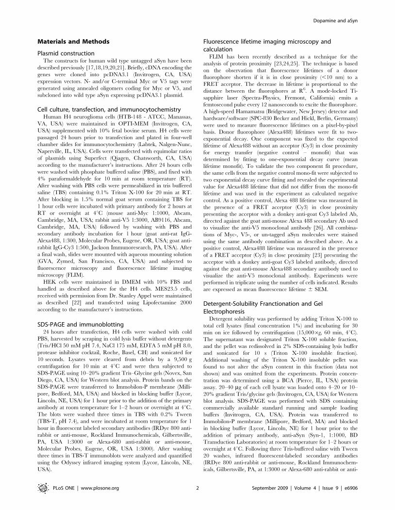
Given the data suggesting that, in vitro, dopamine can impact
aSyn conformation [5,6,7] we next asked if exposing aSyn
transfected neurons to dopamine would affect aSyn conformation.
Primary neurons overexpressing aSyn were treated with 100 mM
dopamine (DA) for 10 min. DA significantly decreased the lifetime
of the donor fluorophore to ,650 ps (n = 3 independent
experiments, total of 57 cells, p,0.01) at the dose tested,
indicating that DA induces the N- and C-termini of aSyn to be
in closer proximity, reflecting a change in conformation (Fig. 3).
By contrast, various other treatments, including treating cells with
60 mM KCl for 10 minutes led to no changes in donor lifetime
(Fig. 3). To tease apart the molecular mechanisms of the observed
DA effect we examined the effect of DA agonists and antagonists
on aSyn conformation. Primary neuronal cultures were immuno-
stained using antibodies against D1 and D2 receptors to verify the
presence of these receptors in our cultured neurons (Fig. 4A). We
then screened a panel of well-characterized DA agonists and
antagonists to determine if the previously observed conformational
changes in aSyn could be mimicked or impeded (Table 1).
Surprisingly, we were unable to detect an effect on aSyn
conformation with any of the DA agonists or antagonists
(Fig. 4B–C).
To further determine if dopamine induced a general effect on
the neurons or whether it was required to enter the cells in order to
induce the conformational change in aSyn, we used nomifensine
(100 mM) to block the activity of the dopamine transporter
(Fig. 5A). Interestingly, we detected a reduction in the dopamine-
induced conformational change in aSyn, suggesting that dopamine
must gain entry into the cells in order to exert its activity on aSyn
(Fig. 5A–D).
To further investigate how the conformational changes in aSyn
affect its biochemical properties, we sought to recapitulate the
Figure 3. FLIM analysis of aSyn conformation in the presence of modulators of neuronal activity (KCl and Dopamine). KCl does notalter aSyn conformation (lifetime ,1000 ps) whereas dopamine induces statistically significant changes (lifetime ,650 ps) (*n = 3, ,50 cells for eachcondition, p,0.01; unpaired, double sided t test).doi:10.1371/journal.pone.0006906.g003
Dopamine and aSyn
PLoS ONE | www.plosone.org 5 September 2009 | Volume 4 | Issue 9 | e6906

Figure 4. Dopamine receptor agonists and antagonists do not alter aSyn conformation. A. D1 and D2 receptors are present in primarycortical neurons. DIV7 neurons were immunostained against D1 and D2 receptors and observed via fluorescence microscopy. B. FLIM study withdopamine agonists/antagonists showing that these drugs do not alter aSyn conformation (*n = 3, 25–30 cells per condition, p,0.01; unpaired, doublesided t test). C. FLIM image of an example compound, SKF38393, showing aSyn conformation is not altered.doi:10.1371/journal.pone.0006906.g004
Dopamine and aSyn
PLoS ONE | www.plosone.org 6 September 2009 | Volume 4 | Issue 9 | e6906

modulation of conformation in immortalized cell lines, which
would enable us to achieve higher transfection efficiencies required
for biochemical studies. First, we transfected three cell lines of
different origins (H4, MES23.5, and HEK) with the Myc-aSyn-V5
construct. These cells were then treated with dopamine and
processed for FLIM. Interestingly, we found that the in the cell
lines of neuronal origin (H4 and MES23.5) dopamine induced a
conformational change similar to that observed in primary
neuronal cultures (Fig. 6A). In limited experiments using HEK
cells, we did not observe such a conformational change.
We previously identified, in brain tissue derived from patients
with dementia with Lewy bodies (DLB), in aSyn transgenic mice,
and in aSyn H4 expressing cells, oligomeric aSyn species which
are detergent-insoluble [17]. Here, we hypothesized that the DA-
induced conformational changes in aSyn might affect its detergent
solubility. To investigate whether DA affected aSyn oligomeriza-
tion we used native polyacrylamide gel electrophoresis (PAGE). In
cells treated with DA, we observed a ,50% decrease in a ,250
KDa band and a ,25% increase in two ,75 KDa bands,
demonstrating that DA induces slight but detectable changes in
aSyn oligomerization (Fig. 6B, arrows). To further assess the effect
of DA on aSyn solubility we performed a triton X-100 detergent
fractionation of H4 cell lysates expressing Myc-aSyn-V5 treated or
untreated with DA. We found no significant differences in the
solubility of aSyn in either groups of cells (Fig. 6C).
In order to determine whether DA induces changes at the level
of the secondary structure of aSyn we used circular dichroism
(CD). Spectra were taken at 37uC in the absence or in the presence
of different DA concentrations (10, 100 and 1000 mM of DA,
corresponding to ratios of DA:aSyn of 0.14; 1.4 and 14.0) for a
fixed concentration of aSyn of 70 mM (Fig. 7). The insets show
small, but significant changes (values represent the average of 5
different experiments +/2 SD) at the level of the secondary
structure of aSyn, in agreement the conformational changes
observed in neuronal cells, where the distance between the N- and
C-termini of aSyn is modified by DA treatment.
Discussion
In PD, cell death affects primarily the dopaminergic neurons
of the substantia nigra, but the nature of this selective
vulnerability is still unclear. A common pathway, involving
DA-dependent oxidative stress, has been put forward to explain
the death of dopamine neurons. Defects in the sequestration of
dopamine into synaptic vesicles in dopaminergic neurons from
the substantia nigra, enabling undesired DA-aSyn interactions,
may explain their increased vulnerability. It has been reported
that DA can undergo auto-oxidation and form DA-quinone
adducts with aSyn which prevent aSyn fibrillization and lead to
the accumulation of toxic intermediates [5,6], but the relevance
of these findings in the context of living cells has been difficult to
determine [32]. In the current study we sought to investigate
whether DA influences the conformation of aSyn in primary
neurons in culture.
We used FLIM to study alterations in the conformation of
aSyn by monitoring the interactions between the N- and C-
termini of the protein, as we had previously reported for studies
in mammalian cell lines [31]. Here we demonstrate that aSyn
adopts different conformations throughout the axon and
dendrites. In vitro, purified aSyn does not display any secondary
structure, and is considered a natively unfolded protein [10,33],
but it is highly likely that it adopts specific conformations inside
neurons in order to perform its normal function(s). Our data
show that different subcellular microenvironments, with poten-
tially different redox conditions, lipid compositions, or other
conditions known to influence the behavior of aSyn in vitro,
afford aSyn the possibility of adopting distinct conformations
inside living cells. For example, lipid rafts mediate the synaptic
localization of aSyn in neurons [34], which may also explain the
selective distribution of aSyn. Interestingly, we were not able to
identify a specific association of a particular aSyn conformation
with any subcellular organelle, suggesting local microenviron-
ments may be more important in determining the structure/
function of the protein. The interaction of aSyn with synaptic
vesicles is highly dynamic [34], which may also explain the
variety of aSyn conformations detected throughout the axons
and dendrites.
Our data also demonstrate that aSyn changes its structure in
response to DA, or possibly dopamine oxidation by-products,
adopting a conformation where its N- and C-termini become
closer together. DA or DA by-products inhibit aSyn fibril
formation, which may, in turn, lead to the accumulation of
aSyn oligomeric species via an alternative folding pathway
[5,6,35,36]. Although our results do not show whether the DA-
induced change in conformation of aSyn is the precursor for the
formation of toxic oligomeric species, our data support the
model that DA-induced conformational changes in aSyn, either
through a direct covalent interaction or indirectly, may favor
changes in the oligomerization state of the protein which may
explain the increased vulnerability of dopaminergic neurons in
comparison to others. These conformational changes may
underlie the recently reported effect of dopamine-modified
aSyn on autophagy mediated degradation of the protein, and its
subsequent impact on misfolded protein degradation in
cells [16].
Defining the role of the identified aSyn conformations will shed
light into the pathogenic mechanisms involved in PD, and may
pave the way for the identification of novel targets for therapeutic
intervention in different synucleinopathies.
Author Contributions
Conceived and designed the experiments: TFO AQ PJM BTH. Performed
the experiments: TFO JK KB JT PP LMAO. Analyzed the data: TFO.
Wrote the paper: TFO PJM BTH.
Table 1. Compounds tested in the FLIM assay in primaryneurons.
Compound ActivityFluorescencelifetime (ps)
Effect on aSynconformation
SKF-38393 Agonist 10586232 2
Sp-cAMPS Agonist 900656 2
Quinpirole Agonist 938696 2
Ropinirole Agonist 10146108 2
Dopamine 2 6326140* +
SCH-23390 Antagonist 11316130 2
Haloperidol Antagonist 8526123 2
Nomifensine DAT blocker 10046162 2
Dopamine +Nomifensine
2 959689 2
Compounds with different activities were used and their effects on theconformation of aSyn were assessed via the FLIM assay. Only dopamineaffected the conformation of aSyn (+) (*n = 3, 30–50 cells per condition,p,0.01).doi:10.1371/journal.pone.0006906.t001
Dopamine and aSyn
PLoS ONE | www.plosone.org 7 September 2009 | Volume 4 | Issue 9 | e6906

Figure 5. Dopamine enters cells to modulate aSyn conformation. A. ICC of cells treated with DA or DA + nomifensine (catecholamine uptakeblocker) showing DA is able to enter cells. B. Quantification of the fluorescence intensity in A (,200 cells) (*p,0.01, t test). C. FLIM analysis showingthat DA needs to enter cells to alter aSyn conformation (n = 3, 25–30 cells per condition, *p,0.01; unpaired, double sided t test). D. RepresentativeFLIM images of C.doi:10.1371/journal.pone.0006906.g005
Dopamine and aSyn
PLoS ONE | www.plosone.org 8 September 2009 | Volume 4 | Issue 9 | e6906
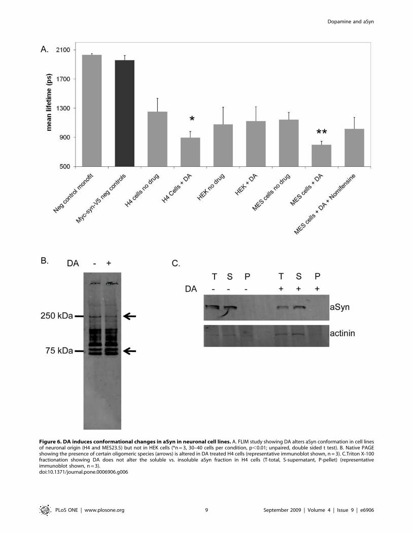
Figure 6. DA induces conformational changes in aSyn in neuronal cell lines. A. FLIM study showing DA alters aSyn conformation in cell linesof neuronal origin (H4 and MES23.5) but not in HEK cells (*n = 3, 30–40 cells per condition, p,0.01; unpaired, double sided t test). B. Native PAGEshowing the presence of certain oligomeric species (arrows) is altered in DA treated H4 cells (representative immunoblot shown, n = 3). C.Triton X-100fractionation showing DA does not alter the soluble vs. insoluble aSyn fraction in H4 cells (T-total, S-supernatant, P-pellet) (representativeimmunoblot shown, n = 3).doi:10.1371/journal.pone.0006906.g006
Dopamine and aSyn
PLoS ONE | www.plosone.org 9 September 2009 | Volume 4 | Issue 9 | e6906

References
1. Winklhofer KF, Tatzelt J, Haass C (2008) The two faces of protein misfolding:gain- and loss-of-function in neurodegenerative diseases. Embo J 27: 336–349.
2. Cole NB, Murphy DD, Grider T, Rueter S, Brasaemle D, et al. (2002) Lipid
droplet binding and oligomerization properties of the Parkinson’s disease proteinalpha-synuclein. J Biol Chem 277: 6344–6352.
3. Ito H, Fukuda Y, Murata K, Kimura A (1983) Transformation of intact yeastcells treated with alkali cations. J Bacteriol 153: 163–168.
4. Uversky VN, Lee HJ, Li J, Fink AL, Lee SJ (2001) Stabilization of partially
folded conformation during alpha-synuclein oligomerization in both purifiedand cytosolic preparations. J Biol Chem 276: 43495–43498.
5. Conway KA, Rochet JC, Bieganski RM, Lansbury PT, Jr. (2001) Kinetic
stabilization of the alpha-synuclein protofibril by a dopamine-alpha-synucleinadduct. Science 294: 1346–1349.
6. Li J, Zhu M, Manning-Bog AB, Di Monte DA, Fink AL (2004) Dopamine andL-dopa disaggregate amyloid fibrils: implications for Parkinson’s and Alzhei-
mer’s disease. Faseb J 18: 962–964.
7. Norris EH, Giasson BI, Hodara R, Xu S, Trojanowski JQ, et al. (2005)Reversible inhibition of alpha-synuclein fibrillization by dopaminochrome-
mediated conformational alterations. J Biol Chem 280: 21212–21219.
8. Soto C, Estrada LD (2008) Protein misfolding and neurodegeneration. ArchNeurol 65: 184–189.
9. Lucking CB, Brice A (2000) Alpha-synuclein and Parkinson’s disease. Cell MolLife Sci 57: 1894–1908.
10. Weinreb PH, Zhen W, Poon AW, Conway KA, Lansbury PT, Jr. (1996) NA-
CP, a protein implicated in Alzheimer’s disease and learning, is nativelyunfolded. Biochemistry 35: 13709–13715.
11. Perrin RJ, Woods WS, Clayton DF, George JM (2000) Interaction of human
alpha-Synuclein and Parkinson’s disease variants with phospholipids. Structuralanalysis using site-directed mutagenesis. J Biol Chem 275: 34393–34398.
12. Ulmer TS, Bax A, Cole NB, Nussbaum RL (2005) Structure and dynamics of
micelle-bound human alpha-synuclein. J Biol Chem 280: 9595–9603.
13. Zhu M, Fink AL (2003) Lipid binding inhibits alpha-synuclein fibril formation.J Biol Chem 278: 16873–16877.
14. Spillantini MG, Schmidt ML, Lee VM, Trojanowski JQ, Jakes R, et al. (1997)
Alpha-synuclein in Lewy bodies. Nature 388: 839–840.
15. Galvin JE (2006) Interaction of alpha-synuclein and dopamine metabolites in the
pathogenesis of Parkinson’s disease: a case for the selective vulnerability of thesubstantia nigra. Acta Neuropathol (Berl) 112: 115–126.
16. Martinez-Vicente M, Talloczy Z, Kaushik S, Massey AC, Mazzulli J, et al.
(2008) Dopamine-modified alpha-synuclein blocks chaperone-mediated autoph-agy. J Clin Invest 118: 777–788.
17. Klucken J, Shin Y, Masliah E, Hyman BT, McLean PJ (2004) Hsp70 reduces
alpha-synuclein aggregation and toxicity. J Biol Chem.
18. McLean PJ, Klucken J, Shin Y, Hyman BT (2004) Geldanamycin induces
Hsp70 and prevents alpha-synuclein aggregation and toxicity in vitro. BiochemBiophys Res Commun 321: 665–669.
19. McLean PJ, Kawamata H, Hyman BT (2001) Alpha-synuclein-enhanced green
fluorescent protein fusion proteins form proteasome sensitive inclusions inprimary neurons. Neuroscience 104: 901–912.
20. McLean PJ, Kawamata H, Shariff S, Hewett J, Sharma N, et al. (2002) TorsinA
and heat shock proteins act as molecular chaperones: suppression of alpha-synuclein aggregation. J Neurochem 83: 846–854.
21. Outeiro TF, Klucken J, Strathearn KE, Liu F, Nguyen P, et al. (2006) Small heatshock proteins protect against alpha-synuclein-induced toxicity and aggregation.
Biochem Biophys Res Commun 351: 631–638.
22. Crawford GD, Jr., Le WD, Smith RG, Xie WJ, Stefani E, et al. (1992) A novelN18TG2 x mesencephalon cell hybrid expresses properties that suggest a
dopaminergic cell line of substantia nigra origin. J Neurosci 12: 3392–3398.
23. Berezovska O, Bacskai BJ, Hyman BT (2003) Monitoring proteins in intact cells.Sci Aging Knowledge Environ 2003: PE14.
24. Bacskai BJ, Skoch J, Hickey GA, Allen R, Hyman BT (2003) Fluorescence
resonance energy transfer determinations using multiphoton fluorescence
Figure 7. aSyn secondary structure alterations induced by DA. Conformational changes of aSyn in the absence (. . . .) and in the presence of10 mM (- - -); 100 mM (-- --) and 1000 mM (___ ) of dopamine were monitored by CD. The concentration of aSyn used was 70 mM and the spectra weretaken at 37uC in a 0.1 mm path quartz cuvette at 25uC. The inset graphs show the percentage of random coil and beta-sheet as a function of DAconcentration and was calculated after deconvolution of the CD spectra according to the description in the material and methods section (average of5 independent experiments +/2 SD).doi:10.1371/journal.pone.0006906.g007
Dopamine and aSyn
PLoS ONE | www.plosone.org 10 September 2009 | Volume 4 | Issue 9 | e6906

lifetime imaging microscopy to characterize amyloid-beta plaques. J Biomed Opt
8: 368–375.25. Lleo A, Berezovska O, Herl L, Raju S, Deng A, et al. (2004) Nonsteroidal anti-
inflammatory drugs lower Abeta42 and change presenilin 1 conformation. Nat
Med 10: 1065–1066.26. Berezovska O, Ramdya P, Skoch J, Wolfe MS, Bacskai BJ, et al. (2003) Amyloid
precursor protein associates with a nicastrin-dependent docking site on thepresenilin 1-gamma-secretase complex in cells demonstrated by fluorescence
lifetime imaging. J Neurosci 23: 4560–4566.
27. Kessler JC, Rochet JC, Lansbury PT, Jr. (2003) The N-terminal repeat domainof alpha-synuclein inhibits beta-sheet and amyloid fibril formation. Biochemistry
42: 672–678.28. Johnson WC (1999) Analyzing protein circular dichroism spectra for accurate
secondary structures. Proteins 35: 307–312.29. Lobley A, Whitmore L, Wallace BA (2002) DICHROWEB: an interactive
website for the analysis of protein secondary structure from circular dichroism
spectra. Bioinformatics 18: 211–212.30. Whitmore L, Wallace BA (2004) DICHROWEB, an online server for protein
secondary structure analyses from circular dichroism spectroscopic data. NucleicAcids Res 32: W668–673.
31. Klucken J, Outeiro TF, Nguyen P, McLean PJ, Hyman BT (2006) Detection of
novel intracellular alpha-synuclein oligomeric species by fluorescence lifetime
imaging. Faseb J 20: 2050–2057.
32. Mazzulli JR, Armakola M, Dumoulin M, Parastatidis I, Ischiropoulos H (2007)
Cellular oligomerization of alpha-synuclein is determined by the interaction of
oxidized catechols with a C-terminal sequence. J Biol Chem 282: 31621–31630.
33. Uversky VN, Gillespie JR, Fink AL (2000) Why are ‘‘natively unfolded’’ proteins
unstructured under physiologic conditions? Proteins 41: 415–427.
34. Fortin DL, Nemani VM, Voglmaier SM, Anthony MD, Ryan TA, et al. (2005)
Neural activity controls the synaptic accumulation of alpha-synuclein. J Neurosci
25: 10913–10921.
35. Norris EH, Giasson BI (2005) Role of oxidative damage in protein aggregation
associated with Parkinson’s disease and related disorders. Antioxid Redox Signal
7: 672–684.
36. Cappai R, Leck SL, Tew DJ, Williamson NA, Smith DP, et al. (2005) Dopamine
promotes alpha-synuclein aggregation into SDS-resistant soluble oligomers via a
distinct folding pathway. Faseb J 19: 1377–1379.
Dopamine and aSyn
PLoS ONE | www.plosone.org 11 September 2009 | Volume 4 | Issue 9 | e6906
Related Documents