Dopamine D 2 receptor function is compromised in the brain of the methionine sulfoxide reductase A knockout mouse Derek B. Oien * , Andrea N. Ortiz † , Alexander G. Rittel * , Rick T. Dobrowsky * , Michael A. Johnson † , Beth Levant ‡ , Stephen C. Fowler * , and Jackob Moskovitz * * Department of Pharmacology and Toxicology, School of Pharmacy, University of Kansas, Lawrence, Kansas, USA † Department of Chemistry, University of Kansas, Lawrence, Kansas, USA ‡ Department of Pharmacology, Toxicology and Therapeutics, University of Kansas Medical Center, Kansas City, Kansas, USA Abstract Previous research suggests that brain oxidative stress and altered rodent locomotor behavior are linked. We observed bio-behavioral changes in methionine sulfoxide reductase A knockout mice associated with abnormal dopamine signaling. Compromised ability of these knockout mice to reduce methionine sulfoxide enhances accumulation of sulfoxides in proteins. We examined the dopamine D 2 -receptor function and expression, which has an atypical arrangement and quantity of methionine residues. Indeed, protein expression levels of dopamine D 2 -receptor were higher in knockout mice compared with wild-type. However, the binding of dopamine D 2 -receptor agonist was compromised in the same fractions of knockout mice. Coupling efficiency of dopamine D 2 -receptors to G-proteins was also significantly reduced in knockout mice, supporting the compromised agonist binding. Furthermore, pre-synaptic dopamine release in knockout striatal sections was less responsive than control sections to dopamine D 2 -receptor ligands. Behaviorally, the locomotor activity of knockout mice was less responsive to the inhibitory effect of quinpirole than wild-type mice. Involvement of specific methionine residue oxidation in the dopamine D 2 -receptor third intracellular loop is suggested by in vitro studies. We conclude that ablation of methionine sulfoxide reductase can affect dopamine signaling through altering dopamine D 2 -receptor physiology and may be related to symptoms associated with neurological disorders and diseases. Keywords dopamine; dopamine receptor; locomotor activity; methionine oxidation; oxidative stress; post- translation modification Post-translational modifications resulting from reactive oxygen species can alter the functions of many proteins. While most protein modifications by reactive oxygen species are irreversible, Address correspondence and reprint requests to Dr. Jackob Moskovitz, Department of Pharmacology and Toxicology, University of Kansas, 1251 Wescoe Hall Drive, Malott 5064, Lawrence, KS 66045, USA. [email protected]. Supporting Information Additional Supporting information may be found in the online version of this article: Figure S1. Gαi/o protein expression levels in MsrA–/– and WT brains. As a service to our authors and readers, this journal provides supporting information supplied by the authors. Such materials are peer- reviewed and may be re-organized for online delivery, but are not copy-edited or typeset. Technical support issues arising from supporting information (other than missing files) should be addressed to the authors. NIH Public Access Author Manuscript J Neurochem. Author manuscript; available in PMC 2011 July 1. Published in final edited form as: J Neurochem. 2010 July ; 114(1): 51–61. doi:10.1111/j.1471-4159.2010.06721.x. NIH-PA Author Manuscript NIH-PA Author Manuscript NIH-PA Author Manuscript

Welcome message from author
This document is posted to help you gain knowledge. Please leave a comment to let me know what you think about it! Share it to your friends and learn new things together.
Transcript
Dopamine D2 receptor function is compromised in the brain of themethionine sulfoxide reductase A knockout mouse
Derek B. Oien*, Andrea N. Ortiz†, Alexander G. Rittel*, Rick T. Dobrowsky*, Michael A.Johnson†, Beth Levant‡, Stephen C. Fowler*, and Jackob Moskovitz** Department of Pharmacology and Toxicology, School of Pharmacy, University of Kansas,Lawrence, Kansas, USA† Department of Chemistry, University of Kansas, Lawrence, Kansas, USA‡ Department of Pharmacology, Toxicology and Therapeutics, University of Kansas Medical Center,Kansas City, Kansas, USA
AbstractPrevious research suggests that brain oxidative stress and altered rodent locomotor behavior arelinked. We observed bio-behavioral changes in methionine sulfoxide reductase A knockout miceassociated with abnormal dopamine signaling. Compromised ability of these knockout mice to reducemethionine sulfoxide enhances accumulation of sulfoxides in proteins. We examined the dopamineD2-receptor function and expression, which has an atypical arrangement and quantity of methionineresidues. Indeed, protein expression levels of dopamine D2-receptor were higher in knockout micecompared with wild-type. However, the binding of dopamine D2-receptor agonist was compromisedin the same fractions of knockout mice. Coupling efficiency of dopamine D2-receptors to G-proteinswas also significantly reduced in knockout mice, supporting the compromised agonist binding.Furthermore, pre-synaptic dopamine release in knockout striatal sections was less responsive thancontrol sections to dopamine D2-receptor ligands. Behaviorally, the locomotor activity of knockoutmice was less responsive to the inhibitory effect of quinpirole than wild-type mice. Involvement ofspecific methionine residue oxidation in the dopamine D2-receptor third intracellular loop issuggested by in vitro studies. We conclude that ablation of methionine sulfoxide reductase can affectdopamine signaling through altering dopamine D2-receptor physiology and may be related tosymptoms associated with neurological disorders and diseases.
Keywordsdopamine; dopamine receptor; locomotor activity; methionine oxidation; oxidative stress; post-translation modification
Post-translational modifications resulting from reactive oxygen species can alter the functionsof many proteins. While most protein modifications by reactive oxygen species are irreversible,
Address correspondence and reprint requests to Dr. Jackob Moskovitz, Department of Pharmacology and Toxicology, University ofKansas, 1251 Wescoe Hall Drive, Malott 5064, Lawrence, KS 66045, USA. [email protected] InformationAdditional Supporting information may be found in the online version of this article:Figure S1. Gαi/o protein expression levels in MsrA–/– and WT brains.As a service to our authors and readers, this journal provides supporting information supplied by the authors. Such materials are peer-reviewed and may be re-organized for online delivery, but are not copy-edited or typeset. Technical support issues arising from supportinginformation (other than missing files) should be addressed to the authors.
NIH Public AccessAuthor ManuscriptJ Neurochem. Author manuscript; available in PMC 2011 July 1.
Published in final edited form as:J Neurochem. 2010 July ; 114(1): 51–61. doi:10.1111/j.1471-4159.2010.06721.x.
NIH
-PA Author Manuscript
NIH
-PA Author Manuscript
NIH
-PA Author Manuscript
methionine sulfoxide (MetO) modifications can be reversed by the methionine sulfoxidereductase (Msr) system, which consists of MsrA (reduces S-MetO) and MsrB (reduces R-MetO) (Moskovitz 2005). Reduction action of the Msr system may prevent irreversible proteindamage, contribute to cellular antioxidant resistance, and regulate protein function. Evidencefor a role of the Msr system in antioxidant defense is demonstrated by the adverse effectsresulting from MsrA ablation (Moskovitz et al. 1995, 1997, 2001). Furthermore, over-expression of MsrA in various organisms and cells protects them from oxidative stress toxicity(Moskovitz et al. 1998; Ruan et al. 2002; Romero et al. 2004). The MsrA null mouse (MsrA−/−) is hypersensitive to oxidative stress, accumulates higher levels of carbonylated proteinand expresses brain pathologies associated with neurodegenerative diseases (Moskovitz etal. 2001; Pal et al. 2007); this evidence suggests a major biological role of the Msr system.Furthermore, the Msr enzymes have been shown to switch several proteins between non-activeand active forms by reducing specific MetO residues to methionine (Oien and Moskovitz2008).
The MsrA−/− mouse exhibits age-dependent lower locomotor activity and abnormal gaitindices (Oien et al. 2008) relative to control mice. Lower dopamine (DA) levels are associatedwith altered motor performance (Carlsson et al. 2007). Surprisingly, our recent observationsshowed that MsrA−/− brains in adult mice contained significantly higher levels of DA (Oienet al. 2008). Moreover, pre-synaptic neuronal DA in MsrA−/− striatal slices is released at higherDA pulses than wild-type (WT) slices as measured by fast scan cyclic voltammetry. Theseresults raise the possibility that DA function is impaired in MsrA−/− mice. Moreover, MsrA−/− mice were less responsive to amphetamine treatment as assayed by locomotor activity andstereotypy, suggesting alteration of DA signaling. Dysfunctions of the corpus striatum anddopaminergic signaling are linked to a range of disorders including Parkinson’s disease(Hornykiewicz 1962), Huntington’s disease (Hickey et al. 2002), schizophrenia, andobsessive-compulsive disorder (Saxena et al. 1998).
The observed MsrA−/− locomotor deficits and DA abnormalities prompted us to furtherexamine DA signaling events that are associated with striatal movement pathways. Thedopamine D1 receptor (D1DR) and dopamine D2 receptor (D2DR) are closely associated withmovement regulation, and theoretically are the DA receptors most related to the MsrA−/−locomotor phenotypes (Oien et al. 2008). In addition, D2DR is also expressed in its short form(D2DRS) that serves as an autoreceptor for dopaminergic neurons. Thus, methionine oxidationin either the long form of D2DR or D2DRS or a combination of both may be related to theelevation in DA levels observed in MsrA−/− brains (Oien et al. 2008). In the current study weinvestigate the ligand-binding capabilities of DA receptors and their coupling efficiency to G-proteins in MsrA−/− versus WT mice. The presented data support the hypothesis that MetOmodification to D2DRs participates in the MsrA−/− behavioral and biochemical phenotypes.
Materials and methodsMaterials
The radioligands [3H]SCH23390 (85 Ci/mmol), [3H]raclopride (84 Ci/mmol), [3H]quinpirole(50 Ci/mmol), were purchased from Perkin Elmer (Waltham, MA, USA). Non-radioactivesulpiride, quinpirole, SKF82958, and GTPγS were purchased from Sigma, St Louis, MO, USA.Mouse and goat antibodies against D2DR were purchased from Abcam (Cambridge, MA,USA). Mouse β-actin antibodies were purchased from Molecular Probes, Eugene, OR, USA.
MiceMice used in these studies were WT (C57BL6/129 Sv) and MsrA−/− on the same geneticbackground at an age of 6–8 months unless otherwise noted. Both types of mice were littermates
Oien et al. Page 2
J Neurochem. Author manuscript; available in PMC 2011 July 1.
NIH
-PA Author Manuscript
NIH
-PA Author Manuscript
NIH
-PA Author Manuscript
born to MsrA+/− heterozygous parents (Moskovitz et al. 2001). All procedures using micewere performed within guidelines of the NIH and KU Institutional Animal Care and UseCommittee.
Immunohistochemistry analysisCoronal brain sections from 12-month-old WT and MsrA−/− mice were processed as 20 μm-thick brain sections. Sections were treated with 3% H2O2 in methanol for 30 min. Afterblocking with 1% bovine serum albumin and 3% horse serum in phosphate-buffered saline,the sections were incubated for 24 h with primary goat antibodies against D2DR (1 : 500dilution, stock concentration of 1.5 mg/mL). Sections were then incubated with biotinylatedmouse anti-goat IgG antibodies (Santa Cruz Biotechnology, Santa Cruz, CA, USA), avidin–horseradish peroxide solution, 0.015% diaminobenzidine, 0.001% H2O2, and finally counter-stained with hematoxylin. The brain slides were washed, dehydrated, mounted, and visualizedusing a brightfield microscope with a 100× objective lens.
Immunoblot analysisEqual amounts of striatal membranal protein extracts or cytosolic brain proteins (extracted asdescribed below for receptors fraction) were subjected to sodium dodecyl sulfate-gelelectrophoresis. Immunoblot analyses with anti-goat D2DR antibodies (1 : 1000 dilution, stockconcentration of 1.5 mg/mL) were performed according to common procedures. β-actin(loading control) was detected by primary anti-β-actin mouse antibodies.
Antagonists and agonists binding of D1DR and D2DR ligandsSaturation curves of receptor binding were determined by using the common membranefiltration assay (Bylund and Toews 1993). Briefly, brains (striatal region of each mouse type)were dissected and glass-Teflon homogenized in phosphate-buffered saline with proteaseinhibitor cocktail (Roche Molecular Biochemicals, Indianapolis, IN, USA). Followingcentrifugal precipitation at 20 000 g for 20 min, the supernatant was removed and saved(cytosolic fraction in immunoblots), and the membranal pellet was washed and precipitatedagain by centrifugation. Final pellet was homogenized in 1.0 mL of 50 mM Tris pH 7.4.Membranal protein fractions of 100 μg were used per assay, determined by BCA Protein AssayKit (Thermo Scientific, Waltham, MA, USA). Total ligand binding was determined followingincubation of membranal fractions with incrementing concentrations of radioligand for 2 h at25°C. Non-specific ligand binding was determined using the same conditions with the additionof non-radioactive ligands, 1 mM DA for D1DR and 1 μM sulpiride for D2DR. Boundradioligand was separated using a Brandel harvester (Brandel, Gaithersburg, MD, USA).Radioligand bound to the filter was measured by liquid scintillation counting. Specific ligandbinding values were calculated by subtracting non-specific ligand binding from total ligandbinding. The ligands used in the tests were: [3H]SCH23390 and [3H]raclopride as D1DR andD2DR antagonists, respectively, and [3H]quinpirole as a D2DR agonist. The range forradioactive materials used was between 0.03 nM and 4.0 nM (increments by serial dilution).
The effect of MsrA enzyme on quinpirole and raclopride binding was tested by usingrecombinant yeast MsrA (10 μg) (Moskovitz et al. 1997) with 15 mM dithiothreitol (DTT; thisalone did not affect binding). D1DR binding to the agonist SKF82958 was determined usingthe same conditions above, except for the addition of 2 nM [3H]SCH23390 to quantify thecompetitive binding of non-radiolabeled SKF82958 ligand (performed because of limitedsources of commercially available radiolabeled D1DR agonists).
Oien et al. Page 3
J Neurochem. Author manuscript; available in PMC 2011 July 1.
NIH
-PA Author Manuscript
NIH
-PA Author Manuscript
NIH
-PA Author Manuscript
GTPγS-induced inhibition of radiolabeled quinpirole binding to D2DRThe ability of agonists to stimulate the dissociation of GDP and association of GTP from G-protein was assessed using GTPγS, a non-hydrolyzable analogue of GTP (Zhang et al. 2001).When the agonist binds to the receptor, GDP is released from the G-protein and GTPγS binds(Harrison and Traynor 2003). The G-protein uncoupling on [3H]quinpirole binding to theD2DR was investigated by measuring the inhibition of [3H]quinpirole binding at incrementingGTPγS concentrations. The membrane portion of homogenized striatal regions were isolatedand lysed in 5 mM Tris buffer by glass-Teflon homogenation. Membranal extracts (30 mg/mL; 80 μg per assay) were incubated with [3H]quinpirole (1 nM) and GTPγS (0.1, 1, 10, 100,and 1000 nM) in 50 mM Tris buffer for 2 h at 25°C in a total volume of 1 mL. To determinenon-specific binding, identical assays were performed in the presence of sulpiride (1 μM;racemic mixture). Unbound ligand was removed by Brandel filtration and the remaining boundligand was quantified by liquid scintillation counting.
Stimulated release of DA in striatal brain sections in the presence of quinpirole and sulpiridemeasured by fast scan cyclic voltammetry
Brain sections were prepared as previously described (Johnson et al. 2006). Mice wereanesthetized by isoflurane inhalation. The brain was immediately removed and placed in icecold artificial CSF. Artificial CSF consisted of 126 mM NaCl, 2.5 mM KCl, 1.2 mMNaH2PO4, 2.4 mM CaCl2, 1.2 mM MgCl2, 25 mM NaHCO3, 20 mM HEPES, and 11 mM D-Glucose at pH 7.4 and was continuously saturated with 95% O2/5% CO2 throughout theexperiment. Coronal sections of 300 μm in thickness were made using a vibratome slicer (LeicaMicrosystems, Bannockburn, IL, USA). A single section was placed in the superfusionchamber with artificial CSF flowing at 34°C and a continuous rate of 2 mL/min. Each brainsection was equilibrated for 60 min prior to obtaining measurements. Quinpirole and sulpiridein artificial CSF were maintained in a separate reservoir and introduced through a three-wayvalve.
Carbon-fiber microelectrodes were fabricated as previously described (Kraft et al. 2009). Asingle carbon-fiber with a 7 μm diameter (Goodfellow Cambridge Ltd., Oakdale, PA, USA)was aspirated through a glass capillary tube (1.2 mm outer diameter, 0.68 mm inner diameter,4 inches long, A-M Systems, Inc., Sequim, WA, USA) and pulled using a heated coil puller(Narishige International USA, Inc., East Meadow, NY, USA). Electrodes were trimmed to 20μm from the glass seal, further insulated with epoxy resin (EPON resin 815C, EPIKURE 3234curing agent, Miller-Stephenson, Morton Grove, IL, USA), and then cured at 100°C for 1 h.Prior to experimentation, electrodes were backfilled with 0.5 M potassium acetate.
A triangular waveform starting at −0.4 V, increasing to +1.0 V, and scanning back to −0.4 Vwas applied at the carbon-fiber working electrode. A scan rate of 300 V/s was used with anupdate rate of 10 Hz. A head-stage amplifier (UNC Chemistry Department Electronics Design)was interfaced with a computer via breakout box and custom software provided by R.M.Wightman and M.L.A.V. Heien. A choridated silver wire was used as an Ag/AgCl referenceelectrode. The carbon fiber was inserted 100 μm into the dorsolateral caudate-putamen regionof the striatum. The fiber was between the prongs of a bipolar stimulation electrode (PlasticsOne, Roanoke, VA, USA), which were separated by 200 μm. Current was measured at 0.6 V,which is the oxidation potential for DA. DA release was measured in the presence of either 1μM quinpirole or 5 μM sulpiride as previously described (Fawaz et al. 2009) using a 30-pulsestimulus train at a stimulation frequency of 60 Hz. During stimulation and DA release,stabilized scans of 15 s were collected every 2 min and averaged as 4-min time blocks.
Oien et al. Page 4
J Neurochem. Author manuscript; available in PMC 2011 July 1.
NIH
-PA Author Manuscript
NIH
-PA Author Manuscript
NIH
-PA Author Manuscript
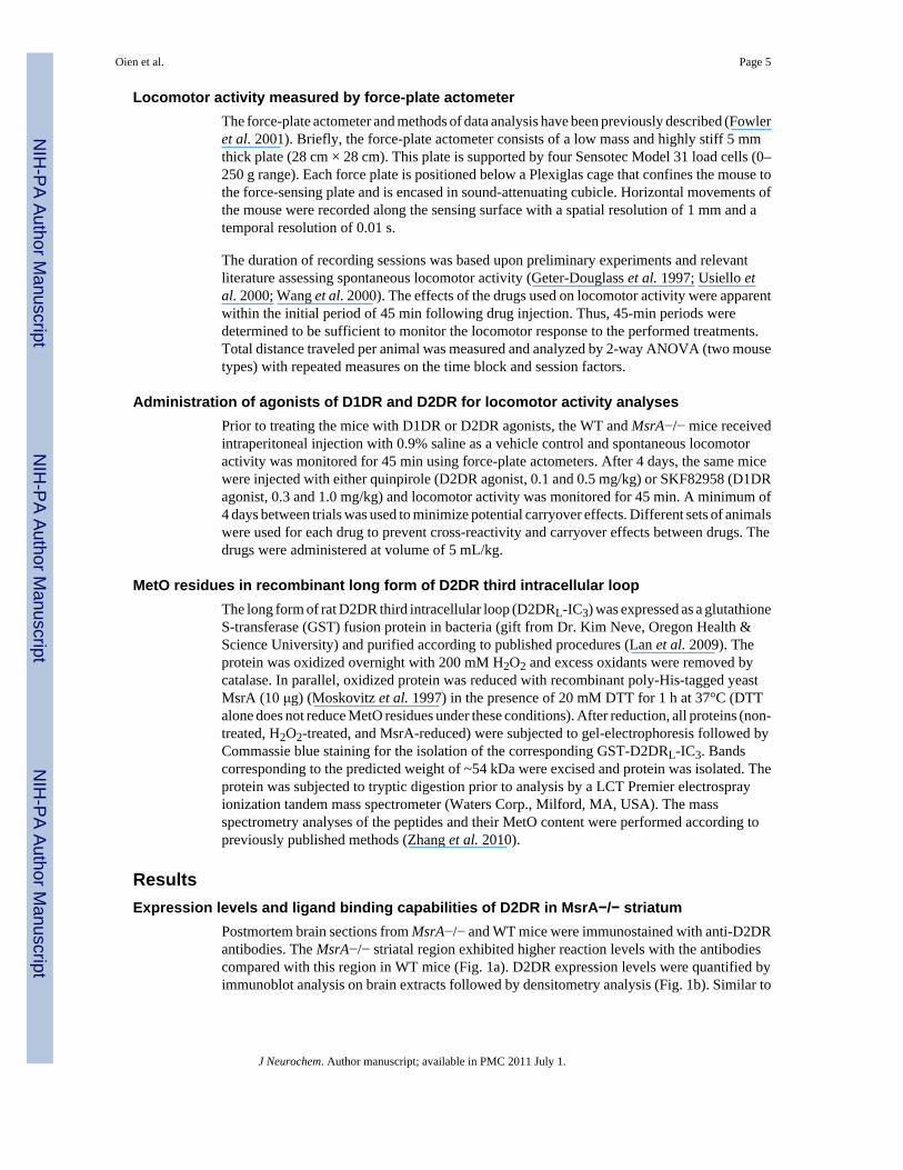
Locomotor activity measured by force-plate actometerThe force-plate actometer and methods of data analysis have been previously described (Fowleret al. 2001). Briefly, the force-plate actometer consists of a low mass and highly stiff 5 mmthick plate (28 cm × 28 cm). This plate is supported by four Sensotec Model 31 load cells (0–250 g range). Each force plate is positioned below a Plexiglas cage that confines the mouse tothe force-sensing plate and is encased in sound-attenuating cubicle. Horizontal movements ofthe mouse were recorded along the sensing surface with a spatial resolution of 1 mm and atemporal resolution of 0.01 s.
The duration of recording sessions was based upon preliminary experiments and relevantliterature assessing spontaneous locomotor activity (Geter-Douglass et al. 1997; Usiello etal. 2000; Wang et al. 2000). The effects of the drugs used on locomotor activity were apparentwithin the initial period of 45 min following drug injection. Thus, 45-min periods weredetermined to be sufficient to monitor the locomotor response to the performed treatments.Total distance traveled per animal was measured and analyzed by 2-way ANOVA (two mousetypes) with repeated measures on the time block and session factors.
Administration of agonists of D1DR and D2DR for locomotor activity analysesPrior to treating the mice with D1DR or D2DR agonists, the WT and MsrA−/− mice receivedintraperitoneal injection with 0.9% saline as a vehicle control and spontaneous locomotoractivity was monitored for 45 min using force-plate actometers. After 4 days, the same micewere injected with either quinpirole (D2DR agonist, 0.1 and 0.5 mg/kg) or SKF82958 (D1DRagonist, 0.3 and 1.0 mg/kg) and locomotor activity was monitored for 45 min. A minimum of4 days between trials was used to minimize potential carryover effects. Different sets of animalswere used for each drug to prevent cross-reactivity and carryover effects between drugs. Thedrugs were administered at volume of 5 mL/kg.
MetO residues in recombinant long form of D2DR third intracellular loopThe long form of rat D2DR third intracellular loop (D2DRL-IC3) was expressed as a glutathioneS-transferase (GST) fusion protein in bacteria (gift from Dr. Kim Neve, Oregon Health &Science University) and purified according to published procedures (Lan et al. 2009). Theprotein was oxidized overnight with 200 mM H2O2 and excess oxidants were removed bycatalase. In parallel, oxidized protein was reduced with recombinant poly-His-tagged yeastMsrA (10 μg) (Moskovitz et al. 1997) in the presence of 20 mM DTT for 1 h at 37°C (DTTalone does not reduce MetO residues under these conditions). After reduction, all proteins (non-treated, H2O2-treated, and MsrA-reduced) were subjected to gel-electrophoresis followed byCommassie blue staining for the isolation of the corresponding GST-D2DRL-IC3. Bandscorresponding to the predicted weight of ~54 kDa were excised and protein was isolated. Theprotein was subjected to tryptic digestion prior to analysis by a LCT Premier electrosprayionization tandem mass spectrometer (Waters Corp., Milford, MA, USA). The massspectrometry analyses of the peptides and their MetO content were performed according topreviously published methods (Zhang et al. 2010).
ResultsExpression levels and ligand binding capabilities of D2DR in MsrA−/− striatum
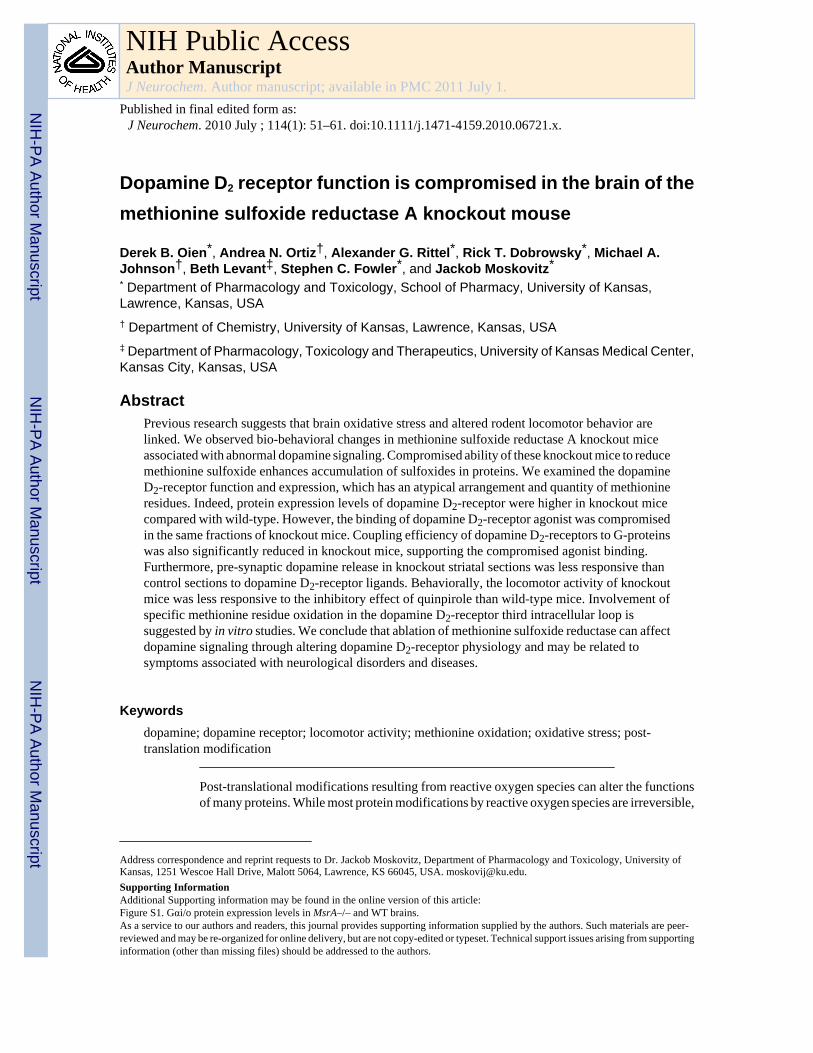
Postmortem brain sections from MsrA−/− and WT mice were immunostained with anti-D2DRantibodies. The MsrA−/− striatal region exhibited higher reaction levels with the antibodiescompared with this region in WT mice (Fig. 1a). D2DR expression levels were quantified byimmunoblot analysis on brain extracts followed by densitometry analysis (Fig. 1b). Similar to
Oien et al. Page 5
J Neurochem. Author manuscript; available in PMC 2011 July 1.
NIH
-PA Author Manuscript
NIH
-PA Author Manuscript
NIH
-PA Author Manuscript
the increase of DA in MsrA−/− brains (Oien et al. 2008), the up-regulation of the D2DR maybe part of compensatory response for the dysfunction of the DA system in the MsrA−/− brain.
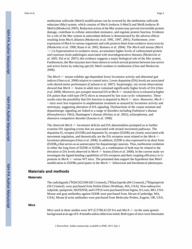
The binding of the D2DR to appropriate ligands can reflect their expression levels and functionin signaling. Binding of the D2DR antagonists are independent of G-protein coupling and morereflective of D2DR expression levels. Binding of the D2DR agonists are dependent on G-protein coupling and more reflective of D2DR function in signaling. Accordingly, thecalculated Bmax value of D2DR antagonist [3H]raclopride was higher in MsrA−/− membranalfractions (250 ± 20 fmole/mg protein) compared with the value of WT (140 ± 4 fmole/mgprotein) (Fig. 2a). In addition, the Kd value for [3H]raclopride in both mouse types was similar(Kd of 0.70 ± 0.10 nM), suggesting no difference in the binding affinity of the bound antagoniststo D2DR (it is noteworthy that the Kd values in these mice are different than rats, see‘Discussion’). The [3H]raclopride Bmax values also provide further supportive evidence to theobserved elevated levels of the D2DR in MsrA−/− brain (Fig. 1). The calculated Bmax valueof D2DR agonist [3H]quinpirole was lower in MsrA−/− membranal fractions (56 ± 7 fmole/mg protein) when compared with the value of the WT (140 ± 10 fmole/mg protein), while theKd value for [3H]quinpirole binding was similar in both mouse types (0.40 ± 0.05 nM) (Fig.2a). These observations suggest that the coupling ability of the MsrA−/− D2DR to the respectiveG-proteins is compromised, in spite of the higher number of receptors in MsrA−/− mice (Fig.1). In addition, adding recombinant MsrA to the binding reaction mixture caused significantrecovery of the MsrA−/− [3H]quinpirole Bmax value to the WT Bmax value (Fig. 2b). This latterresult strengthens the hypothesis that lack of MsrA enhances MetO formation in D2DR, whichaffects D2DR function and expression. In contrast to relative D2DR agonist-to-antagonistBmax differences between the two mouse types (Fig. 2a and b), no significant differences werefound for D1DR ligand binding to the antagonist SCH23390 or agonist SKF82958 betweenthese mouse types (Fig. 2c and d). At relatively high concentrations, [3H]SCH23390 was boundto MsrA−/− and WT striatal fractions to a similar extent (Fig. 2c). Similar results were foundwhen SKF82958 was used (Fig. 2d). The SKF82958 ligand was not radiolabeled, thus bindingefficiency was determined by its competitive binding in the presence of 2 nM [3H]SCH23390as previously described (Nwaneshiudu and Unterwald 2009). The amounts of [3H]SCH23390bound to the striatal fractions were significantly decreased (p < 0.05) by the presence of 16nM of SKF82958. This supports the hypothesis that MsrA ablation primarily impacts theD2DR.
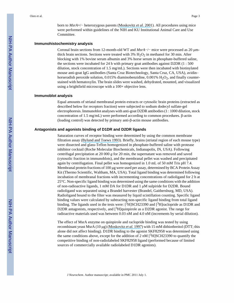
G-protein coupling efficiency to D2DR in MsrA−/− striatumThe non-hydrolyzable GTPγS analog will reduce the binding of [3H]quinpirole to D2DR(quinpirole binds with lower affinity to uncoupled D2DR compared with coupled receptors).Consequently, competition curves were performed by inhibiting [3H]quinpirole binding withincreasing concentrations of GTPγS. Accordingly, the maximal inhibition from thecompetition curve reflects the number of D2DR coupled to G-proteins, while the 50% bindinginhibition concentration (IC50) from the curve reflects the potency of GTPγS to dissociate theD2DR and G-protein complex. The maximum binding of [3H]quinpirole to D2DR in MsrA−/− was ~50% of the maximum binding of the agonist to the receptors in the WT brain (Fig. 3;WT = 50 ± 6 and MsrA−/− = 26 ± 3 fmole/mg protein in the presence of 1 μM [3H]quinpirole).The IC50 was found to be similar for both mouse types (10 ± 1 nM), which is in agreementwith current literature (Zhang et al. 2001). These data suggest reduced G-protein and D2DRcoupling in the MsrA−/− brain. Moreover, the similar IC50 in both mouse types suggests thatthe dissociation rates of the receptors from the G-proteins are similar. The reduced interactionof G-proteins with D2DR in MsrA−/− brains could be a consequence of either lower G-proteinexpression or interference in the initial binding of G-proteins to the third intracellular loop ofD2DR. There was no significant difference in expression of Gαi/o proteins between the twomouse types (Figure S1), suggesting that the observed limitation in MsrA−/− D2DR agonist
Oien et al. Page 6
J Neurochem. Author manuscript; available in PMC 2011 July 1.
NIH
-PA Author Manuscript
NIH
-PA Author Manuscript
NIH
-PA Author Manuscript
binding is probably not because of reduced Gαi/o levels. Since the expression levels of theseG-proteins seem to be similar in the two mouse types (Figure S1), data in Fig. 3 are supportiveof compromised D2DR agonist binding in MsrA−/− brains (Fig. 2a) and also may reflectinterference in binding of MsrA−/− G-proteins to the third intracellular loop of D2DR.
Pre-synaptic stimulated dopamine release of MsrA−/− striatal tissue sections in response toD2DR ligands
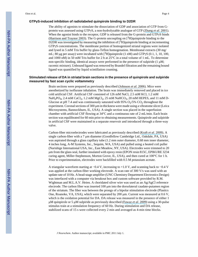
A shorter form of the D2DR, D2DRS, serves as an autoreceptor on the pre-synaptic membraneof dopaminergic neurons. This receptor has a third intracellular loop that is truncated by 29amino acids, but still retains four of the eight methionine residues. D2DRS has been previouslydemonstrated to respond to the D2DR agonist quinpirole and D2DR antagonist sulpiride bydecreasing and increasing the pre-synaptic release of DA, respectively (Fawaz et al. 2009).Using a similar experimental design as Fawaz et al., the pre-synaptic DA release in the striatalarea from MsrA−/− and WT coronal sections was measured in the presence of quinpirole (Fig.4a and b) and sulpiride (Fig. 4c and d). Pre-synaptic DA release was detected by fast scan cyclicvoltammetry every 2 min, initially without any treatment and followed by treatment with asaturating dose of quinpirole. In the presence of quinpirole, MsrA−/− sections were lessresponsive than WT sections when both were compared to the respective initial stimulationand detection without the presence of drug. The DA release of the MsrA−/− pre-synapticneurons was significantly higher than the decreased release of the WT pre-synaptic neurons(p < 0.05; two-way repeated measures ANOVA). Moreover, the MsrA−/− pre-synaptic neuronswere also less responsive to sulpiride than WT. After sulpiride treatment, the changes in DAconcentration from the initial non-treated measurement of MsrA−/− slices were significantlylower than the increased DA concentration changes of the WT (p < 0.05). These results suggestthat the D2DRS signaling is disrupted in MsrA−/− mice.
Locomotor activity of MsrA−/− mice after injections of dopamine receptor agonistsThe in vitro and brain section findings suggest functional changes in the DA system that shouldbe expressed at the behavioral level. Consequently, we investigated the behavioral effects ofD1DR and D2DR selective agonists on locomotor activity in MsrA−/− and WT mice. Asignificant difference in locomotor activity was observed between the mouse strains wheninjected with quinpirole. At a dose of 0.5 mg/kg, the MsrA−/− exhibited ~40% more locomotoractivity than WT, and at a dose of 0.1 mg/kg the MsrA−/− exhibited ~35% more locomotoractivity than WT (Fig. 5a; repeated measures ANOVA for mouse type effect, p < 0.02). Nosignificant difference in locomotor activity was observed between the two mouse types wheninjected with saline (Fig. 5), which is consistent with previous experiments (Oien et al.2008). Given the higher expression level of the D2DR in MsrA−/− mice, these data suggestthat the compromised responsiveness of the MsrA−/− D2DRs to the inhibitory effect ofquinpirole may be because of irregularities in either DA binding or abnormal function of furtherdownstream signaling events. Moreover, the lack of a significant dose effect in the quinpiroleconcentrations used is consistent with published data, illustrating by similar analyses that therewas no dose effect in mice even at broader and higher dose ranges (Wang et al. 2000).
In addition, MsrA−/− and WT mice were injected with two doses of the D1DR agonistSKF82958, and ensuing locomotor activity behavioral effects were assessed in 45-min sessions(Fig. 5b). The ANOVA applied to these distance traveled data indicated no effect of mousetype, a significant effect of SKF82958 dose effect (repeated measures ANOVA; p < 0.01), andno dose-by-type interaction. These data show that the D1DR agonist did not differentially affectthe two types of mice (no type effect, no interaction effect). As expected from the literaturefor several types of inbred mice, SKF82958 substantially increased locomotor activity in thedose range used here (Niimi et al. 2009). These data strengthen the idea that methionineoxidation in the D2DR third cytoplasmic loop plays an important role in the MsrA−/−
Oien et al. Page 7
J Neurochem. Author manuscript; available in PMC 2011 July 1.
NIH
-PA Author Manuscript
NIH
-PA Author Manuscript
NIH
-PA Author Manuscript
phenotype (the D2DR third intracellular loop contains eight methionines, and the D1DR C-terminus cytoplasmic region contains only one methionine; both of these regions are directlyinvolved in signal transduction).
MetO residues in recombinant long form D2DR third intracellular loopOxidation vulnerability was examined with recombinant GST-D2DRL-IC3 and peroxideexposure. Non-H2O2 oxidized GST-D2DRL-IC3 protein, H2O2 oxidized GST-D2DRL-IC3protein, and H2O2 oxidized GST-D2DRL-IC3 proteins treated with MsrA were all subjectedto mass spectrometry analysis following gel-electrophoresis separation and tryptic digestion.The procedure used to detect peptides by this mass spectrometry analysis (Zhang et al. 2010)normally causes some methionine oxidation. To compensate for this, averaged MetO levelsdetected in the non-H2O2 oxidized GST-D2DRL-IC3 peptides were subtracted from theH2O2 oxidized forms of the protein (+/− MsrA and DTT). A 17-amino-acid peptide producedby collision-induced dissociation was found to contain the highest quantity of oxidizedmethionine (276-AQELEMEMLSSTSPPER-292; Fig. 6). Furthermore, the Met283 residue(present in daughter ion Y10) of the peptide was found to be relatively more oxidized incomparison with the Met281 residue (present in daughter ion Y12; data not shown). The netoxidation level of Met283 treated with H2O2 in vitro was on average 2% (Fig. 6). This oxidationlevel resembles the basal physiological level of MetO in unpublished observations and in yeastcells without H2O2 in the growth media (Moskovitz et al. 1997). Accordingly, the current invitro condition of oxidation may resemble physiological methionine oxidation. The additionof MsrA and DTT to oxidized GST-D2DRL-IC3 protein was able to reduce the sulfoxide ofMet283 by 60% (Fig. 6). The MsrA enzyme can reduce only the S-form of MetO. Empirically,the distribution ratio of S to R forms of MetO is 1 : 1 (Oien and Moskovitz 2008). Typically,we observed that in vitro oxidation of methionine by H2O2 results in 60% S-MetO and 40%R-MetO (determined by amino acid analysis separation; unpublished results). Thus, the abilityof the MsrA to reduce 60% of the total oxidized Met283 reflects the expected maximumreduction capability of the enzyme in an expected MetO racemic mixture of R and S formsfollowing methionine oxidation by H2O2. In addition, the preferred oxidation of Met283suggests this residue is more vulnerable to oxidation and also readily accessible to the reducingfunction of MsrA.
DiscussionThis study describes for the first time the possible effects of methionine oxidation on DAreceptor function in vivo by using MsrA−/− mice. Previously, we observed an inverserelationship between abnormally high levels of DA accompanied by lowered locomotoractivity in MsrA−/− mice (Oien et al. 2008). This is paradoxical because chronically low levelsof brain DA are associated with Parkinson’s disease-like motor effects including low levels oflocomotor activity. These unexpected results led us to investigate that the alteration of DAreceptor function and expression may be involved.
Both the D1DR and the D2DR modulate striatal information processing that is integral to thetranslation of cortical plans into actions by providing for the expression of selected motorresponses with concurrent suppression of unwanted responses (‘responses’ may be cognitiveor motor). The D2DR are of particular clinical interest because of their role in neuropsychiatricdisorders, such as schizophrenia, where D2DR antagonists are therapeutic (Lieberman et al.2008), or their role in the movement disorder, Parkinson’s disease, where D2DR agonists areknown to benefit patients (Pahwa et al. 2004). Thus, we predicted a reduced expression ofstriatal D2DR in MsrA−/− mice to explain the relative lower locomotor activity (Oien et al.2008). Surprisingly, there was an increase of D2DR expression in MsrA−/− mice (Fig. 1).Higher levels of [3H]raclopride binding to D2DR confirmed this increase (Fig. 2). The higher
Oien et al. Page 8
J Neurochem. Author manuscript; available in PMC 2011 July 1.
NIH
-PA Author Manuscript
NIH
-PA Author Manuscript
NIH
-PA Author Manuscript
protein levels of the D2DR in MsrA−/− may be part of a compensatory mechanism for theaccumulation of oxidized D2DR. In spite of the higher D2DR levels observed, the binding of[3H]quinpirole suggests a compromised agonist binding of the D2DR in MsrA−/− brain (Fig.2a). Supportive in vivo data to this phenomenon are demonstrated by the lesser D2DR agonistinhibitory effect on locomotor function in MsrA−/− mice (Fig. 5a). Agonist occupation of G-protein coupled receptors leads to a cellular response that wanes, or desensitizes, withprolonged agonist exposure. MetO in the intracellular loops of the D2DR may cause aconformational change that may result in uncoupling of the receptor to G-protein subunits. Therelative compromised binding of the D2DR to [3H]quinpirole suggests lower efficiency inD2DR-G-protein coupling. Indeed, our data show that although the levels of Gαi/o are similarin both mouse types (Figure S1), the coupling of D2DR to G-proteins is reduced by ~50% inMsrA−/− relative to WT brain (Fig. 3). This suggests that the total Gαi/o coupling capacity isreduced in MsrA−/− brain rather than their coupling affinity to D2DR.
The ability of MsrA to restore D2DR agonist binding in MsrA−/− striatal extracts (Fig. 2b)supports the theory that methionine oxidation of D2DR occurs in the MsrA−/− brain.Accordingly, it may be beneficial to cause striatal over-expression of Msr to prevent MetOrelated abnormalities manifested by oxidized D2DR. Furthermore, the selectivity of the effectof methionine oxidation on D2DR (by MsrA ablation) was demonstrated by the similar bindingof D1DR to striatal extracts in both mouse types (Fig. 2c and d). Data presented in Fig. 4 suggestthe autoreceptor function of D2DR in MsrA−/− striatum is compromised in its functionalresponse to quinpirole and sulpiride compared to WT D2DRS. The in vivo observation thatMsrA−/− mice are less responsive to the inhibitory effect of quinpirole on locomotor activity(Fig. 5a) supports the conclusion that there is a general malfunction of MsrA−/− D2DR.Furthermore, the in vivo specificity of the MsrA ablation effect to D2DR is strengthened bythe similar response of both mouse types to D1DR agonist (Fig. 5b). According to the literature,the use of the administrated doses of 0.1 and 0.5 mg/kg quinpirole are within the common rangeused in mice. In mice, there is no increased locomotor response as the dose is increased; instead,a suppression of locomotor activity is observed (Geter-Douglass et al. 1997;Usiello et al.2000;Wang et al. 2000). Mice are different from rats in their response to quinpirole. In ratsthere is an increase of locomotor response with a higher quinpirole dose (Eilam et al.1991;Koeltzow et al. 2003). This observation maybe is linked to observed difference in theligand Kd values in rats (Levant et al. 1992) vs. mice (current study).
Taken together, it is suggested that lack of MsrA leads to methionine oxidation in both longand short forms of D2DR and diminishes their overall function efficacy. The general loweredMsrA−/− response to quinpirole, both in vitro and in vivo (Figs 2 and 5) implies that conservedregions of both D2DR subtypes are primarily affected in the MsrA−/− mouse (this does notexclude involvement of the 29 amino acid sequence that is not present in the D2DRS). Thecompromised G-protein coupling in MsrA−/− may be a result of oxidative modification ofspecific methionine residues, especially in the cytoplasmic loops of the D2DR that areimportant for G-proteins coupling (i.e. D2DRL-IC3) (Montmayeur et al. 1993;Ilani et al.2002). To assess the possibility of methionine oxidation in D2DRL-IC3 and reversal by MsrAin vitro, a recombinant GST-D2DRL-IC3 protein was oxidized and treated with recombinantMsrA. An oxidation of a specific methionine (Met283) in recombinant GST-D2DRL-IC3protein occurred and was reduced to methionine by 60% with MsrA (Fig. 6). Met283 is presentin both subtypes of D2DR and may be an important target for oxidation that affects the generalfunction of this receptor. Future experiments are underway to monitor in vivo MetO levels andlocation in the D2DR of MsrA−/− mice. Overall, the collective evidence indicates that primarilyD2DR related signaling is affected in MsrA−/− mice. However, the contributions of othersignaling events to the observed abnormalities will still need further investigations.
Oien et al. Page 9
J Neurochem. Author manuscript; available in PMC 2011 July 1.
NIH
-PA Author Manuscript
NIH
-PA Author Manuscript
NIH
-PA Author Manuscript
Mechanistically, we suggest that oxidation of specific methionine residues may also affectphosphorylation of adjacent amino acids in D2DRL-IC3, thereby altering DA signaltransduction pathways. For example, a regulatory role of MetO in protein phosphorylation wasrecently reported (Emes 2009; Hardin et al. 2009; Oien et al. 2009). Accordingly, it is possiblethat specific MetO residues in D2DRL-IC3 inhibit phosphorylation events that are importantfor proper coupling of the receptor to G-proteins as well (Namkung et al. 2009).
Our data can be interpreted to suggest that lack of MsrA in mouse brain causes D2DRmalfunction via G-protein uncoupling leading to the production of higher DA levels (as apotential compensatory mechanism), reduced locomotor activity, and diminished behavioralresponsiveness to D2DR agonists (Oien et al. 2008). Understanding the role of MsrA and MetOin DA physiology may result in identifying other proteins and neurotransmitter receptors thatare altered by oxidative modification of methionine.
Recently, it was reported that mutations in the upstream region of MsrA may be potentialmarkers for schizophrenia. If this proves true, research clearly supports the relevance of theMsrA−/− DA system to the occurrence of DA-related abnormal behavior (Walss-Bass et al.2009).
Supplementary MaterialRefer to Web version on PubMed Central for supplementary material.
AcknowledgmentsThis study was supported by the KIDDRC Center (HD02528), National Institutes of Health (AG027363, NS054847,DK073594), and the KU School of Pharmacy (General Research Fund). We thank Jade M. Franklin for her technicalassistance.
Abbreviations used
D1DR dopamine D1 receptor
D2DR dopamine D2 receptor
D2DRL-IC3 third intracellular loop of dopamine D2 receptor
D2DRS short form dopamine D2 receptor
DA dopamine
DTT dithiothreitol
GST glutathione S-transferase
MetO methionine sulfoxide
Msr methionine sulfoxide reductase
WT wild-type
ReferencesBylund DB, Toews ML. Radioligand binding methods: practical guide and tips. Am J Physiol
1993;265:L421–L429. [PubMed: 8238529]Carlsson T, Bjorklund T, Kirik D. Restoration of the striatal dopamine synthesis for Parkinson’s disease:
viral vector-mediated enzyme replacement strategy. Curr Gene Ther 2007;7:109–120. [PubMed:17430130]
Oien et al. Page 10
J Neurochem. Author manuscript; available in PMC 2011 July 1.
NIH
-PA Author Manuscript
NIH
-PA Author Manuscript
NIH
-PA Author Manuscript
Eilam D, Clements KV, Szechtman H. Differential effects of D1 and D2 dopamine agonists on stereotypedlocomotion in rats. Behav Brain Res 1991;45:117–124. [PubMed: 1686397]
Emes MJ. Oxidation of methionine residues: the missing link between stress and signalling responses inplants. Biochem J 2009;422:e1–e2. [PubMed: 19663808]
Fawaz CS, Martel P, Leo D, Trudeau LE. Presynaptic action of neurotensin on dopamine release throughinhibition of D(2) receptor function. BMC Neurosci 2009;10:96. [PubMed: 19682375]
Fowler SC, Birkestrand BR, Chen R, Moss SJ, Vorontsova E, Wang G, Zarcone TJ. A force-plateactometer for quantitating rodent behaviors: illustrative data on locomotion, rotation, spatialpatterning, stereotypies, and tremor. J Neurosci Methods 2001;107:107–124. [PubMed: 11389948]
Geter-Douglass B, Katz JL, Alling K, Acri JB, Witkin JM. Characterization of unconditioned behavioraleffects of dopamine D3/D2 receptor agonists. J Pharmacol Exp Ther 1997;283:7–15. [PubMed:9336302]
Hardin SC, Larue CT, Oh MH, Jain V, Huber SC. Coupling oxidative signals to protein phosphorylationvia methionine oxidation in Arabidopsis. Biochem J 2009;422:305–312. [PubMed: 19527223]
Harrison C, Traynor JR. The [35S]GTPgammaS binding assay: approaches and applications inpharmacology. Life Sci 2003;74:489–508. [PubMed: 14609727]
Hickey MA, Reynolds GP, Morton AJ. The role of dopamine in motor symptoms in the R6/2 transgenicmouse model of Huntington’s disease. J Neurochem 2002;81:46–59. [PubMed: 12067237]
Hornykiewicz O. Dopamine (3-hydroxytyramine) in the central nervous system and its relation to theParkinson syndrome in man. Dtsch Med Wochenschr 1962;87:1807–1810. [PubMed: 14448985]
Ilani T, Fishburn CS, Levavi-Sivan B, Carmon S, Raveh L, Fuchs S. Coupling of dopamine receptors toG proteins: studies with chimeric D2/D3 dopamine receptors. Cell Mol Neurobiol 2002;22:47–56.[PubMed: 12064517]
Johnson MA, Rajan V, Miller CE, Wightman RM. Dopamine release is severely compromised in theR6/2 mouse model of Huntington’s disease. J Neurochem 2006;97:737–746. [PubMed: 16573654]
Koeltzow TE, Austin JD, Vezina P. Behavioral sensitization to quinpirole is not associated with increasednucleus accumbens dopamine overflow. Neuropharmacology 2003;44:102–110. [PubMed:12559127]
Kraft JC, Osterhaus GL, Ortiz AN, Garris PA, Johnson MA. In vivo dopamine release and uptakeimpairments in rats treated with 3-nitropropionic acid. Neuroscience 2009;161:940–949. [PubMed:19362126]
Lan H, Liu Y, Bell MI, Gurevich VV, Neve KA. A dopamine D2 receptor mutant capable of G protein-mediated signaling but deficient in arrestin binding. Mol Pharmacol 2009;75:113–123. [PubMed:18809670]
Levant B, Grigoriadis DE, DeSouza EB. Characterization of [3H]quinpirole binding to D2-like dopaminereceptors in rat brain. J Pharmacol Exp Ther 1992;262:929–935. [PubMed: 1356154]
Lieberman JA, Bymaster FP, Meltzer HY, et al. Antipsychotic drugs: comparison in animal models ofefficacy, neurotransmitter regulation, and neuroprotection. Pharmacol Rev 2008;60:358–403.[PubMed: 18922967]
Montmayeur JP, Guiramand J, Borrelli E. Preferential coupling between dopamine D2 receptors and G-proteins. Mol Endocrinol 1993;7:161–170. [PubMed: 7682286]
Moskovitz J. Methionine sulfoxide reductases: ubiquitous enzymes involved in antioxidant defense,protein regulation, and prevention of aging-associated diseases. Biochim Biophys Acta2005;1703:213–219. [PubMed: 15680229]
Moskovitz J, Rahman MA, Strassman J, Yancey SO, Kushner SR, Brot N, Weissbach H. Escherichiacoli peptide methionine sulfoxide reductase gene: regulation of expression and role in protectingagainst oxidative damage. J Bacteriol 1995;177:502–507. [PubMed: 7836279]
Moskovitz J, Berlett BS, Poston JM, Stadtman ER. The yeast peptide-methionine sulfoxide reductasefunctions as an antioxidant in vivo. Proc Natl Acad Sci USA 1997;94:9585–9589. [PubMed:9275166]
Moskovitz J, Flescher E, Berlett BS, Azare J, Poston JM, Stadtman ER. Overexpression of peptide-methionine sulfoxide reductase in Saccharomyces cerevisiae and human T cells provides them withhigh resistance to oxidative stress. Proc Natl Acad Sci USA 1998;95:14071–14075. [PubMed:9826655]
Oien et al. Page 11
J Neurochem. Author manuscript; available in PMC 2011 July 1.
NIH
-PA Author Manuscript
NIH
-PA Author Manuscript
NIH
-PA Author Manuscript
Moskovitz J, Bar-Noy S, Williams WM, Requena J, Berlett BS, Stadtman ER. Methionine sulfoxidereductase (MsrA) is a regulator of antioxidant defense and lifespan in mammals. Proc Natl Acad SciUSA 2001;98:12920–12925. [PubMed: 11606777]
Namkung Y, Dipace C, Javitch JA, Sibley DR. G protein-coupled receptor kinase-mediatedphosphorylation regulates post-endocytic trafficking of the D2 dopamine receptor. J Biol Chem2009;284:15038–15051. [PubMed: 19332542]
Niimi K, Takahashi E, Itakura C. Age dependence of motor activity and sensitivity to dopamine receptor1 agonist, SKF82958, of inbred AKR/J, BALB/c, C57BL/6J, SAMR1, and SAMP6 strains. BrainRes 2009;1250:175–182. [PubMed: 19007759]
Nwaneshiudu CA, Unterwald EM. Blockade of neurokinin-3 receptors modulates dopamine-mediatedbehavioral hyper-activity. Neuropharmacology 2009;57:295–301. [PubMed: 19500601]
Oien DB, Moskovitz J. Substrates of the methionine sulfoxide reductase system and their physiologicalrelevance. Curr Top Dev Biol 2008;80:93–133. [PubMed: 17950373]
Oien DB, Osterhaus GL, Latif SA, Pinkston JW, Fulks J, Johnson M, Fowler SC, Moskovitz J. MsrAknockout mouse exhibits abnormal behavior and brain dopamine levels. Free Radic Biol Med2008;45:193–200. [PubMed: 18466776]
Oien DB, Shinogle HE, Moore DS, Moskovitz J. Clearance and phosphorylation of alpha-synuclein areinhibited in methionine sulfoxide reductase a null yeast cells. J Mol Neurosci 2009;39:323–332.[PubMed: 19653131]
Pahwa R, Lyons KE, Hauser RA. Ropinirole therapy for Parkinson’s disease. Expert Rev Neurother2004;4:581–588. [PubMed: 15853577]
Pal R, Oien DB, Ersen FY, Moskovitz J. Elevated levels of brain-pathologies associated withneurodegenerative diseases in the methionine sulfoxide reductase A knockout mouse. Exp Brain Res2007;180:765–774. [PubMed: 17333008]
Romero HM, Berlett BS, Jensen PJ, Pell EJ, Tien M. Investigations into the role of the plastidial peptidemethionine sulfoxide reductase in response to oxidative stress in Arabidopsis. Plant Physiol2004;136:3784–3794. [PubMed: 15516509]
Ruan H, Tang XD, Chen ML, et al. High-quality life extension by the enzyme peptide methioninesulfoxide reductase. Proc Natl Acad Sci USA 2002;99:2748–2753. [PubMed: 11867705]
Saxena S, Brody AL, Schwartz JM, Baxter LR. Neuroimaging and frontal-subcortical circuitry inobsessive-compulsive disorder. Br J Psychiatry Suppl 1998;35:26–37. [PubMed: 9829024]
Usiello A, Baik JH, Rouge-Pont F, Picetti R, Dierich A, LeMeur M, Piazza PV, Borrelli E. Distinctfunctions of the two isoforms of dopamine D2 receptors. Nature 2000;408:199–203. [PubMed:11089973]
Walss-Bass C, Soto-Bernardini MC, Johnson-Pais T, et al. Methionine sulfoxide reductase: a novelschizophrenia candidate gene. Am J Med Genet B Neuropsychiatr Genet 2009;150B:219–225.[PubMed: 18506707]
Wang Y, Xu R, Sasaoka T, Tonegawa S, Kung MP, Sankoorikal EB. Dopamine D2 long receptor-deficient mice display alterations in striatum-dependent functions. J Neurosci 2000;20:8305–8314.[PubMed: 11069937]
Zhang Y, D’Souza D, Raap DK, Garcia F, Battaglia G, Muma NA, Van de Kar LD. Characterization ofthe functional heterologous desensitization of hypothalamic 5-HT(1A) receptors after 5-HT(2A)receptor activation. J Neurosci 2001;21:7919–7927. [PubMed: 11588165]
Zhang L, Yu C, Vasquez FE, Galeva N, Onyango I, Swerdlow RH, Dobrowsky RT. Hyperglycemia altersthe schwann cell mitochondrial proteome and decreases coupled respiration in the absence ofsuperoxide production. J Proteome Res 2010;9:458–471. [PubMed: 19905032]
Oien et al. Page 12
J Neurochem. Author manuscript; available in PMC 2011 July 1.
NIH
-PA Author Manuscript
NIH
-PA Author Manuscript
NIH
-PA Author Manuscript
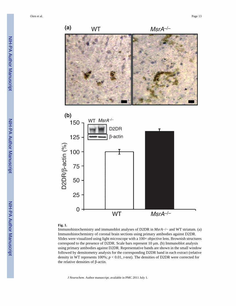
Fig. 1.Immunohistochemistry and immunoblot analyses of D2DR in MsrA−/− and WT striatum. (a)Immunohistochemistry of coronal brain sections using primary antibodies against D2DR.Slides were visualized using light microscope with a 100× objective lens. Brownish structurescorrespond to the presence of D2DR. Scale bars represent 10 μm. (b) Immunoblot analysisusing primary antibodies against D2DR. Representative bands are shown in the small windowfollowed by densitometry analysis for the corresponding D2DR band in each extract (relativedensity in WT represents 100%; p < 0.01, t-test). The densities of D2DR were corrected forthe relative densities of β-actin.
Oien et al. Page 13
J Neurochem. Author manuscript; available in PMC 2011 July 1.
NIH
-PA Author Manuscript
NIH
-PA Author Manuscript
NIH
-PA Author Manuscript
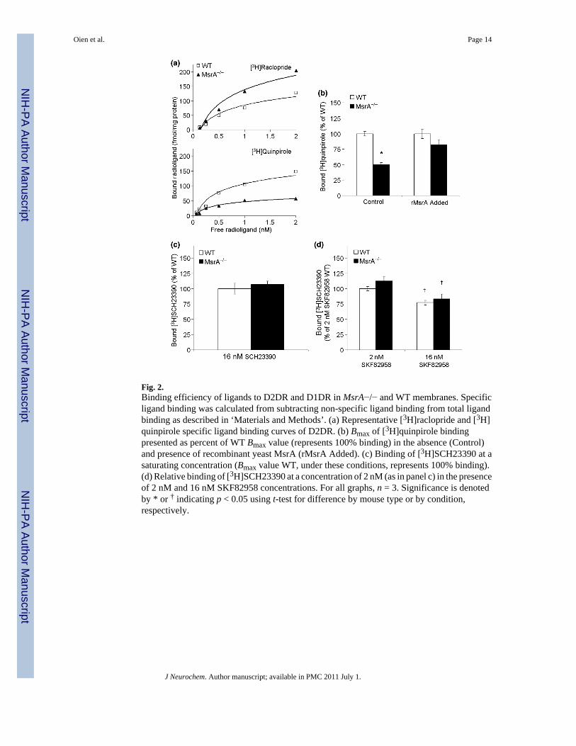
Fig. 2.Binding efficiency of ligands to D2DR and D1DR in MsrA−/− and WT membranes. Specificligand binding was calculated from subtracting non-specific ligand binding from total ligandbinding as described in ‘Materials and Methods’. (a) Representative [3H]raclopride and [3H]quinpirole specific ligand binding curves of D2DR. (b) Bmax of [3H]quinpirole bindingpresented as percent of WT Bmax value (represents 100% binding) in the absence (Control)and presence of recombinant yeast MsrA (rMsrA Added). (c) Binding of [3H]SCH23390 at asaturating concentration (Bmax value WT, under these conditions, represents 100% binding).(d) Relative binding of [3H]SCH23390 at a concentration of 2 nM (as in panel c) in the presenceof 2 nM and 16 nM SKF82958 concentrations. For all graphs, n = 3. Significance is denotedby * or † indicating p < 0.05 using t-test for difference by mouse type or by condition,respectively.
Oien et al. Page 14
J Neurochem. Author manuscript; available in PMC 2011 July 1.
NIH
-PA Author Manuscript
NIH
-PA Author Manuscript
NIH
-PA Author Manuscript
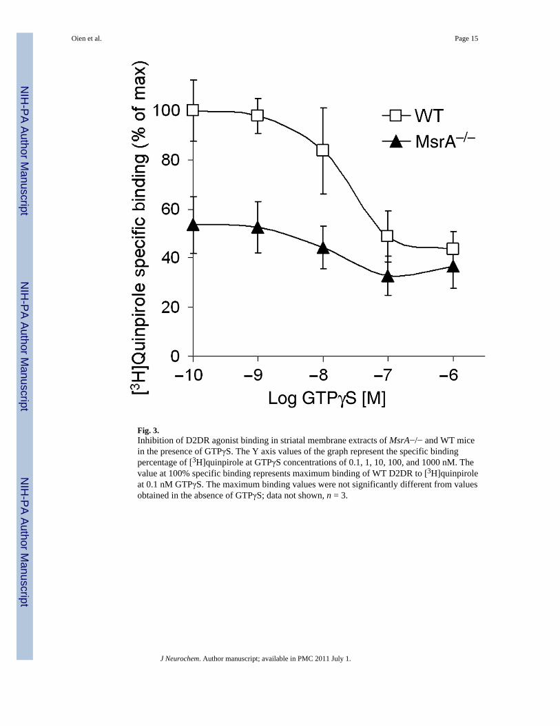
Fig. 3.Inhibition of D2DR agonist binding in striatal membrane extracts of MsrA−/− and WT micein the presence of GTPγS. The Y axis values of the graph represent the specific bindingpercentage of [3H]quinpirole at GTPγS concentrations of 0.1, 1, 10, 100, and 1000 nM. Thevalue at 100% specific binding represents maximum binding of WT D2DR to [3H]quinpiroleat 0.1 nM GTPγS. The maximum binding values were not significantly different from valuesobtained in the absence of GTPγS; data not shown, n = 3.
Oien et al. Page 15
J Neurochem. Author manuscript; available in PMC 2011 July 1.
NIH
-PA Author Manuscript
NIH
-PA Author Manuscript
NIH
-PA Author Manuscript
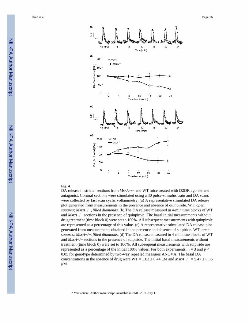
Fig. 4.DA release in striatal sections from MsrA−/− and WT mice treated with D2DR agonist andantagonist. Coronal sections were stimulated using a 30 pulse-stimulus train and DA scanswere collected by fast scan cyclic voltammetry. (a) A representative stimulated DA releaseplot generated from measurements in the presence and absence of quinpirole. WT, opensquares; MsrA−/−, filled diamonds. (b) The DA release measured in 4-min time blocks of WTand MsrA−/− sections in the presence of quinpirole. The basal initial measurements withoutdrug treatment (time block 0) were set to 100%. All subsequent measurements with quinpiroleare represented as a percentage of this value. (c) A representative stimulated DA release plotgenerated from measurements obtained in the presence and absence of sulpiride. WT, opensquares; MsrA−/−, filled diamonds. (d) The DA release measured in 4-min time blocks of WTand MsrA−/− sections in the presence of sulpiride. The initial basal measurements withouttreatment (time block 0) were set to 100%. All subsequent measurements with sulpiride arerepresented as a percentage of the initial 100% values. For both experiments, n = 3 and p <0.05 for genotype determined by two-way repeated measures ANOVA. The basal DAconcentrations in the absence of drug were WT = 1.63 ± 0.44 μM and MsrA−/− = 5.47 ± 0.36μM.
Oien et al. Page 16
J Neurochem. Author manuscript; available in PMC 2011 July 1.
NIH
-PA Author Manuscript
NIH
-PA Author Manuscript
NIH
-PA Author Manuscript
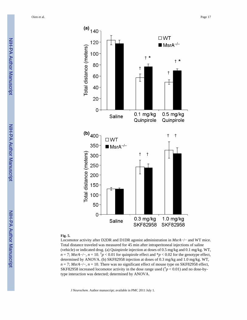
Fig. 5.Locomotor activity after D2DR and D1DR agonist administration in MsrA−/− and WT mice.Total distance traveled was measured for 45 min after intraperitoneal injections of saline(vehicle) or indicated drug. (a) Quinpirole injection at doses of 0.5 mg/kg and 0.1 mg/kg. WT,n = 7; MsrA−/−, n = 10. †p < 0.01 for quinpirole effect and *p < 0.02 for the genotype effect,determined by ANOVA. (b) SKF82958 injection at doses of 0.3 mg/kg and 1.0 mg/kg. WT,n = 7; MsrA−/−, n = 10. There was no significant effect of mouse type on SKF82958 effect,SKF82958 increased locomotor activity in the dose range used (†p < 0.01) and no dose-by-type interaction was detected; determined by ANOVA.
Oien et al. Page 17
J Neurochem. Author manuscript; available in PMC 2011 July 1.
NIH
-PA Author Manuscript
NIH
-PA Author Manuscript
NIH
-PA Author Manuscript
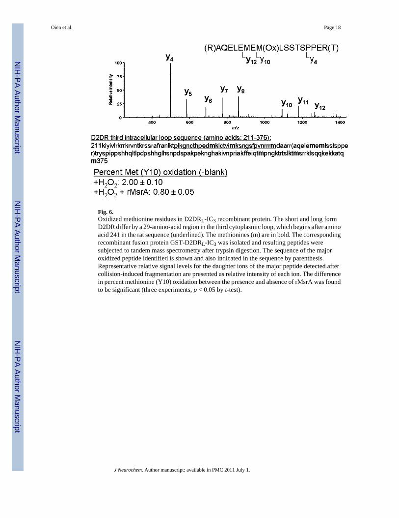
Fig. 6.Oxidized methionine residues in D2DRL-IC3 recombinant protein. The short and long formD2DR differ by a 29-amino-acid region in the third cytoplasmic loop, which begins after aminoacid 241 in the rat sequence (underlined). The methionines (m) are in bold. The correspondingrecombinant fusion protein GST-D2DRL-IC3 was isolated and resulting peptides weresubjected to tandem mass spectrometry after trypsin digestion. The sequence of the majoroxidized peptide identified is shown and also indicated in the sequence by parenthesis.Representative relative signal levels for the daughter ions of the major peptide detected aftercollision-induced fragmentation are presented as relative intensity of each ion. The differencein percent methionine (Y10) oxidation between the presence and absence of rMsrA was foundto be significant (three experiments, p < 0.05 by t-test).
Oien et al. Page 18
J Neurochem. Author manuscript; available in PMC 2011 July 1.
NIH
-PA Author Manuscript
NIH
-PA Author Manuscript
NIH
-PA Author Manuscript
Related Documents