Dopamine D1–D2 Receptor Heteromer in Dual Phenotype GABA/Glutamate-Coexpressing Striatal Medium Spiny Neurons: Regulation of BDNF, GAD67 and VGLUT1/2 Melissa L. Perreault 1,2 , Theresa Fan 1,2 , Mohammed Alijaniaram 1,2 , Brian F. O’Dowd 1,2 , Susan R. George 1,2,3 * 1 Centre for Addiction and Mental Health, Toronto, Ontario, Canada, 2 Department of Pharmacology and Toxicology, University of Toronto, Toronto, Ontario, Canada, 3 Department of Medicine, University of Toronto, Toronto, Ontario, Canada Abstract In basal ganglia a significant subset of GABAergic medium spiny neurons (MSNs) coexpress D1 and D2 receptors (D1R and D2R) along with the neuropeptides dynorphin (DYN) and enkephalin (ENK). These coexpressing neurons have been recently shown to have a region-specific distribution throughout the mesolimbic and basal ganglia circuits. While the functional relevance of these MSNs remains relatively unexplored, they have been shown to exhibit the unique property of expressing the dopamine D1–D2 receptor heteromer, a novel receptor complex with distinct pharmacology and cell signaling properties. Here we showed that MSNs coexpressing the D1R and D2R also exhibited a dual GABA/glutamate phenotype. Activation of the D1R–D2R heteromer in these neurons resulted in the simultaneous, but differential regulation of proteins involved in GABA and glutamate production or vesicular uptake in the nucleus accumbens (NAc), ventral tegmental area (VTA), caudate putamen and substantia nigra (SN). Additionally, activation of the D1R–D2R heteromer in NAc shell, but not NAc core, differentially altered protein expression in VTA and SN, regions rich in dopamine cell bodies. The identification of a MSN with dual inhibitory and excitatory intrinsic functions provides new insights into the neuroanatomy of the basal ganglia and demonstrates a novel source of glutamate in this circuit. Furthermore, the demonstration of a dopamine receptor complex with the potential to differentially regulate the expression of proteins directly involved in GABAergic inhibitory or glutamatergic excitatory activation in VTA and SN may potentially provide new insights into the regulation of dopamine neuron activity. This could have broad implications in understanding how dysregulation of neurotransmission within basal ganglia contributes to dopamine neuronal dysfunction. Citation: Perreault ML, Fan T, Alijaniaram M, O’Dowd BF, George SR (2012) Dopamine D1–D2 Receptor Heteromer in Dual Phenotype GABA/Glutamate- Coexpressing Striatal Medium Spiny Neurons: Regulation of BDNF, GAD67 and VGLUT1/2. PLoS ONE 7(3): e33348. doi:10.1371/journal.pone.0033348 Editor: Laurent Groc, Institute for Interdisciplinary Neuroscience, France Received September 30, 2011; Accepted February 13, 2012; Published March 12, 2012 Copyright: ß 2012 Perreault et al. This is an open-access article distributed under the terms of the Creative Commons Attribution License, which permits unrestricted use, distribution, and reproduction in any medium, provided the original author and source are credited. Funding: This work was supported by a grant from the National Institute on Drug Abuse (to SRG and BFO) and a Canadian Institutes on Health Research Postdoctoral Fellowship (to MLP). SRG holds a Canada Research Chair in Molecular Neuroscience. The funders had no role in study design, data collection and analysis, decision to publish, or preparation of the manuscript. Competing Interests: The authors have declared that no competing interests exist. * E-mail: [email protected] Introduction In contrast to classical thinking which depicts the dopamine D1 and D2 receptors (D1R and D2R) as being completely segregated to dynorphin (DYN)-expressing and enkephalin (ENK)-expressing striatonigral and striatopallidal pathways respectively, a growing accumulation of functional [1–6], and neuroanatomical [7–14] evidence now indicates that a physiologically relevant subset of medium spiny neurons (MSNs) exhibits a mixed phenotype, coexpressing the dopamine D1R and D2R in addition to substance P (SP)/DYN and ENK. Indeed, it has recently been reported that these MSNs exhibit a region-specific distribution throughout the mesolimbic and basal ganglia circuits [13]. More specifically, while a relatively low number of D1R-containing MSNs express the D2R (,6%) in caudate putamen (CP), higher coexpression levels are evident in ventral pallidum and entopeduncular nucleus, with the highest levels in the nucleus accumbens shell (NAc) (,17–34%) and globus pallidus (,60%) [13,15]. In addition, as D1R and D2R coexpression has been reported to occur selectively at presynaptic, but not postsynaptic terminals [13], together these findings suggest that MSNs coexpressing the D1R and D2R may have a unique physiological function at a local level as well as distal effects through their efferent projections that potentially impact on both the striatonigral and striatopallidal pathways. Although the physiological relevance of D1R and D2R coexpres- sing MSNs remains relatively unexplored, these neurons have also been shown to have the unique property of expressing the dopamine D1R–D2R heteromer, a novel receptor complex with discrete pharmacology and cell signaling properties [12,14,16,17]. Specifical- ly, the D1R–D2R heteromer has been shown to be distinct from its constituent receptors in that it is coupled to Gq/11 to activate phospholipase C and generate intracellular calcium release, repre- senting a novel signaling pathway directly linking dopamine action to calcium [12,17]. More recently, the activity of the D1R–D2R heteromer has been shown to be upregulated in rat striatum following repeated amphetamine administration and in the globus PLoS ONE | www.plosone.org 1 March 2012 | Volume 7 | Issue 3 | e33348

Welcome message from author
This document is posted to help you gain knowledge. Please leave a comment to let me know what you think about it! Share it to your friends and learn new things together.
Transcript
Dopamine D1–D2 Receptor Heteromer in DualPhenotype GABA/Glutamate-Coexpressing StriatalMedium Spiny Neurons: Regulation of BDNF, GAD67 andVGLUT1/2Melissa L. Perreault1,2, Theresa Fan1,2, Mohammed Alijaniaram1,2, Brian F. O’Dowd1,2, Susan R.
George1,2,3*
1 Centre for Addiction and Mental Health, Toronto, Ontario, Canada, 2 Department of Pharmacology and Toxicology, University of Toronto, Toronto, Ontario, Canada,
3 Department of Medicine, University of Toronto, Toronto, Ontario, Canada
Abstract
In basal ganglia a significant subset of GABAergic medium spiny neurons (MSNs) coexpress D1 and D2 receptors (D1R andD2R) along with the neuropeptides dynorphin (DYN) and enkephalin (ENK). These coexpressing neurons have been recentlyshown to have a region-specific distribution throughout the mesolimbic and basal ganglia circuits. While the functionalrelevance of these MSNs remains relatively unexplored, they have been shown to exhibit the unique property of expressingthe dopamine D1–D2 receptor heteromer, a novel receptor complex with distinct pharmacology and cell signalingproperties. Here we showed that MSNs coexpressing the D1R and D2R also exhibited a dual GABA/glutamate phenotype.Activation of the D1R–D2R heteromer in these neurons resulted in the simultaneous, but differential regulation of proteinsinvolved in GABA and glutamate production or vesicular uptake in the nucleus accumbens (NAc), ventral tegmental area(VTA), caudate putamen and substantia nigra (SN). Additionally, activation of the D1R–D2R heteromer in NAc shell, but notNAc core, differentially altered protein expression in VTA and SN, regions rich in dopamine cell bodies. The identification of aMSN with dual inhibitory and excitatory intrinsic functions provides new insights into the neuroanatomy of the basalganglia and demonstrates a novel source of glutamate in this circuit. Furthermore, the demonstration of a dopaminereceptor complex with the potential to differentially regulate the expression of proteins directly involved in GABAergicinhibitory or glutamatergic excitatory activation in VTA and SN may potentially provide new insights into the regulation ofdopamine neuron activity. This could have broad implications in understanding how dysregulation of neurotransmissionwithin basal ganglia contributes to dopamine neuronal dysfunction.
Citation: Perreault ML, Fan T, Alijaniaram M, O’Dowd BF, George SR (2012) Dopamine D1–D2 Receptor Heteromer in Dual Phenotype GABA/Glutamate-Coexpressing Striatal Medium Spiny Neurons: Regulation of BDNF, GAD67 and VGLUT1/2. PLoS ONE 7(3): e33348. doi:10.1371/journal.pone.0033348
Editor: Laurent Groc, Institute for Interdisciplinary Neuroscience, France
Received September 30, 2011; Accepted February 13, 2012; Published March 12, 2012
Copyright: � 2012 Perreault et al. This is an open-access article distributed under the terms of the Creative Commons Attribution License, which permitsunrestricted use, distribution, and reproduction in any medium, provided the original author and source are credited.
Funding: This work was supported by a grant from the National Institute on Drug Abuse (to SRG and BFO) and a Canadian Institutes on Health ResearchPostdoctoral Fellowship (to MLP). SRG holds a Canada Research Chair in Molecular Neuroscience. The funders had no role in study design, data collection andanalysis, decision to publish, or preparation of the manuscript.
Competing Interests: The authors have declared that no competing interests exist.
* E-mail: [email protected]
Introduction
In contrast to classical thinking which depicts the dopamine D1
and D2 receptors (D1R and D2R) as being completely segregated to
dynorphin (DYN)-expressing and enkephalin (ENK)-expressing
striatonigral and striatopallidal pathways respectively, a growing
accumulation of functional [1–6], and neuroanatomical [7–14]
evidence now indicates that a physiologically relevant subset of
medium spiny neurons (MSNs) exhibits a mixed phenotype,
coexpressing the dopamine D1R and D2R in addition to substance
P (SP)/DYN and ENK. Indeed, it has recently been reported that
these MSNs exhibit a region-specific distribution throughout the
mesolimbic and basal ganglia circuits [13]. More specifically, while
a relatively low number of D1R-containing MSNs express the D2R
(,6%) in caudate putamen (CP), higher coexpression levels are
evident in ventral pallidum and entopeduncular nucleus, with the
highest levels in the nucleus accumbens shell (NAc) (,17–34%) and
globus pallidus (,60%) [13,15]. In addition, as D1R and D2R
coexpression has been reported to occur selectively at presynaptic,
but not postsynaptic terminals [13], together these findings suggest
that MSNs coexpressing the D1R and D2R may have a unique
physiological function at a local level as well as distal effects through
their efferent projections that potentially impact on both the
striatonigral and striatopallidal pathways.
Although the physiological relevance of D1R and D2R coexpres-
sing MSNs remains relatively unexplored, these neurons have also
been shown to have the unique property of expressing the dopamine
D1R–D2R heteromer, a novel receptor complex with discrete
pharmacology and cell signaling properties [12,14,16,17]. Specifical-
ly, the D1R–D2R heteromer has been shown to be distinct from its
constituent receptors in that it is coupled to Gq/11 to activate
phospholipase C and generate intracellular calcium release, repre-
senting a novel signaling pathway directly linking dopamine action to
calcium [12,17]. More recently, the activity of the D1R–D2R
heteromer has been shown to be upregulated in rat striatum
following repeated amphetamine administration and in the globus
PLoS ONE | www.plosone.org 1 March 2012 | Volume 7 | Issue 3 | e33348

pallidus of patients who had schizophrenia [13], signifying a
potential role for this receptor complex in pathophysiologies
involving elevated dopamine transmission. In this study we sought
to further elucidate the importance of the dopamine D1R–D2R
heteromer in mediating neurotransmission within regions of the
basal ganglia and associated mesolimbic system by assessing the
expression of proteins known to be involved in GABA or glutamate
production and release. We showed that MSNs coexpressing D1R
and D2R exhibited a unique dual GABA/glutamate phenotype and
activation of the D1R–D2R heteromer by the selective agonist SKF
83959 in these neurons differentially and simultaneously regulated
the expression of proteins involved in GABA and glutamate activity
in regions of the mesolimbic and nigrostriatal pathways.
Results
D1R and D2R coexpressing MSNs also express bothGABA and glutamate
Striatal MSNs, which make up approximately 95% of all
neurons in this region, are consistently characterized as being
solely GABAergic. However we found that the subtype of GABA
MSNs that coexpressed the D1R and D2R also exhibited a
glutamatergic phenotype. We assessed coexpression of the D1R
and D2R with protein markers for GABA and glutamate neuron
identification, glutamate decarboxylase 67 (GAD67) and the
vesicular glutamate transporters 1 and 2 (VGLUT1, VGLUT2).
The specificity of the D1R and D2R antibodies has been strictly
validated and previously reported [13]. Specifically, dopamine
receptor antibodies for the D1R and D2R were tested using the
five dopamine receptors (D1–D5) expressed individually in
HEK293 cells, and testing was also performed in striatal tissue
of D1R or D2R gene-deleted mice where we showed no reactivity
of the D1R or D2R antibody respectively. When the primary D1R
and D2R antibodies and the relevant secondary antibodies were
combined, no cross-excitation of the secondary fluorophores was
evident and controls were also performed in the absence of the
primary or secondary antibodies to exclude cross-reactivity.
It was observed in cultured neonatal striatal neurons, almost all
of which exhibit the D1R/D2R-DYN/ENK phenotype [10,14],
that these neurons also coexpressed GAD67, as well as VGLUT1
and VGLUT2 (Fig. 1). To determine whether this mixed GABA/
glutamate phenotype was retained in D1R/D2R-DYN/ENK
neurons into adulthood, D1R and D2R coexpression with
GAD67, VGLUT1 and VGLUT2 was examined in adult rat
NAc (Fig. 2) and CP (Fig. 3). We showed in these regions that
almost all neurons coexpressing the D1R and D2R also expressed
GAD67, VGLUT1 or VGLUT2 (Figs. 2A and 3A), signifying that
these MSNs were unique in potentially having both inhibitory and
excitatory capabilities in adult striatum as well as in neonatal
striatal neurons. A very small minority of neurons that coexpressed
the D1R and D2R in the absence of VGLUT1 (Fig. 2B) or
VGLUT2 (Fig. 3B) was visualized, suggesting that these neurons
either did not express VGLUT1 or VGLUT2 or, alternatively,
may have expressed the VGLUT subtype not examined in that
particular experiment. Nonetheless, these results suggest that the
large majority of D1R/D2R coexpressing neurons expressed both
VGLUT1 and VGLUT2. Given the prevalence of these D1R/
D2R-DYN/ENK coexpressing MSNs throughout both the
striatopallidal and striatonigral pathways of the basal ganglia
[13], these findings emphasize the potential importance of these
mixed phenotype neurons not only in the regulation of thalamic
output, but additionally in the regulation of its associated neuronal
connections to regions rich in dopamine cell bodies such as to the
ventral tegmental area (VTA) and substantia nigra (SN).
Brain region-specific modification of GABA andglutamate production and/or vesicular uptake bydopamine D1R–D2R heteromer
Neurons that coexpress the D1R and D2R have a unique
function in that they express the dopamine D1R–D2R heteromer,
a novel receptor complex linked to Gq-mediated intracellular
calcium release and brain-derived neurotrophic factor (BDNF)
production [12,14,16,17]. We have shown that the D1R–D2R
Figure 1. Dopamine D1R and D2R colocalized in dualphenotype GABA/glutamate-expressing MSNs in culturedneonatal striatal neurons. Confocal images revealed D1R and D2Rcolocalization with the GABA neuronal marker, GAD67 (top row), andthe glutamate markers, VGLUT1 and VGLUT2 in striatal neuronscultured 7–10 days. Scale bar 10 mm.doi:10.1371/journal.pone.0033348.g001
Figure 2. Dopamine D1R and D2R colocalized in GABA/glutamate-coexpressing MSNs in adult rat NAc. (A) Confocalimages revealed D1R and D2R colocalization with GAD67 (top row) andVGLUT1 and VGLUT2 in NAc core. GAD67 was also shown to colocalizewith VGLUT1 and VGLUT2 in these neurons (bottom row). (B)Colocalization of the D1R and D2R with VGLUT1 (white arrows) andin the absence of VGLUT1 (yellow arrows) in NAc shell. Scale bar 10 mm.doi:10.1371/journal.pone.0033348.g002
D1R–D2R Heteromer in GABA/Glutamate Neurons
PLoS ONE | www.plosone.org 2 March 2012 | Volume 7 | Issue 3 | e33348

heteromer is localized to both cell soma and at presynaptic
terminals in NAc and CP [13] a finding suggestive of a possible
role in presynaptic GABA or glutamate neurotransmission from
these neurons, as well as the potential for both local and distal
effects on other neuronal subtypes such as those individually
expressing the D1R or D2R. To further elucidate the impact of
D1R–D2R heteromer activity on overall changes in GABA and
glutamate activity, we assessed the effects of the selective
heteromer agonist SKF 83959 on the expression of GAD67,
VGLUT1 and VGLUT2 in cultured neonatal striatal neurons.
These proteins are highly specific neuronal markers and
additionally provide a suitable index of GABA and glutamate
neurotransmission given their role in neurotransmitter production
or presynaptic vesicular uptake. We first examined GAD67, the
major enzyme involved in neuronal GABA production and the
expression of which has been shown to be associated with BDNF
signaling [18,19]. Treatment of cultured striatal neurons with
100 nM SKF 83959 led to a time-dependent increase in BDNF
and GAD67 expression as well as a decline in the expression of
VGLUT1 and VGLUT2 (Fig. 4). To determine the effects of
D1R–D2R heteromer activation on these proteins in vivo, we next
administered an acute systemic injection of SKF 83959 and
examined the expression of BDNF, GAD67, VGLUT1 and
VGLUT2, as well as the vesicular GABA transporter (VGAT), in
regions of the mesolimbic system and basal ganglia of the brain
(Fig. 5). Systemic activation of the D1R–D2R heteromer by SKF
83959 led to increased BDNF and GAD67 expression in the NAc
and VTA {NAc: BDNF P = 0.016, GAD67 P = 0.021; VTA:
BDNF P = 0.044, GAD67 P = 0.006} (Fig. 5B and 5C). In
contrast, in SN a significant decrease in the expression of both
proteins was observed {BDNF, P = 0.0001; GAD67 P = 0.041}
with no changes in CP (Fig. 5D,E). SKF 83959 did not alter the
expression of VGAT in any of the regions examined. We showed
no effect of SKF 83959 on VGLUT1 or VGLUT2 expression in
NAc (Fig. 5B) and a modest but significant increase of VGLUT2
in VTA {P = 0.050} (Fig. 5C). However, an elevation in the
expression of both VGLUT1 and VGLUT2 in SN {VGLUT1
P = 0.042; VGLUT2 P = 0.012} (Fig. 5E), and VGLUT2 in CP
Figure 3. Dopamine D1R and D2R colocalized in GABA/glutamate-coexpressing MSNs in adult rat CP. (A) Confocalimages showing D1R and D2R colocalization with GAD67 (top row), andVGLUT1 and VGLUT2 in CP. GAD67 also colocalized with VGLUT1 andVGLUT2 in CP (bottom row). Note the high levels of dendritic stainingfor the D1R in this region. (B) A neuron showing colocalization of theD1R and D2R in with in the absence of VGLUT2. Scale bar 10 mm.doi:10.1371/journal.pone.0033348.g003
Figure 4. Enhanced BDNF and GAD67, and reduced VGLUT1/2 expression following D1R–D2R heteromer activation. (A)Representative blots depicting the effects treatment of striatal neuronal cultures with vehicle or the D1R–D2R heteromer-selective agonist SKF 83959(100 nM) for 30, 60 or 120 min on BDNF, GAD67, VGLUT1 and VGLUT2 expression. (B) Treatment of the neuronal cultures with SKF 83959 for 120 minincreased the expression of BDNF and GAD67. In contrast, the expression of VGLUT1 and VGLUT2 was reduced with a 30 min treatment of SKF 83959.Values shown are mean 6 S.D.doi:10.1371/journal.pone.0033348.g004
D1R–D2R Heteromer in GABA/Glutamate Neurons
PLoS ONE | www.plosone.org 3 March 2012 | Volume 7 | Issue 3 | e33348

{P = 0.025} was observed (Fig. 5D). A direct relationship between
vesicular glutamate uptake by VGLUTs and glutamate release has
been demonstrated [20,21]. These results are indicative of a
potential role for the D1R–D2R heteromer in mediating
glutamate release in regions of the nigrostriatal pathway with no
effect on GABA. In contrast, the D1R–D2R heteromer had a
direct role in GABA production in regions of the mesolimbic
pathway with little effect on glutamate.
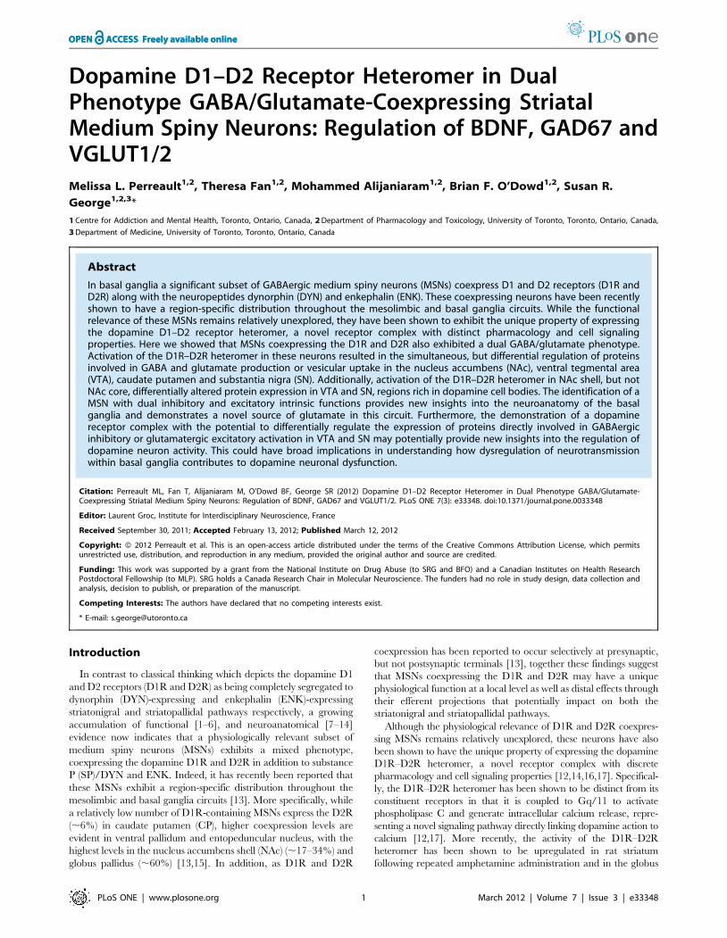
Region-specific regulation of pCaMKII and pERK by theD1R–D2R heteromer
The expression of BDNF has been shown to be regulated by
calcium/calmodulin kinase IIa (CaMKII) activation via phos-
phorylation at Thr286 [22], while phosphorylation of extracellular
regulated kinase (ERK) has been implicated in VGLUT
expression [23]. We showed a significant increase in pCaMKII
expression in NAc following acute systemic SKF 83959 admin-
istration {P = 0.042} (Fig. 6A,B). It should be noted, that while
SKF 83959 activates the D1R–D2R heteromer, the drug also
activates the D5 receptor (D5R). Although D5R expression in
NAc and CP is relatively low, being localized predominantly to
cholinergic interneurons [24] that comprise only ,1–2% of
neurons in striatum, we did confirm that the increased NAc
pCaMKII levels were induced by the D1R–D2R heteromer, and
not by D5R activation, as the expression of pCaMKII was not
elevated in response to SKF 83959 in mice gene deleted for the
D1R (D1R2/2 mice) (Fig. 6F). An increase in pCaMKII in VTA
by SKF 83959 was not evident (Fig. 6C) despite the increased
Figure 5. D1R–D2R heteromer discretely regulates the expression of proteins involved in GABA or glutamate activity. (A)Representative blots depicting the effects of a single injection of the D1R–D2R heteromer agonist SKF 83959 (1.5 mg/kg, sc) on BDNF, GAD67, VGAT,VGLUT1 and VGLUT2 expression in NAc, CP, VTA and SN (n = 8–9 rats/group). GAPDH was used as a loading control. (B, C) SKF 83959 increasedexpression of BDNF and GAD67 in NAc and VTA. No drug effects were observed on VGAT, VGLUT1 or VGLUT2 levels in NAc, while a significantincrease in VGLUT2 only was seen in VTA. (D, E) SKF 83959 had no effect on BDNF and GAD67 expression in CP, but diminished expression in SN.Increased levels of VGLUT2 were also evident in response to SKF 83959 in CP, with both VGLUT1 and VGLUT2 being elevated in SN. Bars shownrepresent means 6 s.e.m. and are expressed as a percentage of saline controls. *P,0.05, ** P,0.01.doi:10.1371/journal.pone.0033348.g005
D1R–D2R Heteromer in GABA/Glutamate Neurons
PLoS ONE | www.plosone.org 4 March 2012 | Volume 7 | Issue 3 | e33348
BDNF expression in this region (Fig. 5C). We postulate that this
lack of an overall region-wide effect of SKF 83959 on VTA
pCaMKII expression may have been the result of a D5R-
mediated suppression of pCaMKII levels as, in the absence of the
D5R (in D5R2/2 mice), there was a significant increase in
pCaMKII in response to the drug {P = 0.036} (Fig. 6G). The idea
of an attenuating effect of the D5R on pCaMKII is also consistent
with the results found in SN, a region with relatively higher
abundance of D5R [25], and which showed an overall reduction
in pCaMKII expression following SKF 83959 (Fig. 6E). In VTA,
CP, and SN, regions that showed increased expression of
VGLUT1 and/or VGLUT2 (Fig. 5), SKF 83959 induced an
increase in the levels of pERK42 and pERK44 {VTA: pERK42/
44 P = 0.010/0.001; CP: pERK44 P = 0.047; SN: pERK42/44
P = 0.018/0.007} (Fig. 6C–E), a finding that supports the
previously documented role for pERK in VGLUT expression
Figure 6. Region-specific regulation of pCaMKII, pERK, GABA and glutamate expression by the dopamine D1R–D2R heteromer. (A)Representative blots depicting the effects of a single systemic injection of SKF 83959 (1.5 mg/kg, sc) on pCaMKII and pERK expression in NAc, CP, VTAand SN (n = 8 rats/group). GAPDH was used as a loading control. (B–E) SKF 83959 increased CaMKII phosphorylation in NAc, had no effects in VTA andCP, and reduced pCaMKII levels in SN. Phosphorylation of ERK42 and ERK44 was elevated in both VTA and SN, and pERK44 levels were increased inCP. Data are expressed as a percentage of saline controls. (F–I) In the presence of SKF 83959 D1R2/2 mice exhibited significantly lower levels ofpCaMKII than D5R2/2 mice in NAc and VTA. There were no changes in pERK42 or pERK44 expression by SKF 83959 in either gene-deleted strain inthese regions. SKF 83959 did not alter pCaMKII expression in either the D1R2/2 or D5R2/2 mice in CP or SN. However, SKF 83959 induced asignificant increase in SN pERK expression in D5R2/2 mice, but not D1R2/2 mice. (J) Representative dot blots showing effects of SKF 83959 on totalGABA and glutamate levels in NAc (left panels) and CP (right panels). (K) SKF 83959 increased GABA levels, relative to glutamate, in NAc, but reducedlevels in CP. No drug effects were observed in VTA or SN (N = 8–9 rats/group). Bars shown represent means 6 s.e.m. *P,0.05, ** P,0.01, *** P,0.001,#P,0.05 compared to D5R2/2 mice.doi:10.1371/journal.pone.0033348.g006
D1R–D2R Heteromer in GABA/Glutamate Neurons
PLoS ONE | www.plosone.org 5 March 2012 | Volume 7 | Issue 3 | e33348

[23]. Both the D1R–D2R heteromer and D5R appeared to be
involved in pERK expression as the induction of pERK by SKF
83959 was absent in both D1R2/2 and D5R2/2 mice in VTA
and CP (Fig. 6G,H). In SN, however (Fig. 6I), the D1R–D2R
heteromer was solely responsible for the increased expression of
pERK as these effects were absent in the D1R2/2 mice, but
present in mice gene-deleted for the D5R {pERK42/44
P = 0.050/0.045}.
Total striatal GABA and glutamate levels alteredfollowing D1R–D2R heteromer activation
Thus far, these findings indicated that activation of the
dopamine D1R–D2R heteromer by SKF 83959 resulted in the
pathway-specific regulation of protein expression associated with
GABA and glutamate activity, with predominantly increased
expression of proteins involved with GABA activation in regions of
the mesolimbic pathway along with an associated elevation in
proteins involved with glutamate activity in regions of the
nigrostriatal pathway. To further characterize this, we assessed
total levels of GABA and glutamate expression in NAc, VTA, CP
and SN (Fig. 6J,K) following activation of the D1R–D2R
heteromer. SKF 83959 induced a significant increase in the
expression of GABA, relative to glutamate, in NAc {P = 0.019}
signifying a potential shift towards GABA neurotransmission in
this region. In contrast, a significant reduction in the ratio of
GABA to glutamate expression was evident in CP {P = 0.029},
indicative of a shift toward glutamate transmission. No changes in
the relative expression of GABA were present in SN or VTA
following SKF 83959.
D1R–D2R heteromers in NAc shell discretely alter theexpression of proteins involved in GABA and glutamateactivity in SN and VTA
As the NAc shows a relatively high abundance of D1R–D2R
heteromers [13], we next sought to determine the importance of
D1R–D2R heteromers localized to NAc core and shell in
regulating GABA- and glutamate-related protein expression in
VTA and SN (Fig. 7). Systemic administration of SKF 83959 has
been shown previously to induce orofacial movements and
grooming in rats [13,26]. It was noted that SKF 83959 injection
directly into NAc shell, but not NAc core, resulted in the
development of orofacial movements such as facial twitching and
teeth grinding in anaesthetized animals. Upon awakening, these
animals additionally exhibited elevated grooming behaviour, an
effect mediated by the D1R–D2R heteromer as grooming
behaviour was absent in D1R2/2 mice {P = 0.81}, but retained
in D5R 2/2 mice {P = 0.019} (Fig. 8). Activation of the D1R–
D2R heteromer by SKF 83959 in NAc core had no effect on
expression of BDNF, GAD67, VGLUT1 or VGLUT2 in the VTA
or SN (Fig. 7A,B). Activation of the D1R–D2R heteromer in NAc
shell however, induced significant increases in GAD67 in VTA
{P = 0.009} (Fig. 7C, left panel) and both BDNF and GAD67 in SN
{BDNF, P = 0.039; GAD67 P = 0.044} (Fig. 7C, right panel). These
effects in SN were opposite to that observed with the systemic
injection of SKF 83959, supporting a negative role for SN D5R in
the pCaMKII-BDNF-GAD67 signaling cascade. In addition, a
NAc shell injection also elevated the expression of both VGLUT1
{P = 0.036} and VGLUT2 {P = 0.035} supporting that the
systemic effects of SKF 83959 on VGLUT and pERK expression
in SN were mediated by the D1R–D2R heteromer. We cannot
presently determine whether there is a specific contribution of
D1R–D2R heteromer-induced GABA- versus glutamate-related
protein expression in the subregions of SN, namely the SN pars
compacta (SNc) and SN pars reticulata (SNr). Nonetheless, as
activation of the D1R–D2R heteromer in NAc shell appears to
mediate both inhibitory and excitatory outputs to SN, but only
inhibitory outputs to VTA, these findings indicate that the
regulation of dopamine neurons in these regions by the dual
GABA/glutamate MSNs is fundamentally different.
Discussion
In the present study we have identified a striatal MSN subtype
coexpressing GABA and glutamate as well as the D1R and D2R
together with DYN and ENK, and likely having both inhibitory
and excitatory capabilities. In cultured neonatal striatal neurons
the majority of neurons exhibited this mixed D1R/D2R-GABA/
glutamate expressing phenotype, and a significant fraction of these
coexpressing neurons was retained into adulthood in NAc. It was
further shown in adult rat that the activity of these D1R/D2R-
GABA/glutamate MSNs was differentially regulated by the
dopamine D1R–D2R heteromer, and that this regulation
occurred in a pathway-specific manner. These findings not only
demonstrate the existence of a novel source of glutamate in the
basal ganglia circuitry, but additionally provide new insights into
the neuroanatomy and physiology of basal ganglia functioning and
thereby having the potential to improve the understanding of the
plasticity underlying neuronal communication.
Mixed phenotype D1R/D2R-DYN/ENK-GABA/glutamateneurons in basal ganglia
It has been previously documented that the D1R and D2R are
coexpressed exclusively in a subset of striatal neurons that
coexpress the neuropeptides DYN and ENK [13]. In the present
study it was shown that these coexpressing MSNs are also positive
for markers of GABA and glutamate, indicative of neurons with
dual inhibitory and excitatory regulatory properties, and a finding
supported by a previous neuroanatomical study that showed some
striatal projection neurons possessed a high affinity uptake system
for glutamate and aspartate [27]. The pervasive presence of these
D1R/D2R-DYN/ENK-GABA/glutamate coexpressing neurons
in neonatal striatum suggests that these neurons may contribute to
the ontogeny of neuronal development, which has been previously
suggested for developing hippocampal GABA/glutamate-express-
ing granular neurons [28]. However, we additionally demonstrat-
ed that a proportion of these MSNs retained this mixed phenotype
into adulthood, a finding suggestive of a role for these specialized
MSNs in mediating neurotransmission in adult brain. Indeed, we
showed that these neurons were physiologically active in adult
striatum and provided a substrate for the direct coupling of
dopamine signaling to GABA and glutamate production and
release. Specifically, D1R/D2R-coexpressing MSNs differentially
regulated the expression of proteins involved in GABA and
glutamate activation in the NAc and VTA, as well as the CP and
SN, effects that were mediated by the dopamine D1R–D2R
heteromer and that are indicative of a significant role for these
neurons in the control of striatal MSN signaling. Furthermore, as
we have recently reported that D1R and D2R coexpressing
neurons exhibit a region-dependent distribution within both the
striatonigral and striatopallidal pathways of the basal ganglia
circuitry [13], we propose that there exists a subcircuitry of D1R/
D2R-DYN/ENK-GABA/glutamate MSNs, which interconnects
the basal ganglia nuclei, and which has the potential to impact on
mesolimbic and thalamic output. Further studies will be required
to clarify the specific neuronal subtypes that contribute to the
alterations in protein expression reported herein, neurons that
D1R–D2R Heteromer in GABA/Glutamate Neurons
PLoS ONE | www.plosone.org 6 March 2012 | Volume 7 | Issue 3 | e33348

may have included MSNs expressing individual D1R or D2R,
cholinergic neurons, and cortical glutamatergic afferents.
Regulation of mixed phenotype MSNs by the D1R–D2Rheteromer in regions of the mesolimbic and nigrostriatalpathways
We identified a novel GABA/glutamate coexpressing efferent
projection from NAc shell, regulated by the dopamine D1R–D2R
heteromer, which mediated GABA-related protein expression in
VTA. Similarly, systemic activation of the dopamine D1R–D2R
heteromer by SKF 83959 induced CaMKII activation and BDNF
and GAD67 expression in the NAc and VTA, a finding also
indicative of increased GABAergic tone in regions of the
mesolimbic pathway of the brain. As the activity of VTA
dopamine neurons is modulated by plasticity related to inhibitory
and excitatory inputs, and dysregulation of mesolimbic dopamine
signaling has been widely shown to be pivotal to neuropsychiatric
dopamine disorders, the present results may be indicative of a
mechanistic link between dopamine D1R–D2R heteromer-
induced signaling and disorders involving abnormal dopamine
transmission.
The idea of a role for the D1R–D2R heteromer in contributing
to disorders characterized by abnormal dopamine signaling is
supported by studies showing an integral involvement of CaMKII,
BDNF or GAD67 in the pathological processes underlying drug
addiction and schizophrenia. For instance, NAc shell CaMKII has
Figure 7. NAc core versus shell activation of dopamine D1R–D2R heteromer differentially regulates protein expression in VTA andSN. (A) Representative blots depicting the effects of a single intra-NAc core or shell injection of SKF 83959 (0.75 mg/0.5 ml unilateral) on BDNF,GAD67, VGLUT1 and VGLUT2 expression in VTA and SN (n = 9 rats/group). GAPDH was used as a loading control. (B) There was no effect of an intra-NAc core injection of SKF 83959 on protein expression in VTA (left panel) or SN (right panel). (C) SKF 83959 administration into NAc shell induced asignificant increase in GAD67 expression in VTA, but had no effect on BDNF or VGLUT expression (left panel). In contrast, SN showed elevated levelsof BDNF, GAD67, VGLUT1 and VGLUT2 (right panel). Bars shown represent means 6 s.e.m. and are expressed as a percentage of controls. *P,0.05,** P,0.01.doi:10.1371/journal.pone.0033348.g007
D1R–D2R Heteromer in GABA/Glutamate Neurons
PLoS ONE | www.plosone.org 7 March 2012 | Volume 7 | Issue 3 | e33348

been previously shown to be a critical component underlying
cocaine seeking by serving as a biochemical link between
dopamine and glutamate [29]. In addition, NAc and VTA BDNF
signaling mediates the magnitude of the reward responses to
cocaine [30–32], an interesting finding given the present results,
and previous reports, linking the dopamine D1R–D2R heteromer
to calcium signaling, CaMKII activation and BDNF expression in
NAc [12,14,17]. Along the same lines, the pathogenesis of
schizophrenia has been repeatedly associated with GABA
dysfunction in a number of regions including striatum, an effect
mediated in part by reduced expression of GAD67 [33,34], and
which we postulate may contribute to the increased VTA
dopamine neuronal activation inherent in the disorder. Interest-
ingly, mice deficient in CaMKII, a protein that may potentially
contribute to gene expression of GAD67 via phosphorylation of
the transcription repressor protein MeCP2 [22,35,36], also
displayed attributes similar to animal models of schizophrenia
[37]. As we have also previously shown an increase in the
activation state of the D1R–D2R heteromer in rat striatum
following repeated amphetamine administration, or in the globus
pallidus of patients who had schizophrenia [13], together these
findings suggest that further research into a role for the D1R–D2R
heteromer in these disorders involving dopamine dysfunction is
warranted.
Following intra-NAc shell activation of the D1R–D2R hetero-
mer there was increased expression of BDNF and GAD67 in SN,
that was also concurrent with stimulation of VGLUT1 and
VGLUT2 expression. At first glance, this dual increase in both
GABA- and glutamate-related protein expression may appear to
be redundant. However it is unlikely these changes simply negate
one another as alterations in neurotransmission would most likely
occur in distinct localized areas, such as at discrete neuronal
synapses or within different SN subregions. Indeed, neuroana-
tomical studies examining striatonigral projection neurons coex-
pressing D1R/D2R or SP/ENK have shown that these striato-
nigral efferents terminate in both subregions of SN, namely the
SNr and SNc [11,38]. We therefore suggest that D1R–D2R
heteromer-induced changes in GABA and glutamate activity in
SN may possibly occur via discrete projections from striatal
efferents to SNr and SNc.
Although the relative involvement of the SNr and SNc in
mediating the observed effects on D1R–D2R heteromer-induced
protein expression in SN could not be elucidated in the present
study, the SNr has been shown to be a pivotal region in the
stimulation of jaw movements and grooming in rodents [39,40],
behaviors that manifested following systemic [13] or intra-NAc
shell SKF 83959 administration as shown herein. Interestingly,
jaw movements elicited by ventral striatal costimulation of the
D1R and D2R are regulated by SNr NMDA receptor activation
[39], a finding thus linking increased D1R–D2R heteromer-
induced SNr glutamate, and VGLUT1 and VGLUT2 expres-
sion, to this behavior in rats. In addition, in SNc, studies have
shown neuroprotective effects of the D1R–D2R heteromer
agonist SKF 83959 on dopamine neurons following MPTP
lesioning in mice [41]. We postulate that these protective effects
of SKF 83959 on SNc neurons may be reflective of increased
D1R–D2R heteromer-induced BDNF expression, as protective
effects of BDNF on SNc dopamine neurons in parkinsonian
animals have been reported [42–44]. Notably, SKF 83959 has
been associated with strong anti-parkinsonian effects, with an
absence of dyskinesias, in non-human primate and rodent animal
models of Parkinson’s disease [45–48], and significantly reduced
L-Dopa-induced dyskinesias in 6-OHDA lesioned rats [48].
Therefore we posit that the D1R–D2R heteromer may mediate,
at least in part, the therapeutic effects of L-dopa, but not the
deleterious side effects induced by this treatment, implicating
this receptor complex as a valid novel therapeutic target in
Parkinson’s disease.
In summary, we demonstrated that in basal ganglia there exists
a subset of striatal MSNs that exhibit a novel mixed D1R/D2R-
DYN/ENK-GABA/glutamate phenotype, which may have pro-
jections, direct or indirect, from the NAc shell to areas rich in
dopamine cell bodies where the mesolimbic and nigrostriatal
pathways originate. We further showed that these neurons were
regulated by the dopamine D1R–D2R heteromer, a receptor
complex that exhibited selective, but differential control of the
expression of proteins associated with GABA and glutamate
activity in the NAc and VTA as well as the CP and SN. That the
dopamine D1R–D2R heteromer had the ability to differentially
regulate D1R/D2R-DYN/ENK/-GABA/glutamate MSNs, neu-
rons with postulated intrinsic inhibitory and excitatory functions,
in discrete regions of the basal ganglia emphasizes a potential role
of this receptor complex in mediating the plasticity underlying the
transition between GABAergic and glutamatergic dominance.
This could potentially have broad implications in furthering the
understanding of the pathophysiology and therapeutic manage-
ment of mesolimbic and basal ganglia disorders, such as
schizophrenia, drug addiction and Parkinson’s disease.
Materials and Methods
Neuronal CulturesNeonatal rat striata (1 day of age) were trypsinized in Hanks’
balanced salt solution (HBSS) with 0.25% trypsin and 0.05%
DNase (Sigma) at 37uC, and cells were washed three times in
HBSS with 12 mM MgSO4. Cells were dissociated in DMEM
with 2 mM glutamine and 10% FBS and plated at 26105 cells per
poly-L-lysine-coated well (Sigma; 50 mg/mL). The next day,
media were changed to Neurobasal medium with 506 B27
Supplement and 2 mM glutamine (Invitrogen). On day 3 of
culture, 5 mM cytosine arabinoside was added to inhibit glial cell
proliferation. Half of the medium was changed every 3 days.
Figure 8. Grooming induced by SKF 83959 is mediated by thedopamine D1R–D2R heteromer. A single systemic injection of SKF83959 (1.5 mg/kg, sc) induced a significant elevation in the amount oftime spent grooming (n = 7 mice/group). This effect was also present inmice gene-deleted for the D5R (D5R2/2) but absent in mice gene-deleted for the D1R (D1R2/2). D1R2/2 mice, but not D5R2/2 mice,also exhibited reduced basal levels of grooming compared to wildtype(WT). (Strain {F(2,36) = 12.1, P,0.001}; Drug {F(1,36} = 17.8, P,0.001};Strain x Drug {F(2,36) = 4.0, P,0.03}). Bars shown represent means 6s.e.m. and are expressed in seconds (s). *P,0.05, ** P,0.01 comparedto basal levels within the same strain. #P = 0.025 compared to basalwildtype.doi:10.1371/journal.pone.0033348.g008
D1R–D2R Heteromer in GABA/Glutamate Neurons
PLoS ONE | www.plosone.org 8 March 2012 | Volume 7 | Issue 3 | e33348

AnimalsSixty adult male Sprague-Dawley rats (Charles River, Canada)
and forty adult gene-deleted (D1R2/2 or D5R2/2) or control
mice were used. D1R2/2 and D5R2/2 mice were congenic,
having been backcrossed 12 times (N12). All procedures involving
animals complied with the guidelines described in the Guide to the
Care and Use of Experimental Animals (Canadian Council on
Animal Care, 1993), and were approved by the Animal Care
Ethics Committee of the University of Toronto (permit numbers
20008894 and 20008895).
DrugsSKF 83959 hydrobromide (Tocris Bioscience) was dissolved in
0.9% saline containing 5% DMSO. Systemic injections were given
subcutaneously at a dose of 1.5 mg/kg. For non-drug injections,
an equivalent volume of saline was administered. All injections
were administered at a volume of 1.0 ml/kg for rats and 5.0 ml/
kg for mice. For intra-NAc core or shell injections SKF 83959 was
dissolved in DMSO and aCSF at a volume of 0.75 mg/0.5 ml.
Fluorescence ImmunohistochemistryFluorescence immunohistochemistry was performed as previ-
ously described [14]. Paraformaldehyde-fixed striatal neurons or
coronal sections from untreated rat brain CP and NAc were
incubated with primary antibodies (1:200) for 60 hours at 4uC(D1R, Sigma-Aldrich; D2R, Chemicon; GAD67, VGLUT1,
VGLUT2, Millipore). Specificity of the dopamine receptor
antibodies for the D1R and D2R have been previously tested
and were validated in D1R or D2R gene-deleted mice [13]. To
minimize background and prevent cross-excitation of the second-
ary antibody-linked fluorophores, only three primary antibodies
were used on the cultured neurons or tissue at any given time.
Images were obtained using an Olympus Fluoview 1000 confocal
microscope at 636magnification.
GroomingAnimals were administered SKF 83959 (1.5 mg/kg, sc) and
placed immediately inside an empty cage similar in dimensions to
the home cage. Grooming activity was then monitored for
30 minutes. The measurement of grooming behavior followed a
previously described protocol [49] with the following modifica-
tions. The animal’s grooming was scored randomly for 30 seconds
for a total of 4 minutes (2 minutes sampled from the first
15 minutes of testing and 2 minutes sampled from the last
15 minutes of testing).
ImmunoblotFifteen or ninety minutes following SKF 83959 administration
brains were rapidly removed and tissue from the NAc, CP, SN and
VTA dissected and flash frozen until ready for use. Tissue was
suspended in cell lysis buffer and 10–30 micrograms of protein
were incubated in sample buffer for 3 minutes at 95uC. Samples
were separated by SDS-PAGE on a 10% tris-glycine gel and
electroblotted on PVDF transfer membrane for 2.5 hours. For
GABA and glutamate, a specific volume of homogenate,
standardized to total protein content, was pipetted directly onto
nitrocellulose membrane and BSA was used as a negative control.
Membranes were blocked and incubated overnight at 4uC with
gentle shaking with primary antibody to BDNF 1:10000, GAD67
1:6000, VGLUT1 1:10000, and VGLUT2 1:10000 (Millipore), to
VGAT 1:5000, pCaMKII 1:5000, (Pierce), to pERK1(42) and
pERK2(44) 1:3000, GABA 1:4000, glutamate 1:10000 (Sigma
Aldrich). Membranes were then washed in TBS-Tween and
incubated for 2 hours at room temperature with species-specific
secondary antibody (Bio-Rad Laboratories, Hercules, CA, USA).
Antibody labeling of proteins were detected with enhanced
chemiluminescence (Mandel Scientific or Millipore) and signal
intensity was quantified using Zeiss AxioVision4 software.
SurgeryRats were anesthetized using isoflurane, administered an
injection of the analgesic ketoprofen (5 mg/kg, sc) and secured
in a stereotaxic frame. Each rat was unilaterally injected into NAc
core or shell with SKF 83959 (0.75 mg/0.5 ml) according to the
following stereotaxic anterior–posterior (AP), mediolateral (ML),
and dorsoventral (DV) coordinates: NAc Core: AP +1.8 mm, ML
61.6 mm, DV 27.0 mm; NAc Shell: AP +1.8 mm, ML
60.8 mm, DV 27.5 mm. AP and ML coordinates were taken
from bregma, DV coordinates from skull surface (Paxinos and
Watson, 1998). Following removal from the stereotaxic frame,
animals were monitored for involuntary orofacial movements, and
upon recovery for grooming behaviour. Ninety minutes following
injection animals were decapitated, brains rapidly removed, and
injection sites into Nac core or shell confirmed. SN and VTA were
then dissected and flash frozen for use in immunoblot analysis.
Data AnalysisAll values are reported as mean 6 s.e.m. Comparisons of
means for protein expression was performed by the Student’s t
test (two-tailed, unpaired). For the grooming data, the statistical
significance of each dependent measure was first evaluated using
an ANOVA with Strain and Drug as the between subjects factors
followed by post-hoc Student’s t tests. The immunoblot data was
collected by densitometry and the main dependent variable was
Grey x area of band, expressed as a percent of saline controls.
Computations were performed using the SPSS/PC+ statistical
package.
Acknowledgments
The authors would like to thank Dr. Ahmed Hasbi for his technical
assistance.
Author Contributions
Conceived and designed the experiments: MLP SRG. Performed the
experiments: MLP TF MA. Analyzed the data: MLP BFO. Wrote the
paper: MLP SRG.
References
1. Walters JR, Bergstrom DA, Carlson JH, Chase TN, Braun AR (1987) D1
dopamine receptor activation required for postsynaptic expression of D2 agonist
effects. Science 236: 719–722.
2. DeBoer P, Abercrombie ED (1996) Physiological release of striatal acetylcholine
in vivo: modulation by D1 and D2 dopamine receptor subtypes. J Pharmacol
Exp Ther 277: 775–783.
3. White FJ, Bednarz LM, Wachtel SR, Hjorth S, Brooderson RJ (1988) Is
stimulation of both D1 and D2 receptors necessary for the expression of
dopamine-mediated behaviors? Pharmacol Biochem Behav 30: 189–193.
4. Capper-Loup C, Canales JJ, Kadaba N, Graybiel AM (2002) Concurrent
activation of dopamine D1 and D2 receptors is required to evoke neural
and behavioral phenotypes of cocaine sensitization. J Neurosci 22: 6218–
6227.
5. Calabresi P, Maj R, Mercuri NB, Bernardi G (1992) Coactivation of D1 and D2
dopamine receptors is required for long-term synaptic depression in the
striatum. Neurosci Lett 142: 95–99.
6. Hu XT, Wachtel SR, Galloway MP, White FJ (1990) Lesions of the nigrostriatal
dopamine projection increase the inhibitory effects of D1 and D2 dopamine
D1R–D2R Heteromer in GABA/Glutamate Neurons
PLoS ONE | www.plosone.org 9 March 2012 | Volume 7 | Issue 3 | e33348

agonists on caudate-putamen neurons and relieve D2 receptors from the
necessity of D1 receptor stimulation. J Neurosci 10: 2318–2329.
7. Meador-Woodruff JH, Mansour A, Healy DJ, Kuehn R, Zhou QY, et al. (1991)Comparison of the distributions of D1 and D2 dopamine receptor mRNAs in rat
brain. Neuropsychopharmacology 5: 231–242.
8. Lester J, Fink S, Aronin N, DiFiglia M (1993) Colocalization of D1 and D2
dopamine receptor mRNAs in striatal neurons. Brain Res 621: 106–110.
9. Surmeier DJ, Song WJ, Yan Z (1996) Coordinated expression of dopaminereceptors in neostriatal medium spiny neurons. J Neurosci 16: 6579–6591.
10. Aizman O, Brismar H, Uhlen P, Zettergren E, Levey AI, et al. (2000)
Anatomical and physiological evidence for D1 and D2 dopamine receptorcolocalization in neostriatal neurons. Nat Neurosci 3: 226–230.
11. Deng YP, Lei WL, Reiner A (2006) Differential perikaryal localization in rats of
D1 and D2 dopamine receptors on striatal projection neuron types identified byretrograde labeling. J Chem Neuroanat 32: 101–116.
12. Lee SP, So CH, Rashid AJ, Varghese G, Cheng R, et al. (2004) Dopamine D1
and D2 receptor co-activation generates a novel phospholipase C-mediated
calcium signal. J Biol Chem 279: 35671–35678.
13. Perreault ML, Hasbi A, Alijaniaram M, Fan T, Varghese G, et al. (2010) Thedopamine D1–D2 receptor heteromer localizes in dynorphin/enkephalin
neurons: increased high affinity state following amphetamine and in schizo-phrenia. J Biol Chem 285: 36625–36634.
14. Hasbi A, Fan T, Alijaniaram M, Nguyen T, Perreault ML, et al. (2009) Calcium
signaling cascade links dopamine D1–D2 receptor heteromer to striatal BDNFproduction and neuronal growth. Proc Natl Acad Sci U S A 106: 21377–21382.
15. Bertran-Gonzalez J, Bosch C, Maroteaux M, Matamales M, Herve D, et al.
(2008) Opposing patterns of signaling activation in dopamine D1 and D2
receptor-expressing striatal neurons in response to cocaine and haloperidol.J Neurosci 28: 5671–5685.
16. Verma V, Hasbi A, O’Dowd BF, George SR (2010) Dopamine D1–D2 receptor
Heteromer-mediated calcium release is desensitized by D1 receptor occupancywith or without signal activation: dual functional regulation by G protein-
coupled receptor kinase 2. J Biol Chem 285: 35092–35103.
17. Rashid AJ, So CH, Kong MM, Furtak T, El-Ghundi M, et al. (2007) D1–D2dopamine receptor heterooligomers with unique pharmacology are coupled to
rapid activation of Gq/11 in the striatum. Proc Natl Acad Sci U S A 104:
654–659.
18. Hashimoto T, Bergen SE, Nguyen QL, Xu B, Monteggia LM, et al. (2005)Relationship of brain-derived neurotrophic factor and its receptor TrkB to
altered inhibitory prefrontal circuitry in schizophrenia. J Neurosci 25: 372–383.
19. Pillai A, Mahadik SP (2008) Increased truncated TrkB receptor expression anddecreased BDNF/TrkB signaling in the frontal cortex of reeler mouse model of
schizophrenia. Schizophr Res 100: 325–333.
20. Moechars D, Weston MC, Leo S, Callaerts-Vegh Z, Goris I, et al. (2006)Vesicular glutamate transporter VGLUT2 expression levels control quantal size
and neuropathic pain. J Neurosci 26: 12055–12066.
21. Wilson NR, Kang J, Hueske EV, Leung T, Varoqui H, et al. (2005) Presynaptic
regulation of quantal size by the vesicular glutamate transporter VGLUT1.J Neurosci 25: 6221–6234.
22. Zhou Z, Hong EJ, Cohen S, Zhao WN, Ho HY, et al. (2006) Brain-specific
phosphorylation of MeCP2 regulates activity-dependent Bdnf transcription,dendritic growth, and spine maturation. Neuron 52: 255–269.
23. Doyle S, Pyndiah S, De Gois S, Erickson JD (2010) Excitation-transcription
coupling via calcium/calmodulin-dependent protein kinase/ERK1/2 signalingmediates the coordinate induction of VGLUT2 and Narp triggered by a
prolonged increase in glutamatergic synaptic activity. J Biol Chem 285:
14366–14376.
24. Berlanga ML, Simpson TK, Alcantara AA (2005) Dopamine D5 receptorlocalization on cholinergic neurons of the rat forebrain and diencephalon: a
potential neuroanatomical substrate involved in mediating dopaminergicinfluences on acetylcholine release. J Comp Neurol 492: 34–49.
25. Khan ZU, Gutierrez A, Martin R, Penafiel A, Rivera A, et al. (2000) Dopamine
D5 receptors of rat and human brain. Neuroscience 100: 689–699.
26. Deveney AM, Waddington JL (1995) Pharmacological characterization ofbehavioural responses to SK&F 83959 in relation to ‘D1-like’ dopamine
receptors not linked to adenylyl cyclase. Br J Pharmacol 116: 2120–2126.
27. White LE, Hodges HD, Carnes KM, Price JL, Dubinsky JM (1994)
Colocalization of excitatory and inhibitory neurotransmitter markers in striatalprojection neurons in the rat. J Comp Neurol 339: 328–340.
28. Gutierrez R (2003) The GABAergic phenotype of the ‘‘glutamatergic’’ granule
cells of the dentate gyrus. Prog Neurobiol 71: 337–358.
29. Anderson SM, Famous KR, Sadri-Vakili G, Kumaresan V, Schmidt HD, et al.
(2008) CaMKII: a biochemical bridge linking accumbens dopamine andglutamate systems in cocaine seeking. Nat Neurosci 11: 344–353.
30. Bahi A, Boyer F, Chandrasekar V, Dreyer JL (2008) Role of accumbens BDNF
and TrkB in cocaine-induced psychomotor sensitization, conditioned-placepreference, and reinstatement in rats. Psychopharmacology (Berl) 199: 169–182.
31. Graham DL, Edwards S, Bachtell RK, DiLeone RJ, Rios M, et al. (2007)Dynamic BDNF activity in nucleus accumbens with cocaine use increases self-
administration and relapse. Nat Neurosci 10: 1029–1037.
32. Graham DL, Krishnan V, Larson EB, Graham A, Edwards S (2009)Tropomyosin-related kinase B in the mesolimbic dopamine system: region-
specific effects on cocaine reward. Biol Psychiatry 65: 696–701.33. Akbarian S, Huang HS (2006) Molecular and cellular mechanisms of altered
GAD1/GAD67 expression in schizophrenia and related disorders. Brain ResRev 52: 293–304.
34. Veldic M, Kadriu B, Maloku E, Agis-Balboa RC, Guidotti A, et al. (2007)
Epigenetic mechanisms expressed in basal ganglia GABAergic neuronsdifferentiate schizophrenia from bipolar disorder. Schizophr Res 91: 51–61.
35. Kundakovic M, Chen Y, Guidotti A, Grayson DR (2009) The reelin andGAD67 promoters are activated by epigenetic drugs that facilitate the disruption
of local repressor complexes. Mol Pharmacol 75: 342–354.
36. Dong E, Agis-Balboa RC, Simonini MV, Grayson DR, Costa E, et al. (2005)Reelin and glutamic acid decarboxylase67 promoter remodeling in an epigenetic
methionine-induced mouse model of schizophrenia. Proc Natl Acad Sci U S A102: 12578–12583.
37. Novak G, Seeman P (2010) Hyperactive mice show elevated D2(High) receptors,a model for schizophrenia: Calcium/calmodulin-dependent kinase II alpha
knockouts. Synapse 64: 794–800.
38. Wang HB, Laverghetta AV, Foehring R, Deng YP, Sun Z, et al. (2006) Single-cell RT-PCR, in situ hybridization histochemical, and immunohistochemical
studies of substance P and enkephalin co-occurrence in striatal projectionneurons in rats. J Chem Neuroanat 31: 178–199.
39. Uchida T, Lee J, Fujita S, Kiguchi M, Matsumoto M, et al. (2005) Effects of
NMDA and MK-801 injected into the substantia nigra pars reticulata on jawmovements evoked by dopamine D12/D2 receptor stimulation in the
ventrolateral striatum: studies in freely moving rats. Methods Find Exp ClinPharmacol 27: 31–37.
40. Fletcher GH, Starr MS (1987) Role of the substantia nigra in the expression ofdopamine D1 receptor-mediated and D2 receptor-mediated behaviours.
Neuroscience 23: 1001–1010.
41. Zhang X, Zhou Z, Wang D, Li A, Yin Y, et al. (2009) Activation ofphosphatidylinositol-linked D1-like receptor modulates FGF-2 expression in
astrocytes via IP3-dependent Ca2+ signaling. J Neurosci 29: 7766–7775.42. Levivier M, Przedborski S, Bencsics C, Kang UJ (1995) Intrastriatal
implantation of fibroblasts genetically engineered to produce brain-derived
neurotrophic factor prevents degeneration of dopaminergic neurons in a ratmodel of Parkinson’s disease. J Neurosci 15: 7810–7820.
43. Shults CW, Kimber T, Altar CA (1995) BDNF attenuates the effects ofintrastriatal injection of 6-hydroxydopamine. Neuroreport 6: 1109–1112.
44. Tsukahara T, Takeda M, Shimohama S, Ohara O, Hashimoto N (1995) Effectsof brain derived neurotrophic factor on 1-methyl-4-phenyl-1,2,3,6-tetrahydro-
pyridine-induced parkinsonism in monkeys. Neurosurgery 1995 37: 733–739.
discussion 739–741.45. Andringa G, Stoof JC, Cools AR (1999) Sub-chronic administration of the
dopamine D(1) antagonist SKF 83959 in bilaterally MPTP-treated rhesusmonkeys: stable therapeutic effects and wearing-off dyskinesia. Psychopharma-
cology (Berl) 146: 328–334.
46. Gnanalingham KK, Hunter AJ, Jenner P, Marsden CD (1995) The differentialbehavioural effects of benzazepine D1 dopamine agonists with varying efficacies,
co-administered with quinpirole in primate and rodent models of Parkinson’sdisease. Psychopharmacology (Berl) 117: 287–297.
47. Gnanalingham KK, Hunter AJ, Jenner P, Marsden CD (1995) Selective
dopamine antagonist pretreatment on the antiparkinsonian effects of benzaze-pine D1 dopamine agonists in rodent and primate models of Parkinson’s
disease–the differential effects of D1 dopamine antagonists in the primate.Psychopharmacology (Berl) 117: 403–412.
48. Zhang H, Ma L, Wang F, Chen J, Zhen X (2007) Chronic SKF83959 inducedless severe dyskinesia and attenuated L-DOPA-induced dyskinesia in 6-OHDA-
lesioned rat model of Parkinson’s disease. Neuropharmacology 53: 125–133.
49. Culver KE, Rosenfeld JM, Szechtman H (2000) A switch mechanism betweenlocomotion and mouthing implicated in sensitization to quinpirole in rats.
Psychopharmacology (Berl) 151: 202–210.
D1R–D2R Heteromer in GABA/Glutamate Neurons
PLoS ONE | www.plosone.org 10 March 2012 | Volume 7 | Issue 3 | e33348
Related Documents