53 Bioremediation of uranium was investigated in microcosm experiments containing contaminated sediments from Oak Ridge, Tennessee to explore the importance of electron donor selection for uranium reduction rate and extent. In these experiments, all of the electron donors, including ethanol, glucose, methanol, and methanol with added humic acids, stimulated the reduction and immobilization of aqueous uranium by the indigenous microbial community. Uranium loss from solution began after the completion of nitrate reduction but essentially concurrent with sulfate reduction. When electron donor concentrations were normalized for their equivalent electron donor potential yield, the rates of uranium reduction were nearly equivalent for all treatments (0.55–0.95 μmol L −1 d −1 ). Uranium reduction with methanol proceeded after a 15-d longer lag time relative to that of ethanol or glucose. Significant differences were not found with the inclusion of humic acids. e extent of U reduction in sediment slurries measured by XANES at various time periods after the start of the experiment increased in the order of ethanol (5–7% reduced at 77 and 153 d), glucose (49% reduced at 53 d), and methanol (93% reduced at 90 d). e microbial diversity of ethanol- and methanol-amended microcosms in their late stage of U reduction was analyzed with 16S rRNA gene amplification. Members of the Geobacteraceae were found in all microcosms as well as other potential uranium-reducing organisms, such as Clostridium and Desulfosporosinus. e effectiveness of methanol relative to ethanol at reducing aqueous and sediment-hosted uranium suggests that bioremediation strategies that encourage fermentative poising of the subsurface to a lower redox potential may be more effective for long-term uranium immobilization as compared with selecting an electron donor that is efficiently metabolized by known uranium-reducing microorganisms. Donor-dependent Extent of Uranium Reduction for Bioremediation of Contaminated Sediment Microcosms Andrew S. Madden and Anthony V. Palumbo* Oak Ridge National Laboratory Bruce Ravel Argonne National Laboratory Tatiana A. Vishnivetskaya, Tommy J. Phelps, Christopher W. Schadt, and Craig C. Brandt Oak Ridge National Laboratory M etal and radionuclide contaminants have infiltrated water and sediment at many facilities under the management of the Department of Energy (DOE). More than $50 billion has been spent on remediation of DOE facilities, with an estimated 151 to $195 billion expected in the future with existing treatment technologies (NRC, 2000). One major research effort focuses on the stabilization of metal contaminants, such as uranium and chromium, through in situ stimulation of natural microbial communities to reduce the metals to less soluble forms (NABIR, 2003). ese communities in the subsurface have the capacity to immobilize several metal contaminants but are often limited by the lack of suitable electron donors. As electron donors (e.g., methanol, ethanol, pyruvate, glucose) are added, oxygen is consumed through aerobic respiration. en, alternate electron acceptors are used by bacteria, typically in a sequence determined by community metabolic potential, electron donor bioavailability, and decreasing energy yield of reaction. Oxidized soluble metals, such as U(VI) and Cr(VI), may be transformed to reduced, insoluble forms through various metabolic and coupled biogeochemical processes. Previous studies, summarized in Suzuki and Suko (2006) and Wall and Krumholz (2006), have shown that although uranium bioremediation through subsurface electron donor addition is possible, significant challenges persist. For example, electron do- nors shown to stimulate aqueous uranium reduction may reduce only 51% or less sediment-bound uranium (Ortiz-Bernad et al., 2004; Wu et al., 2006). Often, the electron donor is chosen to target organisms known to rapidly reduce uranium in laboratory settings. Nonlinear relationships between microbial community structure and subsurface biogeochemistry have been demonstrat- ed for immobilization of metals (Palumbo et al., 2004; Schryver et al., 2006), suggesting that, although enrichment of specific tar- get organisms has been documented in field experiments (Ander- son et al., 2003; Chang et al., 2005; Holmes et al., 2002; Istok et al., 2004; Nevin et al., 2003; North et al., 2004; Wu et al., Abbreviations: cgDNA, community genomic DNA; DOE, Department of Energy; ORFRC, Oak Ridge Field Research Center; XAS, X-ray absorption spectroscopy. A.S. Madden, Oak Ridge National Lab., Biosciences Division, P.O. Box 2008, Oak Ridge, TN 37831-6038. Present address: Univ. of Oklahoma, School of Geology and Geophysics, 100 East Boyd St., Norman, OK 73019. A.V. Palumbo, T.A. Vishnivetskaya, T.J. Phelps, C. Schadt, and C.C. Brandt, Oak Ridge National Lab., Biosciences Division, P.O. Box 2008, Oak Ridge TN 37831-6038. B. Ravel, Argonne National Lab., Molecular Environmental Science Group, Building 203, Argonne, IL 60439. Present address: National Institute of Standards and Technology Building 535A, Room M7 c/o Brookhaven National Lab., Upton, NY 11973. Copyright © 2009 by the American Society of Agronomy, Crop Science Society of America, and Soil Science Society of America. All rights reserved. No part of this periodical may be reproduced or transmitted in any form or by any means, electronic or mechanical, including pho- tocopying, recording, or any information storage and retrieval system, without permission in writing from the publisher. Published in J. Environ. Qual. 38:53–60 (2009). doi:10.2134/jeq2008.0071 Received 8 Feb. 2008. *Corresponding author ([email protected]). © ASA, CSSA, SSSA 677 S. Segoe Rd., Madison, WI 53711 USA TECHNICAL REPORTS: BIOREMEDIATION AND BIODEGRADATION

Welcome message from author
This document is posted to help you gain knowledge. Please leave a comment to let me know what you think about it! Share it to your friends and learn new things together.
Transcript
53
Bioremediation of uranium was investigated in microcosm experiments containing contaminated sediments from Oak Ridge, Tennessee to explore the importance of electron donor selection for uranium reduction rate and extent. In these experiments, all of the electron donors, including ethanol, glucose, methanol, and methanol with added humic acids, stimulated the reduction and immobilization of aqueous uranium by the indigenous microbial community. Uranium loss from solution began after the completion of nitrate reduction but essentially concurrent with sulfate reduction. When electron donor concentrations were normalized for their equivalent electron donor potential yield, the rates of uranium reduction were nearly equivalent for all treatments (0.55–0.95 μmol L−1 d−1). Uranium reduction with methanol proceeded after a 15-d longer lag time relative to that of ethanol or glucose. Signifi cant diff erences were not found with the inclusion of humic acids. Th e extent of U reduction in sediment slurries measured by XANES at various time periods after the start of the experiment increased in the order of ethanol (5–7% reduced at 77 and 153 d), glucose (49% reduced at 53 d), and methanol (93% reduced at 90 d). Th e microbial diversity of ethanol- and methanol-amended microcosms in their late stage of U reduction was analyzed with 16S rRNA gene amplifi cation. Members of the Geobacteraceae were found in all microcosms as well as other potential uranium-reducing organisms, such as Clostridium and Desulfosporosinus. Th e eff ectiveness of methanol relative to ethanol at reducing aqueous and sediment-hosted uranium suggests that bioremediation strategies that encourage fermentative poising of the subsurface to a lower redox potential may be more eff ective for long-term uranium immobilization as compared with selecting an electron donor that is effi ciently metabolized by known uranium-reducing microorganisms.
Donor-dependent Extent of Uranium Reduction for Bioremediation of Contaminated
Sediment Microcosms
Andrew S. Madden and Anthony V. Palumbo* Oak Ridge National Laboratory
Bruce Ravel Argonne National Laboratory
Tatiana A. Vishnivetskaya, Tommy J. Phelps, Christopher W. Schadt, and Craig C. Brandt Oak Ridge National Laboratory
Metal and radionuclide contaminants have infi ltrated water and sediment at many facilities under the management of
the Department of Energy (DOE). More than $50 billion has been spent on remediation of DOE facilities, with an estimated 151 to $195 billion expected in the future with existing treatment technologies (NRC, 2000). One major research eff ort focuses on the stabilization of metal contaminants, such as uranium and chromium, through in situ stimulation of natural microbial communities to reduce the metals to less soluble forms (NABIR, 2003). Th ese communities in the subsurface have the capacity to immobilize several metal contaminants but are often limited by the lack of suitable electron donors. As electron donors (e.g., methanol, ethanol, pyruvate, glucose) are added, oxygen is consumed through aerobic respiration. Th en, alternate electron acceptors are used by bacteria, typically in a sequence determined by community metabolic potential, electron donor bioavailability, and decreasing energy yield of reaction. Oxidized soluble metals, such as U(VI) and Cr(VI), may be transformed to reduced, insoluble forms through various metabolic and coupled biogeochemical processes.
Previous studies, summarized in Suzuki and Suko (2006) and Wall and Krumholz (2006), have shown that although uranium bioremediation through subsurface electron donor addition is possible, signifi cant challenges persist. For example, electron do-nors shown to stimulate aqueous uranium reduction may reduce only 51% or less sediment-bound uranium (Ortiz-Bernad et al., 2004; Wu et al., 2006). Often, the electron donor is chosen to target organisms known to rapidly reduce uranium in laboratory settings. Nonlinear relationships between microbial community structure and subsurface biogeochemistry have been demonstrat-ed for immobilization of metals (Palumbo et al., 2004; Schryver et al., 2006), suggesting that, although enrichment of specifi c tar-get organisms has been documented in fi eld experiments (Ander-son et al., 2003; Chang et al., 2005; Holmes et al., 2002; Istok et al., 2004; Nevin et al., 2003; North et al., 2004; Wu et al.,
Abbreviations: cgDNA, community genomic DNA; DOE, Department of Energy; ORFRC, Oak Ridge Field Research Center; XAS, X-ray absorption spectroscopy.
A.S. Madden, Oak Ridge National Lab., Biosciences Division, P.O. Box 2008, Oak Ridge, TN 37831-6038. Present address: Univ. of Oklahoma, School of Geology and Geophysics, 100 East Boyd St., Norman, OK 73019. A.V. Palumbo, T.A. Vishnivetskaya, T.J. Phelps, C. Schadt, and C.C. Brandt, Oak Ridge National Lab., Biosciences Division, P.O. Box 2008, Oak Ridge TN 37831-6038. B. Ravel, Argonne National Lab., Molecular Environmental Science Group, Building 203, Argonne, IL 60439. Present address: National Institute of Standards and Technology Building 535A, Room M7 c/o Brookhaven National Lab., Upton, NY 11973.
Copyright © 2009 by the American Society of Agronomy, Crop Science Society of America, and Soil Science Society of America. All rights reserved. No part of this periodical may be reproduced or transmitted in any form or by any means, electronic or mechanical, including pho-tocopying, recording, or any information storage and retrieval system, without permission in writing from the publisher.
Published in J. Environ. Qual. 38:53–60 (2009).doi:10.2134/jeq2008.0071Received 8 Feb. 2008. *Corresponding author ([email protected]).© ASA, CSSA, SSSA677 S. Segoe Rd., Madison, WI 53711 USA
TECHNICAL REPORTS: BIOREMEDIATION AND BIODEGRADATION
54 Journal of Environmental Quality • Volume 38 • January–February 2009
2006), the prevalence of individual taxonomic groups may not be suffi cient to describe the potential for long-term uranium immobilization. Sorbed or mineral U(VI) forms may continue to release uranium to ground water after cessation of electron donor addition, especially as carbonate complexes.
Microcosms and columns containing contaminated sedi-ments and ground water amended with electron donor have provided signifi cant insights into the relationships between microbial communities and aqueous geochemistry of uranium bioremediation (Edwards et al., 2007; Finneran et al., 2002a; Gu et al., 2005; Liu et al., 2005; Nyman et al., 2006). How-ever, none of these studies has directly compared the aqueous uranium concentrations with the extent of uranium reduc-tion for a suite of electron donors. In this study, we relate the electron donors to the rate and extent of uranium reduction in biostimulated sediments using microcosms. Th e pattern of electron acceptor use, the rate of U removal from solution in microcosms, the extent of reduction in sediment slurries, and the members of the microbial community are considered.
Materials and MethodsMicrocosm Experiments
Microcosms were prepared with materials collected from the Environmental Remediation Sciences Program Oak Ridge Field Research Center (ORFRC) located on the Department of Energy Oak Ridge Reservation in Oak Ridge, Tennessee. Adjacent to a historical liquid waste disposal area for low-level radioactive waste, the ORFRC is contaminated with acidic uranium, nitrate, and various minor inorganic, radionuclide, and organic constituents. An uncontaminated background area served as a source of humic materials. Sediment coring, isolation of humic acids, and ground water collection were ac-complished by ORFRC staff .
Sediment and ground water manipulations, microcosm con-struction, and sampling occurred within an anaerobic chamber (97:3 N2:H2). Sediment samples were homogenized and added to serum bottles. Four replicates were performed for each elec-tron donor treatment, and each microcosm used 20 g of sedi-ment (Area 2) and 80 mL of ORFRC ground water. Th e pH of the microcosms was adjusted to 7.0 using sodium bicarbonate. Electron donors were added at equivalent electron donor poten-tial, as determined by balancing of half-reactions (e.g., Th auer et al., 1977): methanol (40 mmol L−1), ethanol (20 mmol L−1), and glucose (5 mmol L−1). An additional duplicate microcosm set was performed with 10 mmol L−1 glucose. Unamended controls were also included in each experiment. Th e microcosms were kept in an anaerobic chamber during the experiment. Additional uranium or other solutes were not added beyond that found in the ORFRC sediment and ground water.
Aqueous Chemical MeasurementsSamples for chemical analysis were withdrawn using sterile
needles and syringes within the anaerobic chamber. Samples for U(VI) determination were centrifuged, diluted 50-fold in 0.1 mol L−1 HNO3, and analyzed by kinetic phosphorescence
analysis (Chemchek Instruments, Inc., Richland, WA). Ni-trate and sulfate were analyzed by ion chromatograpy (IonPac AS-14 analytical column and an AG-14 guard column; Dionex DX-120; Dionex, Sunnyvale, CA) after dilution in double de-ionized/ultrafi ltered water.
X-ray Absorption SpectroscopyX-ray absorption spectroscopy (XAS) was used to determine
the average valence state of U in microcosm samples. Measure-ments were performed at the Advanced Photon Source on two diff erent occasions using two diff erent beamlines. For all samples except ethanol (77 d) and glucose (5 mmol L−1), sediment slurries were transferred within an anaerobic chamber to 0.8 × 2 cm poly-carbonate sample holders. Hydroxy-sulfate green rust was synthe-sized and reacted with a U(VI)-acetate solution (O’Loughlin et al., 2003) for use as a U(IV) standard. Within the anaerobic chamber, sample holder openings were covered by sections of Kapton fi lm, sealed from air with Kapton tape, and placed in an additional bag of Kapton fi lm as secondary containment. Th e samples were stored in N2–fl ushed centrifuge tubes until measurement. Most spectra were collected at PNC-XOR, beamline 20BM (Heald et al., 1999). Simultaneous measurements in the transmission and fl uorescence were recorded. An yttrium foil was placed between the transmission and reference ionization chambers and used as an energy reference standard. Several scans were averaged for each measurement, and periodically the Y K-edge was measured for subsequent scan alignments. Spectra were collected using an un-focused beam on the samples as prepared. On a separate occasion, ethanol (77 d) and glucose amended microcosm sediments were analyzed at MRCAT, beamline 10ID (Segre et al., 2000). Data processing, including alignment, scan averaging, and linear com-bination fi tting, was performed using Athena software (Ravel and Newville, 2005). Th e averaged spectrum for ethanol (153 d) was smoothed with three iterations of interpolative smoothing before linear combination fi tting analysis.
16S Clone LibrariesDetailed microbial community analyses were performed in
duplicate on sediments from ethanol- and methanol-amended microcosms. Community genomic DNA (cgDNA) was extract-ed from approximately 1.5 mL of ground water/sediment (v/w, 4/1) using the PowerSoil DNA Isolation Kit (Mo Bio Laborato-ries, Inc., Carlsbad, CA). Approximately 0.1 to 0.6 μg of cgDNA was isolated from each sample. Th e purifi ed cgDNA was ampli-fi ed using Taq polymerase and bacteria-specifi c primers targeted to Escherichia coli 16S rRNA positions 8 to 27 (5′-AGA GTT TGA TCC TGG CTC AG-3′) and 1510 to 1492 (5′-GGT TAC CTT TTA CGA CTT-3′). Th e resulting polymerase chain reaction product of approximately 1.5 kb contained essentially the complete 16S rRNA gene. Polymerase chain reaction prod-ucts were purifi ed from UltraPure Agarose (Invitrogen, Carls-bad, CA) using QIAquick Gel Extraction kit (QUAGEN Inc., Valencia, CA). Polymerase chain reaction products were ligated in pCR 2.1-TOPO vectors (Invitrogen), transformed into One Shot Mach1–T1 chemically competent E. coli, and plated onto LB agar containing 50 μg mL−1 kanamycin and X-gal. Transfor-
Madden et al.: Uranium Bioremediation 55
mants were incubated overnight at 37°C and 96 white colonies were selected. Th ose colonies were grown separately overnight in LB broth containing 50 μg mL−1 kanamycin at 37°C with aeration. Clones were then sequenced using the BigDye Termi-nator v3.1 Cycle Sequencing kit and a reverse primer targeting positions 536 to 519 of E. coli 16S rRNA gene (5′-G(A/T)A TTA CCG CGG C(G/T)G CTG-3′) (Lane, 1991). Sequences were determined by resolving the sequence reactions on an Ap-plied Biosystems 3730 automated sequencer. Clone libraries were not checked for chimeric sequences because of the short length of sequenced 16S rRNA fragments (<500 bp). Sequences were aligned against the most similar sequences in the Ribosomal Database Project II and assigned to a set of hierarchical taxa us-ing a Naïve Bayesian rRNA classifi er (http://rdp.cme.msu.edu/classifi er/classifi er.jsp) version 1.0.
Results and DiscussionTime Course Aqueous Geochemical Data from Microcosm Experiments
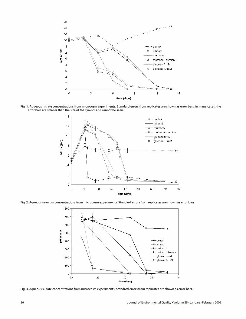
Th e evolution of nitrate (Fig. 1), uranium (Fig. 2), and sulfate (Fig. 3) concentrations followed expected patterns for electron-accepting processes with nitrate used fi rst. Signifi cant reductions in electron-acceptor concentrations were not observed in una-mended control samples. Nitrate was completely consumed in all microcosms with added electron donor by 9 to 14 d (Fig. 1), depending on the donor. Microcosms with added methanol and methanol with humic acids reduced nitrate more slowly than those with added glucose or ethanol and required an additional 2 to 3 d before the onset of nitrate reduction. During this initial period of oxygen and nitrate depletion, uranium concentrations increased (Fig. 2). Various processes likely contributed to the ini-tial increase in uranium concentrations within the microcosms. After addition of the ORFRC ground water to the microcosm sediments, changes in the ionic strength and solid/solution ratio were expected to cause the establishment of a new equilibrium between solution and solid. Although some small eff ect was seen in the control sample, the large eff ects observed in the donor-amended samples indicated that the increased uranium in solu-tion was likely the result of a biologically mediated process, most likely the production of CO2 during aerobic respiration and den-itrifi cation. At pH ?7, the addition of aqueous carbonate spe-cies would result in the formation of aqueous uranyl-carbonate complexes and would be expected to increase the concentration of aqueous U(VI) species (Langmuir, 1978). Th ere may also have been an increase in pH during nitrate reduction, which could have resulted in desorption of U(VI) from sediment and biomass surfaces; the adsorption edge for U(VI) on similar ORFRC sedi-ments was found to be extremely sensitive to pH in the range of 7 to 8 (Barnett et al., 2000). Uranium bound within sediment micropores or microprecipitates would likely have taken days to respond to increases in solution carbonate/bicarbonate spe-cies (Giammar and Hering, 2001; Liu et al., 2004; Mason et al., 1997; Phillips et al., 1995; Qafoku et al., 2005).
For microcosms with equivalent donor potential (excluding the 10 mmol L−1 glucose sample), the onset of uranium reduc-
tion did not begin until 20 d or more after donor addition (Fig. 2). Th e onset of reduction was signifi cantly delayed for methanol-amended microcosms by as much as 15 d. Slight decreases in the U(VI) concentrations between days 12 and 28 were likely due to decreases in pH from fermentation reactions, such as 4CH3OH + 2HCO3
− = 3CH3COO− + H+ + 4H2O (Adamse and Velzeboer, 1982), that produce acetate and other low-molecular weight or-ganic acids. For all donor-amended microcosms, the total aqueous U(VI) concentrations reached relatively steady values after 34 d (ethanol and 5 mmol L−1 glucose) or 42 d (methanol, methanol with humic acids) in the range of 0.4 to 1 μmol L−1. After the period of rapid reduction of most aqueous U(VI), concentrations continued to fall slowly over the period of 42 to 92 d, reaching fi nal concentrations below the EPA MCL of 30 ppb.
Despite the off set in initiation of U(VI) reduction, the rates of reduction vary by less than a factor of two, in the order 0.55 μmol L−1 d−1 (5 mmol L−1 glucose), 0.8 μmol L−1 d−1 (etha-nol), and 0.95 μmol L−1 d−1 (methanol), based on the instanta-neous linear slope of the U(VI)(aq) concentration data (Fig. 2). Estimates of total biomass were made for the two treatments sub-jected to microbial community analysis based on total DNA (El-lenbroek and Cappenberg, 1991). When the instantaneous rates were normalized on an estimated “per bacterial cell” basis, the biomass-adjusted uranium reduction rate was signifi cantly higher for the ethanol-amended microcosms as compared with the meth-anol-amended microcosms (39.4 nmol h−1 cell−1 vs. 3.4 nmol h−1 cell−1). Biomass estimates are not available from the 5 mmol L−1 glucose microcosms due to the lack of DNA extracted.
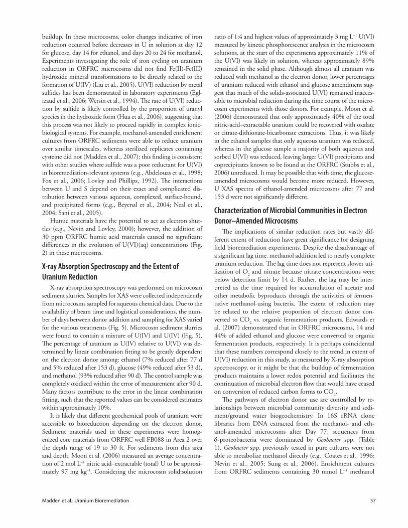
In many laboratory and fi eld studies, uranium reduction has been linked to or coincident with iron reduction (e.g., Abdelouas et al., 1998; Anderson et al., 2003; Finneran et al., 2002b; Nevin et al., 2003) and/or sulfate reduction (e.g., Nevin et al., 2003; Nyman et al., 2006; Suzuki et al., 2003; Wu et al., 2006), of-ten with sulfate reduction not occurring until after a time where iron reduction appeared to be the dominant terminal electron accepting process. Th e time-course data for sulfate in these mi-crocosm experiments did not provide such a straightforward re-lationship to uranium reduction. Sulfate concentrations ranged from ?0.6 to 0.8 mmol L−1 initially and reached fi nal values <0.1 μmol L−1 (Fig. 3) and varied signifi cantly in their evolution depending on the electron donor. In the case of glucose as the electron donor, sulfate reduction occurred rapidly between days 20 and 25 and took place slightly before and perhaps concurrent with the start of uranium reduction for the 5 mmol L−1 glucose microcosms but after uranium reduction for the 10 mmol L−1 glucose microcosms (Fig. 4). On the other hand, sulfate reduc-tion appeared nearly coincident with uranium reduction for the ethanol-amended microcosms. In the methanol and methanol with humic acid treatments, the bulk of the decrease of U in solution occurred after sulfate reduction (Fig. 4).
One possible explanation for the relationship between sulfate and uranium in these microcosms was that freshly formed metal sulfi des, observed in dark color development in the sediment, subsequently reduced U(VI). An accumulation of Fe(II) would be required before the precipitation of iron sulfi des, yet uranium reduction did not likely coincide with the timescale for Fe(II)
56 Journal of Environmental Quality • Volume 38 • January–February 2009
Fig. 1. Aqueous nitrate concentrations from microcosm experiments. Standard errors from replicates are shown as error bars. In many cases, the error bars are smaller than the size of the symbol and cannot be seen.
Fig. 2. Aqueous uranium concentrations from microcosm experiments. Standard errors from replicates are shown as error bars.
Fig. 3. Aqueous sulfate concentrations from microcosm experiments. Standard errors from replicates are shown as error bars.
Madden et al.: Uranium Bioremediation 57
buildup. In these microcosms, color changes indicative of iron reduction occurred before decreases in U in solution at day 12 for glucose, day 14 for ethanol, and days 20 to 24 for methanol. Experiments investigating the role of iron cycling on uranium reduction in ORFRC microcosms did not fi nd Fe(II)-Fe(III) hydroxide mineral transformations to be directly related to the formation of U(IV) (Liu et al., 2005). U(VI) reduction by metal sulfi des has been demonstrated in laboratory experiments (Egl-izaud et al., 2006; Wersin et al., 1994). Th e rate of U(VI) reduc-tion by sulfi de is likely controlled by the proportion of uranyl species in the hydroxide form (Hua et al., 2006), suggesting that this process was not likely to proceed rapidly in complex ionic-biological systems. For example, methanol-amended enrichment cultures from ORFRC sediments were able to reduce uranium over similar timescales, whereas sterilized replicates containing cysteine did not (Madden et al., 2007); this fi nding is consistent with other studies where sulfi de was a poor reductant for U(VI) in bioremediation-relevant systems (e.g., Abdelouas et al., 1998; Fox et al., 2006; Lovley and Phillips, 1992). Th e interactions between U and S depend on their exact and complicated dis-tribution between various aqueous, complexed, surface-bound, and precipitated forms (e.g., Beyenal et al., 2004; Neal et al., 2004; Sani et al., 2005).
Humic materials have the potential to act as electron shut-tles (e.g., Nevin and Lovley, 2000); however, the addition of 30 ppm ORFRC humic acid materials caused no signifi cant diff erences in the evolution of U(VI)(aq) concentrations (Fig. 2) in these microcosms.
X-ray Absorption Spectroscopy and the Extent of Uranium Reduction
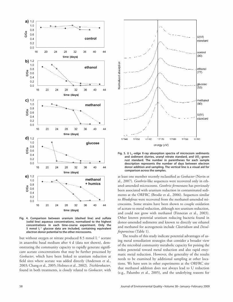
X-ray absorption spectroscopy was performed on microcosm sediment slurries. Samples for XAS were collected independently from microcosms sampled for aqueous chemical data. Due to the availability of beam time and logistical considerations, the num-ber of days between donor addition and sampling for XAS varied for the various treatments (Fig. 5). Microcosm sediment slurries were found to contain a mixture of U(IV) and U(IV) (Fig. 5). Th e percentage of uranium as U(IV) relative to U(VI) was de-termined by linear combination fi tting to be greatly dependent on the electron donor among: ethanol (7% reduced after 77 d and 5% reduced after 153 d), glucose (49% reduced after 53 d), and methanol (93% reduced after 90 d). Th e control sample was completely oxidized within the error of measurement after 90 d. Many factors contribute to the error in the linear combination fi tting, such that the reported values can be considered estimates within approximately 10%.
It is likely that diff erent geochemical pools of uranium were accessible to bioreduction depending on the electron donor. Sediment materials used in these experiments were homog-enized core materials from ORFRC well FB088 in Area 2 over the depth range of 19 to 30 ft. For sediments from this area and depth, Moon et al. (2006) measured an average concentra-tion of 2 mol L−1 nitric acid–extractable (total) U to be approxi-mately 97 mg kg−1. Considering the microcosm solid:solution
ratio of 1:4 and highest values of approximately 3 mg L−1 U(VI) measured by kinetic phosphorescence analysis in the microcosm solutions, at the start of the experiments approximately 11% of the U(VI) was likely in solution, whereas approximately 89% remained in the solid phase. Although almost all uranium was reduced with methanol as the electron donor, lower percentages of uranium reduced with ethanol and glucose amendment sug-gest that much of the solids-associated U(VI) remained inacces-sible to microbial reduction during the time course of the micro-cosm experiments with those donors. For example, Moon et al. (2006) demonstrated that only approximately 40% of the total nitric-acid–extractable uranium could be recovered with oxalate or citrate-dithionate-bicarbonate extractions. Th us, it was likely in the ethanol samples that only aqueous uranium was reduced, whereas in the glucose sample a majority of both aqueous and sorbed U(VI) was reduced, leaving larger U(VI) precipitates and coprecipitates known to be found at the ORFRC (Stubbs et al., 2006) unreduced. It may be possible that with time, the glucose-amended microcosms would become more reduced. However, U XAS spectra of ethanol-amended microcosms after 77 and 153 d were not signifi cantly diff erent.
Characterization of Microbial Communities in Electron Donor–Amended Microcosms
Th e implications of similar reduction rates but vastly dif-ferent extent of reduction have great signifi cance for designing fi eld bioremediation experiments. Despite the disadvantage of a signifi cant lag time, methanol addition led to nearly complete uranium reduction. Th e lag time does not represent slower uti-lization of O2 and nitrate because nitrate concentrations were below detection limit by 14 d. Rather, the lag may be inter-preted as the time required for accumulation of acetate and other metabolic byproducts through the activities of fermen-tative methanol-using bacteria. Th e extent of reduction may be related to the relative proportion of electron donor con-verted to CO2 vs. organic fermentation products. Edwards et al. (2007) demonstrated that in ORFRC microcosms, 14 and 44% of added ethanol and glucose were converted to organic fermentation products, respectively. It is perhaps coincidental that these numbers correspond closely to the trend in extent of U(VI) reduction in this study, as measured by X-ray absorption spectroscopy, or it might be that the buildup of fermentation products maintains a lower redox potential and facilitates the continuation of microbial electron fl ow that would have ceased on conversion of reduced carbon forms to CO2.
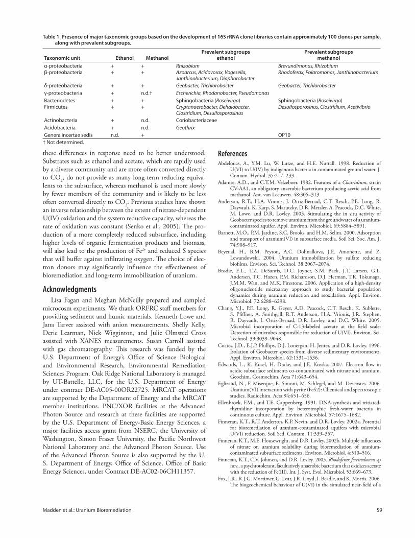
Th e pathways of electron donor use are controlled by re-lationships between microbial community diversity and sedi-ment/ground water biogeochemistry. In 16S rRNA clone libraries from DNA extracted from the methanol- and eth-anol-amended microcosms after Day 77, sequences from δ-proteobacteria were dominated by Geobacter spp. (Table 1). Geobacter spp. previously tested in pure cultures were not able to metabolize methanol directly (e.g., Coates et al., 1996; Nevin et al., 2005; Sung et al., 2006). Enrichment cultures from ORFRC sediments containing 30 mmol L−1 methanol
58 Journal of Environmental Quality • Volume 38 • January–February 2009
but without oxygen or nitrate produced 8.5 mmol L−1 acetate in anaerobic basal medium after 4 d (data not shown), dem-onstrating the community capacity to rapidly generate signifi -cant acetate concentrations that may be further processed by Geobacter, which have been linked to uranium reduction at fi eld sites where acetate was added directly (Anderson et al., 2003; Chang et al., 2005; Holmes et al., 2002). Trichlorobacter, found in both treatments, is closely related to Geobacter, with
at least one member recently reclassifi ed as Geobacter (Nevin et al., 2007). Geothrix-like sequences were recovered only in eth-anol-amended microcosms. Geothrix fermentans has previously been associated with uranium reduction in contaminated sedi-ments at the ORFRC (Brodie et al., 2006). Sequences similar to Rhodoferax were recovered from the methanol-amended mi-crocosms. Some strains have been shown to couple oxidation of acetate to metal reduction, although not uranium reduction, and could not grow with methanol (Finneran et al., 2003). Other known potential uranium reducing bacteria found in donor-amended sediments and known to directly use ethanol and methanol for acetogenesis include Clostridium and Desul-fosporosinus (Table 1).
Th e results of this study indicate potential advantages of us-ing metal remediation strategies that consider a broader view of the microbial community metabolic capacity for poising the redox potential toward metal reduction and also rapid enzy-matic metal reduction. However, the generality of the results needs to be examined by additional sampling at other loca-tions. We have seen in other experiments at the ORFRC site that methanol addition does not always lead to U reduction (e.g., Palumbo et al., 2005), and the underlying reasons for
Fig. 4. Comparison between uranium (dashed line) and sulfate (solid line) aqueous concentrations, normalized to the highest concentrations in each time-course experiment. Only the 5 mmol L−1 glucose data are included, containing equivalent electron donor potential to the other microcosms.
Fig. 5. U L3–edge X-ray absorption spectra of microcosm sediments and sediment slurries, uranyl nitrate standard, and UO2–green rust standard. The number in parentheses for each sample description represents the number of days between electron donor addition and sampling. The vertical line is a visual aid for comparison across the samples.
Madden et al.: Uranium Bioremediation 59
these diff erences in response need to be better understood. Substrates such as ethanol and acetate, which are rapidly used by a diverse community and are more often converted directly to CO2, do not provide as many long-term reducing equiva-lents to the subsurface, whereas methanol is used more slowly by fewer members of the community and is likely to be less often converted directly to CO2. Previous studies have shown an inverse relationship between the extent of nitrate-dependent U(IV) oxidation and the system reductive capacity, whereas the rate of oxidation was constant (Senko et al., 2005). Th e pro-duction of a more completely reduced subsurface, including higher levels of organic fermentation products and biomass, will also lead to the production of Fe2+ and reduced S species that will buff er against infi ltrating oxygen. Th e choice of elec-tron donors may signifi cantly infl uence the eff ectiveness of bioremediation and long-term immobilization of uranium.
AcknowledgmentsLisa Fagan and Meghan McNeilly prepared and sampled
microcosm experiments. We thank ORFRC staff members for providing sediment and humic materials. Kenneth Lowe and Jana Tarver assisted with anion measurements. Shelly Kelly, Deric Learman, Nick Wigginton, and Julie Olmsted Cross assisted with XANES measurements. Susan Carroll assisted with gas chromatography. Th is research was funded by the U.S. Department of Energy’s Offi ce of Science Biological and Environmental Research, Environmental Remediation Sciences Program. Oak Ridge National Laboratory is managed by UT-Battelle, LLC, for the U.S. Department of Energy under contract DE-AC05-00OR22725. MRCAT operations are supported by the Department of Energy and the MRCAT member institutions. PNC/XOR facilities at the Advanced Photon Source and research at these facilities are supported by the U.S. Department of Energy-Basic Energy Sciences, a major facilities access grant from NSERC, the University of Washington, Simon Fraser University, the Pacifi c Northwest National Laboratory and the Advanced Photon Source. Use of the Advanced Photon Source is also supported by the U. S. Department of Energy, Offi ce of Science, Offi ce of Basic Energy Sciences, under Contract DE-AC02-06CH11357.
ReferencesAbdelouas, A., Y.M. Lu, W. Lutze, and H.E. Nuttall. 1998. Reduction of
U(VI) to U(IV) by indigenous bacteria in contaminated ground water. J. Contam. Hydrol. 35:217–233.
Adamse, A.D., and C.T.M. Velzeboer. 1982. Features of a Clostridium, strain CV-AA1, an obligatory anaerobic bacterium producing acetic acid from methanol. Ant. van Leeuwen. 48:305–313.
Anderson, R.T., H.A. Vrionis, I. Ortiz-Bernad, C.T. Resch, P.E. Long, R. Dayvault, K. Karp, S. Marutzky, D.R. Metzler, A. Peacock, D.C. White, M. Lowe, and D.R. Lovley. 2003. Stimulating the in situ activity of Geobacter species to remove uranium from the groundwater of a uranium-contaminated aquifer. Appl. Environ. Microbiol. 69:5884–5891.
Barnett, M.O., P.M. Jardine, S.C. Brooks, and H.M. Selim. 2000. Adsorption and transport of uranium(VI) in subsurface media. Soil Sci. Soc. Am. J. 74:908–917.
Beyenal, H., B.M. Peyton, A.C. Dohnalkova, J.E. Amonette, and Z. Lewandowski. 2004. Uranium immobilization by sulfate reducing biofi lms. Environ. Sci. Technol. 38:2067–2074.
Brodie, E.L., T.Z. DeSantis, D.C. Joyner, S.M. Baek, J.T. Larsen, G.L. Andersen, T.C. Hazen, P.M. Richardson, D.J. Herman, T.K. Tokunaga, J.M.M. Wan, and M.K. Firestone. 2006. Application of a high-density oligonucleotide microarray approach to study bacterial population dynamics during uranium reduction and reoxidation. Appl. Environ. Microbiol. 72:6288–6298.
Chang, Y.J., P.E. Long, R. Geyer, A.D. Peacock, C.T. Resch, K. Sublette, S. Pfi ff ner, A. Smithgall, R.T. Anderson, H.A. Vrionis, J.R. Stephen, R. Dayvault, I. Ortiz-Bernad, D.R. Lovley, and D.C. White. 2005. Microbial incorporation of C-13-labeled acetate at the fi eld scale: Detection of microbes responsible for reduction of U(VI). Environ. Sci. Technol. 39:9039–9048.
Coates, J.D., E.J.P. Phillips, D.J. Lonergan, H. Jenter, and D.R. Lovley. 1996. Isolation of Geobacter species from diverse sedimentary environments. Appl. Environ. Microbiol. 62:1531–1536.
Edwards, L., K. Kusel, H. Drake, and J.E. Kostka. 2007. Electron fl ow in acidic subsurface sediments co-contaminated with nitrate and uranium. Geochim. Cosmochim. Acta 71:643–654.
Eglizaud, N., F. Miserque, E. Simoni, M. Schlegel, and M. Descostes. 2006. Uranium(VI) interaction with pyrite (FeS2): Chemical and spectroscopic studies. Radiochim. Acta 94:651–656.
Ellenbroek, F.M., and T.E. Cappenberg. 1991. DNA-synthesis and tritiated-thymidine incorporation by heterotrophic fresh-water bacteria in continuous culture. Appl. Environ. Microbiol. 57:1675–1682.
Finneran, K.T., R.T. Anderson, K.P. Nevin, and D.R. Lovley. 2002a. Potential for bioremediation of uranium-contaminated aquifers with microbial U(VI) reduction. Soil Sed. Contam. 11:339–357.
Finneran, K.T., M.E. Housewright, and D.R. Lovley. 2002b. Multiple infl uences of nitrate on uranium solubility during bioremediation of uranium-contaminated subsurface sediments. Environ. Microbiol. 4:510–516.
Finneran, K.T., C.V. Johnsen, and D.R. Lovley. 2003. Rhodoferax ferrireducens sp nov., a psychrotolerant, facultatively anaerobic bacterium that oxidizes acetate with the reduction of Fe(III). Int. J. Syst. Evol. Microbiol. 53:669–673.
Fox, J.R., R.J.G. Mortimer, G. Lear, J.R. Lloyd, I. Beadle, and K. Morris. 2006. Th e biogeochemical behaviour of U(VI) in the simulated near-fi eld of a
Table 1. Presence of major taxonomic groups based on the development of 16S rRNA clone libraries contain approximately 100 clones per sample, along with prevalent subgroups.
Taxonomic unit Ethanol MethanolPrevalent subgroups
ethanolPrevalent subgroups
methanolα-proteobacteria + + Rhizobium Brevundimonas, Rhizobiumβ-proteobacteria + + Azoarcus, Acidovorax, Vogesella,
Janthinobacterium, DiaphorobacterRhodoferax, Polaromonas, Janthinobacterium
δ-proteobacteria + + Geobacter, Trichlorobacter Geobacter, Trichlorobacterγ-proteobacteria + n.d.† Escherichia, Rhodanobacter, PseudomonasBacteriodetes + + Sphingobacteria (Roseivirga) Sphingobacteria (Roseivirga)Firmicutes + + Cryptanaerobacter, Dehalobacter,
Clostridium, Desulfosporosinus Desulfosporosinus, Clostridium, Acetivibrio
Actinobacteria + n.d. CoriobacteriaceaeAcidobacteria + n.d. GeothrixGenera incertae sedis n.d. + OP10
† Not determined.
60 Journal of Environmental Quality • Volume 38 • January–February 2009
low-level radioactive waste repository. Appl. Geochem. 21:1539–1550.Giammar, D.E., and J.G. Hering. 2001. Time scales for sorption-desorption
and surface precipitation of uranyl on goethite. Environ. Sci. Technol. 35:3332–3337.
Gu, B.H., W.M. Wu, M.A. Ginder-Vogel, H. Yan, M.W. Fields, J. Zhou, S. Fendorf, C.S. Criddle, and P.M. Jardine. 2005. Bioreduction of uranium in a contaminated soil column. Environ. Sci. Technol. 39:4841–4847.
Heald, S.M., D.L. Brewe, E.A. Stern, K.H. Kim, F.C. Brown, D.T. Jiang, E.D. Crozier, and R.A. Gordon. 1999. XAFS and micro-XAFS at the PNC-CAT beamlines. J. Synch. Rad. 6:347–349.
Holmes, D.E., K.T. Finneran, R.A. O’Neil, and D.R. Lovley. 2002. Enrichment of members of the family Geobacteraceae associated with stimulation of dissimilatory metal reduction in uranium-contaminated aquifer sediments. Appl. Environ. Microbiol. 68:2300–2306.
Hua, B., H.F. Xu, J. Terry, and B.L. Deng. 2006. Kinetics of uranium(VI) reduction by hydrogen sulfi de in anoxic aqueous systems. Environ. Sci. Technol. 40:4666–4671.
Istok, J.D., J.M. Senko, L.R. Krumholz, D. Watson, M.A. Bogle, A. Peacock, Y.J. Chang, and D.C. White. 2004. In situ bioreduction of technetium and uranium in a nitrate-contaminated aquifer. Environ. Sci. Technol. 38:468–475.
Lane, D.J. 1991. 16S/23S rRNA sequencing. p. 15–176. In E. Stackebrandt and M. Goodfellow (ed.) Nucleic acid techniques in bacterial systematics. John Wiley & Sons, Chichester, NY.
Langmuir, D. 1978. Uranium solution-mineral equilibria at low temperatures with applications to sedimentary ore deposits. Geochim. Cosmochim. Acta 42:547–569.
Liu, C.X., J.M. Zachara, O. Qafoku, J.P. McKinley, S.M. Heald, and Z.M. Wang. 2004. Dissolution of uranyl microprecipitates in subsurface sediments at Hanford site, USA. Geochim. Cosmochim. Acta 68:4519–4537.
Liu, C.X., J.M. Zachara, L.R. Zhong, R. Kukkadupa, J.E. Szecsody, and D.W. Kennedy. 2005. Infl uence of sediment bioreduction and reoxidation on uranium sorption. Environ. Sci. Technol. 39:4125–4133.
Lovley, D.R., and E.J.P. Phillips. 1992. Bioremediation of uranium contamination with enzymatic uranium reduction. Environ. Sci. Technol. 26:2228–2234.
Madden, A.S., A.C. Smith, D.L. Balkwill, L.F. Fagan, and T.J. Phelps. 2007. Uranium immobilization independent of nitrate reduction. Environ. Microbiol. 9:2321–2330.
Mason, C.F.V., W. Turney, B.M. Th omson, N. Lu, P.A. Longmire, and C.J. Chisholm-Brause. 1997. Carbonate leaching of uranium from contaminated soils. Environ. Sci. Technol. 31:2707–2711.
Moon, J.W., Y. Roh, T.J. Phelps, D.H. Phillips, D.B. Watson, Y.J. Kim, and S.C. Brooks. 2006. Physicochemical and mineralogical characterization of soil-saprolite cores from a fi eld research site, Tennessee. J. Environ. Qual. 35:1731–1741.
NABIR. 2003. Bioremediation of metals and radionuclides: What it is and how it works. Natural and Accelerated Bioremediation Research (NABIR) Program, Offi ce of Biological and Environmental Research, Offi ce of Science, U.S. Department of Energy, Washington, DC.
Neal, A.L., J.E. Amonette, B.M. Peyton, and G.G. Geesey. 2004. Uranium complexes formed at hematite surfaces colonized by sulfate-reducing bacteria. Environ. Sci. Technol. 38:3019–3027.
Nevin, K.P., K.T. Finneran, and D.R. Lovley. 2003. Microorganisms associated with uranium bioremediation in a high-salinity subsurface sediment. Appl. Environ. Microbiol. 69:3672–3675.
Nevin, K.P., D.E. Holmes, T.L. Woodard, S.F. Covalla, and D.R. Lovley. 2007. Reclassifi cation of Trichlorobacter thiogenes as Geobacter thiogenes comb. nov. Int. J. Syst. Evol. Microbiol. 57:463–466.
Nevin, K.P., D.E. Holmes, T.L. Woodard, E.S. Hinlein, D.W. Ostendorf, and D.R. Lovley. 2005. Geobacter bemidjiensis sp nov and Geobacter psychrophilus sp nov., two novel Fe(III)-reducing subsurface isolates. Int. J. Syst. Evol. Microbiol. 55:1667–1674.
Nevin, K.P., and D.R. Lovley. 2000. Potential for nonenzymatic reduction of Fe(III) via electron shuttling in subsurface sediments. Environ. Sci. Technol. 34:2472–2478.
North, N.N., S.L. Dollhopf, L. Petrie, J.D. Istok, D.L. Balkwill, and J.E. Kostka. 2004. Change in bacterial community structure during in situ biostimulation of subsurface sediment cocontaminated with uranium and nitrate. Appl. Environ. Microbiol. 70:4911–4920.
NRC. 2000. Long-term institutional management of U.S. Department of Energy legacy waste sites. National Academy Press, Washington, DC.
Nyman, J.L., T.L. Marsh, M.A. Ginder-Vogel, M. Gentile, S. Fendorf, and C.S. Criddle. 2006. Heterogeneous response to biostimulation for U(VI) reduction in replicated sediment microcosms. Biodegradation 17:303–316.
O’Loughlin, E.J., S.D. Kelly, R.E. Cook, R. Csencsits, and K.M. Kemner. 2003. Reduction of Uranium(VI) by mixed iron(II)/iron(III) hydroxide (green rust): Formation of UO2 nanoparticies. Environ. Sci. Technol. 37:721–727.
Ortiz-Bernad, I., R.T. Anderson, H.A. Vrionis, and D.R. Lovley. 2004. Resistance of solid-phase U(VI) to microbial reduction during in situ bioremediation of uranium-contaminated groundwater. Appl. Environ. Microbiol. 70:7558–7560.
Palumbo, A.V., S.M. Pfi ff ner, L. Fagan, M. McNeilly, S. Rishell, J.E. Kostka, and C.C. Brandt. 2005. Substrate and community structure determinants of uranium reduction rates in subsurface sediments. 3rd European Bioremediation Conf., 4–7 July 2005. Chania, Crete.
Palumbo, A.V., J.C. Schryver, M.W. Fields, C.E. Bagwell, J.Z. Zhou, T. Yan, X. Liu, and C.C. Brandt. 2004. Coupling of functional gene diversity and geochemical data from environmental samples. Appl. Environ. Microbiol. 70:6525–6534.
Phillips, E.J.P., E.R. Landa, and D.R. Lovley. 1995. Remediation of uranium contaminated soils with bicarbonate extraction and microbial U(VI) reduction. J. Ind. Microbiol. 14:203–207.
Qafoku, N.P., J.M. Zachara, C.X. Liu, P.L. Gassman, O.S. Qafoku, and S.C. Smith. 2005. Kinetic desorption and sorption of U(VI) during reactive transport in a contaminated Hanford sediment. Environ. Sci. Technol. 39:3157–3165.
Ravel, B., and M. Newville. 2005. ATHENA, ARTEMIS, HEPHAESTUS: Data analysis for X-ray absorption spectroscopy using IFEFFIT. J. Synch. Rad. 12:537–541.
Sani, R.K., B.M. Peyton, A. Dohnalkova, and J.E. Amonette. 2005. Reoxidation of reduced uranium with iron(III) (hydr)oxides under sulfate-reducing conditions. Environ. Sci. Technol. 39:2059–2066.
Schryver, J.C., C.C. Brandt, S.M. Pfi ff ner, A.V. Palumbo, A.D. Peacock, D.C. White, J.P. McKinley, and P.E. Long. 2006. Application of nonlinear analysis methods for identifying relationships between microbial community structure and groundwater geochemistry. Microb. Ecol. 51:177–188.
Segre, C.U., N.E. Leyarovska, L.D. Chapman, W.M. Lavender, P.W. Plag, A.S. King, A.J. Kropf, B.A. Bunker, K.M. Kemner, P. Dutta, and J. Kaduk. 2000. Th e MRCAT insertion device beamline at the advanced photon source. p. 419–422. In P. Pianetta (ed.) Synchrotron radiation instrumentation, 11th U.S. Conf., Vol. 521. American Inst. of Physics.
Senko, J.M., J.M. Sufl ita, and L.R. Krumholz. 2005. Geochemical controls on microbial nitrate-dependent U(IV) oxidation. Geomicrobiol. J. 22:371–378.
Stubbs, J.E., D.C. Elbert, D.R. Veblen, and C. Zhu. 2006. Electron microbeam investigation of uranium-contaminated soils from Oak Ridge, TN, USA. Environ. Sci. Technol. 40:2108–2113.
Sung, Y., K.F. Fletcher, K.M. Ritalaliti, R.P. Apkarian, N. Ramos-Hernandez, R.A. Sanford, N.M. Mesbah, and F.E. Loffl er. 2006. Geobacter lovleyi sp nov strain SZ, a novel metal-reducing and tetrachloroethene-dechlorinating bacterium. Appl. Environ. Microbiol. 72:2775–2782.
Suzuki, Y., S.D. Kelly, K.A. Kemner, and J.F. Banfi eld. 2003. Microbial populations stimulated for hexavalent uranium reduction in uranium mine sediment. Appl. Environ. Microbiol. 69:1337–1346.
Suzuki, Y., and T. Suko. 2006. Geomicrobiological factors that control uranium mobility in the environment: Update on recent advances in the bioremediation of uranium-contaminated sites. J. Miner. Petrol. Sci. 101:299–307.
Th auer, R.K., K. Jungerman, and K. Decker. 1977. Energy conservation in anaerobic chemotrophic bacteria. Bacteriol. Rev. 41:100–180.
Wall, J.D., and L.R. Krumholz. 2006. Uranium reduction. Annu. Rev. Microbiol. 60:149–166.
Wersin, P., M.F. Hochella, P. Persson, G. Redden, J.O. Leckie, and D.W. Harris. 1994. Interaction between aqueous uranium(VI) and sulfi de minerals: Spectroscopic evidence for sorption and reduction. Geochim. Cosmochim. Acta 58:2829–2843.
Wu, W.M., J. Carley, T. Gentry, M.A. Ginder-Vogel, M. Fienen, T. Mehlhorn, H. Yan, S. Caroll, M.N. Pace, J. Nyman, J. Luo, M.E. Gentile, M.W. Fields, R.F. Hickey, B.H. Gu, D. Watson, O.A. Cirpka, J.Z. Zhou, S. Fendorf, P.K. Kitanidis, P.M. Jardine, and C.S. Criddle. 2006. Pilot-scale in situ bioremedation of uranium in a highly contaminated aquifer: II. Reduction of U(VI) and geochemical control of U(VI) bioavailability. Environ. Sci. Technol. 40:3986–3995.
Related Documents