Chapter 13 Assessing Collagen Deposition During Aging in Mammalian Tissue and in Caenorhabditis elegans Alina C. Teuscher, Cyril Statzer, Sophia Pantasis, Mattia R. Bordoli, and Collin Y. Ewald Abstract Proper collagen homeostasis is essential for development and aging of any multicellular organism. During aging, two extreme scenarios are commonly occurring: a local excess in collagen deposition, for instance during fibrosis, or a gradual overall reduction of collagen mass. Here, we describe a histological and a colorimetric method to assess collagen levels in mammalian tissues and in the nematode Caenorhabditis elegans. The first method is the polychrome Herovici staining to distinguish between young and mature collagen ratios. The second method is based on hydroxyproline measurements to estimate collagen protein levels. In addition, we show how to decellularize the multicellular organism C. elegans in order to harvest its cuticle, one of the two major extracellular matrices, mainly composed of collagen. These methods allow assessing collagen deposition during aging either in tissues or in whole organisms. Key words Collagen, Aging, Tissue, Herovici staining, Hydroxyproline, Cuticle, Isolation, Freeze- cracking, Age-synchronizing, Extracellular matrix, C. elegans 1 Introduction Cells and tissues are embedded within extracellular matrices (ECM), which are important for tissue geometry, integrity, and function [1]. In mammals, collagens constitute about 30% of the total protein mass in the body and are the major component of the ECM [2]. In humans, collagen turnover can be extremely slow as observed in the lens of the eye and in cartilage structures (with a half-life time of 114 years [3–5]) or extremely fast, within 72 h after physical exercise, in Achilles tendons [6]. Hence, depending on the tissue or organ, some collagens are synthesized, secreted, and integrated in the ECM once early in life and are not replaced over Irit Sagi and Nikolaos A. Afratis (eds.), Collagen: Methods and Protocols, Methods in Molecular Biology, vol. 1944, https://doi.org/10.1007/978-1-4939-9095-5_13, © Springer Science+Business Media, LLC, part of Springer Nature 2019 Alina C. Teuscher and Cyril Statzer contributed equally to this chapter. 169

Welcome message from author
This document is posted to help you gain knowledge. Please leave a comment to let me know what you think about it! Share it to your friends and learn new things together.
Transcript
Chapter 13
Assessing Collagen Deposition During Aging in MammalianTissue and in Caenorhabditis elegans
Alina C. Teuscher, Cyril Statzer, Sophia Pantasis, Mattia R. Bordoli,and Collin Y. Ewald
Abstract
Proper collagen homeostasis is essential for development and aging of any multicellular organism. Duringaging, two extreme scenarios are commonly occurring: a local excess in collagen deposition, for instanceduring fibrosis, or a gradual overall reduction of collagen mass. Here, we describe a histological and acolorimetric method to assess collagen levels in mammalian tissues and in the nematode Caenorhabditiselegans. The first method is the polychrome Herovici staining to distinguish between young and maturecollagen ratios. The second method is based on hydroxyproline measurements to estimate collagen proteinlevels. In addition, we show how to decellularize the multicellular organismC. elegans in order to harvest itscuticle, one of the two major extracellular matrices, mainly composed of collagen. These methods allowassessing collagen deposition during aging either in tissues or in whole organisms.
Key words Collagen, Aging, Tissue, Herovici staining, Hydroxyproline, Cuticle, Isolation, Freeze-cracking, Age-synchronizing, Extracellular matrix, C. elegans
1 Introduction
Cells and tissues are embedded within extracellular matrices(ECM), which are important for tissue geometry, integrity, andfunction [1]. In mammals, collagens constitute about 30% of thetotal protein mass in the body and are the major component of theECM [2]. In humans, collagen turnover can be extremely slow asobserved in the lens of the eye and in cartilage structures (with ahalf-life time of 114 years [3–5]) or extremely fast, within 72 h afterphysical exercise, in Achilles tendons [6]. Hence, depending on thetissue or organ, some collagens are synthesized, secreted, andintegrated in the ECM once early in life and are not replaced over
Irit Sagi and Nikolaos A. Afratis (eds.), Collagen: Methods and Protocols, Methods in Molecular Biology, vol. 1944,https://doi.org/10.1007/978-1-4939-9095-5_13, © Springer Science+Business Media, LLC, part of Springer Nature 2019
Alina C. Teuscher and Cyril Statzer contributed equally to this chapter.
169
the entire lifetime, whereas other collagens have a higherturnover rate.
During aging, the connective tissue or ECM integrity declinesdue to accumulation of damage from collagen fragmentation, oxi-dation, and glycation [2, 7–12] leading to a continuous reductionof collagen mass, as best illustrated by wrinkles and sagging skin[13, 14], but also observed in other connective tissues that supportorgan function. This deterioration in ECM integrity has beenimplicated in many age-dependent diseases, such as diabetes, can-cer, chronic liver diseases, and cardiovascular diseases [2, 7–12]. Inparallel, the accumulation of molecular damage [15], chronicinflammation (inflammaging) [16], or injury during aging mightlocally drive abnormal collagen deposition resulting in fibrosis[17]. These observations suggest that in general the overall colla-gen mass tends to decline as a function of age, but due to theincreasing incidence of damage or injury, atypical collagen accumu-lation might occur more locally in certain parts of the body. There-fore, proper assessment of the extent of collagen deposition mightreveal novel insights in maintenance and remodelling of the ECMduring aging.
A direct role for proper collagen deposition and remodellingduring aging has become evident by studying model organisms,such as mice and C. elegans. For instance, deficits in the ECMremodelling enzyme (MMP14) lead to premature aging, shortlifespan, and cell senescence in mice [18]. Interestingly, long-livedmice preserve connective tissue elasticity and integrity during aging[19, 20]. Consistent with these findings, in the nematodeC. elegans, collagen synthesis declines during aging. Moreover,most if not all long-lived C. elegans mutants delay this progressivedecrease in collagen mass [21]. Delaying this progressive reductionin collagen mass by actively prolonging the biosynthesis of col-lagens during lifetime is required and sufficient for healthy agingand longevity in C. elegans [21].
In addition to studying the progressive age-dependent decreasein ECM integrity in mice, C. elegans provides additional uniqueopportunities to explore this phenomenon during aging, since(1) C. elegans is transparent, thereby allowing ECM componentsto be tagged by fluorescent proteins to directly monitor ECMhomeostasis and integrity noninvasively in vivo [21], and(2) C. elegans is a well-established aging model because of itsshort lifespan (about 3 weeks) and powerful established genetics.However, histological methods are not as commonly used or evendeveloped in C. elegans compared to mice. Below, we describe ahistological method (Herovici staining) to be used for bothC. elegans and mammalian tissue, a colorimetric method (measure-ment of hydroxyproline levels) to estimate collagen levels in bothC. elegans and mammalian tissue, and a technique to isolate the
170 Alina C. Teuscher et al.
C. elegans cuticle (extracellular matrix) to be used for furtherassessment of collagen deposition or other analysis.
The Herovici staining protocol [22] is one of the several histo-logical methods used to assess the extent of collagen deposition in atissue. Compared to other widely used collagen staining techniques(e.g., picrosirius red staining or Masson’s trichrome staining), theHerovici protocol allows to distinguish between young (e.g., colla-gen III) and mature collagen (e.g., collagen I) without cross-polarized filters, providing an obvious advantage in the visualiza-tion and analysis of processes, where changes in collagen ratios areexpected or known to occur. The sequential use of several dyesresults in a polychromatic staining, with young collagen appearingblue, mature collagen colored pink to red, and cytoplasm counter-stained in yellow (Fig. 1). Moreover, cell nuclei are stained darkblue to black by hematoxylin. The Herovici method is mostly usedfor histological studies in skin; however, the staining protocolworks well in other tissues or organs too (e.g., liver or lung).
Collagen levels can also be determined by using hydroxyprolinemeasurements. Collagens have characteristic [Gly-X-Y] repeats,whereby glycine is at every third position, X is frequently proline,and Y is frequently 4-hydroxyproline. These prolonged stretches of[Gly-X-Y] repeats in the collagen protein are important to form astable triple helix from the three constitutive collagen chains[2]. Since collagens are enriched in hydroxyproline and this post-translational modification is not as common as in other proteins,measuring hydroxyproline abundancy results in an estimate of totalcollagen levels [23, 24]. For a more accurate estimation of collagenamounts by hydroxyproline measurements, one could decellularizetissue samples of interest [25] in order to decrease the levels ofother hydroxyproline-containing proteins found in the cytoplasm.
Here we show techniques to isolate the C. elegans cuticle, aprocedure to further enrich for collagen content during sampleharvesting for instance for hydroxyproline measurements. The cuti-cle and basement membranes are the two major extracellular matri-ces of C. elegans [26]. The C. elegans cuticle is mainly composed ofcollagens [27]. Two methods are described to isolate this cuticle.The first and simpler method was modified from Leushner et al.[28], and the secondmethod was adapted fromCox et al. [29]. Theadvantages and differences of each method are explained in Sub-heading 3 below.
In summary, collagen deposition and remodelling becomefaulty during aging resulting either in a generalized decline incollagen biosynthesis or, locally, in excessive and amorphous colla-gen deposition (fibrosis). The methods described here are tailoredfor mammalian tissue samples and the organism C. elegans toenable the quantification of total collagen protein levels (hydroxy-proline measurement), to distinguish between young and maturecollagen ratios, and to enrich for collagen-containing extracellular
Collagen Deposition During Aging 171
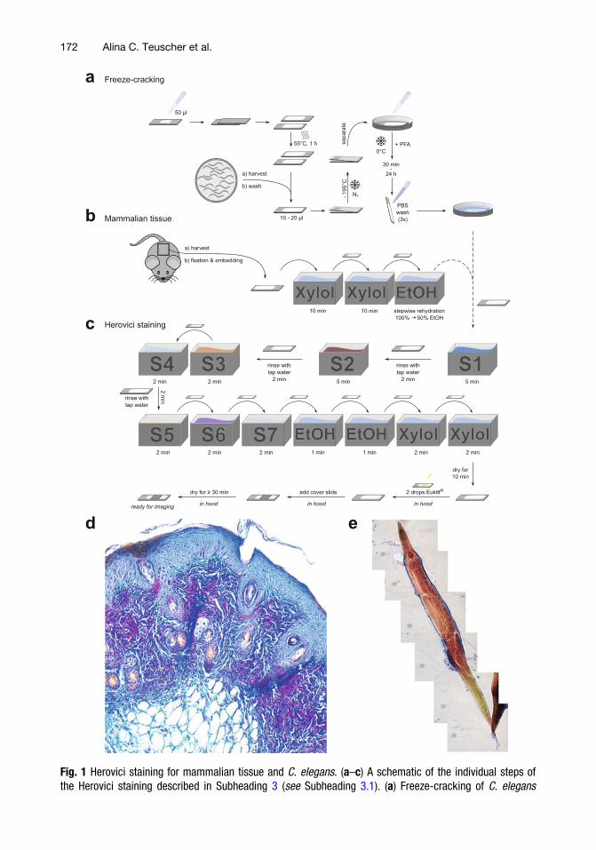
Fig. 1 Herovici staining for mammalian tissue and C. elegans. (a–c) A schematic of the individual steps ofthe Herovici staining described in Subheading 3 (see Subheading 3.1). (a) Freeze-cracking of C. elegans
172 Alina C. Teuscher et al.
matrices in C. elegans. In addition, we describe in detail how tosynchronize C. elegans in order to start a time course for agingstudies. With these methods in hand, the investigator should beable to assess collagen deposition during aging and in age-relatedpathologies.
2 Materials
General mouse necropsy and mammalian tissue harvesting orC. elegans maintenance are described elsewhere [30, 31].C. elegans strains can be acquired from the CaenorhabditisGeneticsCenter (CGC, https://cgc.umn.edu, [31]). For the preparation ofall solution, distilled water and analytical grade reagents are recom-mended. All Herovici staining solutions are stored in glass bottles atroom temperature protected from light. Disposal of waste materialsshould be carried out according to specific local regulations. Suit-able protection (lab coat, safety glasses, and gloves) and the use of afume hood are recommended for the handling of acids and otherhazardous substances.
2.1 Herovici Staining
for Mammalian Tissue
and C. elegans
1. 70–100% EtOH.
2. Microscope slides with cut edges and frosted end, 1 mm thick,76 � 26 mm.
3. Poly-L-Solution: 400 mg Sigma P1524 poly-L-Lysine; 0.2 gsodium azide (0.1%) in 200 ml ddH2O. Can be reused andstored at 4 �C.
4. PBS: 8 g NaCl, 0.2 g KCl, 1.42 g Na2HPO4, 0.24 g KH2PO4,pH adjusted with HCl to pH 7.4.
5. PFA: 4% paraformaldehyde solution in PBS.
6. Herovici’s Solution 1: Add 270 ml of deionized water to a glassbeaker. Weigh 1.25 g celestine blue powder, and transfer it tothe glass beaker. Weigh 15 g of aluminum potassium sulfatedodecahydrate (CAS: 7784-24-9; 31242, Merck) and add it tothe glass beaker. Heat and boil the solution until the powdersare dissolved (approximately 3–5 min). Let the solution cooldown to room temperature, and transfer to a 500 ml graduatedcylinder, and add distilled water to a final volume of 300 ml.
�
Fig. 1 (continued) (see Subheading 3.1.1). (b) Mammalian tissue harvesting and deparaffinization (seeSubheading 3.1.2). (c) Shared part of the Herovici staining protocol for both C. elegans and mammaliantissue (see Subheading 3.1.3). (d) Herovici-stained mouse skin specimen. The young collagen (collagen typeIII) is stained in blue, whereas the mature collagen (collagen type I) is stained in red. Cytoplasm is counter-stained in yellow and cell nuclei dark blue to black. Three layers forming the skin (epidermis, dermis,hypodermis) can be identified. (e) Herovici-stained C. elegans at day 1 of adulthood
Collagen Deposition During Aging 173
Add 60 ml of glycerol once the solution has cooled down toroom temperature (see Note 1).
7. Herovici’s Solution 2: Weigh 1 g of hematoxylin powder,transfer it to a glass beaker, and dissolve it in 100 ml of 95%ethanol in distilled water (alcoholic hematoxylin). Weigh 4.5 gof ferrous II sulfate (FeSO4) powder and 2.5 g ferric III chlo-ride (FeCl3) powder, and transfer them to a separate glassbeaker containing 298 ml distilled water. Add 2 ml of 36%(concentrated) hydrochloric acid (toxic, see Note 2). Stir thesolution on a magnetic stirrer till powders dissolve (seeNote 3).Mix this solution with the alcoholic hematoxylin from step 1(see Notes 4–6).
8. Herovici’s Solution 3: Add 300 ml of distilled water to a glassbeaker. Weigh 1.25 g of metanil yellow, and transfer it to theglass beaker. Add 25 drops (use a 1 ml pipette tip) of glacialacetic acid (see Notes 7 and 8).
9. Herovici’s Solution 4: Add 1800 ml of distilled water to a glassbeaker. Add 9ml of glacial acetic acid (toxic, seeNotes 7 and 9).
10. Herovici’s Solution 5: Weigh >1 g of lithium carbonate(Li2CO3) powder, and dissolve it in 78 ml distilled water(aqueous Li2CO3). The solution will be saturated (see Note10). Add 1 ml of saturated aqueous Li2CO3 to 500 ml distilledwater to a glass beaker (see Notes 11 and 12).
11. Herovici’s Solution 6: Weigh 0.15 g of methyl blue powder,and dissolve it in 150 ml distilled water in a glass beaker. Weigh0.2 g of acid fuchsin powder, and dissolve it in 150 ml ofsaturated aqueous picric acid in a separate glass beaker. Mixthe two solutions together. Add 30 ml of glycerol and 0.15 mlof saturated aqueous Li2CO3 (see Notes 11 and 13).
12. Herovici’s Solution 7: Add 10 ml of glacial acetic acid to 1 L ofdistilled water (see Notes 7 and 14).
2.2 Total Collagen
Quantification
for Mammalian Tissue
and C. elegans
2.2.1 Age
Synchronization
of C. elegans
1. 5 N NaOH.
2. 5% sodium hypochlorite solution (or household bleach).
3. Sterile ddH2O.
4. M9 buffer: 3 g KH2PO4, 7.52 g Na2HPO4, 5 g NaCl, and0.05 g MgSO4 in 1 L ddH2O, autoclaved [31].
2.2.2 Total Protein
Quantification
1. QuickZyme Protein Assay Kit (QuickZyme Biosciences) orequivalent kit from other manufactures suitable for proteinquantification of acid hydrolyzed samples.
2. Pierce™ BCA Protein Assay Kit (Thermo Fisher) or equivalentkit from other manufactures.
174 Alina C. Teuscher et al.
2.2.3 Total Collagen
Quantification
1. QuickZyme Total Collagen Kit (QuickZyme Biosciences) orequivalent kit from other manufactures.
2. Aqueous hydrogen chloride solution: 4 M, 6 M, and 12 M.
2.3 Materials
for Cuticle
(Extracellular Matrix)
Isolation in C. elegans
2.3.1 Cuticle Isolation
(Freeze-Thaw Protocol)
1. Sodium dodecyl sulfate (SDS) buffer: 1% SDS in ddH2O.
2. Triton X-100 buffer: 0.5% Triton X-100 in ddH2O.
3. 15 ml glass conical centrifuge tubes with screw gaps.
2.3.2 Cuticle Isolation
(ST Buffer Protocol)
1. M9 buffer: 3 g KH2PO4, 7.52 g Na2HPO4, 5 g NaCl,0.0493 g MgSO4 in 1 L ddH2O, autoclaved.
2. Sonication buffer: 10 mM Tris–HCl, pH 7.4; 1 mM EDTA; inddH2O.
3. 0.1 M PMSF buffer: 1 mM phenylmethanesulfonylfluoride inisopropanol.
4. ST buffer: 1% SDS; 0.125 M Tris–HCl, pH 6.8; in ddH2O.
3 Methods
3.1 Herovici Staining
for Mammalian Tissue
and C. elegans
All procedures are carried out at room temperature, if not indicatedotherwise. Glass slides with tissue sections fixed in 4% paraformal-dehyde (PFA in PBS) are used for the Herovici staining protocol.Reusable Herovici solutions can be poured back in their originalcontainer. At the end of this procedure, tissue sections will be readyto be imaged under the microscope. In this section, we firstdescribe the harvesting and preparation for mammalian tissue (seeSubheading 3.1.1) and then for C. elegans (see Subheading 3.1.2)followed by the shared methodical part (see Subheading 3.1.4).
3.1.1 Harvesting
and Preparing Mammalian
Tissue Sections (Fig. 1a)
1. Organs or tissues of interest are harvested as described else-where [30], fixed in 4% paraformaldehyde for 24 h at 4 �C,submersed in increasing concentrations of ethanol, embeddedinto paraffin [32], and sectioned with a microtome (3.5 μmsections).
2. Deparaffinize tissue sections by immerging the glass slides inxylol for 10 min. Repeat this step once for a total of two.
3. Rehydrate the tissue sections stepwise from 100% to 50%(100%, 96%, 90%, 80%, 70%, 50%) ethanol by immerging theglass slides for 1 min in each solution.
4. Continue the protocol; see Subheading 3.1.4.
Collagen Deposition During Aging 175
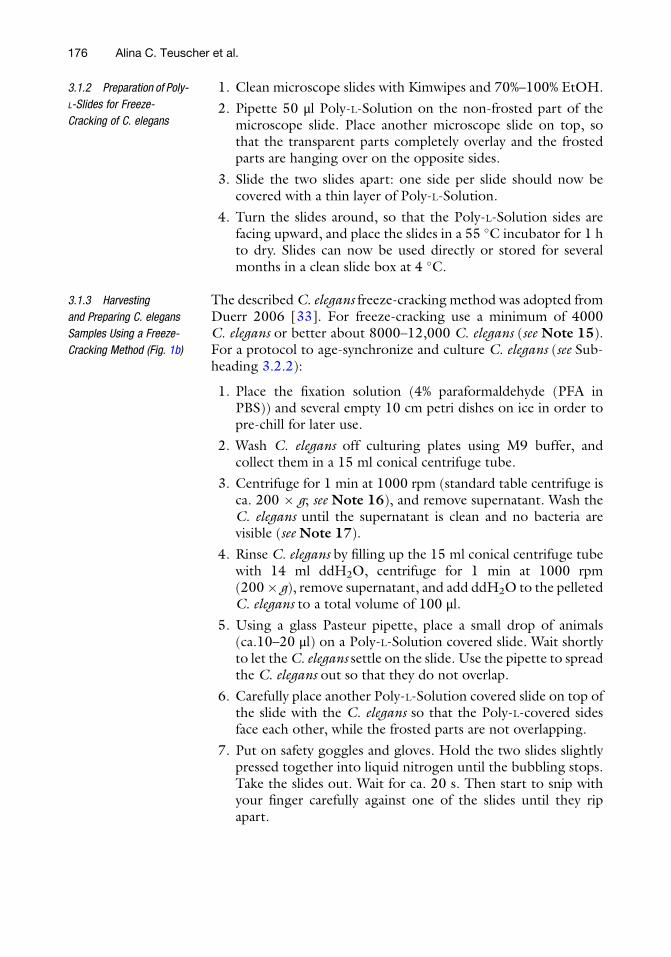
3.1.2 Preparation of Poly-
L-Slides for Freeze-
Cracking of C. elegans
1. Clean microscope slides with Kimwipes and 70%–100% EtOH.
2. Pipette 50 μl Poly-L-Solution on the non-frosted part of themicroscope slide. Place another microscope slide on top, sothat the transparent parts completely overlay and the frostedparts are hanging over on the opposite sides.
3. Slide the two slides apart: one side per slide should now becovered with a thin layer of Poly-L-Solution.
4. Turn the slides around, so that the Poly-L-Solution sides arefacing upward, and place the slides in a 55 �C incubator for 1 hto dry. Slides can now be used directly or stored for severalmonths in a clean slide box at 4 �C.
3.1.3 Harvesting
and Preparing C. elegans
Samples Using a Freeze-
Cracking Method (Fig. 1b)
The describedC. elegans freeze-cracking method was adopted fromDuerr 2006 [33]. For freeze-cracking use a minimum of 4000C. elegans or better about 8000–12,000 C. elegans (see Note 15).For a protocol to age-synchronize and culture C. elegans (see Sub-heading 3.2.2):
1. Place the fixation solution (4% paraformaldehyde (PFA inPBS)) and several empty 10 cm petri dishes on ice in order topre-chill for later use.
2. Wash C. elegans off culturing plates using M9 buffer, andcollect them in a 15 ml conical centrifuge tube.
3. Centrifuge for 1 min at 1000 rpm (standard table centrifuge isca. 200 � g; see Note 16), and remove supernatant. Wash theC. elegans until the supernatant is clean and no bacteria arevisible (see Note 17).
4. Rinse C. elegans by filling up the 15 ml conical centrifuge tubewith 14 ml ddH2O, centrifuge for 1 min at 1000 rpm(200� g), remove supernatant, and add ddH2O to the pelletedC. elegans to a total volume of 100 μl.
5. Using a glass Pasteur pipette, place a small drop of animals(ca.10–20 μl) on a Poly-L-Solution covered slide. Wait shortlyto let theC. elegans settle on the slide. Use the pipette to spreadthe C. elegans out so that they do not overlap.
6. Carefully place another Poly-L-Solution covered slide on top ofthe slide with the C. elegans so that the Poly-L-covered sidesface each other, while the frosted parts are not overlapping.
7. Put on safety goggles and gloves. Hold the two slides slightlypressed together into liquid nitrogen until the bubbling stops.Take the slides out. Wait for ca. 20 s. Then start to snip withyour finger carefully against one of the slides until they ripapart.
176 Alina C. Teuscher et al.
8. Check which slide has most of the C. elegans attached to it, andplace either only this slide or both slides separately into indi-vidual empty 10 cm petri dishes, which are standing on ice.
9. Pipette 100–200 μl 4% PFA pre-chilled onto the C. eleganscovered part of the slides. Fix the C. elegans by incubating theslides at 4 �C for at least 30 min or up to 24 h.
10. After fixation, rinse the slides gently at least three times withPBS. Proceed immediately with staining, or store the slides in aCoplin jar or petri dish containing PBS at 4 �C for several days.
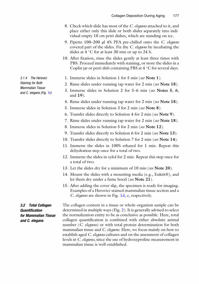
3.1.4 The Herovici
Staining for Both
Mammalian Tissue
and C. elegans (Fig. 1c)
1. Immerse slides in Solution 1 for 5 min (see Note 1).
2. Rinse slides under running tap water for 2 min (see Note 18).
3. Immerse slides in Solution 2 for 5–6 min (see Notes 5, 6,and 19).
4. Rinse slides under running tap water for 2 min (see Note 18).
5. Immerse slides in Solution 3 for 2 min (see Note 8).
6. Transfer slides directly to Solution 4 for 2 min (see Note 9).
7. Rinse slides under running tap water for 2 min (see Note 18).
8. Immerse slides in Solution 5 for 2 min (see Note 12).
9. Transfer slides directly to Solution 6 for 2 min (see Note 13).
10. Transfer slides directly to Solution 7 for 2 min (see Note 14).
11. Immerse the slides in 100% ethanol for 1 min. Repeat thisdehydration step once for a total of two.
12. Immerse the slides in xylol for 2 min. Repeat this step once fora total of two.
13. Let the slides dry for a minimum of 10 min (see Note 20).
14. Mount the slides with a mounting media (e.g., Eukitt®), andlet them dry under a fume hood (see Note 21).
15. After adding the cover slip, the specimen is ready for imaging.Examples of a Herovici-stained mammalian tissue section and aC. elegans are shown in Fig. 1d, e, respectively.
3.2 Total Collagen
Quantification
for Mammalian Tissue
and C. elegans
The collagen content in a tissue or whole-organism sample can bedetermined in multiple ways (Fig. 2). It is generally advised to selectthe normalization entity to be as conclusive as possible. Here, totalcollagen quantification is combined with either absolute animalnumber (C. elegans) or with total protein determination for bothmammalian tissue and C. elegans. Here, we focus mainly on how toestablish aged C. elegans cultures and on the assessment of collagenlevels in C. elegans, since the use of hydroxyproline measurement inmammalian tissue is well-established.
Collagen Deposition During Aging 177
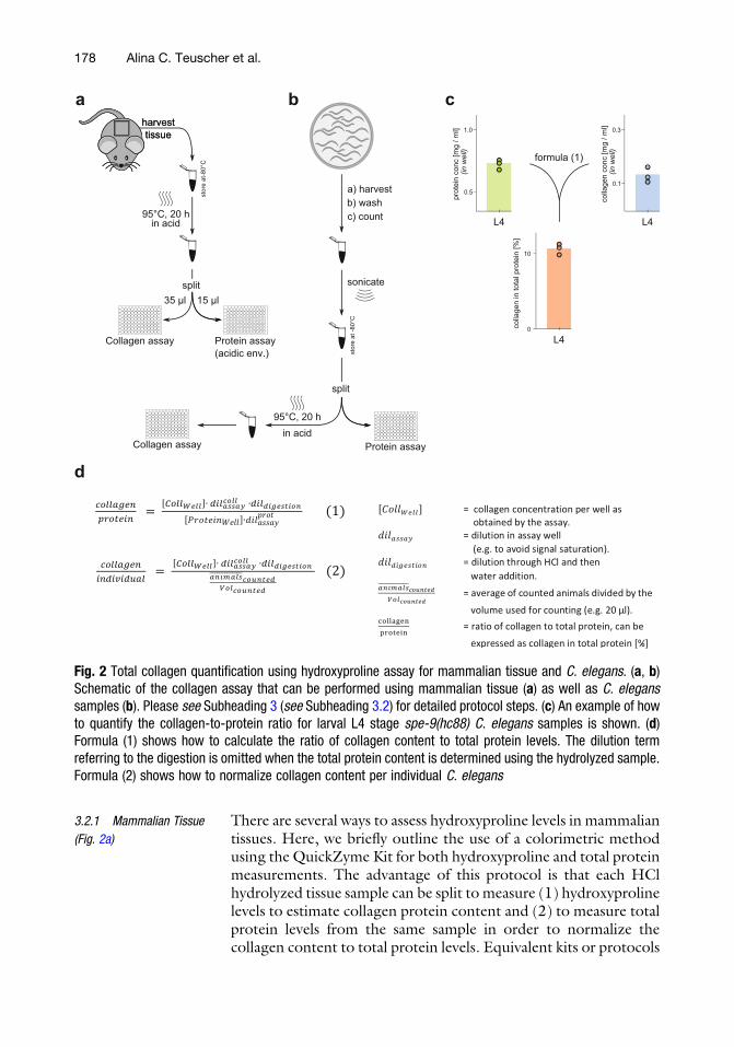
3.2.1 Mammalian Tissue
(Fig. 2a)
There are several ways to assess hydroxyproline levels in mammaliantissues. Here, we briefly outline the use of a colorimetric methodusing the QuickZyme Kit for both hydroxyproline and total proteinmeasurements. The advantage of this protocol is that each HClhydrolyzed tissue sample can be split to measure (1) hydroxyprolinelevels to estimate collagen protein content and (2) to measure totalprotein levels from the same sample in order to normalize thecollagen content to total protein levels. Equivalent kits or protocols
prot
ein
conc
[mg
/ ml]
L4
0.5
1.0
colla
gen
in to
tal p
rote
in [%
]
0
10
formula (1)
a
(in w
ell)
colla
gen
conc
[mg
/ ml]
L4
0.1
0.3
(in w
ell)
L4st
ore
at -8
0°C
sonicate
c) count
a) harvestb) wash
12A
B
C
D
E
F
G
H
1 2 3 4 5 6 7 8 9 01 11
Protein assay
95°C, 20 hin acid
12A
B
C
D
E
F
G
H
1 2 3 4 5 6 7 8 9 01 11
Collagen assay
split
harvesttissue
harvesttissue
stor
e at
-80°
C
12A
B
C
D
E
F
G
H
1 2 3 4 5 6 7 8 9 01 11
Collagen assay
split
12A
B
C
D
E
F
G
H
1 2 3 4 5 6 7 8 9 01 11
Protein assay(acidic env.)
35 µl
95°C, 20 hin acid
b c
d
15 µl
Fig. 2 Total collagen quantification using hydroxyproline assay for mammalian tissue and C. elegans. (a, b)Schematic of the collagen assay that can be performed using mammalian tissue (a) as well as C. eleganssamples (b). Please see Subheading 3 (see Subheading 3.2) for detailed protocol steps. (c) An example of howto quantify the collagen-to-protein ratio for larval L4 stage spe-9(hc88) C. elegans samples is shown. (d)Formula (1) shows how to calculate the ratio of collagen content to total protein levels. The dilution termreferring to the digestion is omitted when the total protein content is determined using the hydrolyzed sample.Formula (2) shows how to normalize collagen content per individual C. elegans
178 Alina C. Teuscher et al.
from other manufacturers can be used. For more details, pleasefollow the manufacturer’s instruction.
1. Organs or tissues of interest are harvested as described else-where [30]. Frozen organs/tissues and formalin-fixed orparaffin-embedded tissues can also be used.
2. Hydrolyze the tissue in 6 M HCl in a safety centrifuge tube for16 h at 95 �C in a one 1:10 ratio of wet tissue weight to acidvolume (see Note 22). Split the hydrolysate: 35 μl is used forcollagen quantification and 15 μl to assess the total proteincontent (see Note 23).
3. To generate the standard curve for the colorimetric quantifica-tion of collagen and total protein levels, please follow themanufacturer’s instructions.
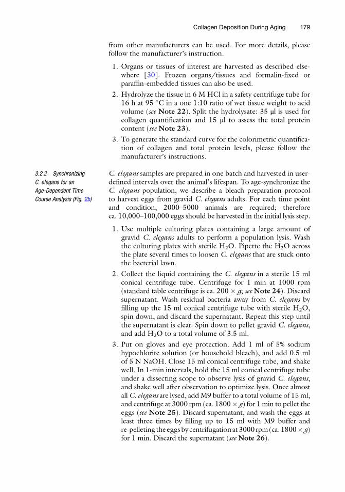
3.2.2 Synchronizing
C. elegans for an
Age-Dependent Time
Course Analysis (Fig. 2b)
C. elegans samples are prepared in one batch and harvested in user-defined intervals over the animal’s lifespan. To age-synchronize theC. elegans population, we describe a bleach preparation protocolto harvest eggs from gravid C. elegans adults. For each time pointand condition, 2000–5000 animals are required; thereforeca. 10,000–100,000 eggs should be harvested in the initial lysis step.
1. Use multiple culturing plates containing a large amount ofgravid C. elegans adults to perform a population lysis. Washthe culturing plates with sterile H2O. Pipette the H2O acrossthe plate several times to loosen C. elegans that are stuck ontothe bacterial lawn.
2. Collect the liquid containing the C. elegans in a sterile 15 mlconical centrifuge tube. Centrifuge for 1 min at 1000 rpm(standard table centrifuge is ca. 200 � g; seeNote 24). Discardsupernatant. Wash residual bacteria away from C. elegans byfilling up the 15 ml conical centrifuge tube with sterile H2O,spin down, and discard the supernatant. Repeat this step untilthe supernatant is clear. Spin down to pellet gravid C. elegans,and add H2O to a total volume of 3.5 ml.
3. Put on gloves and eye protection. Add 1 ml of 5% sodiumhypochlorite solution (or household bleach), and add 0.5 mlof 5 N NaOH. Close 15 ml conical centrifuge tube, and shakewell. In 1-min intervals, hold the 15 ml conical centrifuge tubeunder a dissecting scope to observe lysis of gravid C. elegans,and shake well after observation to optimize lysis. Once almostallC. elegans are lysed, addM9 buffer to a total volume of 15ml,and centrifuge at 3000 rpm (ca. 1800� g) for 1min to pellet theeggs (see Note 25). Discard supernatant, and wash the eggs atleast three times by filling up to 15 ml with M9 buffer andre-pelleting theeggsby centrifugationat3000rpm(ca.1800� g)for 1 min. Discard the supernatant (see Note 26).
Collagen Deposition During Aging 179
4. After lysis and subsequent washing steps, the samples are incu-bated while rotating for 12 h in M9 buffer supplemented with5 μg/ml cholesterol.
5. 500–1000 larval L1 C. elegans larvae are placed on each 10 cmculturing plate. If a temperature-sensitive sterile strain is used,the sterility mechanism has to be activated at the needed timepoint (see Note 27). If floxuridine (FUDR) is used to avoidoffsprings, the animals are transferred at the larval L4 stage to50 μg/ml FUDR culturing plates (see Notes 28 and 29).
6. 2000–5000 C. elegans (2–10 plates) are harvested for eachcondition and time point. Transfer all animals from the platesto a 15 ml conical centrifuge tube. The animals are centrifugedbriefly at low speed (200 rpm, ca. 8 � g) to pellet aged C. ele-gans adults. The suspension containing unhatched eggs orlarvae (if FUDR is used) is aspirated and discarded. Repeatthis washing step three times or until only the desiredC. elegans adults are present and supernatant is clear (see Note30).
7. From the last wash that was filled up with M9 buffer to a totalvolume of 14 ml, take five times 20 μl aliquots, and place thesealiquots on a petri dish lid to count the number of animals ineach 20 μl drop using a dissecting scope (seeNotes 31 and 32).To estimate total number of C. elegans, average the number ofC. elegans from each 20 μl drop and multiply by 700 to get anestimate of the total number of C. elegans present in 14 mlsample.
8. Centrifuge for 1 min at 1000 rpm (ca. 200 � g) to softly pelletC. elegans. Discard supernatant. Use a glass Pasteur pipette totransfer the C. elegans pellet to a labelled 1.5 ml Eppendorfcollection tube, fill up with sterile H2O to a total volume of200 μl, and store at �80 �C (see Notes 16, 33, and 34).
9. At the end of the time course, once all samples have beencollected, proceed by thawing all tubes. At least 3–5 freeze-thaw cycles should be performed by thawing at room tempera-ture and freezing in liquid nitrogen (or dry ice with EtOH orby placing back into the �80 �C).
10. Then sonicate all samples on ice until all animals are disrupted.The C. elegans cuticle is the hardest to break. With a standardsonication device, it will take several repeats to break down allC. elegans (see Note 35).
3.2.3 Total Protein
Quantification
for C. elegans (Fig. 2)
To determine the total collagen content as a fraction of totalprotein abundance, the total protein content has to be determinedseparately.
180 Alina C. Teuscher et al.
1. Pipette 25 μl of each sonicated sample as well as of the providedstandard solutions into a 96-well plate, and quantify the totalprotein content following the manufacturer’s instructions(Pierce™ BCA Protein Assay Kit, Thermo Fisher, or equivalentkit from other manufacturers). If needed, dilute the samplewith M9.
2. Use a plate reader to quantify the color change at the specifiedwavelength.
3.2.4 Total Collagen
Quantification
for C. elegans (Fig. 2)
Here, we determine the total collagen abundance by digesting thesample and directly measuring the concentration of the amino acidhydroxyproline in each sample.
1. The sonicated samples are transferred into heat-stable tubesprovided by the kit manufacturer (QuickZyme Total CollagenKit, QuickZyme Biosciences, or equivalent kit from other man-ufacturers). The provided collagen standard does not require tobe sonicated but is otherwise treated identically as the samples.
2. According to the manufacturer’s instructions, the sample andstandard are mixed with 12 M HCl solution and incubated for20 h at 95 �C.
3. In brief, after the digestion the supernatant is isolated, the acidcontent lowered, assay buffer added, and the plate incubated atroom temperature for 20 min followed by the addition of thedetection reagent and subsequent incubation at 60 �C for 1 h.The color change is quantified using a plate reader (seeNote 36).
4. The total collagen content per sample in a well can be directlydetermined through the measured color change. In a first step,the collagen reference sample (1.2 mg/ml of rat tail collagen) isused to generate a standard curve relating the magnitude of thecolor change to the collagen content of the well. Over the usedconcentration range, this relationship is approximately linear.With the standard curve available, the collagen content of thesamples can be quantified. It must be noted that the collagenstandard supplied in the QuickZyme Biosciences Kit containsrat tail collagen. To validate the use of rat tail collagen as areference for hydroxyproline levels for C. elegans collagens, thepotential hydroxyproline occurrences on the Y position in theC. elegans [Gly-X-Y] triple repeats in collagens were estimatedin silico. The relative proline abundance on the Y position issimilar between C. elegans and rat tail collagens (personal com-munication Jan M. Gebauer). Example results for C. elegansprotein and collagen concentration are shown in Fig. 2b.
5. The total collagen-to-total protein ratio can be determined bycombining the measured collagen and protein concentrationsaccording to formula (1). In the case of C. elegans, the totalcollagen abundance per animal can be calculated by subjecting
Collagen Deposition During Aging 181
the measured collagen concentration to formula (2). All for-mulas to determine the relative collagen contents are depictedin Fig. 2c.
3.3 Methods
for Cuticle
(Extracellular Matrix)
Isolation in C. elegans
3.3.1 Cuticle Isolation
(Freeze-Thaw Protocol)
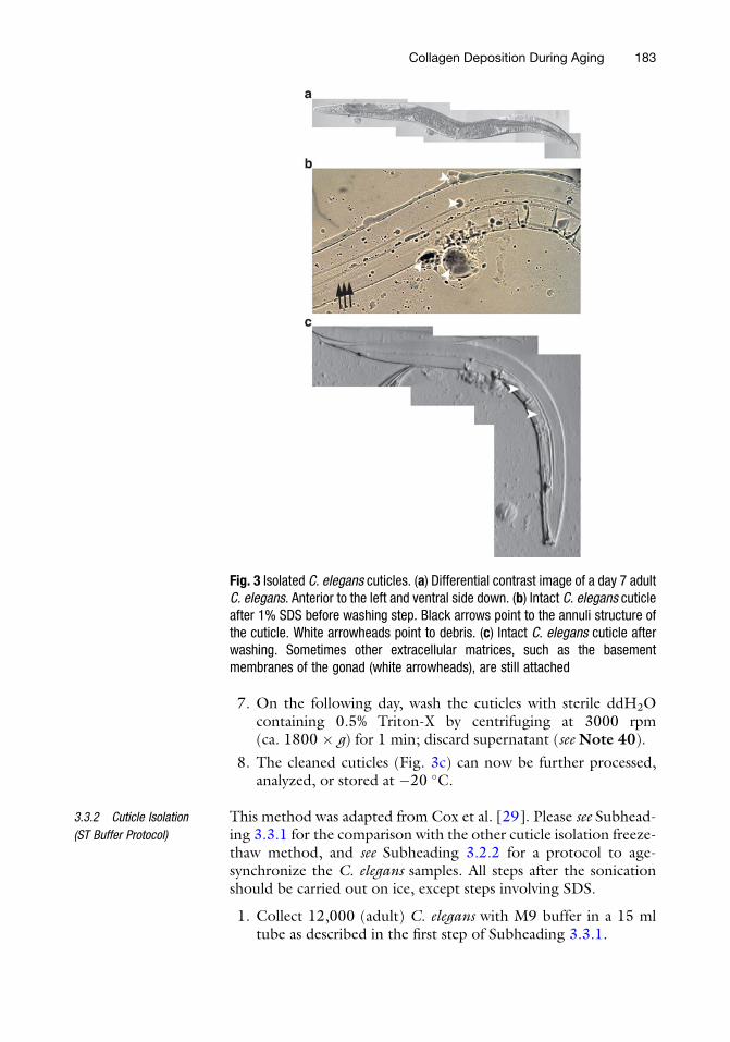
This method was modified from Leushner et al. [28], for the use inC. elegans. The two advantages of using this protocol compared tothe ST protocol is (1) its simplicity and (2) the SDS-cleaned cuticlesmight still contain Schiff base products or other adducts that mightbe of importance of the desired analysis [34]. The major problemwith this protocol is that the C. elegans cuticle tends to stick onplastic; this can be avoided by using glass materials instead. All stepsafter the freeze-thawing should be carried out on ice, except stepsinvolving SDS. We recommend to use about 12,000 C. elegans persample to isolate a sufficient number of cuticles. Please see Subhead-ing 3.2.2 for a protocol to age-synchronize the C. elegans samples.An example of an aged C. elegans (7-day adult) is shown in Fig. 3afor comparison with the isolated cuticles Fig. 3b, c.
1. Use 12,000 adult C. elegans (about 8–12 full culturing plates).Pipette around 3 ml M9 buffer on each plate. Gently tilt platesto collect the liquid on one side, and carefully pipette the M9buffer several times across the plate to loosen up the C. elegansstuck on the bacterial food lawn. Transfer the animals from theplates into a 15 ml conical centrifuge tube.
2. Wash the C. elegans three times with 15 ml ddH2O by centri-fugation at around 1000 rpm (ca. 200 � g) for 1 min, anddiscard the supernatant (see Note 33).
3. After the last washing step, discard the supernatant again, anduse a glass Pasteur pipette to transfer theC. elegans pellet into a1.5 ml Eppendorf microfuge tube (see Note 37). Centrifuge1.5 ml tube at around 100 � g for 30 s, discard supernatant,and fill up with ddH2O to 100 μl.
4. Freeze the tubes in liquid nitrogen for 1 min (or until frozen),followed by a thawing step at room temperature. Repeat thisfreeze-thaw cycle at least three times.
5. Sonicate the samples five times in intervals of 20 s each at anamplitude of 80% with 20 s breaks on ice in between (Sonoplusmini20 from Bandelin) (see Note 38).
6. Use a glass Pasteur pipette to transfer the ruptured C. elegansto 15 ml glass tubes (with screw gaps) (see Note 39), andsuspend them in 10 ml 1% SDS solution, followed by overnightincubation onto a rotor at 37 �C. The 1% SDS should wash outall the internal cells and organs from the disrupted cuticles(Fig. 3b).
182 Alina C. Teuscher et al.
7. On the following day, wash the cuticles with sterile ddH2Ocontaining 0.5% Triton-X by centrifuging at 3000 rpm(ca. 1800 � g) for 1 min; discard supernatant (see Note 40).
8. The cleaned cuticles (Fig. 3c) can now be further processed,analyzed, or stored at �20 �C.
3.3.2 Cuticle Isolation
(ST Buffer Protocol)
This method was adapted from Cox et al. [29]. Please see Subhead-ing 3.3.1 for the comparison with the other cuticle isolation freeze-thaw method, and see Subheading 3.2.2 for a protocol to age-synchronize the C. elegans samples. All steps after the sonicationshould be carried out on ice, except steps involving SDS.
1. Collect 12,000 (adult) C. elegans with M9 buffer in a 15 mltube as described in the first step of Subheading 3.3.1.
Fig. 3 Isolated C. elegans cuticles. (a) Differential contrast image of a day 7 adultC. elegans. Anterior to the left and ventral side down. (b) Intact C. elegans cuticleafter 1% SDS before washing step. Black arrows point to the annuli structure ofthe cuticle. White arrowheads point to debris. (c) Intact C. elegans cuticle afterwashing. Sometimes other extracellular matrices, such as the basementmembranes of the gonad (white arrowheads), are still attached
Collagen Deposition During Aging 183
2. Wash the C. elegans pellet three times by centrifugation ataround 1000 rpm (ca. 200 � g) for 1 min, and discard thesupernatant (see Note 33).
3. After the last washing step, discard the supernatant, and resus-pend the C. elegans pellet in 5 ml sonication buffer. Incubateon ice for 10 min.
4. Add 30 μl 0.1 M PMSF to the solution, and sonicate five timesfor 20 s with an amplitude of 80% with 20 s breaks on ice inbetween (Sonoplus mini20 from Bandelin).
5. Wash the ruptured C. elegans cuticles three times with thesonication buffer by centrifugation at 1500 rpm (around450 � g) for 1 min, and discard the supernatant.
6. Discard the supernatant, and transfer the cuticles with a glassPasteur pipette to a 1.5 ml microfuge tube (see Note 37).Centrifuge the 1.5 ml tube at around 100 � g for 30 s, discardsupernatant, and fill up with sonication buffer to 100 μl.
7. Add 1 ml ST buffer, and heat the tube for 2.5 min at 95 �C.
8. Incubate the tubes on a rotator overnight at roomtemperature.
9. Wash cuticles three times with 0.5% Triton-X in sterile ddH2Oby centrifuging at 3000 rpm (ca. 1800 � g) for 1 min; discardsupernatant.
10. The cleaned cuticles can now be further processed, analyzed, orstored at �20 �C.
4 Notes
4.1 Herovici Staining
for Mammalian Tissue
and C. elegans
1. Solution 1 can be reused.
2. Caution. Hydrochloric acid should be handled with care andadded to the solution under a fume hood.
3. If required filter the solution to eliminate visible impurities.
4. Alcoholic hematoxylin and the ferrous solution are preparedseparately and mixed at the end.
5. Solution 2 has a shelf life of approximately 2–3 months due tooxidation.
6. Solution 2 can be reused.
7. Caution. Glacial acetic acid should be handled with care andadded to the solution under a fume hood.
8. Solution 3 can be reused.
9. Do not reuse Solution 4.
184 Alina C. Teuscher et al.
10. By adding more than 1 g of Li2CO3 to 78 ml distilled water,the solution will be saturated. Do not shake to try to dissolvethe precipitate. Let the solution stand.
11. Pipette the aqueous Li2CO3 from the top without disturbingthe precipitate accumulated on the bottom of the container.
12. Solution 5 can be reused.
13. Solution 6 can be reused.
14. Do not reuse Solution 7.
15. Per plate you can make 2–3 freeze-cracked C. elegans slides.
16. To prevent adhesion of animals to plastic pipette tips, one caneither add 0.5% Triton-X as a detergent or switch to glasspipettes.
17. Bacteria prevent the animals to properly stick to the slides.
18. Regular water. This step can be performed directly in the sink.
19. A longer incubation may be needed if the solution has not beenfreshly prepared.
20. A longer drying time (30 min–1 h) is perfectly fine.
21. Eukitt® will need approximately 30 min to dry.
4.2 Total Collagen
Quantification
for Mammalian Tissue
and C. elegans
22. Ideally, a 1:10 ratio of wet tissue weight to acid volume isrecommended, with a minimal volume of 200 μl 6 M HClper tube. For example, 50 mg of tissue mixed with 500 μl of6 M HCl can be used.
23. To be able to relate the total collagen content of the sample asrobustly as possible to the total protein abundance, we recom-mend to measure the later directly in the hydrolyzed sample.To be able to quantify the free amino acid level present in theacidic hydrolyzed sample, a suitable kit has to be chosen. Werecommend the Total Protein Assay, QuickZyme Biosciences.
24. When centrifugingC. elegans, either turn off the breaks or turndown the deceleration to a minimum.
25. In general, complete lysis can take about 4–8 min. Hence, it isimportant to regularly check if all C. elegans are lysed. If lysis isperformed for too long, the eggs will suffer from increasingdamage.
26. Sometimes three washes are not enough to wash out all bleachsolution. Hence, check by smelling the 15 ml conical centri-fuge tube if bleach odor is still present.
27. We recommend the C. elegans mutant spe-9(hc88). Due to atemperature-sensitive defect in spermatogenesis, the animalsare not able to lay fertilized eggs when shifted to 25 �C duringdevelopment. To ensure all animals are sterile, L1 animals canbe cultured at 25 �C until day 1 or 2 of adulthood.
Collagen Deposition During Aging 185
28. To avoid food scarcity, check the grow-up plates often, and ifneeded, transfer to fresh plates.
29. To check the effectiveness of the intervention, a selected num-ber of plates can be used for validation by, for example, placinga fluorescent reporter strain on them.
30. Since the collagen content is changing over the lifespan ofC. elegans, it is crucial to work with age-synchronous popula-tions. Thus, all eggs and larvae must be removed as best aspossible. This can be achieved by transferring animals often tonew plates and rigorous washing or through filtration.
31. Because adult C. elegans will sink to bottom of the conicalcentrifuge tube, shake the 15 ml conical centrifuge tube wellbetween taking the aliquots for counting. Try to pipette fromthe middle of the 15 ml conical centrifuge tube for consistency.
32. Use a pipette with a large orifice to sample C. elegans duringthe counting procedure. Since adults grow in size, a too smallpipette tip can lead to an underestimation of older populations.
33. At this step of the protocol is a good time point for a break orto wait until all time points from before are collected, forinstance, if you collect daily samples over the 2–3 weeks life-span of C. elegans.
34. It is recommended to fill up all sample C. elegans pellets to200 μl to simplify the downstream steps.
35. Example sonication configuration using a Bandelin Sonoplus,UW mini20 device: Amplitude 80%, 30s intervals, pulse 1.0 son, 1.0 s off. However, using a more powerful sonicator isrecommended to accelerate C. elegans fragmentation.
36. It is advised to perform a few test reactions prior to processingthe entire batch for both quantification methods to be able toadjust sample dilution should it be too concentrated.
4.3 Cuticle
(Extracellular Matrix)
Isolation in C. elegans
37. C. elegans tend to stick on the plastic tip or on the plastic tubes.Hence, make sure to use a glass Pasteur pipette and pipette theC. elegans on the bottom of the plastic tube to avoid stickingon the side of the tube.
38. If the cuticles in the freezing protocol are ruptured too much,you can consider performing only the freezing and thawingcycles or only sonication.
39. One of the major problems is losing too many cuticles orC. elegans. Therefore, use always glass pipettes to transferworms and cuticles.
40. Triton-X prevents the cuticles from sticking to the tubes duringthe washing steps.
186 Alina C. Teuscher et al.
Acknowledgments
We thank Marjolein Wildwater for sharing her improved freeze-cracking protocol, Eline Jongsma for her assistance in adapting thefreeze-cracking protocol, Salome Brutsch and Hayley Hiebert fortheir help to develop the C. elegans Herovici staining protocol,Anna Bircher for contributing to the early stages of the cuticleisolation protocols and imaging cuticles, Max Hess for providingthe C. elegans example image, and Jan M. Gebauer for bioinfor-matic prediction of potential prolines in collagens that mightbecome hydroxylated in C. elegans. Some C. elegans strains wereprovided by the CGC, which is funded by NIH Office of ResearchInfrastructure Programs (P40 OD010440). This work was sup-ported by the Swiss National Science Foundation [PZ00P3161512] to S.P. and M.R.B. and [PP00P3 163898] to A.C.T., C.S., and C.Y.E. Alina C. Teuscher and Cyril Statzer contributedequally to this work.
References
1. Hynes RO (2009) The extracellular matrix: notjust pretty fibrils. Science 326:1216–1219
2. Ricard-Blum S (2011) The collagen family.Cold Spring Harb Perspect Biol 3:a004978–a004978
3. Sivan S-S, Wachtel E, Tsitron E et al (2008)Collagen turnover in normal and degeneratehuman intervertebral discs as determined bythe racemization of aspartic acid. J Biol Chem283:8796–8801
4. Heinemeier KM, Schjerling P, Heinemeier Jet al (2016) Radiocarbon dating reveals mini-mal collagen turnover in both healthy andosteoarthritic human cartilage. Sci Transl Med8:346ra90–346ra90
5. Toyama BH, Hetzer MW (2013) Proteinhomeostasis: live long, won’t prosper. NatRev Mol Cell Biol 14:55–61
6. Kjaer M, Langberg H, Miller BF et al (2005)Metabolic activity and collagen turnover inhuman tendon in response to physical activity.J Musculoskelet Neuronal Interact 5:41–52
7. Myllyharju J, Kivirikko KI (2001) Collagensand collagen-related diseases. Ann Med33:7–21
8. Fisher GJ, Quan T, Purohit T et al (2009)Collagen fragmentation promotes oxidativestress and elevates matrix metalloproteinase-1in fibroblasts in aged human skin. Am J Pathol174:101–114
9. Shoulders MD, Raines RT (2009) Collagenstructure and stability. Annu Rev Biochem78:929–958
10. Sell DR, Monnier VM (2012) Molecular basisof arterial stiffening: role of glycation–a mini-review. Gerontology 58:227–237
11. Snedeker JG, Snedeker JG, Gautieri A et al(2014) The role of collagen crosslinks in ageingand diabetes–the good, the bad, and the ugly.Muscles Ligaments Tendons J 4:303–308
12. Myllyharju J (2004) Collagens, modifyingenzymes and their mutations in humans, fliesand worms. Trends Genet 20:33–43
13. Fenske NA, Lober CW (1986) Structural andfunctional changes of normal aging skin. J AmAcad Dermatol 15:571–585
14. Shuster S, Black MM, McVitie E (1975) Theinfluence of age and sex on skin thickness, skincollagen and density. Br J Dermatol93:639–643
15. Lopez-Otın C, Blasco MA, Partridge L et al(2013) The hallmarks of aging. Cell153:1194–1217
16. Franceschi C, Campisi J (2014) Chronicinflammation (inflammaging) and its potentialcontribution to age-associated diseases. J Ger-ontol A Biol Sci Med Sci 69(Suppl 1):S4–S9
17. Wynn TA (2007) Common and uniquemechanisms regulate fibrosis in various fibro-proliferative diseases. J Clin Investig117:524–529
18. Gutierrez-Fernandez A, Soria-Valles C, OsorioFG et al (2015) Loss of MT1-MMP causes cellsenescence and nuclear defects which can bereversed by retinoic acid. EMBO J34:1875–1888
Collagen Deposition During Aging 187
19. Flurkey K, Papaconstantinou J, Miller RA et al(2001) Lifespan extension and delayedimmune and collagen aging in mutant micewith defects in growth hormone production.Proc Natl Acad Sci U S A 98:6736–6741
20. Wilkinson JE, Burmeister L, Brooks SV et al(2012) Rapamycin slows aging in mice. AgingCell 11:675–682
21. Ewald CY, Landis JN, Porter Abate J et al(2015) Dauer-independent insulin/IGF-1-sig-nalling implicates collagen remodelling in lon-gevity. Nature 519:97–101
22. Herovici C (1963) A polychrome stain for dif-ferentiating precollagen from collagen. StainTechnol 38:204–205
23. McAnulty RJ (2005) Methods for measuringhydroxyproline and estimating in vivo rates ofcollagen synthesis and degradation. MethodsMol Med 117:189–207
24. Qiu B, Wei F, Sun X et al (2014) Measurementof hydroxyproline in collagen with three differ-ent methods. Mol Med Rep 10:1157–1163
25. Naba A, Clauser KR, Hynes RO (2015)Enrichment of extracellular matrix proteinsfrom tissues and digestion into peptides formass spectrometry analysis. J Vis Exp 101:e53057
26. Kramer JM (2005) Basement membranes,WormBook : the online review of C elegansbiology. pp 1–15
27. Page AP and Johnstone IL (2007) The cuticle,WormBook : the online review of C elegansbiology. pp 1–15
28. Leushner JR, Semple NL, Pasternak J (1979)Isolation and characterization of the cuticlefrom the free-living nematode Panagrellus silu-siae. Biochim Biophys Acta 580:166–174
29. Cox GN, Kusch M, Edgar RS (1981) Cuticleof Caenorhabditis elegans: its isolation and par-tial characterization. J Cell Biol 90:7–17
30. Treuting PM, Snyder JM (2015) Mouse nec-ropsy. Curr Protoc Mouse Biol 5:223–233
31. Stiernagle T (2006) Maintenance of C. elegans,WormBook : the online review of C elegansbiology. pp 1–11
32. Fischer AH, Jacobson KA, Rose J et al (2008)Paraffin embedding tissue samples for section-ing. CSH Protoc 2008:pdb.prot4989
33. Duerr JS (2006) Immunohistochemistry,WormBook : the online review of C elegansbiology. pp 1–61
34. Davis BO, Anderson GL, Dusenbery DB(1982) Total luminescence spectroscopy offluorescence changes during aging in Caenor-habditis elegans. Biochemistry 21:4089–4095
188 Alina C. Teuscher et al.
Related Documents