Does Sleep Promote False Memories? A. Darsaud 1* , H. Dehon 2* , V. Sterpenich 1 , M. Boly 1 , T. Dang-Vu 1 , M. Desseilles 1 , S. Gais 1 , L. Matarazzo 1 , F. Peters 1 , M. Schabus 1 , C. Schmidt C 1 , G. Tinguely 1 , G. Vandewalle 1 , A. Luxen 1 , F. Collette 1,2 , P. Maquet 1 *These authors contributed equally to this work Affiliation: 1 Cyclotron Research Centre, University of Liège, Belgium 2 Cognitive sciences Department, University of Liège, Belgium Corresponding author : Pierre MAQUET, Cyclotron Research Centre (B30), 6, Allée du 8 Août, University of Liège, Belgium Phone number: +32 43 66 36 87; Fax number: +32 43 66 29 46 E-mail address: [email protected] Web: http://www.ulg.ac.be/crc

Welcome message from author
This document is posted to help you gain knowledge. Please leave a comment to let me know what you think about it! Share it to your friends and learn new things together.
Transcript
Does Sleep Promote False Memories?
A. Darsaud1*, H. Dehon2*, V. Sterpenich1, M. Boly1, T. Dang-Vu1,
M. Desseilles1, S. Gais1, L. Matarazzo1, F. Peters1, M. Schabus1,
C. Schmidt C1, G. Tinguely1, G. Vandewalle1, A. Luxen1, F.
Collette1,2, P. Maquet1
*These authors contributed equally to this work
Affiliation:1Cyclotron Research Centre, University of Liège, Belgium2Cognitive sciences Department, University of Liège, Belgium
Corresponding author:
Pierre MAQUET, Cyclotron Research Centre (B30), 6, Allée du 8
Août, University of Liège, Belgium
Phone number: +32 43 66 36 87; Fax number: +32 43 66 29 46
E-mail address: [email protected]
Web: http://www.ulg.ac.be/crc
ABSTRACT
Memory is constructive in nature so that it may sometimes lead
to the retrieval of distorted or illusory information. Sleep
facilitates accurate declarative memory consolidation but might
also promote such memory distortions. We examined the influence
of sleep and lack of sleep on the cerebral correlates of
accurate and false recollections using fMRI. After encoding
lists of semantically related word associates, half of the
participants were allowed to sleep, whereas the others were
totally sleep deprived on the first postencoding night. During
a subsequent retest fMRI session taking place 3 days later,
participants made recognition memory judgments about the
previously studied associates, critical theme words (which had
not been previously presented during encoding), and new words
unrelated to the studied items. Sleep, relative to sleep
deprivation, enhanced accurate and false recollections. No
significant difference was observed in brain responses to false
or illusory recollection between sleep and sleep deprivation
conditions. However, after sleep but not after sleep
deprivation (exclusive masking), accurate and illusory
recollections were both associated with responses in the
hippocampus and retrosplenial cortex. The data suggest that

sleep does not selectively enhance illusory memories but rather
tends to promote systems-level consolidation in hippocampo-
neocortical circuits of memories subsequently associated with
both accurate and illusory recollections. We further observed
that during encoding, hippocampal responses were selectively
larger for items subsequently accurately retrieved than for
material leading to illusory memories. The data indicate that
the early organization of memory during encoding is a major
factor influencing subsequent production of accurate or false
memories.
INTRODUCTION
Declarative memory is our ability to recollect everyday
events and factual knowledge (e.g., Eichenbaum, 2000). Memories
can vary according to several descriptive features, including
the relation to the memory of a specific context and a mode of
retrieval. That is, remembering and knowing are states of
awareness that accompany the retrieval of experiences from our
past determining whether participants recollect (i.e.,
“Remembering”) their memories or used a rough feeling of
familiarity (i.e., “Knowing”) to base their decision. Imaging
studies have shown that information that is later remembered is
specifically associated with hippocampal responses (e.g.,
Davachi & Wagner, 2002). Of particular interest for the current
study, our memories are interconnected associations
constituting a record of our personal experiences that is
continuously updated (i.e., new information is continuously
reorganized within the context of previous knowledge). Hence,
when we remember a specific episode, related associations or
connected concepts might also come to mind. Consequently, our
memories are not literal records of past events. What is
retrieved from memory can substantially differ from the actual
episode due to distortion or addition of various details
(Schacter, 1999; Bartlett, 1932). False memories are good
examples of such memory distortions, during which an event is
remembered, although in actual fact, it never happened
(Roediger & McDermott, 1995). As a rule, such false memories
are strongly semantically associated to the actually encoded
items (Schacter, 1996). The “Deese–Roediger–McDermott” (DRM)
paradigm capitalizes on these strong semantic relationships to
reliably elicit high proportions of false memories. In this
paradigm, participants learn word lists that compile
semantically associated words, except the strongest associate,
the theme of the list. The latter has high probability to be
subsequently incorrectly retrieved as a critical lure. Besides
the structure of the learned material, a number of factors
influence the formation of false memories during both encoding
and retrieval (Gallo, 2006). Inaddition, like their veridical
counterparts, false memories seem to undergo a consolidation
process. Over time, false memories become increasingly
resistant and even persist better than veridical memories
(Seamon et al., 2002; Mc Dermott, 1996; Reyna & Brained, 1995).
Sleep has been shown to promote the consolidation of
declarative memory (Rasch & Born, 2007). The sleep-dependent
off-line processing of recent memories would entail not only
the adaptation of synaptic strength to maintain synaptic
homeostasis (Tononi & Cirelli, 2006) but also the macroscopic
reorganization within cerebral networks (Albouy et al., 2008;
Gais et al., 2007; Sterpenich et al., 2007; Orban et al.,
2006). In particular, for hippocampaldependent memories, the
burden of retention is thought to be progressively transferred
from hippocampal to neocortical stores. This process would
particularly involve an interplay between the hippocampus and
themesial prefrontal areas (Frankland & Bontempi, 2005), the
activity of which grows progressively at retrieval as the
interval because encoding increases (Sterpenich et al., 2007,
2009; Gais et al., 2007; Takashima et al., 2006).
Importantly, there is evidence that sleep does not promote
only the consolidation of declarative memories but can lead to
behavioral modifications that entail the generation during
sleep of novel representations on the basis of information
extracted fromlearned exemplars (Ellenbogen, Hu, Payne, Titone,
& Walker, 2007; Wagner, Gais, Haider, Verleger, & Born, 2004).
Similarly, false memories are suggested to arise fromthe
activation by studiedwords of other semantically related
representations, including the critical lure (Roediger, Watson,
McDermott, & Gallo, 2001). Alternatively, false memory would
imply the extraction of the gist of the memory from the studied
word lists (Brainerd & Reyna, 2002). Collectively, these
experimental and theoretical elements raise the possibility
that sleep might also promote the formation of false memories.
At present, this hypothesis has received mixed
experimental support. One study using the DRM paradigm reported
that false, in contrast to veridical, recall is better
preserved after (both nocturnal and diurnal) sleep than after
equivalent periods of wakefulness (Payne et al., 2009). In
contrast, in another study also based on DRM, the proportion of
false recognitions was not changed after sleep but was enhanced
only by sleep deprivation immediately preceding retrieval
(Diekelmann, Landolt, Lahl, Born, & Wagner, 2008).
Here, we used fMRI and DRM paradigm to investigate the
influence of sleep and lack of sleep during the postencoding
night, on the neural correlates of veridical and illusory
recollection, that is, memory retrieval processes accompanied
by the recovery of specific contextual details. We found that
sleep had only a moderate effect on the proportion of false
recognitions and modified their neural correlates to the same
extent as accurate memories. We further show that in contrast,
response patterns elicited during encoding of the material
significantly influence the subsequent production of false
recognitions.
METHODS
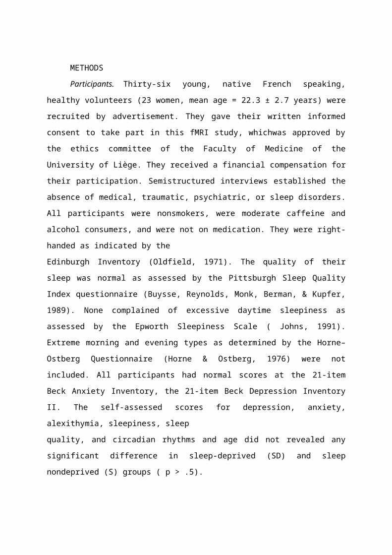
Participants. Thirty-six young, native French speaking,
healthy volunteers (23 women, mean age = 22.3 ± 2.7 years) were
recruited by advertisement. They gave their written informed
consent to take part in this fMRI study, whichwas approved by
the ethics committee of the Faculty of Medicine of the
University of Liège. They received a financial compensation for
their participation. Semistructured interviews established the
absence of medical, traumatic, psychiatric, or sleep disorders.
All participants were nonsmokers, were moderate caffeine and
alcohol consumers, and were not on medication. They were right-
handed as indicated by the
Edinburgh Inventory (Oldfield, 1971). The quality of their
sleep was normal as assessed by the Pittsburgh Sleep Quality
Index questionnaire (Buysse, Reynolds, Monk, Berman, & Kupfer,
1989). None complained of excessive daytime sleepiness as
assessed by the Epworth Sleepiness Scale ( Johns, 1991).
Extreme morning and evening types as determined by the Horne–
Ostberg Questionnaire (Horne & Ostberg, 1976) were not
included. All participants had normal scores at the 21-item
Beck Anxiety Inventory, the 21-item Beck Depression Inventory
II. The self-assessed scores for depression, anxiety,
alexithymia, sleepiness, sleep
quality, and circadian rhythms and age did not revealed any
significant difference in sleep-deprived (SD) and sleep
nondeprived (S) groups ( p > .5).

Experimental Design. Volunteers followed a 7-day constant sleep
schedule (according to their own circadian rhythm ± 1 hr)
before the first visit and kept the same schedule for three
more days until their second visit. Compliance to the schedule
was assessed during the preceding week, using wrist actigraphy
(Cambridge Neuroscience, Cambridge, UK) and sleep diaries.
Volunteers were requested to refrain from all caffeine and
alcohol-containing beverages 1 week before participating in the
study.
Each subject was scanned during two separate fMRI sessions
(Figure 1). Following the DRM paradigm, during the first
session (encoding, between 6:30 and 8:30 p.m.), participants
listened to a set of 32 auditorily presented thematic lists,
each of which consisted 15 semantic associates (i.e., a total
of 480 target items) converging to a critical nonpresented
theme word (lure). Each of the lists was controlled and
selected in a previous behavioral study in which participants
strongly tended to falsely recall or recognize these critical
lures (unpublished data). Each list was recorded using
SoundEdit resource files by a female speaker, and the interval
between the presentation of two words was 2000 msec. Words were
presented in order of decreasing strength of association to the
theme word. Participants were instructed to listen carefully
to each list and to try to remember the words in preparation
for a later test. They were not informed of the specific nature
and modality of the recognition test. During the data
acquisition period, all participants interacted with the same
investigator who used a standardized set of sentences to
minimize social interferences. To minimize the rehearsal
process, we separated the presentation of each of the 32 lists
by a 20-sec interval, during which participants completed an
oddball task. Participants were required to count the odd tones
and to respond to them by pressing a key as fast as possible.
Ten auditory stimuli per oddball task were presented,
consisting of frequent (600 Hz) and odd tones (400 Hz). The
first three auditory stimuli were always frequent tones; the
others were presented in pseudorandomized order. Each tone was
600 msec long, with an SOA of 2000 msec. Tones and words were
delivered with COGENT2000
(http://www.vislab.ulc.ac.uk/Cogent/ ) and were transmitted to
the participants by using MR CONTROL audio system (MR Confon,
Germany). The volume level of both tones and words was set by
the volunteer before the session.
Participants were pseudorandomly assigned to two groups
according to whether they were allowed to sleep (S, n = 18, 12
women, mean age = 22.6 ± 3.0 years) or were totally sleep
deprived (SD, n = 18, 11 women, mean age = 21.9 ± 3.8 years)
during the first postencoding night. In the S group,
participants went home after the encoding session and slept
regularly as during the week before during the three
postencoding nights. In the SDgroup, the participants stayed
awake in the laboratory during the first postencoding night
(from 11:00 p.m. to 7:00 a.m.). During this night, the
participants remained under the constant supervision of
experimenters, and their physical activity was maintained as
low as possible. Light was kept under 30 lux. Every hour,

participants were allowed to stand up and to eat a small
calibrated snack. These measures aimed at decreasing the
physiological stress inherent to sleep deprivation procedures
(Meerlo, Sgoifo, & Suchecki, 2008). During the following day,
participants were instructed to continue their usual
activities. They slept at home during the two remaining nights.
Participants were informed of their attribution to SD or S
group only after the end of the encoding session. Pseudorandom
assignment to S or SD groups was specified by the day of the
encoding session.
The retest session took place between 3:30 and 5:30 p.m.,
after two recovery nights, and consisted of a remember/know/new
judgment on the target items, lures, and new words. During this
recognition task, participants had to classify 10 items per
list as either previously studied (word listened) or unstudied:
(i) four words from each of the 32 lists that had actually been
listened during learning (in Positions 1, 4, 8, and 12 of a
list; later referred to as old items [O]); (ii) four
semantically unrelated distractor words matched in terms of
length, gender, frequency, and imageability to the four studied
words (DS); (iii) thecritical theme word (lure; L); and (iv)
the semantically unrelated distractor word matched to the
critical theme word (DL). In total, 320 words were presented in
random order, including 128 studied words, 32 lures, and 160
new words.
Each word was displayed for 2000 msec at the center of the
screen, and participants had amaximumof 10,000 msec to choose
one of three possible choices: “Remember,” “Know,” or “New.” A
“Remember” response indicated that the recognition of the item
was associated with retrieval of specific contextual details
during encoding (e.g., the order in the list or the
pronunciation). A “Know” response was associated with the
feeling of having encoded the item but without being able to
retrieve any further specific details. The instructions given
for these judgments were based on those provided by Tulving
(1985). “Remember” responses indicated a specific recollection
about the item (i.e., episodic retrieval), whereas “Know”
responses were given when participants had no specific
recollection of the item’s occurrence but believed that the
item had been in the list (i.e., a familiarity response based
on semantic processing). A “New” response was given when the
participant thought the item had not been presented during
encoding. Participants gave their responses on a three-button
keypad that they held in their right hand.
Behavioral analysis. Statistical analyses consisted of two-way
ANOVAs on the groups of S and SD participants. A statistical
level of p < .05 was used for all analyses. When significant
effects were observed, planned comparisons were next performed,
also with a statistical level of p < .05. Overall recognition
performance (R + K responses), recollection (R responses) and
familiarity (K responses) processes were compared for studied
items, distracters, and lures.
Actigraphic data analysis. Actigraphy data were analyzed for the
three nights between the encoding and the retest session. The
bounds of the nights were estimated as a period of decrease in
movements. For the deprivation night, mean of movements was
estimated from 11:00 p.m. to 7:00 a.m., corresponding to the
period during which participants stayed awake in the
laboratory. We performed a repeated measure ANOVA with mean of
movements for three periods of night (first night of sleep or
sleep deprivation, second night, and third night before retest)
as within-subject factors and sleep (SD vs. S) as between-
subject factor. Planned comparison tested the difference
between both groups for the three nights separately.
fMRI data analysis and scan acquisition. FunctionalMRI time series
were acquired using a 3-T Allegra MR scanner (Siemens,
Erlangen, Germany). Multislice T2*- weighted fMRI images were
obtained with a gradient-echoplanar sequence using axial slice
orientation (32 transverse slices; voxel size = 3.4 × 3.4 × 3
mm3; matrix size = 64 × 64×32; repetition time=2130msec; echo
time=40 msec; flip angle = 90°). Encoding session consisted
1065 ± 4.2 scans and retest session 950 ± 7.6 scans. The three
initial scans were discarded to allow for magnetic saturation
effects. Head movements were minimized using a vacuum cushion.
A structural T1-weigthed three-dimensional MPRAGEsequence
(repetition time=1960 msec, echo time= 4.43 msec, inversion
time = 1100 msec, field of view =230 × 173 cm2, matrix size =
256 × 256 × 176, voxel size = 0.9 × 0.9 × 0.9 mm) was also
acquired in all participants at the end of the retest session.
Stimuli were displayed on a screen positioned at the rear of
the scanner, which the subject could comfortably see through a
mirror mounted on the standard head coil.
Functional volumes were preprocessed and analyzed using
Statistical Parametric Mapping 2
(http://www.fil.ion.ucl.ac.uk/spm/software/spm2/; Wellcome
Department of Imaging Neuroscience, London, UK) implemented in
MATLAB (MathWorks Inc., Sherbom, MA). They were realigned using
iterative rigid body transformations that minimize the residual
sum of square between the first and the subsequent images. They
were corrected for head motion, spatially normalized to an EPI
template conforming to the Montreal Neurological Institute
space, and spatially smoothed with a Gaussian Kernel of 8-mm
FWHM.
The analysis of fMRI data, on the basis of a mixed effects
model, was conducted in two serial steps, accounting for fixed
and random effects, respectively. For each subject, changes in
brain regional responses were estimated using a general linear
model.
During the retest session, 12 trial types were modeled:
studied words correctly remembered (“old” words, O-R), studied
words identified as known (O-K), studied words identified as
new (O-N), critical theme words (lures) correctly identified as
new (L-N), lures identified as remembered (L-R), lures
identified as known (L-K), distractor words related to
presented words correctly identified as new [D(O)-N],
distractor words related to presented words identified as
remembered [D(O)-R], distractor words related to presented
words identified as known [D(O)-K], distractor words related to
lures correctly identified as new [D(L)-N], distractor words
related to lures identified as remembered [D(L)-R], and
distractor words related to lures identified as known [D(L)-K].
For each trial type, a given item was modeled as a delta
function representing its onset. The ensuing vector was
convolved with the canonical hemodynamic response function and
used as a regressor in the individual design matrix.Movement
parameters derived from realignment of the functional volumes
and constant vector were included in the matrix design as a
variable of no interest. High-pass filtering was implemented
using a cut-off period of 128 sec to remove low frequency
drifts from the time series. Serial correlations in fMRI signal
were estimated using an autoregressive (Order 1) plus white
noise model and a restricted maximum likelihood (ReML)
algorithm. Linear contrasts tested for the differential effect
of recollection versus familiarity responses for studied words
(O), lure (L), and distractor related. The resulting set of
voxel values generated statistical parametric maps [(SPM(T)].
The individual summary statistic images resulting from these
different contrasts were then further spatially smoothed (6-mm
FWHM Gaussian kernel) and entered in a second-level analysis,
corresponding to a random effects model. This second step
accounted for intersubject variance and consisted of two-sample
t tests testing the differences between groups and one-sample t
tests testing for the effect of interest separately in each
group. The resulting set of voxel values for each contrast
constituted a map of the t statistics [SPM(T)], thresholded at
p < .001 (uncorrected for multiple comparisons). Exclusive
masks were created with SPM maps thresholded at p < .05.
Statistical inferences were performed at a threshold of p < .05
after correction for multiple comparisons over either the
entire brain volume or the small spherical volumes (10 mm
radius). Small volume corrections (SVCs) were conducted around
a priori locations of activation in structures of interest,
reported by published works (see Tables 2 and 3).
To analyze the encoding session, we first categorized for
each subject the lists listened at the encoding session in
lists that ultimately would generate a lure, either through
recollection (List-L-R) or through familiarity (List-L-K), and
the lists that did not produce any lure (List-L-N). The
analysis of fMRI data was conducted in two serial steps, taking
into account the intraindividual and the interindividual
variance, respectively. For each subject, changes in brain
regional responses were estimated by a general linear model
including the three trial types: List-L-R, List-L-K, and List-
L-N. Linear contrasts estimated the main effect of lure
categorization. The resulting set of voxel values constituted a
map of t statistics [SPM(T)]. The summary statistical images
were fed into the second level analysis that consisted of one-
sample t tests testing for the effect of interest. Statistical
inferences were conducted as for the retrieval session.
RESULTS
Sleep Parameters. No significant difference on mean subjective
sleep duration was observed between SD and S groups on the
night preceding the encoding, F(1, 36) = 0.64, p = .6, and the
night preceding the retest session, F(1, 36) = 1.47, p = .3.
Mean subjective sleep duration was longer for the SD than for
the S group for the second postencoding night, F(1, 36) =
14.83, p < .0001, consistent with the expected sleep rebound
after deprivation.
Subjective sleep quality was determined with a 10- point
scale. Mean subjective sleep quality was not significantly
different between groups on the night precedingthe encoding,
F(1, 36) = 0.008, p = .92, and the night preceding retest
session, F(1, 36) = 0.12, p = .8. Mean subjective sleep quality
was equivalent between groups on the second postencoding night,
F(1, 36) = 2.46, p = .38. The night preceding the encoding
session and the three nights after this encoding session were
recorded with actigraphy. Significant difference on actigraphic
data was observed between groups, F(1, 34) = 142.32, p < .001,
and between nights, F(3, 102) = 171.9, p < .001. The
interaction group by night was also significant, F(3, 102) =
148.5, p < .001. The activity of SD and S groups did not differ
during the night before encoding session, F(1, 34) = 0.26, p
= .82. During the first night after encoding session, as
expected, the activity was larger in the SD than that in the S
group, F(1, 34) = 127.2, p < .001. A rebound of deep sleep
after sleep deprivation is suspected by a lower activity in SD
than that in S participants during the second night after
encoding session, F(1,34) = 7,43, p = .042. This effect was no
longer present on the third night after encoding session, which
preceded the retest session, F(1, 34) = 2.14, p = .38,
suggesting that two nights were sufficient to recover from
sleep deprivation.
Alertness. Alertness was objectively measured right before
fMRI sessions. RTs in a psychomotor vigilance task (PVT) did
not differ between groups in the two sessions: encoding
session, S = 277 ± 6.3, SD = 281 ± 5.8, F(1, 34) = 1.43, p
=.23; retest session, S = 278 ± 5.2, SD = 280 ± 4.7, F(1, 34) =
0.99, p = .32.
Behavioral Results. The behavioral results are detailed in
Table 1 and Figure 2. Overall Recognition Performance (R + K
Responses) To examine the overall recognition performance (R +
K responses), we performed a 2 (Group: S vs. SD) × 4 (Items:
old O vs. Lures L vs. distractors matched to studied Item DS or
to critical lures DL) ANOVA with repeated measures on the last
factor on the rates of “old” responses assigned to the
different kinds of items. This analysis revealed no significant
effect of Group, F(1, 34) = 0.67, p = .41, showing that SD
participants (46.02 ± 14.14%) did not recognized less items
than S group (42.53 ± 15.22%), consistent with a previous
report conflating R and K responses (Diekelmann et al., 2008).
Only the main effect of Item was significant, F(2, 102) =
183,79, p <.0001. Planned comparisons showed that lures (66.75
± 17.68%) were recognized (R + K responses) more often than
studied items (61.5 ± 14.46%). Both of these responses were
recognized more often than both kinds of distractors. However,
distractors matched to lures (26.3 ± 16.83%) were recognized
more often than distractors matched to studied items (22.55 ±
14.39%), which is also in agreement with previous literature
(Gallo, 2006). This means that participants rejected correctly
more often DS (73.70 ± 16.83) and DL (74.44 ± 14.4) than they
falsely rejected studied (38.5 ± 14.5) and correctly rejected
lures (33.18 ± 17.7). The Group × Item interaction was not
found significant, F(3, 102) = 0.094, p = .96.

Recollection versus Familiarity. The ANOVA performed on the
proportion of recollection responses (i.e., “Remember”
judgments assigned to recognized responses) showed a
significant main effect of Group, F(1, 34) = 6.99, p = .012,
with S participants (20.56 ± 14.96%) correctly assigning more
remember responses than SD participants (11.64 ± 8.21%). The
main effect of Item was also significant, F(3, 102) = 74.70, p
< .0001. Planned comparisons showed that the rates of
recollection responses were similar between studied items
(27.13 ± 16.18%) and lures (28.30 ± 17.69%) and higher than the
recollection responses assigned to both kind of distractors.
The rates of recollection assigned to DS (4.77 ± 10.06%) and DL
(4.21 ± 8.23%) were similar. Importantly, there was no
significant Group × Item interaction, F(3, 102) = 1.58, p
= .20. Exploratory planned comparisons showed, however, that S
participants assigned more recollection judgments than SD
participants to studied items (33.8 ± 16.7% vs. 20.5 ± 12.9%, p
= .011), lures (33.9 ± 18.7 vs. 22.7 ± 15.2, p = .058), DL ( p
= .056), and DS ( p = .069). Quantitatively, the enhancement of
memory retention after sleep, relative to sleep deprivation,
was similar for false and true recollection (i.e., a gain in
recollection of about 12% was observed for both trial types in
S relative to SD) and was also observed for distractors.
Finally, the analysis performed on the proportion of “Familiar”
judgments showed that overall S participants did not
producemore familiar judgments (25.46±13.42%) than SD
participants (30.89 ± 12.86%), F(1, 34) = 1.62, p = .21. The
main effect of item was significant, F(3, 102) = 34.85, p
< .0001. Planned comparisons showed that overall lures (38.45
± 17.36%) received more “Familiar” judgments than studied items
(34.38 ± 11.2%). These proportions were higher than that
assigned to DL (21.53 ± 14.07%), in turn more important than
the judgments assigned to DS (18.34 ± 11.15%). Although the
Group × Item interaction was not significant, F(3, 102) = 1,55,
p = .20, planned comparisons showed that S participants
assigned familiar judgments to lures, DS, and DL in similar way
than that of SD participants. However, SD participants were
more likely to be assigned to familiar judgments than to
studied items (39.8 ± 9.5%) than S participants (28.9 ± 10.3%,
p < .005). In summary, sleep deprivation did not affect the
overall proportion of recognition. However, SD participants
tended to assign less recollection responses to studied items,
lures, and distractors while they were more likely to assign
familiar judgments to studied items.
Functional MRI results. Brain responses associated with accurate
recollection (R relative to K responses for “old” words, OR >
OK) did not significantly differ between groups, likely because
of a large response variance in the SDgroup.However, the
responses elicited by accurate recollection (OR>OK)were
differently distributed between groups. In the S group but not
in the SD group (exclusive masking), recollection was
associated with significant responses in the left posterior
hippocampus, the right parahippocampal gyrus, the left ventral
medial prefrontal, and the right retrosplenial cortices (Table
2, Figure 3, green). In contrast, participants in the SD group
(but not in the S group, exclusivemasking) recruited a large
set of associative frontal, parietal, temporal, and occipital
areas during accurate recollection (Table 2, Figure 4A).
Similarly, false recollections (R relative to K responses for
critical lures, LR > LK) observed at behavioral level did not
significantly differ between groups. However, they were
associated with increased cerebral activity in the left
posterior hippocampus and right retrosplenial cortex, only if
sleep was allowed on the first postencoding night (exclusive
masking by SD group; Table 2, Figure 3, red). In the S group,
the hippocampal and the retrosplenial responses associated with
accurate and false recollections spatially overlapped,
suggesting the contribution of a common network for the
retrieval of true and false memories (Figure 3A and C).
However, in the S group, accurate recollection differed from
illusory recollection (OR > LR) by significantly larger
responses in the left parahippocampal gyrus, left inferior
parietal lobule, and right fusiform gyrus (Table 2, Figure 5).
In contrast, in the SD group (and not in the S group,
exclusive masking), recollection of lures was associated with
responses in a distributed set of frontal, parietal, temporal,
and occipital areas (Figure 4B). Although the response patterns
elicited by recollection (i.e., episodic memory retrieval)
differed between S and SD groups and suggested a different off-
line processing during the postencoding night, the absence of
significant between-group differences suggested that the novel
associations giving rise to illusory recollections did not
primarily arise during sleep. Indeed, we gained objective
evidence that the engrams created during encoding already
differ between the lists that will and those that will not
subsequently lead to false recollection, irrespective of
whether the participants are subsequently sleep deprived.
Across all participants (S and SD), the lists that eventually
did not produce false recollection of lures were characterized
during encoding by a larger activity in the posterior
hippocampus compared with the lists that were associated with a
later recollection of the lure (Table 3, Figure 6). This is an
important result because it implies that off-line processes
potentially taking place during sleep were modifying
qualitatively different memories depending on the way they were
encoded in the first place.
DISCUSSION
One objective of this study was to assess the effect of
sleep and lack of sleep during the postencoding night on the
neural correlates of subsequent accurate and false memories,
using DRM paradigm and fMRI. Our results show that after both
sleep and sleep deprivation, accurate and illusory
recollections were enhanced and were associated with
significant responses in the hippocampus and retrosplenial
cortex. After sleep, the accurate recollection differed from
false recollection by the recruitment of parahippocampal gyrus.
Nevertheless, direct between_group comparison of fMRI data did
not reveal any significant difference between sleep and sleep
deprivation conditions, suggesting that sleep during the first
postencoding night does not prominently influence the offline
processing of false memories. In contrast, we show that the
early recruitment of hippocampus during encoding strongly
conditions the subsequent quality of recollection 3 days
later. Although sleep to some extent modifies brain responses
associated with accurate and false recollections, the eventual
behavioral performance at retrieval seems more dependent on the
encoding strategy than to a differential effect of sleep on
memories subsequently accurately or falsely recollected.
Sleep Enhances Accurate and Illusory Recollections, Relative to Sleep
Deprivation
Sleep, relative to sleep deprivation, did not significantly
modify overall behavioral recognition performance (R + K
responses). However, recollection, that is, the process of
correctly recognizing an item on the basis of the retrieval of
specific contextual details (R responses) rather than by
familiarity (K responses), was specifically enhanced after
sleep as compared with sleep deprivation. These results confirm
earlier reports showing a beneficial effect of sleep on
contextually rich episodic memories (Gais et al., 2007; Gais,
Lucas, & Born, 2006) and support the theory that sleep promotes
the consolidation of veridical declarative memories.
We also observed an increased proportion of false
recollections (R responses to lures) in the sleep group,
relative to the sleep deprivation condition, although the
difference just fell short of significance ( p = .058). These
results are in line with a recent study that showed an increase
in recall of critical lures over studied words after sleep
compared with equivalent periods of wakefulness (Payne et al.,
2009). Collectively, the results suggest a beneficial effect of

sleep on the production of false memories. However, contrary to
that study, we do not confirm that sleep has a bigger impact on
false than accurate memories. The results do not show a
selective enhancement of false recollections, relative to other
trial types after sleep, as compared with sleep deprivation
(the Group × Item interaction was not significant). In
particular, the enhancement of memory retention across sleep
conditions was comparable for accurate and illusory
recollections (∼12%). This discrepancy can result from
differences in retrieval processes between studies. Although we
tested recognition using a remember/know procedure, Payne et
al. (2009) resorted to free recall, which might be differently
sensitive to the production of false memories.
Our results contrast with a recent study that did not
report any significant enhancement of false recognition after
sleep, relative to wakefulness (Diekelmann et al., 2008). In
particular, similarly to the present study, participants in
their Group 3 were either allowed to sleep or were sleep
deprived during the postencoding night and were tested after
one recovery night. They report a larger proportion of false
recollections after sleep deprivation(43%) than after sleep
(39%), a nonsignificant difference. In contrast to our own and
previous findings, accurate recognition rates were also not
modified by sleep condition (Diekelmann et al., 2008). Finally,
they also report a larger proportion of false alarms (14–19% in
the study of Diekelmann et al., 2008, 1–7% in our study), which
suggests that their participants adopted a more liberal
retrieval strategy than ours. These discrepancies are
potentially explained by various experimental factors: the
number of lists (18 lists instead of 32 lists, in the study of
Diekelmann et al., 2008, and our study, respectively), the time
limitation for retrieval (no time limit instead of 10 sec), the
procedure of sleep deprivation (stronger control on light
level, activity, food intake in our study), and the conditions
inherent to fMRI acquisitions during encoding and retrieval.
All of these factors can eventually alter the subtle effects
elicited by the DRM paradigm.
Nevertheless, our results are consistent with the study of
Diekelmann et al. (2008) in that the differential effect of
sleep and sleep deprivation on false recollection rates is
statistically weak. Taken together, it seems fair to say that
the behavioral studies available so far show at best only a
moderate enhancement of subsequent false recollection by sleep,
relative to sleep deprivation.
Sleep Moderately Influences the Neural Correlates of Subsequent False and
Illusory Recollection Elicited by the DRM Paradigm.
Functional MRI data show definite but moderate changes in
the neural correlates of accurate and false recollection (R >
K) after sleep, relative to sleep deprivation. On the one hand,
no significant difference is detected when comparing brain
responses associated with (accurate or false) recollection
after sleep relative to sleep deprivation. This negative result
can find various explanations. First, a large variability in
brain response is observed in sleep-deprived participants (see
several examples on Figure 3), which considerably weakens the
sensitivity of the statistical analysis and might suggest a
lack of power of the experiment. However, other experiments
conducted in our laboratory, assessing sleep-dependent memory
consolidation on the basis of the same sleep deprivation
protocol and similar sample sizes, did show significant changes
in neural correlates at retrieval between groups (Sterpenich et
al., 2007, 2009; Gais et al., 2007; Orban et al., 2006).
Second, the lack of effect might be related to the DRM paradigm
itself and its adaptation to fMRI settings. The difficulty of
memorizing a large number of strongly semantically related
words (32 lists of 15 words, i.e., 480 words instead 8 lists of
15 words in most behavioral studies) could result in a lesser
activation of the associated themes in our learning phase and
subsequent decayed traces at recognition. However, the overall
recognition rates observed in the present study (around 60%)
are not dramatically lower than that in our previous
experiments (see for instance Sterpenich et al., 2007) or in
experiments assessing effects of delay comparable to what we
used in the DRM paradigm (e.g., Seamon et al., 2002; Thapar &
McDermott, 2001). Third, a residual influence of sleep
deprivation on retrieval processes might be considered because
false recollections have been associated with different
retrieval strategies in sleep-deprived volunteers (Diekelmann
et al., 2008). However, in our case, participants were allowed
to sleep at least two complete nights before retrieval, which
makes unlikely any difference in alertness between groups that
might arise from earlier sleep deprivation. Objective measures
of alertness sampled in all participants before fMRI sessions
using PVT confirmed that alertness was comparable between
groups.
On the other hand, the distribution of brain responses
associated with recollection differed between groups, as
revealed by exclusive masking. In the sleep group, significant
responses in the left posterior hippocampus and in the
retrosplenial cortex were associated with both accurate and
false recollections (R > K). These two regions are well known
to be recruited by accurate recollection (Yonelinas, Otten,
Shaw, & Rugg, 2005; Ranganath et al.,
2004). A significant hippocampal response has also been
previously associated with the retrieval of false memories, and
it was suggested that it was involved in the recovery of
semantic rather than sensory information (Cabeza, Rao,Wagner,
Mayer, & Schacter, 2001). In contrast, although the precuneus
(Cabeza et al., 2001) and the midcingulate gyrus (Kim & Cabeza,
2007) have already been associated with retrieval of false
memories, it is, to the best of our knowledge, the first time
that retrosplenial responses are observed during illusory
recollection. This result is important because retrosplenial
cortex has been proposed to process items on a general,
prototypical level, analyzing long-term associations of the
current stimulus (i.e., the memory gist) rather than its
perceptual details (Bar & Aminoff, 2003). Beyond these
commonalities, there were also significant differences between
accurate and false recollections in the sleep group: Responses
were larger for the former than for the latter in left
parahippocampal gyrus, left inferior parietal lobule, and right
fusiform gyrus. The selective involvement of these posterior
areas during accurate recollection has been attributed to the
recovery (parahippocampal gyrus; Cabeza et al., 2001) and
accumulation (parietal cortex; Wagner, Shannon, Kahn, &
Buckner, 2005) of detailed perceptual information and their
association with contextual information (visual cortices; Bar &
Aminoff, 2003). Their larger response to accurate than illusory
recollection suggests a better access in the former case to
perceptual information before memory decision.
The detection of these responses selectively in the group
of participants allowed to sleep during the postencoding night
supports the hypothesis that sleep promotes the systems-level
reorganization of recent memories in hippocampal–neocortical
networks in keeping with previous work on veridical declarative
memory retrieval (Gais et al., 2007; Sterpenich et al., 2007;
Takashima et al., 2006). In contrast, in the sleep deprivation
group, the response pattern to both accurate and illusory
recognition was characterized by the recruitment of distributed
cortical areas, suggesting that sleep-deprived participants
developed more effortful and controlled strategies than the
sleep group to retrieve information (Chein & Schneider, 2005).
Early Recruitment of Hippocampus during Encoding Predicts Subsequent False
Memories, Irrespective of Sleep or Lack of Sleep during the Postencoding Night.
Although the results detailed above suggest that sleep
reorganized both accurate and false memories, this effect seems
moderate and does not suggest that the formation of novel
associations leading to false memories arise primarily during
sleep. For this reason and because we previously showed using
the same material that participants often conjure up the
critical lures during encoding (Dehon & Brédart, 2004), we
tested the hypothesis that brain responses recorded during
encoding would differ between the lists that would subsequently
produce a false recollection and those who would not. These
responses taking place before the experimental manipulation of
sleep should be observed across the whole population. Indeed,
irrespective of the subject group (S or SD), the lists that
eventually did not produce false recollection of lures were
characterized during encoding by a larger activity in the
hippocampus compared with lists that were associated with a
later recollection of the lure. It is established that the
posterior hippocampus supports deep associative encoding that
selectively contributes to later accurate recollection
(Ranganath et al., 2004). In particular, the activity in left
posterior hippocampus during encoding has been associated with
high confidence in accurate recollection (hits) rather than
false recognitions (Kim & Cabeza, 2007; Okado & Stark, 2005).
In the absence of substantial recruitment of the posterior
hippocampus during encoding, the high similarity of the
associates composing the lists and the critical theme word
would simply render more difficult the distinction between
accurate and illusory memories during retrieval (Chalfonte &
Johnson, 1996).
CONCLUSION
Sleep promotes both accurate and illusory recollections,
which are both associated with conspicuous hippocampal and
retrosplenial responses.The absence of significantdifference
between experimental groups does not suggest that sleep is a
prominent factor in determining the occurrence of subsequent
false recollections. In contrast, response patterns during
encoding, especially in the hippocampus, seem to foreshadow the
production of accurate recollections rather than illusory
memories. The proportion of lures evoked at retrieval seems to
depend primarily on the quality of encoding because false
recollections essentially relate to the lists encoded in a
superficial manner. Our data do not support the view of a
specific effect of sleep in extracting and consolidating the
gist of memories. In contrast, they support a general favorable
effect of sleep on declarative/ episodic memories, through a
reinforcement of associations of various kinds and strength
involving the hippocampus and the neocortical areas.
ACKNOWLEDGEMENTS
This research was supported by the Belgian Fonds National
de la Recherche Scientifique (FNRS), the Fondation Médicale
Reine Elisabeth (FMRE), the Research Fund of the University of
Liège, and the “Interuniversity Attraction Poles Programme-
Belgian State-Belgian Science Policy.” A. D., H. D., V. S., M.
B., T. D. V., M. D., L. M., C. S., G. V., F. C., and P. M. were
supported by the FNRS. M. S. was supported by an Erwin
Schrödinger fellowship of the Austrian Science Fund (FWF;
J2470-B02). S. G. was supported by an Emmy Noether fellowship
of the Deutsche Forschungsgemeinschaft. Reprint requests should
be sent to Pierre Maquet, Cyclotron Research Centre (B30),
University of Liège, 6, Allée du 8 Août, 4000 Liège, Belgium,
or via e-mail: [email protected], Web: http://
www.ulg.ac.be/crc.
BIBLIOGRAPHY1. Maquet P. The role of sleep in learning and memory. Science.
294, 1048-52 (2001)2. Maquet P, Schwartz S, Passingham R, Frith C. Sleep-related
consolidation of a visuomotor skill:brain mechanisms asassessed by functional magnetic resonance imaging. JNeurosci. 23, 1432-40 (2003)
3. Ekstrand BR. Effect of sleep on memory. J Exp. Psychol. 75,64-72 (1967).
4. Lovatt DJ & Warr PB. Recall after sleep. Am J Psychol. 81,253-257 (1968).
5. Benson K & Feinberg I. Sleep and memory: retention 8 and 24hours after initial learning. Psychophysiology 12, 192-195(1975).
6. Fischer S, Hallschmid M, Elsner AL, Born J. Sleep formsmemory for finger skills. Proc. Natl. Acad. Sci. USA. 99,11987-11991 (2002)
7. Walker MP, Brakefield T, Morgan A, Hobson JA, Stickgold R.Practice with sleep makes perfect: sleep-dependent motorskill learning. Neuron. 35, 205-211 (2002)
8. Albouy G, Sterpenich V, Balteau E, Vandewalle G, DesseillesM, Dang-Vu T, Darsaud A, Ruby P, Luppi PH, Degueldre C,Peigneux P, Luxen A, Maquet P. Both the hippocampus and thestriatum are involved in consolidation of motor sequencememory. Neuron. in press (2008)
9. Stickgold R, LaTanya J, Hobson A. Visual discriminationlearning requires sleep after training. Nat. Neurosci. 3,1237-1238 (2000)
10. Fenn KM, Nusbaum HC, Margollash D. Consolidationduring sleep of perceptual learning of spoken language.Nature. 425, 614-616 (2003)
11. Wagner U, Gais S, Haider H, Verleger R, Born J. Sleepinspires insight. Nature. 427, 352-355 (2004)
12. Roediger HL. Memory illusions. J. Mem. Lang. 35, 76-100 (1996)
13. Spiro RJ. Accommodative reconstruction in proserecall. J. Verb. Learn. Verb. Be. 19, 84-95 (1980)
14. Braun KA, Ellis R, Loftus EF. Make my memory: Howadvertising can change our memories of the past. Psychol.Market. 19, 1-23 (2002)
15. Deese J. On the prediction of occurence of particularverbal intrusions in immediate recall. J. Exp. psychol. 58,17-22 (1959)
16. Roediger HL & McDermott KB. Creating false memories:Remembering words not presented in lists. J. Exp. Psychol.Learn. Mem. Cogn. 21, 803-814 (1995)
17. Toglia MP, Neuschatz JS, Goodwin KA. Recall accuracyand illusory: When more is less Memory 7, 233-256 (1999)
18. Seamon JG, Luo CR, Kopecky JJ, Price CA, Rothschild L,Fung NS, Schwartz MA. Are false memories more difficult toforget than accurate memories? The effect of retentioninterval on recall and recognition. Mem. Cogn. 30, 1054-1064(2002)
19. Payne DG, Elie CJ, Blackwell JM, Neuschatz JS. Memoryillusions: Recalling, recognizing, and recollecting eventsthat never occured. J. Mem. Lang. 35, 261-285 (1996)
20. Plihal W & Born J. Effects of early and latenocturnal sleep on declarative and procedural memory. J.Cogn. Neurosci. 9, 534-547 (1997)
21. Smith C. Sleep states and memory processes inhumans: procedural versus declarative memory systems.Sleep Med. Rev. 5, 491-506 (2001)
22. Cabeza R, Rao SM, Wagner AD, Mayer AR, Schacter DL.Can medial temporal lobe region distinguish true from false?An event related functional MRI study of veridical andillusory recognition memory. Proc. Natl. Acad. Sci. USA. 98,4805-4810 (2001)
23. Underwood BJ. False recognition produced by implicitverbal responses. J. Exp. Psychol. 70, 122-129 (1965)
24. Reyna VF & Brainerd CJ. Fuzzy-trace theory: Aninterim synthesis. Learn. Ind. Diff. 7, 1-75 (1995)
25. Collins MA & Loftus EFA. A spreading-activationtheory of semantic processing. Psychol. Rev. 82, 407-428(1975)
26. Mitchell K & Johnson MK. Source monitoring:Attributing mental experiences. In Tulving E. & Craik FIM.(Eds.) The Oxford handbook of memory pp:179-195 New York:Oxford University Press (2000)
27. Roediger HL, Watson JM, McDermott KB, Gallo DA.factors that determine false recall: A multiple regressionanalysis. Psychon. Bull. Rev. 8, 385-407 (2001)
28. Stickgold R, Scott L, Rittenhouse C, Hobson JA.Sleep induced changes in associative memory. J. Cogn.Neurosci. 11, 182-193 (1999)
29. Gais S & Born J. Declarative memory consolidation:mechanisms acting during human sleep. Lear. Mem. 11, 679-685 (2004)
30. Gais S, Albouy G, Boly M, Dang-Vu T, Darsaud A,Rauchs G, Schabus M, Sterpenich V, Vandewalle G, Maquet P,Peigneux P. Sleep transforms the cerebral trace ofdeclarative memories. Proc. Natl. Acad. Sci. USA. in press(2007)
Related Documents