DOCTORAL THESIS 2018 Clonal epidemiology and antimicrobial resistance in Pseudomonas aeruginosa chronic respiratory infections: interpatient transmission and resistome evolution of an international cystic fibrosis clone. Carla López Causapé

Welcome message from author
This document is posted to help you gain knowledge. Please leave a comment to let me know what you think about it! Share it to your friends and learn new things together.
Transcript
DOCTORAL THESIS
2018
Clonal epidemiology and antimicrobial resistance in
Pseudomonas aeruginosa chronic respiratory infections:
interpatient transmission and resistome evolution of an
international cystic fibrosis clone.
Carla López Causapé

DOCTORAL THESIS
2018
Doctoral Degree in Environmental and Biomedical Microbiology
Clonal epidemiology and antimicrobial resistance in
Pseudomonas aeruginosa chronic respiratory infections:
interpatient transmission and resistome evolution of an
international cystic fibrosis clone.
Thesis Supervisor and Tutor:
Dr Antonio Oliver Palomo
PhD Candidate:
Carla López Causapé
To obtain de Degree of Doctor by the Universitat de les Illes Balears

Dr. Antonio Oliver Palomo, Servicio de Microbiología y Unidad de
Investigación, Hospital Universitario Son Espases.
DECLARO:
Que la tesis doctoral que lleva por título “Clonal epidemiology and
antimicrobial resistance in Pseudomonas aeruginosa chronic respiratory
infections: interpatient transmission and resistome evolution of an
international cystic fibrosis clone”, presentada por Carla López Causapé para
la obtención del título de doctor, ha sido dirigida bajo mi supervisión y
cumple con los requisitos necesarios para optar al título de Doctor
Internacional.
Y para que quede constancia de ello firmo el presente documento.
Dr. Antonio Oliver Palomo
En Palma de Mallorca, septiembre de 2018


LIST OF PUBLICATIONS DERIVED FROM THIS THESIS
López-Causapé C, Cabot G, Del Barrio-Tofiño E, Oliver A. The versatile mutational
resistome of Pseudomonas aeruginosa. Front Microbiol. 2018; 9:685.
López-Causapé C, Rubio R, Cabot G, Oliver A. Evolution of the Pseudomonas aeruginosa
aminoglycoside mutational resistome in vitro and in the cystic fibrosis setting. Antimicrob
Agents Chemother. 2018; 62(4). pii: e02583-17.
López-Causapé C, Oliver A. Insights into the evolution of the mutational resistome of
Pseudomonas aeruginosa in cystic fibrosis. Future Microbiol. 2017; 12:1445-1448.
López-Causapé C, de Dios-Caballero J, Cobo M, Escribano A, Asensio Ó, Oliver A, Del
Campo R, Cantón R. Antibiotic resistance and population structure of cystic fibrosis
Pseudomonas aeruginosa isolates from a Spanish multi-centre study. Int J Antimicrob
Agents. 2017; 50(3):334-341.
López-Causapé C, Sommer LM, Cabot G, Rubio R, Ocampo-Sosa AA, Johansen HK,
Figuerola J, Cantón R, Kidd TJ, Molin S, Oliver A. Evolution of the Pseudomonas aeruginosa
mutational resistome in an international Cystic Fibrosis clone. Sci Rep. 2017; 7(1):5555.
López-Causapé C, Rojo-Molinero E, Macià MD, Oliver A. The problems of antibiotic
resistance in cystic fibrosis and solutions. Expert Rev Respir Med. 2015; 9(1):73-88.
López-Causapé C, Rojo-Molinero E, Mulet X, Cabot G, Moyà B, Figuerola J, Togores B,
Pérez JL, Oliver A. Clonal dissemination, emergence of mutator lineages and antibiotic
resistance evolution in Pseudomonas aeruginosa cystic fibrosis chronic lung infection. PLoS
One. 2013; 8(8):e71001.


A mis padres.
Por ser, por velar siempre por mi felicidad.

····································································································· Agradecimientos
Un folio en blanco y tanto por agradecer. Y es que han pasado más de ocho años desde aquel día en
que salía de mi casa con una maleta, cargada de miedos e ilusión a partes iguales, para coger el
avión que me traería a esta nueva vida, y, ocho años, la verdad, dan para mucho.
Al Dr. Antonio Oliver, director de esta tesis doctoral, tutor durante la residencia, compañero y,
sobretodo, amigo. Gran microbiólogo y mejor persona. Gracias, porque sin ti esta tesis doctoral hoy no
sería una realidad. Gracias por creer y confiar en mí, por haber compartido conmigo tú pasión por
nuestra profesión hasta hacerla mía, por haberme hecho crecer tanto a nivel profesional como
personal, gracias. Ha sido, y es, un enorme placer trabajar y aprender de ti cada día.
Al Dr. José Luis Pérez, jefe del Servicio de Microbiología, por hacerme sentir parte del equipo desde el
primer día. No miento al decir que si volviera a tomar este camino lo haría exactamente en este mismo
sitio, por ello, gracias. Gracias por todas las enseñanzas, por los consejos, por las recomendaciones
literarias, por el chocolate, por cuidar de mí, gracias.
A Aina, por escucharme, por abrazarme, por las conversaciones de pasillo, por ser compañera y
amiga, gracias. A Xavi, por brindarme su ayuda desde el primer día, por los besos en la frente,
gracias. A Mariló, por tener siempre unas palabras bonitas, por compartir, por estar ahí, gracias. A
Enrique, por su gran corazón, por descubrirme la sierra, por tantos buenos momentos, gracias. Y al
resto de adjuntos del servicio de Microbiología: Pepe, Nùria, Xisco, Jordi, Antonio, Eva, por todo lo
aprendido y lo vivido, gracias.
A Estrella, mi mitad en este camino. Por evolucionar juntas, por ser la mejor mitad que podía imaginar,
gracias.
A Rosa, por ser. Por llegar un día y coger mi mano para crecer juntas. Por tener siempre una sonrisa,
un abrazo, por escucharme, por quererme cuando me han fallado las fuerzas, por ser parte de mi
misma, gracias petita. T’estim.
A los que fueron mis compañeros de residencia y a los que han ido llegando después, por todos y
cada uno de los momentos vividos, de todos y con todos he aprendido, gracias. A Irene y Elena, por
guiarme en este camino. A Rubén, a Ester, a mi Pablito, a Loreto, por vuestro apoyo y amistad dentro
y fuera del trabajo, gracias. A Ricardo, por calmarme cuando las tablas podían conmigo. A Candi, a
Tony, a las Cristinas, a Paula, a Sara, por su alegría y los buenos ratos en el laboratorio. Al personal
administrativo y técnico del servicio de Microbiología, gracias.
A mis compañeros de la Unidad de Investigación, especialmente a Laura, por hacerlo fácil, por ser
compañera y amiga, gracias. A Gabriel Cabot, porque a pesar de nuestras diferencias nos adentramos
juntos en el mundo de la secuenciación y logramos convertirlo en una realidad. A Tomeu y Carlos, por
compartir conmigo sus conocimientos, por los buenos momentos. A Rebe, por darme fuerza, por su
contagiosa risa. Y a todas las otras personas que conforman el grupo y al personal del IdisBa, a todos
los que me han ayudado cuando lo he necesitado, gracias.
Al Dr. Bernat Togores y a mi querido Dr. Joan Figuerola, por su trabajo y dedicación en el campo de
la fibrosis quística, por su apoyo y ayuda, gracias. A los pacientes, por su ejemplo, por su
colaboración, por luchar con valentía y firmeza, gracias.
Agradecimientos ·····································································································
Al Dr. Sebastián Albertí, por toda su ayuda en el proceso de depósito de esta tesis doctoral, gracias.
Al Dr. Rafael Cantón, jefe de servicio de Microbiología del Hospital Ramón y Cajal, por acogerme en
su servicio como una más, por su cercanía, gracias. A la Dra. Rosa del Campo, por ser tan
maravillosa, por ser una de esas personas que ama sin condiciones, por tu luz, gracias. Y a todos y
cada una del resto de personas que forman ese maravilloso equipo, a Bea, a Sergio, a mi Juande, por
compartir camino, por vuestra generosidad y por vuestra amistad, gracias. A Ana, por ser, por llegar
un día a mi vida y decidir quedarse, gracias.
Thanks to Dr. Soeren Molin and Helle K. Johansen for hosting me in their research group at the
Technical University of Denmark. Thanks to Lea, who introduced me to the field of bioinformatics, and
shared with me all her knowledge, thanks also for your daily smile. To Alicia, to Jannus, and to the rest
of the group, thanks for those wonderful days at Copenhagen!
Thanks also to Dr. Timothy J Kidd for kindly send me all the Australian isolates of the P. aeruginosa
CC274 collection and for his contribution to that part of this work.
Al Instituto de Salud Carlos III y al MInisterio de Ciencia, Innovación y Universidades del Gobierno de
España, por otorgarme el Contrato Río Hortega y permitirme así continuar formándome como
profesional sanitario. Por invertir en investigación, por invertir en salud, gracias. A la Sociedad
Española de Enfermedades Infecciosas y Microbiología Clínica y a la Red Española de Investigación
en Patología Infecciosa por las ayudas concedidas y por su gran labor, gracias.
A mis amigos, esa familia que eliges y que aunque no formen parte directa de esta tesis son parte
imprescindible. Cada día doy gracias por haber coincidido en el camino, por hacerme reír tanto, por
tanto amor, porque juntos todo es más fácil, os amo. A Irene y Alex por los años de Universidad, y por
todo lo que ha venido después, por su amistad incondicional. A mis piratas, Javi y Lau, por siempre
estar. A Judith, Anais y Blanca, por todos los momentos compartidos, por seguir estando. A Paloma y
Jorge, por volver a mi vida años después para no marcharse. A Basi, por aguantarme al llegar a casa
cada día, por hacer de nuestra casa un hogar. A Ale, por ser, por cuidar de mí, por crecer conmigo,
por Lu, porque sobran los motivos, te quiero amiga. A Lagun, mi fiel compañero. Y a todas y cada una
de esas personas que en este tiempo se han ido cruzando en mi vida y han ido dándole sentido día a
día, a Carol, a Cristina, a todos los que me habéis apoyado cuando lo he necesitado y me habéis
ayudado a creer en mí. A todos vosotros, gracias.
A “mi pequeña gran familia”, la de sangre. A mis abuelos, estén donde estén, a mi tía Chus y a mi tía
Angelines, a mis tíos Rafa y Juanjo, a mis primas Nerea y Ángela, mil gracias. Gracias por tanto amor,
por valorarme, por formar parte de mi vida, gracias.
A mis padres, Javier y Milagros, por todo su esfuerzo y la confianza depositada en mí desde el inicio,
os amo. Gracias por estar siempre, por quererme incluso en mi peor versión, por empujarme cuando
lo he necesitado, por apoyarme en mis decisiones, por los consejos, por haberme educado para ser
libre, por abrazarme, por escucharme, por enseñarme que lo esencial es invisible a los ojos, por haber
hecho de mí la persona que hoy soy, gracias. Porque no imagino regalo mejor en esta vida que
teneros a vosotros como padres. Por ser siempre mí ejemplo a seguir, gracias.

······················································································································ Index
I. SUMMARY ............................................................................................................................ 1
II. RESUMEN EN LENGUA CASTELLANA ............................................................................. 3
III. RESUM EN LLENGUA CATALANA ................................................................................... 5
IV. LIST OF ABBREVIATIONS ................................................................................................ 7
1. INTRODUCTION .................................................................................................................. 9
1.1. Pseudomonas aeruginosa GENERAL MICROBIOLOGICAL ASPECTS ....................... 11
1.2. NATURAL HABITATS AND CLINICAL SIGNIFICANCE ................................................ 12
1.3. INTRINSIC ANTIBIOTIC RESISTANCE ......................................................................... 14
1.3.1. A first barrier to antibiotics: the outer membrane ..................................................... 14
1.3.2. AmpC-inducible expression ..................................................................................... 15
1.3.3. Efflux-pumps systems: constitutive and inducible expression ................................. 18
1.3.3.1. Constitutive expression of MexAB-OprM .......................................................... 20
1.3.3.2. Inducible expression of MexXY ......................................................................... 21
1.4. CHRONIC RESPIRATORY INFECTIONS ...................................................................... 23
1.5. EVOLUTION AND ADAPTATION TO THE CYSTIC FIBROSIS AIRWAYS .................. 26
1.6. PHYSIOLOGICAL RESISTANCE DURING CYSTIC FIBROSIS CHRONIC
RESPIRATORY INFECTIONS............................................................................................... 30
1.6.1. From the planktonic to the biofilm mode of growth .................................................. 30
1.6.2. Inherent antimicrobial tolerance of biofilms ............................................................. 31
1.7. HYPERMUTATION: A MARKER OF CYSTIC FIBROSIS CHRONIC RESPIRATORY
INFECTIONS ......................................................................................................................... 33
1.7.1. Genetic basis for hypermutation .............................................................................. 33
1.7.2. Prevalence of P. aeruginosa mutators in the CF airways ........................................ 35
1.7.3. Hypermutation drivers in the CF airways ................................................................. 36
1.7.4. Mutators and antibiotic resistance ........................................................................... 36
1.8. ACQUIRED ANTIBIOTIC RESISTANCE ........................................................................ 38
Index ······················································································································
1.8.1. Transferable resistance determinants in CF isolates ............................................... 38
1.8.2. Mutation-driven resistance ....................................................................................... 38
1.9. P. aeruginosa POPULATION STRUCTURE: CF EPIDEMIC CLONES ......................... 42
1.9.1. The Liverpool Epidemic Strain: a new paradigm in the CF setting .......................... 45
1.9.2. Other successful CF strains ..................................................................................... 46
2. HYPOTHESIS AND OBJECTIVES .................................................................................... 49
3. MATERIALS AND METHODS ........................................................................................... 53
3.1. LABORATORY STRAINS, PLASMIDS AND PRIMERS ................................................. 55
3.2. Pseudomonas aeruginosa CYSTIC FIBROSIS ISOLATES ............................................ 59
3.2.1. The Balearic Islands P. aeruginosa collection. ........................................................ 59
3.2.2. The Spanish P. aeruginosa collection. ..................................................................... 59
3.2.3. The 274 clonal complex P. aeruginosa collection. ................................................... 60
3.2.4. Colony morphotype .................................................................................................. 61
3.3. PAO1 P. aeruginosa IN VITRO EVOLUTION EXPERIMENT UNDER
AMINOGLYCOSIDE PRESSURE .......................................................................................... 62
3.4. MOLECULAR EPIDEMIOLOGY STUDIES ..................................................................... 63
3.4.1. Pulsed-field gel electrophoresis ............................................................................... 63
3.4.2. Multilocus sequence typing ...................................................................................... 64
3.4.3. Array-tube genotyping .............................................................................................. 65
3.5. ANTIMICROBIAL SUSCEPTIBILITY PROFILES AND RESISTANCE MECHANISMS . 67
3.5.1. Antimicrobial susceptibility testing ............................................................................ 67
3.5.1.1. P. aeruginosa clinical isolates ........................................................................... 67
3.5.1.2. P. aeruginosa laboratory strains ....................................................................... 68
3.5.1.3. Clinical breakpoints and definitions ................................................................... 68
3.5.2. Relative expression of chromosomically encoded P. aeruginosa resistance genes
by real time qRT-PCR ........................................................................................................ 69
3.5.3. Isolation and analysis of the outer membrane protein OprD .................................... 70
3.5.4. DNA sequencing of P. aeruginosa antibiotic-resistance related genes ................... 70
······················································································································ Index
3.6. MUTATOR PHENOTYPE AND GENETIC BASIS FOR HYPERMUTATION ................. 72
3.6.1. Estimation of mutation frequencies .......................................................................... 72
3.6.2. Mismatch repair system deficiency complementation assays ................................. 73
3.6.3. mutS and mutL sequencing ..................................................................................... 73
3.7. WHOLE GENOME SEQUENCING ................................................................................. 75
3.7.1. Library preparation and sequencing methodology ................................................... 75
3.7.2. Variant calling ........................................................................................................... 75
3.7.3. De novo assemblies ................................................................................................. 76
3.7.4. Phylogenetic reconstructions and Beast analysis .................................................... 76
3.7.5. Genome annotation: resistome and mutome profiling ............................................. 77
4. RESULTS ........................................................................................................................... 79
4.1. POPULATION STRUCTURE AND ANTIBIOTIC RESISTANCE OF Pseudomonas
aeruginosa CYSTIC FIBROSIS RESPIRATORY INFECTIONS ........................................... 81
4.1.1. Clonal epidemiology studies .................................................................................... 81
4.1.1.1. Longitudinal analysis of the Balearic Islands collection .................................... 81
4.1.1.2. Cross-sectional analysis of the Balearic Islands collection ............................... 82
4.1.1.3. Cross-sectional analysis of the Spanish collection ........................................... 85
4.1.2. Antimicrobial resistance ........................................................................................... 90
4.1.2.1. Antibiotic susceptibility profiles ......................................................................... 90
4.1.2.2. Antibiotic resistance mechanisms ..................................................................... 92
4.1.3. Prevalence of mutators, mutant frequencies and genetic basis for hypermutation . 95
4.1.3.1. Analysis of the Spanish collection ..................................................................... 95
4.1.3.2. Analysis of the Balearic Island collection .......................................................... 96
4.2. Pseudomonas aeruginosa RESISTOME EVOLUTION IN CYSTIC FIBROSIS CHRONIC
RESPIRATORY INFECTIONS............................................................................................... 98
4.2.1. Mutational resistome evolution of the international CC274 cystic fibrosis clone ..... 98
4.2.1.1. Prevalence and genetic basis for hypermutation .............................................. 98
4.2.1.2. PHYLOGENETIC ANALYSIS ......................................................................... 100
Index ······················································································································
4.2.1.3. THE CC274 RESISTOME ............................................................................... 102
4.2.2. EVOLUTIONARY DYNAMICS OF Pseudomonas aeruginosa AMINOGLYCOSIDE
RESISTANCE DEVELOPMENT ...................................................................................... 112
5. DISCUSSION ................................................................................................................... 117
6. CONCLUSIONS ............................................................................................................... 135
7. REFERENCES ................................................................................................................. 139
8. ANNEX 1 .......................................................................................................................... 171
9. ANNEX 2 .......................................................................................................................... 177
10. ANNEX 3 ........................................................................................................................ 185
11. ANNEX 4 ........................................................................................................................ 189
12. ANNEX 5 ........................................................................................................................ 199
13. ANNEX 6 ........................................................................................................................ 205
14. ANNEX 7 ........................................................................................................................ 221
15. Publications derived from this thesis .............................................................................. 233
················································································································ Summary
1
I. SUMMARY
Chronic respiratory infection (CRI) by Pseudomonas aeruginosa is the main cause of
morbidity and mortality in cystic fibrosis (CF). During the progression from early infection to
chronic non-eradicable colonization P. aeruginosa undergoes a complex evolutionary
adaptation and diversification process which eventually leads to a mixed and persistent
infecting population in which multidrug resistant variants frequently rise compromising the
selection of appropriate antibiotic therapies.
In this work the interplay between three key microbiological aspects of these infections was
investigated: the occurrence of transmissible and persistent strains, the emergence of
variants with enhanced mutation rates (mutators) and the evolution of resistance to
antibiotics. Clonal epidemiology, antibiotic susceptibility profiles, contribution of P.
aeruginosa classical resistance mechanisms and the role of mutator variants were
investigated in two large collections of CF P. aeruginosa isolates from the Balearic Islands
and Spain. As well, whole genome sequencing (WGS) was used to decipher the phylogeny,
interpatient dissemination, within-host evolution, WGS mutator genotypes (mutome) and
resistome of widespread P. aeruginosa clonal complex 274 (CC274), in isolates from two
highly-distant countries, Australia and Spain, covering an 18-year period. Finally, due to the
relevance of aminoglycosides in the management of CF-CRI, the dynamics of P. aeruginosa
resistance development to aminoglycosides was also studied in vitro by WGS approaches.
Despite discrepancies between molecular genotyping methods, a high degree of genetic
diversity was documented among CF isolates from the Balearic Islands and Spain with
scarce representation of CF epidemic strains. However, for the first time in Spain, we
documented a superinfection with the multidrug resistant Liverpool Epidemic Strain (LES) in
a chronically colonized patient. As well, P. aeruginosa CC274, previously detected in several
CF individuals from Austria, Australia and France, was detected in 5 unrelated chronically
colonized patients from the Balearic Islands and, therefore, this clone-type should be added
to the growing list of CF epidemic clones. Subsequent analysis of the whole genomes
sequences of P. aeruginosa isolates from the CC274 P. aeruginosa collection provides
evidence of interpatient dissemination of mutator sublineages and denotes their potential for
unexpected short-term sequence type (ST) evolution and antibiotic resistance spread,
illustrating the complexity of P. aeruginosa population biology in CF. As well, epidemiological
studies demonstrated the coexistence of two divergent lineages but without evident
geographical barrier.
Antibiotic resistance significantly accumulated overtime accompanied by hypersusceptibility
to certain antibiotics such as aztreonam, which can be explained in terms of collateral
susceptibility. Correlation between phenotypes and WGS genotypes of clonal isolates from
the CC274 collection allowed us to define the mutational resistome of CF P. aeruginosa
Summary ················································································································
2
which extends beyond the classical mutational resistance mechanisms. Among the new
chromosomic resistance determinants encountered, mutations within the penicillin-binding-
protein 3 (PBP3), shaping up β-lactam resistance, are noteworthy as well as mutations within
the fusA1 gene, coding for elongation factor G, which along with MexXY overexpresion
contribute to high-level aminoglycoside resistance. Strikingly, we encountered that MexXY
overexpression is dispensable for in vitro resistance development to aminoglycosides which
suggests an evolutionary advantage of its overexpression in the CF respiratory tract.
Altogether this work demonstrates that clonal epidemiology and antibiotic resistance
evolution in the CF setting results from the complex interplay among mutation-driven
resistance mechanisms, within host diversification and interpatient transmission of epidemic
strains.

················································································ Resumen en lengua castellana
3
II. RESUMEN EN LENGUA CASTELLANA
La infección respiratoria crónica por P. aeruginosa es la principal causa de morbilidad y
mortalidad en pacientes con fibrosis quística (FQ). Durante la progresión desde la infección
temprana a la colonización crónica, P. aeruginosa experimenta un complejo proceso
adaptativo y de diversificación que resulta en una población heterogénea y persistente en la
que la aparición de resistencias a los antibióticos comprometen la selección de terapias
apropiadas.
En este trabajo se investigó la interacción entre tres aspectos microbiológicos clave de estas
infecciones: la presencia de cepas transmisibles y persistentes, la aparición de variantes
con tasas de mutación incrementadas y la evolución de la resistencia a los antibióticos. La
epidemiología clonal, los perfiles de sensibilidad antibiótica, la contribución de los
mecanismos clásicos de resistencia de P. aeruginosa y el papel de las variantes
hipermutadoras se estudiaron en dos grandes colecciones de aislados procedentes de
pacientes con fibrosis quística de las Islas Baleares y España. Asimismo, mediante
secuenciación de genoma completo, se determinó la filogenia, diseminación interpaciente,
evolución intrapaciente, genotipo hipermutador y resistoma de una colección de aislados
clonales pertenecientes al complejo clonal 274 (CC274), proviniendo dichos aislados de dos
países muy distantes, Australia y España, y cubriendo un período de 18 años. Finalmente,
dada la relevancia de los aminoglucósidos en el manejo de estos pacientes, se estudió la
dinámica del desarrollo de resistencia a aminoglucósidos in vitro mediante secuenciación de
genoma completo.
A pesar de encontrarse discrepancias entre los métodos de genotipado molecular, se
documentó un alto grado de diversidad genética en las colecciones de las Islas Baleares y
España, siendo escasa la representación de cepas epidémicas. No obstante, por primera
vez en España, se documentó un caso de sobreinfección con el clon epidémico
multirresistente de Liverpool. Además, en 5 pacientes de Baleares, crónicamente
colonizados y sin aparente relación epidemiológica, se detectó el CC274. Puesto que este
complejo clonal también ha sido detectado en pacientes de países como Austria, Australia y
Francia, éste debería incluirse en la creciente lista de cepas epidémicas. El análisis
posterior de las secuencias de genoma completo de los aislados del CC274 evidenció la
diseminación interpaciente de un sublinaje hipermutador, denotando además el potencial de
estas variantes para la inesperada evolución a corto plazo del secuenciotipo y la rápida
diseminación de resistencias. Además, los estudios epidemiológicos demostraron la
coexistencia de dos linajes divergentes, no evidenciándose barrera geográfica.
Asimismo se documentó una tendencia generalizada a la acumulación de resistencias a los
antibióticos en el tiempo, acompañada de hipersensibilidad a ciertos antibióticos como
aztreonam, lo cual se puede explicar en términos de sensibilidad colateral. La correlación
Resumen en lengua castellana ················································································
4
entre los fenotipos y genotipos determinados mediante secuenciación del genoma completo
de los aislados pertenecientes al CC274 nos permitió definir el resistoma mutacional de P.
aeruginosa en la FQ, el cual se extiende más allá de los mecanismos mutacionales clásicos.
Entre los nuevos determinantes de resistencia cromosómica encontrados caben destacar
tanto las mutaciones en la proteína fijadora de penicilina PBP3, que confieren resistencia a
betalactámicos, como las mutaciones en fusA1, que codifica para el factor de elongación G,
y que junto con la hiperexpresión de MexXY contribuyen a la resistencia de alto nivel a
aminoglucósidos. Paradójicamente, encontramos que la hiperexpresión de MexXY es
prescindible para el desarrollo de resistencia in vitro a aminoglucósidos, lo que sugiere que
dicha hiperexpresión confiere una ventaja evolutiva in vivo.
En conjunto, este trabajo demuestra que, en la FQ, la epidemiología clonal y la evolución de
la resistencia a los antibióticos son el resultado de una compleja interacción entre los
mecanismos de resistencia mutacionales, la diversificación de la población infectante y la
transmisión interpaciente de cepas epidémicas.
······························································ List of publications derived from this thesis
5
III. RESUM EN LLENGUA CATALANA
La infecció respiratòria crònica per P. aeruginosa és la principal causa de morbiditat i
mortalitat en els pacients amb fibrosi quística (FQ). Durant la progressió des de la infecció
primerenca a la colonització crònica, P. aeruginosa experimenta un complexe procés
adaptatiu i de diversificació que resulta en una població heterogènia i persistent en la qual
l'aparició de variants resistents a múltiples antibiòtics comprometen la selecció de teràpies
antibiòtiques apropiades.
En aquest treball es va investigar la interacció entre tres aspectes microbiològics clau: la
presència de soques transmissibles i persistents, l'aparició de variants amb taxes de
mutació incrementades i l'evolució de la resistència als antibiòtics. L'epidemiologia clonal,
els perfils de sensibilitat antibiòtica, la contribució dels mecanismes clàssics de resistència i
el paper de les variants hipermutadores es van estudiar en dos grans col·leccions d'aïllats
procedents de pacients amb FQ de les Illes Balears i Espanya. Així mateix, mitjançant
seqüenciació del genoma complet, es va determinar la filogènia, disseminació interpacient,
evolució intrapacient, genotip hipermutador i resistoma d'una col·lecció d'aïllats pertanyents
al complexe clonal 274 (CC274), provenint de dos països molt distants, Austràlia i Espanya,
i cobrint un període de 18 anys. Finalment, donada la rellevància dels aminoglicòsids en el
maneig d’aquests pacients, es va estudiar la dinàmica del desenvolupament de resistència a
aminoglicòsids in vitro mitjançant seqüenciació de genoma complet.
Tot i trobar discrepàncies entre els mètodes de genotipat molecular, es va documentar un alt
grau de diversitat genètica en les col·leccions de les Illes Balears i Espanya, sent escassa la
representació de soques epidèmiques. No obstant això, per primera vegada a Espanya, es
va documentar un cas de sobreinfecció amb el clon epidèmic multiresistent de Liverpool. A
més, en 5 pacients de les Illes Balears, crònicament colonitzats i sense aparent relació
epidemiològica, es va detectar el CC274. Ja que aquest complexe clonal també ha estat
detectat en països com Àustria, Austràlia i França, aquest clon hauria d'incloure a la creixent
llista de soques epidèmiques. L'anàlisi posterior de les seqüències de genoma complet dels
aïllats pertanyents al CC274, va evidenciar la disseminació interpaciente d'un subllinatge
hipermutador, denotant a més el potencial d'aquestes variants per a la inesperada evolució
a curt termini del sequenciotip i per a la ràpida disseminació de la resistència antibiòtica. A
més, els estudis epidemiològics van demostrar la coexistència de dos llinatges divergents,
no existint barrera geogràfica.
Així mateix es va evidenciar una tendència generalitzada a l'acumulació de resistències en
el temps, acompanyada d'hipersensibilitat a certs antibiòtics com l’aztreonam, la qual cosa
es pot explicar en termes de sensibilitat col·lateral. La correlació entre els fenotips i genotips
determinats mitjançant seqüenciació del genoma complet dels aïllats pertanyents al CC274
ens va permetre definir el resistoma mutacional de P. aeruginosa en la FQ, el qual s'estén
6
més enllà dels mecanismes de resistència mutacionals clàssics. Entre els nous
determinants de resistència cromosòmica trobats cal destacar tant les mutacions en la
proteïna fixadora de penicil·lina PBP3, que confereixen resistència a betalactàmics, així com
les mutacions en fusA1, que codifica per al factor d'elongació G, i que juntament amb la
hiperexpressió de MexXY contribueixen a la resistència d'alt nivell a aminoglucòsids.
Paradoxalment, vam trobar a més que la hiperexpressió de MexXY és prescindible per al
desenvolupament de resistència in vitro a aminoglucòsids, el que suggereix que aquesta
hiperexpressió suposa un avantatge evolutiu in vivo.
En conjunt, aquest treball demostra que l'epidemiologia clonal i l'evolució de la resistència
als antibiòtics en el context de la FQ són el resultat d'una complexa interacció entre els
mecanismes de resistència mutacionals, la diversificació de la població infectant i la
transmissió interpaciente de ceps epidèmiques.
································································································ List of abbreviations
7
IV. LIST OF ABBREVIATIONS
AK: amikacin
AMG: aminoglycosides
AT: aztreonam
CC: clonal complex
CF: cystic fibrosis
CFTR: cystic fibrosis transmembrane conductance regulator
CFU: colony forming unit
CI: ciprofloxacin
CO: colistin
COPD: chronic obstructive pulmonary disease
CRI: chronic respiratory infection
DGCs: diguanylate cyclases
DNA: Deoxyribonucleic acid
EUCAST: European Committee on Antimicrobial Susceptibility Testing
FO: fosfomycin
FQ: fluoroquinolones
GM: gentamycin
GlcNAc: N-acetil-glucosamine
ID: identification
IP: imipenem
LB: Luria-Bertani
LE: levofloxacin
LES: Liverpool Epidemic Strain
LPS: lipopolysaccharide
MDR: multidrug resistant
MH: Mueller Hinton
MHA: Mueller Hinton agar
MHB: Mueller Hinton broth
MIC: Minimun Inhibitory Concentrations
min: minutes
MLST: Multilocus Sequence Typing
MMR: Mismatch Repair
MP: meropenem
MST: Minimum Spanning Tree
mRNA: messenger ribonucleic acid
MurNAc: N-acetyl-muramic-acid
nm: nanometre
List of abbreviations ································································································
8
OMP: outer membrane protein
PBP: penicillin-binding protein
PCR: polymerase chain reaction
PDR: pandrug resistant
PFGE: Pulsed Field Gel Electrophoresis
PGN: peptidoglycan
PM: cefepime
PMN: polymorphonuclear phagocyte
PPT: piperacillin/tazobactam
QS: Quorum-sensing
qRT-PCR: real-time quantitative Reverse Transcription-PCR
RIF: rifampicin
RNA: ribonucleic acid
RND: resistance-nodulation-division
ROS: reactive oxygen species
SCV: small colony variants
sec: seconds
SNP: single nucleotide polymorphism
ST: sequence type
TI: ticarcillin
TM: tobramycin
TOL/TAZ: ceftolozane/tazobactam
TZ: ceftazidime
TZ/AVI: ceftazidime/avibactam
WGS: whole genome sequencing
WT: Wild-type
XDR: extensively drug resistant

9
1. INTRODUCTION
La levedad y el peso

10
············································································································· Introduction
11
1.1. Pseudomonas aeruginosa GENERAL MICROBIOLOGICAL ASPECTS
Pseudomonas aeruginosa is the major pathogenic species in the family
Pseudomonadaceae. It is a non-spore-forming, Gram-negative straight or slightly curved rod
with a length ranging from 1 to 3 µm and a width of 0.5 to 1.0 µm. P. aeruginosa produces
many cell surface fimbriae or pili and a polar flagellum which confers its motility.
In the laboratory, P. aeruginosa is able to grow on a wide variety of media, ranging from
minimal to complex. Most isolates are easily recognizable on primary isolation media on the
basis of colonial morphology, a grape-like odor and production of hydrosoluble pigments
such as pyocyanin (blue), pyorubin (red), pyomelanin (brown-black) and/or pyoverdin
(yellow-green or yellow-brown). In fact, the name aeruginosa (from Latin aerūgō “copper rust
or verdigris” plus -ōsus, added to a noun to form an adjective indicating an abundance of that
noun) stems from the greenish-blue color of bacterial colonies when pyocyanin and
pyoverdin pigments are co-produced. Colonies are usually flat and spreading and have a
serrated edge, but other morphologies can exist, including, among others, the mucoid or the
small colony variants (SCV, section 1.5.).
P. aeruginosa can metabolize a large array of carbon sources. It does not ferment
carbohydrates but produces acid from sugars such as glucose, fructose and xylose, but not
from lactose or sucrose. Additionally, it is strongly positive in indophenol oxidase, catalase
and arginine tests. P. aeruginosa grows best aerobically but can also be grown anaerobically
in the presence of nitrate as a terminal electron acceptor. As well, although optimal
temperature for growing is 37º C, it can also grow at 42ºC, characteristic that differentiates
this species from other rarely pathogenic fluorescent Pseudomonas such as P. fluorescens
or P. putida.
Introduction ············································································································
12
1.2. NATURAL HABITATS AND CLINICAL SIGNIFICANCE
P. aeruginosa possesses a complex and large genome (5-7 Mb), including a large proportion
of regulatory genes (>8%). These features, along with its metabolic versatility, the large
number of genes involved in transport and efflux and the documented genome plasticity of
individual strains, explain the ability of this opportunistic pathogen to adapt, survive and
persist in virtually any environment.
Related to the persistence of P. aeruginosa in nature it should be highlighted its ability to
form polysaccharide-encased surface-attached communities, known as biofilms (section
1.6.). Moreover, its genome encodes a remarkable repertoire of virulence determinants and
outstanding intrinsic antibiotic resistance machinery that confers P. aeruginosa an
impressive capacity to cause opportunistic infections in humans and evade the activity of
antimicrobial treatments [Breidenstein EB et al, 2011; Gellatly SL & Hancock RE, 2013; Silby
MW et al, 2011].
Within the hospital setting, P. aeruginosa can be isolated from moist inanimate environments
including water in sinks and drains, toilets, showers and hospital equipment that come in
contact with water such as mops, respiratory therapy equipment, antiseptics, cleaning
solutions, etc. [Pier GB & Ramphal R, 2005]. On the community, its reservoirs include
swimming pools, whirlpools and hot tubes, home humidifiers, contact lens solutions,
vegetables and soil, among others [Pier GB & Ramphal R, 2005]. Additionally, although not
considered part of the resident human microbiota, gastrointestinal, upper respiratory tract or
cutaneous colonization may occur, especially in hospitalized and immunocompromised
patients [Pier GB & Ramphal R, 2005], and can be an important preliminary step before
infection [Taconneli et al, 2009]. Representative colonization rates for specific sites in non-
hospitalized humans are 0 to 2% for skin, 0 to 3.3% for the nasal mucosa, 0 to 6.6% for the
throat, and 2.6 to 24% for fecal samples; rates that may exceed 50% during hospitalization,
especially among patients who have experienced trauma or a breach in cutaneous or
mucosal barriers by mechanical ventilation, tracheostomy, catheters, surgery, or severe
burns [Lister et al, 2009]. As well, disruption in the normal microbial flora as a result of
antimicrobial therapy has also been shown to increase colonization by P. aeruginosa [Lister
et al, 2009].
Thus, P. aeruginosa is a ubiquitous microorganism that can be implicated in both, hospital
and community acquired infections. Indeed, it is recognized as one of the most frequent and
severe causes of acute nosocomial infections, accounting for about 10% of all such
infections in most European Union hospitals [de Bentzmann et al, 2011] and particularly
affecting patients with compromised immune systems (especially neutropenic) or those who
are admitted to the Intensive Care Units. P. aeruginosa is the number one pathogen causing
ventilator associated pneumonia and burn wound infections, being both entities associated

············································································································· Introduction
13
with a very high (>30%) mortality rate [Vincent JL, 2003]. Likewise, it is the most frequent
and severe driver of CRI in patients suffering from CF or other chronic underlying respiratory
diseases such as bronchiectasis or chronic obstructive pulmonary disease (COPD) [Oliver A
et al, 2009]. As well, this opportunistic pathogen may also be implicated in bloodstream
infections, septic shocks, urinary tract or gastrointestinal infections, keratitis,
endophthalmitis, otitis, enterocolitis, osteomyelitis, meningitis or folliculitis, among other
types of infections [Pier GB & Ramphal R, 2005].

Introduction ············································································································
14
1.3. INTRINSIC ANTIBIOTIC RESISTANCE
As abovementioned, P. aeruginosa is genetically equipped with outstanding intrinsic
antibiotic resistance machinery [Breidenstein EB et al, 2011; Lister et al, 2009; Poole, 2011].
Indeed, P. aeruginosa wild-type (WT) susceptible strains exhibit a basal reduced
susceptibility to a wide variety of antibiotic classes, including β-lactams, aminoglycosides
and fluoroquinolones. Specifically, it is naturally resistant to many β-lactams compounds,
such as benzylpenicillin and oxacillin, aminopenicillins (including those with β-lactamase
inhibitors), 1st and 2nd generation cephalosporins (e.g. cephalotin, cefoxitin and cefuroxime),
several 3rd generation cephalosporins (e.g. cefotaxime) and to the carbapenem ertapenem.
As well, it shows natural resistance to the aminoglycoside kanamycin and lower susceptibility
to fluoroquinolones.
P. aeruginosa intrinsinc antibiotic resistance has been shown to be combinatorial and results
from the interplay of several chromosomally-encoded resistance mechanisms, including the
production of a narrow spectrum oxacillinase (PoxB/OXA-50) [Girlich D et al, 2004; Kong KF
et al, 2005] and a more recently described imipenemase (PA5542) [Fajardo A et al, 2014],
the inducible chromosomal AmpC cephalosporinase [Nordmann P & Guibert M, 1998], the
constitutive expression of MexAB-OprM efflux pump [Livermore DM, 2001], the inducible
expression of MexXY efflux pump [Aires JR et al, 1999] and the reduced permeability of its
outer membrane [Livermore DM, 1984]. Whereas its outer membrane acts as a first barrier
reducing the penetration of antibiotic compounds into the bacterial cell, its chromosomally-
encoded oxacillinase, its imipenemase, its cephalosporinase AmpC and its efflux pumps act
removing efficiently the antibiotics that do penetrate into the cell.
Moreover, in addition to the abovementioned resistance mechanisms, recent works have
demonstrated that inactivation of a large number of genes, mainly involved in basic functions
of the physiology of P. aeruginosa, are also involved in antibiotic susceptibility changes
[Breidenstein EB et al, 2008; Fajardo A et al, 2008; Schurek KN et al, 2008; Dötsch A et al,
2009; Alvarez-Ortega C et al, 2010; Khran T et al, 2012; Fernandez L et al, 2013]. On the
whole, these works have demonstrated that intrinsic resistance to antibiotics involves a
complex network of elements. Of note, although inactivation of many of these genes just
lead to slight decreases in susceptibility (1-2 fold), an overlap between them and genes
dysregulated upon antibiotic exposure has been observed, which indicates that P.
aeruginosa adaptively activates resistance mechanisms to combat the inhibitory effects of
antibiotics.
1.3.1. A first barrier to antibiotics: the outer membrane
When compared to other Gram-negative bacteria, P. aeruginosa exhibits a lower outer
membrane permeability (approximately 8% that of E. coli outer membrane) [Nikaido H, 1985]

············································································································· Introduction
15
but a large exclusion limit allowing the entrance of large compounds (3000 molecular weight
vs 500 in E. coli) [Bellido F et al, 1992].
However, in order to survive, P. aeruginosa must allow the entrance of nutrients into the cell
and this exchange is accomplished through a collection of β-barrel proteins producing water-
filled diffusion channels called porins. Up to 163 known or predicted outer membrane
proteins (OMPs) have been described within P. aeruginosa genomes, of which 64 are found
as part of 3 families of porins: the OprD-specific porin family, the TonB-dependent gated
porin family, and the OprM efflux/secretion family. Most of these porins have low molecular
masses, being the OprF porin the largest one (37.6 kDa). Thus, the low permeability
documented for P. aeruginosa strains can be explained in terms of a limited number of large
general diffusion porins [Hancock RE & Brinkman FS, 2002].
Porins play an important physiological role in the transport of sugars, aminoacids,
phosphates, divalent cations and siderophores [Hancock RE & Brinkman FS, 2002] and they
have also be implicated in the transport of certain hydrophilic antibiotics such as β-lactams,
aminoglycosides, tetracyclines and some fluoroquinolones [Nikaido H et al, 1991; Yoshimura
F & Nikaido H, 1985]. Therefore, in addition to their contribution to the intrinsic antibiotic
resistance, porins can further diminish P. aeruginosa susceptibility by regulating their
expression or by acquiring mutations with effects onto their structures and functionality
(section 1.8).
1.3.2. AmpC-inducible expression
P. aeruginosa possesses an inducible chromosomally-encoded AmpC cephalosporinase
which is similar to that found in several members of the Enterobacteriaceae [Jacoby GA,
2009]. According to the Bush-Jacoby-Medeiros classification, AmpC is a serine β-lactamase
belonging to group I and, based on the Ambler structural classification, to class C β-
lactamases. Possibly, AmpC is the most relevant antibiotic resistance mechanism of this
opportunistic pathogen.
WT P. aeruginosa strains produce only low basal amounts of this enzyme remaining
susceptible to antipseudomonal penicillins, penicillin-inhibitor combinations,
antipseudomonal cephalosporins (ceftazidime and cefepime) and carbapenems.
Nevertheless, AmpC production can significantly be increased under particular
circumstances, conferring resistance to all β-lactams. AmpC increased production can occur
either, through mutations within its regulatory genes (section 1.8.) or by induction of the
ampC gene. AmpC induction is a reversible process which occurs under exposure to specific
β-lactams and β-lactamase inhibitors such as cefoxitin, imipenem and/or clavulanate [Lister
PD et al, 2009]. As following detailed, AmpC induction is a complex process intimately linked
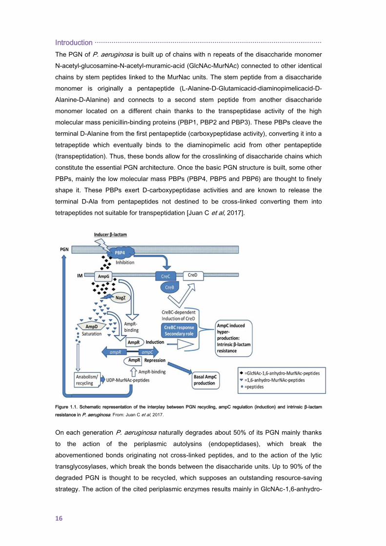
with peptidoglycan (PGN) recycling (Figure 1.1.).
Introduction ············································································································
16
The PGN of P. aeruginosa is built up of chains with n repeats of the disaccharide monomer
N-acetyl-glucosamine-N-acetyl-muramic-acid (GlcNAc-MurNAc) connected to other identical
chains by stem peptides linked to the MurNac units. The stem peptide from a disaccharide
monomer is originally a pentapeptide (L-Alanine-D-Glutamicacid-diaminopimelicacid-D-
Alanine-D-Alanine) and connects to a second stem peptide from another disaccharide
monomer located on a different chain thanks to the transpeptidase activity of the high
molecular mass penicillin-binding proteins (PBP1, PBP2 and PBP3). These PBPs cleave the
terminal D-Alanine from the first pentapeptide (carboxypeptidase activity), converting it into a
tetrapeptide which eventually binds to the diaminopimelic acid from other pentapeptide
(transpeptidation). Thus, these bonds allow for the crosslinking of disaccharide chains which
constitute the essential PGN architecture. Once the basic PGN structure is built, some other
PBPs, mainly the low molecular mass PBPs (PBP4, PBP5 and PBP6) are thought to finely
shape it. These PBPs exert D-carboxypeptidase activities and are known to release the
terminal D-Ala from pentapeptides not destined to be cross-linked converting them into
tetrapeptides not suitable for transpeptidation [Juan C et al, 2017].
Figure 1.1. Schematic representation of the interplay between PGN recycling, ampC regulation (induction) and intrinsic β-lactam
resistance in P. aeruginosa. From: Juan C et al, 2017.
On each generation P. aeruginosa naturally degrades about 50% of its PGN mainly thanks
to the action of the periplasmic autolysins (endopeptidases), which break the
abovementioned bonds originating not cross-linked peptides, and to the action of the lytic
transglycosylases, which break the bonds between the disaccharide units. Up to 90% of the
degraded PGN is thought to be recycled, which supposes an outstanding resource-saving
strategy. The action of the cited periplasmic enzymes results mainly in GlcNAc-1,6-anhydro-
············································································································· Introduction
17
MurNAc tri-, tetra- and penta- peptides [Vollmer W & Höltje JV, 2001], resulting fragments
that are transported through the permease AmpG into the cytosol [Korfmann G & Sanders
CC, 1989; Dietz H & Wiedemann B, 1996; Cheng Q & Park JT, 2002]. This is a key step for
the downstream AmpC regulation and recycling events, as AmpG is the specific door for the
entrance of PGN-derived mediators with AmpC regulator capacity [Zamorano L, 2011]. Once
in the cytosol, the cytosolic L, D-carboxypeptidase LdcA cleaves the D-Ala from the
tetrapeptides units, avoiding the potential accumulation of UDP-MurNAc tetrapeptides which
are thought to be toxic for the bacterial cell [Templin MF et al, 1999]. As well, a glycoside
hydrolase called NagZ removes the GlcNAc residues [Zamorano L et al, 2010] resulting in a
pool of cytosolic GlcNAc units plus 1,6-anhydro-MurNAc peptides [Cheng Q et al, 2000;
Vötsch W & Templin MF, 2000] that, in non-inducer standard conditions, would eventually be
recycled into UDP-MurNAc pentapeptides and exported to the nascent PGN.
Classically, it has been believed that the 1,6-anhydro-MurNAc tri- and penta- peptides units
[Jacobs C et al, 1994; Dietz H et al, 1997] are signal molecules that induce ampC
transcription and, indeed, the UDP-MurNAc pentapeptide has been identified as a repressor
of ampC transcription to basal levels. Thus, these metabolites have been suggested to
competitively regulate ampC transcription by directly binding to the LysR-type transcriptional
regulator AmpR [Jacobs C et al, 1994]. AmpR and AmpC coding genes are located next to
each other within the genome, divergently codified and with overlapping promoter regions to
which AmpR binds to regulate their transcription [Lindquist S et al, 1989; Bartowsky E &
Normark S, 1993]. Under non-inducer standard conditions, the cytosolic AmpD, through its
N-acetyl-muramyl-L-alanine amidase activity, cleaves the stem peptide from both the
GlcNAc-1,6-anhydro-MurNAc and the 1,6-anhydro-MurNAc peptides [Höltje JV & Glauner B,
1990; Jacobs C et al, 1994], which results in low amounts of activation ligands. On the
contrary, the amount of UDP-MurNAc pentapeptides can be increased thanks to the anabolic
pathways starting from the pool of AmpD cleaved peptides and, thus, can both, enter into the
PGN recycling route and bind to AmpR promoting the formation of an AmpR-
deoxyribonucleic acid (DNA) complex that represses ampC transcription to basal levels.
In this sense, it has been proposed that exposure to certain β-lactams known to be AmpC
inducers, such as cefoxitin and imipenem, triggers the accumulation of 1,6-anhydro-MurNAc
peptides within the cytosol, reaching levels that cannot be efficiently processed by AmpD
[Dietz H & Wiedemann B, 1996; Wiedemann B et al, 1998; Vollmer W & Höltje JV, 2001].
This accumulation would presumably displace the UDP-MurNAc pentapeptide from AmpR,
generating a new complex that would act as an activator of ampC transcription and, thus,
leading to clinically significant resistance against the inducer and other hydrolysable β-
lactams [Jacobs C et al, 1994]. The molecular basis for the mentioned increase in the 1,6-
anhydro-MurNAc pentapeptides amount during induction is believed to be related with the
capacity of the inducer β-lactams to inhibit the DD-carboxypeptidase activity of the low
Introduction ············································································································
18
molecular mass PBPs [Sanders CC et al, 1997; Tayler AE et al, 2010; Fisher JF &
Mobashery S, 2014]. In this sense, Moyà et al. showed that the inducer β-lactams can inhibit
the non-essential low molecular mass PBP4 (dacB) [Moyà B et al, 2009], affecting the PGN
composition and favouring the entrance of activation ligands through AmpG. Interestingly,
the authors also showed that PBP4 inducer-inhibition additionally triggers the activation of
the two-component system CreBC which plays a collateral and minor role during the
process. Thus, it has been proposed that PBP4 acts as a sentinel for the cell wall damage
caused by the inducers, triggering an AmpR-dependent overproduction of AmpC and
activating the CreBC system. The induction mechanism is a reversible process and ampC
expression returns to basal levels in the absence of the antibiotic inducers [Mark BL et al,
2012]. Also it should be highlighted that the hydrolytic effect of AmpC onto a β-lactam will not
only depend on the antibiotic inducer capacity but also on the hydrolysing efficiency of the
cephalosporinase. Therefore, the inducible expression of AmpC plays a major role in the
intrinsic resistance of P. aeruginosa to aminopenicillins and most cephalosporins (particularly
cephamycins such as cefoxitin) since these molecules are potent inducers of the expression
and efficiently hydrolyzed by this enzyme. Likewise, the inducible AmpC plays a major role in
the basal reduced susceptibility level of P. aeruginosa to the carbapenem imipenem, as the
relatively stability of this molecule to the hydrolysis by the cephalosporinase is to some
extent compromised by its extremely high potency as inducer [Livermore DM, 1992].
Non-reversible mutational derepression leading to constitutive high-level expression of
AmpC will be discussed later in section 1.8.
1.3.3. Efflux-pumps systems: constitutive and inducible expression
Efflux pumps play an important role in antibiotic resistance. These pumps may be specific for
a substrate or may extrude a broad range of compounds including dyes, detergents, fatty
acids and antibiotics of multiple classes structurally unrelated. Thus, it is probable that efflux
pumps were created so that harmful substances could be transported out of the bacterial
cell, thus, allowing for survival.
Based primarily on amino acid sequence identity, on the energy source required to drive
export and on substrate specificities, efflux pumps have been categorized in five
superfamilies including (i) the ATP-binding cassette family, (ii) the small multidrug resistance
family, (iii) the major facilitator superfamily, (iv) the resistance-nodulation-division (RND)
family, and (v) the multidrug and toxic compound extrusion family. In P. aeruginosa, genome
sequence analysis has revealed the presence of efflux systems from all five superfamilies,
being the RND family the most prevalent with 12 different systems identified.
············································································································· Introduction
19
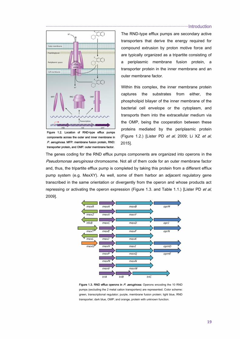
Figure 1.2. Location of RND-type efflux pumps
components across the outer and inner membrane in
P. aeruginosa. MFP: membrane fusion protein, RND:
transporter protein, and OMF: outer membrane factor.
The RND-type efflux pumps are secondary active
transporters that derive the energy required for
compound extrusion by proton motive force and
are typically organized as a tripartite consisting of
a periplasmic membrane fusion protein, a
transporter protein in the inner membrane and an
outer membrane factor.
Within this complex, the inner membrane protein
captures the substrates from either, the
phospholipid bilayer of the inner membrane of the
bacterial cell envelope or the cytoplasm, and
transports them into the extracellular medium via
the OMP, being the cooperation between these
proteins mediated by the periplasmic protein
(Figure 1.2.) [Lister PD et al, 2009; Li XZ et al,
2015].
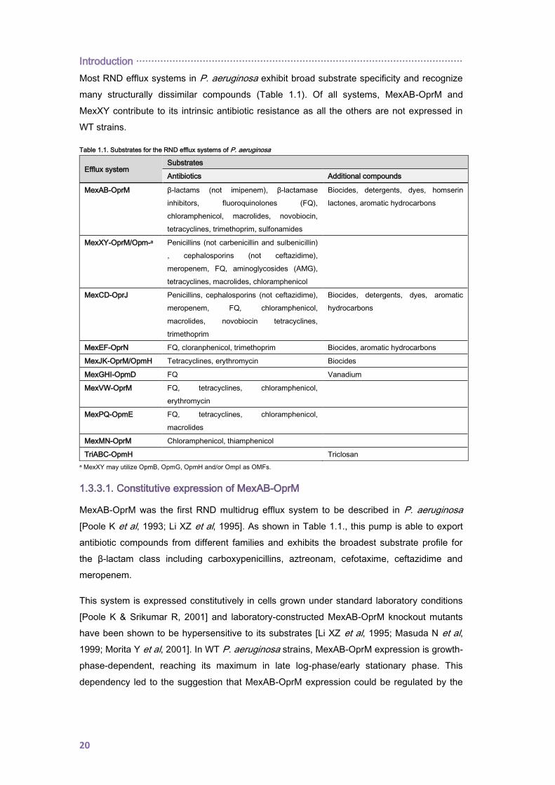
The genes coding for the RND efflux pumps components are organized into operons in the
Pseudomonas aeruginosa chromosome. Not all of them code for an outer membrane factor
and, thus, the tripartite efflux pump is completed by taking this protein from a different efflux
pump system (e.g. MexXY). As well, some of them harbor an adjacent regulatory gene
transcribed in the same orientation or divergently from the operon and whose products act
repressing or activating the operon expression (Figure 1.3. and Table 1.1.) [Lister PD et al,
2009].
Figure 1.3. RND efflux operons in P. aeruginosa. Operons encoding the 10 RND
pumps (excluding the 2 metal cation transporters) are represented. Color scheme:
green, transcriptional regulator; purple, membrane fusion protein; light blue, RND
transporter; dark blue, OMP; and orange, protein with unknown function.
Introduction ············································································································
20
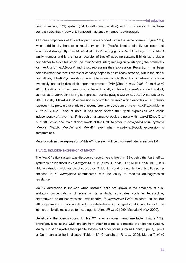
Most RND efflux systems in P. aeruginosa exhibit broad substrate specificity and recognize
many structurally dissimilar compounds (Table 1.1). Of all systems, MexAB-OprM and
MexXY contribute to its intrinsic antibiotic resistance as all the others are not expressed in
WT strains.
Table 1.1. Substrates for the RND efflux systems of P. aeruginosa
Efflux system Substrates
Antibiotics Additional compounds
MexAB-OprM β-lactams (not imipenem), β-lactamase
inhibitors, fluoroquinolones (FQ),
chloramphenicol, macrolides, novobiocin,
tetracyclines, trimethoprim, sulfonamides
Biocides, detergents, dyes, homserin
lactones, aromatic hydrocarbons
MexXY-OprM/Opm-a Penicillins (not carbenicillin and sulbenicillin)
, cephalosporins (not ceftazidime),
meropenem, FQ, aminoglycosides (AMG),
tetracyclines, macrolides, chloramphenicol
MexCD-OprJ Penicillins, cephalosporins (not ceftazidime),
meropenem, FQ, chloramphenicol,
macrolides, novobiocin tetracyclines,
trimethoprim
Biocides, detergents, dyes, aromatic
hydrocarbons
MexEF-OprN FQ, cloranphenicol, trimethoprim Biocides, aromatic hydrocarbons
MexJK-OprM/OpmH Tetracyclines, erythromycin Biocides
MexGHI-OpmD FQ Vanadium
MexVW-OprM FQ, tetracyclines, chloramphenicol,
erythromycin
MexPQ-OpmE FQ, tetracyclines, chloramphenicol,
macrolides
MexMN-OprM Chloramphenicol, thiamphenicol
TriABC-OpmH Triclosan
a MexXY may utilize OpmB, OpmG, OpmH and/or OmpI as OMFs.
1.3.3.1. Constitutive expression of MexAB-OprM
MexAB-OprM was the first RND multidrug efflux system to be described in P. aeruginosa
[Poole K et al, 1993; Li XZ et al, 1995]. As shown in Table 1.1., this pump is able to export
antibiotic compounds from different families and exhibits the broadest substrate profile for
the β-lactam class including carboxypenicillins, aztreonam, cefotaxime, ceftazidime and
meropenem.
This system is expressed constitutively in cells grown under standard laboratory conditions
[Poole K & Srikumar R, 2001] and laboratory-constructed MexAB-OprM knockout mutants
have been shown to be hypersensitive to its substrates [Li XZ et al, 1995; Masuda N et al,
1999; Morita Y et al, 2001]. In WT P. aeruginosa strains, MexAB-OprM expression is growth-
phase-dependent, reaching its maximum in late log-phase/early stationary phase. This
dependency led to the suggestion that MexAB-OprM expression could be regulated by the
············································································································· Introduction
21
quorum sensing (QS) system (cell to cell communication) and, in this sense, it has been
demonstrated that N-butyryl-L-homoserin-lactones enhance its expression.
All three components of this efflux pump are encoded within the same operon (Figure 1.3.),
which additionally harbors a regulatory protein (MexR) located directly upstream but
transcribed divergently from MexA-MexB-OprM coding genes. MexR belongs to the MarR
family member and is the major regulator of this efflux pump system. It binds as a stable
homodimer to two sites within the mexR-mexA intergenic region overlapping the promoters
for mexR and mexAB-oprM and, thus, repressing their expression. Recently, it has been
demonstrated that MexR repressor capacity depends on its redox state as, within the stable
homodimer, MexR-Cys residues form intermonomer disulfide bonds whose oxidation
eventually lead to its dissociation from the promoter DNA [Chen H et al, 2008; Chen H et al,
2010]. MexR activity has been found to be additionally controlled by armR encoded product,
as it binds to MexR diminishing its repressor activity [Daigle DM et al, 2007; Wilke MS et al,
2008]. Finally, MexAB-OprM expression is controlled by nalD, which encodes a TetR family
repressor-like protein that binds to a second promoter upstream of mexA-mexB-oprM [Morita
Y et al, 2006a]. Also of note, it has been shown that oprM expression can occur
independently of mexA-mexB, through an alternative weak promoter within mexB [Zhao Q et
al, 1998], which ensures sufficient levels of this OMP to other P. aeruginosa efflux systems
(MexXY, MexJK, MexVW and MexMN) even when mexA-mexB-oprM expression is
compromised.
Mutation-driven overexpression of this efflux system will be discussed later in section 1.8.
1.3.3.2. Inducible expression of MexXY
The MexXY efflux system was discovered several years later, in 1999, being the fourth efflux
system to be identified in P. aeruginosa PAO1 [Aires JR et al, 1999; Mine T et al, 1999]. It is
able to extrude a wide variety of substrates (Table 1.1.) and, of note, is the only efflux pump
encoded in P. aeruginosa chromosome with the ability to mediate aminoglycoside
resistance.
MexXY expression is induced when bacterial cells are grown in the presence of sub-
inhibitory concentrations of some of its antibiotic substrates such as tetracycline,
erythromycin or aminoglycosides. Additionally, P. aeruginosa PAO1 mutants lacking this
efflux system are hypersusceptible to its substrates which suggests that it contributes to the
intrinsic antibiotic resistance to these agents [Aires JR et al, 1999; Masuda N et al, 2000].
Genetically, the operon coding for MexXY lacks an outer membrane factor (Figure 1.3.).
Therefore, it takes the OMF protein from other operons to complete the tripartite system.
Mainly, OprM completes the tripartite system but other porins such as OpmB, OpmG, OpmH
or OpmI can also be implicated (Table 1.1.) [Chuanchuen R et al, 2005; Murata T et al,
Introduction ············································································································
22
2002]. Located upstream but transcribed divergently from mexX-mexY, is encountered mexZ
which encodes a protein that belongs to the TetR family of transcriptional regulators and
negatively regulates its expression (Figure 1.3.). Similar to MexR (section 1.3.3.1.), MexZ
binds as a homodimer to an inverted repeated sequence within the intergenic region mexZ-
mexX, overlapping the putative mexX-mexY promoter [Matsuo Y et al, 2004] and repressing
its expression.
In contrast to other drug-inducible multidrug efflux systems, MexXY inducers do not alter
MexZ and mexZ-mexX interactions. Instead, induction has been shown to be dependent on
drug-ribosome interactions and to occur, although in a lesser extent, even in the mexZ
mutant [Jeannot K et al, 2005]. Therefore, these data suggest an alternative biological role
for the MexXY system beyond antibiotics efflux. Multiple pathways participate in the
regulation of mexX-mexY induction. Although ribosome disruption has been shown to impact
the expression of a myriad of genes, by using a transposon insertion mutant library PA5471
was found to be not only drug-inducible but also required for mexX-mexY induced
expression [Morita Y et al, 2006b]. Later on, it was demonstrates that the antimicrobial-
inducible PA5471 gene product has interacts with the repressor MexZ and, thus, interfere
with its DNA binding activity [Yamamoto M et al, 2009].
More recently, it has been also demonstrated the involvement of parR, a gene coding for the
response regulator of the two-component regulatory system ParR-ParS, in promoting either
induced or constitutive mexX-mexY upregulation. In addition, this gene was demonstrated to
be also implicated in OprD porin downregulation and in lipopolysaccharide (LPS)
modification in a MexZ-independent manner [Muller C et al, 2011].
Mutation-driven overexpression of this efflux system will be discussed later in section 1.8.
············································································································· Introduction
23
1.4. CHRONIC RESPIRATORY INFECTIONS
On average, about 10,000 L of air are inhaled per person per day and, thus, the respiratory
tract is continuously exposed to a wide variety of potential pathogenic microorganisms.
However, and due to sophisticated host defence mechanisms at the lung mucosa, infections
are rare among healthy individuals. Airway bronchial and alveolar epithelial cells constitute
the first line of defense against invading bacteria, providing not only a physical barrier and
exhibiting local antimicrobial activity but also acting as sentinels stimulating downstream
recruitment and activation of immune cells which clear invading bacteria. As well, resident
alveolar macrophages and occasionally dendritic cells are also found in the alveolar
epithelium and are key mediators of innate and adaptive immunity [Eisele NA & Anderson
DM, 2011]. On the opposite, the immune response within the respiratory airway of patients
suffering chronic respiratory underlying diseases such as CF, non-CF bronchiectasias or
COPD, is impaired and, therefore, these disorders are characterized by repeated cycles of
inflammation, tissue damage and bacterial infections that may eventually lead to the
establishment of chronic non-eradicable respiratory infections and a rapid decline of the
pulmonary function [Döring G et al, 2011]. In fact, P. aeruginosa CRI acquired a major
relevance within the CF setting, being the most frequent and severe driver of morbidity and
mortality.
CF is the most prevalent autosomal recessive hereditary disease affecting Caucasian
populations, with approximately 70,000 people affected worldwide and with an estimated
incidence of 1 per 2500-5000 newborns in white populations from Europe, Canada and USA
[O'Sullivan BP & Freedman SD, 2009]. This chronic respiratory disease is caused by
mutations (two-thirds F508Δ) disrupting the function of the CF transmembrane conductance
regulator (CFTR) gene, which encodes a chloride channel that is expressed on the apical
surface of many epithelial and blood cells. The clinical spectrum of the CF disease is wide;
however, pulmonary insufficiency is the first cause of morbidity and mortality among CF
patients being approximately 80% of CF deaths related with chronic lung infection
[O'Sullivan BP & Freedman SD, 2009]. Fortunately, over the past decades, CF respiratory
infections management has considerably been improved and median age of survival of CF
patients is now set in more than 40 years in developed countries [McCormick J et al, 2010].
As a result, the number of CF adults (age ≥18 years) is larger than the number of children in
several EU countries with well-established healthcare systems [McCormick J et al, 2010] and
forecasts predict a large increase in the number of CF adults by 2025 [Burgel PR et al,
2015].
By the time of birth, the respiratory tract of CF children is normal but, soon after, becomes
inflamed and infected. Mechanisms underlying the early acquisition of infection and the
establishment of P. aeruginosa CRI are complex and several factors participate as following
described.
Introduction ············································································································
24
One contributing factor is the inability of mutated CFTR to effectively secrete chloride from
respiratory epithelial cells into the airway surface liquid which eventually causes excessive
water absorption from the airway surface liquid, leading to an impaired mucociliary
clearance. Likewise, the viscosity of the secretions may impair the transport of antimicrobial
oligopeptides onto the epithelium and, thus, may also negatively affect the migration of
neutrophils towards the pathogens. Furthermore, within the highly viscous mucus, a
microaerobic/anaerobic milieu prevails due to oxygen consumption by bacterial pathogens or
invading neutrophils which abolish the generation of reactive oxygen species (ROS) by
neutrophils and other cells impairing bacterial killing. As well, other investigators have
demonstrated that the abnormal accumulation of ceramide in the lungs of CF mice and in the
epithelial cells from CF patients, results in an increased death rate of respiratory epithelial
cells and DNA deposits on the respiratory epithelium, which in turns facilitates bacterial
adherence. Finally, P. aeruginosa infection may also be facilitated directly by defective
CFTR, as in its functional state can bind the pathogen within lipid rafts removing it from the
epithelial surface via internalization [Döring G et al, 2011]. Of course, P. aeruginosa also
plays a major role as, thanks to the enormous armamentarium of immunoevasive strategies
encoded within its genome, is capable of evading not only host defenses but also repeated
courses of antibiotics.
All the above mentioned alterations in the CF airway surface provide an ideal environment
for infection/colonization which is not only exploited by P. aeruginosa. In fact, CF patients
experience multiple bacterial infections throughout their life and whereas, overall,
Staphylococcus aureus and Haemophilus influenzae are typically first cultured in young
children, P. aeruginosa and other opportunistic multidrug resistant pathogens such as
Achromobacter spp., Stenotrophomonas maltophilia or Burkholderia cepacia complex are
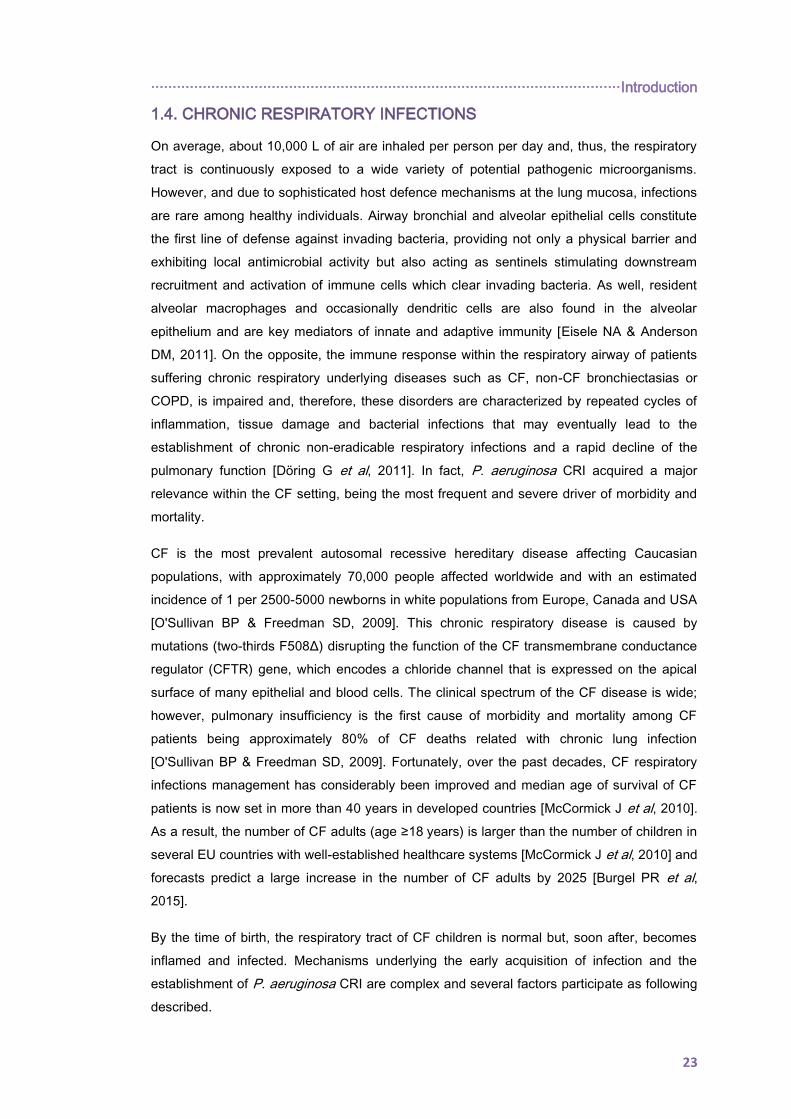
first cultured during adolescence and young adulthood (Figure 1.4.) [CFF Patient Registry,
2016 Annual Data Report].
Figure 1.4. Prevalence of respiratory microorganisms by age cohort, 2016. From: Cystic Fibrosis Foundation Patient Registry, 2016
Annual Data Report. Bethesda, Maryland, ©2017 Cystic Fibrosis Foundation.

············································································································· Introduction
25
As shown, P. aeruginosa is by far the most significant CF pathogen. Early infection occurs in
a large number of patients before the age of 3 years [Speert DP et al, 2002], after, and for a
variable period of time, P. aeruginosa isolation from CF respiratory samples can be
intermittent and, usually, involving multiple strains. Eventually, by the age of 25, over 70% of
the patients are chronically colonized and a single well adapted strain (or clonal lineage)
predominates [Cystic Fibrosis Foundation Patient Registry, 2016 Annual Data Report].
Introduction ············································································································
26
1.5. EVOLUTION AND ADAPTATION TO THE CYSTIC FIBROSIS
AIRWAYS
During the progression from early infection to chronic non-eradicable colonization, P.
aeruginosa undergoes a complex evolutionary adaptation and diversification process that
implies both phenotypic and genotypic variations.
Usually, first P. aeruginosa CF isolate resembles those from the environment or from acute
infections in terms of phenotype and genotype. Thus, during long time, it was extensively
accepted that early P. aeruginosa acquisition occurs from diverse environmental reservoirs
so, generally, each patient harbors its unique non-clonal unrelated strain. However, this
classical perception changed in 1986, when an outbreak caused by a P. aeruginosa strain
resistant to several antibiotics was reported in a CF center in Denmark [Pedersen SS et al,
1986]. Since this first description, other strains infecting a large proportion of CF patients
have been detected and, in some cases, strongly associated with multidrug resistant profiles.
Therefore, nowadays “person-to-person” transmission is also an accepted route of P.
aeruginosa acquisition among CF patients and certain clones are extensively recognized as
epidemic and/or transmissible being worldwide distributed (section 1.8.).
Regardless to the source of infection, if not eradicated, the cell density and the collective
growth pattern of P. aeruginosa change and a complex diversification process occurs within
the bacterial population which, in turns, improve its capacity for survive and persist in the CF
airways throughout the lifespan of a CF patient [Renders N et al, 2001; Munck A et al, 2001].
Overtime, during the course of infection, the genome of P. aeruginosa can be modified by
either acquiring new mutations or by the acquisition and/or loose of genomic DNA. Whereas
few works have focused their attention on DNA acquisition/loose and obtained results are
not consistent, different authors have focused on genomic modification based on the
acquisition of SNPs and small insertions stablishing mutation rates from 1 to 3 SNPs/year for
non-mutator isolates [Marvig RL et al, 2015a]. As well, since whole-genome sequencing
technologies have become more affordable, many efforts have been put in identify those
genes that are recurrent mutated among CF isolates and, therefore, directly implicated in the
adaptation to the CF lungs, the so-called pathoadaptative genes. Whereas some genes have
been found to be frequently mutated, others have just been found to be mutated in single
studies which suggest that different evolutionary pathways exist [Marvig et al, 2015a].
Addtionally, the study of longitudinal isolates within single patients have provided evidence
that population diversification and stable maintenance of these genetically distinct
subpopulations frequently occurs, even including mutator and non-mutator sublineages
[Chung JC et al, 2012; Marvig RL et al, 2013; Feliziani S et al, 2014]; diversification procces
that have been suggested to be triggered by the spatial heterogeneity of the CF airways
[Markussen T et al, 2014].
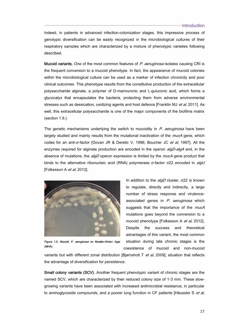
············································································································· Introduction
27
Indeed, in patients in advanced infection-colonization stages, this impressive process of
genotypic diversification can be easily recognized in the microbiological cultures of their
respiratory samples which are characterized by a mixture of phenotypic varieties following
described.
Mucoid variants. One of the most common features of P. aeruginosa isolates causing CRI is
the frequent conversion to a mucoid phenotype. In fact, the appearance of mucoid colonies
within the microbiological culture can be used as a marker of infection chronicity and poor
clinical outcomes. This phenotype results from the constitutive production of the extracellular
polysaccharide alginate, a polymer of D-mannuronic and L-guluronic acid, which forms a
glycocalyx that encapsulates the bacteria, protecting them from adverse environmental
stresses such as dessication, oxidizing agents and host defence [Franklin MJ et al, 2011]. As
well, this extracellular polysaccharide is one of the major components of the biofilms matrix
(section 1.6.).
The genetic mechanisms underlying the switch to mucoidity in P. aeruginosa have been
largely studied and mainly results from the mutational inactivation of the mucA gene, which
codes for an anti-σ-factor [Govan JR & Deretic V, 1996; Boucher JC et al, 1997]. All the
enzymes required for alginate production are encoded in the operon algD-algA and, in the
absence of mutations, the algD operon expression is limited by the mucA gene product that
binds to the alternative ribonucleic acid (RNA) polymerase σ-factor σ22 encoded in algU
[Folkesson A et al, 2012].
In addition to the algD cluster, σ22 is known
to regulate, directly and indirectly, a large
number of stress response and virulence-
associated genes in P. aeruginosa which
suggests that the importance of the mucA
mutations goes beyond the conversion to a
mucoid phenotype [Folkesson A et al, 2012].
Despite the success and theoretical
advantages of this variant, the most common
situation during late chronic stages is the
coexistence of mucoid and non-mucoid
variants but with different zonal distribution [Bjarnsholt T et al, 2009]; situation that reflects
the advantage of diversification for persistence.
Small colony variants (SCV). Another frequent phenotypic variant of chronic stages are the
named SCV, which are characterized by their reduced colony size of 1-3 mm. These slow-
growing variants have been associated with increased antimicrobial resistance, in particular
to aminoglycoside compounds, and a poorer lung function in CF patients [Häussler S et al,
Figure 1.5. Mucoid P. aeruginosa on Mueller-Hinton Agar
(MHA).
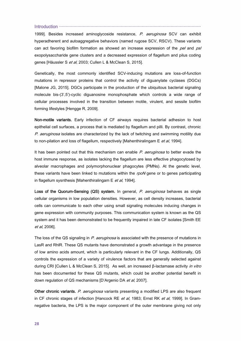
Introduction ············································································································
28
1999]. Besides increased aminoglycoside resistance, P. aeruginosa SCV can exhibit
hyperadherent and autoaggregative behaviors (named rugose SCV, RSCV). These variants
can act favoring biofilm formation as showed an increase expression of the pel and psl
exopolysaccharide gene clusters and a decreased expression of flagellum and pilus coding
genes [Häussler S et al, 2003; Cullen L & McClean S, 2015].
Genetically, the most commonly identified SCV-inducing mutations are loss-of-function
mutations in repressor proteins that control the activity of diguanylate cyclases (DGCs)
[Malone JG, 2015]. DGCs participate in the production of the ubiquitous bacterial signaling
molecule bis-(3’,5’)-cyclic diguanosine monophosphate which controls a wide range of
cellular processes involved in the transition between motile, virulent, and sessile biofilm
forming lifestyles [Hengge R, 2009].
Non-motile variants. Early infection of CF airways requires bacterial adhesion to host
epithelial cell surfaces, a process that is mediated by flagellum and pilli. By contrast, chronic
P. aeruginosa isolates are characterized by the lack of twitching and swimming motility due
to non-pilation and loss of flagellum, respectively [Mahenthiralingam E et al, 1994].
It has been pointed out that this mechanism can enable P. aeruginosa to better evade the
host immune response, as isolates lacking the flagellum are less effective phagocytosed by
alveolar macrophages and polymorphonuclear phagocytes (PMNs). At the genetic level,
these variants have been linked to mutations within the rpoN gene or to genes participating
in flagellum sysnthesis [Mahenthiralingam E et al, 1994].
Loss of the Quorum-Sensing (QS) system. In general, P. aeruginosa behaves as single
cellular organisms in low population densities. However, as cell density increases, bacterial
cells can communicate to each other using small signaling molecules inducing changes in
gene expression with community purposes. This communication system is known as the QS
system and it has been demonstrated to be frequently impaired in late CF isolates [Smith EE
et al, 2006].
The loss of the QS signaling in P. aeruginosa is associated with the presence of mutations in
LasR and RhlR. These QS mutants have demonstrated a growth advantage in the presence
of low amino acids amount, which is particularly relevant in the CF lungs. Additionally, QS
controls the expression of a variety of virulence factors that are generally selected against
during CRI [Cullen L & McClean S, 2015]. As well, an increased β-lactamase activity in vitro
has been documented for these QS mutants, which could be another potential benefit in
down regulation of QS mechanisms [D’Argenio DA et al, 2007].
Other chronic variants. P. aeruginosa variants presenting a modified LPS are also frequent
in CF chronic stages of infection [Hancock RE et al, 1983; Ernst RK et al, 1999]. In Gram-
negative bacteria, the LPS is the major component of the outer membrane giving not only
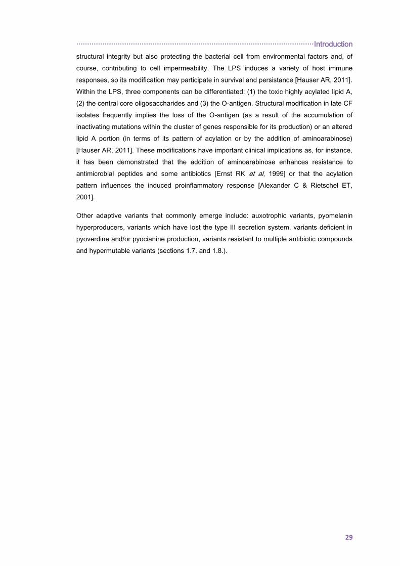
············································································································· Introduction
29
structural integrity but also protecting the bacterial cell from environmental factors and, of
course, contributing to cell impermeability. The LPS induces a variety of host immune
responses, so its modification may participate in survival and persistance [Hauser AR, 2011].
Within the LPS, three components can be differentiated: (1) the toxic highly acylated lipid A,
(2) the central core oligosaccharides and (3) the O-antigen. Structural modification in late CF
isolates frequently implies the loss of the O-antigen (as a result of the accumulation of
inactivating mutations within the cluster of genes responsible for its production) or an altered
lipid A portion (in terms of its pattern of acylation or by the addition of aminoarabinose)
[Hauser AR, 2011]. These modifications have important clinical implications as, for instance,
it has been demonstrated that the addition of aminoarabinose enhances resistance to
antimicrobial peptides and some antibiotics [Ernst RK et al, 1999] or that the acylation
pattern influences the induced proinflammatory response [Alexander C & Rietschel ET,
2001].
Other adaptive variants that commonly emerge include: auxotrophic variants, pyomelanin
hyperproducers, variants which have lost the type III secretion system, variants deficient in
pyoverdine and/or pyocianine production, variants resistant to multiple antibiotic compounds
and hypermutable variants (sections 1.7. and 1.8.).
Introduction ············································································································
30
1.6. PHYSIOLOGICAL RESISTANCE DURING CYSTIC FIBROSIS
CHRONIC RESPIRATORY INFECTIONS
1.6.1. From the planktonic to the biofilm mode of growth
It has been set that the CF lung is a heterogeneous, hostile and stressful environment for
invading bacteria. In order to overcome all these challenges, and apart from population
diversification, P. aeruginosa shifts its mode of growth from free-living cells (planktonic state)
to biofilm-forming cells, change that is currently recognized as one of the hallmarks of
chronic infections. In fact, both processes are part of the same evolutionary path as all the
above described variants (section 1.5.) live together within the biofilm community and
contribute to its formation and existence, which constitutes an amazing example of how
bacterial populations can enhance its survival and persistence in hostile environments by
acting in a cooperative manner.
Biofilms are defined as organized bacterial communities surrounded by an extracellular
polymeric matrix that confers resistance against the hostile environment. Biofilm formation
classically involves the following stages: attachment, microcolonies formation, biofilm
maturation and dispersal or detachment (Figure 1.6.) [O’Toole G et al, 2000].
Figure 1.6. Stages of Biofilms formation
As shown, its development starts with the adherence of individual planktonic bacterial cells
to a surface with the help of pili and flagella [O’Toole G et al, 2000]. Although most biofilm-
related infections generally require an attachment to a solid surface, in the case of CF, some
studies indicate that the biofilm found in the lung is directly formed on the mucus instead of
being in contact with the lung epithelium [Bjarnsholt T et al, 2009; Worlitzsch D et al, 2002].
Attachment is then followed by bacteria multiplication, thus forming microcolonies, matrix
building and eventual biofilm maturation. The extracellular polymeric matrix plays an
important role during CRI not only giving cohesion to the structure and acting as a nutrient
source but also providing a protective barrier against host defense, desiccation, ROS and
antibiotics [Flemming HC & Wingender J, 2010]. This matrix is mainly composed of a
conglomerate of exopolyscharides (including alginate provided by mucoid variants),
extracellular DNA, proteins, surfactants, lipids, bacterial lytic products and host compounds.
············································································································· Introduction
31
Finally, once matured, biofilm population ensure its persistence in the hostile environment by
releasing or dispersing small aggregates or even individual cells to uncolonized sites and
reinitiating the biofilm lifecycle during the dispersal stage [O’Toole G et al, 2000].
Such a tangled process is known to be closely regulated by intra- and extracellular cues that
modulate the levels of diffusible signal molecules, second messengers and small RNAs
[Bjarnsholt T, 2013]. QS systems detect these signals as cell density evidences and trigger
changes in bacterial gene transcription, including virulence factors and diverse proteins
involved in the innate resistance of biofilms to antibiotics and the immune system. In this
sense, P. aeruginosa biofilms are known to be able to initiate detachment on their own and
this process can be mediated by either, alginate lyase overexpression [Boyd A &
Chakrabarty AM, 1994] or by up-regulation of motility factors such as the rhamnolipid and
type IV pili [Pamp SJ & Tolker-Nielsen T, 2007].
1.6.2. Inherent antimicrobial tolerance of biofilms
One of the most relevant aspects of biofilms is that they determine the persistence of the
infection despite long-term antimicrobial treatment. In fact, it is estimated that biofilms can
tolerate up to 100–1000 fold higher concentrations of antibiotics than the planktonic cells
[Høiby N et al, 2010]. As following described, the documented inherent biofilm antimicrobial
tolerance is multifactorial (Figure 1.7.).
Figure 1.7. Schematic representation of the factors contributing to inherent biofilm antimicrobial resistance. ATB: antibiotics. From:
The Problems of antibiotic resistance in CF and soultions. López-Causapé et al., Expert Rev Respir Med 2015
Limited antibiotic penetration. The biofilm matrix acts as a primary barrier preventing the
entrance of some compounds such as polar and charged antibiotics [Lewis K, 2008]. This
restricted penetration has been linked to some components of the matrix such as alginate or
eDNA which have shown antibiotic chelating activity [Alipour M et al, 2009] or to the
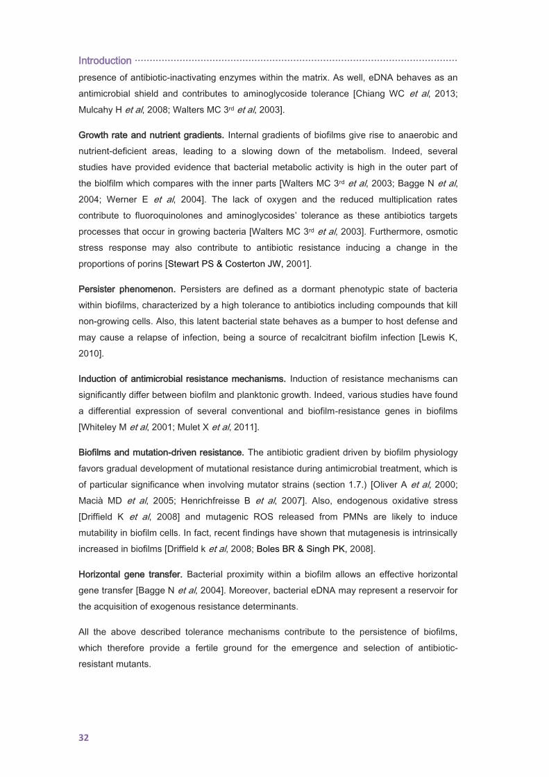
Introduction ············································································································
32
presence of antibiotic-inactivating enzymes within the matrix. As well, eDNA behaves as an
antimicrobial shield and contributes to aminoglycoside tolerance [Chiang WC et al, 2013;
Mulcahy H et al, 2008; Walters MC 3rd et al, 2003].
Growth rate and nutrient gradients. Internal gradients of biofilms give rise to anaerobic and
nutrient-deficient areas, leading to a slowing down of the metabolism. Indeed, several
studies have provided evidence that bacterial metabolic activity is high in the outer part of
the biolfilm which compares with the inner parts [Walters MC 3rd et al, 2003; Bagge N et al,
2004; Werner E et al, 2004]. The lack of oxygen and the reduced multiplication rates
contribute to fluoroquinolones and aminoglycosides’ tolerance as these antibiotics targets
processes that occur in growing bacteria [Walters MC 3rd et al, 2003]. Furthermore, osmotic
stress response may also contribute to antibiotic resistance inducing a change in the
proportions of porins [Stewart PS & Costerton JW, 2001].
Persister phenomenon. Persisters are defined as a dormant phenotypic state of bacteria
within biofilms, characterized by a high tolerance to antibiotics including compounds that kill
non-growing cells. Also, this latent bacterial state behaves as a bumper to host defense and
may cause a relapse of infection, being a source of recalcitrant biofilm infection [Lewis K,
2010].
Induction of antimicrobial resistance mechanisms. Induction of resistance mechanisms can
significantly differ between biofilm and planktonic growth. Indeed, various studies have found
a differential expression of several conventional and biofilm-resistance genes in biofilms
[Whiteley M et al, 2001; Mulet X et al, 2011].
Biofilms and mutation-driven resistance. The antibiotic gradient driven by biofilm physiology
favors gradual development of mutational resistance during antimicrobial treatment, which is
of particular significance when involving mutator strains (section 1.7.) [Oliver A et al, 2000;
Macià MD et al, 2005; Henrichfreisse B et al, 2007]. Also, endogenous oxidative stress
[Driffield K et al, 2008] and mutagenic ROS released from PMNs are likely to induce
mutability in biofilm cells. In fact, recent findings have shown that mutagenesis is intrinsically
increased in biofilms [Driffield k et al, 2008; Boles BR & Singh PK, 2008].
Horizontal gene transfer. Bacterial proximity within a biofilm allows an effective horizontal
gene transfer [Bagge N et al, 2004]. Moreover, bacterial eDNA may represent a reservoir for
the acquisition of exogenous resistance determinants.
All the above described tolerance mechanisms contribute to the persistence of biofilms,
which therefore provide a fertile ground for the emergence and selection of antibiotic-
resistant mutants.
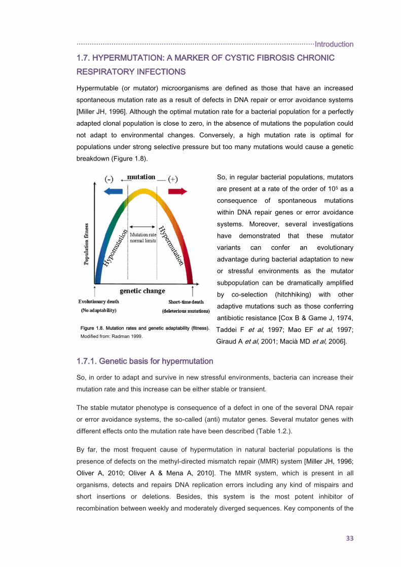
············································································································· Introduction
33
1.7. HYPERMUTATION: A MARKER OF CYSTIC FIBROSIS CHRONIC
RESPIRATORY INFECTIONS
Hypermutable (or mutator) microorganisms are defined as those that have an increased
spontaneous mutation rate as a result of defects in DNA repair or error avoidance systems
[Miller JH, 1996]. Although the optimal mutation rate for a bacterial population for a perfectly
adapted clonal population is close to zero, in the absence of mutations the population could
not adapt to environmental changes. Conversely, a high mutation rate is optimal for
populations under strong selective pressure but too many mutations would cause a genetic
breakdown (Figure 1.8).
So, in regular bacterial populations, mutators
are present at a rate of the order of 105 as a
consequence of spontaneous mutations
within DNA repair genes or error avoidance
systems. Moreover, several investigations
have demonstrated that these mutator
variants can confer an evolutionary
advantage during bacterial adaptation to new
or stressful environments as the mutator
subpopulation can be dramatically amplified
by co-selection (hitchhiking) with other
adaptive mutations such as those conferring
antibiotic resistance [Cox B & Game J, 1974,
Taddei F et al, 1997; Mao EF et al, 1997;
Giraud A et al, 2001; Macià MD et al, 2006].
1.7.1. Genetic basis for hypermutation
So, in order to adapt and survive in new stressful environments, bacteria can increase their
mutation rate and this increase can be either stable or transient.
The stable mutator phenotype is consequence of a defect in one of the several DNA repair
or error avoidance systems, the so-called (anti) mutator genes. Several mutator genes with
different effects onto the mutation rate have been described (Table 1.2.).
By far, the most frequent cause of hypermutation in natural bacterial populations is the
presence of defects on the methyl-directed mismatch repair (MMR) system [Miller JH, 1996;
Oliver A, 2010; Oliver A & Mena A, 2010]. The MMR system, which is present in all
organisms, detects and repairs DNA replication errors including any kind of mispairs and
short insertions or deletions. Besides, this system is the most potent inhibitor of
recombination between weekly and moderately diverged sequences. Key components of the
Figure 1.8. Mutation rates and genetic adaptability (fitness).
Modified from: Radman 1999.
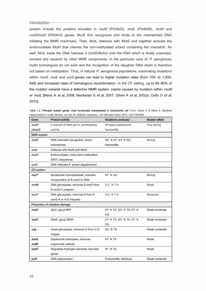
Introduction ············································································································
34
system include the proteins encoded in mutS (PA3620), mutL (PA4946), mutH and
uvrD/mutU (PA5443) genes. MutS first recognizes and binds to the mismatched DNA
initiating the MMR machinery. Then, MutL interacts with MutS and together activate the
endonuclease MutH that cleaves the non-methylated strand containing the mismatch. As
well, MutL loads the DNA helicase II (UvrD/MutU) onto the DNA which is finally unwinded,
excised and repaired by other MMR components. In the particular case of P. aeruginosa,
mutH homologues do not exist and the recognition of the daughter DNA strain is therefore
not based on methylation. Thus, in natural P. aeruginosa populations, inactivating mutations
within mutS, mutL and uvrD genes can lead to higher mutation rates (from 100- to 1,000-
fold) and increased rates of homologous recombination. In the CF setting, up to 60–90% of
the mutator variants have a defective MMR system, mainly caused by mutation within mutS
or mutL [Mena A et al, 2008; Montanari S et al, 2007; Oliver A et al, 2002a; Ciofu O et al,
2010].
Table 1.2. Principal mutator genes, most functionally characterized in Escherichia coli. From: Oliver A & Mena A. Bacterial
hypermutation in cystic fibrosis, not only for antibiotic resistance. Clin Microbiol Infect. 2010; 16(7):798-808.
Gene Product activity Mutations produced Mutator effect
mutD
(dnaQ)
Ε subunit of DNA pol III, proofreading
activity
All base substitutions,
frameshifts
Very strong
MMR system
mutS DNA mismatch recognition, binds
mismatches
GC AT, AT GC,
frameshifts
Strong
mutL Interacts with MutS and MutH
mutH Endonuclease, nicks hemi-methylated
GATC sequences
uvrD DNA Helicase II, strand displacement
GO system
mutT Nucleoside triphosphatase, prevents
incorporation of 8-oxoG to DNA
AT CG Strong
mutM DNA glicosylase, removes 8-oxoG from
8-oxoG-C mispairs
G:C T:A Weak
mutY DNA glicosylase, removes A from 8-
oxoG-A or A-G mispairs
G:C T:A Moderate
Prevention of oxidative damage
mutA GlyV, glycyl tRN AT TA, GC TA, AT
CG
Weak-moderate
mutC GlyW, glycyl tRNA AT TA, GC TA, AT
CG
Weak-moderate
ung Uracil glicosylase, removes U from U-G
mispair
GC TA Weak-moderate
sodA,
sodB
Superoxide dismutase, removes
superoxide radicals
AT TA Weak
oxyR Regulates hydrogen peroxide inducible
genes
AT TA Weak
polA DNA polymerase I Frameshifts, deletions Weak-moderate
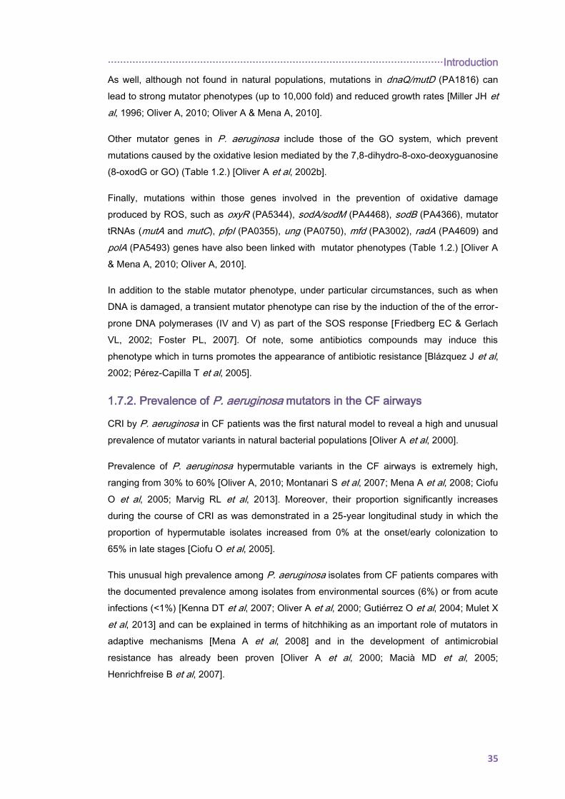
············································································································· Introduction
35
As well, although not found in natural populations, mutations in dnaQ/mutD (PA1816) can
lead to strong mutator phenotypes (up to 10,000 fold) and reduced growth rates [Miller JH et
al, 1996; Oliver A, 2010; Oliver A & Mena A, 2010].
Other mutator genes in P. aeruginosa include those of the GO system, which prevent
mutations caused by the oxidative lesion mediated by the 7,8-dihydro-8-oxo-deoxyguanosine
(8-oxodG or GO) (Table 1.2.) [Oliver A et al, 2002b].
Finally, mutations within those genes involved in the prevention of oxidative damage
produced by ROS, such as oxyR (PA5344), sodA/sodM (PA4468), sodB (PA4366), mutator
tRNAs (mutA and mutC), pfpI (PA0355), ung (PA0750), mfd (PA3002), radA (PA4609) and
polA (PA5493) genes have also been linked with mutator phenotypes (Table 1.2.) [Oliver A
& Mena A, 2010; Oliver A, 2010].
In addition to the stable mutator phenotype, under particular circumstances, such as when
DNA is damaged, a transient mutator phenotype can rise by the induction of the of the error-
prone DNA polymerases (IV and V) as part of the SOS response [Friedberg EC & Gerlach
VL, 2002; Foster PL, 2007]. Of note, some antibiotics compounds may induce this
phenotype which in turns promotes the appearance of antibiotic resistance [Blázquez J et al,
2002; Pérez-Capilla T et al, 2005].
1.7.2. Prevalence of P. aeruginosa mutators in the CF airways
CRI by P. aeruginosa in CF patients was the first natural model to reveal a high and unusual
prevalence of mutator variants in natural bacterial populations [Oliver A et al, 2000].
Prevalence of P. aeruginosa hypermutable variants in the CF airways is extremely high,
ranging from 30% to 60% [Oliver A, 2010; Montanari S et al, 2007; Mena A et al, 2008; Ciofu
O et al, 2005; Marvig RL et al, 2013]. Moreover, their proportion significantly increases
during the course of CRI as was demonstrated in a 25-year longitudinal study in which the
proportion of hypermutable isolates increased from 0% at the onset/early colonization to
65% in late stages [Ciofu O et al, 2005].
This unusual high prevalence among P. aeruginosa isolates from CF patients compares with
the documented prevalence among isolates from environmental sources (6%) or from acute
infections (<1%) [Kenna DT et al, 2007; Oliver A et al, 2000; Gutiérrez O et al, 2004; Mulet X
et al, 2013] and can be explained in terms of hitchhiking as an important role of mutators in
adaptive mechanisms [Mena A et al, 2008] and in the development of antimicrobial
resistance has already been proven [Oliver A et al, 2000; Macià MD et al, 2005;
Henrichfreise B et al, 2007].
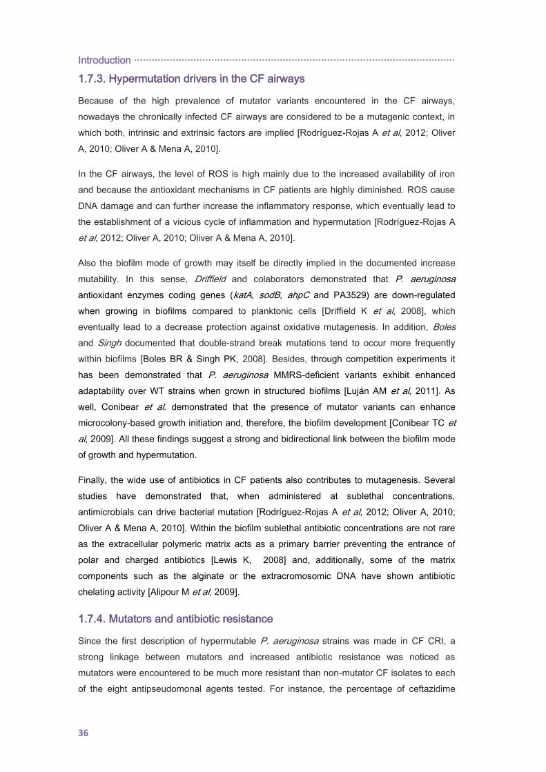
Introduction ············································································································
36
1.7.3. Hypermutation drivers in the CF airways
Because of the high prevalence of mutator variants encountered in the CF airways,
nowadays the chronically infected CF airways are considered to be a mutagenic context, in
which both, intrinsic and extrinsic factors are implied [Rodríguez-Rojas A et al, 2012; Oliver
A, 2010; Oliver A & Mena A, 2010].
In the CF airways, the level of ROS is high mainly due to the increased availability of iron
and because the antioxidant mechanisms in CF patients are highly diminished. ROS cause
DNA damage and can further increase the inflammatory response, which eventually lead to
the establishment of a vicious cycle of inflammation and hypermutation [Rodríguez-Rojas A
et al, 2012; Oliver A, 2010; Oliver A & Mena A, 2010].
Also the biofilm mode of growth may itself be directly implied in the documented increase
mutability. In this sense, Driffield and colaborators demonstrated that P. aeruginosa
antioxidant enzymes coding genes (katA, sodB, ahpC and PA3529) are down-regulated
when growing in biofilms compared to planktonic cells [Driffield K et al, 2008], which
eventually lead to a decrease protection against oxidative mutagenesis. In addition, Boles
and Singh documented that double-strand break mutations tend to occur more frequently
within biofilms [Boles BR & Singh PK, 2008]. Besides, through competition experiments it
has been demonstrated that P. aeruginosa MMRS-deficient variants exhibit enhanced
adaptability over WT strains when grown in structured biofilms [Luján AM et al, 2011]. As
well, Conibear et al. demonstrated that the presence of mutator variants can enhance
microcolony-based growth initiation and, therefore, the biofilm development [Conibear TC et
al, 2009]. All these findings suggest a strong and bidirectional link between the biofilm mode
of growth and hypermutation.
Finally, the wide use of antibiotics in CF patients also contributes to mutagenesis. Several
studies have demonstrated that, when administered at sublethal concentrations,
antimicrobials can drive bacterial mutation [Rodríguez-Rojas A et al, 2012; Oliver A, 2010;
Oliver A & Mena A, 2010]. Within the biofilm sublethal antibiotic concentrations are not rare
as the extracellular polymeric matrix acts as a primary barrier preventing the entrance of
polar and charged antibiotics [Lewis K, 2008] and, additionally, some of the matrix
components such as the alginate or the extracromosomic DNA have shown antibiotic
chelating activity [Alipour M et al, 2009].
1.7.4. Mutators and antibiotic resistance
Since the first description of hypermutable P. aeruginosa strains was made in CF CRI, a
strong linkage between mutators and increased antibiotic resistance was noticed as
mutators were encountered to be much more resistant than non-mutator CF isolates to each
of the eight antipseudomonal agents tested. For instance, the percentage of ceftazidime
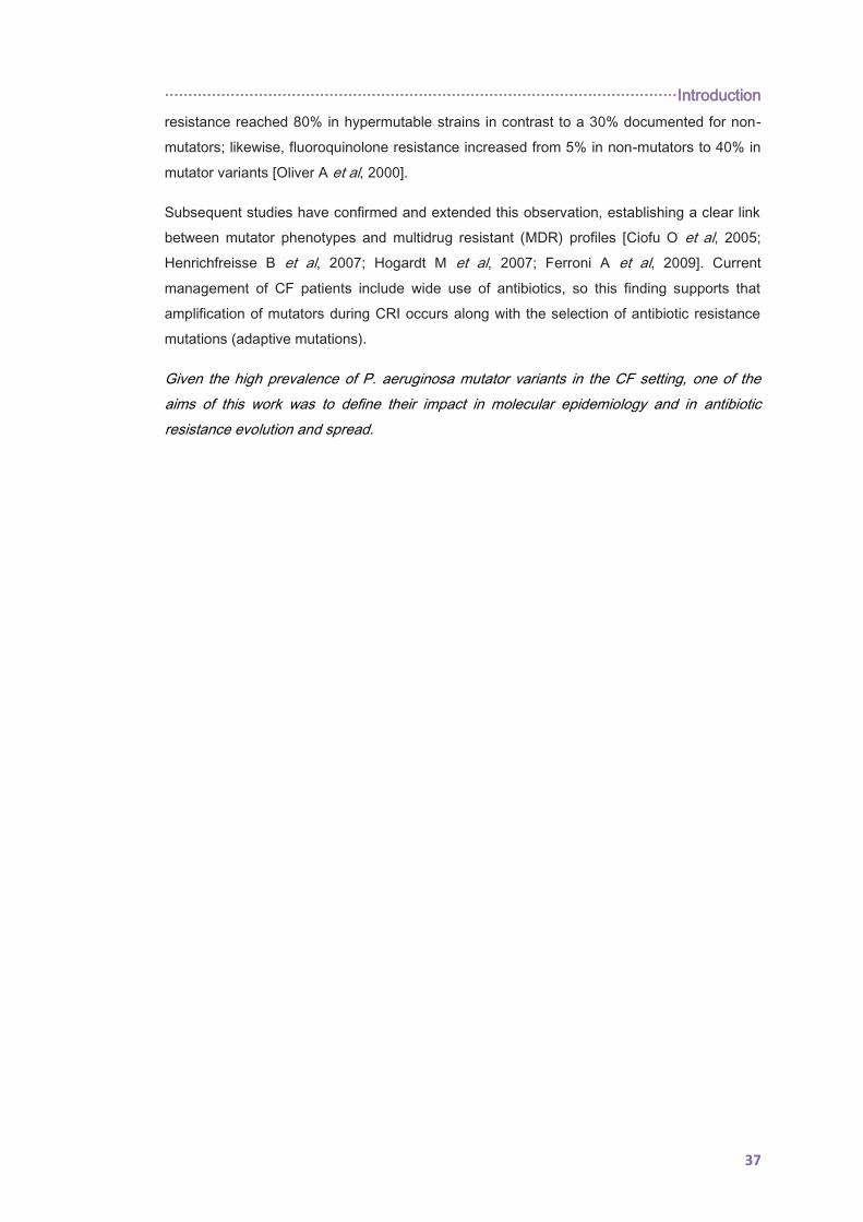
············································································································· Introduction
37
resistance reached 80% in hypermutable strains in contrast to a 30% documented for non-
mutators; likewise, fluoroquinolone resistance increased from 5% in non-mutators to 40% in
mutator variants [Oliver A et al, 2000].
Subsequent studies have confirmed and extended this observation, establishing a clear link
between mutator phenotypes and multidrug resistant (MDR) profiles [Ciofu O et al, 2005;
Henrichfreisse B et al, 2007; Hogardt M et al, 2007; Ferroni A et al, 2009]. Current
management of CF patients include wide use of antibiotics, so this finding supports that
amplification of mutators during CRI occurs along with the selection of antibiotic resistance
mutations (adaptive mutations).
Given the high prevalence of P. aeruginosa mutator variants in the CF setting, one of the
aims of this work was to define their impact in molecular epidemiology and in antibiotic
resistance evolution and spread.
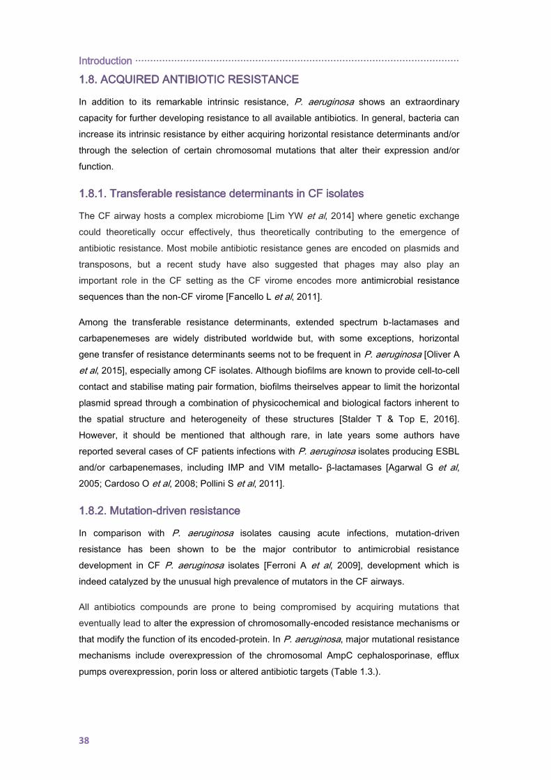
Introduction ············································································································
38
1.8. ACQUIRED ANTIBIOTIC RESISTANCE
In addition to its remarkable intrinsic resistance, P. aeruginosa shows an extraordinary
capacity for further developing resistance to all available antibiotics. In general, bacteria can
increase its intrinsic resistance by either acquiring horizontal resistance determinants and/or
through the selection of certain chromosomal mutations that alter their expression and/or
function.
1.8.1. Transferable resistance determinants in CF isolates
The CF airway hosts a complex microbiome [Lim YW et al, 2014] where genetic exchange
could theoretically occur effectively, thus theoretically contributing to the emergence of
antibiotic resistance. Most mobile antibiotic resistance genes are encoded on plasmids and
transposons, but a recent study have also suggested that phages may also play an
important role in the CF setting as the CF virome encodes more antimicrobial resistance
sequences than the non-CF virome [Fancello L et al, 2011].
Among the transferable resistance determinants, extended spectrum b-lactamases and
carbapenemeses are widely distributed worldwide but, with some exceptions, horizontal
gene transfer of resistance determinants seems not to be frequent in P. aeruginosa [Oliver A
et al, 2015], especially among CF isolates. Although biofilms are known to provide cell-to-cell
contact and stabilise mating pair formation, biofilms theirselves appear to limit the horizontal
plasmid spread through a combination of physicochemical and biological factors inherent to
the spatial structure and heterogeneity of these structures [Stalder T & Top E, 2016].
However, it should be mentioned that although rare, in late years some authors have
reported several cases of CF patients infections with P. aeruginosa isolates producing ESBL
and/or carbapenemases, including IMP and VIM metallo- β-lactamases [Agarwal G et al,
2005; Cardoso O et al, 2008; Pollini S et al, 2011].
1.8.2. Mutation-driven resistance
In comparison with P. aeruginosa isolates causing acute infections, mutation-driven
resistance has been shown to be the major contributor to antimicrobial resistance
development in CF P. aeruginosa isolates [Ferroni A et al, 2009], development which is
indeed catalyzed by the unusual high prevalence of mutators in the CF airways.
All antibiotics compounds are prone to being compromised by acquiring mutations that
eventually lead to alter the expression of chromosomally-encoded resistance mechanisms or
that modify the function of its encoded-protein. In P. aeruginosa, major mutational resistance
mechanisms include overexpression of the chromosomal AmpC cephalosporinase, efflux
pumps overexpression, porin loss or altered antibiotic targets (Table 1.3.).
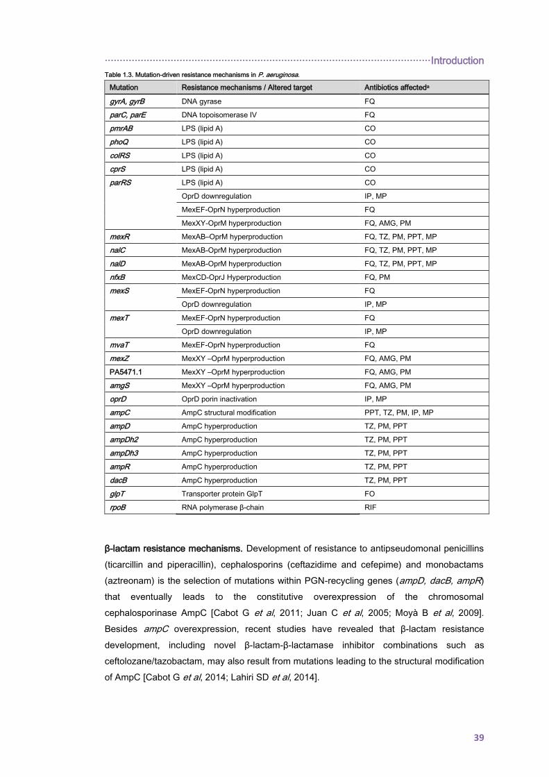
············································································································· Introduction
39
Table 1.3. Mutation-driven resistance mechanisms in P. aeruginosa.
Mutation Resistance mechanisms / Altered target Antibiotics affecteda
gyrA, gyrB DNA gyrase FQ
parC, parE DNA topoisomerase IV FQ
pmrAB LPS (lipid A) CO
phoQ LPS (lipid A) CO
colRS LPS (lipid A) CO
cprS LPS (lipid A) CO
parRS LPS (lipid A) CO
OprD downregulation IP, MP
MexEF-OprN hyperproduction FQ
MexXY-OprM hyperproduction FQ, AMG, PM
mexR MexAB–OprM hyperproduction FQ, TZ, PM, PPT, MP
nalC MexAB-OprM hyperproduction FQ, TZ, PM, PPT, MP
nalD MexAB-OprM hyperproduction FQ, TZ, PM, PPT, MP
nfxB MexCD-OprJ Hyperproduction FQ, PM
mexS MexEF-OprN hyperproduction FQ
OprD downregulation IP, MP
mexT MexEF-OprN hyperproduction FQ
OprD downregulation IP, MP
mvaT MexEF-OprN hyperproduction FQ
mexZ MexXY –OprM hyperproduction FQ, AMG, PM
PA5471.1 MexXY –OprM hyperproduction FQ, AMG, PM
amgS MexXY –OprM hyperproduction FQ, AMG, PM
oprD OprD porin inactivation IP, MP
ampC AmpC structural modification PPT, TZ, PM, IP, MP
ampD AmpC hyperproduction TZ, PM, PPT
ampDh2 AmpC hyperproduction TZ, PM, PPT
ampDh3 AmpC hyperproduction TZ, PM, PPT
ampR AmpC hyperproduction TZ, PM, PPT
dacB AmpC hyperproduction TZ, PM, PPT
glpT Transporter protein GlpT FO
rpoB RNA polymerase β-chain RIF
β-lactam resistance mechanisms. Development of resistance to antipseudomonal penicillins
(ticarcillin and piperacillin), cephalosporins (ceftazidime and cefepime) and monobactams
(aztreonam) is the selection of mutations within PGN-recycling genes (ampD, dacB, ampR)
that eventually leads to the constitutive overexpression of the chromosomal
cephalosporinase AmpC [Cabot G et al, 2011; Juan C et al, 2005; Moyà B et al, 2009].
Besides ampC overexpression, recent studies have revealed that β-lactam resistance
development, including novel β-lactam-β-lactamase inhibitor combinations such as
ceftolozane/tazobactam, may also result from mutations leading to the structural modification
of AmpC [Cabot G et al, 2014; Lahiri SD et al, 2014].
Introduction ············································································································
40
Beyond the chromosomal cephalosporinase AmpC, another contributing factor to β-lactam
resistance is MexAB-OprM overexpression. This efflux system displays the broadest
substrate profile (Table 1.1. and 1.3.) and its mutational overexpression determines reduced
susceptibility to all β-lactams with the single exception of imipenem. MexAB-OprM-
overproducing mutants can be readily generated in vitro in the presence of antibiotic by the
selection of any mutational event leading to the inactivation or impairment of the mexR, nalC
or nalD regulator genes. As well, these mutants have been shown to be very prevalent
among multiresistant non-CF strains, and, of note, rates of MexAB-OprM overproducers of
near 50% have been recorded in subpopulations of isolates exhibiting a reduced
susceptibility to ticarcillin (≥32 μg/ml) [Li XZ et al, 2015].
Likewise, the mutational overexpression of MexXY or MexCD-OprJ can also confer
resistance to cefepime. MexCD-OprJ overexpression, which is more frequent among P.
aeruginosa isolates recovered from CRI not only confers increased cefepime resistance but
have also been shown to determine hypersusceptibility to most β-lactams and
aminoglycosides [Mulet X et al, 2011].
Finally, screenings of transposon mutant libraries have shown that inactivation of galU, a
gene which code for an enzyme involved in the LPS core, increases ceftazidime and
meropenem minimum inhibitory concentrations (MICs) [Dötsch A et al, 2009; Álvarez-Ortega
C et al, 2010].
Carbapenem resistance mechanisms. Mutational inactivation of the porin OprD, together
with the inducible expression of AmpC, confers resistance to imipenem and reduced
susceptibility to meropenem [Livermore DM et al, 1992]. Indeed, the prevalence of imipenem
resistant isolates frequently exceeds 20%, and nearly all them are OprD deficient [Cabot G
et al, 2011; Riera E et al, 2011]. As well, MexAB-OprM mutational overexpression
determines reduced susceptibility to meropenem and its overexpression plus OprD
inactivation is one of the most relevant causes of clinical resistance to this carbapenem
[Riera E et al, 2011].
Finally, although less frequent, mutation-driven resistance to carbapenems can also result
from MexEF-OprN overexpression, as mutations within mexT, mexS and/or the ParRS two-
component system not only lead to MexEF-OprN overexpression but also to OprD
downregulation, which in turns determine a reduced susceptibility to carbapenems [Köhler T
et al, 1999; Li XZ et al, 2015].
Aminoglycoside resistance mechanisms. Resistance to this antibiotic class has been
clasically linked to the mutational overexpression of MexXY efflux pump, being its
overexpression very frequent among clinical isolates and mainly caused by mexZ, amgS, or
parRS mutations [Guénard S et al, 2014]. However, recent studies have revealed that the
aminoglycoside mutational resistome extends far beyond MexXY overexpression and
············································································································· Introduction
41
several novel resistance determinants have been described; moreover accumulation of
mutations within these genes can eventually lead to high-level antibiotic resistance [El’Garch
F et al, 2007; Schurek KN et al, 2008].
Fluoroquinolone resistance mechanisms. Fluoroquinolone resistance in P. aeruginosa
frequently results from gain-of-function mutations in topoisomerases, including DNA gyrases
(GyrA/GyrB) and type IV topoisomerases (ParC/ParE) [Bruchmann S et al, 2013].
Besides, overexpression of all 4 major efflux-pumps systems also contributes to
fluoroquinolone resistance (Table 1.1. and 1.3.). The overexpression of MexAB-OprM and
MexXY-OprM is globally more frequent among clinical strains but its contribution to clinical
fluoroquinolone resistance is likely more modest [Bruchmann S et al, 2013]. On the other
hand, mutational overexpression of MexEF-OprN or MexCD-OprJ efflux pump is associated
with high-level fluoroquinolone resistance, and although their prevalence is considered low
except in the CF-CRI setting, recent data show that it might be higher than expected
[Richardot C et al, 2015].
Polymyxin resistance mechanisms. The prevalence of polymyxin (polymyxin B and colistin)
resistance is still very low (<5%) among P. aeruginosa isolates. Resistance to polymyxins
most frequently results from the modification of the LPS caused by the addition of a 4-amino-
4-deoxy-l-arabinose moiety in the lipid A structure [Olaitan AO et al, 2014] and the
underlying mutations are frequently tracked to the PmrAB or PhoPQ two-component
regulators, which in turns lead to the activation of the arnBCADTEF operon [Barrow K &
Kwon DH, 2009]. More recent studies have also revealed that mutations within the two-
component regulator ParRS, in addition to conferring colistin resistance due to the activation
of the arnBCADTEF operon, lead to a MDR profile caused by the overprexpression of
MexXY, MexEF and the repression of OprD [Muller C et al, 2011]. Finally, two additional two-
component regulators, ColRS and CprRS, have also been shown to play a role in polymyxin
resistance [Gutu AD et al, 2013]. Moreover, recent in vitro evolution assays have revealed,
through WGS, the implication of additional mutations in high level colistin resistance,
facilitated by the emergence of mutator (mutS deficient) phenotypes [Döβelmann B et al,
2017]. Particularly noteworthy among them are those occurring in LptD (essentaial OMP
involved in LPS transport), LpxC (UDP-3-O-[hydroxymyristoyl]-N-acetylglucosamine
deacetylase involved in lipid A biosynthesis) or MigA (α-1,6-rhamnosyltransferase, involved
in the synthesis of the LPS core region [Döβelmann B et al, 2017].
P. aeruginosa possesses a complex and large genome, thus, and given the current gaps
and the crutial role of mutatrion-driven resistance mechanisms for acquiring antibiotic
resistance, one of the aims of this work was to decipher the P. aeruginosa mutational
resistome.
Introduction ············································································································
42
1.9. P. aeruginosa POPULATION STRUCTURE: CF EPIDEMIC CLONES
Bacterial population structures can range from panmintic or fully sexual, showing random
association between loci due to unrestricted recombination (such as Neisseria gonorrhoeae),
to clonal, characterized by non-random association of alleles and evolving mainly through
mutation (such as Salmonella enterica) [Smith JM et al, 1993].
Early studies suggested a panmintic population structure for P. aeruginosa [Denamur E et al,
1993; Picard B et al, 1994]. Kiewitz and Tümmler later described that P. aeruginosa shows a
net-like population structure with a high frequency of recombination between isolates
[Kiewitz C & Tümmler B, 2000]. Wide consensus was finally reached to conclude that P.
aeruginosa has a non-clonal epidemic population structure [Curran B et al, 2004; Kidd TJ et
al, 2012; Maâtallah M et al, 2011; Pirnay JP et al, 2009; Pirnay JP et al, 2002]. This means
that the population structure of P. aeruginosa, similarly to that described in N. meningitidis, is
composed of a limited number of widespread clones which are selected from a background
of a large number of rare and unrelated genotypes that are recombining at high frequency.
Population structure analysis have also revealed that P. aeruginosa contains a conserved
core and an accessory genome made up of extrachromosomal elements, such as plasmids
and blocks of DNA inserted into the chromosome at several loci [Klockgether J et al, 2011].
The accessory genome is believed to be acquired through horizontal gene transfer
(frequently phage-mediated) from different sources including other species.
Several experimental approaches have been used to define the population structure of P.
aeruginosa, ranging from single loci to whole genome, mapping or sequencing [Curran B et
al, 2004; Denamur E et al, 1993; Maâtallah M et al, 2013; Pirnay JP et al, 2009; Wiehlmann
L et al, 2007]. Strategies based in the combined analysis of up to eight different genomic
markers [O-serotype, total genome profile by fluorescent amplified-fragment length
polymorphism analysis, nucleotide sequence of the OMP genes (oprI, oprL, and oprD),
pyoverdine receptor gene profile (fpvA type and fpvB prevalence), prevalence of exoenzyme
genes exoS and exoU and prevalence of group I pilin glycosyltransferase gene tfpO] [Pirnay
JP et al, 2009] or a 58-binary genotypic markers microarray [Cramer N et al, 2012;
Wiehlmann L et al, 2007] have provided very useful and complete information on the core
and accessory genomes to define P. aeruginosa population structure. P. aeruginosa
widespread C or PA14 clones, O11/O12 MDR nosocomial clones, or the LES were likely the
most notorious successful epidemic lineages identified. Increasing access to WGS data is
providing even more detailed information on the population structure and dynamics of
epidemic and nonepidemic strains [Cramer N et al, 2011; Dettman JR et al, 2013; Jeukens J
et al, 2014; Yang L et al, 2011]. However, despite only providing information on the core
genome, the likely most popular standardized approach for the analysis of P. aeruginosa
populations is still the multilocus sequence typing (MLST) scheme developed by Curran et
············································································································· Introduction
43
al. in 2004, based in the sequencing of 7 locci evenly distributed in the core genome of P.
aeruginosa. They include acsA, aroE, guaA, mutL, nuoD, ppsA, and trpE, considered
housekeeping genes, and therefore not subjected to positive selection. However, mutation of
mutL, encoding a component of the DNA MMR system, is a frequent cause of the mutator
phenotypes, positively selected in CRI [García-Castillo M et al, 2012; Kidd TJ et al, 2012;
Mena A et al, 2008; Oliver A et al, 2002a].
The MLST database (http://pubmlst.org/paeruginosa), although biased by the deposition of
only a small fraction of the isolates, is a source of very valuable epidemiological information.
As shown in Figure 1.9., most of the registered STs (June 4th 2015) are represented by
single isolates but up to 18 of them are represented by more than 10 isolates from at least
three different countries, thus likely indicating that they are successful clones. Among others
they include the wide spread clone C (ST17) and PA14 (ST253) clones, the high-risk clones
associated with MDR or extensively drug resistant (XDR) nosocomial infections (such as
ST111, ST175, or ST235) or the CF epidemic clone ST146 (LES). Of note, many of these
frequent clones are the founder clones of one of the 297 clonal groups or complexes
detected by eburst analysis, each including from 2 to 116 STs [Oliver A et al, 2015].
As mentioned previously in section 1.5., during long time it was extensively accepted that
early P. aeruginosa acquisition occurs from environmental sources. However, successful
strains infecting a large proportion of CF patients are nowadays recognized and, in some
cases, strongly associated with MDR profiles.

Introduction ·····································································································································································································
44
Figure 1.9. Population snapshot of P. aeruginosa. The 2106 STs listed on the P. aeruginosa PubMLST database (http://pubmlst.org/paeruginosa, 2015/06/03) are displayed in a single eBURST diagram by setting the group
definition to zero of seven shared alleles. Each dot representes a ST, and lines connect single-locus variants. In each group of related STs the predictive primary founder is shown in pink, and subgroup founders are shown in
blue. STs detected in at least 3 different countries with more than 10 recorded isolates are indicated; note that the ST corresponds to the primary founder or subgroup founder of the CC.
············································································································· Introduction
45
1.9.1. The Liverpool Epidemic Strain: a new paradigm in the CF setting
The LES is likely the most prominent epidemic clone infecting and chronically colonizing CF
patients. It was originally described in the mid-1990s affecting a unique CF center in
Liverpool [Cheng K et al, 1996] but it was soon detected in other CF centers across England
[Scott FW & Pitt TL, 2004] and Scotland [Edenborough FP et al, 2004], and more recently, it
has also been detected in CF patients from Canada [Aaron SD et al, 2010].
LES isolates show some unique characteristics. For instance, it cannot only infect previously
uncolonized patients but also patients already colonized with unique P. aeruginosa strains
[McCallum SJ et al, 2001] as well as non-CF patients [McCallum SJ et al, 2002]. Moreover,
several studies have demonstrated that patients chronically infected with LES strains have a
worse prognosis when compared with patients chronically colonized with unique non-clonal
strains [Aaron SD et al, 2010; Al-Aloul M et al, 2004; Ashish A et al, 2012]; however,
fortunately, being infected with this clone was not associated with poorer post-transplant
outcomes [Srour N et al, 2015]. When compared with other CF strains, LES isolates are
more frequently resistant to antibiotics and further resistance is also more likely to be
developed over time [Ashish A et al, 2012; Salunkhe P et al, 2005; Tomás M et al, 2010].
In order to determine the underlying factors explaining the success of this epidemic clone
among CF patients, environmental surveys have been performed but, to date, environmental
LES isolates have only been detected in close temporospatial proximity to LES-colonized
patients and therefore its high transmissibility cannot be explained by long-term
environmental persistence [Panagea S et al, 2005]. Therefore, its successful transmission
and lung colonization might be due to intrinsic phenotypic and genotypic features.
An unusual phenotype characterized by the overproduction of QS-regulated exoproducts,
including pyocyanin and elastase A, is common among LES isolates and can persist within
CF patients for several years. As this virulence-related exoproducts have a number of toxic
effects directly relevant to CF, this unusual phenotype could explain the greater morbidity
and mortality associated LES isolates and may play an important role in the success of this
clone [Fothergill JL et al, 2007]. Moreover, it has been demonstrated that LESB58 produced
more biofilm but was less motile than PAO1 and PA14, properties that can favor its
persistence in the CF airway [Kukavica-Ibrulj I et al, 2008].
At the genomic level, this epidemic strain also exhibits some unique and relevant features.
LESB58 was the first LES isolate sequenced and an accessory genome encoding many
large genomic islands and prophages not previously found in other sequenced P. aeruginosa
strains was revealed [Winstanley C et al, 2009]. At first, the presence of this accessory
genomic material was thought to be essential for in vivo competitiveness [Winstanley C et al,
2009] but further comparative genomic studies with other representative LES isolates have
Introduction ············································································································
46
revealed a wide diversity in prophage and genomic islands among isolates [Carter ME et al,
2010; Jeukens J et al, 2014] even during very short episodes of exacerbations and antibiotic
therapy [Fothergill JL et al, 2010; Fothergill JL et al, 2011]. Recently, it has been also
suggested that LES phages may play an important role in host invasions and may confer a
large fitness advantage during mixed infections by mediating bacteria-bacteria competition
[Burns N et al, 2015]. Thus, this accessory genomic material seems to play an important role
but further research is needed.
Another interesting point is the common coexistence of distinct LES lineages, exhibiting
widely variable phenotypic and genotypic characteristics, within individuals [Williams D et al,
2015]. Nevertheless, this characteristic is not exclusive for this lineage and some authors
have already demonstrated the coexistence of distinct lineages in the respiratory tract for
other successful CF strains [Feliziani S et al, 2014, Marvig RL et al, 2013]. Thus, it seems
that the phenotypic and the genetic diversity observed among LES isolates play a key role in
the successful spread of this lineage throughout the CF population, diversity that is indeed
enhanced by the frequent presence of mutators in the CF lung [Oliver A et al, 2000].
1.9.2. Other successful CF strains
Epidemic strains have also been detected in Australia, the so-called Australian Epidemic
Strains AES-1 (also denominated Melbourne Epidemic Strain), AES-2, a cluster of related
strains [Anthony M et al, 2002; Armstrong D et al, 2003; Armstrong DS et al, 2002] and, the
more recently described, AES-3 [Bradbury R et al, 2008]. AES show increased antibiotic
resistance, increased virulence gene expression and higher morbidity and mortality during
CRI [Armstrong D et al, 2003; Griffiths AL et al, 2012; Hare NJ et al, 2012; Manos J et al,
2009; Naughton S et al, 2011; Tingpej P et al, 2010]. As occurs for LES, AES has not been
recovered from any source other than the respiratory secretions of CF patients [Bradbury RS
et al, 2009; Cramer N et al, 2012].
Besides these well-recognized CF epidemic strains, other P. aeruginosa MDR transmissible
strains have been reported in European countries or Canada [Fluge G et al, 2001; Jelsbak L
et al, 2007; Jones AM et al, 2001; Logan C et al, 2012; Luna RA et al, 2013; Parkins MD et
al, 2014; Pedersen SS et al, 1986; Scott FW & Pitt TL, 2004; van Mansfeld R et al, 2009].
Overall, these strains show increased antibiotic resistance, long-term persistence within
individuals, and are frequently associated with higher morbidity and mortality. Worldwide
distribution of these strains is shown in Figure 1.10.

············································································································· Introduction
47
Figure 1.10. Worldwide distribution of P. aeruginosa CF epidemic/transmissible strains.
P. aeruginosa CF clonal epidemiology has remained unexplored among CF patients from the
Balearic Islands and Spain. Thus, given the relevance of epidemic clones for CF patients
outcome and management, one of the aims of this work was to define the P. aeruginosa
population structure and to explore the presence of highly transmissible P. aeruginosa
clones in our setting.

48

49
2. HYPOTHESIS AND OBJECTIVES
El alma y el cuerpo

50

······················································································· Hypothesis and objectives
51
P. aeruginosa CRI is the main cause of morbidity and mortality in CF individuals. One of the
hallmarks of these infections, led by the opportunistic pathogen P. aeruginosa, is their long-
term (lifelong) persistence despite of the host immune response and intensive antimicrobial
therapies. Naturally equipped with a set of chromosomal genes that confer resistance to
some antibiotic compounds (intrinsic resistome), P. aeruginosa can further develop
resistance to virtually all available antimicrobials. Antimicrobial resistance in CF is a
multifactorial problem which not only includes bacterial physiological changes, represented
by the transition from the planktonic to the biofilm mode of growth, but also the acquisition of
multiple chromosomal antibiotic resistance (mutational resistome) and adaptive mutations
that eventually lead to a diversified infecting P. aeruginosa population. As well, in late years,
there is increasing evidence suggesting that adaptation to the CF respiratory tract and
antimicrobial resistance development may escape from the scale of the individual patients
(epidemic strains).
Besides, recent advances in sequencing technologies have made it possible to obtain the
whole genome of bacterial pathogens shaping up a new dimension to explore CF P.
aeruginosa clonal epidemiology and antimicrobial resistance evolution. Therefore, the
objectives of this thesis were:
1. To define the population structure of P. aeruginosa isolates infecting CF patients from the
Balearic Islands and Spain.
2. To perform a longitudinal and cross-sectional analysis of the antibiotic susceptibility
profiles and resistance mechanisms of CF P. aeruginosa.
3. To determine the role of mutators onto CF P. aeruginosa clonal epidemiology and onto the
evolution and spread of antibiotic resistance.
4. To characterize by WGS approaches the phylogeny and evolution of widespread P.
aeruginosa CC274.
5. To decipher the P. aeruginosa CC274 resistome evolution.
6. To assess the evolutionary dynamics and mechanisms of aminoglycoside resistance
development in vitro and in vivo, given their key role in the management of CF patients.

52

53
3. MATERIALS AND METHODS
Palabras incomprendidas

54
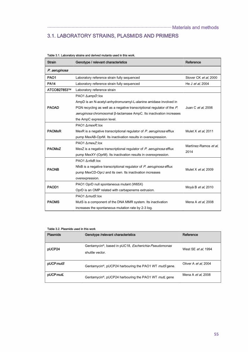
···························································································· Materials and methods
55
3.1. LABORATORY STRAINS, PLASMIDS AND PRIMERS
Table 3.1. Laboratory strains and derived mutants used in this work.
Strain Genotype / relevant characteristics Reference
P. aeruginosa
PAO1 Laboratory reference strain fully sequenced Stover CK et al, 2000
PA14 Laboratory reference strain fully sequenced He J et al, 2004
ATCC®27853TM Laboratory reference strain
PAOAD
PAO1 ΔampD::lox
AmpD is an N-acetyl-anhydromuramyl-L-alanine amidase involved in
PGN recycling as well as a negative transcriptional regulator of the P.
aeruginosa chromosomal β-lactamase AmpC. Its inactivation increases
the AmpC expression level.
Juan C et al, 2006
PAOMxR
PAO1 ΔmexR::lox
MexR is a negative transcriptional regulator of P. aeruginosa efflux
pump MexAB-OprM. Its inactivation results in overexpression.
Mulet X et al, 2011
PAOMxZ
PAO1 ΔmexZ::lox
MexZ is a negative transcriptional regulator of P. aeruginosa efflux
pump MexXY-(OprM). Its inactivation results in overexpression.
Martínez-Ramos et al,
2014
PAONB
PAO1 ΔnfxB::lox
NfxB is a negative transcriptional regulator of P. aeruginosa efflux
pump MexCD-OprJ and its own. Its inactivation increases
overexpression.
Mulet X et al, 2009
PAOD1 PAO1 OprD null spontaneous mutant (W65X)
OprD is an OMP related with carbapenems extrusion. Moyà B et al, 2010
PAOMS
PAO1 ΔmutS::lox
MutS is a component of the DNA MMR system. Its inactivation
increases the spontaneous mutation rate by 2-3 log.
Mena A et al, 2008
Table 3.2. Plasmids used in this work
Plasmids Genotype /relevant characteristics Reference
pUCP24 GentamycinR, based in pUC18, Escherichia-Pseudomonas
shuttle vector. West SE et al, 1994
pUCPmutS GentamycinR, pUCP24 harbouring the PAO1 WT mutS gene.
Oliver A et al, 2004
pUCPmutL GentamycinR, pUCP24 harbouring the PAO1 WT mutL gene
Mena A et al, 2008
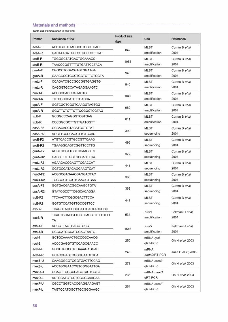
Materials and methods ···························································································
56
Table 3.3. Primers used in this work
Primer Sequence 5’3’ Product size
(bp) Use Reference
acsA-F ACCTGGTGTACGCCTCGCTGAC 842
MLST
amplification
Curran B et al,
2004 acsA-R GACATAGATGCCCTGCCCCTTGAT
aroE-F TGGGGCTATGACTGGAAACC 1053
MLST
amplification
Curran B et al,
2004 aroE-R TAACCCGGTTTTGTGATTCCTACA
guaA-F CGGCCTCGACGTGTGGATGA 940
MLST
amplification
Curran B et al,
2004 guaA-R GAACGCCTGGCTGGTCTTGTGGTA
mutL-F CCAGATCGCCGCCGGTGAGGTG 940
MLST
amplification
Curran B et al,
2004 mutL-R CAGGGTGCCATAGAGGAAGTC
nuoD-F ACCGCCACCCGTACTG 1042
MLST
amplification
Curran B et al,
2004 nuoD-R TCTCGCCCATCTTGACCA
ppsA-F GGTCGCTCGGTCAAGGTAGTGG 989
MLST
amplification
Curran B et al,
2004 ppsA-R GGGTTCTCTTCTTCCGGCTCGTAG
trpE-F GCGGCCCAGGGTCGTGAG 811
MLST
amplification
Curran B et al,
2004 trpE-R CCCGGCGCTTGTTGATGGTT
acsA-F2 GCCACACCTACATCGTCTAT 390
MLST
sequencing
Curran B et al,
2004 acsA-R2 AGGTTGCCGAGGTTGTCCAC
aroE-F2 ATGTCACCGTGCCGTTCAAG 495
MLST
sequencing
Curran B et al,
2004 aroE-R2 TGAAGGCAGTCGGTTCCTTG
guaA-F2 AGGTCGGTTCCTCCAAGGTC 372
MLST
sequencing
Curran B et al,
2004 guaA-R2 GACGTTGTGGTGCGACTTGA
mutL-F2 AGAAGACCGAGTTCGACCAT 441
MLST
sequencing
Curran B et al,
2004 mutL-R2 GGTGCCATAGAGGAAGTCAT
nuoD-F2 ACGGCGAGAACGAGGACTAC 366
MLST
sequencing
Curran B et al,
2004 nuoD-R2 TGGCGGTCGGTGAAGGTGAA
ppsA-F2 GGTGACGACGGCAAGCTGTA 369
MLST
sequencing
Curran B et al,
2004 ppsA-R2 GTATCGCCTTCGGCACAGGA
trpE-F2 TTCAACTTCGGCGACTTCCA 441
MLST
sequencing
Curran B et al,
2004 trpE-R2 GGTGTCCATGTTGCCGTTCC
exoS-F TCAGGTACCCGGCATTCACTACGCGG
534 exoS
amplification
Feltman H et al,
2001 exoS-R TCACTGCAGGTTCGTGACGTCTTTCTTT
TA
exoU-F AGCGTTAGTGACGTGCG 1546
exoU
amplification
Feltman H et al,
2001 exoU-R GCGCATGGCATCGAGTAATG
rpsl-1 GCTGCAAAACTGCCCGCAACG 250
mRNA rpsL
qRT-PCR Oh H et al, 2003
rpsl-2 ACCCGAGGTGTCCAGCGAACC
acrna-F GGGCTGGCCTCGAAAGAGGAC 246
mRNA
ampCqRT-PCR Juan C et al, 2006
acrna-R GCACCGAGTCGGGGAACTGCA
mexB-U CAAGGGCGTCGGTGACTTCCAG 273
mRNA mexB
qRT-PCR Oh H et al, 2003
mexB-L ACCTGGGAACCGTCGGGATTGA
mexD-U GGAGTTCGGCCAGGTAGTGCTG 236
mRNA mexD
qRT-PCR Oh H et al, 2003
mexD-L ACTGCATGTCCTCGGGGAAGAA
mexF-U CGCCTGGTCACCGAGGAAGAGT 254
mRNA mexF
qRT-PCR Oh H et al, 2003
mexF-L TAGTCCATGGCTTGCGGGAAGC
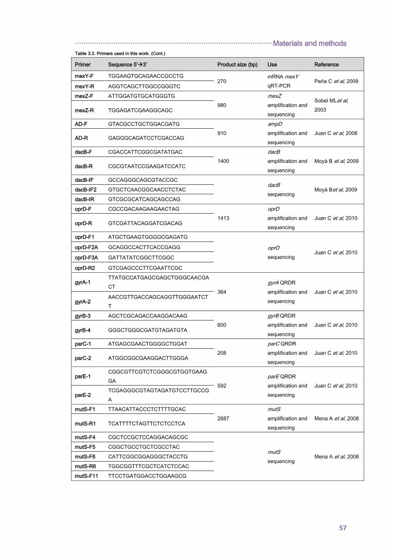
···························································································· Materials and methods
57
Table 3.3. Primers used in this work. (Cont.)
Primer Sequence 5’3’ Product size (bp) Use Reference
mexY-F TGGAAGTGCAGAACCGCCTG 270
mRNA mexY
qRT-PCR Peña C et al, 2009
mexY-R AGGTCAGCTTGGCCGGGTC
mexZ-F ATTGGATGTGCATGGGTG
980
mexZ
amplification and
sequencing
Sobel MLet al,
2003 mexZ-R TGGAGATCGAAGGCAGC
AD-F GTACGCCTGCTGGACGATG
910
ampD
amplification and
sequencing
Juan C et al, 2006 AD-R GAGGGCAGATCCTCGACCAG
dacB-F CGACCATTCGGCGATATGAC
1400
dacB
amplification and
sequencing
Moyà B et al, 2009 dacB-R CGCGTAATCCGAAGATCCATC
dacB-IF GCCAGGGCAGCGTACCGC
dacB
sequencing Moyà Bet al, 2009 dacB-IF2 GTGCTCAACGGCAACCTCTAC
dacB-IR GTCGCGCATCAGCAGCCAG
oprD-F CGCCGACAAGAAGAACTAG
1413
oprD
amplification and
sequencing
Juan C et al, 2010 oprD-R GTCGATTACAGGATCGACAG
oprD-F1 ATGCTGAAGTGGGGCGAGATG
oprD
sequencing Juan C et al, 2010
oprD-F2A GCAGGCCACTTCACCGAGG
oprD-F3A GATTATATCGGCTTCGGC
oprD-R2 GTCGAGCCCTTCGAATTCGC
gyrA-1 TTATGCCATGAGCGAGCTGGGCAACGA
CT 364
gyrA QRDR
amplification and
sequencing
Juan C et al, 2010
gyrA-2 AACCGTTGACCAGCAGGTTGGGAATCT
T
gyrB-3 AGCTCGCAGACCAAGGACAAG
600
gyrB QRDR
amplification and
sequencing
Juan C et al, 2010 gyrB-4 GGGCTGGGCGATGTAGATGTA
parC-1 ATGAGCGAACTGGGGCTGGAT
208
parC QRDR
amplification and
sequencing
Juan C et al, 2010 parC-2 ATGGCGGCGAAGGACTTGGGA
parE-1 CGGCGTTCGTCTCGGGCGTGGTGAAG
GA 592
parE QRDR
amplification and
sequencing
Juan C et al, 2010
parE-2 TCGAGGGCGTAGTAGATGTCCTTGCCG
A
mutS-F1 TTAACATTACCCTCTTTTGCAC
2687
mutS
amplification and
sequencing
Mena A et al, 2008 mutS-R1 TCATTTTCTAGTTCTCTCCTCA
mutS-F4 CGCTCCGCTCCAGGACAGCGC
mutS
sequencing Mena A et al, 2008
mutS-F5 CGGCTGCCTGCTCGCCTAC
mutS-F6 CATTCGGCGGAGGGCTACCTG
mutS-R6 TGGCGGTTTCGCTCATCTCCAC
mutS-F11 TTCCTGATGGACCTGGAAGCG

Materials and methods ···························································································
58
Table 3.3. Primers used in this work. (Cont.)
Primer Sequence 5’3’ Product size (bp) Use Reference
mutL-F CGATGATCGCCCAGCGCT
2299
mutL
amplification and
sequencing
Mena A et al, 2008 mutL-R TCCGCCGGGTCGCGGATA
mutL-F2 TAGCGCGCCTGACCATGA
mutL
sequencing Mena A et al, 2008
mutL-F3 GCGCATGGTGCGCGACAA
mutL-F4 GCCTCCGGCGGCTCCTCCG
mutL-R2 GCAGGTCGGCGATGACAT
···························································································· Materials and methods
59
3.2. Pseudomonas aeruginosa CYSTIC FIBROSIS ISOLATES
3.2.1. The Balearic Islands P. aeruginosa collection.
Since 2003, all P. aeruginosa isolates recovered from routine respiratory cultures from CF
patients attending the Son Espases University Hospital in Palma de Mallorca (former Son
Dureta), reference hospital of the Balearic Islands, have been regularly stored frozen at -
80°C. Culture, isolation and identification of P. aeruginosa from respiratory samples have
always been carried out following the current established microbiological diagnostic
procedures and expert recommendations. In this work, different subsets from this huge
collection were used as detailed below.
In 2010, with the aim of determine the long-term clonal epidemiology and antibiotic
resistance evolution of P. aeruginosa CRI in CF patients from the Balearic Islands, 10
sequential isolates from each of 10 chronically colonized CF patients were studied. The 10
included patients were selected based on the fulfilment of the following criteria: (i) wider
follow-up period, (ii) higher temporal distribution of isolates and (iii) inclusion of the first P.
aeruginosa isolate within the 8-year studied period (2003-2010). Likewise, each of the
sequential isolates included per patient were separated by at least a 6-month interval.
Occasioned by the results obtained when long-term clonal epidemiology was explored, in
2013 we decided to extend the molecular epidemiology studies to all CF patients, including
both children and adults, which had been attended at Son Espases CF Units since 2003. For
this purpose, last available P. aeruginosa isolate of each CF patient was included. From
2003 to 2013, more than 50 CF patients had been attended at Son Espases University
Hospital adult and paediatric CF Units and about the 80% had had a positive sputum culture
for P. aeruginosa at some time point; thus, a total of 40 isolates were studied. Infection-
colonization patterns and basic demographic data were recorded.
3.2.2. The Spanish P. aeruginosa collection.
From 2013 to 2014 the first Spanish multicentre study on the microbiology of CF was
conducted. The study involved 24 CF Units, 12 paediatric and 12 adult, from 17 different
Spanish hospitals. In Spain no national CF patient registry exists and, therefore, the precise
number of people suffering from CF is still unknown. Nevertheless, participating hospitals are
the reference ones within their geographical areas, attending the majority of the Spanish CF
population, thus, a representative patient population from across Spain was included (Figure
3.1.) [de Dios Caballero et al, 2016].
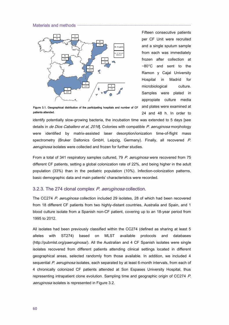
Materials and methods ···························································································
60
Fifteen consecutive patients
per CF Unit were recruited
and a single sputum sample
from each was immediately
frozen after collection at
−80°C and sent to the
Ramon y Cajal University
Hospital in Madrid for
microbiological culture.
Samples were plated in
appropiate culture media
and plates were examined at
24 and 48 h. In order to
identify potentially slow-growing bacteria, the incubation time was extended to 5 days [see
details in de Dios Caballero et al, 2016]. Colonies with compatible P. aeruginosa morphology
were identified by matrix-assisted laser desorption/ionization time-of-flight mass
spectrometry (Bruker Daltonics GmbH, Leipzig, Germany). Finally, all recovered P.
aeruginosa isolates were collected and frozen for further studies.
From a total of 341 respiratory samples cultured, 79 P. aeruginosa were recovered from 75
different CF patients, setting a global colonization rate of 22%, and being higher in the adult
population (33%) than in the pediatric population (10%). Infection-colonization patterns,
basic demographic data and main patients’ characteristics were recorded.
3.2.3. The 274 clonal complex P. aeruginosa collection.
The CC274 P. aeruginosa collection included 29 isolates, 28 of which had been recovered
from 18 different CF patients from two highly-distant countries, Australia and Spain, and 1
blood culture isolate from a Spanish non-CF patient, covering up to an 18-year period from
1995 to 2012.
All isolates had been previously classified within the CC274 (defined as sharing at least 5
alleles with ST274) based on MLST available protocols and databases
(http://pubmlst.org/paeruginosa/). All the Australian and 4 CF Spanish isolates were single
isolates recovered from different patients attending clinical settings located in different
geographical areas, selected randomly from those available. In addition, we included 4
sequential P. aeruginosa isolates, each separated by at least 6-month intervals, from each of
4 chronically colonized CF patients attended at Son Espases University Hospital, thus
representing intrapatient clone evolution. Sampling time and geographic origin of CC274 P.
aeruginosa isolates is represented in Figure 3.2.
Figure 3.1. Geographical distribution of the participating hospitals and number of CF
patients attended.
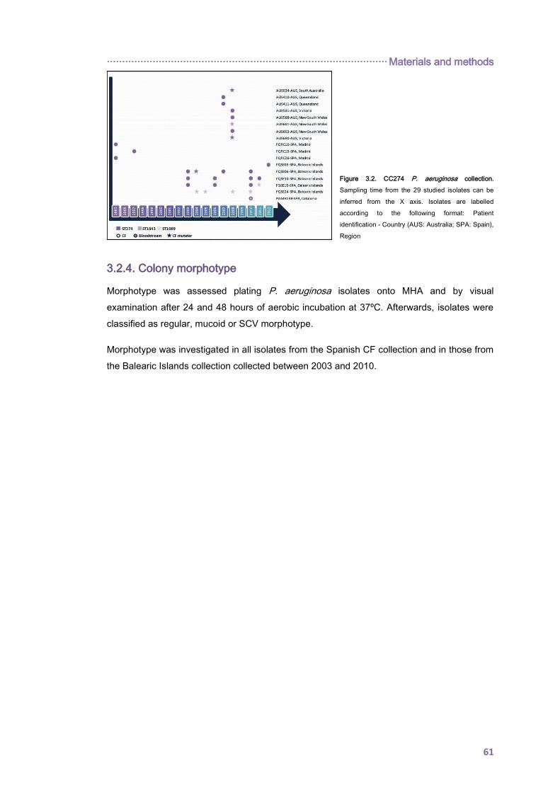
···························································································· Materials and methods
61
Figure 3.2. CC274 P. aeruginosa collection.
Sampling time from the 29 studied isolates can be
inferred from the X axis. Isolates are labelled
according to the following format: Patient
identification - Country (AUS: Australia; SPA: Spain),
Region
3.2.4. Colony morphotype
Morphotype was assessed plating P. aeruginosa isolates onto MHA and by visual
examination after 24 and 48 hours of aerobic incubation at 37ºC. Afterwards, isolates were
classified as regular, mucoid or SCV morphotype.
Morphotype was investigated in all isolates from the Spanish CF collection and in those from
the Balearic Islands collection collected between 2003 and 2010.
Materials and methods ···························································································
62
3.3. PAO1 P. aeruginosa IN VITRO EVOLUTION EXPERIMENT UNDER
AMINOGLYCOSIDE PRESSURE
To determine and in-depth study the dynamics of P. aeruginosa resistance development to
aminoglycosides, 10-ml Mueller-Hinton broth (MHB) tubes (Annex 1) containing 0.25, 0.5, 1,
2, 4, 8, 16, 32, 64, 128, 256, 512 and 1024 mg/l of tobramycin were inoculated with
approximately 106 CFU/ml of exponentially growing P. aeruginosa PAO1 reference strain
and incubated for 24 h at 37°C and 180 r.p.m. After incubation, the tubes from the highest
antibiotic concentration showing growth were reinoculated, at a 1:1,000 dilution, in fresh 10-
ml MHB tubes containing tobramycin concentrations up to 1024 mg/l and incubated again for
24 h at 37ºC and 180 r.p.m. This step was repeated during 14 consecutive days in order to
get PAO1-derived tobramycin high-resistant mutants. This procedure was performed in
quintuplicate.
At days 1, 7 and 14, two colonies from the highest antibiotic concentration tubes showing
growth were purified in antibiotic-free MHA plates (Annex 1) and frozen at -80ºC for further
characterization.
···························································································· Materials and methods
63
3.4. MOLECULAR EPIDEMIOLOGY STUDIES
3.4.1. Pulsed-field gel electrophoresis
Pulsed field gel electrophoresis (PFGE) is a highly discriminatory and reproducible typing
method used to determine the genetic relationship between microbial isolates from the same
species level which is based on the analysis of the chromosomic DNA. Basically, this
genotyping method consists in fragmenting the bacterial chromosomic DNA by using
appropriate restriction endonucleases of low cut frequency and then separate the resulted
DNA fragments by PFGE in order to obtain a unique macrorestriction pattern for each
microbial isolate and, eventually, compare all these unique molecular fingerprints to
determine their genetic relationship. In this work, previously defined protocols [Kaufmann
ME, 1998] with slight modifications were used, as set forth in detail below.
P. aeruginosa isolates were grown in suspension on 5 ml of Brain Heart Infusion (BHI) broth
(Annex 1) under aerobic conditions for 16-20 hours at 37ºC and shaking at 180 r.p.m.
Afterwards, a volume of 250 µl (500 µl for mucoid phenotype isolates) was centrifuged at
13000 r.p.m. for 5 minutes and bacterial pellets were washed twice with PIV solution (Annex
1). Then, bacterial pellets were resuspended in 200 µl of this PIV solution, mixed with an
equal volume of 1.6% low-melting temperature agarose (Annex 1) at 42ºC and, finally, this
molten mixture was used to prepare plugs in adequate molds (Bio-Rad). Once the agarose
was set, the plugs were collected and incubated within 1 ml of EC-Lysis solution (Annex 1) at
37ºC for at least 5 hours and, then, in 1 ml of ESP solution (Annex 1) at 50ºC for 16-20 hours
in order to release DNA from bacteria embebbed in the agarose plugs. After overnight
incubation, ESP solution was removed and plugs were washed in quintuplicate with TE
buffer (Annex 1), rinsed with 1 ml of sterile distilled water at 37ºC for at least 10 minutes,
transferred to microfuge tubes containing 20 units of SpeI restriction enzyme (New England
BioLabs) and incubated in appropriate conditions (20 hours, 37ºC). Then, each plug was
soaked in 1 ml of TE buffer at 37ºC for 1 hour. In the meantime, a 1% megabase agarose
gel (Annex 1) was prepared and, once soaked, plugs were placed and sealed in individual
wells of the gel. Right after, fragments were separated in a CHEF- DR III contour-clamped
homogeneous-electric-field apparatus (Bio-Rad, La Jolla, CA) in standard 0.5X TBE running
buffer (Annex 1) chilled at 14ºC for 26 hours at 6V/cm with pulse times of 5 to 40 s and using
a 120º included angle. After electrophoresis, the gel was stained in 0.5X TBE buffer with
either a 1 µg/ml solution of ethidium bromide or with an appropriate solution of RedSafe
20.000X and photographed under UV light.
This genotyping method was used to infer clonal relatedness among isolates within the two
studied subsets of the Balearic Islands P. aeruginosa collection (longitudinal and cross
sectional), as well as for isolates from the Spanish and the CC274 collections. For this
purpose, the criteria established by Tenover et al. [Tenover FC et al, 1995] were applied with

Materials and methods ···························································································
64
the single exception of inferring the genetic relationship within the CC274 P. aeruginosa
collection in which the Unweighted Pair Group Method with Arithmetic mean (UPGMA)
clustering method was used [CLIQS 1D Pro, Totallab].
3.4.2. Multilocus sequence typing
Multilocus Sequence Typing (MLST) is a molecular genotyping tool that not only results into
highly discriminatory but also electronically portable data between laboratories worldwide.
This unambiguous typing method is based in the use and comparison of the nucleotidic
sequences of internal fragments of approximately 450-500 bp of seven established house-
keeping genes. For each house-keeping gene, the different sequences present within a
bacterial species are assigned as distinct alleles and, for each isolate, the alleles at each of
the seven loci define the allelic profile or ST.
In this work the MLST scheme for P. aeruginosa proposed by Curran B et al., which is based
in the house-keeping genes acsA (acetil coenzyme A synthetase), aroE (shikimate
dehydrogenase), guaA (GMP synthase), mutL (DNA MMR protein), nuoD (NADH
dehydrogenase I chain C, D), ppsA (phosphoenolpyruvate synthase) and trpE (anthralite
synthetase component I), was applied [Curran B et al, 2004;
http://pubmlst.org/paeruginosa/]. Briefly, workflow was as follows.
Genomic DNA was obtained by using a commercially available extraction kit (DNeasy Blood
& Tissue kit, Qiagen or High Pure PCR template preparation kit, Roche Diagnostics) and
polymerase chain reaction (PCR) amplification of the seven house-keeping genes was
performed (primers and PCR Master Mix details in Table 3.3 in section 3.1. and Annex 1,
respectively) under the next reaction conditions: initial denaturation at 94ºC for 12 min; 35
cycles of denaturation at 94ºC for 1 min, primer annealing at 59ºC (60ºC for mutL
amplification) for 1 min, extension at 72ºC for 1 min; followed by a final extension step of
72ºC for 10 min. Hereafter, PCR amplification products were purified and sequenced with
the BigDye Terminator kit (PE Applied Biosystems) on an ABI Prism 3100 DNA sequencer
(PE Applied Biosystems, Foster City, CA). The P. aeruginosa MLST database
(https://pubmlst.org/paeruginosa/) was used to assign an allele to each resulting sequence
and to assign a ST.
This technique was performed within the two studied subsets of the Balearic Islands
collection (see above), as well for the isolates from the Spanish CF P. aeruginosa collection.
Phylogenetic relationship was assessed by constructing a Minimum Spanning Tree (MST)
using the goeBURST algorithm, available at www.phyloviz.net ST were considered to belong
to a same CC when sharing at least five of the seven sequenced loci.
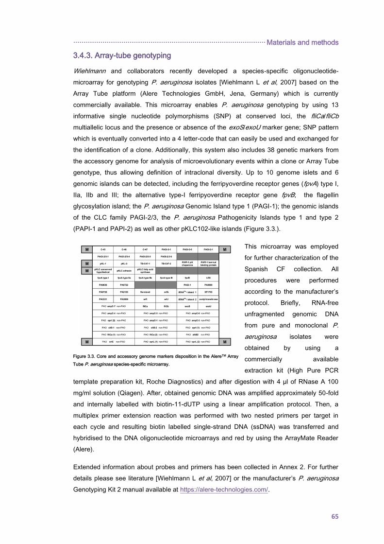
···························································································· Materials and methods
65
3.4.3. Array-tube genotyping
Wiehlmann and collaborators recently developed a species-specific oligonucleotide-
microarray for genotyping P. aeruginosa isolates [Wiehlmann L et al, 2007] based on the
Array Tube platform (Alere Technologies GmbH, Jena, Germany) which is currently
commercially available. This microarray enables P. aeruginosa genotyping by using 13
informative single nucleotide polymorphisms (SNP) at conserved loci, the fliCa/fliCb
multiallelic locus and the presence or absence of the exoS/exoU marker gene; SNP pattern
which is eventually converted into a 4 letter-code that can easily be used and exchanged for
the identification of a clone. Additionally, this system also includes 38 genetic markers from
the accessory genome for analysis of microevolutionary events within a clone or Array Tube
genotype, thus allowing definition of intraclonal diversity. Up to 10 genome islets and 6
genomic islands can be detected, including the ferripyoverdine receptor genes (fpvA) type I,
IIa, IIb and III; the alternative type-I ferripyoverdine receptor gene fpvB; the flagellin
glycosylation island; the P. aeruginosa Genomic Island type 1 (PAGI-1); the genomic islands
of the CLC family PAGI-2/3, the P. aeruginosa Pathogenicity Islands type 1 and type 2
(PAPI-1 and PAPI-2) as well as other pKLC102-like islands (Figure 3.3.).
This microarray was employed
for further characterization of the
Spanish CF collection. All
procedures were performed
according to the manufacturer’s
protocol. Briefly, RNA-free
unfragmented genomic DNA
from pure and monoclonal P.
aeruginosa isolates were
obtained by using a
commercially available
extraction kit (High Pure PCR
template preparation kit, Roche Diagnostics) and after digestion with 4 µl of RNase A 100
mg/ml solution (Qiagen). After, obtained genomic DNA was amplified approximately 50-fold
and internally labelled with biotin-11-dUTP using a linear amplification protocol. Then, a
multiplex primer extension reaction was performed with two nested primers per target in
each cycle and resulting biotin labelled single-strand DNA (ssDNA) was transferred and
hybridised to the DNA oligonucleotide microarrays and red by using the ArrayMate Reader
(Alere).
Extended information about probes and primers has been collected in Annex 2. For further
details please see literature [Wiehlmann L et al, 2007] or the manufacturer’s P. aeruginosa
Genotyping Kit 2 manual available at https://alere-technologies.com/.
Figure 3.3. Core and accessory genome markers disposition in the AlereTM Array
Tube P. aeruginosa species-specific microarray.

Materials and methods ···························································································
66
The Type 3 Secretion System (T3SS). To further characterize the type 3 Secretion System
(T3SS) protein effectors (ExoS, ExoU) independent specific PCR assays were performed.
Protocols previously described by Feltman H and collaborators were used with slight
modifications [Feltman H et al, 2001]. Primers and PCR Master Mix details are collected in
Table 3.3. and Annex 1, respectively. Reaction conditions were as follow: initial denaturation
at 94°C for 12 min, followed by 35 cycles of 94°C for 30 sec, 58°C for 30 sec, and 72°C for
30 sec and a final extension step of 10 min at 72°C. Amplified DNA products were resolved
by electrophoresis on agarose 1% w/v gels.
···························································································· Materials and methods
67
3.5. ANTIMICROBIAL SUSCEPTIBILITY PROFILES AND RESISTANCE
MECHANISMS
3.5.1. Antimicrobial susceptibility testing
3.5.1.1. P. aeruginosa clinical isolates
One of the most important tasks in a Clinical Microbiology Laboratory is to determine the
antimicrobial susceptibility profiles of significant bacterial isolates. Currently, there is not a
universal standardized method that perfectly reproduces the conditions in which infecting
bacteria are grown in vivo and different technical approaches can be used. This point is
particularly striking in the CF setting, especially at chronic stages in which bacteria change
their mode of growth from planktonic to biofilm and in which there is, indeed, a high
prevalence of mucoid and hypermutable isolates and, therefore, mutant resistant
subpopulations frequently rise.
In this work different susceptibility testing methods were used. Regardless of the technical
approach used, it should be highlighted that the following CF expert recommendations were
in all cases taken into account: (i) the incubation time was extended up to 24 hours (36-48
hours for slow growing variants) and (ii) a 0.5 or 1 McFarland standard suspension was used
for inoculum standardization of non-mucoid or mucoid isolates, respectively.
MICs for all isolates belonging to both, the Balearic Islands and the CC274 P. aeruginosa
collection, were determined by using commercially available strips containing a gradient of
antibiotic concentrations in MHA plates. Standard suspensions were prepared in accordance
with morphotypes and MICs were recorded after aerobic incubation of plates at 37°C. The
antimicrobials compounds tested included the antipseudomal cephalosporins ceftazidime
(TZ) and cefepime (PM), the carbapenems imipenem (IP) and meropenem (MP), the
quinolone ciprofloxacin (CI), the aminoglycoside tobramycin (TM) and the polymyxin colistin
(CO) (AB bioMèrieux, Solna, Sweden). Besides, for selected subsets of these P. aeruginosa
collections, MICs for the monobactam aztreonam (AT) (AB bioMèrieux, Solna, Sweden), for
amikacin (AK) and for the β-lactam β-lactamase combinations piperacillin/tazobactam (PPT)
and ceftolozane/tazobactam (TOL/TAZ) (Liofilchen) were also determined. P. aeruginosa
PAO1 reference strain was used as control.
In the case of the Spanish P. aeruginosa CF collection the disk-difussion method was used
to determine the antibiotic susceptibility profiles, except for fosfomycin (FO) for which the
agar dilution method was performed [Díez-Aguilar M et al, 2013]. The antimicrobials tested
included the antipseudomal cephalosporins ceftazidime and cefepime, the monobactam
aztreonam, the penicillin- β-lactamase combination piperacillin/tazobactam, the
carbapenems imipenem and meropenem, the quinolones ciprofloxacin and levofloxacin (LE),
the aminoglycosides tobramycin, gentamicin (GM) and amikacin, and the polymyxin colistin.
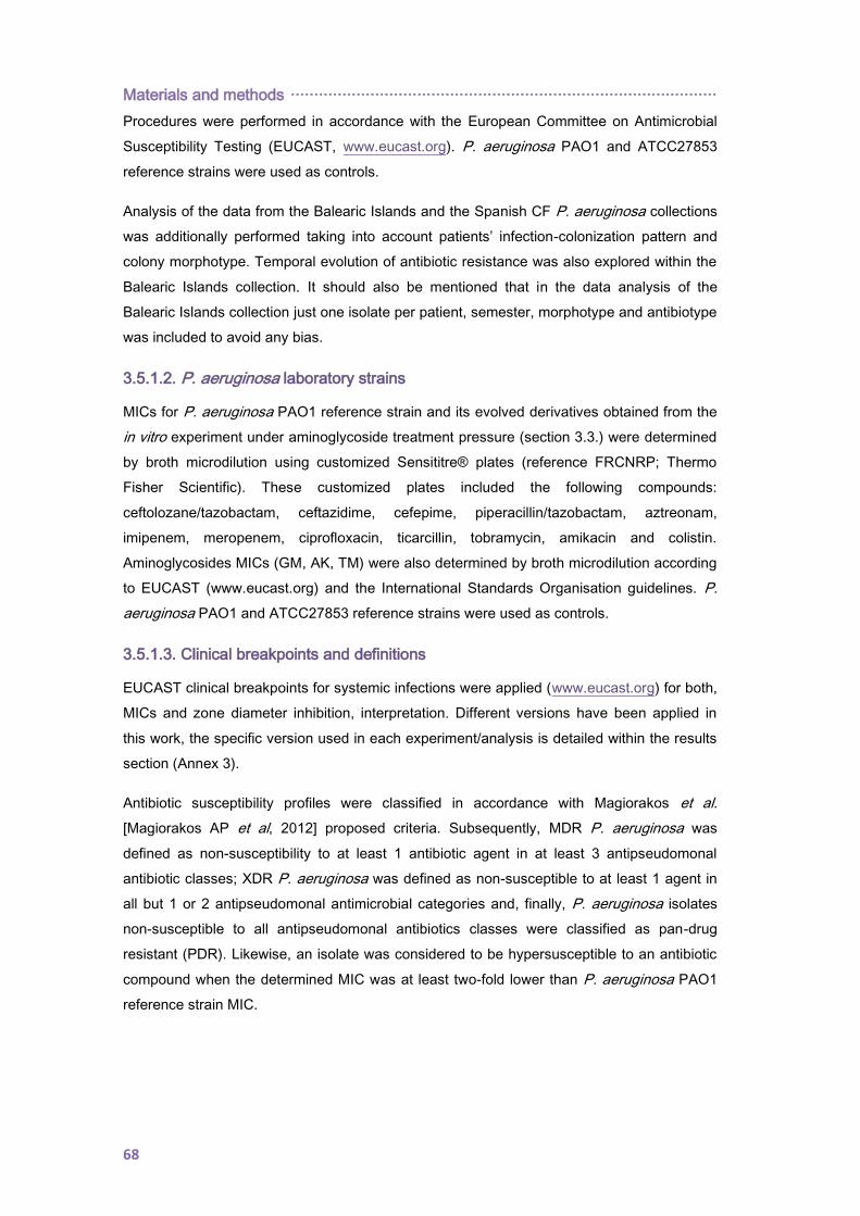
Materials and methods ···························································································
68
Procedures were performed in accordance with the European Committee on Antimicrobial
Susceptibility Testing (EUCAST, www.eucast.org). P. aeruginosa PAO1 and ATCC27853
reference strains were used as controls.
Analysis of the data from the Balearic Islands and the Spanish CF P. aeruginosa collections
was additionally performed taking into account patients’ infection-colonization pattern and
colony morphotype. Temporal evolution of antibiotic resistance was also explored within the
Balearic Islands collection. It should also be mentioned that in the data analysis of the
Balearic Islands collection just one isolate per patient, semester, morphotype and antibiotype
was included to avoid any bias.
3.5.1.2. P. aeruginosa laboratory strains
MICs for P. aeruginosa PAO1 reference strain and its evolved derivatives obtained from the
in vitro experiment under aminoglycoside treatment pressure (section 3.3.) were determined
by broth microdilution using customized Sensititre® plates (reference FRCNRP; Thermo
Fisher Scientific). These customized plates included the following compounds:
ceftolozane/tazobactam, ceftazidime, cefepime, piperacillin/tazobactam, aztreonam,
imipenem, meropenem, ciprofloxacin, ticarcillin, tobramycin, amikacin and colistin.
Aminoglycosides MICs (GM, AK, TM) were also determined by broth microdilution according
to EUCAST (www.eucast.org) and the International Standards Organisation guidelines. P.
aeruginosa PAO1 and ATCC27853 reference strains were used as controls.
3.5.1.3. Clinical breakpoints and definitions
EUCAST clinical breakpoints for systemic infections were applied (www.eucast.org) for both,
MICs and zone diameter inhibition, interpretation. Different versions have been applied in
this work, the specific version used in each experiment/analysis is detailed within the results
section (Annex 3).
Antibiotic susceptibility profiles were classified in accordance with Magiorakos et al.
[Magiorakos AP et al, 2012] proposed criteria. Subsequently, MDR P. aeruginosa was
defined as non-susceptibility to at least 1 antibiotic agent in at least 3 antipseudomonal
antibiotic classes; XDR P. aeruginosa was defined as non-susceptible to at least 1 agent in
all but 1 or 2 antipseudomonal antimicrobial categories and, finally, P. aeruginosa isolates
non-susceptible to all antipseudomonal antibiotics classes were classified as pan-drug
resistant (PDR). Likewise, an isolate was considered to be hypersusceptible to an antibiotic
compound when the determined MIC was at least two-fold lower than P. aeruginosa PAO1
reference strain MIC.
···························································································· Materials and methods
69
3.5.2. Relative expression of chromosomically encoded P. aeruginosa
resistance genes by real time qRT-PCR
The expression of the genes encoding the chromosomal β-lactamase AmpC (ampC) and
four P. aeruginosa efflux pumps representative components, MexAB-OprM (mexB), MexCD-
OprJ (mexD), MexXY (mexY), and MexEF-OprN (mexF), were determined by real-time
quantitative Reverse Transcription-PCR (qRT-PCR).
P. aeruginosa isolates were grown in 10 ml of Luria-Bertani (LB) broth (Annex 1) at 37°C and
180 r.p.m. to the late-log-phase (optical density at 600 nm [OD600] of 1) and collected by
centrifugation. Total RNA was obtained by using the RNeasy minikit (Qiagen), dissolved in
RNAse-free water and treated with 2 units of TURBO DNA-freeTM (Ambion) during 30
minutes at 37ºC for remove any contaminating DNA trace. Reaction was then stopped with 5
µl of DNase inactivation reagent (Ambion) and adjusted to a final RNA concentration of 50
ng per µl. A 50 ng sample of purified RNA was then used for one-step reverse transcription
and qRT-PCR amplification using the QuantiTect SYBR green RT-PCR kit (Qiagen, Hilden,
Germany) on either a SmartCycler II instrument (Cepheid, Sunnyvale, CA) or an Eco real-
time PCR system (Illumina). Previously described primers were used for the amplification of
ampC, mexB, mexY, mexD, mexF and rpsL (gene used as a reference to normalize the
relative amount of mRNA). Primers and PCR Master Mix details are collected in Table 3.3.
and Annex 1, respectively. Real time qRT-PCR conditions were as follows: reverse
transcription at 50ºC for 20 min, followed by Taq activation at 95ºC for 15 min and 40 cycles
of 95°C for 15 sec, 62°C for 30 sec and 72°C for 30 sec, measuring fluorescence emission in
the second step of each amplification cycle. As controls, previously characterized knockout
mutants and/or clinical strains overexpressing these antibiotic resistance mechanisms were
used.
Isolates were considered positive for AmpC, MexCD-OprJ, MexEF-OprN or MexXY(-OprM)
overexpression when the corresponding mRNA levels (ampC, mexD, mexF or mexY) were
at least 10-fold higher than that of P. aeruginosa PAO1 reference strain, negative if lower
than 5-fold, and borderline if between 5- and 10-fold. Likewise, isolates were considered
positive for mexB overexpression when the corresponding mRNA level was at least 3-fold
higher than that of PAO1, negative if lower than 2-fold and borderline if between 2- and 3-
fold. All real time qRT-PCRs were performed in duplicate and mean values (± standard
deviations) of mRNA levels were obtained from three independent experiments.
Relative expression of ampC, mexB, mexD, mexF and mexY was investigated in the subset
of 100 CF P. aeruginosa isolates (10 sequential isolates from each of 10 chronically
colonized patients) of the Balearic Islands and in all isolates from the CC274 collection. As
well, mexY relative expression was investigated in all PAO1 derivatives isolates obtained in
the in vitro experiment evolution under aminoglycoside treatment pressure.
Materials and methods ···························································································
70
3.5.3. Isolation and analysis of the outer membrane protein OprD
The presence or absence of the carbapenem porin OprD was evaluated by analyzing the
OMPs profiles. For this purpose, a protocol adapted from those previously described by Filip
et al was followed [Filip C et al, 1973]. First, P. aeruginosa isolates were grown in 10 ml of
LB broth (Annex 1) at 37°C and 180 r.p.m. to the late-log-phase (optical density at 600 nm
[OD600] of 1) and about 3 ml were collected by centrifugation, washed and resuspended in
5ml of cold Tris-Mg buffer pH 7.3 (Annex 1). Cells were then sonicated (4 cycles of 35 sec,
ON/OFF pulses of 5/2 sec, amplitude 10%) and centrifuged at 5000 r.p.m. for 2 min to
eliminate unbroken cells. Carefully supernatant fluids were transferred and centrifuged at full
speed for 30 min at 4ºC to pellet cell envelopes. Afterwards, the obtained pellets were twice
resuspended in 1.5 ml of 1% sodium lauryl sarcosinate in Tris-Mg buffer, incubated for 30
min at room temperature and centrifuged again at full speed for 30 min at room temperature.
These pellets were finally resuspended in 40 µl of Laemmli’s electrophoresis sample buffer
(BioRad), boiled for 5 min, centrifuged for remove any insoluble material and stored at -4ºC.
Once isolated, OMPs were separated by sodium dodecyl sulfate-polyacrylamide gel
electrophoresis (SDS-PAGE) and visualized using Coomassie staining. Stacking and
separating gel compositions as well as running buffer details are collected in Annex 1. An
unstained protein standard ladder (Precision ProteinTM Standards, BioRad) was also loaded
to detect the presence or absence of OprD (45 kDa). P. aeruginosa reference strain PAO1
and its OprD null spontaneous mutant PAOD1 were used as controls (Table 3.1.).
The presence or absence of the carbapenem porin OprD was evaluated within the 100 P.
aeruginosa CF isolates chronically colonizing 10 CF patients from the Balearic Islands.
3.5.4. DNA sequencing of P. aeruginosa antibiotic-resistance related genes
When needed, for isolates showing AmpC or MexXY overexpression, genes encoding their
main transcriptional regulators (ampD dacB, mexZ) were fully sequenced. Briefly, workflow
was as follows. Genomic DNA was obtained by using a commercially available extraction kit
(DNeasy Blood & Tissue kit, Qiagen or High Pure PCR template preparation kit, Roche
Diagnostics) and PCR amplification of the regulatory genes was performed (primers and
PCR Master Mix details in Table 3.3. and Annex 1, respectively) under the following reaction
conditions: initial denaturation at 94ºC for 12 min; 35 cycles of denaturation at 94ºC for 1
min, primer annealing at 60ºC or 64ºC (for mexZ or ampD/dacB, respectively) for 1 min,
extension at 72ºC for 1 min; followed by a final extension step of 72ºC for 10 min. Hereafter,
PCR amplification products were purified and sequenced with the BigDye Terminator kit (PE
Applied Biosystems, Foster City, CA) on an ABI Prism 3100 DNA sequencer (PE Applied
Biosystems) using appropiate primers (Table 3.3.).
As well, other antibiotic resistance related genes such as oprD, gyrA, gyrB, parC and/or
parE, were sometimes sequenced in order to explain or confirm the documented antibiotic

···························································································· Materials and methods
71
susceptibility profiles. The procedure was exactly as it has been described above but using
appropriate primers and adjusting PCR conditions (Table 3.3.)
In all cases, obtained DNA sequences were compared with PAO1 DNA and protein
sequences using the Basic Local Alignment Search Tool (BLAST)
(http://www.ncbi.nlm.nih.gov/BLAST.)

Materials and methods ···························································································
72
3.6. MUTATOR PHENOTYPE AND GENETIC BASIS FOR
HYPERMUTATION
3.6.1. Estimation of mutation frequencies
In order to determine which P. aeruginosa isolates had an increased spontaneous mutation
rate, rifampicin (RIF) resistance mutant frequencies were determined following previously
described protocols with slight modifications [Mena A et al, 2008; Oliver A et al, 2000].
First, a screening was performed in order to detect all P. aeruginosa isolates showing an
increased spontaneous mutation rate. For this purpose, bacterial suspensions containing
approximately 5·108 CFU/ml were prepared and a volume of 100 µl was plated on MHA
plates supplemented with 300 mg/l of RIF (MHA-RIF) (Annex 1). After 36 h (48 h for slow
growing variants) of incubation at 37ºC, plates were carefully examined. In the absence of P.
aeruginosa colonies, the corresponding isolate was considered to have a normal mutation
rate; otherwise, isolates were considered to have an increased spontaneous mutation rate
and, therefore, its mutation frequency was estimated as following described.
For each P. aeruginosa isolate, 5 independent 10 ml MHB-containing tubes (Annex 1) were
inoculated with approximately 103 bacterial cells and incubated overnight at 37ºC and 180
r.p.m. in aerobic conditions. Then, cells were recovered by centrifugation (4ºC, 10 min, 3000
r.p.m.) and pellets were resuspended in 1 ml of saline sterile solution. Finally, 1:10 serial
dilutions were prepared and plated onto MHA plates and onto RIF-supplemented MHA
plates (MHA-RIF) (Annex 1). MHA and MHA-RIF plates were incubated at 37ºC for 24 h and
36h, respectively (36-48 h for slow growing variants). Once incubated, colonies were
counted and mutation frecuencies were estimated as the median number of mutant colonies
(colonies grown in MHA-RIF/ml) divided by the median number of total cells (colonies grown
in MHA/ml). Following previous recommendations [Mena A et al, 2008; Oliver A et al, 2000],
isolates with a RIF resistance mutant frequency higher than 2·10-7 were classified as
hypermutators.
In all these procedures, both, P. aeruginosa PAO1 reference strain and its derivative
hypermutator knockout mutant defective in mutS, PAOMS, were used as controls (see Table
3.1. section 3.1.).
Screening was performed in all CF P. aeruginosa isolates from the Spanish and the CC274
collection, as well as in the 100 CF isolates chronically infecting the 10 selected patients of
the Balearic Islands. Mutation frequencies were determined for all mutator isolates detected
in both, the Spanish CF collection and the CC274 collection.
···························································································· Materials and methods
73
3.6.2. Mismatch repair system deficiency complementation assays
Genetic basis for hypermutation were explored in all P. aeruginosa isolates with a
demonstrated increased spontaneous mutation rate.
Deficiencies within the MMR system are the most frequent cause of bacterial hypermutation
[Mena A et al, 2008; Oliver A et al, 2000]. Thus, to characterize the mutator phenotype,
complementation assays using WT MMR genes mutS and mutL were performed.
For this end, gentamycin resistant plasmids pUCPmutS and pUCPmutL, harbouring P.
aeruginosa PAO1 WT mutS and mutL genes respectively, were electroporated into the
mutator isolates as follows. Plasmid pUCP24 was also electroporated as control cloning
vector (Table 3.2.). First, isolates were inoculated in 50ml-tubes containing 5 ml of LB broth
(Annex 1) and incubated overnight at 37ºC with shaking at 180 r.p.m. in aerobic conditions.
Then, 1 ml of the overnight culture was inoculated into 50 ml of fresh LB broth (Annex 1) and
incubated at 37ºC and 180 r.p.m. until the log-phase (optical density at 600 nm [OD600] of
0.5). After, the flask was chilled on ice for 10 min and cells were collected by centrifugation
(4ºC, 15 min, 3000 r.p.m.), washed and finally resuspended in 500 µl of sterile chilled
Sucrose Magnesium Electroporation Buffer (SMEB) buffer (Annex 1).
Electrocompetent P. aeruginosa cells were aliquoted (100 µl), incubated on ice with each of
the purified plasmids (5 µl) for 10 min and electroporated on a Gene Pulser Xcell (BioRad)
under the following conditions: voltage 2.5 kV, pulse time 2 min, resistance 200 Ω,
capacitance 25 µF. Finally, electroporated cells were recovered, incubated for 1h at 37ºC
and 180 r.p.m. in 1 ml of Super Optimal broth with Catabolite repression (SOC, Annex 1) and
plated onto MHA-GEN plates (Annex 1). After 36-48 h of incubation at 37ºC, independent
transformant colonies harbouring pUCP24, pUCPmutS and pUCPmutL were selected and
their mutation frequencies were estimated as previously described (section 3.6.1). A positive
complementation result was obtained when the mutation frequency decreased back to basal
levels, 1·108 approximately.
3.6.3. mutS and mutL sequencing
To complete the genetic characterization of hypermutation and, based on complementation
assays results, mutS or mutL genes were fully sequenced. Briefly, after obtaining genomic
DNA by using a commercially available extraction kit (DNeasy Blood & Tissue kit, Qiagen or
High Pure PCR template preparation kit, Roche Diagnostics), PCR amplification was
performed (see primers and PCR Master Mix details in Table 3.3. and Annex 1, respectively)
under the following reaction conditions: initial denaturation at 94ºC for 12 min; 35 cycles of
denaturation at 94ºC for 1 min, primer annealing at 60ºC/62ºC for 3 min (mutS/mutL,
respectively), extension at 72ºC for 1 min; followed by a final extension step of 72ºC for 10
min. Then, PCR amplification products were purified and sequenced with the BigDye

Materials and methods ···························································································
74
Terminator kit (PE Applied Biosystems, Foster City, CA) on an ABI Prism 3100 DNA
sequencer (PE Applied Biosystems) and appropiate primers (Table 3.3.).
Obtained DNA and protein sequences were compared with P. aeruginosa PAO1 DNA
sequence using the Basic Local Alignment Search Tool (BLAST)
(http://www.ncbi.nlm.nih.gov/BLAST).
All P. aeruginosa isolates with a demonstrated increased spontaneous mutation rate were
studied.
···························································································· Materials and methods
75
3.7. WHOLE GENOME SEQUENCING
WGS approaches were used for in-depth study of the CC274 P. aeruginosa collection and
for the genotypic characterization of PAO1 aminoglycoside-resistant derivatives (variant
calling and genome annotation).
3.7.1. Library preparation and sequencing methodology
P. aeruginosa whole genome DNA sequences were obtained as follows. First, genomic DNA
was obtained by using a commercially available extraction kit (High Pure PCR template
preparation kit, Roche Diagnostics). DNA purity was assessed with a NanoDrop
(ThermoScientifics) using the UV absorbance ratios 260/280nm and 260/230nm; DNA
samples with ratio values outside ranges 1.8-2.0 and 2.0-2.2, respectively, were excluded.
Then, genomic DNA was quantified using a fluorometric-based method (Quant-iTTM
PicoGreen ® dsDNA assay kit, LifeTechnologies) and adjusted to a final concentration of
0.1ng/µl.
Therefore, indexed paired-end libraries were prepared with the Nextera® XT DNA library
preparation kit (Illumina Inc, USA) according to the manufacturer’s protocol with slight
modifications. Briefly, genomic DNA (0.5 ng) was first tagmented and, shortafter, amplified
and indexed using a limited-cycle PCR program. Libraries were then cleaned-up and
normalized by using either, the manual or the bead-based normalization approaches. For
manual normalization a fluorometric-based method was used (Quant-iTTM PicoGreen ®
dsDNA assay kit, LifeTechnologies), and the conversion factor 1 ng/µl = 1.5 nM was applied
to prepare a 4 nM library. The selection of this conversion factor was based on the results
obtained on an Agilent Technology 2100 Bioanalyzer using a High Sensitivity DNA chip.
Once normalized, libraries were pooled and appropriately denatured and diluted to result in a
20 pM denatured pooled library that was finally loaded in, either, a MiSeq Reagent Kit v2 or
v3 (Illumina inc., USA), and sequenced on an Illumina Miseq ® benchtop sequencer,
resulting in 250 bp paired-end reads.
Nextera® XT DNA Library Prep Kit reference guide (#15031942), MiSeq System Denature
and Dilute Libraries guide (#15039740) and MiSeq® System User Guide (#15027617)
available on Illumina webpage (https://www.illumina.com/index-d.html) can be consulted for
further procedures details.
3.7.2. Variant calling
Previously defined and validated protocols were used with slight modifications [Marvig RL et
al, 2013; Marvig RL et al, 2015b]. In short, after mapping obtained 250 bp paired-end reads
to the P. aeruginosa PAO1 reference genome (GenBank accession number: NC_002516.2)
by using Bowtie 2 v2.2.4. (http://bowtie-bio.sourceforge.net/bowtie2/index.shtml), pileup and
raw files were generated by using SAMtools v0.1.16
Materials and methods ···························································································
76
(https://sourceforge.net/projects/samtools/files/samtools/) and PicardTools v1.140
(https://github.com/broadinstitute/picard). Additionally, the Genome Analysis Toolkit (GATK)
v3.4-46 (https://www.broadinstitute.org/gatk/) was used for realignment around InDels. Then,
from raw files, SNPs were extracted if they met the following criteria: a quality score (Phred-
scaled probability of the samples reads being homozygous reference) of at least 50, a root-
mean-square (RMS) mapping quality of at least 25 and a coverage depth of at least 3 reads;
excluding all ambiguous variants. As well, microInDels were extracted from the totalpileup
files applying the following criteria: a quality score of at least 500, an RMS mapping quality of
at least 25 and support from at least one-fifth of the covering reads. Pipeline and used
scripts are detailed in Annex 4.
For exceptional cases, some positions were indeed manually and individually checked in raw
and pileup files without applying any filtering.
3.7.3. De novo assemblies
Sequence reads from each isolate were de novo assembled using Velvet v1.2.10
(https://www.ebi.ac.uk/~zerbino/velvet/) with a k-mer length of 31 and the following
parameters: scaffolding = no, ins_length = 500, cov_cutoff = 3, and min_contig_lgth = 500.
The script used for this purpose is detailed in Annex 4.
3.7.4. Phylogenetic reconstructions and Beast analysis
Core genome phylogenetic reconstructions were performed using the Parsnp tool from the
Harvest Suite package v1.2 with default parameters but forcing the inclusion of all desired
genomes (-c) and selecting randomly the reference genome (r!)
(http://harvest.readthedocs.io/en/latest/content/parsnp.html).
Bayesian analysis of divergence times was performed using BEAST v2.4.2
(http://beast2.org/). For this purpose, a nexus file including all the curated positions at which
at least one of the isolates differed from the reference strain PAO1 was constructed and
converted into an.xml file with BEAUTi. Hereafter, BEAST was run with the following user-
determined settings; a lognormal relaxed molecular clock model and a general time-
reversible substitution model with gamma correction. Divergence times were calculated from
a chain length of 50 million steps, sampled every 1,000 steps and discarding the first 5
million steps as a burn-in. Finally, the maximum clade credibility tree was generated using
the TreeAnnotator program from the BEAST package and tree parameters were calculated
with Tracer v1.6 (http://beast.bio.ed.ac.uk/Tracer). Used scripts are detailed in Annex 4.
Both Phylogenetic reconstructions were displayed using FigTree v1.4.2
(http://tree.bio.ed.ac.uk/software/figtree/).
···························································································· Materials and methods
77
3.7.5. Genome annotation: resistome and mutome profiling
SNP and InDels files were annotated by using SnpEff software v4.2
(http://snpeff.sourceforge.net/index.html) with default options.
For the CC274 P. aeruginosa collection, annotated SNP and InDels files were then filtered
based on an exhaustive literature review that led us to obtain a set of 164 genes known to be
related to chromosomal antibiotic resistance in P. aeruginosa (see Annex 5). Additionally,
the online available tool ResFinder v2.1 (https://cge.cbs.dtu.dk//services/ResFinder/) was
used to identify possible horizontally acquired antimicrobial resistance genes. As well, the
genetic basis of hypermutation was investigated from WGS data through the analysis of an
exhaustive panel of so called mutator genes, thus designated mutome. Genes included
within the mutome panel are collected in Annex 5.

78

79
4. RESULTS
El alma y el cuerpo

80
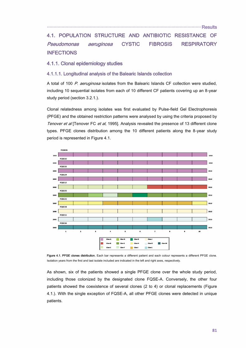
··················································································································· Results
81
4.1. POPULATION STRUCTURE AND ANTIBIOTIC RESISTANCE OF
Pseudomonas aeruginosa CYSTIC FIBROSIS RESPIRATORY
INFECTIONS
4.1.1. Clonal epidemiology studies
4.1.1.1. Longitudinal analysis of the Balearic Islands collection
A total of 100 P. aeruginosa isolates from the Balearic Islands CF collection were studied,
including 10 sequential isolates from each of 10 different CF patients covering up an 8-year
study period (section 3.2.1.).
Clonal relatedness among isolates was first evaluated by Pulse-field Gel Electrophoresis
(PFGE) and the obtained restriction patterns were analysed by using the criteria proposed by
Tenover et al [Tenover FC et al, 1995]. Analysis revealed the presence of 13 different clone
types. PFGE clones distribution among the 10 different patients along the 8-year study
period is represented in Figure 4.1.
Figure 4.1. PFGE clones distribution. Each bar represents a different patient and each colour represents a different PFGE clone.
Isolation years from the first and last isolate included are indicated in the left and right axes, respectively.
As shown, six of the patients showed a single PFGE clone over the whole study period,
including those colonized by the designated clone FQSE-A. Conversely, the other four
patients showed the coexistence of several clones (2 to 4) or clonal replacements (Figure
4.1.). With the single exception of FQSE-A, all other PFGE clones were detected in unique
patients.
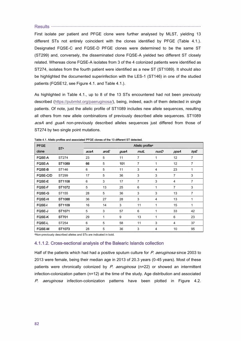
Results ··················································································································
82
First isolate per patient and PFGE clone were further analysed by MLST, yielding 13
different STs not entirely coincident with the clones identified by PFGE (Table 4.1.).
Designated FQSE-C and FQSE-D PFGE clones were determined to be the same ST
(ST299) and, conversely, the disseminated clone FQSE-A yielded two different ST closely
related. Whereas clone FQSE-A isolates from 3 of the 4 colonized patients were identified as
ST274, isolates from the fourth patient were identified as a new ST (ST1089). It should also
be highlighted the documented superinfection with the LES-1 (ST146) in one of the studied
patients (FQSE12, see Figure 4.1. and Table 4.1.).
As highlighted in Table 4.1., up to 8 of the 13 STs encountered had not been previously
described (https://pubmlst.org/paeruginosa/), being, indeed, each of them detected in single
patients. Of note, just the allelic profile of ST1089 includes new allele sequences, resulting
all others from new allele combinations of previously described allele sequences. ST1089
acsA and guaA non-previously described alleles sequences just differed from those of
ST274 by two single point mutations.
Table 4.1. Allelic profiles and associated PFGE clones of the 13 different ST detected.
PFGE
clone STa
Allelic profilea
acsA aroE guaA mutL nuoD ppsA trpE
FQSE-A ST274 23 5 11 7 1 12 7
FQSE-A ST1089 66 5 101 7 1 12 7
FQSE-B ST146 6 5 11 3 4 23 1
FQSE-C/D ST299 17 5 36 3 3 7 3
FQSE-E ST1108 6 3 17 7 3 4 7
FQSE-F ST1072 5 13 25 6 1 7 3
FQSE-G ST155 28 5 36 3 3 13 7
FQSE-H ST1088 36 27 28 3 4 13 1
FQSE-I ST1109 16 14 3 11 1 15 1
FQSE-J ST1071 5 3 57 6 1 33 42
FQSE-K ST701 29 1 9 13 1 6 23
FQSE-L ST254 6 5 58 11 3 4 37
FQSE-M ST1073 28 5 36 3 4 10 95
aNon-previously described alleles and STs are indicated in bold.
4.1.1.2. Cross-sectional analysis of the Balearic Islands collection
Half of the patients which had had a positive sputum culture for P. aeruginosa since 2003 to
2013 were female, being their median age in 2013 of 20.3 years (0-45 years). Most of these
patients were chronically colonized by P. aeruginosa (n=22) or showed an intermittent
infection-colonization pattern (n=12) at the time of the study. Age distribution and associated
P. aeruginosa infection-colonization patterns have been plotted in Figure 4.2.
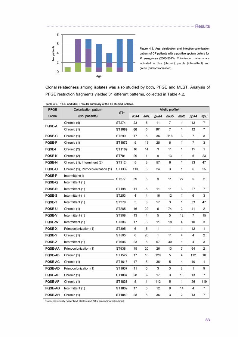
··················································································································· Results
83
Clonal relatedness among isolates was also studied by both, PFGE and MLST. Analysis of
PFGE restriction fragments yielded 31 different patterns, collected in Table 4.2.
Table 4.2. PFGE and MLST results summary of the 40 studied isolates.
PFGE
Clone
Colonization pattern
(No. patients) STa
Allelic profileb
acsA aroE guaA nuoD mutL ppsA trpE
FQSE-A Chronic (4) ST274 23 5 11 7 1 12 7
Chronic (1) ST1089 66 5 101 7 1 12 7
FQSE-C Chronic (1) ST299 17 5 36 116 3 7 3
FQSE-F Chronic (1) ST1072 5 13 25 6 1 7 3
FQSE-I Chronic (2) ST1109 16 14 3 11 1 15 1
FQSE-K Chronic (2) ST701 29 1 9 13 1 6 23
FQSE-N Chronic (1), Intermittent (2) ST312 5 3 57 6 1 33 47
FQSE-O Chronic (1), Primocolonization (1) ST1339 113 5 24 3 1 6 25
FQSE-P Intermittent(1) ST277 39 5 9 11 27 5 2
FQSE-Q Intermittent (1)
FQSE-R Intermittent (1) ST198 11 5 11 11 3 27 7
FQSE-S Intermittent (1) ST253 4 4 16 12 1 6 3
FQSE-T Intermittent (1) ST279 5 3 57 3 1 33 47
FQSE-U Chronic (1) ST285 16 22 6 74 2 41 2
FQSE-V Intermittent (1) ST308 13 4 5 5 12 7 15
FQSE-W Intermittent (1) ST386 17 5 11 18 4 10 3
FQSE-X Primocolonization (1) ST395 6 5 1 1 1 12 1
FQSE-Y Chronic (1) ST505 6 20 1 11 4 4 2
FQSE-Z Intermittent (1) ST606 23 5 57 30 1 4 3
FQSE-AA Primocolonization (1) ST938 15 20 26 13 3 64 2
FQSE-AB Chronic (1) ST1527 17 10 129 5 4 112 10
FQSE-AC Chronic (1) ST1613 17 5 36 5 4 10 1
FQSE-AD Primocolonization (1) ST1637 11 5 3 3 8 1 9
FQSE-AE Chronic (1) ST1837 28 62 17 3 13 13 7
FQSE-AF Chronic (1) ST1838 5 1 112 5 1 26 119
FQSE-AG Intermittent (1) ST1839 17 5 12 9 14 4 7
FQSE-AH Chronic (1) ST1840 28 5 36 3 2 13 7
aNon-previously described alleles and STs are indicated in bold.
Figure 4.2. Age distribution and infection-colonization
pattern of CF patients with a positive sputum culture for
P. aeruginosa (2003-2013). Colonization patterns are
indicated in blue (chronic), purple (intermittent) and
green (primocolonization).
0
2
4
6
8
No
. p
atie
nts
Age
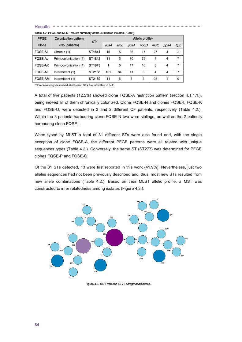
Results ··················································································································
84
Table 4.2. PFGE and MLST results summary of the 40 studied isolates. (Cont.)
PFGE
Clone
Colonization pattern
(No. patients) STa
Allelic profileb
acsA aroE guaA nuoD mutL ppsA trpE
FQSE-AI Chronic (1) ST1841 15 5 36 17 27 4 2
FQSE-AJ Primocolonization (1) ST1842 11 5 30 72 4 4 7
FQSE-AK Primocolonization (1) ST1843 1 5 17 16 3 4 7
FQSE-AL Intermittent (1) ST2188 101 84 11 3 4 4 7
FQSE-AM Intermittent (1) ST2189 11 5 3 3 93 1 9
aNon-previously described alleles and STs are indicated in bold.
A total of five patients (12.5%) showed clone FQSE-A restriction pattern (section 4.1.1.1.),
being indeed all of them chronically colonized. Clone FQSE-N and clones FQSE-I, FQSE-K
and FQSE-O, were detected in 3 and 2 different CF patients, respectively (Table 4.2.).
Within the 3 patients harbouring clone FQSE-N two were siblings, as well as the 2 patients
harbouring clone FQSE-I.
When typed by MLST a total of 31 different STs were also found and, with the single
exception of clone FQSE-A, the different PFGE patterns were all related with unique
sequences types (Table 4.2.). Conversely, the same ST (ST277) was determined for PFGE
clones FQSE-P and FQSE-Q.
Of the 31 STs detected, 13 were first reported in this work (41.9%). Nevertheless, just two
alleles sequences had not been previously described and, thus, most new STs resulted from
new allele combinations (Table 4.2.). Based on their MLST allelic profile, a MST was
constructed to infer relatedness among isolates (Figure 4.3.).
Figure 4.3. MST from the 40 P. aeruginosa isolates.
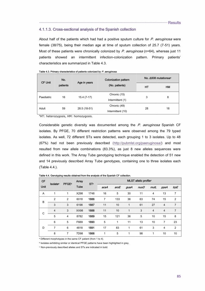
··················································································································· Results
85
4.1.1.3. Cross-sectional analysis of the Spanish collection
About half of the patients which had had a positive sputum culture for P. aeruginosa were
female (38/75), being their median age at time of sputum collection of 25.7 (7-51) years.
Most of these patients were chronically colonized by P. aeruginosa (n=64), whereas just 11
patients showed an intermittent infection-colonization pattern. Primary patients’
characteristics are summarized in Table 4.3.
Table 4.3. Primary characteristics of patients colonized by P. aeruginosa.
CF Unit No.
patients Age in years
Colonization pattern
(No. patients)
No. Δ508 mutationsaa
HT HM
Paediatric 16 15.4 (7-17) Chronic (15)
Intermittent (1) 3 8
Adult 59 28.5 (18-51) Chronic (49)
Intermittent (10) 28 18
aHT: heterozygosis, HM: homozygosis.
Considerable genetic diversity was documented among the P. aeruginosa Spanish CF
isolates. By PFGE, 70 different restriction patterns were observed among the 79 typed
isolates. As well, 72 different STs were detected, each grouping 1 to 3 isolates. Up to 48
(67%) had not been previously described (http://pubmlst.org/paeruginosa/) and most
resulted from new allele combinations (83.3%), as just 8 new alleles sequences were
defined in this work. The Array Tube genotyping technique enabled the detection of 51 new
and 14 previously described Array Tube genotypes, containing one to three isolates each
(Table 4.4.).
Table 4.4. Genotyping results obtained from the analysis of the Spanish CF collection.
CF
Unit Isolatea PFGEb
Array
Tube STc
MLST allelic profilec
acsA aroE guaA nuoD mutL ppsA trpE
A 1 1 X298 1748 16 5 30 11 4 13 7
B 2 2 6018 1886 7 133 36 63 74 15 2
3 3 6198 1887 11 10 1 61 27 4 7
C 4 3 XX98 1888 11 10 1 3 4 4 7
5 4 8782 1889 15 121 36 5 10 15 8
D
6 5 F669 1890 5 1 11 13 10 7 23
7 6 4818 1891 17 83 1 61 3 4 2
8 7 7D98 1866 1 5 1 98 1 10 10
a Different morphotypes in the same CF patient (from 1 to 4).
b Isolates exhibiting similar or identical PFGE patterns have been highlighted in grey.
c Non-previously described alleles and STs are indicated in bold.

Results ··················································································································
86
Table 4.4. Genotyping results obtained from the analysis of the Spanish CF collection. (Cont.)
CF
Unit Isolatea PFGEb
Array
Tube STc
MLST allelic profilec
acsA aroE guaA nuoD mutL ppsA trpE
E
9 8 CC0A 1109 16 14 3 11 1 15 1
10 9 8C2C 1867 6 94 133 1 1 12 1
11 10 2C22 1868 23 5 11 98 1 12 7
12 11 6FA8 1869 15 24 5 5 50 4 14
F 13 12 F468 1870 17 11 3 13 1 2 4
14 13 AD80 1892 17 5 4 66 4 15 19
G
15 14 ED98 1893 101 8 78 72 4 13 7
16 15 882A 270 22 3 17 5 2 10 7
17 16 6010 116 28 24 22 18 3 15 7
18 17 0C2E 395 6 5 1 1 1 12 1
19 18 0C2A 1894 17 187 1 3 4 15 3
20 19 2F80 1895 17 6 4 14 4 6 2
21 20 741C 1228 11 5 5 29 4 4 26
22 21 0C2A 253 4 4 16 12 1 6 3
23 22 0C2C 395 6 5 1 1 1 12 1
24 23 F428 1896 5 3 5 3 1 33 189
H
25 24 0422 1897 7 162 12 3 3 4 185
26 25 2C18 1871 28 22 5 43 3 14 19
27 26 C40A 1872 11 5 1 117 9 4 190
28 27 E022 1873 1 5 11 3 2 4 3
29 28 859A 575 11 5 83 2 4 13 7
30 29 8428 1898 15 5 83 11 4 62 7
31 30 F419 1899 39 5 70 28 4 4 63
I 32 30 B01A 1900 39 5 68 28 4 4 63
33 31 2C20 617 16 101 11 97 4 69 88
J
34 32 B01X 1901 16 101 11 13 4 69 88
35 33 F421 560 5 5 57 13 1 40 3
36 34 262A 1902 11 94 7 98 2 7 33
37 31 2C20 1903 23 5 11 13 1 12 137
38 35 E42A 1874 29 188 95 13 8 6 11
K
39 35 E428 1874 29 188 95 13 8 6 11
40 36 A998 609 16 22 5 11 4 6 10
41 35 E428 1875 29 188 95 127 8 6 11
42 31 2C20 268 23 5 70 7 1 12 7
43 37 2F88 1904 6 103 11 63 4 15 2
a Different morphotypes in the same CF patient (from 1 to 4).
b Isolates exhibiting similar or identical PFGE patterns have been highlighted in grey.
c Non-previously described alleles and STs are indicated in bold.

··················································································································· Results
87
Table 4.4. Genotyping results obtained from the analysis of the Spanish CF collection. (Cont.)
CF
Unit Isolatea PFGEb
Array
Tube STc
MLST allelic profilec
acsA aroE guaA nuoD mutL ppsA trpE
L
44 38 2418 1876 40 181 11 11 3 4 9
45 39 E418 564 6 10 5 11 2 15 2
46 40 2C18 1905 6 4 42 5 1 4 26
47 41 4DA8 1906 16 3 1 28 1 55 61
48 42 2708 644 28 3 94 13 1 4 10
49 43 7C2C 132 6 20 1 3 4 4 2
50 44 3E18 1877 119 10 83 43 3 6 77
M
51a1 45 EC18 1907 11 5 11 131 3 53 1
52 a1 46 2C2C 1908 11 6 19 5 4 15 9
53 47 6A20 1909 17 5 5 4 4 4 191
54 48 6FA8 1910 15 5 5 85 8 4 14
N
55 a2 49 AF90 1911 11 3 70 3 1 4 60
56 a2 50 F420 1251 35 11 25 6 13 6 84
57 48 6FA8 1878 15 100 66 5 50 4 14
58 51 2610 1912 11 48 11 3 1 15 14
O 59 52 4498 1913 11 48 98 5 3 10 85
P
60 53 2810 1914 6 14 12 7 1 4 20
61 a3 54 E020 1879 15 5 11 3 67 4 3
62 a3 55 E020 508 15 5 11 3 2 4 3
Q 63 56 8E18 189 26 143 1 3 4 4 10
64 57 E429 1880 13 8 9 97 52 124 9
R
65 58 D421 253 4 4 16 12 1 6 3
66 59 2398 1881 22 20 11 48 3 3 7
67 60 XC10 1882 16 5 26 124 4 3 26
68 59 2398 348 22 20 11 3 3 3 7
S
69 61 DF88 1883 17 5 11 97 4 12 56
70 62 EC10 676 28 5 11 77 3 4 92
71 a4 63 E020 508 15 5 11 3 2 4 3
72 a4 64 E020 508 15 5 11 3 2 4 3
73 65 A598 575 11 5 83 2 4 13 7
74 66 F429 313 47 8 7 6 8 11 40
75 67 0C48 198 11 5 11 11 3 27 7
76 68 6E10 569 11 5 11 11 3 6 27
77 69 0810 1884 17 5 12 3 99 4 7
T 78 70 EA08 27 6 5 6 7 4 6 7
79 70 EA08 27 6 5 6 7 4 6 7
a Different morphotypes in the same CF patient (from 1 to 4).
b Isolates exhibiting similar or identical PFGE patterns have been highlighted in grey.
c Non-previously described alleles and STs are indicated in bold.
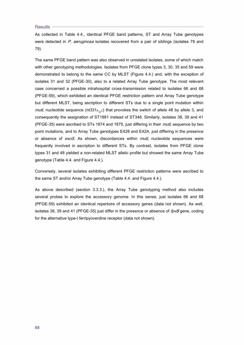
Results ··················································································································
88
As collected in Table 4.4., identical PFGE band patterns, ST and Array Tube genotypes
were detected in P. aeruginosa isolates recovered from a pair of siblings (isolates 78 and
79).
The same PFGE band pattern was also observed in unrelated isolates, some of which match
with other genotyping methodologies. Isolates from PFGE clone types 3, 30, 35 and 59 were
demonstrated to belong to the same CC by MLST (Figure 4.4.) and, with the exception of
isolates 31 and 32 (PFGE-30), also to a related Array Tube genotype. The most relevant
case concerned a possible intrahospital cross-transmission related to isolates 66 and 68
(PFGE-59), which exhibited an identical PFGE restriction pattern and Array Tube genotype
but different MLST, being ascription to different STs due to a single point mutation within
mutL nucleotide sequence (nt331A
C) that provokes the switch of allele 48 by allele 3, and
consequently the assignation of ST1881 instead of ST348. Similarly, isolates 38, 39 and 41
(PFGE-35) were ascribed to STs 1874 and 1875, just differing in their mutL sequence by two
point mutations, and to Array Tube genotypes E428 and E42A, just differing in the presence
or absence of exoS. As shown, discordances within mutL nucleotide sequences were
frequently involved in ascription to different STs. By contrast, isolates from PFGE clone
types 31 and 48 yielded a non-related MLST allelic profile but showed the same Array Tube
genotype (Table 4.4. and Figure 4.4.).
Conversely, several isolates exhibiting different PFGE restriction patterns were ascribed to
the same ST and/or Array Tube genotype (Table 4.4. and Figure 4.4.).
As above described (section 3.3.3.), the Array Tube genotyping method also includes
several probes to explore the accessory genome. In this sense, just isolates 66 and 68
(PFGE-59) exhibited an identical repertoire of accessory genes (data not shown). As well,
isolates 38, 39 and 41 (PFGE-35) just differ in the presence or absence of fpvB gene, coding
for the alternative type-I ferripyoverdine receptor (data not shown).
··················································································································· Results
89
Figure 4.4. MST based on the MLST allelic profiles. Isolates with the same Array Tube genotype are represented with the same
colour. As well, isolates ascribed to the same PFGE clone type are indicated with red lines.
Overall, ferripyoverdine receptor genes (fpvA) type I, IIa, IIb and III were detected in 23, 22,
6 and 11 isolates respectively, and the alternative type-I ferripyoverdine receptor gene fpvB
was present only in 45 isolates. Ferripyoverdine receptor genes were not detected in 15
isolates, and all but two were isolates from adult patients. On average, isolates possess 2.3
genome islets and 2.4 genome islands, ranging from 0 to 5. The flagellin glycosylation island
was encountered to be the most prevalent one (n=53, 67%). Nevertheless, 2 of these
isolates lacked the a-type flagellin and, on the contrary, in 3 isolates expressing the a-type
flagellin the flagellin glycosylation island was not detected. PAGI-1 was detected in only 45
isolates (57%) and the genomic islands of the CLC family PAGI-2/3 in 23 isolates (29%).
Two isolates harboured both PAPI-1 and PAPI-2, and 43 isolates (54%) harboured only
PAPI-2; other pKLC102-like islands were detected in 30 isolates (38%). Statistical
differences within the global collection and the different subsets, adult or paediatric
population, were not observed (Table 4.5.).
mutL 13/127 mutL 3/48
mutL 61/3 nuoD 27/4
brothers
mutL 61/3 nuoD 27/4
guaA 70/11 mutL 7/13
trpE 7/137
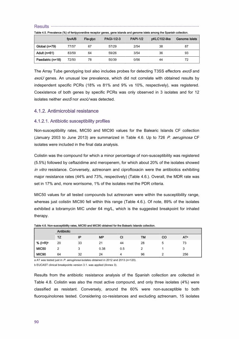
Results ··················································································································
90
Table 4.5. Prevalence (%) of ferripyoverdine receptor genes, gene islands and genome islets among the Spanish collection.
fpvA/B Fla-glyc PAGI-1/2-3 PAPI-1/2 pKLC102-like Genome Islets
Global (n=79) 77/57 67 57/29 2/54 38 87
Adult (n=61) 83/59 64 59/26 3/54 36 93
Paediatric (n=18) 72/50 78 50/39 0/56 44 72
The Array Tube genotyping tool also includes probes for detecting T3SS effectors exoS and
exoU genes. An unusual low prevalence, which did not correlate with obtained results by
independent specific PCRs (18% vs 81% and 9% vs 10%, respectively), was registered.
Coexistence of both genes by specific PCRs was only observed in 3 isolates and for 12
isolates neither exoS nor exoU was detected.
4.1.2. Antimicrobial resistance
4.1.2.1. Antibiotic susceptibility profiles
Non-susceptibility rates, MIC50 and MIC90 values for the Balearic Islands CF collection
(January 2003 to June 2013) are summarized in Table 4.6. Up to 726 P. aeruginosa CF
isolates were included in the final data analysis.
Colistin was the compound for which a minor percentage of non-susceptibility was registered
(5.5%) followed by ceftazidime and meropenem, for which about 20% of the isolates showed
in vitro resistance. Conversely, aztreonam and ciprofloxacin were the antibiotics exhibiting
major resistance rates (44% and 73%, respectively) (Table 4.6.). Overall, the MDR rate was
set in 17% and, more worrisome, 1% of the isolates met the PDR criteria.
MIC50 values for all tested compounds but aztreonam were within the susceptibility range,
whereas just colistin MIC90 fell within this range (Table 4.6.). Of note, 89% of the isolates
exhibited a tobramycin MIC under 64 mg/L, which is the suggested breakpoint for inhaled
therapy.
Table 4.6. Non-susceptibility rates, MIC50 and MIC90 obtained for the Balearic Islands collection.
Antibiotic
TZ IP MP CI TM CO ATa
% (I+R)b 20 33 21 44 28 5 73
MIC50 2 3 0.38 0.5 2 1 3
MIC90 64 32 24 4 96 2 256
a AT was tested just in P. aeruginosa isolates obtained in 2012 and 2013 (n=120).
b EUCAST clinical breakpoints version 3.1. was applied (Annex 3).
Results from the antibiotic resistance analysis of the Spanish collection are collected in
Table 4.8. Colistin was also the most active compound, and only three isolates (4%) were
classified as resistant. Conversely, around the 60% were non-susceptible to both
fluoroquinolones tested. Considering co-resistances and excluding aztreonam, 15 isolates
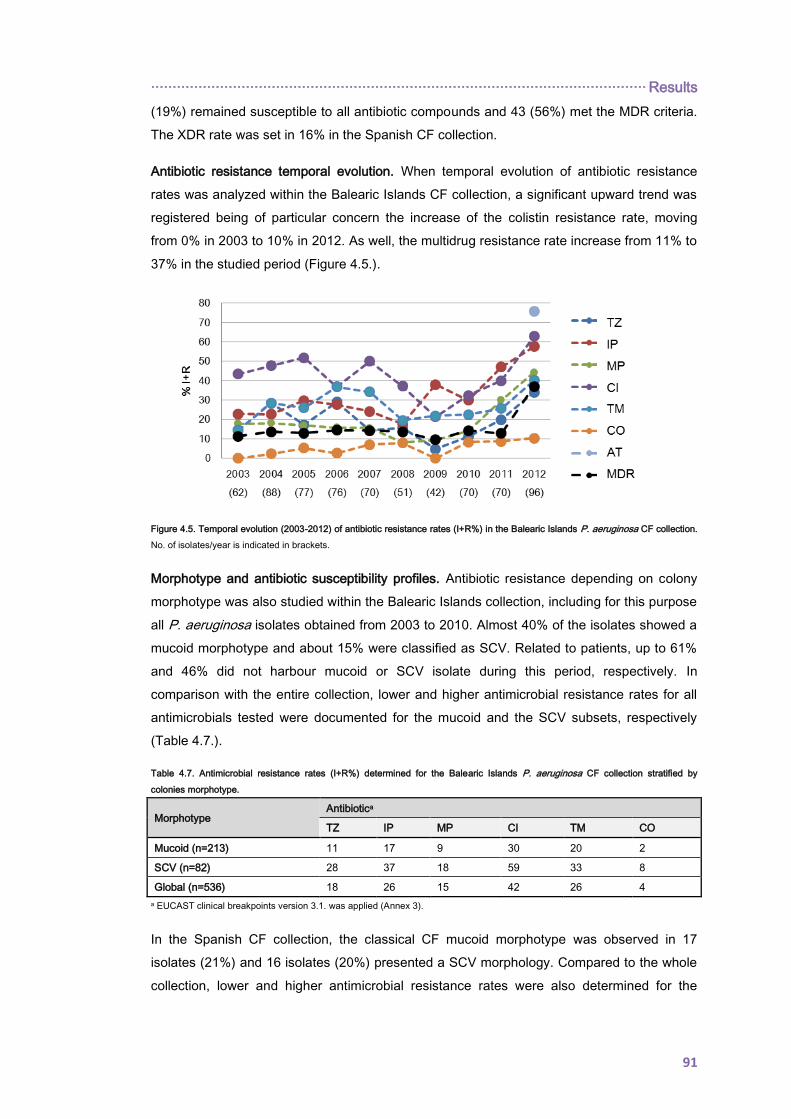
··················································································································· Results
91
(19%) remained susceptible to all antibiotic compounds and 43 (56%) met the MDR criteria.
The XDR rate was set in 16% in the Spanish CF collection.
Antibiotic resistance temporal evolution. When temporal evolution of antibiotic resistance
rates was analyzed within the Balearic Islands CF collection, a significant upward trend was
registered being of particular concern the increase of the colistin resistance rate, moving
from 0% in 2003 to 10% in 2012. As well, the multidrug resistance rate increase from 11% to
37% in the studied period (Figure 4.5.).
Figure 4.5. Temporal evolution (2003-2012) of antibiotic resistance rates (I+R%) in the Balearic Islands P. aeruginosa CF collection.
No. of isolates/year is indicated in brackets.
Morphotype and antibiotic susceptibility profiles. Antibiotic resistance depending on colony
morphotype was also studied within the Balearic Islands collection, including for this purpose
all P. aeruginosa isolates obtained from 2003 to 2010. Almost 40% of the isolates showed a
mucoid morphotype and about 15% were classified as SCV. Related to patients, up to 61%
and 46% did not harbour mucoid or SCV isolate during this period, respectively. In
comparison with the entire collection, lower and higher antimicrobial resistance rates for all
antimicrobials tested were documented for the mucoid and the SCV subsets, respectively
(Table 4.7.).
Table 4.7. Antimicrobial resistance rates (I+R%) determined for the Balearic Islands P. aeruginosa CF collection stratified by
colonies morphotype.
Morphotype Antibiotica
TZ IP MP CI TM CO
Mucoid (n=213) 11 17 9 30 20 2
SCV (n=82) 28 37 18 59 33 8
Global (n=536) 18 26 15 42 26 4
a EUCAST clinical breakpoints version 3.1. was applied (Annex 3).
In the Spanish CF collection, the classical CF mucoid morphotype was observed in 17
isolates (21%) and 16 isolates (20%) presented a SCV morphology. Compared to the whole
collection, lower and higher antimicrobial resistance rates were also determined for the
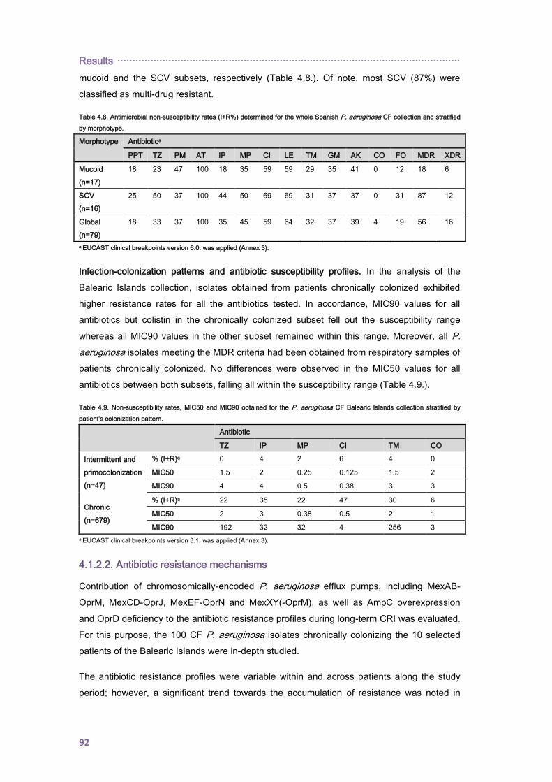
Results ··················································································································
92
mucoid and the SCV subsets, respectively (Table 4.8.). Of note, most SCV (87%) were
classified as multi-drug resistant.
Table 4.8. Antimicrobial non-susceptibility rates (I+R%) determined for the whole Spanish P. aeruginosa CF collection and stratified
by morphotype.
Morphotype Antibiotica
PPT TZ PM AT IP MP CI LE TM GM AK CO FO MDR XDR
Mucoid
(n=17)
18 23 47 100 18 35 59 59 29 35 41 0 12 18 6
SCV
(n=16)
25 50 37 100 44 50 69 69 31 37 37 0 31 87 12
Global
(n=79)
18 33 37 100 35 45 59 64 32 37 39 4 19 56 16
a EUCAST clinical breakpoints version 6.0. was applied (Annex 3).
Infection-colonization patterns and antibiotic susceptibility profiles. In the analysis of the
Balearic Islands collection, isolates obtained from patients chronically colonized exhibited
higher resistance rates for all the antibiotics tested. In accordance, MIC90 values for all
antibiotics but colistin in the chronically colonized subset fell out the susceptibility range
whereas all MIC90 values in the other subset remained within this range. Moreover, all P.
aeruginosa isolates meeting the MDR criteria had been obtained from respiratory samples of
patients chronically colonized. No differences were observed in the MIC50 values for all
antibiotics between both subsets, falling all within the susceptibility range (Table 4.9.).
Table 4.9. Non-susceptibility rates, MIC50 and MIC90 obtained for the P. aeruginosa CF Balearic Islands collection stratified by
patient’s colonization pattern.
Antibiotic
TZ IP MP CI TM CO
Intermittent and
primocolonization
(n=47)
% (I+R)a 0 4 2 6 4 0
MIC50 1.5 2 0.25 0.125 1.5 2
MIC90 4 4 0.5 0.38 3 3
Chronic
(n=679)
% (I+R)a 22 35 22 47 30 6
MIC50 2 3 0.38 0.5 2 1
MIC90 192 32 32 4 256 3
a EUCAST clinical breakpoints version 3.1. was applied (Annex 3).
4.1.2.2. Antibiotic resistance mechanisms
Contribution of chromosomically-encoded P. aeruginosa efflux pumps, including MexAB-
OprM, MexCD-OprJ, MexEF-OprN and MexXY(-OprM), as well as AmpC overexpression
and OprD deficiency to the antibiotic resistance profiles during long-term CRI was evaluated.
For this purpose, the 100 CF P. aeruginosa isolates chronically colonizing the 10 selected
patients of the Balearic Islands were in-depth studied.
The antibiotic resistance profiles were variable within and across patients along the study
period; however, a significant trend towards the accumulation of resistance was noted in
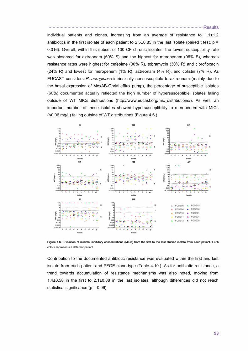
··················································································································· Results
93
individual patients and clones, increasing from an average of resistance to 1.1±1.2
antibiotics in the first isolate of each patient to 2.5±0.85 in the last isolate (paired t test, p =
0.016). Overall, within this subset of 100 CF chronic isolates, the lowest susceptibility rate
was observed for aztreonam (60% S) and the highest for meropenem (96% S), whereas
resistance rates were highest for cefepime (30% R), tobramycin (30% R) and ciprofloxacin
(24% R) and lowest for meropenem (1% R), aztreonam (4% R), and colistin (7% R). As
EUCAST considers P. aeruginosa intrinsically nonsusceptible to aztreonam (mainly due to
the basal expression of MexAB-OprM efflux pump), the percentage of susceptible isolates
(60%) documented actually reflected the high number of hypersusceptible isolates falling
outside of WT MICs distributions (http://www.eucast.org/mic_distributions/). As well, an
important number of these isolates showed hypersusceptibility to meropenem with MICs
(<0.06 mg/L) falling outside of WT distributions (Figure 4.6.).
Figure 4.6.. Evolution of minimal inhibitory concentrations (MICs) from the first to the last studied isolate from each patient. Each
colour represents a different patient.
Contribution to the documented antibiotic resistance was evaluated within the first and last
isolate from each patient and PFGE clone type (Table 4.10.). As for antibiotic resistance, a
trend towards accumulation of resistance mechanisms was also noted, moving from
1.4±0.58 in the first to 2.1±0.88 in the last isolates, although differences did not reach
statistical significance (p = 0.06).
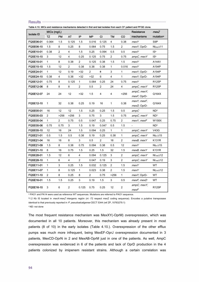
Results ··················································································································
94
Table 4.10. MICs and resistance mechanisms detected in first and last isolates from each CF patient and PFGE clone.
Isolate ID MICs (mg/L) Resistance
mechanisms
mexZ
mutationa TZ PM AT IP MP CI TM CO
FQSE06-01 0.064 1.5 0.125 1.5 0.016 0.125 4 0.38 mexY S9P
FQSE06-10 1.5 8 0.25 8 0.064 0.75 1.5 2 mexY, OprD- Nt292∆11
FQSE10-01 0.38 2 4 1.5 0.25 0.094 0.5 0.5 mexY ISb
FQSE10-10 3 12 4 0.25 0.125 0.75 2 0.75 ampC, mexY ISb
FQSE15-01 1 8 0.38 2 0.125 0.38 1.5 1.5 mexY A144V
FQSE15-10 1.5 12 2 0.38 0.38 0.38 1 0.016 mexY A194P
FQSE24-01 1 12 0.19 >32 2 8 3 1 mexY, OprD- A194P
FQSE24-10 0.38 4 0.38 >32 >32 6 4 1 mexY, OprD- A194P
FQSE12-01 0.75 8 0.125 1 0.064 0.25 24 0.75 mexY R125P
FQSE12-06 8 8 8 2 0.5 2 24 4 ampC, mexY R125P
FQSE12-07 24 24 12 >32 1.5 4 4 >256 ampC, mexY,
mexF, OprD- Q164X
FQSE12-10 1 32 0.38 0.25 0.19 16 1 0.38 mexY, mexF,
mexD, OprD- Q164X
FQSE05-01 16 12 12 1.5 0.25 0.25 1.5 0.5 ampC NDC
FQSE05-03 2 >256 >256 3 0.75 3 1.5 0.75 ampC, mexY NDC
FQSE05-04 1 2 0.75 0.5 0.047 0.25 0.75 2 mexY, mexF W158X
FQSE05-06 0.75 0.75 3 1.5 0.19 0.047 0.5 1.5 - NDC
FQSE05-10 12 16 24 1.5 0.094 0.25 1 1 ampC, mexY V43G
FQSE21-01 0.5 1.5 0.5 0.38 0.19 0.25 0.38 1 ampC, mexY Nt61∆15
FQSE21-04 16 16 8 3 0.5 2 16 2 mexB, mexY K131R
FQSE21-09 1.5 8 0.38 0.75 0.094 0.38 0.5 12 mexY Nt61∆15
FQSE21-10 8 16 0.75 1.5 0.25 1.5 32 1.5 mexB, mexY K131R
FQSE28-01 1.5 12 6 4 0.094 0.125 3 2 ampC, mexY Nt189∆12
FQSE28-10 1 6 4 2 0.047 0.19 3 2 ampC, mexY Nt189∆12
FQSE11-01 1 3 0.25 1.5 0.032 0.125 2 1.5 mexY WT
FQSE11-07 1 8 0.125 1 0.023 0.38 2 1.5 mexY Nt279∆12
FQSE11-10 2 8 0.25 8 2 0.75 >256 1 mexY, OprD- WT
FQSE16-01 1.5 1.5 0.25 3 0.19 1.5 3 0.5 mexF, mexD WT
FQSE16-10 3 6 2 0.125 0.75 0.25 12 2 ampC, mexY,
mexF R125P
a PAO1 and PA14 were used as reference WT sequences. Mutations are referred to PAO1 sequence.
b1.2 Kb IS located in mexX-mexZ intergenic región (nt -72 respect mexZ coding sequence). Encodes a putative transposase
identical to that previously reported in P. pseudoalcaligenes CECT 5344 (ref ZP_10763279.1)
c ND: not done
The most frequent resistance mechanism was MexXY(-OprM) overexpression, which was
documented in all 10 patients. Moreover, this mechanism was already present in most
patients (8 of 10) in the early isolates (Table 4.10.). Overexpression of the other efflux
pumps was much more infrequent, being MexEF-OprJ overexpression documented in 3
patients, MexCD-OprN in 2 and MexAB-OprM just in one of the patients. As well, AmpC
overexpression was evidenced in 6 of the patients and lack of OprD production in the 4
patients colonized by imipenem resistant strains. Although a certain correlation was
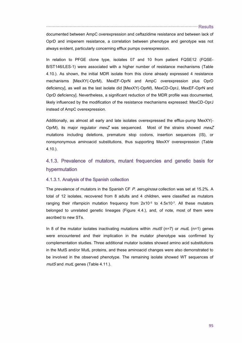
··················································································································· Results
95
documented between AmpC overexpression and ceftazidime resistance and between lack of
OprD and imipenem resistance, a correlation between phenotype and genotype was not
always evident, particularly concerning efflux pumps overexpression.
In relation to PFGE clone type, isolates 07 and 10 from patient FQSE12 (FQSE-
B/ST146/LES-1) were associated with a higher number of resistance mechanisms (Table
4.10.). As shown, the initial MDR isolate from this clone already expressed 4 resistance
mechanisms [MexXY(-OprM), MexEF-OprN and AmpC overexpression plus OprD
deficiency], as well as the last isolate did [MexXY(-OprM), MexCD-OprJ, MexEF-OprN and
OprD deficiency]. Nevertheless, a significant reduction of the MDR profile was documented,
likely influenced by the modification of the resistance mechanisms expressed: MexCD-OprJ
instead of AmpC overexpression.
Additionally, as almost all early and late isolates overexpressed the efflux-pump MexXY(-
OprM), its major regulator mexZ was sequenced. Most of the strains showed mexZ
mutations including deletions, premature stop codons, insertion sequences (IS), or
nonsynonymous aminoacid substitutions, thus supporting MexXY overexpression (Table
4.10.).
4.1.3. Prevalence of mutators, mutant frequencies and genetic basis for
hypermutation
4.1.3.1. Analysis of the Spanish collection
The prevalence of mutators in the Spanish CF P. aeruginosa collection was set at 15.2%. A
total of 12 isolates, recovered from 8 adults and 4 children, were classified as mutators
ranging their rifampicin mutation frequency from 2x10⁻⁵ to 4.5x10⁻⁷. All these mutators
belonged to unrelated genetic lineages (Figure 4.4.), and, of note, most of them were
ascribed to new STs.
In 8 of the mutator isolates inactivating mutations within mutS (n=7) or mutL (n=1) genes
were encountered and their implication in the mutator phenotype was confirmed by
complementation studies. Three additional mutator isolates showed amino acid substitutions
in the MutS and/or MutL proteins, and these aminoacid changes were also demonstrated to
be involved in the observed phenotype. The remaining isolate showed WT sequences of
mutS and mutL genes (Table 4.11.).
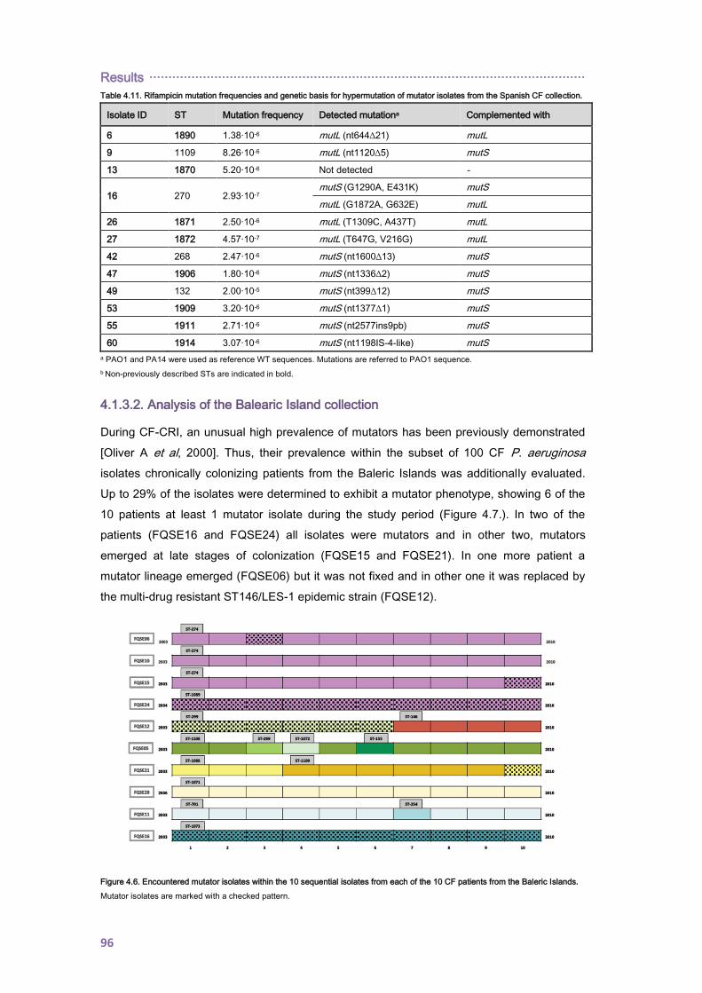
Results ··················································································································
96
Table 4.11. Rifampicin mutation frequencies and genetic basis for hypermutation of mutator isolates from the Spanish CF collection.
Isolate ID ST Mutation frequency Detected mutationa Complemented with
6 1890 1.38·10-6 mutL (nt644∆21) mutL
9 1109 8.26·10-6 mutL (nt1120∆5) mutS
13 1870 5.20·10-6 Not detected -
16 270 2.93·10-7 mutS (G1290A, E431K) mutS
mutL (G1872A, G632E) mutL
26 1871 2.50·10-6 mutL (T1309C, A437T) mutL
27 1872 4.57·10-7 mutL (T647G, V216G) mutL
42 268 2.47·10-6 mutS (nt1600∆13) mutS
47 1906 1.80·10-6 mutS (nt1336∆2) mutS
49 132 2.00·10-5 mutS (nt399∆12) mutS
53 1909 3.20·10-6 mutS (nt1377∆1) mutS
55 1911 2.71·10-6 mutS (nt2577ins9pb) mutS
60 1914 3.07·10-6 mutS (nt1198IS-4-like) mutS
a PAO1 and PA14 were used as reference WT sequences. Mutations are referred to PAO1 sequence.
b Non-previously described STs are indicated in bold.
4.1.3.2. Analysis of the Balearic Island collection
During CF-CRI, an unusual high prevalence of mutators has been previously demonstrated
[Oliver A et al, 2000]. Thus, their prevalence within the subset of 100 CF P. aeruginosa
isolates chronically colonizing patients from the Baleric Islands was additionally evaluated.
Up to 29% of the isolates were determined to exhibit a mutator phenotype, showing 6 of the
10 patients at least 1 mutator isolate during the study period (Figure 4.7.). In two of the
patients (FQSE16 and FQSE24) all isolates were mutators and in other two, mutators
emerged at late stages of colonization (FQSE15 and FQSE21). In one more patient a
mutator lineage emerged (FQSE06) but it was not fixed and in other one it was replaced by
the multi-drug resistant ST146/LES-1 epidemic strain (FQSE12).
Figure 4.6. Encountered mutator isolates within the 10 sequential isolates from each of the 10 CF patients from the Baleric Islands.
Mutator isolates are marked with a checked pattern.
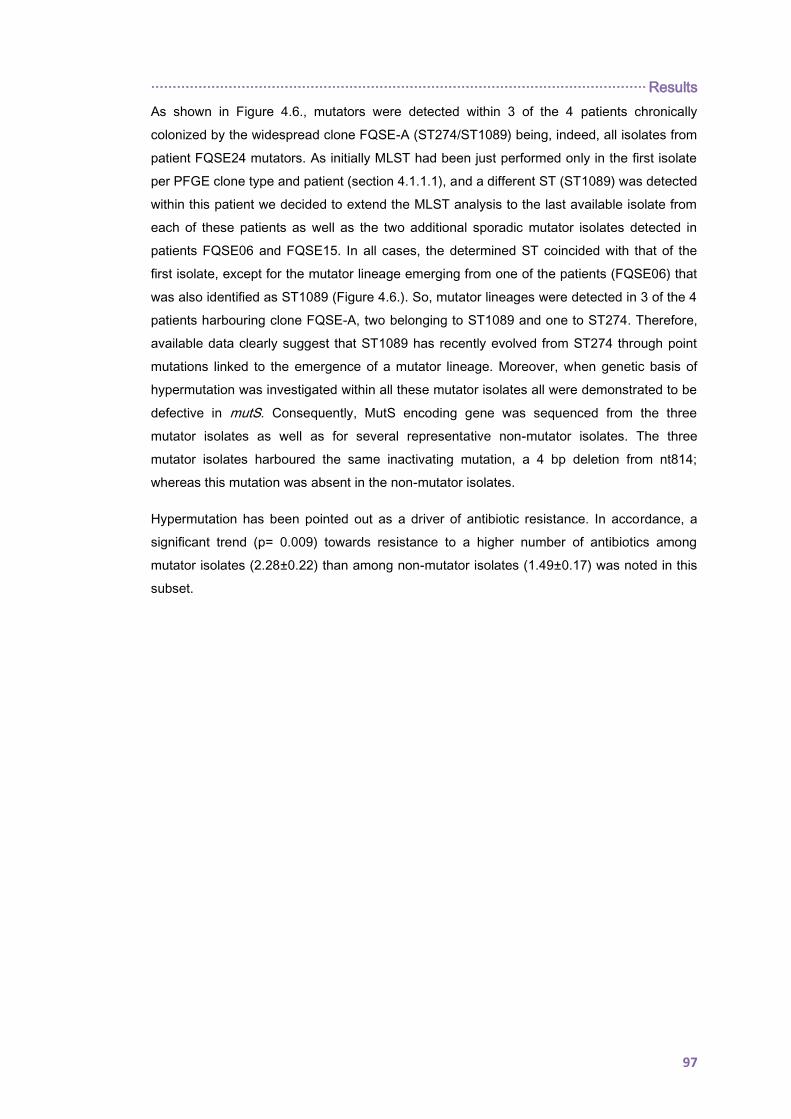
··················································································································· Results
97
As shown in Figure 4.6., mutators were detected within 3 of the 4 patients chronically
colonized by the widespread clone FQSE-A (ST274/ST1089) being, indeed, all isolates from
patient FQSE24 mutators. As initially MLST had been just performed only in the first isolate
per PFGE clone type and patient (section 4.1.1.1), and a different ST (ST1089) was detected
within this patient we decided to extend the MLST analysis to the last available isolate from
each of these patients as well as the two additional sporadic mutator isolates detected in
patients FQSE06 and FQSE15. In all cases, the determined ST coincided with that of the
first isolate, except for the mutator lineage emerging from one of the patients (FQSE06) that
was also identified as ST1089 (Figure 4.6.). So, mutator lineages were detected in 3 of the 4
patients harbouring clone FQSE-A, two belonging to ST1089 and one to ST274. Therefore,
available data clearly suggest that ST1089 has recently evolved from ST274 through point
mutations linked to the emergence of a mutator lineage. Moreover, when genetic basis of
hypermutation was investigated within all these mutator isolates all were demonstrated to be
defective in mutS. Consequently, MutS encoding gene was sequenced from the three
mutator isolates as well as for several representative non-mutator isolates. The three
mutator isolates harboured the same inactivating mutation, a 4 bp deletion from nt814;
whereas this mutation was absent in the non-mutator isolates.
Hypermutation has been pointed out as a driver of antibiotic resistance. In accordance, a
significant trend (p= 0.009) towards resistance to a higher number of antibiotics among
mutator isolates (2.28±0.22) than among non-mutator isolates (1.49±0.17) was noted in this
subset.
Results ··················································································································
98
4.2. Pseudomonas aeruginosa RESISTOME EVOLUTION IN CYSTIC
FIBROSIS CHRONIC RESPIRATORY INFECTIONS
4.2.1. Mutational resistome evolution of the international CC274 cystic
fibrosis clone
4.2.1.1. Prevalence and genetic basis for hypermutation
Among the CC274 study collection, nine isolates (31%) were mutators, belonging to six
(35%) different patients, residing in both Australia (n=3) and Spain (n=3). Data from
sequential isolates were available for the Spanish isolates: one was chronically infected with
a persistent mutator lineage (FQSE24), whereas the other two harboured a mixed population
of mutator and non-mutator isolates (FQSE06 and FQSE15) (Figure 3.2. and section 4.1.3.).
Sequence variation within an exhaustive panel of so called mutator genes (Annex 5) was
analyzed in all mutator and non-mutator isolates. The three Australian mutator isolates
showed unique mutations in either mutL or mutS, whereas all mutator isolates from the three
Spanish patients were found to share the same inactivating mutation in mutS.
Complementation studies with plasmids harbouring WT MMR system genes
(mutS and mutL) were performed in these mutator isolates. As shown in Table 4.12., WT
rifampicin resistance mutation frequencies were restored in all mutator isolates upon mutS or
mutL complementation, which correlated in all cases with the presence of specific mutations
in these genes. Of note, while mutator phenotypes could be explained in all cases by specific
mutations in MMR genes, the contrary was not always true, since one of the non-mutator
isolates (FQSE03) showed a missense mutation in mutS. Moreover, the presence of
polymorphisms in other mutator genes was frequent, but showed no association with mutator
phenotypes (Table 4.12.).
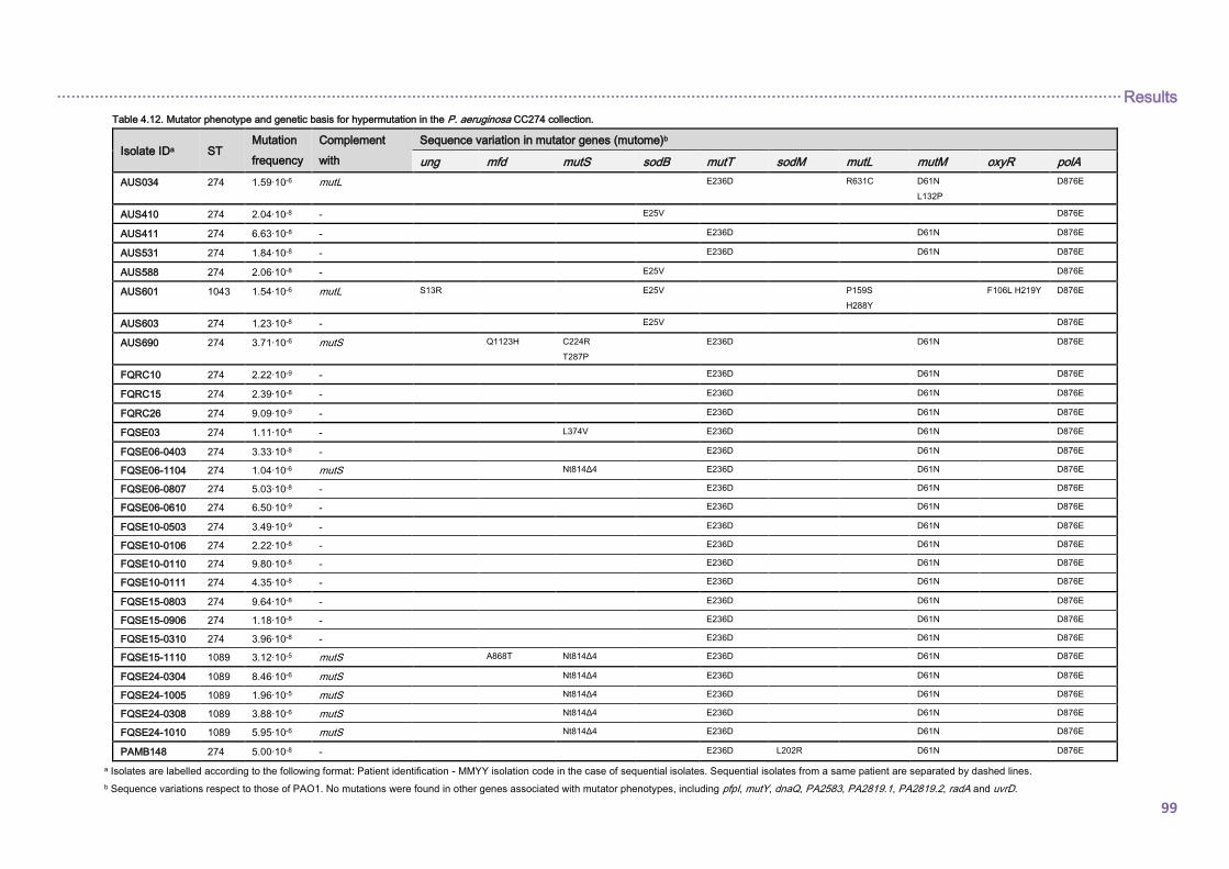
································································································································································································································· Results
99
Table 4.12. Mutator phenotype and genetic basis for hypermutation in the P. aeruginosa CC274 collection.
Isolate IDa ST Mutation
frequency
Complement
with
Sequence variation in mutator genes (mutome)b
ung mfd mutS sodB mutT sodM mutL mutM oxyR polA
AUS034 274 1.59·10-6 mutL E236D R631C D61N
L132P
D876E
AUS410 274 2.04·10-8 - E25V D876E
AUS411 274 6.63·10-8 - E236D D61N D876E
AUS531 274 1.84·10-8 - E236D D61N D876E
AUS588 274 2.06·10-8 - E25V D876E
AUS601 1043 1.54·10-6 mutL S13R E25V P159S
H288Y
F106L H219Y D876E
AUS603 274 1.23·10-8 - E25V D876E
AUS690 274 3.71·10-6 mutS Q1123H C224R
T287P
E236D D61N D876E
FQRC10 274 2.22·10-9 - E236D D61N D876E
FQRC15 274 2.39·10-8 - E236D D61N D876E
FQRC26 274 9.09·10-9 - E236D D61N D876E
FQSE03 274 1.11·10-8 - L374V E236D D61N D876E
FQSE06-0403 274 3.33·10-8 - E236D D61N D876E
FQSE06-1104 274 1.04·10-6 mutS Nt814Δ4 E236D D61N D876E
FQSE06-0807 274 5.03·10-8 - E236D D61N D876E
FQSE06-0610 274 6.50·10-9 - E236D D61N D876E
FQSE10-0503 274 3.49·10-9 - E236D D61N D876E
FQSE10-0106 274 2.22·10-8 - E236D D61N D876E
FQSE10-0110 274 9.80·10-8 - E236D D61N D876E
FQSE10-0111 274 4.35·10-8 - E236D D61N D876E
FQSE15-0803 274 9.64·10-8 - E236D D61N D876E
FQSE15-0906 274 1.18·10-8 - E236D D61N D876E
FQSE15-0310 274 3.96·10-8 - E236D D61N D876E
FQSE15-1110 1089 3.12·10-5 mutS A868T Nt814Δ4 E236D D61N D876E
FQSE24-0304 1089 8.46·10-6 mutS Nt814Δ4 E236D D61N D876E
FQSE24-1005 1089 1.96·10-5 mutS Nt814Δ4 E236D D61N D876E
FQSE24-0308 1089 3.88·10-6 mutS Nt814Δ4 E236D D61N D876E
FQSE24-1010 1089 5.95·10-6 mutS Nt814Δ4 E236D D61N D876E
PAMB148 274 5.00·10-8 - E236D L202R D61N D876E
a Isolates are labelled according to the following format: Patient identification - MMYY isolation code in the case of sequential isolates. Sequential isolates from a same patient are separated by dashed lines.
b Sequence variations respect to those of PAO1. No mutations were found in other genes associated with mutator phenotypes, including pfpI, mutY, dnaQ, PA2583, PA2819.1, PA2819.2, radA and uvrD.
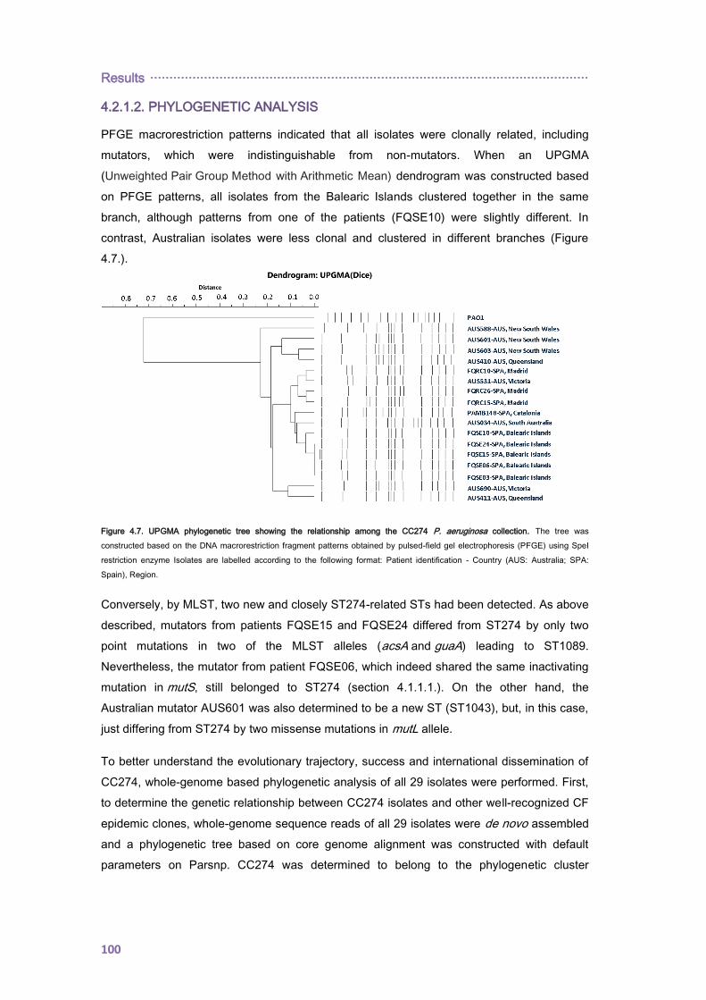
Results ··················································································································
100
4.2.1.2. PHYLOGENETIC ANALYSIS
PFGE macrorestriction patterns indicated that all isolates were clonally related, including
mutators, which were indistinguishable from non-mutators. When an UPGMA
(Unweighted Pair Group Method with Arithmetic Mean) dendrogram was constructed based
on PFGE patterns, all isolates from the Balearic Islands clustered together in the same
branch, although patterns from one of the patients (FQSE10) were slightly different. In
contrast, Australian isolates were less clonal and clustered in different branches (Figure
4.7.).
Figure 4.7. UPGMA phylogenetic tree showing the relationship among the CC274 P. aeruginosa collection. The tree was
constructed based on the DNA macrorestriction fragment patterns obtained by pulsed-field gel electrophoresis (PFGE) using SpeI
restriction enzyme Isolates are labelled according to the following format: Patient identification - Country (AUS: Australia; SPA:
Spain), Region.
Conversely, by MLST, two new and closely ST274-related STs had been detected. As above
described, mutators from patients FQSE15 and FQSE24 differed from ST274 by only two
point mutations in two of the MLST alleles (acsA and guaA) leading to ST1089.
Nevertheless, the mutator from patient FQSE06, which indeed shared the same inactivating
mutation in mutS, still belonged to ST274 (section 4.1.1.1.). On the other hand, the
Australian mutator AUS601 was also determined to be a new ST (ST1043), but, in this case,
just differing from ST274 by two missense mutations in mutL allele.
To better understand the evolutionary trajectory, success and international dissemination of
CC274, whole-genome based phylogenetic analysis of all 29 isolates were performed. First,
to determine the genetic relationship between CC274 isolates and other well-recognized CF
epidemic clones, whole-genome sequence reads of all 29 isolates were de novo assembled
and a phylogenetic tree based on core genome alignment was constructed with default
parameters on Parsnp. CC274 was determined to belong to the phylogenetic cluster
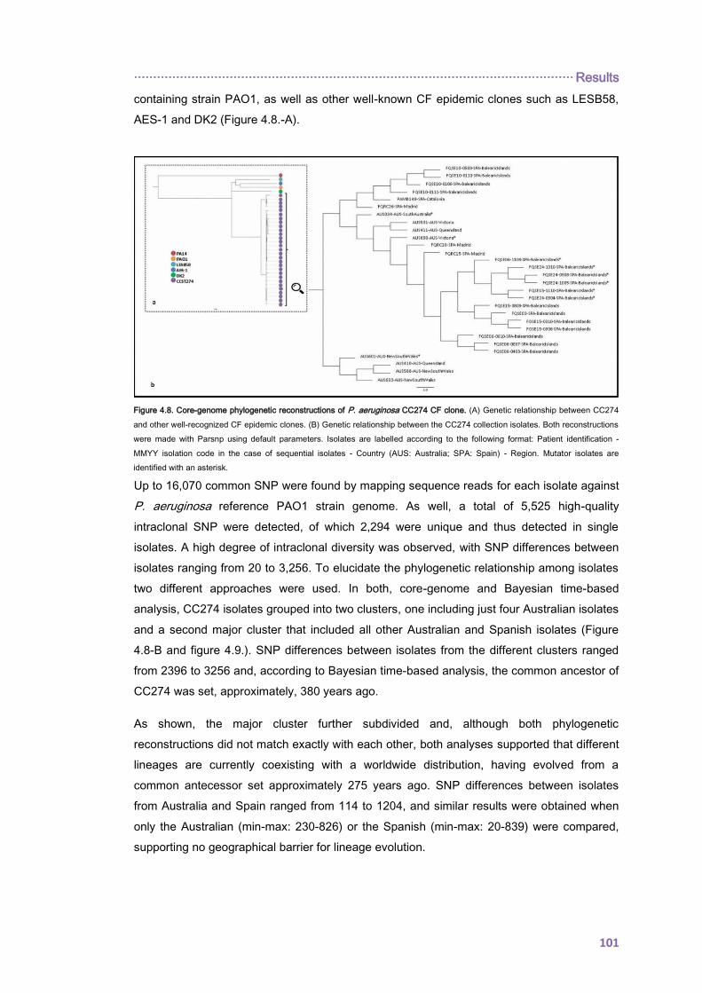
··················································································································· Results
101
containing strain PAO1, as well as other well-known CF epidemic clones such as LESB58,
AES-1 and DK2 (Figure 4.8.-A).
Up to 16,070 common SNP were found by mapping sequence reads for each isolate against
P. aeruginosa reference PAO1 strain genome. As well, a total of 5,525 high-quality
intraclonal SNP were detected, of which 2,294 were unique and thus detected in single
isolates. A high degree of intraclonal diversity was observed, with SNP differences between
isolates ranging from 20 to 3,256. To elucidate the phylogenetic relationship among isolates
two different approaches were used. In both, core-genome and Bayesian time-based
analysis, CC274 isolates grouped into two clusters, one including just four Australian isolates
and a second major cluster that included all other Australian and Spanish isolates (Figure
4.8-B and figure 4.9.). SNP differences between isolates from the different clusters ranged
from 2396 to 3256 and, according to Bayesian time-based analysis, the common ancestor of
CC274 was set, approximately, 380 years ago.
As shown, the major cluster further subdivided and, although both phylogenetic
reconstructions did not match exactly with each other, both analyses supported that different
lineages are currently coexisting with a worldwide distribution, having evolved from a
common antecessor set approximately 275 years ago. SNP differences between isolates
from Australia and Spain ranged from 114 to 1204, and similar results were obtained when
only the Australian (min-max: 230-826) or the Spanish (min-max: 20-839) were compared,
supporting no geographical barrier for lineage evolution.
1 Figure 4.8. Core-genome phylogenetic reconstructions of P. aeruginosa CC274 CF clone. (A) Genetic relationship between CC274
and other well-recognized CF epidemic clones. (B) Genetic relationship between the CC274 collection isolates. Both reconstructions
were made with Parsnp using default parameters. Isolates are labelled according to the following format: Patient identification -
MMYY isolation code in the case of sequential isolates - Country (AUS: Australia; SPA: Spain) - Region. Mutator isolates are
identified with an asterisk.
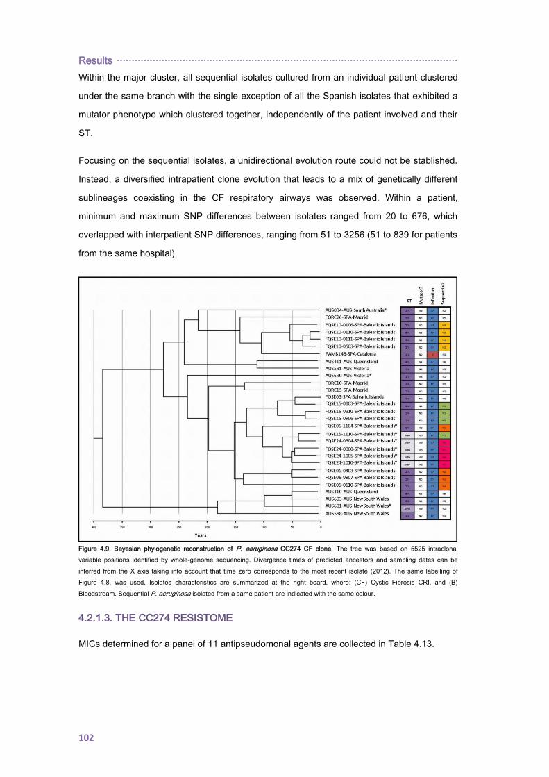
Results ··················································································································
102
Within the major cluster, all sequential isolates cultured from an individual patient clustered
under the same branch with the single exception of all the Spanish isolates that exhibited a
mutator phenotype which clustered together, independently of the patient involved and their
ST.
Focusing on the sequential isolates, a unidirectional evolution route could not be stablished.
Instead, a diversified intrapatient clone evolution that leads to a mix of genetically different
sublineages coexisting in the CF respiratory airways was observed. Within a patient,
minimum and maximum SNP differences between isolates ranged from 20 to 676, which
overlapped with interpatient SNP differences, ranging from 51 to 3256 (51 to 839 for patients
from the same hospital).
Figure 4.9. Bayesian phylogenetic reconstruction of P. aeruginosa CC274 CF clone. The tree was based on 5525 intraclonal
variable positions identified by whole-genome sequencing. Divergence times of predicted ancestors and sampling dates can be
inferred from the X axis taking into account that time zero corresponds to the most recent isolate (2012). The same labelling of
Figure 4.8. was used. Isolates characteristics are summarized at the right board, where: (CF) Cystic Fibrosis CRI, and (B)
Bloodstream. Sequential P. aeruginosa isolated from a same patient are indicated with the same colour.
4.2.1.3. THE CC274 RESISTOME
MICs determined for a panel of 11 antipseudomonal agents are collected in Table 4.13.
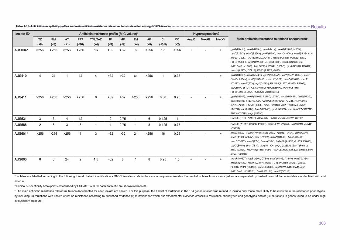
································································································································································································································· Results
103
Isolate IDa Antibiotic resistance profile (MIC values)b Hyperexpression?
Main antibiotic resistance mutations encounteredc TZ
(≤8)
PM
(≤8)
AT
(≤1)
PPT
(≤16)
TOL/TAZ
(≤4)
IP
(≤4)
MP
(≤2)
TM
(≤4)
AK
(≤8)
CI
(≤0.5)
CO
(≤2)
AmpC MexAB MexXY
AUS034* >256 >256 >256 >256 16 >32 >32 6 >256 1.5 >256 + - + gyrB (R441L), mexR (R85H), mexA (M1X), mexB (F178S, M555I),
oprD(E264X), phoQ(E266X), parR (M59I), mexY(V1000L), mexZ(Nt334Δ13),
fusA2(P329L), PA2489(R12L, A244T), mexS (P254Q), mexT(L157M),
PBP4(W350R), capD (I7M, S51G), gyrA(T83I), mexK (S426G), mpl
(Nt112ins1, V124G), fusA1 (V93A, P554L, D588G), rpoB (D831G, D964G ),
mexW (A627V, Q771P), PBP3 (P527T, G63S)
AUS410 4 24 1 12 4 >32 >32 64 >256 1 0.38 - - + gyrB (S466F), mexB(M552T), oprD (Nt583Δ1), lasR (A50V, D73G), sucC
(V44G, A384V), oprF (Nt574Δ31), mexY (V32A), mexZ (Q164X), mexT
(D327Y), mexE (F7Y), mpl (D168Y), PA2489(A125T, G185S, P260S),
capD(I7M, S51G), fusA1(P618L), rpoC(E386K), mexW(Q511R),
PBP3(G216S), pagL(Nt286Δ1), amgS(S64L)
AUS411 >256 >256 >256 >256 6 >32 >32 >256 >256 0.38 0.25 - - + gyrB (S466F), mexB (Q104E, F246C, L376V), phoQ (H248P), lasR (D73G),
parS (D381E, T163N), sucC (C261G), mexY (D201A, G287A), PA2489
(R12L, A244T), fusA2 (I640L), mexE (V104G), htpX (Nt683Δ5), mexK
(S426G), capD (I7M), fusA1 (K504E), rpoC (N690S), mexW (A627V,Q771P),
PBP3 (Q372P), pagL (N159D)
AUS531 3 3 4 12 1 2 0.75 1 6 0.125 1 - - - PA2489 (R12L, A244T), capD (I7M, S51G), mexW (A627V, Q771P)
AUS588 2 8 3 8 1 1 0.75 1 8 0.125 0.75 - - - PA2489 (A125T, G185S, P260S), mexE (F7Y, V276M), capD (I7M), mexW
(Q511R)
AUS601* >256 >256 >256 1 3 >32 >32 24 >256 16 0.25 - - + mexB (M552T), oprD (Nt1044ins4), phoQ (K234N, T315A), lasR (A50V),
sucC (T102I, A384V), mexY (V32A), mexZ (Q164X), fusA2 (S445X),
mexT(D327Y), mexE(F7Y), ftsK (A152V), PA2489 (A125T, G185S, P260S),
capD (S51G), gyrA (T83I), mpl (G113D), ampC (V239A), fusA1 (P618L),
rpoC (E386K), mexW (Q511R), PBP3 (R504C), pagL (E163G), pmrB (L31P),
amgR (E204D)
AUS603 6 8 24 2 1.5 >32 8 1 8 0.25 1.5 + - + mexB (M552T), lasR (A50V, D73G), sucC (V44G, A384V), mexY (V32A),
mexZ (Q164X), mexT (D327Y), mexE (F7Y), PA2489 (A125T, G185S,
P260S), PBP4 (S315G), opmE (E204D), capD (I7M, Nt1438Δ1), mpl
(Nt112ins1, Nt1317Δ1), fusA1 (P618L), mexW (Q511R)
a Isolates are labelled according to the following format: Patient identification - MMYY isolation code in the case of sequential isolates. Sequential isolates from a same patient are separated by dashed lines. Mutators isolates are identified with and
asterisk.
b Clinical susceptibility breakpoints established by EUCAST v7.0 for each antibiotic are shown in brackets.
c The main antibiotic resistance related mutations documented for each isolate are shown. For this purpose, the full list of mutations in the 164 genes studied was refined to include only those more likely to be involved in the resistance phenotypes,
by including: (i) mutations with known effect on resistance according to published evidence (ii) mutations for which our experimental evidence crosslinks resistance phenotypes and genotypes and/or (iii) mutations in genes found to be under high
evolutionary pressure.
Table 4.13. Antibiotic susceptibility profiles and main antibiotic resistance related mutations detected among CC274 isolates.

Results ································································································································································································································
104
Isolate IDa Antibiotic resistance profile (MIC values)b Hyperexpression?
Main antibiotic resistance mutations encounteredc TZ
(≤8)
PM
(≤8)
AT
(≤1)
PPT
(≤16)
TOL/TAZ
(≤4)
IP
(≤4)
MP
(≤2)
TM
(≤4)
AK
(≤8)
CI
(≤0.5)
CO
(≤2)
AmpC MexAB MexXY
AUS690* 6 12 0.75 3 6 4 2 24 >256 12 0.125 - + + gyrB (Q467R), mexR (H133P), mexB (Nt712Δ1), phoP (T221I), lasR (T178I),
parS (L10P), oprF (K250R), mexY (G402S, A850T), mexZ (Nt529Δ1),
PA2489 (R12L, A244T), fusA2 (L104P, Nt889Δ1), htpX (G187D), capD (I7M,
S51G), gyrA (T83A, T325I), mexK (G487E), mexH (Nt1086ins1), fusA1
(Y552C, T671I), rpoC (E136G, D616G, V808L), rpoB (F1046S), mexW
(A627V, Q771P), pagL (P158L), pmrB (F124L), amgS (R188C), parE
(P438S)
FQRC10 2 2 4 12 1 1.5 1 1 8 0.094 0.5 - - - PA2489 (R12L, A244T), capD (I7M, S51G), mexH (D356N), mexW (A627V,
Q771P)
FQRC15 1 0.75 6 6 1 1.5 1 0.75 8 0.19 1 - - - PA2489 (R12L, A244T), capD (I7M), mexW (A627V, Q771P)
FQRC26 4 6 24 24 1 0.25 1.5 1 6 1.5 0.38 - + - mexY (V875M), mexT (R164H), PA2489 (R12L, A244T), capD (I7M, S51G),
gyrA(Q106L), mexW (A627V, Q771P)
FQSE03 3 8 0.5 2 1.5 2 0.38 1 6 3 0.25 - - + mexA (L338P), lasR (P117G), mexZ (A144V), PA2489 (R12L, A244T), capD
(I7M, S51G), gyrA (D87N), mexW (A627V, Q771P)
FQSE06-
0403
0.75 2 0.25 4 0.38 1 0.5 24 16 0.19 0.19 - - + mexA (L338P), lasR (P117G), mexY (G287A), mexZ (S9P), PA2489 (R12L,
A244T), mpl (S257L), capD (I7M, S51G), fusA1 (Y552C, T671I), mexW
(A627V, Q771P), PBP3 (P215L), amgR (A8V)
FQSE06-
1104*
0.38 1 0.094 0.38 0.38 6 0.19 1 24 0.75 2 - - + mexA (L338P), lasR (P117G), mexZ (A194P), PA2489 (R12L, A244T), fusA2
(N236S, N561S), capD (I7M, S51G), gyrA (D87G), mexK (Q585X), rpoB
(Y583C), mexW (A627V, Q771P), pmrB (V185I, G221D, R287Q), PBP1A
(E161G), amgR(A8V)
FQSE06-
0807
4 8 0.75 4 2 1.5 0.75 24 >256 0.5 1 - - + mexA (L338P), lasR (P117G), mexY (G287A), mexZ (S9P), mexT (P270Q),
PA2489 (R12L, A244T), mpl (S257L), capD (I7M, S51G), fusA1 (N482S,
Y552C, T671I), mexW (A627V, Q771P), PBP3 (P215L), amgR (A8V)
FQSE06-
0610
4 24 0.75 8 1.5 1 0.25 1.5 24 0.75 0.19 - - + mexA (L338P), lasR (P117G), mexZ (Nt290Δ11), PA2489 (R12L, A244T),
mexW (A627V, Q771P), capD (I7M, S51G), amgR (A8V)
a Isolates are labelled according to the following format: Patient identification - MMYY isolation code in the case of sequential isolates. Sequential isolates from a same patient are separated by dashed lines. Mutators isolates are identified with and
asterisk.
b Clinical susceptibility breakpoints established by EUCAST v7.0 for each antibiotic are shown in brackets.
c The main antibiotic resistance related mutations documented for each isolate are shown. For this purpose, the full list of mutations in the 164 genes studied was refined to include only those more likely to be involved in the resistance phenotypes,
by including: (i) mutations with known effect on resistance according to published evidence (ii) mutations for which our experimental evidence crosslinks resistance phenotypes and genotypes and/or (iii) mutations in genes found to be under high
evolutionary pressure.
Table 4.13. Antibiotic susceptibility profiles and main antibiotic resistance related mutations detected among CC274 isolates. (Cont.)
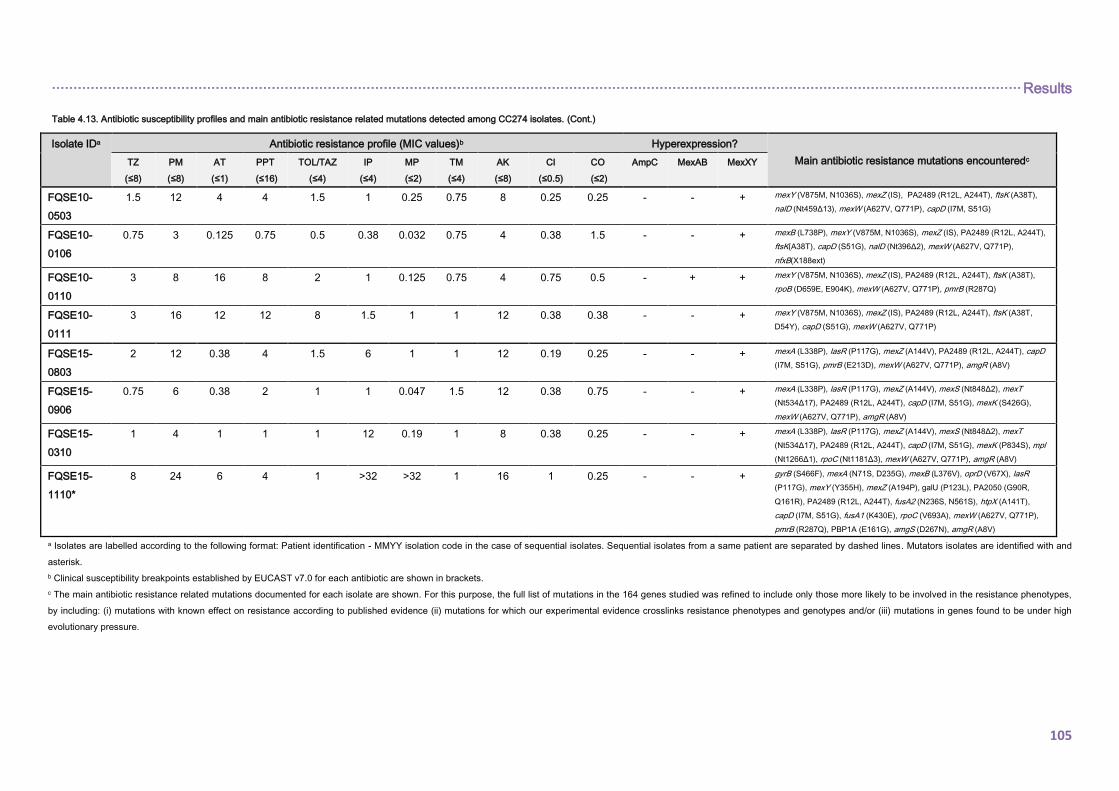
································································································································································································································· Results
105
Isolate IDa Antibiotic resistance profile (MIC values)b Hyperexpression?
Main antibiotic resistance mutations encounteredc TZ
(≤8)
PM
(≤8)
AT
(≤1)
PPT
(≤16)
TOL/TAZ
(≤4)
IP
(≤4)
MP
(≤2)
TM
(≤4)
AK
(≤8)
CI
(≤0.5)
CO
(≤2)
AmpC MexAB MexXY
FQSE10-
0503
1.5 12 4 4 1.5 1 0.25 0.75 8 0.25 0.25 - - + mexY (V875M, N1036S), mexZ (IS), PA2489 (R12L, A244T), ftsK (A38T),
nalD (Nt459Δ13), mexW (A627V, Q771P), capD (I7M, S51G)
FQSE10-
0106
0.75 3 0.125 0.75 0.5 0.38 0.032 0.75 4 0.38 1.5 - - + mexB (L738P), mexY (V875M, N1036S), mexZ (IS), PA2489 (R12L, A244T),
ftsK(A38T), capD (S51G), nalD (Nt396Δ2), mexW (A627V, Q771P),
nfxB(X188ext)
FQSE10-
0110
3 8 16 8 2 1 0.125 0.75 4 0.75 0.5 - + + mexY (V875M, N1036S), mexZ (IS), PA2489 (R12L, A244T), ftsK (A38T),
rpoB (D659E, E904K), mexW (A627V, Q771P), pmrB (R287Q)
FQSE10-
0111
3 16 12 12 8 1.5 1 1 12 0.38 0.38 - - + mexY (V875M, N1036S), mexZ (IS), PA2489 (R12L, A244T), ftsK (A38T,
D54Y), capD (S51G), mexW (A627V, Q771P)
FQSE15-
0803
2 12 0.38 4 1.5 6 1 1 12 0.19 0.25 - - + mexA (L338P), lasR (P117G), mexZ (A144V), PA2489 (R12L, A244T), capD
(I7M, S51G), pmrB (E213D), mexW (A627V, Q771P), amgR (A8V)
FQSE15-
0906
0.75 6 0.38 2 1 1 0.047 1.5 12 0.38 0.75 - - + mexA (L338P), lasR (P117G), mexZ (A144V), mexS (Nt848Δ2), mexT
(Nt534Δ17), PA2489 (R12L, A244T), capD (I7M, S51G), mexK (S426G),
mexW (A627V, Q771P), amgR (A8V)
FQSE15-
0310
1 4 1 1 1 12 0.19 1 8 0.38 0.25 - - + mexA (L338P), lasR (P117G), mexZ (A144V), mexS (Nt848Δ2), mexT
(Nt534Δ17), PA2489 (R12L, A244T), capD (I7M, S51G), mexK (P834S), mpl
(Nt1266Δ1), rpoC (Nt1181Δ3), mexW (A627V, Q771P), amgR (A8V)
FQSE15-
1110*
8 24 6 4 1 >32 >32 1 16 1 0.25 - - + gyrB (S466F), mexA (N71S, D235G), mexB (L376V), oprD (V67X), lasR
(P117G), mexY (Y355H), mexZ (A194P), galU (P123L), PA2050 (G90R,
Q161R), PA2489 (R12L, A244T), fusA2 (N236S, N561S), htpX (A141T),
capD (I7M, S51G), fusA1 (K430E), rpoC (V693A), mexW (A627V, Q771P),
pmrB (R287Q), PBP1A (E161G), amgS (D267N), amgR (A8V)
a Isolates are labelled according to the following format: Patient identification - MMYY isolation code in the case of sequential isolates. Sequential isolates from a same patient are separated by dashed lines. Mutators isolates are identified with and
asterisk.
b Clinical susceptibility breakpoints established by EUCAST v7.0 for each antibiotic are shown in brackets.
c The main antibiotic resistance related mutations documented for each isolate are shown. For this purpose, the full list of mutations in the 164 genes studied was refined to include only those more likely to be involved in the resistance phenotypes,
by including: (i) mutations with known effect on resistance according to published evidence (ii) mutations for which our experimental evidence crosslinks resistance phenotypes and genotypes and/or (iii) mutations in genes found to be under high
evolutionary pressure.
Table 4.13. Antibiotic susceptibility profiles and main antibiotic resistance related mutations detected among CC274 isolates. (Cont.)
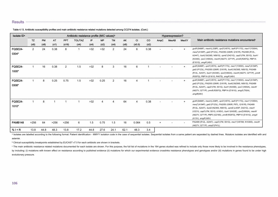
Results ································································································································································································································
106
Isolate IDa Antibiotic resistance profile (MIC values)b Hyperexpression?
Main antibiotic resistance mutations encounteredc TZ
(≤8)
PM
(≤8)
AT
(≤1)
PPT
(≤16)
TOL/TAZ
(≤4)
IP
(≤4)
MP
(≤2)
TM
(≤4)
AK
(≤8)
CI
(≤0.5)
CO
(≤2)
AmpC MexAB MexXY
FQSE24-
0304*
2 24 0.38 8 1 >32 >32 2 24 6 0.38 - - + gyrB (S466F), mexA (L338P), oprD (V67X), lasR (P117G), mexY (Y355H),
mexZ (A194P), galU (P123L), PA2050 (G90R, Q161R), PA2489 (R12L,
A244T), fusA2 (N236S, N561S), opmE (D421G), capD (I7M, S51G), fusA1
(K430E), rpoC (V693A), mexW (A627V, Q771P), pmrB (R287Q), PBP1A
(E161G), amgR (A8V)
FQSE24-
1005*
1 16 0.38 2 1.5 >32 8 3 16 6 1 - - + gyrB (S466F), oprD (V67X), lasR (P117G), mexY (Y355H), mexZ (A194P),
galU (P123L), PA2050 (G90R, Q161R), fusA2 (N236S, N561S), PA2489
(R12L, A244T), fusA1 (K430E), rpoC(V693A), mexW (A627V, Q771P), pmrB
(R287Q), PBP1A (E161G, R407S), amgR (A8V)
FQSE24-
0308*
1 8 0.25 0.75 1.5 >32 0.25 2 16 4 1 - - + gyrB (S466F), oprD (V67X), lasR (P117G), mexY (Y355H), mexZ (A194P),
galU (P123L), PA2050 (G90R, Q161R), fusA2 (N236S, N561S), PA2489
(R12L, A244T), capD (I7M, S51G), fusA1 (K430E), rpoC (V693A), mexW
(A627V, Q771P), pmrB (R287Q), PBP1A (E161G), amgS (T92A),
amgR(A8V)
FQSE24-
1010*
1 8 1 1 1 >32 4 4 64 4 0.38 - - + gyrB (S466F), mexA (L338P), oprD (V67X), lasR (P117G), mexY (Y355H),
mexZ (A194P), galU (P123L), PA2050 (G90R, P97L, Q161R), PA2489
(R12L, A244T), fusA2 (N236S, N561S), opmE (L400P, D421G), mexH
(V221I), capD (I7M, S51G, A165V), fusA1 (K430E), rpoC(V693A), mexW
(A627V, Q771P), PBP3 (G216S), pmrB (R287Q), PBP1A (E161G), amgS
(A13V), amgR (A8V)
PAMB148 >256 64 >256 >256 6 1.5 0.75 1.5 16 0.064 0.5 + - - PA2489 (R12L, A244T), capD (I7M, S51G), mexY (V875M, N1036S), mexW
(A627V, Q771P), ampD (P41L)
% I + R 13.8 44.8 48.3 13.8 17.2 44.8 27.6 24.1 62.1 48.3 3.4
a Isolates are labelled according to the following format: Patient identification - MMYY isolation code in the case of sequential isolates. Sequential isolates from a same patient are separated by dashed lines. Mutators isolates are identified with and
asterisk.
b Clinical susceptibility breakpoints established by EUCAST v7.0 for each antibiotic are shown in brackets.
c The main antibiotic resistance related mutations documented for each isolate are shown. For this purpose, the full list of mutations in the 164 genes studied was refined to include only those more likely to be involved in the resistance phenotypes,
by including: (i) mutations with known effect on resistance according to published evidence (ii) mutations for which our experimental evidence crosslinks resistance phenotypes and genotypes and/or (iii) mutations in genes found to be under high
evolutionary pressure.
Table 4.13. Antibiotic susceptibility profiles and main antibiotic resistance related mutations detected among CC274 isolates. (Cont.)
··················································································································· Results
107
As shown, resistance rates were lowest for colistin (3.4%), distantly followed by ceftazidime
and piperacillin/tazobactam (13.8%). In contrast, resistance to cefepime, aztreonam,
imipenem, amikacin and ciprofloxacin was observed in 44.8 to 62% of the isolates.
Remarkably, 17.2% of the isolates were resistant to the new combination
ceftolozane/tazobactam. As well, antibiotic resistance was shown to be more frequent
among mutators, and in Australian isolates in comparison with those from Spain. In fact, all 9
mutator isolates were classified as MDR, as compared to only 3 of 20 non-mutators.
Moreover, one of the Australian mutator isolates met the pan-drug resistant (PDR) definition.
The presence of horizontally acquired resistance determinants was explored in the whole-
genome sequences using the ResFinder tool. None of the 29 isolates harbored any
horizontally acquired genes encoding resistance determinants. The complete list of
missense and non-sense mutations encountered in the 164 antibiotic resistance related
chromosomal genes investigated (n=164, Annex 5) can be downloaded in the following link
https://www.nature.com/articles/s41598-017-05621-5; as well, a summary of these mutations
by antibiotic class has been collected in Annex 6. Up to 127 (77.4%) of the 164 studied
genes showed non-synonymous mutations in at least one of the isolates studied. Moreover,
after discarding non-synonymous mutations present in all isolates (and thus considered
intrinsic CC274 polymorphisms), this figure only decreased to 106 (64.6%). The number and
distribution of mutations among the 164 antibiotic resistance related genes studied in the
CC274 collection has been represented in Figure 4.10. Seventy-three (68.9%) of these
genes showed no more than two different mutational events being 44 of them mutated in
unique isolates. In contrast, 33 (31.1%) genes appeared to be under high evolutionary
pressure showing evidence of at least 3 different mutational events. Particularly noteworthy
among them were mexB or mexY, (coding for efflux pumps proteins), mexZ (the main
MexXY repressor), gyrA (which codes for DNA gyrase subunit A) and fusA1 (coding for the
elongation factor G).
The main antibiotic resistance related mutations documented are listed in Table 4.13. For
this purpose, the full list of mutations in the 164 genes studied was refined to include only
those more likely to be involved in the resistance phenotypes, by including: (i) mutations with
known effect on resistance according to published evidence, (ii) mutations for which our
experimental evidence crosslinks resistance phenotypes and genotypes (e.g. mutations in
genes involved in AmpC, efflux or OprD regulation and β-lactam resistance phenotypes are
crosslinked by integrating the analysis of the expression of ampC, efflux pumps genes and
oprD) and/or (iii) mutations in genes found to be under high evolutionary pressure (those
with at least 3 different mutational events documented). Overall, the number of mutations
was much higher (unpaired T test p<0.0001) in mutator (19.2±3.1) than in non-mutator
isolates (6.7±3.1). Nevertheless, some Australian non-mutator isolates (e.g. AUS410 or
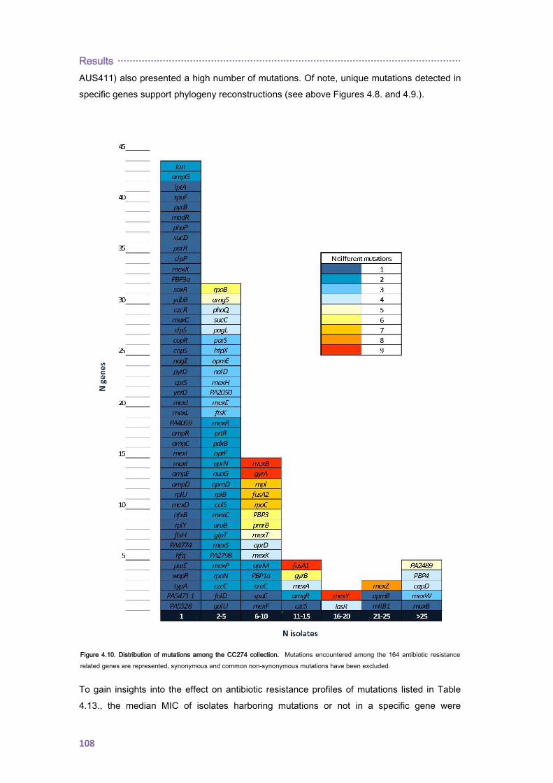
Results ··················································································································
108
AUS411) also presented a high number of mutations. Of note, unique mutations detected in
specific genes support phylogeny reconstructions (see above Figures 4.8. and 4.9.).
To gain insights into the effect on antibiotic resistance profiles of mutations listed in Table
4.13., the median MIC of isolates harboring mutations or not in a specific gene were
Figure 4.10. Distribution of mutations among the CC274 collection. Mutations encountered among the 164 antibiotic resistance
related genes are represented, synonymous and common non-synonymous mutations have been excluded.
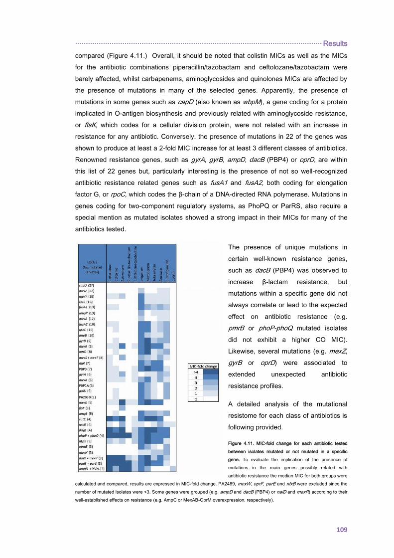
··················································································································· Results
109
compared (Figure 4.11.) Overall, it should be noted that colistin MICs as well as the MICs
for the antibiotic combinations piperacillin/tazobactam and ceftolozane/tazobactam were
barely affected, whilst carbapenems, aminoglycosides and quinolones MICs are affected by
the presence of mutations in many of the selected genes. Apparently, the presence of
mutations in some genes such as capD (also known as wbpM), a gene coding for a protein
implicated in O-antigen biosynthesis and previously related with aminoglycoside resistance,
or ftsK, which codes for a cellular division protein, were not related with an increase in
resistance for any antibiotic. Conversely, the presence of mutations in 22 of the genes was
shown to produce at least a 2-fold MIC increase for at least 3 different classes of antibiotics.
Renowned resistance genes, such as gyrA, gyrB, ampD, dacB (PBP4) or oprD, are within
this list of 22 genes but, particularly interesting is the presence of not so well-recognized
antibiotic resistance related genes such as fusA1 and fusA2, both coding for elongation
factor G, or rpoC, which codes the β-chain of a DNA-directed RNA polymerase. Mutations in
genes coding for two-component regulatory systems, as PhoPQ or ParRS, also require a
special mention as mutated isolates showed a strong impact in their MICs for many of the
antibiotics tested.
The presence of unique mutations in
certain well-known resistance genes,
such as dacB (PBP4) was observed to
increase β-lactam resistance, but
mutations within a specific gene did not
always correlate or lead to the expected
effect on antibiotic resistance (e.g.
pmrB or phoP-phoQ mutated isolates
did not exhibit a higher CO MIC).
Likewise, several mutations (e.g. mexZ,
gyrB or oprD) were associated to
extended unexpected antibiotic
resistance profiles.
A detailed analysis of the mutational
resistome for each class of antibiotics is
following provided.
Figure 4.11. MIC-fold change for each antibiotic tested
between isolates mutated or not mutated in a specific
gene. To evaluate the implication of the presence of
mutations in the main genes possibly related with
antibiotic resistance the median MIC for both groups were
calculated and compared, results are expressed in MIC-fold change. PA2489, mexW, oprF, parE and nfxB were excluded since the
number of mutated isolates were <3. Some genes were grouped (e.g. ampD and dacB (PBP4) or nalD and mexR) according to their
well-established effects on resistance (e.g. AmpC or MexAB-OprM overexpression, respectively).
Results ··················································································································
110
β-lactam resistome. Just three isolates (AUS034, AUS603 and PAMB148) of the CC274
collection were demonstrated to overproduce AmpC. By contrast, at the genomic level,
almost all isolates (26/29) contained some variation within dacB which codes for the PBP4.
Crosslinking phenotypic and genotypic results through ampC expression data, suggested
that most observed dacB allele variations were, in fact, ancestral polymorphisms not
involved in antibiotic resistance. AmpC overproduction in the two CF isolates was explained
by the presence of specific mutations in dacB (S315G or W350R) and by an ampD (P41L)
mutation in the case of the bloodstream infection isolate PAMB148. Whilst ampC
overexpression in isolates AUS034 and PAMB148 correlated well with ceftazidime and
piperacillin/tazobactam resistance, this was not the case for isolate AUS603 which was
documented to be susceptible to these antibiotics. However, unexpected AUS603 β-lactam
susceptibility could be explained by the presence of chromosomal mutations whose effects
eventually compensate the expected increase in β-lactam resistance. Indeed, this isolate
showed an additional non-sense mutation in OprM (Q93X), the OMP of the constitutive
MexAB-OprM efflux pump which is well known to play a major role in intrinsic β-lactam
resistance. As well, it should also be mentioned that isolate AUS601, exhibiting high-level
resistance to ceftazidime, cefepime and aztreonam in the absence of AmpC overproduction,
showed an additional mutation (V239A) in AmpC compared to all other CC274 isolates
(Table 4.13, Annex 6-β-lactams).
On the other hand, numerous sequence variations were encountered within the essential
PBPs coding genes. While some unique mutations were detected in genes coding for PBP1
and PBP3a, the main mutational resistance target among PBPs was found to be PBP3. Up
to 7 of 29 isolates presented a mutated PBP3 nucleotide sequence, although β-lactam
resistance contribution of each derived ftsI (PBP3) allele, if any, depends on the specific
point mutation encountered. Missense mutations within the PBP3 (R504C and Q372P) were
apparently contributing to resistance in isolates AUS601 and AUS411, since they do not
hyperproduce AmpC. Likewise, the P527T mutation of AUS034 likely contributes, together
with AmpC overexpression, to the very high-level β-lactam resistance of this isolate,
including the new antipseudomonal combination ceftolozane/tazobactam. On the other hand,
the P215L and G216S mutations were apparently not linked with phenotypic resistance
(Table 4.13. and Annex 6-β-lactams).
Obtained data also demonstrated that the constitutive efflux pump MexAB-OprM is under
strong mutational pressure during CF CRI, frequently including inactivating mutations. On
the contrary, just 3 isolates showed mutations in regulators leading to MexAB-OprM
overexpression (Table 4.13 and Annex 6- β-lactams).
Carbapenem resistome. Imipenem and meropenem resistance correlated in all but two
isolates with the presence of non-sense mutations affecting the OMP OprD. High-level
meropenem resistance was additionally associated with the presence of PBP3 mutations.
··················································································································· Results
111
Remarkably, all ST1089 mutator isolates shared the same point mutation in oprD (V67X) as
well as in galU (P123L), also related with carbapenem resistance (Table 4.13. and Annex 6-
Carbapenems).
MexEF-OprN overexpression was documented just in isolate FQRC26 and MexAB-OprM in
3 isolates; being all susceptible to both carbapenems tested. On the contrary, resistant
isolates AUS411 and AUS603 exhibited WT OprD sequences and no efflux pump
overexpression was demonstrated (Table 4.13. and Annex 6-Carbapenems).
Aminoglycoside resistome. Among the CC274 collection, a high proportion of the isolates
(23/29) were shown to overexpress MexXY and all but one were mutated in mexZ, which
codes for the mayor MexXY expression regulator. Remarkably, the same point mutation was
detected among different and independent isolates. The single MexXY-overproducing isolate
showing no mutations in mexZ, presented a unique mutation in parS, a gene also involved in
the modulation of MexXY expression. Nevertheless, MexXY hyperproduction per se cannot
explain aminoglycoside resistance in the majority of the isolates. Of note, all high-level
resistant isolates hyperproduced MexXY and harboured additional mutations in both genes
coding for elongation factor G, fusA1 and fusA2 (Table 4.13. and Annex 6-Aminoglycosides).
Fluoroquinolone resistome. Obtained data suggest that contribution of efflux pumps
overexpression to high-level resistance to fluoroquinolones is very limited, if any. As shown,
just isolate FQSE10-0106 (MIC=0.38 mg/L) was demonstrated to hyperproduce MexCD-
OprJ due to a non-sense mutation in nfxB. On the other hand, our data shows that high-level
fluoroquinolone resistance was associated with the presence of missense mutations in gyrA,
gyrB and/or parC quinolone resistance-determining regions (QRDRs). Specifically, up to 9
isolates were mutated in gyrB QRDR and all but two harbored the same mutation (S466F), 6
showed mutations in gyrA QRDR (T83I, T83A, D87N, D87G and Q106L), and just one
isolate was mutated in parE (P438S). (Table 4.13. and Annex 6-Fluoroquinolones).
Polymyxin resistome. Many isolates were found to be mutated in genes such as pagL, phoQ
or pmrB, but with one exception (i.e. isolate AUS034) phenotypic resistance was not
observed. For isolate AUS034, a specific non-sense mutation was detected in the two-
component sensor PhoQ, as well as two other specific point mutations within parR and colS.
Five additional isolates were shown to harbour mutations in more than one polymyxin-
resistance related genes and showed colistin MICs ranging from 0.125 to 2 mg/L. Up to six
different and independent mutational events were registered in PmrB sensor and, strikingly,
all Spanish mutators shared the same mutation (Table 4.13. and Annex 6-
Aminoglycosides).
Results ··················································································································
112
4.2.2. EVOLUTIONARY DYNAMICS OF Pseudomonas aeruginosa
AMINOGLYCOSIDE RESISTANCE DEVELOPMENT
When deciphering the aminoglycoside CC274 P. aeruginosa resistome, obtained data
demonstrated that for high-level resistance development (in absence of aminoglycoside
modifying enzymes) the selection of chromosomal mutations that lead to an enhanced
membrane impermeability or MexXY-OprM efflux pump overexpression was not enough.
Thus, an in vitro evolution experiment was performed in an attempt to elucidate the
dynamics and chromosomal mutations involved in aminoglycoside resistance development.
As shown in panels A-E of Figure 4.12., in vitro resistance development occurred in a
stepwise manner, reaching concentrations ranging from 128 to 512 higher than the initial
MIC (0.5 µg/mL). The corresponding tobramycin MICs of the purified colonies at day 14
ranged from 64 to 512 µg/mL, whereas those of gentamycin and amikacin were typically 1 or
2 dilutions higher (Annex 7).
Results obtained from whole-genome sequencing experiments are summarized in Figure
4.12. (detailed in Annex 7). Up to 35 different genes were found to be mutated in at least one
of the isolates. Mutants from day 14 showed between 3 and 7 mutations and comparison
with those from days 1 and 7 evidenced a stepwise acquisition. However, a few mutations
documented at these intermediate stages were not fixed in the population and thus were not
seen at day 14.
Among the mutated genes, fusA1 certainly deserves especial attention since non-
synonymous mutations within this gene were detected in all 5 replicate experiments. It
should also be noted that the time of detection of fusA1 mutations varied from day 1 to day
14, and that in 3 of them the same amino acid substitution occurred (I61M). Associated
aminoglycoside MICs increments were registered (Figure 4.12. and Annex 7).
Another frequently (3 of 5 replicates) mutated gene was pmrB. Emergence of pmrB
mutations at day seven correlated with increased colistin MICs. However, despite the pmrB
mutations persisted at day 14, colistin resistance disappeared, likely indicating the
acquisition of compensatory mutations. One of these isolates showed an additional mutation
in pagL, involved in lipid A deacylation and polymyxins resistance (31) was documented
(Figure 4.12. and Annex 7).
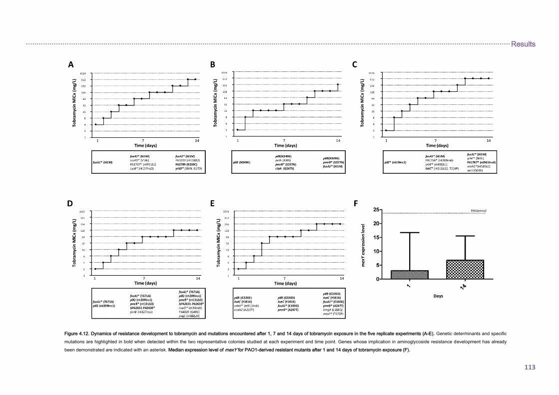
································································································································································································································· Results
113
Figure 4.12. Dynamics of resistance development to tobramycin and mutations encountered after 1, 7 and 14 days of tobramycin exposure in the five replicate experiments (A-E). Genetic determinants and specific
mutations are highlighted in bold when detected within the two representative colonies studied at each experiment and time point. Genes whose implication in aminoglycoside resistance development has already
been demonstrated are indicated with an asterisk. Median expression level of mexY for PAO1-derived resistant mutants after 1 and 14 days of tobramycin exposure (F).
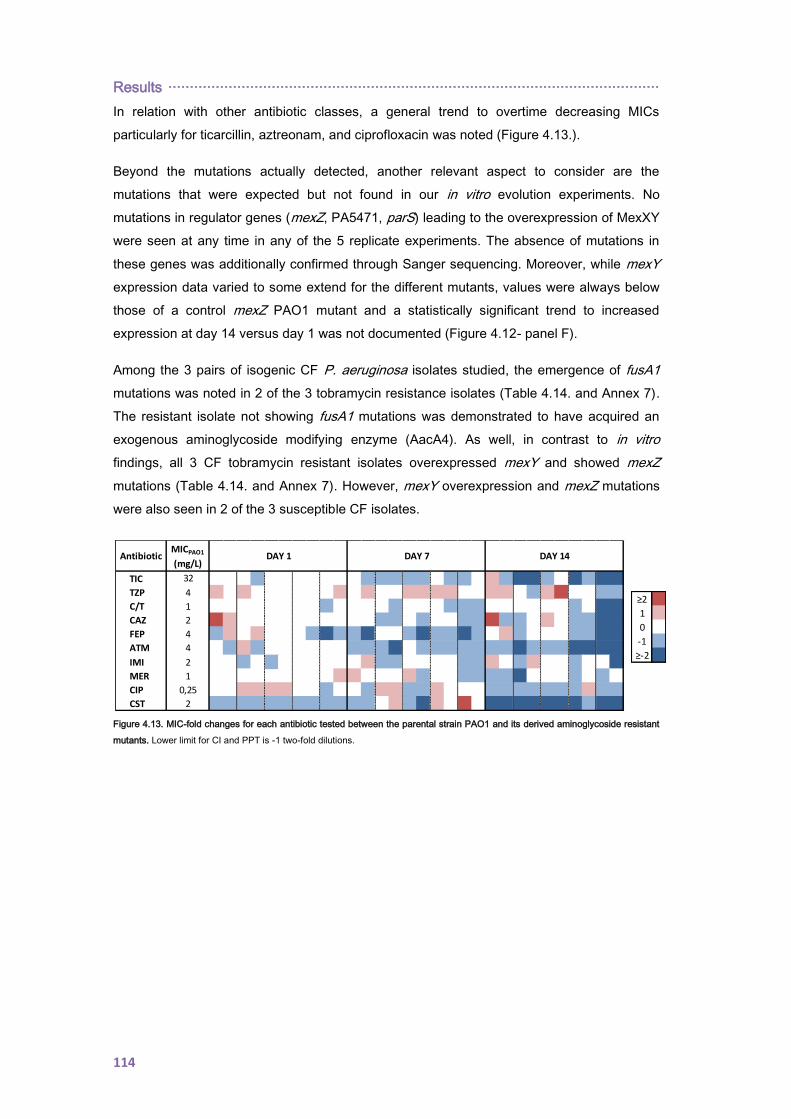
Results ··················································································································
114
In relation with other antibiotic classes, a general trend to overtime decreasing MICs
particularly for ticarcillin, aztreonam, and ciprofloxacin was noted (Figure 4.13.).
Beyond the mutations actually detected, another relevant aspect to consider are the
mutations that were expected but not found in our in vitro evolution experiments. No
mutations in regulator genes (mexZ, PA5471, parS) leading to the overexpression of MexXY
were seen at any time in any of the 5 replicate experiments. The absence of mutations in
these genes was additionally confirmed through Sanger sequencing. Moreover, while mexY
expression data varied to some extend for the different mutants, values were always below
those of a control mexZ PAO1 mutant and a statistically significant trend to increased
expression at day 14 versus day 1 was not documented (Figure 4.12- panel F).
Among the 3 pairs of isogenic CF P. aeruginosa isolates studied, the emergence of fusA1
mutations was noted in 2 of the 3 tobramycin resistance isolates (Table 4.14. and Annex 7).
The resistant isolate not showing fusA1 mutations was demonstrated to have acquired an
exogenous aminoglycoside modifying enzyme (AacA4). As well, in contrast to in vitro
findings, all 3 CF tobramycin resistant isolates overexpressed mexY and showed mexZ
mutations (Table 4.14. and Annex 7). However, mexY overexpression and mexZ mutations
were also seen in 2 of the 3 susceptible CF isolates.
Figure 4.13. MIC-fold changes for each antibiotic tested between the parental strain PAO1 and its derived aminoglycoside resistant
mutants. Lower limit for CI and PPT is -1 two-fold dilutions.
MICPAO1
(mg/L)
TIC 32
TZP 4
C/T 1
CAZ 2
FEP 4
ATM 4
IMI 2
MER 1
CIP 0,25
CST 2
Antibiotic DAY 1 DAY 14DAY 7
≥2
1
0
-1
≥-2
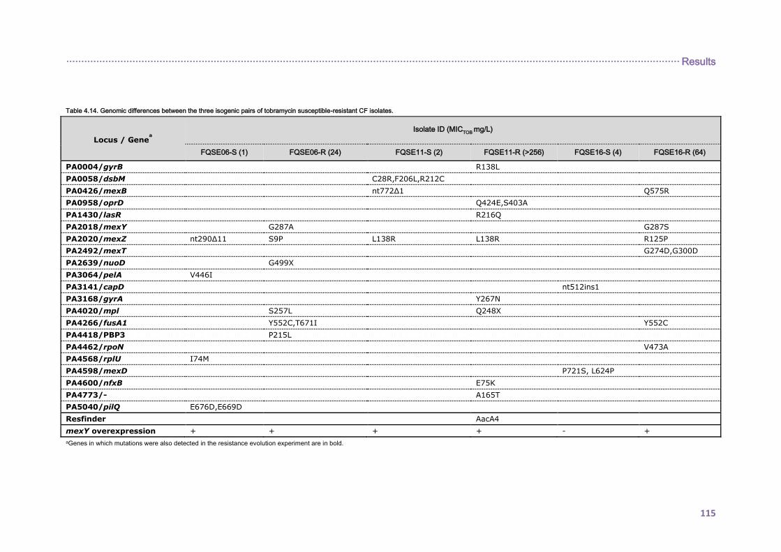
············································································································································································································ Results
115
Table 4.14. Genomic differences between the three isogenic pairs of tobramycin susceptible-resistant CF isolates.
Locus / Genea
Isolate ID (MICTOB
mg/L)
FQSE06-S (1) FQSE06-R (24) FQSE11-S (2) FQSE11-R (>256) FQSE16-S (4) FQSE16-R (64) PA0004/gyrB R138L PA0058/dsbM C28R,F206L,R212C PA0426/mexB nt772∆1 Q575R PA0958/oprD Q424E,S403A PA1430/lasR R216Q PA2018/mexY G287A G287S PA2020/mexZ nt290∆11 S9P L138R L138R R125P PA2492/mexT G274D,G300D PA2639/nuoD G499X PA3064/pelA V446I PA3141/capD nt512ins1 PA3168/gyrA Y267N PA4020/mpl S257L Q248X PA4266/fusA1 Y552C,T671I Y552C PA4418/PBP3 P215L PA4462/rpoN V473A PA4568/rplU I74M PA4598/mexD P721S, L624P PA4600/nfxB E75K PA4773/- A165T PA5040/pilQ E676D,E669D Resfinder AacA4 mexY overexpression + + + + - + aGenes in which mutations were also detected in the resistance evolution experiment are in bold.

116

117
5. DISCUSSION
La levedad y el peso

118

·············································································································· Discussion
119
High genetic diversity among CF P. aeruginosa isolates. Cross-sectional studies revealed
considerable genetic diversity among P. aeruginosa isolates infecting CF patients from both
Spain and the Balearic Islands. Up to 65% of the CF patients from the Balearic Islands
harbored unique PFGE restriction patterns, percentage that increase to 88.6% in the
Spanish cohort (65% and 91.1% unique STs, respectively). Moreover, 42% and 67% of the
different STs detected within the Balearic Islands and the Spanish collection have not been
previously described, finding which supports the extended idea that most CF patients
acquire unique P. aeruginosa strains from environmental sources.
Globally, the documented genetic diversity is in accordance with those reported from other
CF cohorts in which segregation policies applies. In 2001, Burns et al. investigated the
genetic background of P. aeruginosa isolates infecting a cohort of 40 CF paediatric patients
from 3 different hospitals of the United States demonstrating a high degree of genotypic
variability. Recently, Kidd et al. also investigated this issue in a paediatric cohort from
Australia and New Zealand, finally concluding that the environment is the most frequent
route for P. aeruginosa acquisition among CF children. Likewise, a national observational
study across Canada including 1,537 isolates from both adult and paediatric CF patients
(n=402) has been conducted by Middleton and collaborators. In this work, 403 unique STs
were detected and, although 39% of STs were shared, most were only detected among a
small number of subjects.
With the exception of clone FQSE-A (CC274), shared STs detected among the Balearic
Islands and the Spanish collections were also limited to a small number of patients. Indeed,
a direct epidemiological relation could be stablished in the majority of the cases as shared
STs were mainly detected infecting pairs of siblings. Transmission of P. aeruginosa strains
between siblings with CF has already been well documented [Kelly NM et al, 1982;
Thomassen MJ et al, 1985; Grothues D et al, 1988; Renders NH et al, 1997; Tubbs D et al,
2001; Abdul Wahab A et al, 2014]. In a study performed in Israel, Picard et al. showed that
when P. aeruginosa was isolated from the first-born sibling, up to 91% of the second siblings
were also infected; whereas when the first-born was not positive, only 50% of subsequent
siblings were infected. Likewise, they also showed that the age of first isolation was
significantly earlier in the second sibling compared to the first-born [Picard E et al, 2004],
finding that other authors have also reported [Slieker MG et al, 2010]. Furthermore, worse
clinical outcomes (including lower FEV1, faster decline rate of FEV1, more bacterial airway
colonization, increased frequency of lung transplants and a trend towards more
hospitalizations) have been found in families with multiple CF patients compared to families
with only one CF patient, which may reflect the burden and complexity care of this disease
[Lavie M et al, 2015].
Scarce representation of P. aeruginosa CF epidemic strains. Several European and non-
European countries have reported the presence of international epidemic P. aeruginosa
Discussion ·············································································································
120
strains infecting a wide number of CF patients. Likewise, high-risk clone ST175 has been
documented to be widely distributed in the Spanish nosocomial setting [Cabot G et al, 2012;
Cabot G et al, 2016a; del Barrio-Tofiño E et al, 2017].
In the Spanish collection, the C40A AT-genotype was determined in one of the 79 studied
isolates. This genotype has been previously described for P. aeruginosa Clone C (ST17)
[Hilker R et al, 2015; Hall AJ et al, 2014] but, curiously, this isolate was ascribed to ST1872
which is a double locus variant of ST17, differing in just two point mutations in mutL and trpE
MLST alleles.
More worrisome, when investigating long-term clonal epidemiology of P. aeruginosa
colonizing the respiratory tract of CF patients from the Balearic Islands, a clonal replacement
of a MDR mutator strain by the MDR LES (ST146) was documented in one of the patients,
alerting of the first detection of the likely more world-wide concerning CF epidemic clone in
Spain. This unusual and awesome characteristic was also reported by McCallum et al. in 4
CF patients infected with unique strains after admission for treatment in a CF center
[McCallum SJ et al, 2001]. In this case, although the epidemiological driver of LES
colonization was not specifically investigated, the fact that the patient has family links with a
northern European country could help to explain the acquisition of this CF epidemic clone.
As well, clone FQSE-A was detected in 5 unrelated chronically colonized CF patients from
the Balearic Islands, clone that was ascribed to the CC274 by MLST. Moreover, in 4 of them,
long-term clonal epidemiology was investigated and this strain was demonstrated to persist
during the whole 8-years study period. Therefore, results so far suggested that clone FQSE-
A is a CF adapted strain: transmissible and persistent. Furthermore, according to the publicly
available MLST database (http://pubmlst.org/paeruginosa/), P. aeruginosa ST274 has also
been detected infecting multiple CF patients from France, Austria and Australia. Thus, our
results add further evidence pointing out that ST274 should be added to the growing list of
CF epidemic clones.
Discrepant molecular typing results: role of mutators. PFGE and MLST methods are
currently considered the gold-standard tecniques for the establishment of epidemiological
links. Compared with MLST, PFGE exhibits a higher discriminatory power (or lower stability)
and, conversely, MLST results are more reproducible among different laboratories. Thus,
PFGE is the preferred technique for studying local epidemiology and to perform outbreaks
investigations whereas MLST has been posed as the golden molecular typing tool for global
epidemiological studies and for tracking long-term epidemiological relations.
When exploring the Balearic Islands and the Spanish CF P. aeruginosa collections, some
discrepancies between these molecular typing methods were detected. Not surprising, due
to the overall higher discriminatory power of PFGE, several isolates showing different PFGE
·············································································································· Discussion
121
patterns were ascribed to the same ST. Conversely, and much more intriguingly, for some
isolates showing identical PFGE patterns different STs were determined.
Clone FQSE-A was detected in 5 CF patients from the Balearic Islands. Whereas isolates
from 4 of the patients were ascribed to ST274 by MLST, mutator isolates from the fifth
patient were ascribed to ST1089. As ST1089 just differs from ST274 by two point mutations
in two of the MLST genes each leading to a non-previously described allele, the available
data clearly suggest that mutator ST1089 has recently evolved from ST274. Likewise,
recently, García-Castillo and collaborators also reported a ST shift within isolates from a
chronically colonized CF patient directly linked to the emergence of a mutator phenotype
caused by mutL mutations [García-Castillo M et al, 2012]. As well, within the CC274
collection one of the Australian mutator isolates was ascribed to a new ST which just differs
from ST74 by two missense mutations in mutL allele, being one of them (H288Y) responsible
for the generation of the new ST.
Although not linked with the emergence of stable mutator phenotypes, similar discrepancies
were also documented within the Spanish collection. These discrepancies could be
explained in terms of an increase prevalence of transient mutator phenotypes (SOS system)
during CF CRI, as the CF lungs are known to be a very stressful environment for bacteria in
which mutation supply rate is very high. Of note, although not linked to a stable mutator
phenotype, the mutL gene was frequently involved in the ascription of clonal isolates (PFGE)
to different STs.
As well, other authors have also reported that some P. aeruginosa strains are not typable by
MLST due to the presence of InDel mutations within the mutL fragment analyzed [Kidd TJ et
al, 2011; del Barrio-Tofiño E et al, 2017].
All together these results stress the point that mutL lacks the neutrality required for an
appropriate MLST marker, especially for epidemiological studies involving isolates causing
CF CRI in which not only transient mutator phenotypes frequently rise but also MMR
deficient mutators are positively selected [Mena A et al, 2008] and, therefore, may determine
a lower stability of the MLST profiles than expected (leading to discrepant results) both
directly (mutL inactivating mutations within the gene fragment evaluated in MLST analysis)
and indirectly through the increased spontaneous mutagenesis which can facilitate the
emergence of novel alleles through point mutations in any of the 7 house-keeping genes
evaluated.
Genomic analysis of the phylogeny, within-host evolution and interpatient transmission of the
international CC274 CF P. aeruginosa clone. CC274 population structure analysis
demonstrated the worldwide coexistence of two separated and divergent clonal lineages, but
without evident geographical barrier.
Discussion ·············································································································
122
Coexistence of distinct evolved CC274 sublineages within a patient was documented.
Similar results have been recently reported by Williams et al. concerning the LES [Williams D
et al, 2015]. In that work, they found that multiple coexisting LES lineages are typically
infecting CF patients and that genetic divergence between lineages within patients was
greater than interpatient diversity, implying acquisition of diverse genetic populations
[Williams D et al, 2015]. On the opposite, another study focusing on the LES isolated from
patients residing the UK and Canada showed less genetic differences, even when
transoceanic isolates were compared [Jeukens J et al, 2014]. Likewise, Yang et al. also
documented a lower genetic divergence in the DK2 epidemic clone [Yang L et al, 2011] as
well as other previous studies with other relevant and/or persistent CF clones which have
also reported divergent results [Feliziani S et al, 2014; Marvig RL et al, 2013; Cramer N et al,
2011]. A possible explanation for these observations could be that different routes for
adaptation and survival in the CF lung environment are possible and depend on the specific
clonal lineages.
The documented within-host diversity may reflect the coexistence of divergent lineages
within the infecting inoculum or the occurrence of several independent transmission events
during the course of infection. Based on the substantial phenotypic variation previously
observed between samples of the LES taken from patients at successive time points [Mowat
E et al, 2011; Fothergill JL et al, 2010], Williams et al. finally pointed out recurrent
transmissions as the most suitable driver of rapid population genomic flux in LES infections
of the CF airway. To gain more insights, Williams and collaborators have recently published
a work in which they examined the genetic diversity of chronic P. aeruginosa LES infections
over 13 months among seven chronically infected CF patients attending the same CF center
by genome sequencing, documenting rapid and substantial shifts in the relative abundance
of lineages and replacement of dominant lineages likely to represent super-infection by
repeated transmissions [Williams D et al, 2018]. In the case of CC274, and with the
exception of mutators, all isolates from an individual patient clustered together in the same
branch which makes the acquisition of a mix of genetically different sublineages a more
suitable explanation. Nevertheless, whole-genome sequencing of more longitudinal isolates
could help to definitely resolve this issue.
By contrast, and more revealing, both phylogenetic reconstructions and mutational resistome
analysis based on WGS data allow us to confirm interpatient transmission of mutators
(ST274/ST1089). So, compared with classical molecular typing tools, WGS provides detailed
genome fingerprints that might be essential for epidemiological studies in which prevalent
and ubiquitous clonal lineages are involved. Indeed, WGS closely clustered isolates from
four of the patients from the Balearic Islands, likely indicating interpatient transmission or a
common source of colonization, whereas isolates from a fifth patient from the same hospital
was distantly related.
·············································································································· Discussion
123
Insights into the CF P. aeruginosa accessory genome. The pangenome of P. aeruginosa
consists of two different parts: the conserved core genome (90%) and a combinatorial
accesory genome (10%), being the accesory genetic elements esential for surviving under
certain selective conditions.
A great example of P. aeruginosa adaptability is its ability for producing three different types
of pyoverdine and four binding-receptors. The major finding in this variable locus was the
absence of the alternative receptor for pyoverdine type I (fpvB) in 43% of the isolates from
the CF Spanish collection, results that do not correlate with previous studies [Pirnay JP et al,
2009; De Vos D et al, 2001] in which almost all isolates were demonstrated to harbour it.
Recently, Dingemans et al also found a significant proportion of CF isolates lacking this
alternative receptor (22%) and they hypothesized that this receptor may be relieved from
selection because P. aeruginosa can utilize multiple iron uptake systems in the CF lung to
acquire iron in both its ferric and ferrous forms [Dingemans J et al, 2014; Hunter RC et al,
2013; Konings AF et al, 2013]. An alternative hypothesis for the documented absence may
be that loss of fpvB can be an advantage for evading the immune system and the action of
pyocines [Dingemans et al, 2014].
As well, with the exception of the flagellin-glycosylation island, other genomic islands
included in the Array Tube genotyping tool were underrepresented when compared with
other previous studied collections, which maybe reflects the extraordinary ability of P.
aeruginosa to explote different paths for adaptation and survival in different environments
[Liang X et al, 2001; Klockgether J et al, 2007; Rakhimova E et al, 2009]. The high
proportion of isolates harbouring this island clearly suggests that glycosylation may confer
some advantages in the CF respiratory tract.
Finally, within its genome, P. aeruginosa has a large armamentarium of secreted virulence
factors that rely on specialized export systems, including the type III secretion system
(T3SS) [Frank, Molecular Microbiology 2007]. In accordance with previously published data
for CF respiratory isolates, we encountered that up to 81% and 10% of the isolates possess
the ExoS and the ExoU encoding genes, respectively, which reflects a diminished virulence
during chronic respiratory infections [Feltman H et al, 2001; Pirnay J et al, 2009].
Antibiotic resistance trends in CF P. aeruginosa isolates. Overall, higher non-susceptibility
rates to individual agents were documented for the Spanish (2013-2014) collection in
comparison with the Balearic Islands (2003-2012) collection. In both collections, aztreonam
and ciprofloxacin were the less active antibiotis and colistin the one for which a minor
resistance rate was registered. However, it should be mentioned that EUCAST considers P.
aeruginosa intrinsically resistant to aztreonam (mainly because of the constitutive expression
of MexAB-OprM efflux pump), so, all those isolates showing susceptibility deserve spetial
mention (discuss later). High resistance rates to individual agents have also been recently
Discussion ·············································································································
124
reported by Mustafa et al. when studying the antimicrobial susceptibilities of 153 P.
aeruginosa isolates collected from 2006 to 2012 in 118 CF patients from the United
Kingdom, Belgium and Germany [Mustafa MH et al, 2016]. Moreover, MDR isolates were
also highly prevalent in this Northern European study and within the Spanish collection,
finding that compares with the global MDR rate in the Balearic Islands P. aeruginosa
collection. Of note, a high genetic diversity was documented among the Spanish isolates so
maybe the documented higher resistance rates reflect a trend towards increased antibiotic
resistance rates as documented in P. aeruginosa causing acute infections. Indeed, results
from the analysis of antibiotic resistance temporal evolution in the Balearic Islands collection
demonstrated a significant upward trend.
Lower non-susceptibility rates were documented for mucoid isolates and, conversely, higher
ones were registered for SCV isolates compared with the entire collections. As during CF-
CRI an impressive diversification process occur within the infecting population eventually
leading to different variants, these results support the importance of perform antibiotic
susceptibility testing to at least all different colonies morphotypes encountered within a
patient sample.
Non-susceptibility rates values were documented to be higher among chronically colonized
CF patients, finding that can be linked to a major antibiotic pressure and, therefore, to an
accumulation of resistance mechanisms overtime. Correlation with antibiotics usage was
early suggested [Mouton JW et al, 1993] but it remains to be demonstrated. In this sense,
when we studied long-term CRI, we documented a significant trend towards the
accumulation of resistance which was accompanied by a trend towards the accumulation of
antibiotic resistance mechanisms.
Mutators as a driver of antibiotic resistance. High proportions of mutator isolates among the
CF P. aeruginosa population have been demonstrated previously, being frequently
associated with antimicrobial resistance [Oliver A, 2010; Montanari S et al, 2007; Mena A et
al, 2008; Ciofu O et al, 2005; Marvig RL et al, 2013]. Similar rates were documented within
the subset of 100 isolates from the Balearic Islands collection and within the CC274
collection, whereas a lower proportion of mutators was found in the Spanish one which
maybe reflects earlier stages of chronic colonization.
Defects in the MMR system (mutS and mutL) were the most frequent cause for
hypermutation, which correlate with previous studies [Miller JH, 1996; Oliver A, 2010; Oliver
A & Mena A, 2010]. Genetic basis for hypermutation of isolates from the CC274 collection
was studied from WGS data (mutome) and, of note, unique missense mutations were
encountered in several of the so-called mutator genes in isolates exhibiting a normomutator
phenotype, even when located within the MMR system coding-genes.
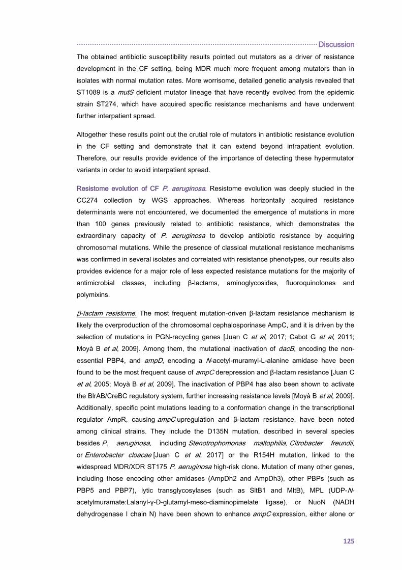
·············································································································· Discussion
125
The obtained antibiotic susceptibility results pointed out mutators as a driver of resistance
development in the CF setting, being MDR much more frequent among mutators than in
isolates with normal mutation rates. More worrisome, detailed genetic analysis revealed that
ST1089 is a mutS deficient mutator lineage that have recently evolved from the epidemic
strain ST274, which have acquired specific resistance mechanisms and have underwent
further interpatient spread.
Altogether these results point out the crutial role of mutators in antibiotic resistance evolution
in the CF setting and demonstrate that it can extend beyond intrapatient evolution.
Therefore, our results provide evidence of the importance of detecting these hypermutator
variants in order to avoid interpatient spread.
Resistome evolution of CF P. aeruginosa. Resistome evolution was deeply studied in the
CC274 collection by WGS approaches. Whereas horizontally acquired resistance
determinants were not encountered, we documented the emergence of mutations in more
than 100 genes previously related to antibiotic resistance, which demonstrates the
extraordinary capacity of P. aeruginosa to develop antibiotic resistance by acquiring
chromosomal mutations. While the presence of classical mutational resistance mechanisms
was confirmed in several isolates and correlated with resistance phenotypes, our results also
provides evidence for a major role of less expected resistance mutations for the majority of
antimicrobial classes, including β-lactams, aminoglycosides, fluoroquinolones and
polymixins.
β-lactam resistome. The most frequent mutation-driven β-lactam resistance mechanism is
likely the overproduction of the chromosomal cephalosporinase AmpC, and it is driven by the
selection of mutations in PGN-recycling genes [Juan C et al, 2017; Cabot G et al, 2011;
Moyà B et al, 2009]. Among them, the mutational inactivation of dacB, encoding the non-
essential PBP4, and ampD, encoding a N-acetyl-muramyl-L-alanine amidase have been
found to be the most frequent cause of ampC derepression and β-lactam resistance [Juan C
et al, 2005; Moyà B et al, 2009]. The inactivation of PBP4 has also been shown to activate
the BlrAB/CreBC regulatory system, further increasing resistance levels [Moyà B et al, 2009].
Additionally, specific point mutations leading to a conformation change in the transcriptional
regulator AmpR, causing ampC upregulation and β-lactam resistance, have been noted
among clinical strains. They include the D135N mutation, described in several species
besides P. aeruginosa, including Stenotrophomonas maltophilia, Citrobacter freundii,
or Enterobacter cloacae [Juan C et al, 2017] or the R154H mutation, linked to the
widespread MDR/XDR ST175 P. aeruginosa high-risk clone. Mutation of many other genes,
including those encoding other amidases (AmpDh2 and AmpDh3), other PBPs (such as
PBP5 and PBP7), lytic transglycosylases (such as SltB1 and MltB), MPL (UDP-N-
acetylmuramate:Lalanyl-γ-D-glutamyl-meso-diaminopimelate ligase), or NuoN (NADH
dehydrogenase I chain N) have been shown to enhance ampC expression, either alone or
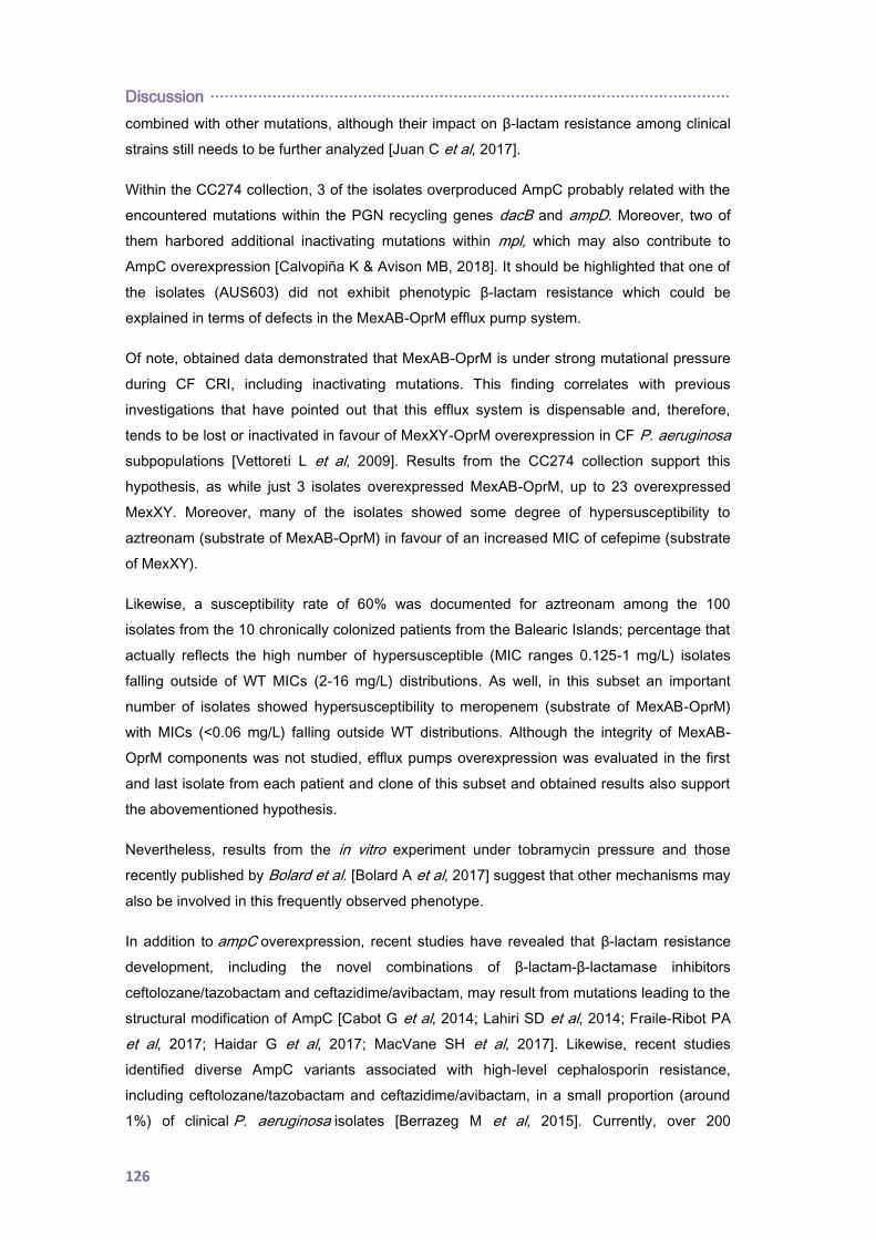
Discussion ·············································································································
126
combined with other mutations, although their impact on β-lactam resistance among clinical
strains still needs to be further analyzed [Juan C et al, 2017].
Within the CC274 collection, 3 of the isolates overproduced AmpC probably related with the
encountered mutations within the PGN recycling genes dacB and ampD. Moreover, two of
them harbored additional inactivating mutations within mpl, which may also contribute to
AmpC overexpression [Calvopiña K & Avison MB, 2018]. It should be highlighted that one of
the isolates (AUS603) did not exhibit phenotypic β-lactam resistance which could be
explained in terms of defects in the MexAB-OprM efflux pump system.
Of note, obtained data demonstrated that MexAB-OprM is under strong mutational pressure
during CF CRI, including inactivating mutations. This finding correlates with previous
investigations that have pointed out that this efflux system is dispensable and, therefore,
tends to be lost or inactivated in favour of MexXY-OprM overexpression in CF P. aeruginosa
subpopulations [Vettoreti L et al, 2009]. Results from the CC274 collection support this
hypothesis, as while just 3 isolates overexpressed MexAB-OprM, up to 23 overexpressed
MexXY. Moreover, many of the isolates showed some degree of hypersusceptibility to
aztreonam (substrate of MexAB-OprM) in favour of an increased MIC of cefepime (substrate
of MexXY).
Likewise, a susceptibility rate of 60% was documented for aztreonam among the 100
isolates from the 10 chronically colonized patients from the Balearic Islands; percentage that
actually reflects the high number of hypersusceptible (MIC ranges 0.125-1 mg/L) isolates
falling outside of WT MICs (2-16 mg/L) distributions. As well, in this subset an important
number of isolates showed hypersusceptibility to meropenem (substrate of MexAB-OprM)
with MICs (<0.06 mg/L) falling outside WT distributions. Although the integrity of MexAB-
OprM components was not studied, efflux pumps overexpression was evaluated in the first
and last isolate from each patient and clone of this subset and obtained results also support
the abovementioned hypothesis.
Nevertheless, results from the in vitro experiment under tobramycin pressure and those
recently published by Bolard et al. [Bolard A et al, 2017] suggest that other mechanisms may
also be involved in this frequently observed phenotype.
In addition to ampC overexpression, recent studies have revealed that β-lactam resistance
development, including the novel combinations of β-lactam-β-lactamase inhibitors
ceftolozane/tazobactam and ceftazidime/avibactam, may result from mutations leading to the
structural modification of AmpC [Cabot G et al, 2014; Lahiri SD et al, 2014; Fraile-Ribot PA
et al, 2017; Haidar G et al, 2017; MacVane SH et al, 2017]. Likewise, recent studies
identified diverse AmpC variants associated with high-level cephalosporin resistance,
including ceftolozane/tazobactam and ceftazidime/avibactam, in a small proportion (around
1%) of clinical P. aeruginosa isolates [Berrazeg M et al, 2015]. Currently, over 200
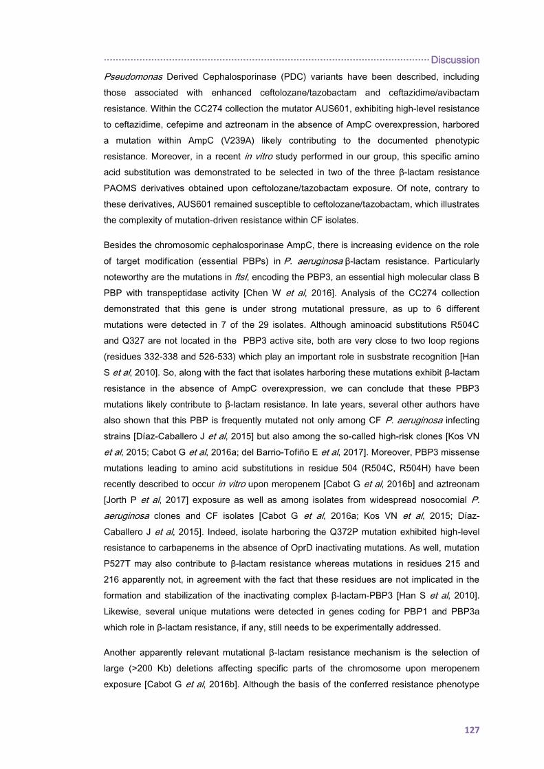
·············································································································· Discussion
127
Pseudomonas Derived Cephalosporinase (PDC) variants have been described, including
those associated with enhanced ceftolozane/tazobactam and ceftazidime/avibactam
resistance. Within the CC274 collection the mutator AUS601, exhibiting high-level resistance
to ceftazidime, cefepime and aztreonam in the absence of AmpC overexpression, harbored
a mutation within AmpC (V239A) likely contributing to the documented phenotypic
resistance. Moreover, in a recent in vitro study performed in our group, this specific amino
acid substitution was demonstrated to be selected in two of the three β-lactam resistance
PAOMS derivatives obtained upon ceftolozane/tazobactam exposure. Of note, contrary to
these derivatives, AUS601 remained susceptible to ceftolozane/tazobactam, which illustrates
the complexity of mutation-driven resistance within CF isolates.
Besides the chromosomic cephalosporinase AmpC, there is increasing evidence on the role
of target modification (essential PBPs) in P. aeruginosa β-lactam resistance. Particularly
noteworthy are the mutations in ftsI, encoding the PBP3, an essential high molecular class B
PBP with transpeptidase activity [Chen W et al, 2016]. Analysis of the CC274 collection
demonstrated that this gene is under strong mutational pressure, as up to 6 different
mutations were detected in 7 of the 29 isolates. Although aminoacid substitutions R504C
and Q327 are not located in the PBP3 active site, both are very close to two loop regions
(residues 332-338 and 526-533) which play an important role in susbstrate recognition [Han
S et al, 2010]. So, along with the fact that isolates harboring these mutations exhibit β-lactam
resistance in the absence of AmpC overexpression, we can conclude that these PBP3
mutations likely contribute to β-lactam resistance. In late years, several other authors have
also shown that this PBP is frequently mutated not only among CF P. aeruginosa infecting
strains [Díaz-Caballero J et al, 2015] but also among the so-called high-risk clones [Kos VN
et al, 2015; Cabot G et al, 2016a; del Barrio-Tofiño E et al, 2017]. Moreover, PBP3 missense
mutations leading to amino acid substitutions in residue 504 (R504C, R504H) have been
recently described to occur in vitro upon meropenem [Cabot G et al, 2016b] and aztreonam
[Jorth P et al, 2017] exposure as well as among isolates from widespread nosocomial P.
aeruginosa clones and CF isolates [Cabot G et al, 2016a; Kos VN et al, 2015; Díaz-
Caballero J et al, 2015]. Indeed, isolate harboring the Q372P mutation exhibited high-level
resistance to carbapenems in the absence of OprD inactivating mutations. As well, mutation
P527T may also contribute to β-lactam resistance whereas mutations in residues 215 and
216 apparently not, in agreement with the fact that these residues are not implicated in the
formation and stabilization of the inactivating complex β-lactam-PBP3 [Han S et al, 2010].
Likewise, several unique mutations were detected in genes coding for PBP1 and PBP3a
which role in β-lactam resistance, if any, still needs to be experimentally addressed.
Another apparently relevant mutational β-lactam resistance mechanism is the selection of
large (>200 Kb) deletions affecting specific parts of the chromosome upon meropenem
exposure [Cabot G et al, 2016b]. Although the basis of the conferred resistance phenotype
Discussion ·············································································································
128
still needs to be further clarified, these mutants can be recognized by the characteristic
brown pigment (pyomelanine) caused by the deletion of one of the affected genes, hmgA,
coding for a homogentisate-1,2-dioxygenase. This type of deletion has been documented in
both, in vitro evolved β-lactam resistant mutants and CF isolates [Cabot G et al, 2016b;
Hocquet D et al, 2016]. However, the deletion of hmgA is not responsible of the resistance
phenotype, which might be linked to the deletion of another of the affected genes, galU,
coding for a UDP-glucose pyrophosphorylase required for LPS core synthesis; indeed,
analysis of transposon mutant libraries have shown that the inactivation of galU increases
ceftazidime and meropenem MICs [Alvarez-Ortega C et al, 2010; Dötsch A et al, 2009].
None of the CC274 isolates showed these large deletions neither exhibited brown
pigmentation; however, it should be noted that all ST1089 mutator isolates showed the same
missense mutation in galU, likely contributing to their carbapenem resistance.
Apart from these emerging β-lactam resistance mechanisms, phenotypic carbapenem
resistance has been clasically linked to the mutational inactivation of the carbapenem porin
OprD [Lister PD et al, 2009; Castanheira M et al, 2014]. Overall, our results, from both the
CC274 collection and the subset of 100 isolates from the Balearic Islands, confirm that
carbapenem resistance is frequently associated with inactivating mutations within OprD.
Additionally, Richardot and collaborators recently reported that some amino acid
substitutions within OprD can also confer carbapenem resistance, particularly in the CF
setting [Richardot C et al, 2015]; nevertheless, no amino acid substitutions were detected
within the CC274 isolates. As well, carbapenem resistance may also result
from oprD repression caused by mutations in the MexEF-OprN efflux pump regulators
(mexS/T) or the ParRS two-component system [Li XZ et al, 2015]; however, our results
showed that overexpression of MexEF-OprN is not frequent among CF isolates.
Aminoglycoside resistome. Intravenous antimicrobial combinations including an
aminoglycoside are frequently used to manage CF exacerbations. Moreover, in the last
decade, tobramycin inhalation has become an important contributor to CF treatment as a
means to control CRI as well as a first-line treatment for the eradication of early acquisition
of P. aeruginosa and several aminoglycoside-based inhaled formulations are currently
available [Shteinberg M & Elborn JS, 2015].
Whereas resistance to these agents in acute infections are mainly attributed to the
production of aminoglycoside modifying enzymes or 16S rRNA methyltransferases,
resistance development in the CRI setting has been linked to the selection of chromosomal
mutations leading to enhanced membrane impermeability or MexXY overexpression [Pricket
MH et al, 2017; Guenard S et al, 2014; Poole K, 2015; Vogne C et al, 2004]. In accordance,
most of the CF clinical studied isolates overexpressed this efflux pump system linked to the
presence of mutations within mexZ, amgS and/or parRS; moreover, these mutations occur
early and were associated with low-level resistance. Of note, mutations leading to

·············································································································· Discussion
129
MexXYoverexpression were not seen in any of the five replicate in vitro evolution
experiments upon tobramycin pressure. Moreover, while mexY expression data varied to
some extent for the different mutants, values were always below those of a control mexZ
PAO1 mutant, and a statistically significant trend toward increased expression at day 14
versus day 1 was no documented. Thus, these results indicate that mutational
overexpression of MexXY is not required for the evolution of high-level tobramycin resistance
in vitro. Altogether these results may indicate that positive selection of mutations leading to
the overexpression of MexXY in CF might be driven by factors beyond exposure to
aminoglycosides.
Beyond MexXY overexpression, recent studies have revealed that the aminoglycoside
mutational resistome extends far, and that high-level resistance may result from the
accumulation of multiple mutations, and the involvement of several novel resistance
determinants has been recently documented [El’Garch F et al, 2007; Schurek KN et al, 2008;
Feng Y et al, 2016]. WGS data revealed that all high-level resistant CC274 isolates not only
overexpressed MexXY but also harbored additional mutations in some of these genes,
especially highlighting the presence of mutations in both genes coding for elongation factor
G, fusA1 and fusA2. Moreover, Greipel and collaborators have also recently reported that
these genes are under high evolutionary pressure in the CF environment [Greipel L et al,
2016], which can be explained in terms of a wide aminoglycoside use in this setting. As well,
several works have associated some specific mutations in FusA1 with aminoglycoside
resistance in vitro [Feng Y et al, 2016] and among clinical, particularly CF, strains [Chung JC
et al, 2012; Markussen T et al, 2014], and, more recently Bolard and collaborators have
confirmed the implication of such fusA1 mutations in aminoglycoside resistance through site-
directed mutagenesis [Bolard A et al, 2017].
In accordance, FusA1 mutations were encountered in all 5 replicates from the in vitro
evolution experiment associated with a 1- to 3-fold increase in MICs of tobramycin,
gentamicin and amikacin, which correlates with Bolard et al. observations [Bolard A et al,
2017]. As well, resistance development was shown to occur in a stepwise manner, reaching
MICs at day 14 close to the maximum tobramycin concentrations achieved through inhaled
administration and on the range of the breakpoints suggested for inhaled therapy. Morevoer,
22 of the 35 (63%) different mutated genes have also been related to aminoglycoside
resistance development by other authors [Bolard A et al, 2017; Yen P & Papin JA, 2017;
Feng Y et al, 2016; Islam S et al, 2009; Schurek KN et al, 2009; El’Garch F et al, 2007], and
thus, our data confirm their relevance in this stepwise process. Mutations within pmrB,
traditionally linked with polymyxin resistance development [Moskowitz SM et al, 2012;
Barrow K & Kwon DH, 2009], rise frequently which alerts from a possible mechanism of co-
resistance to two relevant antipseudomonal agents.
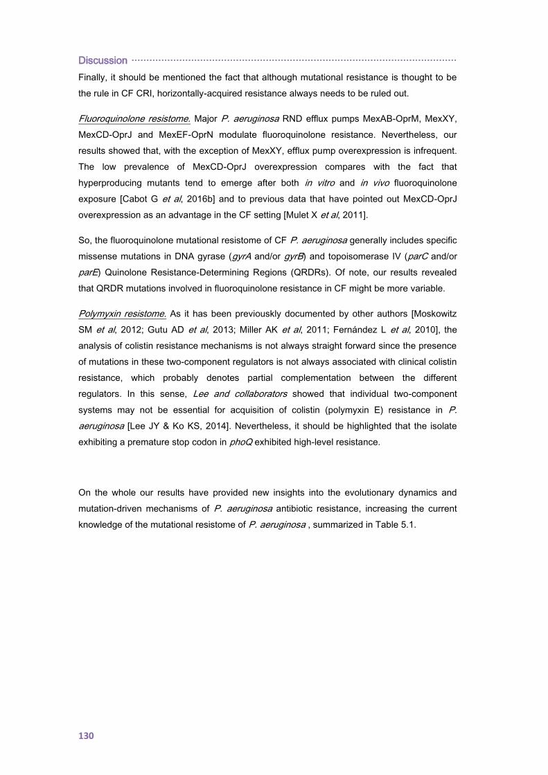
Discussion ·············································································································
130
Finally, it should be mentioned the fact that although mutational resistance is thought to be
the rule in CF CRI, horizontally-acquired resistance always needs to be ruled out.
Fluoroquinolone resistome. Major P. aeruginosa RND efflux pumps MexAB-OprM, MexXY,
MexCD-OprJ and MexEF-OprN modulate fluoroquinolone resistance. Nevertheless, our
results showed that, with the exception of MexXY, efflux pump overexpression is infrequent.
The low prevalence of MexCD-OprJ overexpression compares with the fact that
hyperproducing mutants tend to emerge after both in vitro and in vivo fluoroquinolone
exposure [Cabot G et al, 2016b] and to previous data that have pointed out MexCD-OprJ
overexpression as an advantage in the CF setting [Mulet X et al, 2011].
So, the fluoroquinolone mutational resistome of CF P. aeruginosa generally includes specific
missense mutations in DNA gyrase (gyrA and/or gyrB) and topoisomerase IV (parC and/or
parE) Quinolone Resistance-Determining Regions (QRDRs). Of note, our results revealed
that QRDR mutations involved in fluoroquinolone resistance in CF might be more variable.
Polymyxin resistome. As it has been previouskly documented by other authors [Moskowitz
SM et al, 2012; Gutu AD et al, 2013; Miller AK et al, 2011; Fernández L et al, 2010], the
analysis of colistin resistance mechanisms is not always straight forward since the presence
of mutations in these two-component regulators is not always associated with clinical colistin
resistance, which probably denotes partial complementation between the different
regulators. In this sense, Lee and collaborators showed that individual two-component
systems may not be essential for acquisition of colistin (polymyxin E) resistance in P.
aeruginosa [Lee JY & Ko KS, 2014]. Nevertheless, it should be highlighted that the isolate
exhibiting a premature stop codon in phoQ exhibited high-level resistance.
On the whole our results have provided new insights into the evolutionary dynamics and
mutation-driven mechanisms of P. aeruginosa antibiotic resistance, increasing the current
knowledge of the mutational resistome of P. aeruginosa , summarized in Table 5.1.
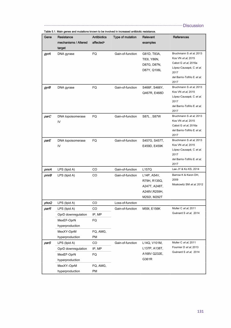
·············································································································· Discussion
131
Table 5.1. Main genes and mutations known to be involved in increased antibiotic resistance.
Gene Resistance
mechanisms / Altered
target
Antibiotics
affecteda
Type of mutation Relevant
examples
References
gyrA DNA gyrase FQ Gain-of-function G81D, T83A,
T83I, Y86N,
D87G, D87N,
D87Y, Q106L
Bruchmann S et al, 2013
Kos VN et al, 2015
Cabot G et al, 2016a
López-Causapé, C et al,
2017
del Barrio-Tofiño E et al,
2017
gyrB DNA gyrase FQ Gain-of-function S466F, S466Y,
Q467R, E468D
Bruchmann S et al, 2013
Kos VN et al, 2015
López-Causapé, C et al,
2017
del Barrio-Tofiño E et al,
2017
parC DNA topoisomerase
IV
FQ Gain-of-function S87L , S87W Bruchmann S et al, 2013
Kos VN et al, 2015
Cabot G et al, 2016a
del Barrio-Tofiño E et al,
2017
parE DNA topoisomerase
IV
FQ Gain-of-function S457G, S457T,
E459D, E459K
Bruchmann S et al, 2013
Kos VN et al, 2015
López-Causapé, C et al,
2017
del Barrio-Tofiño E et al,
2017
pmrA LPS (lipid A) CO Gain-of-function L157Q Lee JY & Ko KS, 2014
pmrB LPS (lipid A) CO Gain-of-function L14P, A54V,
R79H, R135Q,
A247T, A248T,
A248V,R259H,
M292I, M292T
Barrow K & Kwon DH,
2009
Moskowitz SM et al, 2012
phoQ LPS (lipid A) CO Loss-of-function
parR LPS (lipid A) CO Gain-of-function M59I, E156K Muller C et al, 2011
Guénard S et al, 2014
OprD downregulation IP, MP
MexEF-OprN
hyperproduction
FQ
MexXY-OprM
hyperproduction
FQ, AMG,
PM
parS LPS (lipid A) CO Gain-of-function L14Q, V101M,
L137P, A138T,
A168V Q232E,
G361R
Muller C et al, 2011
Fournier D et al, 2013
Guénard S et al, 2014
OprD downregulation IP, MP
MexEF-OprN
hyperproduction
FQ
MexXY–OprM
hyperproduction
FQ, AMG,
PM

Discussion ·············································································································
132
Table 5.1. Main genes and mutations known to be involved in increased antibiotic resistance. (Cont.)
Gene Resistance
mechanisms /
Altered target
Antibiotics
affecteda
Type of mutation Relevant
examples
References
cprS LPS (lipid A) CO Gain-of-function R241C Gutu AD et al, 2013
colR LPS (lipid A) CO Gain-of-function D32N Gutu AD et al, 2013
colS LPS (lipid A) CO Gain-of-function A106V Gutu AD et al, 2013
mexR MexAB –OprM
hyperproduction
FQ, TZ,
PM, PPT,
MP,
TZ/AVI
Loss-of-function
nalC MexAB-OprM
hyperproduction
FQ, TZ,
PM, PPT,
MP,
TZ/AVI
Loss-of-function
nalD MexAB-OprM
hyperproduction
FQ, TZ,
PM, PPT,
MP,
TZ/AVI
Loss-of-function
nfxB MexCD-OprJ
Hyperproduction
FQ, PM Loss-of-function
mexS MexEF-OprN
hyperproduction
FQ
Loss-of-function
OprD
downregulation
IP, MP
mexT MexEF-OprN
hyperproduction
FQ
Gain-of-function
G257S, G257T Juarez P et al, 2018
OprD
downregulation
IP, MP
cmrA MexEF-OprN
hyperproduction
MP, FQ Gain-of-function
A68V, L89Q,
H204L, N214K
Juarez P et al, 2017
mvaT MexEF-OprN
hyperproduction
FQ Loss-of-function
PA3271 MexEF-OprN
hyperproduction
FQ Loss-of-function
mexZ MexXY –OprM
hyperproduction
FQ, AMG,
PM Loss-of-function
PA5471.1 MexXY –OprM
hyperproduction
FQ, AMG,
PM Loss-of-function
amgS MexXY –OprM
hyperproduction
FQ, AMG,
PM Gain of function
V121G, R182C Lau CH et al, 2013
oprD OprD inactivation IP, MP Loss-of-function
ampC AmpC structural
modification
TZ/AVI,
TOL/TAZ Gain-of-function
T96I, G183D,
E247K
Cabot G et al, 2014
Fraile-Ribot PA et al,
2017
ampD AmpC
hyperproduction
TZ, PM,
PPT Loss-of-function

·············································································································· Discussion
133
Table 5.1. Main genes and mutations known to be involved in increased antibiotic resistance. (Cont.)
Gene Resistance
mechanisms /
Altered target
Antibiotics
affecteda
Type of mutation Relevant
examples
References
ampDh2 AmpC
hyperproduction
TZ, PM,
PPT Loss-of-function
ampDh3 AmpC
hyperproduction
TZ, PM,
PPT Loss-of-function
ampR AmpC
hyperproduction
TZ, PM,
PPT Gain-of-function
D135N, G154R Cabot G et al, 2016a
Bagge N et al, 2002
dacB AmpC
hyperproduction
TZ, PM,
PPT Loss-of-function
ftsI Penicillin-binding-
protein 3 (PBP3)
TZ, PM,
PPT, MP,
TZ/AVI,
TOL/TAZ Gain-of-function
R504C, R504H,
P527T F533L
Diaz Caballero J et al,
2015
Cabot G et al, 2016a
Cabot G et al, 2016b
López-Causapé C et al,
2017
del Barrio-Tofiño E et
al, 2017
fusA1 Elongation factor G AMG
Gain-of-function
V93A, K504E,
Y552C, P554L,
A555E, N592I,
P618L, T671A,
T671I
Feng Y et al, 2016
López-Causapé C et al,
2017
del Barrio-Tofiño E et
al, 2017
Bolard A et al, 2017
glpT Transporter protein
GlpT
FO Loss-of-function
rpoB RNA polymerase β-
chain
RI
Gain-of-function
S517F, Q518R,
Q518L, D521G,
H531Y, H531L,
S536F, L538I,
S579F, S579Y,
N629S, D636Y
Jatsenko T et al, 2010

134

135
6. CONCLUSIONS
La Gran Marcha

136
············································································································ Conclusions
137
1. The population structure of P. aeruginosa isolates infecting CF individuals from the
Balearic Islands and Spain is highly diverse, which points out environmental sources as the
main route of P. aeruginosa acquisition.
2. Epidemic strains were scarce and shared P. aeruginosa clones were mainly detected in
pairs of siblings involving non-epidemic strains. However, the multidrug resistant Liverpool
Epidemic Strain was detected for the first time in Spain.
3. Discrepanices between PFGE and MLST genotyping methods were frequently detected
when studying CF isolates mainly due to the lack of neutrality of the mutL gene caused by
the positive selection of mutator phenotypes. WGS based approaches are a powerful tool
that can help in solving this issue.
4. The global trend towards the accumulation of antibiotic resistance during CF-CRI is
accompanied by collateral susceptibility to some antibiotics such as aztreonam, which can
be explained by the overexpression of MexXY leading to the impairment of MexAB-OprM.
5. Worldwide distributed P. aeruginosa CC274 is a well-adapted CF strain, transmissible and
persistant and, therefore, it should be added to the list of CF P. aeruginosa epidemic clones.
6. Dissemination of evolved mutator lineages, frequently linked to multidrug resistant profiles,
between CF patients constitutes a step forward on the spread of antibiotic resistance.
7. Correlation between phenotypes and WGS genotypes of clonal isolates from an epidemic
strain allowed us to decipher the P. aeruginosa mutational resistome in the CF setting.
8. The β-lactam mutational resistome extends beyond the chromosomic cephalosporinase
AmpC. Especial mention deserves gain-of-function mutations within the PBPs, being the
PBP3 under high evolutionary pressure in CF isolates.
9. Mutation-driven aminoglycoside resistance development is a stepwise process in which
gain-of-function mutations within fusA1 and loss-of-function mutations within mexZ are highly
prevalent among CF isolates. The absence of mexZ mutations in vitro suggests an
evolutionary advantage of MexXY overexpression within the respiratory tract of CF patients.
10. Altogether this work demonstrates that clonal epidemiology and antibiotic resistance
evolution in the CF setting results from the complex interplay among mutation-driven
resistance mechanisms, within host diversification and interpatient transmission of epidemic
strains.

138

139
7. REFERENCES
La sonrisa de Karenin

140
············································································································· References
141
Aaron SD, Vandemheen KL, Ramotar K, Giesbrecht-Lewis T, Tullis E, Freitag A, Paterson
N, Jackson M, Lougheed MD, Dowson C, Kumar V, Ferris W, Chan F, Doucette S,
Fergusson D. Infection with transmissible strains of Pseudomonas aeruginosa and clinical
outcomes in adults with cystic fibrosis. JAMA. 2010; 304(19):2145-53.
Abdul Wahab A, Taj-Aldeen SJ, Hagen F, Diophode S, Saadoon A, Meis JF, Klaassen CH.
Genotypic diversity of Pseudomonas aeruginosa in cystic fibrosis siblings in Qatar using
AFLP fingerprinting. Eur J Clin Microbiol Infect Dis. 2014; 33(2):265-71.
Agarwal G, Kapil A, Kabra SK, Das BK, Dwivedi SN. Characterization of Pseudomonas
aeruginosa isolated from chronically infected children with cystic fibrosis in India. BMC
Microbiol. 2005; 5:43.
Aires JR, Köhler T, Nikaido H, Plésiat P. Involvement of an active efflux system in the natural
resistance of Pseudomonas aeruginosa to aminoglycosides. Antimicrob Agents Chemother.
1999; 43(11):2624-8.
Al-Aloul M, Crawley J, Winstanley C, Hart CA, Ledson MJ, Walshaw MJ. Increased morbidity
associated with chronic infection by an epidemic Pseudomonas aeruginosa strain in CF
patients. Thorax. 2004; 59(4):334-6.
Alexander C, Rietschel ET. Bacterial lipopolysaccharides and innate immunity. J Endotoxin
Res. 2001; 7(3):167-202.
Alipour M, Suntres ZE, Omri A. Importance of DNase and alginate lyase for enhancing free
and liposome encapsulated aminoglycoside activity against Pseudomonas aeruginosa. J
Antimicrob Chemother. 2009; 64(2):317-25.
Alvarez-Ortega C, Wiegand I, Olivares J, Hancock RE, Martínez JL. The intrinsic resistome
of Pseudomonas aeruginosa to β-lactams. Virulence. 2011; 2(2):144-6.
Alvarez-Ortega C, Wiegand I, Olivares J, Hancock RE, Martínez JL. Genetic determinants
involved in the susceptibility of Pseudomonas aeruginosa to beta-lactam antibiotics.
Antimicrob Agents Chemother. 2010; 54(10):4159-67.
Anthony M, Rose B, Pegler MB, Elkins M, Service H, Thamotharampillai K, Watson J,
Robinson M, Bye P, Merlino J, Harbour C. Genetic analysis of Pseudomonas aeruginosa
isolates from the sputa of Australian adult cystic fibrosis patients. J Clin Microbiol. 2002;
40(8):2772-8.
Armstrong D, Bell S, Robinson M, Bye P, Rose B, Harbour C, Lee C, Service H, Nissen M,
Syrmis M, Wainwright C. Evidence for spread of a clonal strain of Pseudomonas aeruginosa
among cystic fibrosis clinics. J Clin Microbiol. 2003; 41(5):2266-7.
References ············································································································
142
Armstrong DS, Nixon GM, Carzino R, Bigham A, Carlin JB, Robins-Browne RM, Grimwood
K. Detection of a widespread clone of Pseudomonas aeruginosa in a pediatric cystic fibrosis
clinic. Am J Respir Crit Care Med. 2002; 166(7):983-7.
Arora SK, Bangera M, Lory S, Ramphal R. A genomic island in Pseudomonas aeruginosa
carries the determinants of flagellin glycosylation. Proc Natl Acad Sci U S A. 2001;
98(16):9342-7.
Ashish A, Shaw M, McShane J, Ledson MJ, Walshaw MJ. Health-related quality of life in
Cystic Fibrosis patients infected with transmissible Pseudomonas aeruginosa strains: cohort
study. JRSM Short Rep. 2012; 3(2):12.
Ashish A, Shaw M, Winstanley C, Ledson MJ, Walshaw MJ. Increasing resistance of the
Liverpool Epidemic Strain (LES) of Pseudomonas aeruginosa (Psa) to antibiotics in cystic
fibrosis (CF)-a cause for concern? J Cyst Fibros. 2012; 11(3):173-9.
Bagge N, Hentzer M, Andersen JB, Ciofu O, Givskov M, Høiby N. Dynamics and spatial
distribution of beta-lactamase expression in Pseudomonas aeruginosa biofilms. Antimicrob
Agents Chemother. 2004; 48(4):1168-74.
Bagge N, Ciofu O, Hentzer M, Campbell JI, Givskov M, Høiby N. Constitutive high
expression of chromosomal beta-lactamase in Pseudomonas aeruginosa caused by a new
insertion sequence (IS1669) located in ampD. Antimicrob Agents Chemother. 2002;
46:3406-11.
Barrow K, Kwon DH. Alterations in two-component regulatory systems of phoPQ and pmrAB
are associated with polymyxin B resistance in clinical isolates of Pseudomonas aeruginosa.
Antimicrob Agents Chemother. 2009; 53(12):5150-4.
Bartowsky E, Normark S. Interactions of wild-type and mutant AmpR of Citrobacter freundii
with target DNA. Mol Microbiol. 1993; 10:555–65.
Bellido F, Martin NL, Siehnel RJ, Hancock RE. Reevaluation, using intact cells, of the
exclusion limit and role of porin OprF in Pseudomonas aeruginosa outer membrane
permeability. J Bacteriol. 1992; 174(16):5196-203.
Sobel ML, McKay GA, Poole K. Contribution of the MexXY multidrug transporter to
aminoglycoside resistance in Pseudomonas aeruginosa clinical isolates. Antimicrob Agents
Chemother. 2003; 47(10):3202-7.
Berrazeg M, Jeannot K, Ntsogo Enguéné VY, Broutin I, Loeffert S, Fournier D, Plésiat P.
Mutations in β-Lactamase AmpC increase resistance of Pseudomonas aeruginosa isolates
to antipseudomonal cephalosporins. Antimicrob Agents Chemother. 2015; 59(10):6248-55.
············································································································· References
143
Bjarnsholt T. The role of bacterial biofilms in chronic infections. PMIS Suppl. 2013; (136):1-
51.
Bjarnsholt T, Jensen PØ, Fiandaca MJ, Pedersen J, Hansen CR, Andersen CB, Pressler T,
Givskov M, Høiby N. Pseudomonas aeruginosa biofilms in the respiratory tract of cystic
fibrosis patients. Pediatr Pulmonol. 2009; 44(6):547-58.
Blázquez J, Oliver A, Gómez-Gómez JM. Mutation and evolution of antibiotic resistance:
antibiotics as promoters of antibiotic resistance? Curr Drug Targets. 2002; 3(4):345-9.
Bolard A, Plésiat P, Jeannot K. Mutations in gene fusA1 as a novel mechanism of
aminoglycoside resistance in clinical strains of Pseudomonas aeruginosa. Antimicrob Agents
Chemother. 2018; 62(2). pii: e01835-17.
Boles BR, Singh PK. Endogenous oxidative stress produces diversity and adaptability in
biofilm communities. Proc Natl Acad Sci USA. 2008; 105(34):12503-8.
Boucher JC, Yu H, Mudd MH, Deretic V. Mucoid Pseudomonas aeruginosa in cystic fibrosis:
characterization of muc mutations in clinical isolates and analysis of clearance in a mouse
model of respiratory infection. Infect Immun. 1997; 65(9):3838-46.
Boyd A, Chakrabarty AM. Role of alginate lyase in cell detachment of Pseudomonas
aeruginosa. Appl Environ Microbiol. 1994; 60(7):2355-9.
Bradbury RS, Roddam LF, Merritt A, Reid DW, Champion AC. Virulence gene distribution in
clinical, nosocomial and environmental isolates of Pseudomonas aeruginosa. J Med
Microbiol. 2010; 59(Pt8):881-90.
Bradbury R, Champion A, Reid DW. Poor clinical outcomes associated with a multi-drug
resistant clonal strain of Pseudomonas aeruginosa in the Tasmanian cystic fibrosis
population. Respirology. 2008; 13(6):886-92.
Breidenstein EB, de la Fuente-Núñez C, Hancock RE. Pseudomonas aeruginosa: all roads
lead to resistance. Trends Microbiol. 2011; 19(8):419-26.
Breidenstein EB, Khaira BK, Wiegand I, Overhage J, Hancock RE. Complex ciprofloxacin
resistome revealed by screening a Pseudomonas aeruginosa mutant library for altered
susceptibility. Antimicrob Agents Chemother. 2008; 52(12):4486-91.
Bruchmann S, Dötsch A, Nouri B, Chaberny IF, Häussler S. Quantitative contributions of
target alteration and decreased drug accumulation to Pseudomonas aeruginosa
fluoroquinolone resistance. Antimicrob Agents Chemother. 2013; 57(3) :1361-8.
References ············································································································
144
Burgel PR, Bellis G, Olesen HV, Viviani L, Zolin A, Blasi F, Elborn JS; ERS/ECFS Task
Force on Provision of Care for Adults with Cystic Fibrosis in Europe. Future trends in cystic
fibrosis demography in 34 European countries. Eur Respir J. 2015; 46(1):133-41.
Burns N, James CE, Harrison E. Polylysogeny magnifies competitiveness of a bacterial
pathogen in vivo. Evol Appl. 2015; 8(4):346-51.
Cabot G, López-Causapé C, Ocampo-Sosa AA, Sommer LM, Domínguez MÁ, Zamorano L,
Juan C, Tubau F, Rodríguez C, Moyà B, Peña C, Martínez-Martínez L, Plesiat P, Oliver A.
Deciphering the resistome of the widespread Pseudomonas aeruginosa Sequence Type 175
international high-risk clone through Whole-Genome Sequencing. Antimicrob Agents
Chemother. 2016a; 60(12):7415-7423.
Cabot G, Zamorano L, Moyà B, Juan C, Navas A, Blázquez J, Oliver A. Evolution of
Pseudomonas aeruginosa antimicrobial resistance and fitness under low and high mutation
rates. Antimicrob Agents Chemother. 2016b; 60(3):1767-78.
Cabot G, Bruchmann S, Mulet X, Zamorano L, Moyà B, Juan C, Haussler S, Oliver A.
Pseudomonas aeruginosa ceftolozane-tazobactam resistance development requires multiple
mutations leading to overexpression and structural modification of AmpC. Antimicrob Agents
Chemother. 2014; 58(6):3091-9.
Cabot G, Ocampo-Sosa AA, Domínguez MA, Gago JF, Juan C, Tubau F, Rodríguez C,
Moyà B, Peña C, Martínez-Martínez L, Oliver A; Spanish Network for Research in Infectious
Diseases (REIPI). Genetic markers of widespread extensively drug-resistant Pseudomonas
aeruginosa high-risk clones. Antimicrob Agents Chemother. 2012; 56(12):6349-57.
Cabot G, Ocampo-Sosa AA, Tubau F, Macia MD, Rodríguez C, Moya B, Zamorano L,
Suárez C, Peña C, Martínez-Martínez L, Oliver A; Spanish Network for Research in
Infectious Diseases (REIPI). Overexpression of AmpC and efflux pumps in Pseudomonas
aeruginosa isolates from bloodstream infections: prevalence and impact on resistance in a
Spanish multicenter study. Antimicrob Agents Chemother. 2011; 55(5):1906-11.
Calvopiña K, Avison MB. Disruption of mpl activates β-lactamase production in
Stenotrophomonas maltophilia and Pseudomonas aeruginosa clinical isolates. Antimicrob
Agents Chemother. 2018. pii: AAC.00638-18.
Cardoso O, Alves AF, Leitão R. Metallo-beta-lactamase VIM-2 in Pseudomonas aeruginosa
isolates from a cystic fibrosis patient. Int J Antimicrob Agents. 2008; 31(4):375-9.
Carter ME, Fothergill JL, Walshaw MJ, Rajakumar K, Kadioglu A, Winstanley C. A subtype
of a Pseudomonas aeruginosa cystic fibrosis epidemic strain exhibits enhanced virulence in
a murine model of acute respiratory infection. J Infect Dis. 2010; 202(6):935-42.
············································································································· References
145
Castanheira M, Deshpande LM, Costello A, Davies TA, Jones RN. Epidemiology and
carbapenem resistance mechanisms of carbapenem-non-susceptible Pseudomonas
aeruginosa collected during 2009-11 in 14 European and Mediterranean countries. J
Antimicrob Chemother. 2014; 69(7):1804-14.
Chen H, Yi C, Zhang J, Zhang W, Ge Z, Yang CG, He C. Structural insight into the oxidation-
sensing mechanism of the antibiotic resistance of regulator MexR. EMBO Rep. 2010;
11(9):685-90. Erratum in: EMBO Rep. 2010; 11(9):717.
Chen H, Hu J, Chen PR, Lan L, Li Z, Hicks LM, Dinner AR, He C. The Pseudomonas
aeruginosa multidrug efflux regulator MexR uses an oxidation-sensing mechanism. Proc Natl
Acad Sci USA. 2008; 105(36):13586-91.
Chen W, Zhang YM, Davies C. Penicillin-binding protein 3 is essential for growth of
Pseudomonas aeruginosa. Antimicrob Agents Chemother. 2016; 61(1). pii: e01651-16.
Cheng K, Smyth RL, Govan JR, Doherty C, Winstanley C, Denning N, Heaf DP, van Saene
H, Hart CA. Spread of beta-lactam-resistant Pseudomonas aeruginosa in a cystic fibrosis
clinic. Lancet. 1996; 348(9028):639-42.
Cheng Q, Park JT. Substrate specificity of the AmpG permease required for recycling of cell
wall anhydro-muropeptides. J Bacteriol. 2002; 184(23):6434-6.
Cheng Q, Li H, Merdek K, Park JT. Molecular characterization of the beta-N-
acetylglucosaminidase of Escherichia coli and its role in cell wall recycling. J Bacteriol. 2000;
182(17):4836-40.
Chiang WC, Nilsson M, Jensen PØ, Høiby N, Nielsen TE, Givskov M, Tolker-Nielsen T.
Extracellular DNA shields against aminoglycosides in Pseudomonas aeruginosa biofilms.
Antimicrob Agents Chemother. 2013; 57(5):2352-61.
Chuanchuen R, Wannaprasat W, Schweizer HP. Functional characterization of MexXY and
OpmG in aminoglycoside efflux in Pseudomonas aeruginosa. Southeast Asian J Trop Med
Public Health. 2008; 39(1):115-22.
Chung JC, Becq J, Fraser L, Schulz-Trieglaff O, Bond NJ, Foweraker J, Bruce KD, Smith
GP, Welch M. Genomic variation among contemporary Pseudomonas aeruginosa isolates
from chronically infected cystic fibrosis patients. J Bacteriol. 2012; 194(18):4857-66.
Ciofu O, Mandsberg LF, Bjarnsholt T, Wassermann T, Høiby N. Genetic adaptation of
Pseudomonas aeruginosa during chronic lung infection of patients with cystic fibrosis: strong
and weak mutators with heterogeneous genetic backgrounds emerge in mucA and/or lasR
mutants. Microbiology. 2010; 156(Pt4):1108-19.
References ············································································································
146
Conibear TC, Collins SL, Webb JS. Role of mutation in Pseudomonas aeruginosa biofilm
development. PLoS One. 2009; 4(7):e6289.
Cox B, Game J. Repair systems in Saccharomyces. Mutat Res. 1974; 26(4):257-64.
Cramer N, Wiehlmann L, Ciofu O, Tamm S, Høiby N, Tümmler B. Molecular epidemiology of
chronic Pseudomonas aeruginosa airway infections in cystic fibrosis. PLoS One. 2012;
7(11):e50731.
Cramer N, Klockgether J, Wrasman K, Schmidt M, Davenport CF, Tümmler B.
Microevolution of the major common Pseudomonas aeruginosa clones C and PA14 in cystic
fibrosis lungs. Environ Microbiol. 2011; 13(7):1690-704.
Cullen L, McClean S. Bacterial Adaptation during Chronic Respiratory Infections. Pathogens.
2015; 4(1):66-89.
Curran B, Jonas D, Grundmann H, Pitt T, Dowson CG. Development of a multilocus
sequence typing scheme for the opportunistic pathogen Pseudomonas aeruginosa. J Clin
Microbiol. 2004; 42(12):5644-9.
Cystic Fibrosis Foundation Patient Registry, 2016 Annual Data Report. Bethesda, Maryland,
©2017 Cystic Fibrosis Foundation.
Daigle DM, Cao L, Fraud S, Wilke MS, Pacey A, Klinoski R, Strynadka NC, Dean CR, Poole
K. Protein modulator of multidrug efflux gene expression in Pseudomonas aeruginosa. J
Bacteriol. 2007; 189(15):5441-51.
D'Argenio DA, Wu M, Hoffman LR, Kulasekara HD, Déziel E, Smith EE, Nguyen H, Ernst
RK, Larson Freeman TJ, Spencer DH, Brittnacher M, Driffield K, Miller K, Bostock JM,
O'Neill AJ, Chopra I. Increased mutability of Pseudomonas aeruginosa in biofilms. J
Antimicrob Chemother. 2008; 61(5):1053-6.
de Bentzmann S, Plésiat P. The Pseudomonas aeruginosa opportunistic pathogen and
human infections. Environ Microbiol. 2011; 13(7):1655-65.
de Champs C, Poirel L, Bonnet R, Sirot D, Chanal C, Sirot J, Nordmann P. Prospective
survey of beta-lactamases produced by ceftazidime-resistant Pseudomonas aeruginosa
isolated in a French hospital in 2000. Antimicrob Agents Chemother. 2002; 46(9):3031-4.
de Chial M, Ghysels B, Beatson SA, Geoffroy V, Meyer JM, Pattery T, Baysse C, Chablain
P, Parsons YN, Winstanley C, Cordwell SJ, Cornelis P. Identification of type II and type III
pyoverdine receptors from Pseudomonas aeruginosa. Microbiology. 2003; 149(Pt4):821-31.
············································································································· References
147
de Dios Caballero J, Del Campo R, Royuela A, Solé A, Máiz L, Olveira C, Quintana-Gallego
E, de Gracia J, Cobo M, de la Pedrosa EG, Oliver A, Cantón R; GEIFQ (Grupo Español para
el Estudio de la Colonización/Infección Broncopulmonar en Fibrosis Quística).
Bronchopulmonary infection-colonization patterns in Spanish cystic fibrosis patients: Results
from a national multicenter study. J Cyst Fibros. 2016; 15:357-65.
del Barrio-Tofiño E, López-Causapé C, Cabot G, Rivera A, Benito N, Segura C, Montero
MM, Sorlí L, Tubau F, Gómez-Zorrilla S, Tormo N, Durá-Navarro R, Viedma E, Resino-Foz
E, Fernández-Martínez M, González-Rico C, Alejo-Cancho I, Martínez JA, Labayru-
Echverria C, Dueñas C, Ayestarán I, Zamorano L, Martinez-Martinez L, Horcajada JP, Oliver
A. Genomics and susceptibility profiles of extensively drug-resistant Pseudomonas
aeruginosa isolates from Spain. Antimicrob Agents Chemother. 2017; 61(11). pii: e01589-17.
Erratum in: Antimicrob Agents Chemother. 2017; 62(1). pii: e02352-17.
Denamur E, Picard B, Decoux G, Denis JB, Elion J. The absence of correlation between
allozyme and rrn RFLP analysis indicates a high gene flow rate within human clinical
Pseudomonas aeruginosa isolates. FEMS Microbiol Lett. 1993; 110(3):275-80.
Dettman JR, Rodrigue N, Aaron SD, Kassen R. Evolutionary genomics of epidemic and
nonepidemic strains of Pseudomonas aeruginosa. Proc Natl Acad Sci USA. 2013;
110(52):21065-70.
De Vos D, De Chial M, Cochez C, Jansen S, Tümmler B, Meyer JM, Cornelis P. Study of
pyoverdine type and production by Pseudomonas aeruginosa isolated from cystic fibrosis
patients: prevalence of type II pyoverdine isolates and accumulation of pyoverdine-negative
mutations. Arch Microbiol. 2001; 175(5):384-8.
Diaz Caballero J, Clark ST, Coburn B, Zhang Y, Wang PW, Donaldson SL, Tullis DE, Yau
YC, Waters VJ, Hwang DM, Guttman DS. Selective sweeps and parallel pathoadaptation
drive Pseudomonas aeruginosa evolution in the cystic fibrosis lung. MBio. 2015;
6(5):e00981-15.
Dietz H, Pfeifle D, Wiedemann B. The signal molecule for beta-lactamase induction in
Enterobacter cloacae is the anhydromuramyl-pentapeptide. Antimicrob Agents Chemother.
1997; 41:2113–20.
Dietz H, Wiedemann B. The role of N-actylglucosaminyl-1,6 anhydro N-acetylmuramyl-L-
alanyl-D-glutamyl-meso-diaminopimelic acid-D-alanine for the induction of beta-lactamase in
Enterobacter cloacae. Zentralbl Bakteriol. 1996; 284(2-3):207-17.
Diez-Aguilar M, Morosini MI, del Campo R, Garcia-Castillo M, Zamora J, Canton R. In vitro
activity of fosfomycin against a collection of clinical Pseudomonas aeruginosa isolates from
References ············································································································
148
16 Spanish hospitals: establishing the validity of standard broth microdilution as susceptibility
testing method. Antimicrob Agents Chemother 2013; 57:5701–3.
Dingemans J, Ye L, Hildebrand F, Tontodonati F, Craggs M, Bilocq F, De Vos D, Crabbé A,
Van Houdt R, Malfroot A, Cornelis P. The deletion of TonB-dependent receptor genes is part
of the genome reduction process that occurs during adaptation of Pseudomonas aeruginosa
to the cystic fibrosis lung. Pathog Dis. 2014; 71(1):26-38.
Dößelmann B, Willmann M, Steglich M, Bunk B, Nübel U, Peter S, Neher RA. Rapid and
Consistent Evolution of Colistin Resistance in Extensively Drug-Resistant Pseudomonas
aeruginosa during Morbidostat Culture. Antimicrob Agents Chemother. 2017; 61(9). pii:
e00043-17.
Döring G, Parameswaran IG, Murphy TF. Differential adaptation of microbial pathogens to
airways of patients with cystic fibrosis and chronic obstructive pulmonary disease. FEMS
Microbiol Rev. 2011; 35(1):124-46.
Dötsch A, Becker T, Pommerenke C, Magnowska Z, Jänsch L, Häussler S. Genomewide
identification of genetic determinants of antimicrobial drug resistance in Pseudomonas
aeruginosa. Antimicrob Agents Chemother. 2009; 53(6):2522-31.
Edenborough FP, Stone HR, Kelly SJ, Zadik P, Doherty CJ, Govan JR. Genotyping of
Pseudomonas aeruginosa in cystic fibrosis suggests need for segregation. J Cyst Fibros.
2004; 3(1):37-44.
Eisele NA, Anderson DM. Host defense and the airway epithelium: frontline responses that
protect against bacterial invasion and pneumonia. J Pathog. 2011; 2011:249802.
El'Garch F, Jeannot K, Hocquet D, Llanes-Barakat C, Plésiat P. Cumulative effects of
several nonenzymatic mechanisms on the resistance of Pseudomonas aeruginosa to
aminoglycosides. Antimicrob Agents Chemother. 2007; 51(3):1016-21.
Ernst RK, Yi EC, Guo L, Lim KB, Burns JL, Hackett M, Miller SI. Specific lipopolysaccharide
found in cystic fibrosis airway Pseudomonas aeruginosa. Science. 1999; 286(5444):1561-5.
Fajardo A, Hernando-Amado S, Oliver A, Ball G, Filloux A, Martinez JL. Characterization of a
novel Zn²⁺-dependent intrinsic imipenemase from Pseudomonas aeruginosa. J Antimicrob
Chemother. 2014; 69(11):2972-8.
Fajardo A, Martínez-Martín N, Mercadillo M, Galán JC, Ghysels B, Matthijs S, Cornelis P,
Wiehlmann L, Tümmler B, Baquero F, Martínez JL. The neglected intrinsic resistome of
bacterial pathogens. PLoS One. 2008; 3(2):e1619.
············································································································· References
149
Fancello L, Desnues C, Raoult D, Rolain JM. Bacteriophages and diffusion of genes
encoding antimicrobial resistance in cystic fibrosis sputum microbiota. J Antimicrob
Chemother. 2011; 66(11):2448-54.
Feliziani S, Marvig RL, Luján AM, Moyano AJ, Di Rienzo JA, Krogh Johansen H, Molin S,
Smania AM. Coexistence and within-host evolution of diversified lineages of hypermutable
Pseudomonas aeruginosa in long-term cystic fibrosis infections. PLoS Genet. 2014;
10(10):e1004651.
Feltman H, Schulert G, Khan S, Jain M, Peterson L, Hauser AR. Prevalence of type III
secretion genes in clinical and environmental isolates of Pseudomonas aeruginosa.
Microbiology. 2001; 147(Pt10):2659-69.
Feng Y, Jonker MJ, Moustakas I, Brul S, Ter Kuile BH. Dynamics of mutations during
development of resistance by Pseudomonas aeruginosa against five antibiotics. Antimicrob
Agents Chemother. 2016; 60(7):4229-36.
Fernández L, Alvarez-Ortega C, Wiegand I, Olivares J, Kocíncová D, Lam JS, Martínez JL,
Hancock RE. Characterization of the polymyxin B resistome of Pseudomonas aeruginosa.
Antimicrob Agents Chemother. 2013; 57(1):110-9.
Fernández L, Gooderham WJ, Bains M, McPhee JB, Wiegand I, Hancock RE. Adaptive
resistance to the "last hope" antibiotics polymyxin B and colistin in Pseudomonas aeruginosa
is mediated by the novel two-component regulatory system ParR-ParS. Antimicrob Agents
Chemother. 2010; 54(8):3372-82.
Ferroni A, Guillemot D, Moumile K, Bernede C, Le Bourgeois M, Waernessyckle S,
Descamps P, Sermet-Gaudelus I, Lenoir G, Berche P, Lim YW, Evangelista JS 3rd,
Schmieder R, Bailey B, Haynes M, Furlan M, Maughan H, Edwards R, Rohwer F, Conrad D.
Clinical insights from metagenomic analysis of sputum samples from patients with cystic
fibrosis. J Clin Microbiol. 2014; 52(2):425-37.
Filip C, Fletcher G, Wulf JL, Earhart CF. Solubilization of the cytoplasmic membrane of
Escherichia coli by the ionic detergent sodium-lauryl sarcosinate. J Bacteriol. 1973; 115:717–
722.
Fisher JF, Mobashery S. The sentinel role of peptidoglycan recycling in the β-lactam
resistance of the Gram-negative Enterobacteriaceae and Pseudomonas aeruginosa. Bioorg
Chem. 2014; 56:41–8.
Flemming HC, Wingender J. The biofilm matrix. Nat Rev Microbiol. 2010; 8(9):623-33.
References ············································································································
150
Fluge G, Ojeniyi B, Høiby N, Digranes A, Ciofu O, Hunstad E, Haanaes OC, Storrøsten OT.
Typing of Pseudomonas aeruginosa strains in Norwegian cystic fibrosis patients. Clin
Microbiol Infect. 2001; 7(5):238-43.
Folkesson A, Jelsbak L, Yang L, Johansen HK, Ciofu O, Høiby N, Molin S. Adaptation of
Pseudomonas aeruginosa to the cystic fibrosis airway: an evolutionary perspective. Nat Rev
Microbiol. 2012; 10(12):841-51.
Foster PL. Stress-induced mutagenesis in bacteria. Crit Rev Biochem Mol Biol. 2007;
42(5):373-97.
Fothergill JL, Mowat E, Walshaw MJ, Ledson MJ, James CE, Winstanley C. Effect of
antibiotic treatment on bacteriophage production by a cystic fibrosis epidemic strain of
Pseudomonas aeruginosa. Antimicrob Agents Chemother. 2011; 55(1):426-8.
Fothergill JL, Mowat E, Ledson MJ, Walshaw MJ, Winstanley C. Fluctuations in phenotypes
and genotypes within populations of Pseudomonas aeruginosa in the cystic fibrosis lung
during pulmonary exacerbations. J Med Microbiol. 2010; 59(Pt4):472-81.
Fothergill JL, Panagea S, Hart CA, Walshaw MJ, Pitt TL, Winstanley C. Widespread
pyocyanin over-production among isolates of a cystic fibrosis epidemic strain. BMC
Microbiol. 2007; 7:45.
Fournier D, Richardot C, Müller E, Robert-Nicoud M, Llanes C, Plésiat P, Jeannot K.
Complexity of resistance mechanisms to imipenem in intensive care unit strains of
Pseudomonas aeruginosa. J Antimicrob Chemother. 2013; 68:1772-80.
Fraile-Ribot PA, Cabot G, Mulet X, Periañez L, Martín-Pena ML, Juan C, Pérez JL, Oliver A.
Mechanisms leading to in vivo ceftolozane/tazobactam resistance development during the
treatment of infections caused by MDR Pseudomonas aeruginosa. J Antimicrob Chemother.
2017.
Franklin MJ, Nivens DE, Weadge JT, Howell PL. Biosynthesis of the Pseudomonas
aeruginosa extracellular polysaccharides, alginate, Pel, and Psl. Front Microbiol. 2011;
2:167.
Friedberg EC, Gerlach VL. Novel DNA polymerases offer clues to the molecular basis of
mutagenesis. Cell. 1999; 98(4):413-6.
García-Castillo M, Máiz L, Morosini MI, Rodríguez-Baños M, Suarez L, Fernández-Olmos A,
Baquero F, Cantón R, del Campo R. Emergence of a mutL mutation causing multilocus
sequence typing-pulsed-field gel electrophoresis discrepancy among Pseudomonas
aeruginosa isolates from a cystic fibrosis patient. J Clin Microbiol. 2012; 50(5):1777-8.
············································································································· References
151
Gellatly SL, Hancock RE. Pseudomonas aeruginosa: new insights into pathogenesis and
host defenses. Pathog Dis. 2013; 67(3):159-73.
Giraud A, Matic I, Tenaillon O, Clara A, Radman M, Fons M, Taddei F. Costs and benefits of
high mutation rates: adaptive evolution of bacteria in the mouse gut. Science. 2001;
291(5513):2606-8. Antimicrob Agents Chemother. 2006 Mar;50(3):975-83.
Girlich D, Naas T, Nordmann P. Biochemical characterization of the naturally occurring
oxacillinase OXA-50 of Pseudomonas aeruginosa. Antimicrob Agents Chemother. 2004;
48(6):2043-8.
Giske CG, Libisch B, Colinon C, Scoulica E, Pagani L, Füzi M, Kronvall G, Rossolini GM.
Establishing clonal relationships between VIM-1-like metallo-beta-lactamase-producing
Pseudomonas aeruginosa strains from four European countries by multilocus sequence
typing. J Clin Microbiol. 2006; 44:4309-15.
Govan JR, Deretic V. Microbial pathogenesis in cystic fibrosis: mucoid Pseudomonas
aeruginosa and Burkholderia cepacia. Microbiol Rev. 1996; 60(3):539-74.
Greipel L, Fischer S, Klockgether J, Dorda M, Mielke S, Wiehlmann L, Cramer N, Tümmler
B. Molecular epidemiology of mutations in antimicrobial resistance loci of Pseudomonas
aeruginosa isolates from airways of cystic fibrosis patients. Antimicrob Agents Chemother.
2016; 60(11):6726-6734.
Griffiths AL, Wurzel DF, Robinson PJ, Carzino R, Massie J. Australian epidemic strain
pseudomonas (AES-1) declines further in a cohort segregated cystic fibrosis clinic. J Cyst
Fibros. 2012; 11(1):49-52.
Grothues D, Koopmann U, von der Hardt H, Tümmler B. Genome fingerprinting of
Pseudomonas aeruginosa indicates colonization of cystic fibrosis siblings with closely related
strains. J Clin Microbiol. 1988; 26(10):1973-7.
Guénard S, Muller C, Monlezun L, Benas P, Broutin I, Jeannot K, Plésiat P. Multiple
mutations lead to MexXY-OprM-dependent aminoglycoside resistance in clinical strains of
Pseudomonas aeruginosa. Antimicrob Agents Chemother. 2014; 58(1):221-8.
Gutiérrez O, Juan C, Pérez JL, Oliver A. Lack of association between hypermutation and
antibiotic resistance development in Pseudomonas aeruginosa isolates from intensive care
unit patients. Antimicrob Agents Chemother. 2004; 48(9):3573-5.
Gutu AD, Sgambati N, Strasbourger P, Brannon MK, Jacobs MA, Haugen E, Kaul RK,
Johansen HK, Høiby N, Moskowitz SM. Polymyxin resistance of Pseudomonas aeruginosa
phoQ mutants is dependent on additional two-component regulatory systems. Antimicrob
Agents Chemother. 2013; 57(5):2204-15.
https://www.ncbi.nlm.nih.gov/pubmed/?term=Scoulica%20E%5BAuthor%5D&cauthor=true&cauthor_uid=17021059

References ············································································································
152
Haidar G, Philips NJ, Shields RK, Snyder D, Cheng S, Potoski B, Doi Y, Hao B, Press EG,
Cooper VS, Clancy CJ, Nguyen MH. Ceftolozane-Tazobactam for the treatment of multidrug-
resistant Pseudomonas aeruginosa infections: clinical effectiveness and evolution of
resistance. Clin Infect Dis. 2017; 65(1):110-120.
Hall AJ, Fothergill JL, McNamara PS, Southern KW, Winstanley C. Turnover of strains and
intraclonal variation amongst Pseudomonas aeruginosa isolates from paediatric CF patients.
Diagn Microbiol Infect Dis. 2014; 80(4):324-6.
Han S, Zaniewski RP, Marr ES, Lacey BM, Tomaras AP, Evdokimov A, Miller JR,
Shanmugasundaram V. Structural basis for effectiveness of siderophore-conjugated
monocarbams against clinically relevant strains of Pseudomonas aeruginosa. Proc Natl
Acad Sci USA. 2010; 107(51):22002-7.
Hancock RE, Brinkman FS. Function of Pseudomonas porins in uptake and efflux. Annu Rev
Microbiol. 2002; 56:17-38.
Hancock RE, Mutharia LM, Chan L, Darveau RP, Speert DP, Pier GB. Pseudomonas
aeruginosa isolates from patients with cystic fibrosis: a class of serum-sensitive, nontypable
strains deficient in lipopolysaccharide O side chains. Infect Immun. 1983; 42(1):170-7.
Hare NJ, Solis N, Harmer C, Marzook NB, Rose B, Harbour C, Crossett B, Manos J,
Cordwell SJ. Proteomic profiling of Pseudomonas aeruginosa AES-1R, PAO1 and PA14
reveals potential virulence determinants associated with a transmissible cystic fibrosis-
associated strain. BMC Microbiol. 2012; 12:16.
Hauser AR. Pseudomonas aeruginosa: so many virulence factors, so little time. Crit Care
Med. 2011; 39(9):2193-4.
Häussler S, Ziegler I, Löttel A, von Götz F, Rohde M, Wehmhöhner D, Saravanamuthu S,
Tümmler B, Steinmetz I. Highly adherent small-colony variants of Pseudomonas aeruginosa
in cystic fibrosis lung infection. J Med Microbiol. 2003; 52(Pt4):295-301.
Häussler S, Tümmler B, Weissbrodt H, Rohde M, Steinmetz I. Small-colony variants of
Pseudomonas aeruginosa in cystic fibrosis. Clin Infect Dis. 1999; 29(3):621-5.
Hayden HS, Selgrade S, Klausen M, Goodlett DR, Burns JL, Ramsey BW, Miller SI. Growth
phenotypes of Pseudomonas aeruginosa lasR mutants adapted to the airways of cystic
fibrosis patients. Mol Microbiol. 2007; 64(2):512-33.
He J, Baldini RL, Déziel E, Saucier M, Zhang Q, Liberati NT, Lee D, Urbach J, Goodman
HM, Rahme LG. The broad host range pathogen Pseudomonas aeruginosa strain PA14
carries two pathogenicity islands harboring plant and animal virulence genes. Proc Natl Acad
Sci USA. 2004; 101(8):2530-5.
············································································································· References
153
Hengge R. Principles of c-di-GMP signalling in bacteria. Nat Rev Microbiol. 2009; 7(4):263-
73.
Henrichfreise B, Wiegand I, Pfister W, Wiedemann B. Resistance mechanisms of
multiresistant Pseudomonas aeruginosa strains from Germany and correlation with
hypermutation. Antimicrob Agents Chemother. 2007; 51(11):4062-70.
Hilker R, Munder A, Klockgether J, Losada PM, Chouvarine P, Cramer N, Davenport CF,
Dethlefsen S, Fischer S, Peng H, Schönfelder T, Türk O, Wiehlmann L, Wölbeling F, Gulbins
E, Goesmann A, Tümmler B. Interclonal gradient of virulence in the Pseudomonas
aeruginosa pangenome from disease and environment. Environ Microbiol. 2015; 17(1):29-
46.
Hocquet D, Petitjean M, Rohmer L, Valot B, Kulasekara HD, Bedel E, Bertrand X, Plésiat P,
Köhler T, Pantel A, Jacobs MA, Hoffman LR, Miller SI. Pyomelanin-producing Pseudomonas
aeruginosa selected during chronic infections have a large chromosomal deletion which
confers resistance to pyocins. Environ Microbiol. 2016; 18(10):3482-3493.
Hogardt M, Hoboth C, Schmoldt S, Henke C, Bader L, Heesemann J. Stage-specific
adaptation of hypermutable Pseudomonas aeruginosa isolates during chronic pulmonary
infection in patients with cystic fibrosis. J Infect Dis. 2007; 195(1):70-80
Høiby N, Bjarnsholt T, Givskov M, Molin S, Ciofu O. Antibiotic resistance of bacterial
biofilms. Int J Antimicrob Agents. 2010; 35(4):322-32.
Höltje JV, Glauner B. Structure and metabolism of the murein sacculus. Res Microbiol. 1990;
141:75–89.
Hunter RC, Asfour F, Dingemans J, Osuna BL, Samad T, Malfroot A, Cornelis P, Newman
DK. Ferrous iron is a significant component of bioavailable iron in cystic fibrosis airways.
MBio. 2013; 4(4).
Islam S, Oh H, Jalal S, Karpati F, Ciofu O, Høiby N, Wretlind B. Chromosomal mechanisms
of aminoglycoside resistance in Pseudomonas aeruginosa isolates from cystic fibrosis
patients. Clin Microbiol Infect. 2009; 15(1):60-6.
Jacobs C, Huang LJ, Bartowsky E, Normark S, Park JT. Bacterial cell wall recycling provides
cytosolic muropeptides as effectors for beta-lactamase induction. EMBO J. 1994;
13(19):4684-94.
Jacoby GA. AmpC beta-lactamases. Clin Microbiol Rev. 2009; 22(1):161-82.
Jatsenko T, Tover A, Tegova R, Kivisaar M. Molecular characterization of Rif(r) mutations in
Pseudomonas aeruginosa and Pseudomonas putida. Mutat Res. 2010; 683(1-2):106-14.
References ············································································································
154
Jeannot K, Sobel ML, El Garch F, Poole K, Plésiat P. Induction of the MexXY efflux pump in
Pseudomonas aeruginosa is dependent on drug-ribosome interaction. J Bacteriol. 2005;
187(15):5341-6.
Jelsbak L, Johansen HK, Frost AL, Thøgersen R, Thomsen LE, Ciofu O, Yang L,
Haagensen JA, Høiby N, Molin S. Molecular epidemiology and dynamics of Pseudomonas
aeruginosa populations in lungs of cystic fibrosis patients. Infect Immun. 2007; 75(5):2214-
24
Jeukens J, Boyle B, Kukavica-Ibrulj I, Ouellet MM, Aaron SD, Charette SJ, Fothergill JL,
Tucker NP, Winstanley C, Levesque RC. Comparative genomics of isolates of a
Pseudomonas aeruginosa epidemic strain associated with chronic lung infections of cystic
fibrosis patients. PLoS One. 2014; 9(2):e87611.
Jones AM, Govan JR, Doherty CJ, Dodd ME, Isalska BJ, Stanbridge TN, Webb AK. Spread
of a multiresistant strain of Pseudomonas aeruginosa in an adult cystic fibrosis clinic. Lancet.
2001; 358(9281):557-8.
Jorth P, McLean K, Ratjen A, Secor PR, Bautista GE, Ravishankar S, Rezayat A, Garudathri
J, Harrison JJ, Harwood RA, Penewit K, Waalkes A, Singh PK, Salipante SJ. Evolved
aztreonam resistance is multifactorial and can produce hypervirulence in Pseudomonas
aeruginosa. MBio. 2017; 8(5). pii: e00517-17.
Juan C, Torrens G, González-Nicolau M, Oliver A. Diversity and regulation of intrinsic β-
lactamases from non-fermenting and other Gram-negative opportunistic pathogens. FEMS
Microbiol Rev. 2017; 41(6):781-815.
Juan C, Zamorano L, Pérez JL, Ge Y, Oliver A; Spanish Group for the Study of
Pseudomonas; Spanish Network for Research in Infectious Diseases. Activity of a new
antipseudomonal cephalosporin, CXA-101 (FR264205), against carbapenem-resistant and
multidrug-resistant Pseudomonas aeruginosa clinical strains. Antimicrob Agents Chemother.
2010; 54(2):846-51.
Juan C, Moyá B, Pérez JL, Oliver A. Stepwise upregulation of the Pseudomonas aeruginosa
chromosomal cephalosporinase conferring high-level beta-lactam resistance involves three
AmpD homologues. Antimicrob Agents Chemother. 2006; 50(5):1780-7.
Juan C, Maciá MD, Gutiérrez O, Vidal C, Pérez JL, Oliver A. Molecular mechanisms of beta-
lactam resistance mediated by AmpC hyperproduction in Pseudomonas aeruginosa clinical
strains. Antimicrob Agents Chemother. 2005; 49(11):4733-8.
············································································································· References
155
Juarez P, Broutin I, Bordi C, Plésiat P, Llanes C. Constitutive activation of MexT by amino
acid substitutions results in MexEF-OprN overproduction in clinical isolates of Pseudomonas
aeruginosa. Antimicrob Agents Chemother. 2018; 62(5).
Juarez P, Jeannot K, Plésiat P, Llanes C. Toxic electrophiles induce expression of the
multidrug efflux pump MexEF-OprN in Pseudomonas aeruginosa through a novel
transcriptional regulator, CmrA. Antimicrob Agents Chemother. 2017; 61(8).
Kaufmann ME. Pulsed-field gel electrophoresis. Methods Mol Med. 1998; 15:33-50.
Kelly NM, Fitzgerald MX, Tempany E, O'Boyle C, Falkiner FR, Keane CT. Does
Pseudomonas cross-infection occur between cystic-fibrosis patients. Lancet. 1982;
2(8300):688-90.
Kenna DT, Doherty CJ, Foweraker J, Macaskill L, Barcus VA, Govan JR. Hypermutability in
environmental Pseudomonas aeruginosa and in populations causing pulmonary infection in
individuals with cystic fibrosis. Microbiology. 2007; 153(Pt6):1852-9.
Kidd TJ, Ritchie SR, Ramsay KA, Grimwood K, Bell SC, Rainey PB. Pseudomonas
aeruginosa exhibits frequent recombination, but only a limited association between genotype
and ecological setting. PLoS One. 2012; 7(9):e44199.
Kidd TJ, Grimwood K, Ramsay KA, Rainey PB, Bell SC. Comparison of three molecular
techniques for typing Pseudomonas aeruginosa isolates in sputum samples from patients
with cystic fibrosis. J Clin Microbiol. 2011; 49(1):263-8.
Kiewitz C, Tümmler B. Sequence diversity of Pseudomonas aeruginosa: impact on
population structure and genome evolution. J Bacteriol. 2000; 182(11):3125-35.
Klockgether J, Cramer N, Wiehlmann L, Davenport CF, Tümmler B. Pseudomonas
aeruginosa Genomic Structure and Diversity. Front Microbiol. 2011; 2:150.
Klockgether J, Würdemann D, Reva O, Wiehlmann L, Tümmler B. Diversity of the abundant
pKLC102/PAGI-2 family of genomic islands in Pseudomonas aeruginosa. J Bacteriol. 2007;
189(6):2443-59.
Klockgether J, Reva O, Larbig K, Tümmler B. Sequence analysis of the mobile genome
island pKLC102 of Pseudomonas aeruginosa C. J Bacteriol. 2004; 186(2):518-34.
Köhler T, Epp SF, Curty LK, Pechère JC. Characterization of MexT, the regulator of the
MexE-MexF-OprN multidrug efflux system of Pseudomonas aeruginosa. J Bacteriol. 1999;
181(20):6300-5.
References ············································································································
156
Konings AF, Martin LW, Sharples KJ, Roddam LF, Latham R, Reid DW, Lamont IL.
Pseudomonas aeruginosa uses multiple pathways to acquire iron during chronic infection in
cystic fibrosis lungs. Infect Immun. 2013; 81: 2697–2704.
Kong KF, Jayawardena SR, Del Puerto A, Wiehlmann L, Laabs U, Tümmler B, Mathee K.
Characterization of poxB, a chromosomal-encoded Pseudomonas aeruginosa oxacillinase.
Gene. 2005; 358:82-92.
Korfmann G, Sanders CC. ampG is essential for high-level expression of AmpC beta-
lactamase in Enterobacter cloacae. Antimicrob Agents Chemother. 1989; 33(11):1946-51.
Kos VN, Déraspe M, McLaughlin RE, Whiteaker JD, Roy PH, Alm RA, Corbeil J, Gardner H.
The resistome of Pseudomonas aeruginosa in relationship to phenotypic susceptibility.
Antimicrob Agents Chemother. 2015; 59(1):427-36.
Krahn T, Gilmour C, Tilak J, Fraud S, Kerr N, Lau CH, Poole K. Determinants of intrinsic
aminoglycoside resistance in Pseudomonas aeruginosa. Antimicrob Agents Chemother.
2012; 56(11):5591-602.
Kukavica-Ibrulj I, Bragonzi A, Paroni M, Winstanley C, Sanschagrin F, O'Toole GA,
Levesque RC. In vivo growth of Pseudomonas aeruginosa strains PAO1 and PA14 and the
hypervirulent strain LESB58 in a rat model of chronic lung infection. J Bacteriol. 2008;
190(8):2804-13.
Lahiri SD, Johnstone MR, Ross PL, McLaughlin RE, Olivier NB, Alm RA. Avibactam and
class C β-lactamases: mechanism of inhibition, conservation of the binding pocket, and
implications for resistance. Antimicrob Agents Chemother. 2014; 58(10):5704-13.
Larbig KD, Christmann A, Johann A, Klockgether J, Hartsch T, Merkl R, Wiehlmann L, Fritz
HJ, Tümmler B. Gene islands integrated into tRNA(Gly) genes confer genome diversity on
a Pseudomonas aeruginosa clone. J Bacteriol. 2002; 184:6665-80.
Lau CH, Krahn T, Gilmour C, Mullen E, Poole K. AmgRS-mediated envelope stress-inducible
expression of the mexXY multidrug efflux operon of Pseudomonas aeruginosa. Microbiology
open. 2015; 4(1):121-35.
Lavie M, Shemer O, Sarouk I, Bar Aluma Be, Dagan A, Efrati O, Vilozni D. Several siblings
with Cystic Fibrosis as a risk factor for poor outcome. Respir Med. 2015; 109(1):74-8.
Lee DG, Urbach JM, Wu G, Liberati NT, Feinbaum RL, Miyata S, Diggins LT, He J, Saucier
M, Déziel E, Friedman L, Li L, Grills G, Montgomery K, Kucherlapati R, Rahme LG, Ausube l
FM. Genomic analysis reveals that Pseudomonas aeruginosa virulence is combinatorial.
Genome Biol. 2006; 7:R90.

············································································································· References
157
Lee JY, Ko KS. Mutations and expression of PmrAB and PhoPQ related with colistin
resistance in Pseudomonas aeruginosa clinical isolates. Diagn Microbiol Infect Dis. 2014;
78(3):271-6.
Lewis K. Persister cells. Annu Rev Microbiol. 2010; 64:357-72.
Lewis K. Multidrug tolerance of biofilms and persister cells. Curr Top Microbiol Immunol.
2008; 322:107-31.
Li XZ, Plésiat P, Nikaido H. The challenge of efflux-mediated antibiotic resistance in Gram-
negative bacteria. Clin Microbiol Rev. 2015; 28(2):337-418.
Li XZ, Nikaido H, Poole K. Role of mexA-mexB-oprM in antibiotic efflux in Pseudomonas
aeruginosa. Antimicrob Agents Chemother. 1995; 39(9):1948-53.
Liang X, Pham XQ, Olson MV, Lory S. Identification of a genomic island present in the
majority of pathogenic isolates of Pseudomonas aeruginosa. J Bacteriol. 2001; 183(3):843-
53.
Lindquist S, Lindberg F, Normark S. Binding of the Citrobacter freundii AmpR regulator to a
single DNA site provides both autoregulation and activation of the inducible ampC beta-
lactamase gene. J Bacteriol. 1989; 171:3746–53.
Lister PD, Wolter DJ, Hanson ND. Antibacterial-resistant Pseudomonas aeruginosa: clinical
impact and complex regulation of chromosomally encoded resistance mechanisms. Clin
Microbiol Rev. 2009; 22(4):582-610.
Livermore DM. Of Pseudomonas, porins, pumps and carbapenems. J Antimicrob
Chemother. 2001; 47(3):247-50.
Livermore DM. Interplay of impermeability and chromosomal beta-lactamase activity in
imipenem-resistant Pseudomonas aeruginosa. Antimicrob Agents Chemother. 1992;
36(9):2046-8.
Livermore DM. Penicillin-binding proteins, porins and outer-membrane permeability of
carbenicillin-resistant and -susceptible strains of Pseudomonas aeruginosa. J Med Microbiol.
1984; 18(2):261-70.
Logan C, Habington A, Lennon G, Grogan J, Byrne M, O'Leary J, O'Sullivan N. Genetic
relatedness of Pseudomonas aeruginosa isolates among a paediatric cystic fibrosis patient
cohort in Ireland. J Med Microbiol. 2012; 61(Pt1):64-70.
Luján AM, Maciá MD, Yang L, Molin S, Oliver A, Smania AM. Evolution and adaptation in
Pseudomonas aeruginosa biofilms driven by mismatch repair system-deficient mutators.
PLoS One. 2011; 6(11):e27842.
References ············································································································
158
Luna RA, Millecker LA, Webb CR, Mason SK, Whaley EM, Starke JR, Hiatt PW, Versalovic
J. Molecular epidemiological surveillance of multidrug-resistant Pseudomonas aeruginosa
isolates in a pediatric population of patients with cystic fibrosis and determination of risk
factors for infection with the Houston-1 strain. J Clin Microbiol. 2013; 51(4):1237-40.
Maâtallah M, Bakhrouf A, Habeeb MA, Turlej-Rogacka A, Iversen A, Pourcel C, Sioud O,
Giske CG. Four genotyping schemes for phylogenetic analysis of Pseudomonas aeruginosa:
comparison of their congruence with multi-locus sequence typing. PLoS One. 2013;
8(12):e82069.
Maatallah M, Cheriaa J, Backhrouf A, Iversen A, Grundmann H, Do T, Lanotte P, Mastouri
M, Elghmati MS, Rojo F, Mejdi S, Giske CG. Population structure of Pseudomonas
aeruginosa from five Mediterranean countries: evidence for frequent recombination and
epidemic occurrence of CC235. PLoS One. 2011; 6(10):e25617.
Maciá MD, Borrell N, Segura M, Gómez C, Pérez JL, Oliver A. Efficacy and potential for
resistance selection of antipseudomonal treatments in a mouse model of lung infection by
hypermutable Pseudomonas aeruginosa. Antimicrob Agents Chemother. 2006; 50(3):975-
83.
Maciá MD, Blanquer D, Togores B, Sauleda J, Pérez JL, Oliver A. Hypermutation is a key
factor in development of multiple-antimicrobial resistance in Pseudomonas aeruginosa
strains causing chronic lung infections. Antimicrob Agents Chemother. 2005; 49(8):3382-6.
MacVane SH, Pandey R, Steed LL, Kreiswirth BN, Chen L. Emergence of ceftolozane-
tazobactam-resistant Pseudomonas aeruginosa during treatment is mediated by a single
AmpC structural mutation. Antimicrob Agents Chemother. 2017; 61(12). pii: e01183-17.
Magiorakos AP, Srinivasan A, Carey RB, Carmeli Y, Falagas ME, Giske CG, Harbarth S,
Hindler JF, Kahlmeter G, Olsson-Liljequist B, Paterson DL, Rice LB, Stelling J, Struelens MJ,
Vatopoulos A, Weber JT, Monnet DL. Multidrug-resistant, extensively drug-resistant and
pandrug-resistant bacteria: an international expert proposal for interim standard definitions
for acquired resistance. Clin Microbiol Infect. 2012; 18(3):268-81.
Mahenthiralingam E, Campbell ME, Speert DP. Nonmotility and phagocytic resistance of
Pseudomonas aeruginosa isolates from chronically colonized patients with cystic fibrosis.
Infect Immun. 1994; 62(2):596-605.
Malone JG. Role of small colony variants in persistence of Pseudomonas aeruginosa
infections in cystic fibrosis lungs. Infect Drug Resist. 2015; 8:237-47.
Manos J, Arthur J, Rose B, Bell S, Tingpej P, Hu H, Webb J, Kjelleberg S, Gorrell MD, Bye
P, Harbour C. Gene expression characteristics of a cystic fibrosis epidemic strain of
············································································································· References
159
Pseudomonas aeruginosa during biofilm and planktonic growth. FEMS Microbiol Lett. 2009;
292(1):107-14.
Mao EF, Lane L, Lee J, Miller JH. Proliferation of mutators in A cell population. J Bacteriol.
1997; 179(2):417-22.
Mark BL, Vocadlo DJ, Oliver A. Providing β-lactams a helping hand: targeting the AmpC β-
lactamase induction pathway. Future Microbiol. 2011; 6:1415–27.
Markussen T, Marvig RL, Gómez-Lozano M, Aanæs K, Burleigh AE, Høiby N, Johansen HK,
Molin S, Jelsbak L. Environmental heterogeneity drives within-host diversification and
evolution of Pseudomonas aeruginosa. MBio. 2014; 5(5):e01592-14.
Martínez-Ramos I, Mulet X, Moyá B, Barbier M, Oliver A, Albertí S. Overexpression of
MexCD-OprJ reduces Pseudomonas aeruginosa virulence by increasing its susceptibility to
complement-mediated killing. Antimicrob Agents Chemother. 2014; 58(4): 2426–2429.
Marvig RL, Sommer LM, Jelsbak L, Molin S, Johansen HK. Evolutionary insight from whole-
genome sequencing of Pseudomonas aeruginosa from cystic fibrosis patients. Future
Microbiol. 2015a; 10(4):599-611.
Marvig RL, Sommer LM, Molin S, Johansen HK. Convergent evolution and adaptation of
Pseudomonas aeruginosa within patients with cystic fibrosis. Nat Genet. 2015b; 47: 57-64.
Marvig RL, Johansen HK, Molin S, Jelsbak L. Genome analysis of a transmissible lineage of
Pseudomonas aeruginosa reveals pathoadaptive mutations and distinct evolutionary paths
of hypermutators. PLoS Genet. 2013; 9(9):e1003741.
Masuda N, Sakagawa E, Ohya S, Gotoh N, Tsujimoto H, Nishino T. Contribution of the
MexX-MexY-oprM efflux system to intrinsic resistance in Pseudomonas aeruginosa.
Antimicrob Agents Chemother. 2000; 44(9):2242-6.
Masuda N, Gotoh N, Ishii C, Sakagawa E, Ohya S, Nishino T. Interplay between
chromosomal beta-lactamase and the MexAB-OprM efflux system in intrinsic resistance to
beta-lactams in Pseudomonas aeruginosa. Antimicrob Agents Chemother. 1999; 43(2):400-
2.
Matsuo Y, Eda S, Gotoh N, Yoshihara E, Nakae T. MexZ-mediated regulation of mexXY
multidrug efflux pump expression in Pseudomonas aeruginosa by binding on the mexZ-
mexX intergenic DNA. FEMS Microbiol Lett. 2004; 238(1):23-8.
McCallum SJ, Gallagher MJ, Corkill JE, Hart CA, Ledson MJ, Walshaw MJ. Spread of an
epidemic Pseudomonas aeruginosa strain from a patient with cystic fibrosis (CF) to non-CF
relatives. Thorax. 2002; 57(6):559-60.
References ············································································································
160
McCallum SJ, Corkill J, Gallagher M, Ledson MJ, Hart CA, Walshaw MJ. Superinfection with
a transmissible strain of Pseudomonas aeruginosa in adults with cystic fibrosis chronically
colonised by P aeruginosa. Lancet. 2001; 358(9281):558-60.
McCormick J, Mehta G, Olesen HV, Viviani L, Macek M Jr, Mehta A; European Registry
Working Group. Comparative demographics of the European cystic fibrosis population: a
cross-sectional database analysis. Lancet. 2010; 375(9719):1007-13.
Mena A, Smith EE, Burns JL, Speert DP, Moskowitz SM, Perez JL, Oliver A. Genetic
adaptation of Pseudomonas aeruginosa to the airways of cystic fibrosis patients is catalyzed
by hypermutation. J Bacteriol. 2008; 190(24):7910-7.
Miller AK, Brannon MK, Stevens L, Johansen HK, Selgrade SE, Miller SI, Høiby N,
Moskowitz SM. PhoQ mutations promote lipid A modification and polymyxin resistance of
Pseudomonas aeruginosa found in colistin-treated cystic fibrosis patients. Antimicrob Agents
Chemother. 2011; 55(12):5761-9.
Miller JH. Spontaneous mutators in bacteria: insights into pathways of mutagenesis and
repair. Annu Rev Microbiol. 1996; 50:625-43.
Mine T, Morita Y, Kataoka A, Mizushima T, Tsuchiya T. Expression in Escherichia coli of a
new multidrug efflux pump, MexXY, from Pseudomonas aeruginosa. Antimicrob Agents
Chemother. 1999; 43(2):415-7.
Montanari S, Oliver A, Salerno P, Mena A, Bertoni G, Tümmler B, Cariani L, Conese M,
Döring G, Bragonzi A. Biological cost of hypermutation in Pseudomonas aeruginosa strains
from patients with cystic fibrosis. Microbiology. 2007; 153(Pt5):1445-54.
Morales G, Wiehlmann L, Gudowius P, van Delden C, Tümmler B, Martínez JL, Rojo F.
Structure of Pseudomonas aeruginosa populations analyzed by single nucleotide
polymorphism and pulsed-field gel electrophoresis genotyping. J Bacteriol. 2004; 186:4228-
37.
Morita Y, Cao L, Gould VC, Avison MB, Poole K. nalD encodes a second repressor of the
mexAB-oprM multidrug efflux operon of Pseudomonas aeruginosa. J Bacteriol. 2006a;
188(24):8649-54.
Morita Y, Sobel ML, Poole K. Antibiotic inducibility of the MexXY multidrug efflux system of
Pseudomonas aeruginosa: involvement of the antibiotic-inducible PA5471 gene product. J
Bacteriol. 2006b; 188(5):1847-55.
Morita Y, Kimura N, Mima T, Mizushima T, Tsuchiya T. Roles of MexXY- and MexAB-
multidrug efflux pumps in intrinsic multidrug resistance of Pseudomonas aeruginosa PAO1. J
Gen Appl Microbiol. 2001; 47(1):27-32.
············································································································· References
161
Moskowitz SM, Brannon MK, Dasgupta N, Pier M, Sgambati N, Miller AK, Selgrade SE,
Miller SI, Denton M, Conway SP, Johansen HK, Høiby N. PmrB mutations promote
polymyxin resistance of Pseudomonas aeruginosa isolated from colistin-treated cystic
fibrosis patients. Antimicrob Agents Chemother. 2012; 56(2):1019-30.
Mouton JW, den Hollander JG, Horrevorts AM. Emergence of antibiotic resistance amongst
Pseudomonas aeruginosa isolates from patients with cystic fibrosis. J Antimicrob
Chemother. 1993; 31(6):919-26.
Mowat E, Paterson S, Fothergill JL, Wright EA, Ledson MJ, Walshaw MJ, Brockhurst MA,
Winstanley C. Pseudomonas aeruginosa population diversity and turnover in cystic fibrosis
chronic infections. Am J Respir Crit Care Med. 2011; 183(12):1674-9.
Moya B, Zamorano L, Juan C, Pérez JL, Ge Y, Oliver A. Activity of a new cephalosporin,
CXA-101 (FR264205), against beta-lactam-resistant Pseudomonas aeruginosa mutants
selected in vitro and after antipseudomonal treatment of intensive care unit patients.
Antimicrob Agents Chemother. 2010; 54(3):1213-7.
Moya B, Dötsch A, Juan C, Blázquez J, Zamorano L, Haussler S, Oliver A. Beta-lactam
resistance response triggered by inactivation of a nonessential penicillin-binding protein.
PLoS Pathog. 2009; 5(3):e1000353.
Mulcahy H, Charron-Mazenod L, Lewenza S. Extracellular DNA chelates cations and
induces antibiotic resistance in Pseudomonas aeruginosa biofilms. PLoS Pathog. 2008;
4(11):e1000213.
Mulet X, Cabot G, Ocampo-Sosa AA, Domínguez MA, Zamorano L, Juan C, Tubau F,
Rodríguez C, Moyà B, Peña C, Martínez-Martínez L, Oliver A; Spanish Network for
Research in Infectious Diseases (REIPI). Biological markers of Pseudomonas aeruginosa
epidemic high-risk clones. Antimicrob Agents Chemother. 2013; 57(11):5527-35.
Mulet X, Moyá B, Juan C, Macià MD, Pérez JL, Blázquez J, Oliver A. Antagonistic
interactions of Pseudomonas aeruginosa antibiotic resistance mechanisms in planktonic but
not biofilm growth. Antimicrob Agents Chemother. 2011; 55(10):4560-8.
Mulet X, Maciá MD, Mena A, Juan C, Pérez JL, Oliver A. Azithromycin in Pseudomonas
aeruginosa biofilms: bactericidal activity and selection of nfxB mutants. Antimicrob Agents
Chemother. 2009; 53(4): 1552–1560.
Muller C, Plésiat P, Jeannot K. A two-component regulatory system interconnects resistance
to polymyxins, aminoglycosides, fluoroquinolones, and β-lactams in Pseudomonas
aeruginosa. Antimicrob Agents Chemother. 2011; 55(3):1211-21.
References ············································································································
162
Munck A, Bonacorsi S, Mariani-Kurkdjian P, Lebourgeois M, Gérardin M, Brahimi N, Navarro
J, Bingen E. Genotypic characterization of Pseudomonas aeruginosa strains recovered from
patients with cystic fibrosis after initial and subsequent colonization. Pediatr Pulmonol. 2001;
32(4):288-92.
Murata T, Gotoh N, Nishino T. Characterization of outer membrane efflux proteins OpmE,
OpmD and OpmB of Pseudomonas aeruginosa: molecular cloning and development of
specific antisera. FEMS Microbiol Lett. 2002; 217(1):57-63.
Mustafa MH, Chalhoub H, Denis O, Deplano A, Vergison A, Rodriguez-Villalobos H, Tunney
MM, Elborn JS, Kahl BC, Traore H, Vanderbist F, Tulkens PM, Van Bambeke F.
Antimicrobial susceptibility of Pseudomonas aeruginosa isolated from Cystic Fibrosis
patients in Northern Europe. Antimicrob Agents Chemother. 2016; 60(11):6735-6741.
Naughton S, Parker D, Seemann T, Thomas T, Turnbull L, Rose B, Bye P, Cordwell S,
Whitchurch C, Manos J. Pseudomonas aeruginosa AES-1 exhibits increased virulence gene
expression during chronic infection of cystic fibrosis lung. PLoS One. 2011; 6(9):e24526.
Nikaido H, Nikaido K, Harayama S. Identification and characterization of porins in
Pseudomonas aeruginosa. J Biol Chem. 1991; 266(2):770-9.
Nikaido H. Role of permeability barriers in resistance to beta-lactam antibiotics. Pharmacol
Ther. 1985; 27(2):197-231.
Nordmann P, Guibert M. Extended-spectrum beta-lactamases in Pseudomonas aeruginosa.
J Antimicrob Chemother. 1998; 42(2):128-31.
Oh H, Stenhoff J, Jalal S, Wretlind B. Role of efflux pumps and mutations in genes for
topoisomerases II and IV in fluoroquinolone-resistant Pseudomonas aeruginosa strains.
Microb Drug Resist. 2003; 9(4):323-8.
Olaitan AO, Morand S, Rolain JM. Mechanisms of polymyxin resistance: acquired and
intrinsic resistance in bacteria. Front Microbiol. 2014; 5:643.
Oliver A, Mulet X, López-Causapé C, Juan C. The increasing threat of Pseudomonas
aeruginosa high-risk clones. Drug Resist Updat. 2015; 21-22:41-59.
Oliver A. Mutators in cystic fibrosis chronic lung infection: Prevalence, mechanisms, and
consequences for antimicrobial therapy. Int J Med Microbiol. 2010a; 300(8):563-72.
Oliver A, Mena A. Bacterial hypermutation in cystic fibrosis, not only for antibiotic resistance.
Clin Microbiol Infect. 2010b; 16(7):798-808.
Oliver A, Alarcón T, Caballero E, Cantón R. Microbiological diagnosis of bronchopulmonary
colonization-infection in cystic fibrosis. Enferm Infecc Microbiol Clin. 2009; 27(2):89-104.
············································································································· References
163
Oliver A, Levin BR, Juan C, Baquero F, Blázquez J. Hypermutation and the preexistence of
antibiotic-resistant Pseudomonas aeruginosa mutants: implications for susceptibility testing
and treatment of chronic infections. Antimicrob Agents Chemother. 2004; 48(11):4226-33.
Oliver A, Baquero F, Blázquez J. The mismatch repair system (mutS, mutL and uvrD genes)
in Pseudomonas aeruginosa: molecular characterization of naturally occurring mutants. Mol
Microbiol. 2002a; 43(6):1641-50.
Oliver A, Sánchez JM, Blázquez J. Characterization of the GO system of Pseudomonas
aeruginosa. FEMS Microbiol Lett. 2002b; 217(1):31-5.
Oliver A, Cantón R, Campo P, Baquero F, Blázquez J. High frequency of hypermutable
Pseudomonas aeruginosa in cystic fibrosis lung infection. Science. 2000; 288(5469):1251-4.
Olson MV. Genetic adaptation by Pseudomonas aeruginosa to the airways of cystic fibrosis
patients. Proc Natl Acad Sci USA. 2006; 103(22):8487-92.
O'Sullivan BP, Freedman SD. Cystic fibrosis. Lancet. 2009; 373(9678):1891-904.
O'Toole G, Kaplan HB, Kolter R. Biofilm formation as microbial development. Annu Rev
Microbiol. 2000; 54:49-79.
Panagea S, Winstanley C, Walshaw MJ, Ledson MJ, Hart CA. Environmental contamination
with an epidemic strain of Pseudomonas aeruginosa in a Liverpool cystic fibrosis centre, and
study of its survival on dry surfaces. J Hosp Infect. 2005; 59(2):102-7.
Pamp SJ, Tolker-Nielsen T. Multiple roles of biosurfactants in structural biofilm development
by Pseudomonas aeruginosa. J Bacteriol. 2007; 189(6):2531-9.
Parkins MD, Glezerson BA, Sibley CD, Sibley KA, Duong J, Purighalla S, Mody CH,
Workentine ML, Storey DG, Surette MG, Rabin HR. Twenty-five-year outbreak of
Pseudomonas aeruginosa infecting individuals with cystic fibrosis: identification of the prairie
epidemic strain. J Clin Microbiol. 2014; 52(4):1127-35.
Pedersen SS, Koch C, Høiby N, Rosendal K. An epidemic spread of multiresistant
Pseudomonas aeruginosa in a cystic fibrosis centre. J Antimicrob Chemother. 1986;
17(4):505-16.
Peña C, Suarez C, Tubau F, Juan C, Moya B, Dominguez MA, Oliver A, Pujol M, Ariza J.
Nosocomial outbreak of a non-cefepime-susceptible ceftazidime-susceptible Pseudomonas
aeruginosa strain overexpressing MexXY-OprM and producing an integron-borne PSE-1
betta-lactamase. J Clin Microbiol. 2009; 47(8):2381-7.
References ············································································································
164
Pérez-Capilla T, Baquero MR, Gómez-Gómez JM, Ionel A, Martín S, Blázquez J. SOS-
independent induction of dinB transcription by beta-lactam-mediated inhibition of cell wall
synthesis in Escherichia coli. J Bacteriol. 2005; 187(4):1515-8.
Picard B, Denamur E, Barakat A, Elion J, Goullet P. Genetic heterogeneity of Pseudomonas
aeruginosa clinical isolates revealed by esterase electrophoretic polymorphism and
restriction fragment length polymorphism of the ribosomal RNA gene region. J Med
Microbiol. 1994; 40(5):313-22.
Picard E, Aviram M, Yahav Y, Rivlin J, Blau H, Bentur L, Avital A, Villa Y, Schwartz S, Kerem
B, Kerem E. Familial concordance of phenotype and microbial variation among siblings with
CF. Pediatr Pulmonol. 2004; 38(4):292-7.
Pier GB, Ramphal R. Pseudomonas aeruginosa. In Mandell GL, Bennet JE, Dolin R (eds).
Mandell, Douglas and Bennett’s principles and practice of infectious diseases. 6th edition.
Elsevier, Churchill, Livingstone; Philadelphia. 2005; 2587-2615.
Pirnay JP, Bilocq F, Pot B, Cornelis P, Zizi M, Van Eldere J, Deschaght P, Vaneechoutte M,
Jennes S, Pitt T, De Vos D. Pseudomonas aeruginosa population structure revisited. PLoS
One. 2009; 4(11):e7740.
Pirnay JP, De Vos D, Cochez C, Bilocq F, Vanderkelen A, Zizi M, Ghysels B, Cornelis P.
Pseudomonas aeruginosa displays an epidemic population structure. Environ Microbiol.
2002; 4(12):898-911.
Pollini S, Fiscarelli E, Mugnaioli C, Di Pilato V, Ricciotti G, Neri AS, Rossolini GM.
Pseudomonas aeruginosa infection in cystic fibrosis caused by an epidemic metallo-β-
lactamase-producing clone with a heterogeneous carbapenem resistance phenotype. Clin
Microbiol Infect. 2011; 17(8):1272-5.
Poole K. Pseudomonas aeruginosa: resistance to the max. Front Microbiol. 2011; 2:65.
Poole K. Aminoglycoside resistance in Pseudomonas aeruginosa. Antimicrob Agents
Chemother. 2005; 49(2):479-87.
Poole K, Srikumar R. Multidrug efflux in Pseudomonas aeruginosa: components,
mechanisms and clinical significance. Curr Top Med Chem. 2001; 1(1):59-71.
Poole K, Krebes K, McNally C, Neshat S. Multiple antibiotic resistance in Pseudomonas
aeruginosa: evidence for involvement of an efflux operon. J Bacteriol. 1993; 175(22):7363-
72.
············································································································· References
165
Prickett MH, Hauser AR, McColley SA, Cullina J, Potter E, Powers C, Jain M.
Aminoglycoside resistance of Pseudomonas aeruginosa in cystic fibrosis results from
convergent evolution in the mexZ gene. Thorax. 2017; 72(1):40-47.
Rakhimova E, Wiehlmann L, Brauer AL, Sethi S, Murphy TF, Tümmler B. Pseudomonas
aeruginosa population biology in chronic obstructive pulmonary disease. J Infect Dis. 2009;
200(12):1928-35.
Renders N, Verbrugh H, Van Belkum A. Dynamics of bacterial colonisation in the respiratory
tract of patients with cystic fibrosis. Infect Genet Evol. 2001; 1(1):29-39.
Renders NH, Sijmons MA, van Belkum A, Overbeek SE, Mouton JW, Verbrugh HA.
Exchange of Pseudomonas aeruginosa strains among cystic fibrosis siblings. Res Microbiol.
1997; 148(5):447-54.
Richardot C, Plésiat P, Fournier D, Monlezun L, Broutin I, Llanes C. Carbapenem resistance
in cystic fibrosis strains of Pseudomonas aeruginosa as a result of amino acid substitutions
in porin OprD. Int J Antimicrob Agents. 2015; 45(5):529-32.
Riera E, Cabot G, Mulet X, García-Castillo M, del Campo R, Juan C, Cantón R, Oliver A.
Pseudomonas aeruginosa carbapenem resistance mechanisms in Spain: impact on the
activity of imipenem, meropenem and doripenem. J Antimicrob Chemother. 2011;
66(9):2022-7.
Rodríguez-Rojas A, Oliver A, Blázquez J. Intrinsic and environmental mutagenesis drive
diversification and persistence of Pseudomonas aeruginosa in chronic lung infections. J
Infect Dis. 2012; 205(1):121-7.
Sanders CC, Bradford PA, Ehrhardt AF, Bush K, Young KD, Henderson TA, Sanders WE Jr.
Penicillin-binding proteins and induction of AmpC beta-lactamase. Antimicrob Agents
Chemother. 1997; 41(9):2013-5.
Salunkhe P, Smart CH, Morgan JA, Panagea S, Walshaw MJ, Hart CA, Geffers R, Tümmler
B, Winstanley C. A cystic fibrosis epidemic strain of Pseudomonas aeruginosa displays
enhanced virulence and antimicrobial resistance. J Bacteriol. 2005; 187(14):4908-20.
Schurek KN, Marr AK, Taylor PK, Wiegand I, Semenec L, Khaira BK, Hancock RE. Novel
genetic determinants of low-level aminoglycoside resistance in Pseudomonas aeruginosa.
Antimicrob Agents Chemother. 2008; 52(12):4213-9.
Scott FW, Pitt TL. Identification and characterization of transmissible Pseudomonas
aeruginosa strains in cystic fibrosis patients in England and Wales. J Med Microbiol. 2004;
53(Pt7):609-15.
References ············································································································
166
Shteinberg M, Elborn JS. Use of inhaled tobramycin in cystic fibrosis. Adv Ther. 2015;
32(1):1-9.
Silby MW, Winstanley C, Godfrey SA, Levy SB, Jackson RW. Pseudomonas genomes:
diverse and adaptable. FEMS Microbiol Rev. 2011; 35(4):652-80.
Slieker MG, van den Berg JM, Kouwenberg J, van Berkhout FT, Heijerman HG, van der Ent
CK. Long-term effects of birth order and age at diagnosis in cystic fibrosis: a sibling cohort
study. Pediatr Pulmonol. 2010; 45(6):601-7.
Smith EE, Buckley DG, Wu Z, Saenphimmachak C, Hoffman LR, D'Argenio DA, Miller SI,
Ramsey BW, Speert DP, Moskowitz SM, Burns JL, Kaul R, Sobel ML, McKay GA, Poole K.
Contribution of the MexXY multidrug transporter to aminoglycoside resistance
in Pseudomonas aeruginosa clinical isolates. Antimicrob Agents Chemother. 2003;
47(10):3202-7.
Smith JM, Smith NH, O'Rourke M, Spratt BG. How clonal are bacteria? Proc Natl Acad Sci
USA. 1993; 90(10):4384-8.
Spangenberg C, Montie TC, Tümmler B. Structural and functional implications of sequence
diversity of Pseudomonas aeruginosa genes oriC, ampC and fliC. Electrophoresis. 1998;
19(4):545-50.
Speert DP, Campbell ME, Henry DA, Milner R, Taha F, Gravelle A, Davidson AG, Wong LT,
Mahenthiralingam E. Epidemiology of Pseudomonas aeruginosa in cystic fibrosis in British
Columbia, Canada. Am J Respir Crit Care Med. 2002; 166(7):988-93.
Spencer DH, Kas A, Smith EE, Raymond CK, Sims EH, Hastings M, Burns JL, Kaul R,
Olson MV. Whole-genome sequence variation among multiple isolates
of Pseudomonas aeruginosa. J Bacteriol. 2003; 185:1316-25.
Srour N, Chaparro C, Vandemheen K, Singer LG, Keshavjee S, Aaron SD. Effect of infection
with transmissible strains of Pseudomonas aeruginosa on lung transplantation outcomes in
patients with cystic fibrosis. J Heart Lung Transplant. 2015; 34(4):588-93.
Stewart PS, Costerton JW. Antibiotic resistance of bacteria in biofilms. Lancet. 2001;
358(9276):135-8.
Stover CK, Pham XQ, Erwin AL, Mizoguchi SD, Warrener P, Hickey MJ, Brinkman FS,
Hufnagle WO, Kowalik DJ, Lagrou M, Garber RL, Goltry L, Tolentino E, Westbrock-Wadman
S, Yuan Y, Brody LL, Coulter SN, Folger KR, Kas A, Larbig K, Lim R, Smith K, Spencer D,
Wong GK, Wu Z, Paulsen IT, Reizer J, Saier MH, Hancock RE, Lory S, Olson MV. Complete
genome sequence of Pseudomonas aeruginosa PAO1, an opportunistic pathogen. Nature.
2000; 406(6799):959-64.
············································································································· References
167
Tacconelli E, De Angelis G, Cataldo MA, Mantengoli E, Spanu T, Pan A, Corti G, Radice A,
Stolzuoli L, Antinori S, Paradisi F, Carosi G, Bernabei R, Antonelli M, Fadda G, Rossolini
GM, Cauda R. Antibiotic usage and risk of colonization and infection with antibiotic-resistant
bacteria: a hospital population-based study. Antimicrob Agents Chemother. 2009;
53(10):4264-9.
Taddei F. Effect of mutator P. aeruginosa on antibiotic resistance acquisition and respiratory
function in cystic fibrosis. Pediatr Pulmonol. 2009; 44(8):820-5.
Taddei F, Vulić M, Radman M, Matić I. Genetic variability and adaptation to stress. EXS.
1997; 83:271-90.
Tayler AE, Ayala JA, Niumsup P, Westphal K, Baker JA, Zhang L, Walsh TR, Wiedemann B,
Bennett PM, Avison MB. Induction of beta-lactamase production in Aeromonas hydrophila is
responsive to beta-lactam-mediated changes in peptidoglycan composition. Microbiology.
2010; 156(Pt8):2327-35.
Templin MF, Ursinus A, Höltje JV. A defect in cell wall recycling triggers autolysis during the
stationary growth phase of Escherichia coli. EMBO J. 1999; 18(15):4108-17.
Tenover FC, Arbeit RD, Goering RV, Mickelsen PA, Murray BE, Persing DH, Swaminathan
B. Interpreting chromosomal DNA restriction patterns produced by pulsed-field gel
electrophoresis: criteria for bacterial strain typing. J Clin Microbiol. 1995; 33(9):2233-9.
Thibault Stalder, Eva Top. Plasmid transfer in biofilms: a perspective on limitations and
opportunities. NPJ Biofilms Microbiomes. 2016; 2:16022.
Thomassen MJ, Demko CA, Doershuk CF, Root JM. Pseudomonas aeruginosa isolates:
comparisons of isolates from campers and from sibling pairs with cystic fibrosis. Pediatr
Pulmonol. 1985; 1(1):40-5.
Tingpej P, Elkins M, Rose B, Hu H, Moriarty C, Manos J, Barras B, Bye P, Harbour C.
Clinical profile of adult cystic fibrosis patients with frequent epidemic clones of Pseudomonas
aeruginosa. Respirology. 2010; 15(6):923-9.
Tomás M, Doumith M, Warner M, Turton JF, Beceiro A, Bou G, Livermore DM, Woodford N.
Efflux pumps, OprD porin, AmpC beta-lactamase, and multiresistance in Pseudomonas
aeruginosa isolates from cystic fibrosis patients. Antimicrob Agents Chemother. 2010;
54(5):2219-24.
Totten PA, Lory S. Characterization of the type a flagellin gene from Pseudomonas
aeruginosa PAK. J Bacteriol. 1990; 172:7188-99.
References ············································································································
168
Tubbs D, Lenney W, Alcock P, Campbell CA, Gray J, Pantin C. Pseudomonas aeruginosa in
cystic fibrosis: cross-infection and the need for segregation. Respir Med. 2001; 95(2):147-52.
van Mansfeld R, Willems R, Brimicombe R, Heijerman H, van Berkhout FT, Wolfs T, van der
Ent C, Bonten M. Pseudomonas aeruginosa genotype prevalence in Dutch cystic fibrosis
patients and age dependency of colonization by various P. aeruginosa sequence types. J
Clin Microbiol. 2009; 47(12):4096-101. Erratum in: J Clin Microbiol. 2013; 51(1):386.
Vettoretti L, Plésiat P, Muller C, El Garch F, Phan G, Attrée I, Ducruix A, Llanes C. Efflux
unbalance in Pseudomonas aeruginosa isolates from cystic fibrosis patients. Antimicrob
Agents Chemother. 2009; 53(5):1987-97.
Vincent JL. Nosocomial infections in adult intensive-care units. Lancet. 2003;
361(9374):2068-77.
Vogne C, Aires JR, Bailly C, Hocquet D, Plésiat P. Role of the multidrug efflux system
MexXY in the emergence of moderate resistance to aminoglycosides among Pseudomonas
aeruginosa isolates from patients with cystic fibrosis. Antimicrob Agents Chemother. 2004;
48(5):1676-80.
Vollmer W, Höltje JV. Morphogenesis of Escherichia coli. Curr Opin Microbiol. 2001;
4(6):625-33.
Vötsch W, Templin MF. Characterization of a beta-N-acetylglucosaminidase of Escherichia
coli and elucidation of its role in muropeptide recycling and beta-lactamase induction. J Biol
Chem. 2000;275(50):39032-8.
Walters MC 3rd, Roe F, Bugnicourt A, Franklin MJ, Stewart PS. Contributions of antibiotic
penetration, oxygen limitation, and low metabolic activity to tolerance of Pseudomonas
aeruginosa biofilms to ciprofloxacin and tobramycin. Antimicrob Agents Chemother. 2003;
47(1):317-23.
Werner E, Roe F, Bugnicourt A, Franklin MJ, Heydorn A, Molin S, Pitts B, Stewart PS.
Stratified growth in Pseudomonas aeruginosa biofilms. Appl Environ Microbiol. 2004;
70(10):6188-96.
West SE, Schweizer HP, Dall C, Sample AK, Runyen-Janecky LJ. Construction of improved
Escherichia-Pseudomonas shuttle vectors derived from pUC18/19 and sequence of the
region required for their replication in Pseudomonas aeruginosa. Gene. 1994; 148(1):81-6.
Whiteley M, Bangera MG, Bumgarner RE, Parsek MR, Teitzel GM, Lory S, Greenberg EP.
Gene expression in Pseudomonas aeruginosa biofilms. Nature. 2001; 413(6858):860-4.
············································································································· References
169
Wiedemann B, Pfeifle D, Wiegand I, Janas E. Beta-lactamase induction and cell wall
recycling in gram-negative bacteria. Drug Resist Updat. 1998; 1(4):223-6.
Wiehlmann L, Wagner G, Cramer N, Siebert B, Gudowius P, Morales G, Köhler T, van
Delden C, Weinel C, Slickers P, Tümmler B. Population structure of Pseudomonas
aeruginosa. Proc Natl Acad Sci USA. 2007; 104(19):8101-6.
Wilke MS, Heller M, Creagh AL, Haynes CA, McIntosh LP, Poole K, Strynadka NC. The
crystal structure of MexR from Pseudomonas aeruginosa in complex with its antirepressor
ArmR. Proc Natl Acad Sci USA. 2008; 105(39):14832-7.
Williams D, Fothergill JL, Evans B, Caples J, Haldenby S, Walshaw MJ, Brockhurst MA,
Winstanley C, Paterson S. Transmission and lineage displacement drive rapid population
genomic flux in cystic fibrosis airway infections of a Pseudomonas aeruginosa epidemic
strain. Microb Genom. 2018.
Williams D, Evans B, Haldenby S, Walshaw MJ, Brockhurst MA, Winstanley C, Paterson S.
Divergent, coexisting Pseudomonas aeruginosa lineages in chronic cystic fibrosis lung
infections. Am J Respir Crit Care Med. 2015; 191(7):775-85.
Winstanley C, Langille MG, Fothergill JL, Kukavica-Ibrulj I, Paradis-Bleau C, Sanschagrin F,
Thomson NR, Winsor GL, Quail MA, Lennard N, Bignell A, Clarke L, Seeger K, Saunders D,
Harris D, Parkhill J, Hancock RE, Brinkman FS, Levesque RC. Newly introduced genomic
prophage islands are critical determinants of in vivo competitiveness in the Liverpool
Epidemic Strain of Pseudomonas aeruginosa. Genome Res. 2009; 19(1):12-23.
Worlitzsch D, Tarran R, Ulrich M, Schwab U, Cekici A, Meyer KC, Birrer P, Bellon G, Berger
J, Weiss T, Botzenhart K, Yankaskas JR, Randell S, Boucher RC, Döring G. Effects of
reduced mucus oxygen concentration in airway Pseudomonas infections of cystic fibrosis
patients. J Clin Invest. 2002; 109(3):317-25.
Yamamoto M, Ueda A, Kudo M, Matsuo Y, Fukushima J, Nakae T, Kaneko T, Ishigatsubo Y.
Role of MexZ and PA5471 in transcriptional regulation of mexXY in Pseudomonas
aeruginosa. Microbiology. 2009; 155(Pt10):3312-21.
Yang L, Jelsbak L, Marvig RL, Damkiær S, Workman CT, Rau MH, Hansen SK, Folkesson
A, Johansen HK, Ciofu O, Høiby N, Sommer MO, Molin S. Evolutionary dynamics of bacteria
in a human host environment. Proc Natl Acad Sci USA. 2011; 108(18):7481-6.
Yen P, Papin JA. History of antibiotic adaptation influences microbial evolutionary dynamics
during subsequent treatment. PLoS Biol. 2017; 15(8):e2001586.
Yoshimura F, Nikaido H. Diffusion of beta-lactam antibiotics through the porin channels of
Escherichia coli K-12. Antimicrob Agents Chemother. 1985; 27(1):84-92.
References ············································································································
170
Zamorano L, Reeve TM, Juan C, Moyá B, Cabot G, Vocadlo DJ, Mark BL, Oliver A. AmpG
inactivation restores susceptibility of pan-beta-lactam-resistant Pseudomonas aeruginosa
clinical strains. Antimicrob Agents Chemother. 2011; 55(5):1990-6.
Zamorano L, Reeve TM, Deng L, Juan C, Moyá B, Cabot G, Vocadlo DJ, Mark BL, Oliver A.
NagZ inactivation prevents and reverts beta-lactam resistance, driven by AmpD and PBP 4
mutations, in Pseudomonas aeruginosa. Antimicrob Agents Chemother. 2010; 54(9):3557-
63.
Zhao Q, Li XZ, Srikumar R, Poole K. Contribution of outer membrane efflux protein OprM to
antibiotic resistance in Pseudomonas aeruginosa independent of MexAB. Antimicrob Agents
Chemother. 1998; 42(7):1682-8.

171
8. ANNEX 1
BACTERIAL CULTURE MEDIA AND LABORATORY REAGENTS

172
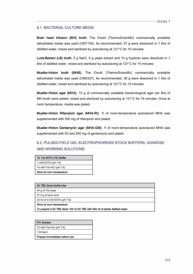
·················································································································· Annex 1
173
8.1. BACTERIAL CULTURE MEDIA
Brain heart infusion (BHI) broth. The Oxoid (ThermoScientific) commercially available
dehydrated media was used (CM1135). As recommended, 37 g were dissolved in 1 litre of
distilled water, mixed and sterilized by autoclaving at 121°C for 15 minutes.
Luria-Bertani (LB) broth. 5 g NaCl, 5 g yeast extract and 10 g tryptone were dissolved in 1
litre of distilled water, mixed and sterilized by autoclaving at 121°C for 15 minutes.
Mueller-Hinton broth (MHB). The Oxoid (ThermoScientific) commercially available
dehydrated media was used (CM0337). As recommended, 38 g were dissolved in 1 litre of
distilled water, mixed and sterilized by autoclaving at 121°C for 15 minutes.
Mueller-Hinton agar (MHA). 15 g of commercially available bacteriological agar per litre of
MH broth were added, mixed and sterilized by autoclaving at 121°C for 15 minutes. Once at
room temperature, media was plated.
Mueller-Hinton Rifampicin agar (MHA-RI). 1l of room-temperature autoclaved MHA was
supplemented with 300 mg of rifampicin and plated.
Mueller-Hinton Gentamycin agar (MHA-GM). 1l of room-temperature autoclaved MHA was
supplemented with 50 and 250 mg of gentamycin and plated.
8.2. PULSED-FIELD GEL ELECTROPHORESIS STOCK BUFFERS, AGAROSE
AND WORKING SOLUTIONS
1X Tris EDTA (TE) Buffer
1 mM EDTA (pH 7.6)
10 mM Tris-HCl (pH 7.6)
Store at room temperature
5X TBE Stock Buffer/Liter
54 g of Tris base
27.5 g of boric acid
20 ml of 0.5 M EDTA (pH 7.6)
Store at room temperature.
To prepare 0.5X TBE dilute 100 ml 5X TBE with 900 ml of sterile distilled water.
PIV Solution
10 mM Tris-HCl (pH 7.6)
1 M NaCl
Prepare immediately before use.
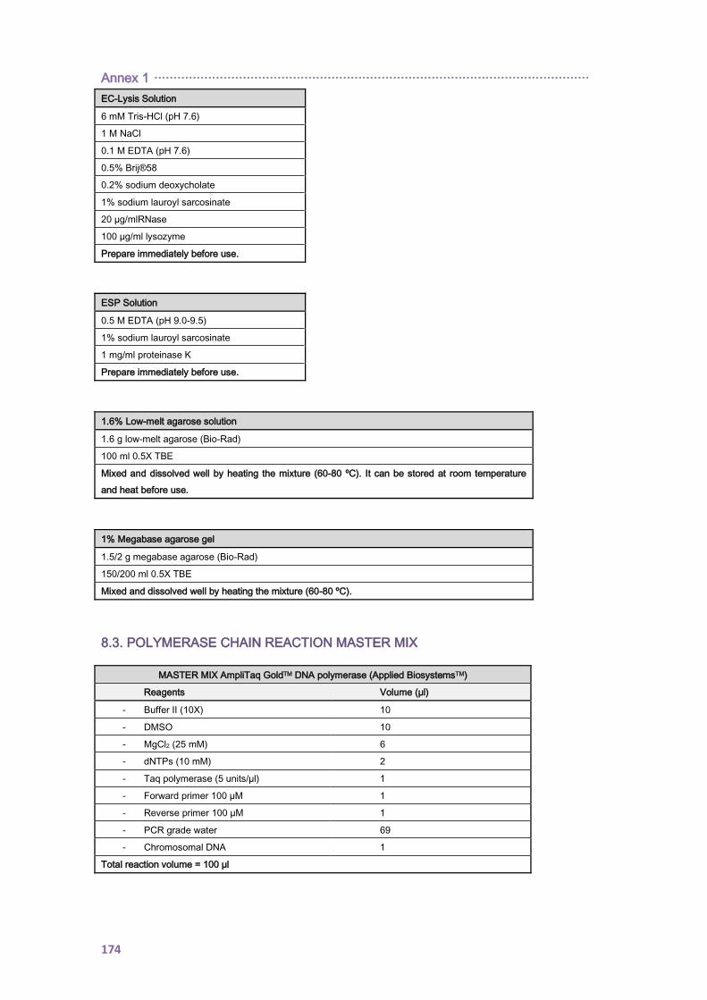
Annex 1 ·················································································································
174
EC-Lysis Solution
6 mM Tris-HCl (pH 7.6)
1 M NaCl
0.1 M EDTA (pH 7.6)
0.5% Brij®58
0.2% sodium deoxycholate
1% sodium lauroyl sarcosinate
20 µg/mlRNase
100 µg/ml lysozyme
Prepare immediately before use.
ESP Solution
0.5 M EDTA (pH 9.0-9.5)
1% sodium lauroyl sarcosinate
1 mg/ml proteinase K
Prepare immediately before use.
1.6% Low-melt agarose solution
1.6 g low-melt agarose (Bio-Rad)
100 ml 0.5X TBE
Mixed and dissolved well by heating the mixture (60-80 ºC). It can be stored at room temperature
and heat before use.
1% Megabase agarose gel
1.5/2 g megabase agarose (Bio-Rad)
150/200 ml 0.5X TBE
Mixed and dissolved well by heating the mixture (60-80 ºC).
8.3. POLYMERASE CHAIN REACTION MASTER MIX
MASTER MIX AmpliTaq GoldTM DNA polymerase (Applied BiosystemsTM)
Reagents Volume (µl)
- Buffer II (10X) 10
- DMSO 10
- MgCl2 (25 mM) 6
- dNTPs (10 mM) 2
- Taq polymerase (5 units/µl) 1
- Forward primer 100 µM 1
- Reverse primer 100 µM 1
- PCR grade water 69
- Chromosomal DNA 1
Total reaction volume = 100 µl
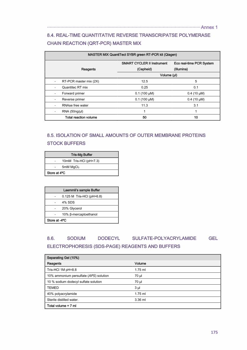
·················································································································· Annex 1
175
8.4. REAL-TIME QUANTITATIVE REVERSE TRANSCRIPATSE POLYMERASE
CHAIN REACTION (QRT-PCR) MASTER MIX
MASTER MIX QuantiTect SYBR green RT-PCR kit (Qiagen)
Reagents
SMART CYCLER II Instrument
(Cepheid)
Eco real-time PCR System
(Illumina)
Volume (µl)
- RT-PCR master mix (2X) 12.5 5
- Quantitec RT mix 0.25 0.1
- Forward primer 0.1 (100 µM) 0.4 (10 µM)
- Reverse primer 0.1 (100 µM) 0.4 (10 µM)
- RNAse free water 11.3 3.1
- RNA (50ng/µl) 1 1
Total reaction volume 50 10
8.5. ISOLATION OF SMALL AMOUNTS OF OUTER MEMBRANE PROTEINS
STOCK BUFFERS
Tris-Mg Buffer
- 10mM Tris-HCl (pH=7.3)
- 5mM MgCl2
Store at 4ºC
Laemmli’s sample Buffer
- 0.125 M Tris-HCl (pH=6.8)
- 4% SDS
- 20% Glycerol
- 10% β-mercaptoethanol
Store at -4ºC
8.6. SODIUM DODECYL SULFATE-POLYACRYLAMIDE GEL
ELECTROPHORESIS (SDS-PAGE) REAGENTS AND BUFFERS
Separating Gel (10%)
Reagents Volume
Tris-HCl 1M pH=8.8 1.75 ml
10% ammonium persulfate (APS) solution 70 µl
10 % sodium dodecyl sulfate solution 70 µl
TEMED 3 µl
40% polyacrylamide 1.75 ml
Sterile distilled water. 3.36 ml
Total volume = 7 ml
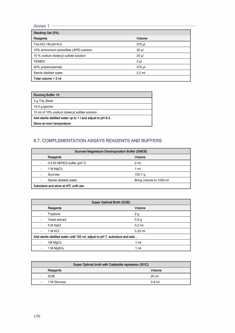
Annex 1 ·················································································································
176
Stacking Gel (5%)
Reagents Volume
Tris-HCl 1M pH=6.9 375 µl
10% ammonium persulfate (APS) solution 30 µl
10 % sodium dodecyl sulfate solution 30 µl
TEMED 3 µl
40% polyacrylamide 375 µl
Sterile distilled water. 2.2 ml
Total volume = 3 ml
Running Buffer 1X
3 g Tris_Base
14.4 g glycine
10 ml of 10% sodium dodecyl sulfate solution
Add sterile distilled water up to 1 l and adjust to pH 8.3.
Store at room temperature
8.7. COMPLEMENTATION ASSAYS REAGENTS AND BUFFERS
Sucrose Magnesium Electroporation Buffer (SMEB)
Reagents Volume
- 0.5 M HEPES buffer (pH 7) 2 ml
- 1 M MgCl2 1 ml
- Sucrose 102.7 g
- Sterile distilled water. Bring volume to 1000 ml
Autoclave and store at 4ºC until use
Super Optimal Broth (SOB)
Reagents Volume
- Tryptone 2 g
- Yeast extract 0.5 g
- 5 M NaCl 0.2 ml
- 1 M KCl 0.25 ml
Add sterile distilled water until 100 ml, adjust to pH 7, autoclave and add…
- 1M MgCl2 1 ml
- 1 M MgSO4 1 ml
Super Optimal broth with Catabolite repression (SOC)
Reagents Volume
- SOB 20 ml
- 1 M Glucose 0.4 ml

177
9. ANNEX 2
ARRAY-TUBE GENOTYPING SYSTEM, PROBES AND
PRIMERS INFORMATION

178
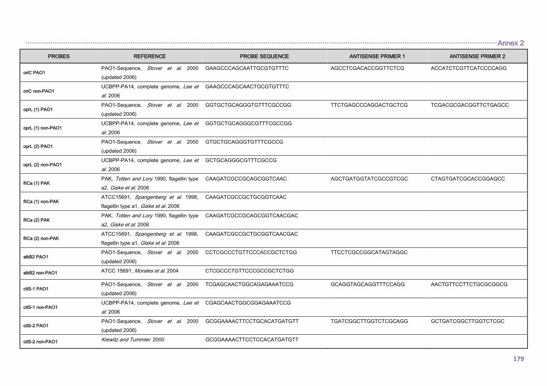
································································································································································································································ Annex 2
179
PROBES REFERENCE PROBE SEQUENCE ANTISENSE PRIMER 1 ANTISENSE PRIMER 2
oriC PAO1 PAO1-Sequence, Stover et al. 2000
(updated 2006)
GAAGCCCAGCAATTGCGTGTTTC AGCCTCGACACCGGTTCTCG ACCATCTCGTTCATCCCCAGG
oriC non-PAO1 UCBPP-PA14, complete genome, Lee et
al. 2006
GAAGCCCAGCAACTGCGTGTTTC
oprL (1) PAO1 PAO1-Sequence, Stover et al. 2000
(updated 2006)
GGTGCTGCAGGGTGTTTCGCCGG TTCTGAGCCCAGGACTGCTCG TCGACGCGACGGTTCTGAGCC
oprL (1) non-PAO1 UCBPP-PA14, complete genome, Lee et
al. 2006
GGTGCTGCAGGGCGTTTCGCCGG
oprL (2) PAO1 PAO1-Sequence, Stover et al. 2000
(updated 2006)
GTGCTGCAGGGTGTTTCGCCG
oprL (2) non-PAO1 UCBPP-PA14, complete genome, Lee et
al. 2006
GCTGCAGGGCGTTTCGCCG
fliCa (1) PAK PAK, Totten and Lory 1990, flagellin type
a2, Giske et al. 2006
CAAGATCGCCGCAGCGGTCAAC AGCTGATGGTATCGCCGTCGC CTAGTGATCGCACCGGAGCC
fliCa (1) non-PAK ATCC15691, Spangenberg et al. 1998,
flagellin type a1, Giske et al. 2006
CAAGATCGCCGCTGCGGTCAAC
fliCa (2) PAK PAK, Totten and Lory 1990, flagellin type
a2, Giske et al. 2006
CAAGATCGCCGCAGCGGTCAACGAC
fliCa (2) non-PAK ATCC15691, Spangenberg et al. 1998,
flagellin type a1, Giske et al. 2006
CAAGATCGCCGCTGCGGTCAACGAC
alkB2 PAO1 PAO1-Sequence, Stover et al. 2000
(updated 2006)
CCTCGCCCTGTTCCCACCGCTCTGG TTCCTCGCCGGCATAGTAGGC
alkB2 non-PAO1 ATCC 15691, Morales et al. 2004 CTCGCCCTGTTCCCGCCGCTCTGG
citS-1 PAO1 PAO1-Sequence, Stover et al. 2000
(updated 2006)
TCGAGCAACTGGCAGAGAAATCCG GCAGGTAGCAGGTTTCCAGG AACTGTTCCTTCTGCGCGGCG
citS-1 non-PAO1 UCBPP-PA14, complete genome, Lee et
al. 2006
CGAGCAACTGGCGGAGAAATCCG
citS-2 PAO1 PAO1-Sequence, Stover et al. 2000
(updated 2006)
GCGGAAAACTTCCTGCACATGATGTT TGATCGGCTTGGTCTCGCAGG GCTGATCGGCTTGGTCTCGC
citS-2 non-PAO1 Kiewitz and Tummler. 2000 GCGGAAAACTTCCTCCACATGATGTT
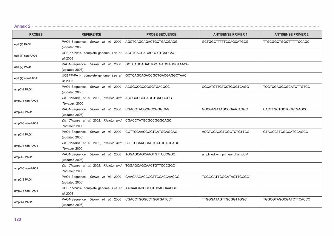
Annex 2 ·······························································································································································································································
180
PROBES REFERENCE PROBE SEQUENCE ANTISENSE PRIMER 1 ANTISENSE PRIMER 2
oprI (1) PAO1 PAO1-Sequence, Stover et al. 2000
(updated 2006)
AGCTCAGCAGACTGCTGACGAGG GCTGGCTTTTTCCAGCATGCG TTGCGGCTGGCTTTTTCCAGC
oprI (1) non-PAO1 UCBPP-PA14, complete genome, Lee et
al. 2006
AGCTCAGCAGACCGCTGACGAG
oprI (2) PAO1 PAO1-Sequence, Stover et al. 2000
(updated 2006)
GCTCAGCAGACTGCTGACGAGGCTAACG
oprI (2) non-PAO1 UCBPP-PA14, complete genome, Lee et
al. 2006
GCTCAGCAGACCGCTGACGAGGCTAAC
ampC-1 PAO1 PAO1-Sequence, Stover et al. 2000
(updated 2006)
ACGGCCGCCGGGTGACGCC CGCATCTTGTCCTGGGTCAGG TCGTCGAGGCGCATCTTGTCC
ampC-1 non-PAO1 De Champs et al. 2002, Kiewitz and
Tummler, 2000
ACGGCCGCCAGGTGACGCCG
ampC-3 PAO1 PAO1-Sequence, Stover et al. 2000
(updated 2006)
CGACCTACGCGCCGGGCAG GGCGAGATAGCCGAACAGGC CACTTGCTGCTCCATGAGCC
ampC-3 non-PAO1 De Champs et al. 2002, Kiewitz and
Tummler. 2000
CGACCTATGCGCCGGGCAGC
ampC-4 PAO1 PAO1-Sequence, Stover et al. 2000
(updated 2006)
CGTTCGAACGGCTCATGGAGCAG ACGTCGAGGTGGGTCTGTTCG GTAGCCTTCGGCATCCAGCG
ampC-4 non-PAO1 De Champs et al. 2002, Kiewitz and
Tummler 2000
CGTTCGAACGACTCATGGAGCAGC
ampC-5 PAO1 PAO1-Sequence, Stover et al. 2000
(updated 2006)
TGGAGCAGCAAGTGTTCCCGGC amplified with primers of ampC-4
ampC-5 non-PAO1 De Champs et al. 2002, Kiewitz and
Tummler. 2000
TGGAGCAGCAACTGTTCCCGGC
ampC-6 PAO1 PAO1-Sequence, Stover et al. 2000
(updated 2006)
GAACAAGACCGGTTCCACCAACGG TCGGCATTGGGATAGTTGCGG
ampC-6 non-PAO1 UCBPP-PA14, complete genome, Lee et
al. 2006
AACAAGACCGGCTCCACCAACGG
ampC-7 PAO1 PAO1-Sequence, Stover et al. 2000
(updated 2006)
CGACCTGGGCCTGGTGATCCT TTGGGATAGTTGCGGTTGGC TGGCGTAGGCGATCTTCACCC

································································································································································································································ Annex 2
181
PROBES REFERENCE PROBE SEQUENCE ANTISENSE PRIMER 1 ANTISENSE PRIMER 2
fliC a ATCC15691, Spangenberg et al. 1998 GTCGCTGAACGGCACCTACTTCA CGATCGCGATGTCGACGGTGC TGCCGATCGCGATGTCGACG
fliC b PAO1-Sequence, Stover et al. 2000
(updated 2006)
GCCGACCAACTGAACTCCAACTCG TGACGTTCTCGCCGGTAGCG CAGTAGCGGTACCGGTCTGC
exoS PAO1-Sequence, Stover et al. 2000
(updated 2006)
CAGCCCAGTCAGGACGCGCA CAGGGTCGCCAGCTCGCTCGCC AGGGTCGCCAGCTCGCTCGC
exoU UCBPP-PA14, complete genome, Lee et
al. 2006
CGCCAGTTTGAGAACGGAGTCACC AGTGATCTGCCGCGGCCCTGCC GTGATCTGCCGCGGCCCTGC
fpvA type I PAO1-Sequence, Stover et al. 2000
(updated 2006)
CCTGAATCCGACCATTCGCGAGTC CGTTCAGGTCGTAGACCGCGC GCGATACCAACTGTCCTGCGGC
fpvA type IIa de Chial et al. 2003 TCGGACTGTACTCCTACGAAGCAGC TGCCGAAGGTGAATGGCTTGCC CCTGATGGTCCGATCCCAGC
fpvA type IIb Spencer et al. 2003 CCAATCCCTATCGCTGGAACCGTACC GCCGAGGGTCAAGAACCACTGG TCTTGGCCCAGTCATAGCGGC
fpvA type III de Chial et al. 2003 GCTCGGGACTCGCATTTCGTCC TAACCCCAAGGCCCATTGGAGG GCCACCGCCTTCGAATAACCCC
fpvB PAO1-Sequence, Stover et al. 2000
(updated 2006)
GCGTTATTGCTCGGTCTCTCCTCG AATTGCTCGAGGGATGCGGC GGTCGAAACGGATGCGCAGG
LES LES400 (personal communication C.
Winstanley)
TGCATAGGAGTCATGCCGACAGCA GCCCCGCGTCATTTTCACGTCG AATGCTCTGGGCAACGAGCC
PA0636 PAO1-Sequence, Stover et al. 2000
(updated 2006)
GCCAATTGGGTCAGCAAGCAACG ATGCCATCGTTGAAGGCACCGC TGCCATCGTTGAAGGCACCG
PA0722 PAO1-Sequence, Stover et al. 2000
(updated 2006)
CGTGTCGCGAACTCGCATGGC TCTGGCGGAATCAGGTAGGCC CTTCCGGGGAGAAACCACCG
PA0728 PAO1-Sequence, Stover et al. 2000
(updated 2006)
CTGGAGCCTGCGAAAGTGGCTC AGCCAAGACGGTTGTTCGCGG TCAATGACGCCGAGTTGGCGC
PA2185 PAO1-Sequence, Stover et al. 2000
(updated 2006)
ACGAGGGTGATGGCTGGGAATACG CTCGGACAGGTTCACGCTGG GCCATTCGCTGCAACACCTCC
PA2221 PAO1-Sequence, Stover et al. 2000
(updated 2006)
CAGTTGTCGCCAGGTCTGGAGAATCC TTCCTGGGCCAGAGTTGGACC AGCTTAAGGCCGTGGCACTCG
PA3835 PAO1-Sequence, Stover et al. 2000
(updated 2006)
CACATCAATGTCAGCCCACGCCA CCGGAGAATTCGCGTCCACC TGCTGACGATGAAGCCCCAGC
fla-island Arora et al. 2001 ACCTGTGTCGCTGGAGGGTATGTT CCCGTGTTTCCGTAGACCTTGC
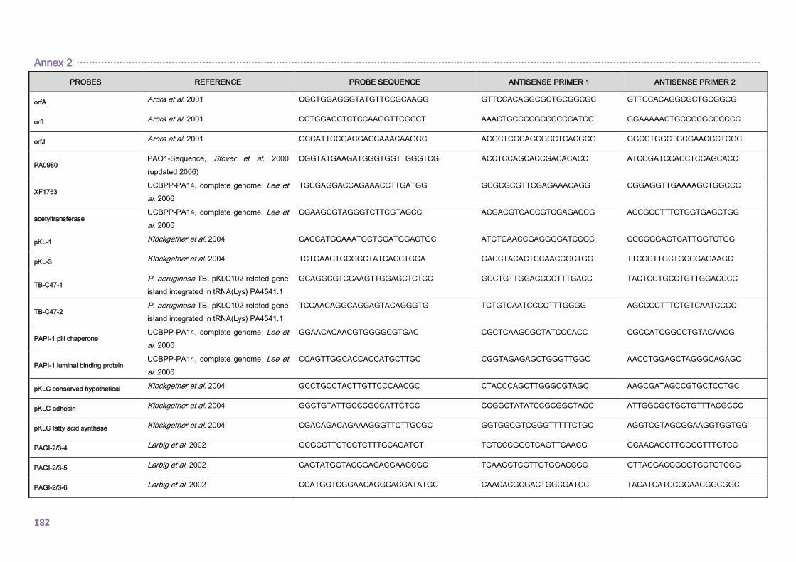
Annex 2 ·······························································································································································································································
182
PROBES REFERENCE PROBE SEQUENCE ANTISENSE PRIMER 1 ANTISENSE PRIMER 2
orfA Arora et al. 2001 CGCTGGAGGGTATGTTCCGCAAGG GTTCCACAGGCGCTGCGGCGC GTTCCACAGGCGCTGCGGCG
orfI Arora et al. 2001 CCTGGACCTCTCCAAGGTTCGCCT AAACTGCCCCGCCCCCCATCC GGAAAAACTGCCCCGCCCCCC
orfJ Arora et al. 2001 GCCATTCCGACGACCAAACAAGGC ACGCTCGCAGCGCCTCACGCG GGCCTGGCTGCGAACGCTCGC
PA0980 PAO1-Sequence, Stover et al. 2000
(updated 2006)
CGGTATGAAGATGGGTGGTTGGGTCG ACCTCCAGCACCGACACACC ATCCGATCCACCTCCAGCACC
XF1753 UCBPP-PA14, complete genome, Lee et
al. 2006
TGCGAGGACCAGAAACCTTGATGG GCGCGCGTTCGAGAAACAGG CGGAGGTTGAAAAGCTGGCCC
acetyltransferase UCBPP-PA14, complete genome, Lee et
al. 2006
CGAAGCGTAGGGTCTTCGTAGCC ACGACGTCACCGTCGAGACCG ACCGCCTTTCTGGTGAGCTGG
pKL-1 Klockgether et al. 2004 CACCATGCAAATGCTCGATGGACTGC ATCTGAACCGAGGGGATCCGC CCCGGGAGTCATTGGTCTGG
pKL-3 Klockgether et al. 2004 TCTGAACTGCGGCTATCACCTGGA GACCTACACTCCAACCGCTGG TTCCCTTGCTGCCGAGAAGC
TB-C47-1 P. aeruginosa TB, pKLC102 related gene
island integrated in tRNA(Lys) PA4541.1
GCAGGCGTCCAAGTTGGAGCTCTCC GCCTGTTGGACCCCTTTGACC TACTCCTGCCTGTTGGACCCC
TB-C47-2 P. aeruginosa TB, pKLC102 related gene
island integrated in tRNA(Lys) PA4541.1
TCCAACAGGCAGGAGTACAGGGTG TCTGTCAATCCCCTTTGGGG AGCCCCTTTCTGTCAATCCCC
PAPI-1 pili chaperone UCBPP-PA14, complete genome, Lee et
al. 2006
GGAACACAACGTGGGGCGTGAC CGCTCAAGCGCTATCCCACC CGCCATCGGCCTGTACAACG
PAPI-1 luminal binding protein UCBPP-PA14, complete genome, Lee et
al. 2006
CCAGTTGGCACCACCATGCTTGC CGGTAGAGAGCTGGGTTGGC AACCTGGAGCTAGGGCAGAGC
pKLC conserved hypothetical Klockgether et al. 2004 GCCTGCCTACTTGTTCCCAACGC CTACCCAGCTTGGGCGTAGC AAGCGATAGCCGTGCTCCTGC
pKLC adhesin Klockgether et al. 2004 GGCTGTATTGCCCGCCATTCTCC CCGGCTATATCCGCGGCTACC ATTGGCGCTGCTGTTTACGCCC
pKLC fatty acid synthase Klockgether et al. 2004 CGACAGACAGAAAGGGTTCTTGCGC GGTGGCGTCGGGTTTTTCTGC AGGTCGTAGCGGAAGGTGGTGG
PAGI-2/3-4 Larbig et al. 2002 GCGCCTTCTCCTCTTTGCAGATGT TGTCCCGGCTCAGTTCAACG GCAACACCTTGGCGTTTGTCC
PAGI-2/3-5 Larbig et al. 2002 CAGTATGGTACGGACACGAAGCGC TCAAGCTCGTTGTGGACCGC GTTACGACGGCGTGCTGTCGG
PAGI-2/3-6 Larbig et al. 2002 CCATGGTCGGAACAGGCACGATATGC CAACACGCGACTGGCGATCC TACATCATCCGCAACGGCGGC
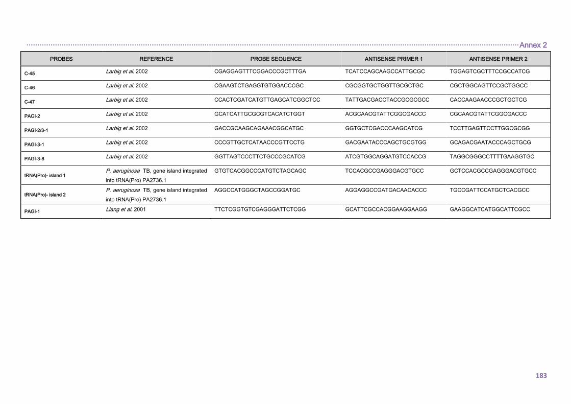
································································································································································································································ Annex 2
183
PROBES REFERENCE PROBE SEQUENCE ANTISENSE PRIMER 1 ANTISENSE PRIMER 2
C-45 Larbig et al. 2002 CGAGGAGTTTCGGACCCGCTTTGA TCATCCAGCAAGCCATTGCGC TGGAGTCGCTTTCCGCCATCG
C-46 Larbig et al. 2002 CGAAGTCTGAGGTGTGGACCCGC CGCGGTGCTGGTTGCGCTGC CGCTGGCAGTTCCGCTGGCC
C-47 Larbig et al. 2002 CCACTCGATCATGTTGAGCATCGGCTCC TATTGACGACCTACCGCGCGCC CACCAAGAACCCGCTGCTCG
PAGI-2 Larbig et al. 2002 GCATCATTGCGCGTCACATCTGGT ACGCAACGTATTCGGCGACCC CGCAACGTATTCGGCGACCC
PAGI-2/3-1 Larbig et al. 2002 GACCGCAAGCAGAAACGGCATGC GGTGCTCGACCCAAGCATCG TCCTTGAGTTCCTTGGCGCGG
PAGI-3-1 Larbig et al. 2002 CCCGTTGCTCATAACCCGTTCCTG GACGAATACCCAGCTGCGTGG GCAGACGAATACCCAGCTGCG
PAGI-3-8 Larbig et al. 2002 GGTTAGTCCCTTCTGCCCGCATCG ATCGTGGCAGGATGTCCACCG TAGGCGGGCCTTTTGAAGGTGC
tRNA(Pro)- island 1 P. aeruginosa TB, gene island integrated
into tRNA(Pro) PA2736.1
GTGTCACGGCCCATGTCTAGCAGC TCCACGCCGAGGGACGTGCC GCTCCACGCCGAGGGACGTGCC
tRNA(Pro)- island 2 P. aeruginosa TB, gene island integrated
into tRNA(Pro) PA2736.1
AGGCCATGGGCTAGCCGGATGC AGGAGGCCGATGACAACACCC TGCCGATTCCATGCTCACGCC
PAGI-1 Liang et al. 2001 TTCTCGGTGTCGAGGGATTCTCGG GCATTCGCCACGGAAGGAAGG GAAGGCATCATGGCATTCGCC

184

185
10. ANNEX 3
EUROPEAN COMMITTEE ON ANTIBIOTIC SUSCEPTIBILITY
TESTING (EUCAST) CLINICAL BREAKPOINTS FOR
Pseudomonas spp.

186
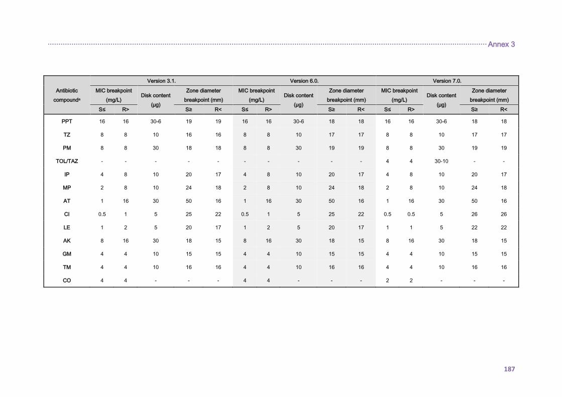
··········································································································································································································· Annex 3
187
Antibiotic
compounda
Version 3.1. Version 6.0. Version 7.0.
MIC breakpoint
(mg/L) Disk content
(µg)
Zone diameter
breakpoint (mm)
MIC breakpoint
(mg/L) Disk content
(µg)
Zone diameter
breakpoint (mm)
MIC breakpoint
(mg/L) Disk content
(µg)
Zone diameter
breakpoint (mm)
S≤ R> S≥ R< S≤ R> S≥ R< S≤ R> S≥ R<
PPT 16 16 30-6 19 19 16 16 30-6 18 18 16 16 30-6 18 18
TZ 8 8 10 16 16 8 8 10 17 17 8 8 10 17 17
PM 8 8 30 18 18 8 8 30 19 19 8 8 30 19 19
TOL/TAZ - - - - - - - - - - 4 4 30-10 - -
IP 4 8 10 20 17 4 8 10 20 17 4 8 10 20 17
MP 2 8 10 24 18 2 8 10 24 18 2 8 10 24 18
AT 1 16 30 50 16 1 16 30 50 16 1 16 30 50 16
CI 0.5 1 5 25 22 0.5 1 5 25 22 0.5 0.5 5 26 26
LE 1 2 5 20 17 1 2 5 20 17 1 1 5 22 22
AK 8 16 30 18 15 8 16 30 18 15 8 16 30 18 15
GM 4 4 10 15 15 4 4 10 15 15 4 4 10 15 15
TM 4 4 10 16 16 4 4 10 16 16 4 4 10 16 16
CO 4 4 - - - 4 4 - - - 2 2 - - -

188

189
11. ANNEX 4
USED SCRIPTS IN THE ANALYISIS OF WHOLE-GENOME
SEQUENCING DATA

190
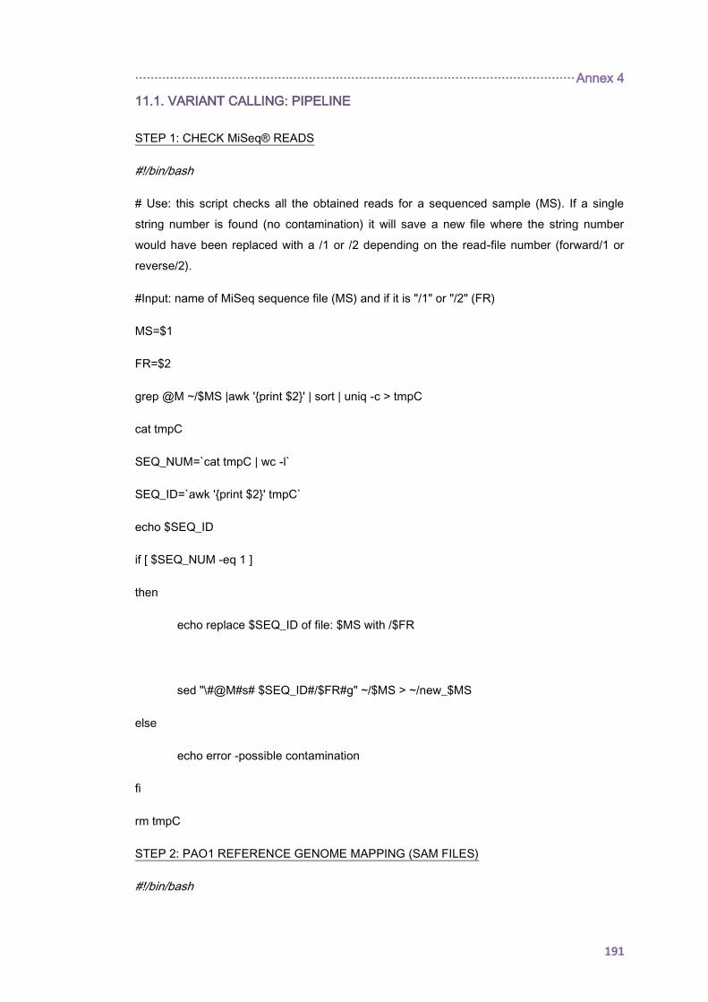
·················································································································· Annex 4
191
11.1. VARIANT CALLING: PIPELINE
STEP 1: CHECK MiSeq® READS
#!/bin/bash
# Use: this script checks all the obtained reads for a sequenced sample (MS). If a single
string number is found (no contamination) it will save a new file where the string number
would have been replaced with a /1 or /2 depending on the read-file number (forward/1 or
reverse/2).
#Input: name of MiSeq sequence file (MS) and if it is "/1" or "/2" (FR)
MS=$1
FR=$2
grep @M ~/$MS |awk 'print $2' | sort | uniq -c > tmpC
cat tmpC
SEQ_NUM=`cat tmpC | wc -l`
SEQ_ID=`awk 'print $2' tmpC`
echo $SEQ_ID
if [ $SEQ_NUM -eq 1 ]
then
echo replace $SEQ_ID of file: $MS with /$FR
sed "\#@M#s# $SEQ_ID#/$FR#g" ~/$MS > ~/new_$MS
else
echo error -possible contamination
fi
rm tmpC
STEP 2: PAO1 REFERENCE GENOME MAPPING (SAM FILES)
#!/bin/bash

Annex 4 ·················································································································
192
# Use: this script maps all checked reads (forward/reverse) for a sequenced sample
(new_MS) to P. aeruginosa PAO1 reference genome (previously indexed:
“indexed_PAO1_file”).
~/bowtie2-2.2.6/bowtie2 --phred33 –x ~/indexed_PAO1_file -q -1
~/new_MS_L001_R1_001.fastq -2 ~/new_MS_L001_R2_001.fastq -X 1000 -S
~/MS_mapPAO1.sam 2> ~/output_DO_bowtie2_PAO1_MS.txt
STEP 3: GENERATING PILEUP AND RAW FILES FROM SAM FILES
#!/bin/bash
# Use: this script will generate the raw and totalpileup files from the sam_file obtained in step
2.
~/samtools-0.1.16/samtools view -b –S ~/MS_mapPAO1.sam >
~/MS_mapPAO1.bam
java -jar ~/picard-tools-1.140/picard.jar SortSam INPUT=~/MS_mapPAO1.bam
OUTPUT=~/MS_mapPAO1_sorted.bam SORT_ORDER=coordinate
java -jar ~/picard-tools-1.140/picard.jar MarkDuplicates
METRICS_FILE=~/MS_metrics.txt
INPUT=~/MS_mapPAO1_sorted.bam OUTPUT=~/MS_mapPAO1_sorted_dedup.bam
java -jar ~/picard-tools-1.140/picard.jar AddOrReplaceReadGroups
INPUT=~/MS_mapPAO1_sorted_dedup.bam OUTPUT=~/MS_mapPAO1_addrg.bam
LB=XXX PL=Illumina PU=XXX SM=XXX
java -jar ~/picard-tools-1.140/picard.jar BuildBamIndex
INPUT=~/MS_mapPAO1_addrg.bam
java -jar ~/GenomeAnalysisTK-3.4-46/GenomeAnalysisTK.jar -T
RealignerTargetCreator -I ~/MS_mapPAO1_addrg.bam -R ~/PAO1complete.fasta -o
/MS_mapPAO1.realigned.intervals
java -jar ~/GenomeAnalysisTK-3.4-46/GenomeAnalysisTK.jar -T IndelRealigner –I
~/MS_mapPAO1_addrg.bam –R ~/PAO1complete.fasta --maxConsensuses 60 --
maxReadsForConsensuses 240 --maxReadsForRealignment 6000 --targetIntervals
~/MS_mapPAO1.realigned.intervals -o ~/MS_mapPAO1.realigned.bam
~/samtools-0.1.16/samtools sort ~/MS_mapPAO1.realigned.bam
/MS_mapPAO1.realigned.sorted
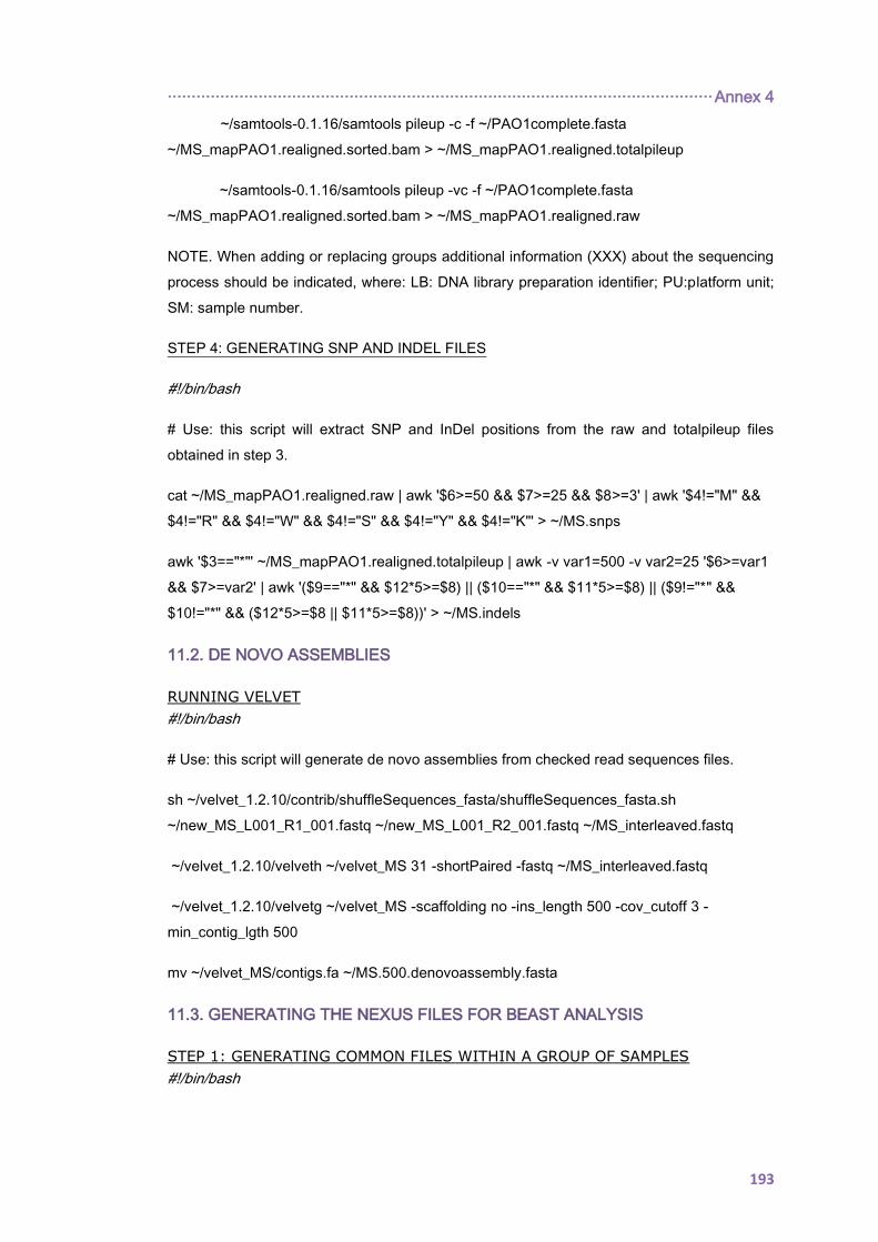
·················································································································· Annex 4
193
~/samtools-0.1.16/samtools pileup -c -f ~/PAO1complete.fasta
~/MS_mapPAO1.realigned.sorted.bam > ~/MS_mapPAO1.realigned.totalpileup
~/samtools-0.1.16/samtools pileup -vc -f ~/PAO1complete.fasta
~/MS_mapPAO1.realigned.sorted.bam > ~/MS_mapPAO1.realigned.raw
NOTE. When adding or replacing groups additional information (XXX) about the sequencing
process should be indicated, where: LB: DNA library preparation identifier; PU:platform unit;
SM: sample number.
STEP 4: GENERATING SNP AND INDEL FILES
#!/bin/bash
# Use: this script will extract SNP and InDel positions from the raw and totalpileup files
obtained in step 3.
cat ~/MS_mapPAO1.realigned.raw | awk '$6>=50 && $7>=25 && $8>=3' | awk '$4!="M" &&
$4!="R" && $4!="W" && $4!="S" && $4!="Y" && $4!="K"' > ~/MS.snps
awk '$3=="*"' ~/MS_mapPAO1.realigned.totalpileup | awk -v var1=500 -v var2=25 '$6>=var1
&& $7>=var2' | awk '($9=="*" && $12*5>=$8) || ($10=="*" && $11*5>=$8) || ($9!="*" &&
$10!="*" && ($12*5>=$8 || $11*5>=$8))' > ~/MS.indels
11.2. DE NOVO ASSEMBLIES
RUNNING VELVET
#!/bin/bash
# Use: this script will generate de novo assemblies from checked read sequences files.
sh ~/velvet_1.2.10/contrib/shuffleSequences_fasta/shuffleSequences_fasta.sh
~/new_MS_L001_R1_001.fastq ~/new_MS_L001_R2_001.fastq ~/MS_interleaved.fastq
~/velvet_1.2.10/velveth ~/velvet_MS 31 -shortPaired -fastq ~/MS_interleaved.fastq
~/velvet_1.2.10/velvetg ~/velvet_MS -scaffolding no -ins_length 500 -cov_cutoff 3 -
min_contig_lgth 500
mv ~/velvet_MS/contigs.fa ~/MS.500.denovoassembly.fasta
11.3. GENERATING THE NEXUS FILES FOR BEAST ANALYSIS
STEP 1: GENERATING COMMON FILES WITHIN A GROUP OF SAMPLES
#!/bin/bash
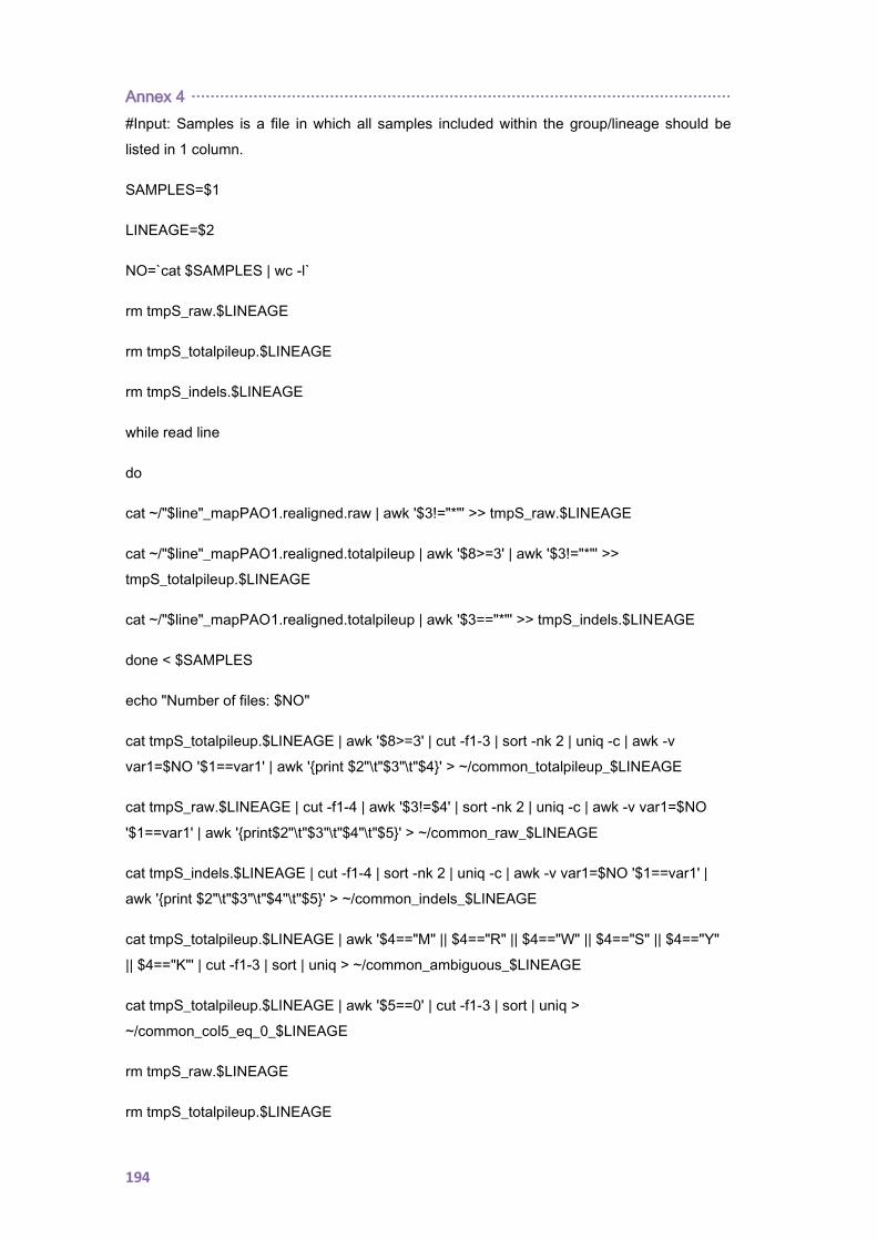
Annex 4 ·················································································································
194
#Input: Samples is a file in which all samples included within the group/lineage should be
listed in 1 column.
SAMPLES=$1
LINEAGE=$2
NO=`cat $SAMPLES | wc -l`
rm tmpS_raw.$LINEAGE
rm tmpS_totalpileup.$LINEAGE
rm tmpS_indels.$LINEAGE
while read line
do
cat ~/"$line"_mapPAO1.realigned.raw | awk '$3!="*"' >> tmpS_raw.$LINEAGE
cat ~/"$line"_mapPAO1.realigned.totalpileup | awk '$8>=3' | awk '$3!="*"' >>
tmpS_totalpileup.$LINEAGE
cat ~/"$line"_mapPAO1.realigned.totalpileup | awk '$3=="*"' >> tmpS_indels.$LINEAGE
done < $SAMPLES
echo "Number of files: $NO"
cat tmpS_totalpileup.$LINEAGE | awk '$8>=3' | cut -f1-3 | sort -nk 2 | uniq -c | awk -v
var1=$NO '$1==var1' | awk 'print $2"\t"$3"\t"$4' > ~/common_totalpileup_$LINEAGE
cat tmpS_raw.$LINEAGE | cut -f1-4 | awk '$3!=$4' | sort -nk 2 | uniq -c | awk -v var1=$NO
'$1==var1' | awk 'print$2"\t"$3"\t"$4"\t"$5' > ~/common_raw_$LINEAGE
cat tmpS_indels.$LINEAGE | cut -f1-4 | sort -nk 2 | uniq -c | awk -v var1=$NO '$1==var1' |
awk 'print $2"\t"$3"\t"$4"\t"$5' > ~/common_indels_$LINEAGE
cat tmpS_totalpileup.$LINEAGE | awk '$4=="M" || $4=="R" || $4=="W" || $4=="S" || $4=="Y"
|| $4=="K"' | cut -f1-3 | sort | uniq > ~/common_ambiguous_$LINEAGE
cat tmpS_totalpileup.$LINEAGE | awk '$5==0' | cut -f1-3 | sort | uniq >
~/common_col5_eq_0_$LINEAGE
rm tmpS_raw.$LINEAGE
rm tmpS_totalpileup.$LINEAGE
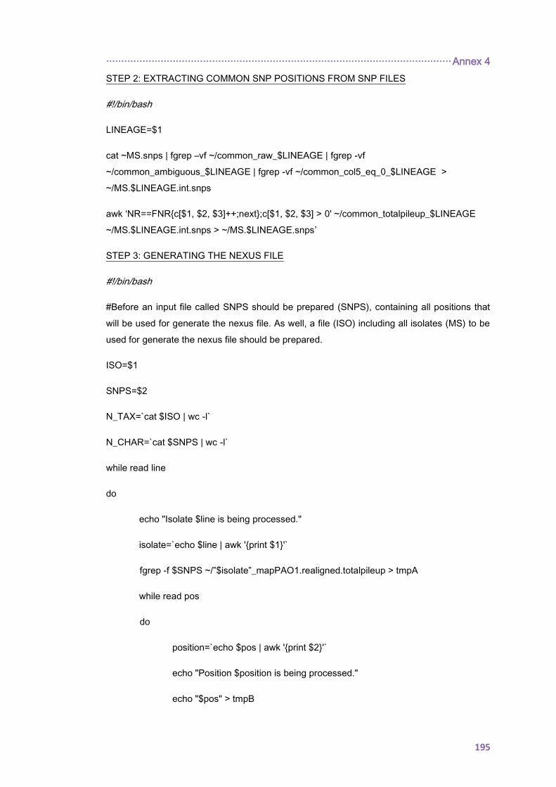
·················································································································· Annex 4
195
STEP 2: EXTRACTING COMMON SNP POSITIONS FROM SNP FILES
#!/bin/bash
LINEAGE=$1
cat ~MS.snps | fgrep –vf ~/common_raw_$LINEAGE | fgrep -vf
~/common_ambiguous_$LINEAGE | fgrep -vf ~/common_col5_eq_0_$LINEAGE >
~/MS.$LINEAGE.int.snps
awk ‘NR==FNRc[$1, $2, $3]++;next;c[$1, $2, $3] > 0' ~/common_totalpileup_$LINEAGE
~/MS.$LINEAGE.int.snps > ~/MS.$LINEAGE.snps’
STEP 3: GENERATING THE NEXUS FILE
#!/bin/bash
#Before an input file called SNPS should be prepared (SNPS), containing all positions that
will be used for generate the nexus file. As well, a file (ISO) including all isolates (MS) to be
used for generate the nexus file should be prepared.
ISO=$1
SNPS=$2
N_TAX=`cat $ISO | wc -l`
N_CHAR=`cat $SNPS | wc -l`
while read line
do
echo "Isolate $line is being processed."
isolate=`echo $line | awk 'print $1'`
fgrep -f $SNPS ~/”$isolate”_mapPAO1.realigned.totalpileup > tmpA
while read pos
do
position=`echo $pos | awk 'print $2'`
echo "Position $position is being processed."
echo "$pos" > tmpB

Annex 4 ·················································································································
196
fgrep -f tmpB tmpA > tmpC
echo "position $position in $line:"
echo `cat tmpB`
count=`cat tmpC| wc -l`
echo $count
if [ $count -lt 1 ]
then
echo "Position $position is not present"
echo $position $line >> missing_positions.txt
echo "?" >> $isolate.tree
else
if [ $count -eq 1 ]
then
echo "Position $position is found"
awk '
if ($6>=50 || $7>=25 && $8>=3)
print $4
else
print "?"
' tmpC > $isolate.tmp
awk '
if ($1=="*")
print "-"
else
print $0

·················································································································· Annex 4
197
' $isolate.tmp >> $isolate.tree
fi
fi
done<$SNPS
sh ~/transpose.sh $isolate.tree | sed 's/[[:blank:]]//g' > $isolate. trans.tmp
echo "'$isolate'" > name_file_$isolate. tmp
paste name_file_$isolate. tmp $isolate. trans.tmp > alignment.$isolate. tmp
done<$ISO
echo "#nexus\n\n[file created from SNP positions found with Bowtie2-GATK-Samtools on
`date`]\n\n\
begin data;\n\
\tdimensions ntax = $N_TAX nchar = $N_CHAR;\n\
\tformat datatype = DNA gap = - missing = ?;\n\n\
\tmatrix\n" > head_nex.tmp
echo "\t;\nend;" > tail_nex.tmp
cat head_nex.tmp alignment.*.*.tmp tail_nex.tmp > alignment.nexus.totalpileup
clean
#rm *tmp*
#rm *.*.tree
NOTE. The following script shoud be disposable to run the abovementioned (transpose.sh):
#!/bin/bash
FILE=$1
awk '
for (i=1; i<=NF; i++)
a[NR,i] = $i
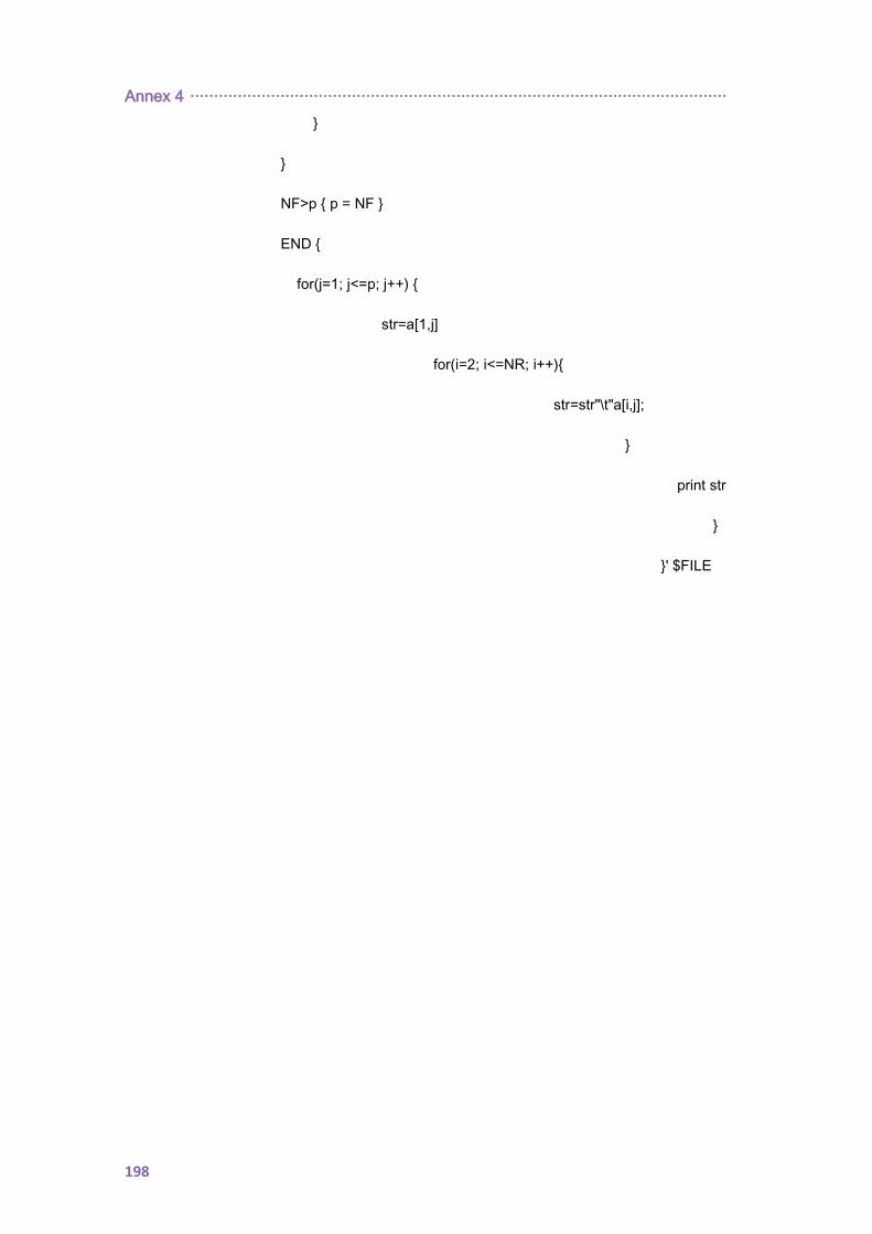
Annex 4 ·················································································································
198
NF>p p = NF
END
for(j=1; j<=p; j++)
str=a[1,j]
for(i=2; i<=NR; i++)
str=str"\t"a[i,j];
print str
' $FILE

199
12. ANNEX 5
Pseudomonas aeruginosa ANTIBIOTIC-RESISTANCE AND
HYPERMUTATION RELATED CHROMOSOMAL GENES

200
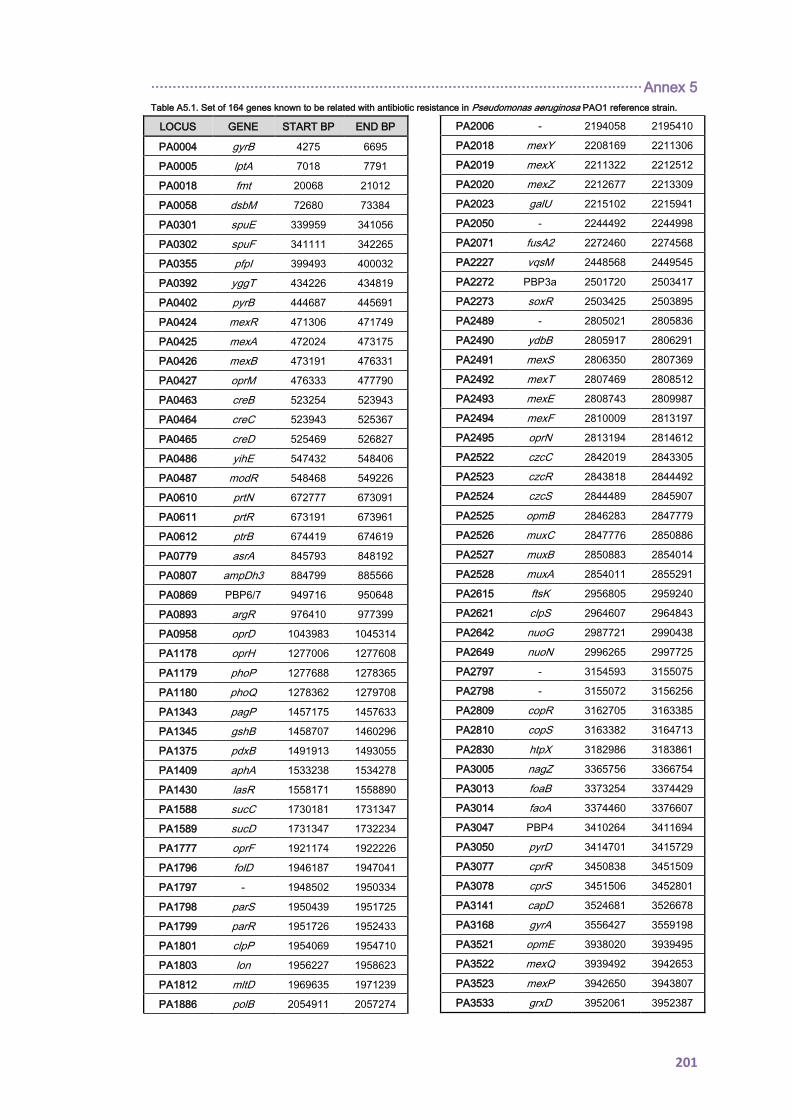
·················································································································· Annex 5
201
Table A5.1. Set of 164 genes known to be related with antibiotic resistance in Pseudomonas aeruginosa PAO1 reference strain.
LOCUS GENE START BP END BP
PA0004 gyrB 4275 6695
PA0005 lptA 7018 7791
PA0018 fmt 20068 21012
PA0058 dsbM 72680 73384
PA0301 spuE 339959 341056
PA0302 spuF 341111 342265
PA0355 pfpI 399493 400032
PA0392 yggT 434226 434819
PA0402 pyrB 444687 445691
PA0424 mexR 471306 471749
PA0425 mexA 472024 473175
PA0426 mexB 473191 476331
PA0427 oprM 476333 477790
PA0463 creB 523254 523943
PA0464 creC 523943 525367
PA0465 creD 525469 526827
PA0486 yihE 547432 548406
PA0487 modR 548468 549226
PA0610 prtN 672777 673091
PA0611 prtR 673191 673961
PA0612 ptrB 674419 674619
PA0779 asrA 845793 848192
PA0807 ampDh3 884799 885566
PA0869 PBP6/7 949716 950648
PA0893 argR 976410 977399
PA0958 oprD 1043983 1045314
PA1178 oprH 1277006 1277608
PA1179 phoP 1277688 1278365
PA1180 phoQ 1278362 1279708
PA1343 pagP 1457175 1457633
PA1345 gshB 1458707 1460296
PA1375 pdxB 1491913 1493055
PA1409 aphA 1533238 1534278
PA1430 lasR 1558171 1558890
PA1588 sucC 1730181 1731347
PA1589 sucD 1731347 1732234
PA1777 oprF 1921174 1922226
PA1796 folD 1946187 1947041
PA1797 - 1948502 1950334
PA1798 parS 1950439 1951725
PA1799 parR 1951726 1952433
PA1801 clpP 1954069 1954710
PA1803 lon 1956227 1958623
PA1812 mltD 1969635 1971239
PA1886 polB 2054911 2057274
PA2006 - 2194058 2195410
PA2018 mexY 2208169 2211306
PA2019 mexX 2211322 2212512
PA2020 mexZ 2212677 2213309
PA2023 galU 2215102 2215941
PA2050 - 2244492 2244998
PA2071 fusA2 2272460 2274568
PA2227 vqsM 2448568 2449545
PA2272 PBP3a 2501720 2503417
PA2273 soxR 2503425 2503895
PA2489 - 2805021 2805836
PA2490 ydbB 2805917 2806291
PA2491 mexS 2806350 2807369
PA2492 mexT 2807469 2808512
PA2493 mexE 2808743 2809987
PA2494 mexF 2810009 2813197
PA2495 oprN 2813194 2814612
PA2522 czcC 2842019 2843305
PA2523 czcR 2843818 2844492
PA2524 czcS 2844489 2845907
PA2525 opmB 2846283 2847779
PA2526 muxC 2847776 2850886
PA2527 muxB 2850883 2854014
PA2528 muxA 2854011 2855291
PA2615 ftsK 2956805 2959240
PA2621 clpS 2964607 2964843
PA2642 nuoG 2987721 2990438
PA2649 nuoN 2996265 2997725
PA2797 - 3154593 3155075
PA2798 - 3155072 3156256
PA2809 copR 3162705 3163385
PA2810 copS 3163382 3164713
PA2830 htpX 3182986 3183861
PA3005 nagZ 3365756 3366754
PA3013 foaB 3373254 3374429
PA3014 faoA 3374460 3376607
PA3047 PBP4 3410264 3411694
PA3050 pyrD 3414701 3415729
PA3077 cprR 3450838 3451509
PA3078 cprS 3451506 3452801
PA3141 capD 3524681 3526678
PA3168 gyrA 3556427 3559198
PA3521 opmE 3938020 3939495
PA3522 mexQ 3939492 3942653
PA3523 mexP 3942650 3943807
PA3533 grxD 3952061 3952387
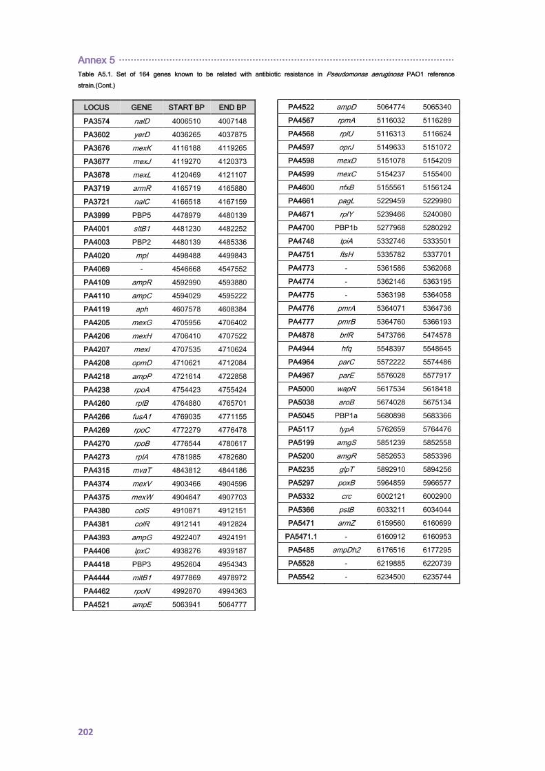
Annex 5 ·················································································································
202
Table A5.1. Set of 164 genes known to be related with antibiotic resistance in Pseudomonas aeruginosa PAO1 reference
strain.(Cont.)
LOCUS GENE START BP END BP
PA3574 nalD 4006510 4007148
PA3602 yerD 4036265 4037875
PA3676 mexK 4116188 4119265
PA3677 mexJ 4119270 4120373
PA3678 mexL 4120469 4121107
PA3719 armR 4165719 4165880
PA3721 nalC 4166518 4167159
PA3999 PBP5 4478979 4480139
PA4001 sltB1 4481230 4482252
PA4003 PBP2 4480139 4485336
PA4020 mpl 4498488 4499843
PA4069 - 4546668 4547552
PA4109 ampR 4592990 4593880
PA4110 ampC 4594029 4595222
PA4119 aph 4607578 4608384
PA4205 mexG 4705956 4706402
PA4206 mexH 4706410 4707522
PA4207 mexI 4707535 4710624
PA4208 opmD 4710621 4712084
PA4218 ampP 4721614 4722858
PA4238 rpoA 4754423 4755424
PA4260 rplB 4764880 4765701
PA4266 fusA1 4769035 4771155
PA4269 rpoC 4772279 4776478
PA4270 rpoB 4776544 4780617
PA4273 rplA 4781985 4782680
PA4315 mvaT 4843812 4844186
PA4374 mexV 4903466 4904596
PA4375 mexW 4904647 4907703
PA4380 colS 4910871 4912151
PA4381 colR 4912141 4912824
PA4393 ampG 4922407 4924191
PA4406 lpxC 4938276 4939187
PA4418 PBP3 4952604 4954343
PA4444 mltB1 4977869 4978972
PA4462 rpoN 4992870 4994363
PA4521 ampE 5063941 5064777
PA4522 ampD 5064774 5065340
PA4567 rpmA 5116032 5116289
PA4568 rplU 5116313 5116624
PA4597 oprJ 5149633 5151072
PA4598 mexD 5151078 5154209
PA4599 mexC 5154237 5155400
PA4600 nfxB 5155561 5156124
PA4661 pagL 5229459 5229980
PA4671 rplY 5239466 5240080
PA4700 PBP1b 5277968 5280292
PA4748 tpiA 5332746 5333501
PA4751 ftsH 5335782 5337701
PA4773 - 5361586 5362068
PA4774 - 5362146 5363195
PA4775 - 5363198 5364058
PA4776 pmrA 5364071 5364736
PA4777 pmrB 5364760 5366193
PA4878 brlR 5473766 5474578
PA4944 hfq 5548397 5548645
PA4964 parC 5572222 5574486
PA4967 parE 5576028 5577917
PA5000 wapR 5617534 5618418
PA5038 aroB 5674028 5675134
PA5045 PBP1a 5680898 5683366
PA5117 typA 5762659 5764476
PA5199 amgS 5851239 5852558
PA5200 amgR 5852653 5853396
PA5235 glpT 5892910 5894256
PA5297 poxB 5964859 5966577
PA5332 crc 6002121 6002900
PA5366 pstB 6033211 6034044
PA5471 armZ 6159560 6160699
PA5471.1 - 6160912 6160953
PA5485 ampDh2 6176516 6177295
PA5528 - 6219885 6220739
PA5542 - 6234500 6235744
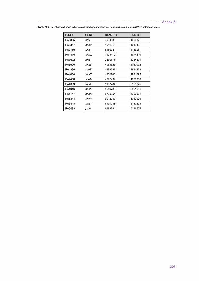
·················································································································· Annex 5
203
Table A5.2. Set of genes known to be related with hypermutation in Pseudomonas aeruginosa PAO1 reference strain.
LOCUS GENE START BP END BP
PA0355 pfpI 399493 400032
PA0357 mutY 401131 401943
PA0750 ung 818003 818698
PA1816 dnaQ 1973470 1974210
PA3002 mfd 3360875 3364321
PA3620 mutS 4054525 4057092
PA4366 sodB 4893697 4894278
PA4400 mutT 4930748 4931695
PA4468 sodM 4997439 4998050
PA4609 radA 5167284 5168645
PA4946 mutL 5549780 5551681
PA5147 mutM 5795954 5797021
PA5344 oxyR 6012047 6012979
PA5443 uvrD 6131088 6133274
PA5493 polA 6183784 6186525

204

205
13. ANNEX 6
MAIN MUTATIONS RELATED WITH ANTIBIOTIC RESISTANCE
ENCOUNTERED IN THE CLONAL COMPLEX 274
COLLECTION

206
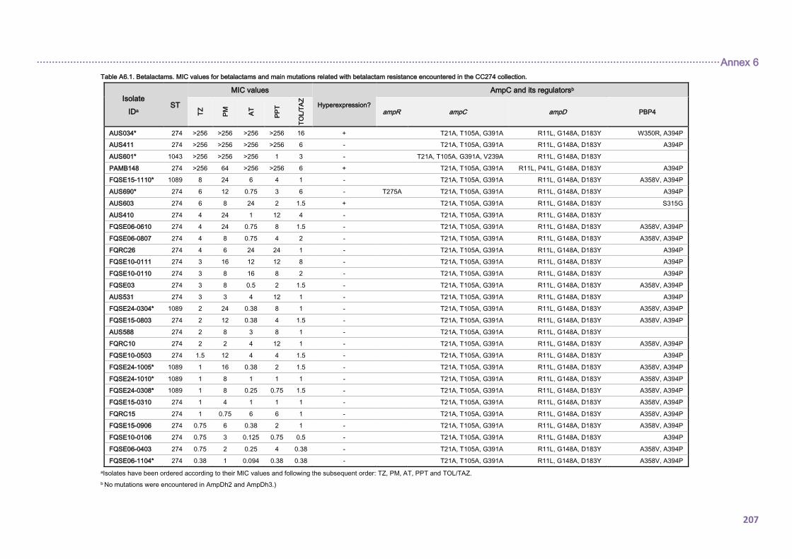
································································································································································································································ Annex 6
207
Table A6.1. Betalactams. MIC values for betalactams and main mutations related with betalactam resistance encountered in the CC274 collection.
Isolate
IDa ST
MIC values
Hyperexpression?
AmpC and its regulatorsb
TZ
PM
AT
PP
T
TO
L/T
AZ
ampR ampC ampD PBP4
AUS034* 274 >256 >256 >256 >256 16 + T21A, T105A, G391A R11L, G148A, D183Y W350R, A394P
AUS411 274 >256 >256 >256 >256 6 - T21A, T105A, G391A R11L, G148A, D183Y A394P
AUS601* 1043 >256 >256 >256 1 3 - T21A, T105A, G391A, V239A R11L, G148A, D183Y
PAMB148 274 >256 64 >256 >256 6 + T21A, T105A, G391A R11L, P41L, G148A, D183Y A394P
FQSE15-1110* 1089 8 24 6 4 1 - T21A, T105A, G391A R11L, G148A, D183Y A358V, A394P
AUS690* 274 6 12 0.75 3 6 - T275A T21A, T105A, G391A R11L, G148A, D183Y A394P
AUS603 274 6 8 24 2 1.5 + T21A, T105A, G391A R11L, G148A, D183Y S315G
AUS410 274 4 24 1 12 4 - T21A, T105A, G391A R11L, G148A, D183Y
FQSE06-0610 274 4 24 0.75 8 1.5 - T21A, T105A, G391A R11L, G148A, D183Y A358V, A394P
FQSE06-0807 274 4 8 0.75 4 2 - T21A, T105A, G391A R11L, G148A, D183Y A358V, A394P
FQRC26 274 4 6 24 24 1 - T21A, T105A, G391A R11L, G148A, D183Y A394P
FQSE10-0111 274 3 16 12 12 8 - T21A, T105A, G391A R11L, G148A, D183Y A394P
FQSE10-0110 274 3 8 16 8 2 - T21A, T105A, G391A R11L, G148A, D183Y A394P
FQSE03 274 3 8 0.5 2 1.5 - T21A, T105A, G391A R11L, G148A, D183Y A358V, A394P
AUS531 274 3 3 4 12 1 - T21A, T105A, G391A R11L, G148A, D183Y A394P
FQSE24-0304* 1089 2 24 0.38 8 1 - T21A, T105A, G391A R11L, G148A, D183Y A358V, A394P
FQSE15-0803 274 2 12 0.38 4 1.5 - T21A, T105A, G391A R11L, G148A, D183Y A358V, A394P
AUS588 274 2 8 3 8 1 - T21A, T105A, G391A R11L, G148A, D183Y
FQRC10 274 2 2 4 12 1 - T21A, T105A, G391A R11L, G148A, D183Y A358V, A394P
FQSE10-0503 274 1.5 12 4 4 1.5 - T21A, T105A, G391A R11L, G148A, D183Y A394P
FQSE24-1005* 1089 1 16 0.38 2 1.5 - T21A, T105A, G391A R11L, G148A, D183Y A358V, A394P
FQSE24-1010* 1089 1 8 1 1 1 - T21A, T105A, G391A R11L, G148A, D183Y A358V, A394P
FQSE24-0308* 1089 1 8 0.25 0.75 1.5 - T21A, T105A, G391A R11L, G148A, D183Y A358V, A394P
FQSE15-0310 274 1 4 1 1 1 - T21A, T105A, G391A R11L, G148A, D183Y A358V, A394P
FQRC15 274 1 0.75 6 6 1 - T21A, T105A, G391A R11L, G148A, D183Y A358V, A394P
FQSE15-0906 274 0.75 6 0.38 2 1 - T21A, T105A, G391A R11L, G148A, D183Y A358V, A394P
FQSE10-0106 274 0.75 3 0.125 0.75 0.5 - T21A, T105A, G391A R11L, G148A, D183Y A394P
FQSE06-0403 274 0.75 2 0.25 4 0.38 - T21A, T105A, G391A R11L, G148A, D183Y A358V, A394P
FQSE06-1104* 274 0.38 1 0.094 0.38 0.38 - T21A, T105A, G391A R11L, G148A, D183Y A358V, A394P
aIsolates have been ordered according to their MIC values and following the subsequent order: TZ, PM, AT, PPT and TOL/TAZ.
b No mutations were encountered in AmpDh2 and AmpDh3.)
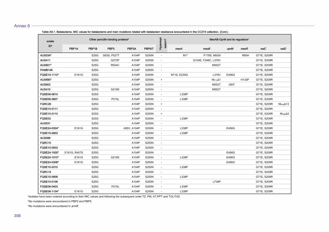
Annex 6 ·······························································································································································································································
208
Table A6.1. Betalactams. MIC values for betalactams and main mutations related with betalactam resistance encountered in the CC274 collection. (Cont.)
solate
IDa
Other penicillin-binding proteinsb
Hype
rexpr
essio
n?
MexAB-OprM and its regulatorsc
PBP1A PBP1B PBP3 PBP3A PBP6/7 mexA mexB oprM mexR nalC nalD
AUS034* S25G G63S, P527T A104P S250N - M1* F178S, M555I R85H G71E, S209R
AUS411 S25G Q372P A104P S250N - Q104E, F246C, L376V G71E, S209R
AUS601* S25G R504C A104P S250N - M552T G71E, S209R
PAMB148 S25G A104P S250N - G71E, S209R
FQSE15-1110* E161G S25G A104P S250N - N71S, D235G L376V E456G G71E, S209R
AUS690* S25G A104P S250N + Nt712Δ1 H133P G71E, S209R
AUS603 S25G A104P S250N - M552T Q93* G71E, S209R
AUS410 S25G G216S A104P S250N - M552T G71E, S209R
FQSE06-0610 S25G A104P S250N - L338P G71E, S209R
FQSE06-0807 S25G P215L A104P S250N - L338P G71E, S209R
FQRC26 S25G A104P S250N + G71E, S209R Nt459Δ13
FQSE10-0111 S25G A104P S250N - G71E, S209R
FQSE10-0110 S25G A104P S250N + G71E, S209R Nt396Δ2
FQSE03 S25G A104P S250N - L338P G71E, S209R
AUS531 S25G A104P S250N - G71E, S209R
FQSE24-0304* E161G S25G A95V, A104P S250N - L338P E456G G71E, S209R
FQSE15-0803 S25G A104P S250N - L338P G71E, S209R
AUS588 S25G A104P S250N - G71E, S209R
FQRC10 S25G A104P S250N - G71E, S209R
FQSE10-0503 S25G A104P S250N - G71E, S209R
FQSE24-1005* E161G, R407S S25G A104P S250N - E456G G71E, S209R
FQSE24-1010* E161G S25G G216S A104P S250N - L338P E456G G71E, S209R
FQSE24-0308* E161G S25G A104P S250N - E456G G71E, S209R
FQSE15-0310 S25G A104P S250N - L338P G71E, S209R
FQRC15 S25G A104P S250N - G71E, S209R
FQSE15-0906 S25G A104P S250N - L338P G71E, S209R
FQSE10-0106 S25G A104P S250N - L738P G71E, S209R
FQSE06-0403 S25G P215L A104P S250N - L338P G71E, S209R
FQSE06-1104* E161G S25G A104P S250N - L338P G71E, S209R
aIsolates have been ordered according to their MIC values and following the subsequent order:TZ, PM, AT,PPT and TOL/TAZ.
bNo mutations were encountered in PBP2 and PBP5.
cNo mutations were encountered in armR.
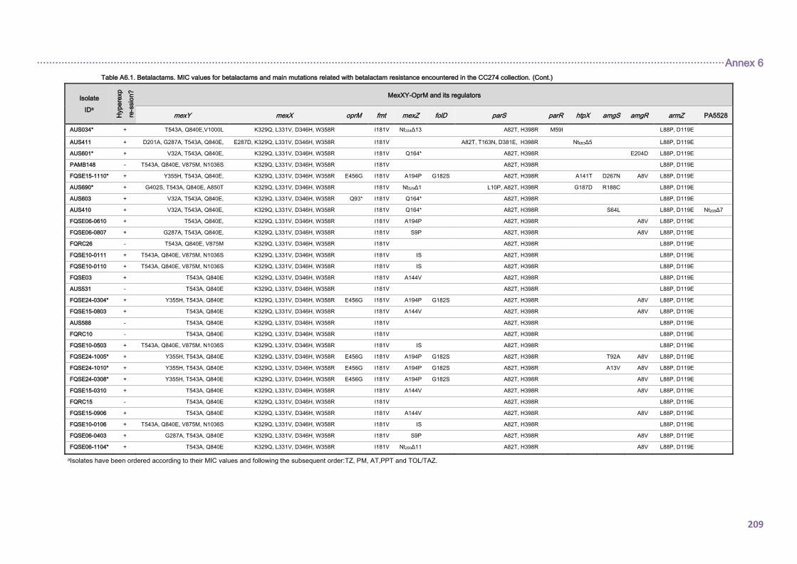
································································································································································································································ Annex 6
209
Table A6.1. Betalactams. MIC values for betalactams and main mutations related with betalactam resistance encountered in the CC274 collection. (Cont.)
Isolate
IDa
Hype
rexp
re-s
sio
n?
MexXY-OprM and its regulators
mexY mexX oprM fmt mexZ folD parS parR htpX amgS amgR armZ PA5528
AUS034* + T543A, Q840E,V1000L K329Q, L331V, D346H, W358R I181V Nt334Δ13 A82T, H398R M59I L88P, D119E
AUS411 + D201A, G287A, T543A, Q840E, E287D, K329Q, L331V, D346H, W358R I181V A82T, T163N, D381E, H398R Nt683Δ5 L88P, D119E
AUS601* + V32A, T543A, Q840E, K329Q, L331V, D346H, W358R I181V Q164* A82T, H398R E204D L88P, D119E
PAMB148 - T543A, Q840E, V875M, N1036S K329Q, L331V, D346H, W358R I181V A82T, H398R L88P, D119E
FQSE15-1110* + Y355H, T543A, Q840E, K329Q, L331V, D346H, W358R E456G I181V A194P G182S A82T, H398R A141T D267N A8V L88P, D119E
AUS690* + G402S, T543A, Q840E, A850T K329Q, L331V, D346H, W358R I181V Nt529Δ1 L10P, A82T, H398R G187D R188C L88P, D119E
AUS603 + V32A, T543A, Q840E, K329Q, L331V, D346H, W358R Q93* I181V Q164* A82T, H398R L88P, D119E
AUS410 + V32A, T543A, Q840E, K329Q, L331V, D346H, W358R I181V Q164* A82T, H398R S64L L88P, D119E Nt208Δ7
FQSE06-0610 + T543A, Q840E, K329Q, L331V, D346H, W358R I181V A194P A82T, H398R A8V L88P, D119E
FQSE06-0807 + G287A, T543A, Q840E, K329Q, L331V, D346H, W358R I181V S9P A82T, H398R A8V L88P, D119E
FQRC26 - T543A, Q840E, V875M K329Q, L331V, D346H, W358R I181V A82T, H398R L88P, D119E
FQSE10-0111 + T543A, Q840E, V875M, N1036S K329Q, L331V, D346H, W358R I181V IS A82T, H398R L88P, D119E
FQSE10-0110 + T543A, Q840E, V875M, N1036S K329Q, L331V, D346H, W358R I181V IS A82T, H398R L88P, D119E
FQSE03 + T543A, Q840E K329Q, L331V, D346H, W358R I181V A144V A82T, H398R L88P, D119E
AUS531 - T543A, Q840E K329Q, L331V, D346H, W358R I181V A82T, H398R L88P, D119E
FQSE24-0304* + Y355H, T543A, Q840E K329Q, L331V, D346H, W358R E456G I181V A194P G182S A82T, H398R A8V L88P, D119E
FQSE15-0803 + T543A, Q840E K329Q, L331V, D346H, W358R I181V A144V A82T, H398R A8V L88P, D119E
AUS588 - T543A, Q840E K329Q, L331V, D346H, W358R I181V A82T, H398R L88P, D119E
FQRC10 - T543A, Q840E K329Q, L331V, D346H, W358R I181V A82T, H398R L88P, D119E
FQSE10-0503 + T543A, Q840E, V875M, N1036S K329Q, L331V, D346H, W358R I181V IS A82T, H398R L88P, D119E
FQSE24-1005* + Y355H, T543A, Q840E K329Q, L331V, D346H, W358R E456G I181V A194P G182S A82T, H398R T92A A8V L88P, D119E
FQSE24-1010* + Y355H, T543A, Q840E K329Q, L331V, D346H, W358R E456G I181V A194P G182S A82T, H398R A13V A8V L88P, D119E
FQSE24-0308* + Y355H, T543A, Q840E K329Q, L331V, D346H, W358R E456G I181V A194P G182S A82T, H398R A8V L88P, D119E
FQSE15-0310 + T543A, Q840E K329Q, L331V, D346H, W358R I181V A144V A82T, H398R A8V L88P, D119E
FQRC15 - T543A, Q840E K329Q, L331V, D346H, W358R I181V A82T, H398R L88P, D119E
FQSE15-0906 + T543A, Q840E K329Q, L331V, D346H, W358R I181V A144V A82T, H398R A8V L88P, D119E
FQSE10-0106 + T543A, Q840E, V875M, N1036S K329Q, L331V, D346H, W358R I181V IS A82T, H398R L88P, D119E
FQSE06-0403 + G287A, T543A, Q840E K329Q, L331V, D346H, W358R I181V S9P A82T, H398R A8V L88P, D119E
FQSE06-1104* + T543A, Q840E K329Q, L331V, D346H, W358R I181V Nt290Δ11 A82T, H398R A8V L88P, D119E
aIsolates have been ordered according to their MIC values and following the subsequent order:TZ, PM, AT,PPT and TOL/TAZ.
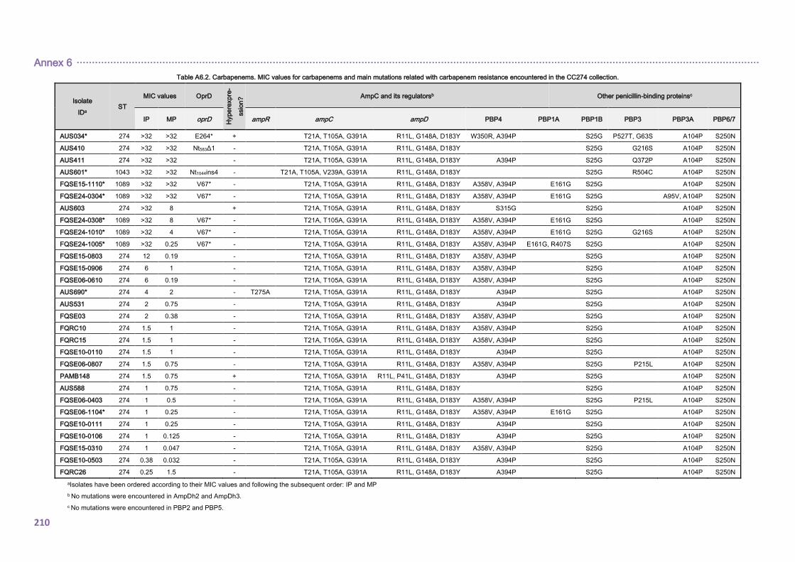
Annex 6 ·······························································································································································································································
210
Table A6.2. Carbapenems. MIC values for carbapenems and main mutations related with carbapenem resistance encountered in the CC274 collection.
Isolate
IDa ST
MIC values OprD
Hype
rexpre
-
ssio
n? AmpC and its regulatorsb Other penicillin-binding proteinsc
IP MP oprD ampR ampC ampD PBP4 PBP1A PBP1B PBP3 PBP3A PBP6/7
AUS034* 274 >32 >32 E264* + T21A, T105A, G391A R11L, G148A, D183Y W350R, A394P S25G P527T, G63S A104P S250N
AUS410 274 >32 >32 Nt583Δ1 - T21A, T105A, G391A R11L, G148A, D183Y S25G G216S A104P S250N
AUS411 274 >32 >32 - T21A, T105A, G391A R11L, G148A, D183Y A394P S25G Q372P A104P S250N
AUS601* 1043 >32 >32 Nt1044ins4 - T21A, T105A, V239A, G391A R11L, G148A, D183Y S25G R504C A104P S250N
FQSE15-1110* 1089 >32 >32 V67* - T21A, T105A, G391A R11L, G148A, D183Y A358V, A394P E161G S25G A104P S250N
FQSE24-0304* 1089 >32 >32 V67* - T21A, T105A, G391A R11L, G148A, D183Y A358V, A394P E161G S25G A95V, A104P S250N
AUS603 274 >32 8 + T21A, T105A, G391A R11L, G148A, D183Y S315G S25G A104P S250N
FQSE24-0308* 1089 >32 8 V67* - T21A, T105A, G391A R11L, G148A, D183Y A358V, A394P E161G S25G A104P S250N
FQSE24-1010* 1089 >32 4 V67* - T21A, T105A, G391A R11L, G148A, D183Y A358V, A394P E161G S25G G216S A104P S250N
FQSE24-1005* 1089 >32 0.25 V67* - T21A, T105A, G391A R11L, G148A, D183Y A358V, A394P E161G, R407S S25G A104P S250N
FQSE15-0803 274 12 0.19 - T21A, T105A, G391A R11L, G148A, D183Y A358V, A394P S25G A104P S250N
FQSE15-0906 274 6 1 - T21A, T105A, G391A R11L, G148A, D183Y A358V, A394P S25G A104P S250N
FQSE06-0610 274 6 0.19 - T21A, T105A, G391A R11L, G148A, D183Y A358V, A394P S25G A104P S250N
AUS690* 274 4 2 - T275A T21A, T105A, G391A R11L, G148A, D183Y A394P S25G A104P S250N
AUS531 274 2 0.75 - T21A, T105A, G391A R11L, G148A, D183Y A394P S25G A104P S250N
FQSE03 274 2 0.38 - T21A, T105A, G391A R11L, G148A, D183Y A358V, A394P S25G A104P S250N
FQRC10 274 1.5 1 - T21A, T105A, G391A R11L, G148A, D183Y A358V, A394P S25G A104P S250N
FQRC15 274 1.5 1 - T21A, T105A, G391A R11L, G148A, D183Y A358V, A394P S25G A104P S250N
FQSE10-0110 274 1.5 1 - T21A, T105A, G391A R11L, G148A, D183Y A394P S25G A104P S250N
FQSE06-0807 274 1.5 0.75 - T21A, T105A, G391A R11L, G148A, D183Y A358V, A394P S25G P215L A104P S250N
PAMB148 274 1.5 0.75 + T21A, T105A, G391A R11L, P41L, G148A, D183Y A394P S25G A104P S250N
AUS588 274 1 0.75 - T21A, T105A, G391A R11L, G148A, D183Y S25G A104P S250N
FQSE06-0403 274 1 0.5 - T21A, T105A, G391A R11L, G148A, D183Y A358V, A394P S25G P215L A104P S250N
FQSE06-1104* 274 1 0.25 - T21A, T105A, G391A R11L, G148A, D183Y A358V, A394P E161G S25G A104P S250N
FQSE10-0111 274 1 0.25 - T21A, T105A, G391A R11L, G148A, D183Y A394P S25G A104P S250N
FQSE10-0106 274 1 0.125 - T21A, T105A, G391A R11L, G148A, D183Y A394P S25G A104P S250N
FQSE15-0310 274 1 0.047 - T21A, T105A, G391A R11L, G148A, D183Y A358V, A394P S25G A104P S250N
FQSE10-0503 274 0.38 0.032 - T21A, T105A, G391A R11L, G148A, D183Y A394P S25G A104P S250N
FQRC26 274 0.25 1.5 - T21A, T105A, G391A R11L, G148A, D183Y A394P S25G A104P S250N
aIsolates have been ordered according to their MIC values and following the subsequent order: IP and MP
b No mutations were encountered in AmpDh2 and AmpDh3.
c No mutations were encountered in PBP2 and PBP5.
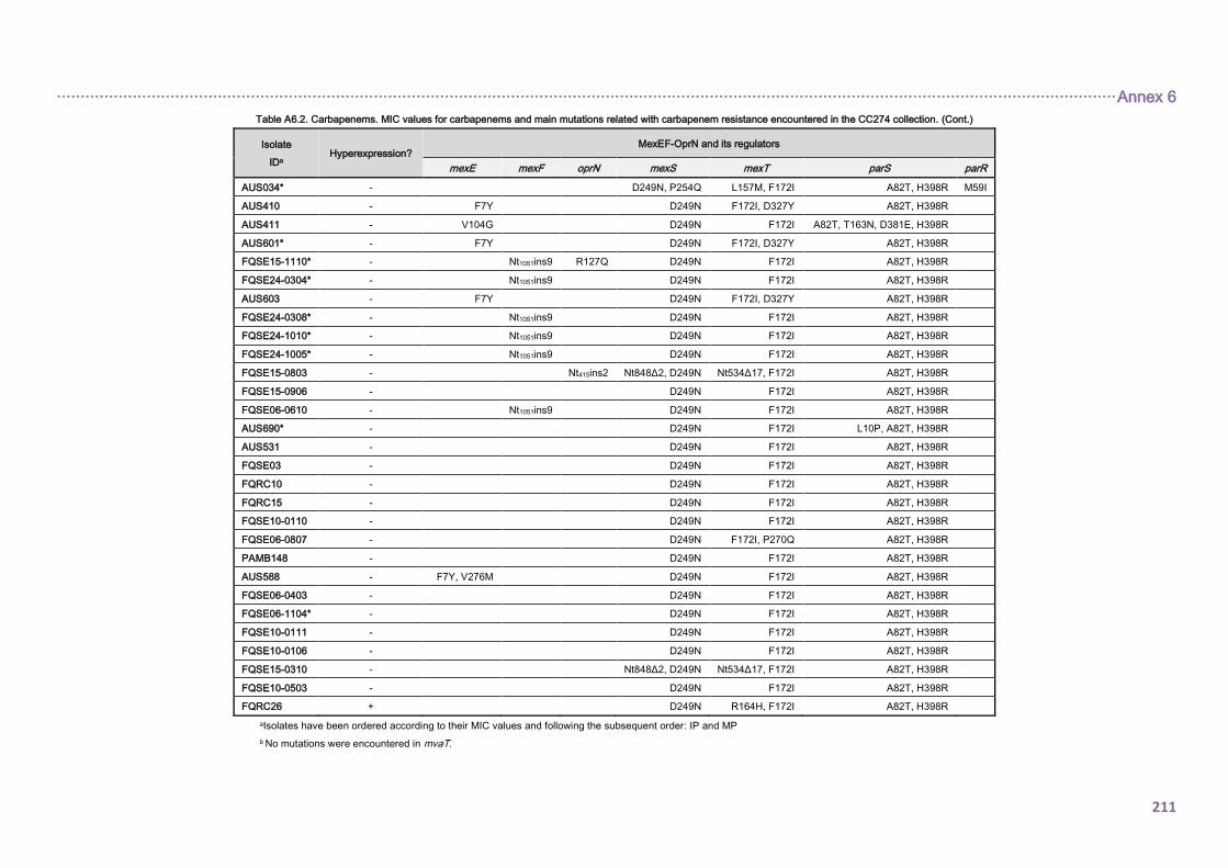
································································································································································································································ Annex 6
211
Table A6.2. Carbapenems. MIC values for carbapenems and main mutations related with carbapenem resistance encountered in the CC274 collection. (Cont.)
Isolate
IDa Hyperexpression?
MexEF-OprN and its regulators
mexE mexF oprN mexS mexT parS parR
AUS034* - D249N, P254Q L157M, F172I A82T, H398R M59I
AUS410 - F7Y D249N F172I, D327Y A82T, H398R
AUS411 - V104G D249N F172I A82T, T163N, D381E, H398R
AUS601* - F7Y D249N F172I, D327Y A82T, H398R
FQSE15-1110* - Nt1051ins9 R127Q D249N F172I A82T, H398R
FQSE24-0304* - Nt1051ins9 D249N F172I A82T, H398R
AUS603 - F7Y D249N F172I, D327Y A82T, H398R
FQSE24-0308* - Nt1051ins9 D249N F172I A82T, H398R
FQSE24-1010* - Nt1051ins9 D249N F172I A82T, H398R
FQSE24-1005* - Nt1051ins9 D249N F172I A82T, H398R
FQSE15-0803 - Nt415ins2 Nt848Δ2, D249N Nt534Δ17, F172I A82T, H398R
FQSE15-0906 - D249N F172I A82T, H398R
FQSE06-0610 - Nt1051ins9 D249N F172I A82T, H398R
AUS690* - D249N F172I L10P, A82T, H398R
AUS531 - D249N F172I A82T, H398R
FQSE03 - D249N F172I A82T, H398R
FQRC10 - D249N F172I A82T, H398R
FQRC15 - D249N F172I A82T, H398R
FQSE10-0110 - D249N F172I A82T, H398R
FQSE06-0807 - D249N F172I, P270Q A82T, H398R
PAMB148 - D249N F172I A82T, H398R
AUS588 - F7Y, V276M D249N F172I A82T, H398R
FQSE06-0403 - D249N F172I A82T, H398R
FQSE06-1104* - D249N F172I A82T, H398R
FQSE10-0111 - D249N F172I A82T, H398R
FQSE10-0106 - D249N F172I A82T, H398R
FQSE15-0310 - Nt848Δ2, D249N Nt534Δ17, F172I A82T, H398R
FQSE10-0503 - D249N F172I A82T, H398R
FQRC26 + D249N R164H, F172I A82T, H398R
aIsolates have been ordered according to their MIC values and following the subsequent order: IP and MP
b No mutations were encountered in mvaT.
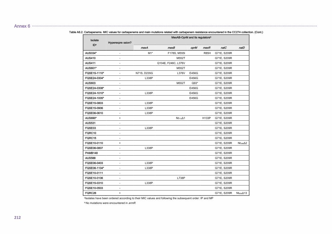
Annex 6 ·······························································································································································································································
212
Table A6.2. Carbapenems. MIC values for carbapenems and main mutations related with carbapenem resistance encountered in the CC274 collection. (Cont.)
Isolate
IDa Hyperexpre-ssion?
MexAB-OprM and its regulatorsb
mexA mexB oprM mexR nalC nalD
AUS034* - M1* F178S, M555I R85H G71E, S209R
AUS410 - M552T G71E, S209R
AUS411 - Q104E, F246C, L376V G71E, S209R
AUS601* - M552T G71E, S209R
FQSE15-1110* - N71S, D235G L376V E456G G71E, S209R
FQSE24-0304* - L338P E456G G71E, S209R
AUS603 - M552T Q93* G71E, S209R
FQSE24-0308* - E456G G71E, S209R
FQSE24-1010* - L338P E456G G71E, S209R
FQSE24-1005* - E456G G71E, S209R
FQSE15-0803 - L338P G71E, S209R
FQSE15-0906 - L338P G71E, S209R
FQSE06-0610 - L338P G71E, S209R
AUS690* + Nt712Δ1 H133P G71E, S209R
AUS531 - G71E, S209R
FQSE03 - L338P G71E, S209R
FQRC10 - G71E, S209R
FQRC15 - G71E, S209R
FQSE10-0110 + G71E, S209R Nt396Δ2
FQSE06-0807 - L338P G71E, S209R
PAMB148 - G71E, S209R
AUS588 - G71E, S209R
FQSE06-0403 - L338P G71E, S209R
FQSE06-1104* - L338P G71E, S209R
FQSE10-0111 - G71E, S209R
FQSE10-0106 - L738P G71E, S209R
FQSE15-0310 - L338P G71E, S209R
FQSE10-0503 - G71E, S209R
FQRC26 + G71E, S209R Nt459Δ13
aIsolates have been ordered according to their MIC values and following the subsequent order: IP and MP
b No mutations were encountered in armR.

································································································································································································································ Annex 6
213
Table A6.2. Carbapenems. MIC values for carbapenems and main mutations related with carbapenem resistance encountered in the CC274 collection. (Cont.)
Isolate
IDa
Hype
rexpre
-
ssio
n? MexXY-OprM and its regulators
mexY mexX oprM fmt mexZ folD parS parR htpX amgS amgR armZ PA5528
AUS034* + T543A, Q840E,V1000L K329Q, L331V, D346H, W358R I181V Nt334Δ13 A82T, H398R M59I L88P, D119E
AUS410 + V32A, T543A, Q840E, K329Q, L331V, D346H, W358R I181V Q164* A82T, H398R S64L L88P, D119E Nt208Δ7
AUS411 + T543A, Q840E,V1000L K329Q, L331V, D346H, W358R I181V Nt334Δ13 A82T, T163N, D381E, H398R M59I L88P, D119E
AUS601* + V32A, T543A, Q840E, K329Q, L331V, D346H, W358R I181V Q164* A82T, H398R E204D L88P, D119E
FQSE15-1110* + Y355H, T543A, Q840E, K329Q, L331V, D346H, W358R E456G I181V A194P G182S A82T, H398R A141T D267N A8V L88P, D119E
FQSE24-0304* + Y355H, T543A, Q840E K329Q, L331V, D346H, W358R E456G I181V A194P G182S A82T, H398R A8V L88P, D119E
AUS603 + V32A, T543A, Q840E, K329Q, L331V, D346H, W358R Q93* I181V Q164* A82T, H398R L88P, D119E
FQSE24-0308* + Y355H, T543A, Q840E K329Q, L331V, D346H, W358R E456G I181V A194P G182S A82T, H398R A8V L88P, D119E
FQSE24-1010* + Y355H, T543A, Q840E K329Q, L331V, D346H, W358R E456G I181V A194P G182S A82T, H398R A13V A8V L88P, D119E
FQSE24-1005* + Y355H, T543A, Q840E K329Q, L331V, D346H, W358R E456G I181V A194P G182S A82T, H398R T92A A8V L88P, D119E
FQSE15-0803 + Y355H, T543A, Q840E K329Q, L331V, D346H, W358R E456G I181V A194P G182S A82T, H398R A8V L88P, D119E
FQSE15-0906 + T543A, Q840E K329Q, L331V, D346H, W358R I181V A144V A82T, H398R A8V L88P, D119E
FQSE06-0610 + T543A, Q840E, K329Q, L331V, D346H, W358R I181V A194P A82T, H398R A8V L88P, D119E
AUS690* + G402S, T543A, Q840E, A850T K329Q, L331V, D346H, W358R I181V Nt529Δ1 L10P, A82T, H398R G187D R188C L88P, D119E
AUS531 - T543A, Q840E K329Q, L331V, D346H, W358R I181V A82T, H398R L88P, D119E
FQSE03 + T543A, Q840E K329Q, L331V, D346H, W358R I181V A144V A82T, H398R L88P, D119E
FQRC10 - T543A, Q840E K329Q, L331V, D346H, W358R I181V A82T, H398R L88P, D119E
FQRC15 - T543A, Q840E K329Q, L331V, D346H, W358R I181V A82T, H398R L88P, D119E
FQSE10-0110 + T543A, Q840E, V875M, N1036S K329Q, L331V, D346H, W358R I181V IS A82T, H398R L88P, D119E
FQSE06-0807 + G287A, T543A, Q840E, K329Q, L331V, D346H, W358R I181V S9P A82T, H398R A8V L88P, D119E
PAMB148 - T543A, Q840E, V875M, N1036S K329Q, L331V, D346H, W358R I181V A82T, H398R L88P, D119E
AUS588 - T543A, Q840E K329Q, L331V, D346H, W358R I181V A82T, H398R L88P, D119E
FQSE06-0403 + G287A, T543A, Q840E K329Q, L331V, D346H, W358R I181V S9P A82T, H398R A8V L88P, D119E
FQSE06-1104* + T543A, Q840E K329Q, L331V, D346H, W358R I181V Nt290Δ11 A82T, H398R A8V L88P, D119E
FQSE10-0111 + T543A, Q840E, V875M, N1036S K329Q, L331V, D346H, W358R I181V IS A82T, H398R L88P, D119E
FQSE10-0106 + T543A, Q840E, V875M, N1036S K329Q, L331V, D346H, W358R I181V IS A82T, H398R L88P, D119E
FQSE15-0310 + T543A, Q840E K329Q, L331V, D346H, W358R I181V A144V A82T, H398R A8V L88P, D119E
FQSE10-0503 + T543A, Q840E, V875M, N1036S K329Q, L331V, D346H, W358R I181V IS A82T, H398R L88P, D119E
FQRC26 - T543A, Q840E, V875M K329Q, L331V, D346H, W358R I181V A82T, H398R L88P, D119E
aIsolates have been ordered according to their MIC values and following the subsequent order: IP and MP
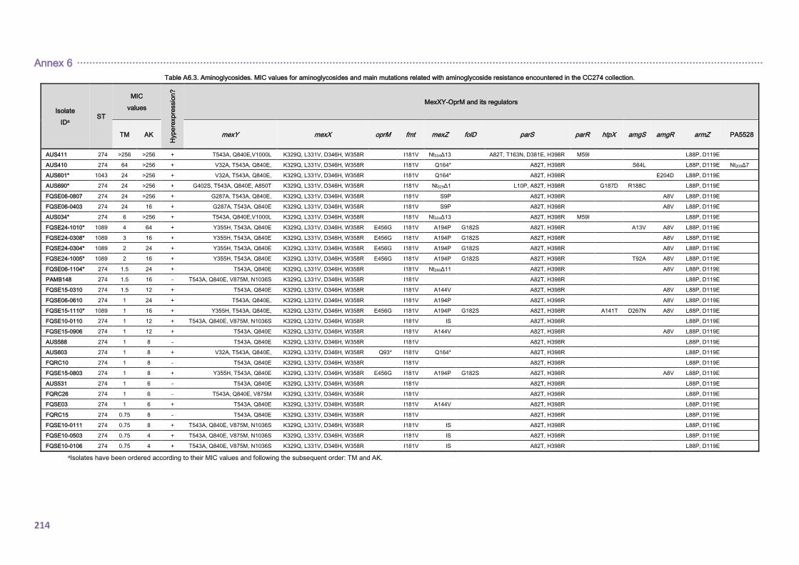
Annex 6 ·······························································································································································································································
214
Table A6.3. Aminoglycosides. MIC values for aminoglycosides and main mutations related with aminoglycoside resistance encountered in the CC274 collection.
Isolate
IDa ST
MIC
values
Hype
rexpre
ssio
n?
MexXY-OprM and its regulators
TM AK mexY mexX oprM fmt mexZ folD parS parR htpX amgS amgR armZ PA5528
AUS411 274 >256 >256 + T543A, Q840E,V1000L K329Q, L331V, D346H, W358R I181V Nt334Δ13 A82T, T163N, D381E, H398R M59I L88P, D119E
AUS410 274 64 >256 + V32A, T543A, Q840E, K329Q, L331V, D346H, W358R I181V Q164* A82T, H398R S64L L88P, D119E Nt208Δ7
AUS601* 1043 24 >256 + V32A, T543A, Q840E, K329Q, L331V, D346H, W358R I181V Q164* A82T, H398R E204D L88P, D119E
AUS690* 274 24 >256 + G402S, T543A, Q840E, A850T K329Q, L331V, D346H, W358R I181V Nt529Δ1 L10P, A82T, H398R G187D R188C L88P, D119E
FQSE06-0807 274 24 >256 + G287A, T543A, Q840E, K329Q, L331V, D346H, W358R I181V S9P A82T, H398R A8V L88P, D119E
FQSE06-0403 274 24 16 + G287A, T543A, Q840E K329Q, L331V, D346H, W358R I181V S9P A82T, H398R A8V L88P, D119E
AUS034* 274 6 >256 + T543A, Q840E,V1000L K329Q, L331V, D346H, W358R I181V Nt334Δ13 A82T, H398R M59I L88P, D119E
FQSE24-1010* 1089 4 64 + Y355H, T543A, Q840E K329Q, L331V, D346H, W358R E456G I181V A194P G182S A82T, H398R A13V A8V L88P, D119E
FQSE24-0308* 1089 3 16 + Y355H, T543A, Q840E K329Q, L331V, D346H, W358R E456G I181V A194P G182S A82T, H398R A8V L88P, D119E
FQSE24-0304* 1089 2 24 + Y355H, T543A, Q840E K329Q, L331V, D346H, W358R E456G I181V A194P G182S A82T, H398R A8V L88P, D119E
FQSE24-1005* 1089 2 16 + Y355H, T543A, Q840E K329Q, L331V, D346H, W358R E456G I181V A194P G182S A82T, H398R T92A A8V L88P, D119E
FQSE06-1104* 274 1.5 24 + T543A, Q840E K329Q, L331V, D346H, W358R I181V Nt290Δ11 A82T, H398R A8V L88P, D119E
PAMB148 274 1.5 16 - T543A, Q840E, V875M, N1036S K329Q, L331V, D346H, W358R I181V A82T, H398R L88P, D119E
FQSE15-0310 274 1.5 12 + T543A, Q840E K329Q, L331V, D346H, W358R I181V A144V A82T, H398R A8V L88P, D119E
FQSE06-0610 274 1 24 + T543A, Q840E, K329Q, L331V, D346H, W358R I181V A194P A82T, H398R A8V L88P, D119E
FQSE15-1110* 1089 1 16 + Y355H, T543A, Q840E, K329Q, L331V, D346H, W358R E456G I181V A194P G182S A82T, H398R A141T D267N A8V L88P, D119E
FQSE10-0110 274 1 12 + T543A, Q840E, V875M, N1036S K329Q, L331V, D346H, W358R I181V IS A82T, H398R L88P, D119E
FQSE15-0906 274 1 12 + T543A, Q840E K329Q, L331V, D346H, W358R I181V A144V A82T, H398R A8V L88P, D119E
AUS588 274 1 8 - T543A, Q840E K329Q, L331V, D346H, W358R I181V A82T, H398R L88P, D119E
AUS603 274 1 8 + V32A, T543A, Q840E, K329Q, L331V, D346H, W358R Q93* I181V Q164* A82T, H398R L88P, D119E
FQRC10 274 1 8 - T543A, Q840E K329Q, L331V, D346H, W358R I181V A82T, H398R L88P, D119E
FQSE15-0803 274 1 8 + Y355H, T543A, Q840E K329Q, L331V, D346H, W358R E456G I181V A194P G182S A82T, H398R A8V L88P, D119E
AUS531 274 1 6 - T543A, Q840E K329Q, L331V, D346H, W358R I181V A82T, H398R L88P, D119E
FQRC26 274 1 6 - T543A, Q840E, V875M K329Q, L331V, D346H, W358R I181V A82T, H398R L88P, D119E
FQSE03 274 1 6 + T543A, Q840E K329Q, L331V, D346H, W358R I181V A144V A82T, H398R L88P, D119E
FQRC15 274 0.75 8 - T543A, Q840E K329Q, L331V, D346H, W358R I181V A82T, H398R L88P, D119E
FQSE10-0111 274 0.75 8 + T543A, Q840E, V875M, N1036S K329Q, L331V, D346H, W358R I181V IS A82T, H398R L88P, D119E
FQSE10-0503 274 0.75 4 + T543A, Q840E, V875M, N1036S K329Q, L331V, D346H, W358R I181V IS A82T, H398R L88P, D119E
FQSE10-0106 274 0.75 4 + T543A, Q840E, V875M, N1036S K329Q, L331V, D346H, W358R I181V IS A82T, H398R L88P, D119E
aIsolates have been ordered according to their MIC values and following the subsequent order: TM and AK.
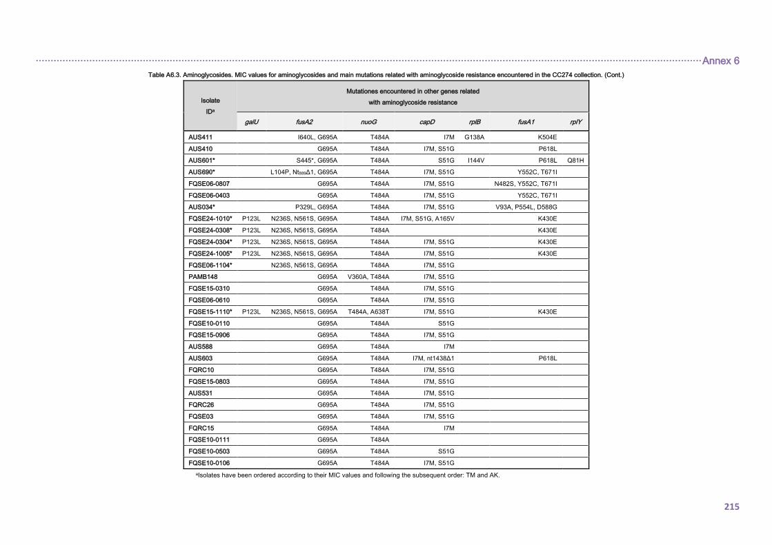
································································································································································································································ Annex 6
215
Table A6.3. Aminoglycosides. MIC values for aminoglycosides and main mutations related with aminoglycoside resistance encountered in the CC274 collection. (Cont.)
Isolate
IDa
Mutationes encountered in other genes related
with aminoglycoside resistance
galU fusA2 nuoG capD rplB fusA1 rplY
AUS411 I640L, G695A T484A I7M G138A K504E
AUS410 G695A T484A I7M, S51G P618L
AUS601* S445*, G695A T484A S51G I144V P618L Q81H
AUS690* L104P, Nt889Δ1, G695A T484A I7M, S51G Y552C, T671I
FQSE06-0807 G695A T484A I7M, S51G N482S, Y552C, T671I
FQSE06-0403 G695A T484A I7M, S51G Y552C, T671I
AUS034* P329L, G695A T484A I7M, S51G V93A, P554L, D588G
FQSE24-1010* P123L N236S, N561S, G695A T484A I7M, S51G, A165V K430E
FQSE24-0308* P123L N236S, N561S, G695A T484A K430E
FQSE24-0304* P123L N236S, N561S, G695A T484A I7M, S51G K430E
FQSE24-1005* P123L N236S, N561S, G695A T484A I7M, S51G K430E
FQSE06-1104* N236S, N561S, G695A T484A I7M, S51G
PAMB148 G695A V360A, T484A I7M, S51G
FQSE15-0310 G695A T484A I7M, S51G
FQSE06-0610 G695A T484A I7M, S51G
FQSE15-1110* P123L N236S, N561S, G695A T484A, A638T I7M, S51G K430E
FQSE10-0110 G695A T484A S51G
FQSE15-0906 G695A T484A I7M, S51G
AUS588 G695A T484A I7M
AUS603 G695A T484A I7M, nt1438Δ1 P618L
FQRC10 G695A T484A I7M, S51G
FQSE15-0803 G695A T484A I7M, S51G
AUS531 G695A T484A I7M, S51G
FQRC26 G695A T484A I7M, S51G
FQSE03 G695A T484A I7M, S51G
FQRC15 G695A T484A I7M
FQSE10-0111 G695A T484A
FQSE10-0503 G695A T484A S51G
FQSE10-0106 G695A T484A I7M, S51G
aIsolates have been ordered according to their MIC values and following the subsequent order: TM and AK.
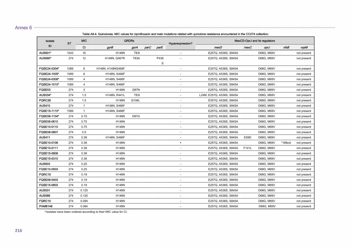
Annex 6 ·······························································································································································································································
216
Table A6.4. Quinolones. MIC values for ciprofloxacin and main mutations related with quinolone resistance encountered in the CC274 collection.
Isolate
ID ST
MIC QRDRs Hyperexpression?
MexCD-OprJ and its regulators
CI gyrB gyrA parC parE mexD mexC oprJ nfxB vqsM
AUS601* 1043 16 H148N T83I - E257Q, A536S, S845A D68G, M69V not present
AUS690* 274 12 H148N, Q467R T83A P438
S
- E257Q, A536S, S845A D68G, M69V not present
FQSE24-0304* 1089 6 H148N, H148NS466F - E257Q, A536S, S845A D68G, M69V not present
FQSE24-1005* 1089 6 H148N, S466F - E257Q, A536S, S845A D68G, M69V not present
FQSE24-0308* 1089 4 H148N, S466F - E257Q, A536S, S845A D68G, M69V not present
FQSE24-1010* 1089 4 H148N, S466F - E257Q, A536S, S845A D68G, M69V not present
FQSE03 274 3 H148N D87N - E257Q, A536S, S845A D68G, M69V not present
AUS034* 274 1.5 H148N, R441L T83I - L24M, E257Q, A536S, S845A D68G, M69V not present
FQRC26 274 1.5 H148N Q106L - E257Q, A536S, S845A D68G, M69V not present
AUS410 274 1 H148N, S466F - E257Q, A536S, S845A D68G, M69V not present
FQSE15-1110* 1089 1 H148N, S466F - E257Q, A536S, S845A D68G, M69V not present
FQSE06-1104* 274 0.75 H148N D87G - E257Q, A536S, S845A D68G, M69V not present
FQSE06-0610 274 0.75 H148N - E257Q, A536S, S845A D68G, M69V not present
FQSE10-0110 274 0.75 H148N - E257Q, A536S, S845A D68G, M69V not present
FQSE06-0807 274 0.5 H148N - E257Q, A536S, S845A D68G, M69V not present
AUS411 274 0.38 H148N, S466F - E257Q, A536S, S845A E59D D68G, M69V not present
FQSE10-0106 274 0.38 H148N + E257Q, A536S, S845A D68G, M69V *188ext not present
FQSE10-0111 274 0.38 H148N - E257Q, A536S, S845A F141L D68G, M69V not present
FQSE15-0906 274 0.38 H148N - E257Q, A536S, S845A D68G, M69V not present
FQSE15-0310 274 0.38 H148N - E257Q, A536S, S845A D68G, M69V not present
AUS603 274 0.25 H148N - E257Q, A536S, S845A D68G, M69V not present
FQSE10-0503 274 0.25 H148N - E257Q, A536S, S845A D68G, M69V not present
FQRC15 274 0.19 H148N - E257Q, A536S, S845A D68G, M69V not present
FQSE06-0403 274 0.19 H148N - E257Q, A536S, S845A D68G, M69V not present
FQSE15-0803 274 0.19 H148N - E257Q, A536S, S845A D68G, M69V not present
AUS531 274 0.125 H148N - E257Q, A536S, S845A D68G, M69V not present
AUS588 274 0.125 H148N - E257Q, A536S, S845A D68G, M69V not present
FQRC10 274 0.094 H148N - E257Q, A536S, S845A D68G, M69V not present
PAMB148 274 0.064 H148N - E257Q, A536S, S845A D68G, M69V not present
a Isolates have been ordered according to their MIC value for CI.
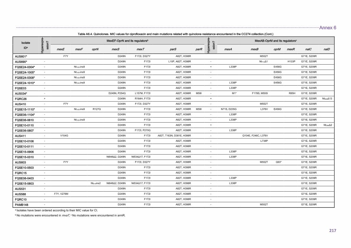
································································································································································································································ Annex 6
217
Table A6.4. Quinolones. MIC values for ciprofloxacin and main mutations related with quinolone resistance encountered in the CC274 collection.(Cont.)
Isolate
IDa
Hype
rexpre
-
ssio
n? MexEF-OprN and its regulatorsb
Hype
rexpre
-
ssio
n? MexAB-OprM and its regulatorsc
mexE mexF oprN mexS mexT parS parR mexA mexB oprM mexR nalC nalD
AUS601* F7Y D249N F172I, D327Y A82T, H398R M552T G71E, S209R
AUS690* - D249N F172I L10P, A82T, H398R - Nt712Δ1 H133P G71E, S209R
FQSE24-0304* - Nt1051ins9 D249N F172I A82T, H398R + L338P E456G G71E, S209R
FQSE24-1005* - Nt1051ins9 D249N F172I A82T, H398R - E456G G71E, S209R
FQSE24-0308* - Nt1051ins9 D249N F172I A82T, H398R - E456G G71E, S209R
FQSE24-1010* - Nt1051ins9 D249N F172I A82T, H398R - L338P E456G G71E, S209R
FQSE03 - D249N F172I A82T, H398R - L338P G71E, S209R
AUS034* - D249N, P254Q L157M, F172I A82T, H398R M59I - M1* F178S, M555I R85H G71E, S209R
FQRC26 + D249N R164H, F172I A82T, H398R + G71E, S209R Nt459Δ13
AUS410 - F7Y D249N F172I, D327Y A82T, H398R - M552T G71E, S209R
FQSE15-1110* - Nt1051ins9 R127Q D249N F172I A82T, H398R M59I - N71S, D235G L376V E456G G71E, S209R
FQSE06-1104* - D249N F172I A82T, H398R - L338P G71E, S209R
FQSE06-0610 - Nt1051ins9 D249N F172I A82T, H398R - L338P G71E, S209R
FQSE10-0110 - D249N F172I A82T, H398R + G71E, S209R Nt396Δ2
FQSE06-0807 - D249N F172I, P270Q A82T, H398R - L338P G71E, S209R
AUS411 - V104G D249N F172I A82T, T163N, D381E, H398R - Q104E, F246C, L376V G71E, S209R
FQSE10-0106 - D249N F172I A82T, H398R - L738P G71E, S209R
FQSE10-0111 - D249N F172I A82T, H398R - G71E, S209R
FQSE15-0906 - D249N F172I A82T, H398R - L338P G71E, S209R
FQSE15-0310 - Nt848Δ2, D249N Nt534Δ17, F172I A82T, H398R - L338P G71E, S209R
AUS603 - F7Y D249N F172I, D327Y A82T, H398R - M552T Q93* G71E, S209R
FQSE10-0503 - D249N F172I A82T, H398R - G71E, S209R
FQRC15 - D249N F172I A82T, H398R - G71E, S209R
FQSE06-0403 - D249N F172I A82T, H398R - L338P G71E, S209R
FQSE15-0803 - Nt415ins2 Nt848Δ2, D249N Nt534Δ17, F172I A82T, H398R - L338P G71E, S209R
AUS531 - D249N F172I A82T, H398R - G71E, S209R
AUS588 - F7Y, V276M D249N F172I A82T, H398R - G71E, S209R
FQRC10 - D249N F172I A82T, H398R - G71E, S209R
PAMB148 - D249N F172I A82T, H398R - M552T G71E, S209R
a Isolates have been ordered according to their MIC value for CI.
b No mutations were encountered in mvaT, c No mutations were encountered in armR.
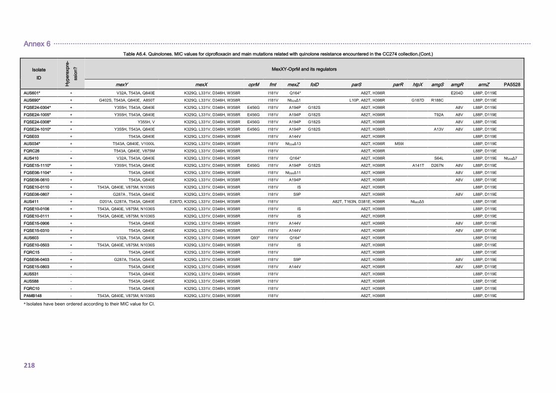
Annex 6 ·······························································································································································································································
218
Table A6.4. Quinolones. MIC values for ciprofloxacin and main mutations related with quinolone resistance encountered in the CC274 collection.(Cont.)
Isolate
ID
Hype
rexpre
-
ssio
n?
MexXY-OprM and its regulators
mexY mexX oprM fmt mexZ folD parS parR htpX amgS amgR armZ PA5528
AUS601* + V32A, T543A, Q840E K329Q, L331V, D346H, W358R I181V Q164* A82T, H398R E204D L88P, D119E
AUS690* + G402S, T543A, Q840E, A850T K329Q, L331V, D346H, W358R I181V Nt529Δ1 L10P, A82T, H398R G187D R188C L88P, D119E
FQSE24-0304* + Y355H, T543A, Q840E K329Q, L331V, D346H, W358R E456G I181V A194P G182S A82T, H398R A8V L88P, D119E
FQSE24-1005* + Y355H, T543A, Q840E K329Q, L331V, D346H, W358R E456G I181V A194P G182S A82T, H398R T92A A8V L88P, D119E
FQSE24-0308* + Y355H, V K329Q, L331V, D346H, W358R E456G I181V A194P G182S A82T, H398R A8V L88P, D119E
FQSE24-1010* + Y355H, T543A, Q840E K329Q, L331V, D346H, W358R E456G I181V A194P G182S A82T, H398R A13V A8V L88P, D119E
FQSE03 + T543A, Q840E K329Q, L331V, D346H, W358R I181V A144V A82T, H398R L88P, D119E
AUS034* + T543A, Q840E, V1000L K329Q, L331V, D346H, W358R I181V Nt334Δ13 A82T, H398R M59I L88P, D119E
FQRC26 - T543A, Q840E, V875M K329Q, L331V, D346H, W358R I181V A82T, H398R L88P, D119E
AUS410 + V32A, T543A, Q840E K329Q, L331V, D346H, W358R I181V Q164* A82T, H398R S64L L88P, D119E Nt208Δ7
FQSE15-1110* + Y355H, T543A, Q840E K329Q, L331V, D346H, W358R E456G I181V A194P G182S A82T, H398R A141T D267N A8V L88P, D119E
FQSE06-1104* + T543A, Q840E K329Q, L331V, D346H, W358R I181V Nt290Δ11 A82T, H398R A8V L88P, D119E
FQSE06-0610 + T543A, Q840E K329Q, L331V, D346H, W358R I181V A194P A82T, H398R A8V L88P, D119E
FQSE10-0110 + T543A, Q840E, V875M, N1036S K329Q, L331V, D346H, W358R I181V IS A82T, H398R L88P, D119E
FQSE06-0807 + G287A , T543A, Q840E K329Q, L331V, D346H, W358R I181V S9P A82T, H398R A8V L88P, D119E
AUS411 + D201A, G287A, T543A, Q840E E287D, K329Q, L331V, D346H, W358R I181V A82T, T163N, D381E, H398R Nt683Δ5 L88P, D119E
FQSE10-0106 + T543A, Q840E, V875M, N1036S K329Q, L331V, D346H, W358R I181V IS A82T, H398R L88P, D119E
FQSE10-0111 + T543A, Q840E, V875M, N1036S K329Q, L331V, D346H, W358R I181V IS A82T, H398R L88P, D119E
FQSE15-0906 + T543A, Q840E K329Q, L331V, D346H, W358R I181V A144V A82T, H398R A8V L88P, D119E
FQSE15-0310 + T543A, Q840E K329Q, L331V, D346H, W358R I181V A144V A82T, H398R A8V L88P, D119E
AUS603 + V32A, T543A, Q840E K329Q, L331V, D346H, W358R Q93* I181V Q164* A82T, H398R L88P, D119E
FQSE10-0503 + T543A, Q840E, V875M, N1036S K329Q, L331V, D346H, W358R I181V IS A82T, H398R L88P, D119E
FQRC15 - T543A, Q840E K329Q, L331V, D346H, W358R I181V A82T, H398R L88P, D119E
FQSE06-0403 + G287A, T543A, Q840E K329Q, L331V, D346H, W358R I181V S9P A82T, H398R A8V L88P, D119E
FQSE15-0803 + T543A, Q840E K329Q, L331V, D346H, W358R I181V A144V A82T, H398R A8V L88P, D119E
AUS531 - T543A, Q840E K329Q, L331V, D346H, W358R I181V A82T, H398R L88P, D119E
AUS588 - T543A, Q840E K329Q, L331V, D346H, W358R I181V A82T, H398R L88P, D119E
FQRC10 - T543A, Q840E K329Q, L331V, D346H, W358R I181V A82T, H398R L88P, D119E
PAMB148 - T543A, Q840E, V875M, N1036S K329Q, L331V, D346H, W358R I181V A82T, H398R L88P, D119E
a Isolates have been ordered according to their MIC value for CI.
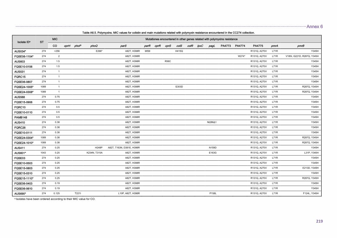
································································································································································································································ Annex 6
219
Table A6.5. Polymyxins. MIC values for colistin and main mutations related with polymyxin resistance encountered in the CC274 collection.
Isolate IDa ST MIC Mutationes encountered in other genes related with polymyxins resistance
CO oprH phoP phoQ parS parR cprR cprS colS colR lpxC pagL PA4773 PA4774 PA4775 pmrA pmrB
AUS034* 274 >256 E266* A82T, H398R M59I H415Q R131Q, A270V L71R Y345H
FQSE06-1104* 274 2 A82T, H398R W274* R131Q, A270V L71R V185I, G221D, R287Q, Y345H
AUS603 274 1.5 A82T, H398R R56C R131Q, A270V L71R Y345H
FQSE10-0106 274 1.5 A82T, H398R R131Q, A270V L71R Y345H
AUS531 274 1 A82T, H398R R131Q, A270V L71R Y345H
FQRC15 274 1 A82T, H398R R131Q, A270V L71R Y345H
FQSE06-0807 274 1 A82T, H398R R131Q, A270V L71R Y345H
FQSE24-1005* 1089 1 A82T, H398R E303D R131Q, A270V L71R R287Q, Y345H
FQSE24-0308* 1089 1 A82T, H398R R131Q, A270V L71R R287Q, Y345H
AUS588 274 0.75 A82T, H398R R131Q, A270V L71R Y345H
FQSE15-0906 274 0.75 A82T, H398R R131Q, A270V L71R Y345H
FQRC10 274 0.5 A82T, H398R R131Q, A270V L71R Y345H
FQSE10-0110 274 0.5 A82T, H398R R131Q, A270V L71R Y345H
PAMB148 274 0.5 A82T, H398R R131Q, A270V L71R Y345H
AUS410 274 0.38 A82T, H398R Nt286Δ1 R131Q, A270V L71R Y345H
FQRC26 274 0.38 A82T, H398R R131Q, A270V L71R Y345H
FQSE10-0111 274 0.38 A82T, H398R R131Q, A270V L71R Y345H
FQSE24-0304* 1089 0.38 A82T, H398R R131Q, A270V L71R R287Q, Y345H
FQSE24-1010* 1089 0.38 A82T, H398R R131Q, A270V L71R R287Q, Y345H
AUS411 274 0.25 H248P A82T, T163N, D381E, H398R N159D R131Q, A270V L71R Y345H
AUS601* 1043 0.25 K234N, T315A A82T, H398R E163G R131Q, A270V L71R L31P, Y345H
FQSE03 274 0.25 A82T, H398R R131Q, A270V L71R Y345H
FQSE10-0503 274 0.25 A82T, H398R R131Q, A270V L71R Y345H
FQSE15-0803 274 0.25 A82T, H398R R131Q, A270V L71R E213D, Y345H
FQSE15-0310 274 0.25 A82T, H398R R131Q, A270V L71R Y345H
FQSE15-1110* 274 0.25 A82T, H398R R131Q, A270V L71R R287Q, Y345H
FQSE06-0403 274 0.19 A82T, H398R R131Q, A270V L71R Y345H
FQSE06-0610 274 0.19 A82T, H398R R131Q, A270V L71R Y345H
AUS690* 274 0.125 T221I L10P, A82T, H398R P158L R131Q, A270V L71R F124L, Y345H
a Isolates have been ordered according to their MIC value for CO.

220

221
14. ANNEX 7
EVOLUTIONARY DYNAMICS OF Pseudomonas aeruginosa
AMINOGLYCOSIDE RESISTANCE DEVELOPMENT: EXPANDED RESULTS

222
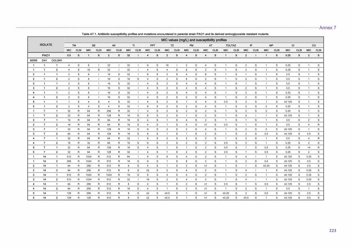
································································································································································································································ Annex 7
223
Table A7.1. Antibiotic susceptibility profiles and mutations encountered in parental strain PAO1 and its derived aminoglycoside resistant mutants.
ISOLATE
MIC values (mg/L) and susceptibility profiles
TM GE AK TI PPT TZ PM AT TOL/TAZ IP MP CI CO
MIC CLSI MIC CLSI MIC CLSI MIC CLSI MIC CLSI MIC CLSI MIC CLSI MIC CLSI MIC CLSI MIC CLSI MIC CLSI MIC CLSI MIC CLSI
PAO1 0.5 S 1 S 2 S 32 I 4 S 2 S 4 S 4 S 1 S 2 I 1 S 0.25 S 2 S
SERIE DAY COLONY
1 1 1 4 S 8 I 32 I 32 I 8 S 16 I 2 S 4 S 1 S 2 S 1 S 0.25 S 1 S
1 1 2 4 S 16 R 32 I 32 I 4 S 4 S 8 S 2 S 1 S 2 S 1 S 0.25 S 1 S
2 1 1 2 S 8 I 16 S 32 I 8 S 2 S 4 S 8 S 1 S 1 S 1 S 0.5 S 1 S
2 1 2 2 S 8 I 16 S 16 S 4 S 2 S 8 S 2 S 1 S 2 S 1 S 0.5 S 1 S
3 1 1 2 S 8 I 16 S 32 I 4 S 2 S 4 S 4 S 1 S 1 S 1 S 0.5 S 1 S
3 1 2 2 S 8 I 16 S 32 I 4 S 2 S 4 S 4 S 1 S 2 S 1 S 0.5 S 1 S
4 1 1 2 S 8 I 16 S 32 I 4 S 2 S 4 S 4 S 1 S 2 S 1 S 0.25 S 1 S
4 1 2 2 S 8 I 16 S 32 I 4 S 2 S 2 S 4 S 1 S 2 S 1 S 0.25 S 1 S
5 1 1 1 S 4 S 4 S 32 I 4 S 2 S 1 S 4 S 0.5 S 2 S 1 S ≤0.125 S 1 S
5 1 2 2 S 4 S 4 S 32 I 8 S 2 S 2 S 4 S 1 S 2 S 2 S 0.25 S 1 S
1 7 1 32 R 64 R 256 R 32 I 4 S 2 S 2 S 2 S 1 S 2 S 2 S 0.25 S 1 S
1 7 2 32 R 64 R 128 R 16 S 8 S 2 S 1 S 2 S 1 S 4 I 1 S ≤0.125 S 1 S
2 7 1 16 R 64 R 64 R 16 S 4 S 1 S 4 S 2 S 1 S 1 S 1 S 0.5 S 2 S
2 7 2 16 R 64 R 64 R 16 S 4 S 1 S 4 S ≤1 S 0.5 S 1 S 1 S 0.5 S 4 R
3 7 1 32 R 64 R 128 R 16 S 8 S 2 S 2 S 4 S 1 S 2 S 2 S ≤0.125 S 1 S
3 7 2 64 R 64 R 128 R 16 S 8 S 1 S 1 S 2 S 1 S 2 S 0,5 S ≤0.125 S 0.5 S
4 7 1 32 R 64 R 64 R 32 I 8 S 2 S 2 S 2 S 1 S 2 S 1 S 0.5 S 4 R
4 7 2 16 R 32 R 64 R 16 S 8 S 2 S 2 S 2 S 0.5 S 2 S 1 S 0.25 S 2 S
5 7 1 32 R 64 R 128 R 16 S 4 S 1 S 1 S 2 S 0.5 S 1 S 0,5 S 0.25 S >4 R
5 7 2 32 R 64 R 128 R 32 I 4 S 1 S 2 S 2 S 0.5 S 1 S 0,5 S 0.25 S 2 S
1 14 1 512 R 1024 R 512 R 64 I 8 S 8 S 4 S 2 S 1 S 4 I 1 S ≤0.125 S 0.25 S
1 14 2 256 R 1024 R 512 R 16 S 8 S 1 S 8 S 2 S 1 S 2 S 0,5 S ≤0.125 S 0.5 S
2 14 1 64 R 256 R 512 R 8 S 4 S 1 S 2 S ≤1 S 1 S 1 S ≤0.25 S ≤0.125 S 0.5 S
2 14 2 64 R 256 R 512 R 8 S ≤2 S 2 S 4 S 2 S 1 S 4 I 1 S ≤0.125 S 0.25 S
3 14 1 512 R 1024 R 1024 R 16 S 8 S 4 S 4 S 2 S 1 S 2 S 1 S ≤0.125 S 0.25 S
3 14 2 512 R 1024 R 512 R 32 I 16 S 2 S 4 S 2 S 1 S 4 I 1 S ≤0.125 S 0.25 S
4 14 1 64 R 256 R 512 R 8 S 4 S 1 S 2 S ≤1 S 0.5 S 1 S 0,5 S ≤0.125 S 0.5 S
4 14 2 64 R 256 R 512 R 16 S 4 S 1 S 2 S ≤1 S 1 S 2 S 1 S 0.5 S 1 S
5 14 1 128 R 256 R 512 R 8 S ≤2 S ≤0.5 S 1 S ≤1 S ≤0.25 S 2 S 0,5 S ≤0.125 S 0.5 S
5 14 2 128 R 128 R 512 R 8 S ≤2 S ≤0.5 S 1 S ≤1 S ≤0.25 S ≤0.5 S 1 S ≤0.125 S 0.5 S
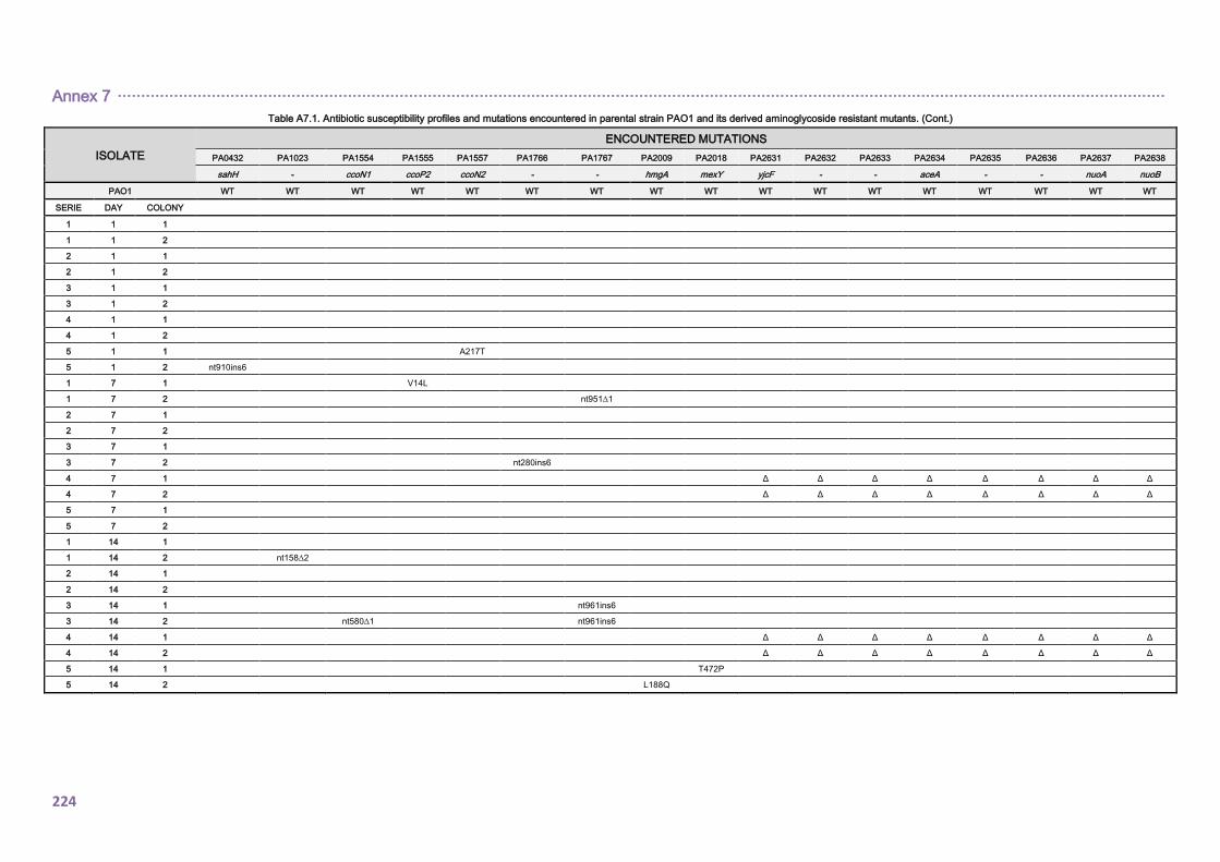
Annex 7 ·······························································································································································································································
224
Table A7.1. Antibiotic susceptibility profiles and mutations encountered in parental strain PAO1 and its derived aminoglycoside resistant mutants. (Cont.)
ISOLATE
ENCOUNTERED MUTATIONS
PA0432 PA1023 PA1554 PA1555 PA1557 PA1766 PA1767 PA2009 PA2018 PA2631 PA2632 PA2633 PA2634 PA2635 PA2636 PA2637 PA2638
sahH - ccoN1 ccoP2 ccoN2 - - hmgA mexY yjcF - - aceA - - nuoA nuoB
PAO1 WT WT WT WT WT WT WT WT WT WT WT WT WT WT WT WT WT
SERIE DAY COLONY
1 1 1
1 1 2
2 1 1
2 1 2
3 1 1
3 1 2
4 1 1
4 1 2
5 1 1 A217T
5 1 2 nt910ins6
1 7 1 V14L
1 7 2 nt951∆1
2 7 1
2 7 2
3 7 1
3 7 2 nt280ins6
4 7 1 Δ Δ Δ Δ Δ Δ Δ Δ
4 7 2 Δ Δ Δ Δ Δ Δ Δ Δ
5 7 1
5 7 2
1 14 1
1 14 2 nt158∆2
2 14 1
2 14 2
3 14 1 nt961ins6
3 14 2 nt580∆1 nt961ins6
4 14 1 Δ Δ Δ Δ Δ Δ Δ Δ
4 14 2 Δ Δ Δ Δ Δ Δ Δ Δ
5 14 1 T472P
5 14 2 L188Q
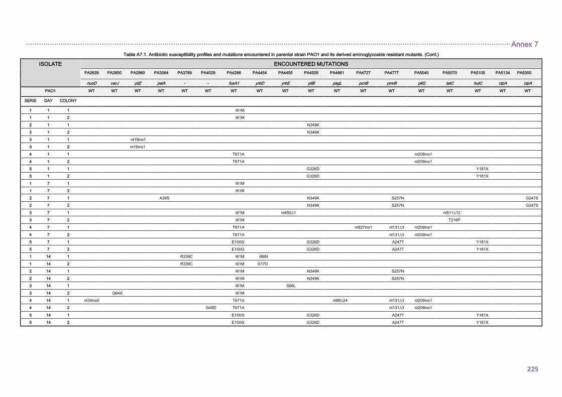
································································································································································································································ Annex 7
225
Table A7.1. Antibiotic susceptibility profiles and mutations encountered in parental strain PAO1 and its derived aminoglycoside resistant mutants. (Cont.)
ISOLATE ENCOUNTERED MUTATIONS
PA2639 PA2800 PA2960 PA3064 PA3789 PA4029 PA4266 PA4454 PA4455 PA4526 PA4661 PA4727 PA4777 PA5040 PA5070 PA5105 PA5134 PA5300
nuoD vacJ pilZ pelA - - fusA1 yrbD yrbE pilB pagL pcnB pmrB pilQ tatC hutC ctpA ctpA
PAO1 WT WT WT WT WT WT WT WT WT WT WT WT WT WT WT WT WT WT
SERIE DAY COLONY
1 1 1 I61M
1 1 2 I61M
2 1 1 N349K
2 1 2 N349K
3 1 1 nt19ins1
3 1 2 nt19ins1
4 1 1 T671A nt209ins1
4 1 2 T671A nt209ins1
5 1 1 G326D Y181X
5 1 2 G326D Y181X
1 7 1 I61M
1 7 2 I61M
2 7 1 A39S N349K S257N G247S
2 7 2 N349K S257N G247S
3 7 1 I61M nt450∆1 nt511∆12
3 7 2 I61M T216P
4 7 1 T671A nt827ins1 nt131∆3 nt209ins1
4 7 2 T671A nt131∆3 nt209ins1
5 7 1 E100G G326D A247T Y181X
5 7 2 E100G G326D A247T Y181X
1 14 1 R339C I61M I86N
1 14 2 R339C I61M G17D
2 14 1 I61M N349K S257N
2 14 2 I61M N349K S257N
3 14 1 I61M S66L
3 14 2 Q64X I61M
4 14 1 nt34ins5 T671A nt88∆24 nt131∆3 nt209ins1
4 14 2 G49D T671A nt131∆3 nt209ins1
5 14 1 E100G G326D A247T Y181X
5 14 2 E100G G326D A247T Y181X
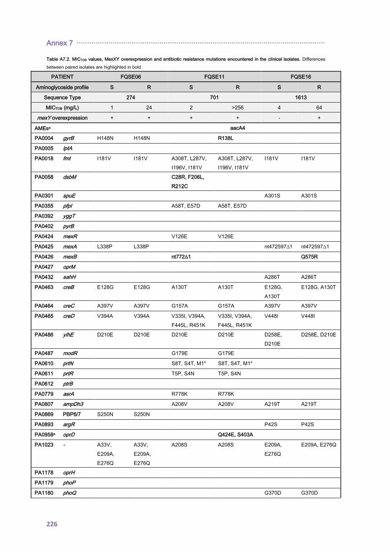
Annex 7 ·····················································································································
226
Table A7.2. MICTOB values, MexXY overexpression and antibiotic resistance mutations encountered in the clinical isolates. Differences
between paired isolates are highlighted in bold.
PATIENT FQSE06 FQSE11 FQSE16
Aminoglycoside profile S R S R S R
Sequence Type 274 701 1613
MICTOB (mg/L) 1 24 2 >256 4 64
mexY overexpression + + + + - +
AMEsa aacA4
PA0004 gyrB H148N H148N R138L
PA0005 lptA
PA0018 fmt I181V I181V A308T, L287V,
I196V, I181V
A308T, L287V,
I196V, I181V
I181V I181V
PA0058 dsbM C28R, F206L,
R212C
PA0301 spuE A301S A301S
PA0355 pfpI A58T, E57D A58T, E57D
PA0392 yggT
PA0402 pyrB
PA0424 mexR V126E V126E
PA0425 mexA L338P L338P nt472597∆1 nt472597∆1
PA0426 mexB nt772∆1 Q575R
PA0427 oprM
PA0432 sahH A286T A286T
PA0463 creB E128G E128G A130T A130T E128G,
A130T
E128G, A130T
PA0464 creC A397V A397V G157A G157A A397V A397V
PA0465 creD V394A V394A V335I, V394A,
F445L, R451K
V335I, V394A,
F445L, R451K
V448I V448I
PA0486 yihE D210E D210E D210E D210E D258E,
D210E
D258E, D210E
PA0487 modR G179E G179E
PA0610 prtN S8T, S4T, M1* S8T, S4T, M1*
PA0611 prtR T5P, S4N T5P, S4N
PA0612 ptrB
PA0779 asrA R778K R778K
PA0807 ampDh3 A208V A208V A219T A219T
PA0869 PBP6/7 S250N S250N
PA0893 argR P42S P42S
PA0958a oprD Q424E, S403A
PA1023 - A33V,
E209A,
E276Q
A33V,
E209A,
E276Q
A208S A208S E209A,
E276Q
E209A, E276Q
PA1178 oprH
PA1179 phoP
PA1180 phoQ G370D G370D
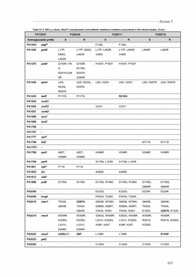
······················································································································ Annex 7
227
Table A7.2. MICTOB values, MexXY overexpression and antibiotic resistance mutations encountered in the clinical isolates. (Cont.)
PATIENT FQSE06 FQSE11 FQSE16
Aminoglycoside profile S R S R S R
PA1343 pagP F129L F129L
PA1345 gshB L17P,
D65G,
L463R
L17P, D65G,
L463R
L17P, L463R,
V495I
L17P, L463R,
V495I
L463R L463R
PA1375 pdxB Q150R,179
H,
R247H,Q36
5R
Q150R,
R179H,
R247H,
Q365R
H183Y, P192T H183Y, P192T
PA1409 aphA L62I,
R224L,
R297K
L62I, R224L,
R297K
L62I, I320V L62I, I320V L62I, R297K L62I, R297K
PA1430 lasR P117G P117G R216Q
PA1554 ccoN1
PA1555 ccoP2 V231I V231I
PA1557 ccoN2
PA1588 sucC
PA1589 sucD
PA1766 -
PA1767 -
PA1777 oprF
PA1796 folD N171S N171S
PA1797a -
PA1798 parS A82T,
H398R
A82T,
H398R
H398R H398R H398R H398R
PA1799 parR S170N, L153R S170N, L153R
PA1801 clpP P11S P11S
PA1803 lon A499S A499S
PA1812 mltD
PA1886 polB D176G D176G D176G, R196C D176G, R196C D176G,
Q664R
D176G,
Q664R
PA2006 - E232Q E232Q E220K E220K
PA2009 hmgA P303H, T226A P303H, T226A
PA2018 mexY T543A,
Q840E
G287A,
T543A,
Q840E
Q840E, N709H,
G589A, A586T,
T543A, I536V
Q840E, N709H,
G589A, A586T,
T543A, I536V
Q840E,
T543A,
E152D
Q840E,
T543A,
G287S, E152D
PA2019 mexX W358R,
K329Q,
L331V,
D346H
W358R,
K329Q,
L331V,
D346H
S382G, W358R,
L331V, K329Q,
A38P, A30T
S382G, W358R,
L331V, K329Q,
A38P, A30T
W358R,
R351S,
K329Q
W358R,
R351S, K329Q
PA2020 mexZ nt290∆11 S9P L138R L138R R125P
PA2023 galU
PA2050 - C142G C142G C142G C142G
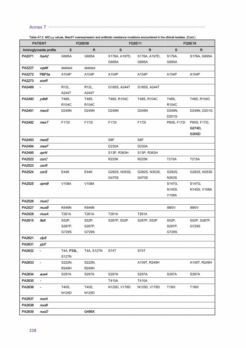
Annex 7 ·····················································································································
228
Table A7.2. MICTOB values, MexXY overexpression and antibiotic resistance mutations encountered in the clinical isolates. (Cont.)
PATIENT FQSE06 FQSE11 FQSE16
Aminoglycoside profile S R S R S R
PA2071 fusA2 G695A G695A S176A, A197D,
G695A
S176A, A197D,
G695A
S176A,
G695A
S176A, G695A
PA2227 vqsM deleted deleted
PA2272 PBP3a A104P A104P A104P A104P A104P A104P
PA2273 soxR
PA2489 - R12L,
A244T
R12L,
A244T
G185S, A244T G185S, A244T
PA2490 ydbB T48S,
R104C
T48S,
R104C
T48S, R104C T48S, R104C T48S,
R104C
T48S, R104C
PA2491 mexS D249N D249N D249N D249N D249N,
D201G
D249N, D201G
PA2492 mexT F172I F172I F172I F172I P60S, F172I P60S, F172I,
G274D,
G300D
PA2493 mexE S8F S8F
PA2494 mexF D230A D230A
PA2495 oprN S13P, R363H S13P, R363H
PA2522 czcC R225K R225K T215A T215A
PA2523 czcR
PA2524 czcS E44K E44K G282S, N353S,
G470S
G282S, N353S,
G470S
G282S,
N353S
G282S, N353S
PA2525 opmB V108A V108A S147G,
N145S,
V108A
S147G,
N145S, V108A
PA2526 muxC
PA2527 muxB K646N K646N I880V I880V
PA2528 muxA T261A T261A T261A T261A
PA2615 ftsK S52P,
S287P,
G729S
S52P,
S287P,
G729S
S287P, S52P S287P, S52P S52P,
S287P,
G729S
S52P, S287P,
G729S
PA2621 clpS
PA2631 yjcF
PA2632 - T4A, P33L,
S127N
T4A, S127N S74T S74T
PA2633 - S222N,
R249H
S222N,
R249H
A109T, R249H A109T, R249H
PA2634 aceA S297A S297A S297A S297A S297A S297A
PA2635 - T410A T410A
PA2636 - T40S,
N125D
T40S,
N125D
N125D, V178D N125D, V178D T180I T180I
PA2637 nuoA
PA2638 nuoB
PA2639 nuoD G499X

······················································································································ Annex 7
229
Table A7.2. MICTOB values, MexXY overexpression and antibiotic resistance mutations encountered in the clinical isolates. (Cont.)
PATIENT FQSE06 FQSE11 FQSE16
Aminoglycoside profile S R S R S R
PA2642 nuoG T484A T484A T484A, K585R T484A, K585R T484A T484A
PA2649 nuoN G326D G326D
PA2797 -
PA2798 - G301S G301S T317S, G301S,
E297D
T317S, G301S,
E297D
G301S G301S
PA2800 vacJ S172G S172G
PA2809 copR
PA2810 copS M48L, R320H M48L, R320H E359G E359G
PA2830 htpX
PA2960 pilZ P9S P9S
PA3005 nagZ
PA3013 foaB I391V, E225Q I391V, E225Q
PA3014 faoA V397I V397I
PA3047 PBP4 A358V,
A394P
A358V,
A394P
PA3050 pyrD R96K, K40E R96K, K40E
PA3064 pelA V446I,
A272T,
H141Y, I19T
A272T,
H141Y, I19T
V946A, V507A,
I377V, T41A
V946A, V507A,
I377V, T41A
R862H,
V825L,
A272T,
H141Y,
C25R, I19T
R862H, V825L,
A272T, H141Y,
C25R, I19T
PA3077 cprR
PA3078 cprS T16S, E386D T16S, E386D
PA3141 capD I7M, S51G I7M, S51G A626V, S51G A626V, S51G S486L,
nt512ins1
S486L
PA3168 gyrA Y267N D87N D87N
PA3521 opmE S175T,
A279S,
W354R
S175T,
A279S,
W354R
W354R, S175T W354R, S175T W354R,
S175T,
S46G
W354R,
S175T, S46G
PA3522 mexQ I294V,
G505D,
D602E,
G645A,
V921M
I294V,
G505D,
D602E,
G645A,
V921M
R656K, G505D,
V384I, I294V
R656K, G505D,
V384I, I294V
R1036H,
P428S,
V355M
R1036H,
P428S, V355M
PA3523 mexP A297E,
R366L
A297E,
R366L
A297E, R366L A297E, R366L
PA3533 grxD
PA3574 nalD A272T,
H141Y,
C25R, I19T
PA3602 yerD L21V L21V
PA3676 mexK K694R, I21L K694R, I21L
PA3677 mexJ A314P A314P

Annex 7 ·····················································································································
230
Table A7.2. MICTOB values, MexXY overexpression and antibiotic resistance mutations encountered in the clinical isolates. (Cont.)
PATIENT FQSE06 FQSE11 FQSE16
Aminoglycoside profile S R S R S R
PA3678 mexL S6P S6P
PA3719 armR S21T S21T
PA3721 nalC G71E,
S209R
G71E,
S209R
G71E, A145V,
S209R
G71E, A145V,
S209R
S209R S209R
PA3789 - G439S,
L195V,
E191D,
V71A
G439S,
L195V,
E191D,
V71A
L205F, L195V,
E191D, V71A
L205F, L195V,
E191D, V71A
PA3999 PBP5
PA4001 sltB1
PA4003 PBP2
PA4020 mpl M297V S257L,
M297V
S306N, M297V S306N, M297V,
Q248*
M297V M297V
PA4029 - A218T A218T
PA4069 - T234I T234I
PA4109 ampR M288R, G283E M288R, G283E
PA4110 ampC T21A,
T105A,
G391A
T21A,
T105A,
G391A
R79Q, T105A,
V205L, V356I,
G391A
R79Q, T105A,
V205L, V356I,
G391A
T105A T105A
PA4119 aph A42V A42V
PA4205 mexG
PA4206 mexH P218T, D302E P218T, D302E A123T A123T
PA4207 mexI A782E A782E A491T,
A782E
A491T, A782E
PA4208 opmD S112N, A270G S112N, A270G S112N,
A243E
S112N, A243E
PA4218 ampP M87I,
T172A
M87I, T172A L98F, M87I,
L74F
L98F, M87I,
L74F
PA4238 rpoA
PA4260 rplB
PA4266 fusA1 Y552C,
T671I
T456A,
K187R
Y552C, T456A,
K187R
PA4269 rpoC R905C,
N606S
R905C, N606S
PA4270 rpoB V51I V51I V51I V51I G158D,
V51I
G158D, V51I
PA4273 rplA
PA4315 mvaT
PA4374 mexV A229G,
Q321R
A229G,
Q321R
A229G, Q321R A229G, Q321R Q321R Q321R
PA4375 mexW A627V,
Q771P
A627V,
Q771P
Q511R, G758S Q511R, G758S Q511R Q511R
PA4380 colS
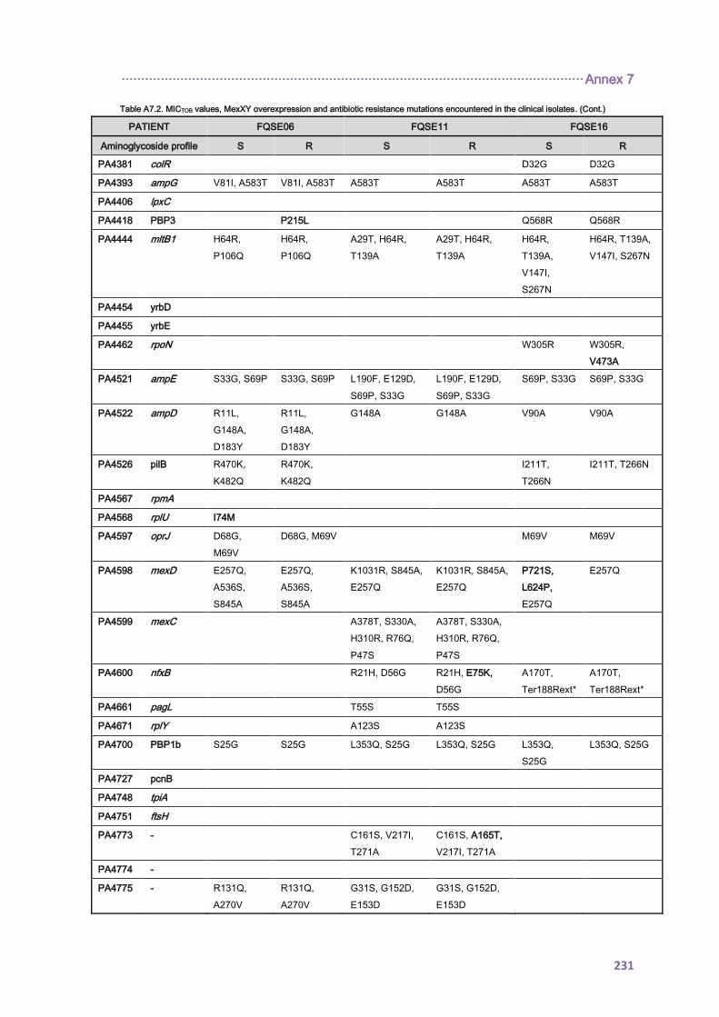
······················································································································ Annex 7
231
Table A7.2. MICTOB values, MexXY overexpression and antibiotic resistance mutations encountered in the clinical isolates. (Cont.)
PATIENT FQSE06 FQSE11 FQSE16
Aminoglycoside profile S R S R S R
PA4381 colR D32G D32G
PA4393 ampG V81I, A583T V81I, A583T A583T A583T A583T A583T
PA4406 lpxC
PA4418 PBP3 P215L Q568R Q568R
PA4444 mltB1 H64R,
P106Q
H64R,
P106Q
A29T, H64R,
T139A
A29T, H64R,
T139A
H64R,
T139A,
V147I,
S267N
H64R, T139A,
V147I, S267N
PA4454 yrbD
PA4455 yrbE
PA4462 rpoN W305R W305R,
V473A
PA4521 ampE S33G, S69P S33G, S69P L190F, E129D,
S69P, S33G
L190F, E129D,
S69P, S33G
S69P, S33G S69P, S33G
PA4522 ampD R11L,
G148A,
D183Y
R11L,
G148A,
D183Y
G148A G148A V90A V90A
PA4526 pilB R470K,
K482Q
R470K,
K482Q
I211T,
T266N
I211T, T266N
PA4567 rpmA
PA4568 rplU I74M
PA4597 oprJ D68G,
M69V
D68G, M69V M69V M69V
PA4598 mexD E257Q,
A536S,
S845A
E257Q,
A536S,
S845A
K1031R, S845A,
E257Q
K1031R, S845A,
E257Q
P721S,
L624P,
E257Q
E257Q
PA4599 mexC A378T, S330A,
H310R, R76Q,
P47S
A378T, S330A,
H310R, R76Q,
P47S
PA4600 nfxB R21H, D56G R21H, E75K,
D56G
A170T,
Ter188Rext*
A170T,
Ter188Rext*
PA4661 pagL T55S T55S
PA4671 rplY A123S A123S
PA4700 PBP1b S25G S25G L353Q, S25G L353Q, S25G L353Q,
S25G
L353Q, S25G
PA4727 pcnB
PA4748 tpiA
PA4751 ftsH
PA4773 - C161S, V217I,
T271A
C161S, A165T,
V217I, T271A
PA4774 -
PA4775 - R131Q,
A270V
R131Q,
A270V
G31S, G152D,
E153D
G31S, G152D,
E153D
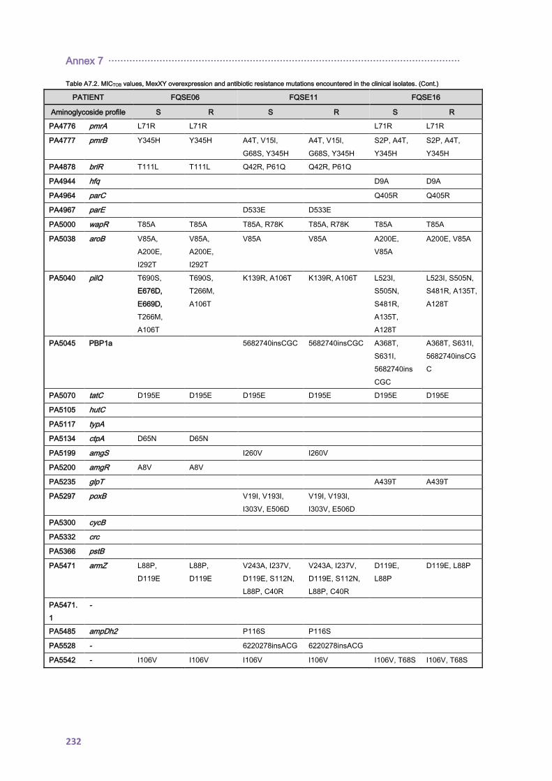
Annex 7 ·····················································································································
232
Table A7.2. MICTOB values, MexXY overexpression and antibiotic resistance mutations encountered in the clinical isolates. (Cont.)
PATIENT FQSE06 FQSE11 FQSE16
Aminoglycoside profile S R S R S R
PA4776 pmrA L71R L71R L71R L71R
PA4777 pmrB Y345H Y345H A4T, V15I,
G68S, Y345H
A4T, V15I,
G68S, Y345H
S2P, A4T,
Y345H
S2P, A4T,
Y345H
PA4878 brlR T111L T111L Q42R, P61Q Q42R, P61Q
PA4944 hfq D9A D9A
PA4964 parC Q405R Q405R
PA4967 parE D533E D533E
PA5000 wapR T85A T85A T85A, R78K T85A, R78K T85A T85A
PA5038 aroB V85A,
A200E,
I292T
V85A,
A200E,
I292T
V85A V85A A200E,
V85A
A200E, V85A
PA5040 pilQ T690S,
E676D,
E669D,
T266M,
A106T
T690S,
T266M,
A106T
K139R, A106T K139R, A106T L523I,
S505N,
S481R,
A135T,
A128T
L523I, S505N,
S481R, A135T,
A128T
PA5045 PBP1a 5682740insCGC 5682740insCGC A368T,
S631I,
5682740ins
CGC
A368T, S631I,
5682740insCG
C
PA5070 tatC D195E D195E D195E D195E D195E D195E
PA5105 hutC
PA5117 typA
PA5134 ctpA D65N D65N
PA5199 amgS I260V I260V
PA5200 amgR A8V A8V
PA5235 glpT A439T A439T
PA5297 poxB V19I, V193I,
I303V, E506D
V19I, V193I,
I303V, E506D
PA5300 cycB
PA5332 crc
PA5366 pstB
PA5471 armZ L88P,
D119E
L88P,
D119E
V243A, I237V,
D119E, S112N,
L88P, C40R
V243A, I237V,
D119E, S112N,
L88P, C40R
D119E,
L88P
D119E, L88P
PA5471.
1
-
PA5485 ampDh2 P116S P116S
PA5528 - 6220278insACG 6220278insACG
PA5542 - I106V I106V I106V I106V I106V, T68S I106V, T68S

233
15. Publications derived from this thesis

234
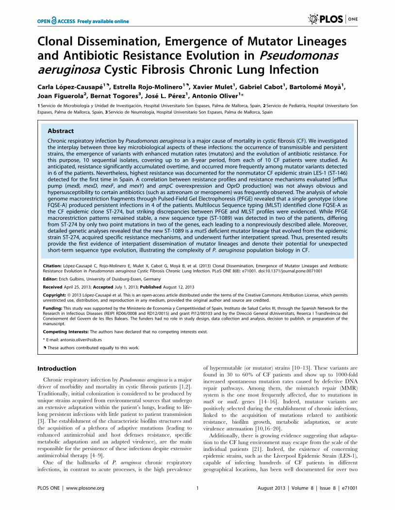
Clonal Dissemination, Emergence of Mutator Lineagesand Antibiotic Resistance Evolution in Pseudomonasaeruginosa Cystic Fibrosis Chronic Lung InfectionCarla Lopez-Causape1., Estrella Rojo-Molinero1., Xavier Mulet1, Gabriel Cabot1, Bartolome Moya1,
Joan Figuerola2, Bernat Togores3, Jose L. Perez1, Antonio Oliver1*
1 Servicio de Microbiologıa y Unidad de Investigacion, Hospital Universitario Son Espases, Palma de Mallorca, Spain, 2 Servicio de Pediatrıa, Hospital Universitario Son
Espases, Palma de Mallorca, Spain, 3 Servicio de Neumologıa, Hospital Universitario Son Espases, Palma de Mallorca, Spain
Abstract
Chronic respiratory infection by Pseudomonas aeruginosa is a major cause of mortality in cystic fibrosis (CF). We investigatedthe interplay between three key microbiological aspects of these infections: the occurrence of transmissible and persistentstrains, the emergence of variants with enhanced mutation rates (mutators) and the evolution of antibiotic resistance. Forthis purpose, 10 sequential isolates, covering up to an 8-year period, from each of 10 CF patients were studied. Asanticipated, resistance significantly accumulated overtime, and occurred more frequently among mutator variants detectedin 6 of the patients. Nevertheless, highest resistance was documented for the nonmutator CF epidemic strain LES-1 (ST-146)detected for the first time in Spain. A correlation between resistance profiles and resistance mechanisms evaluated [effluxpump (mexB, mexD, mexF, and mexY) and ampC overexpression and OprD production] was not always obvious andhypersusceptibility to certain antibiotics (such as aztreonam or meropenem) was frequently observed. The analysis of wholegenome macrorestriction fragments through Pulsed-Field Gel Electrophoresis (PFGE) revealed that a single genotype (cloneFQSE-A) produced persistent infections in 4 of the patients. Multilocus Sequence typing (MLST) identified clone FQSE-A asthe CF epidemic clone ST-274, but striking discrepancies between PFGE and MLST profiles were evidenced. While PFGEmacrorestriction patterns remained stable, a new sequence type (ST-1089) was detected in two of the patients, differingfrom ST-274 by only two point mutations in two of the genes, each leading to a nonpreviously described allele. Moreover,detailed genetic analyses revealed that the new ST-1089 is a mutS deficient mutator lineage that evolved from the epidemicstrain ST-274, acquired specific resistance mechanisms, and underwent further interpatient spread. Thus, presented resultsprovide the first evidence of interpatient dissemination of mutator lineages and denote their potential for unexpectedshort-term sequence type evolution, illustrating the complexity of P. aeruginosa population biology in CF.
Citation: Lopez-Causape C, Rojo-Molinero E, Mulet X, Cabot G, Moya B, et al. (2013) Clonal Dissemination, Emergence of Mutator Lineages and AntibioticResistance Evolution in Pseudomonas aeruginosa Cystic Fibrosis Chronic Lung Infection. PLoS ONE 8(8): e71001. doi:10.1371/journal.pone.0071001
Editor: Erich Gulbins, University of Duisburg-Essen, Germany
Received April 25, 2013; Accepted July 1, 2013; Published August 12, 2013
Copyright: 2013 Lopez-Causape et al. This is an open-access article distributed under the terms of the Creative Commons Attribution License, which permitsunrestricted use, distribution, and reproduction in any medium, provided the original author and source are credited.
Funding: This study was supported by the Ministerio de Economıa y Competitividad of Spain, Instituto de Salud Carlos III, through the Spanish Network for theResearch in Infectious Diseases (REIPI RD06/0008 and RD12/0015) and grant PI12/00103 and by the Direccio General dUniversitats, Reserca I Transferencia delConeixement del Govern de les Illes Balears. The funders had no role in study design, data collection and analysis, decision to publish, or preparation of themanuscript.
Competing Interests: The authors have declared that no competing interests exist.
* E-mail: [email protected]
. These authors contributed equally to this work.
Introduction
Chronic respiratory infection by Pseudomonas aeruginosa is a major
driver of morbidity and mortality in cystic fibrosis patients [1,2].
Traditionally, initial colonization is considered to be produced by
unique strains acquired from environmental sources that undergo
an extensive adaptation within the patient’s lungs, leading to life-
long persistent infections with little patient to patient transmission
[3]. The establishment of the characteristic biofilm structures and
the acquisition of a plethora of adaptive mutations (leading to
enhanced antimicrobial and host defenses resistance, specific
metabolic adaptation and an adapted virulence), are the main
responsible for the persistence of these infections despite extensive
antimicrobial therapy [4–9].
One of the hallmarks of P. aeruginosa chronic respiratory
infections, in contrast to acute processes, is the high prevalence
of hypermutable (or mutator) strains [10–13]. These variants are
found in 30 to 60% of CF patients and show up to 1000-fold
increased spontaneous mutation rates caused by defective DNA
repair pathways. Among them, the mismatch repair (MMR)
system is the one most frequently affected, due to mutations in
mutS or mutL genes [14–16]. Indeed, mutator variants are
positively selected during the establishment of chronic infections,
linked to the acquisition of mutations related to antibiotic
resistance, biofilm growth, metabolic adaptation, or acute
virulence attenuation [10,16–20].
Additionally, there is growing evidence suggesting that adapta-
tion to the CF lung environment may escape from the scale of the
individual patients [21]. Indeed, the existence of concerning
epidemic strains, such as the Liverpool Epidemic Strain (LES-1),
capable of infecting hundreds of CF patients in different
geographical locations, has been well documented for over two
PLOS ONE | www.plosone.org 1 August 2013 | Volume 8 | Issue 8 | e71001

decades [22,23]. Mutator variants have also been detected in a
small proportion of isolates from patients infected by the LES-1
epidemic strain [24,25], but interpatient spread of mutator
variants has never been demonstrated. Moreover, recent whole
genome sequence analyses have revealed that the origination of
CF adapted epidemic strains may result from a limited number of
specific mutations with pleiotropic effects [26].
Although whole genome sequencing and microarray analysis
will soon take the lead, the current gold standard for typing P.
aeruginosa strains with the purpose of investigating patient to patient
transmission is still the analysis of whole genome macrorestriction
fragments through Pulsed-Field Gel Electrophoresis (PFGE) [27].
However, the instability of PFGE profiles, mainly consequence of
frequent genome rearrangements, makes this procedure unsuitable
for long-term and global epidemiology studies [28]. On the other
hand, Multilocus Sequence Typing (MLST), based on sequencing
of 7 house keeping genes, provides a much more stable genetic
signature and is still currently considered the gold standard for
global epidemiology and population structure analyses [29].
In this work, we investigated the interplay between the three
above described key microbiological aspects of P. aeruginosa CF
chronic lung infections: the occurrence of transmissible and
persistent strains (PFGE-MLST clonal epidemiology), the emer-
gence of variants with enhanced mutation rates (mutators) and the
evolution of antibiotic resistance.
Results and Discussion
Long-term Clonal Epidemiology of P. aeruginosa in CF:Transmissible and Persistent Strains
A total of 100 P. aeruginosa isolates were studied, including 10
sequential isolates from each of 10 CF patients attended at the
reference hospital of the Balearic Islands, Spain. Each of the
sequential isolates included were separated by at least a 6-month
interval, covering up to an 8-year period from 2003 to 2010.
PFGE analysis revealed the presence of 13 different clones; one of
them (clone FQSE-A) was detected in four patients while the other
twelve were detected in single patients. Figure 1 shows the
distribution of the different clones among the different patients
along the 8-year study period. Six patients, including the 4
colonized by clone FQSE-A, showed a single clone over the study
period, while the other 4 showed the coexistence of several (2 to 4)
clones or clonal replacements (Figure 1). Therefore, results so far
suggested that clone FQSE-A is a CF adapted (transmissible and
persistent) strain. The epidemiological setting driving (direct or
indirect) interpatient transmission is uncertain, since recommen-
dations on segregation of patients colonized by P. aeruginosa from
those free of this pathogen are followed in all hospital visits.
Discrepant MLST vs PFGE Results: Role of MutatorsThe first isolate per patient and clone were further analyzed by
MLST, yielding 13 sequence types (ST) not entirely coincident
with the clones identified by PFGE. The allelic profiles and
relevant features of the 13 STs identified are shown in Table 1.
Not surprisingly due to the overall higher discriminatory power (or
lower stability) of PFGE compared to MLST [28–30], two
different PFGE clones (FQSE-C and FQSE-D) shared the same
ST (ST-299). Much more intriguingly, the disseminated clone
FQSE-A yielded two different STs (Table 1, Figure 1). Clone
FQSE-A from 3 of the 4 patients was identified as ST-274,
previously detected in multiple CF patients from France, Austria
and Australia according to the MLST database (http://pubmlst.
org/paeruginosa/). Moreover, this clone has been simultaneously
detected in several patients from another CF Unit in Madrid [31]
and in a few cases of hospital-acquired infections in recent Spanish
multicentre studies [32,33]. Thus, our results add further evidence
pointing out that ST-274 should be added to the growing list of CF
epidemic clones [22,29]. In contrast, clone FQSE-A from the
fourth patient was identified as a new ST (ST-1089) differing from
ST-274 by only two point mutations in two of the genes (acsA and
guaA) each leading to a nonpreviously described allele (the only two
novel alleles found in the complete collection). Additionally, in
contrast to ST-274, ST-1089 showed a mutator phenotype
(Table 1). Therefore, the available data clearly suggested that
ST-1089 has recently evolved from the CF epidemic clone ST-274
through point mutations linked to the emergence of a mutator
lineage. As expected, due to the mutational spectra of DNA MMR
deficient strains [12], both mutations were G to A transversions.
Moreover, both mutations were apparently not neutral, since they
lead to nonsynonymous substitutions in the acetyl-coenzyme A
synthetase (G435D) and the GMP synthase (G312S), which are
key metabolic enzymes. Whether these mutations were positively
selected because the play a role in the intense metabolic adaptation
to the CF chronic lung infection setting [18] remains to be
explored.
Consistently with our findings, while mutators series appeared
to be no more variable in their MLST haplotypes than
nonmutator series in a previous study, the only novel alleles found
were also from patients with mutator strains [34]. Moreover, a
recent study has reported discrepant MLST vs PFGE results
directly linked to the emergence of a mutator phenotype caused by
mutL mutations [35], stressing the point that this gene lacks the
neutrality required for an appropriate MLST marker, since MMR
deficient mutators (mutS and mutL) are positively selected in CF
chronic infection [15]. Likewise, a recent work has reported a
strain that was not typable by MLST due to the presence of a
deletion in the mutL fragment analyzed [30]. All together, these
results indicate that frequent mutator variants from chronic
infection may determine a lower stability of the MLST profiles
than expected (leading to discrepant results compared to PFGE)
both directly (mutL inactivating mutations within the gene
fragment evaluated in MLST analysis) and indirectly through
the increased spontaneous mutagenesis (facilitating the emergence
of novel alleles through point mutations in any of the 7 genes
evaluated in MLST analysis).
Regarding the other STs detected, as expected from the
frequent acquisition of unique clones by CF patients from
environmental sources [3,36], 8 of the 13 MLST clones detected
corresponded to nonpreviously described STs, each found in single
patients (Table 1). Nevertheless, in addition to the above described
clone FQSE-A/ST-274 CF epidemic strain, clonal replacement (of
a MDR mutator strain) by the MDR LES-1 epidemic strain ST-
146 [29,37] was documented in one of the patients, alerting of the
first detection of the likely more world-wide concerning CF
epidemic clone in Spain. Although the epidemiological driver of
LES-1 colonization was not specifically investigated, the fact that
the patient has family links with a northern European country
could help to explain the acquisition of this clone not previously
detected in Spain.
Evidence for Interpatient Spread of a Mutator Lineage(ST-1089) Evolved from a CF Epidemic Strain (ST-274)
Since ST-274 and ST-1089 show the same PFGE pattern and
MLST was initially performed only in the first isolate per patient
and clone, MLST analysis was extended to the last available
isolate from each patient colonized by clone FQSE-A as well as the
two additional sporadic mutator isolates detected in two of the
patients colonized by this clone (Figure 1). In all cases, the MLST
Clonal Dissemination of Mutators
PLOS ONE | www.plosone.org 2 August 2013 | Volume 8 | Issue 8 | e71001
Figure 1. Schematic representation of the 10 sequential isolates from each of the 10 CF patients in the time frame of the studyperiod. Each different clone is represented by a different colour. Resistance profiles and presence mutator phenotypes are indicated for each isolate.Results of MLST analysis are also provided for specific isolates.doi:10.1371/journal.pone.0071001.g001
Table 1. Allelic profiles and relevant features of the different ST detected.
Clone Sequence type Allelic profileaRelevant features
acsA aroE guaA mutL nuoD ppsA trpE
FQSE-A ST-274 23 5 11 7 1 12 7 Detected in CF patients in Australia, Austria andFrance
FQSE-A ST-1089 66 5 101 7 1 12 7 New Sequence Type, DLV of ST-274 (Mutator)
FQSE-B ST-146 6 5 11 3 4 23 1 MDR Liverpool Epidemic Strain (LES-1)
FQSE-C/D ST-299 17 5 36 3 3 7 3 Detected in CF patients in Australia
FQSE-E ST-1108 6 3 17 7 3 4 7 New Sequence Type
FQSE-F ST-1072 5 13 25 6 1 7 3 New Sequence Type
FQSE-G ST-155 28 5 36 3 3 13 7 Detected in CF patients in Australia, Canada andFrance
FQSE-H ST-1088 36 27 28 3 4 13 1 New Secuence Type
FQSE-I ST-1109 16 14 3 11 1 15 1 New Sequence Type
FQSE-J ST-1071 5 3 57 6 1 33 42 New Sqcuence Type
FQSE-K ST-701 29 1 9 13 1 6 23 New Sequence Type
FQSE-L ST-254 6 5 58 11 3 4 37 Detected in CF patients in Australia, Canada
FQSE-M ST-1073 28 5 36 3 4 10 95 New Sequence Type (mutator)
aNonpreviously described alleles are shown in bold.doi:10.1371/journal.pone.0071001.t001
Clonal Dissemination of Mutators
PLOS ONE | www.plosone.org 3 August 2013 | Volume 8 | Issue 8 | e71001
profiles coincided with that of the first isolate, except for the
mutator lineage emerging from one of the patients colonized by
ST-274 that was also identified as ST-1089 (Figure 1). Thus, the
mutator lineage ST-1089 was detected from the first to the last
isolate analyzed in one of the patients and only in the last isolate
(the only one with mutator phenotype) from a second one.
Nevertheless, extended analysis of available isolates of this patient
from 2010 to 2012 confirmed the persistence of the ST-1089
mutator lineage. On the other hand, the sporadic mutator lineage
detected in the third patient was still ST-274. Therefore, mutator
lineages were detected in 3 of the 4 patients with clone FQSE-A,
two belonging to ST-1089 and one to ST-274. In order to evaluate
the genetic basis of hypermutation, complementation studies with
plasmids harboring wild-type MMR genes (mutS and mutL) were
performed in mutator isolates from these three patients. In all cases
the isolates were found to be defective in mutS. Thus, mutS was
sequenced from the three mutator isolates and representative
nonmutator isolates. Surprisingly, the three mutator isolates had
the same inactivating mutation in mutS (4 bp deletion from nt 814),
obviously absent in the nonmutator isolates. This specific mutation
has not been previously noted in dozens of ofmutator variants
sequenced so far [13,14,16,38,39], and is not reasonable to believe
that it might have emerged independently in three different
occasions. Therefore, these results provide evidence for the first
time of interpatient spread of mutators. Moreover, they demon-
strate the interpatient spread of a mutator lineage (ST-1089)
evolved from a CF epidemic strain (ST-274).
Interplay between Clonal Lineages, Mutator Phenotypes,Antimicrobial Susceptibility and Resistance Mechanisms
The overall susceptibility data for the collection of 100 isolates
to 8 antipseudomonal agents is summarized in Table 2. Lowest
susceptibility was observed for aztreonam (60% S) and highest for
meropenem (96% S). However, resistance rates were highest for
cefepime (30% R), tobramycin (30% R) and ciprofloxacin (24% R)
and lowest for meropenem (1% R), aztreonam (4% R), and colistin
(7% R). A significant trend towards increased MICs overtime to
individual antibiotics was not noted, although all colistin resistant
isolates occurred in the second half of the study (Figure 2). The low
percentages of Aztreonam resistance might be of particular
interest, considering its recent introduction for the treatment of
CF chronic lung infection as inhaled therapy [40]. It should also
be noted that EUCAST considers P. aeruginosa intrinsically
nonsusceptible to this antibiotic (mainly due to the basal
expression of MexAB-OprM efflux pump and pharmacokinetic/
pharmacodynamic issues) (http://www.eucast.org/
clinical_breakpoints/). Therefore, the percentage (60%) of suscep-
tible isolates documented actually reflects the high number of
hypersusceptible (MIC ranges 0.125–1 mg/L) isolates falling
outside of wild-type MICs (2–16 mg/L) distributions (http://
www.eucast.org/mic_distributions/). In addition of aztreonam, an
important number of isolates showed hypersusceptibility to
meropenem with MICs (,0.06 mg/L) falling outside of wild-type
distributions.
Resistance profiles (I+R) to the 8 antipseudomonal agents and
mutator phenotypes for the 100 isolates are also indicated in
Figure 1. Although the resistance profiles were variable within and
across patients along the study period, a significant trend towards
the accumulation of resistances was noted, increasing from an
average of resistance to 1.161.2 antibiotics in the first isolate of
each patient to 2.560.85 in the last isolate (paired t test,
p = 0.016).Consistently with previous data [10,11,15], 29% of
the isolates showed mutator phenotypes and 6 of the 10 patients
showed at least 1 mutator isolate. In two patients all isolates were
mutators and in other two, mutators emerged at late stages of
colonization. In one more patient a mutator lineage emerged but it
was not fixed and in other one it was replaced by the LES-1
epidemic strain (Figure 1). As described above, mutator variants
were detected in 3 of the 4 patients colonized by epidemic clon A
(ST-274/ST-1089). Mutator variants have also been previously
detected in a small proportion of isolates of the LES-1 epidemic
strain, but interpatient spread was not previously evidenced
[24,25]. In agreement with previous findings [10,11,19,20,41] a
significant trend (p = 0.009) towards resistance to a higher number
of antibiotics among mutator isolates (2.2860.22) than among
nonmutator isolates (1.4960.17) was also noted, although not all
mutator isolates were resistant and some nonmutator isolates,
particularly noteworthy the LES-1 epidemic strain ST-146, were
resistant to multiple antibiotics (Figure 1).
Resistance mechanisms [efflux pump (mexB, mexD, mexF, and
mexY) and ampC overexpression and OprD production] were
evaluated in the first and last isolate from each patient and clone
and results are shown in Table 3. A trend towards accumulation of
resistance mechanisms was noted, from 1.460.58 in the first to
2.160.88 in the last isolates, although the differences did not reach
statistical significance (p = 0.06). The most frequent mechanism
was mexY overexpression, documented in all 10 patients. More-
over, this mechanism was present in most patients (8 of 10) already
in the early isolates (Table 3). The overexpression of the other
efflux pumps was far more infrequent; mexF was documented in 3
patients, mexD in 2 and mexB only in one. AmpC overexpression
was evidenced in 6 of the patients and lack of OprD production in
the 4 patients colonized by imipenem resistant strains. Although a
certain correlation was documented between ampC overexpression
and ceftazidime resistance and lack of OprD and imipenem
resistance, as previously observed [42,43], a correlation between
phenotype and genotype was not always evident, particularly
concerning efflux pumps overexpression. Previously described
efflux unbalance in CF [44] and antagonistic interactions between
certain resistance mechanisms [45] could explain these discrep-
ancies. Particularly, the previously documented frequent inactiva-
tion of the constitutive MexAB efflux system could well be
responsible of the frequently observed aztreonam and meropenem
hypersusceptibily (Table 3, Figure 2). The clone associated with a
higher number of resistance mechanisms was ST-146/LES-1
previously associated with MDR phenotypes [43]. The initial
MDR isolate from this clone already expressed 4 resistance
mechanisms (MexY, MexF, MexD and OprD). The last isolate
also expressed 4 resistance mechanisms (MexY, MexD, MexF, and
OprD), but showed a significant reduction of the MDR profile,
likely influenced by the modification of the mechanisms expressed
(MexD instead of AmpC). Although not particularly associated
with a high number of resistance mechanisms, all early and late
isolates from the epidemic clone FQSE-A (ST-274/ST-1089)
overexpressed mexY. Thus, mexZ was sequenced in all strains that
overexpressed mexY, in order to determine the underlying genetic
mechanism of resistance and to use mexZ mutations as epidemi-
ological marker. As expected [46] most of the strains overexpress-
ing mexY showed mexZ mutations, including deletions, premature
stop codons, insertion sequences (IS), or nonsynonymous substi-
tutions (Table 3). Remarkably, clone FQSE-A (ST-274/ST-1089)
isolates from the different patients showed different mexZ
mutations, denoting that interpatient spread precedes resistance
development, except for the ST-274/ST-1089 mutS deficient
mutator lineages (Table 3). Indeed, the ST-274/ST-1089 mutS
deficient isolates showed the same mexZ mutation, demonstrating
that the interpatient spread of the mutator lineages occurred after
the acquisition of the resistance mechanism.
Clonal Dissemination of Mutators
PLOS ONE | www.plosone.org 4 August 2013 | Volume 8 | Issue 8 | e71001

In summary, the presented results provide evidence for the
interpatient spread of the CF epidemic strain ST-274. Much more
importantly, they strongly suggest that ST-274 bacterial popula-
tions spreading among different patients were not a single
genotype, but rather included a mutS deficient subpopulation that
had already evolved into the new mutator lineage ST-1089 and
had acquired specific resistance mutations. In other words,
presented results provide the first evidence of interpatient
dissemination of mutator lineages and denote their potential for
unexpected short-term sequence type evolution (leading to MLST
vs PFGE discrepancies), illustrating the complexity of P. aeruginosa
population biology in CF.
Materials and Methods
Ethics StatementThis study was approved by the Research Committee of
Hospital Son Espases. All clinical isolates used were obtained from
a preexisting collection recovered over years from routine cultures
and the study does not include patients data.
Clinical Isolates and Susceptibility TestingThe collection studied included 10 sequential P. aeruginosa
isolates from each of 10 CF patients attended at hospital Son
Espases, reference hospital of the Balearic Islands, Spain. Each of
the sequential isolates included were separated by at least a 6-
month interval, covering up to an 8-year period from 2003 to
2010. The first available isolate and the last available isolate (when
the project was initiated) from each of the patients was always
included in the studied collection. PAO1 strain was used as
Figure 2. Evolution of minimal inhibitory concentrations (MICs) from the first to the last studied isolate from each patient. Each colorrepresents a different patient. CI, ciprofloxacin; TM, tobramycin; CO, colistin; TZ, ceftazidime; PM, cefepime; AT, aztreonam; IP, impenem; MP,meropenem.doi:10.1371/journal.pone.0071001.g002
Table 2. Antimicrobial susceptibility of the studied P.aeruginosa isolates.
Antibiotic
No. of isolates(n = 100)
No. of patients(n = 10)
Sa Ia Ra I+Ra Ra
Ceftazidime 89 NAb 11 4 4
Cefepime 70 NA 30 8 8
Imipenem 79 5 16 7 4
Meropenem 96 3 1 3 1
Aztreonam 60 36 4 10 2
Ciprofloxacin 69 7 24 8 5
Tobramycin 70 NA 30 6 6
Colistin 93 NA 7 3 3
doi:10.1371/journal.pone.0071001.t002
Clonal Dissemination of Mutators
PLOS ONE | www.plosone.org 5 August 2013 | Volume 8 | Issue 8 | e71001
Ta
ble
3.
An
tim
icro
bia
lsu
sce
pti
bili
tyre
sult
san
dre
sist
ant
me
chan
ism
sd
ete
cte
din
firs
tan
dla
stst
ud
ied
iso
late
sfr
om
eac
hp
atie
nt
and
clo
ne
.
Pa
tie
nt-
Iso
late
PF
GE
Clo
ne
ST
Mu
tato
r(Y
/N)
MIC
(mg
/L)
Re
sist
an
cem
ech
an
ism
sm
exZ
mu
tati
on
sa
TZ
PM
AT
IPM
PC
IT
OC
O
FQSE
06
-04
03
FQSE
-A2
74
N0
.06
41
.50
.12
51
.50
.01
60
.12
54
0.3
8m
exY
S9P
FQSE
06
-11
04
FQSE
-A2
74
Y0
.09
41
0.0
94
10
.25
0.2
51
2m
exY
A1
94
P
FQSE
06
-06
10
FQSE
-A2
74
N1
.58
0.2
58
0.0
64
0.7
51
.52
mex
Y,
Op
rD-
Nt 2
92D
11
FQSE
10
-05
03
FQSE
-A2
74
N0
.38
24
1.5
0.2
50
.09
40
.50
.5m
exY
ISb
FQSE
10
-01
11
FQSE
-A2
74
N3
12
40
.25
0.1
25
0.7
52
0.7
5a
mp
C,
mex
YIS
FQSE
15
-08
03
FQSE
-A2
74
N1
80
.38
20
.12
50
.38
1.5
1.5
mex
YA
14
4V
FQSE
15
-01
10
FQSE
-A1
08
9Y
1.5
12
20
.38
0.3
80
.38
10
.01
6m
exY
A1
94
P
FQSE
24
-03
04
FQSE
-A1
08
9Y
11
20
.19
.3
22
83
1m
exY
,Op
rD-
A1
94
P
FQSE
24
-10
10
FQSE
-A1
08
9Y
0.3
84
0.3
8.
32
.3
26
41
mex
Y,O
prD
-A
19
4P
FQSE
12
-06
03
FQSE
-C2
99
N0
.75
80
.12
51
0.0
64
0.2
52
40
.75
mex
YR
12
5P
FQSE
12
-12
06
FQSE
-C2
99
N8
88
20
.52
24
4a
mp
C,
mex
YR
12
5P
FQSE
12
-10
07
FQSE
-B1
46
N2
42
41
2.
32
1.5
44
.2
56
am
pC
,m
exY
,m
exF,
Op
rD-
Q1
64
X
FQSE
12
-11
10
FQSE
-B1
46
N1
32
0.3
80
.25
0.1
91
61
0.3
8m
exY
,m
exF,
mex
D,O
prD
-Q
16
4X
FQSE
05
-04
03
FQSE
-E1
10
8N
16
12
12
1.5
0.2
50
.25
1.5
0.5
am
pC
WT
FQSE
05
-07
04
FQSE
-D2
99
N2
.2
56
.2
56
30
.75
31
.50
.75
am
pC
,m
exY
ND
FQSE
05
-03
05
FQSE
-F1
07
2N
12
0.7
50
.50
.04
70
.25
0.7
52
mex
Y,
mex
FW
18
5X
FQSE
05
-08
07
FQSE
-G1
55
N0
.75
0.7
53
1.5
0.1
90
.04
70
.51
.5–
ND
FQSE
05
-01
11
FQSE
-E1
10
8N
12
16
24
1.5
0.0
94
0.2
51
1a
mp
C,
mex
YV
43
G
FQSE
21
-10
03
FQSE
-H1
08
8N
0.5
1.5
0.5
0.3
80
.19
0.2
50
.38
1a
mp
C,
mex
YN
t 61D
15
FQSE
21
-05
05
FQSE
-I1
10
9N
16
16
83
0.5
21
62
mex
B,
mex
YK
13
1R
FQSE
21
-04
10
FQSE
-H1
08
8N
1.5
80
.38
0.7
50
.09
40
.38
0.5
12
mex
YN
t 61D
15
FQSE
21
-11
10
FQSE
-I1
10
9Y
81
60
.75
1.5
0.2
51
.53
21
.5m
exB
,m
exY
K1
31
R
FQSE
28
-10
06
FQSE
-J1
07
1N
1.5
12
64
0.0
94
0.1
25
32
am
pC
,m
exY
Nt 1
89D
12
FQSE
28
-11
10
FQSE
-J1
07
1N
16
42
0.0
47
0.1
93
2a
mp
C,
mex
YN
t 18
9D
12
FQSE
11
-06
03
FQSE
-K7
01
N1
30
.25
1.5
0.0
32
0.1
25
21
.5m
exY
WT
FQSE
11
-06
08
FQSE
-L2
54
N1
80
.12
51
0.0
23
0.3
82
1.5
mex
YN
t 27
9D
12
FQSE
11
-10
10
FQSE
-K7
01
N2
80
.25
82
0.7
5.
25
61
mex
Y,
Op
rD-
WT
FQSE
16
-08
03
FQSE
-M1
07
3Y
1.5
1.5
0.2
53
0.1
91
.53
0.5
mex
F,m
exD
WT
FQSE
16
-09
10
FQSE
-M1
07
3Y
36
20
.12
50
.75
0.2
51
22
am
pC
,m
exY
,m
exF
R1
25
P
aP
AO
1an
dP
A1
4w
ere
use
das
refe
ren
cew
ild-t
ype
sse
qu
en
ces
(ww
w.p
seu
do
mo
nas
.co
m).
b1
.2K
bIS
loca
ted
inm
exX
-mex
Zin
terg
en
icre
gio
n(n
t-7
2re
spe
ctm
exZ
cod
ing
seq
ue
nce
).En
cod
es
ap
uta
tive
tran
spo
sase
ide
nti
cal
toth
atp
revi
ou
sly
rep
ort
ed
inP
seu
do
mo
na
sp
seu
do
alc
alig
enes
CEC
T5
34
4(r
ef
ZP
_1
07
63
27
9.1
).d
oi:1
0.1
37
1/j
ou
rnal
.po
ne
.00
71
00
1.t
00
3
Clonal Dissemination of Mutators
PLOS ONE | www.plosone.org 6 August 2013 | Volume 8 | Issue 8 | e71001
reference. The antibiotic susceptibility profiles (ceftazidime,
cefepime, aztreonam, imipenem, meropenem, ciprofloxacin,
tobramycin and colistin) were determined by Etest, using
EUCAST breakpoints (http://www.eucast.org/).
Molecular TypingClonal relatedness was evaluated in all isolates by PFGE. For
this purpose, bacterial DNA embedded in agarose plugs prepared
as described previously [47] was digested with SpeI. DNA
separation was then performed in a contour-clamped homoge-
neous-electric-field DRIII apparatus (Bio-Rad, La Jolla, CA)
under the following conditions: 6 V/cm2 for 26 h with pulse times
of 5 to 40 s. DNA macrorestriction patterns were interpreted
according to the criteria established by Tenover et al. [48].
Representative isolates from each clone and patient were further
analyzed by MLST using available protocols and databases
(http://pubmlst.org/paeruginosa/).
Characterization of Resistance MechanismsThe levels of expression of ampC, mexB,mexD, mexF and mexY
were determined by Real-time reverse transcription (RT)-PCR
according to previously described protocols [49,50]. Briefly, strains
were grown in 10 ml of LB broth at 37uC and 180 rpm to the late
log phase (optical density at 600 nm [OD600] of 1) and collected
by centrifugation. Total RNA was isolated by using the RNeasy
minikit (Qiagen), dissolved in water, and treated with 2 U of
Turbo DNase (Ambion) for 30 min at 37uC to remove contam-
inating DNA. The reaction was stopped by the addition of 5 ml of
DNase inactivation reagent to the mixture. A 50-ng sample of
purified RNA was then used for one-step reverse transcription and
real-time PCR amplification using the Quanti Tect SYBR green
RT-PCR kit (Qiagen) with a SmartCycler II instrument (Cepheid).
Previously described primers [49,50] were used for the amplifica-
tion of ampC, mexB, mexD, mexF, mexY, and rpsL (used as a reference
to normalize the relative amount of mRNA). Strains were
considered positive for ampC, mexD, mexF or mexY overexpression
when the corresponding mRNA level was at least 10-fold higher
than that of PAO1, negative if lower than 5-fold, and borderline if
between 5- and 10-fold. Strains were considered positive for mexB
overexpression when the corresponding mRNA level was at least
3-fold higher than that of PAO1, negative if lower than 2-fold and
borderline if between 2- and 3-fold. All PCRs were performed in
duplicate. Mean values (6 standard deviations) of mRNA levels
obtained in three independent duplicate experiments were
considered. Previously characterized strains overexpressing these
mechanisms were used as controls [50]. Additionally, the gene
encoding the transcriptional regulator of MexXY-OprM efflux
pump, mexZ, was fully sequenced in representative isolates, from
each patient and clone, showing mexY overexpression [32]. After
duplicate PCR amplification, sequencing reactions were per-
formed with the Big Dye Terminator kit (PE Applied Biosystems,
Foster City, CA), and sequences were analyzed on an ABI Prism
3100 DNA sequencer (PE Applied Biosystems). The resulting
sequences were then compared with that of wild-type PAO1 and
those available at GenBank. Finally, outer membrane protein
(OMP) profiles were analyzed by sodium dodecyl sulfate-
polyacrylamide gel electrophoresis (SDS-PAGE) and stained with
Coomassie blue following previously described protocols [45].
Obtained OprD profiles were compared with those of PAO1 and
its OprD-deficient mutant.
Mutant Frequencies and Genetic Basis of HypermutationRifampicin (300 mg/L) resistance mutant frequencies were
determined in all strains following previously established proce-
dures [10]. To explore the genetic basis for the mutator
phenotypes, complementation studies were performed as de-
scribed previously [15]. Briefly, plasmid pUCPMSharbouring
PAO1 wild-type mutS, plasmid pUCPML harbouring PAO1 wild-
type mutL, and plasmid pUCP24, a control cloning vector, were
electroporated into the mutator isolates. Complementation was
demonstrated by reversion of the increased rifampicin resistance
mutant frequencies in two independent transformant colonies for
each strain. Previously described primers and protocols [15] were
used for the amplification and sequencing of mutS or mutL genes
according to the results of complementation experiments.
Statistical AnalysisThe Graph Pad Prism 5 software was used for graphical
representation and statistical analysis. Quantitative variables were
compared using the Mann-Whitney U-test or the Student’s t test as
appropriate. Categorical variables were compared using the x2
test. A p value of less than 0.05 was considered statistically
significant.
Author Contributions
Conceived and designed the experiments: AO. Performed the experiments:
CL ER XM GC BM. Analyzed the data: CL ER XM BM JF BT JLP AO.
Contributed reagents/materials/analysis tools: JF BT. Wrote the paper:
CL ER AO.
References
1. Lyczak JB, Cannon CL, Pier GB (2002) Lung infection associated with cystic
fibrosis. Clin Microbiol Rev 15: 194–222.
2. Gibson RL, Burns JL, Rammsey BW (2003) Pathophysiology and management
of pulmonary infections in cystic fibrosis. Am J Respir Crit Care Med 168: 918–
951.
3. Burns JL, Gibson RL, McNamara S, Yim D, Emerson J, et al. (2001)
Longitudinal assessment of Pseudomonas aeruginosa in young children with cystic
fibrosis. J Infect Dis 183: 444–52.
4. Costerton J, Stewart P, Greenberg E (1999) Bacterial biofilms: a common cause
of persistent infections. Science 284: 1318–1322.
5. Smith EE, Buckley DG, Wu Z, Saenphimmachack C, Hoffman LR, et al. (2006)
Genetic adaptation by Pseudomonas aeruginosa to the airways of cystic fibrosis
patients. Proc Natl Acad Sci USA 103: 8487–8492.
6. Hogardt M, Heesemann J (2010) Adaptation of Pseudomonas aeruginosa during
persistence in the CF lung. Int J Med Microbiol 300: 557–532.
7. Hauser AR, Jain M, Bar-Meir M, McColley SA (2011) Clinical significance of
microbial infection and adaptation in cystic fibrosis. Clin Microbiol Rev 24: 29–
70.
8. Bragonzi A, Paroni M, Nonis A, Cramer N, Montanari S, et al. (2009)
Pseudomonas aeruginosa microevolution during cystic fibrosis lung infection
establishes clones with adapted virulence. Am J Respir Crit Care Med 180:
138–45.
9. Rodrıguez-Rojas A, Oliver A, Blazquez J (2012) Intrinsic and environmental
mutagenesis drive diversification and persistence of Pseudomonas aeruginosa in
chronic lung infections. J Infect Dis 205: 121–7.
10. Oliver A, Canton R, Campo P, Baquero F, Blazquez J (2000) High frequency of
hypermutable Pseudomonas aeruginosa in cystic fibrosis lung infection. Science 288:
1251–4.
11. Ciofu O, Riis B, Pressler T, Poulsen HE, Hoiby N (2005) Occurrence of
hypermutable Pseudomonas aeruginosa in cystic fibrosis patients is associated with
the oxidative stress caused by chronic lung inflammation. Antimicrob Agents
Chemother 49: 2276–2282.
12. Oliver A, Mena A (2010) Bacterial hypermutation in cystic fibrosis, not only for
antibiotic resistance. Clin Microbiol Infect 16: 798–808.
13. Oliver A (2010) Mutators in cystic fibrosis chronic lung infection: Prevalence,
mechanisms, and consequences for antimicrobial therapy. Int J Med Microbiol
300: 563–72.
14. Oliver A, Baquero F, Blazquez J (2002) The mismatch repair system (mutS,
mutL and uvrD genes) in Pseudomonas aeruginosa: molecular characterization of
naturally occurring mutants. Mol Microbiol 43: 641–50.
Clonal Dissemination of Mutators
PLOS ONE | www.plosone.org 7 August 2013 | Volume 8 | Issue 8 | e71001
15. Mena A, Smith EE, Burns JL, Speert DP, Moskowitz SM, et al. (2008) Genetic
adaptation of Pseudomonas aeruginosa to the airways of cystic fibrosis patients iscatalyzed by hypermutation. J Bacteriol 190: 7910–7.
16. Feliziani S, Lujan AM, Moyano AJ, Sola C, Bocco JL (2010) Mucoidy, quorum
sensing, mismatch repair and antibiotic resistance in Pseudomonas aeruginosa fromcystic fibrosis chronic airways infections. PLoS One 5: e12669.
17. Lujan AM, Macia MD, Yang L, Molin S, Oliver A, et al. (2011) Evolution andadaptation in Pseudomonas aeruginosa biofilms driven by mismatch repair system-
deficient mutators. PLoS One 6: e27842.
18. Hoboth C, Hoffmann R, Eichner A, Henke C, Schmoldt S (2009) Dynamics ofadaptive microevolution of hypermutable Pseudomonas aeruginosa during chronic
pulmonary infection in patients with cystic fibrosis. J Infect Dis 200: 118–130.19. Ferroni A, Guillemot D, Moumile K, Bernede C, Le Bourgeois M, et al. (2009)
Effect of mutatorP. aeruginosa on antibiotic resistance acquisition and respiratoryfunction in cystic fibrosis. Pediatr Pulmonol 44: 820–5.
20. Henrichfreise B, Wiegand I, Pfister W, Wiedemann B (2007) Resistance
mechanisms of multiresistant Pseudomonasaeruginosa strains from Germany andcorrelation with hypermutation. Antimicrob Agents Chemother 51: 4062–70.
21. Folkesson A, Jelsbak L, Yang L, Johansen HK, Ciofu O, et al. (2012) Adaptationof Pseudomonas aeruginosa to the cystic fibrosis airway: an evolutionary perspective.
Nat Rev Microbiol 10: 41–51.
22. Cramer N, Wiehlmann L, Tummler B (2010) Clonal epidemiology ofPseudomonas aeruginosa in cystic fibrosis. Int J Med Microbiol 300: 526–533.
23. Cramer N, Wielhlmann L, Ciofu O, Tamm S, Hoiby N, et al. (2012) Molecularepidemiology of chronic Pseudomonas aeruginosa airway infections in cystic fibrosis.
PLoS One 7: e50731.24. Kenna DT, Doherty CJ, Foweraker J, Macaskill L, Barcus VA, et al. (2007)
Hypermutability in environmental Pseudomonas aeruginosa and in populations
causing pulmonary infection in individuals with cystic fibrosis. Microbiology153: 1852–9.
25. Mowat E, Paterson S, Fothergill JL, Wright EA, Ledson MJ, et al. (2011)Pseudomonas aeruginosa population diversity and turnover in cystic fibrosis chronic
infections. Am J RespirCrit Care Med 183: 1674–9.
26. Yang L, Jelsbak L, Marvig RL, Damkiær S, Workman CT, et al. (2011)Evolutionary dynamics of bacteria in a human host environment. Proc Natl
Acad Sci USA 108: 481–6.27. Romling U, Grothues D, Heuer T, Tummler B (1992) Physical genome analysis
of bacteria. Electrophoresis 13: 626–631.28. Fothergill JL, White J, Foweraker JE, Walshaw MJ, Ledson MJ, et al. (2010).
Impact of Pseudomonas aeruginosa genomic instability on the application of typing
methods for chronic cystic fibrosis infections. J ClinMicrobiol 48: 2053–9.29. Curran B, Jonas D, Grundmann H, Pitt T, Dowson CG (2004) Development of
a multilocus sequence typing scheme for the opportunistic pathogen Pseudomonas
aeruginosa. J Clin Microbiol 42: 5644–9.
30. Kidd TJ, Grimwood K, Ramsay KA, Rainey P, Bell SC (2011) Comparison of
three molecular techniques for typing Pseudomonas aeruginosa isolates in sputumsamples. J Clin Microbiol 49: 263–268.
31. Fernandez-Olmos A, Garcıa-Castillo M, Alba JM, Morosini MI, Lamas A, et al.(2013) Population structure and antimicrobial susceptibility of both non-
persistent and persistent Pseudomonas aeruginosa isolates recovered in cystic fibrosispatients. J Clin Microbiol. In press.
32. Cabot G, Ocampo-Sosa AA, Domınguez MA, Gago JF, Juan C, et al. (2012)
Genetic markers of widespread extensively drug-resistant Pseudomonas aeruginosa
high-risk clones. Antimicrob Agents Chemother 56: 6349–57.
33. Garcıa-Castillo M, Del Campo R, Morosini MI, Riera E, Cabot G, et al. (2011)Wide dispersion of ST175 clone despite high genetic diversity of carbapenem-
nonsusceptible Pseudomonas aeruginosa clinical strains in 16 Spanish hospitals. J Clin
Microbiol 49: 2905–10.
34. Warren AE, Boulianne-Larsen CM, Chandler CB, Chiotti K, Kroll E, et al.
(2011) Genotypic and phenotypic variation in Pseudomonas aeruginosa revealssignatures of secondary infection and mutator activity in certain cystic fibrosis
patients with chronic lung infections. Infect Immun 79: 4802–4818.
35. Garcıa-Castillo M, Maiz L, Morosini MI, Rodrıguez-Banos M, Suarez L, et al.(2012) Emergence of a mutL mutation causing multilocus sequence typing-
pulsed-field gel electrophoresis discrepancy among Pseudomonas aeruginosa isolatesfrom a cystic fibrosis patient. J Clin Microbiol 50: 1777–8.
36. Kidd TJ, Ritchie SR, Ramsay KA, Grimwood K, Bell SC, et al. (2012)
Pseudomonas aeruginosa exhibits frequent recombination, but only a limitedassociation between genotype and ecological setting. PLoS One 7: e44199.
37. Salunkhe P, Smart CH, Morgan JA, Panagea S, Walshaw MJ, et al. (2005) Acystic fibrosis epidemic strain of Pseudomonas aeruginosa displays enhanced
virulence and antimicrobial resistance J Bacteriol 187: 4908–20.38. Hogardt M, Schubert S, Adler K, Gotzfried M, Heesemann J (2006) Sequence
variability and functional analysis of MutS of hypermutable Pseudomonasaeruginosa
cystic fibrosis isolates. Int J Med Microbiol 296: 313–20.39. Montanari S, Oliver A, Salerno P, Mena A, Bertoni G, et al. (2007) Biological
cost of hypermutation in Pseudomonas aeruginosa strains from patients with cysticfibrosis. Microbiology 153: 1445–54.
40. Parkins MD, Elborn JS (2010) Aztreonam lysine: a novel inhalational antibiotic
for cystic fibrosis. Expert Rev Respir Med 4: 435–44.41. Macia MD, Blanquer D, Togores B, Sauleda J, Perez JL, et al. (2005)
Hypermutation is a key factor in development of multiple-antimicrobialresistance in Pseudomonas aeruginosa strains causing chronic lung infections.
Antimicrob Agents Chemother 49: 3382–3386.42. Wolter DJ, Black JA, Lister PD, Hanson ND (2009) Multiple genotypic changes
in hypersusceptible strains of Pseudomonas aeruginosa isolated from cystic fibrosis
patients do not always correlate with the phenotype. J Antimicrob Chemother64: 294–300.
43. Tomas M, Doumith M, Warner M, Turton JF, Beceiro A, et al. (2010) Effluxpumps, OprD porin, AmpC beta-lactamase, and multiresistance in Pseudomonas
aeruginosa isolates from cystic fibrosis patients. Antimicrob Agents Chemother 54:
2219–24.44. Vettoretti L, Plesiat P, Muller C, El Garch F, Phan G, et al. (2009) Efflux
unbalance in Pseudomonasaeruginosa isolates from cystic fibrosis patients.Antimicrob Agents Chemother 53: 1987–97.
45. Mulet X, Moya B, Juan C, Macia MD, Perez JL, et al. (2011) Antagonisticinteractions of Pseudomonas aeruginosa antibiotic resistance mechanisms in
planktonic but not biofilm growth. Antimicrob Agents Chemother 55: 4560–
4568.46. Vogne C, Aires JR, Bailly C, Hocquet D, Plesiat P (2004) Role of the multidrug
efflux system MexXY in the emergence of moderate resistance to aminoglyco-sides among Pseudomonas aeruginosa isolates from patients with cystic fibrosis.
Antimicrob Agents Chemother 48: 1676–80.
47. Kaufmann ME (1998) Pulsed-field gel electrophoresis. Methods Mol Med 15:33–50.
48. Tenover FC, Arbeit RD, Goering RV, Mickelsen A, Murray BE, et al. (1995)Interpreting chromosomal DNA restriction patterns produced by pulsed-field gel
electrophoresis: criteria for bacterial strain typing. J Clin Microbiol33: 2233–2239.
49. Oh H, Stenhoff J, Jalal S, Wretlind B (2003) Role of efflux pumps and mutations
in genes for topoisomerases II and IV in fluoroquinolone-resistant Pseudomonas
aeruginosa strains. Microb Drug Resist 8: 323–328.
50. Cabot G, Ocampo-Sosa AA, Tubau F, Macia MD, Rodrıguez C, et al. (2011)Overexpression of AmpC and efflux pumps in Pseudomonas aeruginosa isolates from
bloodstream infections: prevalence and impact on resistance in a Spanish
multicenter study. Antimicrob Agents Chemother 55: 1906–11.
Clonal Dissemination of Mutators
PLOS ONE | www.plosone.org 8 August 2013 | Volume 8 | Issue 8 | e71001
The problems of antibioticresistance in cystic fibrosisand solutionsExpert Rev. Respir. Med. 9(1), 73–88 (2015)
Carla Lopez-Causape,Estrella Rojo-Molinero,Marıa D Macia andAntonio Oliver*Servicio de Microbiologıa and Unidad
de Investigacion, Hospital Universitario
Son Espases, Instituto de Investigacion
Sanitaria de Palma (IdISPa), Ctra.
Valldemossa 79, 07010 Palma de
Mallorca, Spain
*Author for correspondence:
Tel.: +34 871 206 262;
Chronic respiratory infection is the main cause of morbidity and mortality in cystic fibrosis(CF) patients. One of the hallmarks of these infections, led by the opportunistic pathogenPseudomonas aeruginosa, is their long-term (lifelong) persistence despite intensiveantimicrobial therapy. Antimicrobial resistance in CF is indeed a multifactorial problem, whichincludes physiological changes, represented by the transition from the planktonic to thebiofilm mode of growth and the acquisition of multiple (antibiotic resistance) adaptivemutations catalyzed by frequent mutator phenotypes. Emerging multidrug-resistant CFpathogens, transmissible epidemic strains and transferable genetic elements (such as thoseencoding class B carbapenemases) also significantly contribute to this concerning scenario.Strategies directed to combat biofilm growth, prevent the emergence of mutationalresistance, promote the development of novel antimicrobial agents against multidrug-resistantstrains and implement strict infection control measures are thus needed.
KEYWORDS: biofilms . combined treatment . cystic fibrosis . epidemic strains . hypermutation . infection control. multidrug resistance . PK/PD parameters . Pseudomonas aeruginosa . sequential treatment
Cystic fibrosis (CF) is the most prevalentautosomal recessive hereditary disease affect-ing Caucasian populations. Approximately70,000 people are affected worldwide, butthe estimated incidence varies considerablyfrom country to country, within countriesand with ethnic background. White popula-tions from Europe, Canada and the USAaccount for the highest estimated incidences,among whom the disease occurs in 1 in2500–5000 newborns [1].
CF is caused by mutations disrupting thefunction of the CF transmembrane conduc-tance regulator (CFTR) gene, which encodes achloride channel that is expressed on the apicalsurface of many epithelial and blood cells.Since the discovery of the CFTR gene, over1950 different variations have been identified.The most prevalent mutation worldwide is thethree-base pair deletion F508del whichaccounts for approximately two-thirds of allCFTR mutations.
The clinical spectrum of CF disease is wide,and depends not only on the CFTR genotypebut also on other genetic and environmental fac-tors [2]. When CF disease was first recognized in
1938 by Dorothy Hansine Andersen, malnutri-tion was the leading cause of death among CFpatients. The introduction of pancreatic enzymereplacement therapy prompted pulmonaryinsufficiency to be the first cause of CF morbid-ity and mortality. Nowadays, approximately80% of CF-related deaths are associated withchronic lung infection [1].
The respiratory tract of CF children isapparently normal by the time of birth, butsoon after, it becomes inflamed and infected.The mechanisms underlying the early acquisi-tion of infection and the establishment ofchronic respiratory infection (CRI) are com-plex. So, several hypotheses have been pro-posed over the years. Recently, it has beendemonstrated that an impaired mucociliarytransport is a primary defect in CF, whichfavors bacterial trapping and persistence in CFlungs [3].
Microorganisms infecting and/or coloniz-ing the CF airway, as well as its frequencyvary with CF patients’ age. During the initialyears, viral pathogens or species such asMycoplasma pneumoniae or Chlamydophilapneumoniae are usually involved. Shortly
informahealthcare.com 10.1586/17476348.2015.995640 2015 Informa UK Ltd ISSN 1747-6348 73
Review
Exp
ert R
evie
w o
f R
espi
rato
ry M
edic
ine
Dow
nloa
ded
from
info
rmah
ealth
care
.com
by
Was
hing
ton
Uni
vers
ity L
ibra
ry o
n 01
/18/
15Fo
r pe
rson
al u
se o
nly.
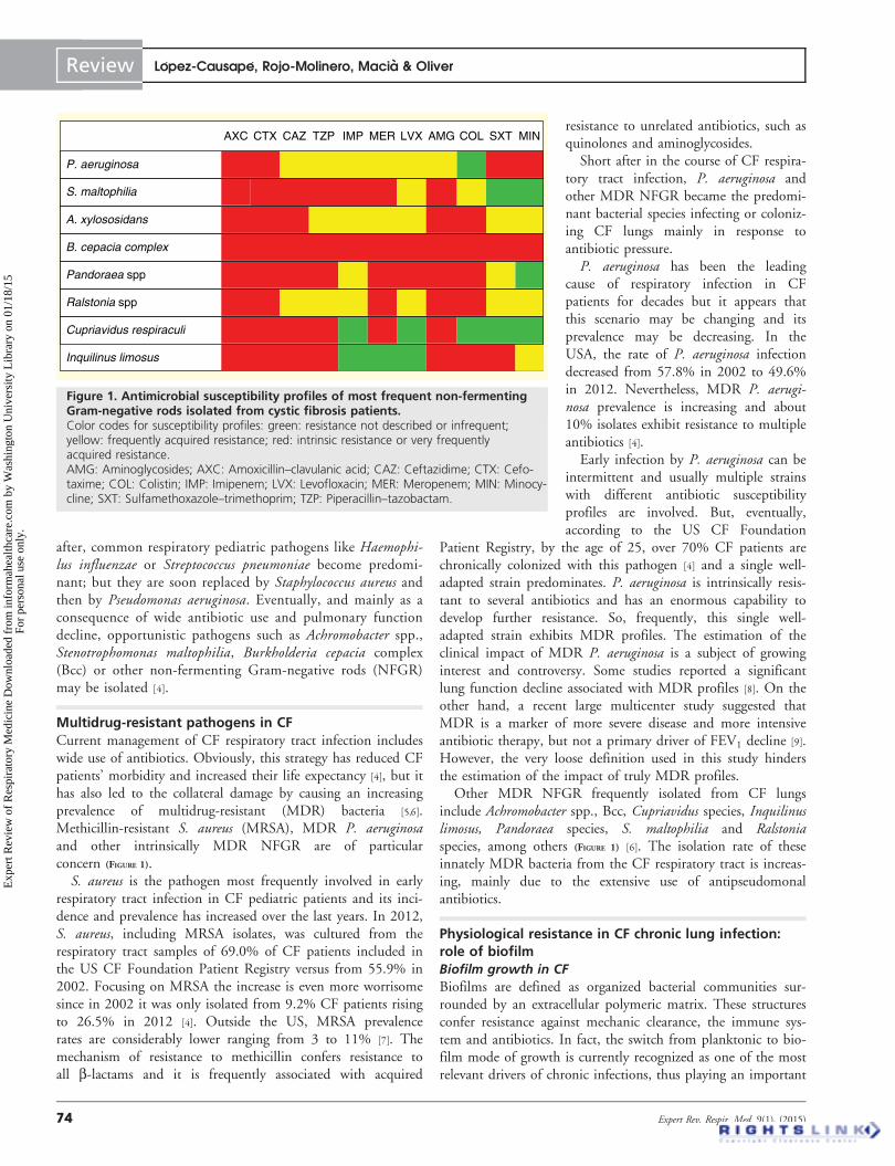
after, common respiratory pediatric pathogens like Haemophi-lus influenzae or Streptococcus pneumoniae become predomi-nant; but they are soon replaced by Staphylococcus aureus andthen by Pseudomonas aeruginosa. Eventually, and mainly as aconsequence of wide antibiotic use and pulmonary functiondecline, opportunistic pathogens such as Achromobacter spp.,Stenotrophomonas maltophilia, Burkholderia cepacia complex(Bcc) or other non-fermenting Gram-negative rods (NFGR)may be isolated [4].
Multidrug-resistant pathogens in CFCurrent management of CF respiratory tract infection includeswide use of antibiotics. Obviously, this strategy has reduced CFpatients’ morbidity and increased their life expectancy [4], but ithas also led to the collateral damage by causing an increasingprevalence of multidrug-resistant (MDR) bacteria [5,6].Methicillin-resistant S. aureus (MRSA), MDR P. aeruginosaand other intrinsically MDR NFGR are of particularconcern (FIGURE 1).
S. aureus is the pathogen most frequently involved in earlyrespiratory tract infection in CF pediatric patients and its inci-dence and prevalence has increased over the last years. In 2012,S. aureus, including MRSA isolates, was cultured from therespiratory tract samples of 69.0% of CF patients included inthe US CF Foundation Patient Registry versus from 55.9% in2002. Focusing on MRSA the increase is even more worrisomesince in 2002 it was only isolated from 9.2% CF patients risingto 26.5% in 2012 [4]. Outside the US, MRSA prevalencerates are considerably lower ranging from 3 to 11% [7]. Themechanism of resistance to methicillin confers resistance toall b-lactams and it is frequently associated with acquired
resistance to unrelated antibiotics, such asquinolones and aminoglycosides.
Short after in the course of CF respira-tory tract infection, P. aeruginosa andother MDR NFGR became the predomi-nant bacterial species infecting or coloniz-ing CF lungs mainly in response toantibiotic pressure.
P. aeruginosa has been the leadingcause of respiratory infection in CFpatients for decades but it appears thatthis scenario may be changing and itsprevalence may be decreasing. In theUSA, the rate of P. aeruginosa infectiondecreased from 57.8% in 2002 to 49.6%in 2012. Nevertheless, MDR P. aerugi-nosa prevalence is increasing and about10% isolates exhibit resistance to multipleantibiotics [4].
Early infection by P. aeruginosa can beintermittent and usually multiple strainswith different antibiotic susceptibilityprofiles are involved. But, eventually,according to the US CF Foundation
Patient Registry, by the age of 25, over 70% CF patients arechronically colonized with this pathogen [4] and a single well-adapted strain predominates. P. aeruginosa is intrinsically resis-tant to several antibiotics and has an enormous capability todevelop further resistance. So, frequently, this single well-adapted strain exhibits MDR profiles. The estimation of theclinical impact of MDR P. aeruginosa is a subject of growinginterest and controversy. Some studies reported a significantlung function decline associated with MDR profiles [8]. On theother hand, a recent large multicenter study suggested thatMDR is a marker of more severe disease and more intensiveantibiotic therapy, but not a primary driver of FEV1 decline [9].However, the very loose definition used in this study hindersthe estimation of the impact of truly MDR profiles.
Other MDR NFGR frequently isolated from CF lungsinclude Achromobacter spp., Bcc, Cupriavidus species, Inquilinuslimosus, Pandoraea species, S. maltophilia and Ralstoniaspecies, among others (FIGURE 1) [6]. The isolation rate of theseinnately MDR bacteria from the CF respiratory tract is increas-ing, mainly due to the extensive use of antipseudomonalantibiotics.
Physiological resistance in CF chronic lung infection:role of biofilmBiofilm growth in CF
Biofilms are defined as organized bacterial communities sur-rounded by an extracellular polymeric matrix. These structuresconfer resistance against mechanic clearance, the immune sys-tem and antibiotics. In fact, the switch from planktonic to bio-film mode of growth is currently recognized as one of the mostrelevant drivers of chronic infections, thus playing an important
AXC CTX CAZ TZP IMP MER LVX AMG COL SXT MIN
P. aeruginosa
S. maltophilia
A. xylososidans
B. cepacia complex
Pandoraea spp
Ralstonia spp
Cupriavidus respiraculi
Inquilinus limosus
Figure 1. Antimicrobial susceptibility profiles of most frequent non-fermentingGram-negative rods isolated from cystic fibrosis patients.Color codes for susceptibility profiles: green: resistance not described or infrequent;yellow: frequently acquired resistance; red: intrinsic resistance or very frequentlyacquired resistance.AMG: Aminoglycosides; AXC: Amoxicillin–clavulanic acid; CAZ: Ceftazidime; CTX: Cefo-taxime; COL: Colistin; IMP: Imipenem; LVX: Levofloxacin; MER: Meropenem; MIN: Minocy-cline; SXT: Sulfamethoxazole–trimethoprim; TZP: Piperacillin–tazobactam.
Review Lopez-Causape, Rojo-Molinero, Macia & Oliver
74 Expert Rev. Respir. Med. 9(1), (2015)
Exp
ert R
evie
w o
f R
espi
rato
ry M
edic
ine
Dow
nloa
ded
from
info
rmah
ealth
care
.com
by
Was
hing
ton
Uni
vers
ity L
ibra
ry o
n 01
/18/
15Fo
r pe
rson
al u
se o
nly.
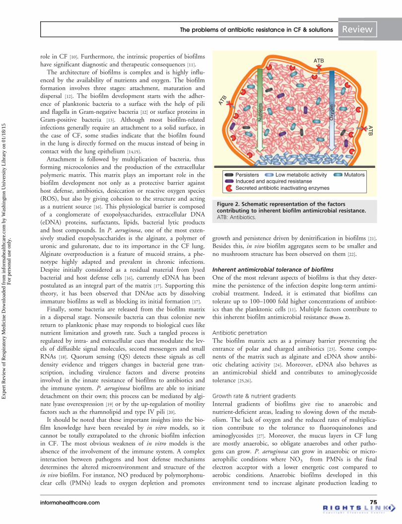
role in CF [10]. Furthermore, the intrinsic properties of biofilmshave significant diagnostic and therapeutic consequences [11].
The architecture of biofilms is complex and is highly influ-enced by the availability of nutrients and oxygen. The biofilmformation involves three stages: attachment, maturation anddispersal [12]. The biofilm development starts with the adher-ence of planktonic bacteria to a surface with the help of piliand flagella in Gram-negative bacteria [12] or surface proteins inGram-positive bacteria [13]. Although most biofilm-relatedinfections generally require an attachment to a solid surface, inthe case of CF, some studies indicate that the biofilm foundin the lung is directly formed on the mucus instead of being incontact with the lung epithelium [14,15].
Attachment is followed by multiplication of bacteria, thusforming microcolonies and the production of the extracellularpolymeric matrix. This matrix plays an important role in thebiofilm development not only as a protective barrier againsthost defense, antibiotics, desiccation or reactive oxygen species(ROS), but also by giving cohesion to the structure and actingas a nutrient source [16]. This physiological barrier is composedof a conglomerate of exopolysaccharides, extracellular DNA(eDNA) proteins, surfactants, lipids, bacterial lytic productsand host compounds. In P. aeruginosa, one of the most exten-sively studied exopolysaccharides is the alginate, a polymer ofuronic and guluronate, due to its importance in the CF lung.Alginate overproduction is a feature of mucoid strains, a phe-notype highly adapted and prevalent in chronic infections.Despite initially considered as a residual material from lysedbacterial and host defense cells [16], currently eDNA has beenpostulated as an integral part of the matrix [17]. Supporting thistheory, it has been observed that DNAse acts by dissolvingimmature biofilms as well as blocking its initial formation [17].
Finally, some bacteria are released from the biofilm matrixin a dispersal stage. Nonsessile bacteria can thus colonize newreturn to planktonic phase may responds to biological cues likenutrient limitation and growth rate. Such a tangled process isregulated by intra- and extracellular cues that modulate the lev-els of diffusible signal molecules, second messengers and smallRNAs [18]. Quorum sensing (QS) detects these signals as celldensity evidence and triggers changes in bacterial gene tran-scription, including virulence factors and diverse proteinsinvolved in the innate resistance of biofilms to antibiotics andthe immune system. P. aeruginosa biofilms are able to initiatedetachment on their own; this process can be mediated by algi-nate lyase overexpression [19] or by the up-regulation of motilityfactors such as the rhamnolipid and type IV pili [20].
It should be noted that these important insights into the bio-film knowledge have been revealed by in vitro models, so itcannot be totally extrapolated to the chronic biofilm infectionin CF. The most obvious weakness of in vitro models is theabsence of the involvement of the immune system. A complexinteraction between pathogens and host defense mechanismsdetermines the altered microenvironment and structure of thein vivo biofilm. For instance, NO produced by polymorphonu-clear cells (PMNs) leads to oxygen depletion and promotes
growth and persistence driven by denitrification in biofilms [21].Besides this, in vivo biofilm aggregates seem to be smaller andno mushroom structure has been observed on them [22].
Inherent antimicrobial tolerance of biofilms
One of the most relevant aspects of biofilms is that they deter-mine the persistence of the infection despite long-term antimi-crobial treatment. Indeed, it is estimated that biofilms cantolerate up to 100–1000 fold higher concentrations of antibiot-ics than the planktonic cells [11]. Multiple factors contribute tothis inherent biofilm antimicrobial resistance (FIGURE 2).
Antibiotic penetration
The biofilm matrix acts as a primary barrier preventing theentrance of polar and charged antibiotics [23]. Some compo-nents of the matrix such as alginate and eDNA show antibi-otic chelating activity [24]. Moreover, eDNA also behaves asan antimicrobial shield and contributes to aminoglycosidetolerance [25,26].
Growth rate & nutrient gradients
Internal gradients of biofilms give rise to anaerobic andnutrient-deficient areas, leading to slowing down of the metab-olism. The lack of oxygen and the reduced rates of multiplica-tion contribute to the tolerance to fluoroquinolones andaminoglycosides [27]. Moreover, the mucus layers in CF lungare mostly anaerobic, so obligate anaerobes and other patho-gens can grow. P. aeruginosa can grow in anaerobic or micro-aerophilic conditions where NO3
from PMNs is the finalelectron acceptor with a lower energetic cost compared toaerobic conditions. Anaerobic biofilms developed in thisenvironment tend to increase alginate production leading to
ATB
ATB
AT
B
Nut
rient
s
Oxy
gen
Persisters MutatorsLow metabolic activityInduced and acquired resistanseSecreted antibiotic inactivating enzymes
Figure 2. Schematic representation of the factorscontributing to inherent biofilm antimicrobial resistance.ATB: Antibiotics.
The problems of antibiotic resistance in CF & solutions Review
informahealthcare.com 75
Exp
ert R
evie
w o
f R
espi
rato
ry M
edic
ine
Dow
nloa
ded
from
info
rmah
ealth
care
.com
by
Was
hing
ton
Uni
vers
ity L
ibra
ry o
n 01
/18/
15Fo
r pe
rson
al u
se o
nly.
aminoglycoside tolerance. Furthermore, osmotic stress responsemay contribute to antibiotic resistance inducing a change inthe proportions of porins [10].
Persister phenomenon
Persisters are defined as a dormant phenotypic state of bacteriawithin biofilms, characterized by a high tolerance to antibiotics.Also, this latent bacterial state behaves as a bumper to hostdefense and may cause a relapse of infection, being a source ofrecalcitrant biofilm infection [28].
Induction of resistance mechanisms
Induction of resistance mechanism can significantly differbetween biofilm and planktonic growth [29]. Various studieshave found a differential expression of several conventional andbiofilm-resistance genes in biofilms [30].
Biofilms & mutational resistance
The antibiotic gradient driven by biofilm physiology favorsgradual development of mutational resistance during antimicro-bial treatment, which is of significance particularly wheninvolving mutator strains which are highly prevalent inCRI [31–33]. Also, endogenous oxidative stress [34] and muta-genic ROS released from PMNs are likely to induce mutabilityin biofilm cells. In fact, recent findings have shown that muta-genesis is intrinsically increased in biofilms [34,35].
Horizontal gene transfer
Bacterial proximity within a biofilm allows an effective hori-zontal gene transfer [36]. Moreover, bacterial eDNA may rep-resent a reservoir for the acquisition of exogenous resistancedeterminants.
Mutational resistance in CF chronic lung infection: roleof mutatorsMutational antimicrobial resistance mechanisms in CF
pathogens
In addition to intrinsic antibiotic resistance, the antimicrobialsusceptibility of bacterial populations can be significantly fur-ther compromised by the acquisition of certain chromosomalmutations. This is particularly relevant for the major CF patho-gen P. aeruginosa, given its extraordinary ability to acquireresistance through mutations that alter the expression and/orfunction of its chromosomally encoded resistance mechanisms.Although no single mutation can lead to MDR profiles, allantibiotics are prone to being compromised by acquiring muta-tions that eventually lead to overexpression of efflux pumps,hyperproduction of the chromosomal AmpC cephalosporinase,porin loss or altered antibiotic targets. TABLE 1 summarizes themost relevant genes involved and the corresponding resistanceprofiles generated. In addition to these classical resistance muta-tions, recent whole genome screening mutant libraries reveal aplethora of genes, collectively known as the resistome, whichhave an impact on antimicrobial susceptibility, including manywith central metabolic functions [37].
While it has been shown that mutational resistance can vir-tually affect all antibiotics, the spontaneous mutation frequen-cies vary according to the antibiotic agent, the bacterial speciesand the specific environmental conditions [38]. Moreover, muta-tional resistance can be significantly enhanced by the presenceof mutator phenotypes, which are highly prevalent in CF [39].
Hypermutation and antibiotic resistance in CF
Hypermutable (or mutator) microorganisms are defined asthose that have an increased spontaneous mutation rate due todefects in DNA repair or error avoidance systems.
The most frequent cause of hypermutation in natural bacte-rial populations is the presence of defects on the methyl-directed mismatch repair system. mutS, mutL and uvrD (mutU)are the key genes of the methyl-directed mismatch repair sys-tem and their inactivation leads to a stable mutator phenotypewith an increased rate of mutation from 100- to 1000-fold [40]. Mutations in MutM, MutY and MutT, the three keyproteins that compose the GO system, as well as mutations ofgenes involved in the prevention of oxidative damage producedby ROS, such as oxyR and sodA (mutA and mutC) [40] or therecently described pfpI [41], also lead to this phenotype.
Under particular circumstances, a transient mutator pheno-type can also arise. For instance, bacterial DNA damage indu-ces the SOS response and its error-prone DNA polymerasespromote an elevated mutation rate [42] Moreover, some antibi-otics can induce a transient mutator phenotype through thismechanism, thus promoting the development of antimicrobialresistance [43–45].
In natural bacterial populations, the presence of the mutatorphenotype involves an evolutionary advantage as it can notonly enhance mutational resistance but also facilitate bacterialadaptation to new or stressful environments. CRI with P. aeru-ginosa in CF patients represents a major example in nature.Prevalence of mutator P. aeruginosa in the CF airways isextremely high, approximately 10–30% of isolates [46], and itspresence has been strongly associated with adaptive mecha-nisms [47] and development of antibiotic resistance [31–33]. Theproportion of hypermutable isolates significantly increases dur-ing the course of P. aeruginosa CRI, as was demonstrated in a25-year longitudinal study in which the proportion of hyper-mutable isolates increased from 0% at the onset/early coloniza-tion to 65% [48]. Genetic hitch-hiking can explain thisobservation, which means that mutator alleles reach high fre-quency by being co-selected with linked beneficial mutations.
Fortunately, CF epidemic strains have not shown anincreased prevalence of mutators [49]. Nevertheless, transmissionof P. aeruginosa hypermutable strains between CF patients hasbeen recently demonstrated [50].
A higher prevalence of mutators in the CF setting has alsobeen noticed for other microorganisms such as S. pneumoniae,H. influenzae, S. aureus, S. maltophilia and Bcc [51–56].Prunier et al. found that approximately 14% of CF S. aureusisolates were hypermutable in contrast with 1% in non-CF iso-lates, and that hypermutability was strongly associated with
Review Lopez-Causape, Rojo-Molinero, Macia & Oliver
76 Expert Rev. Respir. Med. 9(1), (2015)
Exp
ert R
evie
w o
f R
espi
rato
ry M
edic
ine
Dow
nloa
ded
from
info
rmah
ealth
care
.com
by
Was
hing
ton
Uni
vers
ity L
ibra
ry o
n 01
/18/
15Fo
r pe
rson
al u
se o
nly.
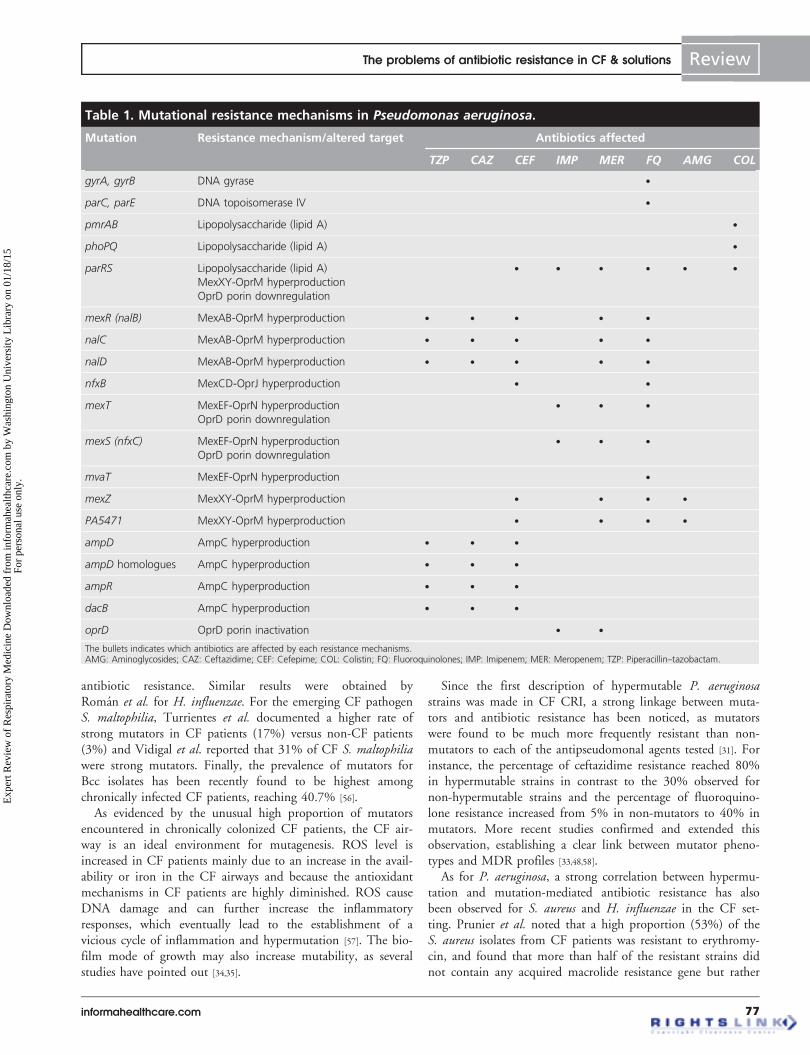
antibiotic resistance. Similar results were obtained byRoman et al. for H. influenzae. For the emerging CF pathogenS. maltophilia, Turrientes et al. documented a higher rate ofstrong mutators in CF patients (17%) versus non-CF patients(3%) and Vidigal et al. reported that 31% of CF S. maltophiliawere strong mutators. Finally, the prevalence of mutators forBcc isolates has been recently found to be highest amongchronically infected CF patients, reaching 40.7% [56].
As evidenced by the unusual high proportion of mutatorsencountered in chronically colonized CF patients, the CF air-way is an ideal environment for mutagenesis. ROS level isincreased in CF patients mainly due to an increase in the avail-ability or iron in the CF airways and because the antioxidantmechanisms in CF patients are highly diminished. ROS causeDNA damage and can further increase the inflammatoryresponses, which eventually lead to the establishment of avicious cycle of inflammation and hypermutation [57]. The bio-film mode of growth may also increase mutability, as severalstudies have pointed out [34,35].
Since the first description of hypermutable P. aeruginosastrains was made in CF CRI, a strong linkage between muta-tors and antibiotic resistance has been noticed, as mutatorswere found to be much more frequently resistant than non-mutators to each of the antipseudomonal agents tested [31]. Forinstance, the percentage of ceftazidime resistance reached 80%in hypermutable strains in contrast to the 30% observed fornon-hypermutable strains and the percentage of fluoroquino-lone resistance increased from 5% in non-mutators to 40% inmutators. More recent studies confirmed and extended thisobservation, establishing a clear link between mutator pheno-types and MDR profiles [33,48,58].
As for P. aeruginosa, a strong correlation between hypermu-tation and mutation-mediated antibiotic resistance has alsobeen observed for S. aureus and H. influenzae in the CF set-ting. Prunier et al. noted that a high proportion (53%) of theS. aureus isolates from CF patients was resistant to erythromy-cin, and found that more than half of the resistant strains didnot contain any acquired macrolide resistance gene but rather
Table 1. Mutational resistance mechanisms in Pseudomonas aeruginosa.
Mutation Resistance mechanism/altered target Antibiotics affected
TZP CAZ CEF IMP MER FQ AMG COL
gyrA, gyrB DNA gyrase .
parC, parE DNA topoisomerase IV .
pmrAB Lipopolysaccharide (lipid A) .
phoPQ Lipopolysaccharide (lipid A) .
parRS Lipopolysaccharide (lipid A)
MexXY-OprM hyperproduction
OprD porin downregulation
. . . . . .
mexR (nalB) MexAB-OprM hyperproduction . . . . .
nalC MexAB-OprM hyperproduction . . . . .
nalD MexAB-OprM hyperproduction . . . . .
nfxB MexCD-OprJ hyperproduction . .
mexT MexEF-OprN hyperproduction
OprD porin downregulation
. . .
mexS (nfxC) MexEF-OprN hyperproduction
OprD porin downregulation
. . .
mvaT MexEF-OprN hyperproduction .
mexZ MexXY-OprM hyperproduction . . . .
PA5471 MexXY-OprM hyperproduction . . . .
ampD AmpC hyperproduction . . .
ampD homologues AmpC hyperproduction . . .
ampR AmpC hyperproduction . . .
dacB AmpC hyperproduction . . .
oprD OprD porin inactivation . .
The bullets indicates which antibiotics are affected by each resistance mechanisms.AMG: Aminoglycosides; CAZ: Ceftazidime; CEF: Cefepime; COL: Colistin; FQ: Fluoroquinolones; IMP: Imipenem; MER: Meropenem; TZP: Piperacillin–tazobactam.
The problems of antibiotic resistance in CF & solutions Review
informahealthcare.com 77
Exp
ert R
evie
w o
f R
espi
rato
ry M
edic
ine
Dow
nloa
ded
from
info
rmah
ealth
care
.com
by
Was
hing
ton
Uni
vers
ity L
ibra
ry o
n 01
/18/
15Fo
r pe
rson
al u
se o
nly.
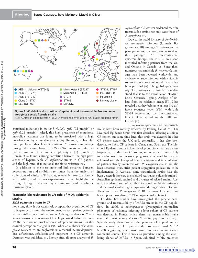
contained mutations in rrl (23S rRNA), rplD (L4 protein) orrplV (L22 protein); indeed, this high prevalence of mutationalmacrolide resistance was found to be associated with a highprevalence of hypermutable strains [51]. Recently, it has alsobeen published that linezolid-resistant S. aureus can emergethrough the accumulation of 23S rRNA mutations linked tothe acquisition of a mutator phenotype [59]. Similarly,Roman et al. found a strong correlation between the high prev-alence of hypermutable H. influenzae strains in CF patientsand the high rates of mutational antibiotic resistance [52].
In addition to the clear statistical link obtained betweenhypermutation and antibiotic resistance from the analysis ofcollections of clinical CF isolates, several in vitro (planktonicand biofilm) and in vivo experiments further highlight thestrong linkage between hypermutation and antibioticresistance [60–65].
Transmissible resistance in CF: role of MDR epidemicstrainsMDR epidemic strains in CF
For a long time, it was extensively accepted that acquisition of CFpathogens occurs from the environment, so each patient generallyharbors his/her own unrelated strain. Although evidence of P. aer-uginosa cross-infection among CF siblings existed, before the mid-1980s, there was no proof of spread of epidemic strains. But thisclassical perception changed in 1986 when an outbreak of P. aeru-ginosa resistant to aminoglycosides, carbenicillin, ureidopenicil-lins, ceftazidime, cefsulodin and imipenem in a CF center inDenmark was published [66]. Shortly after, ribotype analysis of B.
cepacia from CF centers evidenced that thetransmissible strains not only were those ofP. aeruginosa [67].
Due to the rapid increase of Burkholde-ria cenocepacia infection (formerly Bccgenomovar III) among CF patients and itspoor prognosis, attention was focused onthis pathogen. An intercontinentalepidemic lineage, the ET-12, was soonidentified infecting patients from the UKand Ontario in Canada [68]. Since then,numerous transmissible B. cenocepacia line-ages have been reported worldwide, andevidence of superinfection with epidemicstrains in previously colonized patients hasbeen provided [69]. The global epidemiol-ogy of B. cenocepacia is now better under-stood thanks to the introduction of MultiLocus Sequence Typing. Analysis of iso-lates from the epidemic lineage ET-12 hasrevealed that they belong to at least five dif-ferent sequence types (STs), with onlyST-28 representing the intercontinentalET-12 clone spread in the UK andCanada [70].
P. aeruginosa epidemic and transmissiblestrains have been recently reviewed by Fothergill et al. [71]. TheLiverpool Epidemic Strain was first described affecting a uniqueCF center, but some time later, this strain was detected in otherCF centers across the UK and, eventually, it has also beendetected to infect CF patients in Canada and Spain [50]. The Liv-erpool Epidemic Strain isolates develop antibiotic resistance morefrequently than the other CF strains, and resistance is more likelyto develop over time. A worse prognosis is predicted for patientscolonized with the Liverpool Epidemic Strain, and superinfectionof patients already colonized with P. aeruginosa strains has alsobeen reported; thus, strict patient segregation policies are to beimplemented. In Australia, some transmissible strains have alsobeen detected; these are the so-called Australian epidemic strain-1,Australian epidemic strain-2 and a cluster of related strains. Aus-tralian epidemic strain-1 exhibits increased antibiotic resistanceand increased virulence gene expression during chronic infection.These and other P. aeruginosa MDR transmissible strains havebeen reported worldwide [72,73] are represented in FIGURE 3.
To date, few studies have investigated the genetic back-ground and transmissibility of MRSA strains in the CF popula-tion. In 2006, a heterogeneous glycopeptide-intermediatephenotype of resistance infecting a long cohort of CF patientswas detected in France, which alerts that transmissible strainscould also exist among MRSA CF strains [74]. Shortly after, aSpanish study demonstrated the presence of a predominantclone among their CF patients, the hospital-acquired MRSAST228, suggesting either cross-transmission or a common envi-ronmental source. This clone, also prevalent among the circu-lating clones of MRSA in Spain, exhibited MDR, presented
AES-1 (Melbourne/ST649)AES-2 (ST775)
AES-3 (ST242)
Clone C (ST17)LES (ST146)
Manchester 1 (ST217)Midlands 1 (ST 148)
ST274
ST782DK1/DK2
Norway cluster 1
Houston-1
PES (ST192)ST406, ST497
Figure 3. Worldwide distribution of epidemic and transmissible Pseudomonasaeruginosa cystic fibrosis strains.AES: Australian epidemic strain; LES: Liverpool epidemic strain; PES: Prairie epidemic strain.
Review Lopez-Causape, Rojo-Molinero, Macia & Oliver
78 Expert Rev. Respir. Med. 9(1), (2015)
Exp
ert R
evie
w o
f R
espi
rato
ry M
edic
ine
Dow
nloa
ded
from
info
rmah
ealth
care
.com
by
Was
hing
ton
Uni
vers
ity L
ibra
ry o
n 01
/18/
15Fo
r pe
rson
al u
se o
nly.
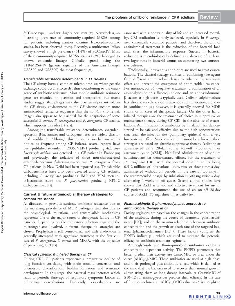
SCCmec type 1 and was highly persistent [75]. Nevertheless, anincreasing prevalence of community-acquired MRSA amongCF patients, including panton valentine leukocydin-positivestrains, has been observed [76–78]. Recently, a multicenter Italiansurvey showed a high prevalence (31.4%) of SCCmecIV. Mostof these community-acquired MRSA strains (73%) belonged toknown epidemic lineages Globally spread being theST8-MRSA-IV (genetic signature of the American lineagesUSA500 and USA300) the most frequent [79].
Transferable resistance determinants in CF isolates
The CF airway hosts a complex microbiome [80] where geneticexchange could occur effectively, thus contributing to the emer-gence of antibiotic resistance. Most mobile antibiotic resistancegenes are encoded on plasmids and transposons, but recentstudies suggest that phages may also play an important role inthe CF airway environment as the CF virome encodes moreantimicrobial resistance sequences than the non-CF virome [81].Phages also appear to be essential for the adaptation of somesuccessful S. aureus, B. cenocepacia and P. aeruginosa CF strains,which supports this idea [74,82,83].
Among the transferable resistance determinants, extended-spectrum b-lactamases and carbapenemeses are widely distrib-uted worldwide. Although this resistance mechanism seemsnot to be frequent among CF isolates, several reports havebeen published recently. In 2006, VEB-1 producing Achromo-bacter xylosoxidans was detected in a CF patient in France [84]
and previously, the isolation of three non-characterizedextended-spectrum b-lactamases–positive P. aeruginosa fromCF patients in New Delhi had been reported [85]. Transferablecarbapenemases have also been detected among CF isolates,including P. aeruginosa producing IMP and VIM metallo-b-lactamases [86,87] and K. pneumoniae producing KPC-2carbapenemase [88].
Current & future antimicrobial therapy strategies tocombat resistanceAs discussed in previous sections, antibiotic resistance due tothe increasing prevalence of MDR pathogens and also due tothe physiological, mutational and transmissible mechanismsrepresents one of the major causes of therapeutic failure in CFpatients. Depending on the respiratory infection stage and themicroorganisms involved, different therapeutic strategies arechosen. Prophylaxis is still controversial and early eradication isgenerally attempted with aggressive treatment at the first cul-ture of P. aeruginosa, S. aureus and MRSA, with the objectiveof preventing CRI [89].
Classical systemic & inhaled therapy in CF
During CRI, CF patients experience a progressive decline oflung function correlating with strain mucoid conversion andphenotypic diversification, biofilm formation and resistancedevelopment. In this stage, the bacterial mass increases whichleads to periodic flare-ups of respiratory symptoms known aspulmonary exacerbations. Frequently, exacerbations are
associated with a poorer quality of life and an increased mortal-ity. CRI eradication is rarely achieved, especially in P. aerugi-nosa chronically colonized patients, and therefore, the aim ofantimicrobial treatment is the reduction of the bacterial loadand, thus, the inflammatory response. Success in bacterialreduction is microbiologically defined as a decrease of, at least,two logarithms in bacterial counts on comparing two consecu-tive cultures.
Traditionally, intravenous antibiotics are used to treat exacer-bations. The classical strategy consists of combining two agentsfrom different antimicrobial classes to enhance the treatmenteffect and prevent the emergence of antimicrobial resistance.For instance, for P. aeruginosa treatment, a combination of anaminoglycoside or a fluoroquinolone and an antipseudomonalb-lactam at high doses is typically used. Colistin sulphomethatehas also shown efficacy on intravenous administration, alone orin combination [90]; however, it is generally reserved for MDRstrains or in cases of therapeutic failure. On the other hand,inhaled therapies are the treatment of choice in suppressive ormaintenance therapy during CF CRI, in the absence of exacer-bations. Administration of antibiotics by inhalation has demon-strated to be safe and effective due to the high concentrationsthat reach the infection site (pulmonary epithelia) with a verylow systemic effect. Since eradication cannot be achieved, thesestrategies are based on chronic suppressive therapy (colistin) oradministered as a 28-day course (on–off) (tobramycin oraztreonam-lysine [AZLI]). Nebulized administration of sodiumcolistimethate has demonstrated efficacy for the treatment ofP. aeruginosa CRI, with the normal dose in adults being0.5–2 millions of international units, two- or three-times a day,administered without off periods. In the case of tobramycin,the recommended dosage by inhalation is 300 mg twice a day,alternating 4 weeks on–off cycles. Several clinical studies haveshown that AZLI is a safe and effective treatment for use inCF patients and recommend the use of an on–off 28-daycourse of AZLI (75 mg, three-times daily) [89].
Pharmacokinetic & pharmacodynamic approach to
antimicrobial therapy in CF
Dosing regimens are based on the changes in the concentrationof the antibiotic during the course of treatment (pharmacoki-netics [PK]) and on the in vitro relationship between antibioticconcentration and the growth or death rate of the targeted bac-teria (pharmacodynamics [PD]). These factors comprise thePK/PD indices [91], which are used to estimate the potentialefficacy of antibiotic treatment regimens.
Aminoglycoside and fluoroquinolone antibiotics exhibit aconcentration-dependent activity. The PK/PD parameters thatbetter predict their activity are Cmax/MIC or area under thecurve (AUC24h)/MIC. These antibiotics are used at high dosesand their prolonged post-antibiotic effect, which is defined asthe time that the bacteria need to recover their normal growth,allows using them at long dosage intervals. A Cmax/MIC of‡10–12 for aminoglycosides predicts their efficacy. In the caseof fluoroquinolones, an AUC24h/MIC value >125 is thought to
The problems of antibiotic resistance in CF & solutions Review
informahealthcare.com 79
Exp
ert R
evie
w o
f R
espi
rato
ry M
edic
ine
Dow
nloa
ded
from
info
rmah
ealth
care
.com
by
Was
hing
ton
Uni
vers
ity L
ibra
ry o
n 01
/18/
15Fo
r pe
rson
al u
se o
nly.
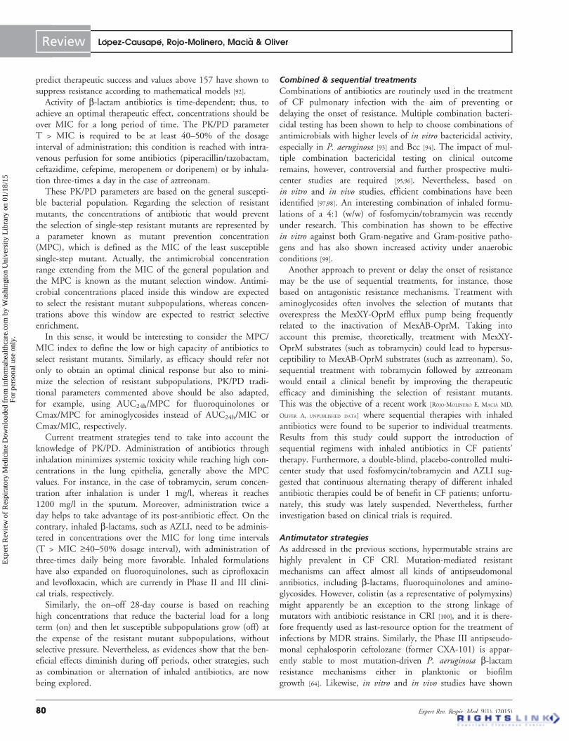
predict therapeutic success and values above 157 have shown tosuppress resistance according to mathematical models [92].
Activity of b-lactam antibiotics is time-dependent; thus, toachieve an optimal therapeutic effect, concentrations should beover MIC for a long period of time. The PK/PD parameterT > MIC is required to be at least 40–50% of the dosageinterval of administration; this condition is reached with intra-venous perfusion for some antibiotics (piperacillin/tazobactam,ceftazidime, cefepime, meropenem or doripenem) or by inhala-tion three-times a day in the case of aztreonam.
These PK/PD parameters are based on the general suscepti-ble bacterial population. Regarding the selection of resistantmutants, the concentrations of antibiotic that would preventthe selection of single-step resistant mutants are represented bya parameter known as mutant prevention concentration(MPC), which is defined as the MIC of the least susceptiblesingle-step mutant. Actually, the antimicrobial concentrationrange extending from the MIC of the general population andthe MPC is known as the mutant selection window. Antimi-crobial concentrations placed inside this window are expectedto select the resistant mutant subpopulations, whereas concen-trations above this window are expected to restrict selectiveenrichment.
In this sense, it would be interesting to consider the MPC/MIC index to define the low or high capacity of antibiotics toselect resistant mutants. Similarly, as efficacy should refer notonly to obtain an optimal clinical response but also to mini-mize the selection of resistant subpopulations, PK/PD tradi-tional parameters commented above should be also adapted,for example, using AUC24h/MPC for fluoroquinolones orCmax/MPC for aminoglycosides instead of AUC24h/MIC orCmax/MIC, respectively.
Current treatment strategies tend to take into account theknowledge of PK/PD. Administration of antibiotics throughinhalation minimizes systemic toxicity while reaching high con-centrations in the lung epithelia, generally above the MPCvalues. For instance, in the case of tobramycin, serum concen-tration after inhalation is under 1 mg/l, whereas it reaches1200 mg/l in the sputum. Moreover, administration twice aday helps to take advantage of its post-antibiotic effect. On thecontrary, inhaled b-lactams, such as AZLI, need to be adminis-tered in concentrations over the MIC for long time intervals(T > MIC ‡40–50% dosage interval), with administration ofthree-times daily being more favorable. Inhaled formulationshave also expanded on fluoroquinolones, such as ciprofloxacinand levofloxacin, which are currently in Phase II and III clini-cal trials, respectively.
Similarly, the on–off 28-day course is based on reachinghigh concentrations that reduce the bacterial load for a longterm (on) and then let susceptible subpopulations grow (off) atthe expense of the resistant mutant subpopulations, withoutselective pressure. Nevertheless, as evidences show that the ben-eficial effects diminish during off periods, other strategies, suchas combination or alternation of inhaled antibiotics, are nowbeing explored.
Combined & sequential treatments
Combinations of antibiotics are routinely used in the treatmentof CF pulmonary infection with the aim of preventing ordelaying the onset of resistance. Multiple combination bacteri-cidal testing has been shown to help to choose combinations ofantimicrobials with higher levels of in vitro bactericidal activity,especially in P. aeruginosa [93] and Bcc [94]. The impact of mul-tiple combination bactericidal testing on clinical outcomeremains, however, controversial and further prospective multi-center studies are required [95,96]. Nevertheless, based onin vitro and in vivo studies, efficient combinations have beenidentified [97,98]. An interesting combination of inhaled formu-lations of a 4:1 (w/w) of fosfomycin/tobramycin was recentlyunder research. This combination has shown to be effectivein vitro against both Gram-negative and Gram-positive patho-gens and has also shown increased activity under anaerobicconditions [99].
Another approach to prevent or delay the onset of resistancemay be the use of sequential treatments, for instance, thosebased on antagonistic resistance mechanisms. Treatment withaminoglycosides often involves the selection of mutants thatoverexpress the MexXY-OprM efflux pump being frequentlyrelated to the inactivation of MexAB-OprM. Taking intoaccount this premise, theoretically, treatment with MexXY-OprM substrates (such as tobramycin) could lead to hypersus-ceptibility to MexAB-OprM substrates (such as aztreonam). So,sequential treatment with tobramycin followed by aztreonamwould entail a clinical benefit by improving the therapeuticefficacy and diminishing the selection of resistant mutants.This was the objective of a recent work [ROJO-MOLINERO E, MACIÀ MD,
OLIVER A, UNPUBLISHED DATA] where sequential therapies with inhaledantibiotics were found to be superior to individual treatments.Results from this study could support the introduction ofsequential regimens with inhaled antibiotics in CF patients’therapy. Furthermore, a double-blind, placebo-controlled multi-center study that used fosfomycin/tobramycin and AZLI sug-gested that continuous alternating therapy of different inhaledantibiotic therapies could be of benefit in CF patients; unfortu-nately, this study was lately suspended. Nevertheless, furtherinvestigation based on clinical trials is required.
Antimutator strategies
As addressed in the previous sections, hypermutable strains arehighly prevalent in CF CRI. Mutation-mediated resistantmechanisms can affect almost all kinds of antipseudomonalantibiotics, including b-lactams, fluoroquinolones and amino-glycosides. However, colistin (as a representative of polymyxins)might apparently be an exception to the strong linkage ofmutators with antibiotic resistance in CRI [100], and it is there-fore frequently used as last-resource option for the treatment ofinfections by MDR strains. Similarly, the Phase III antipseudo-monal cephalosporin ceftolozane (former CXA-101) is appar-ently stable to most mutation-driven P. aeruginosa b-lactamresistance mechanisms either in planktonic or biofilmgrowth [64]. Likewise, in vitro and in vivo studies have shown
Review Lopez-Causape, Rojo-Molinero, Macia & Oliver
80 Expert Rev. Respir. Med. 9(1), (2015)
Exp
ert R
evie
w o
f R
espi
rato
ry M
edic
ine
Dow
nloa
ded
from
info
rmah
ealth
care
.com
by
Was
hing
ton
Uni
vers
ity L
ibra
ry o
n 01
/18/
15Fo
r pe
rson
al u
se o
nly.
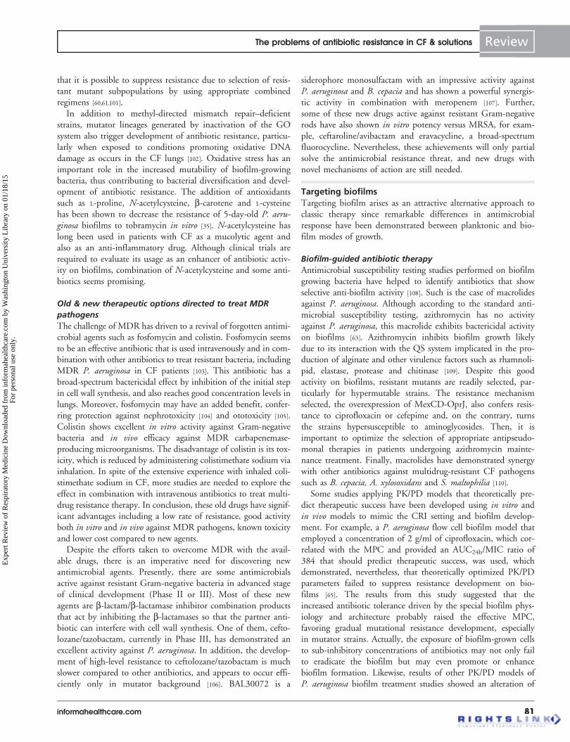
that it is possible to suppress resistance due to selection of resis-tant mutant subpopulations by using appropriate combinedregimens [60,61,101].
In addition to methyl-directed mismatch repair–deficientstrains, mutator lineages generated by inactivation of the GOsystem also trigger development of antibiotic resistance, particu-larly when exposed to conditions promoting oxidative DNAdamage as occurs in the CF lungs [102]. Oxidative stress has animportant role in the increased mutability of biofilm-growingbacteria, thus contributing to bacterial diversification and devel-opment of antibiotic resistance. The addition of antioxidantssuch as L-proline, N-acetylcysteine, b-carotene and L-cysteinehas been shown to decrease the resistance of 5-day-old P. aeru-ginosa biofilms to tobramycin in vitro [35]. N-acetylcysteine haslong been used in patients with CF as a mucolytic agent andalso as an anti-inflammatory drug. Although clinical trials arerequired to evaluate its usage as an enhancer of antibiotic activ-ity on biofilms, combination of N-acetylcysteine and some anti-biotics seems promising.
Old & new therapeutic options directed to treat MDR
pathogens
The challenge of MDR has driven to a revival of forgotten antimi-crobial agents such as fosfomycin and colistin. Fosfomycin seemsto be an effective antibiotic that is used intravenously and in com-bination with other antibiotics to treat resistant bacteria, includingMDR P. aeruginosa in CF patients [103]. This antibiotic has abroad-spectrum bactericidal effect by inhibition of the initial stepin cell wall synthesis, and also reaches good concentration levels inlungs. Moreover, fosfomycin may have an added benefit, confer-ring protection against nephrotoxicity [104] and ototoxicity [105].Colistin shows excellent in vitro activity against Gram-negativebacteria and in vivo efficacy against MDR carbapenemase-producing microorganisms. The disadvantage of colistin is its tox-icity, which is reduced by administering colistimethate sodium viainhalation. In spite of the extensive experience with inhaled coli-stimethate sodium in CF, more studies are needed to explore theeffect in combination with intravenous antibiotics to treat multi-drug resistance therapy. In conclusion, these old drugs have signif-icant advantages including a low rate of resistance, good activityboth in vitro and in vivo against MDR pathogens, known toxicityand lower cost compared to new agents.
Despite the efforts taken to overcome MDR with the avail-able drugs, there is an imperative need for discovering newantimicrobial agents. Presently, there are some antimicrobialsactive against resistant Gram-negative bacteria in advanced stageof clinical development (Phase II or III). Most of these newagents are b-lactam/b-lactamase inhibitor combination productsthat act by inhibiting the b-lactamases so that the partner anti-biotic can interfere with cell wall synthesis. One of them, cefto-lozane/tazobactam, currently in Phase III, has demonstrated anexcellent activity against P. aeruginosa. In addition, the develop-ment of high-level resistance to ceftolozane/tazobactam is muchslower compared to other antibiotics, and appears to occur effi-ciently only in mutator background [106]. BAL30072 is a
siderophore monosulfactam with an impressive activity againstP. aeruginosa and B. cepacia and has shown a powerful synergis-tic activity in combination with meropenem [107]. Further,some of these new drugs active against resistant Gram-negativerods have also shown in vitro potency versus MRSA, for exam-ple, ceftaroline/avibactam and eravacycline, a broad-spectrumfluorocycline. Nevertheless, these achievements will only partialsolve the antimicrobial resistance threat, and new drugs withnovel mechanisms of action are still needed.
Targeting biofilmsTargeting biofilm arises as an attractive alternative approach toclassic therapy since remarkable differences in antimicrobialresponse have been demonstrated between planktonic and bio-film modes of growth.
Biofilm-guided antibiotic therapy
Antimicrobial susceptibility testing studies performed on biofilmgrowing bacteria have helped to identify antibiotics that showselective anti-biofilm activity [108]. Such is the case of macrolidesagainst P. aeruginosa. Although according to the standard anti-microbial susceptibility testing, azithromycin has no activityagainst P. aeruginosa, this macrolide exhibits bactericidal activityon biofilms [63]. Azithromycin inhibits biofilm growth likelydue to its interaction with the QS system implicated in the pro-duction of alginate and other virulence factors such as rhamnoli-pid, elastase, protease and chitinase [109]. Despite this goodactivity on biofilms, resistant mutants are readily selected, par-ticularly for hypermutable strains. The resistance mechanismselected, the overexpression of MexCD-OprJ, also confers resis-tance to ciprofloxacin or cefepime and, on the contrary, turnsthe strains hypersusceptible to aminoglycosides. Then, it isimportant to optimize the selection of appropriate antipseudo-monal therapies in patients undergoing azithromycin mainte-nance treatment. Finally, macrolides have demonstrated synergywith other antibiotics against multidrug-resistant CF pathogenssuch as B. cepacia, A. xylosoxidans and S. maltophilia [110].
Some studies applying PK/PD models that theoretically pre-dict therapeutic success have been developed using in vitro andin vivo models to mimic the CRI setting and biofilm develop-ment. For example, a P. aeruginosa flow cell biofilm model thatemployed a concentration of 2 g/ml of ciprofloxacin, which cor-related with the MPC and provided an AUC24h/MIC ratio of384 that should predict therapeutic success, was used, whichdemonstrated, nevertheless, that theoretically optimized PK/PDparameters failed to suppress resistance development on bio-films [65]. The results from this study suggested that theincreased antibiotic tolerance driven by the special biofilm phys-iology and architecture probably raised the effective MPC,favoring gradual mutational resistance development, especiallyin mutator strains. Actually, the exposure of biofilm-grown cellsto sub-inhibitory concentrations of antibiotics may not only failto eradicate the biofilm but may even promote or enhancebiofilm formation. Likewise, results of other PK/PD models ofP. aeruginosa biofilm treatment studies showed an alteration of
The problems of antibiotic resistance in CF & solutions Review
informahealthcare.com 81
Exp
ert R
evie
w o
f R
espi
rato
ry M
edic
ine
Dow
nloa
ded
from
info
rmah
ealth
care
.com
by
Was
hing
ton
Uni
vers
ity L
ibra
ry o
n 01
/18/
15Fo
r pe
rson
al u
se o
nly.

expected PK/PD antibiotic parameters when acting on biofilms.In the study of Hengzhuang et al., b-lactam antibiotics showedtime-dependent killing and ciprofloxacin, colistin and tobramy-cin showed concentration- or dose-dependent killing being thensimilar to planktonic growth [111]. However, the concentrationsof antibiotics needed were, in all cases, very much higher evenin the case of time-dependent killing, where on b-lactamase–overproducing strains, the killing pattern of ceftazidime waschanged to concentration-dependent killing for biofilm cells.These results alert us of the complexity of mechanisms takingplace on P. aeruginosa CRI and the difficulty to predict thera-peutic efficacy even applying optimal PK/PD parameters.
Anti-biofilm strategies: new alternatives to classic
antimicrobial therapy
In recent years, significant efforts have been made trying to elu-cidate new therapeutic approaches. Some of the different strate-gies against biofilms are presented below and in FIGURE 4.
Avoiding biofilm formation
The inhibition of molecules involved in the attachment processseems to be a good approach for this objective. This could beachieved by using specific neutralizing antibodies against fla-gella, pili, eDNA and exopolysaccharides.
Avoiding biofilm maturation
At this point, the strategies should be directed to weakening ofthe formed biofilm, mainly targeting the virulence factors,eDNA, QS, small RNAs and iron metabolism. The problem isthat during the maturation phase, biofilm loses most of the vir-ulence factors and no drugs have indicated activity in thisstage [24].
DNAse
The inhaled recombinant human DNase has shown clinicalefficacy in CF patients. This enzyme reduces the viscosity ofmucus not only by clearing the DNA released from PMNs butalso by dissolving preformed biofilms [17] and facilitating theeffects of aminoglycosides [27].
Alginate lyase
The co-administration of inhaled alginate lyase with antibi-otics degrades alginate from the extracellular polymericmatrix leading to the elimination of mucoid bacteria fromthe biofilms [112].
Bacteriophages
Bacteriophage therapy is becoming an attractive co-adjuvant ofantibiotics. Phages are able to break through the extracellular
Attachment Proliferation Maturation Dispersal
eDNA
Quorum sensing molecules
Quorum sensing inhibitors
Planktonic bacteria Biofilm bacteria
Biofilm components
Extracellular polymeric matrix
Antimicrobial therapy
Antibodies
Bactoriophages Dispersal promoters
DNAse
Siderophores
Anti-biofilm strategies
Figure 4. Schematic representation of the different anti-biofilm strategies proposed.eDNA: Extracellular DNA.
Review Lopez-Causape, Rojo-Molinero, Macia & Oliver
82 Expert Rev. Respir. Med. 9(1), (2015)
Exp
ert R
evie
w o
f R
espi
rato
ry M
edic
ine
Dow
nloa
ded
from
info
rmah
ealth
care
.com
by
Was
hing
ton
Uni
vers
ity L
ibra
ry o
n 01
/18/
15Fo
r pe
rson
al u
se o
nly.

polymeric matrix and reach bacteria within the biofilm, starting alytic process inside them. The problem is that bacteriophages arespecies-specific and make the isolation of bacteria necessary to selectthe suitable phage. Also, bacteria can develop resistance to the lyticactivity of phages [113]. Regardless of this, some studies using bacter-iophages against P. aeruginosa have demonstrated efficacy [114,115].
Iron metabolism
Since iron availability shows up as a critical factor in biofilm for-mation, chelating agents may be a good option to eliminate thebiofilm. The strategy is to take advantage of the iron transportsystem to introduce inactive metal ions (Sc3+, In3+ or Ga3+)or antibiotics conjugated with a siderophore. Several com-pounds have been shown to interfere with biofilm formationin vitro, but most of them have only been effective on abioticsurfaces. Gallium nitrate formulated for inhalation has showneffectiveness in chronic airway infection in animal models [116],although gallium-resistant P. aeruginosa mutants have beenreported [117]. Also, lactoferrin, a human iron-binding protein,seems to impair biofilm formation in respiratory infections bystimulation of twitching motility [118].
Virulence factors
The development of antibodies against b-lactamase observed inCF [119] supports the development of specific antibodies or sub-stances that bind virulence factors as a good strategy to fightbiofilm maturation. The disadvantage of this approach is thatonly single virulence factors are targeted and they are species-or even strain-specific. Also, there is a risk of inducing immu-nopathology as a result of increased inflammation owing to animmune complex–mediated reaction.
Quorum sensing inhibitors
Since QS plays a major role in biofilm formation and regulationof the expression of virulence factors, there is an emerging inter-est in the research of new molecules able to block the QS path-way. The advantage of quorum sensing inhibitors (QSIs) overantibiotics is that the development of resistance is minimizedsince these molecules target the virulence factor instead of bacte-rial growth [120]. However, the theoretical benefits of these mol-ecules should be considered with caution since resistance toQSIs has been recently reported in literature [121,122]. EffectiveQSIs can be found in nature among the secondary metabolitesproduced by algae, sponges, fungi, food products and higherplants [120]. For example, natural halogenated furanones,solenopsin A, manoalide and its derivate, garlic, patulin andgingseng have shown activity against pathogens implicated inbiofilm chronic infections. Nevertheless, activity of these com-pounds has only been assessed in experimental studies; hence,further clinical studies are needed to establish their efficacy andsafety to truly introduce them in the treatment of CF patients.
Driving biofilm dispersal
When the biofilm is mature, some bacteria are released fromthe biofilm matrix, probably due to the lack of nutrients.
Planktonic floating bacteria are more susceptible to antibiotics;taking advantage of this, an adequate antimicrobial treatmentwith the compounds that promote biofilm disruption couldachieve a higher therapeutic success. Studies have investigatedthe effect of different molecules in the dispersal of biofilms andhave obtained promising results. For example, unsaturated fattyacids [123], nitric oxide [124], succinic acid, citrate and com-pounds that interfere with c-di-GMP levels [125] have shownactivity in vitro. Curiously, 2-aminoimidazole/triazole may re-sensitize MDR strains to the effects of conventional antibiotics,apart from its ability to inhibit and disperse biofilms [126].
Infection control in CFThe incorporation of appropriate control measures is one ofthe most effective available strategies to prevent early infec-tion and transmission of epidemic strains between patients.In the recent past, the use of molecular typing methods hasled to an improved understanding of the epidemiology ofCF pathogens, highlighting the relevance of epidemic strainsamong CF patients, described above. In most cases, the ini-tial source of microorganism acquisition is unknown,although the environment seems to be an important reservoirfor CF pathogens.
In order to reduce the days of hospitalization and also toimprove the quality of life of CF patients, there has been ashift in healthcare delivery from hospitals to ambulatory andhome settings. Even though specific measures should be imple-mented in this new scenario, the lessons learned about trans-mission of nosocomial pathogens can be applied as infectioncontrol strategies for CF.
The American CF Foundation in its last update of infectionprevention and control guidelines for CF [127] sets recommen-dations to prevent transmission, taking into account that allCF patients should be treated as potential transmitters ofpathogens.
The most important preventive measure is hand hygiene inhealthcare and non-healthcare settings. To prevent droplettransmission, all CF patients, regardless of the respiratory tractculture results, should be separated by at least a distance of3–6 feet, which has been proposed as the minimal distance toavoid transmission. While the use of mask, gloves and gown isonly required for staff in the case of colonization by MDR orepidemic transmissible strains, people with CF should berequired to wear a mask in hospitals to reduce the risk of trans-mission, or even to reduce the acquisition of new pathogens. Inparticular, CF clinics should schedule and manage patients toavoid contact, minimizing the waiting time in common areasand segregating the patients with MDR or epidemic transmissi-ble strains. In the same way, summer camps and other groupactivities are not encouraged.
Surfaces and respiratory therapy equipments have to beassiduously cleaned and disinfected to reduce contamination ofenvironmental sources (following institutional policies of con-trol of MDR pathogens). To reduce transmission by medicalequipment, the use of single-patient disposable items should be
The problems of antibiotic resistance in CF & solutions Review
informahealthcare.com 83
Exp
ert R
evie
w o
f R
espi
rato
ry M
edic
ine
Dow
nloa
ded
from
info
rmah
ealth
care
.com
by
Was
hing
ton
Uni
vers
ity L
ibra
ry o
n 01
/18/
15Fo
r pe
rson
al u
se o
nly.
facilitated. Moreover, it is recommended to clean and disinfectthe exam rooms between patients.
Surveillance reports are a valuable tool to document the inci-dence and prevalence of MDR pathogens, and to review theacquisition of epidemic transmissible strains. The moleculartyping of CF pathogens, integrated as a routine in the microbi-ological diagnosis of respiratory infection, can be used as amarker of success of infection control strategies.
Expert commentary & five-year viewAntimicrobial resistance in CF is a multifactorial problemwhich includes aspects related to bacterial physiology (develop-ment of biofilms), genetic evolution (acquisition of antibioticresistance mutations linked to mutator phenotypes) and epide-miology (such as the acquisition of infections by MDR patho-gens or epidemic strains). Therefore, our present and futurestrategies should target these key aspects. Among them, the ini-tiatives directed to prevent and/or disrupt biofilms, exemplifiedby research on QSIs, are particularly encouraging. Likewise,strategies intended to avoid mutational resistance, such as the
optimization of PK/PD parameters, innovative combined andsequential regimens, or the use of ‘antimutator’ adjuvants, arepromising as well. Finally, despite being insufficient, novelcompounds currently under clinical development will mitigateour needs for the treatment of MDR strains to some extent,but strict infection control measures will remain a key issue.
Financial & competing interests disclosure
A Oliver is supported by the Ministerio de Economıa y Competitividad of
Spain and the Instituto de Salud Carlos III, through the Spanish Network
for the Research in Infectious Diseases (RD06/0008 and RD12/0015),
and by the Direccio General d´Universitats, Recerca i Transferencia del
Coneixement del Govern de les Illes Balears. A Oliver has received research
grants from Jannsen Cilag, Cubist Pharmaceuticals and Gilead Sciences.
The authors have no other relevant affiliations or financial involvement
with any organization or entity with a financial interest in or financial
conflict with the subject matter or materials discussed in the manuscript
apart from those disclosed.
No writing assistance was utilized in the production of this
manuscript.
Key issues
. The isolation of multidrug resistant (MDR) pathogens from the cystic fibrosis (CF) respiratory tract is increasing, mainly due to the
extensive use of antibiotics.
. Biofilm growth is an efficient adaptive strategy for survival and persistence of bacteria in the CF lungs due to its inherent tolerance to
the immune system and antibiotics.
. Mutators are highly prevalent in CF chronic respiratory infection and play a major role in resistance development.
. Epidemic and transmissible Burkholderia cepacia, Pseudomonas aeruginosa and Staphylococcus aureus strains have been identified
infecting CF patients and frequently show MDR profiles.
. The CF airway hosts a complex microbiome in which genetic exchange can occur contributing to development of resistance.
. Treatments should be based on pharmacokinetic/pharmacodynamic parameters and on the pathogen resistance mechanisms; combined
and sequential inhaled antibiotic treatments seem to be a promising alternative.
. Targeting the biofilms and the antimutator strategies arise as innovative approaches to overcome the current lack of effective
antimicrobial treatments.
. Strict infection control measures are required to prevent the inter-patient transmission of MDR and epidemic strains.
References
Papers of special note have been highlighted as:. of interest.. of considerable interest
1. O’Sullivan BP, Freedman SD. Cystic
fibrosis. Lancet 2009;373(9678):1891-904
2. Hampton TH, Green DM, Cutting GR,
et al. The microbiome in pediatric cystic
fibrosis patients: the role of shared
environment suggests a window of
intervention. Microbiome 2014;28:2-14
3. Hoegger MJ, Fischer AJ, McMenimen JD,
et al. Cystic fibrosis. Impaired mucus
detachment disrupts mucociliary transport
in a piglet model of cystic fibrosis. Science
2014;345(6198):818-22
4. Cystic fibrosis foundation patient registry.
2012 Annual data report. Cystic Fibrosis
Foundation; Bethesda, MD
5. Emerson J, McNamara S, Buccat AM, et al.
Changes in cystic fibrosis sputum
microbiology in the United States between
1995 and 2008. Pediatr Pulmonol 2010;
45(4):363-70
. Changes in the prevalence of common
cystic fibrosis (CF) pathogens and their
antibiotic resistance profiles were studied
by comparing a contemporary (2008) and
an historical cohort (1995) of CF
patients.
6. LiPuma JJ. The changing microbial
epidemiology in cystic fibrosis. Clin
Microbiol Rev 2010;23(2):299-323
7. Goss CH, Muhlebach MS. Review:
staphylococcus aureus and MRSA in cystic
fibrosis. J Cystic Fibros 2011;10(5):298-306
8. Lechtzin N, John M, Irizarry R, et al.
Outcomes of adults with cystic fibrosis
infected with antibiotic-resistant
Pseudomonas aeruginosa. Respiration 2006;
73(1):27-33
9. Ren CL, Konstan MW, Yegin A, et al.
Multiple antibiotic-resistant Pseudomonas
aeruginosa and lung function decline in
patients with cystic fibrosis. J Cyst Fibros
2012;11(4):293-9
10. Stewart PS, Costerton JW. Antibiotic
resistance of bacteria in biofilms. Lancet
2001;358(9276):135-8
Review Lopez-Causape, Rojo-Molinero, Macia & Oliver
84 Expert Rev. Respir. Med. 9(1), (2015)
Exp
ert R
evie
w o
f R
espi
rato
ry M
edic
ine
Dow
nloa
ded
from
info
rmah
ealth
care
.com
by
Was
hing
ton
Uni
vers
ity L
ibra
ry o
n 01
/18/
15Fo
r pe
rson
al u
se o
nly.

11. Høiby N, Bjarnsholt T, Givskov M, et al.
Antibiotic resistance of bacterial biofilms.
Int J Antimicrob Agents 2010;35(4):322-32
. An excellent review that focused on the
antimicrobial resistance mechanisms of
Pseudomonas aeruginosa biofilms.
12. O’Toole G, Kaplan HB, Kolter R. Biofilm
formation as microbial development. Annu
Rev Microbiol 2000;54:49-79
13. Cucarella C, Solano C, Valle J, et al. Bap, a
Staphylococcus aureus surface protein
involved in biofilm formation. J Bacteriol
2001;183(9):2888-96
14. Bjarnsholt T, Jensen PØ, Fiandaca MJ,
et al. Pseudomonas aeruginosa biofilms in
the respiratory tract of cystic fibrosis
patients. Pediatr Pulmonol 2009;44(6):
547-58
15. Worlitzsch D, Tarran R, Ulrich M, et al.
Effects of reduced mucus oxygen
concentration in airway Pseudomonas
infections of cystic fibrosis patients. J Clin
Invest 2002;109(3):317-25
16. Flemming HC, Wingender J. The biofilm
matrix. Nat Rev Microbiol 2010;8(9):
623-33
17. Whitchurch CB, Tolker-Nielsen T,
Ragas PC, Mattick JS. Extracellular
DNA required for bacterial biofilm
formation. Science 2002;295(5559):1487
18. Bjarnsholt T, Ciofu O, Molin S, et al.
Applying insights from biofilm biology to
drug development - can a new approach be
developed? Nat Rev Drug Discov 2013;
12(10):791-808
19. Boyd A, Chakrabarty AM. Role of alginate
lyase in cell detachment of Pseudomonas
aeruginosa. Appl Environ Microbiol 1994;
60(7):2355-9
20. Pamp SJ, Tolker-Nielsen T. Multiple roles
of biosurfactants in structural biofilm
development by Pseudomonas aeruginosa. J
Bacteriol 2007;189(6):2531-9
21. Kolpen M, Bjarnsholt T, Moser C, et al.
Nitric oxide production by
polymorphonuclear leucocytes in infected
cystic fibrosis sputum consumes oxygen.
Clin Exp Immunol 2014;177(1):310-19
22. Bjarnsholt T, Alhede M, Alhede M, et al.
The in vivo biofilm. Trends Microbiol
2013;21(9):466-74
23. Lewis K. Multidrug tolerance of biofilms
and persister cells. Curr Top Microbiol
Immunol 2008;322:107-31
24. Alipour M, Suntres ZE, Omri A.
Importance of DNase and alginate lyase for
enhancing free and liposome encapsulated
aminoglycoside activity against
Pseudomonas aeruginosa. J Antimicrob
Chemother 2009;64(2):317-25
25. Chiang WC, Nilsson M, Jensen PØ, et al.
Extracellular DNA shields against
aminoglycosides in Pseudomonas aeruginosa
biofilms. Antimicrob Agents Chemother
2013;57(5):2352-61
26. Mulcahy H, Charron-Mazenod L,
Lewenza S. Extracellular DNA chelates
cations and induces antibiotic resistance in
pseudomonas aeruginosa biofilms. PLoS
Pathog 2008;4:11
27. Walters MC, Roe F, Bugnicourt A, et al.
Contributions of antibiotic penetration,
oxygen limitation, and low metabolic
activity to tolerance of Pseudomonas
aeruginosa biofilms to ciprofloxacin and
tobramycin. Antimicrob Agents Chemother
2003;47(1):317-23
28. Lewis K. Persister cells. Annu Rev
Microbiol 2010;64:357-72
29. Mulet X, Moya B, Juan C, et al.
Antagonistic interactions of Pseudomonas
aeruginosa antibiotic resistance mechanisms
in planktonic but not biofilm growth.
Antimicrob Agents Chemother 2011;55(10):
4560-8
30. Whiteley M, Bangera MG, Bumgarner RE,
et al. Gene expression in Pseudomonas
aeruginosa biofilms. Nature 2001;
413(6858):860-4
31. Oliver A, Canton R, Campo P, et al. High
frequency of hypermutable Pseudomonas
aeruginosa in cystic fibrosis lung infection.
Science 2000;288(5469):1251-4
. First study demonstrating a high
prevalence of mutators in CF chronic
respiratory infection and their linkage to
antimicrobial resistance.
32. Macia MD, Blanquer D, Togores B, et al.
Hypermutation is a key factor in
development of multiple-antimicrobial
resistance in Pseudomonas aeruginosa strains
causing chronic lung infections. Antimicrob
Agents Chemother 2005;49(8):3382-6
33. Henrichfreise B, Wiegand I, Pfister W,
Wiedemann B. Resistance mechanisms of
multiresistant Pseudomonas aeruginosa
strains from Germany and correlation with
hypermutation. Antimicrob Agents
Chemother 2007;51(11):4062-70
. Comprehensive analysis of the
antimicrobial resistance mechanisms of
multidrug-resistant P. aeruginosa strains
from CF and non-CF patients and their
association with mutator phenotypes.
34. Driffield K, Miller K, Bostock JM, et al.
Increased mutability of Pseudomonas
aeruginosa in biofilms. J Antimicrob
Chemother 2008;61(5):1053-6
35. Boles BR, Singh PK. Endogenous oxidative
stress produces diversity and adaptability in
biofilm communities. Proc Natl Acad Sci
USA 2008;105(34):12503-8
36. Bagge N, Hentzer M, Andersen JB, et al.
Dynamics and spatial distribution of
beta-lactamase expression in Pseudomonas
aeruginosa biofilms. Antimicrob Agents
Chemother 2004;48(4):1168-74
37. Olivares J, Bernardini A, Garcia-Leon G,
et al. The intrinsic resistome of bacterial
pathogens. Front Microbiol 2013;4:103
38. Breidenstein EB, de la Fuente-Nunez C,
Hancock RE. Pseudomonas aeruginosa: all
roads lead to resistance. Trends Microbiol
2011;19(8):419-26
39. Blazquez J. Hypermutation as a factor
contributing to the acquisition of
antimicrobial resistance. Clin Infect Dis
2003;37(9):1201-9
40. Miller JH. Spontaneous mutators in
bacteria: insights into pathways of
mutagenesis and repair. Annu Rev
Microbiol 1996;50:625-43
41. Rodriguez-Rojas A, Blazquez J. The
Pseudomonas aeruginosa pfpI gene plays an
antimutator role and provides general stress
protection. J Bacteriol 2009;191(3):844-50
42. Zgur-Bertok D. DNA damage repair and
bacterial pathogens. PLoS Pathog 2013;
9(11):e1003711
43. Blazquez J, Oliver A, Gomez-Gomez JM.
Mutation and evolution of antibiotic
resistance: antibiotics as promoters of
antibiotic resistance? Curr Drug Targets
2002;3(4):345-9
44. Blazquez J, Gomez-Gomez JM, Oliver A,
et al. PBP3 inhibition elicits adaptive
responses in Pseudomonas aeruginosa. Mol
Microbiol 2006;62(1):84-99
45. Perez-Capilla T, Baquero MR,
Gomez-Gomez JM, et al. SOS independent
induction of dinB transcription by
beta-lactam-mediated inhibition of cell wall
synthesis in Escherichia coli. J Bacteriol
2005;187(4):1515-18
46. Oliver A. Mutators in cystic fibrosis chronic
lung infection: prevalence, mechanisms, and
consequences for antimicrobial therapy. Int
J Med Microbiol 2010;300(8):563-72
47. Mena A, Smith EE, Burns JL, et al. Genetic
adaptation of Pseudomonas aeruginosa to
the airways of cystic fibrosis patients is
catalyzed by hypermutation. J Bacteriol
2008;190(24):7910-17
The problems of antibiotic resistance in CF & solutions Review
informahealthcare.com 85
Exp
ert R
evie
w o
f R
espi
rato
ry M
edic
ine
Dow
nloa
ded
from
info
rmah
ealth
care
.com
by
Was
hing
ton
Uni
vers
ity L
ibra
ry o
n 01
/18/
15Fo
r pe
rson
al u
se o
nly.
48. Ciofu O, Riis B, Pressler T, et al.
Occurence of hypermutable Pseudomonas
aeruginosa in cystic fibrosis patients is
associated with the oxidative stress caused by
chronic lung inflammation. Antimicrob
Agents Chemother 2005;49(6):2276-82
49. Kenna DT, Doherty CJ, Foweraker J, et al.
Hypermutability in environmental
Pseudomonas aeruginosa and in populations
causing pulmonary infection in individuals
with cystic fibrosis. Microbiology 2007;
153(Pt 6):1852-9
50. Lopez-Causape C, Rojo-Molinero E,
Mulet X, et al. Clonal dissemination,
emergence of mutator lineages and
antibiotic resistance evolution in
Pseudomonas aeruginosa cystic fibrosis
chronic lung infection. PLoS One 2013;
8(8):e71001
51. Prunier AL, Malbruny B, Laurans M, et al.
High rate of macrolide resistance in
Staphylococcus aureus strains from patients
with cystic fibrosis reveals high proportions
of hypermutable strains. J Infect Dis 2003;
187(11):1709-16
52. Roman F, Canton R, Perez-Vazquez M,
et al. Dynamics of long-term colonization of
respiratory tract by Haemophilus influenzae
in cystic fibrosis patients shows a marked
increase in hypermutable strains. J Clin
Microbiol 2004;42(4):1450-9
53. Del Campo R, Morosini MI,
de la Pedrosa EG, et al. Population
structure, antimicrobial resistance, and
mutation frequencies of Streptococcus
pneumoniae isolates from cystic fibrosis
patients. J Clin Microbiol 2005;43(5):
2207-14
54. Turrientes MC, Baquero MR, Sanchez MB,
et al. Polymorphic mutation frequencies of
clinical and environmental
Stenotrophomonas maltophilia populations.
Appl Environ Microbiol 2010;76(6):
1746-58
55. Vidigal PG, Dittmer S, Steinmann E, et al.
Adaptation of Stenotrophomonas
maltophilia in cystic fibrosis: molecular
diversity, mutation frequency and antibiotic
resistance. Int J Med Microbiol 2014;
304(5-6):613-19
56. Martina P, Feliziani S, Juan C, et al.
Hypermutation in Burkholderia cepacia
complex is mediated by DNA mismatch
repair inactivation and is highly prevalent in
cystic fibrosis chronic respiratory infection.
Int J Med Microbiol 2014;304(8):1182-91
57. Rodrıguez-Rojas A, Oliver A, Blazquez J.
Intrinsic and environmental mutagenesis
drive diversification and persistence of
Pseudomonas aeruginosa in chronic lung
infections. J Infect Dis 2012;205(1):121-7
58. Ferroni A, Guillemot D, Moumile K, et al.
Effect of mutator P. aeruginosa on
antibiotic resistance acquisition and
respiratory function in cystic fibrosis.
Pediatr Pulmonol 2009;44(8):820-5
59. Tazi A, Chapron J, Touak G, et al. Rapid
emergence of resistance to linezolid and
mutator phenotypes in Staphylococcus
aureus isolates from an adult cystic fibrosis
patient. Antimicrob Agents Chemother
2013;57(10):5186-8
60. Macia MD, Borrell N, Segura M, et al.
Efficacy and potential for resistance selection
of antipseudomonal treatments in a mouse
model of lung infection by hypermutable
Pseudomonas aeruginosa. Antimicrob
Agents Chemother 2006;50(3):975-83
61. Plasencia V, Borrell N, Macia MD, et al.
Influence of high mutation rates on the
mechanisms and dynamics of in vitro and
in vivo resistance development to single or
combined antipseudomonal agents.
Antimicrob Agents Chemother 2007;51(7):
2574-81
62. Henrichfreise B, Wiegand I,
Luhmer-Becker I, Wiedemann B.
Development of resistance in wild-type and
hypermutable Pseudomonas aeruginosa
exposed to clinical pharmacokinetic profiles
of meropenem and ceftazidime simulated in
vitro. Antimicrob Agents Chemother
2007;51:3642-9
63. Mulet X, Macia MD, Mena A, et al.
Azithromycin in Pseudomonas aeruginosa
biofilms: bactericidal activity and selection
of nfxB mutants. Antimicrob Agents
Chemother 2009;53(4):1552-60
64. Riera E, Macia MD, Mena A, et al.
Anti-biofilm and resistance suppression
activities of CXA-101 against chronic
respiratory infection phenotypes of
Pseudomonas aeruginosa strain PAO1. J
Antimicrob Chemother 2010;65(7):
1399-404
65. Macia MD, Perez JL, Molin S, Oliver A.
Dynamics of mutator and
antibiotic-resistant populations in a
pharmacokinetic/pharmacodynamic model
of Pseudomonas aeruginosa biofilm
treatment. Antimicrob Agents Chemother
2011;55(11):5230-7
. First report demonstrating the
amplification of mutator populations
under antibiotic treatment in
P. aeruginosa biofilms.
66. Pedersen SS, Koch C, Høiby N,
Rosendal K. An epidemic spread of
multiresistant Pseudomonas aeruginosa in a
cystic fibrosis centre. J Antimicrob
Chemother 1986;17(4):505-16
67. LiPuma JJ, Mortensen JE, Dasen SE, et al.
Ribotype analysis of Pseudomonas cepacia
from cystic fibrosis treatment centers. J
Pediatr 1988;113(5):859-62
68. Johnson WM, Tyler SD, Rozee KR.
Linkage analysis of geographic and clinical
clusters in Pseudomonas cepacia infections
by multilocus enzyme electrophoresis and
ribotyping. J Clin Microbiol 1994;32(4):
924-30
69. Ledson MJ, Gallagher MJ, Corkill JE, et al.
Cross infection between cystic fibrosis
patients colonised with Burkholderia
cepacia. Thorax 1998;53(5):432-6
70. Drevinek P, Mahenthiralingam E.
Burkholderia cenocepacia in cystic fibrosis:
epidemiology and molecular mechanisms of
virulence. Clin Microbiol Infect 2010;16(7):
821-30
71. Fothergill JL, Walshaw MJ, Winstanley C.
Transmissible strains of Pseudomonas
aeruginosa in cystic fibrosis lung infections.
Eur Respir J 2012;40(1):227-38
. The prevalence, epidemiology, genotypic
and phenotypic features, virulence and
clinical impact of recently identified
P. aeruginosa CF epidemic and
transmissible strains are reviewed.
72. Fluge G, Ojeniyi B, Høiby N, et al. Typing
of Pseudomonas aeruginosa strains in
Norwegian cystic fibrosis patients. Clin
Microbiol Infect 2001;7(5):238-43
73. Parkins MD, Glezerson BA, Sibley CD,
et al. Twenty-five-year outbreak of
Pseudomonas aeruginosa infecting
individuals with cystic fibrosis: identification
of the prairie epidemic strain. J Clin
Microbiol 2014;52(4):1127-35
74. Rolain JM, Francois P, Hernandez D, et al.
Genomic analysis of an emerging
multiresistant Staphylococcus aureus strain
rapidly spreading in cystic fibrosis patients
revealed the presence of an antibiotic
inducible bacteriophage. Biol Direct
2009;4:1
75. Molina A, Del Campo R, Maiz L, et al.
High prevalence in cystic fibrosis patients of
multiresistant hospital-acquired methicillin-
resistant Staphylococcus aureus
ST228-SCCmecI capable of biofilm
formation. J Antimicrob Chemother 2008;
62(5):961-7
76. Elizur A, Orscheln RC, Ferkol TW, et al.
Panton-Valentine Leukocidin-positive
methicillin-resistant Staphylococcus aureus
Review Lopez-Causape, Rojo-Molinero, Macia & Oliver
86 Expert Rev. Respir. Med. 9(1), (2015)
Exp
ert R
evie
w o
f R
espi
rato
ry M
edic
ine
Dow
nloa
ded
from
info
rmah
ealth
care
.com
by
Was
hing
ton
Uni
vers
ity L
ibra
ry o
n 01
/18/
15Fo
r pe
rson
al u
se o
nly.
lung infection in patients with cystic
fibrosis. Chest 2007;131(6):1718-25
77. Campana S, Cocchi P, Doring G, et al.
Emergence of an epidemic clone of
community-associated methicillin-resistant
panton-valentine leucocidin-negative
Staphylococcus aureus in cystic fibrosis
patient populations. J Clin Microbiol 2007;
45(9):3146
78. Glikman D, Siegel JD, David MZ, et al.
Complex molecular epidemiology of
methicillin-resistant Staphylococcus aureus
isolates from children with cystic fibrosis in
the era of epidemic community-associated
methicillin-resistant S aureus. Chest 2008;
133(6):381-7
79. Cocchi P, Cariani L, Favari F, et al.
Molecular epidemiology of
meticillin-resistant Staphylococcus aureus in
Italian cystic fibrosis patients: a national
overview. J Cyst Fibros 2011;10(6):407-11
80. Lim YW, Evangelista JS 3rd, Schmieder R,
et al. Clinical insights from metagenomic
analysis of sputum samples from patients
with cystic fibrosis. J Clin Microbiol 2014;
52(2):425-37
81. Fancello L, Desnues C, Raoult D,
Rolain JM. Bacteriophages and diffusion of
genes encoding antimicrobial resistance in
cystic fibrosis sputum microbiota. J
Antimicrob Chemother 2011;66(11):
2448-54
. This study shows that phages in the CF
sputum microbiota represent a reservoir
of mobilizable genes associated with
antimicrobial resistance.
82. Holden MT, Seth-Smith HM,
Crossman LC, et al. The genome of
Burkholderia cenocepacia J2315, an
epidemic pathogen of cystic fibrosis patients.
J Bacteriol 2009;191(1):261-77
83. Winstanley C, Langille MG, Fothergill JL,
et al. Newly introduced genomic prophage
islands are critical determinants of in vivo
competitiveness in the Liverpool Epidemic
Strain of Pseudomonas aeruginosa. Genome
Res 2009;19(1):12-23
84. Neuwirth C, Freby C, Ogier-Desserrey A,
et al. VEB-1 in Achromobacter xylosoxidans
from cystic fibrosis patient. Emerg Infect
Dis 2006;12(11):1737-9
85. Agarwal G, Kapil A, Kabra SK, et al.
Characterization of Pseudomonas aeruginosa
isolated from chronically infected children
with cystic fibrosis in India. BMC
Microbiol 2005;5:43
86. Pollini S, Fiscarelli E, Mugnaioli C, et al.
Pseudomonas aeruginosa infection in cystic
fibrosis caused by an epidemic metallo-b-
lactamase-producing clone with a
heterogeneous carbapenem resistance
phenotype. Clin Microbiol Infect 2011;
17(8):1272-5
87. Cardoso O, Alves AF, Leitao R.
Metallo-beta-lactamase VIM-2 in
Pseudomonas aeruginosa isolates from a
cystic fibrosis patient. Int J Antimicrob
Agents 2008;31(4):375-9
88. Leao RS, Pereira RH, Folescu TW, et al.
KPC-2 carbapenemase-producing Klebsiella
pneumoniae isolates from patients with
Cystic Fibrosis. J Cyst Fibros 2011;10(2):
140-2
89. McCaughey G, Gilpin DF, Elborn JS,
Tunney MM. The future of antimicrobial
therapy in the era of antibiotic resistance in
cystic fibrosis pulmonary infection. Expert
Rev Respir Med 2013;7(4):385-96
90. Conway SP, Pond MN, Watson A, et al.
Intravenous colistin sulphomethate in acute
respiratory exacerbations in adult patients
with cystic fibrosis. Thorax 1997;52(11):
987-93
91. Drusano GL. Pharmacokinetics and
pharmacodynamics of antimicrobials. Clin
Infect Dis 2007;45(Suppl 1):S89-95
92. Jumbe N, Louie A, Leary R, et al.
Application of a mathematical model to
prevent in vivo amplification of
antibiotic-resistant bacterial populations
during therapy. J Clin Invest 2003;112(2):
275-85
93. Lang BJ, Aaron SD, Ferris W, et al.
Multiple combination bactericidal antibiotic
testing for patients with cystic fibrosis
infected with multiresistant strains of
Pseudomonas aeruginosa. Am J Resp Crit
Care Med 2000;162(6):2241-5
94. Aaron SD, Ferris W, Henry DA, et al.
Multiple combination bactericidal antibiotic
testing for patients with cystic fibrosis
infected with Burkholderia cepacia. Am J
Respir Crit Care Med 2000;161(4 Pt 1):
1206-12
95. Aaron SD, Vandemheen KL, Ferris W,
et al. Combination antibiotic susceptibility
testing to treat exacerbations of cystic
fibrosis associated with multiresistant
bacteria: a randomised, double-blind,
controlled clinical trial. Lancet 2005;
366(9484):463-71
96. Haja Mydin H, Corris PA, Nicholson A,
et al. Targeted antibiotic prophylaxis for
lung transplantation in cystic fibrosis
patients colonised with Pseudomonas
aeruginosa using multiple combination
bactericidal testing. J Transplant
2012;2012:135738
97. Herrmann G, Yang L, Wu H, et al.
Colistin-tobramycin combinations are
superior to monotherapy concerning the
killing of biofilm Pseudomonas aeruginosa.
J Infect Dis 2010;202(10):1585-92
. Report showing the usefulness of colistin–
tobramycin combinations against
P. aeruginosa biofilms both in vitro and
in vivo.
98. Pamp SJ, Gjermansen M, Johansen HK,
Tolker-Nielsen T. Tolerance to the
antimicrobial peptide colistin in
Pseudomonas aeruginosa biofilms is linked
to metabolically active cells, and depends on
the pmr and mexAB-oprM genes. Mol
Microbiol 2008;68(1):223-40
. This study reveals the mechanisms of
colistin tolerance in P. aeruginosabiofilms.
99. Trapnell BC, McColley SA, Kissner DG,
et al. Fosfomycin/tobramycin for inhalation
in patients with cystic fibrosis with
Pseudomonas airway infection. Am J Respir
Crit Care Med 2012;185(2):171-8
100. Macia MD, Mena A, Borrell N, et al.
Increased susceptibility to colistin in
hypermutable Pseudomonas aeruginosa
strains from chronic respiratory infections.
Antimicrob Agents Chemother 2007;51(12):
4531-2
101. Macia MD, Borrell N, Perez JL, Oliver A.
Detection and susceptibility testing of
hypermutable Pseudomonas aeruginosa
strains with the Etest and disk diffusion.
Antimicrob Agents Chemother 2004;48(7):
2665-72
102. Mandsberg LF, Ciofu O, Kirkby N, et al.
Antibiotic resistance in Pseudomonas
aeruginosa strains with increased mutation
frequency due to inactivation of the
DNA oxidative repair system. Antimicrob
Agents Chemother 2009;53(6):2483-91
103. Mirakhur A, Gallagher MJ, Ledson MJ,
et al. Fosfomycin therapy for multiresistant
Pseudomonas aeruginosa in cystic fibrosis. J
Cyst Fibros 2003;2(1):19-24
104. Morin JP, Olier B, Voitte G, Fillastre JP.
Can fosfomycin reduce the nephrotoxicity
of aminoglycosides? Pathol Biol 1984;32(5):
338-42
105. Ohtani I, Ohtsuki K, Aikawa T, et al.
Protective effect of fosfomycin against
aminoglycoside ototoxicity. ORL J
Otorhinolaryngol Relat Spec 1985;47(1):
42-8
106. Cabot G, Bruchmann S, Mulet X, et al.
Pseudomonas aeruginosa
ceftolozane-tazobactam resistance
development requires multiple mutations
The problems of antibiotic resistance in CF & solutions Review
informahealthcare.com 87
Exp
ert R
evie
w o
f R
espi
rato
ry M
edic
ine
Dow
nloa
ded
from
info
rmah
ealth
care
.com
by
Was
hing
ton
Uni
vers
ity L
ibra
ry o
n 01
/18/
15Fo
r pe
rson
al u
se o
nly.
leading to overexpression and structural
modification of AmpC. Antimicrob Agents
Chemother 2014;58(6):3091-9
107. Hornsey M, Phee L, Stubbings W,
Wareham DW. In vitro activity of the novel
monosulfactam BAL30072 alone and in
combination with meropenem versus a
diverse collection of important
Gram-negative pathogens. Int J Antimicrob
Agents 2013;42(4):343-6
108. Moskowitz SM, Foster JM, Emerson J,
Burns JL. Clinically feasible biofilm
susceptibility assay for isolates of
Pseudomonas aeruginosa from patients with
cystic fibrosis. J Clin Microbiol 2004;42(5):
1915-22
. Report documenting the advantages of
adapting biofilm susceptibility methods to
the clinical microbiology laboratory.
109. Skindersoe ME, Alhede M, Phipps R, et al.
Effects of antibiotics on quorum sensing in
Pseudomonas aeruginosa. Antimicrob
Agents Chemother 2008;52(10):3648-63
110. Saiman L, Chen Y, Gabriel PS, Knirsch C.
Synergistic activities of macrolide antibiotics
against Pseudomonas aeruginosa,
Burkholderia cepacia, Stenotrophomonas
maltophilia, and Alcaligenes xylosoxidans
isolated from patients with cystic fibrosis.
Antimicrob Agents Chemother 2002;46(4):
1105-7
111. Hengzhuang W, Ciofu O, Yang L, et al.
High b-lactamase levels change the
pharmacodynamics of b-lactam antibiotics
in Pseudomonas aeruginosa biofilms.
Antimicrob Agents Chemother 2013;57(1):
196-204
. First study demonstrating that
pharmacokinetic/pharmacodynamic
parameters may differ significantly in
conventional (planktonic) and
biofilm-based therapeutic models.
112. Alkawash MA, Soothill JS, Schiller NL.
Alginate lyase enhances antibiotic killing of
mucoid Pseudomonas aeruginosa in
biofilms. APMIS 2006;114(2):131-8
113. Moons P, Werckx W, Van Houdt R, et al.
Resistance development of bacterial biofilms
against bacteriophage attack. Commun Agr
Appl Biol Sci 2006;71(1):297-300
114. Morello E, Saussereau E, Maura D, et al.
Pulmonary bacteriophage therapy on
Pseudomonas aeruginosa cystic fibrosis
strains: first steps towards treatment and
prevention. PLoS One 2011;6(2):e16963
115. Saussereau E, Vachier I, Chiron R, et al.
Effectiveness of bacteriophages in the
sputum of cystic fibrosis patients. Clin
Microbiol Infect 2014. [Epub ahead of
print]
116. Kaneko Y, Thoendel M, Olakanmi O, et al.
The transition metal gallium disrupts
Pseudomonas aeruginosa iron metabolism
and has antimicrobial and antibiofilm
activity. J Clin Invest 2007;117(4):877-88
117. Garcıa-Contreras R, Lira-Silva E,
Jasso-Chavez R, et al. Isolation and
characterization of gallium resistant
Pseudomonas aeruginosa mutants. Int J
Med Microbiol 2013;303(8):574-82
118. Singh PK, Parsek MR, Greenberg EP,
Welsh MJ. A component of innate
immunity prevents bacterial biofilm
development. Nature 2002;417(6888):552-5
119. Ciofu O, Bagge N, Høiby N. Antibodies
against beta-lactamase can improve
ceftazidime treatment of lung infection with
beta-lactam-resistant Pseudomonas
aeruginosa in a rat model of chronic lung
infection. APMIS 2002;110(12):881-91
120. Jakobsen TH, Bjarnsholt T, Jensen PØ,
et al. Targeting quorum sensing in
Pseudomonas aeruginosa biofilms: current
and emerging inhibitors. Future Microbiol
2013;8(7):901-21
. An excellent review on the present and
future of research on quorum sensing
inhibitors.
121. Garcıa-Contreras R, Nunez-Lopez L,Jasso-Chavez R, et al. Quorum sensing
enhancement of the stress response
promotes resistance to quorum quenching
and prevents social cheating. ISME J 2014.
[Epub ahead of print]
122. Kalia VC, Wood TK, Kumar P. Evolution
of resistance to quorum-sensing inhibitors.
Microb Ecol 2014;68(1):13-23
123. Davies DG, Marques CN. A fatty acid
messenger is responsible for inducing
dispersion in microbial biofilms. J Bacteriol
2009;191(5):1393-403
124. Barraud N, Hassett DJ, Hwang SH, et al.
Involvement of nitric oxide in biofilm
dispersal of Pseudomonas aeruginosa. J
Bacteriol 2006;188(21):7344-53
125. Gjermansen M, Ragas P, Tolker-Nielsen T.
Proteins with GGDEF and EAL domains
regulate Pseudomonas putida biofilm
formation and dispersal. FEMS Microbiol
Lett 2006;265(2):215-24
126. Rogers SA, Huigens RW 3rd, Cavanagh J,
Melander C. Synergistic effects between
conventional antibiotics and
2-aminoimidazole-derived antibiofilm
agents. Antimicrob Agents Chemother
2010;54(5):2112-18
127. Saiman L, Siegel JD, LiPuma JJ, et al.
Infection prevention and control guideline
for cystic fibrosis: 2013 update. Infect
Control Hosp Epidemiol 2014;35(Suppl 1):
S1-S67
Review Lopez-Causape, Rojo-Molinero, Macia & Oliver
88 Expert Rev. Respir. Med. 9(1), (2015)
Exp
ert R
evie
w o
f R
espi
rato
ry M
edic
ine
Dow
nloa
ded
from
info
rmah
ealth
care
.com
by
Was
hing
ton
Uni
vers
ity L
ibra
ry o
n 01
/18/
15Fo
r pe
rson
al u
se o
nly.

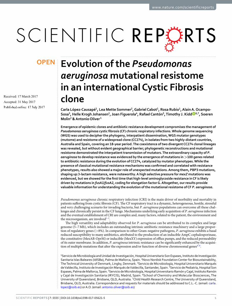
1Scientific RepoRts | 7: 5555 | DOI:10.1038/s41598-017-05621-5
www.nature.com/scientificreports
Evolution of the Pseudomonas aeruginosa mutational resistome in an international Cystic Fibrosis cloneCarla López-Causapé1, Lea Mette Sommer2, Gabriel Cabot1, Rosa Rubio1, Alain A. Ocampo-Sosa3, Helle Krogh Johansen2, Joan Figuerola4, Rafael Cantón5, Timothy J. Kidd 6,7, Soeren Molin2 & Antonio Oliver 1
Emergence of epidemic clones and antibiotic resistance development compromises the management of Pseudomonas aeruginosa cystic fibrosis (CF) chronic respiratory infections. Whole genome sequencing (WGS) was used to decipher the phylogeny, interpatient dissemination, WGS mutator genotypes (mutome) and resistome of a widespread clone (CC274), in isolates from two highly-distant countries, Australia and Spain, covering an 18-year period. The coexistence of two divergent CC274 clonal lineages was revealed, but without evident geographical barrier; phylogenetic reconstructions and mutational resistome demonstrated the interpatient transmission of mutators. The extraordinary capacity of P. aeruginosa to develop resistance was evidenced by the emergence of mutations in >100 genes related to antibiotic resistance during the evolution of CC274, catalyzed by mutator phenotypes. While the presence of classical mutational resistance mechanisms was confirmed and correlated with resistance phenotypes, results also showed a major role of unexpected mutations. Among them, PBP3 mutations, shaping up β-lactam resistance, were noteworthy. A high selective pressure for mexZ mutations was evidenced, but we showed for the first time that high-level aminoglycoside resistance in CF is likely driven by mutations in fusA1/fusA2, coding for elongation factor G. Altogether, our results provide valuable information for understanding the evolution of the mutational resistome of CF P. aeruginosa.
Pseudomonas aeruginosa chronic respiratory infection (CRI) is the main driver of morbidity and mortality in patients suffering from cystic fibrosis (CF). The CF respiratory tract is a dynamic, heterogeneous, hostile, stressful and very challenging scenario for invading bacteria, but P. aeruginosa populations can overcome all these chal-lenges and chronically persist in the CF lungs. Mechanisms underlying early acquisition of P. aeruginosa infection and the eventual establishment of CRI are complex and, many factors, related to the patient, the environment and the microorganism, are involved1–3.
The high versatility and adaptability observed for P. aeruginosa can be attributed to its complex and large genome (5–7 Mb), which includes an outstanding intrinsic antibiotic resistance machinery and a large propor-tion of regulatory genes (>8%). In comparison to other Gram-negative pathogens, P. aeruginosa exhibits a basal reduced susceptibility to many antibiotics, attributed to the production of an inducible AmpC cephalosporinase, the constitutive (MexAB-OprM) or inducible (MexXY) expression of efflux pumps, and the reduced permeability of its outer membrane. In addition, P. aeruginosa intrinsic resistance can be significantly enhanced by the acquisi-tion of multiple mutations that alter the expression and/or function of diverse chromosomal genes4–6.
1Servicio de Microbiología and Unidad de Investigación, Hospital Universitario Son Espases, Instituto de Investigación Sanitaria Islas Baleares (IdISBa), Palma de Mallorca, Spain. 2Novo Nordisk Foundation Center for Biosustainability, The Technical University of Denmark, Lingby, Denmark. 3Servicio de Microbiología, Hospital Universitario Marqués de Valdecilla, Instituto de Investigación Marqués de Valdecilla, Santander, Spain. 4Servicio de Pediatría, Hospital Son Espases, Palma de Mallorca, Spain. 5Servicio de Microbiología, Hospital Universitario Ramón y Cajal, Instituto Ramón y Cajal de Investigación Sanitaria (IRYCIS), Madrid, Spain. 6School of Chemistry and Molecular Biosciences, The University of Queensland, Brisbane, QLD, Australia. 7Child Health Research Centre, The University of Queensland, Brisbane, QLD, Australia. Correspondence and requests for materials should be addressed to C.L.-C. (email: [email protected]) or A.O. (email: [email protected])
Received: 17 March 2017
Accepted: 31 May 2017
Published: xx xx xxxx
OPEN
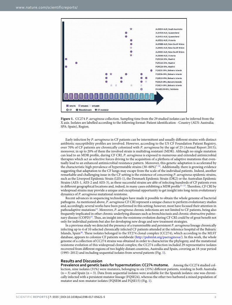
www.nature.com/scientificreports/
2Scientific RepoRts | 7: 5555 | DOI:10.1038/s41598-017-05621-5
Early infection by P. aeruginosa in CF patients can be intermittent and usually different strains with distinct antibiotic susceptibility profiles are involved. However, according to the US CF Foundation Patient Registry, over 70% of CF patients are chronically colonized with P. aeruginosa by the age of 25 (Annual Report 2015); moreover, in up to 20% of them the involved strain is multidrug resistant (MDR). Although no single mutation can lead to an MDR profile, during CF CRI, P. aeruginosa is exposed to numerous and extended antimicrobial therapies which act as selective forces driving to the acquisition of a plethora of adaptive mutations that even-tually lead to an enhanced antimicrobial resistance pattern. Moreover, this genetic adaptation is accelerated by the characteristic high prevalence of hypermutable strains (30–60%)7–10. Additionally, there is growing evidence suggesting that adaptation to the CF lungs may escape from the scale of the individual patients. Indeed, another remarkable and challenging issue in the CF setting is the existence of concerning P. aeruginosa epidemic strains, such as the Liverpool Epidemic Strain (LES-1), the Denmark Epidemic Strain (DK2) or the Australian Epidemic Strains (AES-1, AES-2 and AES-3), as these successful strains are able of infecting hundreds of CF patients even in different geographical locations and, indeed, in many cases exhibiting a MDR profile11–13. Therefore, CF CRI by widespread strains may provide a unique and exceptional opportunity to get insight into long-term evolutionary dynamics of P. aeruginosa mutational resistome.
Recent advances in sequencing technologies have made it possible to obtain the whole genome of bacterial pathogens. As mentioned above, P. aeruginosa CF CRI represent a unique chance to perform evolutionary studies and, accordingly, several works have been performed in this setting; however, most have focused their attention in pathoadaptive mutations14. Moreover, P. aeruginosa chronic infections are not limited to CF patients, being also frequently implicated in other chronic underlying diseases such as bronchiectasis and chronic obstructive pulmo-nary disease (COPD)15. Thus, an insight into the resistome evolution during CF CRI could be of great benefit not only for individual patients but also for developing new drugs and new treatment strategies.
In a previous study we detected the presence of a transmissible and persistent P. aeruginosa lineage chronically infecting up to 4 of 10 selected chronically infected CF patients attended at the reference hospital of the Balearic Islands, Spain16. These isolates belonged to the ST274 clonal complex (CC274), which according to the MLST database, appears to colonize CF patients worldwide (http://pubmlst.org/paeruginosa/). In this work, the whole genome of a collection of CC274 strains was obtained in order to characterize the phylogeny, and the mutational resistome evolution of this widespread clonal complex; the CC274 collection included 29 representative isolates recovered from different regions of two highly distant countries, Australia and Spain, covering an 18-year period (1995–2012) and including sequential isolates from several patients (Fig. 1).
Results and DiscussionPrevalence and genetic basis for hypermutation: CC274 mutome. Among the CC274 studied col-lection, nine isolates (31%) were mutators, belonging to six (35%) different patients, residing in both Australia (n = 3) and Spain (n = 3). Data from sequential isolates were available for the Spanish isolates: one was chroni-cally infected with a persistent mutator lineage (FQSE24), whereas the other two harbored a mixed population of mutator and non-mutator isolates (FQSE06 and FQSE15) (Fig. 1).
Figure 1. CC274 P. aeruginosa collection. Sampling time from the 29 studied isolates can be inferred from the X axis. Isolates are labelled according to the following format: Patient identification - Country (AUS: Australia; SPA: Spain), Region.

www.nature.com/scientificreports/
3Scientific RepoRts | 7: 5555 | DOI:10.1038/s41598-017-05621-5
In order to evaluate the genetic basis of hypermutation, complementation studies with plasmids harboring wild-type Mismatch Repair system (MMR) genes (mutS and mutL) were performed in mutator isolates from these six patients. As shown in Table 1, wild-type rifampicin resistance mutation frequencies were restored in all mutator isolates upon mutS or mutL complementation, which correlated in all cases with the presence of specific mutations in these genes, documented through whole-genome sequencing. The three Australian muta-tor isolates showed unique mutations in either mutL and mutS. Interestingly, all mutator isolates from the three Spanish patients were found to share the same inactivating mutation in mutS. On the other hand, while mutator phenotypes could be explained in all cases by specific mutations in MMR genes, the contrary was not always true, since one of the non-mutator isolates showed a missense mutation in mutS. Moreover, the presence of poly-morphisms in other mutator genes was frequent, but showed no association with mutator phenotypes (Table 1). Overall, the prevalence and genetic basis of hypermutation in CC274 was similar to that previously documented for non-clonal CF populations9, 10; this study is however, to our knowledge, the first investigating the genetic basis of hypermutation from whole genome sequence data, through the analysis of the sequence of an exhaustive panel of so called mutator genes, thus designated mutome.
Phylogeny and interpatient dissemination of the international CC274 CF clone. Pulsed Field Gel Electrophoresis (PFGE) macrorestriction patterns indicated that all isolates were clonally related, including muta-tors, which were indistinguishable from non-mutators. When an UPGMA (Unweighted Pair Group Method with Arithmetic Mean) dendrogram was constructed based on PFGE patterns, all isolates from the Balearic Islands clustered together in the same branch, although patterns from one of the patients (FQSE10) were slightly differ-ent. In contrast, Australian isolates were less clonal and clustered in different branches (Supplementary Fig. S1).
Conversely, by Multi Locus Sequence Typing (MLST), two new and closely ST274-related sequence types (ST) were detected. Discrepant MLST and PFGE results were linked, directly or indirectly, to the emergence of a muta-tor phenotype, an event that has already been documented in the CF context16–18. Mutators from patients FQSE15 and FQSE24 differed from ST274 by only two point mutations in two of the MLST alleles (acsA and guaA) leading
Isolate IDa ST Mutator?Complement with
Sequence variation in mutator genes (mutome)b
ung mfd mutS sodB mutT sodM mutL mutM oxyR polA
AUS034 274 Yes mutL E236D R631C D61N L132P D876E
AUS410 274 No — E25V D876E
AUS411 274 No — E236D D61N D876E
AUS531 274 No — E236D D61N D876E
AUS588 274 No — E25V D876E
AUS601 1043 Yes mutL S13R E25V P159S H288Y F106L H219Y D876E
AUS603 274 No — E25V D876E
AUS690 274 Yes mutS Q1123H C224R T287P E236D D61N D876E
FQRC10 274 No — E236D D61N D876E
FQRC15 274 No — E236D D61N D876E
FQRC26 274 No — E236D D61N D876E
FQSE03 274 No — L374V E236D D61N D876E
FQSE06-0403 274 No — E236D D61N D876E
FQSE06-1104 274 Yes mutS Nt814Δ4 E236D D61N D876E
FQSE06-0807 274 No — E236D D61N D876E
FQSE06-0610 274 No — E236D D61N D876E
FQSE10-0503 274 No — E236D D61N D876E
FQSE10-0106 274 No — E236D D61N D876E
FQSE10-0110 274 No — E236D D61N D876E
FQSE10-0111 274 No — E236D D61N D876E
FQSE15-0803 274 No — E236D D61N D876E
FQSE15-0906 274 No — E236D D61N D876E
FQSE15-0310 274 No — E236D D61N D876E
FQSE15-1110 1089 Yes mutS A868T Nt814Δ4 E236D D61N D876E
FQSE24-0304 1089 Yes mutS Nt814Δ4 E236D D61N D876E
FQSE24-1005 1089 Yes mutS Nt814Δ4 E236D D61N D876E
FQSE24-0308 1089 Yes mutS Nt814Δ4 E236D D61N D876E
FQSE24-1010 1089 Yes mutS Nt814Δ4 E236D D61N D876E
PAMB148 274 No — E236D L202R D61N D876E
Table 1. Mutator phenotype and genetic basis of hypermutation in CC274. aIsolates are labelled according to the following format: Patient identification - MMYY isolation code in the case of sequential isolates. bSequence variations respect to those of PAO1. No mutations were found in other genes associated with mutator phenotypes, including pfpI, mutY, dnaQ, PA2583, PA2819.1, PA2819.2, radA and uvrD.
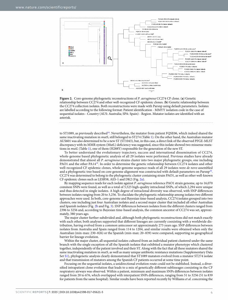
www.nature.com/scientificreports/
4Scientific RepoRts | 7: 5555 | DOI:10.1038/s41598-017-05621-5
to ST1089, as previously described16. Nevertheless, the mutator from patient FQSE06, which indeed shared the same inactivating mutation in mutS, still belonged to ST274 (Table 1). On the other hand, the Australian mutator AUS601 was also determined to be a new ST (ST1043), but, in this case, a direct link of the observed PFGE-MLST discrepancy with its MMR system (MutL) deficiency was suggested, since this isolate showed two missense muta-tions in mutL (Table 1), one of them (H288Y) responsible for the generation of the new ST.
To better understand the evolutionary trajectory, success and international dissemination of CC274, whole-genome based phylogenetic analysis of all 29 isolates were performed. Previous studies have already demonstrated that almost all P. aeruginosa strains cluster into two major phylogenetic groups, one including PAO1 and the other PA1419. In order to determine the genetic relationship between CC274 isolates and other well-recognized CF epidemic clones, whole-genome sequence reads of all 29 isolates were de novo assembled and a phylogenetic tree based on core genome alignment was constructed with default parameters on Parsnp20. CC274 was determined to belong to the phylogenetic cluster containing strain PAO1, as well as other well-known CF epidemic clones such as LESB58, AES-1 and DK2 (Fig. 2a).
By mapping sequence reads for each isolate against P. aeruginosa reference PAO1 strain genome, up to 16,070 common SNPs were found, as well as a total of 5,525 high-quality intraclonal SNPs, of which 2,294 were unique and thus detected in single isolates. A high degree of intraclonal diversity was observed, with SNP differences between isolates ranging from 20 to 3,256. To elucidate the phylogenetic relationship among isolates two different approaches were used. In both, core-genome and Bayesian time-based analysis, CC274 isolates grouped into two clusters, one including just four Australian isolates and a second major cluster that included all other Australian and Spanish isolates (Fig. 2b and Fig. 3). SNP differences between isolates from the different clusters ranged from 2396 to 3256 and, according to Bayesian time-based analysis, the common ancestor of CC274 was set, approxi-mately, 380 years ago.
The major cluster further subdivided and, although both phylogenetic reconstructions did not match exactly with each other, both analyses supported that different lineages are currently coexisting with a worldwide dis-tribution, having evolved from a common antecessor set approximately 275 years ago. SNP differences between isolates from Australia and Spain ranged from 114 to 1204, and similar results were obtained when only the Australian (min-max: 230–826) or the Spanish (min-max: 20–839) were compared, supporting no geographical barrier for lineage evolution.
Within the major cluster, all sequential isolates cultured from an individual patient clustered under the same branch with the single exception of all the Spanish isolates that exhibited a mutator phenotype which clustered together, independently of the patient involved and their ST. Along with the fact that all these mutators shared the same inactivating mutation in mutS, as well as many unique antibiotic resistance mutations (Supplementary Data Set S1), phylogenetic analysis clearly demonstrated that ST1089 mutators evolved from a mutator ST274 isolate and that transmission of mutators among the Spanish CF patients occurred at some time point.
Focusing on the sequential isolates, a unidirectional evolution route could not be stablished. Instead, a diver-sified intrapatient clone evolution that leads to a mix of genetically different sublineages coexisting in the CF respiratory airways was observed. Within a patient, minimum and maximum SNPs differences between isolates ranged from 20 to 676, which overlapped with interpatient SNPs differences, ranging from 51 to 3256 (51 to 839 for patients from the same hospital). Similar results have been reported recently by Williams et al. concerning the
Figure 2. Core-genome phylogenetic reconstructions of P. aeruginosa CC274 CF clone. (a) Genetic relationship between CC274 and other well-recognized CF epidemic clones. (b) Genetic relationship between the CC274 collection isolates. Both reconstructions were made with Parsnp using default parameters. Isolates are labelled according to the following format: Patient identification - MMYY isolation code in the case of sequential isolates - Country (AUS: Australia; SPA: Spain) - Region. Mutator isolates are identified with an asterisk.
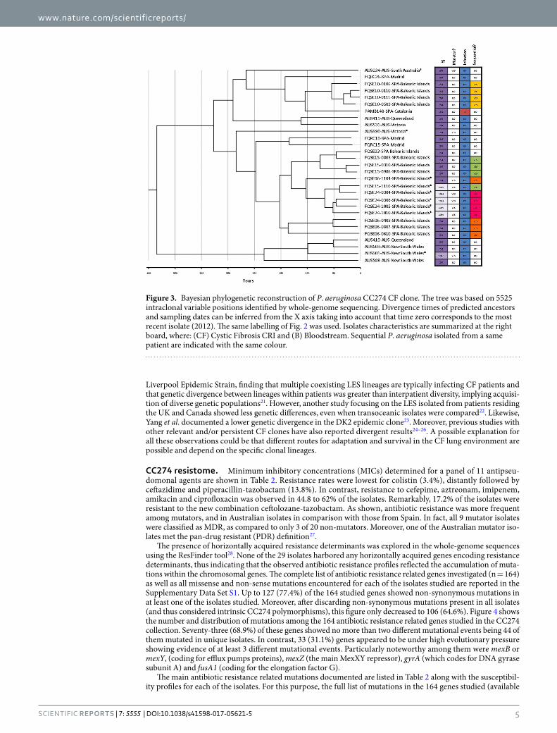
www.nature.com/scientificreports/
5Scientific RepoRts | 7: 5555 | DOI:10.1038/s41598-017-05621-5
Liverpool Epidemic Strain, finding that multiple coexisting LES lineages are typically infecting CF patients and that genetic divergence between lineages within patients was greater than interpatient diversity, implying acquisi-tion of diverse genetic populations21. However, another study focusing on the LES isolated from patients residing the UK and Canada showed less genetic differences, even when transoceanic isolates were compared22. Likewise, Yang et al. documented a lower genetic divergence in the DK2 epidemic clone23. Moreover, previous studies with other relevant and/or persistent CF clones have also reported divergent results24–26. A possible explanation for all these observations could be that different routes for adaptation and survival in the CF lung environment are possible and depend on the specific clonal lineages.
CC274 resistome. Minimum inhibitory concentrations (MICs) determined for a panel of 11 antipseu-domonal agents are shown in Table 2. Resistance rates were lowest for colistin (3.4%), distantly followed by ceftazidime and piperacillin-tazobactam (13.8%). In contrast, resistance to cefepime, aztreonam, imipenem, amikacin and ciprofloxacin was observed in 44.8 to 62% of the isolates. Remarkably, 17.2% of the isolates were resistant to the new combination ceftolozane-tazobactam. As shown, antibiotic resistance was more frequent among mutators, and in Australian isolates in comparison with those from Spain. In fact, all 9 mutator isolates were classified as MDR, as compared to only 3 of 20 non-mutators. Moreover, one of the Australian mutator iso-lates met the pan-drug resistant (PDR) definition27.
The presence of horizontally acquired resistance determinants was explored in the whole-genome sequences using the ResFinder tool28. None of the 29 isolates harbored any horizontally acquired genes encoding resistance determinants, thus indicating that the observed antibiotic resistance profiles reflected the accumulation of muta-tions within the chromosomal genes. The complete list of antibiotic resistance related genes investigated (n = 164) as well as all missense and non-sense mutations encountered for each of the isolates studied are reported in the Supplementary Data Set S1. Up to 127 (77.4%) of the 164 studied genes showed non-synonymous mutations in at least one of the isolates studied. Moreover, after discarding non-synonymous mutations present in all isolates (and thus considered intrinsic CC274 polymorphisms), this figure only decreased to 106 (64.6%). Figure 4 shows the number and distribution of mutations among the 164 antibiotic resistance related genes studied in the CC274 collection. Seventy-three (68.9%) of these genes showed no more than two different mutational events being 44 of them mutated in unique isolates. In contrast, 33 (31.1%) genes appeared to be under high evolutionary pressure showing evidence of at least 3 different mutational events. Particularly noteworthy among them were mexB or mexY, (coding for efflux pumps proteins), mexZ (the main MexXY repressor), gyrA (which codes for DNA gyrase subunit A) and fusA1 (coding for the elongation factor G).
The main antibiotic resistance related mutations documented are listed in Table 2 along with the susceptibil-ity profiles for each of the isolates. For this purpose, the full list of mutations in the 164 genes studied (available
Figure 3. Bayesian phylogenetic reconstruction of P. aeruginosa CC274 CF clone. The tree was based on 5525 intraclonal variable positions identified by whole-genome sequencing. Divergence times of predicted ancestors and sampling dates can be inferred from the X axis taking into account that time zero corresponds to the most recent isolate (2012). The same labelling of Fig. 2 was used. Isolates characteristics are summarized at the right board, where: (CF) Cystic Fibrosis CRI and (B) Bloodstream. Sequential P. aeruginosa isolated from a same patient are indicated with the same colour.
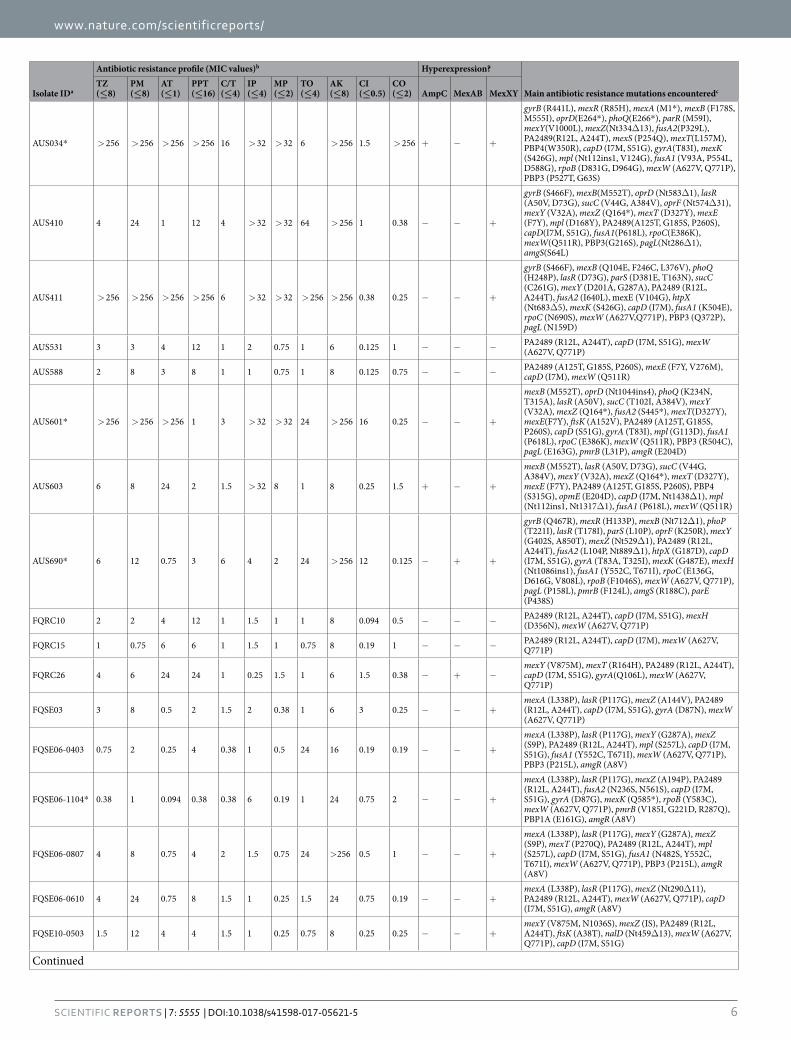
www.nature.com/scientificreports/
6Scientific RepoRts | 7: 5555 | DOI:10.1038/s41598-017-05621-5
Isolate IDa
Antibiotic resistance profile (MIC values)b Hyperexpression?
Main antibiotic resistance mutations encounteredcTZ (≤8)
PM (≤8)
AT (≤1)
PPT (≤16)
C/T (≤4)
IP (≤4)
MP (≤2)
TO (≤4)
AK (≤8)
CI (≤0.5)
CO (≤2) AmpC MexAB MexXY
AUS034* > 256 > 256 > 256 > 256 16 > 32 > 32 6 > 256 1.5 > 256 + − +
gyrB (R441L), mexR (R85H), mexA (M1*), mexB (F178S, M555I), oprD(E264*), phoQ(E266*), parR (M59I), mexY(V1000L), mexZ(Nt334Δ13), fusA2(P329L), PA2489(R12L, A244T), mexS (P254Q), mexT(L157M), PBP4(W350R), capD (I7M, S51G), gyrA(T83I), mexK (S426G), mpl (Nt112ins1, V124G), fusA1 (V93A, P554L, D588G), rpoB (D831G, D964G), mexW (A627V, Q771P), PBP3 (P527T, G63S)
AUS410 4 24 1 12 4 > 32 > 32 64 > 256 1 0.38 − − +
gyrB (S466F), mexB(M552T), oprD (Nt583Δ1), lasR (A50V, D73G), sucC (V44G, A384V), oprF (Nt574Δ31), mexY (V32A), mexZ (Q164*), mexT (D327Y), mexE (F7Y), mpl (D168Y), PA2489(A125T, G185S, P260S), capD(I7M, S51G), fusA1(P618L), rpoC(E386K), mexW(Q511R), PBP3(G216S), pagL(Nt286Δ1), amgS(S64L)
AUS411 > 256 > 256 > 256 > 256 6 > 32 > 32 > 256 > 256 0.38 0.25 − − +
gyrB (S466F), mexB (Q104E, F246C, L376V), phoQ (H248P), lasR (D73G), parS (D381E, T163N), sucC (C261G), mexY (D201A, G287A), PA2489 (R12L, A244T), fusA2 (I640L), mexE (V104G), htpX (Nt683Δ5), mexK (S426G), capD (I7M), fusA1 (K504E), rpoC (N690S), mexW (A627V,Q771P), PBP3 (Q372P), pagL (N159D)
AUS531 3 3 4 12 1 2 0.75 1 6 0.125 1 − − − PA2489 (R12L, A244T), capD (I7M, S51G), mexW (A627V, Q771P)
AUS588 2 8 3 8 1 1 0.75 1 8 0.125 0.75 − − − PA2489 (A125T, G185S, P260S), mexE (F7Y, V276M), capD (I7M), mexW (Q511R)
AUS601* > 256 > 256 > 256 1 3 > 32 > 32 24 > 256 16 0.25 − − +
mexB (M552T), oprD (Nt1044ins4), phoQ (K234N, T315A), lasR (A50V), sucC (T102I, A384V), mexY (V32A), mexZ (Q164*), fusA2 (S445*), mexT(D327Y), mexE(F7Y), ftsK (A152V), PA2489 (A125T, G185S, P260S), capD (S51G), gyrA (T83I), mpl (G113D), fusA1 (P618L), rpoC (E386K), mexW (Q511R), PBP3 (R504C), pagL (E163G), pmrB (L31P), amgR (E204D)
AUS603 6 8 24 2 1.5 > 32 8 1 8 0.25 1.5 + − +
mexB (M552T), lasR (A50V, D73G), sucC (V44G, A384V), mexY (V32A), mexZ (Q164*), mexT (D327Y), mexE (F7Y), PA2489 (A125T, G185S, P260S), PBP4 (S315G), opmE (E204D), capD (I7M, Nt1438Δ1), mpl (Nt112ins1, Nt1317Δ1), fusA1 (P618L), mexW (Q511R)
AUS690* 6 12 0.75 3 6 4 2 24 > 256 12 0.125 − + +
gyrB (Q467R), mexR (H133P), mexB (Nt712Δ1), phoP (T221I), lasR (T178I), parS (L10P), oprF (K250R), mexY (G402S, A850T), mexZ (Nt529Δ1), PA2489 (R12L, A244T), fusA2 (L104P, Nt889Δ1), htpX (G187D), capD (I7M, S51G), gyrA (T83A, T325I), mexK (G487E), mexH (Nt1086ins1), fusA1 (Y552C, T671I), rpoC (E136G, D616G, V808L), rpoB (F1046S), mexW (A627V, Q771P), pagL (P158L), pmrB (F124L), amgS (R188C), parE (P438S)
FQRC10 2 2 4 12 1 1.5 1 1 8 0.094 0.5 − − − PA2489 (R12L, A244T), capD (I7M, S51G), mexH (D356N), mexW (A627V, Q771P)
FQRC15 1 0.75 6 6 1 1.5 1 0.75 8 0.19 1 − − − PA2489 (R12L, A244T), capD (I7M), mexW (A627V, Q771P)
FQRC26 4 6 24 24 1 0.25 1.5 1 6 1.5 0.38 − + −mexY (V875M), mexT (R164H), PA2489 (R12L, A244T), capD (I7M, S51G), gyrA(Q106L), mexW (A627V, Q771P)
FQSE03 3 8 0.5 2 1.5 2 0.38 1 6 3 0.25 − − +mexA (L338P), lasR (P117G), mexZ (A144V), PA2489 (R12L, A244T), capD (I7M, S51G), gyrA (D87N), mexW (A627V, Q771P)
FQSE06-0403 0.75 2 0.25 4 0.38 1 0.5 24 16 0.19 0.19 − − +
mexA (L338P), lasR (P117G), mexY (G287A), mexZ (S9P), PA2489 (R12L, A244T), mpl (S257L), capD (I7M, S51G), fusA1 (Y552C, T671I), mexW (A627V, Q771P), PBP3 (P215L), amgR (A8V)
FQSE06-1104* 0.38 1 0.094 0.38 0.38 6 0.19 1 24 0.75 2 − − +
mexA (L338P), lasR (P117G), mexZ (A194P), PA2489 (R12L, A244T), fusA2 (N236S, N561S), capD (I7M, S51G), gyrA (D87G), mexK (Q585*), rpoB (Y583C), mexW (A627V, Q771P), pmrB (V185I, G221D, R287Q), PBP1A (E161G), amgR (A8V)
FQSE06-0807 4 8 0.75 4 2 1.5 0.75 24 >256 0.5 1 − − +
mexA (L338P), lasR (P117G), mexY (G287A), mexZ (S9P), mexT (P270Q), PA2489 (R12L, A244T), mpl (S257L), capD (I7M, S51G), fusA1 (N482S, Y552C, T671I), mexW (A627V, Q771P), PBP3 (P215L), amgR (A8V)
FQSE06-0610 4 24 0.75 8 1.5 1 0.25 1.5 24 0.75 0.19 − − +mexA (L338P), lasR (P117G), mexZ (Nt290Δ11), PA2489 (R12L, A244T), mexW (A627V, Q771P), capD (I7M, S51G), amgR (A8V)
FQSE10-0503 1.5 12 4 4 1.5 1 0.25 0.75 8 0.25 0.25 − − +mexY (V875M, N1036S), mexZ (IS), PA2489 (R12L, A244T), ftsK (A38T), nalD (Nt459Δ13), mexW (A627V, Q771P), capD (I7M, S51G)
Continued
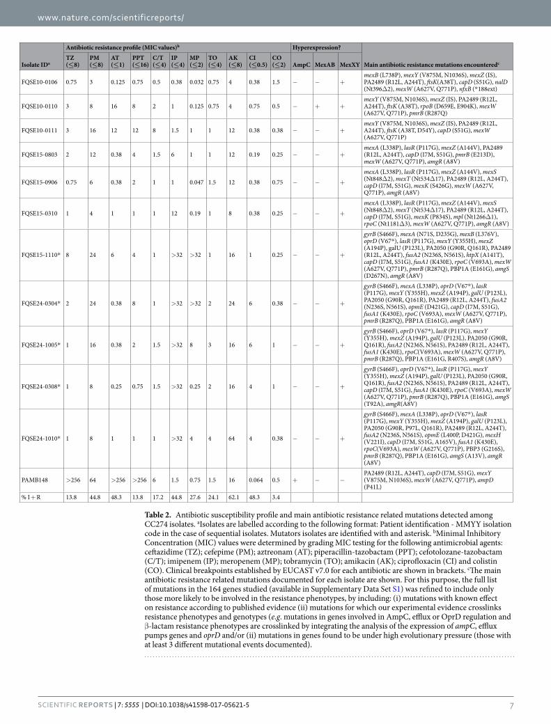
www.nature.com/scientificreports/
7Scientific RepoRts | 7: 5555 | DOI:10.1038/s41598-017-05621-5
Isolate IDa
Antibiotic resistance profile (MIC values)b Hyperexpression?
Main antibiotic resistance mutations encounteredcTZ (≤8)
PM (≤8)
AT (≤1)
PPT (≤16)
C/T (≤4)
IP (≤4)
MP (≤2)
TO (≤4)
AK (≤8)
CI (≤0.5)
CO (≤2) AmpC MexAB MexXY
FQSE10-0106 0.75 3 0.125 0.75 0.5 0.38 0.032 0.75 4 0.38 1.5 − − +mexB (L738P), mexY (V875M, N1036S), mexZ (IS), PA2489 (R12L, A244T), ftsK(A38T), capD (S51G), nalD (Nt396Δ2), mexW (A627V, Q771P), nfxB (*188ext)
FQSE10-0110 3 8 16 8 2 1 0.125 0.75 4 0.75 0.5 − + +mexY (V875M, N1036S), mexZ (IS), PA2489 (R12L, A244T), ftsK (A38T), rpoB (D659E, E904K), mexW (A627V, Q771P), pmrB (R287Q)
FQSE10-0111 3 16 12 12 8 1.5 1 1 12 0.38 0.38 − − +mexY (V875M, N1036S), mexZ (IS), PA2489 (R12L, A244T), ftsK (A38T, D54Y), capD (S51G), mexW (A627V, Q771P)
FQSE15-0803 2 12 0.38 4 1.5 6 1 1 12 0.19 0.25 − − +mexA (L338P), lasR (P117G), mexZ (A144V), PA2489 (R12L, A244T), capD (I7M, S51G), pmrB (E213D), mexW (A627V, Q771P), amgR (A8V)
FQSE15-0906 0.75 6 0.38 2 1 1 0.047 1.5 12 0.38 0.75 − − +
mexA (L338P), lasR (P117G), mexZ (A144V), mexS (Nt848Δ2), mexT (Nt534Δ17), PA2489 (R12L, A244T), capD (I7M, S51G), mexK (S426G), mexW (A627V, Q771P), amgR (A8V)
FQSE15-0310 1 4 1 1 1 12 0.19 1 8 0.38 0.25 − − +
mexA (L338P), lasR (P117G), mexZ (A144V), mexS (Nt848Δ2), mexT (Nt534Δ17), PA2489 (R12L, A244T), capD (I7M, S51G), mexK (P834S), mpl (Nt1266Δ1), rpoC (Nt1181Δ3), mexW (A627V, Q771P), amgR (A8V)
FQSE15-1110* 8 24 6 4 1 >32 >32 1 16 1 0.25 − − +
gyrB (S466F), mexA (N71S, D235G), mexB (L376V), oprD (V67*), lasR (P117G), mexY (Y355H), mexZ (A194P), galU (P123L), PA2050 (G90R, Q161R), PA2489 (R12L, A244T), fusA2 (N236S, N561S), htpX (A141T), capD (I7M, S51G), fusA1 (K430E), rpoC (V693A), mexW (A627V, Q771P), pmrB (R287Q), PBP1A (E161G), amgS (D267N), amgR (A8V)
FQSE24-0304* 2 24 0.38 8 1 >32 >32 2 24 6 0.38 − − +
gyrB (S466F), mexA (L338P), oprD (V67*), lasR (P117G), mexY (Y355H), mexZ (A194P), galU (P123L), PA2050 (G90R, Q161R), PA2489 (R12L, A244T), fusA2 (N236S, N561S), opmE (D421G), capD (I7M, S51G), fusA1 (K430E), rpoC (V693A), mexW (A627V, Q771P), pmrB (R287Q), PBP1A (E161G), amgR (A8V)
FQSE24-1005* 1 16 0.38 2 1.5 >32 8 3 16 6 1 − − +
gyrB (S466F), oprD (V67*), lasR (P117G), mexY (Y355H), mexZ (A194P), galU (P123L), PA2050 (G90R, Q161R), fusA2 (N236S, N561S), PA2489 (R12L, A244T), fusA1 (K430E), rpoC(V693A), mexW (A627V, Q771P), pmrB (R287Q), PBP1A (E161G, R407S), amgR (A8V)
FQSE24-0308* 1 8 0.25 0.75 1.5 >32 0.25 2 16 4 1 − − +
gyrB (S466F), oprD (V67*), lasR (P117G), mexY (Y355H), mexZ (A194P), galU (P123L), PA2050 (G90R, Q161R), fusA2 (N236S, N561S), PA2489 (R12L, A244T), capD (I7M, S51G), fusA1 (K430E), rpoC (V693A), mexW (A627V, Q771P), pmrB (R287Q), PBP1A (E161G), amgS (T92A), amgR(A8V)
FQSE24-1010* 1 8 1 1 1 >32 4 4 64 4 0.38 − − +
gyrB (S466F), mexA (L338P), oprD (V67*), lasR (P117G), mexY (Y355H), mexZ (A194P), galU (P123L), PA2050 (G90R, P97L, Q161R), PA2489 (R12L, A244T), fusA2 (N236S, N561S), opmE (L400P, D421G), mexH (V221I), capD (I7M, S51G, A165V), fusA1 (K430E), rpoC(V693A), mexW (A627V, Q771P), PBP3 (G216S), pmrB (R287Q), PBP1A (E161G), amgS (A13V), amgR (A8V)
PAMB148 >256 64 >256 >256 6 1.5 0.75 1.5 16 0.064 0.5 + − −PA2489 (R12L, A244T), capD (I7M, S51G), mexY (V875M, N1036S), mexW (A627V, Q771P), ampD (P41L)
% I + R 13.8 44.8 48.3 13.8 17.2 44.8 27.6 24.1 62.1 48.3 3.4
Table 2. Antibiotic susceptibility profile and main antibiotic resistance related mutations detected among CC274 isolates. aIsolates are labelled according to the following format: Patient identification - MMYY isolation code in the case of sequential isolates. Mutators isolates are identified with and asterisk. bMinimal Inhibitory Concentration (MIC) values were determined by grading MIC testing for the following antimicrobial agents: ceftazidime (TZ); cefepime (PM); aztreonam (AT); piperacillin-tazobactam (PPT); cefotolozane-tazobactam (C/T); imipenem (IP); meropenem (MP); tobramycin (TO); amikacin (AK); ciprofloxacin (CI) and colistin (CO). Clinical breakpoints established by EUCAST v7.0 for each antibiotic are shown in brackets. cThe main antibiotic resistance related mutations documented for each isolate are shown. For this purpose, the full list of mutations in the 164 genes studied (available in Supplementary Data Set S1) was refined to include only those more likely to be involved in the resistance phenotypes, by including: (i) mutations with known effect on resistance according to published evidence (ii) mutations for which our experimental evidence crosslinks resistance phenotypes and genotypes (e.g. mutations in genes involved in AmpC, efflux or OprD regulation and β-lactam resistance phenotypes are crosslinked by integrating the analysis of the expression of ampC, efflux pumps genes and oprD and/or (ii) mutations in genes found to be under high evolutionary pressure (those with at least 3 different mutational events documented).
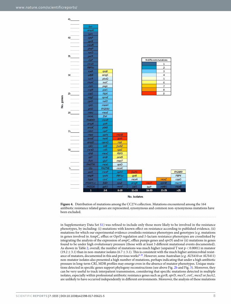
www.nature.com/scientificreports/
8Scientific RepoRts | 7: 5555 | DOI:10.1038/s41598-017-05621-5
in Supplementary Data Set S1) was refined to include only those more likely to be involved in the resistance phenotypes, by including: (i) mutations with known effect on resistance according to published evidence, (ii) mutations for which our experimental evidence crosslinks resistance phenotypes and genotypes (e.g. mutations in genes involved in AmpC, efflux or OprD regulation and β-lactam resistance phenotypes are crosslinked by integrating the analysis of the expression of ampC, efflux pumps genes and oprD) and/or (ii) mutations in genes found to be under high evolutionary pressure (those with at least 3 different mutational events documented). As shown in Table 2, overall, the number of mutations was much higher (unpaired T test p < 0.0001) in mutator (19.2 ± 3.1) than in non-mutator isolates (6.7 ± 3.1). This is consistent with the much higher antimicrobial resist-ance of mutators, documented in this and previous works8, 29. However, some Australian (e.g. AUS410 or AUS411) non-mutator isolates also presented a high number of mutations, perhaps indicating that under a high antibiotic pressure in long-term CRI, MDR profiles may emerge even in the absence of mutator phenotypes. Unique muta-tions detected in specific genes support phylogeny reconstructions (see above Fig. 2b and Fig. 3). Moreover, they can be very useful to track interpatient transmission, considering that specific mutations detected in multiple isolates, especially within professional antibiotic resistance genes such as gyrB, oprD, mexY, creC, mexZ or fusA2, are unlikely to have occurred independently in different environments. Moreover, the analysis of these mutations
Figure 4. Distribution of mutations among the CC274 collection. Mutations encountered among the 164 antibiotic resistance related genes are represented, synonymous and common non-synonymous mutations have been excluded.
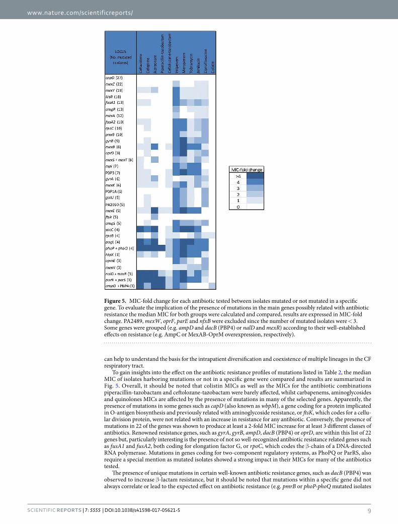
www.nature.com/scientificreports/
9Scientific RepoRts | 7: 5555 | DOI:10.1038/s41598-017-05621-5
can help to understand the basis for the intrapatient diversification and coexistence of multiple lineages in the CF respiratory tract.
To gain insights into the effect on the antibiotic resistance profiles of mutations listed in Table 2, the median MIC of isolates harboring mutations or not in a specific gene were compared and results are summarized in Fig. 5. Overall, it should be noted that colistin MICs as well as the MICs for the antibiotic combinations piperacillin-tazobactam and ceftolozane-tazobactam were barely affected, whilst carbapenems, aminoglycosides and quinolones MICs are affected by the presence of mutations in many of the selected genes. Apparently, the presence of mutations in some genes such as capD (also known as wbpM), a gene coding for a protein implicated in O-antigen biosynthesis and previously related with aminoglycoside resistance, or ftsK, which codes for a cellu-lar division protein, were not related with an increase in resistance for any antibiotic. Conversely, the presence of mutations in 22 of the genes was shown to produce at least a 2-fold MIC increase for at least 3 different classes of antibiotics. Renowned resistance genes, such as gyrA, gyrB, ampD, dacB (PBP4) or oprD, are within this list of 22 genes but, particularly interesting is the presence of not so well-recognized antibiotic resistance related genes such as fusA1 and fusA2, both coding for elongation factor G, or rpoC, which codes the β-chain of a DNA-directed RNA polymerase. Mutations in genes coding for two-component regulatory systems, as PhoPQ or ParRS, also require a special mention as mutated isolates showed a strong impact in their MICs for many of the antibiotics tested.
The presence of unique mutations in certain well-known antibiotic resistance genes, such as dacB (PBP4) was observed to increase β-lactam resistance, but it should be noted that mutations within a specific gene did not always correlate or lead to the expected effect on antibiotic resistance (e.g. pmrB or phoP-phoQ mutated isolates
Figure 5. MIC-fold change for each antibiotic tested between isolates mutated or not mutated in a specific gene. To evaluate the implication of the presence of mutations in the main genes possibly related with antibiotic resistance the median MIC for both groups were calculated and compared, results are expressed in MIC-fold change. PA2489, mexW, oprF, parE and nfxB were excluded since the number of mutated isolates were < 3. Some genes were grouped (e.g. ampD and dacB (PBP4) or nalD and mexR) according to their well-established effects on resistance (e.g. AmpC or MexAB-OprM overexpression, respectively).
www.nature.com/scientificreports/
1 0Scientific RepoRts | 7: 5555 | DOI:10.1038/s41598-017-05621-5
did not exhibit a higher colistin MIC). Likewise, several mutations (e.g. mexZ, gyrB or oprD) were associated to extended unexpected antibiotic resistance profiles, perhaps suggesting the co-selection of different resistance mutations during P. aeruginosa evolution in CF CRI. A detailed analysis of the mutational resistome for each class of antibiotics is provided below and in the Supplementary Data Set S1.
β-lactam resistome. Overproduction of the chromosomally encoded cephalosporinase AmpC is the primary pathway for developing resistance to the antipseudomonal β-lactams, and it is driven by the selec-tion of mutations in peptidoglycan-recycling genes (ampD, dacB and ampR)30, 31. Just three isolates (AUS034, AUS603 and PAMB148) of the CC274 collection were demonstrated to overproduce AmpC (Table 2). By con-trast, at the genomic level, almost all isolates (26/29) contained some variation within dacB which codes for the penicillin-binding protein PBP4 (Supplementary Data Set S1-Betalactams). Crosslinking phenotypic and gen-otypic results through ampC expression data, suggested that most observed dacB allele variations were, in fact, ancestral polymorphisms not involved in antibiotic resistance. However, AmpC overproduction in the two CF isolates was explained by the presence of specific mutations in dacB (S315G or W350R) and by an ampD (P41L) mutation in the case of the bloodstream infection isolate PAMB148 (Table 2). Whilst ampC overexpression in iso-lates AUS034 and PAMB148 correlated well with ceftazidime and piperacillin-tazobactam resistance, this was not the case for isolate AUS603 which was documented to be susceptible to these antibiotics. However, unexpected AUS603 β-lactam susceptibility could be explained by the presence of chromosomal mutations whose effects eventually compensate the expected increase in β-lactam resistance. Indeed, this isolate showed a non-sense mutation in OprM (Q93X), the outer membrane protein of the constitutive MexAB efflux pump, which is well known to play a major role in intrinsic β-lactam resistance.
In addition, P. aeruginosa may eventually develop β-lactam resistance by acquiring mutations within their macromolecular targets: the essential penicillin-binding proteins (PBPs). While some mutations without appar-ent effect on resistance were detected in genes coding for PBP1 and PBP3a, the main mutational resistance target among PBPs was found to be PBP3, an essential high molecular class B PBP with transpeptidase activity, in agreement with recent data from CF patients32 and in vitro studies33. Indeed, we documented that PBP3 muta-tions had often occurred (7/29 isolates) among the CC274 collection (Supplementary Data Set S1-Betalactams). Nevertheless, β-lactam resistance contribution of each derived ftsI (PBP3) allele, if any, depends on the specific point mutation encountered. Missense mutations within the PBP3 (R504C and Q372P) were apparently the cause of β-lactam resistance in isolates AUS601 and AUS411, since they do not hyperproduce AmpC. Although these mutations are not located in the PBP3 active site, both are very close to two loop regions (residues 332–338 and 526–533) which play an important role in substrate recognition34. In fact, PBP3 mutations in residue 504 (R504C, R504H) have been recently described in vitro33 and among isolates from widespread nosocomial P. aeruginosa clones35, 36. Likewise, the P527T mutation of AUS034 likely contributes, together with the overex-pression of AmpC, to the very high-level β-lactam resistance of this isolate, including the new antipseudomonal combination ceftolozane-tazobactam. On the other hand, the P215L and G216S mutations were apparently not linked with phenotypic resistance, in agreement with the fact that residues 215 and 216 are not implicated in the formation and stabilization of the inactivating complex β-lactam-PBP334.
Obtained data also demonstrated that the constitutive efflux pump MexAB-OprM is under strong mutational pressure during CF CRI, frequently including inactivating mutations, which correlates with previous investiga-tions that pointed out that this efflux pump is dispensable and, therefore, tends to be lost or inactivated in favor of MexXY-OprM hyperproduction in CF P. aeruginosa subpopulations37. Our data also support this hypothesis, as just 3 isolates showed mutations in regulators leading to MexAB-OprM overexpression, whereas up to 23 isolates hyperproduced the efflux-pump MexXY-OprM (Supplementary Data Set S1 -Betalactams). Moreover, many of the isolates showed some degree of hypersusceptibiltiy to aztreonam (substrate of MexAB-OprM) in favor of an increased MIC of cefepime (substrate of MexXY-OprM) (Supplementary Data Set S1- Betalactams).
Carbapenem resistome. Imipenem and meropenem resistance correlated in all but two isolates with the presence of non-sense mutations affecting the outer membrane protein OprD (Table 2, Supplementary Data Set S1-Carbapenems). High-level meropenem resistance was additionally associated with the presence of PBP3 mutations, in agreement with recent in vitro studies showing the selection of PBP3 mutations upon meropenem exposure33. Remarkably, all ST1089 mutator isolates shared the same point mutation in oprD (V67X) as well as in galU (P123L), also related with carbapenem resistance, supporting interpatient transmission of this mutator lineage among CF patients attending the reference hospital of the Balearic Islands.
The expression of OprD is known to be modulated by mutations (mexS or mexT) leading to the overexpression of the efflux pump MexEF-OprN, and meropenem is a well-known substrate for the efflux pump MexAB-OprM. However, carbapenem resistant isolates AUS411 and AUS603 harboring a wild-type oprD allele did not over-produce neither of these two efflux pumps. Thus, the observed phenotype could be related with the presence of specific mutations within the genes coding for PBP3 (ftsI) and PBP4 (dacB), hypothesis that is current being evaluated in our laboratory.
Aminoglycoside resistome. Intravenous antimicrobial combinations including an aminoglycoside plus a fluroroquinolone or a β-lactam antibiotic are frequently used to manage CF exacerbations. Moreover, in the last decade, tobramycin inhalation has become an important contributor to CF treatment as a means to control chronic infection as well as a first-line treatment for the eradication of early acquisition of P. aeruginosa and sev-eral aminoglycoside-based inhaled formulations are currently available. Resistance to antipseudomonal amino-glycosides is frequently attributed to the presence of acquired aminoglycoside-modifying enzymes, membrane impermeability or MexXY efflux pump overexpression38. Moreover, adaptive resistance, due to MexXY efflux
www.nature.com/scientificreports/
1 1Scientific RepoRts | 7: 5555 | DOI:10.1038/s41598-017-05621-5
system overexpression, to this class of antibiotics has been well documented in the CF setting in response to sublethal concentrations39.
Among the CC274 collection, a high proportion of the isolates (23/29) were shown to overexpress MexXY and all but one were mutated in mexZ, which codes for the mayor MexXY expression regulator. In agreement with recent work, which pointed out that mutation of mexZ is part of a strongly selected evolutionary pathway40, several different mutational events were encountered within this regulator. Remarkably, the same point mutation was detected among different and independent isolates, probably indicating interpatient transmission events (Table 2, Supplementary Data Set S1-Aminoglycosides). The single MexXY-overproducing isolate showing no mutations in mexZ, presented a unique mutation in parS, a gene also involved in the modulation of MexXY expression. Nevertheless, as it has been largely observed in the CF clinical setting41, MexXY hyperproduction per se cannot explain aminoglycoside resistance in the majority of the isolates. In this sense, there is growing evi-dence that high-level resistance is a stepwise process which arises by the accumulation of several non-enzymatic mechanisms and, moreover, novel genetic resistance determinants have been proposed42–44. To our knowledge, no published work has yet investigated the in vivo contribution to aminoglycoside resistance of these novel genetic determinants proposed, and many questions remain unresolved. Thus, our work reveals for the first time that all high-level resistant isolates hyperproduced MexXY, but also harbored additional mutations in some of these genes, especially highlighting the presence of mutations in both genes coding for elongation factor G, fusA1 and fusA2 (Supplementary Data Set S1 - Aminoglycosides). In fact, fusA1 and fusA2 have been recently demonstrated to be under high evolutionary pressure in the CF environment, which can be explained in terms of a wide amino-glycoside use in this setting45.
Fluoroquinolone resistome. P. aeruginosa RND (Resistance-Nodulation-Division) efflux pumps MexAB-OprM, MexXY-OprM, MexCD-OprJ and MexEF-OprN are well-known to extrude fluoroquinolones. Nevertheless, our data suggest that the contribution of the overexpression of these efflux pumps to high-level resistance to fluoroquinolones is very limited, if any (Supplementary Data Set S1 -Fluoroquinoles). Concerning MexCD-OprJ overproduction, it has been shown that, although wild type P. aeruginosa strains generally do not express this efflux system46, hyperproducing mutants tend to emerge after both in vitro and in vivo fluoro-quinolone exposure33. Moreover, there is some data suggesting that MexCD-OprJ hyperproduction could be an advantage in the CF environment47. Among the CC274 collection, however, just 1 isolate (FQSE10-0106), show-ing aa ciprofloxacin MIC below the resistance breakpoint (MIC = 0.38 mg/L), was demonstrated to hyperproduce MexCD-OprJ due to a non-sense mutation in nfxB.
On the other hand, our data shows that high-level fluoroquinolone resistance was associated with the pres-ence of missense mutations in gyrA, gyrB and/or parC quinolone resistance-determining regions (QRDRs). Specifically, up to 9 isolates were mutated in gyrB QRDR and all but two harbored the same mutation (S466F), 6 showed mutations in gyrA QRDR (T83I, T83A, D87N, D87G and Q106L), and just one isolate was mutated in parE (P438S). Mutations in GyrA residues 83 and 87 are well-known to be relevant in the clinical setting and are frequently encountered in fluoroquinolone-resistant P. aeruginosa35, 36, being both residues situated on helix-4. Mutations in residue 106 are in the other hand very infrequent, with only one previous reference of its existence in 1 of 335 quinolone resistant P. aeruginosa clinical strains48.
Polymyxin resistome. Two component-regulatory systems as well as other genes implicated in lipopoly-saccharide biosynthesis have been related with polymyxin resistance49–51. Several in vitro works have addressed the implication of the two-component regulatory systems in polymyxin resistance development, demonstrating that individual alterations in these systems are generally not sufficient to develop high-level resistance50, 52–54 and that individual two-component systems may not be essential for acquisition of colistin (polymyxin E) resist-ance in P. aeruginosa55. In agreement with these in vitro studies, we found that many isolates were mutated in genes such as pagL, phoQ or pmrB, but with one exception (i.e. isolate AUS034) phenotypic resistance was not observed (Table 2, Supplementary Data Set S1-Polymyxins). For isolate AUS034, a specific non-sense mutation was detected in the two-component sensor PhoQ, as well as two other specific point mutations within parR and colS. Five additional isolates were shown to harbour mutations in more than one polymyxin-resistance related genes and showing colistin MICs from 0.125 to 2 mg/L (Supplementary Data Set S1- Polymyxins). Remarkably, none of the mutations detected in the two-component regulatory systems PmrAB and PhoPQ have been previ-ously described in the clinical setting, reflecting an individual strain adaptation to the CF lung36, 52, 53, 56. Six differ-ent and independent mutational events were registered in PmrB sensor, being all but two located near the active site (H249). Moreover, Spanish mutators shared the same mutation, again reflecting the interpatient transmission of a CF-adapted mutator lineage.
Considering that colistin is widely used for the management of CF patients, the frequent documentation of mutations in genes such as phoQ, pmrB or pagL suggests a role in polymyxin resistance, tolerance or adaptation in vivo, even when phenotypic resistance is not demonstrated in vitro. Thus, further in vivo and clinical studies should be performed to decipher the impact of these mutations for the therapeutic management of CF patients.
Concluding remarks. Emergence of international epidemic CF clonal lineages, along with the extraordinary capacity of P. aeruginosa to develop resistance to all antibiotic classes, catalyzed by frequent mutator phenotypes, severely compromises the clinical management of P. aeruginosa CF CRI. In addition to the assessment of the emerge of mutator phenotypes within an international CF clone, we analyzed for the first time the genetic basis of hypermutation from whole genome sequence data, through the analysis of the sequence of an exhaustive panel of so called mutator genes, thus designated mutome.
CC274 population structure analysis demonstrated the coexistence of two separated and divergent clonal lineages, but without evident geographical barrier. Coexistence of distinct evolved sublineages within a patient

www.nature.com/scientificreports/
1 2Scientific RepoRts | 7: 5555 | DOI:10.1038/s41598-017-05621-5
was documented, reflecting coexistence of divergent lineages within the infecting inoculum or alternatively, and less probable, multiple interpatient transmission events. More revealing is the confirmation, by both phyloge-netic reconstructions and mutational resistome analysis, of interpatient transmission of mutators. Compared with classical molecular typing tools, WGS provides detailed genome fingerprints that might be essential for epidemiological studies in which prevalent and ubiquitous clonal lineages are involved. Indeed, WGS closely clus-tered isolates from four of the patients from the Balearic Islands, likely indicating interpatient transmission or a common source of colonization, whereas isolates from a fifth patient from the same hospital was distantly related.
We have documented at whole genome level the extraordinary capacity of P. aeruginosa to acquire resistance by mutational events, evidencing the emergence of mutations in over 100 genes related to antibiotic resistance during the evolution of a CF epidemic clone. Moreover, our results confirm that the evolution of P. aeruginosa resistome is greatly enhanced when mutator phenotypes are selected. However, the difficulty for correlating geno-typic with phenotypic variation (due to random drift mutations among other causes) has been a hallmark of WGS approaches. To minimize this limitation, the full list of mutations in the 164 genes studied was refined to include only those more likely to be involved in the resistance phenotypes. While the presence of classical mutational mechanisms, such as the overexpression of the β-lactamase AmpC, the inactivation of the carbapenem porin OprD, or QRDR mutations, was confirmed in a number of isolates and correlated with the resistance phenotypes, our results also provided evidence for the existence and important role of less expected resistance mutations and their phenotypes. Among them, PBP3 mutations, shaping up β-lactam resistance are particularly noteworthy. Likewise, our work, as previously others, denote the very high selective pressure for mexZ mutations, leading to the overexpression of MexXY, associated with aminoglycoside resistance. However, we show for the first time that high-level aminoglycoside resistance in CF is driven by the acquisition of additional mutations, particularly those in fusA1 or fusA2, coding for elongation factor G. Finally, a complex repertoire of mutations in genes related to polymyxin resistance is evidenced, but with limited correlation with in vitro phenotypic resistance. Altogether, our results provide valuable information for understanding the evolution and dynamics of the mutational resi-stome of P. aeruginosa CF clones and it is correlation with resistance phenotypes, which might be useful for guiding new diagnostic tools and therapeutic strategies in CRI.
Material and MethodsP. aeruginosa CC274 collection and susceptibility testing. The CC274 collection included 29 iso-lates: 28 recovered from 18 CF patients from Australia and Spain and 1 blood culture isolate from a Spanish non-CF patient, covering up to an 18-year period from 1995 to 2012. All isolates had been previously classified within the CC274 (sharing at least 5 alleles with ST274) based on MLST using available protocols and databases (http://pubmlst.org/paeruginosa/). All the Australian and 4 CF Spanish isolates were single isolates recovered from patients attending clinical settings located in different geographical areas, being each area represented by at least 2 independent isolates, selected randomly from those available. In addition, we included 4 sequential P. aeruginosa, each separated by at least 6-month intervals, from each of 4 CF patients attended at the reference hos-pital of the Balearic Islands (Son Espases Hospital, Spain)(Fig. 1), thus representing intrapatient clone evolution. These patients were shown to be chronically colonized with this persistent strain in a previous study16. P. aerugi-nosa PAO1 strain was used as reference when needed. Minimal inhibitory concentrations (MICs) of ceftazidime, cefepime, aztreonam, piperacillin-tazobactam, ceftolozane-tazobactam, imipenem, meropenem, tobramycin, amikacin, ciprofloxacin and colistin were determined by Etest and classified according EUCAST clinical break-points (http://www.eucast.org/).
Molecular typing. Clonal relatedness among isolates was evaluated by PFGE. For this purpose, bacterial DNA embedded in agarose plugs prepared as described previously was digested with SpeI. DNA separation was then performed in a contour-clamped homogeneous-electric-field DRIII apparatus (Bio-Rad, La Jolla, CA) under the following conditions: 6 V/cm2 for 26 h with pulse times of 5 to 40 s. DNA macrorestriction patterns were ana-lyzed with UPGMA to infer clonal relatedness (CLIQS 1D Pro, Totallab).
Mutant frequencies and genetic basis of hypermutation. Rifampicin (300 mg/L) resistance mutant frequencies were determined in all strains following previously established procedures9, 10. To explore the genetic basis for the mutator phenotypes, complementation studies were performed as described previously9. Briefly, plasmid pUCPMS harbouring PAO1 wild-type mutS, plasmid pUCPML harbouring PAO1 wild-type mutL, and plasmid pUCP24, a control cloning vector, were electroporated into the mutator isolates. Complementation was demonstrated by reversion of the increased rifampicin resistance mutant frequencies in two independent trans-formant colonies for each strain. Additionally, the genetic basis of hypermutation was investigated from whole genome sequence data, through the analysis of an exhaustive panel of so called mutator genes, thus designated mutome. Genes included within the mutome panel, selected according to available informatio8 were the fol-lowing: PA0355/pfpI, PA0357/mutY, PA0750/ung, PA1816/dnaQ, PA3002/mfd, PA3620/mutS, PA4366/sodB, PA4400/mutT, PA4468/sodM, PA4609/radA, PA4946/mutL, PA5147/mutM, PA5344/oxyR, PA5443/uvrD and PA5493/polA.
Characterization of resistance mechanisms. The levels of expression of ampC, mexB, mexD, mexY, and mexF were determined by real-time reverse transcription (RT)-PCR according to previously described pro-tocols57. Additionally, for selected isolates, the sequences of resistance genes, such as oprD or mexZ was obtained by Sanger sequencing in order to confirm whole-genome sequencing data as needed. Briefly, after duplicate PCR amplification, sequencing reactions were performed with the BigDye Terminator kit (PE Applied Biosystems, Foster City, CA), and sequences were analyzed on an ABI Prism 3100 DNA sequencer (PE Applied Biosystems). The resulting sequences were then compared with that yielded by WGS technology.
www.nature.com/scientificreports/
13Scientific RepoRts | 7: 5555 | DOI:10.1038/s41598-017-05621-5
Library preparation and whole-genome sequencing. Genomic DNA was obtained by using a commercially available extraction kit (High Pure PCR template preparation kit; Roche Diagnostics). Indexed paired-end libraries were prepared with Nextera XT DNA library preparation kit (Illumina Inc, USA) and sequenced on an Illumina MiSeq® benchtop sequencer with MiSeq reagent kit v2 (Illumina Inc., USA), resulting in 250 bp paired-end reads.
Variant calling. Previously defined and validated protocols were used with slight modifications25, 58. Briefly, paired-ended reads were aligned to the P. aeruginosa PAO1 reference genome (GenBank accession: NC_002516.2) with Bowtie 2 v2.2.4 (http://bowtie-bio.sourceforge.net/bowtie2/index.shtml)59 and, eventually, pileup and raw files were obtained by using SAMtools v0.1.16 (https://sourceforge.net/projects/samtools/files/samtools/)60 and PicardTools v1.140 (https://github.com/broadinstitute/picard). The Genome Analysis Toolkit (GATK) v3.4-46 (https://www.broadinstitute.org/gatk/) was used for realignment around InDels61. Median PAO1 coverage was 95.75% (range: 90.4–97.6%). SNPs were extracted from the raw files if they met the following cri-teria: a quality score (Phred-scaled probability of the samples reads being homozygous reference) of at least 50, a root-mean-square (RMS) mapping quality of at least 25 and a coverage depth of at least 3 reads; excluding all ambiguous variants. MicroInDels were extracted from the totalpileup files applying the following criteria: a qual-ity score of at least 500, an RMS mapping quality of at least 25 and support from at least one-fifth of the covering reads. Finally, all positions in which at least one of the isolates showed some variation were manually and individ-ually checked in all other isolates without applying any filtering.
De novo assembly. Sequence reads from each isolate were de novo assembled using Velvet v1.2.10 (https://www.ebi.ac.uk/~zerbino/velvet/)62 with a k-mer length of 31 and the following parameters: scaffolding = no, ins_length = 500, cov_cutoff = 3, and min_contig_lgth = 500. The median size of the de novo assembled obtained genomes was 6.1Mbp, ranging from 5.4 to 6.6Mbp. MUMmer3 v3.2363 was used to align the obtained genomes against each other in order to confirm that all belong to the same clone type (genomes differing < 10,000 SNPs).
Phylogenetic reconstructions and BEAST analysis. Core genome phylogenetic reconstructions were performed using Parsnp from the Harvest Suite package v1.2 with default parameters forcing the inclusion of all genomes and a randomly selected reference genome (flags: -c / -r!) (http://harvest.readthedocs.io/en/latest/content/parsnp.html)20. Bayesian analysis of divergence times was performed using BEAST v2.4.2 (http://beast2.org/)64. For this purpose, a nexus file including all the curated positions at which at least one of the isolates differed from the reference strain PAO1 was constructed and converted into an.xml file with BEAUTi. BEAST was run with the following user-determined settings; a lognormal relaxed molecular clock model and a general time-reversible substitution model with gamma correction25. Divergence times were calculated from a chain length of 50 million steps, sampled every 1,000 steps and discarding the first 5 million steps as a burn-in. The maximum clade credibility tree was generated using the TreeAnnotator program from the BEAST package and tree parameters were calculated with Tracer v1.6 (http://beast.bio.ed.ac.uk/Tracer). Both Phylogenetic recon-structions were displayed using FigTree v1.4.2 (http://tree.bio.ed.ac.uk/software/figtree/).
Profiling of antibiotic resistance genes. SNPs and InDels for each isolate were annotated by using SnpEff software v4.2 (http://snpeff.sourceforge.net/index.html)65 with default options. These files were then fil-tered based on an exhaustive literature review35 that led us to obtain a set of 164 genes known to be related to chromosomal antibiotic resistance in P. aeruginosa (Supplementary Data Set S1). Additionally, we used the online tool ResFinder v2.1 (https://cge.cbs.dtu.dk//services/ResFinder/)28 to identify possible horizontally acquired anti-microbial resistance genes.
Ethics statement. The study has been approved by the Research Committee from Son Espases University Hospital. All methods were performed in accordance with the relevant guidelines and regulations. Used isolates derived from frozen stocks of laboratory collections obtained from routine cultures. Patient’s information or tissue samples were not used in this study.
References 1. Folkesson, A. et al. Adaptation of Pseudomonas aeruginosa to the cystic fibrosis airway: an evolutionary perspective. Nat Rev
Microbiol 10, 841–51 (2012). 2. Rodríguez-Rojas, A., Oliver, A. & Blázquez, J. Intrinsic and environmental mutagenesis drive diversification and persistence of
Pseudomonas aeruginosa in chronic lung infections. J Infect Dis. 05, 121–7 (2012). 3. Hogardt, M. & Heesemann, J. Adaptation of Pseudomonas aeruginosa during persistence in the cystic fibrosis lung. Int J Med
Microbiol. 300, 557–62 (2010). 4. Breidenstein, E. B., de la Fuente-Núñez, C. & Hancock, R. E. Pseudomonas aeruginosa: all roads lead to resistance. Trends Microbiol.
19, 419–426 (2011). 5. Silby, M. W., Winstanley, C., Godfrey, S. A., Levy, S. B. & Jackson, R. W. Pseudomonas genomes: diverse and adaptable. FEMS
Microbiol. Rev. 35, 652–680 (2011). 6. Aghazadeh, M. et al. Role of efflux pumps: MexAB-OprM and MexXY(-OprA), AmpC cephalosporinase and OprD porin in non-
metallo-β-lactamase producing Pseudomonas aeruginosa isolated from cystic fibrosis and burn patients. Infect Genet Evol. 24, 187–92 (2014).
7. Ciofu, O., Riis, B., Pressler, T., Poulsen, H. E. & Høiby, N. Occurrence of hypermutable Pseudomonas aeruginosa in cystic fibrosis patients is associated with the oxidative stress caused by chronic lung inflammation. Antimicrob Agents Chemother. 49, 2276–82 (2005).
8. Oliver, A. Mutators in cystic fibrosis chronic lung infection: Prevalence, mechanisms, and consequences for antimicrobial therapy. Int J Med Microbiol. 300, 563–72 (2010).
9. Mena, A. et al. Genetic adaptation of Pseudomonas aeruginosa to the airways of cystic fibrosis patients is catalyzed by hypermutation. J Bacteriol. 190, 7910–7 (2008).
www.nature.com/scientificreports/
1 4Scientific RepoRts | 7: 5555 | DOI:10.1038/s41598-017-05621-5
10. Oliver, A., Cantón, R., Campo, P., Baquero, F. & Blázquez, J. High frequency of hypermutable Pseudomonas aeruginosa in cystic fibrosis lung infection. Science. 288, 1251–4 (2000).
11. Pedersen, S. S., Koch, C., Hoiby, N. & Rosendal, K. An epidemic spread of multiresistant Pseudomonas aeruginosa in a cystic fibrosis centre. J Antimicrob Chemother. 17, 505–516 (1986).
12. Cheng, K. et al. Spread of beta-lactam-resistant Pseudomonas aeruginosa in a cystic fibrosis clinic. Lancet. 348, 639–642 (1996). 13. Armstrong, D. et al. Evidence for spread of a clonal strain of Pseudomonas aeruginosa among cystic fibrosis clinics. J Clin Microbiol.
41, 2266–2267 (2003). 14. Winstanley, C., O’Brien, S. & Brockhurst, M. A. Pseudomonas aeruginosa evolutionary adaptation and diversification in cystic
fibrosis chronic lung infections. Trends Microbiol. 24, 327–37 (2016). 15. Oliver, A., Mena, A., Macià, M. D. Evolution of Pseudomonas aeruginosa pathogenicity: from acute to chronic infections. In:
Baquero, F., Nombela, C., Cassell, G. H., Gutiérrez, J. A. (Eds), Evolutionary Biology of Bacterial and Fungal Pathogens, ISBN 978-1-55581-414-4, 433–444, (ASM Press, 2008).
16. López-Causapé, C. et al. Clonal dissemination, emergence of mutator lineages and antibiotic resistance evolution in Pseudomonas aeruginosa cystic fibrosis chronic lung infection. PLoS One. 8, e71001, doi:10.1371/journal.pone.0071001 (2013).
17. García-Castillo, M. et al. Emergence of a mutL mutation causing multilocus sequence typing-pulsed-field gel electrophoresis discrepancy among Pseudomonas aeruginosa isolates from a cystic fibrosis patient. J Clin Microbiol. 50, 1777–8 (2012).
18. Kidd, T. J., Grimwood, K., Ramsay, K. A., Rainey, P. & Bell, S. C. Comparison of three molecular techniques for typing Pseudomonas aeruginosa isolates in sputum samples. J Clin Microbiol. 49, 263–268 (2011).
19. Freschi, L. et al. Clinical utilization of genomics data produced by the international Pseudomonas aeruginosa consortium. Front Microbiol. 6, 1036 (2015).
20. Treangen, T. J., Ondov, B. D., Koren, S. & Phillippy, A. M. The Harvest suite for rapid core-genome alignment and visualization of thousands of intraspecific microbial genomes. Genome Biol. 15, 524 (2014).
21. Williams, D. et al. Divergent, coexisting Pseudomonas aeruginosa lineages in chronic cystic fibrosis lung infections. Am J Respir Crit Care Med. 191, 775–785 (2015).
22. Jeukens, J. et al. Comparative genomics of isolates of a Pseudomonas aeruginosa epidemic strain associated with chronic lung infections of cystic fibrosis patients. PLoSOne. 9, e87611, doi:10.1371/journal.pone.0087611 (2014).
23. Yang, L. et al. Evolutionary dynamics of bacteria in a human host environment. Proc Natl Acad Sci USA. 108, 7481–6 (2011). 24. Feliziani, S. et al. Coexistence and within-host evolution of diversified lineages of hypermutable Pseudomonas aeruginosa in long-
term cystic fibrosis infections. PLoS Genet. 10, e1004651, doi:10.1371/journal.pgen.1004651 (2014). 25. Marvig, R. L., Johansen, H. K., Molin, S. & Jelsbak, L. Genome analysis of a transmissible lineage of Pseudomonas aeruginosa reveals
pathoadaptive mutations and distinct evolutionary paths of hypermutators. PLoS Genet. 9, e1003741, doi:10.1371/journal.pgen.1003741 (2013).
26. Cramer, N. et al. Microevolution of the major common Pseudomonas aeruginosa clones C and PA14 in cystic fibrosis lungs. Environ Microbiol. 13, 1690–704 (2011).
27. Magiorakos, A. P. et al. Multidrug-resistant, extensively drug-resistant and pandrug-resistant bacteria: an international expert proposal for interim standard definitions for acquired resistance. Clin Microbiol Infect. 18, 268–81 (2012).
28. Zankari, E. et al. Identification of acquired antimicrobial resistance genes. J Antimicrob Chemother. 67, 2640–2644 (2012). 29. Maciá, M. D. et al. Hypermutation is a key factor in development of multiple-antimicrobial resistance in Pseudomonas aeruginosa
strains causing chronic lung infections. Antimicrob Agents Chemother. 49, 3382–6 (2005). 30. Cabot, G. et al. Overexpression of AmpC and efflux pumps in Pseudomonas aeruginosa isolates from bloodstream infections:
prevalence and impact on resistance in a Spanish multicenter study. Antimicrob. Agents Chemother. 55, 1906–1911 (2011). 31. Moyà, B. et al. Beta-lactam resistance response triggered by inactivation of a nonessential penicillin-binding protein. PLoS Pathog.
5, e1000353, doi:10.1371/journal.ppat.1000353 (2009). 32. Díaz-Caballero, J. et al. Selective sweeps and parallel pathoadaptation drive Pseudomonas aeruginosa evolution in the cystic fibrosis
lung. MBio. 6, e00981–15, doi:10.1128/mBio.00981-15 (2015). 33. Cabot, G. et al. Evolution of Pseudomonas aeruginosa antimicrobial resistance and fitness under low and high mutation rates.
Antimicrob Agents Chemother. 60, 1767–1778 (2016). 34. Han, S. et al. Structural basis for effectiveness of siderophore-conjugated monocarbams against clinically relevant strains of
Pseudomonas aeruginosa. Proc Natl Acad Sci USA 107, 22002–22007 (2010). 35. Cabot, G. et al. Deciphering the resistome of the widespread Pseudomonas aeruginosa sequence type 175 international high-risk
clone through whole-genome sequencing. Antimicrob Agents Chemother. 60, 7415–7423 (2016). 36. Kos, V. N. et al. The resistome of Pseudomonas aeruginosa in relationship to phenotypic susceptibility. Antimicrob Agents Chemother.
59, 427–36 (2015). 37. Vettoretti, L. et al. Efflux unbalance in Pseudomonas aeruginosa isolates from cystic fibrosis patients. Antimicrob Agents Chemother.
53, 1987–97 (2009). 38. Poole, K. Aminoglycoside resistance in Pseudomonas aeruginosa. Antimicrob Agents Chemother. 49, 479–87 (2005). 39. Hocquet, D. et al. MexXY-OprM efflux pump is necessary for adaptive resistance of Pseudomonas aeruginosa to aminoglycosides.
Antimicrob Agents Chemother. 47, 1371–5 (2003). 40. Prickett, M. H. et al. Aminoglycoside resistance of Pseudomonas aeruginosa in cystic fibrosis results from convergent evolution in
the mexZ gene. Thorax. 72, 40–47 (2017). 41. Vogne, C., Aires, J. R., Bailly, C., Hocquet, D. & Plésiat, P. Role of the multidrug efflux system MexXY in the emergence of moderate
resistance to aminoglycosides among Pseudomonas aeruginosa isolates from patients with cystic fibrosis. Antimicrob Agents Chemother. 48, 1676–80 (2004).
42. Feng, Y., Jonker, M. J., Moustakas, I., Brul, S. & Ter Kuile, B. H. Dynamics of mutations during development of resistance by Pseudomonas aeruginosa against five antibiotics. Antimicrob Agents Chemother. 60, 4229–36 (2016).
43. Schurek, K. N. et al. Novel genetic determinants of low-level aminoglycoside resistance in Pseudomonas aeruginosa. Antimicrob. Agents Chemother. 52, 4213–4219 (2008).
44. El’Garch, F., Jeannot, K., Hocquet, D., Llanes-Barakat, C. & Plésiat, P. Cumulative effects of several nonenzymatic mechanisms on the resistance of Pseudomonas aeruginosa to aminoglycosides. Antimicrob Agents Chemother. 51, 1016–21 (2007).
45. Greipel, L. et al. Molecular epidemiology of mutations in antimicrobial resistance loci of Pseudomonas aeruginosa isolates from airways of cystic fibrosis patients. Antimicrob Agents Chemother. 60, 6726–6734 (2016).
46. Poole, K. et al. Overexpression of the mexC-mexD-oprJ efflux operon in nfxB-type multidrug-resistant strains of Pseudomonas aeruginosa. Mol Microbiol. 21, 713–724 (1996).
47. Mulet, X. et al. Antagonistic interactions of Pseudomonas aeruginosa antibiotic resistance mechanisms in planktonic but not biofilm growth. Antimicrob Agents Chemother. 55, 4560–4568 (2011).
48. Takenouchi, T., Sakagawa, E. & Sugawara, M. Detection of gyrA mutations among 335 Pseudomonas aeruginosa strains isolated in Japan and their susceptibilities to fluoroquinolones. Antimicrob Agents Chemother. 43, 406–9 (1999).
49. Tomaras, A. P. et al. LpxC inhibitors as new antibacterial agents and tools for studying regulation of lipid A biosynthesis in Gram-negative pathogens. MBio. 5, e01551–14, doi:10.1128/mBio.01551-14 (2014).
50. Gutu, A. D. et al. Polymyxin resistance of Pseudomonas aeruginosa phoQ mutants is dependent on additional two-component regulatory systems. Antimicrob Agents Chemother. 57, 2204–15 (2013).
www.nature.com/scientificreports/
1 5Scientific RepoRts | 7: 5555 | DOI:10.1038/s41598-017-05621-5
51. Fernández, L. et al. The two-component system CprRS senses cationic peptides and triggers adaptive resistance in Pseudomonas aeruginosa independently of ParRS. Antimicrob Agents Chemother. 56, 6212–22 (2012).
52. Moskowitz, S. M. et al. PmrB mutations promote polymyxin resistance of Pseudomonas aeruginosa isolated from colistin-treated cystic fibrosis patients. Antimicrob Agents Chemother. 56, 1019–30 (2012).
53. Miller, A. K. et al. PhoQ mutations promote lipid A modification and polymyxin resistance of Pseudomonas aeruginosa found in colistin-treated cystic fibrosis patients. Antimicrob Agents Chemother. 55, 5761–9 (2011).
54. Fernández, L. et al. Adaptive resistance to the “last hope” antibiotics polymyxin B and colistin in Pseudomonas aeruginosa is mediated by the novel two-component regulatory system ParR-ParS. Antimicrob Agents Chemother. 54, 3372–82 (2010).
55. Lee, J. Y. et al. Development of colistin resistance in pmrA-, phoP-, parR- and cprR-inactivated mutants of Pseudomonas aeruginosa. J Antimicrob Chemother. 69, 2966–71 (2014).
56. Barrow, K. & Kwon, D. H. Alterations in two-component regulatory systems of phoPQ and pmrAB are associated with polymyxin B resistance in clinical isolates of Pseudomonas aeruginosa. Antimicrob Agents Chemother. 53, 5150–4 (2009).
57. Juan, C., Moyá, B., Pérez, J. L. & Oliver, A. Stepwise upregulation of the Pseudomonas aeruginosa chromosomal cephalosporinase conferring high-level beta-lactam resistance involves three AmpD homologues. Antimicrob Agents Chemother. 50, 1780–7 (2006).
58. Marvig, R. L., Sommer, L. M., Molin, S. & Johansen, H. K. Convergent evolution and adaptation of Pseudomonas aeruginosa within patients with cystic fibrosis. Nat Genet. 47, 57–64 (2015).
59. Langmead, B. & Salzberg, S. L. Fast gapped-read alignment with Bowtie 2. Nat Methods. 9, 357–9 (2012). 60. Li, H. et al. The Sequence Alignment/Map format and SAMtools. Bioinformatics. 25, 2078–9 (2009). 61. DePristo, M. A. A framework for variation discovery and genotyping using next-generation DNA sequencing data. Nat Genet. 43,
491–8 (2011). 62. Zerbino, D. R. & Birney, E. Velvet: algorithms for de novo short read assembly using de Bruijn graphs. Genome Res. 18, 821–9
(2008). 63. Kurtz, S. et al. Versatile and open software for comparing large genomes. Genome Biol. 5, R12 (2004). 64. Drummond, A. J., Suchard, M. A., Xie, D. & Rambaut, A. Bayesian phylogenetics with BEAUti and the BEAST 1.7. Mol Biol Evol 29,
1969–1973 (2012). 65. Cingolani, P. et al. A program for annotating and predicting the effects of single nucleotide polymorphisms, SnpEff: SNPs in the
genome of Drosophila melanogaster strain w1118; iso-2; iso-3. Fly (Austin). 6, 80–92 (2012).
AcknowledgementsWe gratefully acknowledge the participants, associated staff (Women’s and Children’s Hospital, Adelaide; Royal Children’s Hospital, Brisbane; Royal Children’s Hospital, Melbourne; Mater Misericordiae Hospital, Brisbane; Children’s Hospital at Westmead, Sydney, John Hunter Adult Hospital, Newcastle), Prof Scott Bell (QIMR Berghofer), Prof Claire Wainwright (The University of Queensland) and Prof Keith Grimwood (Griffith University) for their assistance with providing access to the Australian isolates used in this study. The contribution to this study of the Cystic Fibrosis Units from Hospital Ramon y Cajal and Hospital Son Espases is also acknowledged. This work was supported by the Ministerio de Economía y Competitividad of Spain, Instituto de Salud Carlos III, and was cofinanced by the European Regional Development Fund (ERDF) project “A way to achieve Europe” through the Spanish Network for Research in Infectious Diseases (REIPI) (RD12/0015 and RD16/0016) and grants PI15/00088. C.L.-C. received a fellowship from the Spanish Society of Microbiology and Infectious Diseases (SEIMC) and from the REIPI. T.J.K is a National Health and Medical Research Council Early Career Fellowship (GNT1088448).
Author ContributionsC.L.-C. conceived the study, performed laboratory experiments and bioinformatics analysis, analyzed results and wrote the manuscript. L.M.S. performed bioinformatics analysis. G.C. performed laboratory experiments and analyzed results. R.R. performed laboratory experiments. A.A.O.-S. contributed materials. H.K.J. contributed analysis tools. J.F. contributed materials. R.C. contributed materials and critically reviewed the manuscript. T.J.K. contributed materials and critically reviewed the manuscript. S.M. contributed analysis tools and critically reviewed the manuscript. A.O. conceived the study, analyzed results and wrote the manuscript.
Additional InformationSupplementary information accompanies this paper at doi:10.1038/s41598-017-05621-5Competing Interests: The authors declare that they have no competing interests.Accesion codes: Sequence files have been deposited in the European Nucleotide Archive under study number PRJEB19788 and accession numbers ERS1575502 to ERS1575530.Publisher's note: Springer Nature remains neutral with regard to jurisdictional claims in published maps and institutional affiliations.
Open Access This article is licensed under a Creative Commons Attribution 4.0 International License, which permits use, sharing, adaptation, distribution and reproduction in any medium or
format, as long as you give appropriate credit to the original author(s) and the source, provide a link to the Cre-ative Commons license, and indicate if changes were made. The images or other third party material in this article are included in the article’s Creative Commons license, unless indicated otherwise in a credit line to the material. If material is not included in the article’s Creative Commons license and your intended use is not per-mitted by statutory regulation or exceeds the permitted use, you will need to obtain permission directly from the copyright holder. To view a copy of this license, visit http://creativecommons.org/licenses/by/4.0/. © The Author(s) 2017
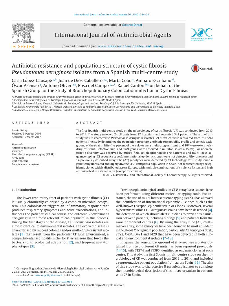
Antibiotic resistance and population structure of cystic fibrosisPseudomonas aeruginosa isolates from a Spanish multi-centre studyCarla López-Causapé a,b, Juan de Dios-Caballero b,c, Marta Cobo c, Amparo Escribano d,Óscar Asensio e, Antonio Oliver a,b, Rosa del Campo b,c,*, Rafael Cantón b,c on behalf of theSpanish Group for the Study of Bronchopulmonary Colonization/Infection in Cystic Fibrosisa Servicio de Microbiología and Unidad de Investigación, Hospital Universitario Son Espases, Instituto de Investigación Sanitaria Illes Balears, Palma de Mallorca, Spainb Red Española de Investigación en Patología Infecciosa, Instituto de Salud Carlos III, Madrid, Spainc Servicio de Microbiología, Hospital Universitario Ramón y Cajal and Instituto Ramón y Cajal de Investigación Sanitaria, Madrid, Spaind Unidad de Neumología Pediátrica y Fibrosis Quística, Servicio de Pediatría, Hospital Clínico Universitario and Universidad de Valencia, Valencia, Spaine Unidad de Neumología y Alergia Pediátrica, Hospital Universitario de Sabadell, Corporació Sanitària Parc Taulí, Sabadell, Barcelona, Spain
A R T I C L E I N F O
Article history:Received 9 October 2016Accepted 15 March 2017
Keywords:Antibiotic resistanceVirulenceMulti-locus sequence typing (MLST)Array tubeCystic fibrosisPseudomonas aeruginosa
A B S T R A C T
The first Spanish multi-centre study on the microbiology of cystic fibrosis (CF) was conducted from 2013to 2014. The study involved 24 CF units from 17 hospitals, and recruited 341 patients. The aim of thisstudy was to characterise Pseudomonas aeruginosa isolates, 79 of which were recovered from 75 (22%)patients. The study determined the population structure, antibiotic susceptibility profile and genetic back-ground of the strains. Fifty-five percent of the isolates were multi-drug-resistant, and 16% were extensively-drug-resistant. Defective mutS and mutL genes were observed in mutator isolates (15.2%). Considerablegenetic diversity was observed by pulsed-field gel electrophoresis (70 patterns) and multi-locus se-quence typing (72 sequence types). International epidemic clones were not detected. Fifty-one new and14 previously described array tube (AT) genotypes were detected by AT technology. This study found agenetically unrelated and highly diverse CF P. aeruginosa population in Spain, not represented by the ep-idemic clones widely distributed across Europe, with multiple combinations of virulence factors and highantimicrobial resistance rates (except for colistin).
© 2017 Elsevier B.V. and International Society of Chemotherapy. All rights reserved.
1. Introduction
The lower respiratory tract of patients with cystic fibrosis (CF)is usually chronically colonised by a complex microbial ecosys-tem. This colonisation triggers an inflammatory response thatproduces respiratory symptoms and acute exacerbations, and in-fluences the patients’ clinical course and outcome. Pseudomonasaeruginosa is the most relevant micro-organism in this process.During the first stages of the disease, CF P. aeruginosa isolates arealmost identical to environmental isolates. The evolved disease ischaracterised by mucoid colonies and/or multi-drug-resistant iso-lates [1] that result from the particular CF lung environment, acompartmentalised hostile niche for P. aeruginosa that forces thebacteria to an ecological adaptation [2], and frequent mutatorphenotypes [3].
Previous epidemiological studies on CF P. aeruginosa isolates havebeen performed using different molecular typing tools. For in-stance, the use of multi-locus sequence typing (MLST) has allowedthe identification of international epidemic CF clones, such as thewell-known Liverpool epidemic strain or Clone C. Moreover, severalhypertransmissible CF P. aeruginosa strains have been described [4],the detection of which should alert clinicians to prevent transmis-sion between patients, including siblings [5] and patients from thesame or different centres [6]. By using the array tube (AT) multi-marker array, some genotypes have been found to be most abundantin the global P. aeruginosa population, particularly AT genotypes 0C2E,2C22, C40A, D421 and F429 that have been detected in both clin-ical and environmental isolates [7–13].
In Spain, the genetic background of P. aeruginosa isolates ob-tained from two different CF units has been reported previously[14,15], with ST274 and ST395 identified as endemic clones at eachcentre. This study, the first Spanish multi-centre study on the mi-crobiology of CF, was conducted from 2013 to 2014, and includeda representative patient population from across Spain [16]. The aimof this study was to characterise P. aeruginosa isolates to completethe microbiological description of this micro-organism in patientswith CF in Spain.
* Corresponding author. Servicio de Microbiología, Hospital Universitario Ramóny Cajal, Ctra. Colmenar, Km 9,1, Madrid 28034, Spain.
E-mail address: [email protected] (R. del Campo).
http://dx.doi.org/10.1016/j.ijantimicag.2017.03.0340924-8579/© 2017 Elsevier B.V. and International Society of Chemotherapy. All rights reserved.
International Journal of Antimicrobial Agents 50 (2017) 334–341
Contents lists available at ScienceDirect
International Journal of Antimicrobial Agents
journal homepage: www.elsevier.com/ locate / i jant imicag
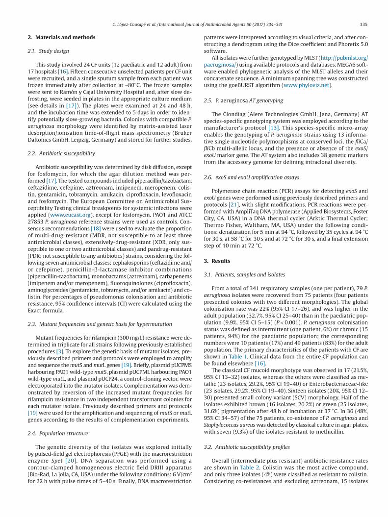
2. Materials and methods
2.1. Study design
This study involved 24 CF units (12 paediatric and 12 adult) from17 hospitals [16]. Fifteen consecutive unselected patients per CF unitwere recruited, and a single sputum sample from each patient wasfrozen immediately after collection at −80°C. The frozen sampleswere sent to Ramón y Cajal University Hospital and, after slow de-frosting, were seeded in plates in the appropriate culture medium(see details in [17]). The plates were examined at 24 and 48 h,and the incubation time was extended to 5 days in order to iden-tify potentially slow-growing bacteria. Colonies with compatible P.aeruginosa morphology were identified by matrix-assisted laserdesorption/ionisation time-of-flight mass spectrometry (BrukerDaltonics GmbH, Leipzig, Germany) and stored for further studies.
2.2. Antibiotic susceptibility
Antibiotic susceptibility was determined by disk diffusion, exceptfor fosfomycin, for which the agar dilution method was per-formed [17]. The tested compounds included piperacillin/tazobactam,ceftazidime, cefepime, aztreonam, imipenem, meropenem, colis-tin, gentamicin, tobramycin, amikacin, ciprofloxacin, levofloxacinand fosfomycin. The European Committee on Antimicrobial Sus-ceptibility Testing clinical breakpoints for systemic infections wereapplied (www.eucast.org), except for fosfomycin. PAO1 and ATCC27853 P. aeruginosa reference strains were used as controls. Con-sensus recommendations [18] were used to evaluate the proportionof multi-drug-resistant (MDR, not susceptible to at least threeantimicrobial classes), extensively-drug-resistant (XDR, only sus-ceptible to one or two antimicrobial classes) and pandrug-resistant(PDR; not susceptible to any antibiotics) strains, considering the fol-lowing seven antimicrobial classes: cephalosporins (ceftazidime and/or cefepime), penicillin-β-lactamase inhibitor combinations(piperacillin-tazobactam), monobactams (aztreonam), carbapenems(imipenem and/or meropenem), fluoroquinolones (ciprofloxacin),aminoglycosides (gentamicin, tobramycin, and/or amikacin) and co-listin. For percentages of pseudomonas colonisation and antibioticresistance, 95% confidence intervals (CI) were calculated using theExact formula.
2.3. Mutant frequencies and genetic basis for hypermutation
Mutant frequencies for rifampicin (300 mg/L) resistance were de-termined in triplicate for all strains following previously establishedprocedures [3]. To explore the genetic basis of mutator isolates, pre-viously described primers and protocols were employed to amplifyand sequence the mutS and mutL genes [19]. Briefly, plasmid pUCPMSharbouring PAO1 wild-type mutS, plasmid pUCPML harbouring PAO1wild-type mutL, and plasmid pUCP24, a control-cloning vector, wereelectroporated into the mutator isolates. Complementation was dem-onstrated by reversion of the increased mutant frequencies forrifampicin resistance in two independent transformant colonies foreach mutator isolate. Previously described primers and protocols[19] were used for the amplification and sequencing of mutS or mutLgenes according to the results of complementation experiments.
2.4. Population structure
The genetic diversity of the isolates was explored initiallyby pulsed-field gel electrophoresis (PFGE) with the macrorestrictionenzyme SpeI [20]. DNA separation was performed using acontour-clamped homogeneous electric field DRIII apparatus(Bio-Rad, La Jolla, CA, USA) under the following conditions: 6 V/cm2
for 22 h with pulse times of 5–40 s. Finally, DNA macrorestriction
patterns were interpreted according to visual criteria, and after con-structing a dendrogram using the Dice coefficient and Phoretix 5.0software.
All isolates were further genotyped by MLST (http://pubmlst.org/paeruginosa/) using available protocols and databases. MEGA6 soft-ware enabled phylogenetic analysis of the MLST alleles and theirconcatenate sequence. A minimum spanning tree was constructedusing the goeBURST algorithm (www.phyloviz.net).
2.5. P. aeruginosa AT genotyping
The Clondiag (Alere Technologies GmbH, Jena, Germany) ATspecies-specific genotyping system was employed according to themanufacturer’s protocol [13]. This species-specific micro-arrayenables the genotyping of P. aeruginosa strains using 13 informa-tive single nucleotide polymorphisms at conserved loci, the fliCa/fliCb multi-allelic locus, and the presence or absence of the exoS/exoU marker gene. The AT system also includes 38 genetic markersfrom the accessory genome for defining intraclonal diversity.
2.6. exoS and exoU amplification assays
Polymerase chain reaction (PCR) assays for detecting exoS andexoU genes were performed using previously described primers andprotocols [21], with slight modifications. PCR reactions were per-formed with AmpliTaq DNA polymerase (Applied Biosystems, FosterCity, CA, USA) in a DNA thermal cycler (Arktic Thermal Cycler;Thermo Fisher, Waltham, MA, USA) under the following condi-tions: denaturation for 5 min at 94 °C, followed by 35 cycles at 94 °Cfor 30 s, at 58 °C for 30 s and at 72 °C for 30 s, and a final extensionstep of 10 min at 72 °C.
3. Results
3.1. Patients, samples and isolates
From a total of 341 respiratory samples (one per patient), 79 P.aeruginosa isolates were recovered from 75 patients (four patientspresented colonies with two different morphologies). The globalcolonisation rate was 22% (95% CI 17–26), and was higher in theadult population (32.7%, 95% CI 25–40) than in the paediatric pop-ulation (9.9%, 95% CI 5–15) (P < 0.001). P. aeruginosa colonisationstatus was defined as intermittent (one patient, 6%) or chronic (15patients, 94%) for the paediatric population; the correspondingnumbers were 10 patients (17%) and 49 patients (83%) for the adultpopulation. The primary characteristics of the patients with CF areshown in Table 1. Clinical data from the entire CF population canbe found elsewhere [16].
The classical CF mucoid morphotype was observed in 17 (21.5%,95% CI 13–32) isolates, whereas the others were classified as me-tallic (23 isolates, 29.2%, 95% CI 19–40) or Enterobacteriaceae-like(23 isolates, 29.2%, 95% CI 19–40). Sixteen isolates (20%, 95% CI 12–30) presented small colony variant (SCV) morphology. Half of theisolates exhibited brown (16 isolates, 20.2%) or green (25 isolates,31.6%) pigmentation after 48 h of incubation at 37 °C. In 36 (48%,95% CI 34–57) of the 75 patients, co-existence of P. aeruginosa andStaphylococcus aureus was detected by classical culture in agar plates,with seven (9.3%) of the isolates resistant to methicillin.
3.2. Antibiotic susceptibility profiles
Overall (intermediate plus resistant) antibiotic resistance ratesare shown in Table 2. Colistin was the most active compound,and only three isolates (4%) were classified as resistant to colistin.Considering co-resistances and excluding aztreonam, 15 isolates
335C. López-Causapé et al. / International Journal of Antimicrobial Agents 50 (2017) 334–341
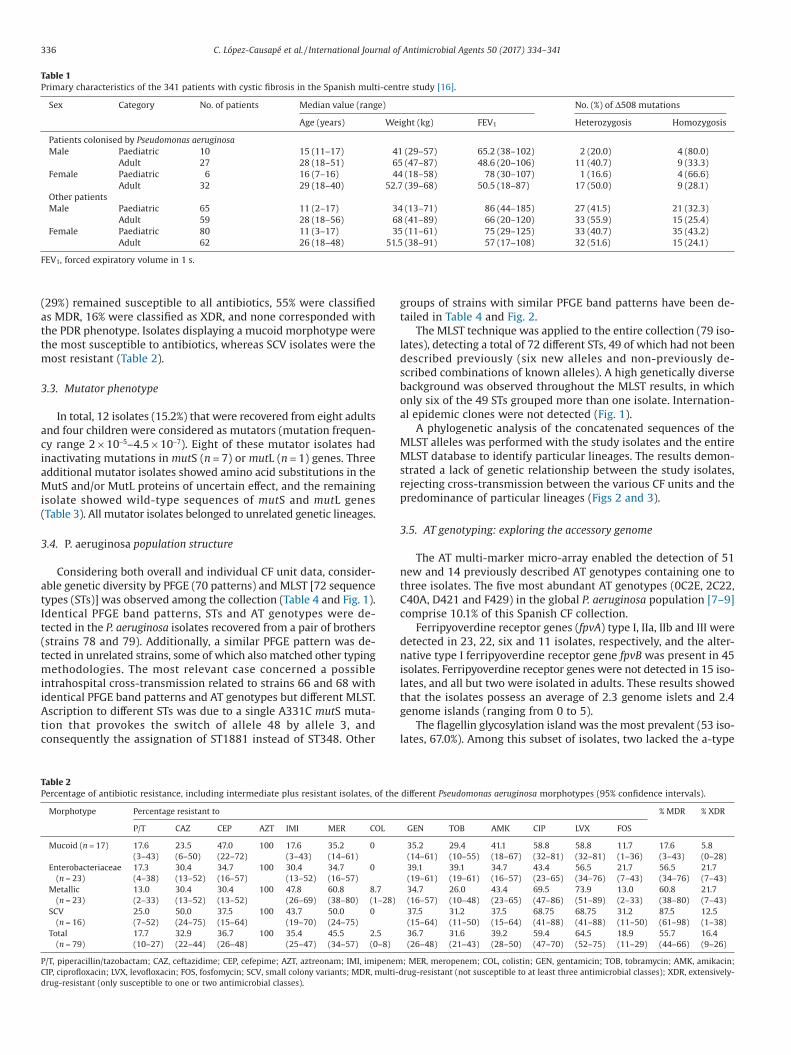
(29%) remained susceptible to all antibiotics, 55% were classifiedas MDR, 16% were classified as XDR, and none corresponded withthe PDR phenotype. Isolates displaying a mucoid morphotype werethe most susceptible to antibiotics, whereas SCV isolates were themost resistant (Table 2).
3.3. Mutator phenotype
In total, 12 isolates (15.2%) that were recovered from eight adultsand four children were considered as mutators (mutation frequen-cy range 2 × 10⁻⁵–4.5 × 10⁻⁷). Eight of these mutator isolates hadinactivating mutations in mutS (n = 7) or mutL (n = 1) genes. Threeadditional mutator isolates showed amino acid substitutions in theMutS and/or MutL proteins of uncertain effect, and the remainingisolate showed wild-type sequences of mutS and mutL genes(Table 3). All mutator isolates belonged to unrelated genetic lineages.
3.4. P. aeruginosa population structure
Considering both overall and individual CF unit data, consider-able genetic diversity by PFGE (70 patterns) and MLST [72 sequencetypes (STs)] was observed among the collection (Table 4 and Fig. 1).Identical PFGE band patterns, STs and AT genotypes were de-tected in the P. aeruginosa isolates recovered from a pair of brothers(strains 78 and 79). Additionally, a similar PFGE pattern was de-tected in unrelated strains, some of which also matched other typingmethodologies. The most relevant case concerned a possibleintrahospital cross-transmission related to strains 66 and 68 withidentical PFGE band patterns and AT genotypes but different MLST.Ascription to different STs was due to a single A331C mutS muta-tion that provokes the switch of allele 48 by allele 3, andconsequently the assignation of ST1881 instead of ST348. Other
groups of strains with similar PFGE band patterns have been de-tailed in Table 4 and Fig. 2.
The MLST technique was applied to the entire collection (79 iso-lates), detecting a total of 72 different STs, 49 of which had not beendescribed previously (six new alleles and non-previously de-scribed combinations of known alleles). A high genetically diversebackground was observed throughout the MLST results, in whichonly six of the 49 STs grouped more than one isolate. Internation-al epidemic clones were not detected (Fig. 1).
A phylogenetic analysis of the concatenated sequences of theMLST alleles was performed with the study isolates and the entireMLST database to identify particular lineages. The results demon-strated a lack of genetic relationship between the study isolates,rejecting cross-transmission between the various CF units and thepredominance of particular lineages (Figs 2 and 3).
3.5. AT genotyping: exploring the accessory genome
The AT multi-marker micro-array enabled the detection of 51new and 14 previously described AT genotypes containing one tothree isolates. The five most abundant AT genotypes (0C2E, 2C22,C40A, D421 and F429) in the global P. aeruginosa population [7–9]comprise 10.1% of this Spanish CF collection.
Ferripyoverdine receptor genes (fpvA) type I, IIa, IIb and III weredetected in 23, 22, six and 11 isolates, respectively, and the alter-native type I ferripyoverdine receptor gene fpvB was present in 45isolates. Ferripyoverdine receptor genes were not detected in 15 iso-lates, and all but two were isolated in adults. These results showedthat the isolates possess an average of 2.3 genome islets and 2.4genome islands (ranging from 0 to 5).
The flagellin glycosylation island was the most prevalent (53 iso-lates, 67.0%). Among this subset of isolates, two lacked the a-type
Table 1Primary characteristics of the 341 patients with cystic fibrosis in the Spanish multi-centre study [16].
Sex Category No. of patients Median value (range) No. (%) of Δ508 mutations
Age (years) Weight (kg) FEV1 Heterozygosis Homozygosis
Patients colonised by Pseudomonas aeruginosaMale Paediatric 10 15 (11–17) 41 (29–57) 65.2 (38–102) 2 (20.0) 4 (80.0)
Adult 27 28 (18–51) 65 (47–87) 48.6 (20–106) 11 (40.7) 9 (33.3)Female Paediatric 6 16 (7–16) 44 (18–58) 78 (30–107) 1 (16.6) 4 (66.6)
Adult 32 29 (18–40) 52.7 (39–68) 50.5 (18–87) 17 (50.0) 9 (28.1)Other patientsMale Paediatric 65 11 (2–17) 34 (13–71) 86 (44–185) 27 (41.5) 21 (32.3)
Adult 59 28 (18–56) 68 (41–89) 66 (20–120) 33 (55.9) 15 (25.4)Female Paediatric 80 11 (3–17) 35 (11–61) 75 (29–125) 33 (40.7) 35 (43.2)
Adult 62 26 (18–48) 51.5 (38–91) 57 (17–108) 32 (51.6) 15 (24.1)
FEV1, forced expiratory volume in 1 s.
Table 2Percentage of antibiotic resistance, including intermediate plus resistant isolates, of the different Pseudomonas aeruginosa morphotypes (95% confidence intervals).
Morphotype Percentage resistant to % MDR % XDR
P/T CAZ CEP AZT IMI MER COL GEN TOB AMK CIP LVX FOS
Mucoid (n = 17) 17.6(3–43)
23.5(6–50)
47.0(22–72)
100 17.6(3–43)
35.2(14–61)
0 35.2(14–61)
29.4(10–55)
41.1(18–67)
58.8(32–81)
58.8(32–81)
11.7(1–36)
17.6(3–43)
5.8(0–28)
Enterobacteriaceae(n = 23)
17.3(4–38)
30.4(13–52)
34.7(16–57)
100 30.4(13–52)
34.7(16–57)
0 39.1(19–61)
39.1(19–61)
34.7(16–57)
43.4(23–65)
56.5(34–76)
21.7(7–43)
56.5(34–76)
21.7(7–43)
Metallic(n = 23)
13.0(2–33)
30.4(13–52)
30.4(13–52)
100 47.8(26–69)
60.8(38–80)
8.7(1–28)
34.7(16–57)
26.0(10–48)
43.4(23–65)
69.5(47–86)
73.9(51–89)
13.0(2–33)
60.8(38–80)
21.7(7–43)
SCV(n = 16)
25.0(7–52)
50.0(24–75)
37.5(15–64)
100 43.7(19–70)
50.0(24–75)
0 37.5(15–64)
31.2(11–50)
37.5(15–64)
68.75(41–88)
68.75(41–88)
31.2(11–50)
87.5(61–98)
12.5(1–38)
Total(n = 79)
17.7(10–27)
32.9(22–44)
36.7(26–48)
100 35.4(25–47)
45.5(34–57)
2.5(0–8)
36.7(26–48)
31.6(21–43)
39.2(28–50)
59.4(47–70)
64.5(52–75)
18.9(11–29)
55.7(44–66)
16.4(9–26)
P/T, piperacillin/tazobactam; CAZ, ceftazidime; CEP, cefepime; AZT, aztreonam; IMI, imipenem; MER, meropenem; COL, colistin; GEN, gentamicin; TOB, tobramycin; AMK, amikacin;CIP, ciprofloxacin; LVX, levofloxacin; FOS, fosfomycin; SCV, small colony variants; MDR, multi-drug-resistant (not susceptible to at least three antimicrobial classes); XDR, extensively-drug-resistant (only susceptible to one or two antimicrobial classes).
336 C. López-Causapé et al. / International Journal of Antimicrobial Agents 50 (2017) 334–341
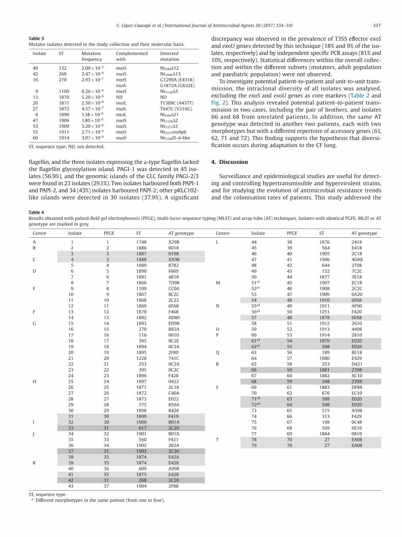
flagellin, and the three isolates expressing the a-type flagellin lackedthe flagellin glycosylation island. PAGI-1 was detected in 45 iso-lates (56.9%), and the genomic islands of the CLC family PAGI-2/3were found in 23 isolates (29.1%). Two isolates harboured both PAPI-1and PAPI-2, and 34 (43%) isolates harboured PAPI-2; other pKLC102-like islands were detected in 30 isolates (37.9%). A significant
discrepancy was observed in the prevalence of T3SS effector exoSand exoU genes detected by this technique (18% and 9% of the iso-lates, respectively) and by independent specific PCR assays (81% and10%, respectively). Statistical differences within the overall collec-tion and within the different subsets (mutators, adult populationand paediatric population) were not observed.
To investigate potential patient-to-patient and unit-to-unit trans-mission, the intraclonal diversity of all isolates was analysed,excluding the exoS and exoU genes as core markers (Table 2 andFig. 2). This analysis revealed potential patient-to-patient trans-mission in two cases, including the pair of brothers, and isolates66 and 68 from unrelated patients. In addition, the same ATgenotype was detected in another two patients, each with twomorphotypes but with a different repertoire of accessory genes (61,62, 71 and 72). This finding supports the hypothesis that diversi-fication occurs during adaptation to the CF lung.
4. Discussion
Surveillance and epidemiological studies are useful for detect-ing and controlling hypertransmissible and hypervirulent strains,and for studying the evolution of antimicrobial resistance trendsand the colonisation rates of patients. This study addressed the
Table 3Mutator isolates detected in the study collection and their molecular basis.
Isolate ST Mutationfrequency
Complementedwith
Detectedmutation
49 132 2.00 × 10⁻⁵ mutS Nt399Δ1242 268 2.47 × 10⁻⁶ mutS Nt1600Δ1316 270 2.93 × 10⁻⁷ mutS G1290A (E431K)
mutL G1872A (G632E)9 1109 8.26 × 10⁻⁶ mutS Nt1120Δ5
13 1870 5.20 × 10⁻⁶ ND ND26 1871 2.50 × 10⁻⁶ mutL T1309C (A437T)27 1872 4.57 × 10⁻⁷ mutL T647G (V216G)
6 1890 1.38 × 10⁻⁶ mutL Nt644Δ2147 1906 1.80 × 10⁻⁶ mutS Nt1336Δ253 1909 3.20 × 10⁻⁶ mutS Nt1377Δ155 1911 2.71 × 10⁻⁶ mutS Nt2577ins9pb60 1914 3.07 × 10⁻⁶ mutS Nt1198IS-4-like
ST, sequence type; ND, not detected.
Table 4Results obtained with pulsed-field gel electrophoresis (PFGE), multi-locus sequence typing (MLST) and array tube (AT) techniques. Isolates with identical PGFE, MLST or ATgenotype are marked in grey.
ST, sequence type.a Different morphotypes in the same patient (from one to four).
337C. López-Causapé et al. / International Journal of Antimicrobial Agents 50 (2017) 334–341
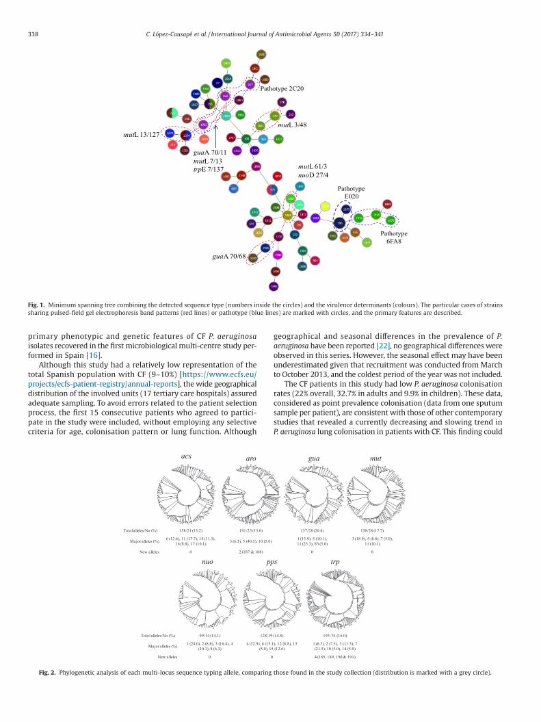
primary phenotypic and genetic features of CF P. aeruginosaisolates recovered in the first microbiological multi-centre study per-formed in Spain [16].
Although this study had a relatively low representation of thetotal Spanish population with CF (9–10%) [https://www.ecfs.eu/projects/ecfs-patient-registry/annual-reports], the wide geographicaldistribution of the involved units (17 tertiary care hospitals) assuredadequate sampling. To avoid errors related to the patient selectionprocess, the first 15 consecutive patients who agreed to partici-pate in the study were included, without employing any selectivecriteria for age, colonisation pattern or lung function. Although
geographical and seasonal differences in the prevalence of P.aeruginosa have been reported [22], no geographical differences wereobserved in this series. However, the seasonal effect may have beenunderestimated given that recruitment was conducted from Marchto October 2013, and the coldest period of the year was not included.
The CF patients in this study had low P. aeruginosa colonisationrates (22% overall, 32.7% in adults and 9.9% in children). These data,considered as point prevalence colonisation (data from one sputumsample per patient), are consistent with those of other contemporarystudies that revealed a currently decreasing and slowing trend inP. aeruginosa lung colonisation in patients with CF. This finding could
Fig. 1. Minimum spanning tree combining the detected sequence type (numbers inside the circles) and the virulence determinants (colours). The particular cases of strainssharing pulsed-field gel electrophoresis band patterns (red lines) or pathotype (blue lines) are marked with circles, and the primary features are described.
Fig. 2. Phylogenetic analysis of each multi-locus sequence typing allele, comparing those found in the study collection (distribution is marked with a grey circle).
338 C. López-Causapé et al. / International Journal of Antimicrobial Agents 50 (2017) 334–341
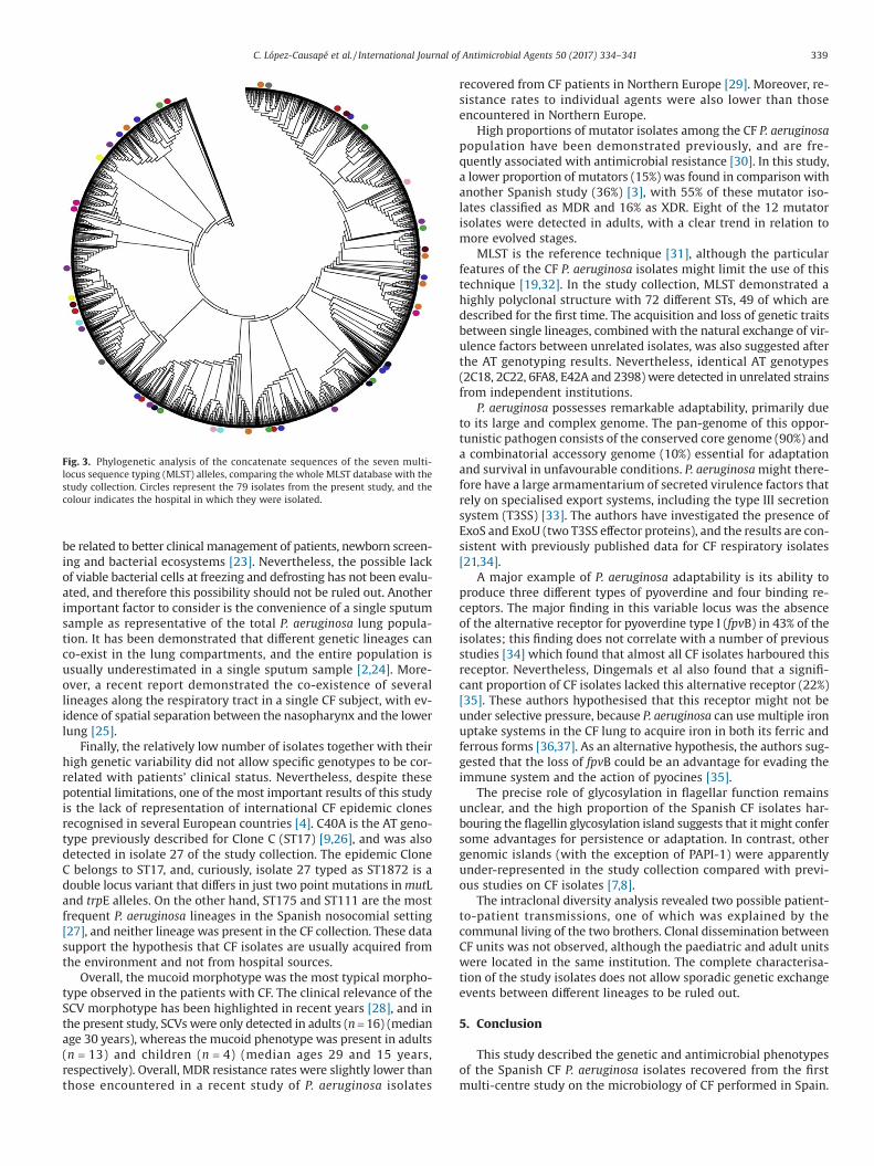
be related to better clinical management of patients, newborn screen-ing and bacterial ecosystems [23]. Nevertheless, the possible lackof viable bacterial cells at freezing and defrosting has not been evalu-ated, and therefore this possibility should not be ruled out. Anotherimportant factor to consider is the convenience of a single sputumsample as representative of the total P. aeruginosa lung popula-tion. It has been demonstrated that different genetic lineages canco-exist in the lung compartments, and the entire population isusually underestimated in a single sputum sample [2,24]. More-over, a recent report demonstrated the co-existence of severallineages along the respiratory tract in a single CF subject, with ev-idence of spatial separation between the nasopharynx and the lowerlung [25].
Finally, the relatively low number of isolates together with theirhigh genetic variability did not allow specific genotypes to be cor-related with patients’ clinical status. Nevertheless, despite thesepotential limitations, one of the most important results of this studyis the lack of representation of international CF epidemic clonesrecognised in several European countries [4]. C40A is the AT geno-type previously described for Clone C (ST17) [9,26], and was alsodetected in isolate 27 of the study collection. The epidemic CloneC belongs to ST17, and, curiously, isolate 27 typed as ST1872 is adouble locus variant that differs in just two point mutations in mutLand trpE alleles. On the other hand, ST175 and ST111 are the mostfrequent P. aeruginosa lineages in the Spanish nosocomial setting[27], and neither lineage was present in the CF collection. These datasupport the hypothesis that CF isolates are usually acquired fromthe environment and not from hospital sources.
Overall, the mucoid morphotype was the most typical morpho-type observed in the patients with CF. The clinical relevance of theSCV morphotype has been highlighted in recent years [28], and inthe present study, SCVs were only detected in adults (n = 16) (medianage 30 years), whereas the mucoid phenotype was present in adults(n = 13) and children (n = 4) (median ages 29 and 15 years,respectively). Overall, MDR resistance rates were slightly lower thanthose encountered in a recent study of P. aeruginosa isolates
recovered from CF patients in Northern Europe [29]. Moreover, re-sistance rates to individual agents were also lower than thoseencountered in Northern Europe.
High proportions of mutator isolates among the CF P. aeruginosapopulation have been demonstrated previously, and are fre-quently associated with antimicrobial resistance [30]. In this study,a lower proportion of mutators (15%) was found in comparison withanother Spanish study (36%) [3], with 55% of these mutator iso-lates classified as MDR and 16% as XDR. Eight of the 12 mutatorisolates were detected in adults, with a clear trend in relation tomore evolved stages.
MLST is the reference technique [31], although the particularfeatures of the CF P. aeruginosa isolates might limit the use of thistechnique [19,32]. In the study collection, MLST demonstrated ahighly polyclonal structure with 72 different STs, 49 of which aredescribed for the first time. The acquisition and loss of genetic traitsbetween single lineages, combined with the natural exchange of vir-ulence factors between unrelated isolates, was also suggested afterthe AT genotyping results. Nevertheless, identical AT genotypes(2C18, 2C22, 6FA8, E42A and 2398) were detected in unrelated strainsfrom independent institutions.
P. aeruginosa possesses remarkable adaptability, primarily dueto its large and complex genome. The pan-genome of this oppor-tunistic pathogen consists of the conserved core genome (90%) anda combinatorial accessory genome (10%) essential for adaptationand survival in unfavourable conditions. P. aeruginosa might there-fore have a large armamentarium of secreted virulence factors thatrely on specialised export systems, including the type III secretionsystem (T3SS) [33]. The authors have investigated the presence ofExoS and ExoU (two T3SS effector proteins), and the results are con-sistent with previously published data for CF respiratory isolates[21,34].
A major example of P. aeruginosa adaptability is its ability toproduce three different types of pyoverdine and four binding re-ceptors. The major finding in this variable locus was the absenceof the alternative receptor for pyoverdine type I (fpvB) in 43% of theisolates; this finding does not correlate with a number of previousstudies [34] which found that almost all CF isolates harboured thisreceptor. Nevertheless, Dingemals et al also found that a signifi-cant proportion of CF isolates lacked this alternative receptor (22%)[35]. These authors hypothesised that this receptor might not beunder selective pressure, because P. aeruginosa can use multiple ironuptake systems in the CF lung to acquire iron in both its ferric andferrous forms [36,37]. As an alternative hypothesis, the authors sug-gested that the loss of fpvB could be an advantage for evading theimmune system and the action of pyocines [35].
The precise role of glycosylation in flagellar function remainsunclear, and the high proportion of the Spanish CF isolates har-bouring the flagellin glycosylation island suggests that it might confersome advantages for persistence or adaptation. In contrast, othergenomic islands (with the exception of PAPI-1) were apparentlyunder-represented in the study collection compared with previ-ous studies on CF isolates [7,8].
The intraclonal diversity analysis revealed two possible patient-to-patient transmissions, one of which was explained by thecommunal living of the two brothers. Clonal dissemination betweenCF units was not observed, although the paediatric and adult unitswere located in the same institution. The complete characterisa-tion of the study isolates does not allow sporadic genetic exchangeevents between different lineages to be ruled out.
5. Conclusion
This study described the genetic and antimicrobial phenotypesof the Spanish CF P. aeruginosa isolates recovered from the firstmulti-centre study on the microbiology of CF performed in Spain.
Fig. 3. Phylogenetic analysis of the concatenate sequences of the seven multi-locus sequence typing (MLST) alleles, comparing the whole MLST database with thestudy collection. Circles represent the 79 isolates from the present study, and thecolour indicates the hospital in which they were isolated.
339C. López-Causapé et al. / International Journal of Antimicrobial Agents 50 (2017) 334–341
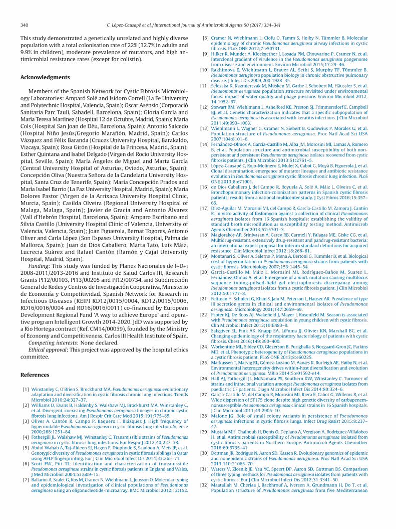
This study demonstrated a genetically unrelated and highly diversepopulation with a total colonisation rate of 22% (32.7% in adults and9.9% in children), moderate prevalence of mutators, and high an-timicrobial resistance rates (except for colistin).
Acknowledgments
Members of the Spanish Network for Cystic Fibrosis Microbiol-ogy Laboratories: Amparó Solé and Isidoro Cortell (La Fe Universityand Polytechnic Hospital, Valencia, Spain); Oscar Asensio (CorporacióSanitaria Parc Taulí, Sabadell, Barcelona, Spain); Gloria García andMaría Teresa Martínez (Hospital 12 de Octubre, Madrid, Spain); MaríaCols (Hospital San Joan de Déu, Barcelona, Spain); Antonio Salcedo(Hospital Niño Jesús/Gregorio Marañón, Madrid, Spain); CarlosVázquez and Félix Baranda (Cruces University Hospital, Barakaldo,Vizcaya, Spain); Rosa Girón (Hospital de la Princesa, Madrid, Spain);Esther Quintana and Isabel Delgado (Virgen del Rocío University Hos-pital, Seville, Spain); María Ángeles de Miguel and Marta García(Central University Hospital of Asturias, Oviedo, Asturias, Spain);Concepción Oliva (Nuestra Señora de la Candelaria University Hos-pital, Santa Cruz de Tenerife, Spain); María Concepción Prados andMaría Isabel Barrio (La Paz University Hospital, Madrid, Spain); MaríaDolores Pastor (Virgen de la Arrixaca University Hospital Clinic,Murcia, Spain); Casilda Olveira (Regional University Hospital ofMalaga, Malaga, Spain); Javier de Gracia and Antonio Álvarez(Vall d’Hebrón Hospital, Barcelona, Spain); Amparo Escribano andSilvia Castillo (University Hospital Clinic of Valencia, University ofValencia, Valencia, Spain); Joan Figuerola, Bernat Togores, AntonioOliver and Carla López (Son Espases University Hospital, Palma deMallorca, Spain); Juan de Dios Caballero, Marta Tato, Luis Máiz,Lucrecia Suárez and Rafael Cantón (Ramón y Cajal UniversityHospital, Madrid, Spain).
Funding: This study was funded by Planes Nacionales de I+D+i2008-2011/2013-2016 and Instituto de Salud Carlos III, ResearchGrants PI12/00103, PI13/00205 and PI12/00734, and SubdirecciónGeneral de Redes y Centros de Investigación Cooperativa, Ministeriode Economía y Competitividad, Spanish Network for Research inInfectious Diseases (REIPI RD12/0015/0004, RD12/0015/0006,RD16/0016/0004 and RD16/0016/0011) co-financed by EuropeanDevelopment Regional Fund ’A way to achieve Europe’ and opera-tive program Intelligent Growth 2014-2020. JdD was supported bya Rio Hortega contract (Ref. CM14/00059), founded by the Ministryof Economy and Competitiveness, Carlos III Health Institute of Spain.
Competing interests: None declared.Ethical approval: This project was approved by the hospital ethics
committee.
References
[1] Winstanley C, O’Brien S, Brockhurst MA. Pseudomonas aeruginosa evolutionaryadaptation and diversification in cystic fibrosis chronic lung infections. TrendsMicrobiol 2016;24:327–37.
[2] Williams D, Evans B, Haldenby S, Walshaw MJ, Brockhurst MA, Winstanley C,et al. Divergent, coexisting Pseudomonas aeruginosa lineages in chronic cysticfibrosis lung infections. Am J Respir Crit Care Med 2015;191:775–85.
[3] Oliver A, Cantón R, Campo P, Baquero F, Blázquez J. High frequency ofhypermutable Pseudomonas aeruginosa in cystic fibrosis lung infection. Science2000;288:1251–84.
[4] Fothergill JL, Walshaw MJ, Winstanley C. Transmissible strains of Pseudomonasaeruginosa in cystic fibrosis lung infections. Eur Respir J 2012;40:227–38.
[5] Abdul Wahab A, Taj-Aldeen SJ, Hagen F, Diophode S, Saadoon A, Meis JF, et al.Genotypic diversity of Pseudomonas aeruginosa in cystic fibrosis siblings in Qatarusing AFLP fingerprinting. Eur J Clin Microbiol Infect Dis 2014;33:265–71.
[6] Scott FW, Pitt TL. Identification and characterization of transmissiblePseudomonas aeruginosa strains in cystic fibrosis patients in England and Wales.J Med Microbiol 2004;53:609–15.
[7] Ballarini A, Scalet G, Kos M, Cramer N, Wiehlmann L, Jousson O. Molecular typingand epidemiological investigation of clinical populations of Pseudomonasaeruginosa using an oligonucleotide-microarray. BMC Microbiol 2012;12:152.
[8] Cramer N, Wiehlmann L, Ciofu O, Tamm S, Høiby N, Tümmler B. Molecularepidemiology of chronic Pseudomonas aeruginosa airway infections in cysticfibrosis. PLoS ONE 2012;7:e50731.
[9] Hilker R, Munder A, Klockgether J, Losada PM, Chouvarine P, Cramer N, et al.Interclonal gradient of virulence in the Pseudomonas aeruginosa pangenomefrom disease and environment. Environ Microbiol 2015;17:29–46.
[10] Rakhimova E, Wiehlmann L, Brauer AL, Sethi S, Murphy TF, Tümmler B.Pseudomonas aeruginosa population biology in chronic obstructive pulmonarydisease. J Infect Dis 2009;200:1928–35.
[11] Selezska K, Kazmierczak M, Müsken M, Garbe J, Schobert M, Häussler S, et al.Pseudomonas aeruginosa population structure revisited under environmentalfocus: impact of water quality and phage pressure. Environ Microbiol 2012;14:1952–67.
[12] Stewart RM, Wiehlmann L, Ashelford KE, Preston SJ, Frimmersdorf E, CampbellBJ, et al. Genetic characterization indicates that a specific subpopulation ofPseudomonas aeruginosa is associated with keratitis infections. J Clin Microbiol2011;49:993–1003.
[13] Wiehlmann L, Wagner G, Cramer N, Siebert B, Gudowius P, Morales G, et al.Population structure of Pseudomonas aeruginosa. Proc Natl Acad Sci USA2007;104:8101–6.
[14] Fernández-Olmos A, García-Castillo M, Alba JM, Morosini MI, Lamas A, RomeroB, et al. Population structure and antimicrobial susceptibility of both non-persistent and persistent Pseudomonas aeruginosa isolates recovered from cysticfibrosis patients. J Clin Microbiol 2013;51:2761–5.
[15] López-Causapé C, Rojo-Molinero E, Mulet X, Cabot G, Moyà B, Figuerola J, et al.Clonal dissemination, emergence of mutator lineages and antibiotic resistanceevolution in Pseudomonas aeruginosa cystic fibrosis chronic lung infection. PLoSONE 2013;8:e71001.
[16] de Dios Caballero J, del Campo R, Royuela A, Solé A, Máiz L, Olveira C, et al.Bronchopulmonary infection-colonization patterns in Spanish cystic fibrosispatients: results from a national multicenter study. J Cyst Fibros 2016;15:357–65.
[17] Díez-Aguilar M, Morosini MI, del Campo R, García-Castillo M, Zamora J, CantónR. In vitro activity of fosfomycin against a collection of clinical Pseudomonasaeruginosa isolates from 16 Spanish hospitals: establishing the validity ofstandard broth microdilution as susceptibility testing method. AntimicrobAgents Chemother 2013;57:5701–3.
[18] Magiorakos AP, Srinivasan A, Carey RB, Carmeli Y, Falagas ME, Giske CG, et al.Multidrug-resistant, extensively drug-resistant and pandrug-resistant bacteria:an international expert proposal for interim standard definitions for acquiredresistance. Clin Microbiol Infect 2012;18:268–81.
[19] Montanari S, Oliver A, Salerno P, Mena A, Bertoni G, Tümmler B, et al. Biologicalcost of hypermutation in Pseudomonas aeruginosa strains from patients withcystic fibrosis. Microbiology 2007;153:1445–54.
[20] García-Castillo M, Máiz L, Morosini MI, Rodríguez-Baños M, Suarez L,Fernández-Olmos A, et al. Emergence of a mutL mutation causing multilocussequence typing-pulsed-field gel electrophoresis discrepancy amongPseudomonas aeruginosa isolates from a cystic fibrosis patient. J Clin Microbiol2012;50:1777–8.
[21] Feltman H, Schulert G, Khan S, Jain M, Peterson L, Hauser AR. Prevalence of typeIII secretion genes in clinical and environmental isolates of Pseudomonasaeruginosa. Microbiology 2001;147:2659–69.
[22] Psoter KJ, De Roos AJ, Wakefield J, Mayer J, Rosenfeld M. Season is associatedwith Pseudomonas aeruginosa acquisition in young children with cystic fibrosis.Clin Microbiol Infect 2013;19:E483–9.
[23] Salsgiver EL, Fink AK, Knapp EA, LiPuma JJ, Olivier KN, Marshall BC, et al.Changing epidemiology of the respiratory bacteriology of patients with cysticfibrosis. Chest 2016;149:390–400.
[24] Workentine ML, Sibley CD, Glezerson B, Purighalla S, Norgaard-Gron JC, ParkinsMD, et al. Phenotypic heterogeneity of Pseudomonas aeruginosa populations ina cystic fibrosis patient. PLoS ONE 2013;8:e60225.
[25] Markussen T, Marvig RL, Gómez-Lozano M, Aanæs K, Burleigh AE, Høiby N, et al.Environmental heterogeneity drives within-host diversification and evolutionof Pseudomonas aeruginosa. MBio 2014;5:e01592-e14.
[26] Hall AJ, Fothergill JL, McNamara PS, Southern KW, Winstanley C. Turnover ofstrains and intraclonal variation amongst Pseudomonas aeruginosa isolates frompaediatric CF patients. Diagn Microbiol Infect Dis 2014;80:324–6.
[27] García-Castillo M, del Campo R, Morosini MI, Riera E, Cabot G, Willems R, et al.Wide dispersion of ST175 clone despite high genetic diversity of carbapenem-nonsusceptible Pseudomonas aeruginosa clinical strains in 16 Spanish hospitals.J Clin Microbiol 2011;49:2905–10.
[28] Malone JG. Role of small colony variants in persistence of Pseudomonasaeruginosa infections in cystic fibrosis lungs. Infect Drug Resist 2015;8:237–47.
[29] Mustafa MH, Chalhoub H, Denis O, Deplano A, Vergison A, Rodriguez-VillalobosH, et al. Antimicrobial susceptibility of Pseudomonas aeruginosa isolated fromcystic fibrosis patients in Northern Europe. Antimicrob Agents Chemother2016;60:6735–41.
[30] Dettman JR, Rodrigue N, Aaron SD, Kassen R. Evolutionary genomics of epidemicand nonepidemic strains of Pseudomonas aeruginosa. Proc Natl Acad Sci USA2013;110:21065–70.
[31] Waters V, Zlosnik JE, Yau YC, Speert DP, Aaron SD, Guttman DS. Comparisonof three typing methods for Pseudomonas aeruginosa isolates from patients withcystic fibrosis. Eur J Clin Microbiol Infect Dis 2012;31:3341–50.
[32] Maatallah M, Cheriaa J, Backhrouf A, Iversen A, Grundmann H, Do T, et al.Population structure of Pseudomonas aeruginosa from five Mediterranean
340 C. López-Causapé et al. / International Journal of Antimicrobial Agents 50 (2017) 334–341

countries: evidence for frequent recombination and epidemic occurrence ofCC235. PLoS ONE 2011;6:e25617.
[33] Frank DW. The exoenzyme S regulon of Pseudomonas aeruginosa. Mol Microbiol1997;26:621–9.
[34] Pirnay JP, Bilocq F, Pot B, Cornelis P, Zizi M, Van Eldere J, et al. Pseudomonasaeruginosa population structure revisited. PLoS ONE 2009;4:e7740.
[35] Dingemans J, Ye L, Hildebrand F, Tontodonati F, Craggs M, Bilocq F, et al. Thedeletion of TonB-dependent receptor genes is part of the genome reduction
process that occurs during adaptation of Pseudomonas aeruginosa to the cysticfibrosis lung. Pathog Dis 2014;71:26–38.
[36] Hunter RC, Asfour F, Dingemans J, Osuna BL, Samad T, Malfroot A, et al. Ferrousiron is a significant component of bioavailable iron in cystic fibrosis airways.MBio 2013;4:e00557–13.
[37] Konings AF, Martin LW, Sharples KJ, Roddam LF, Latham R, Reid DW, et al.Pseudomonas aeruginosa uses multiple pathways to acquire iron during chronicinfection in cystic fibrosis lungs. Infect Immun 2013;81:2697–704.
Amparó Solé, Isidoro Cortell, Oscar Asensio, Gloria García, María Teresa Martínez, María Cols, Antonio Salcedo, Carlos Vázquez, Félix Baranda, Rosa Girón, Esther Quintana, Isabel Delgado, María Ángeles de Miguel, Marta García, Concepción Oliva, María Concepción Prados, María Isabel Barrio, María Dolores Pastor, Casilda Olveira, Javier de Gracia, Antonio Álvarez, Amparo Escribano, Silvia Castillo, Joan Figuerola, Bernat Togores, Antonio Oliver, Carla López, Juan de Dios Caballero, Marta Tato, Luis Máiz, Lucrecia Suárez, Rafael CantónaLa Fe University and Polytechnic Hospital, Valencia, SpainbCorporació Sanitaria Parc Taulí, Sabadell, Barcelona, SpaincHospital 12 de Octubre, Madrid, SpaindHospital San Joan de Déu, Barcelona, SpaineHospital Niño Jesús/Gregorio Marañón, Madrid, SpainfCruces University Hospital, Barakaldo, Vizcaya, SpaingHospital de la Princesa, Madrid, SpainhVirgen del Rocío University Hospital, Seville, Spaini Central University Hospital of Asturias, Oviedo, Asturias, SpainjNuestra Señora de la Candelaria University Hospital, Santa Cruz de Tenerife, SpainkLa Paz University Hospital, Madrid, SpainlVirgen de la Arrixaca University Hospital Clinic, Murcia, SpainmRegional University Hospital of Malaga, Malaga, SpainnVall d’Hebrón Hospital, Barcelona, SpainoUniversity Hospital Clinic of Valencia, University of Valencia, Valencia, SpainpSon Espases University Hospital, Palma de Mallorca, SpainqRamón y Cajal University Hospital, Madrid, Spain
341C. López-Causapé et al. / International Journal of Antimicrobial Agents 50 (2017) 334–341
Editorial
Insights into the evolution of the mutationalresistome of Pseudomonas aeruginosa incystic fibrosisCarla Lopez-Causape1 & Antonio Oliver*,1
1Servicio de Microbiologıa & Unidad de Investigacion, Hospital Universitario Son Espases, Instituto de Investigacion Illes Balears(IdISBa), Palma de Mallorca, Spain* Author for correspondence: [email protected]
“The analysis of WGS mutational resistomes has proven to be useful for understanding theevolutionary dynamics of classical resistance mechanisms and to depict new ones for the majority
of antimicrobial classes”First draft submitted: 6 September 2017; Accepted for publication: 12 September 2017; Publishedonline: 25 October 2017
Keywords: antibiotic resistance • bacterial evolution • chronic infections • cystic fibrosis • hypermutation • Pseu-domonas aeruginosa • resistome
Chronic respiratory infection (CRI) by Pseudomonas aeruginosa is the main driver of morbidity and mortality incystic fibrosis (CF) patients [1]. CRI results from an intense adaptation process, where bacterial evolution is testedagainst host immune responses and years of aggressive antimicrobial treatments [2]. Once established, CRI canseldom be eradicated despite intensive antimicrobial treatments, and therefore our therapeutic goals resignedlymove from attempting to cure the infection to minimizing its long-term impact through chronic suppressivetherapy [3]. The plasticity of P. aeruginosa genome for antimicrobial resistance acquisition, the greatly enhancedmutation supply rate provided by frequent hypermutable variants (mutators) and the highly structured environmentdetermined by the characteristic biofilm growth and the anatomy of the respiratory tract make bacterial evolutionand genetic diversification a hallmark of CF CRI [2,4]. While the enhanced evolution of antimicrobial resistancein CF, frequently linked to mutator phenotypes, was noted many years ago [5], it is with the introduction ofwhole-genome sequencing (WGS) that we are starting to understand its real dimensions [6].
The term resistome was first used to account for the set of primary antibiotic resistance genes that could beeventually transferred to human pathogens [7]. Soon after the concept of intrinsic resistome was introduced to includeall chromosomal genes that are involved in intrinsic resistance, and whose presence in strains of a bacterial species isindependent of previous antibiotic exposure and is not due to horizontal gene transfer (HGT) [8]. Finally, the termmutational resistome was more recently implemented to account for the set of mutations involved in the modulationof antibiotic resistance levels in the absence of HGT [9]. Recent WGS data obtained from in vitro assays on theevolution of antibiotic resistance and clinical isolates, and in particular sequential CF isolates, provide new insightsinto the evolutionary dynamics and mechanisms of P. aeruginosa antibiotic resistance. However, in too many cases,the documented genomic variations fail to provide causative relations in the absence of phenotypic information.The analysis of WGS mutational resistomes has proven to be useful for understanding the evolutionary dynamicsof classical resistance mechanisms and to depict new ones for the majority of antimicrobial classes, includingβ-lactams, aminoglycosides, fluoroquinolones and polymixins.
Regarding β-lactams, the analysis of WGS mutational resistomes has confirmed the major role of classicalresistance mutations such as those leading to the overexpression of the chromosomal β-lactamase AmpC (such asDacB [PBP4], AmpD and/or AmpR mutations) or the inactivation of the carbapenem porin OprD. However,the analysis of mutational resistomes of in vitro evolved strains and sequential CF isolates have identified otherkey mutations, such as those occurring in β-lactam targets (essential PBPs), particularly involving mutations inftsI which encodes PBP3, an essential high molecular class B penicillin binding protein (PBP) with transpeptidase
10.2217/fmb-2017-0197 C© 2017 Future Medicine Ltd Future Microbiol. (2017) 12(16), 1445–1448 ISSN 1746-0913 1445
Editorial Lopez-Causape & Oliver
activity. Indeed, data from CF patients [9,10] as well as from in vitro studies [11] have recently demonstrated thatPBP3 is under strong mutational pressure, with specific mutations contributing to β-lactam resistance development.Among them are particularly relevant and frequent mutations affecting amino acids R504 or F533, located withinthe protein domains responsible for the formation and stabilization of the inactivating complex β-lactam–PBP3.Moreover, PBP3 mutations seem to play a role in the emergence of resistance to novel β-lactam–β-lactamase inhibitorcombinations, such as ceftolozane/tazobactam [9]. Another relevant mutational β-lactam resistance mechanism is theselection of large (>200 kb) deletions affecting specific parts of the chromosome. Although the basis of the conferredresistance phenotype still needs to be further clarified, these mutants can be recognized by the characteristic brownpigment (pyomelanine) caused by the deletion of one of the affected genes, hmgA, coding for a homogentisate-1,2-dioxygenase. This type of deletion has been documented in both, in vitro evolved β-lactam-resistant mutants andCF isolates [11,12]. However, the deletion of hmgA is not responsible for the resistance phenotype, which may belinked to the deletion of another of the affected genes, galU, coding for a UDP-glucose pyrophosphorylase requiredfor lipopolysaccharide core synthesis. Indeed, analysis of transposon mutant libraries has shown that inactivation ofgalU increases ceftazidime and meropenem minimum inhibitory concentrations [13,14]. Finally, another emergingmutational β-lactam resistance mechanism is the structural modification of AmpC [10,11].
With respect to aminoglycosides, results from analysis of mutational resistomes of CF isolates point to theunderlying strong evolutionary pressure of mexZ and the relevance of MexXY overexpression for resistance de-velopment [15–17]. Moreover, recent in vitro studies and findings from CF isolates have revealed that high-levelaminoglycoside resistance requires the acquisition of additional mutations; among them, those in FusA1 seemto be particularly frequent and relevant [9,16,18–19]. Likewise, the fluoroquinolone resistome frequently includesmutations in efflux pump regulators, among which nfxB, leading to the overexpression of MexCD-OprJ, is partic-ularly noteworthy in the CF setting. However, high-level ciprofloxacin resistance generally involves one or severalmutations in the quinolone resistance determining regions of GyrA/B and/or ParC/E [9]. Regarding polymixinresistance, findings from WGS studies of in vitro evolved strains and CF isolates have shown that development ofhigh-level colistin resistance requires the acquisition of multiple mutations, including those in the two-componentregulators (PmrAB, PhoPQ or ParRS) involved in the addition of 4-amino-4-deoxy-L-arabinose to lipid A fromthe lipopolysaccharide [9,20]. Finally, in addition to the resistance mechanisms to classical antipseudomonal agents,the CF mutational resistome may also include resistance to other used antibiotics such as the frequent mutationsof domain V of 23S rRNA – conferring macrolide resistance [21].
The complexity of the CF isolates resistomes is further enhanced when the intrapatient genetic diversity of CFP. aeruginosa populations is introduced. Certainly, to understand the resistance dynamics and evolution, futuresteps should endeavor to analyze the mutational resistomes in CF at the whole population level, as opposed to theanalysis of single isolated colonies. Indeed, full understanding of the evolution of the mutational resistome requiresa longitudinal and transversal analysis of P. aeruginosa populations in the CF patient. Moreover, recent evidencesuggests that interpatient transmission should also be considered when analyzing the evolution of the mutationalresistome, especially when introducing mutator lineages of epidemic clones [9].
Beyond addressing a relevant scientific question, the analysis of mutational resistomes would be useful fortherapeutic strategy design and monitoring the efficacy of administered antibiotic treatments. Obviously, theevolution of the mutational resistome is a direct consequence of antimicrobial exposure. As such, it is not surprisingthat exposure to one antibiotic drives evolution of the mutational resistome for that antibiotic. However, thecomplexity of the actual resistance profile is further increased by the specificity and interactions among differentresistance mechanisms. A classic example is cross resistance (or collateral resistance), which implies that exposure toone antibiotic drives also the development of resistance to a different one. Typically this is caused by the developedresistance mechanism (such as efflux pump overexpression) affecting simultaneous different antibiotics. Indeed,potential development of cross resistance is a major issue to consider when using antibiotic combinations [22].
Perhaps less obvious is collateral susceptibility, which implies that exposure to one antibiotic increases thesusceptibility to a different one. This might be achieved through two mechanisms. One possible mechanism isthat exposure to one antibiotic directly causes increased susceptibility to a different one, for example, mutations inthe β-lactamase AmpC increases cephalosporin hydrolysis while reducing that of penicillins or carbapenems [23].The second possibility is that the development of a resistance mechanism impairs the activity of another existingresistance mechanism, for example, competition between the different efflux pumps, the overexpression of one mayimpair the expression of another [24]. Indeed, in the CF setting it is very frequent that the overexpression of effluxpump MexXY, involved in aminoglycoside resistance, is linked to the impaired expression of efflux pump MexAB,
1446 Future Microbiol. (2017) 12(16) future science group
Insights into the evolution of the mutational resistome of Pseudomonas aeruginosa in cystic fibrosis Editorial
involved in the resistance to a broad range of antibiotics including most β-lactams [25]. Thus, the evolution of themutational resistome for a given antibiotic is not only dependent on the exposure to this antibiotic, but it is alsoconditioned by the simultaneous or even previous exposures to other antibiotics. An illustrative example is providedin a recent in vitro study that demonstrated, for a broad range of antibiotic classes, that the history of exposure andresistance development to a given antibiotic, conditions the dynamics and mechanisms of resistance developmentwhen exposed to a second one [18].
Moreover, knowledge of the interactions between resistance mechanisms could be useful in the design ofsequential treatments that minimize the risk of resistance development. Such is the case for a recent in vitro studyshowing the effectiveness of aztreonam–tobramycin sequential treatment, based on the antagonism between theresistance mechanisms for each of the antibiotics; overexpression of the efflux pump MexAB (aztreonam) or MexXY(tobramycin) which compete for the same outer membrane channel (OprM) [26].
Finally, it should be noted that a hallmark of P. aeruginosa CRI in CF is the biofilm mode of growth. Indeed,biofilm growth, in addition to providing a compact structured environment likely facilitating the evolution ofmutational resistance [27], they also add further complexity due to the major differences in the mechanisms involvedwhen compared with conventional planktonic growth that needs to be considered [28]. Likewise, persistence of P.aeruginosa in the CF lung despite intensive antimicrobial treatments relays in the acquisition of a vast number ofadaptive mutations that extend far beyond classical antibiotic resistance mutations [29].
In summary, the comprehensive analysis of the mutational resistomes of P. aeruginosa in CF CRI is expected tobecome a useful tool for optimizing therapeutic strategies and monitoring the efficacy of administered antibiotictreatments in the near future.
Financial & competing interests disclosure
The authors are supported by the Ministerio de Economıa y Competitividad of Spain, Instituto de Salud Carlos III – co-financed
by European Regional Development Fund ‘A way to achieve Europe’ ERDF, through the Spanish Network for the Research in
Infectious Diseases (RD12/0015 and RD16/0016). The authors have no other relevant affiliations or financial involvement with
any organization or entity with a financial interest in or financial conflict with the subject matter or materials discussed in the
manuscript apart from those disclosed.
No writing assistance was utilized in the production of this manuscript.
References1. Lyczak JB, Cannon CL, Pier GB. Lung infections associated with cystic fibrosis. Clin. Microbiol. Rev. 15(2), 194–222 (2002).
2. Oliver A, Mena A, Macia MD. Evolution of Pseudomonas aeruginosa pathogenicity: from acute to chronic infections. In: EvolutionaryBiology of Bacterial and Fungal Pathogens. Baquero F, Nombela C, Cassell GH, Gutierrez JA (Eds). ASM Press, WA, USA, 433–444(2008).
3. Lopez-Causape C, Rojo-Molinero E, Macia MD, Oliver A. The problems of antibiotic resistance in cystic fibrosis and solutions. Expert.Rev. Respir. Med. 9(1), 73–88 (2015).
4. Fernandez-Barat L, Ciofu O, Kragh KN et al. Phenotypic shift in Pseudomonas aeruginosa populations from cystic fibrosis lungs after2-week antipseudomonal treatment. J. Cyst. Fibros. 16(2), 222–229 (2017).
5. Oliver A, Canton R, Campo P, Baquero F, Blazquez J. High frequency of hypermutable Pseudomonas aeruginosa in cystic fibrosis lunginfection. Science 288(5469), 1251–1254 (2000).
6. Feliziani S, Marvig RL, Lujan AM et al. Coexistence and within-host evolution of diversified lineages of hypermutable Pseudomonasaeruginosa in long-term cystic fibrosis infections. PLoS Genet. 10(10), e1004651 (2014).
7. D’Costa VM, McGrann KM, Hughes DW, Wright GD. Sampling the antibiotic resistome. Science 311(5759), 374–377 (2006).
8. Fajardo A, Martınez-Martın N, Mercadillo M et al. The neglected intrinsic resistome of bacterial pathogens. PLoS ONE 3(2), e1619(2008).
9. Lopez-Causape C, Sommer LM, Cabot G et al. Evolution of the Pseudomonas aeruginosa mutational resistome in an international cysticfibrosis clone. Sci. Rep. 7(1), 5555 (2017).
10. Diaz Caballero J, Clark ST, Coburn B et al. Selective sweeps and parallel pathoadaptation drive Pseudomonas aeruginosa evolution in thecystic fibrosis lung. MBio. 6(5), e00981–15 (2015).
11. Cabot G, Zamorano L, Moya B et al. Evolution of Pseudomonas aeruginosa antimicrobial resistance and fitness under low and highmutation rates. Antimicrob. Agents Chemother. 60(3), 1767–1778 (2016).
12. Hocquet D, Petitjean M, Rohmer L et al. Pyomelanin-producing Pseudomonas aeruginosa selected during chronic infections have a largechromosomal deletion which confers resistance to pyocins. Environ. Microbiol. 18(10), 3482–3493 (2016).
future science group www.futuremedicine.com 1447
Editorial Lopez-Causape & Oliver
13. Alvarez-Ortega C, Wiegand I, Olivares J, Hancock RE, Martınez JL. Genetic determinants involved in the susceptibility of Pseudomonasaeruginosa to beta-lactam antibiotics. Antimicrob. Agents Chemother. 54(10), 4159–4167 (2010).
14. Dotsch A, Becker T, Pommerenke C et al. Genomewide identification of genetic determinants of antimicrobial drug resistance inPseudomonas aeruginosa. Antimicrob. Agents Chemother. 53(6), 2522–2531 (2009).
15. Prickett MH, Hauser AR, McColley SA et al. Aminoglycoside resistance of Pseudomonas aeruginosa in cystic fibrosis results fromconvergent evolution in the mexZ gene. Thorax 72(1), 40–47 (2017).
16. Greipel L, Fischer S, Klockgether J et al. Molecular epidemiology of mutations in antimicrobial resistance loci of Pseudomonas aeruginosaisolates from airways of cystic fibrosis patients. Antimicrob. Agents Chemother. 60(11), 6726–6734 (2016).
17. Marvig RL, Sommer LM, Molin S, Johansen HK. Convergent evolution and adaptation of Pseudomonas aeruginosa within patients withcystic fibrosis. Nat. Genet. 47(1), 57–64 (2015).
18. Yen P, Papin JA. History of antibiotic adaptation influences microbial evolutionary dynamics during subsequent treatment. PLoSBiol. 15(8), e2001586 (2017).
19. Feng Y, Jonker MJ, Moustakas I, Brul S, Ter Kuile BH. Dynamics of mutations during development of resistance by Pseudomonasaeruginosa against five antibiotics. Antimicrob. Agents Chemother. 60(7), 4229–4236 (2016).
20. Jochumsen N, Marvig RL, Damkiær S et al. The evolution of antimicrobial peptide resistance in Pseudomonas aeruginosa is shaped bystrong epistatic interactions. Nat. Commun. 7, 13002 (2016).
21. Mustafa MH, Khandekar S, Tunney MM et al. Acquired resistance to macrolides in Pseudomonas aeruginosa from cystic fibrosis patients.Eur. Respir. J. 49(5), pii:1601847 (2017) (Epub ahead of print).
22. Vestergaard M, Paulander W, Marvig RL et al. Antibiotic combination therapy can select for broad-spectrum multidrug resistance inPseudomonas aeruginosa. Int. J. Antimicrob. Agents. 47(1), 48–55 (2016).
23. Cabot G, Bruchmann S, Mulet X et al. Pseudomonas aeruginosa ceftolozane-tazobactam resistance development requires multiplemutations leading to overexpression and structural modification of AmpC. Antimicrob. Agents Chemother. 58(6), 3091–3099 (2014).
24. Mulet X, Moya B, Juan C et al. Antagonic interactions of Pseudomonas aeruginosa antibiotic resistance mechanisms in planktonic but nobiofilm growth. Antimicrob. Agents Chemother. 55(10), 4560–4568 (2011).
25. Vettoretti L, Plesiat P, Muller C et al. Efflux unbalance in Pseudomonas aeruginosa isolates from cystic fibrosis patients. Antimicrob. AgentsChemother. 53(5), 1987–1997 (2009).
26. Rojo-Molinero E, Macia MD, Rubio R et al. Sequential treatment of biofilms with aztreonam and tobramycin is a novel strategy forcombating Pseudomonas aeruginosa chronic respiratory infections. Antimicrob. Agents Chemother. 60(5), 2912–2922 (2016).
27. Macia MD, Perez JL, Molin S, Oliver A. Dynamics of mutator and antibiotic-resistant populations in apharmacokinetic/pharmacodynamic model of Pseudomonas aeruginosa biofilm treatment. Antimicrob. Agents Chemother. 55(11),5230–5237 (2011).
28. Ciofu O, Rojo-Molinero E, Macia MD, Oliver A. Antibiotic treatment of biofilm infections. APMIS. 125(4), 304–319 (2017).
29. Smith EE, Buckley DG, Wu Z et al. Genetic adaptation by Pseudomonas aeruginosa to the airways of cystic fibrosis patients. Proc. NatlAcad. Sci. USA 103(22), 8487–8492 (2006).
1448 Future Microbiol. (2017) 12(16) future science group
Evolution of the Pseudomonas aeruginosa AminoglycosideMutational Resistome In Vitro and in the Cystic FibrosisSetting
Carla López-Causapé,a Rosa Rubio,a Gabriel Cabot,a Antonio Olivera
aServicio de Microbiología and Unidad de Investigación, Hospital Universitario Son Espases, Instituto deInvestigación Sanitaria de las Islas Baleares (IdISBa), Palma de Mallorca, Spain
ABSTRACT Inhaled administration of high doses of aminoglycosides is a key main-tenance treatment of Pseudomonas aeruginosa chronic respiratory infections in cysticfibrosis (CF). We analyzed the dynamics and mechanisms of stepwise high-level to-bramycin resistance development in vitro and compared the results with those ofisogenic pairs of susceptible and resistant clinical isolates. Resistance developmentcorrelated with fusA1 mutations in vitro and in vivo. pmrB mutations, conferringpolymyxin resistance, were also frequently selected in vitro. In contrast, mutationaloverexpression of MexXY, a hallmark of aminoglycoside resistance in CF, was not ob-served in in vitro evolution experiments.
KEYWORDS Pseudomonas aeruginosa, aminoglycosides, antibiotic resistance, drugresistance mechanisms, mutational resistome, whole-genome sequencing
Pseudomonas aeruginosa is one of the most frequent and severe causes of acutenosocomial infections, as well as the main driver of morbidity and mortality in
patients suffering from cystic fibrosis (CF) or other chronic respiratory diseases (1, 2).Compared with other Gram-negative pathogens, P. aeruginosa exhibits a basal reducedsusceptibility to many antibiotics, and this intrinsic resistance can be much furtherenhanced by the acquisition of transferable resistance determinants and by the selec-tion of mutations that alter the expression and/or function of diverse chromosomalgenes (3–6). This outstanding ability has led to an increasing prevalence of chronic andnosocomial infections produced by multidrug-resistant (MDR) or extensively drug-resistant (XDR) P. aeruginosa strains that sharply compromises the selection of appro-priate treatments (7, 8). Together with polymyxins, aminoglycosides are often amongthe few therapeutic options in this scenario (9, 10). Moreover, provision of high localconcentrations of tobramycin through inhaled administration has been the basis of thetreatment for P. aeruginosa chronic respiratory infections in CF patients for many years(11). Whereas resistance to these agents in acute infections is mainly attributed to theproduction of aminoglycoside-modifying enzymes or 16S rRNA methyltransferases,resistance development in the chronic infection setting has been linked to the selectionof chromosomal mutations that lead to enhanced membrane impermeability orMexXY-OprM efflux pump overexpression (12–14). However, high-level resistance de-velopment likely requires the accumulation of different resistance mechanisms, and,although recent reports suggest the involvement of additional chromosomal mutations(15–20), there are still important knowledge gaps in this field. Thus, the aim of this workwas to analyze the in vitro evolution of the aminoglycoside mutational resistome of P.aeruginosa and to correlate the documented mutations with those observed in vivoduring the course of CF chronic respiratory infection.
To determine the dynamics of aminoglycoside resistance development, 106 CFU/ml ofexponentially growing P. aeruginosa PAO1 reference strain isolates were inoculated into
Received 3 January 2018 Returned formodification 25 January 2018 Accepted 26January 2018
Accepted manuscript posted online 5February 2018
Citation López-Causapé C, Rubio R, Cabot G,Oliver A. 2018. Evolution of the Pseudomonasaeruginosa aminoglycoside mutational resistomein vitro and in the cystic fibrosis setting.Antimicrob Agents Chemother 62:e02583-17.https://doi.org/10.1128/AAC.02583-17.
Copyright © 2018 American Society forMicrobiology. All Rights Reserved.
Address correspondence to Carla López-Causapé, [email protected].
MECHANISMS OF RESISTANCE
crossm
April 2018 Volume 62 Issue 4 e02583-17 aac.asm.org 1Antimicrobial Agents and Chemotherapy
on April 3, 2018 by guest
http://aac.asm.org/
Dow
nloaded from
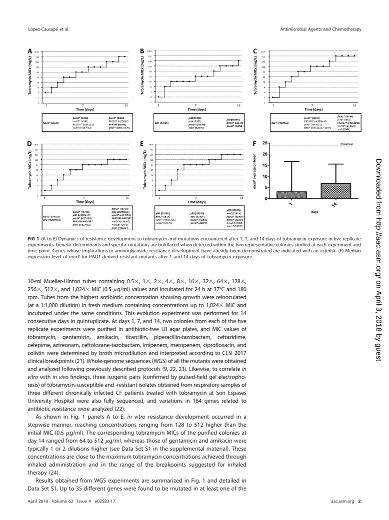
10-ml Mueller-Hinton tubes containing 0.5, 1, 2, 4, 8, 16, 32, 64, 128,256, 512, and 1,024 MIC (0.5 g/ml) values and incubated for 24 h at 37°C and 180rpm. Tubes from the highest antibiotic concentration showing growth were reinoculated(at a 1:1,000 dilution) in fresh medium containing concentrations up to 1,024 MIC andincubated under the same conditions. This evolution experiment was performed for 14consecutive days in quintuplicate. At days 1, 7, and 14, two colonies from each of the fivereplicate experiments were purified in antibiotic-free LB agar plates, and MIC values oftobramycin, gentamicin, amikacin, ticarcillin, piperacillin-tazobactam, ceftazidime,cefepime, aztreonam, ceftolozane-tazobactam, imipenem, meropenem, ciprofloxacin, andcolistin were determined by broth microdilution and interpreted according to CLSI 2017clinical breakpoints (21). Whole-genome sequences (WGS) of all the mutants were obtainedand analyzed following previously described protocols (9, 22, 23). Likewise, to correlate invitro with in vivo findings, three isogenic pairs (confirmed by pulsed-field gel electropho-resis) of tobramycin-susceptible and -resistant isolates obtained from respiratory samples ofthree different chronically infected CF patients treated with tobramycin at Son EspasesUniversity Hospital were also fully sequenced, and variations in 164 genes related toantibiotic resistance were analyzed (22).
As shown in Fig. 1 panels A to E, in vitro resistance development occurred in astepwise manner, reaching concentrations ranging from 128 to 512 higher than theinitial MIC (0.5 g/ml). The corresponding tobramycin MICs of the purified colonies atday 14 ranged from 64 to 512 g/ml, whereas those of gentamicin and amikacin weretypically 1 or 2 dilutions higher (see Data Set S1 in the supplemental material). Theseconcentrations are close to the maximum tobramycin concentrations achieved throughinhaled administration and in the range of the breakpoints suggested for inhaledtherapy (24).
Results obtained from WGS experiments are summarized in Fig. 1 and detailed inData Set S1. Up to 35 different genes were found to be mutated in at least one of the
FIG 1 (A to E) Dynamics of resistance development to tobramycin and mutations encountered after 1, 7, and 14 days of tobramycin exposure in five replicateexperiments. Genetic determinants and specific mutations are boldfaced when detected within the two representative colonies studied at each experiment andtime point. Genes whose implications in aminoglycoside resistance development have already been demonstrated are indicated with an asterisk. (F) Medianexpression level of mexY for PAO1-derived resistant mutants after 1 and 14 days of tobramycin exposure.
López-Causapé et al. Antimicrobial Agents and Chemotherapy
April 2018 Volume 62 Issue 4 e02583-17 aac.asm.org 2
on April 3, 2018 by guest
http://aac.asm.org/
Dow
nloaded from

isolates. Of these, 22 isolates (63%) were already related to aminoglycoside resistancedevelopment in previous studies (15–20, 22), so our data confirm their relevance in thisstepwise process; mutations in other genes are evidenced for the first time in this work,and their relevance will need to be confirmed in further studies. Mutants from day 14showed between 3 and 7 mutations, and comparison with those from days 1 and 7showed a stepwise acquisition. However, a few mutations documented at theseintermediate stages were not fixed in the population and thus were not seen at day 14(Fig. 1).
Among the mutated genes, fusA1, which codes for elongation factor G, deservesspecial attention because nonsynonymous mutations within this gene were detected inall five replicate experiments. Note that the time of detection of fusA1 mutations variedfrom day 1 to day 14 and that the same amino acid substitution (I61M) occurred in 3of them. This novel mechanism was recently confirmed through site-directed mutagen-esis by Bolard et al. (15), being associated with a 1- to 3-fold increase in the MICs oftobramycin, gentamicin, and amikacin, which correlates with our observations (Data SetS1). Mutations in fusA1 were recently noted to be frequent among CF patients (22,25–27). Moreover, the emergence of fusA1 mutations was noted in two of the threetobramycin-resistant CF isolates studied in this work (Table 1; see Data Set S2 in thesupplemental material). Interestingly, using ResFinder, the resistant isolate not showingfusA1 mutations was shown to have acquired an exogenous aminoglycoside-modifyingenzyme (AacA4). This surprising finding highlights the fact that, although mutationalresistance is thought to be the rule in CF chronic infections, horizontally acquiredresistance must be ruled out as well.
Another frequently mutated gene (3 of 5 replicates) was, intriguingly, pmrB, whichcodes for the sensor kinase of the two-component regulatory system PmrA-PmrB (28).pmrB mutations have traditionally been linked to polymyxin resistance development(29, 30), so this finding alerts from a possible mechanism of coresistance to two relevantantipseudomonal agents (polymyxins and aminoglycosides). Indeed, the emergence ofpmrB mutations at day 7 correlated with increased colistin MICs (Data Set S1). However,despite the pmrB mutations persisting at day 14, colistin resistance disappeared, likelyindicating the acquisition of compensatory mutations. In one of these isolates from day
TABLE 1 Genomic differences between the three isogenic pairs of tobramycin-susceptible and -resistant CF isolates
Locus/genea
Isolate ID (MICTOB mg/liter)
FQSE06-S (1) FQSE06-R (24) FQSE11-S (2) FQSE11-R (>256) FQSE16-S (4) FQSE16-R (64)
PA0004/gyrB R138LPA0058/dsbM C28R, F206L, R212CPA0426/mexB nt772Δ1 Q575RPA0958/oprD Q424E, S403APA1430/lasR R216QPA2018/mexY G287A G287SPA2020/mexZ nt290Δ11 S9P L138R L138R R125PPA2492/mexT G274D, G300DPA2639/nuoD G499XPA3064/pelA V446IPA3141/capD nt512ins1PA3168/gyrA Y267NPA4020/mpl S257L Q248XPA4266/fusA1 Y552C, T671I Y552CPA4418/PBP3 P215LPA4462/rpoN V473APA4568/rplU I74 MPA4598/mexD P721S, L624PPA4600/nfxB E75KPA4773/- A165TPA5040/pilQ E676D, E669DResFinder AacA4mexY overexpression
aGenes in which mutations were also detected in the resistance evolution experiment are boldfaced.
P. aeruginosa Aminoglycoside Mutational Resistome Antimicrobial Agents and Chemotherapy
April 2018 Volume 62 Issue 4 e02583-17 aac.asm.org 3
on April 3, 2018 by guest
http://aac.asm.org/
Dow
nloaded from
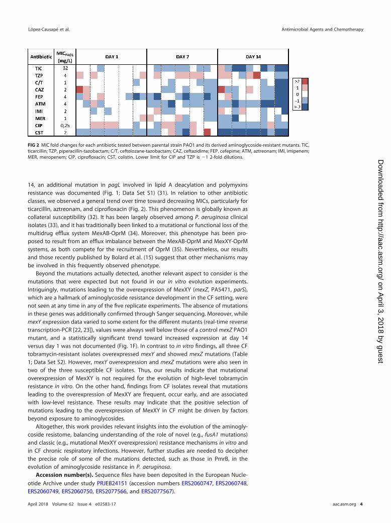
14, an additional mutation in pagL involved in lipid A deacylation and polymyxinsresistance was documented (Fig. 1; Data Set S1) (31). In relation to other antibioticclasses, we observed a general trend over time toward decreasing MICs, particularly forticarcillin, aztreonam, and ciprofloxacin (Fig. 2). This phenomenon is globally known ascollateral susceptibility (32). It has been largely observed among P. aeruginosa clinicalisolates (33), and it has traditionally been linked to a mutational or functional loss of themultidrug efflux system MexAB-OprM (34). Moreover, this phenotype has been pro-posed to result from an efflux imbalance between the MexAB-OprM and MexXY-OprMsystems, as both compete for the recruitment of OprM (35). Nevertheless, our resultsand those recently published by Bolard et al. (15) suggest that other mechanisms maybe involved in this frequently observed phenotype.
Beyond the mutations actually detected, another relevant aspect to consider is themutations that were expected but not found in our in vitro evolution experiments.Intriguingly, mutations leading to the overexpression of MexXY (mexZ, PA5471, parS),which are a hallmark of aminoglycoside resistance development in the CF setting, werenot seen at any time in any of the five replicate experiments. The absence of mutationsin these genes was additionally confirmed through Sanger sequencing. Moreover, whilemexY expression data varied to some extent for the different mutants (real-time reversetranscription-PCR [22, 23]), values were always well below those of a control mexZ PAO1mutant, and a statistically significant trend toward increased expression at day 14versus day 1 was not documented (Fig. 1F). In contrast to in vitro findings, all three CFtobramycin-resistant isolates overexpressed mexY and showed mexZ mutations (Table1; Data Set S2). However, mexY overexpression and mexZ mutations were also seen intwo of the three susceptible CF isolates. Thus, our results indicate that mutationaloverexpression of MexXY is not required for the evolution of high-level tobramycinresistance in vitro. On the other hand, findings from CF isolates reveal that mutationsleading to the overexpression of MexXY are frequent, occur early, and are associatedwith low-level resistance. These results may indicate that the positive selection ofmutations leading to the overexpression of MexXY in CF might be driven by factorsbeyond exposure to aminoglycosides.
Altogether, this work provides relevant insights into the evolution of the aminogly-coside resistome, balancing understanding of the role of novel (e.g., fusA1 mutations)and classic (e.g., mutational MexXY overexpression) resistance mechanisms in vitro andin CF chronic respiratory infections. However, further studies are needed to decipherthe precise role of some of the mutations detected, such as those in PmrB, in theevolution of aminoglycoside resistance in P. aeruginosa.
Accession number(s). Sequence files have been deposited in the European Nucle-otide Archive under study PRJEB24151 (accession numbers ERS2060747, ERS2060748,ERS2060749, ERS2060750, ERS2077566, and ERS2077567).
FIG 2 MIC fold changes for each antibiotic tested between parental strain PAO1 and its derived aminoglycoside-resistant mutants. TIC,ticarcillin; TZP, piperacillin-tazobactam; C/T, ceftolozane-tazobactam; CAZ, ceftazidime; FEP, cefepime; ATM, aztreonam; IMI, imipenem;MER, meropenem; CIP, ciprofloxacin; CST, colistin. Lower limit for CIP and TZP is 1 2-fold dilutions.
López-Causapé et al. Antimicrobial Agents and Chemotherapy
April 2018 Volume 62 Issue 4 e02583-17 aac.asm.org 4
on April 3, 2018 by guest
http://aac.asm.org/
Dow
nloaded from
SUPPLEMENTAL MATERIAL
Supplemental material for this article may be found at https://doi.org/10.1128/AAC.02583-17.
SUPPLEMENTAL FILE 1, XLSX file, 0.1 MB.SUPPLEMENTAL FILE 2, XLSX file, 0.1 MB.
ACKNOWLEDGMENTSThis work was supported by the Ministerio de Economía y Competitividad of Spain,
Instituto de Salud Carlos III, and was cofinanced by the European Regional Develop-ment Fund (ERDF) project “A way to achieve Europe” through the Spanish Network forResearch in Infectious Diseases (REIPI) (RD12/0015 and RD16/0016) and grants PI15/00088.
REFERENCES1. Gellatly SL, Hancock RE. 2013. Pseudomonas aeruginosa: new insights
into pathogenesis and host defenses. Pathog Dis 67:159 –173. https://doi.org/10.1111/2049-632X.12033.
2. Oliver A, Mena A, Macià MD. 2008. Evolution of Pseudomonas aeruginosapathogenicity: from acute to chronic infections, p 433– 444. In BaqueroF, Nombela C, Cassell GH, Gutıerrez JA (ed), Evolutionary biology ofbacterial and fungal pathogens. ASM Press, Washington, DC.
3. Breidenstein EB, de la Fuente-Núñez C, Hancock RE. 2011. Pseudomonasaeruginosa: all roads lead to resistance. Trends Microbiol 19:419 – 426.https://doi.org/10.1016/j.tim.2011.04.005.
4. Silby MW, Winstanley C, Godfrey SA, Levy SB, Jackson RW. 2011. Pseu-domonas genomes: diverse and adaptable. FEMS Microbiol Rev 35:652– 680. https://doi.org/10.1111/j.1574-6976.2011.00269.x.
5. Poole K. 2011. Pseudomonas aeruginosa: resistance to the max. FrontMicrobiol 2:65.
6. Lister PD, Wolter DJ, Hanson ND. 2009. Antibacterial-resistant Pseudomo-nas aeruginosa: clinical impact and complex regulation of chromo-somally encoded resistance mechanisms. Clin Microbiol Rev 22:582– 610.https://doi.org/10.1128/CMR.00040-09.
7. Livermore DM. 2009. Has the era of untreatable infections arrived? JAntimicrob Chemother 64(Suppl)1:i29 –i36. https://doi.org/10.1093/jac/dkp255.
8. Mesaros N, Nordmann P, Plésiat P, Roussel-Delvallez M, Van Eldere J,Glupczynski Y, Van Laethem Y, Jacobs F, Lebecque P, Malfroot A, TulkensPM, Van Bambeke F. 2007. Pseudomonas aeruginosa: resistance andtherapeutic options at the turn of the new millennium. Clin MicrobiolInfect 13:560 –578. https://doi.org/10.1111/j.1469-0691.2007.01681.x.
9. Del Barrio-Tofiño E, López-Causapé C, Cabot G, Rivera A, Benito N, Segura C,Montero MM, Sorlí L, Tubau F, Gómez-Zorrilla S, Tormo N, Durá-Navarro R,Viedma E, Resino-Foz E, Fernández-Martínez M, González-Rico C, Alejo-Cancho I, Martínez JA, Labayru-Echverria C, Dueñas C, Ayestarán I, Zamo-rano L, Martinez-Martinez L, Horcajada JP, Oliver A. 2017. Genomics andsusceptibility profiles of extensively drug-resistant Pseudomonas aeruginosaisolates from Spain. Antimicrob Agents Chemother 61:e01589-17. https://doi.org/10.1128/AAC.01589-17.
10. Cabot G, Ocampo-Sosa AA, Domínguez MA, Gago JF, Juan C, Tubau F,Rodríguez C, Moyà B, Peña C, Martínez-Martínez L, Oliver A, SpanishNetwork for Research in Infectious Diseases (REIPI). 2012. Genetic mark-ers of widespread extensively drug-resistant Pseudomonas aeruginosahigh-risk clones. Antimicrob Agents Chemother 56:6349 – 6357. https://doi.org/10.1128/AAC.01388-12.
11. Shteinberg M, Elborn JS. 2015. Use of inhaled tobramycin in cysticfibrosis. Adv Ther 32:1–9. https://doi.org/10.1007/s12325-015-0179-3.
12. Guénard S, Muller C, Monlezun L, Benas P, Broutin I, Jeannot K, Plésiat P.2014. Multiple mutations lead to MexXY-OprM-dependent aminoglyco-side resistance in clinical strains of Pseudomonas aeruginosa. AntimicrobAgents Chemother 58:221–228. https://doi.org/10.1128/AAC.01252-13.
13. Poole K. 2005. Aminoglycoside resistance in Pseudomonas aeruginosa.Antimicrob Agents Chemother 49:479 – 487. https://doi.org/10.1128/AAC.49.2.479-487.2005.
14. Vogne C, Aires JR, Bailly C, Hocquet D, Plésiat P. 2004. Role of themultidrug efflux system MexXY in the emergence of moderate resis-tance to aminoglycosides among Pseudomonas aeruginosa isolates from
patients with cystic fibrosis. Antimicrob Agents Chemother 48:1676 –1680. https://doi.org/10.1128/AAC.48.5.1676-1680.2004.
15. Bolard A, Plesiat P, Jeannot K. 2018. Mutations in gene fusA1 as a novelmechanism of aminoglycoside resistance in clinical strains of Pseudomo-nas aeruginosa. Antimicrob Agents Chemother 62: e01835-17. https://doi.org/10.1128/AAC.01835-17.
16. Yen P, Papin JA. 2017. History of antibiotic adaptation influences micro-bial evolutionary dynamics during subsequent treatment. PLoS Biol15:e2001586. https://doi.org/10.1371/journal.pbio.2001586.
17. Feng Y, Jonker MJ, Moustakas I, Brul S, Ter Kuile BH. 2016. Dynamics ofmutations during development of resistance by Pseudomonas aerugi-nosa against five antibiotics. Antimicrob Agents Chemother 60:4229 – 4236. https://doi.org/10.1128/AAC.00434-16.
18. Islam S, Oh H, Jalal S, Karpati F, Ciofu O, Høiby N, Wretlind B. 2009.Chromosomal mechanisms of aminoglycoside resistance in Pseudomo-nas aeruginosa isolates from cystic fibrosis patients. Clin Microbiol Infect15:60 – 66. https://doi.org/10.1111/j.1469-0691.2008.02097.x.
19. Schurek KN, Marr AK, Taylor PK, Wiegand I, Semenec L, Khaira BK,Hancock RE. 2008. Novel genetic determinants of low-level aminogly-coside resistance in Pseudomonas aeruginosa. Antimicrob Agents Che-mother 52:4213– 4219. https://doi.org/10.1128/AAC.00507-08.
20. El’Garch F, Jeannot K, Hocquet D, Llanes-Barakat C, Plésiat P. 2007. Cumu-lative effects of several nonenzymatic mechanisms on the resistance ofPseudomonas aeruginosa to aminoglycosides. Antimicrob Agents Che-mother 51:1016–1021. https://doi.org/10.1128/AAC.00704-06.
21. Clinical and Laboratory Standards Institute. 2017. Performance standardsfor antimicrobial susceptibility testing—27th ed, vol 37. CLSI documentM100. Clinical and Laboratory Standards Institute, Wayne, PA.
22. López-Causapé C, Sommer LM, Cabot G, Rubio R, Ocampo-Sosa AA,Johansen HK, Figuerola J, Cantón R, Kidd TJ, Molin S, Oliver A. 2017.Evolution of the Pseudomonas aeruginosa mutational resistome in aninternational cystic fibrosis clone. Sci Rep 7:5555. https://doi.org/10.1038/s41598-017-05621-5.
23. Cabot G, López-Causapé C, Ocampo-Sosa AA, Sommer LM, DomínguezMA, Zamorano L, Juan C, Tubau F, Rodríguez C, Moyà B, Peña C,Martínez-Martínez L, Plesiat P, Oliver A. 2016. Deciphering the resistomeof the widespread Pseudomonas aeruginosa sequence type 175 interna-tional high-risk clone through whole-genome sequencing. AntimicrobAgents Chemother 60:7415–7423.
24. Morosini MI, García-Castillo M, Loza E, Pérez-Vázquez M, Baquero F,Cantón R. 2005. Breakpoints for predicting Pseudomonas aeruginosasusceptibility to inhaled tobramycin in cystic fibrosis patients: use ofhigh-range Etest strips. J Clin Microbiol 43:4480 – 4485. https://doi.org/10.1128/JCM.43.9.4480-4485.2005.
25. Greipel L, Fischer S, Klockgether J, Dorda M, Mielke S, Wiehlmann L,Cramer N, Tümmler B. 2016. Molecular epidemiology of mutations inantimicrobial resistance loci of Pseudomonas aeruginosa isolates fromairways of cystic fibrosis patients. Antimicrob Agents Chemother 60:6726 – 6734. https://doi.org/10.1128/AAC.00724-16.
26. Markussen T, Marvig RL, Gómez-Lozano M, Aanæs K, Burleigh AE, HøibyN, Johansen HK, Molin S, Jelsbak L. 2014. Environmental heterogeneitydrives within-host diversification and evolution of Pseudomonas aerugi-nosa. mBio 5:e01592-14. https://doi.org/10.1128/mBio.01592-14.
27. Chung JC, Becq J, Fraser L, Schulz-Trieglaff O, Bond NJ, Foweraker J,
P. aeruginosa Aminoglycoside Mutational Resistome Antimicrobial Agents and Chemotherapy
April 2018 Volume 62 Issue 4 e02583-17 aac.asm.org 5
on April 3, 2018 by guest
http://aac.asm.org/
Dow
nloaded from
Bruce KD, Smith GP, Welch M. 2012. Genomic variation among contem-porary Pseudomonas aeruginosa isolates from chronically infected cysticfibrosis patients. J Bacteriol 194:4857– 4866. https://doi.org/10.1128/JB.01050-12.
28. Gooderham WJ, Hancock RE. 2009. Regulation of virulence and antibi-otic resistance by two-component regulatory systems in Pseudomonasaeruginosa. FEMS Microbiol Rev 33:279 –294. https://doi.org/10.1111/j.1574-6976.2008.00135.x.
29. Moskowitz SM, Brannon MK, Dasgupta N, Pier M, Sgambati N, Miller AK,Selgrade SE, Miller SI, Denton M, Conway SP, Johansen HK, Høiby N.2012. PmrB mutations promote polymyxin resistance of Pseudomonasaeruginosa isolated from colistin-treated cystic fibrosis patients. Antimi-crob Agents Chemother 56:1019 –1030. https://doi.org/10.1128/AAC.05829-11.
30. Barrow K, Kwon DH. 2009. Alterations in two-component regulatorysystems of phoPQ and pmrAB are associated with polymyxin B resis-tance in clinical isolates of Pseudomonas aeruginosa. Antimicrob AgentsChemother 53:5150 –5154. https://doi.org/10.1128/AAC.00893-09.
31. Han ML, Velkov T, Zhu Y, Roberts KD, Le Brun AP, Chow SH, Gutu AD,
Moskowitz SM, Shen HH, Li J. 2018. Polymyxin-induced lipid A deacyla-tion in Pseudomonas aeruginosa perturbs polymyxin penetration andconfers high-level resistance. ACS Chem Biol 13:121–130. https://doi.org/10.1021/acschembio.7b00836.
32. Pál C, Papp B, Lázár V. 2015. Collateral sensitivity of antibiotic-resistantmicrobes. Trends Microbiol 23:401– 407. https://doi.org/10.1016/j.tim.2015.02.009.
33. Irvin RT, Govan JW, Fyfe JA, Costerton JW. 1981. Heterogeneity ofantibiotic resistance in mucoid isolates of Pseudomonas aeruginosa ob-tained from cystic fibrosis patients: role of outer membrane proteins.Antimicrob Agents Chemother 19:1056 –1063. https://doi.org/10.1128/AAC.19.6.1056.
34. Li XZ, Nikaido H, Poole K. 1995. Role of mexA-mexB-oprM in antibioticefflux in Pseudomonas aeruginosa. Antimicrob Agents Chemother 39:1948 –1953. https://doi.org/10.1128/AAC.39.9.1948.
35. Vettoretti L, Plésiat P, Muller C, El Garch F, Phan G, Attrée I, Ducruix A,Llanes C. 2009. Efflux unbalance in Pseudomonas aeruginosa isolatesfrom cystic fibrosis patients. Antimicrob Agents Chemother 53:1987–1997. https://doi.org/10.1128/AAC.01024-08.
López-Causapé et al. Antimicrobial Agents and Chemotherapy
April 2018 Volume 62 Issue 4 e02583-17 aac.asm.org 6
on April 3, 2018 by guest
http://aac.asm.org/
Dow
nloaded from
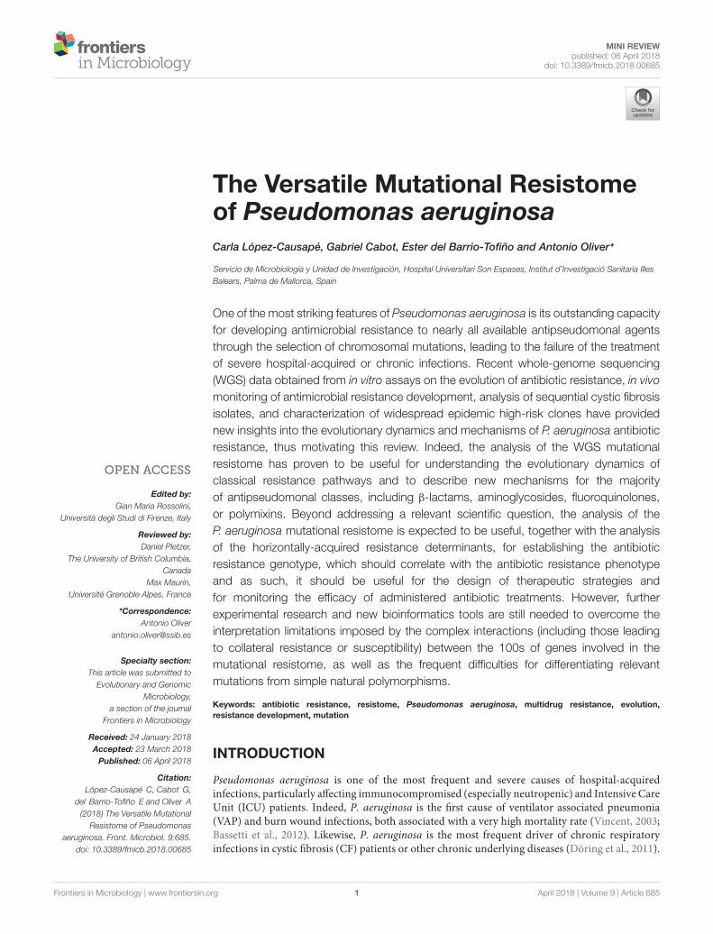
fmicb-09-00685 April 4, 2018 Time: 16:14 # 1
MINI REVIEWpublished: 06 April 2018
doi: 10.3389/fmicb.2018.00685
Edited by:Gian Maria Rossolini,
Università degli Studi di Firenze, Italy
Reviewed by:Daniel Pletzer,
The University of British Columbia,Canada
Max Maurin,Université Grenoble Alpes, France
*Correspondence:Antonio Oliver
Specialty section:This article was submitted to
Evolutionary and GenomicMicrobiology,
a section of the journalFrontiers in Microbiology
Received: 24 January 2018Accepted: 23 March 2018
Published: 06 April 2018
Citation:López-Causapé C, Cabot G,
del Barrio-Tofiño E and Oliver A(2018) The Versatile Mutational
Resistome of Pseudomonasaeruginosa. Front. Microbiol. 9:685.
doi: 10.3389/fmicb.2018.00685
The Versatile Mutational Resistomeof Pseudomonas aeruginosaCarla López-Causapé, Gabriel Cabot, Ester del Barrio-Tofiño and Antonio Oliver*
Servicio de Microbiología y Unidad de Investigación, Hospital Universitari Son Espases, Institut d’Investigació Sanitaria IllesBalears, Palma de Mallorca, Spain
One of the most striking features of Pseudomonas aeruginosa is its outstanding capacityfor developing antimicrobial resistance to nearly all available antipseudomonal agentsthrough the selection of chromosomal mutations, leading to the failure of the treatmentof severe hospital-acquired or chronic infections. Recent whole-genome sequencing(WGS) data obtained from in vitro assays on the evolution of antibiotic resistance, in vivomonitoring of antimicrobial resistance development, analysis of sequential cystic fibrosisisolates, and characterization of widespread epidemic high-risk clones have providednew insights into the evolutionary dynamics and mechanisms of P. aeruginosa antibioticresistance, thus motivating this review. Indeed, the analysis of the WGS mutationalresistome has proven to be useful for understanding the evolutionary dynamics ofclassical resistance pathways and to describe new mechanisms for the majorityof antipseudomonal classes, including β-lactams, aminoglycosides, fluoroquinolones,or polymixins. Beyond addressing a relevant scientific question, the analysis of theP. aeruginosa mutational resistome is expected to be useful, together with the analysisof the horizontally-acquired resistance determinants, for establishing the antibioticresistance genotype, which should correlate with the antibiotic resistance phenotypeand as such, it should be useful for the design of therapeutic strategies andfor monitoring the efficacy of administered antibiotic treatments. However, furtherexperimental research and new bioinformatics tools are still needed to overcome theinterpretation limitations imposed by the complex interactions (including those leadingto collateral resistance or susceptibility) between the 100s of genes involved in themutational resistome, as well as the frequent difficulties for differentiating relevantmutations from simple natural polymorphisms.
Keywords: antibiotic resistance, resistome, Pseudomonas aeruginosa, multidrug resistance, evolution,resistance development, mutation
INTRODUCTION
Pseudomonas aeruginosa is one of the most frequent and severe causes of hospital-acquiredinfections, particularly affecting immunocompromised (especially neutropenic) and Intensive CareUnit (ICU) patients. Indeed, P. aeruginosa is the first cause of ventilator associated pneumonia(VAP) and burn wound infections, both associated with a very high mortality rate (Vincent, 2003;Bassetti et al., 2012). Likewise, P. aeruginosa is the most frequent driver of chronic respiratoryinfections in cystic fibrosis (CF) patients or other chronic underlying diseases (Döring et al., 2011).
Frontiers in Microbiology | www.frontiersin.org 1 April 2018 | Volume 9 | Article 685
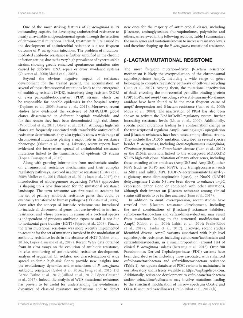
fmicb-09-00685 April 4, 2018 Time: 16:14 # 2
López-Causapé et al. The Mutational Resistome of P. aeruginosa
One of the most striking features of P. aeruginosa is itsoutstanding capacity for developing antimicrobial resistance tonearly all available antipseudomonal agents through the selectionof chromosomal mutations. Indeed, treatment failure caused bythe development of antimicrobial resistance is a too frequentoutcome of P. aeruginosa infections. The problem of mutation-mediated antibiotic resistance is further amplified in the chronicinfection setting, due to the very high prevalence of hypermutablestrains, showing greatly enhanced spontaneous mutation ratescaused by defective DNA repair or error avoidance systems(Oliver et al., 2000; Maciá et al., 2005).
Beyond the obvious negative impact of resistancedevelopment for the treated patient, the accumulation ofseveral of these chromosomal mutations leads to the emergenceof multidrug resistant (MDR), extensively drug-resistant (XDR)or even pan-antibiotic-resistant (PDR) strains, which canbe responsible for notable epidemics in the hospital setting(Deplano et al., 2005; Suarez et al., 2011). Moreover, recentstudies have evidenced the existence of MDR/XDR globalclones disseminated in different hospitals worldwide, andfor that reason they have been denominated high-risk clones(Woodford et al., 2011; Oliver et al., 2015). Although high-riskclones are frequently associated with transferable antimicrobialresistance determinants, they also typically show a wide range ofchromosomal mutations playing a major role in the resistancephenotype (Oliver et al., 2015). Likewise, recent reports haveevidenced the interpatient spread of antimicrobial resistancemutations linked to the transmission of epidemic CF strains(López-Causapé et al., 2017).
Along with growing information from mechanistic studieson chromosomal resistance mechanisms and their complexregulatory pathways, involved in adaptive resistance (Lister et al.,2009; Muller et al., 2011; Skiada et al., 2011; Juan et al., 2017), theintroduction of whole-genome sequencing (WGS) approachesis shaping up a new dimension for the mutational resistancelandscape. The term resistome was first used to account forthe set of primary antibiotic resistance genes that could beeventually transferred to human pathogens (D’Costa et al., 2006).Soon after the concept of intrinsic resistome was introducedto include all chromosomal genes that are involved in intrinsicresistance, and whose presence in strains of a bacterial speciesis independent of previous antibiotic exposure and is not dueto horizontal gene transfer (HGT) (Fajardo et al., 2008). Finally,the term mutational resistome was more recently implementedto account for the set of mutations involved in the modulation ofantibiotic resistance levels in the absence of HGT (Cabot et al.,2016b; López-Causapé et al., 2017). Recent WGS data obtainedfrom in vitro assays on the evolution of antibiotic resistance,in vivo monitoring of antimicrobial resistance development,analysis of sequential CF isolates, and characterization of widespread epidemic high-risk clones provide new insights intothe evolutionary dynamics and mechanisms of P. aeruginosaantibiotic resistance (Cabot et al., 2016a; Feng et al., 2016; DelBarrio-Tofiño et al., 2017; Jaillard et al., 2017; López-Causapéet al., 2017). Indeed, the analysis of WGS mutational resistomeshas proven to be useful for understanding the evolutionarydynamics of classical resistance mechanisms and to depict
new ones for the majority of antimicrobial classes, includingβ-lactams, aminoglycosides, fluoroquinolones, polymixins andothers, as reviewed in the following sections. Table 1 summarizesthe main genes and mutations known to increase resistance levelsand therefore shaping up the P. aeruginosa mutational resistome.
β-LACTAM MUTATIONAL RESISTOME
The most frequent mutation-driven β-lactam resistancemechanism is likely the overproduction of the chromosomalcephalosporinase AmpC, involving a wide range of genesbelonging to complex regulatory pathways of cell-wall recycling(Juan et al., 2017). Among them, the mutational inactivationof dacB, encoding the non-essential penicillin-binding protein(PBP) PBP4, and ampD, encoding a N-acetyl-muramyl-L-alanineamidase have been found to be the most frequent cause ofampC derepression and β-lactam resistance (Juan et al., 2005;Moya et al., 2009). The inactivation of PBP4 has also beenshown to activate the BlrAB/CreBC regulatory system, furtherincreasing resistance levels (Moya et al., 2009). Additionally,specific point mutations leading to a conformation change inthe transcriptional regulator AmpR, causing ampC upregulationand β-lactam resistance, have been noted among clinical strains.They include the D135N mutation, described in several speciesbesides P. aeruginosa, including Stenotrophomonas maltophilia,Citrobacter freundii, or Enterobacter cloacae (Juan et al., 2017)or the R154H mutation, linked to the widespread MDR/XDRST175 high-risk clone. Mutation of many other genes, includingthose encoding other amidases (AmpDh2 and AmpDh3), otherPBPs (such as PBP5 and PBP7), lytic transglycosylases (suchas SltB1 and mltB), MPL (UDP-N-acetylmuramate:Lalanyl-γ-D-glutamyl-meso-diaminopimelate ligase), or NuoN (NADHdehydrogenase I chain N) have been shown to enhance ampCexpression, either alone or combined with other mutations,although their impact on β-lactam resistance among clinicalstrains still needs to be further analyzed (Juan et al., 2017).
In addition to ampC overexpression, recent studies haverevealed that β-lactam resistance development, includingthe novel combinations of β-lactam-β-lactamase inhibitorsceftolozane/tazobactam and ceftazidime/avibactam, may resultfrom mutations leading to the structural modification ofAmpC (Cabot et al., 2014; Lahiri et al., 2014; Fraile-Ribotet al., 2017a; Haidar et al., 2017). Likewise, recent studiesidentified diverse AmpC variants associated with high-levelcephalosporin resistance, including ceftolozane/tazobactam andceftazidime/avibactam, in a small proportion (around 1%) ofclinical P. aeruginosa isolates (Berrazeg et al., 2015). Over 200Pseudomonas Derived Cephalosporinase (PDC) variants havebeen described so far, including those associated with enhancedceftolozane/tazobactam and ceftazidime/avibactam resistance(Table 1). An update database of PDC variants is maintained inour laboratory and is freely available at https://arpbigidisba.com.Additionally, resistance development to ceftolozane/tazobactamand/or ceftazidime/avibactam may involve mutations leadingto the structural modification of narrow spectrum OXA-2 andOXA-10 acquired oxacillinases (Fraile-Ribot et al., 2017a,b).
Frontiers in Microbiology | www.frontiersin.org 2 April 2018 | Volume 9 | Article 685

fmicb-09-00685 April 4, 2018 Time: 16:14 # 3
López-Causapé et al. The Mutational Resistome of P. aeruginosa
TABLE 1 | Main genes and mutations known to be involved in increased P. aeruginosa antibiotic resistance.
Gene Resistancemechanisms/altered target
Antibioticsaffecteda
Type of mutation Relevant examples ofgain-of-functionmutations
Reference
gyrA DNA gyrase FQ Gain-of-function G81D, T83A, T83I,Y86N, D87G, D87N,D87Y, Q106L
Bruchmann et al., 2013; Kos et al.,2015; Cabot et al., 2016b; DelBarrio-Tofiño et al., 2017;López-Causapé et al., 2017
gyrB DNA gyrase FQ Gain-of-function S466F, S466Y, Q467R,E468D
Bruchmann et al., 2013; Kos et al.,2015; Del Barrio-Tofiño et al., 2017;López-Causapé et al., 2017
parC DNA topoisomerase IV FQ Gain-of-function S87L, S87W Bruchmann et al., 2013; Kos et al.,2015; Cabot et al., 2016b; DelBarrio-Tofiño et al., 2017
parE DNA topoisomerase IV FQ Gain-of-function S457G, S457T, E459D,E459K
Bruchmann et al., 2013; Kos et al.,2015; Del Barrio-Tofiño et al., 2017;López-Causapé et al., 2017
pmrA Lipopolysaccharide(lipid A)
COL Gain-of-function L157Q Lee and Ko, 2014
pmrB Lipopolysaccharide(lipid A)
COL Gain-of-function L14P, A54V, R79H,R135Q, A247T, A248T,A248V, R259H, M292I,M292T
Barrow and Kwon, 2009;Moskowitz et al., 2012
phoQ Lipopolysaccharide(lipid A)
COL Loss-of-function
parR Lipopolysaccharide(lipid A)
COL Gain-of-function M59I, E156K Muller et al., 2011; Guénard et al.,2014
OprD downregulation IMP, MER
MexEF-OprNhyperproduction
FQ
MexXY-OprMhyperproduction
FQ, AMG, CEF
parS Lipopolysaccharide(lipid A)
COL Gain-of-function L14Q, V101M, L137P,A138T, A168V Q232E,G361R
Muller et al., 2011; Fournier et al.,2013; Guénard et al., 2014
OprD downregulation IMP, MER
MexEF-OprNhyperproduction
FQ
MexXY-OprMhyperproduction
FQ, AMG, CEF
cprS Lipopolysaccharide(lipid A)
COL Gain-of-function R241C Gutu et al., 2013
colR Lipopolysaccharide(lipid A)
COL Gain-of-function D32N Gutu et al., 2013
colS Lipopolysaccharide(lipid A)
COL Gain-of-function A106V Gutu et al., 2013
mexR MexAB-OprMhyperproduction
FQ, CAZ, CEF, PPT,MER, CAZ/AVI
Loss-of-function
nalC MexAB-OprMhyperproduction
FQ, CAZ, CEF, PPT,MER, CAZ/AVI
Loss-of-function
nalD MexAB-OprMhyperproduction
FQ, CAZ, CEF, PPT,MER, CAZ/AVI
Loss-of-function
nfxB MexCD-OprJhyperproduction
FQ, CEF Loss-of-function
mexS MexEF-OprNhyperproduction
FQ Loss-of-function
OprD downregulation IMP, MER
mexT MexEF-OprNhyperproduction
FQ Gain-of-function G257S, G257A Juarez et al., 2018
OprD downregulation IMP, MER
(Continued)
Frontiers in Microbiology | www.frontiersin.org 3 April 2018 | Volume 9 | Article 685
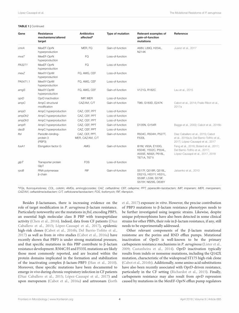
fmicb-09-00685 April 4, 2018 Time: 16:14 # 4
López-Causapé et al. The Mutational Resistome of P. aeruginosa
TABLE 1 | Continued
Gene Resistancemechanisms/alteredtarget
Antibioticsaffecteda
Type of mutation Relevant examples ofgain-of-functionmutations
Reference
cmrA MexEF-OprNhyperproduction
MER, FQ Gain-of-function A68V, L89Q, H204L,N214K
Juarez et al., 2017
mvaT MexEF-OprNhyperproduction
FQ Loss-of-function
PA3271 MexEF-OprNhyperproduction
FQ Loss-of-function
mexZ MexXY-OprMhyperproduction
FQ, AMG, CEF Loss-of-function
PA5471.1 MexXY-OprMhyperproduction
FQ, AMG, CEF Loss-of-function
amgS MexXY-OprMhyperproduction
FQ, AMG, CEF Gain-of-function V121G, R182C Lau et al., 2015
oprD OprD inactivation IMP, MER Loss-of-function
ampC AmpC structuralmodification
CAZ/AVI, C/T Gain-of-function T96I, G183D, E247K Cabot et al., 2014; Fraile-Ribot et al.,2017a
ampD AmpC hyperproduction CAZ, CEF, PPT Loss-of-function
ampDh2 AmpC hyperproduction CAZ, CEF, PPT Loss-of-function
ampDh3 AmpC hyperproduction CAZ, CEF, PPT Loss-of-function
ampR AmpC hyperproduction CAZ, CEF, PPT Gain-of-function D135N, G154R Bagge et al., 2002; Cabot et al., 2016b
dacB AmpC hyperproduction CAZ, CEF, PPT Loss-of-function
ftsI Penicillin-binding-protein 3(PBP3)
CAZ, CEF, PPT,MER, CAZ/AVI, C/T
Gain-of-function R504C, R504H, P527T,F533L
Diaz Caballero et al., 2015; Cabotet al., 2016a,b; Del Barrio-Tofiño et al.,2017; López-Causapé et al., 2017
fusA1 Elongation factor G AMG Gain-of-function I61M, V93A, E100G,K504E, Y552C, P554L,A555E, N592I, P618L,T671A, T671I
Feng et al., 2016; Bolard et al., 2017;Del Barrio-Tofiño et al., 2017;López-Causapé et al., 2017, 2018
glpT Transporter proteinGlpT
FOS Loss-of-function
rpoB RNA polymeraseβ-chain
RIF Gain-of-function S517F, Q518R, Q518L,D521G, H531Y, H531L,S536F, L538I, S579F,S579Y, N629S, D636Y
Jatsenko et al., 2010
aFQs, fluoroquinolones; COL, colistin; AMGs, aminoglycosides; CAZ, ceftazidime; CEF, cefepime; PPT, piperacillin-tazobactam; IMP, imipenem; MER, meropenem;CAZ/AVI, ceftazidime/avibactam; C/T, ceftolozane/tazobactam; FOS, fosfomycin; RIF, rifampicin.
Besides β-lactamases, there is increasing evidence on therole of target modification in P. aeruginosa β-lactam resistance.Particularly noteworthy are the mutations in ftsI, encoding PBP3,an essential high molecular class B PBP with transpeptidaseactivity (Chen et al., 2016). Indeed, data from CF patients (DiazCaballero et al., 2015; López-Causapé et al., 2017), epidemichigh-risk clones (Cabot et al., 2016b; Del Barrio-Tofiño et al.,2017) as well as from in vitro studies (Cabot et al., 2016a) haverecently shown that PBP3 is under strong mutational pressure,and that specific mutations in this PBP contribute to β-lactamresistance development. R504C/H and F533L mutations are likelythose most commonly reported, and are located within theprotein domains implicated in the formation and stabilizationof the inactivating complex β-lactam-PBP3 (Han et al., 2010).Moreover, these specific mutations have been documented toemerge in vivo during chronic respiratory infection in CF patients(Diaz Caballero et al., 2015; López-Causapé et al., 2017) andupon meropenem (Cabot et al., 2016a) and aztreonam (Jorth
et al., 2017) exposure in vitro. However, the precise contributionof PBP3 mutations to β-lactam resistance phenotypes needs tobe further investigated using isogenic strains. Likewise, despiteunique polymorphisms have also been detected in some clinicalstrains for other PBPs, their role in β-lactam resistance, if any, stillneeds to be experimentally addressed.
Other relevant components of the β-lactam mutationalresistome are the porins and RND efflux pumps. Mutationalinactivation of OprD is well-known to be the primarycarbapenem resistance mechanisms in P. aeruginosa (Lister et al.,2009; Castanheira et al., 2014). OprD inactivation typicallyresults from indels or nonsense mutations, including the Q142Xmutation, characteristic of the widespread ST175 high-risk clone(Cabot et al., 2016b). Additionally, some amino acid substitutionshave also been recently associated with OprD-driven resistance,particularly in the CF setting (Richardot et al., 2015). Finally,carbapenem resistance may also result from oprD repressioncaused by mutations in the MexEF-OprN efflux pump regulators
Frontiers in Microbiology | www.frontiersin.org 4 April 2018 | Volume 9 | Article 685
fmicb-09-00685 April 4, 2018 Time: 16:14 # 5
López-Causapé et al. The Mutational Resistome of P. aeruginosa
(mexS/T) or the ParRS two-component system (Li et al.,2015). Overexpression of MexAB-OprM, caused by mutation ofseveral genes involved in its regulation (mexR, nalC, or nalD)increases MICs of most β-lactams except imipenem, whereasoverexpression of MexXY (mexZ, parSR, amgS mutations) isparticularly involved in cefepime resistance (Li et al., 2015).Additionally, sequence variations in unique residues are detectedin the genes coding for the efflux pump (Del Barrio-Tofiño et al.,2017; López-Causapé et al., 2017); however, their contribution toresistance profiles, if any, still needs to be further explored.
Finally, another potentially relevant mutational β-lactamresistance mechanism is the selection of large (>200 kb) deletionsaffecting specific parts of the chromosome (Cabot et al., 2016a).Although the basis of the conferred resistance phenotype stillneeds to be further clarified, these mutants can be recognizedby the characteristic brown pigment (pyomelanine) caused bythe deletion of one of the affected genes, hmgA, coding fora homogentisate-1,2- dioxygenase. This type of deletion hasbeen documented in both, in vitro evolved β-lactam-resistantmutants and CF isolates (Cabot et al., 2016a; Hocquet et al.,2016). However, the deletion of hmgA is not responsible forthe resistance phenotype, which could be linked to the deletionof another of the affected genes, galU. This gene codes for aUDP-glucose pyrophosphorylase involved in the synthesis of thelipopolysaccharide (LPS) core. Indeed, analysis of transposonmutant libraries has shown that inactivation of galU increasesceftazidime and meropenem MICs (Dötsch et al., 2009; Alvarez-Ortega et al., 2010).
AMINOGLYCOSIDE MUTATIONALRESISTOME
In the absence of horizontally-acquired aminoglycosidemodifying enzymes, resistance to this antibiotic class has beenparticularly linked to the mutational overexpression of MexXY-OprM. Indeed, mutational overexpression of this pump, mainlycaused by mexZ, amgS, or parRS mutations (Table 1), is veryfrequent among clinical isolates, from both, CF patients andnosocomial infections (Guénard et al., 2014; Prickett et al., 2017).Moreover, recent studies show that the epidemic high-risk cloneST175 overexpresses MexXY due to a specific mutation in mexZ(G195E) (Cabot et al., 2016b). However, recent studies haverevealed that the aminoglycoside mutational resistome extendsfar beyond MexXY overexpression, and that high-level resistancemay result from the accumulation of multiple mutations, andthe involvement of several novel resistance determinants hasbeen recently documented (El’Garch et al., 2007; Schurek et al.,2008; Feng et al., 2016). Among them is particularly noteworthyfusA1, coding for the elongation factor G. Indeed, specific FusA1mutations have been associated with aminoglycoside resistancein vitro (Feng et al., 2016; López-Causapé et al., 2018) and amongclinical, particularly CF, strains (Chung et al., 2012; Markussenet al., 2014; Greipel et al., 2016; López-Causapé et al., 2017, 2018).Moreover, the implication of fusA1 mutations in aminoglycosideresistance has been recently confirmed through site-directedmutagenesis (Bolard et al., 2017).
FLUOROQUINOLONE MUTATIONALRESISTOME
The fluoroquinolone mutational resistome generally includesspecific missense mutations in DNA gyrase (gyrA and/or gyrB)and topisomerase IV (parC and/or parE) Quinolone Resistance-Determining Regions (QRDRs) (Table 1) (Bruchmann et al.,2013; Kos et al., 2015). High-level fluoroquinolone resistance inP. aeruginosa high-risk clones is nearly universal, and typicallyinvolves combinations of mutations in GyrA-T83 and ParC-S87(Del Barrio-Tofiño et al., 2017). QRDR mutations involved influoroquinolone resistance in CF might be more variable (López-Causapé et al., 2017). It is also well-known that the mutationaloverexpression of efflux pumps modulate fluoroquinoloneresistance (Table 1). While the overexpression of MexAB-OprMand MexXY-OprM is globally more frequent among clinicalstrains, its contribution to clinical fluoroquinolone resistance islikely more modest (Bruchmann et al., 2013). On the other hand,the mutational overexpression of MexEF-OprN or MexCD-OprJis associated with high-level (clinical) fluoroquinolone resistance,and although their prevalence is considered low except in the CFchronic infection setting, recent data show that it might be higherthan expected (Richardot et al., 2015).
POLYMIXIN MUTATIONAL RESISTOME
Whereas the prevalence of polymyxin (colistin and polymyxin B)resistance is still globally low (<5%), it has increased in thelast years because of the frequent use of these last-resourceantibiotics for the treatment of MDR/XDR nosocomial andCF strains. Polymyxin resistance results most frequently fromthe modification of the LPS caused by the addition of a4-amino-4-deoxy-L-arabinose moiety in the lipid A structure(Olaitan et al., 2014; Jeannot et al., 2017). The involvedmutations are frequently located in the PmrAB or PhoPQtwo-component regulators, which lead to the activation ofthe arnBCADTEF operon (Barrow and Kwon, 2009). Morerecent studies have revealed that mutations in the ParRS two-component regulator, not only produce polymyxin resistancedue to the activation of the arnBCADTEF operon, but alsolead to a MDR phenotype determined by the overprexpressionof MexXY and the repression of OprD (Muller et al., 2011).Moreover, two additional two-component regulators, ColRS andCprRS, have been recently found to be involved in polymyxinresistance (Gutu et al., 2013). The analysis of colistin resistancemechanisms among clinical strains is not always straight forward,since the presence of mutations in these two-componentregulators is not always associated with clinical colistin resistance,probably denoting partial complementation between the differentregulators (Moskowitz et al., 2012; Gutu et al., 2013; López-Causapé et al., 2017). Moreover, recent in vitro evolutionassays have revealed the implication of additional mutations inhigh level colistin resistance, facilitated by the emergence ofmutator (mutS deficient) phenotypes (Dößelmann et al., 2017).Particularly noteworthy among them are those occurring in LptD,LpxC, or MigA.
Frontiers in Microbiology | www.frontiersin.org 5 April 2018 | Volume 9 | Article 685
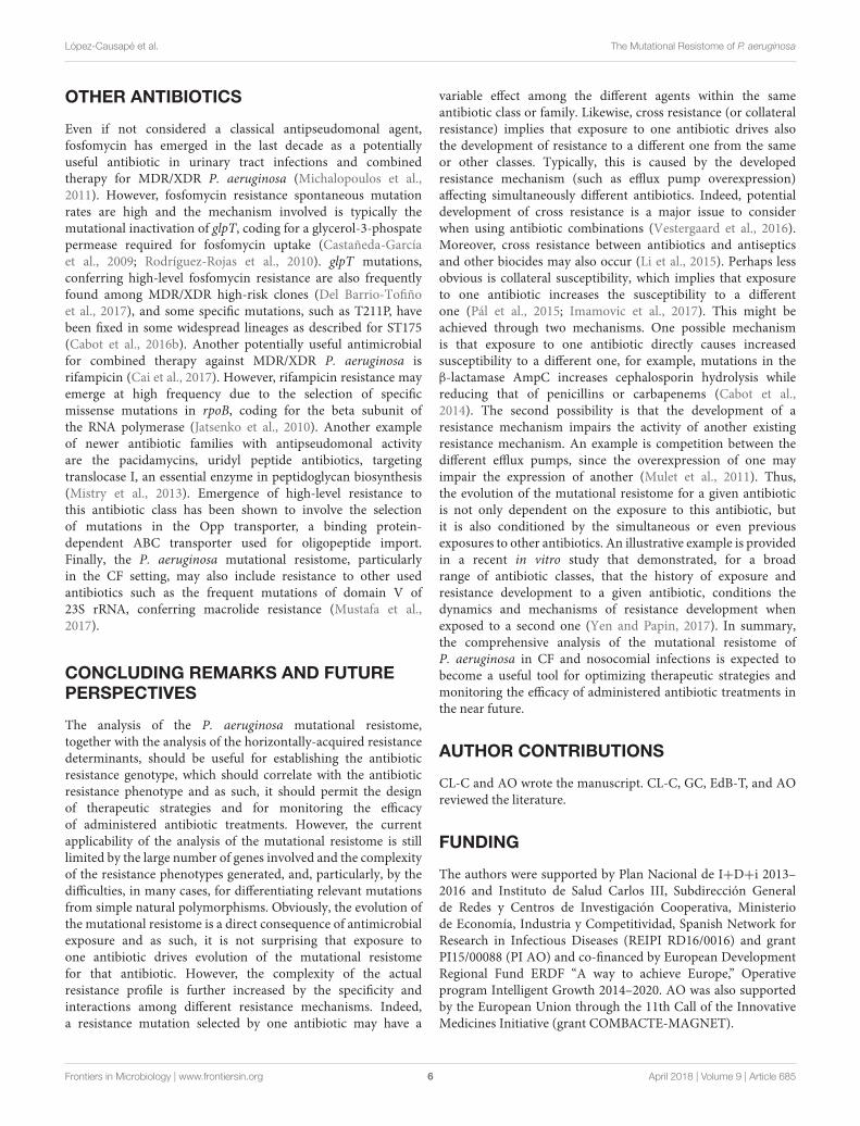
fmicb-09-00685 April 4, 2018 Time: 16:14 # 6
López-Causapé et al. The Mutational Resistome of P. aeruginosa
OTHER ANTIBIOTICS
Even if not considered a classical antipseudomonal agent,fosfomycin has emerged in the last decade as a potentiallyuseful antibiotic in urinary tract infections and combinedtherapy for MDR/XDR P. aeruginosa (Michalopoulos et al.,2011). However, fosfomycin resistance spontaneous mutationrates are high and the mechanism involved is typically themutational inactivation of glpT, coding for a glycerol-3-phospatepermease required for fosfomycin uptake (Castañeda-Garcíaet al., 2009; Rodríguez-Rojas et al., 2010). glpT mutations,conferring high-level fosfomycin resistance are also frequentlyfound among MDR/XDR high-risk clones (Del Barrio-Tofiñoet al., 2017), and some specific mutations, such as T211P, havebeen fixed in some widespread lineages as described for ST175(Cabot et al., 2016b). Another potentially useful antimicrobialfor combined therapy against MDR/XDR P. aeruginosa isrifampicin (Cai et al., 2017). However, rifampicin resistance mayemerge at high frequency due to the selection of specificmissense mutations in rpoB, coding for the beta subunit ofthe RNA polymerase (Jatsenko et al., 2010). Another exampleof newer antibiotic families with antipseudomonal activityare the pacidamycins, uridyl peptide antibiotics, targetingtranslocase I, an essential enzyme in peptidoglycan biosynthesis(Mistry et al., 2013). Emergence of high-level resistance tothis antibiotic class has been shown to involve the selectionof mutations in the Opp transporter, a binding protein-dependent ABC transporter used for oligopeptide import.Finally, the P. aeruginosa mutational resistome, particularlyin the CF setting, may also include resistance to other usedantibiotics such as the frequent mutations of domain V of23S rRNA, conferring macrolide resistance (Mustafa et al.,2017).
CONCLUDING REMARKS AND FUTUREPERSPECTIVES
The analysis of the P. aeruginosa mutational resistome,together with the analysis of the horizontally-acquired resistancedeterminants, should be useful for establishing the antibioticresistance genotype, which should correlate with the antibioticresistance phenotype and as such, it should permit the designof therapeutic strategies and for monitoring the efficacyof administered antibiotic treatments. However, the currentapplicability of the analysis of the mutational resistome is stilllimited by the large number of genes involved and the complexityof the resistance phenotypes generated, and, particularly, by thedifficulties, in many cases, for differentiating relevant mutationsfrom simple natural polymorphisms. Obviously, the evolution ofthe mutational resistome is a direct consequence of antimicrobialexposure and as such, it is not surprising that exposure toone antibiotic drives evolution of the mutational resistomefor that antibiotic. However, the complexity of the actualresistance profile is further increased by the specificity andinteractions among different resistance mechanisms. Indeed,a resistance mutation selected by one antibiotic may have a
variable effect among the different agents within the sameantibiotic class or family. Likewise, cross resistance (or collateralresistance) implies that exposure to one antibiotic drives alsothe development of resistance to a different one from the sameor other classes. Typically, this is caused by the developedresistance mechanism (such as efflux pump overexpression)affecting simultaneously different antibiotics. Indeed, potentialdevelopment of cross resistance is a major issue to considerwhen using antibiotic combinations (Vestergaard et al., 2016).Moreover, cross resistance between antibiotics and antisepticsand other biocides may also occur (Li et al., 2015). Perhaps lessobvious is collateral susceptibility, which implies that exposureto one antibiotic increases the susceptibility to a differentone (Pál et al., 2015; Imamovic et al., 2017). This might beachieved through two mechanisms. One possible mechanismis that exposure to one antibiotic directly causes increasedsusceptibility to a different one, for example, mutations in theβ-lactamase AmpC increases cephalosporin hydrolysis whilereducing that of penicillins or carbapenems (Cabot et al.,2014). The second possibility is that the development of aresistance mechanism impairs the activity of another existingresistance mechanism. An example is competition between thedifferent efflux pumps, since the overexpression of one mayimpair the expression of another (Mulet et al., 2011). Thus,the evolution of the mutational resistome for a given antibioticis not only dependent on the exposure to this antibiotic, butit is also conditioned by the simultaneous or even previousexposures to other antibiotics. An illustrative example is providedin a recent in vitro study that demonstrated, for a broadrange of antibiotic classes, that the history of exposure andresistance development to a given antibiotic, conditions thedynamics and mechanisms of resistance development whenexposed to a second one (Yen and Papin, 2017). In summary,the comprehensive analysis of the mutational resistome ofP. aeruginosa in CF and nosocomial infections is expected tobecome a useful tool for optimizing therapeutic strategies andmonitoring the efficacy of administered antibiotic treatments inthe near future.
AUTHOR CONTRIBUTIONS
CL-C and AO wrote the manuscript. CL-C, GC, EdB-T, and AOreviewed the literature.
FUNDING
The authors were supported by Plan Nacional de I+D+i 2013–2016 and Instituto de Salud Carlos III, Subdirección Generalde Redes y Centros de Investigación Cooperativa, Ministeriode Economía, Industria y Competitividad, Spanish Network forResearch in Infectious Diseases (REIPI RD16/0016) and grantPI15/00088 (PI AO) and co-financed by European DevelopmentRegional Fund ERDF “A way to achieve Europe,” Operativeprogram Intelligent Growth 2014–2020. AO was also supportedby the European Union through the 11th Call of the InnovativeMedicines Initiative (grant COMBACTE-MAGNET).
Frontiers in Microbiology | www.frontiersin.org 6 April 2018 | Volume 9 | Article 685
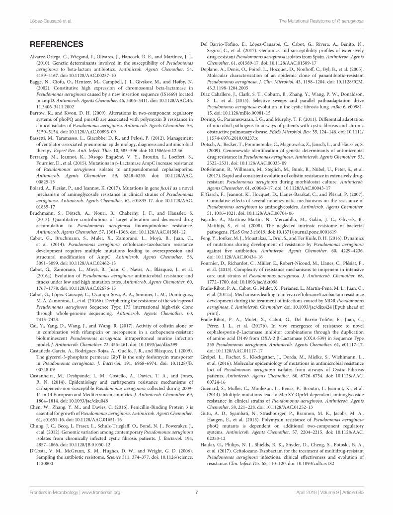
fmicb-09-00685 April 4, 2018 Time: 16:14 # 7
López-Causapé et al. The Mutational Resistome of P. aeruginosa
REFERENCESAlvarez-Ortega, C., Wiegand, I., Olivares, J., Hancock, R. E., and Martínez, J. L.
(2010). Genetic determinants involved in the susceptibility of Pseudomonasaeruginosa to beta-lactam antibiotics. Antimicrob. Agents Chemother. 54,4159–4167. doi: 10.1128/AAC.00257-10
Bagge, N., Ciofu, O., Hentzer, M., Campbell, J. I., Givskov, M., and Høiby, N.(2002). Constitutive high expression of chromosomal beta-lactamase inPseudomonas aeruginosa caused by a new insertion sequence (IS1669) locatedin ampD. Antimicrob. Agents Chemother. 46, 3406–3411. doi: 10.1128/AAC.46.11.3406-3411.2002
Barrow, K., and Kwon, D. H. (2009). Alterations in two-component regulatorysystems of phoPQ and pmrAB are associated with polymyxin B resistance inclinical isolates of Pseudomonas aeruginosa. Antimicrob. Agents Chemother. 53,5150–5154. doi: 10.1128/AAC.00893-09
Bassetti, M., Taramasso, L., Giacobbe, D. R., and Pelosi, P. (2012). Managementof ventilator-associated pneumonia: epidemiology, diagnosis and antimicrobialtherapy. Expert Rev. Anti. Infect. Ther. 10, 585–596. doi: 10.1586/eri.12.36
Berrazeg, M., Jeannot, K., Ntsogo Enguéné, V. Y., Broutin, I., Loeffert, S.,Fournier, D., et al. (2015). Mutations in β-Lactamase AmpC increase resistanceof Pseudomonas aeruginosa isolates to antipseudomonal cephalosporins.Antimicrob. Agents Chemother. 59, 6248–6255. doi: 10.1128/AAC.00825-15
Bolard, A., Plesiat, P., and Jeannot, K. (2017). Mutations in gene fusA1 as a novelmechanism of aminoglycoside resistance in clinical strains of Pseudomonasaeruginosa. Antimicrob. Agents Chemother. 62, e01835-17. doi: 10.1128/AAC.01835-17
Bruchmann, S., Dötsch, A., Nouri, B., Chaberny, I. F., and Häussler, S.(2013). Quantitative contributions of target alteration and decreased drugaccumulation to Pseudomonas aeruginosa fluoroquinolone resistance.Antimicrob. Agents Chemother. 57, 1361–1368. doi: 10.1128/AAC.01581-12
Cabot, G., Bruchmann, S., Mulet, X., Zamorano, L., Moyà, B., Juan, C.,et al. (2014). Pseudomonas aeruginosa ceftolozane-tazobactam resistancedevelopment requires multiple mutations leading to overexpression andstructural modification of AmpC. Antimicrob. Agents Chemother. 58,3091–3099. doi: 10.1128/AAC.02462-13
Cabot, G., Zamorano, L., Moyà, B., Juan, C., Navas, A., Blázquez, J., et al.(2016a). Evolution of Pseudomonas aeruginosa antimicrobial resistance andfitness under low and high mutation rates. Antimicrob. Agents Chemother. 60,1767–1778. doi: 10.1128/AAC.02676-15
Cabot, G., López-Causapé, C., Ocampo-Sosa, A. A., Sommer, L. M., Domínguez,M. Á, Zamorano, L., et al. (2016b). Deciphering the resistome of the widespreadPseudomonas aeruginosa Sequence Type 175 international high-risk clonethrough whole-genome sequencing. Antimicrob. Agents Chemother. 60,7415–7423.
Cai, Y., Yang, D., Wang, J., and Wang, R. (2017). Activity of colistin alone orin combination with rifampicin or meropenem in a carbapenem-resistantbioluminescent Pseudomonas aeruginosa intraperitoneal murine infectionmodel. J. Antimicrob. Chemother. 73, 456–461. doi: 10.1093/jac/dkx399
Castañeda-García, A., Rodríguez-Rojas, A., Guelfo, J. R., and Blázquez, J. (2009).The glycerol-3-phosphate permease GlpT is the only fosfomycin transporterin Pseudomonas aeruginosa. J. Bacteriol. 191, 6968–6974. doi: 10.1128/JB.00748-09
Castanheira, M., Deshpande, L. M., Costello, A., Davies, T. A., and Jones,R. N. (2014). Epidemiology and carbapenem resistance mechanisms ofcarbapenem-non-susceptible Pseudomonas aeruginosa collected during 2009-11 in 14 European and Mediterranean countries. J. Antimicrob. Chemother. 69,1804–1814. doi: 10.1093/jac/dku048
Chen, W., Zhang, Y. M., and Davies, C. (2016). Penicillin-Binding Protein 3 isessential for growth of Pseudomonas aeruginosa. Antimicrob. Agents Chemother.61, e01651-16. doi: 10.1128/AAC.01651-16
Chung, J. C., Becq, J., Fraser, L., Schulz-Trieglaff, O., Bond, N. J., Foweraker, J.,et al. (2012). Genomic variation among contemporary Pseudomonas aeruginosaisolates from chronically infected cystic fibrosis patients. J. Bacteriol. 194,4857–4866. doi: 10.1128/JB.01050-12
D’Costa, V. M., McGrann, K. M., Hughes, D. W., and Wright, G. D. (2006).Sampling the antibiotic resistome. Science 311, 374–377. doi: 10.1126/science.1120800
Del Barrio-Tofiño, E., López-Causapé, C., Cabot, G., Rivera, A., Benito, N.,Segura, C., et al. (2017). Genomics and susceptibility profiles of extensivelydrug-resistant Pseudomonas aeruginosa isolates from Spain. Antimicrob. AgentsChemother. 61, e01589-17. doi: 10.1128/AAC.01589-17
Deplano, A., Denis, O., Poirel, L., Hocquet, D., Nonhoff, C., Byl, B., et al. (2005).Molecular characterization of an epidemic clone of panantibiotic-resistantPseudomonas aeruginosa. J. Clin. Microbiol. 43, 1198–1204. doi: 10.1128/JCM.43.3.1198-1204.2005
Diaz Caballero, J., Clark, S. T., Coburn, B., Zhang, Y., Wang, P. W., Donaldson,S. L., et al. (2015). Selective sweeps and parallel pathoadaptation drivePseudomonas aeruginosa evolution in the cystic fibrosis lung. mBio 6, e00981-15. doi: 10.1128/mBio.00981-15
Döring, G., Parameswaran, I. G., and Murphy, T. F. (2011). Differential adaptationof microbial pathogens to airways of patients with cystic fibrosis and chronicobstructive pulmonary disease. FEMS Microbiol. Rev. 35, 124–146. doi: 10.1111/j.1574-6976.2010.00237.x
Dötsch, A., Becker, T., Pommerenke, C., Magnowska, Z., Jänsch, L., and Häussler, S.(2009). Genomewide identification of genetic determinants of antimicrobialdrug resistance in Pseudomonas aeruginosa. Antimicrob. Agents Chemother. 53,2522–2531. doi: 10.1128/AAC.00035-09
Dößelmann, B., Willmann, M., Steglich, M., Bunk, B., Nübel, U., Peter, S., et al.(2017). Rapid and consistent evolution of colistin resistance in extensively drug-resistant Pseudomonas aeruginosa during morbidostat culture. Antimicrob.Agents Chemother. 61, e00043-17. doi: 10.1128/AAC.00043-17
El’Garch, F., Jeannot, K., Hocquet, D., Llanes-Barakat, C., and Plésiat, P. (2007).Cumulative effects of several nonenzymatic mechanisms on the resistance ofPseudomonas aeruginosa to aminoglycosides. Antimicrob. Agents Chemother.51, 1016–1021. doi: 10.1128/AAC.00704-06
Fajardo, A., Martínez-Martín, N., Mercadillo, M., Galán, J. C., Ghysels, B.,Matthijs, S., et al. (2008). The neglected intrinsic resistome of bacterialpathogens. PLoS One 3:e1619. doi: 10.1371/journal.pone.0001619
Feng, Y., Jonker, M. J., Moustakas, I., Brul, S., and Ter Kuile, B. H. (2016). Dynamicsof mutations during development of resistance by Pseudomonas aeruginosaagainst five antibiotics. Antimicrob. Agents Chemother. 60, 4229–4236.doi: 10.1128/AAC.00434-16
Fournier, D., Richardot, C., Müller, E., Robert-Nicoud, M., Llanes, C., Plésiat, P.,et al. (2013). Complexity of resistance mechanisms to imipenem in intensivecare unit strains of Pseudomonas aeruginosa. J. Antimicrob. Chemother. 68,1772–1780. doi: 10.1093/jac/dkt098
Fraile-Ribot, P. A., Cabot, G., Mulet, X., Periañez, L., Martín-Pena, M. L., Juan, C.,et al. (2017a). Mechanisms leading to in vivo ceftolozane/tazobactam resistancedevelopment during the treatment of infections caused by MDR Pseudomonasaeruginosa. J. Antimicrob. Chemother. doi: 10.1093/jac/dkx424 [Epub ahead ofprint].
Fraile-Ribot, P. A., Mulet, X., Cabot, G., Del Barrio-Tofiño, E., Juan, C.,Pérez, J. L., et al. (2017b). In vivo emergence of resistance to novelcephalosporin-β-Lactamase inhibitor combinations through the duplicationof amino acid D149 from OXA-2 β-Lactamase (OXA-539) in Sequence Type235 Pseudomonas aeruginosa. Antimicrob. Agents Chemother. 61, e01117-17.doi: 10.1128/AAC.01117-17
Greipel, L., Fischer, S., Klockgether, J., Dorda, M., Mielke, S., Wiehlmann, L.,et al. (2016). Molecular epidemiology of mutations in antimicrobial resistanceloci of Pseudomonas aeruginosa isolates from airways of Cystic Fibrosispatients. Antimicrob. Agents Chemother. 60, 6726–6734. doi: 10.1128/AAC.00724-16
Guénard, S., Muller, C., Monlezun, L., Benas, P., Broutin, I., Jeannot, K., et al.(2014). Multiple mutations lead to MexXY-OprM-dependent aminoglycosideresistance in clinical strains of Pseudomonas aeruginosa. Antimicrob. AgentsChemother. 58, 221–228. doi: 10.1128/AAC.01252-13
Gutu, A. D., Sgambati, N., Strasbourger, P., Brannon, M. K., Jacobs, M. A.,Haugen, E., et al. (2013). Polymyxin resistance of Pseudomonas aeruginosaphoQ mutants is dependent on additional two-component regulatorysystems. Antimicrob. Agents Chemother. 57, 2204–2215. doi: 10.1128/AAC.02353-12
Haidar, G., Philips, N. J., Shields, R. K., Snyder, D., Cheng, S., Potoski, B. A.,et al. (2017). Ceftolozane-Tazobactam for the treatment of multidrug-resistantPseudomonas aeruginosa infections: clinical effectiveness and evolution ofresistance. Clin. Infect. Dis. 65, 110–120. doi: 10.1093/cid/cix182
Frontiers in Microbiology | www.frontiersin.org 7 April 2018 | Volume 9 | Article 685
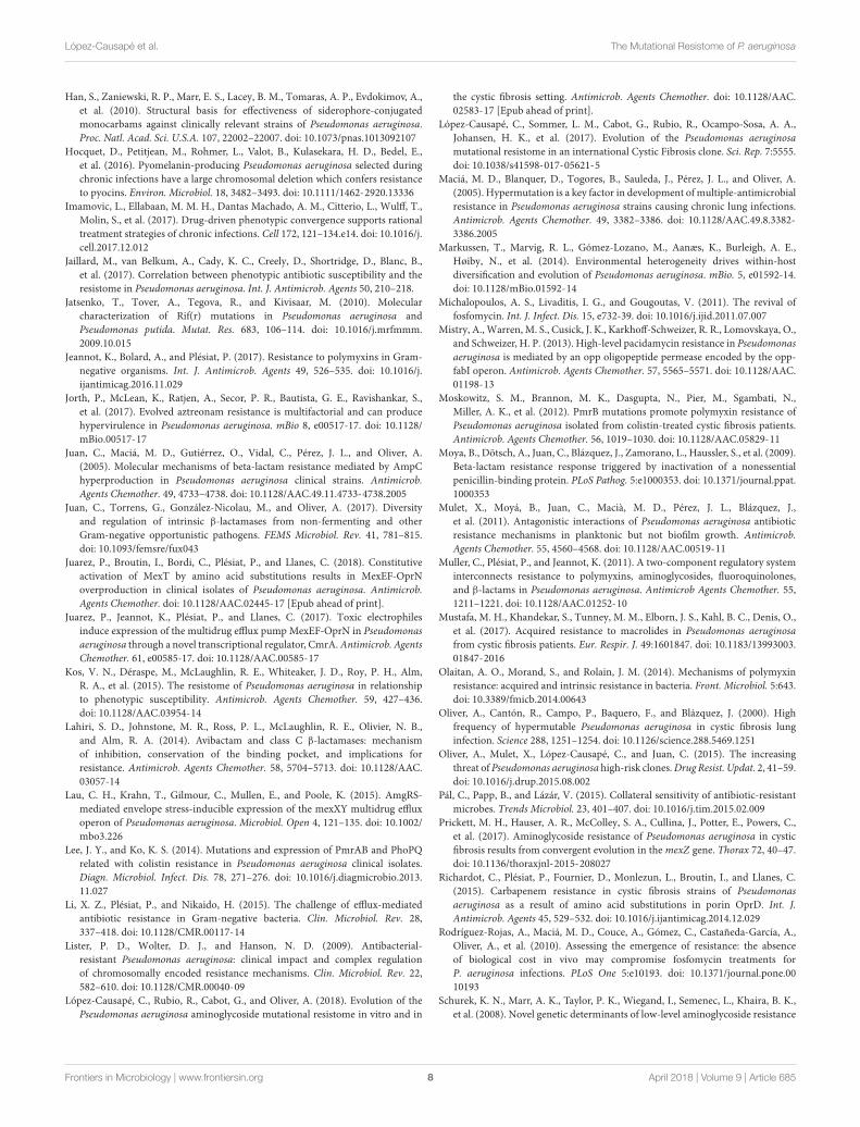
fmicb-09-00685 April 4, 2018 Time: 16:14 # 8
López-Causapé et al. The Mutational Resistome of P. aeruginosa
Han, S., Zaniewski, R. P., Marr, E. S., Lacey, B. M., Tomaras, A. P., Evdokimov, A.,et al. (2010). Structural basis for effectiveness of siderophore-conjugatedmonocarbams against clinically relevant strains of Pseudomonas aeruginosa.Proc. Natl. Acad. Sci. U.S.A. 107, 22002–22007. doi: 10.1073/pnas.1013092107
Hocquet, D., Petitjean, M., Rohmer, L., Valot, B., Kulasekara, H. D., Bedel, E.,et al. (2016). Pyomelanin-producing Pseudomonas aeruginosa selected duringchronic infections have a large chromosomal deletion which confers resistanceto pyocins. Environ. Microbiol. 18, 3482–3493. doi: 10.1111/1462-2920.13336
Imamovic, L., Ellabaan, M. M. H., Dantas Machado, A. M., Citterio, L., Wulff, T.,Molin, S., et al. (2017). Drug-driven phenotypic convergence supports rationaltreatment strategies of chronic infections. Cell 172, 121–134.e14. doi: 10.1016/j.cell.2017.12.012
Jaillard, M., van Belkum, A., Cady, K. C., Creely, D., Shortridge, D., Blanc, B.,et al. (2017). Correlation between phenotypic antibiotic susceptibility and theresistome in Pseudomonas aeruginosa. Int. J. Antimicrob. Agents 50, 210–218.
Jatsenko, T., Tover, A., Tegova, R., and Kivisaar, M. (2010). Molecularcharacterization of Rif(r) mutations in Pseudomonas aeruginosa andPseudomonas putida. Mutat. Res. 683, 106–114. doi: 10.1016/j.mrfmmm.2009.10.015
Jeannot, K., Bolard, A., and Plésiat, P. (2017). Resistance to polymyxins in Gram-negative organisms. Int. J. Antimicrob. Agents 49, 526–535. doi: 10.1016/j.ijantimicag.2016.11.029
Jorth, P., McLean, K., Ratjen, A., Secor, P. R., Bautista, G. E., Ravishankar, S.,et al. (2017). Evolved aztreonam resistance is multifactorial and can producehypervirulence in Pseudomonas aeruginosa. mBio 8, e00517-17. doi: 10.1128/mBio.00517-17
Juan, C., Maciá, M. D., Gutiérrez, O., Vidal, C., Pérez, J. L., and Oliver, A.(2005). Molecular mechanisms of beta-lactam resistance mediated by AmpChyperproduction in Pseudomonas aeruginosa clinical strains. Antimicrob.Agents Chemother. 49, 4733–4738. doi: 10.1128/AAC.49.11.4733-4738.2005
Juan, C., Torrens, G., González-Nicolau, M., and Oliver, A. (2017). Diversityand regulation of intrinsic β-lactamases from non-fermenting and otherGram-negative opportunistic pathogens. FEMS Microbiol. Rev. 41, 781–815.doi: 10.1093/femsre/fux043
Juarez, P., Broutin, I., Bordi, C., Plésiat, P., and Llanes, C. (2018). Constitutiveactivation of MexT by amino acid substitutions results in MexEF-OprNoverproduction in clinical isolates of Pseudomonas aeruginosa. Antimicrob.Agents Chemother. doi: 10.1128/AAC.02445-17 [Epub ahead of print].
Juarez, P., Jeannot, K., Plésiat, P., and Llanes, C. (2017). Toxic electrophilesinduce expression of the multidrug efflux pump MexEF-OprN in Pseudomonasaeruginosa through a novel transcriptional regulator, CmrA. Antimicrob. AgentsChemother. 61, e00585-17. doi: 10.1128/AAC.00585-17
Kos, V. N., Déraspe, M., McLaughlin, R. E., Whiteaker, J. D., Roy, P. H., Alm,R. A., et al. (2015). The resistome of Pseudomonas aeruginosa in relationshipto phenotypic susceptibility. Antimicrob. Agents Chemother. 59, 427–436.doi: 10.1128/AAC.03954-14
Lahiri, S. D., Johnstone, M. R., Ross, P. L., McLaughlin, R. E., Olivier, N. B.,and Alm, R. A. (2014). Avibactam and class C β-lactamases: mechanismof inhibition, conservation of the binding pocket, and implications forresistance. Antimicrob. Agents Chemother. 58, 5704–5713. doi: 10.1128/AAC.03057-14
Lau, C. H., Krahn, T., Gilmour, C., Mullen, E., and Poole, K. (2015). AmgRS-mediated envelope stress-inducible expression of the mexXY multidrug effluxoperon of Pseudomonas aeruginosa. Microbiol. Open 4, 121–135. doi: 10.1002/mbo3.226
Lee, J. Y., and Ko, K. S. (2014). Mutations and expression of PmrAB and PhoPQrelated with colistin resistance in Pseudomonas aeruginosa clinical isolates.Diagn. Microbiol. Infect. Dis. 78, 271–276. doi: 10.1016/j.diagmicrobio.2013.11.027
Li, X. Z., Plésiat, P., and Nikaido, H. (2015). The challenge of efflux-mediatedantibiotic resistance in Gram-negative bacteria. Clin. Microbiol. Rev. 28,337–418. doi: 10.1128/CMR.00117-14
Lister, P. D., Wolter, D. J., and Hanson, N. D. (2009). Antibacterial-resistant Pseudomonas aeruginosa: clinical impact and complex regulationof chromosomally encoded resistance mechanisms. Clin. Microbiol. Rev. 22,582–610. doi: 10.1128/CMR.00040-09
López-Causapé, C., Rubio, R., Cabot, G., and Oliver, A. (2018). Evolution of thePseudomonas aeruginosa aminoglycoside mutational resistome in vitro and in
the cystic fibrosis setting. Antimicrob. Agents Chemother. doi: 10.1128/AAC.02583-17 [Epub ahead of print].
López-Causapé, C., Sommer, L. M., Cabot, G., Rubio, R., Ocampo-Sosa, A. A.,Johansen, H. K., et al. (2017). Evolution of the Pseudomonas aeruginosamutational resistome in an international Cystic Fibrosis clone. Sci. Rep. 7:5555.doi: 10.1038/s41598-017-05621-5
Maciá, M. D., Blanquer, D., Togores, B., Sauleda, J., Pérez, J. L., and Oliver, A.(2005). Hypermutation is a key factor in development of multiple-antimicrobialresistance in Pseudomonas aeruginosa strains causing chronic lung infections.Antimicrob. Agents Chemother. 49, 3382–3386. doi: 10.1128/AAC.49.8.3382-3386.2005
Markussen, T., Marvig, R. L., Gómez-Lozano, M., Aanæs, K., Burleigh, A. E.,Høiby, N., et al. (2014). Environmental heterogeneity drives within-hostdiversification and evolution of Pseudomonas aeruginosa. mBio. 5, e01592-14.doi: 10.1128/mBio.01592-14
Michalopoulos, A. S., Livaditis, I. G., and Gougoutas, V. (2011). The revival offosfomycin. Int. J. Infect. Dis. 15, e732-39. doi: 10.1016/j.ijid.2011.07.007
Mistry, A., Warren, M. S., Cusick, J. K., Karkhoff-Schweizer, R. R., Lomovskaya, O.,and Schweizer, H. P. (2013). High-level pacidamycin resistance in Pseudomonasaeruginosa is mediated by an opp oligopeptide permease encoded by the opp-fabI operon. Antimicrob. Agents Chemother. 57, 5565–5571. doi: 10.1128/AAC.01198-13
Moskowitz, S. M., Brannon, M. K., Dasgupta, N., Pier, M., Sgambati, N.,Miller, A. K., et al. (2012). PmrB mutations promote polymyxin resistance ofPseudomonas aeruginosa isolated from colistin-treated cystic fibrosis patients.Antimicrob. Agents Chemother. 56, 1019–1030. doi: 10.1128/AAC.05829-11
Moya, B., Dötsch, A., Juan, C., Blázquez, J., Zamorano, L., Haussler, S., et al. (2009).Beta-lactam resistance response triggered by inactivation of a nonessentialpenicillin-binding protein. PLoS Pathog. 5:e1000353. doi: 10.1371/journal.ppat.1000353
Mulet, X., Moyá, B., Juan, C., Macià, M. D., Pérez, J. L., Blázquez, J.,et al. (2011). Antagonistic interactions of Pseudomonas aeruginosa antibioticresistance mechanisms in planktonic but not biofilm growth. Antimicrob.Agents Chemother. 55, 4560–4568. doi: 10.1128/AAC.00519-11
Muller, C., Plésiat, P., and Jeannot, K. (2011). A two-component regulatory systeminterconnects resistance to polymyxins, aminoglycosides, fluoroquinolones,and β-lactams in Pseudomonas aeruginosa. Antimicrob Agents Chemother. 55,1211–1221. doi: 10.1128/AAC.01252-10
Mustafa, M. H., Khandekar, S., Tunney, M. M., Elborn, J. S., Kahl, B. C., Denis, O.,et al. (2017). Acquired resistance to macrolides in Pseudomonas aeruginosafrom cystic fibrosis patients. Eur. Respir. J. 49:1601847. doi: 10.1183/13993003.01847-2016
Olaitan, A. O., Morand, S., and Rolain, J. M. (2014). Mechanisms of polymyxinresistance: acquired and intrinsic resistance in bacteria. Front. Microbiol. 5:643.doi: 10.3389/fmicb.2014.00643
Oliver, A., Cantón, R., Campo, P., Baquero, F., and Blázquez, J. (2000). Highfrequency of hypermutable Pseudomonas aeruginosa in cystic fibrosis lunginfection. Science 288, 1251–1254. doi: 10.1126/science.288.5469.1251
Oliver, A., Mulet, X., López-Causapé, C., and Juan, C. (2015). The increasingthreat of Pseudomonas aeruginosa high-risk clones. Drug Resist. Updat. 2, 41–59.doi: 10.1016/j.drup.2015.08.002
Pál, C., Papp, B., and Lázár, V. (2015). Collateral sensitivity of antibiotic-resistantmicrobes. Trends Microbiol. 23, 401–407. doi: 10.1016/j.tim.2015.02.009
Prickett, M. H., Hauser, A. R., McColley, S. A., Cullina, J., Potter, E., Powers, C.,et al. (2017). Aminoglycoside resistance of Pseudomonas aeruginosa in cysticfibrosis results from convergent evolution in the mexZ gene. Thorax 72, 40–47.doi: 10.1136/thoraxjnl-2015-208027
Richardot, C., Plésiat, P., Fournier, D., Monlezun, L., Broutin, I., and Llanes, C.(2015). Carbapenem resistance in cystic fibrosis strains of Pseudomonasaeruginosa as a result of amino acid substitutions in porin OprD. Int. J.Antimicrob. Agents 45, 529–532. doi: 10.1016/j.ijantimicag.2014.12.029
Rodríguez-Rojas, A., Maciá, M. D., Couce, A., Gómez, C., Castañeda-García, A.,Oliver, A., et al. (2010). Assessing the emergence of resistance: the absenceof biological cost in vivo may compromise fosfomycin treatments forP. aeruginosa infections. PLoS One 5:e10193. doi: 10.1371/journal.pone.0010193
Schurek, K. N., Marr, A. K., Taylor, P. K., Wiegand, I., Semenec, L., Khaira, B. K.,et al. (2008). Novel genetic determinants of low-level aminoglycoside resistance
Frontiers in Microbiology | www.frontiersin.org 8 April 2018 | Volume 9 | Article 685
fmicb-09-00685 April 4, 2018 Time: 16:14 # 9
López-Causapé et al. The Mutational Resistome of P. aeruginosa
in Pseudomonas aeruginosa. Antimicrob. Agents Chemother. 52, 4213–4219.doi: 10.1128/AAC.00507-08
Skiada, A., Markogiannakis, A., Plachouras, D., and Daikos, G. L. (2011).Adaptive resistance to cationic compounds in Pseudomonas aeruginosa. Int. J.Antimicrob. Agents 37, 187–193. doi: 10.1016/j.ijantimicag.2010.11.019
Suarez, C., Peña, C., Arch, O., Dominguez, M. A., Tubau, F., Juan, C., et al. (2011).A large sustained endemic outbreak of multiresistant Pseudomonas aeruginosa:a new epidemiological scenario for nosocomial acquisition. BMC Infect. Dis.11:272. doi: 10.1186/1471-2334-11-272
Vestergaard, M., Paulander, W., Marvig, R. L., Clasen, J., Jochumsen, N., Molin, S.,et al. (2016). Antibiotic combination therapy can select for broad-spectrummultidrug resistance in Pseudomonas aeruginosa. Int. J. Antimicrob. Agents 47,48–55. doi: 10.1016/j.ijantimicag.2015.09.014
Vincent, J. L. (2003). Nosocomial infections in adult intensive-care units. Lancet361, 2068–2077. doi: 10.1016/S0140-6736(03)13644-6
Woodford, N., Turton, J. F., and Livermore, D. M. (2011). Multiresistant Gram-negative bacteria: the role of high-risk clones in the dissemination of antibiotic
resistance. FEMS Microbiol. Rev. 35, 736–755. doi: 10.1111/j.1574-6976.2011.00268.x
Yen, P., and Papin, J. A. (2017). History of antibiotic adaptation influencesmicrobial evolutionary dynamics during subsequent treatment. PLoS Biol.15:e2001586. doi: 10.1371/journal.pbio.2001586
Conflict of Interest Statement: The authors declare that the research wasconducted in the absence of any commercial or financial relationships that couldbe construed as a potential conflict of interest.
Copyright © 2018 López-Causapé, Cabot, del Barrio-Tofiño and Oliver. This is anopen-access article distributed under the terms of the Creative Commons AttributionLicense (CC BY). The use, distribution or reproduction in other forums is permitted,provided the original author(s) and the copyright owner are credited and that theoriginal publication in this journal is cited, in accordance with accepted academicpractice. No use, distribution or reproduction is permitted which does not complywith these terms.
Frontiers in Microbiology | www.frontiersin.org 9 April 2018 | Volume 9 | Article 685


¡Quién busque el infinito, que cierre los ojos!
Milan Kundera, 1984

Related Documents


![DOCTORAL THESIS [TESIS DOCTORAL] - UAM](https://static.cupdf.com/doc/110x72/62162d5b17f9b23eb24b8a87/doctoral-thesis-tesis-doctoral-uam.jpg)


