6 Doctor 2015 Osama Asa’ad Khader Mohammad Alsalem

Welcome message from author
This document is posted to help you gain knowledge. Please leave a comment to let me know what you think about it! Share it to your friends and learn new things together.
Transcript

6
Doctor 2015
Osama Asa’ad Khader
Mohammad Alsalem
A quick revision for the spinal cord blood supply: Arterial Blood supply of spinal cord The spinal cord got its arterial supply by two ways:
Longitudinal arteries
Segmental arteries
1- Longitudinal arteries: the vertebral arteries proceed superiorly and enter transverse foramina of cervical vertebrae then they enter foramen magnum in the occipital bone. After right and left vertebral arteries pass through foramen magnum they run medially and meet each other on the lower border of pons (pontomedullary junction) forming basilar artery which run superiorly in the basilar groove on anterior border of pons, on upper border of pons it divides into two posterior cerebral arteries. But before that they give branch on anterior aspect of spinal cord and they meet each other on anterior median fissure to form anterior spinal artery, which descends along the spinal cord. • Right and left vertebral arteries also give posterior inferior cerebellar
arteries which give two posterior spinal arteries (in the posterolateral sulcus). Now we have one anterior spinal artery and two (right and left) posterior spinal arteries. (They are the longitudinal arteries of spinal nerve) Central cord syndrome: temporary occlusion in the anterior spinal artery due to mostly neck hypertension with the presence of sharp edges form bone (spares). It's characterized by:
bilateral (only one artery for both sides) muscle weakness since the anterior spinal artery supplies the anterior grey matter.
bladder dysfunction
variable pain and thermal defects The recovery begins distally then goes upwards.

Brain stem:
Hindbrain:
o Medulla
o Pons
o Cerebellum
Brain stem:
o Medulla
o Pons
o Midbrain: above it the thalamus which part of the diencephalon.
Gross/external features: (explained in lab)

The Pyramid two bulges around the midline, while the olive away
from midline.
10 of the cranial nerves emerge from the brain stem.
The picture shows the order of cranial nerves arising from brain stem.
The first two cranial nerves, olfactory (I) and optic (II), are not related
to brain stem they arise from the forebrain (CNS). Both are sensory.
Oculomotor (III) arises from the floor of the mid-brain, and it is a
motor nerve.
Trochlear N. (IV) is the only one that arises from posterior\dorsal
aspect of brainstem (interpeduncular fossa), it is a motor nerve.
Trigeminal N.(V) largest cranial nerve, arises from mid pontine area,
divides into two sensory branches (ophthalmic and maxillary) and one
motor branch (Mandibular)
Medially to laterally: Abducent (VI), Facial (VII) and
Vestibulocochlear (VIII) nerves arise from pontomedullary
junction
Glossopharyngeal (IX), Vagus (X), accessory (XI) arise from the
groove between the olive and inferior cerebellar peduncle.
Hypoglossal (XII) arises from the groove In between the pyramid and
olive.
The nuclei of these nerves are either sensory, motor, or
parasympathetic. We will talk about them in details when we
discuss the sections.
Q: what is the cavity found in the hindbrain?
The Fourth ventricle, it is related to both pons and medulla oblongata.
It has the shape of a tent, with a roof and floor.
the floor of 4th ventricle is formed of the posterior aspect of pons of
medulla oblongata
the roof is toward the cerebellum
the upper part of medulla oblongata is related to the lower part of the 4th
ventricle (the cavity)

a cross section in the lower part of medulla oblongata will show the central
canal.
Q: What do you expect to see in the brain stem?
ascending and descending tracts.
The nuclei of the cranial nerves (sensory and motor nuclei instead of ventral and dorsal horns as is the spinal gray matter).
Reticular formation.
Internal structure of medulla:
All brainstem will be studied in 8 sections, 4 of them from medulla oblongata, 2 of
these 4 are on the lower medulla, where the cavity is the central canal.
if the section has a big cavity open medulla (2 sections)
if you see a central canal in the section close medulla (2
sections).
From downward to upward:
level of pyramidal decussation (close medulla / motor) level of foramen magnum.
level of decussation of lemnisci (close medulla / sensory)
level of olives (open medulla)
level just inferior to the pons
practically they are 3 decussations because 3rd and 4th decussations are too
similar.
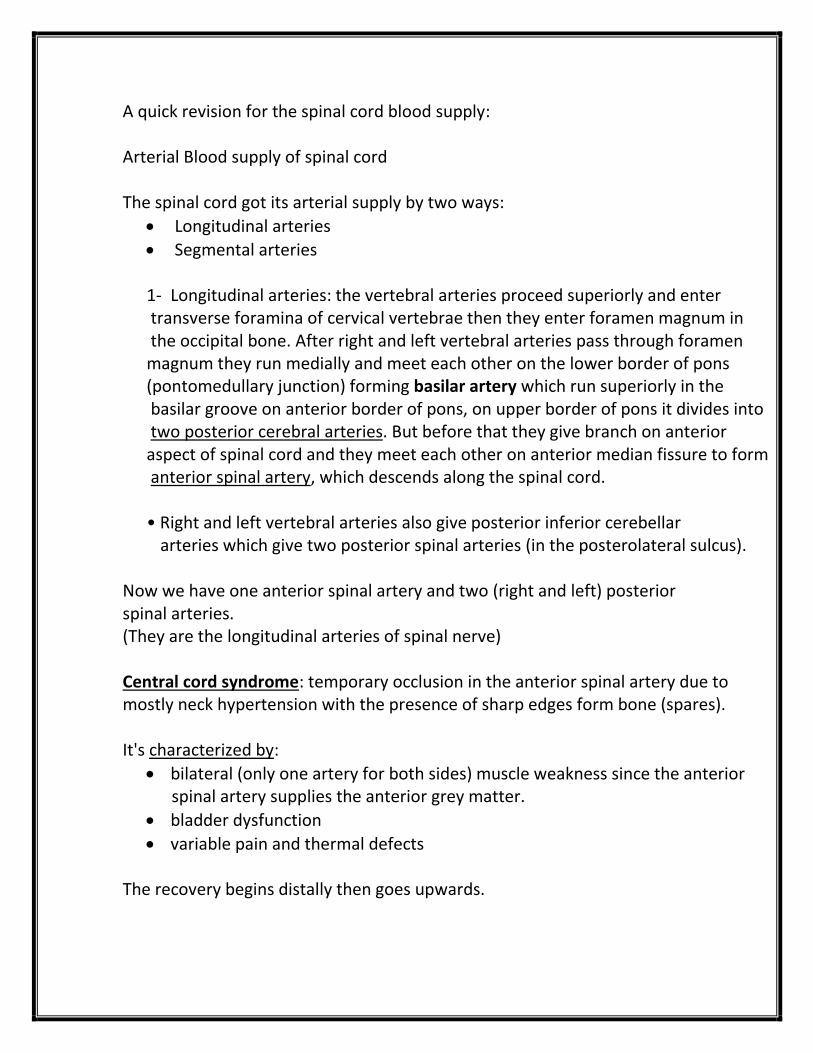
Level of decussation of pyramid:
This is a posterior view.
You can see the floor of the 4th ventricle which has a rhomboid shape
(two triangles).
The lower part of it (the lower triangle) is the part that is related to medulla
oblongata.
Both sections A & B in the picture below will cut into the closed medulla
which mean that they will show the central canal.

First thing to notice is the central canal (make it your reference point).
Pyramidal decussation is anterior to central canal.
This decussation is composed of corticospinal tract fibers crossing each other;
right to left & left to right, except for the anterior corticospinal fibers that go
ipsilateral.
If you look posterior to central canal you will see:
o Gray matter:
Nucleus gracilis: close to midline
Nucleus cuneatus: lateral. Remember:
nucleus indicates gray matter
o White matter:
• Fasciculus gracilis
Fasciculus cuneatus
Just to remind you when we took the posterior column system:
The fibers that go up are divided to:
o Medially gracilis
o Laterally cuneatus
Both are white matter in spinal cord, and they go up to the lower part of medulla and their you’ll see:
The 2 nuclei (gracilis and cuneatus) and their fibers (white matter Fasciculi)
In brainstem: you’ll see the traces of both ascending and descending fibers, all
will pass through the brainstem.
The brainstem consists mainly from the nuclei of the cranial nerves (sensory
or motor).

Spinal nucleus of trigeminal nerve: trigeminal Nerve: originates from mid pontine area (pons)
- mainly sensory: head, neck, nasal cavity, oral cavity and face
(except for the angle of mandible)
- motor: muscles of mastication (tensor tympani, tensor veli
palatine, mylohyoid, ant. Belly of digastric)
- has four nuclei: one motor, three sensory
- sensory nuclei of trigeminal:
I. main sensory (primary sensory): found in pons
II. mesencephalic: name indicates it’s up in the brain
III. spinal nucleus of trigeminal Nerve:
spinal nucleus of trigeminal is named so because it extends along the
brainstem from mid pontine down to spinal cord, it is an extension of the
upper cervical segment substantia gelatinosa (lamina II)
you’ll see the spinal nucleus in upper and lower sections in brain stem.
note it doesn’t extend along all the brainstem, it starts from mid pontine
and below, while above the mid pontine it is replaced by main sensory.
Q: Why are there three nuclei for the Trigeminal nerve?
For modalities.
- spinal: pain and temp.
- mesencephalic: proprioception
- main sensory: crude touch
Spinal nucleus of trigeminal is surrounded by fibers from trigeminal
(white matter) trying to reach nucleus.

Important: the anterolateral system in the spinal cord doesn’t
change very much when reaching the medulla oblongata, fibers
come closer but they still have the same position related to each
other, so you should pay attention to the few things:
1. Nucleus gracilis and nucleus cuneatus (not found in spine)
2. Fasciculus gracilis and Fasciculus cuneatus
3. Pyramidal decussation
Important: spinal nucleus of trigeminal is not only trigeminal fibers,
the only reason to call it trigeminal is that most of fibers are
trigeminal fibers (predominant fibers) but all cranial nerves that
have pain and temperature modality (facial, glossopharyngeal,
vagus) all relay to spinal nucleus of trigeminal (for example, the
glossopharyngeal fibers send input to spinal nucleus of trigeminal).
Medial longitudinal fasciculus:
Vestibular nucleus gives a bundle of white matter that connects the vestibular
nucleus with the motor nucleus of the 3rd, 4th and 6th cranial nerves (oculomotor III,
trochlear IV, abducens VI), to synchronize the movement of eye ball, that bundle of
white matter is called medial longitudinal fasciculus.
The idea of the link between the three cranial nerves is to coordinate the movement
of right and left eye:
When you move your eyes to right or left direction, in one eye it’s the medial rectus
muscle that contracted but for the other eye it’s the lateral rectus that contracted,
and for this to happen at the same time we need the connection between the three
motor nuclei.
What about the vestibular nucleus? The connection between motor nuclei and
vestibular nucleus is to coordinate the movement of head with movement of eye
(vestibular provides information about gravity) to maintain the visual field while
walking and moving your head.
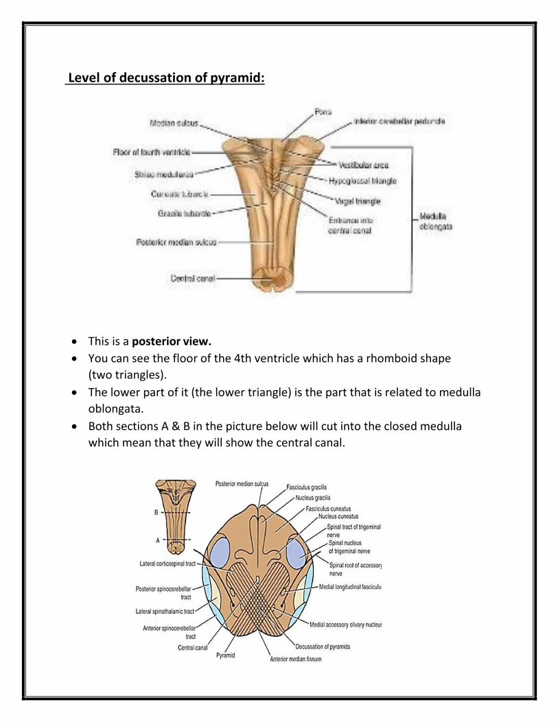
**we will see the Medial longitudinal fasciculus more clearly from upper sections
Remember:
Nerves that control the movement of the eyeball:
1- Oculomotor (III): all external muscles except superior oblique and lateral
rectus.
2- Trochlear (IV): supplies superior oblique.
3- Abducent (VI): supplies lateral rectus.
Now let's take a look at the section of the next higher level:
Level of decussation of lemnisci:
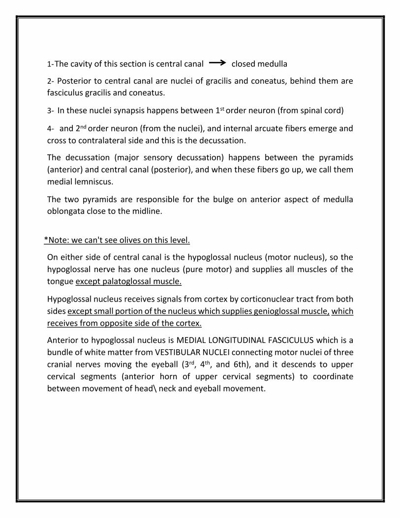
1- The cavity of this section is central canal closed medulla
2- Posterior to central canal are nuclei of gracilis and coneatus, behind them are
fasciculus gracilis and coneatus.
3- In these nuclei synapsis happens between 1st order neuron (from spinal cord)
4- and 2nd order neuron (from the nuclei), and internal arcuate fibers emerge and
cross to contralateral side and this is the decussation.
The decussation (major sensory decussation) happens between the pyramids
(anterior) and central canal (posterior), and when these fibers go up, we call them
medial lemniscus.
The two pyramids are responsible for the bulge on anterior aspect of medulla
oblongata close to the midline.
*Note: we can't see olives on this level.
On either side of central canal is the hypoglossal nucleus (motor nucleus), so the
hypoglossal nerve has one nucleus (pure motor) and supplies all muscles of the
tongue except palatoglossal muscle.
Hypoglossal nucleus receives signals from cortex by corticonuclear tract from both
sides except small portion of the nucleus which supplies genioglossal muscle, which
receives from opposite side of the cortex.
Anterior to hypoglossal nucleus is MEDIAL LONGITUDINAL FASCICULUS which is a
bundle of white matter from VESTIBULAR NUCLEI connecting motor nuclei of three
cranial nerves moving the eyeball (3rd, 4th, and 6th), and it descends to upper
cervical segments (anterior horn of upper cervical segments) to coordinate
between movement of head\ neck and eyeball movement.

Accessory nerve (xi) has cranial root and spinal root:
Spinal root from upper 5 segments (accessory nucleus in anterior horn) enters
foramen magnum to go to cranial cavity and it meets cranial root of accessory
nerve.
The nucleus in this level is a motor nucleus of accessory nerve but it's not exclusive
for this nerve only, in fact it is a motor nucleus for cranial nerves number 9 \ 10 and
11, and we call it nucleus ambiguous.
Cranial root of accessory emerges from nucleus ambiguous to unite with spinal root
of accessory, but they divide after that immediately.
ALS is unchanged on this level also, but here it starts to form spinal lemniscus with
spinotectal tract.
*note: sometimes we can see a small part of inferior olivary nucleus but we don't
consider it the level of the olives.

The third level is the level of olives:
1- The cavity of this section is inferior part of 4th ventricle (open medulla), behind it
is the cerebellum.
Floor of 4th ventricle medulla oblongata
Roof of 4thventricle cerebellum
2- Posterolateral in this section is INFERIOR CEREBELLAR PEDUNCLE (ICP) which
connects medulla oblongata with cerebellum. (one of the structures that cross ICP
is posterior spinocerebellar tract).
3- There are many olivary nuclei on this level (OLIVARY NUCLEAR COMPLEX), the
biggest one is INFERIOR OLIVARY NUCLEUS, which is the cause of the bulging in the
olive (its shape resembles crumpled bag with its opening directed medially).

The function of inferior olivary nucleus is motor, and has a connection with
cerebellum and cerebrum. There are fibers emerging from it that go to cerebellum
by ICP, their name is climbing fibers.
*note: Fibers that go to cerebellum are divided into climbing fibers and Mossy
fibers.
*note: Spino-olivary tract is very similar to spinocerebellar tract and is considered
an alternative pathway from spinal cord to cerebellum.
4- Corticospinal tract gives branches to the olive, they are involved in voluntary
movement and from olive to spinal cord (olivospinal tract which is motor (not
important)).
*note: neurodegenerative diseases that affect olive will always affect the
cerebellum so symptoms are overshadowed clinically.
Now let's talk about the midline structures in this section:
Medial lemniscus which is posterior to the pyramids and very close to the midline
(lemniscus not fasciculus because fibers are elongated in shape and not rounded in
the cross-section) which goes up until it reaches VPL in thalamus (one of the
ventrobasal complex nuclei).
*note: VPM which is one of ventrobasal complex nuclei, is concerned with face and
taste sensory pathway.
posterior to medial lemniscus is tectospinal tract (extrapyramidal tract) which is
very important in visual-spinal reflex (superior colliculus takes signals from visual
pathway and spinotectal tract, then it sends signals through tectospinal tract to
upper cervical segments).
*note: withdrawal reflex is different from spinovisual reflex. Withdrawal reflex is
the sudden and immediate withdrawal movement of the limb after the harm
signals reach the spinal cord. On the other hand, spinovisual reflex is when you
move your head and eyes to see your limb after the harm signals reach the superior
colliculus by tectospinal tract.
posterior to tectospinal tract is medial longitudinal fasciculus which is
underneath the floor of 4th ventricle.
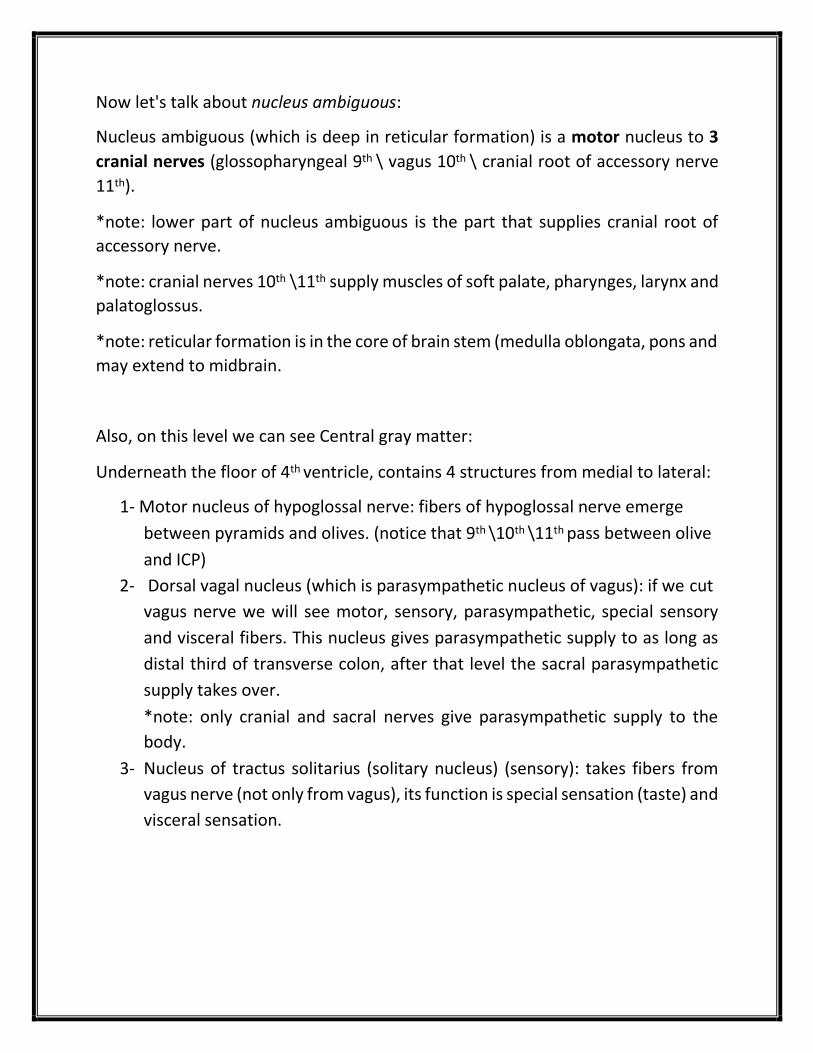
Now let's talk about nucleus ambiguous:
Nucleus ambiguous (which is deep in reticular formation) is a motor nucleus to 3
cranial nerves (glossopharyngeal 9th \ vagus 10th \ cranial root of accessory nerve
11th).
*note: lower part of nucleus ambiguous is the part that supplies cranial root of
accessory nerve.
*note: cranial nerves 10th \11th supply muscles of soft palate, pharynges, larynx and
palatoglossus.
*note: reticular formation is in the core of brain stem (medulla oblongata, pons and
may extend to midbrain.
Also, on this level we can see Central gray matter:
Underneath the floor of 4th ventricle, contains 4 structures from medial to lateral:
1- Motor nucleus of hypoglossal nerve: fibers of hypoglossal nerve emerge
between pyramids and olives. (notice that 9th \10th \11th pass between olive
and ICP)
2- Dorsal vagal nucleus (which is parasympathetic nucleus of vagus): if we cut
vagus nerve we will see motor, sensory, parasympathetic, special sensory
and visceral fibers. This nucleus gives parasympathetic supply to as long as
distal third of transverse colon, after that level the sacral parasympathetic
supply takes over.
*note: only cranial and sacral nerves give parasympathetic supply to the
body.
3- Nucleus of tractus solitarius (solitary nucleus) (sensory): takes fibers from
vagus nerve (not only from vagus), its function is special sensation (taste) and
visceral sensation.

Innervation of the tongue:
Anterior 2\3 Posterior 1\3
1- Lingual and trigeminal nerve (general sensory)
2- Facial nerve (chorda tympani) (taste)
Glossopharyngeal nerve (special
sensory and general sensory)
*note: in infratemporal fossa there is a connection between lingual nerve
and chorda tympani (facial).
**important: what is the connection between vagus nerve and taste? there
are taste fibers on epiglottis which are from vagus nerve.
When vagus nerve gets out of skull by jugular foramen (9th and 11th use
jugular foramen also) it will face 2 ganglia: inferior vagal ganglia and superior
vagal ganglia, the cell body of taste and visceral sensory are in inferior vagal
ganglia (glossopharyngeal is the same), superior vagal ganglia contain cell
body of general sensation.
*note: jugular foramen is between superior and inferior vagal ganglia.
4- Inferior vestibular nucleus (medial vestibular nucleus): sensory nucleus for
vestibular nerve which is part of vestibulocochlear nerve 8th (important for
balance), which leaves the cavity of inner ear then internal acoustic meatus
then brain stem (pontomedullary junction), cell body in Scarpa ganglia which
is relay station in the groove between ICP and olive. Emergence of
vestibulocochlear nerve: from vestibular nucleus to 1- cerebellum 2- medial
longitudinal fasciculus 3- down on spinal cord 4- some people think it may
project on cerebral cortex (VPL then cortex).

17 | P a g e
Now let's talk about 4th and last level of medulla oblongata:
Level just inferior to Pons:
1- No major changes.
2- Most upper level in medulla oblongata
3- Lateral vestibular nucleus replaces inferior vestibular nucleus
4- Cochlear nuclei become visible on anterior(ventral) and posterior(dorsal) of ICP (relay
station for cochlear nerve the second part of cranial nerve vestibulocochlear nerve
8th).
Related Documents