Abstract. Breast cancer is the most common cancer in women in industrialized countries. Environmental factors, such as differences in diet are likely to have an important influence on cancer emergence. Among these factors, n-3 polyunsaturated-fatty acids, such as docosahexaenoic acid (DHA), are good candidates for preventing breast cancer. Here we investigate the effect of DHA on the human breast cancer cell line MDA-MB-231 and show that DHA incorpo- ration i) has an anti-proliferative effect, ii) induces apoptosis via a transient increase in caspase-3 activity and the promotion of nuclear condensation, and iii) reduces the invasive potential of MDA-MB-231 cells. To conclude, DHA may have beneficial effects as a result of slowing the proliferation of tumor cells, and minimizing their metastatic potential. Introduction For several decades, genetic and environmental factors have been explored in order to elucidate the appearance of tumors. Genetic factors are obviously involved in carcinogenesis, but diet is an environmental factor that is likely to have an influence on health (1), and particularly on tumor emergence (2-4). It has been shown that n-3 polyunsaturated, long-chain fatty acids (PUFAs), such as docosahexaenoic acid (DHA) or eicosapentaenoic acid (EPA), are able to play an important preventive role in cardiovascular disease (5) and cancer (6). This conclusion is based partly on the observation that the incidence of these diseases is lower in Japanese people, whose diet is seafood based, resulting in a more balanced ratio between n-3 and n-6 PUFA than that of the Western diet (7). Even though this resource may be depleted in the future by the overharvesting of n-3 PUFA rich fish, it could be re- placed by using marine microalgae, which have been identified as an important alternative source of DHA and EPA (8,9). Breast cancer is one of the cancers most frequently observed in industrialized countries, and the one with the highest incidence in women. Epidemiological studies have shown that the rate of breast cancer is 4 to 5 times higher in Western countries than in Japan (1,10). The mechanism by which DHA and EPA could provide protection against the appear- ance of a tumor, or directly influence cancer cells by reducing their malignancy, remains unclear, since cohort studies do not reveal any correlation between fat intake and breast cancer (1). Nevertheless, some evidence hints that DHA not only acts as an anti-proliferative agent by lengthening the cell cycle between the G2/M transition (11), but is also a pro- apoptotic factor, increasing caspase-3 and Bax (12,13). In addition, DHA has been shown to affect cell proliferation, whatever its source (i.e., fish oil or microalgae) (14). It has also been shown that the n-3 PUFAs and DHA, in particular, can act on lipid peroxidation as well as on the proteins impli- cated in the ROS mechanism leading to cell death (15,16). In this study, the effects of two concentrations of DHA (20 and 100 μM) were investigated on the human breast cancer cell line MDA-MB-231. DHA incorporation into the cells was first monitored, and then its inhibitory effect on cell proliferation was observed. The findings demonstrate the apoptosis-promoting effect of DHA, evidenced by a transient increase in caspase-3 activity, and an enhanced level of nuclear condensation. Finally, DHA was shown to reduce the invasive phenotype of the MDA-MB-231 cell line, for at least 4 passages of the cell culture. This study provides new insights into the timing sequence of the effects of DHA, which could prove useful for future functional and mechanistic investigations. Materials and methods Cell culture. The breast cancer cell line MDA-MB-231 was obtained from the American Type Culture Collection, and INTERNATIONAL JOURNAL OF ONCOLOGY 36: 737-742, 2010 Docosahexaenoic acid intake decreases proliferation, increases apoptosis and decreases the invasive potential of the human breast carcinoma cell line MDA-MB-231 VINCENT BLANCKAERT 1,2 , LIONEL ULMANN 1,2 , VIRGINIE MIMOUNI 1,2 , JOHANN ANTOL 3 , LUCILE BRANCQUART 1 and BENOÎT CHÉNAIS 1 1 Université du Maine, EA2160 Mer Molécules Santé, Le Mans, F-72085; 2 IUT de Laval, Département Génie Biologique, Laval, F-53020; 3 INSERM U908, Université de Lille, Villeneuve d'Ascq, F-59655, France Received July 29, 2009; Accepted September 18, 2009 DOI: 10.3892/ijo_00000549 _________________________________________ Correspondence to: Dr Vincent Blanckaert, EA2160-MMS, IUT de Laval, Département Génie Biologique, 52 rue des Docteurs Calmette et Guérin, BP2045, 53020 Laval Cedex 9, France E-mail: [email protected] Key words: apoptosis, breast cancer, carcinoma cell, docosa- hexaenoic acid, invasion, polyunsaturated fatty acid

Welcome message from author
This document is posted to help you gain knowledge. Please leave a comment to let me know what you think about it! Share it to your friends and learn new things together.
Transcript
Abstract. Breast cancer is the most common cancer inwomen in industrialized countries. Environmental factors,such as differences in diet are likely to have an importantinfluence on cancer emergence. Among these factors, n-3polyunsaturated-fatty acids, such as docosahexaenoic acid(DHA), are good candidates for preventing breast cancer.Here we investigate the effect of DHA on the human breastcancer cell line MDA-MB-231 and show that DHA incorpo-ration i) has an anti-proliferative effect, ii) induces apoptosisvia a transient increase in caspase-3 activity and the promotionof nuclear condensation, and iii) reduces the invasivepotential of MDA-MB-231 cells. To conclude, DHA mayhave beneficial effects as a result of slowing the proliferationof tumor cells, and minimizing their metastatic potential.
Introduction
For several decades, genetic and environmental factors havebeen explored in order to elucidate the appearance of tumors.Genetic factors are obviously involved in carcinogenesis, butdiet is an environmental factor that is likely to have aninfluence on health (1), and particularly on tumor emergence(2-4). It has been shown that n-3 polyunsaturated, long-chainfatty acids (PUFAs), such as docosahexaenoic acid (DHA) oreicosapentaenoic acid (EPA), are able to play an importantpreventive role in cardiovascular disease (5) and cancer (6).This conclusion is based partly on the observation that theincidence of these diseases is lower in Japanese people,
whose diet is seafood based, resulting in a more balancedratio between n-3 and n-6 PUFA than that of the Western diet(7). Even though this resource may be depleted in the futureby the overharvesting of n-3 PUFA rich fish, it could be re-placed by using marine microalgae, which have been identifiedas an important alternative source of DHA and EPA (8,9).Breast cancer is one of the cancers most frequently observedin industrialized countries, and the one with the highestincidence in women. Epidemiological studies have shownthat the rate of breast cancer is 4 to 5 times higher in Westerncountries than in Japan (1,10). The mechanism by whichDHA and EPA could provide protection against the appear-ance of a tumor, or directly influence cancer cells by reducingtheir malignancy, remains unclear, since cohort studies donot reveal any correlation between fat intake and breastcancer (1). Nevertheless, some evidence hints that DHA notonly acts as an anti-proliferative agent by lengthening the cellcycle between the G2/M transition (11), but is also a pro-apoptotic factor, increasing caspase-3 and Bax (12,13). Inaddition, DHA has been shown to affect cell proliferation,whatever its source (i.e., fish oil or microalgae) (14). It hasalso been shown that the n-3 PUFAs and DHA, in particular,can act on lipid peroxidation as well as on the proteins impli-cated in the ROS mechanism leading to cell death (15,16).
In this study, the effects of two concentrations of DHA(20 and 100 μM) were investigated on the human breastcancer cell line MDA-MB-231. DHA incorporation into thecells was first monitored, and then its inhibitory effect on cellproliferation was observed. The findings demonstrate theapoptosis-promoting effect of DHA, evidenced by a transientincrease in caspase-3 activity, and an enhanced level ofnuclear condensation. Finally, DHA was shown to reduce theinvasive phenotype of the MDA-MB-231 cell line, for atleast 4 passages of the cell culture. This study provides newinsights into the timing sequence of the effects of DHA, whichcould prove useful for future functional and mechanisticinvestigations.
Materials and methods
Cell culture. The breast cancer cell line MDA-MB-231 wasobtained from the American Type Culture Collection, and
INTERNATIONAL JOURNAL OF ONCOLOGY 36: 737-742, 2010
Docosahexaenoic acid intake decreases proliferation,increases apoptosis and decreases the invasive potentialof the human breast carcinoma cell line MDA-MB-231
VINCENT BLANCKAERT1,2, LIONEL ULMANN1,2, VIRGINIE MIMOUNI1,2,
JOHANN ANTOL3, LUCILE BRANCQUART1 and BENOÎT CHÉNAIS1
1Université du Maine, EA2160 Mer Molécules Santé, Le Mans, F-72085; 2IUT de Laval, Département Génie Biologique,
Laval, F-53020; 3INSERM U908, Université de Lille, Villeneuve d'Ascq, F-59655, France
Received July 29, 2009; Accepted September 18, 2009
DOI: 10.3892/ijo_00000549
_________________________________________
Correspondence to: Dr Vincent Blanckaert, EA2160-MMS, IUTde Laval, Département Génie Biologique, 52 rue des DocteursCalmette et Guérin, BP2045, 53020 Laval Cedex 9, FranceE-mail: [email protected]
Key words: apoptosis, breast cancer, carcinoma cell, docosa-hexaenoic acid, invasion, polyunsaturated fatty acid
737-742 20/1/2010 01:48 ÌÌ Page 737
routinely grown as monolayers at 37˚C in a humidified atmo-sphere of 5% CO2 in minimum essential medium (MEM-Earle's salts, Sigma-Aldrich, Saint Quentin Fallavier, France)supplemented with 10% fetal calf serum (FCS, Invitrogen,Cergy-Pontoise, France), 20 mM HEPES, 2 mM L-glutamine,100 U/ml penicillin/streptomycin and 0.4 mg/ml insulin.
Proliferation and viability assays. The subconfluent cellsgrown in 75-cm2 flasks were rinsed twice with 10 ml PBS.Two milliliters of non-enzymatic dissociation solution(Sigma-Aldrich) were added to the flasks. The dissociatedcells were counted using a Malassez hematocytometer, and200,000 cells in MEM containing 5% FCS were loaded intoa fresh 75-cm2 flask, and grown until they reached 70%confluence. MEM (10 ml) containing 5% FBS, with orwithout 20 or 100 μM DHA (Sigma-Aldrich) were added tothe flasks, and changed every 24 h. Untreated and DHA-treated cells were dissociated with 2 ml of non-enzymaticdissociation solution after 24, 48 or 72 h. After homogeni-zing, cell viability was determined using the Trypan blue dyeexclusion test (Sigma-Aldrich), and counts were performedusing a Malassez hemocytometer. The remaining cells werewashed with 10 ml sterile phosphate buffer saline (PBS), andcentrifuged for 10 min at 200 x g. The pellet was homo-genized, washed again with PBS, and centrifuged under thesame conditions. The supernatants were discarded, and thecells were either used without delay, or kept at -70˚C forfurther experiments. Four independent experiments wereconducted, each in triplicate.
DHA incorporation assay and total fatty acid analysis.Lipids were extracted from the tissues using dimethoxy-methane/methanol (4/1, v/v) according to Delsal (17). Afterdrying with ethanol, the samples were dissolved in 1 ml ofchloroform, and transferred to methylation vials. After eva-porating under nitrogen, 1 ml of NaOH (0.5 N) in methanolsolution was added. Samples were covered by nitrogen toprevent oxidation, and then incubated for 20 min at 80˚C.This first step corresponded to saponification. The trans-methylation step was then conducted according to themethod of Slover and Lanza (18) as follows: 2 ml of borontrifluoride methanol (BF3-MeOH 14 %, Sigma-Aldrich) wereadded to the samples, which were incubated as describedabove. The fatty acid methyl esters (FAMEs) were extractedusing 1 ml of iso-octane (trimethyl-pentane), and dried on ananhydrous sodium sulfate column. This operation wasconducted twice. FAMEs were analyzed by gas-liquidchromatography using a GC Focus (Thermo Fisher Scientific,Courtaboeuf, France) equipped with a Thermo TR-FAME30 m x 0.25 mm i.d. capillary column (Thermo FisherScientific). Analyses were carried out from 120 to 220˚C witha 6˚C/min gradient. The AS 3000 split/splitless autosampler(Thermo Fisher Scientific) and detector temperatures were250 and 280˚C, respectively. The esters were detected with aflame-ionization detector (air, hydrogen, 29 and 14.5 psi,respectively). The FAMEs were identified by comparingwith the corresponding commercial standards (189-1 and189-15, Sigma-Aldrich). Results are expressed as molarpercentage of total saturated fatty acids (SFA, 14:0, 16:0 and18:0), total monounsaturated fatty acids (MUFA, 16:1n-7 and
18:1n-9), and total polyunsaturated fatty acids (n-6 PUFA;18:2, 18:3, 20:4, 22:5; and n-3 PUFA; 18:4, 20:5, 22:6) for24, 48 and 72 h of incubation with or without 20 or 100 μMDHA.
Caspase-3 assay. The Caspase-3 Colorimetric Assay Kitfrom Sigma Aldrich is based on the hydrolysis of acetyl-Asp-Glu-Val-Asp p-nitroanilide (Ac-DEVD-pNA) by caspase-3,resulting in the release of the p-nitroaniline (pNA) moiety.Assays were performed according to the manufacturer'smethod. Briefly, pellets of 5x10⁶ cells were lysed in 50 μl 1Xlysis buffer and incubated on ice for 20 min. The lysed cellswere centrifuged at 16,000 x g for 15 min at 4˚C. The super-natants were transferred into new tubes. Lysates were thenfrozen and stored at 70˚C until use. Assays were performedwith 15 μl of cell lysates, 980 μl 1X assay buffer and 10 μlac-DEVD-pNA, and compared with a blank prepared with990 μl 1X assay buffer and 10 μl substrate. The tubes werecovered, and the incubation performed at 37˚C for 90 min.Absorbance was read at 405 nm. Caspase-3 activity in termsof μmol of pNA released per min per ml was obtained by thefollowing formula: A405nm x d/ÂmM x t x v; where ÂmM=10.5;v, volume of sample in ml; d, dilution factor; t, reaction timein min. Four independent experiments were conducted, eachin duplicate.
Invasion assay and Hoechst staining. Invasion assays weredone in 12-well Boyden microchambers (Transwell®, FisherScientific, Illkirch, France) with 8-μm pore membranes.Matrigel® (100 μl) (BD Science, Le Pont de Claix, France)at 10% in MEM were introduced in the upper chamber anddried overnight at 37˚C. Cells were treated with 20 or 100 μMDHA for 24, 48, 72 h in 25-cm2 flasks. At the selectedtime, the cells were dissociated with a non-enzymatic disso-ciation solution, and counted by using a Malassez hemato-cytometer. Living cells (40,000) treated with or withoutDHA in 400 μl MEM supplemented with 0.5% FCS and 1%BSA were then loaded into the upper chamber. A volume of800 μl of MEM with 0.5% FCS, and 1% BSA wasintroduced into the lower chamber. After incubating for24 h, the Transwell were rinsed with PBS, and the Matrigelwas scraped off the upper surfaces of the membranes. Thecells remaining on the underside of the membrane werefixed for 30 min at 20˚C in methanol, stained with Hoechststain (Sigma-Aldrich), and mounted on glass slides withglycerol before counting (20 fields per membrane) under aUV microscope. Three independent experiments wereperformed, each in duplicate.
In order to appreciate the long-term effect on invasion,the cells were incubated in the presence or absence of 100 μMDHA for 72 h, and either immediately used for invasion assay,(corresponding to passage, P0) or diluted in a new cultureflask in the absence of DHA before being harvested (P1-P6).Cells were harvested and cultured with MEM containing 5%FCS, and their invasive potential was assayed after 72 h inBoyden chambers, as described above. These assays wereperformed for each stage in triplicate.
Statistical analysis. Statistical studies were performed usingKyPlot® for a one-way ANOVA followed by a Dunnett's test.
BLANCKAERT et al: DOCOSAHEXAENOIC ACID AND BREAST CANCER738
737-742 20/1/2010 01:48 ÌÌ Page 738
Results
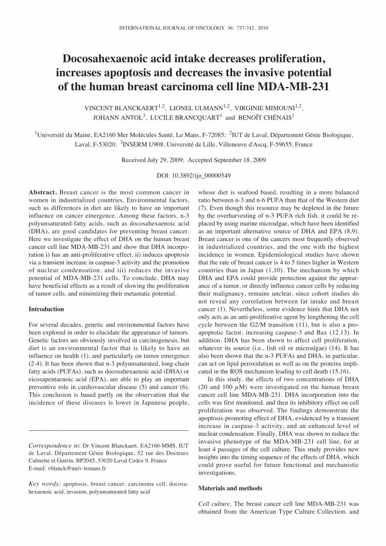
Incorporation of DHA in the MDA-MB-231 cell line and totallipid fraction composition. The incorporation of exogenousDHA was estimated by determining the molar percentage ofDHA in the total lipid fraction, after incubating with 20 or100 μM of DHA. By comparison with the control (DHAlevel in the total lipid fraction in the absence of exogenousDHA), the results reported in Fig. 1 show the dose-dependentincrease in the DHA level, which rose by 13 and 41% with20 and 100 μM of exogenous DHA, respectively. Whicheverexogenous DHA concentration was used, the cellular DHAlevel peaked after incubation for 24 h, and decreased slightlythereafter.
The effect of incubating in exogenous DHA on the com-position of the total lipid fraction was analyzed by measuringthe change in the molar percentage of SFA, MUFA andPUFA over time in the presence or absence of exogenousDHA. SFAs were unchanged in the presence of 20 μM DHA,but decreased when exposed to 100 μM DHA for 24-72 h(Fig. 2A). The proportion of MUFA was reduced in a dose-dependent manner after incubating with DHA for 24-72 h(Fig. 2B). In contrast, PUFAs increased dramatically in thepresence of DHA; this increase was dose-dependent andpeaked after incubating for 24 h (Fig. 2C).
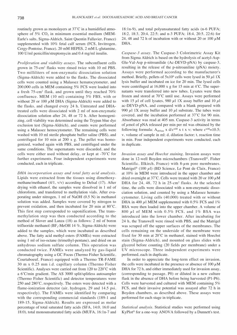
Effect of DHA on MDA-MB-231 cell proliferation. As shownin Fig. 3, cell proliferation decreased slightly following DHAtreatment. This effect was statistically significant at the 72 htime point for both DHA concentrations. Cell proliferationwas also reduced by incubation with DHA, with a significantreduction after exposure to 100 μM DHA for 24 h. Nosignificant difference was noted for the 48 h incubation timefor either DHA concentration.
Effect of DHA on caspase-3 activity and nucleus condensation.In order to show that the DHA-induced reduction of cellularproliferation and/or viability was due at least in part toapoptosis, caspase-3 activity was determined in MDA-MB-231 cells after incubating with DHA. The results clearly
INTERNATIONAL JOURNAL OF ONCOLOGY 36: 737-742, 2010 739
Figure 1. Relative percentage of DHA in total lipid from MDA-MB-231cells after incubating for 24, 48 and 72 h. Black bars correspond to controlwithout any exogenous DHA; gray and white bars correspond to incubationwith 20 and 100 μM DHA, respectively. Results are the mean ± S.D. of 2 inde-pendent experiments each done in triplicate. Statistical significance wasassayed using one-way ANOVA, followed by a Dunnett's test. **P≤0.01.
Figure 2. Total lipid fatty acid composition of MDA-MB-231 cells afterincubating for 24, 48 and 72 h. Results are expressed as the molar percentageof total lipids for (A) saturated, (B) monounsaturated and (C) polyunsatu-rated fatty acids. Black bars correspond to control without any exogenousDHA; gray and white bars correspond to incubation with 20 and 100 μMDHA, respectively. Results are the mean ± S.D. of 2 independent experimentseach done in triplicates. Statistical significance was assayed using one-wayANOVA followed by a Dunnett's test. ** P≤0.01.
Figure 3. Effect of DHA on MDA-MB-231 cell line proliferation. Percentageswere obtained by comparing the number of cells under a given set ofconditions to the number of cells corresponding to the 24-h control. Black barscorrespond to control without any exogenous DHA; gray and white barscorrespond to 20 and 100 μM DHA, respectively. Results are the mean ±S.D. of 4 independent experiments each done in triplicate. Statisticalsignificance was assayed using ANOVA followed by Dunnett's test with thecontrol of the different time of incubation as reference. *P≤0.05; **P≤0.01.
737-742 20/1/2010 01:48 ÌÌ Page 739
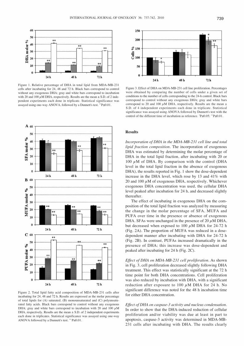
show a strong albeit transient increase in caspase-3 activityafter 24 h in the presence of 100 μM DHA (Fig. 4, P≤0.01).Caspase-3 activity then fell to close to the control level forlonger incubation times. Although not statistically significant,a similar pattern of caspase-3 activity was observed with20 μM DHA, suggesting a dose-effect of DHA (Fig. 4).
Since chromatin condensation is a hallmark of apoptosis,Hoechst staining of nuclei was performed of untreatedcontrol cells and after incubating some cells with DHA. Theslides were observed under a fluorescence microscope, andnormal and condensed nuclei were counted. Some examplesof micrographs showing Hoechst-stained nuclei are shown inFig. 5 B-E, and correspond to the nuclei of MDA-MB-231cells observed in Fig. 5A. Some nuclei appeared to becondensed or fragmented (arrows).
Incubating MDA-MB-231 cells with DHA resulted in adose- and time-dependent increase in condensed apoptoticnuclei (Fig. 5F). A concentration of 100 μM DHA significantlyimpacted the percentage of apoptosis (P≤0.01). A trendtowards an increasing percentage of cells in apoptosis wasobserved with DHA, suggesting a dose-effect; the percentageof apoptosis being representative of nucleus condensation.With 100 μM DHA, the percentage of condensed nucleiincreased between the different time points, and peaked(P≤0.01) at between 5 and 11% after incubating for 72 hunder our experimental conditions.
Effect of DHA on the invasive phenotype of MDA-MB-231cells. The invasive potential of the cells was assayed using
BLANCKAERT et al: DOCOSAHEXAENOIC ACID AND BREAST CANCER740
Figure 4. Caspase-3 activity in MDA-MB-231 cells incubated with DHA.Black bars correspond to control without any exogenous DHA; gray andwhite bars correspond to incubation with 20 and 100 μM DHA, respec-tively. The caspase-3 activity was determined using Ac-DEVD-pNA as asubstrate as detailed in Materials and methods and compared to thecorresponding value in control. Results are the mean ± S.D. of 4 independentexperiments each done in duplicate. Statistical significance was assayedusing one-way ANOVA followed by a Dunnett's test. **P≤0.01.
Figure 5. Effect of DHA on programmed cell death. (A) Light micrograph ofMDA-MB-231 cells in culture. (B-E) Fluorescent micrographs of Hoechststained nuclei: (B) normal nuclei; (C) condensed nucleus (arrow); (D) frag-mented nucleus (arrow); (E) fragmented nucleus or apoptotic bodies (arrows).When the nucleus had undergone multiple fragmentations, this was scoredas one event. Bars correspond to 25 μm. (F) Appearance of condensednuclei in DHA-treated MDA-MB-231 cells. Black bars correspond to controlwithout any exogenous DHA; gray and white bars correspond to incubationwith 20 and 100 μM DHA, respectively. Results are the mean ± S.D. of3 independent experiments each done in duplicate. Statistical significancewas assayed using one-way ANOVA followed by a Dunnett's test. *P≤0.05;**P≤0.01.
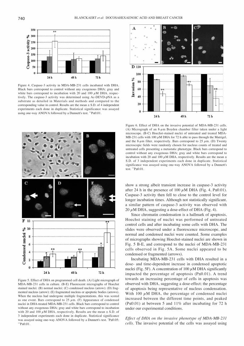
Figure 6. Effect of DHA on the invasive potential of MDA-MB-231 cells.(A) Micrograph of an 8-μm Boyden chamber filter taken under a lightmicroscope. (B-C) Hoechst-stained nuclei of untreated and treated MDA-MB-231 cells with 100 μM DHA for 72 h able to pass through the Matrigel,and the 8-μm filter, respectively. Bars correspond to 25 μm. (D) Twentymicroscopic fields were randomly chosen for nucleus counts of treated anduntreated cells presenting a metastatic phenotype. Black bars correspond tocontrol without any exogenous DHA; gray and white bars correspond toincubation with 20 and 100 μM DHA, respectively. Results are the mean ±S.D. of 3 independent experiments each done in duplicate. Statisticalsignificance was assayed using one-way ANOVA followed by a Dunnett'stest. **P≤0.01.
737-742 20/1/2010 01:48 ÌÌ Page 740

Boyden chambers, and cells were considered to be invasive ifthey were able to get through both the layer of Matrigel, andthe 8-μm porosity filter. The micrographs showing an 8-μmpore filter from a Boyden chamber, and Hoechst-stainednuclei of untreated and DHA-treated cells are shown inFig. 6A-C. The nuclei counts showed that the proportion ofinvasive cells was markedly decreased following incubationwith 100 μM of DHA for 24-72 h (Fig. 6D). However, theresults obtained at the lower DHA concentration (i.e., 20 μM)were not statistically significant.
Furthermore, the inhibitory effect of DHA on the invasivepotential of MDA-MB-231 cells was investigated in long-term experiments, in which cells were maintained in culturefor several passages after DHA treatment. The results showedthat the DHA-induced reduction of invasive cells numberwas sustained for 4 passages, and then became less marked(Fig. 7).
Discussion
Docosahexaenoic acid intake reduces cell proliferation. DHAcan produce this effect at a relatively weak concentration, butwith an incubation time of 72 h. The number of dead cellsincreased slightly over time (data not shown), but theabsence of any statistically significant evidence suggeststhat DHA was not cytotoxic. The obvious question istherefore how DHA induces a reduction in proliferation.Several studies have shown that it might be acting at the levelof the cell cycle, by increasing the duration of the cycle, andmore particularly that of the G2/M transition step (11). Thisis thought to be caused by the inhibition of the CDK1-cyclinB1 complex by DHA. Consequently, the fact that theuntreated cells continued a normal cell cycle would explainthe reduction of cell numbers during the course of DHAtreatment.
Another parameter could also be involved, as we havedemonstrated that DHA acts on the cells in the short-term by
activating caspase-3, an apoptosis effector enzyme. However,the dramatic fall in this activity over longer-term suggeststhat there could be two populations within the cell line, onlyone of which is sensitive to the caspase-3 pathway. Aspreviously described, MDA-MB-231 cells can activate thecaspase-3 pathway via polyunsaturated fatty acids (19).Thus, early activation of this enzymatic activity, resulting inthe rapid death of this population, would be compatiblewith the fall in cell number observed in Fig. 3, where adifference in terms of proliferation was noted at 24 h for levelsof 100 μM (leading to a greater difference in terms of celllosses compared to the untreated cells). Evidence ofincreased apoptosis was also provided by counting thenumber of condensed nuclei. These data showed that theeffect of DHA on MDA-MB-231 cells eventually involvedthe morphological changes typical of apoptosis. However,these data seemed to contradict those obtained by means of thecaspase-3 assay, which displayed activation after 24 h. Thisimplies that a more significant number of condensed nucleiwould have been visible at 24 and 48 h. It is probable that thecells, having reacted to DHA by activating caspase-3, hadcompletely disappeared by 48 h, and this might explain thelow incidence of condensed nuclei; several hours weregenerally needed to ensure that a cell went through thephenomenon of complete apoptosis once it had undergonethe action of caspase-3. Indeed, the increasing numbers ofcondensed nuclei, which under our conditions reached theirhighest level of 11% after a longer incubation time, wasconsistent with the study reported by Siddiqui et al (20), inwhich the percentage of cells undergoing apoptosis leveledoff at 15%. It is also possible that DHA can induce severaldifferent pathways leading to apoptosis, and in particular, theBax pathway that has been described for the HL60 cell line(13). This could account for the delayed appearance ofcondensed cores. Besides, other authors have shown thatDHA has a cytotoxic effect on cancer cells by decreasing thelevel of superoxide dismutase 1, allowing an increase in lipidperoxidation to occur (21). It is probable that these 3pathways are all involved in cells exposed to DHA. Thissuggests that DHA could reduce tumor numbers by acting assoon as a cell begins to change and becomes pre-cancerous;this might explain the low level of breast cancer in populationswith a high DHA diet.
One of the main problems in breast cancer, as well as inother types of cancer, is the ability of cancer cells tometastasize. Several studies have shown that diet can affectthe metastatic potential of cancer cell lines known to have ahigh metastatic phenotype. This effect may be either transientor lasting. For instance, providing the highly metastaticB16BL6 melanoma cell line with low levels of phenylalanineand tyrosine can result in cells with a lower metastaticpotential (2). DHA appears to affect the invasive potential ofMDA-MB-231 cells, which implies that it could modify theirmetastatic phenotype. By extrapolation, this is interesting,because it highlights the importance of the ˆ-3 fatty acids ascompounds that could protect against the appearance ofbreast cancers or their recurrence (22). However, the mecha-nisms by which DHA could directly affect the metastaticphenotype of cancer cell lines remain unclear. The effect ofDHA could result to some extent from a change in the lipids
INTERNATIONAL JOURNAL OF ONCOLOGY 36: 737-742, 2010 741
Figure 7. Long-term effect of DHA on the invasive potential of MDA-MB-231cells. After a 72 h DHA treatment, cells were grown with MEM containing10% FCS for several passages (P0-P6). Prior to invasion assay, the cellswere placed in MEM containing 1% FCS, and the study was carried out asdetailed in the Material and methods section. Black bars correspond to thecontrol without incubation with DHA, and white bars correspond to cellspreviously incubated with 100 μM DHA for 72 h. P number correspondsto the different passage. Results are the mean ± S.D. of 2 independentexperiments each done in triplicates. Statistical significance was assayedusing one-way ANOVA followed by a Dunnett's test. *P≤0.05; **P≤0.01.
737-742 20/1/2010 01:48 ÌÌ Page 741
present in the membrane of the cells, making their plasmamembrane considerably less fluid. If this is so, the lowerinvasive capacity would be explained by a reduction in thedeformability of the cells (23). However, our findingsshowed that i) no reduction in the metastatic phenotype wasdetected when cells were treated with 20 μM DHA, whateverthe incubation time, and ii) cells treated with 100 μM DHAfor 72 h conserved a lower metastatic phenotype at least for alimited number of cell cycles after being returned to a normalmedium. Thus, it would be too restrictive to suggest that thelevel of DHA incorporated alone affects the metastaticphenotype. It is probable that other factors are involved,particularly those that are known to produce the degradationof the Matrigel, which mimics the extracellular matrix(ECM), to occur. Indeed, to be able get across the ECM, themetastatic cells have both the enzymatic tools to digest it,and a composition of integrins that differs from that ofnormal cells (24). Another possible hypothesis is that DHA,and perhaps other n-3 PUFAs, are able to compartmentalizeat the level of the plasma membrane molecules or membraneproteins in particular structures, such as lipid rafts, that arenow considered to be important in cell signaling (25,26).
To conclude, it looks as though, depending on the amountpresent, DHA might play an important role in the processesby which normal and/or differentiated cells gain certaincharacteristics, allowing them to slow their proliferation andminimize their metastatic potential. This finding could beuseful in proteomic analyses to decipher the proteinsimplicated in these phenomena, and should make it possibleto clarify the mechanisms and pathways controlling them.
Acknowledgements
The authors are grateful to Monika Gosh for English revisionof the manuscript. We wish to thank Professor HubertHondermarck for his encouragement and helpful advice. Thiswork was supported by a Grant from the MayenneCollectivities.
References
1. Holmes M and Willett W: Does diet affect breast cancer risk?Breast Cancer Res 6: 170-178, 2004.
2. Blanckaert VD, Schelling ME, Elstad CA and Meadows GG:Differential growth factor production, secretion, and responseby high and low metastatic variants of B16BL6 melanoma.Cancer Res 53: 4075-4081, 1993.
3. Greenwald P, Clifford CK and Milner JA: Diet and cancerprevention. Eur J Cancer 37: 948-965, 2001.
4. Molokhia E and Perkins A: Preventing cancer. Prim Care 35:609-623, 2008.
5. Russo GL: Dietary n-6 and n-3 polyunsaturated fatty acids:From biochemistry to clinical implications in cardiovascularprevention. Biochem Pharmacol 77: 937-946, 2009.
6. Berquin IM, Edwards IJ and Chen YQ: Multi-targeted therapyof cancer by omega-3 fatty acids. Cancer Lett 269: 363-377,2008.
7. Hwang D, Chanmugam P, Ryan D, Boudreau M, Windhauser M,Tulley R, Brooks E and Bray G: Does vegetable oil attenuatethe beneficial effects of fish oil in reducing risk factors forcardiovascular disease? Am J Clin Nutr 66: 89-96, 1997.
8. Ward OP and Singh A: Omega-3/6 fatty acids: alternativesources of production. Process Biochem 40: 3627-3652, 2005.
9. Van Beelen VA, Spenkelink B, Mooibroek H, Sijtsma L, Bosch D,Rietjens IM and Alink GM: An n-3 PUFA-rich microalgal oildiet protects to a similar extent as a fish oil-rich diet againstAOM-induced colonic aberrant crypt foci in F344 rats. FoodChem Toxicol 47: 316-320, 2009.
10. Kinlen L: Diet and breast cancer. Br Med Bull 47: 462-469,1991.
11. Barascu A, Besson P, Le Floch O, Bougnoux P and Jourdan M-L:CDK1-cyclin B1 mediates the inhibition of proliferationinduced by omega-3 fatty acids in MDA-MB-231 breast cancercells. Int J Biochem Cell Biol 38: 196-208, 2006.
12. Chamras H, Ardashian A, Heber D and Glaspy JA: Fatty acidmodulation of MCF-7 human breast cancer cell proliferation,apoptosis and differentiation. J Nutr Biochem 13: 711-716,2002.
13. Miura Y, Takahara K, Murata Y, Utsumi K, Tada M andTakahata K: Docosahexaenoic acid induces apoptosis via theBax-independent pathway in HL-60 cells. Biosci BiotechnolBiochem 68: 2415-2417, 2004.
14. Judé S, Roger S, Martel E, Besson P, Richard S, Bougnoux P,Champeroux P and Le Guennec J-Y: Dietary long-chain omega-3fatty acids of marine origin: a comparison of their protectiveeffects on coronary heart disease and breast cancers. ProgBiophys Mol Biol 90: 299-325, 2006.
15. Vibet S, Goupille C, Bougnoux P, Steghens J-P, Goré J andMahéo K: Sensitization by docosahexaenoic acid (DHA) ofbreast cancer cells to anthracyclines through loss of glutathioneperoxidase (GPx1) response. Free Radic Biol Med 44: 1483-1491,2008.
16. Siddiqui RA, Harvey K and Stillwell W: Anticancer propertiesof oxidation products of docosahexaenoic acid. Chem PhysLipids 153: 47-56, 2008.
17. Delsal J: Nouveau procédé d'extraction des lipides du sérum parle méthylal. Application aux micro-dosages du cholestérol total,des phosphoamino-lipides et des protéines. Bull Soc Chim Biol26: 99-105, 1944.
18. Slover HT and Lanza E: Quantitative analysis of food fattyacids by capillary gas chromatography. J Am Oil Chem Soc 56:933-943, 1979.
19. Winnicka K, Bielawski K, Bielawska A and Surazynski A:Antiproliferative activity of derivatives of ouabain, digoxin andproscillaridin A in human MCF-7 and MDA-MB-231 breastcancer cells. Biol Pharm Bull 31: 1131-1140, 2008.
20. Siddiqui R, Zerouga M, Wu M, Castillo A, Harvey K, Zaloga Gand Stillwell W: Anticancer properties of propofol-docosahexaenoate and propofol-eicosapentaenoate on breastcancer cells. Breast Cancer Res 7: 645-654, 2005.
21. Ding W, Vaught J, Yamauchi H and Lind S: Differentialsensitivity of cancer cells to docosahexaenoic acid-induced cyto-toxicity: the potential importance of down-regulation of super-oxide dismutase 1 expression. Mol Cancer Ther 3: 1109-1117,2004.
22. Stoll B: Breast cancer and the Western diet: role of fatty acidsand antioxidant vitamins. Eur J Cancer 34: 1852-1856, 1998.
23. Zerouga M, Jenski LJ, Booster S and Stillwell W: Can docosa-hexaenoic acid inhibit metastasis by decreasing deformability ofthe tumor cell plasma membrane? Cancer Lett 119: 163-168,1997.
24. Seiki M: Membrane-type 1 matrix metalloproteinase: a keyenzyme for tumor invasion. Cancer Lett 194: 1-11, 2003.
25. Stillwel lW, Shaikh S, Zerouga M, Siddiqui R and Wassall S:Docosahexaenoic acid affects cell signaling by altering lipidrafts. Reprod Nutr Dev 45: 559-579, 2005.
26. Biondo PD, Brindley DN, Sawyer MB and Field CJ: The potentialfor treatment with dietary long-chain polyunsaturated n-3 fattyacids during chemotherapy. J Nutr Biochem 19: 787-796,2008.
BLANCKAERT et al: DOCOSAHEXAENOIC ACID AND BREAST CANCER742
737-742 20/1/2010 01:48 ÌÌ Page 742
Related Documents