Citation: Olivotto, E.; Belluzzi, E.; Pozzuoli, A.; Cigolotti, A.; Scioni, M.; Goldring, S.R.; Goldring, M.B.; Ruggieri, P.; Ramonda, R.; Grigolo, B.; et al. Do Synovial Inflammation and Meniscal Degeneration Impact Clinical Outcomes of Patients Undergoing Arthroscopic Partial Meniscectomy? A Histological Study. Int. J. Mol. Sci. 2022, 23, 3903. https://doi.org/10.3390/ ijms23073903 Academic Editor: Alfonso Baldi Received: 4 March 2022 Accepted: 30 March 2022 Published: 31 March 2022 Publisher’s Note: MDPI stays neutral with regard to jurisdictional claims in published maps and institutional affil- iations. Copyright: © 2022 by the authors. Licensee MDPI, Basel, Switzerland. This article is an open access article distributed under the terms and conditions of the Creative Commons Attribution (CC BY) license (https:// creativecommons.org/licenses/by/ 4.0/). International Journal of Molecular Sciences Article Do Synovial Inflammation and Meniscal Degeneration Impact Clinical Outcomes of Patients Undergoing Arthroscopic Partial Meniscectomy? A Histological Study Eleonora Olivotto 1 , Elisa Belluzzi 2,3, * , Assunta Pozzuoli 2,3 , Augusto Cigolotti 3 , Manuela Scioni 4 , Steven R. Goldring 5 , Mary B. Goldring 5 , Pietro Ruggieri 3 , Roberta Ramonda 6 , Brunella Grigolo 1 , Giovanni Trisolino 7,† and Marta Favero 6,8,† 1 RAMSES Laboratory, RIT Department, IRCCS Istituto Ortopedico Rizzoli, 40136 Bologna, Italy; [email protected] (E.O.); [email protected] (B.G.) 2 Musculoskeletal Pathology and Oncology Laboratory, Orthopaedic and Traumatologic Clinic, Department of Surgery, Oncology and Gastroenterology (DISCOG), University of Padova, 35128 Padova, Italy; [email protected] 3 Orthopedics and Orthopedic Oncology, Department of Surgery, Oncology and Gastroenterology (DiSCOG), University-Hospital of Padova, 35128 Padova, Italy; [email protected] (A.C.); [email protected] (P.R.) 4 Department of Statistical Sciences, University of Padova, 35121 Padova, Italy; [email protected] 5 Hospital for Special Surgery, Weill Cornell Medical College, New York, NY 10021, USA; [email protected] (S.R.G.); [email protected] (M.B.G.) 6 Rheumatology Unit, Department of Medicine (DIMED), University of Padova, 35128 Padova, Italy; [email protected] (R.R.); [email protected] (M.F.) 7 Pediatric Orthopedic and Traumatology, IRCCS Istituto Ortopedico Rizzoli, 40136 Bologna, Italy; [email protected] 8 Medicina Interna I, Ca’ Foncello Hospital, 31100 Treviso, Italy * Correspondence: [email protected]; Tel.: +39-0498213348 † These authors contributed equally to this work. Abstract: The menisci exert a prominent role in joint stabilization and in the distribution of mechanical loading. Meniscal damage is associated with increased risk of knee OA. The aim of this study was to characterize the synovial membrane and meniscal tissues in patients undergoing arthroscopic partial meniscectomy for meniscal tear and to evaluate association with clinical outcomes. A total of 109 patients were recruited. Demographic and clinical data were collected. Visual Analogic Scale (VAS) measuring pain and Knee injury and Osteoarthritis Outcome Score (KOOS) were recorded at baseline and at 2-years follow-up. Histological and immunohistochemical characterizations were performed on synovial membranes and meniscal tissues. More than half of the patients demonstrated synovial mononuclear cell infiltration and hyperplasia. Synovial fibrosis was present in most of the patients; marked vascularity and CD68 positivity were observed. Inflammation had an impact on both pain and knee symptoms. Patients with synovial inflammation had higher values of pre- operative VAS and inflammation. Higher pre-operative pain was observed in patients with meniscal MMP-13 production. In conclusion, multivariate analysis showed that synovial inflammation was associated with pre-operative total KOOS scores, knee symptoms, and pain. Moreover, meniscal MMP-13 expression was found to be associated with pre-operative pain in multivariate analysis. Thus, targeting inflammation of the synovial membrane and meniscus might reduce clinical symptoms and dysfunction at the time of surgery. Keywords: arthroscopic partial meniscectomy; synovial inflammation; meniscal degeneration; pain; clinical outcomes; osteoarthritis Int. J. Mol. Sci. 2022, 23, 3903. https://doi.org/10.3390/ijms23073903 https://www.mdpi.com/journal/ijms

Welcome message from author
This document is posted to help you gain knowledge. Please leave a comment to let me know what you think about it! Share it to your friends and learn new things together.
Transcript
�����������������
Citation: Olivotto, E.; Belluzzi, E.;
Pozzuoli, A.; Cigolotti, A.; Scioni, M.;
Goldring, S.R.; Goldring, M.B.;
Ruggieri, P.; Ramonda, R.; Grigolo, B.;
et al. Do Synovial Inflammation and
Meniscal Degeneration Impact
Clinical Outcomes of Patients
Undergoing Arthroscopic Partial
Meniscectomy? A Histological Study.
Int. J. Mol. Sci. 2022, 23, 3903.
https://doi.org/10.3390/
ijms23073903
Academic Editor: Alfonso Baldi
Received: 4 March 2022
Accepted: 30 March 2022
Published: 31 March 2022
Publisher’s Note: MDPI stays neutral
with regard to jurisdictional claims in
published maps and institutional affil-
iations.
Copyright: © 2022 by the authors.
Licensee MDPI, Basel, Switzerland.
This article is an open access article
distributed under the terms and
conditions of the Creative Commons
Attribution (CC BY) license (https://
creativecommons.org/licenses/by/
4.0/).
International Journal of
Molecular Sciences
Article
Do Synovial Inflammation and Meniscal Degeneration ImpactClinical Outcomes of Patients Undergoing Arthroscopic PartialMeniscectomy? A Histological StudyEleonora Olivotto 1, Elisa Belluzzi 2,3,* , Assunta Pozzuoli 2,3 , Augusto Cigolotti 3, Manuela Scioni 4 ,Steven R. Goldring 5, Mary B. Goldring 5, Pietro Ruggieri 3 , Roberta Ramonda 6 , Brunella Grigolo 1 ,Giovanni Trisolino 7,† and Marta Favero 6,8,†
1 RAMSES Laboratory, RIT Department, IRCCS Istituto Ortopedico Rizzoli, 40136 Bologna, Italy;[email protected] (E.O.); [email protected] (B.G.)
2 Musculoskeletal Pathology and Oncology Laboratory, Orthopaedic and Traumatologic Clinic,Department of Surgery, Oncology and Gastroenterology (DISCOG), University of Padova,35128 Padova, Italy; [email protected]
3 Orthopedics and Orthopedic Oncology, Department of Surgery, Oncology and Gastroenterology (DiSCOG),University-Hospital of Padova, 35128 Padova, Italy; [email protected] (A.C.);[email protected] (P.R.)
4 Department of Statistical Sciences, University of Padova, 35121 Padova, Italy; [email protected] Hospital for Special Surgery, Weill Cornell Medical College, New York, NY 10021, USA;
[email protected] (S.R.G.); [email protected] (M.B.G.)6 Rheumatology Unit, Department of Medicine (DIMED), University of Padova, 35128 Padova, Italy;
[email protected] (R.R.); [email protected] (M.F.)7 Pediatric Orthopedic and Traumatology, IRCCS Istituto Ortopedico Rizzoli, 40136 Bologna, Italy;
[email protected] Medicina Interna I, Ca’ Foncello Hospital, 31100 Treviso, Italy* Correspondence: [email protected]; Tel.: +39-0498213348† These authors contributed equally to this work.
Abstract: The menisci exert a prominent role in joint stabilization and in the distribution of mechanicalloading. Meniscal damage is associated with increased risk of knee OA. The aim of this study wasto characterize the synovial membrane and meniscal tissues in patients undergoing arthroscopicpartial meniscectomy for meniscal tear and to evaluate association with clinical outcomes. A total of109 patients were recruited. Demographic and clinical data were collected. Visual Analogic Scale(VAS) measuring pain and Knee injury and Osteoarthritis Outcome Score (KOOS) were recorded atbaseline and at 2-years follow-up. Histological and immunohistochemical characterizations wereperformed on synovial membranes and meniscal tissues. More than half of the patients demonstratedsynovial mononuclear cell infiltration and hyperplasia. Synovial fibrosis was present in most ofthe patients; marked vascularity and CD68 positivity were observed. Inflammation had an impacton both pain and knee symptoms. Patients with synovial inflammation had higher values of pre-operative VAS and inflammation. Higher pre-operative pain was observed in patients with meniscalMMP-13 production. In conclusion, multivariate analysis showed that synovial inflammation wasassociated with pre-operative total KOOS scores, knee symptoms, and pain. Moreover, meniscalMMP-13 expression was found to be associated with pre-operative pain in multivariate analysis. Thus,targeting inflammation of the synovial membrane and meniscus might reduce clinical symptoms anddysfunction at the time of surgery.
Keywords: arthroscopic partial meniscectomy; synovial inflammation; meniscal degeneration; pain;clinical outcomes; osteoarthritis
Int. J. Mol. Sci. 2022, 23, 3903. https://doi.org/10.3390/ijms23073903 https://www.mdpi.com/journal/ijms
Int. J. Mol. Sci. 2022, 23, 3903 2 of 17
1. Introduction
Knee Osteoarthritis (OA) represents the most common form of joint disease and amajor cause of pain and disability in the adult population [1]. The definition of OA haschanged dramatically over the past decades, and OA is no longer considered a diseaseinvolving only cartilage, but is now recognized as a disorder that might affect all jointcomponents, including the meniscus, synovial membrane, infrapatellar fat pad, and sub-chondral bone [2–4]. The menisci play a critical protective role in joint stabilization andin the transfer and distribution of mechanical loading. Importantly, the integrity of themeniscus is key to joint health, and meniscal damage is associated with an increased riskof OA [5–7]. Untreated meniscal tears can cause pain, joint swelling, recurrent mechani-cal instability leading to significant reduction in quality of life, predominately in youngand active patients [8]. Clinical studies have shown the beneficial effects of meniscalpreservation compared to total meniscectomy and increased degenerative changes wereidentified in knees after total meniscectomy compared with those after partial meniscec-tomy [9]. Synovial inflammation and joint effusions often occur after meniscal injuries asdemonstrated by MRI studies [10–14]. Scanzello et al. reported a relationship betweenthe presence of microscopic synovitis and knee symptoms in a small cohort of patients,without any radiographic signs of OA, undergoing meniscectomy for traumatic meniscaltear [15]. There is additional evidence that synovial inflammation plays a critical role in theseverity of OA symptoms and structural progression, as well as cartilage degeneration rateand osteophytosis appearance [16].
However, it is still not completely clear whether there is an association betweenmicroscopic synovial inflammation and knee pain in patients with meniscal tears. Moreover,no previous studies in the literature have investigated the histological features of bothsynovial and meniscal tissues in order to unravel the possible associations between pre-operative status and post-operative outcome.
Therefore, the aim of this study was to characterize both microscopic features of thesetissues in patients undergoing arthroscopic partial meniscectomy (APM) for meniscal tearin relation to pre-operative status and post-operative outcomes. We hypothesized that thepresence of both synovial inflammation and meniscal degeneration would be associatedwith enhanced clinical symptoms including increased knee pain.
2. Results2.1. Patient Characteristics
Among the 80 patients included in the histological analysis, 56 (70%) were males and24 (30%) were females with a median age of 47 years, interquartile range (IQR) 55.17–39.22.The median BMI of the patients was 27.45 (29.95–23.57), kg/m2 (IQR) (SupplementaryTable S1). Macroscopic cartilage lesions were present in 57 of 78 patients (73%) in whomthe cartilage scoring was complete (Figure S1).
2.2. Synovial Inflammation Features
The histological analysis was performed on 74 patients and 6 synovial samples werediscarded due to the small size of the biopsies. The intra-reader reliability was good formononuclear cell infiltration (0.74) and was fair for all other histological features (hyper-plasia 0.56, vascularity 0.40 and fibrosis 0.42). The inter-reader reliability was moderatefor mononuclear cell infiltration (0.63) and hyperplasia (0.64) and was fair for vascularity(0.55) and poor for fibrosis (0.35). These findings are in line with data available in theliterature [17]. A total of 36 patients (48.6%) did not display any perivascular mononuclearcell infiltration in the synovial tissue.
Among the samples with perivascular mononuclear cell infiltration (38 patients), mostpatients showed a low-grade synovitis (grade 1 = 24 patients (63.2%), grade 2 = 9 patients(23.7%)) and only 5 patients (13.2%) displayed a high grade of synovitis (grade 3). Mostof the synovial specimens showed signs of synovial hyperplasia, with 30 cases (40.5%)displaying grade 1 and 11 patients (14.9%) grade 2. A total of 33 patients (44.6%) exhibited
Int. J. Mol. Sci. 2022, 23, 3903 3 of 17
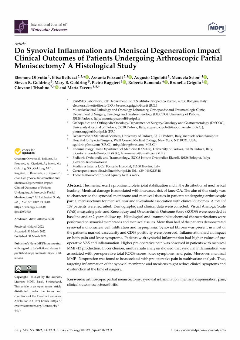
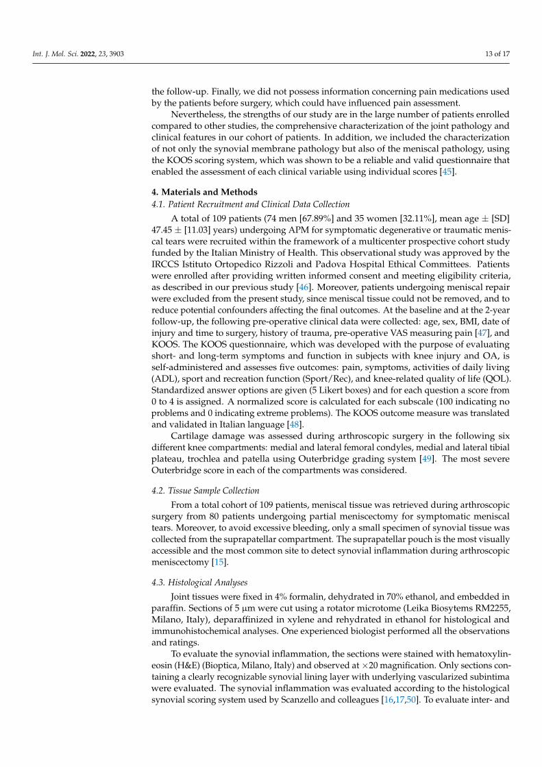
a normal lining. Synovial fibrosis was prevalent and only 5 patients (6.8%) did not showany sign of fibrosis. Vascularity was increased in more than 90% of the patients (Figure 1a).
Int. J. Mol. Sci. 2022, 23, 3903 3 of 17
(40.5%) displaying grade 1 and 11 patients (14.9%) grade 2. A total of 33 patients (44.6%)
exhibited a normal lining. Synovial fibrosis was prevalent and only 5 patients (6.8%) did
not show any sign of fibrosis. Vascularity was increased in more than 90% of the patients
(Figure 1a).
Figure 1. Histological and immunohistochemical analysis of synovial tissue. (a) synovial
inflammation evaluated according to the histological synovial scoring system considering the
following parameters graded from 0 to 3: mononuclear cell infiltration, hyperplasia, fibrosis and
vascularization. (b) in the upper part: number of patients with different grades of production of
CD68 and FVIII; bottom part: a representative patient exhibits high‐grade CD68 production (10×
magnification) and FVIII (grade 3, 20× magnification; the arrow points to immunohistochemical
staining for factor VIII‐related antigen, the marker for endothelial cells of vessels).
In 20 of the 21 patients analyzed, the presence of monocytic infiltration in the synovial
membrane (CD68 positive cells) was detected and all of the 25 patients assessed were
positive for FVIII (Figure 1b).
2.3. Meniscal Features
A total of 6 patients (10%) of 60 analyzed did not show any signs of meniscal
degeneration (grade 1). Most of the patients displayed a range from mild (grade 2) (n = 19,
31.7%) to moderate (grade 3) (n = 30, 50%) meniscal degeneration and five patients (8.3%)
presented a severe grade of meniscal degeneration (grade 4). Analysis of the meniscal
degeneration grading scores is shown in Figure 2a. The histological meniscal degeneration
grade was assessed by a microscopic grading system, which evaluates changes in surface,
cellularity, collagen organization, and Safranin O/Fast green staining. For each single
component related to surface and matrix degradation, only a few patients showed grade
0; on the contrary, all patients showed signs of degradation. Interestingly, most of the
patients showed normal to increased diffuse cellularity. Analysis of the histological
grading scores is shown in Figure 2b.
Immunohistochemical analysis was performed in 51 patients to investigate the
protein expression of metalloproteinase (MMP)‐1, MMP‐13, C1,2C fragment, Collagen
type I, and Collagen type II (Figures 3 and 4).
All samples expressed MMP‐1, mostly with strong signal in almost the entire section
analyzed. On the contrary, most of the samples analyzed (57%) did not show any signal
for MMP‐13, while the remaining showed a grade 2 or 3 score in almost the entire section
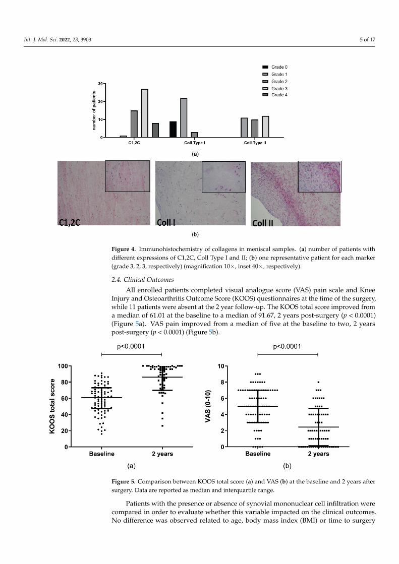
analyzed (Figure 3). Regarding C1,2C, all samples expressed the collagen neoepitope
mostly with strong signal (Figure 4).
As C1,2C is the neoepitope of the three‐quarter length piece generated by the
cleavage of types I and II collagens by collagenases, the presence of the collagen type I in
34 and collagen type II in 33 samples was analyzed. Collagen type II was present in all
samples, while collagen type I was absent in 27% of the samples (Figure 4).
Figure 1. Histological and immunohistochemical analysis of synovial tissue. (a) synovial inflam-mation evaluated according to the histological synovial scoring system considering the followingparameters graded from 0 to 3: mononuclear cell infiltration, hyperplasia, fibrosis and vascularization.(b) in the upper part: number of patients with different grades of production of CD68 and FVIII;bottom part: a representative patient exhibits high-grade CD68 production (10× magnification) andFVIII (grade 3, 20× magnification; the arrow points to immunohistochemical staining for factorVIII-related antigen, the marker for endothelial cells of vessels).
In 20 of the 21 patients analyzed, the presence of monocytic infiltration in the synovialmembrane (CD68 positive cells) was detected and all of the 25 patients assessed werepositive for FVIII (Figure 1b).
2.3. Meniscal Features
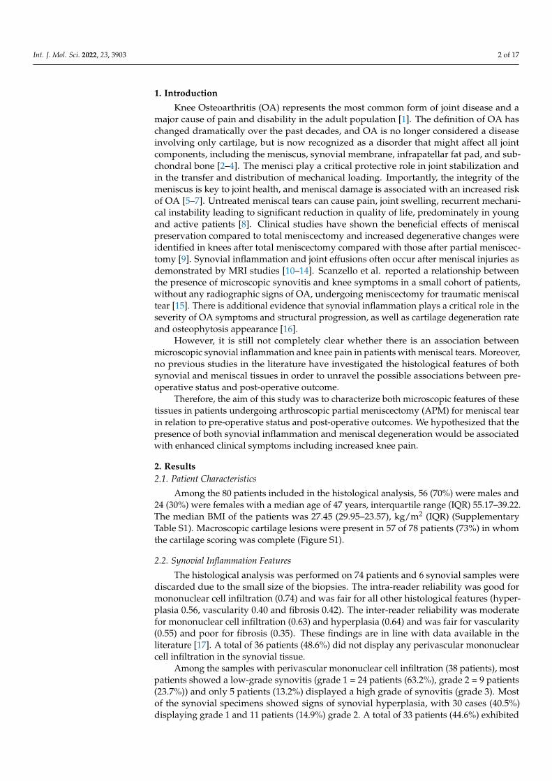
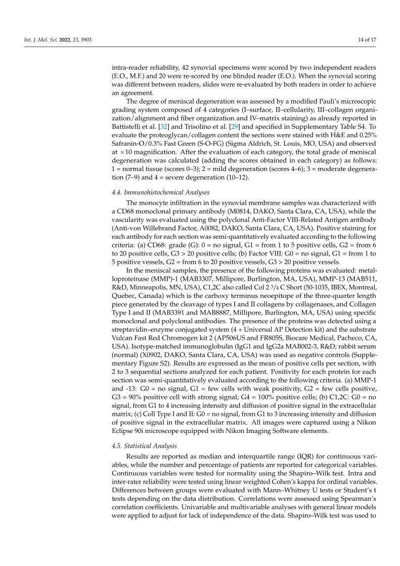
A total of 6 patients (10%) of 60 analyzed did not show any signs of meniscal de-generation (grade 1). Most of the patients displayed a range from mild (grade 2) (n = 19,31.7%) to moderate (grade 3) (n = 30, 50%) meniscal degeneration and five patients (8.3%)presented a severe grade of meniscal degeneration (grade 4). Analysis of the meniscaldegeneration grading scores is shown in Figure 2a. The histological meniscal degenerationgrade was assessed by a microscopic grading system, which evaluates changes in surface,cellularity, collagen organization, and Safranin O/Fast green staining. For each singlecomponent related to surface and matrix degradation, only a few patients showed grade 0;on the contrary, all patients showed signs of degradation. Interestingly, most of the patientsshowed normal to increased diffuse cellularity. Analysis of the histological grading scoresis shown in Figure 2b.
Immunohistochemical analysis was performed in 51 patients to investigate the proteinexpression of metalloproteinase (MMP)-1, MMP-13, C1,2C fragment, Collagen type I, andCollagen type II (Figures 3 and 4).
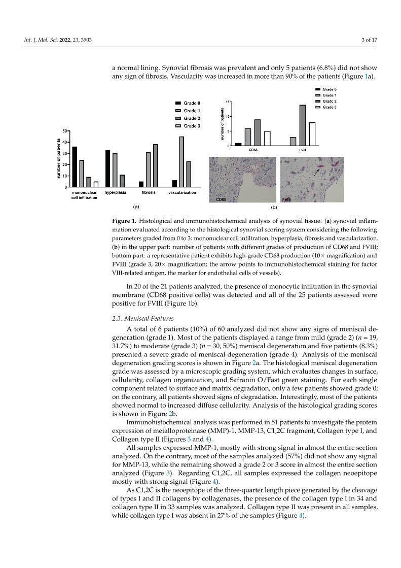
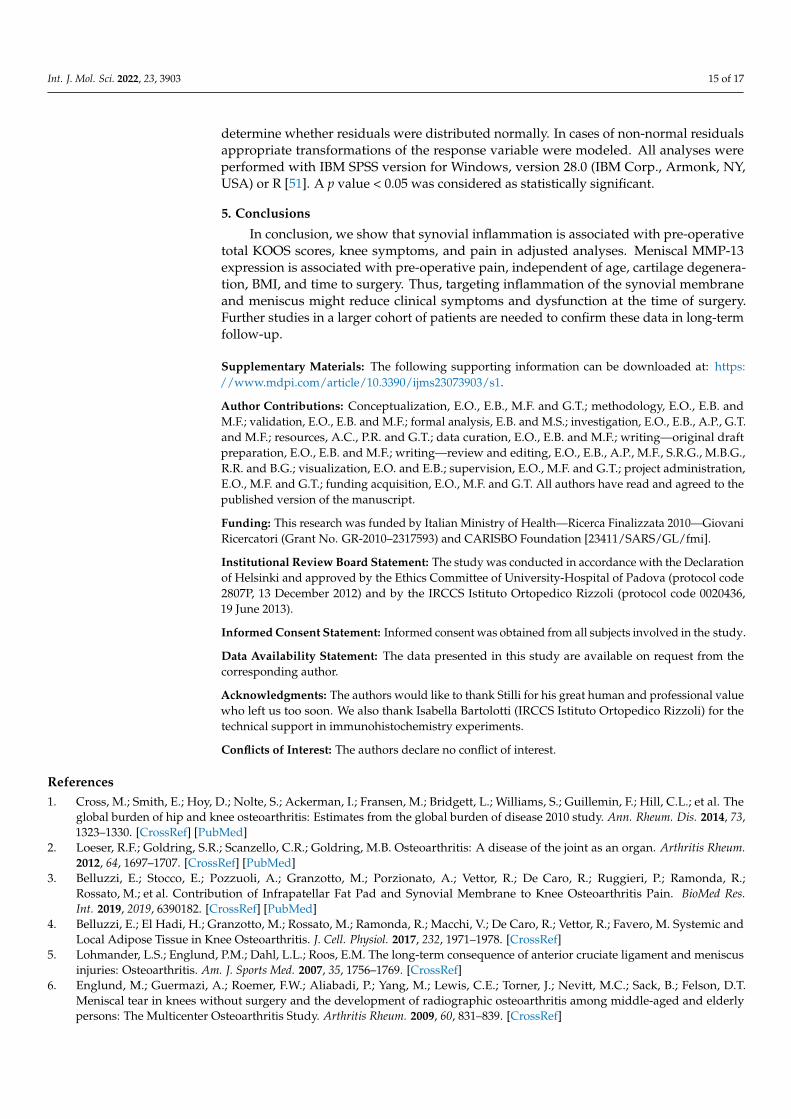
All samples expressed MMP-1, mostly with strong signal in almost the entire sectionanalyzed. On the contrary, most of the samples analyzed (57%) did not show any signalfor MMP-13, while the remaining showed a grade 2 or 3 score in almost the entire sectionanalyzed (Figure 3). Regarding C1,2C, all samples expressed the collagen neoepitopemostly with strong signal (Figure 4).
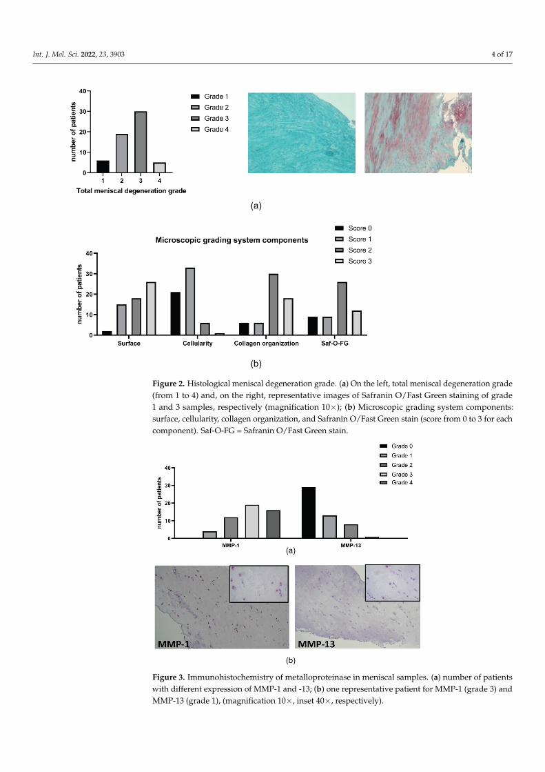
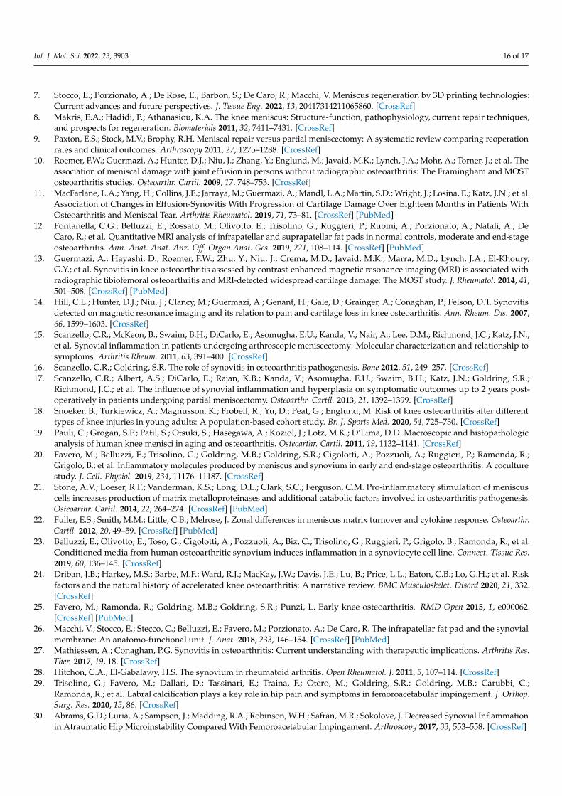
As C1,2C is the neoepitope of the three-quarter length piece generated by the cleavageof types I and II collagens by collagenases, the presence of the collagen type I in 34 andcollagen type II in 33 samples was analyzed. Collagen type II was present in all samples,while collagen type I was absent in 27% of the samples (Figure 4).
Int. J. Mol. Sci. 2022, 23, 3903 4 of 17Int. J. Mol. Sci. 2022, 23, 3903 4 of 17
Figure 2. Histological meniscal degeneration grade. (a) On the left, total meniscal degeneration
grade (from 1 to 4) and, on the right, representative images of Safranin O/Fast Green staining of
grade 1 and 3 samples, respectively (magnification 10×); (b) Microscopic grading system
components: surface, cellularity, collagen organization, and Safranin O/Fast Green stain (score from
0 to 3 for each component). Saf‐O‐FG = Safranin O/Fast Green stain.
Figure 3. Immunohistochemistry of metalloproteinase in meniscal samples. (a) number of patients
with different expression of MMP‐1 and ‐13; (b) one representative patient for MMP‐1 (grade 3) and
MMP‐13 (grade 1), (magnification 10×, inset 40×, respectively).
Figure 2. Histological meniscal degeneration grade. (a) On the left, total meniscal degeneration grade(from 1 to 4) and, on the right, representative images of Safranin O/Fast Green staining of grade1 and 3 samples, respectively (magnification 10×); (b) Microscopic grading system components:surface, cellularity, collagen organization, and Safranin O/Fast Green stain (score from 0 to 3 for eachcomponent). Saf-O-FG = Safranin O/Fast Green stain.
Int. J. Mol. Sci. 2022, 23, 3903 4 of 17
Figure 2. Histological meniscal degeneration grade. (a) On the left, total meniscal degeneration
grade (from 1 to 4) and, on the right, representative images of Safranin O/Fast Green staining of
grade 1 and 3 samples, respectively (magnification 10×); (b) Microscopic grading system
components: surface, cellularity, collagen organization, and Safranin O/Fast Green stain (score from
0 to 3 for each component). Saf‐O‐FG = Safranin O/Fast Green stain.
Figure 3. Immunohistochemistry of metalloproteinase in meniscal samples. (a) number of patients
with different expression of MMP‐1 and ‐13; (b) one representative patient for MMP‐1 (grade 3) and
MMP‐13 (grade 1), (magnification 10×, inset 40×, respectively).
Figure 3. Immunohistochemistry of metalloproteinase in meniscal samples. (a) number of patientswith different expression of MMP-1 and -13; (b) one representative patient for MMP-1 (grade 3) andMMP-13 (grade 1), (magnification 10×, inset 40×, respectively).
Int. J. Mol. Sci. 2022, 23, 3903 5 of 17Int. J. Mol. Sci. 2022, 23, 3903 5 of 17
Figure 4. Immunohistochemistry of collagens in meniscal samples. (a) number of patients with
different expressions of C1,2C, Coll Type I and II; (b) one representative patient for each marker
(grade 3, 2, 3, respectively) (magnification 10×, inset 40×, respectively).
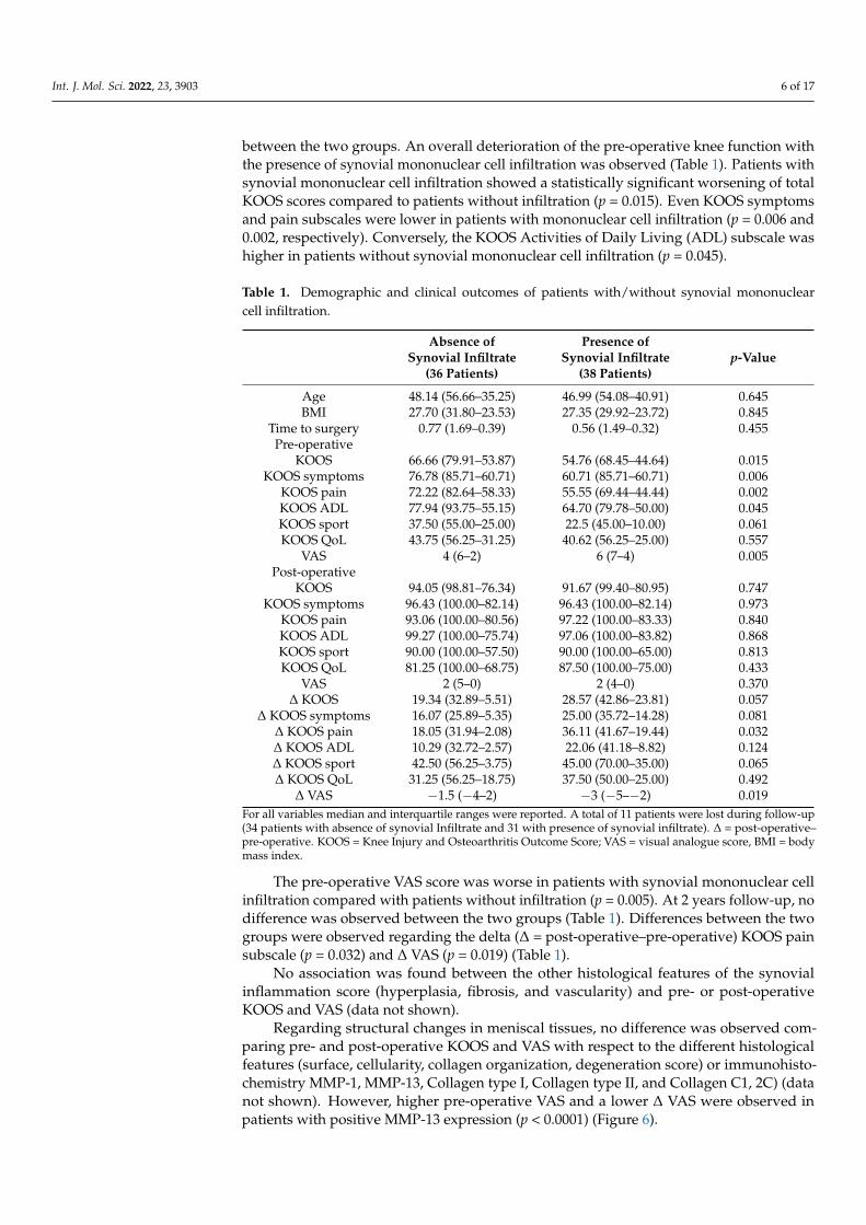
2.4. Clinical Outcomes
All enrolled patients completed visual analogue score (VAS) pain scale and Knee
Injury and Osteoarthritis Outcome Score (KOOS) questionnaires at the time of the surgery,
while 11 patients were absent at the 2 year follow‐up. The KOOS total score improved
from a median of 61.01 at the baseline to a median of 91.67, 2 years post‐surgery (p <
0.0001) (Figure 5a). VAS pain improved from a median of five at the baseline to two, 2
years post‐surgery (p < 0.0001) (Figure 5b).
Figure 5. Comparison between KOOS total score (a) and VAS (b) at the baseline and 2 years after
surgery. Data are reported as median and interquartile range.
Patients with the presence or absence of synovial mononuclear cell infiltration were
compared in order to evaluate whether this variable impacted on the clinical outcomes.
No difference was observed related to age, body mass index (BMI) or time to surgery
between the two groups. An overall deterioration of the pre‐operative knee function with
the presence of synovial mononuclear cell infiltration was observed (Table 1). Patients
with synovial mononuclear cell infiltration showed a statistically significant worsening of
total KOOS scores compared to patients without infiltration (p = 0.015). Even KOOS
Figure 4. Immunohistochemistry of collagens in meniscal samples. (a) number of patients withdifferent expressions of C1,2C, Coll Type I and II; (b) one representative patient for each marker(grade 3, 2, 3, respectively) (magnification 10×, inset 40×, respectively).
2.4. Clinical Outcomes
All enrolled patients completed visual analogue score (VAS) pain scale and KneeInjury and Osteoarthritis Outcome Score (KOOS) questionnaires at the time of the surgery,while 11 patients were absent at the 2 year follow-up. The KOOS total score improved froma median of 61.01 at the baseline to a median of 91.67, 2 years post-surgery (p < 0.0001)(Figure 5a). VAS pain improved from a median of five at the baseline to two, 2 yearspost-surgery (p < 0.0001) (Figure 5b).
Int. J. Mol. Sci. 2022, 23, 3903 5 of 17
Figure 4. Immunohistochemistry of collagens in meniscal samples. (a) number of patients with
different expressions of C1,2C, Coll Type I and II; (b) one representative patient for each marker
(grade 3, 2, 3, respectively) (magnification 10×, inset 40×, respectively).
2.4. Clinical Outcomes
All enrolled patients completed visual analogue score (VAS) pain scale and Knee
Injury and Osteoarthritis Outcome Score (KOOS) questionnaires at the time of the surgery,
while 11 patients were absent at the 2 year follow‐up. The KOOS total score improved
from a median of 61.01 at the baseline to a median of 91.67, 2 years post‐surgery (p <
0.0001) (Figure 5a). VAS pain improved from a median of five at the baseline to two, 2
years post‐surgery (p < 0.0001) (Figure 5b).
Figure 5. Comparison between KOOS total score (a) and VAS (b) at the baseline and 2 years after
surgery. Data are reported as median and interquartile range.
Patients with the presence or absence of synovial mononuclear cell infiltration were
compared in order to evaluate whether this variable impacted on the clinical outcomes.
No difference was observed related to age, body mass index (BMI) or time to surgery
between the two groups. An overall deterioration of the pre‐operative knee function with
the presence of synovial mononuclear cell infiltration was observed (Table 1). Patients
with synovial mononuclear cell infiltration showed a statistically significant worsening of
total KOOS scores compared to patients without infiltration (p = 0.015). Even KOOS
Figure 5. Comparison between KOOS total score (a) and VAS (b) at the baseline and 2 years aftersurgery. Data are reported as median and interquartile range.
Patients with the presence or absence of synovial mononuclear cell infiltration werecompared in order to evaluate whether this variable impacted on the clinical outcomes.No difference was observed related to age, body mass index (BMI) or time to surgery
Int. J. Mol. Sci. 2022, 23, 3903 6 of 17
between the two groups. An overall deterioration of the pre-operative knee function withthe presence of synovial mononuclear cell infiltration was observed (Table 1). Patients withsynovial mononuclear cell infiltration showed a statistically significant worsening of totalKOOS scores compared to patients without infiltration (p = 0.015). Even KOOS symptomsand pain subscales were lower in patients with mononuclear cell infiltration (p = 0.006 and0.002, respectively). Conversely, the KOOS Activities of Daily Living (ADL) subscale washigher in patients without synovial mononuclear cell infiltration (p = 0.045).
Table 1. Demographic and clinical outcomes of patients with/without synovial mononuclearcell infiltration.
Absence ofSynovial Infiltrate
(36 Patients)
Presence ofSynovial Infiltrate
(38 Patients)p-Value
Age 48.14 (56.66–35.25) 46.99 (54.08–40.91) 0.645BMI 27.70 (31.80–23.53) 27.35 (29.92–23.72) 0.845
Time to surgery 0.77 (1.69–0.39) 0.56 (1.49–0.32) 0.455Pre-operative
KOOS 66.66 (79.91–53.87) 54.76 (68.45–44.64) 0.015KOOS symptoms 76.78 (85.71–60.71) 60.71 (85.71–60.71) 0.006
KOOS pain 72.22 (82.64–58.33) 55.55 (69.44–44.44) 0.002KOOS ADL 77.94 (93.75–55.15) 64.70 (79.78–50.00) 0.045KOOS sport 37.50 (55.00–25.00) 22.5 (45.00–10.00) 0.061KOOS QoL 43.75 (56.25–31.25) 40.62 (56.25–25.00) 0.557
VAS 4 (6–2) 6 (7–4) 0.005Post-operative
KOOS 94.05 (98.81–76.34) 91.67 (99.40–80.95) 0.747KOOS symptoms 96.43 (100.00–82.14) 96.43 (100.00–82.14) 0.973
KOOS pain 93.06 (100.00–80.56) 97.22 (100.00–83.33) 0.840KOOS ADL 99.27 (100.00–75.74) 97.06 (100.00–83.82) 0.868KOOS sport 90.00 (100.00–57.50) 90.00 (100.00–65.00) 0.813KOOS QoL 81.25 (100.00–68.75) 87.50 (100.00–75.00) 0.433
VAS 2 (5–0) 2 (4–0) 0.370∆ KOOS 19.34 (32.89–5.51) 28.57 (42.86–23.81) 0.057
∆ KOOS symptoms 16.07 (25.89–5.35) 25.00 (35.72–14.28) 0.081∆ KOOS pain 18.05 (31.94–2.08) 36.11 (41.67–19.44) 0.032∆ KOOS ADL 10.29 (32.72–2.57) 22.06 (41.18–8.82) 0.124∆ KOOS sport 42.50 (56.25–3.75) 45.00 (70.00–35.00) 0.065∆ KOOS QoL 31.25 (56.25–18.75) 37.50 (50.00–25.00) 0.492
∆ VAS −1.5 (−4–2) −3 (−5–−2) 0.019For all variables median and interquartile ranges were reported. A total of 11 patients were lost during follow-up(34 patients with absence of synovial Infiltrate and 31 with presence of synovial infiltrate). ∆ = post-operative–pre-operative. KOOS = Knee Injury and Osteoarthritis Outcome Score; VAS = visual analogue score, BMI = bodymass index.
The pre-operative VAS score was worse in patients with synovial mononuclear cellinfiltration compared with patients without infiltration (p = 0.005). At 2 years follow-up, nodifference was observed between the two groups (Table 1). Differences between the twogroups were observed regarding the delta (∆ = post-operative–pre-operative) KOOS painsubscale (p = 0.032) and ∆ VAS (p = 0.019) (Table 1).
No association was found between the other histological features of the synovialinflammation score (hyperplasia, fibrosis, and vascularity) and pre- or post-operativeKOOS and VAS (data not shown).
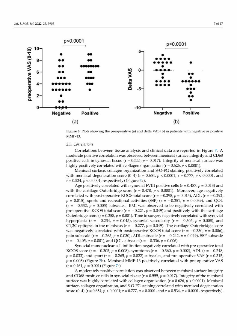
Regarding structural changes in meniscal tissues, no difference was observed com-paring pre- and post-operative KOOS and VAS with respect to the different histologicalfeatures (surface, cellularity, collagen organization, degeneration score) or immunohisto-chemistry MMP-1, MMP-13, Collagen type I, Collagen type II, and Collagen C1, 2C) (datanot shown). However, higher pre-operative VAS and a lower ∆ VAS were observed inpatients with positive MMP-13 expression (p < 0.0001) (Figure 6).
Int. J. Mol. Sci. 2022, 23, 3903 7 of 17Int. J. Mol. Sci. 2022, 23, 3903 7 of 17
Figure 6. Plots showing the preoperative (a) and delta VAS (b) in patients with negative or positive
MMP‐13.
2.5. Correlations
Correlations between tissue analysis and clinical data are reported in Figure 7. A
moderate positive correlation was observed between meniscal surface integrity and CD68
positive cells in synovial tissue (r = 0.555, p = 0.017). Integrity of meniscal surface was
highly positively correlated with collagen organization (r = 0.626, p < 0.0001).
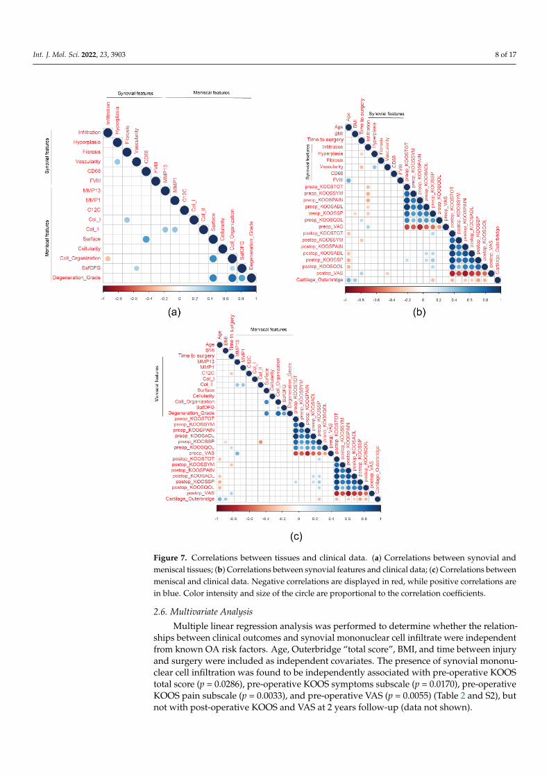
Figure 7. Correlations between tissues and clinical data. (a) Correlations between synovial and
meniscal tissues; (b) Correlations between synovial features and clinical data; (c) Correlations
Figure 6. Plots showing the preoperative (a) and delta VAS (b) in patients with negative or positiveMMP-13.
2.5. Correlations
Correlations between tissue analysis and clinical data are reported in Figure 7. Amoderate positive correlation was observed between meniscal surface integrity and CD68positive cells in synovial tissue (r = 0.555, p = 0.017). Integrity of meniscal surface washighly positively correlated with collagen organization (r = 0.626, p < 0.0001).
Meniscal surface, collagen organization and S-O-FG staining positively correlatedwith meniscal degeneration score (0–4) (r = 0.654, p < 0.0001; r = 0.777, p < 0.0001, andr = 0.534, p < 0.0001, respectively) (Figure 7a).
Age positively correlated with synovial FVIII positive cells (r = 0.487, p = 0.013) andwith the cartilage Outerbridge score (r = 0.470, p < 0.0001). Moreover, age negativelycorrelated with post-operative KOOS total score (r = −0.298, p = 0.013), ADL (r = −0.292,p = 0.015), sports and recreational activities (SSP) (r = −0.351, p = 0.0039), and QOL(r = −0.332, p = 0.005) subscales. BMI was observed to be negatively correlated withpre-operative KOOS total score (r = −0.221, p = 0.049) and positively with the cartilageOuterbridge score (r = 0.358, p = 0.001). Time to surgery negatively correlated with synovialhyperplasia (r = −0.234, p = 0.045), synovial vascularity (r = −0.305, p = 0.008), andC1,2C epitopes in the meniscus (r = −0.277, p = 0.049). The cartilage Outerbridge scorewas negatively correlated with postoperative KOOS total score (r = −0.330, p = 0.006),pain subscale (r = −0.265, p = 0.030), ADL subscale (r = −0.242, p = 0.049), SSP subscale(r = −0.405, p = 0.001), and QOL subscale (r = −0.336, p = 0.006).
Synovial mononuclear cell infiltration negatively correlated with pre-operative totalKOOS score (r = −0.305, p = 0.008), symptoms (r = −0.360, p = 0.002), ADL (r = −0.248,p = 0.033), and sport (r = −0.265, p = 0.022) subscales, and pre-operative VAS (r = 0.315,p = 0.006) (Figure 7b). Meniscal MMP-13 positively correlated with pre-operative VAS(r = 0.461, p = 0.001) (Figure 7c).
A moderately positive correlation was observed between meniscal surface integrityand CD68-positive cells in synovial tissue (r = 0.555, p = 0.017). Integrity of the meniscalsurface was highly correlated with collagen organization (r = 0.626, p < 0.0001). Meniscalsurface, collagen organization, and S-O-FG staining correlated with meniscal degenerationscore (0–4) (r = 0.654, p < 0.0001; r = 0.777, p < 0.0001, and r = 0.534, p < 0.0001, respectively).
Int. J. Mol. Sci. 2022, 23, 3903 8 of 17
Int. J. Mol. Sci. 2022, 23, 3903 7 of 17
Figure 6. Plots showing the preoperative (a) and delta VAS (b) in patients with negative or positive
MMP‐13.
2.5. Correlations
Correlations between tissue analysis and clinical data are reported in Figure 7. A
moderate positive correlation was observed between meniscal surface integrity and CD68
positive cells in synovial tissue (r = 0.555, p = 0.017). Integrity of meniscal surface was
highly positively correlated with collagen organization (r = 0.626, p < 0.0001).
Figure 7. Correlations between tissues and clinical data. (a) Correlations between synovial and
meniscal tissues; (b) Correlations between synovial features and clinical data; (c) Correlations
Figure 7. Correlations between tissues and clinical data. (a) Correlations between synovial andmeniscal tissues; (b) Correlations between synovial features and clinical data; (c) Correlations betweenmeniscal and clinical data. Negative correlations are displayed in red, while positive correlations arein blue. Color intensity and size of the circle are proportional to the correlation coefficients.
2.6. Multivariate Analysis
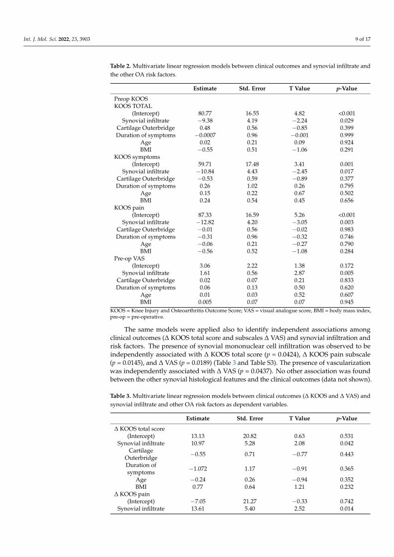
Multiple linear regression analysis was performed to determine whether the relation-ships between clinical outcomes and synovial mononuclear cell infiltrate were independentfrom known OA risk factors. Age, Outerbridge “total score”, BMI, and time between injuryand surgery were included as independent covariates. The presence of synovial mononu-clear cell infiltration was found to be independently associated with pre-operative KOOStotal score (p = 0.0286), pre-operative KOOS symptoms subscale (p = 0.0170), pre-operativeKOOS pain subscale (p = 0.0033), and pre-operative VAS (p = 0.0055) (Table 2 and S2), butnot with post-operative KOOS and VAS at 2 years follow-up (data not shown).
Int. J. Mol. Sci. 2022, 23, 3903 9 of 17
Table 2. Multivariate linear regression models between clinical outcomes and synovial infiltrate andthe other OA risk factors.
Estimate Std. Error T Value p-Value
Preop KOOSKOOS TOTAL
(Intercept) 80.77 16.55 4.82 <0.001Synovial infiltrate −9.38 4.19 −2.24 0.029
Cartilage Outerbridge 0.48 0.56 −0.85 0.399Duration of symptoms −0.0007 0.96 −0.001 0.999
Age 0.02 0.21 0.09 0.924BMI −0.55 0.51 −1.06 0.291
KOOS symptoms(Intercept) 59.71 17.48 3.41 0.001
Synovial infiltrate −10.84 4.43 −2.45 0.017Cartilage Outerbridge −0.53 0.59 −0.89 0.377Duration of symptoms 0.26 1.02 0.26 0.795
Age 0.15 0.22 0.67 0.502BMI 0.24 0.54 0.45 0.656
KOOS pain(Intercept) 87.33 16.59 5.26 <0.001
Synovial infiltrate −12.82 4.20 −3.05 0.003Cartilage Outerbridge −0.01 0.56 −0.02 0.983Duration of symptoms −0.31 0.96 −0.32 0.746
Age −0.06 0.21 −0.27 0.790BMI −0.56 0.52 −1.08 0.284
Pre-op VAS(Intercept) 3.06 2.22 1.38 0.172
Synovial infiltrate 1.61 0.56 2.87 0.005Cartilage Outerbridge 0.02 0.07 0.21 0.833Duration of symptoms 0.06 0.13 0.50 0.620
Age 0.01 0.03 0.52 0.607BMI 0.005 0.07 0.07 0.945
KOOS = Knee Injury and Osteoarthritis Outcome Score; VAS = visual analogue score, BMI = body mass index,pre-op = pre-operative.
The same models were applied also to identify independent associations amongclinical outcomes (∆ KOOS total score and subscales ∆ VAS) and synovial infiltration andrisk factors. The presence of synovial mononuclear cell infiltration was observed to beindependently associated with ∆ KOOS total score (p = 0.0424), ∆ KOOS pain subscale(p = 0.0145), and ∆ VAS (p = 0.0189) (Table 3 and Table S3). The presence of vascularizationwas independently associated with ∆ VAS (p = 0.0437). No other association was foundbetween the other synovial histological features and the clinical outcomes (data not shown).
Table 3. Multivariate linear regression models between clinical outcomes (∆ KOOS and ∆ VAS) andsynovial infiltrate and other OA risk factors as dependent variables.
Estimate Std. Error T Value p-Value
∆ KOOS total score(Intercept) 13.13 20.82 0.63 0.531
Synovial infiltrate 10.97 5.28 2.08 0.042Cartilage
Outerbridge −0.55 0.71 −0.77 0.443
Duration ofsymptoms −1.072 1.17 −0.91 0.365
Age −0.24 0.26 −0.94 0.352BMI 0.77 0.64 1.21 0.232
∆ KOOS pain(Intercept) −7.05 21.27 −0.33 0.742
Synovial infiltrate 13.61 5.40 2.52 0.014
Int. J. Mol. Sci. 2022, 23, 3903 10 of 17
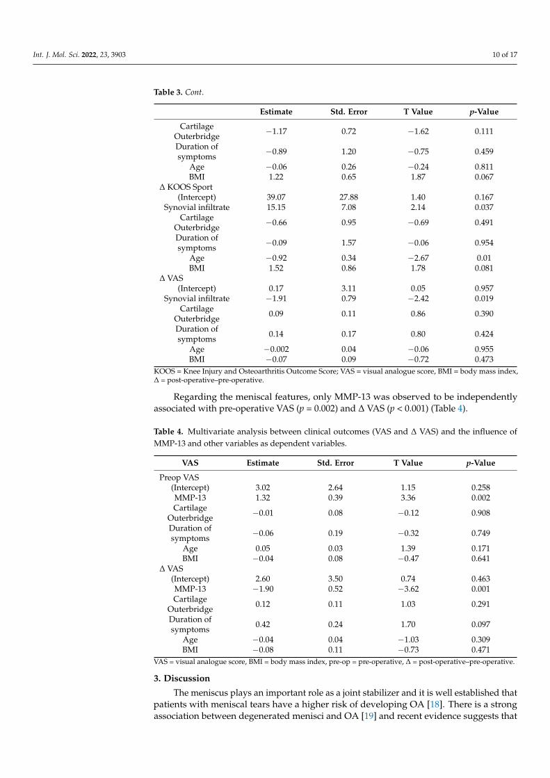
Table 3. Cont.
Estimate Std. Error T Value p-Value
CartilageOuterbridge −1.17 0.72 −1.62 0.111
Duration ofsymptoms −0.89 1.20 −0.75 0.459
Age −0.06 0.26 −0.24 0.811BMI 1.22 0.65 1.87 0.067
∆ KOOS Sport(Intercept) 39.07 27.88 1.40 0.167
Synovial infiltrate 15.15 7.08 2.14 0.037Cartilage
Outerbridge −0.66 0.95 −0.69 0.491
Duration ofsymptoms −0.09 1.57 −0.06 0.954
Age −0.92 0.34 −2.67 0.01BMI 1.52 0.86 1.78 0.081
∆ VAS(Intercept) 0.17 3.11 0.05 0.957
Synovial infiltrate −1.91 0.79 −2.42 0.019Cartilage
Outerbridge 0.09 0.11 0.86 0.390
Duration ofsymptoms 0.14 0.17 0.80 0.424
Age −0.002 0.04 −0.06 0.955BMI −0.07 0.09 −0.72 0.473
KOOS = Knee Injury and Osteoarthritis Outcome Score; VAS = visual analogue score, BMI = body mass index,∆ = post-operative–pre-operative.
Regarding the meniscal features, only MMP-13 was observed to be independentlyassociated with pre-operative VAS (p = 0.002) and ∆ VAS (p < 0.001) (Table 4).
Table 4. Multivariate analysis between clinical outcomes (VAS and ∆ VAS) and the influence ofMMP-13 and other variables as dependent variables.
VAS Estimate Std. Error T Value p-Value
Preop VAS(Intercept) 3.02 2.64 1.15 0.258MMP-13 1.32 0.39 3.36 0.002Cartilage
Outerbridge −0.01 0.08 −0.12 0.908
Duration ofsymptoms −0.06 0.19 −0.32 0.749
Age 0.05 0.03 1.39 0.171BMI −0.04 0.08 −0.47 0.641
∆ VAS(Intercept) 2.60 3.50 0.74 0.463MMP-13 −1.90 0.52 −3.62 0.001Cartilage
Outerbridge 0.12 0.11 1.03 0.291
Duration ofsymptoms 0.42 0.24 1.70 0.097
Age −0.04 0.04 −1.03 0.309BMI −0.08 0.11 −0.73 0.471
VAS = visual analogue score, BMI = body mass index, pre-op = pre-operative, ∆ = post-operative–pre-operative.
3. Discussion
The meniscus plays an important role as a joint stabilizer and it is well established thatpatients with meniscal tears have a higher risk of developing OA [18]. There is a strongassociation between degenerated menisci and OA [19] and recent evidence suggests that
Int. J. Mol. Sci. 2022, 23, 3903 11 of 17
the meniscus also plays a biological role in OA [20–22] through the increased production ofpro-inflammatory mediators and matrix-degrading enzymes [20–23].
In our study, only 27% of the patients had normal cartilage recorded during arthro-scopic surgery. Based on the literature that highlight the importance of the meniscus inOA onset and progression [24], we analyzed not only the impact of synovial inflammation,which was observed both in early and late stages of OA [20,25], but also the potentialinfluence of meniscal structural changes both on pre-operative status and post-operativeclinical outcomes.
The healthy synovial membrane is composed of two layers: the intima, which lies nextto the joint cavity and consists of a layer of 1–4 cells, only 20–40 µm thick and the subintimathat can be up to 5 mm [16,26]. More than half of the patients demonstrated synovialmononuclear cell infiltration and hyperplasia. Synovial fibrosis was present in most of thepatients and marked vascularity was observed in more than 80% of the patients. In OApatients, the synovial membrane is characterized by synovial lining hyperplasia, subliningfibrosis, stromal vascularization and macrophage and T-cell lymphocytes infiltration [27].The same histological features can be also found in the early phase of inflammatory arthritis,even if they are more prominent, whereas organized lymphoid follicles and pannus tissueare features generally associated with inflammatory arthritis [28].
All patients showed cells positive to FVIII. In addition, all patients except one exhibitedCD68 positive cells, a macrophage marker, in the synovial infiltrates, which correlated withan overall deterioration of knee function and pain (measured through VAS scale) beforesurgery. Interestingly, CD68 positive cells were also found in the synovial membrane ofpatients affected by femoro-acetabular impingement, which predisposes patients to hipOA [29,30]. As CD68 does not distinguish pro-inflammatory macrophages (M1) from anti-inflammatory macrophages (M2), other studies need to be performed to further investigatethe macrophage phenotype.
Regarding the intra- and inter-reliability, our data are substantially in agreement withthose obtained by Scanzello et al. [17]. Indeed, moderate to good intra- and inter-reliabilitywas found for mononuclear cell infiltration and hyperplasia, while weak intra- and inter-reliability was found for fibrosis. Weak intra- and moderate inter-reliability was observedfor vascularity.
Lower values of pre-operative KOOS total score were found in patients with synovialinflammation in accordance with the results obtained by Scanzello et al. [15], which usedthe Lysholm score, a knee-specific metric of symptoms and functional disability. Lysholmis a weighted score, with pain and instability-related symptoms having the most weight.In contrast to Scanzello et al. [17], in our study we used the KOOS score to possess thepossibility to evaluate the impact of the synovial inflammation and meniscal changeson the different subscales. We observed that patients with synovial inflammation hadlower pre-operative KOOS symptoms and pain subscales suggesting that inflammationhad an impact on both pain and knee symptoms based on univariate analysis. Moreover,negative correlations were observed between synovial inflammation and pre-operativeKOOS scores. Patients with synovial infiltrate had higher values of pre-operative VAS andsynovial infiltrate correlated positively with the pre-operative VAS scores. On the contrary,no association was observed in the study of Scanzello et al. [17].
Multivariate analysis showed that synovial inflammation was associated with pre-operative KOOS total score, pre-operative KOOS symptoms and pain subscales, and pre-operative VAS independently from the cartilage Outerbridge score, duration of symptoms,BMI or age. An improvement of post-operative KOOS and VAS scores was observed inpatients with and without synovial inflammation, but no difference was observed whencomparing the clinical outcomes between the two groups. These results agree with thoseobtained by Scanzello et al. [17]. A difference in the ∆ KOOS pain subscale and ∆ VASwas observed in comparing patients with or without synovial inflammation, while weidentified a borderline difference for ∆ KOOS total score (p = 0.057) in univariate analysis.Multivariate analysis confirmed all the associations (including ∆ total KOOS). On the
Int. J. Mol. Sci. 2022, 23, 3903 12 of 17
contrary, Scanzello et al. found an impact of synovial inflammation only on ∆16 weekLysholm scores and not ∆2 year Lysholm scores [17]. The impact of synovial inflammationon knee symptoms in patients with meniscal tears was not surprising, as it has alreadybeen reported by previous studies [15,17]. The new and important finding in our cohortof patients is the association of synovial inflammation with pre-operative pain, as alreadyreported in late OA [3].
As predicted, only very few patients showed meniscal integrity (6 out of 60 ana-lyzed) in line with previously reported findings [31,32]. Regarding the components of themicroscopic grading system, most of the patients showed slight to severe fibrillation ofthe surface, diffuse cellularity, collagen fibers unorganized, severe fraying and tears, andmoderate safranin O staining.
Immunohistochemical analysis of MMP-1 expression revealed that it was highlyexpressed in all samples, while a strongly positive signal for MMP-13 was observed inapproximately 50% of the samples. The presence of MMP-1 expression is not surprisingsince it has been shown that pro-inflammatory stimuli induce high expression levels of thismetalloproteinase in meniscal cells [21].
Importantly, in univariate analysis we discovered higher pre-operative pain scores inpatients with positive MMP-13 expression. This metalloproteinase is one of the key effectorsin the cartilage degradation network in OA [33]. Brophy et al. reported a higher expressionof MMP-13 in younger patients with meniscal tears compared to patients over 40 yearswith meniscal tears [34], suggesting that age bears an impact on MMP-13 expression. Inour study, multivariate analysis confirmed the association of MMP-13 expression withpain using age as a covariate. As reported in the literature, in animal models of OA, thedetection of increased expression levels of MMP-13 suggests that this metalloproteinasemay be an important factor involved in the early-onset of OA joint pathology [33]. Wedid not observe an impact of MMP-13 expression on post-operative clinical outcomes after2 years follow-up, but it is possible that a longer follow-up is needed to detect this effect.
As confirmed by the presence of MMP-related activity, the carboxy terminus neoepi-tope C1,2C generated by the cleavage of types I and II collagens was found in all meniscalsamples indicating a high rate of cleaved collagens [35,36]. As shown by immunohisto-chemistry, the meniscal tissues were mainly composed of collagen type II, while collagentype I was present in 74% of the samples. This is in line with the normal compositionand microstructure of the meniscus, in which the inner avascular zone, identified as thewhite-white region, is mainly composed of collagen type II [8,37].
In the future, it would be interesting to investigate the potential involvement in earlyOA of the following cytokines such as IL-15 [38], IL-17 [39–41], IL-18 [42], IL-22 [39],IL-23 [40], and IL-33 [42,43].
Interestingly, a recent study published by Grammens et al. observed a link between asmall medial condyle and medial compartment degeneration in 16 patients [44]. Thus, itwould be important to explore whether the knee morphotype impacts on clinical outcomeof patients undergoing APM.
The main limitations of our study were: the medium-term follow-up of two years;the lack of data on OA progression of the patients hindering the possibility to investigatepossible associations between synovial and meniscal pathology and OA development.Nevertheless, the clinical outcomes of the patients were evaluated using KOOS score, awide range score that enables assessment of different aspects of knee functionality and theimpact on the quality of life. In addition, the synovial membrane was collected only in thesuprapatellar compartment, representing the most frequent site affected by synovial inflam-mation in patients with meniscal tear; nevertheless, this region may not be representativeof the entire joint. Moreover, the meniscus samples were collected mostly from the innerregion which is well known to bear different features from the outer zone. We did not findevidence of a relationship between synovial inflammation and the clinical findings at the2-year follow-up, which may be due to the small sample size and to the short duration of
Int. J. Mol. Sci. 2022, 23, 3903 13 of 17
the follow-up. Finally, we did not possess information concerning pain medications usedby the patients before surgery, which could have influenced pain assessment.
Nevertheless, the strengths of our study are in the large number of patients enrolledcompared to other studies, the comprehensive characterization of the joint pathology andclinical features in our cohort of patients. In addition, we included the characterizationof not only the synovial membrane pathology but also of the meniscal pathology, usingthe KOOS scoring system, which was shown to be a reliable and valid questionnaire thatenabled the assessment of each clinical variable using individual scores [45].
4. Materials and Methods4.1. Patient Recruitment and Clinical Data Collection
A total of 109 patients (74 men [67.89%] and 35 women [32.11%], mean age ± [SD]47.45 ± [11.03] years) undergoing APM for symptomatic degenerative or traumatic menis-cal tears were recruited within the framework of a multicenter prospective cohort studyfunded by the Italian Ministry of Health. This observational study was approved by theIRCCS Istituto Ortopedico Rizzoli and Padova Hospital Ethical Committees. Patientswere enrolled after providing written informed consent and meeting eligibility criteria,as described in our previous study [46]. Moreover, patients undergoing meniscal repairwere excluded from the present study, since meniscal tissue could not be removed, and toreduce potential confounders affecting the final outcomes. At the baseline and at the 2-yearfollow-up, the following pre-operative clinical data were collected: age, sex, BMI, date ofinjury and time to surgery, history of trauma, pre-operative VAS measuring pain [47], andKOOS. The KOOS questionnaire, which was developed with the purpose of evaluatingshort- and long-term symptoms and function in subjects with knee injury and OA, isself-administered and assesses five outcomes: pain, symptoms, activities of daily living(ADL), sport and recreation function (Sport/Rec), and knee-related quality of life (QOL).Standardized answer options are given (5 Likert boxes) and for each question a score from0 to 4 is assigned. A normalized score is calculated for each subscale (100 indicating noproblems and 0 indicating extreme problems). The KOOS outcome measure was translatedand validated in Italian language [48].
Cartilage damage was assessed during arthroscopic surgery in the following sixdifferent knee compartments: medial and lateral femoral condyles, medial and lateral tibialplateau, trochlea and patella using Outerbridge grading system [49]. The most severeOuterbridge score in each of the compartments was considered.
4.2. Tissue Sample Collection
From a total cohort of 109 patients, meniscal tissue was retrieved during arthroscopicsurgery from 80 patients undergoing partial meniscectomy for symptomatic meniscaltears. Moreover, to avoid excessive bleeding, only a small specimen of synovial tissue wascollected from the suprapatellar compartment. The suprapatellar pouch is the most visuallyaccessible and the most common site to detect synovial inflammation during arthroscopicmeniscectomy [15].
4.3. Histological Analyses
Joint tissues were fixed in 4% formalin, dehydrated in 70% ethanol, and embedded inparaffin. Sections of 5 µm were cut using a rotator microtome (Leika Biosytems RM2255,Milano, Italy), deparaffinized in xylene and rehydrated in ethanol for histological andimmunohistochemical analyses. One experienced biologist performed all the observationsand ratings.
To evaluate the synovial inflammation, the sections were stained with hematoxylin-eosin (H&E) (Bioptica, Milano, Italy) and observed at ×20 magnification. Only sections con-taining a clearly recognizable synovial lining layer with underlying vascularized subintimawere evaluated. The synovial inflammation was evaluated according to the histologicalsynovial scoring system used by Scanzello and colleagues [16,17,50]. To evaluate inter- and
Int. J. Mol. Sci. 2022, 23, 3903 14 of 17
intra-reader reliability, 42 synovial specimens were scored by two independent readers(E.O., M.F.) and 20 were re-scored by one blinded reader (E.O.). When the synovial scoringwas different between readers, slides were re-evaluated by both readers in order to achievean agreement.
The degree of meniscal degeneration was assessed by a modified Pauli’s microscopicgrading system composed of 4 categories (I–surface, II–cellularity, III–collagen organi-zation/alignment and fiber organization and IV–matrix staining) as already reported inBattistelli et al. [32] and Trisolino et al. [29] and specified in Supplementary Table S4. Toevaluate the proteoglycan/collagen content the sections were stained with H&E and 0.25%Safranin-O/0.3% Fast Green (S-O-FG) (Sigma Aldrich, St. Louis, MO, USA) and observedat ×10 magnification. After the evaluation of each category, the total grade of meniscaldegeneration was calculated (adding the scores obtained in each category) as follows:1 = normal tissue (scores 0–3); 2 = mild degeneration (scores 4–6); 3 = moderate degenera-tion (7–9) and 4 = severe degeneration (10–12).
4.4. Immunohistochemical Analyses
The monocyte infiltration in the synovial membrane samples was characterized witha CD68 monoclonal primary antibody (M0814, DAKO, Santa Clara, CA, USA), while thevascularity was evaluated using the polyclonal Anti-Factor VIII-Related Antigen antibody(Anti-von Willebrand Factor, A0082, DAKO, Santa Clara, CA, USA). Positive staining foreach antibody for each section was semi-quantitatively evaluated according to the followingcriteria: (a) CD68: grade (G): 0 = no signal, G1 = from 1 to 5 positive cells, G2 = from 6to 20 positive cells, G3 > 20 positive cells; (b) Factor VIII: G0 = no signal, G1 = from 1 to5 positive vessels, G2 = from 6 to 20 positive vessels, G3 > 20 positive vessels.
In the meniscal samples, the presence of the following proteins was evaluated: metal-loproteinase (MMP)-1 (MAB3307, Millipore, Burlington, MA, USA), MMP-13 (MAB511,R&D, Minneapolis, MN, USA), C1,2C also called Col 2 3/4 C Short (50-1035, IBEX, Montreal,Quebec, Canada) which is the carboxy terminus neoepitope of the three-quarter lengthpiece generated by the cleavage of types I and II collagens by collagenases, and CollagenType I and II (MAB3391 and MAB8887, Millipore, Burlington, MA, USA) using specificmonoclonal and polyclonal antibodies. The presence of the proteins was detected using astreptavidin–enzyme conjugated system (4 + Universal AP Detection kit) and the substrateVulcan Fast Red Chromogen kit 2 (AP506US and FR805S, Biocare Medical, Pacheco, CA,USA). Isotype-matched immunoglobulin (IgG1 and IgG2a MAB002-3, R&D; rabbit serum(normal) (X0902, DAKO, Santa Clara, CA, USA) was used as negative controls (Supple-mentary Figure S2). Results are expressed as the mean of positive cells per section, with2 to 3 sequential sections analyzed for each patient. Positivity for each protein for eachsection was semi-quantitatively evaluated according to the following criteria. (a) MMP-1and -13: G0 = no signal, G1 = few cells with weak positivity, G2 = few cells positive,G3 = 90% positive cell with strong signal; G4 = 100% positive cells; (b) C1,2C: G0 = nosignal, from G1 to 4 increasing intensity and diffusion of positive signal in the extracellularmatrix; (c) Coll Type I and II: G0 = no signal, from G1 to 3 increasing intensity and diffusionof positive signal in the extracellular matrix. All images were captured using a NikonEclipse 90i microscope equipped with Nikon Imaging Software elements.
4.5. Statistical Analysis
Results are reported as median and interquartile range (IQR) for continuous vari-ables, while the number and percentage of patients are reported for categorical variables.Continuous variables were tested for normality using the Shapiro–Wilk test. Intra andinter-rater reliability were tested using linear weighted Cohen’s kappa for ordinal variables.Differences between groups were evaluated with Mann–Whitney U tests or Student’s ttests depending on the data distribution. Correlations were assessed using Spearman’scorrelation coefficients. Univariable and multivariable analyses with general linear modelswere applied to adjust for lack of independence of the data. Shapiro–Wilk test was used to
Int. J. Mol. Sci. 2022, 23, 3903 15 of 17
determine whether residuals were distributed normally. In cases of non-normal residualsappropriate transformations of the response variable were modeled. All analyses wereperformed with IBM SPSS version for Windows, version 28.0 (IBM Corp., Armonk, NY,USA) or R [51]. A p value < 0.05 was considered as statistically significant.
5. Conclusions
In conclusion, we show that synovial inflammation is associated with pre-operativetotal KOOS scores, knee symptoms, and pain in adjusted analyses. Meniscal MMP-13expression is associated with pre-operative pain, independent of age, cartilage degenera-tion, BMI, and time to surgery. Thus, targeting inflammation of the synovial membraneand meniscus might reduce clinical symptoms and dysfunction at the time of surgery.Further studies in a larger cohort of patients are needed to confirm these data in long-termfollow-up.
Supplementary Materials: The following supporting information can be downloaded at: https://www.mdpi.com/article/10.3390/ijms23073903/s1.
Author Contributions: Conceptualization, E.O., E.B., M.F. and G.T.; methodology, E.O., E.B. andM.F.; validation, E.O., E.B. and M.F.; formal analysis, E.B. and M.S.; investigation, E.O., E.B., A.P., G.T.and M.F.; resources, A.C., P.R. and G.T.; data curation, E.O., E.B. and M.F.; writing—original draftpreparation, E.O., E.B. and M.F.; writing—review and editing, E.O., E.B., A.P., M.F., S.R.G., M.B.G.,R.R. and B.G.; visualization, E.O. and E.B.; supervision, E.O., M.F. and G.T.; project administration,E.O., M.F. and G.T.; funding acquisition, E.O., M.F. and G.T. All authors have read and agreed to thepublished version of the manuscript.
Funding: This research was funded by Italian Ministry of Health—Ricerca Finalizzata 2010—GiovaniRicercatori (Grant No. GR-2010–2317593) and CARISBO Foundation [23411/SARS/GL/fmi].
Institutional Review Board Statement: The study was conducted in accordance with the Declarationof Helsinki and approved by the Ethics Committee of University-Hospital of Padova (protocol code2807P, 13 December 2012) and by the IRCCS Istituto Ortopedico Rizzoli (protocol code 0020436,19 June 2013).
Informed Consent Statement: Informed consent was obtained from all subjects involved in the study.
Data Availability Statement: The data presented in this study are available on request from thecorresponding author.
Acknowledgments: The authors would like to thank Stilli for his great human and professional valuewho left us too soon. We also thank Isabella Bartolotti (IRCCS Istituto Ortopedico Rizzoli) for thetechnical support in immunohistochemistry experiments.
Conflicts of Interest: The authors declare no conflict of interest.
References1. Cross, M.; Smith, E.; Hoy, D.; Nolte, S.; Ackerman, I.; Fransen, M.; Bridgett, L.; Williams, S.; Guillemin, F.; Hill, C.L.; et al. The
global burden of hip and knee osteoarthritis: Estimates from the global burden of disease 2010 study. Ann. Rheum. Dis. 2014, 73,1323–1330. [CrossRef] [PubMed]
2. Loeser, R.F.; Goldring, S.R.; Scanzello, C.R.; Goldring, M.B. Osteoarthritis: A disease of the joint as an organ. Arthritis Rheum.2012, 64, 1697–1707. [CrossRef] [PubMed]
3. Belluzzi, E.; Stocco, E.; Pozzuoli, A.; Granzotto, M.; Porzionato, A.; Vettor, R.; De Caro, R.; Ruggieri, P.; Ramonda, R.;Rossato, M.; et al. Contribution of Infrapatellar Fat Pad and Synovial Membrane to Knee Osteoarthritis Pain. BioMed Res.Int. 2019, 2019, 6390182. [CrossRef] [PubMed]
4. Belluzzi, E.; El Hadi, H.; Granzotto, M.; Rossato, M.; Ramonda, R.; Macchi, V.; De Caro, R.; Vettor, R.; Favero, M. Systemic andLocal Adipose Tissue in Knee Osteoarthritis. J. Cell. Physiol. 2017, 232, 1971–1978. [CrossRef]
5. Lohmander, L.S.; Englund, P.M.; Dahl, L.L.; Roos, E.M. The long-term consequence of anterior cruciate ligament and meniscusinjuries: Osteoarthritis. Am. J. Sports Med. 2007, 35, 1756–1769. [CrossRef]
6. Englund, M.; Guermazi, A.; Roemer, F.W.; Aliabadi, P.; Yang, M.; Lewis, C.E.; Torner, J.; Nevitt, M.C.; Sack, B.; Felson, D.T.Meniscal tear in knees without surgery and the development of radiographic osteoarthritis among middle-aged and elderlypersons: The Multicenter Osteoarthritis Study. Arthritis Rheum. 2009, 60, 831–839. [CrossRef]
Int. J. Mol. Sci. 2022, 23, 3903 16 of 17
7. Stocco, E.; Porzionato, A.; De Rose, E.; Barbon, S.; De Caro, R.; Macchi, V. Meniscus regeneration by 3D printing technologies:Current advances and future perspectives. J. Tissue Eng. 2022, 13, 20417314211065860. [CrossRef]
8. Makris, E.A.; Hadidi, P.; Athanasiou, K.A. The knee meniscus: Structure-function, pathophysiology, current repair techniques,and prospects for regeneration. Biomaterials 2011, 32, 7411–7431. [CrossRef]
9. Paxton, E.S.; Stock, M.V.; Brophy, R.H. Meniscal repair versus partial meniscectomy: A systematic review comparing reoperationrates and clinical outcomes. Arthroscopy 2011, 27, 1275–1288. [CrossRef]
10. Roemer, F.W.; Guermazi, A.; Hunter, D.J.; Niu, J.; Zhang, Y.; Englund, M.; Javaid, M.K.; Lynch, J.A.; Mohr, A.; Torner, J.; et al. Theassociation of meniscal damage with joint effusion in persons without radiographic osteoarthritis: The Framingham and MOSTosteoarthritis studies. Osteoarthr. Cartil. 2009, 17, 748–753. [CrossRef]
11. MacFarlane, L.A.; Yang, H.; Collins, J.E.; Jarraya, M.; Guermazi, A.; Mandl, L.A.; Martin, S.D.; Wright, J.; Losina, E.; Katz, J.N.; et al.Association of Changes in Effusion-Synovitis With Progression of Cartilage Damage Over Eighteen Months in Patients WithOsteoarthritis and Meniscal Tear. Arthritis Rheumatol. 2019, 71, 73–81. [CrossRef] [PubMed]
12. Fontanella, C.G.; Belluzzi, E.; Rossato, M.; Olivotto, E.; Trisolino, G.; Ruggieri, P.; Rubini, A.; Porzionato, A.; Natali, A.; DeCaro, R.; et al. Quantitative MRI analysis of infrapatellar and suprapatellar fat pads in normal controls, moderate and end-stageosteoarthritis. Ann. Anat. Anat. Anz. Off. Organ Anat. Ges. 2019, 221, 108–114. [CrossRef] [PubMed]
13. Guermazi, A.; Hayashi, D.; Roemer, F.W.; Zhu, Y.; Niu, J.; Crema, M.D.; Javaid, M.K.; Marra, M.D.; Lynch, J.A.; El-Khoury,G.Y.; et al. Synovitis in knee osteoarthritis assessed by contrast-enhanced magnetic resonance imaging (MRI) is associated withradiographic tibiofemoral osteoarthritis and MRI-detected widespread cartilage damage: The MOST study. J. Rheumatol. 2014, 41,501–508. [CrossRef] [PubMed]
14. Hill, C.L.; Hunter, D.J.; Niu, J.; Clancy, M.; Guermazi, A.; Genant, H.; Gale, D.; Grainger, A.; Conaghan, P.; Felson, D.T. Synovitisdetected on magnetic resonance imaging and its relation to pain and cartilage loss in knee osteoarthritis. Ann. Rheum. Dis. 2007,66, 1599–1603. [CrossRef]
15. Scanzello, C.R.; McKeon, B.; Swaim, B.H.; DiCarlo, E.; Asomugha, E.U.; Kanda, V.; Nair, A.; Lee, D.M.; Richmond, J.C.; Katz, J.N.;et al. Synovial inflammation in patients undergoing arthroscopic meniscectomy: Molecular characterization and relationship tosymptoms. Arthritis Rheum. 2011, 63, 391–400. [CrossRef]
16. Scanzello, C.R.; Goldring, S.R. The role of synovitis in osteoarthritis pathogenesis. Bone 2012, 51, 249–257. [CrossRef]17. Scanzello, C.R.; Albert, A.S.; DiCarlo, E.; Rajan, K.B.; Kanda, V.; Asomugha, E.U.; Swaim, B.H.; Katz, J.N.; Goldring, S.R.;
Richmond, J.C.; et al. The influence of synovial inflammation and hyperplasia on symptomatic outcomes up to 2 years post-operatively in patients undergoing partial meniscectomy. Osteoarthr. Cartil. 2013, 21, 1392–1399. [CrossRef]
18. Snoeker, B.; Turkiewicz, A.; Magnusson, K.; Frobell, R.; Yu, D.; Peat, G.; Englund, M. Risk of knee osteoarthritis after differenttypes of knee injuries in young adults: A population-based cohort study. Br. J. Sports Med. 2020, 54, 725–730. [CrossRef]
19. Pauli, C.; Grogan, S.P.; Patil, S.; Otsuki, S.; Hasegawa, A.; Koziol, J.; Lotz, M.K.; D’Lima, D.D. Macroscopic and histopathologicanalysis of human knee menisci in aging and osteoarthritis. Osteoarthr. Cartil. 2011, 19, 1132–1141. [CrossRef]
20. Favero, M.; Belluzzi, E.; Trisolino, G.; Goldring, M.B.; Goldring, S.R.; Cigolotti, A.; Pozzuoli, A.; Ruggieri, P.; Ramonda, R.;Grigolo, B.; et al. Inflammatory molecules produced by meniscus and synovium in early and end-stage osteoarthritis: A coculturestudy. J. Cell. Physiol. 2019, 234, 11176–11187. [CrossRef]
21. Stone, A.V.; Loeser, R.F.; Vanderman, K.S.; Long, D.L.; Clark, S.C.; Ferguson, C.M. Pro-inflammatory stimulation of meniscuscells increases production of matrix metalloproteinases and additional catabolic factors involved in osteoarthritis pathogenesis.Osteoarthr. Cartil. 2014, 22, 264–274. [CrossRef] [PubMed]
22. Fuller, E.S.; Smith, M.M.; Little, C.B.; Melrose, J. Zonal differences in meniscus matrix turnover and cytokine response. Osteoarthr.Cartil. 2012, 20, 49–59. [CrossRef] [PubMed]
23. Belluzzi, E.; Olivotto, E.; Toso, G.; Cigolotti, A.; Pozzuoli, A.; Biz, C.; Trisolino, G.; Ruggieri, P.; Grigolo, B.; Ramonda, R.; et al.Conditioned media from human osteoarthritic synovium induces inflammation in a synoviocyte cell line. Connect. Tissue Res.2019, 60, 136–145. [CrossRef]
24. Driban, J.B.; Harkey, M.S.; Barbe, M.F.; Ward, R.J.; MacKay, J.W.; Davis, J.E.; Lu, B.; Price, L.L.; Eaton, C.B.; Lo, G.H.; et al. Riskfactors and the natural history of accelerated knee osteoarthritis: A narrative review. BMC Musculoskelet. Disord 2020, 21, 332.[CrossRef]
25. Favero, M.; Ramonda, R.; Goldring, M.B.; Goldring, S.R.; Punzi, L. Early knee osteoarthritis. RMD Open 2015, 1, e000062.[CrossRef] [PubMed]
26. Macchi, V.; Stocco, E.; Stecco, C.; Belluzzi, E.; Favero, M.; Porzionato, A.; De Caro, R. The infrapatellar fat pad and the synovialmembrane: An anatomo-functional unit. J. Anat. 2018, 233, 146–154. [CrossRef] [PubMed]
27. Mathiessen, A.; Conaghan, P.G. Synovitis in osteoarthritis: Current understanding with therapeutic implications. Arthritis Res.Ther. 2017, 19, 18. [CrossRef]
28. Hitchon, C.A.; El-Gabalawy, H.S. The synovium in rheumatoid arthritis. Open Rheumatol. J. 2011, 5, 107–114. [CrossRef]29. Trisolino, G.; Favero, M.; Dallari, D.; Tassinari, E.; Traina, F.; Otero, M.; Goldring, S.R.; Goldring, M.B.; Carubbi, C.;
Ramonda, R.; et al. Labral calcification plays a key role in hip pain and symptoms in femoroacetabular impingement. J. Orthop.Surg. Res. 2020, 15, 86. [CrossRef]
30. Abrams, G.D.; Luria, A.; Sampson, J.; Madding, R.A.; Robinson, W.H.; Safran, M.R.; Sokolove, J. Decreased Synovial Inflammationin Atraumatic Hip Microinstability Compared With Femoroacetabular Impingement. Arthroscopy 2017, 33, 553–558. [CrossRef]
Int. J. Mol. Sci. 2022, 23, 3903 17 of 17
31. Long, Y.; Xie, J.; Zhang, Z.-Q.; Zhang, Z.; Meng, F.; He, A. Substantive molecular and histological changes within the meniscuswith tears. BMC Musculoskelet. Disord. 2019, 20, 577. [CrossRef] [PubMed]
32. Battistelli, M.; Favero, M.; Burini, D.; Trisolino, G.; Dallari, D.; De Franceschi, L.; Goldring, S.R.; Goldring, M.B.; Belluzzi, E.;Filardo, G.; et al. Morphological and ultrastructural analysis of normal, injured and osteoarthritic human knee menisci. Eur. J.Histochem. 2019, 63, 17–23. [CrossRef] [PubMed]
33. Li, H.; Wang, D.; Yuan, Y.; Min, J. New insights on the MMP-13 regulatory network in the pathogenesis of early osteoarthritis.Arthritis Res. Ther. 2017, 19, 248. [CrossRef]
34. Brophy, R.H.; Farooq Rai, M.; Zhang, Z.; Torgomyan, A.; Sandell, L.J. Molecular Analysis of Age and Sex-Related Gene Expressionin Meniscal Tears with and without a Concomitant Anterior Cruciate Ligament Tear. J. Bone Jt. Surg. 2012, 94, 385. [CrossRef][PubMed]
35. Billinghurst, R.C.; Dahlberg, L.; Ionescu, M.; Reiner, A.; Bourne, R.; Rorabeck, C.; Mitchell, P.; Hambor, J.; Diekmann, O.; Tschesche,H.; et al. Enhanced cleavage of type II collagen by collagenases in osteoarthritic articular cartilage. J. Clin. Investig. 1997, 99,1534–1545. [CrossRef]
36. Aurich, M.; Poole, A.R.; Reiner, A.; Mollenhauer, C.; Margulis, A.; Kuettner, K.E.; Cole, A.A. Matrix homeostasis in aging normalhuman ankle cartilage. Arthritis Rheum. 2002, 46, 2903–2910. [CrossRef]
37. Cheung, H.S. Distribution of type I, II, III and V in the pepsin solubilized collagens in bovine menisci. Connect. Tissue Res. 1987,16, 343–356. [CrossRef]
38. Scanzello, C.R.; Umoh, E.; Pessler, F.; Diaz-Torne, C.; Miles, T.; Dicarlo, E.; Potter, H.G.; Mandl, L.; Marx, R.; Rodeo, S.; et al.Local cytokine profiles in knee osteoarthritis: Elevated synovial fluid interleukin-15 differentiates early from end-stage disease.Osteoarthr. Cartil. 2009, 17, 1040–1048. [CrossRef]
39. Deligne, C.; Casulli, S.; Pigenet, A.; Bougault, C.; Campillo-Gimenez, L.; Nourissat, G.; Berenbaum, F.; Elbim, C.; Houard, X.Differential expression of interleukin-17 and interleukin-22 in inflamed and non-inflamed synovium from osteoarthritis patients.Osteoarthr. Cartil. 2015, 23, 1843–1852. [CrossRef]
40. Askari, A.; Naghizadeh, M.M.; Homayounfar, R.; Shahi, A.; Afsarian, M.H.; Paknahad, A.; Kennedy, D.; Ataollahi, M.R. IncreasedSerum Levels of IL-17A and IL-23 Are Associated with Decreased Vitamin D3 and Increased Pain in Osteoarthritis. PLoS ONE2016, 11, e0164757. [CrossRef]
41. Snelling, S.J.; Bas, S.; Puskas, G.J.; Dakin, S.G.; Suva, D.; Finckh, A.; Gabay, C.; Hoffmeyer, P.; Carr, A.J.; Lübbeke, A. Presence ofIL-17 in synovial fluid identifies a potential inflammatory osteoarthritic phenotype. PLoS ONE 2017, 12, e0175109. [CrossRef][PubMed]
42. Wang, Y.; Xu, D.; Long, L.; Deng, X.; Tao, R.; Huang, G. Correlation between plasma, synovial fluid and articular cartilageInterleukin-18 with radiographic severity in 33 patients with osteoarthritis of the knee. Clin. Exp. Med. 2014, 14, 297–304.[CrossRef] [PubMed]
43. He, Z.; Song, Y.; Yi, Y.; Qiu, F.; Wang, J.; Li, J.; Jin, Q.; Sacitharan, P.K. Blockade of IL-33 signalling attenuates osteoarthritis. Clin.Transl. Immunol. 2020, 9, e1185. [CrossRef] [PubMed]
44. Grammens, J.; Van Haver, A.; Danckaers, F.; Booth, B.; Sijbers, J.; Verdonk, P. Small medial femoral condyle morphotype isassociated with medial compartment degeneration and distinct morphological characteristics: A comparative pilot study. KneeSurg. Sports Traumatol. Arthrosc. Off. J. ESSKA 2021, 29, 1777–1789. [CrossRef]
45. Collins, N.J.; Misra, D.; Felson, D.T.; Crossley, K.M.; Roos, E.M. Measures of knee function: International Knee DocumentationCommittee (IKDC) Subjective Knee Evaluation Form, Knee Injury and Osteoarthritis Outcome Score (KOOS), Knee Injury andOsteoarthritis Outcome Score Physical Function Short Form (KOOS-PS), Knee Outcome Survey Activities of Daily Living Scale(KOS-ADL), Lysholm Knee Scoring Scale, Oxford Knee Score (OKS), Western Ontario and McMaster Universities OsteoarthritisIndex (WOMAC), Activity Rating Scale (ARS), and Tegner Activity Score (TAS). Arthritis Care Res. 2011, 63 (Suppl. 11), S208–S228.[CrossRef]
46. Trisolino, G.; Favero, M.; Lazzaro, A.; Martucci, E.; Strazzari, A.; Belluzzi, E.; Goldring, S.R.; Goldring, M.B.; Punzi, L.;Grigolo, B.; et al. Is arthroscopic videotape a reliable tool for describing early joint tissue pathology of the knee? Knee 2017, 24,1374–1382. [CrossRef]
47. Haefeli, M.; Elfering, A. Pain assessment. Eur. Spine J. 2006, 15 (Suppl. 1), S17–S24. [CrossRef]48. Monticone, M.; Ferrante, S.; Salvaderi, S.; Rocca, B.; Totti, V.; Foti, C.; Roi, G.S. Development of the Italian version of the knee
injury and osteoarthritis outcome score for patients with knee injuries: Cross-cultural adaptation, dimensionality, reliability, andvalidity. Osteoarthr. Cartil. 2012, 20, 330–335. [CrossRef]
49. Outerbridge, R.E. The etiology of chondromalacia patellae. J. Bone Jt. Surg. 1961, 43, 752–757. [CrossRef]50. Pearle, A.D.; Scanzello, C.R.; George, S.; Mandl, L.A.; DiCarlo, E.F.; Peterson, M.; Sculco, T.P.; Crow, M.K. Elevated high-sensitivity
C-reactive protein levels are associated with local inflammatory findings in patients with osteoarthritis. Osteoarthr. Cartil. 2007,15, 516–523. [CrossRef]
51. R Core Team. R: A Language and Environment for Statistical Computing; R Foundation for Statistical Computing: Vienna,Austria, 2013.
Related Documents