Proc. Nati. Acad. Sci. USA Vol. 87, pp. 4630-4634, June 1990 Biophysics Why do A-T base pairs inhibit Z-DNA formation? LIEM X. DANG, DAVID A. PEARLMAN, AND PETER A. KOLLMANt Department of Pharmaceutical Chemistry, University of California, San Francisco, CA 94143 Communicated by I. Tinoco, Jr., March 20, 1990 ABSTRACT We have carried out free energy perturbation calculations on DNA double-stranded hexanucleotides. The sequence d(CGCGCG)2 has been "mutated" into d(CGTG- CG)-d(CGCACG) with the oligonucleotide in the A, B, and Z structural forms, both in vacuo and in aqueous solution. In addition, model free energy calculations have been carried out in which the electrostatic charges of the H-bonding groups of the bases in the major and minor grooves of the DNA are reduced to zero as a way of assessing the relative salvation effects of these groups in the different structural forms of DNA. Finally, energy component analyses have been carried out to assess the relative roles of different intranucleotide interactions on the B -* Z equilibrium as a function of base sequence. In vacuo, the free energy for changing a GC to an APT base pair is largest in the Z conformation; in the A and B conformations, the free energy cost is 'e2 kcal/mol lower (1 cal = 4.184 J). The results are similar when the simulations are run in explicit solvent: the change costs 3 kcal/mol more in the Z conforma- tion than in the B form. These results are consistent with experimental data, where it is clear that A-T sequences are significantly more "Z-phobic" than G-C sequences. The cal- culations indicate that both intranucleotide and salvation in- teractions contribute to this Z-phobicity. Ever since the discovery of Z-DNA (1, 2), its physical properties and stability have been of considerable interest. The fact that alternating COG base pairs are required for facile formation of Z-DNA is intriguing. The syn conformation of the guanine bases in Z-DNA suggests a clear and compelling reason why alternating pyrimidine-purine sequences are more stable in the Z conformation than nonalternating se- quences: a purine base (e.g., guanine) is approximately equally stable in syn and anti conformations, while pyrimi- dine bases (such as thymine and cytosine), which have a C=-O group at the C-2 position, are significantly less stable in the syn conformation because of steric interactions of the C=O with the sugar ring in the syn conformation (3). Another interesting observation is that an alternating m5CG double-helical DNA sequence is significantly more stable in the Z conformation than in the B conformation, relative to a nonmethylated alternating CG sequence (4). Thus 5Me substitution in cytosine potentiates Z-DNA for- mation. On the other hand, it is more energetically costly for a B -* Z transition to take place in poly d(AC)-poly d(GT) than in poly d(GC)d(GC), suggesting that the presence of APT base pairs inhibits Z-DNA formation (5). This is in spite of the fact that thymine residues have the 5Me group, the addition of which to cytosine causes potentiation of Z-DNA formation. Interestingly, Ho et al. (6) have suggested that the somewhat different location of the methyl group in APT base pairs in Z-DNA can rationalize the differing effects of the 5Me group in A-T and G-C pairs on stabilizing the Z-DNA conformation. They have supported this suggestion with solvent-accessible surface area calculations. With the use of recombinant plasmids and superhelical stress, various sequences can be forced into the Z confor- mation and the energetic cost of this process can be assessed (7). Thus, in principle, one can experimentally determine the free energy cost of the B -* Z conformational transition for any sequence. Theoretical calculations, using molecular mechanics and dynamics, have been used to try to rationalize the B-Z conformational free energy difference as a function of se- quence. The role of the 5Me group in potentiating Z-DNA formation has been rationalized by molecular mechanics (3), and, more recently, by free energy perturbation approaches (7). Free energy calculations have also been successfully applied to study 5BrC and 8BrG contributions to Z stability in DNA and RNA double helices (8). And molecular me- chanics/polyelectrolyte calculations have been applied in a useful way to understanding how salt effects potentiate Z-DNA formation, leading to a quantitative model of the salt dependence of the B-Z conformation transition (9). Notably, some previous molecular mechanics calculations (3) have been unsuccessful in rationalizing why A-T base pairs inhibit Z-DNA formation. Encouraged by our success in simulating the consequences for Z-DNA stability of 5Me, 5Br, and 8Br substitutions by using molecular dynamics/free energy perturbation approaches, we present in this paper a free energy perturbation study of a G-C -* AT base pair mutation. Our previous studies of solvation effects on amino acid side chains and nucleic acid bases (10), as well as the relative binding free energies of 2'-AMP and 2'-GMP to ribonuclease T (11), encouraged us that even a rather large mutation such as that of C-G -- TA had a reasonable chance to be successful. Although a C-G -* TA base pair involves a large change in electronic structure-electrostatic interac- tions, it involves a small change in steric/van der Waals effects and conformation. Thus, it is likely that free energy perturbation methods can be usefully applied to this mutation (10). Below, we present such free energy calculations on the double-helical hexanucleotide d(CGCGCG)2 in its A, B, and Z conformations. The mutation of the third base pair from COG to TEA was carried out both in vacuo and in aqueous solution. These calculations are supplemented by molecular mechanics calculations/energy component analyses to try to understand in more detail what factors play a role in the "Z-phobicity" of an APT base pair. Finally, a set of model free energy calculations, in which the polar groups in the major and minor grooves are neutralized, allow us to quali- tatively assess the role of groove solvation on the "Z- philicity" of different base pairs. METHODOLOGY The calculations were carried out by using an enhanced version (D.A.P., unpublished data; the calculation of intra- as well as intergroup free energies has been included in this version) of the molecular simulation program package AMBER Abbreviation: FEP, free energy perturbation. *To whom reprint requests should be addressed. 4630 The publication costs of this article were defrayed in part by page charge payment. This article must therefore be hereby marked "advertisement" in accordance with 18 U.S.C. §1734 solely to indicate this fact.

Welcome message from author
This document is posted to help you gain knowledge. Please leave a comment to let me know what you think about it! Share it to your friends and learn new things together.
Transcript

Proc. Nati. Acad. Sci. USAVol. 87, pp. 4630-4634, June 1990Biophysics
Why do A-T base pairs inhibit Z-DNA formation?LIEM X. DANG, DAVID A. PEARLMAN, AND PETER A. KOLLMANt
Department of Pharmaceutical Chemistry, University of California, San Francisco, CA 94143
Communicated by I. Tinoco, Jr., March 20, 1990
ABSTRACT We have carried out free energy perturbationcalculations on DNA double-stranded hexanucleotides. Thesequence d(CGCGCG)2 has been "mutated" into d(CGTG-CG)-d(CGCACG) with the oligonucleotide in the A, B, and Zstructural forms, both in vacuo and in aqueous solution. Inaddition, model free energy calculations have been carried outin which the electrostatic charges of the H-bonding groups ofthe bases in the major and minor grooves of the DNA arereduced to zero as a way of assessing the relative salvationeffects of these groups in the different structural forms ofDNA.Finally, energy component analyses have been carried out toassess the relative roles of different intranucleotide interactionson the B -* Z equilibrium as a function of base sequence. Invacuo, the free energy for changing a GC to an APT base pairis largest in the Z conformation; in the A and B conformations,the free energy cost is 'e2 kcal/mol lower (1 cal = 4.184 J). Theresults are similar when the simulations are run in explicitsolvent: the change costs 3 kcal/mol more in the Z conforma-tion than in the B form. These results are consistent withexperimental data, where it is clear that A-T sequences aresignificantly more "Z-phobic" than G-C sequences. The cal-culations indicate that both intranucleotide and salvation in-teractions contribute to this Z-phobicity.
Ever since the discovery of Z-DNA (1, 2), its physicalproperties and stability have been of considerable interest.The fact that alternating COG base pairs are required for facileformation of Z-DNA is intriguing. The syn conformation ofthe guanine bases in Z-DNA suggests a clear and compellingreason why alternating pyrimidine-purine sequences aremore stable in the Z conformation than nonalternating se-quences: a purine base (e.g., guanine) is approximatelyequally stable in syn and anti conformations, while pyrimi-dine bases (such as thymine and cytosine), which have aC=-O group at the C-2 position, are significantly less stablein the syn conformation because of steric interactions of theC=O with the sugar ring in the syn conformation (3).Another interesting observation is that an alternating
m5CG double-helical DNA sequence is significantly morestable in the Z conformation than in the B conformation,relative to a nonmethylated alternating CG sequence (4).Thus 5Me substitution in cytosine potentiates Z-DNA for-mation. On the other hand, it is more energetically costly foraB -* Z transition to take place in poly d(AC)-poly d(GT) thanin poly d(GC)d(GC), suggesting that the presence ofAPT basepairs inhibits Z-DNA formation (5). This is in spite of the factthat thymine residues have the 5Me group, the addition ofwhich to cytosine causes potentiation of Z-DNA formation.Interestingly, Ho et al. (6) have suggested that the somewhatdifferent location of the methyl group in APT base pairs inZ-DNA can rationalize the differing effects of the 5Me groupin A-T and G-C pairs on stabilizing the Z-DNA conformation.They have supported this suggestion with solvent-accessiblesurface area calculations.
With the use of recombinant plasmids and superhelicalstress, various sequences can be forced into the Z confor-mation and the energetic cost of this process can be assessed(7). Thus, in principle, one can experimentally determine thefree energy cost of the B -* Z conformational transition forany sequence.
Theoretical calculations, using molecular mechanics anddynamics, have been used to try to rationalize the B-Zconformational free energy difference as a function of se-quence. The role of the 5Me group in potentiating Z-DNAformation has been rationalized by molecular mechanics (3),and, more recently, by free energy perturbation approaches(7). Free energy calculations have also been successfullyapplied to study 5BrC and 8BrG contributions to Z stabilityin DNA and RNA double helices (8). And molecular me-chanics/polyelectrolyte calculations have been applied in auseful way to understanding how salt effects potentiateZ-DNA formation, leading to a quantitative model of the saltdependence of the B-Z conformation transition (9).
Notably, some previous molecular mechanics calculations(3) have been unsuccessful in rationalizing why A-T basepairs inhibit Z-DNA formation. Encouraged by our successin simulating the consequences for Z-DNA stability of 5Me,5Br, and 8Br substitutions by using molecular dynamics/freeenergy perturbation approaches, we present in this paper afree energy perturbation study of a G-C -* AT base pairmutation. Our previous studies of solvation effects on aminoacid side chains and nucleic acid bases (10), as well as therelative binding free energies of 2'-AMP and 2'-GMP toribonuclease T (11), encouraged us that even a rather largemutation such as that of C-G -- TA had a reasonable chanceto be successful. Although a C-G -* TA base pair involves alarge change in electronic structure-electrostatic interac-tions, it involves a small change in steric/van der Waalseffects and conformation. Thus, it is likely that free energyperturbation methods can be usefully applied to this mutation(10).Below, we present such free energy calculations on the
double-helical hexanucleotide d(CGCGCG)2 in its A, B, andZ conformations. The mutation of the third base pair fromCOG to TEA was carried out both in vacuo and in aqueoussolution. These calculations are supplemented by molecularmechanics calculations/energy component analyses to try tounderstand in more detail what factors play a role in the"Z-phobicity" of an APT base pair. Finally, a set of modelfree energy calculations, in which the polar groups in themajor and minor grooves are neutralized, allow us to quali-tatively assess the role of groove solvation on the "Z-philicity" of different base pairs.
METHODOLOGYThe calculations were carried out by using an enhancedversion (D.A.P., unpublished data; the calculation of intra- aswell as intergroup free energies has been included in thisversion) of the molecular simulation program package AMBER
Abbreviation: FEP, free energy perturbation.*To whom reprint requests should be addressed.
4630
The publication costs of this article were defrayed in part by page chargepayment. This article must therefore be hereby marked "advertisement"in accordance with 18 U.S.C. §1734 solely to indicate this fact.

Proc. Natl. Acad. Sci. USA 87 (1990) 4631
3.0 (12) with an all-atom parameter set (13). The free energyperturbation (FEP) technique was used (14), and the potentialfunction that describes the interactions of DNA moleculeshas the following form:
Vtotal = > Kr(r -req)2 + K0(6 - )2bonds angles
+ [1 + cos(n4 - y)]dihedrals
+ {[AU B;] + }iqj/Rij
+ EndC{R Dij
H bonds R12 Rio [1]
In this equation, Vtow is the potential energy of the system;Kr and req are the bond stretching constant and the equilib-rium bond distance; Ke and Oeq are the bond angle stretchingconstant and the equilibrium bond angle; V", n, and y are thetorsional force constant, the periodicity of the torsional term,and the phase angle; A1j and Bo are the non-bond (Lennard-Jones) repulsion and attraction coefficients; Ru is the inter-atomic distance between atoms i andj; qj and qj are the atomicpartial charges on atoms i and j; and E is the effectivedielectric constant. The simple point charge model TIP3P ofJorgensen et al. (15) was used to model the water-waterinteractions.We have used FEP calculations to study the effects of
"mutating" a C-G base pair to an AT pair in the hexamerd(CGCGCG)2 for the A, B, and Z conformations in vacuo andfor the B and Z conformations in aqueous solution. For thecalculations performed in vacuo, a distance-dependent di-electric constant E = r along with parameters correspondingto the hydrated counter sodium ions (r* = 5.0 A and e = 0.1kcal/mol; 1 cal = 4.184 J) were used to partially mimicsolvent effects. No distance cutoff was used for the in vacuocalculations. The explicit solvent calculations were carriedout in a periodic box run at constant temperature (300 K), andpressure (1 atm; 1 atm = 101.3 kPa). We used roughly 1500and 1700 water molecules to solvate the Z- and B-DNA,respectively, in the aqueous solution simulations. In thesecalculations, counter ions (r* = 1.6 A and E = 0.1 kcal/mol)were included, a nonbonded cutoff radius of 8 A was used,and the nonbonded pair list was updated every 50 time steps.A time step of 1 fs was used and the SHAKE (16) procedure wasadapted to constrain all the bond lengths to their equilibriumvalues. The free energy simulations were carried out on thesesystems by using the slow growth procedure (14) and the free
energies for both the forward (A = 1 -- 0) and reverse (A =
0 -* 1) directions were obtained.
The details of the slow growth procedure have beenpresented elsewhere (14), but we briefly review the approachhere. In this procedure, the free energy change associatedwith changing a system from state A = 0 to state A = 1 canbe written as
AG = G(A = 1)- G(A = 0) = fA=1 KaV(q, A) dA.J=O0 A
[21
Here, V(q, A) is the potential function, q is the coordinates,and A is the coupling parameter between state 0 and state 1.If the increments dA are small enough, Eq. 2 can be approx-imated as
AG = E (aV(q, A) AAM dA A
AV(q, A),M
[3]
whereM is the number of steps and AA = 1/M. Thus, the freeenergy associated with changing a system from one in whichA = 0 to one in which A = 1 can be evaluated by a moleculardynamics simulation during which the potential functionslowly changes in discrete steps as the function of A.
In most calculations, a total of40 ps (40,000 time steps) wasused in each direction. We have also carried out some 80-pssimulations to test the convergence of the calculations. Wefound the results of the 80-ps calculations to be the same,within the reported statistical error of the calculations (Table1). Moreover, in the sequence d(ClG2C3G4C5G6)-d(C1 G2 C3-G4 C5 G6 ), the perturbation group was taken to be the thirdC3G4 base pair alone (Fig. 1). The sugar-phosphate back-bone was not included in the perturbing group; however, bothinter- and intragroup energies were included in the evaluationof AG (D.A.P., unpublished data).We have also carried out 10-ps FEP calculations in which
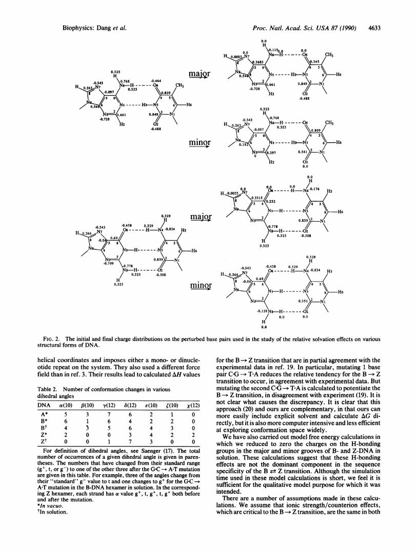
the atomic charges on the H-bonding groups in the major andminor grooves of the DNA are reduced to zero in order toassess the relative solvation effects of these groups in thedifferent structural forms of DNA. The calculations werecarried out in such a way that the total charge of the systemwas unchanged. Thus, the charges on 0-6, 0-4, and 0-2 wereplaced on C-6, C-4, and C-2, respectively; the charges on H-6and H-2 were placed on N-6 and N-2, respectively; and thecharges on N-7 and N-3 (A,G) were distributed to C-8, C-5,C-4, C-2 as shown in Fig. 2.
Finally, the coordinates of the B- and Z-DNA helices wereminimized in vacuo by using a conjugate gradient method andthe potential function given above. All degrees of freedom
Table 1. Free energies in kcal/mol for the C'G -* T-A mutationCondition
Structure* In vacuo In solutionIsolated base pairt 18.44 ± 0.7Z-DNAt 25.96 ± 1.6 38.70 ± 0.3B-DNAt 23.96 ± 1.0 (23.2 ± 1.2)§ 35.73 ± 0.2 (36.2 ± 0.3)§A-DNAt 23.62 ± 0.2Z-DNA1 93.87 ± 0.4B-DNA1 90.15 ± 0.7Free energies reported are an average of forward and backward mutations, with the standard
deviation of these two runs. Unless otherwise noted, simulations were run for 40 ps in each direction.*DNA structural form.tSystem is dC-dG.tSystem is d(CGCGCG)2 -- d(CGTCGC)-d(GCGACG).§Results for simulations for 80 ps in each direction are in parentheses.ISystem is d(CGCGCGCGCGCG)2 -* d(CGCGTATAGCGCG)2.
Biophysics: Dang et al.

Proc. Natl. Acad. Sci. USA 87 (1990)
A T
G C
FIG. 1. The models of perturbed APT and GC base pairs used inthe calculations.
were included. Each minimization proceeded until the rms
derivative in energy was <0.1 kcal/mol. We carried out theenergy component analyses on the minimized sequences
d(CGCGCG)2, d(CGTGCG)-d(CGCACG), d(CACACG)*d(C-GTGTG), and d(CATACG)-d(CGTATG) to assess the rela-tive roles of different intranucleotide interactions on the BZ equilibrium as a function of sequence.
RESULTS
Table 1 contains the results of the free energy calculationsboth in vacuo and in solution. As one can see, if an isolatedC-G base pair is mutated into a T-A base pair in vacuo, thechange in free energy is 18.4 kcal/mol. This is mainly anintramolecular energy. Calculated free energy changes forthe same base pair modification in various d(CGCGCG)2hexamers are all larger, suggesting that the third COG basepair interacts more favorably with its environment (surround-ing bases and sugar-phosphate backbone) than does a TEAbase pair at this position. These more favorable interactionsare worth 5 kcal/mol in the A and B forms and 7 kcal/mol inthe Z form (see Table 1). Thus, in vacuo, the presence of aT-A base pair makes it 2 kcal/mol more difficult to induce aB -* Z transition in a DNA oligomer than a C-G base pair at
that position.We have also carried out a mutation of the four COG base
pairs in the center of d(CGCGCGCGCGCG)2 to TEA, and, asTable 1 indicates, the differential free energy cost of the(C-G)4 -* (TA)4 mutation is 3.7 kcal/mol greater in Z- than in
B-DNA. This is significantly less than 4 times the value of thesingle base mutation in d(CGCGCG)2 and suggests significantnonadditivity in differential sequence stability effects on theB -* Z transition.
Results for the simulations carried out in explicit solventare in qualitative agreement with those determined in vacuo.The C0G APT mutation destabilizes the B-form helix by35.7 kcal/mol, whereas this change destabilizes the Z-formhelix by 38.7 kcal/mol. In other words, in solution a T-A basepair makes the B -+ Z transition 3 kcal/mol less favorable
than with aCG base pair at the same position. This can becompared to the 2 kcal/mol value determined in vacuo. Usingthe 80-ps values for B-DNA in parentheses in Table 1 wouldlead to free energies of 2.8 in vacuo and 2.5 in solution. Thesimilarity of the vacuum and solution results implies that theZ-phobicity of A-T base pairs may be mainly intramolecular.This contrasts with the results of calculations in which a 5Megroup was added to cytosine (7). In those calculations,
potentiation of the Z structure is calculated to be both anintramolecular and a solvation effect.How do the structures of these oligonucleotides change
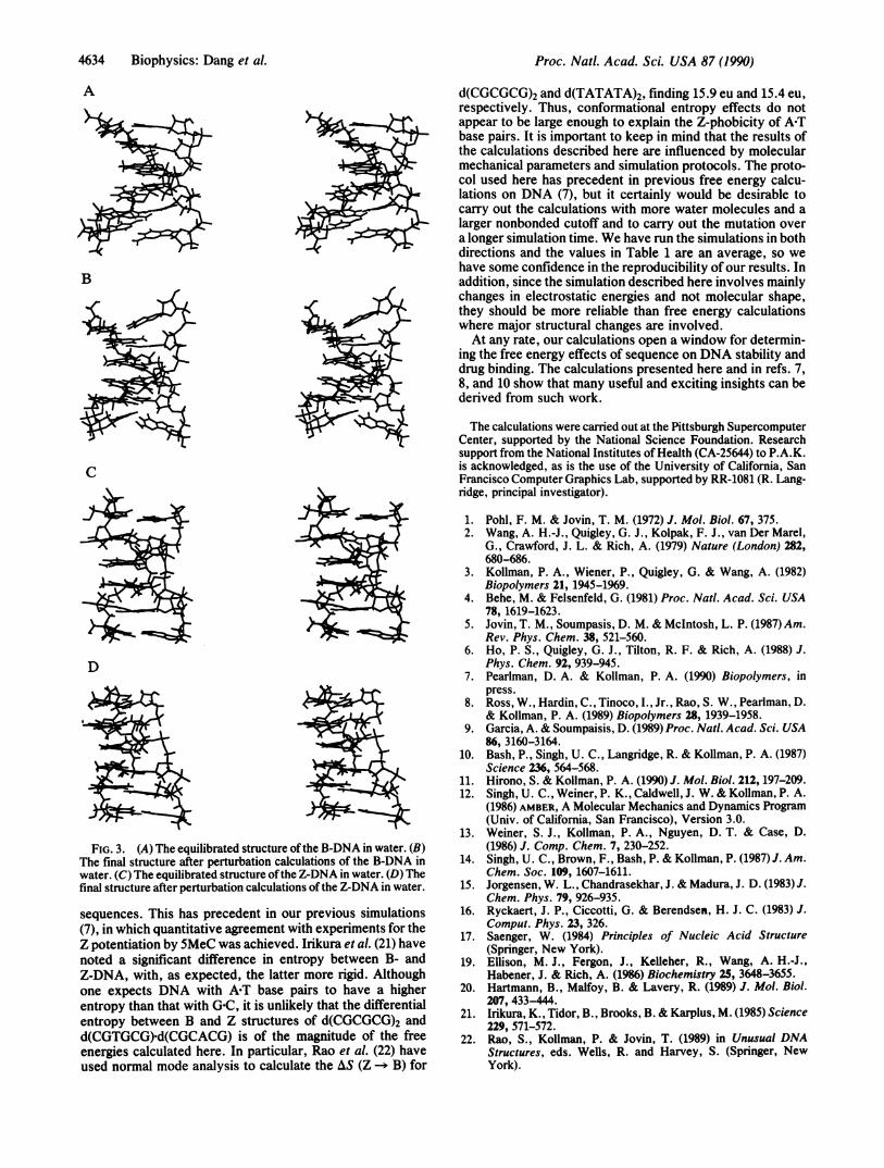
during the molecular dynamics simulations, each of whichstarts as canonical A-, B-, or Z-DNA? The number ofdihedral angles of each type for a given simulation is given inTable 2. As one can see, sugar repuckering has occurredfrequently in all structures, whereas while transitions of yfrom g' -* t occur frequently for A- or B-DNA, this dihedralangle remains in its starting value (alternating g' and t) inZ-DNA. Stereoviews of some of these structures are pre-sented in Fig. 3 and illustrate that, although these structuresare far from "canonical," they are still recognizably doublehelices of the B and Z families, both after dynamics equili-bration and after the perturbation calculations.Can one make a qualitative estimate of how much the
hydrogen-bonding groups in the grooves contribute to theZ-philicity of A-T sequences? We summarize the results ofperturbations in which the atomic charges on the H-bondinggroups of the base pairs were reduced to zero. Fig. 2 presentsthe initial and final charge models for minor and major groupperturbations in the C-G and TEA base pairs. These pertur-bation calculations were performed on the base pair at thethird position of the hexanucleotides in vacuo and on thissame system in solution. For the COG base pair, the major andminor grooves are better solvated in the B-form than in theZ form, with a larger difference in the major groove solvation.In contrast, for the T-A base pair, major groove solvation isoverall much less favorable and is more favorable in the Zform than in the B structure. For T-A, the minor groove issubstantially better solvated in the B form, the same as theG-C base pair. Thus, differential H-bonding to solvent be-tween B and Z forms appears larger for the C-G pair (favoringB) than the APT pair. Since the results for these H-bondinggroups alone do not mirror those for the free energy base pairmutations or experiment, it is likely that solvent H-bondeffects are not the dominant reason for the sequence-specificeffects.To further analyze which interactions cause the differential
stability of a COG base pair compared to a TEA base pair in Z-and B-DNA, we performed energy component analyses onenergy-minimized B and Z structures with sequences d(CG-CGCG)2. The base-base interaction differences do contributein a significant way to the Z-phobicity of the TEA base pairscompared to G-C: both the intragroup energy of a given basepair and the interaction of this base pair (mainly with the basepair G2C5 ) significantly favor B- over Z-DNA, when thegiven base pair is TEA rather than COG. Not only do base-baseinteractions contribute to the Z-phobicity of the ART basepair, but the base-phosphate interactions also make majorcontributions to this preference.
DISCUSSION AND CONCLUSIONSWe have carried out energy minimization and free energycalculations on DNA hexamers to attempt to assess thephysical basis for the Z-phobicity of APT base pairs. The freeenergy calculations, in both the absence and presence ofexplicit solvent, are successful in qualitatively predicting thisZ-phobicity. The quantitative free energies (2-3 kcal/mol)are larger than but of the correct order of magnitude as
experimental estimates (-0.7 kcal/mol) for the differentialfree energy of A-T vs. G-C base pairs in B- vs. Z-DNA (19).Energy component calculations on minimized structuressuggest that base-base and base-phosphate interactions are
the most critical intramolecular contributors to the calculatedZ-phobicity of A-T pairs.
Recently, Hartmann et al. (20) carried out molecular me-
chanics calculations on various DNA sequences in B and Zforms. They used an approach that varies the energy in
4632 Biophysics: Dang et al.
Biophysics: Dang et al.
0.325H
H 0Ni N6-H-- - ---.-04 CH3g- ..097 0.809
N 0. i---N-.H3-N3 45
:
2/\0. 61 0.84 N14.728 N/
4.488
0.325
Proc. Natl. Acad. Sci. USA 87 (1990) 4633
0.0H
0.0 \O.ll19oo 0.0H* 0.00 Ni N6-H-- - - - -04 CH3
.3685034
maor -----05N -H3-N3 6 H60.66 1
N62 0.84 ~Ni-0.728
H2-0.488
H60.325H
-0.543 \.0.768H 0.263 Ni N6-H-------04 CH3
0.097 0.325 0.809
minor 0N /5 6\ - HI-N,3 6 H6
0.182 /
N 0.36 Ni
H2
0.00.0H
m0H 00 0.0/0H176-0.00o55 0.0 ~ 6--H-N4017 Hs
-0.3315
major 2NN--H -3~0.859 2 N
-.77\
I ~ 0.325 -0.508
0.325-H6
0.329H
-0.543 -0.458 0.329 /H026N7 06--- ---H-N40.834 H5
069
minor 4o.56,
0.0
FIG. 2. The initial and final charge distributions on the perturbed base pairs used in the study of the relative solvation effects on variousstructural forms of DNA.
helical coordinates and imposes either a mono- or dinucle-otide repeat on the system. They also used a different forcefield than in ref. 3. Their results lead to calculated AH values
Table 2. Number of conformation changes in variousdihedral anglesDNA a(10) /8(10) y(12) 8(12) E(10) {(10) X(12)A* 5 3 7 6 2 1 0B* 6 1 6 4 2 2 0Bt 4 3 5 6 4 3 0Z* 2 0 0 3 4 2 2Zt 0 0 1 7 3 0 0For definition of dihedral angles, see Saenger (17). The total
number of occurrences of a given dihedral angle is given in paren-theses. The numbers that have changed from their standard range(gW, t, or g-) to one of the other three after the GC -. A-T mutationare given in this table. For example, three of the angles change fromtheir "standard" g- value to t and one changes to g+ for the G-C -.A-T mutation in the B-DNA hexamer in solution. In the correspond-ing Z hexamer, each strand has a value g+, t, g+, t, g+ both beforeand after the mutation.*In vacuo.tIn solution.
for the B -- Z transition that are in partial agreement with theexperimental data in ref. 19. In particular, mutating 1 basepair C-G -- T-A reduces the relative tendency for the B -+ Ztransition to occur, in agreement with experimental data. Butmutating the second C-G -+ T-A is calculated to potentiate theB -* Z transition, in disagreement with experiment (19). It isnot clear what causes the discrepancy. It is clear that thisapproach (20) and ours are complementary, in that ours canmore easily include explicit solvent and calculate AG di-rectly, but it is also more computer intensive and less efficientat exploring conformation space widely.We have also carried out model free energy calculations in
which we reduced to zero the charges on the H-bondinggroups in the major and minor grooves of B- and Z-DNA insolution. These calculations suggest that these H-bondingeffects are not the dominant component in the sequencespecificity of the B ± Z transition. Although the simulationtime used in these model calculations is short, we feel it issufficient for the qualitative model purpose for which it wasintended.There are a number of assumptions made in these calcu-
lations. We assume that ionic strength/counterion effects,which are critical to the B -+ Z transition, are the same in both
Proc. Natl. Acad. Sci. USA 87 (1990)
A
B
c
D
FIG. 3. (A) The equilibrated structure of the B-DNA in water. (B)The final structure after perturbation calculations of the B-DNA inwater. (C) The equilibrated structure ofthe Z-DNA in water. (D) Thefinal structure after perturbation calculations of the Z-DNA in water.
sequences. This has precedent in our previous simulations(7), in which quantitative agreement with experiments for theZ potentiation bySMeC was achieved. Irikura et al. (21) havenoted a significant difference in entropy between B- andZ-DNA, with, as expected, the latter more rigid. Althoughone expects DNA with APT base pairs to have a higherentropy than that with G-C, it is unlikely that the differentialentropy between B and Z structures of d(CGCGCG)2 andd(CGTGCG)-d(CGCACG) is of the magnitude of the freeenergies calculated here. In particular, Rao et al. (22) haveused normal mode analysis to calculate the AS (Z -- B) for
d(CGCGCG)2 and d(TATATA)2, finding 15.9 eu and 15.4 eu,respectively. Thus, conformational entropy effects do notappear to be large enough to explain the Z-phobicity of A-Tbase pairs. It is important to keep in mind that the results ofthe calculations described here are influenced by molecularmechanical parameters and simulation protocols. The proto-col used here has precedent in previous free energy calcu-lations on DNA (7), but it certainly would be desirable tocarry out the calculations with more water molecules and alarger nonbonded cutoff and to carry out the mutation overa longer simulation time. We have run the simulations in bothdirections and the values in Table 1 are an average, so wehave some confidence in the reproducibility ofour results. Inaddition, since the simulation described here involves mainlychanges in electrostatic energies and not molecular shape,they should be more reliable than free energy calculationswhere major structural changes are involved.At any rate, our calculations open a window for determin-
ing the free energy effects of sequence on DNA stability anddrug binding. The calculations presented here and in refs. 7,8, and 10 show that many useful and exciting insights can bederived from such work.
The calculations were carried out at the Pittsburgh SupercomputerCenter, supported by the National Science Foundation. Researchsupport from the National Institutes of Health (CA-25644) to P.A.K.is acknowledged, as is the use of the University of California, SanFrancisco Computer Graphics Lab, supported by RR-1081 (R. Lang-ridge, principal investigator).
1. Pohl, F. M. & Jovin, T. M. (1972) J. Mol. Biol. 67, 375.2. Wang, A. H.-J., Quigley, G. J., Kolpak, F. J., van Der Marel,
G., Crawford, J. L. & Rich, A. (1979) Nature (London) 282,680-686.
3. Kollman, P. A., Wiener, P., Quigley, G. & Wang, A. (1982)Biopolymers 21, 1945-1969.
4. Behe, M. & Felsenfeld, G. (1981) Proc. Natl. Acad. Sci. USA78, 1619-1623.
5. Jovin, T. M., Soumpasis, D. M. & McIntosh, L. P. (1987) Am.Rev. Phys. Chem. 38, 521-560.
6. Ho, P. S., Quigley, G. J., Tilton, R. F. & Rich, A. (1988) J.Phys. Chem. 92, 939-945.
7. Pearlman, D. A. & Kollman, P. A. (1990) Biopolymers, inpress.
8. Ross, W., Hardin, C., Tinoco, I., Jr., Rao, S. W., Pearlman, D.& Kollman, P. A. (1989) Biopolymers 28, 1939-1958.
9. Garcia, A. & Soumpaisis, D. (1989) Proc. Natl. Acad. Sci. USA86, 3160-3164.
10. Bash, P., Singh, U. C., Langridge, R. & Kollman, P. A. (1987)Science 236, 564-568.
11. Hirono, S. & Kollman, P. A. (1990) J. Mol. Biol. 212, 197-209.12. Singh, U. C., Weiner, P. K., Caldwell, J. W. & Kollman, P. A.
(1986) AMBER, A Molecular Mechanics and Dynamics Program(Univ. of California, San Francisco), Version 3.0.
13. Weiner, S. J., Kollman, P. A., Nguyen, D. T. & Case, D.(1986) J. Comp. Chem. 7, 230-252.
14. Singh, U. C., Brown, F., Bash, P. & Kollman, P. (1987) J. Am.Chem. Soc. 109, 1607-1611.
15. Jorgensen, W. L., Chandrasekhar, J. & Madura, J. D. (1983) J.Chem. Phys. 79, 926-935.
16. Ryckaert, J. P., Ciccotti, G. & Berendsen, H. J. C. (1983) J.Comput. Phys. 23, 326.
17. Saenger, W. (1984) Principles of Nucleic Acid Structure(Springer, New York).
19. Ellison, M. J., Fergon, J., Kelleher, R., Wang, A. H.-J.,Habener, J. & Rich, A. (1986) Biochemistry 25, 3648-3655.
20. Hartmann, B., Malfoy, B. & Lavery, R. (1989) J. Mol. Biol.207, 433-444.
21. Irikura, K., Tidor, B., Brooks, B. & Karplus, M. (1985) Science229, 571-572.
22. Rao, S., Kollman, P. & Jovin, T. (1989) in Unusual DNAStructures, eds. Wells, R. and Harvey, S. (Springer, NewYork).
4634 Biophysics: Dang et al.
Related Documents