ARTICLE DNAI2 Mutations Cause Primary Ciliary Dyskinesia with Defects in the Outer Dynein Arm Niki Tomas Loges, 1,2 Heike Olbrich, 1 Lale Fenske, 1 Huda Mussaffi, 3 Judit Horvath, 4 Manfred Fliegauf, 1 Heiner Kuhl, 5 Gyorgy Baktai, 6 Erzsebet Peterffy, 6 Rahul Chodhari, 7 Eddie M.K. Chung, 7 Andrew Rutman, 8 Christopher O’Callaghan, 8 Hannah Blau, 3 Laszlo Tiszlavicz, 9 Katarzyna Voelkel, 10 Michal Witt, 10,11 Ewa Zie ˛ tkiewicz, 10 Juergen Neesen, 12 Richard Reinhardt, 5 Hannah M. Mitchison, 7 and Heymut Omran 1, * Primary ciliary dyskinesia (PCD) is a genetically heterogeneous disorder characterized by chronic destructive airway disease and random- ization of left/right body asymmetry. Males often have reduced fertility due to impaired sperm tail function. The complex PCD pheno- type results from dysfunction of cilia of the airways and the embryonic node and the structurally related motile sperm flagella. This is associated with underlying ultrastructural defects that frequently involve the outer dynein arm (ODA) complexes that generate cilia and flagella movement. Applying a positional and functional candidate-gene approach, we identified homozygous loss-of-function DNAI2 mutations (IVS11þ1G > A) in four individuals from a family with PCD and ODA defects. Further mutational screening of 105 unrelated PCD families detected two distinct homozygous mutations, including a nonsense (c.787C > T) and a splicing mutation (IVS3-3T > G) resulting in out-of-frame transcripts. Analysis of protein expression of the ODA intermediate chain DNAI2 showed sublocalization throughout respiratory cilia. Electron microscopy showed that mutant respiratory cells from these patients lacked DNAI2 protein expres- sion and exhibited ODA defects. High-resolution immunofluorescence imaging demonstrated absence of the ODA heavy chains DNAH5 and DNAH9 from all DNAI2 mutant ciliary axonemes. In addition, we demonstrated complete or distal absence of DNAI2 from ciliary axonemes in respiratory cells of patients with mutations in genes encoding the ODA chains DNAH5 and DNAI1, respectively. Thus, DNAI2 and DNAH5 mutations affect assembly of proximal and distal ODA complexes, whereas DNAI1 mutations mainly disrupt assem- bly of proximal ODA complexes. Introduction Primary ciliary dyskinesia (PCD [MIM 242650]), also known as immotile cilia syndrome (ICS), is a rare, usually autosomal-recessive genetic disorder affecting cilia and fla- gella movement; it has an incidence of 1 in 20,000–30,000 people. 1 Motile cilia covering the epithelia of the upper and lower respiratory tract function to constantly move in- haled particles, cell debris, and microbes toward the throat. 2 Because PCD patients lack mucociliary clearance, recurrent infections of the upper and lower respiratory tract eventually cause permanent lung damage such as bronchiectasis in these patients. 3,4 Dysfunction of nodal cilia during early embryogenesis causes randomization of left/right body asymmetry, which explains why approxi- mately half of affected PCD individuals have situs inver- sus. 5 The association of PCD and situs inversus is also referred to as Kartagener syndrome (KS [MIM 244400]). In- terestingly, a subset of PCD patients have more severe lat- erality defects associated with complex heart defects. 6 Male PCD patients often have reduced fertility, which is explained by dysmotile sperm tails (flagella) that have an ultrastructure resembling that of respiratory cilia. Cilia and flagella are hair-like structures extending from the cell surface. 7 Most motile cilia, such as respiratory cilia, have a 9 þ 2 axoneme with an ultrastructure consisting of nine peripheral doublets surrounding two central tubules. The 9 þ 2 configuration is also found in sperm flagella and has been preserved throughout evolution, but there are also motile cilia with a 9 þ 0 configuration, e.g., nodal cilia. A number of multiprotein complexes, including radial spokes, nexin links, the central sheath, and dynein arms interconnect the different axonemal components. Outer dynein arms (ODAs) are connected to the peripheral mi- crotubule A and generate motion by ATP-dependent reac- tions. The outer dynein arms are complex macromolecular assemblies containing different polypeptide chains classi- fied according to their sizes as heavy (400–500 kDa), inter- mediate (45–110 kDa), and light chains (8–55 kDa), and these have been studied in detail in Chlamydomonas rein- hardtii. 8 The dynein heavy chains form the globular heads and the stem of the ODA complexes and contain the ATPase and microtubule motor domains. We recently showed that recessive mutations of DNAH5 (MIM 603335) encoding an ODA heavy dynein chain are found in approximately 50% of PCD individuals with 1 Department of Pediatrics and Adolescent Medicine, University Hospital Freiburg, Freiburg, Germany; 2 Faculty of Biology, Albert-Ludwigs-University, Frei- burg, Germany; 3 Pulmonary Unit, Schneider Children’s Medical Center of Israel, Petah-Tikva, Sackler School of Medicine, Tel-Aviv University, Tel-Aviv, Israel; 4 National Medical Center, Budapest, Hungaria; 5 Max-Planck Institute for Molecular Genetics, Berlin, Germany; 6 Pediatric Institute Svabhegy, Buda- pest, Hungary; 7 General and Adolescent Paediatric Unit, University College London, Institute of Child Health, London, UK; 8 Department of Infection, Im- munity and Inflammation, Division of Child Health, University of Leicester, Leicester Royal Infirmary, Leicester, UK; 9 University of Szeged, Department of Pathology, Szeged, Hungary; 10 Department of Molecular and Clinical Genetics, Institute of Human Genetics, Polish Academy of Sciences, Poznan, Poland; 11 International Institute of Molecular and Cell Biology, Warsaw, Poland; 12 Department of Medical Genetics, University of Vienna, Vienna, Austria *Correspondence: [email protected] DOI 10.1016/j.ajhg.2008.10.001. ª2008 by The American Society of Human Genetics. All rights reserved. The American Journal of Human Genetics 83, 547–558, November 7, 2008 547

Welcome message from author
This document is posted to help you gain knowledge. Please leave a comment to let me know what you think about it! Share it to your friends and learn new things together.
Transcript

ARTICLE
DNAI2 Mutations Cause Primary Ciliary Dyskinesiawith Defects in the Outer Dynein Arm
Niki Tomas Loges,1,2 Heike Olbrich,1 Lale Fenske,1 Huda Mussaffi,3 Judit Horvath,4 Manfred Fliegauf,1
Heiner Kuhl,5 Gyorgy Baktai,6 Erzsebet Peterffy,6 Rahul Chodhari,7 Eddie M.K. Chung,7
Andrew Rutman,8 Christopher O’Callaghan,8 Hannah Blau,3 Laszlo Tiszlavicz,9 Katarzyna Voelkel,10
Michal Witt,10,11 Ewa Zietkiewicz,10 Juergen Neesen,12 Richard Reinhardt,5 Hannah M. Mitchison,7
and Heymut Omran1,*
Primary ciliary dyskinesia (PCD) is a genetically heterogeneous disorder characterized by chronic destructive airway disease and random-
ization of left/right body asymmetry. Males often have reduced fertility due to impaired sperm tail function. The complex PCD pheno-
type results from dysfunction of cilia of the airways and the embryonic node and the structurally related motile sperm flagella. This is
associated with underlying ultrastructural defects that frequently involve the outer dynein arm (ODA) complexes that generate cilia and
flagella movement. Applying a positional and functional candidate-gene approach, we identified homozygous loss-of-function DNAI2
mutations (IVS11þ1G> A) in four individuals from a family with PCD and ODA defects. Further mutational screening of 105 unrelated
PCD families detected two distinct homozygous mutations, including a nonsense (c.787C > T) and a splicing mutation (IVS3-3T > G)
resulting in out-of-frame transcripts. Analysis of protein expression of the ODA intermediate chain DNAI2 showed sublocalization
throughout respiratory cilia. Electron microscopy showed that mutant respiratory cells from these patients lacked DNAI2 protein expres-
sion and exhibited ODA defects. High-resolution immunofluorescence imaging demonstrated absence of the ODA heavy chains DNAH5
and DNAH9 from all DNAI2 mutant ciliary axonemes. In addition, we demonstrated complete or distal absence of DNAI2 from ciliary
axonemes in respiratory cells of patients with mutations in genes encoding the ODA chains DNAH5 and DNAI1, respectively. Thus,
DNAI2 and DNAH5 mutations affect assembly of proximal and distal ODA complexes, whereas DNAI1 mutations mainly disrupt assem-
bly of proximal ODA complexes.
Introduction
Primary ciliary dyskinesia (PCD [MIM 242650]), also
known as immotile cilia syndrome (ICS), is a rare, usually
autosomal-recessive genetic disorder affecting cilia and fla-
gella movement; it has an incidence of 1 in 20,000–30,000
people.1 Motile cilia covering the epithelia of the upper
and lower respiratory tract function to constantly move in-
haled particles, cell debris, and microbes toward the
throat.2 Because PCD patients lack mucociliary clearance,
recurrent infections of the upper and lower respiratory
tract eventually cause permanent lung damage such as
bronchiectasis in these patients.3,4 Dysfunction of nodal
cilia during early embryogenesis causes randomization of
left/right body asymmetry, which explains why approxi-
mately half of affected PCD individuals have situs inver-
sus.5 The association of PCD and situs inversus is also
referred to as Kartagener syndrome (KS [MIM 244400]). In-
terestingly, a subset of PCD patients have more severe lat-
erality defects associated with complex heart defects.6
Male PCD patients often have reduced fertility, which is
explained by dysmotile sperm tails (flagella) that have an
ultrastructure resembling that of respiratory cilia.
The America
Cilia and flagella are hair-like structures extending from
the cell surface.7 Most motile cilia, such as respiratory cilia,
have a 9 þ 2 axoneme with an ultrastructure consisting of
nine peripheral doublets surrounding two central tubules.
The 9 þ 2 configuration is also found in sperm flagella and
has been preserved throughout evolution, but there are
also motile cilia with a 9þ 0 configuration, e.g., nodal cilia.
A number of multiprotein complexes, including radial
spokes, nexin links, the central sheath, and dynein arms
interconnect the different axonemal components. Outer
dynein arms (ODAs) are connected to the peripheral mi-
crotubule A and generate motion by ATP-dependent reac-
tions. The outer dynein arms are complex macromolecular
assemblies containing different polypeptide chains classi-
fied according to their sizes as heavy (400–500 kDa), inter-
mediate (45–110 kDa), and light chains (8–55 kDa), and
these have been studied in detail in Chlamydomonas rein-
hardtii.8 The dynein heavy chains form the globular heads
and the stem of the ODA complexes and contain the
ATPase and microtubule motor domains.
We recently showed that recessive mutations of DNAH5
(MIM 603335) encoding an ODA heavy dynein chain are
found in approximately 50% of PCD individuals with
1Department of Pediatrics and Adolescent Medicine, University Hospital Freiburg, Freiburg, Germany; 2Faculty of Biology, Albert-Ludwigs-University, Frei-
burg, Germany; 3Pulmonary Unit, Schneider Children’s Medical Center of Israel, Petah-Tikva, Sackler School of Medicine, Tel-Aviv University, Tel-Aviv,
Israel; 4National Medical Center, Budapest, Hungaria; 5Max-Planck Institute for Molecular Genetics, Berlin, Germany; 6Pediatric Institute Svabhegy, Buda-
pest, Hungary; 7General and Adolescent Paediatric Unit, University College London, Institute of Child Health, London, UK; 8Department of Infection, Im-
munity and Inflammation, Division of Child Health, University of Leicester, Leicester Royal Infirmary, Leicester, UK; 9University of Szeged, Department of
Pathology, Szeged, Hungary; 10Department of Molecular and Clinical Genetics, Institute of Human Genetics, Polish Academy of Sciences, Poznan, Poland;11International Institute of Molecular and Cell Biology, Warsaw, Poland; 12Department of Medical Genetics, University of Vienna, Vienna, Austria
*Correspondence: [email protected]
DOI 10.1016/j.ajhg.2008.10.001. ª2008 by The American Society of Human Genetics. All rights reserved.
n Journal of Human Genetics 83, 547–558, November 7, 2008 547

ODA defects.9,10 Mutations in an ODA intermediate dy-
nein chain, DNAI1 (MIM 604366), are reported in 2%–
13% of PCD patients with defined ODA defects.11–13 Other
genes that also encode ODA components, including
TXNDC3 and DNAH11 (MIM 603339), only rarely account
for PCD.14–16 Here, we studied DNAI2 (MIM 605483), the
human ortholog of Chlamydomonas intermediate ODA
chain IC69/IC2 comprising 14 exons extending over
39 kb genomic distance.17–19
Applying a combinatory approach comprising posi-
tional and functional candidate-gene analyses, we identi-
fied three distinct recessive loss-of-function DNAI2 muta-
tions in six affected patients originating from three PCD
families. In addition, we analyzed the role of DNAI2 in
the assembly of ODA complexes by using transmission
electron microscopy and high-resolution immunofluores-
cence imaging, which showed that DNAI2 is essential for
ODA assembly throughout the ciliary axoneme.
Material and Methods
Patients and FamiliesWe obtained signed and informed consent from patients who ful-
filled diagnostic criteria of PCD20 and from family members ac-
cording to protocols approved by the Institutional Ethics Review
Board at the University of Freiburg and collaborating institutions.
We studied DNA from consanguineous kindred by total genome
scan and mutational analysis. In addition, we sequenced all
DNAI2 exons from 105 PCD patients originating from unrelated
families. The 105 affected individuals (56 females; 49 males) we
analyzed are all white (age median 15 years, range from 2 years
to 68 years), except for one affected Asian individual. In this co-
hort, 62 individuals had situs inversus (Kartagener Syndrome)
and 43 situs solitus. Of the screened patients, 48 had ODA defects.
Linkage AnalysisA genome-wide linkage scan was carried out at the Wellcome Trust
Centre for Human Genetics, Oxford, with the Illumina Linkage IV
Panel of 6008 SNPs. In each sample, 98%–99% of the SNPs were
successfully typed. Multipoint linkage analysis was performed
with GeneHunter version 2.1r5, with the assumptions of auto-
somal-recessive inheritance, a disease allele frequency of 0.007,
and complete penetrance. DNA samples from the family
UCL149 and UCL150 parents and from affected (in total n ¼ 8)
but not unaffected offspring were typed in this scan.
Mutational AnalysisGenomic DNA was isolated by standard methods directly from
blood samples or from lymphocyte cultures after Epstein-Barr vi-
rus transformation. Amplification of 14 genomic fragments com-
prising all 14 exons of DNAI2 was performed in a volume of
50 ml containing 30 ng DNA, 50 pmol of each primer, 2 mM
dNTPs, and 1.5 U Taq DNA polymerase (Eppendorf, Hamburg,
Germany). PCR amplifications were carried out by means of an ini-
tial denaturation step at 94�C for 4 min and 33 cycles as follows:
94�C for 30 s, 53�C–64�C for 30 s, and 72�C for 60 s., with a final
extension at 72�C for 10 min. PCR products were verified by aga-
rose gel electrophoresis, column purified (Genomed, Loehne, Ger-
many), and sequenced bidirectionally with BigDye Terminator
548 The American Journal of Human Genetics 83, 547–558, Novemb
v3.1 Cycle Sequencing Kit (Perkin Elmer). Samples were separated
and analyzed on an Applied Biosystems 3730xl DNA Analyzer. Se-
quence data were evaluated with the Codoncode software (Codon-
Code Corporation, Dedham, MA. USA). To aid identification of fre-
quent polymorphisms, we sequenced DNA of 13 healthy white
controls (26 control chromosomes). In the families in which the
affected individual carried the novel DNAI2 mutations, other fam-
ily members were also analyzed. We screened an additional 236
control chromosomes originating from healthy white individuals
for the presence of the novel DNAI2 mutation. Segregation analy-
ses and screening of controls were performed by restriction analy-
sis with MaeI (the MaeI restriction site is abrogated in affected
individuals) and/or sequencing.
cDNA Analysis of Splicing MutationsTotal RNA was isolated from EBV-transformed lymphocytes from
patients OP42-II2, UCL150Pa, and controls with trizol. First-
strand cDNA synthesis was performed with the RevertAid H minus
First Strand cDNA Synthesis Kit (Fermentas) and specific primers.
UCL150Pa: First-strand synthesis was performed with primer
Ex12/13 (50-ACT TGG TTG CTG AGG CAC TG-30). The first PCR
product was amplified with primers Ex9/10F (50-TCC ATC ATG
TGG ACC AAG TAC C-30) and Ex12R1 (50-TTA TGG CGT CTG
CCT CCT TC-30). Nested PCR with primers Ex10F (50-GAG GCC
GAC CGT TTT CTT TAC-30) and Ex12R2 (50-GCT CTG CGA AGA
TGA TGT CG-30) resulted in a wild-type product (425 bp) and
a smaller product in patient UCL150Pa (278 bp). The PCR prod-
ucts were purified and sequenced bidirectionaly.
OP42-II2: The first-strand primer spans the junction of exons 6
and 7 (50-GTC CCA GCA GGC TAT CTG TC-30). The cDNA frag-
ment comprising exons 3 and 5 was amplified with primers span-
ning the junction of exons 2 and 3 (Ex2/3F: 50-GAA CAC GAG
GCC AAC TCA G-30) and exon 5 (Ex5R: 50-GCT GAA AAT CCA
AGC AGG AG-30). Half-nested PCR with primers Ex3F (50-GGG
GAG TTA ACC ATG TCG AG-30) and Ex5R showed a wild-type allele
(351 bp) and in patient OP42-II2 a single-variant allele (229 bp).
PCR products were gel purified and sequenced bidirectionally.
ImmunoblottingProtein extracts were prepared from human respiratory epithelial
cell cultures according to previously published procedures.20,28
Proteins were separated on a NuPAGE 4%–12% bis-tris gel (Invitro-
gen, Karlsruhe, Germany) and blotted onto a PVDF membrane
(Amersham). The blot was processed for ECL plus (Amersham) de-
tection with DNAI2 (1:1000) and anti-mouse-HRP (1:5000) anti-
bodies (Santa Cruz, Heidelberg, Germany).
Immunofluorescence AnalysisRespiratory epithelial cells were obtained by nasal-brush biopsy
(cytobrush plus, Medscand Malmo, Sweden) and suspended in
cell-culture medium. Sperm cells were washed with phosphate-
buffered saline. Samples were spread onto glass slides, air dried,
and stored at �80�C until use. Cells were treated with 4% parafor-
maldehyde, 0.2% Triton X-100, and 1% skim milk prior to incuba-
tion with primary (at least 2 hr) and secondary (30 min) antibodies
at room temperature. Appropriate controls were performed with-
out the primary antibodies. Polyclonal rabbit DNAH5 and Dnali1
antibodies were described previously.20,29 Mouse acetylated-a-
tubulin antibodies were obtained from Sigma (Taufkirchen, Ger-
many), and monoclonal mouse DNAI2 antibodies were obtained
from Abnova Corporation (Taiwan). In addition, polyclonal
er 7, 2008
antibodies directed against DNAH9 were used. Highly cross-
adsorbed secondary antibodies (Alexa Fluor 488, Alexa Fluor
546) were obtained from Molecular Probes (Invitrogen). DNA
was stained with Hoechst 33342 (Sigma). Confocal images were
taken on a Zeiss LSM 510 i-UV.
Transmission Electron MicroscopyThe biopsies were taken from the middle turbinate. The sample of
nasal mucosa was fixed in 2.5% glutaraldehyde in 0.1 M sodium
cacodylate buffer at 4�C, washed overnight, and postfixed in 1%
osmium tetroxide. After dehydration, the samples were embedded
in a mixture of propylene oxide and epoxy resin. After polymeri-
zation, several resin sections were cut with an ultramicrotome.
The sections were picked up onto copper grids. The sections
were stained with Reynold’s lead citrate. Transmission electron
microscopy was performed with a Philips CM10.
Results
Positional and Functional Candidate-Gene Analyses
Identified DNAI2 Mutations
A total-genome linkage scan in a consanguineous Iranian
Jewish kindred via single nucleotide polymorphisms
(SNPs) detected only one genomic region (chromosome
17) with a significant LOD score R3. A 21.4 cM (9.6 Mb)
region of homozygosity shared between the four affected
offspring was identified on chromosome 17q25.1 across
the DNAI2 locus between flanking markers rs755424 and
rs938350; there was a maximum LOD score of 3.97 (Fig-
ure 1A) at marker rs1872076, which is located 780 kb cen-
tromeric to the start of DNAI2. Sequencing of all 14 DNAI2
exons revealed a mutation, IVS11þ1G > A, affecting the
obligatory (100% sequence conservation) donor splice
site of exon 11 (Figures 1B and 1C). Genotype analyses
for this loss-of-function mutation showed cosegregation
with the disease status in both nuclear families of the ex-
tended kindred (Figure 1B). Consistent with homozygosity
by descent in the affected patients UCL149Pa, UCL149Pb,
UCL150Pa, and UCL150Pb, the mutation was present in
a homozygous state (Figures 1B and 1C, upper sequence).
All parents (UCL149M and UCL149F, UCL150M, and
UCL150F, Figure 1B) and one of the unaffected offspring
(UCL149Na, Figure 1B) are heterozygous carriers. The un-
affected offspring UCL149Nb and UCL150Nb (Figure 1B)
are homozygous carriers of the wild-type allele
(Figure 1C, lower sequence). Analysis of mutant cDNA of
the affected individual UCL150Pa confirmed that the
obligatory splicing mutation causes in-frame skipping of
exon 11 (Figure 1D, right sequence), which predicts the
loss of 49 amino acids. The clinical findings of the affected
patients include neonatal pneumonia, recurrent rhinitis
and sinusitis, recurrent otitis media and hearing deficits,
chronic cough, and bronchiectasis. Two affected individ-
uals had normal situs solitus (UCL149Pb, UCL150Pb),
and two exhibited situs inversus (UCL149Pa, UCL150Pa),
consistent with randomization of left/right body asymme-
try. Infertility was reported in one of the affected males.
The Americ
Further sequencing of all 14 coding exons, including the
exon/intron boundaries, in 105 affected individuals origi-
nating from unrelated PCD families, including seven Hun-
garian families, revealed two novel DNAI2 mutations. In
a single white Hungarian PCD patient, OP42-II2, a faculta-
tive splicing mutation, IVS3-3T > G, located within the
evolutionarily conserved (~80% conservation) splice ac-
ceptor site of exon 4, was identified (Figure 2A), and the
change was absent in 236 control chromosomes of white
individuals. The mutation cosegregated with the disease
status in family OP42 and was present in the affected indi-
vidual in a homozygous state (Figure 2B). Previous se-
quencing of all coding DNAH5 exons in OP42 was
normal.10 To confirm that the DNA variant that is located
within the splice acceptor site of intron 3 does indeed re-
sult in aberrant splicing, we performed RT-PCR analysis.
A cDNA fragment comprising exons 3 and 5 was amplified
in two rounds of amplifications with a specific first-strand
primer spanning the junction between the exons 6 and 7.
The PCR product of the affected individual OP42-II2 was
reduced in size (229 bp), demonstrating that the mutation
results in abnormal splicing. No wild-type allele was ampli-
fied (data not shown). Sequencing of the PCR product con-
firmed the absence of exon 4 and out-of-frame fusion of
exon 3 to exon 5 (Figure 2C, upper sequence). RT-PCR
analysis in control RNA (Figure 2C, lower sequence) eli-
cited only wild-type transcripts that contained exon 4
(351bp). We conclude that the mutant splice site is not rec-
ognized by the splicing machinery and that this results in
skipping of exon 4, and we thus predict early premature
termination of translation because of out-of-frame tran-
scripts (p.I116GfsX54). Interestingly, the family history
did not reveal any known consanguinity of the parents.
However, because both parents originate from the same
geographical region (village), a distant relationship of
both parents is most likely responsible for the homozygous
mutation. To test whether the IVS3-3T > G mutation is es-
pecially prevalent in east Europe, we screened 125 Polish
PCD patients and did not find this sequence variant. The
second mutation (c.787C > T) was detected in a single
white German PCD patient, OP254-II1. This homozygous
C > T transversion at position 787 of exon 7 is a nonsense
mutation, which leads us to predict early termination of
translation (p.R263X, Figure 2D). Analysis of parental
DNA revealed that both parents are heterozygous carriers
of the mutation. In the affected members of the PCD fam-
ilies OP42 and OP254, we only detected polymorphisms in
the homozygous state (Table S1 in the Supplemental Data).
This finding is consistent with a distant relationship of the
parents and is suggestive of ‘‘homozygosity by descent.’’
Overall, the frequency of DNAI2 mutations among our co-
hort is 1.9% (2/105 patients) and among the PCD families
with ODA defects is 4.2% (2/48 patients).
The clinical findings of both patients comprised chronic
otitis media, recurrent bronchitis and pneumonia, bron-
chiectasis, and situs inversus, consistent with Kartagener
syndrome.
an Journal of Human Genetics 83, 547–558, November 7, 2008 549

Figure 1. Linkage Scan and Homozygous Loss-of-Function DNAI2 Mutations in a Large Consanguineous Kindred with PrimaryCiliary Dyskinesia(A) Multipoint linkage analysis performed with GeneHunter. A 21.4 cM (9.6 Mb) region of homozygosity shared between the affectedoffspring was identified on chromosome 17q25.1 across the DNAI2 locus (maximum LOD score 3.97). Only the parents and affected chil-dren were used in the analysis.(B) Pedigree of the consanguineous kindred with two related nuclear families. Affected children are represented by black symbols, andunaffected carriers are indicated by black dots. Asterisks indicate situs inversus.(C) Sequence chromatographs of an affected patient (UCL150Pa, upper sequence) and a control individual (wt, lower sequence) showingthe presence of the IVS11þ1G> A mutation (black arrow). The detected loss-of-function mutation affects the obligatory splice donor siteof exon 11 and is present in the homozygous state in all four affected children of the kindred. All parents (UCL149M and F, UCL150M andF) and one of the unaffected offspring (UCL149Na) are heterozygous carriers of the mutant allele. The unaffected offspring UCL149Nb andUCL150Nb are homozygous carriers for the wild-type allele.(D) Results of cDNA analysis assessing the effect of the DNAI2 splicing mutation IVS11þ1G > A. In the mutant cDNA of the affectedindividual UCL150Pa (right sequence), the sequence of exon 11 is always absent. Thus, the splicing mutation leads to in-frame skippingof exon 11. Left sequence chromatograph depicts the cDNA sequence of a control subject.
Subcellular Localization of DNAI2 in Respiratory Cells
and Sperm Cells
On the basis of its evolutionarily conserved homology to
the Chlamydomonas intermediate ODA chain IC69/IC2
(44% identity17,18), we assumed that human DNAI2 plays
550 The American Journal of Human Genetics 83, 547–558, Novem
a role in ODA assembly and function in cilia and flagella.
Consistent with a function in cilia or flagella motility,
DNAI2 expression at the RNA level in respiratory cells
and testis was reported previously.18 To further corroborate
that DNAI2 indeed functions specifically in ODA
ber 7, 2008

Figure 2. Recessive Loss-of-Function DNAI2 Mutations in Patients with Primary Ciliary Dyskinesia(A) A sequence chromatograph from the affected individual OP42-II2 shows a homozygous T > G transversion at position �3 of the exon4 splice acceptor site (IVS3-3T > G; black arrow). For comparison, the wild-type sequence is shown. The mutation destroys a MaeIrestriction site as indicated.(B) Segregation analysis of the T > G transversion within the family. Presence of the mutation in the genomic DNA from all four familymembers was assessed by MaeI digestion of PCR products. The affected child (II2) showed a single (536 bp) band consistent with thepresence of two mutant alleles abolishing the restriction sites in all PCR-amplified products. In contrast, in the control subject (wt),all PCR products are completely digested (324 bp and 212 bp bands). Restriction analyses in the unaffected sibling (II1), the mother(I2), and the father (I1) are consistent with heterozygous carrier status.(C) Results of cDNA analysis assessing the effect of the DNAI2 splicing mutation IVS3-3T> G. The lower sequence chromatograph depictsthe cDNA sequence of a control subject. In the mutant cDNA of the affected individual OP42-II2 (upper sequence), the sequence of exon 4is absent. Thus, the splicing mutation causes skipping of exon 4 and a frame shift that results in a premature stop codon (I116GfsX54).(D) A genomic sequence chromatograph from the affected individual OP254-II1 shows a homozygous C> T transversion at position 787 ofexon 7 (c.787C > T), predicting a premature stop codon and termination of translation (p.R263X, black arrow). For comparison, the wild-type sequence is shown.
complexes within respiratory cilia and sperm tails, we
performed protein expression analyses with monoclonal
antibodies directed against DNAI2. To confirm antibody
specificity, we first performed immunoblot analyses of pro-
tein extracts from human respiratory cell cultures. DNAI2
antibodies detected a single band of ~69 kD, which corre-
sponds to the predicted molecular weight of DNAI2
(Figure 3A). We next analyzed the subcellular localization
The America
of DNAI2 in human respiratory and sperm cells by using
high-resolution immunofluorescence imaging. As a con-
trol, we used antibodies against the ODA heavy chain
DNAH5. DNAI2 staining was observed throughout all
analyzed respiratory ciliary axonemes and sperm flagella
(Figures 3B and 3C), indicating that assembled ODA com-
plexes along the entire length of these axonemes contain
DNAI2.
n Journal of Human Genetics 83, 547–558, November 7, 2008 551

Figure 3. Specific Antibodies Localize the Human Axonemal Outer Dynein Arm Intermediate Chain 2 (DNAI2) to the RespiratoryCiliary Axoneme and Sperm Tail(A) Western blot analysis of protein extracts from human nasal cell culture (M, protein standard; lane 1, silver staining). DNAI2 antibodiesspecifically detect a single band with the predicted size (~69 kD, lane 2).(B and C) Immunofluorescence staining of human respiratory epithelial cells (B) and spermatozoa (C) with DNAI2 antibodies (green).DNAI2 is localized throughout all analyzed respiratory ciliary axonemes and sperm flagella. Ciliary axonemes and antibodies directedagainst axonemal outer sperm flagella were costained with dynein arm heavy chain DNAH5 (red). Note that DNAH5 also localizes through-out all respiratory ciliary axonemes but is only present in proximal sperm flagella, as reported previously.20 Overlay and brightfield imagesare shown on the right side. Nuclei were stained with Hoechst 33342 (blue).
Characterization of ODA Defects Caused by the DNAI2
Mutations
Next, we analyzed protein expression of DNAI2 in respira-
tory cells that were obtained by nasal-brushing biopsy
from patients UCL150Pa, OP42-II2, and OP254-II1, who
harbor DNAI2 mutations. Consistent with loss-of-function
mutations, all analyzed cells lacked DNAI2 expression (Fig-
ure 4), confirming the pathogenic significance of the de-
tected mutations. As a control, we stained cilia with an an-
tibody directed against the inner dynein arm light chain
Dnali1, which is not altered by an ODA defect and can
be used similarly to acetylated a-tubulin, to stain the axo-
neme. We have previously shown that respiratory cilia
contain at least two distinct ODA types: type 1,DNAH9
negative and DNAH5 positive (proximal ciliary axoneme);
type 2: DNAH9 and DNAH5 positive (distal ciliary axo-
neme).20 High-resolution immunofluorescence imaging
to detect the ODA heavy dynein chains DNAH5 and
DNAH9 in respiratory cells from patients UCL150Pa,
OP42-II2, and OP254-II1 showed an aberrant expression
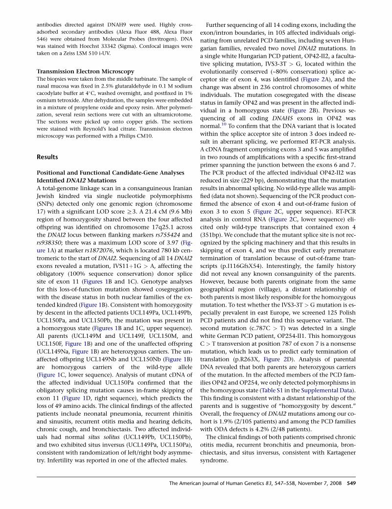
pattern for DNAH5 and DNAH9. DNAH5 protein was ab-
sent or severely reduced (Figure 5), and DNAH9 protein
was undetectable in the ciliary axonemes of these PCD pa-
tients (Figure 6). Thus, we conclude that absence of DNAI2
prevents correct assembly of both ODA complexes (type 1
and type 2) in respiratory cells from patients UCL150Pa,
OP42-II2, and OP254-II1; such a finding has so far only
been reported for DNAH5 mutations.20 Consistent with
these results, transmission electron microscopy of cross-
sections of respiratory cilia from the patient UCL150Pb,
who carries the IVS11þ1G > A DNAI2 mutation, detected
552 The American Journal of Human Genetics 83, 547–558, Novemb
ODA defects in all observed cross-sections at the ultrastruc-
tural level (Figure 5F). As expected, similar ultrastructural
ODA defects were observed in transmission electron mi-
croscopy of respiratory cilia from patient OP42-II2
(Figure 5G).
Aberrant DNAI2 Localization in Respiratory Cells
Harboring Mutations in DNAH5- and DNAI1-
Encoding ODA Components
After the demonstration that DNAI2 deficiency can result
in the complete absence of DNAH5 from respiratory ciliary
axonemes, we investigated the effect of DNAH5 mutations
on DNAI2 expression. For this purpose we used respiratory
cells from PCD patients carrying DNAH5 mutations known
to result in complete absence of DNAH5 from respiratory
ciliary axonemes, as reported previously.20 In respiratory
cells from patient F661, who carries compound heterozy-
gous DNAH5 mutations 4361G > A (exon 28) and
8910þ8911 delAT > insG (exon 53), we found complete
absence of DNAI2 from all analyzed respiratory ciliary ax-
onemes (Figure 7B). We further corroborated this finding
in several other respiratory cells originating from various
PCD patients with loss-of-function DNAH5 mutations10,20
(data not shown). Our findings indicate that DNAH5 is es-
sential for correct assembly of DNAI2 in both types of ODA
complexes.
Previously, we demonstrated that in respiratory cells
of patients carrying homozygous DNAI1 mutations
IVS1þ2_3insT, assembly of proximal ODA complexes
(type 2) in the proximal ciliary axoneme is at least partially
preserved: We found some residual localization of DNAH5
er 7, 2008

Figure 4. Absence of DNAI2 in Respiratory Epithelial Cells from Patients with Primary Ciliary DyskinesiaImages of respiratory epithelial cells from a healthy control (A) and from patients carrying DNAI2 mutations (B–D). Cells were costainedwith antibodies directed against outer dynein arm intermediate chain DNAI2 (green) and the inner-dynein-arm component Dnali1 (red)as control staining. Nuclei were stained with Hoechst 33342 (blue). (A) In control cells, DNAI2 localizes along the entire length of therespiratory ciliary axonemes. The yellow costaining within the ciliary axoneme indicates that both proteins colocalize within respiratorycilia. In cells of patient UCL150Pa (B), OP42-II2 (C) and OP254-II1 (D) DNAI2 is not detectable, consistent with recessive loss-of-func-tion mutations that result in the failure to produce a functional DNAI2 protein.
in this proximal ciliary compartment, whereas DNAH5
was absent from the distal ciliary axoneme.16 To investi-
gate whether DNAI2 shows a similar localization pattern
as DNAH5 in DNAI1 mutant respiratory cells, we analyzed
respiratory epithelial cells from patient OP121-II1, who
carries the homozygous IVS1þ2_3insT DNAI1 mutation.
Consistent with our previous finding that in DNAI1 mu-
tant cells assembly of proximal (type 2) ODA complexes
is at least partially preserved, we found that DNAI2 stain-
ing was moderate in the proximal part but absent in the
distal part of the ciliary compartment in all analyzed respi-
ratory cells (Figure 7C).
Discussion
Outer dynein arm complexes are large multimeric protein
complexes comprising light, intermediate, and heavy dy-
The America
nein chains, which are responsible for the generation of
the cilia beat.21 Recessive mutations of genes encoding
the outer dynein arm components DNAH5, DNAI1,
TXNDC3, and DNAH11 can cause primary ciliary dyskine-
sia.9–12,14–16 On the basis of this observation, we considered
the outer dynein arm intermediate-chain gene DNAI2 to
be a functional candidate for primary ciliary dyskinesia.
Here, we have identified the first PCD-causing DNAI2 muta-
tions affecting ODA assembly. The identified mutations
most likely abrogate DNAI2 function because premature
stop codons (OP42-II2; OP254-II1) regularly result in non-
sense-mediated RNA decay.22 The IVS11þ1G> A mutation
detected in affected individuals of UCL149/150 (Figure 1C)
predicts in-frame absence of 49 amino acids encoded by
exon 11. Probably this truncated protein cannot function
properly. Consistent with loss-of-function mutations,
high-resolution immunofluorescence analysis revealed
n Journal of Human Genetics 83, 547–558, November 7, 2008 553
Figure 5. Mislocalization of the Outer Dynein Arm Heavy-Chain Protein DNAH5 in Respiratory Epithelial Cells from Patients withDNAI2 MutationsImages of respiratory epithelial cells from a healthy control (A) and from the patients UCL150Pa (B), OP42-II2 (C), and OP254-II1 (D)carrying the DNAI2 mutations (B). Cells were costained with antibodies against acetylated a-tubulin (green) and DNAH5 (red). Nucleiwere stained with Hoechst 33342 (blue). (A) In a healthy control, DNAH5 localizes along the entire length of the axonemes. The yellowcolor within the ciliary axoneme indicates that both proteins colocalize to cilia. (B, C, and D) In DNAI2 mutant cells, DNAH5 is absent fromthe ciliary axonemes. (E, F, and G) Transmission electron micrographs of cross-sections from respiratory cilia originating from a healthycontrol (E) and from the DNAI2 patients UCL150Pb (F) and OP42-II2 (G). In the healthy control, outer dynein arms are visible ([E], redarrows), whereas in the patients UCL150Pb (F) and OP42-II2 (G), they are absent.
that affected individuals of all three analyzed families
lacked any detectable DNAI2 protein expression in respira-
tory cells (Figure 4).
The DNAI2 mutations identified appear to result in
randomization of left/right body asymmetry: Of the six
affected individuals carrying DNAI2 mutations, two ex-
554 The American Journal of Human Genetics 83, 547–558, Novemb
hibited situs solitus, and four exhibited situs inversus. All
affected individuals suffered from chronic lung disease. In-
fertility in one male was reported, but no sperm analysis
was available. Consistent with the observed clinical phe-
notype of this PCD subtype, comprising lack of appropri-
ate airway clearance by respiratory cilia and male
er 7, 2008

Figure 6. Absence of the Outer Dynein Arm Heavy-Chain Protein DNAH9 in Respiratory Epithelial Cells from Patients with DNAI2MutationsImages of respiratory epithelial cells from a healthy control (A) and from patients UCL150Pa (B), OP42-II2 (C), and OP254-II1 (D), whocarry DNAI2 mutations. Cells were co-stained with antibodies against acetylated a-tubulin (green) and DNAH9 (red). Nuclei were stainedwith Hoechst 33342 (blue). (A) In control cells DNAH9 localizes in the distal part of the respiratory ciliary axonemes. The yellow co-stainingwithin the ciliary axoneme indicates that both proteins co-localize within respiratory cilia. In respiratory cells of patient UCL150Pa (B),OP42-II2 (C) and OP254-II1 (D) DNAH9 is not detectable in the ciliary axonemes.
infertility, we detected DNAI2 protein expression in respi-
ratory cilia and sperm flagella (Figure 3). The randomiza-
tion of left/right body asymmetry is probably explained
by an altered motility of nodal cilia responsible for deter-
mination of left/right body asymmetry.5
We have previously shown that, in contrast to those in
Chlamydomonas, human ODA complexes vary in their
composition along the respiratory ciliary axoneme and
the axoneme of the sperm tail and that this composition
also differs between these two cell types.20 Respiratory cilia
contain at least two distinct ODA types: type 1, DNAH9
negative and DNAH5 positive (proximal ciliary axoneme);
and type 2, DNAH9 and DNAH5 positive (distal ciliary ax-
oneme). To determine the subcellular localization of
DNAI2 in various cell types carrying motile cilia or flagella,
we used specific monoclonal mouse antibodies directed
against human DNAI2 for high-resolution immunofluores-
The America
cence microscopy (Figure 3A). We demonstrated localiza-
tion of DNAI2 throughout the entire length of axonemes
from respiratory cilia as well as sperm tails (Figures 3B
and 3C). This finding indicates that DNAI2 is present in
both ODA (type 1 and 2) complexes.
Interestingly, mutations of the distinct genes encoding
ODA components do not result in identical defects. We
have shown that DNAH5 and DNAI1 mutations regularly
result in absent and/or shortened ODAs visualized by
transmission electron microscopy.9,10,12,23,24 In contrast
the DNAH11 mutations reported so far do not result in ul-
trastructural defects detectable by routine transmission-
electron microscopy,14,16 and TXNDC3 mutant cilia
exhibit both normal and abnormal cilia cross-sections.15
To determine the effect of DNAI2 mutations on axonemal
ultrastructure, we performed transmission electron micros-
copy. Cilia cross-sections showed ODA defects (Figures 5F
n Journal of Human Genetics 83, 547–558, November 7, 2008 555

Figure 7. Mis-localization of outer dynein arm chain DNAI2 in respiratory epithelial cells from PCD patients carrying mutationsin the outer dynein arm components DNAH5 and DNAI1Images of respiratory epithelial cells from healthy control (A), compound heterozygote patient carrying the DNAH5 mutations 4361G> A(exon 28) and 8910þ8911 delAT > insG (exon 53) (B) and a patient homozygous for the DNAI1 mutation IVS1þ2_3insT (C). Cells arecostained with antibodies directed against DNAI2 (green) and against inner dynein arm component Dnali1 as a control (red). Nuclei werestained with Hoechst 33342 (blue). In DNAH5 mutant cells (B), DNAI2 is absent from the ciliary axonemes, indicating that DNAH5 isnecessary for DNAI2 assembly. In DNAI1 mutant cells (C), DNAI2 (green) is absent from the distal part of the axonemes but is still de-tectable in the proximal ciliary axoneme. This indicates that mutant DNAI1 inhibits assembly of DNAI2 predominantly in the distal ciliaryaxonemes.
and 5G) resembling defects identified in patients with
DNAH5 mutations.9 To analyze this further, we performed
high-resolution immunofluorescence analysis in respira-
tory cilia from patients UCL150Pa, OP42-II2, and OP254-
II1, who had DNAI2 mutations. Consistent with deficient
assembly of ODA types 1 and 2, we found absence or severe
reduction of the ODA heavy chains DNAH5 and DNAH9
from the entire ciliary axoneme (Figures 5 and 6). Our find-
ings indicate that DNAI2 is essential for axonemal assem-
bly of the ODA heavy chains DNAH5 and DNAH9 in the
analyzed respiratory cells of patients UCL150Pa, OP42-
II2, and OP254-II1. This observation resembles findings
reported in respiratory cilia of patients carrying DNAH5
loss-of-function mutations.10,20 Therefore, we also ana-
lyzed respiratory cilia from patients with DNAH5 muta-
tions for DNAI2 expression. DNAI2 expression within
the analyzed respiratory cilia was completely absent
(Figure 7B), demonstrating in a complementary manner
that DNAH5 is also essential for the axonemal assembly
of the ODA protein DNAI2 within ODA types 1 and 2.
Furthermore, we analyzed respiratory cilia from a pa-
tient carrying a homozygous DNAI1 mutation for
DNAI2 localization. DNAI2 expression was restricted to
556 The American Journal of Human Genetics 83, 547–558, Novemb
the proximal part of the respiratory cilia and was com-
pletely missing in the distal part of the ciliary axoneme
(Figure 7C). We previously reported a similar staining pat-
tern for DNAH5 in DNAI1 mutant cells.16 These findings
indicate that mutant DNAI1 in these cells predominantly
inhibits axonemal assembly of distally localized ODA
types, whereas proximal ODAs can at least become par-
tially assembled.
Previous findings in Chlamydomonas reinhardtii support
an evolutionarily conserved functional role for DNAI2 in
ODA assembly; this role is similar to that shown here in
the human axoneme. Fowkes and Mitchell25 analyzed
the stability of ODA complexes in the cytoplasm of wild-
type Chlamydomonas and several outer dynein-arm mutant
strains. Interestingly, in the Chlamydomonas oda6 mutant
carrying mutations of the IC69/IC2 gene, which is orthol-
ogous to DNAI2,26 the ODA complexes were disrupted, in-
dicating the importance of DNAI2/IC69/IC2 for ODA as-
sembly. In addition, a similar phenotype was observed in
the pf28/oda2 mutant carrying a mutation in the g-dynein
heavy chain orthologous to DNAH5,27 which is also consis-
tent with our findings of an absence of DNAI2 assembly in
DNAH5 mutant cells (Figure 7B).
er 7, 2008

Interestingly, our data also show that DNAI1 mutations
do not necessarily inhibit ODA assembly of proximal
ODA complexes, indicating that evolution has given
DNAI1 functional roles not played by DNAI2 because
mutations of the Chlamydomonas orthologs IC1 and IC2
exhibit identical findings.27 Thus, DNAI1 might have ob-
tained similar functions to those of TXNDC3 because
TXNDC3 mutant cilia exhibit both normal and abnormal
cilia cross-sections, suggesting that it also mainly affects
one of the two distinct ODA complexes.15 However, so
far it is not known for which type of ODA complex (dis-
tal or proximal) TXNDC3 plays an important functional
role.
Our findings that mutations in DNAI2 cause PCD due to
ODA defects enhance our understanding of the molecular
basis of this chronic respiratory disease and support previ-
ous data that humans have at least two distinct ODA com-
plexes. Early diagnosis of PCD is important for appropriate
health management with the goal of preventing or delay-
ing lung damage.3 The use of DNAI2 antibodies can aug-
ment antibody-based PCD diagnostic techniques, such as
immunofluorescence analyses. The demonstration of
DNAI2 mutations will aid development of genetic screen-
ing for PCD and enable appropriate genetic counseling in
affected individuals. Further mutational analyses will clar-
ify whether, like DNAH5 mutations, DNAI2 mutations can
also account for heterotaxia with severe congenital heart
defects.6
Supplemental Data
Supplemental Data include one table and are available with this
article online at http://www.ajhg.org/.
Acknowledgments
We are grateful to the patients and their families for their partici-
pation in this study. We thank the German patient support group
Kartagener Syndrom und Primaere Ciliaere Dyskinesie e.V. We
thank R. Nitschke and S. Haxelmans at the life imaging center,
Institute for Biology I, University Freiburg, for their excellent
support with confocal microscopy. We thank C. Reinhard, A.
Heer, A. Schwentek, E. Rutkiewicz, and K. Parker for excellent tech-
nical assistance. We would also like to thank Andrzej Pogorzelski
from the Institute of Tuberculosis and Lung Diseases, Rabka for
clinical diagnosis and recruitment of patients. This work was sup-
ported by Deutsche Forschungsgemeinschaft grants DFG Om 6/4,
GRK1104, and SFB592 (to H.O.), and by grants from the Polish
State Committee for Scientific research (PBZ KBN 122/P05-1, to
E.Z.), the National Specialist Commissioning Advisory Group,
UK (to C.O’C.), and the Milena Carvajal–Prokartagener Founda-
tion (to H.M.M.). This work is dedicated to Bjorn A. Afzelius for
his outstanding research in the field of ciliary structure and immo-
tile-cilia syndrome.
Received: August 8, 2008
Revised: September 19, 2008
Accepted: October 1, 2008
Published online: October 23, 2008
The America
Web Resources
URLs for data presented herein are as follows:
Online Mendelian Inheritance in Man (OMIM), http://www.ncbi.
nlm.nih.gov/Omim/
dbSNP NCBI database, http://www.ncbi.nlm.nih.gov/SNP/
ENSEMBL, http://www.ensembl.org/index.html
References
1. Afzelius, B.A. (1976). A human syndrome caused by immotile
cilia. Science 193, 317–319.
2. Fliegauf,M.,Benzing,T.,andOmran,H. (2007).Whenciliagobad:
Cilia defects and ciliopathies. Nat. Rev. Mol. Cell Biol. 8, 880–893.
3. Meeks, M., and Bush, A. (2000). Primary ciliary dyskinesia
(PCD). Pediatr. Pulmonol. 29, 307–316.
4. Geremek, M., and Witt, M. (2004). Primary ciliary dyskinesia:
Genes, candidate genes and chromosomal regions. J. Appl.
Genet. 45, 347–361.
5. Nonaka, S., Tanaka, Y., Okada, Y., Takeda, S., Harada, A., Kanai,
Y., Kido, M., and Hirokawa, N. (1998). Randomization of left-
right asymmetry due to loss of nodal cilia generating leftward
flow of extraembryonic fluid in mice lacking KIF3B motor pro-
tein. Cell 95, 829–837.
6. Kennedy, M.P., Omran, H., Leigh, M.W., Dell, S., Morgan, L.,
Molina, P.L., Robinson, B.V., Minnix, S.L., Olbrich, H., Severin,
T., et al. (2007). Congenital heart disease and other heterotaxic
defects in a large cohort of patients with primary ciliary dyski-
nesia. Circulation 115, 2814–2821.
7. Zariwala, M.A., Knowles, M.R., and Omran, H. (2007). Genetic
defects in ciliary structure and function. Annu. Rev. Physiol.
69, 423–450.
8. Pazour, G.J., Agrin, N., Walker, B.L., and Witman, G.B. (2006).
Identification of predicted human outer dynein arm genes:
candidates for primary ciliary dyskinesia genes. J. Med. Genet.
43, 62–73.
9. Olbrich, H., Haffner, K., Kispert, A., Volkel, A., Volz, A.,
Sasmaz, G., Reinhardt, R., Hennig, S., Lehrach, H., Konietzko,
N., et al. (2002). Mutations in DNAH5 cause primary ciliary
dyskinesia and randomization of left-right asymmetry. Nat.
Genet. 30, 143–144.
10. Hornef, N., Olbrich, H., Horvath, J., Zariwala, M.A., Fliegauf,
M., Loges, N.T., Wildhaber, J., Noone, P.G., Kennedy, M., An-
tonarakis, S.E., et al. (2006). DNAH5 mutations are a common
cause of primary ciliary dyskinesia with outer dynein arm de-
fects. Am. J. Respir. Crit. Care Med. 15, 120–126.
11. Pennarun, G., Escudier, E., Chapelin, C., Bridoux, A.M., Ca-
cheux, V., Roger, G., Clement, A., Goossens, M., Amselem, S.,
and Duriez, B. (1999). Loss-of-function mutations in a human
gene related to Chlamydomonas reinhardtii dynein IC78 result in
primary ciliary dyskinesia. Am. J. Hum. Genet. 65, 1508–1519.
12. Zariwala, M.A., Leigh, M.W., Ceppa, F., Kennedy, M.P., Noone,
P.G., Carson, J.L., Hazucha, M.J., Lori, A., Horvath, J., Olbrich,
H., et al. (2006). Mutations of DNAI1 in primary ciliary dyski-
nesia: Evidence of founder effect in a common mutation. Am.
J. Respir. Crit. Care Med. 15, 858–866.
13. Failly, M., Saitta, A., Munoz, A., Falconnet, E., Rossier, C., San-
tamaria, F., de Santi, M.M., Lazor, R., Delozier-Blanchet, C.D.,
Bartoloni, L., et al. (2008). DNAI1 mutations explain only 2%
of primary ciliary dykinesia. Respiration 76, 198–204. Pub-
lished online April 23, 2008. 10.1159/000128567.
n Journal of Human Genetics 83, 547–558, November 7, 2008 557

14. Bartoloni, L., Blouin, J.L., Pan, Y., Gehrig, C., Maiti, A.K.,
Scamuffa, N., Rossier, C., Jorissen, M., Armengot, M., Meeks,
M., et al. (2002). Mutations in the DNAH11 (axonemal heavy
chain dynein type 11) gene cause one form of situs inversus
totalis and most likely primary ciliary dyskinesia. Proc. Natl.
Acad. Sci. USA 99, 10282–10286.
15. Duriez, B., Duquesnoy, P., Escudier, E., Bridoux, A.M., Escalier,
D., Rayet, I.,Marcos,E., Vojtek, A.M.,Bercher, J.F., andAmselem,
S. (2007). A common variant in combination with a nonsense
mutation in a member of the thioredoxin family causes primary
ciliary dyskinesia. Proc. Natl. Acad. Sci. USA 104, 3336–3341.
16. Schwabe, G.C., Hoffmann, K., Loges, N.T., Birker, D., Rossier,
C., de Santi, M.M., Olbrich, H., Fliegauf, M., Failly, M., Liebers,
U., et al. (2008). Primary ciliary dyskinesia associated with
normal axoneme ultrastructure is caused by DNAH11 muta-
tions. Hum. Mutat. 29, 289–298.
17. Pfister, K.K., Fay, R.B., and Witman, G.B. (1982). Purification
and polypeptide composition of dynein ATPases from Chla-
mydomonas flagella. Cell Motil. 2, 525–547.
18. Pennarun, G., Chapelin, C., Escudier, E., Bridoux, A.M., Dastot,
F., Cacheux, V., Goossens, M., Amselem, S., and Duriez, B.
(2000). The human dynein intermediate chain 2 gene (DNAI2):
cloning, mapping, expression pattern, and evaluation as a candi-
date for primary ciliary dyskinesia. Hum. Genet. 107, 642–649.
19. Lonergan, K.M., Chari, R., Deleeuw, R.J., Shadeo, A., Chi, B.,
Tsao, M.S., Jones, S., Marra, M., Ling, V., Ng, R., et al. (2006).
Identification of novel lung genes in bronchial epithelium
by serial analysis of gene expression. Am. J. Respir. Cell Mol.
Biol. 35, 651–661.
20. Fliegauf, M., Olbrich, H., Horvath, J., Wildhaber, J.H.,
Zariwala, M.A., Kennedy, M., Knowles, M.R., and Omran, H.
(2005). Mislocalization of DNAH5 and DNAH9 in respiratory
cells from patients with primary ciliary dyskinesia. Am. J. Re-
spir. Crit. Care Med. 171, 1343–1349.
558 The American Journal of Human Genetics 83, 547–558, Novem
21. Ibanez-Tallon, I., Heintz, N., and Omran, H. (2003). To beat or
not to beat: Roles of cilia in development and disease. Hum.
Mol. Genet. 1, R27–R35.
22. Chang, Y.F., Imam, J.S., and Wilkinson, M.F. (2007). The non-
sense-mediated decay RNA surveillance pathway. Annu. Rev.
Biochem. 76, 51–74.
23. Omran, H., Haffner, K., Volkel, A., Kuehr, J., Ketelsen, U.P.,
Ross, U.H., Konietzko, N., Wienker, T., Brandis, M., and Hilde-
brandt, F. (2000). Homozygosity mapping of a gene locus for
primary ciliary dyskinesia on chromosome 5p and identifica-
tion of the heavy dynein chain DNAH5 as a candidate gene.
Am. J. Respir. Cell Mol. Biol. 23, 696–702.
24. Kispert, A., Olbrich, H., Volz, A., Ketelsen, U.P., Horvath, J.,
Melkaoui, R., Petry, M., Zariwala, M., Noone, P.G., Knowles,
M., et al. (2003). Genotype-phenotype correlations in PCD pa-
tients carrying DNAH5 mutations. Thorax 58, 552–554.
25. Fowkes, M.E., and Mitchell, D.R. (1998). The role of preassem-
bled cytoplasmic complexes in assembly of flagellar dynein
subunits. Mol. Biol. Cell 9, 2337–2347.
26. Mitchell, D.R., and Kang, Y. (1991). Identification of oda6 as
a Chlamydomonas dynein mutant by rescue with the wild-
type gene. J. Cell Biol. 113, 835–842.
27. Mitchell, D.R., and Rosenbaum, J.L. (1985). A motile Chlamy-
domonas flagellar mutant that lacks outer dynein arms. J. Cell
Biol. 100, 1228–1234.
28. Olbrich, H., Horvath, J., Fekete, A., Loges, N.T., van’s
Gravesande, K.S., Blum, A., Hormann, K., and Omran, H.
(2006). Axonemal localization of the dynein component
DNAH5 is not altered in secondary ciliary dyskinesia. Pediatr.
Res. 59, 418–422.
29. Rashid, S., Breckle, R., Hupe, M., Geisler, S., Doerwald, N., and
Neesen, J. (2006). The murine Dnali1 gene encodes a flagellar
protein that interacts with the cytoplasmic dynein heavy
chain 1. Mol. Reprod. Dev. 73, 784–794.
ber 7, 2008
Related Documents

![Spirometric indices in primary ciliary dyskinesia ... · Primary ciliary dyskinesia (PCD) is a genetic, heterogeneous disease caused by absence and/or dysfunction of cilia [1]. Its](https://static.cupdf.com/doc/110x72/602d6adf39b4c9715972d1d7/spirometric-indices-in-primary-ciliary-dyskinesia-primary-ciliary-dyskinesia.jpg)