DNA target sequence identification mechanism for dimer-active protein complexes Markita P. Landry 1,2 , Xueqing Zou 3 , Lei Wang 3 , Wai Mun Huang 4 , Klaus Schulten 2,3,5 and Yann R. Chemla 2,5, * 1 Department of Chemistry, University of Illinois at Urbana-Champaign, Urbana, IL 61801, USA, 2 Center for the Physics of Living Cells, Loomis Laboratory of Physics, University of Illinois at Urbana-Champaign, Urbana, IL 61801, USA, 3 Beckman Institute, University of Illinois at Urbana-Champaign, Urbana, IL 61801, USA, 4 Department of Pathology, University of Utah Health Sciences Center, Salt Lake City, UT 84112, USA and 5 Department of Physics, Loomis Laboratory of Physics, University of Illinois at Urbana-Champaign, Urbana, IL 61801, USA Received September 4, 2012; Revised November 19, 2012; Accepted December 4, 2012 ABSTRACT Sequence-specific DNA-binding proteins must quickly and reliably localize specific target sites on DNA. This search process has been well characterized for monomeric proteins, but it remains poorly understood for systems that require assembly into dimers or oligomers at the target site. We present a single-molecule study of the target-search mechanism of protelomerase TelK, a recombinase-like protein that is only active as a dimer. We show that TelK undergoes 1D diffu- sion on non-target DNA as a monomer, and it immo- bilizes upon dimerization even in the absence of a DNA target site. We further show that dimeric TelK condenses non-target DNA, forming a tightly bound nucleoprotein complex. Together with theoretical calculations and molecular dynamics simulations, we present a novel target-search model for TelK, which may be generalizable to other dimer and oligomer-active proteins. INTRODUCTION Many essential cellular processes depend on protein–DNA interactions at specific sequences in the genome. Sequence- specific proteins (SSPs) must quickly and reliably localize target sites that are typically only a few base pairs in length among kilobases of non-target genomic DNA. Several studies of the interaction of SSPs along DNA have revealed some aspects of the mechanism by which target-finding occurs (1–4). According to current models, a protein binds and scans non-target regions of DNA by 1D diffusion facilitated by 3D hopping, until the protein identifies its target sequence (5–7). Most studies till date have focused on the target search mechanism of a particular class of sequence-specific proteins. Experimental and theoretical studies of target search have mainly considered proteins that are either monomeric [e.g. certain restriction enzymes such as FokI and mismatch repair proteins such as MutH and T4 endo- nuclease V, DNA repair protein hOgg1 (8–11)], or when oligomeric, pre-assembled in solution [e.g. BbvCl, LacI repressor, EcoRV, EcoRI, Msh2-Msh6, MutS, Mlh1- Pms1, p53 (12–17)]. These examples bind DNA and locate a target site as a single functional unit. Many proteins, however, function exclusively as dimers or oligo- mers at a target site, but they are monomeric in solution, only assembling into higher-order complexes on DNA. Such proteins are ubiquitous in the cell, and they are involved in a range of cellular functions, including DNA repair, replication, transcription and translation (18–20). Common examples of such proteins include certain re- combinases, select type II and III restriction enzymes, transcription factors, integrases, DNA-repair proteins and signal transducers (20–23). Despite the abundance of cellular proteins that must dimerize or oligomerize onto DNA in addition to identifying a target site, little work has been done to understand the complete mechan- ism by which they localize both their target sequence and protein partner(s). An exception is type III restriction endonucleases (4,24), in which two distinct protein monomers find two separate target sites by a facilitated 1D diffusion and then use sliding to assemble a higher- order complex on the DNA. Proteins that must assemble higher-order complexes on single target sites must not only locate their DNA *To whom correspondence should be addressed. Tel: +1 217 333 6501; Fax: +1 217 244 7187; Email: [email protected] 2416–2427 Nucleic Acids Research, 2013, Vol. 41, No. 4 Published online 28 December 2012 doi:10.1093/nar/gks1345 ß The Author(s) 2012. Published by Oxford University Press. This is an Open Access article distributed under the terms of the Creative Commons Attribution Non-Commercial License (http://creativecommons.org/licenses/ by-nc/3.0/), which permits unrestricted non-commercial use, distribution, and reproduction in any medium, provided the original work is properly cited. Downloaded from https://academic.oup.com/nar/article/41/4/2416/2414816 by guest on 23 January 2022

Welcome message from author
This document is posted to help you gain knowledge. Please leave a comment to let me know what you think about it! Share it to your friends and learn new things together.
Transcript
DNA target sequence identification mechanism fordimer-active protein complexesMarkita P. Landry1,2, Xueqing Zou3, Lei Wang3, Wai Mun Huang4, Klaus Schulten2,3,5
and Yann R. Chemla2,5,*
1Department of Chemistry, University of Illinois at Urbana-Champaign, Urbana, IL 61801, USA, 2Center for thePhysics of Living Cells, Loomis Laboratory of Physics, University of Illinois at Urbana-Champaign, Urbana, IL61801, USA, 3Beckman Institute, University of Illinois at Urbana-Champaign, Urbana, IL 61801, USA, 4Departmentof Pathology, University of Utah Health Sciences Center, Salt Lake City, UT 84112, USA and 5Department ofPhysics, Loomis Laboratory of Physics, University of Illinois at Urbana-Champaign, Urbana, IL 61801, USA
Received September 4, 2012; Revised November 19, 2012; Accepted December 4, 2012
ABSTRACT
Sequence-specific DNA-binding proteins mustquickly and reliably localize specific target siteson DNA. This search process has been wellcharacterized for monomeric proteins, but itremains poorly understood for systems thatrequire assembly into dimers or oligomers at thetarget site. We present a single-molecule study ofthe target-search mechanism of protelomeraseTelK, a recombinase-like protein that is only activeas a dimer. We show that TelK undergoes 1D diffu-sion on non-target DNA as a monomer, and it immo-bilizes upon dimerization even in the absence of aDNA target site. We further show that dimeric TelKcondenses non-target DNA, forming a tightly boundnucleoprotein complex. Together with theoreticalcalculations and molecular dynamics simulations,we present a novel target-search model for TelK,which may be generalizable to other dimer andoligomer-active proteins.
INTRODUCTION
Many essential cellular processes depend on protein–DNAinteractions at specific sequences in the genome. Sequence-specific proteins (SSPs) must quickly and reliably localizetarget sites that are typically only a few base pairs inlength among kilobases of non-target genomic DNA.Several studies of the interaction of SSPs along DNAhave revealed some aspects of the mechanism by whichtarget-finding occurs (1–4). According to current models,a protein binds and scans non-target regions of DNA by
1D diffusion facilitated by 3D hopping, until the proteinidentifies its target sequence (5–7).
Most studies till date have focused on the target searchmechanism of a particular class of sequence-specificproteins. Experimental and theoretical studies of targetsearch have mainly considered proteins that are eithermonomeric [e.g. certain restriction enzymes such as FokIand mismatch repair proteins such as MutH and T4 endo-nuclease V, DNA repair protein hOgg1 (8–11)], or whenoligomeric, pre-assembled in solution [e.g. BbvCl, LacIrepressor, EcoRV, EcoRI, Msh2-Msh6, MutS, Mlh1-Pms1, p53 (12–17)]. These examples bind DNA andlocate a target site as a single functional unit. Manyproteins, however, function exclusively as dimers or oligo-mers at a target site, but they are monomeric in solution,only assembling into higher-order complexes on DNA.Such proteins are ubiquitous in the cell, and they areinvolved in a range of cellular functions, including DNArepair, replication, transcription and translation (18–20).Common examples of such proteins include certain re-combinases, select type II and III restriction enzymes,transcription factors, integrases, DNA-repair proteinsand signal transducers (20–23). Despite the abundanceof cellular proteins that must dimerize or oligomerizeonto DNA in addition to identifying a target site, littlework has been done to understand the complete mechan-ism by which they localize both their target sequenceand protein partner(s). An exception is type III restrictionendonucleases (4,24), in which two distinct proteinmonomers find two separate target sites by a facilitated1D diffusion and then use sliding to assemble a higher-order complex on the DNA.
Proteins that must assemble higher-order complexeson single target sites must not only locate their DNA
*To whom correspondence should be addressed. Tel: +1 217 333 6501; Fax: +1 217 244 7187; Email: [email protected]
2416–2427 Nucleic Acids Research, 2013, Vol. 41, No. 4 Published online 28 December 2012doi:10.1093/nar/gks1345
� The Author(s) 2012. Published by Oxford University Press.This is an Open Access article distributed under the terms of the Creative Commons Attribution Non-Commercial License (http://creativecommons.org/licenses/by-nc/3.0/), which permits unrestricted non-commercial use, distribution, and reproduction in any medium, provided the original work is properly cited.
Dow
nloaded from https://academ
ic.oup.com/nar/article/41/4/2416/2414816 by guest on 23 January 2022

target sites but must also find their protein partner(s). Thisadded complexity gives rise to new questions: How do twoprotein monomers find each other? Is dimerizationrequired for target-site specificity, or do individualmonomers hold the capacity to identify a targetsequence? Do protein monomers or dimers undergo 1Ddiffusion? If proteins diffuse in 1D as monomers, whathappens if two proteins encounter each other alongnon-target DNA? Answers to these questions mayprovide a comprehensive model for the target-searchprocess of dimer-active proteins.
In the prokaryotic organism Klebsiella oxytoca,protelomerase TelK is a protein that is encoded by thelysogenic phage K02 on infection. Unlike the otherwell-known lysogenic � phages, whose DNA is integratedinto the Escherichia coli chromosome, the phage K02DNA instead forms a linear plasmid with hairpin ends.Replication of this linear plasmid generates catenatedgenomes that are resolved by TelK (25). The protein lo-calizes two 56-bp target sequences at opposite poles of theDNA circle replication intermediate, and it generateshairpin-capped ends via site-specific excision, strand ex-change and re-ligation. TelK-induced DNA hairpin for-mation occurs independently of adenosine triphosphate(ATP) or other cofactors such as Mg2+, and TelK is asingle-turnover protein (25). TelK shares sequence andstructural homology to tyrosine recombinases(Y-recombinases) and type IB topoisomerases (26). Thecrystal structure of TelK538, a truncation mutant ofwild-type TelK, complexed with a 44mer target-DNA sub-strate shows two tightly bound TelK monomers that formthe active dimer oriented head-to-head on the dyad sym-metric DNA target site. Dimerization induces a sharp 73�
bend in the DNA substrate (26). Although TelK is knownto be a monomer in solution, target-sequence activityrequires TelK to dimerize at its target site in the correcthead-to-head orientation (25), making it an ideal proteinfor the study of target-search by SSPs that assemble intohigher-order complexes on DNA.
To understand the target-site search mechanism forthis protein, we studied the interaction of TelK withboth DNA lacking and containing the target sequence.Through a combination of single-molecule experi-ments—including total internal reflection fluorescence mi-croscopy (TIRFM) and optical trapping (27,28)—andtheoretical analysis in the form of molecular dynamics(MD) simulations (29) and stochastic simulations, wedetermined the novel search mechanism TelK uses toidentify its DNA target site. As a monomer, TelKundergoes 1D diffusion along non-specific DNA, and itis able to bind to the target site preferentially. There, thetarget-immobilized monomer waits for a second bindingpartner to form an active protein complex. Surprisingly,if two monomers coalesce on non-target DNA, theyalso immobilize and condense DNA. This transienttight-binding is reversible; non-target bound dimers willeventually dissociate into monomers along DNA or intosolution. We propose this target-search model for TelKmay be applicable to other proteins that are active asdimers or oligomers at DNA target sites.
MATERIALS AND METHODS
DNA synthesis
TIRFM non-target DNA substrateA 48-kb DNA molecule with biotin at both ends wassynthesized by filling in the cos ends of �-DNA withbiotinylated dNTPs. Briefly, 1 mg of �-DNA (D1501,Promega, Madison, WI, USA) was incubated at roomtemperature for 10 min with 1 U of DNA Polymerase ILarge Klenow Fragment (M0210S, NEB, Ipswich, MA,USA) and 40 mM of dATP, dGTP, dTTP (R0141, R0171and R0161, respectively; Fermentas, Glen Burnie, MD,USA) and biotinylated dCTP (19518-018, Invitrogen,Carlsbad, CA, USA). The reaction was stopped byheating for 10 min at 75�C and run through a Qiagenpolymerase chain reaction (PCR) purification kit (28104,Qiagen, Valencia, CA, USA) with a 50 -ml DNA elutionvolume.
TIRFM target DNA substrateThe target DNA substrate for use in our TIRFM assayswas synthesized based on the 2.9-kb origin pSKN plasmidharbouring the TelK target site, and the 6.9-kb integrationvector of Bacillus subtilis, pKSV7 (30), plus an unrelated4-kb fragment from phage G of Bacillus megaterium (31).These plasmids were built by enlarging the parent pSKN(25) with non-E. coli sequences to 12.9-kb length to spanacross the TIRFM glass pedestals.
Optical trap non-target DNA substrateA 3.4-kb DNA molecule with a single biotin anddigoxigenin at each end was synthesized by amplifying asegment of the pBR322 DNA plasmid (Fermentas, GlenBurnie, MD, USA). A 50-biotinylated forward and a 50-digoxigenated reverse PCR primer (Integrated DNATechnologies, Coralville, IA, USA) was used for amplifi-cation along with a high fidelity Phusion PCR kit (F-513S,Finnzymes, Woburn, MA, USA). Subsequent DNA puri-fication was performed with a Qiagen PCR purification kit(28104, Qiagen, Valencia, CA, USA) with a 50-ml DNAelution volume. The purity of the DNA product was con-firmed by running an agarose gel.
TIRF microscopy
TIRFM instrumentWe used a total internal reflection fluorescence microscopeas previously described (32). A spot-fitting algorithmallowed for nanometre-scale localization of the fluorescentspot position, as previously detailed (33).
Quantum dot labelling of TelKFull-length TelK640 (referred to as TelK in the text) waspurified using a previously established protocol (25). Thisfull-length wild-type TelK was used for all TIRF andoptical trap experiments. TelK contained a 6-amino acidN-terminal His tag. To label TelK, we incubated theprotein with a 10� excess of Anti-His quantum dots(QDs) (Qdot 565 Antibody Conjugation Kit, Invitrogen,Carlsbad, CA, USA, Q22032MP) on ice for 2 h andre-suspended the solution in a total volume of 20 ml of1� TelK buffer (20mM of Tris–HCl, 50mM of potassium
Nucleic Acids Research, 2013, Vol. 41, No. 4 2417
Dow
nloaded from https://academ
ic.oup.com/nar/article/41/4/2416/2414816 by guest on 23 January 2022

glutamate, 1mM of Dithiothreitol, 0.1mM ofethylenediaminetetraacetic acid). We estimated a QDlabelling efficiency of �84%, measured by an agarosegel-shift assay comparing the mobility of DNA,DNA+TelK and DNA+QD-TelK (SupplementaryFigure S1a). The activity of QD-labelled TelK wasshown to be unaffected by QD labelling when comparedwith unlabelled protein, as shown by agarose gel electro-phoresis (Supplementary Figure S1b).
TIRF chamber designDNA bridges were formed on an etched glass surfacewith glass pedestals measuring 1 mm in height and sep-arated by 7 mm (Institute of Microchemical Technology,Kanagawa, Japan) using a modification of a previousdesign (34). A circular glass coverslip (26022, Ted Pella,Redding, CA, USA) was placed on two 50-mm thickplastic spacers flanking the glass grating, such that a50-mm wide channel was formed over the glass surface.The channel was filled with buffer using a pipette fromone end, and a kimwipe (Kimberly-Clark, Irving, TX,USA) on the opposite end to create fluid flow throughthe channel. A solution of 33 mM neutravidin in water(Thermo Scientific, Waltham, MA, USA) was flowed inand was allowed to adsorb onto the glass surface for5 min. The neutravidin was rinsed with phosphate-buffered saline (PBS). Twenty microlitres of an 80 nMsolution of biotinylated �-DNA (see DNA synthesis,‘Materials and Methods’ section) was flowed in and, sub-sequently, rinsed with PBS. A pre-determined concentra-tion of QD-labelled TelK was flowed in and incubatedfor 5min with the DNA bridges. The unbound labelledTelK was rinsed from the channel with deoxygenatedTelK buffer. Deoxygenation was achieved by adding anoxygen scavenging system (100 nM of glucose oxidase,1.5mM of catalase, 56mM of glucose) to TelK buffer.The 50-mm plastic spacers were removed, leaving behind�50 nm of fluid between the glass pedestals and the cover-slip surface to allow TIRF illumination. The coverslipwas then sealed to the etched glass slide using nail polishbefore imaging.
Mean square displacement and diffusioncoefficient analysisContinuous image sequences were acquired for up to 50 sat 100ms per frame. For each trajectory, the mean squaredisplacement (MSD) of the spot was calculated. Thediffusion coefficient (D) for each TelK spot wasdetermined by fitting the first 0.6 s of the MSD versustime plot to a line through the following relation:MSD ¼ xðtÞ � xð0Þ
�� ��2D E¼ 2Dt, as in previous studies
(15,35–37). Mobile spots showed linear MSD versustime traces (Supplementary Figure S3a) characteristic ofBrownian 1D diffusion. An offset at t=0 because of thesmall thermally driven longitudinal motion of the DNAbridges was most apparent for stationary spots(Supplementary Figure S3b), which exhibited far smallerMSDs than mobile spots. Stationary spots showed alinear MSD versus time regime but also exhibited devi-ations from linearity after �1 s consistent with confineddiffusion (38).
Optical trap
The dual-trap optical tweezers set-up has been describedin detail previously (39). Briefly, the instrument consistedof two optical traps generated by two orthogonallypolarized beams from a single 5-W, 1064-nm fibrecoupled laser (YLR-5-1064-LP; IPG Photonics, Oxford,MA, USA). The position of one trap relative to theother was controlled by a piezoactuated mirror stage(Nano-MTA-2; Mad City Labs, Madison, WI, USA). Acustom flow cell served as the experimental trap chamber,and it could be displaced relative to the two traps in alldirections by a three-axis translational stage (ESP300;Newport, Irvine, CA, USA). Optical trap microsphereswere prepared according to previously published proto-cols (40). Deoxygenated TelK buffer, prepared asdescribed in the TIRFM ‘Materials and Methods’section, was used for all optical trap experiments.
Optical trap experiments were performed in a laminarflow chamber, made by cutting flow channels intoparafilm, which was then melted between two glass cover-slips. This chamber is designed to enable control of DNAtether exposure to a set concentration of protein. Twostreams, one containing buffer only, the other containinga pre-determined concentration of TelK, were flowedside-by-side at 100 mm/s using a syringe pump (702000;Harvard Apparatus, Holliston, MA, USA). A �200-mmboundary between the two streams was thus created.Tethers were formed in the protein-free buffer streamand, subsequently, moved to the TelK stream within 2 s.
Calculation of expected TelK-induced DNA condensationstep size
We calculated the expected condensation step size of theDNA substrate in our optical trap assays based on a pre-viously described model (41). This model takes intoaccount three contributions to the change in free energywhen DNA-binding proteins bend DNA: (i) the increasein distance between adjacent base pairs because of appliedtension F; (ii) DNA kinking by angle � induced by theprotein; and (iii) DNA bending and end-to-end shorteningbeyond the protein-binding site:
Etot ¼ E1+E2+E3 ¼ l � aF
K
� �� F+l � a 1� cos
�
2
� ��
F+�
2
ffiffiffiffiffiffiffiffiffiffiffiffiffiffiLPkBT
p ffiffiffiFp
,
where (l�a)=56� 0.34 nm=19.04 nm represents thelength of DNA contacted by TelK, �=73� is the kinkangle, Lp=50nm and K=1100 pN are the persistencelength and the stretch modulus of DNA under opticaltrap conditions, respectively (deoxygenated TelK buffer;‘Materials and Methods’ section), kB is Boltzmann’s con-stant and T is the absolute temperature. The expectedchange in DNA extension as a function of tension wasdetermined from the derivative of the free energy withrespect to force. For the average tension at which tethersare held in the optical trap of 5.2±1.3 pN, the expectedDNA condensation of 7.5±0.4 nm, is in excellent agree-ment with our average observed large step size of 7.2 nm.
2418 Nucleic Acids Research, 2013, Vol. 41, No. 4
Dow
nloaded from https://academ
ic.oup.com/nar/article/41/4/2416/2414816 by guest on 23 January 2022

Molecular dynamics simulations
An active but C-terminally truncated TelK mutant—TelK538, the same protein used in crystal structures—was used for all MD simulations, complexed to a 44-bpDNA substrate containing the TelK target sequence. Fiveatomic models of TelK–DNA were built based on acrystal structure of a TelK538 dimer complexed withdouble-stranded DNA (Protein Data Bank entry code2V6E): (i) a TelK538 dimer bound to target DNA; (ii) aTelK538 dimer bound to non-target DNA; (iii) a TelK538monomer bound to target DNA; (iv) a TelK538 monomerbound to non-target DNA; and (v) a TelK538 dimerwithout DNA. Non-target DNA was simulated bymutating nucleotides that make contacts with TelK538in the crystal structure; the mutated sites are shown inSupplementary Figure S2. The topology file of DNAand protein along with the missing hydrogen atoms wasgenerated using the psfgen plug-in of VMD (42). Eachcomplex was placed in a water box with 0.15mol/l ofNaCl. The total size of the simulated systems lies in the270 000–350 000 atom range.
Simulations were carried out using the program NAMD2.8 (43) with the CHARMM27 force field for DNA (44),the CHARMM22 force field for proteins with CMAP cor-rections (45) and the TIP3P water model (46). Periodicboundary conditions were applied, and the Particle MeshEwald method (47) was used to calculate full electrostaticinteractions. The van der Waals (vdW) energy wascalculated using a smooth cut-off of 12 A. The system tem-perature was maintained at 295K using a Langevinthermostat that was applied only to the oxygen atoms ofwater with a damping coefficient of 0.1 ps�1 (48).
All systems were energy minimized for 8000 steps andheated to 295K in 4 ps. After that, systems were subjectedto a 500 ps isothermal-isobaric (NPT) equilibration withthe protein backbone constrained and a 2 ns canonicalensemble (NVT) equilibration without constraint beforeproduction runs. Finally, an 80 ns production run in anNVT ensemble was performed for each system(Supplementary Table S1 lists all simulations). Dataanalysis of MD trajectories and snapshots of the molecu-lar structures were realized with VMD (42).
Stochastic simulations
Stochastic simulations were performed to determine thefirst passage times for TelK dimerization and target-finding as a function of occupancy of TelK on DNA.Custom MATLAB code was used to implement the simu-lations. We assumed an L=3.4-kb-long (1.156-mm)molecule of DNA—the same length as that used in theoptical trap measurements—which contained the targetsequence at its centre. Reflecting boundary conditionswere applied. TelK monomers were modelled as pointparticles and randomly assigned uniformly along theDNA. Particles were allowed to diffuse in 1D with coeffi-cient D=1.8mm2/s and dissociate with rate constantkoff=0.24 s�1, average values corresponding to thosedetermined from the TIRFM experiments for TelKmonomers. New particles could also bind to the DNAwith a preset rate constant kon. Binding and dissociation
established a steady-state occupancy of TelK on DNAdetermined by
Nh i ¼konkoff
During the simulations, particles were advanced incre-mentally until two particles came into contact anywherealong the DNA and dimerized (defining the first-passagetime tdimer) or until a protein monomer came into contactwith the target site (defining ttarget). Dimers were assumedto be immobile and not to dissociate. Each simulation wasrun 100 times for 100 000� 0.1 s time steps, for each ofseveral DNA occupancies, which was achieved byvarying kon over the range 0.0015–4 s�1 to cover experi-mentally observed occupancies.
RESULTS
TelK exhibits two distinct modes of interactionto non-target DNA
We first investigated the interaction between TelK andnon-target DNA. We used TIRF microscopy to image in-dividual QD-labelled proteins on linearly extended DNAthat lacked the TelK target sequence. This was achievedby depositing 48.5-kb end-biotinylated �-DNA acrossneutravidin-coated pedestals etched in a glass surface(‘Materials and Methods’ section; Figure 1a). Theseformed extended DNA ‘bridges’ with which QD-TelKinteracted. The etched glass chamber was then sealedwith a glass coverslip, and TIRF imaging was achievedby creating an evanescent light field through this coverslip.TelK640 (the full-length wild-type 640 amino acid proteinhenceforth referred to as TelK) was labelled withanti-histidine conjugated QDs with an �84% labelling ef-ficiency (‘Materials and Methods’ section; SupplementaryFigure S1a and b). Control assays showed that QD-labelled TelK displayed comparable activity tounlabelled TelK in bulk (Supplementary Figure S1c).Before a typical measurement, QD-labelled TelK wasflowed into the chamber and left to incubate with theDNA bridges for 5min to allow TelK to bind to theDNA bridges. Unbound TelK and free QD were, subse-quently, rinsed away with a small volume (50ml) ofimaging buffer (20mM of Tris–HCl, 50mM of potas-sium glutamate, 1mM of Dithiothreitol, 0.1mM ofethylenediaminetetraacetic acid, 100 nM of glucoseoxidase; 1.5mM of catalase; 56mM of glucose;‘Materials and Methods’ section) to reduce the fluores-cence background. A low background concentration ofTelK remained in solution throughout the experimentbecause of unbound protein not removed during rinsingand from dissociation of bound protein from DNA.The positions of individual protein units on DNA were
tracked by Gaussian-fitting the fluorescent intensity (33).In Figure 1b, a kymograph of TelK’s motion along DNAshows Brownian 1D diffusional motion along the DNAbackbone as TelK searches for a target site. However, wealso observed a stationary mode of TelK interaction withnon-target DNA. The same fluorescent spot is observed to
Nucleic Acids Research, 2013, Vol. 41, No. 4 2419
Dow
nloaded from https://academ
ic.oup.com/nar/article/41/4/2416/2414816 by guest on 23 January 2022

switch abruptly from a ‘mobile’ mode undergoing rapid1D diffusion to a clearly distinguishable ‘stationary’mode. Intensity analysis of the mobile and stationarypart of the trace reveals that the mobile part of the traceis roughly half as bright as the stationary part of the trace(Figure 1c). Also, QDs are known to exhibit ‘blinking’behaviour, where the QD fluorescence intensity stochas-tically switches on and off (49). In our image analysis, wedefined blinking spots as those whose intensity dropped tozero for at least one frame (0.1 s). In Figure 1b, the mobilepart of the trace shows on/off QD behaviour, whereas thestationary part of the trace does not blink off. Based ontheir intensity and on/off behaviour, we deduced that themobile part of the trace represents the motion of a singlelabelled TelK monomer, whereas the stationary andnon-blinking part of the trace represents two TelKproteins within a diffraction limited spot, presumablyfrom binding of a second TelK monomer in solution.We performed this TIRFM assay over a range of TelK
concentrations from 70 to 1350 nM. Analysis over manyDNA bridges revealed a clear bimodal distribution in thepopulation of fluorescently labelled protein, showingspecies of mobile and immobile spots (Figure 1d).Mobile spots diffused rapidly along DNA with anaverage diffusion coefficient D=0.74±0.1 mm2/s[mean±standard error of the mean (SEM)], whereas sta-tionary spots had a diffusion coefficient of(3.0±0.4)� 10�4mm2/s (mean±SEM). Diffusion coeffi-cients were calculated by fitting the linear part of the fluor-escent protein’s MSD dependence on time (‘Materials andMethods’ section and Supplementary Figure S3). Based
on the bimodal distribution of diffusion coefficients(Figure 1d), a cut-off of D� 0.01mm2/s was used toclassify spots as mobile or stationary. In a subset ofmovies, we observed stationary fluorescent spots stuckto the glass pedestals. When available, these referencespots were also tracked. The mean diffusion coefficientfrom reference spots was D� 1� 10�4mm2/s, providingan estimate of the instrument noise and of the sensitivitylimit of our measurements. Subtracting the referencespot motion from the DNA-bound TelK positions hadonly a negligible effect on our estimates of the diffusioncoefficients. Beyond differences in mobility on DNA,mobile spots dissociated from the DNA at a rate ofkoff=0.24±0.07 s�1 (mean±SEM), whereas most(88%) stationary spots remained stably bound for theduration of the TIRFM experiments (typically 50 s; i.e.koff< 0.02 s�1) (Figure 1d). On occasions, stationaryspots were observed to dissociate from DNA (12%) orseparate into two mobile spots (2%), and a smallfraction of mobile spots were observed to coalesce intoan immobile spot (2%) (Supplementary Figure S4).
As in Figure 1b, overall protein mobilities displayed adistinct correlation between TelK diffusion coefficient andQD fluorescence behaviour. All mobile diffraction-limitedfluorescent spots showed blinking behaviour, comparedwith only �14% stationary spots (Figure 1d). Thisanalysis strongly suggests that TelK monomers are ableto exhibit the characteristic 1D diffusion of SSPs withnon-target DNA, but that dimerization (or oligomeriza-tion) between TelK monomers causes 1D diffusion tocease. We attribute the small 14% fraction of blinking
Figure 1. TelK monomers diffuse along non-target DNA, whereas dimers immobilize. (a) Schematic of TIRFM experimental set-up and represen-tative fluorescence image. An etched glass slide with 1� 1 mm pedestals separated by 7-mm etches was coated with neutravidin. Dual-biotinylated�-DNA was flowed in to form DNA bridges, and the chamber was, subsequently, incubated with QD-labelled TelK monomers (here, 340 nM). QDsstuck to the glass surface were used as reference spots to ensure that drift and background motion were minimal. TelK concentrations used in allTIRF experiments ranged from 70 to 1350 nM. (b) Kymograph of QD-labelled TelK on �-DNA showing mobile (1–31 s) and stationary (32–50 s)states after analysis and background subtraction with Gaussian fitting of spots. (c) Fluorescence intensity corresponding to the kymograph. Theintensity of the mobile TelK doubles as it becomes immobile along the �-DNA bridge. QD-blinking events (arrows), known to occur for single QDs,are observed only before TelK immobilization. (d) Diffusion coefficient and lifetime for blinking fluorescence spots (n=114, red) and non-blinkingspots (n=81, blue). The shaded area represents the limit of sensitivity of our assay. The distribution of diffusion coefficients (right panel) is bimodal,with blinkers diffusing approximately four orders of magnitude faster than non-blinkers. As shown in the lifetime distributions (bottom panel),blinking spots also remained DNA-bound for shorter times (4.2 s) than non-blinkers (>50 s).
2420 Nucleic Acids Research, 2013, Vol. 41, No. 4
Dow
nloaded from https://academ
ic.oup.com/nar/article/41/4/2416/2414816 by guest on 23 January 2022
spots belonging to the stationary population to the �16%unlabelled ‘dark’ protein from our QD labelling(Supplementary Figure S1a and b).
Trajectories such as that shown in Figure 1b demon-strate that a minimum of two TelK proteins are sufficientto cause the protein immobilization on the �-DNAbridges. In most cases, we were not able to observedirectly a switch from the mobile to stationary state.However, most of the stationary spots observed underlow-TelK concentration (0–200 nM) exhibited the samecharacteristic fluctuating pattern in fluorescence intensityas the dimer in Figure 1b and c (81%). As TelK concen-tration was increased (>400 nM), we observed anincreasing number of trajectories (�85%) exhibitingmultiple plateaus in fluorescence intensity, which weattributed to higher-order (>2) TelK assemblies within adiffraction-limited spot. We believe these results are con-sistent with monomeric TelK being mobile and dimersbeing stationary, although larger aggregates are possibleand may also lead to immobilization. Because TelK isfunctional as a dimer, our results strongly suggest thatthe active state of the protein is not competent to searchfor the target site.
Dimer-induced condensation of non-target DNA by TelKcauses protein immobilization
What is the mechanism for the observed dimer immobil-ization? Crystal structures of the TelK dimer in complexwith target DNA indicate a conformation in whichmonomer–monomer contacts and a tight interaction tothe target sequence kink the DNA by an angle of 73�
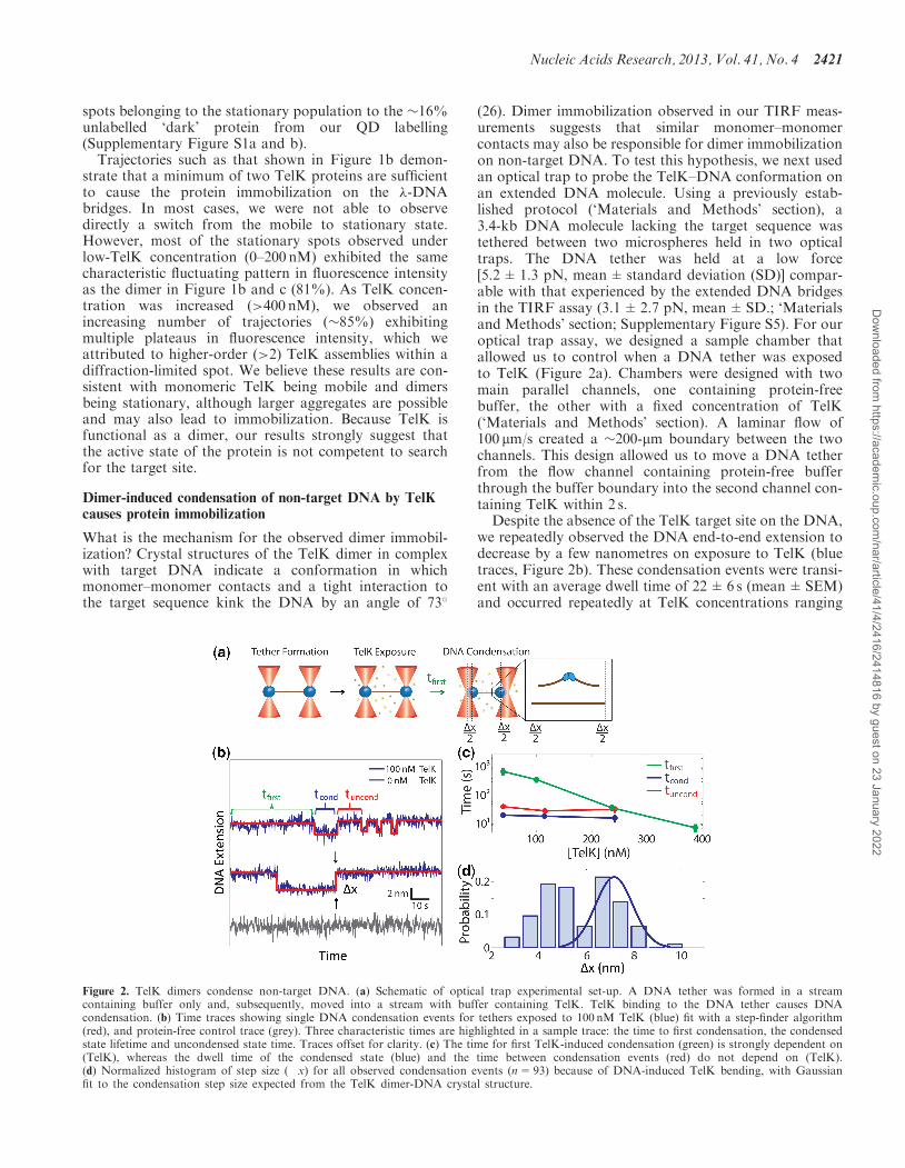
(26). Dimer immobilization observed in our TIRF meas-urements suggests that similar monomer–monomercontacts may also be responsible for dimer immobilizationon non-target DNA. To test this hypothesis, we next usedan optical trap to probe the TelK–DNA conformation onan extended DNA molecule. Using a previously estab-lished protocol (‘Materials and Methods’ section), a3.4-kb DNA molecule lacking the target sequence wastethered between two microspheres held in two opticaltraps. The DNA tether was held at a low force[5.2±1.3 pN, mean±standard deviation (SD)] compar-able with that experienced by the extended DNA bridgesin the TIRF assay (3.1±2.7 pN, mean±SD.; ‘Materialsand Methods’ section; Supplementary Figure S5). For ouroptical trap assay, we designed a sample chamber thatallowed us to control when a DNA tether was exposedto TelK (Figure 2a). Chambers were designed with twomain parallel channels, one containing protein-freebuffer, the other with a fixed concentration of TelK(‘Materials and Methods’ section). A laminar flow of100 mm/s created a �200-mm boundary between the twochannels. This design allowed us to move a DNA tetherfrom the flow channel containing protein-free bufferthrough the buffer boundary into the second channel con-taining TelK within 2 s.Despite the absence of the TelK target site on the DNA,
we repeatedly observed the DNA end-to-end extension todecrease by a few nanometres on exposure to TelK (bluetraces, Figure 2b). These condensation events were transi-ent with an average dwell time of 22±6 s (mean±SEM)and occurred repeatedly at TelK concentrations ranging
Figure 2. TelK dimers condense non-target DNA. (a) Schematic of optical trap experimental set-up. A DNA tether was formed in a streamcontaining buffer only and, subsequently, moved into a stream with buffer containing TelK. TelK binding to the DNA tether causes DNAcondensation. (b) Time traces showing single DNA condensation events for tethers exposed to 100 nM TelK (blue) fit with a step-finder algorithm(red), and protein-free control trace (grey). Three characteristic times are highlighted in a sample trace: the time to first condensation, the condensedstate lifetime and uncondensed state time. Traces offset for clarity. (c) The time for first TelK-induced condensation (green) is strongly dependent on(TelK), whereas the dwell time of the condensed state (blue) and the time between condensation events (red) do not depend on (TelK).(d) Normalized histogram of step size (�x) for all observed condensation events (n=93) because of DNA-induced TelK bending, with Gaussianfit to the condensation step size expected from the TelK dimer-DNA crystal structure.
Nucleic Acids Research, 2013, Vol. 41, No. 4 2421
Dow
nloaded from https://academ
ic.oup.com/nar/article/41/4/2416/2414816 by guest on 23 January 2022

from 40 to 270 nM. In contrast, control experiments per-formed in buffer lacking TelK showed no DNA conden-sation (gray trace, Figure 2b). Our ability to control whenDNA was first exposed to TelK allowed us to measureaccurately the time to the first condensation event. Asexpected, this first condensation time was strongly de-pendent on TelK concentration (green data points,Figure 2c), indicating binding of one or multiple TelKto the DNA. In contrast, the average condensed statelifetime and the time between subsequent condensationevents were independent of TelK concentration (blue,red data points, Figure 2c), indicating that the same indi-vidual TelK complex was responsible for those condensa-tion events. At high (>200 nM) concentrations of TelK,we mostly observed multiple simultaneous condensationevents that were indistinguishable from each other.Therefore, we restricted our analysis to tethers showingindividual separate condensation steps.The size of these individual condensation events was
determined with a step-finding algorithm (50). Thestep-size distribution for DNA condensation by TelK isdisplayed in Figure 2d. Condensation behaviour isexpected as a result of TelK dimer formation at theDNA target site, as the 73� bend of the target DNAobserved in crystal structures condenses the DNA,reducing its end-to-end extension. However, DNA con-densation in the absence of the target site is notexpected. The observed condensation in the optical trapmeasurements suggests that the same dimer-DNA con-formation may be adopted on non-target DNA.To test this suggestion, we performed a series of MD
simulations to study the interaction of (i) a TelK dimerwith target DNA, (ii) a TelK dimer with non-target DNAand (iii) a TelK monomer with non-target DNA. Thesimulations reveal that dimers induce near identical bendangles (�70�) onto DNA with and without the targetsequence (Supplementary Figure S2). We used the bendangle observed in these MD simulations to estimate thechange in DNA end-to-end extension as a result of TelKdimerization on the non-target DNA substrate used in ouroptical trap assays. We compared the extension of thecondensed DNA to that of linearly extended DNAtaking into account the bend angle observed in the MDsimulation, the DNA elastic properties and the forceat which the tether was held in the optical trap (41)(‘Materials and Methods’ section), as shown inFigure 3a. Based on this calculation, we expect a conden-sation step size of 7.5 nm at a tension of 5.2 pN. As shownin Figure 2d, the observed step-size distribution isbimodal, with one peak at 7.2 nm, in excellent agreementwith the prediction. This result indicates that a significantfraction of condensation events observed correspond toformation of DNA–TelK dimer complexes similar tothose seen in crystal structures. Simulations of a TelKmonomer binding to non-target DNA further showthat the monomeric form of TelK is unable to bendDNA as a dimer does. Instead, monomeric TelK inter-action with DNA results in a large decrease in the bendangle (Figure 3b). A histogram of the end-to-end distanceof the 44-bp DNA substrate shows significant condensa-tion in the case of the TelK dimer, but on average
no condensation for a TelK monomer on non-targetDNA (Figure 3c). MD results provide strong supportfor the claim that the condensation events observed inthe optical trap measurements correspond to TelKdimerization.
As shown in Figure 2d, the condensation step-size dis-tribution displays a second peak at 4.5 nm, corresponding
Figure 3. MD simulations confirm that Telk dimers bend non-targetDNA, whereas monomers cannot. (a) Schematic representation ofDNA condensation as would be measured in optical trap experiments(brown line) and MD simulations (boxed region). �L is defined as thedifference between the contour length and the end-to-end distance ofthe 44-bp DNA substrate. MD snapshots of a TelK dimer-DNAcomplex and a TelK monomer-DNA complex are shown. DNAadopts a bent conformation in interaction with the TelK dimer, butnot with the TelK monomer. (b) DNA bend angle induced by a TelKdimer (red) and a TelK monomer (blue). (c) Distribution of the changein end-to-end distance in the 44-bp MD DNA substrate (�L) inducedby a TelK dimer and monomer in MD simulations.
2422 Nucleic Acids Research, 2013, Vol. 41, No. 4
Dow
nloaded from https://academ
ic.oup.com/nar/article/41/4/2416/2414816 by guest on 23 January 2022
to �50% of all observed condensation events. Thissuggests that TelK condensation may have two distincttight-binding conformations on non-target DNA.Unfortunately, MD simulations do not reproduce thisfeature within the simulation time frame, insteadproducing only a single condensed conformation. Wespeculate that the smaller step may result frommisoriented dimers that meet along the DNA(Supplementary Discussion). Such a conformation wouldnot be observable with MD simulations, which start fromthe correctly oriented DNA–TelK crystal structure.
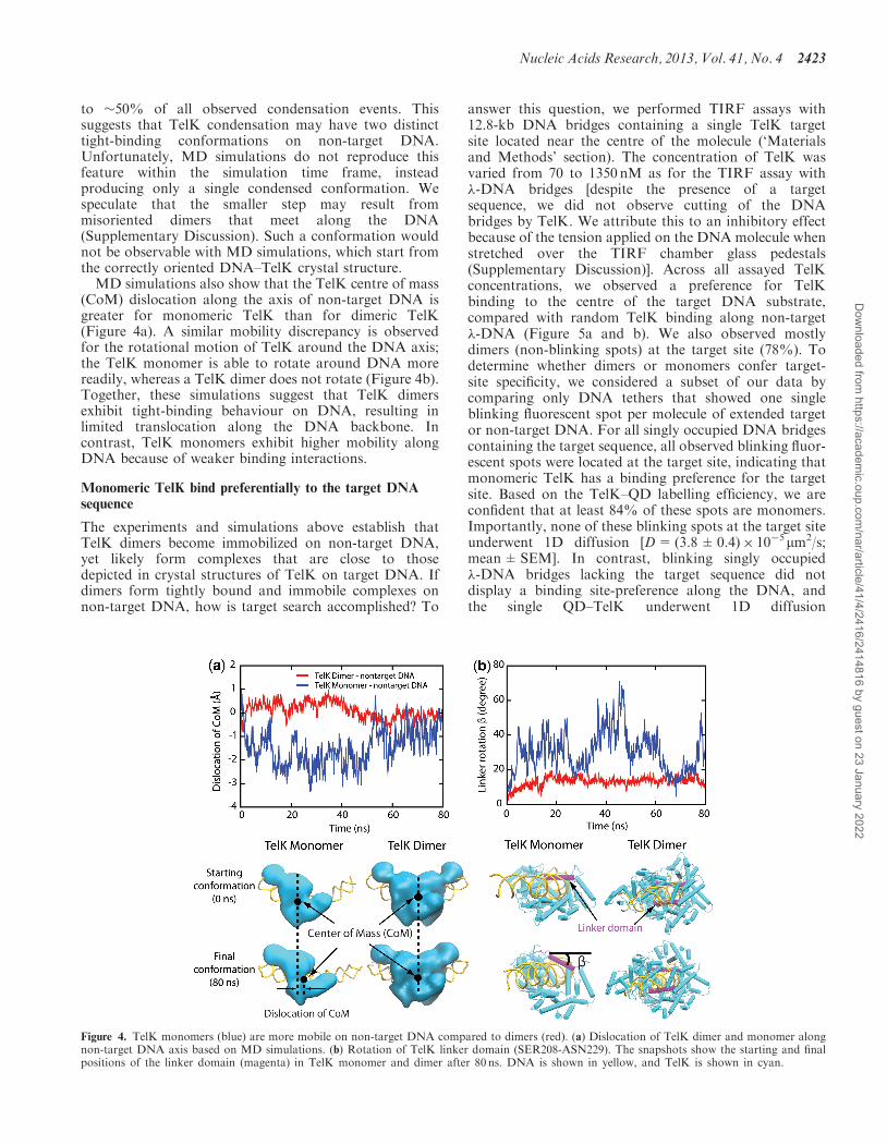
MD simulations also show that the TelK centre of mass(CoM) dislocation along the axis of non-target DNA isgreater for monomeric TelK than for dimeric TelK(Figure 4a). A similar mobility discrepancy is observedfor the rotational motion of TelK around the DNA axis;the TelK monomer is able to rotate around DNA morereadily, whereas a TelK dimer does not rotate (Figure 4b).Together, these simulations suggest that TelK dimersexhibit tight-binding behaviour on DNA, resulting inlimited translocation along the DNA backbone. Incontrast, TelK monomers exhibit higher mobility alongDNA because of weaker binding interactions.
Monomeric TelK bind preferentially to the target DNAsequence
The experiments and simulations above establish thatTelK dimers become immobilized on non-target DNA,yet likely form complexes that are close to thosedepicted in crystal structures of TelK on target DNA. Ifdimers form tightly bound and immobile complexes onnon-target DNA, how is target search accomplished? To
answer this question, we performed TIRF assays with12.8-kb DNA bridges containing a single TelK targetsite located near the centre of the molecule (‘Materialsand Methods’ section). The concentration of TelK wasvaried from 70 to 1350 nM as for the TIRF assay with�-DNA bridges [despite the presence of a targetsequence, we did not observe cutting of the DNAbridges by TelK. We attribute this to an inhibitory effectbecause of the tension applied on the DNA molecule whenstretched over the TIRF chamber glass pedestals(Supplementary Discussion)]. Across all assayed TelKconcentrations, we observed a preference for TelKbinding to the centre of the target DNA substrate,compared with random TelK binding along non-target�-DNA (Figure 5a and b). We also observed mostlydimers (non-blinking spots) at the target site (78%). Todetermine whether dimers or monomers confer target-site specificity, we considered a subset of our data bycomparing only DNA tethers that showed one singleblinking fluorescent spot per molecule of extended targetor non-target DNA. For all singly occupied DNA bridgescontaining the target sequence, all observed blinking fluor-escent spots were located at the target site, indicating thatmonomeric TelK has a binding preference for the targetsite. Based on the TelK–QD labelling efficiency, we areconfident that at least 84% of these spots are monomers.Importantly, none of these blinking spots at the target siteunderwent 1D diffusion [D=(3.8±0.4)� 10�5mm2/s;mean±SEM]. In contrast, blinking singly occupied�-DNA bridges lacking the target sequence did notdisplay a binding site-preference along the DNA, andthe single QD–TelK underwent 1D diffusion
Figure 4. TelK monomers (blue) are more mobile on non-target DNA compared to dimers (red). (a) Dislocation of TelK dimer and monomer alongnon-target DNA axis based on MD simulations. (b) Rotation of TelK linker domain (SER208-ASN229). The snapshots show the starting and finalpositions of the linker domain (magenta) in TelK monomer and dimer after 80 ns. DNA is shown in yellow, and TelK is shown in cyan.
Nucleic Acids Research, 2013, Vol. 41, No. 4 2423
Dow
nloaded from https://academ
ic.oup.com/nar/article/41/4/2416/2414816 by guest on 23 January 2022

(0.88±0.10mm2/s; mean±SEM, comparable with thatobtained for all mobile spots on non-target DNA).Our TIRF results are supported by MD simulations of
a single TelK monomer on target and non-target DNAsubstrates. Monomers along target DNA exhibitedgreater stability than monomers along non-target DNA.As shown in Supplementary Figure S7a and b, the contactarea and number of hydrogen bonds between a TelKmonomer and target DNA remain constant during simu-lations, whereas both decrease in simulations of TelKmonomer-non-target DNA complexes. MD simulationresults indicate that TelK monomers have an increasedaffinity for the target sequence.Repeating the optical trap experiments with DNA con-
taining the TelK target site, we again observed repeatedcondensation of the DNA. The average condensationdwell time was 21±2 s (mean±SEM), with an averagestep-size of 7.7±0.2 nm (mean±SEM; SupplementaryFigure S6 and Supplementary Discussion), in good agree-ment with the value measured with non-target DNA.Interestingly, we observed less of the 4.5-nm condensationevents, which we attributed to misoriented dimers. Wespeculate that the presence of the target site may biasdimer formation into the more ‘proper’ configuration con-sistent with crystal structures.
DISCUSSION
The process by which site-specific proteins find their DNAtarget sites has remained poorly characterized for proteinsthat are functional on DNA as dimers or higher-orderprotein complexes but are monomeric in solution. OurTIRF studies demonstrate that TelK diffuses in 1D as a
monomer along non-target DNA, yet becomes immobileas it encounters its target sequence. Therefore, monomertarget-site immobilization confers specificity for TelK di-merization at the target site. MD simulations confirm ex-perimental observations of monomer target-site specificityand provide molecular details of TelK’s interactions withtarget versus non-target DNA (Supplementary Figure S7).We also showed that TelK dimers become stationary andcondense non-target DNA in our TIRF and trap meas-urements. However, protein immobilization on non-targetDNA is intuitively counter-productive for the localizationof the DNA target site, as it truncates the search processprematurely. Similarly, non-target DNA condensation isunexpected for a protein requiring sequence specificity foractivity because condensation is usually predicted to occurat the target site before catalytic activity (51).
We suspect condensation and immobilization may beessential target-search features for proteins that mustassemble into oligomeric complexes on DNA and thatrequire more complex mechanisms for target-site identifi-cation than monomeric or pre-assembled SSPs. DNAbending has been thought to occur exclusively at targetsites for sequence-specific protein–DNA systems. Only atthe target-site do protein–DNA electrostatic interactionsbecome maximized, and catalysis occurs hand-in-handwith DNA topological distortions (41). However, as hasbeen previously suggested for certain SSPs (52), a protein’sability to bend DNA indiscriminately may aid target-siteidentification. Erie et al. suggest that dimer-active proteinsmay bend DNA to ‘test’ their compatibility with that par-ticular DNA sequence via protein–DNA and protein–protein interactions (53). Transcription factor HoxD9has also been shown by Clore et al. (54,55) to bindnon-target DNA with the same affinity and in the samebinding mode as target-DNA as a means of enhancing therate of target identification. Dimer-active proteins such asTelK may use this mechanism to maximize their electro-static contacts with the DNA during their ‘sampling’ state,not only during site-specific catalysis.
Our optical trap data may provide some support for a‘sampling’ mechanism in TelK. The mean dwell time ofthe condensation events (22±6 s; mean±SEM) in thetrap measurements is noticeably shorter than theimmobilized-state lifetime observed in our TIRF experi-ments, which spanned minutes (Figures 1d and 2c).However, the time between condensation events was inde-pendent of TelK concentration, suggesting that a singleintra-molecular dimerization event was responsible formultiple sequential condensation and de-condensationevents. We suspect this indicates that a TelK dimer mayrepeatedly attempt to bend its DNA substrate.
If non-target DNA condensation occurs too readily, itmay significantly slow or inhibit target-finding and sub-strate formation. At first glance, it may seem that twomonomers will find each other more quickly on DNAabundantly coated with protein, when in direct competi-tion with the process of finding a target site. However,through stochastic simulations (‘Materials and Methods’section), we show there is a wide range of conditions overwhich target localization occurs more rapidly than dimer-ization. Our simulations use experimentally measured
Figure 5. TelK binds preferentially to target DNA. (a) Average TelKfluorescent spot positions along target (green, n=182) and non-target(red, n=253) DNA, obtained by aligning and overlapping fluorescenceimages. TelK concentration range used in all TIRF experiments rangedfrom 70 to 1350 nM. (b) Position probability distribution of TelK alongtarget (green) and non-target (red) DNA.
2424 Nucleic Acids Research, 2013, Vol. 41, No. 4
Dow
nloaded from https://academ
ic.oup.com/nar/article/41/4/2416/2414816 by guest on 23 January 2022

kinetic parameters for TelK monomer unbinding(koff=0.24 s�1) and diffusion (D=0.74 mm2/s) (for sim-plicity, dimers were assumed to be immobile and toremain stably bound). Varying the average protein occu-pancy (the mean number of TelK monomers per DNAmolecule), we calculated the rate of dimerization kdimer
on non-target DNA versus the target-finding rate ktarget.In comparing the two competing processes of protein
dimerization versus protein target-finding, two regimeswere observed as shown in Figure 6b. At low-TelKoccupancies, the rate for target-site identification was sig-nificantly higher than for dimerization. However, at highoccupancies, dimerization occurred faster thantarget-finding. This can be conceptualized by consideringtwo competing processes: a monomer finding a target siteversus a monomer finding another monomer. For a DNAmolecule coated with N monomers, on average, theminimum inter-monomer distance scales as 1/N2
(Supplementary Discussion). Conversely, on average, thedistance between a target site and the closest monomerscales as 1/N. As a result, the rates at which TelKdiffuses over those distances depend differently on thenumber of proteins on DNA. When diffusion is therate-limiting step to dimerization or target finding, kdimer
scales as N4, whereas ktarget scales as N2, as seen inFigure 6b (at low TelK levels, the rate-limiting step isbinding of TelK rather than diffusion, and kdimer scalesinstead as N2). The net result is that, at low-protein con-centrations, the rate for a protein to diffuse to a target siteis consistently higher than a protein diffusing into anotherprotein. At high-protein concentrations, where the occu-pancy of protein along DNA is large, dimerization isfaster than target localization.
TelK’s host organism, Klebsiella oxytoca, is believed tomaintain a cellular TelK copy number <20 for a �52-kbgenome, or less than one dimer per 5 kb of DNA (data notpublished). These estimates are firmly placed in thelow-occupancy regime predicted in our simulations.
Therefore, we expect that target-site localization occursfaster than dimerization at biologically relevant TelK con-centrations, allowing non-target DNA condensation toprovide site-specificity in dimer-active proteins withouthindering the kinetics of target sequence localization.Our simulations would also predict that the activity ofthe protein should decrease at high concentrationsbecause of inhibition of target finding by immobiledimers. This is consistent with the observation of TelKinhibition at high TelK concentrations (>400 nM) inbulk (Supplementary Figure S8). A target-search mechan-ism in which protein dimers immobilize along non-targetDNA is thus viable provided the DNA is sparsely coated.Based on our results, we propose a mechanism fordimer-active TelK in which target site identificationoccurs primarily by protein monomers diffusing to thetarget and preferentially binding to the target sequence,followed by dimerization at the target site.The results of our kinetic simulations in principle can be
compared with our optical trap data. As DNA is exposedto TelK at a well-defined time in our optical trap assay, wecan measure directly the time for the first DNA conden-sation event (the first passage time) as a function of TelKconcentration. Using our TIRF assay to calibrate occu-pancy on DNA against TelK incubation concentration(‘Materials and Methods’ section; SupplementaryFigure S9), we compared our theoretical and experimentaldimerization rates. The measured first passage times in ouroptical trap yield average rates (0.02–0.21 s�1) in goodagreement with dimerization rates obtained in our simu-lations (0.02–0.11 s�1) for comparable mean occupancies(<N>=0.4� 0.8). This agreement further supports ourdimer-active protein model for target search.The literature to-date suggests an all-encompassing
model for protein target-search in which proteins scannon-target DNA in their fully functional form (5,12).However, our studies of protelomerase TelK, a proteinthat is functional only as a dimer, reveal that additional
Figure 6. Kinetics of TelK-induced dimerization and target search. (a) Model of TelK target search. TelK monomers in solution bind DNA withrate kon and scan rapidly along non-target DNA with mean diffusion coefficient Dmonomer=1.8 mm2/s. Monomers localize the target site with ratektarget and bind tightly or dissociate from non-target DNA with average rate koff=0.24 s�1. Preferential and stable binding of a TelK monomerallows a second monomer to dimerize at the target site and form a kinked DNA–TelK complex primed for catalysis. Occasionally, mobile monomersencounter each other on non-target DNA with rate kdimer and form stable, immobile dimers (Ddimer< 1� 10�4mm2/s) that ‘test’ their substrate bycondensing the DNA transiently. Eventually, dimers on non-target DNA dissociate or separate into mobile monomers again (rate< 0.01 s�1). (b)Implementation of kinetic model in (a) via stochastic simulations using experimentally derived kinetic parameters. Average rates for first dimerizationkdimer (blue, black), and first target localization ktarget (red) as a function of average protein occupancy, <N>, on DNA. For mean occupancies >1,target-finding and dimerization rates obey simple scaling laws of N2 and N4, respectively. For mean occupancies <1, dimerization follows of N2
scaling. Provided TelK occupancy is reasonably small, target localization consistently occurs faster than dimerization. Simulated rates are in goodagreement with experimentally derived rates of first dimerization.
Nucleic Acids Research, 2013, Vol. 41, No. 4 2425
Dow
nloaded from https://academ
ic.oup.com/nar/article/41/4/2416/2414816 by guest on 23 January 2022

variables must be taken into consideration when des-cribing target-search mechanisms for proteins thatassemble into dimer-active complexes on DNA.Although 1D scanning is a fundamental characteristic oftarget search by SSPs, we show that, for the dimer-activeprotein TelK, scanning only occurs as a monomer, andimmobilization occurs when it forms a tightly-bounddimer on DNA or localizes the target sequence as amonomer. We speculate that the target-search mechanismproposed as a result of this work is extendable to severaldifferent protein families that share characteristics withTelK, such as those that assemble higher-order complexesonto their DNA target sites. Future studies of othermembers of the recombinase protein family will furtherour understanding of this novel target search mechanism,and its applicability to dimer and oligomer SSPs.
SUPPLEMENTARY DATA
Supplementary Data are available at NAR Online:Supplementary Table 1, Supplementary Figures 1–9,Supplementary Methods, Supplementary Discussion andSupplementary References [56–59].
ACKNOWLEDGEMENTS
The authors thank Prof. Toshio Yanagida, Dr KomoriTomotaka, Thomas Martin and all the members of theHa, Selvin, Yanagida and Chemla laboratories forhelpful discussions and for their generous advice.
FUNDING
National Institutes of Health [9P41GM104601 to X.Z.and K.S.]; Cell mechanics [R01 GM073655 to K.S.];National Science Foundation [082265, Physics FrontierCenter: Center for the Physics of Living Cells];Burroughs-Wellcome Fund: Career Awards at theScientific Interface (to Y.R.C.); National ScienceFoundation Graduate Research Fellowship [0913128 toM.P.L.]; East Asia and Pacific Summer Institutes fellow-ship [ID OISE-0913128 to M.P.L.]; Texas AdvancedComputing Center and the National Center forSupercomputing Applications via XSEDE ResourceAllocation Committee [MCA93S028]. Funding for openaccess charge: Burroughs-Wellcome Fund.
Conflict of interest statement. None declared.
REFERENCES
1. Koslover,E.F., Diaz de la Rosa,M.A. and Spakowitz,A.J. (2011)Theoretical and computational modeling of target-site searchkinetics in vitro and in vivo. Biophys. J., 101, 856–865.
2. van den Broek,B., Lomholt,M.A., Kalisch,S.M.J., Metzler,R. andWuite,G.J.L. (2008) How DNA coiling enhances targetlocalization by proteins. Proc. Natl Acad. Sci. USA, 105,15738–15742.
3. Gowers,D.M., Wilson,G.G. and Halford,S.E. (2005) Measurementof the contributions of 1D and 3D pathways to the translocationof a protein along DNA. Proc. Natl Acad. Sci. USA, 102,15883–15888.
4. Ramanathan,S.P., van Aelst,K., Sears,A., Peakman,L.J.,Diffin,F.M., Szczelkun,M.D. and Seidel,R. (2009) Type IIIrestriction enzymes communicate in 1D without looping betweentheir target sites. Proc. Natl Acad. Sci. USA, 106, 1748–1753.
5. Halford,S.E. and Marko,J.F. (2004) How do site-specificDNA-binding proteins find their targets? Nucleic Acids Res., 32,3040–3052.
6. Kolomeisky,A.B. (2011) Physics of protein-DNA interactions:mechanisms of facilitated target search. Phys. Chem. Chem. Phys.,13, 2088–2095.
7. Sudhanshu,B., Mihardja,S., Koslover,E.F., Mehraeen,S.,Bustamante,C. and Spakowitz,A.J. (2011) Tension-dependentstructural deformation alters single-molecule transition kinetics.Proc. Natl Acad. Sci. USA, 108, 1885–1890.
8. Bitinaite,J., Wah,D.A., Aggarwal,A.K. and Schildkraut,I. (1998)FokI dimerization is required for DNA cleavage. Proc. NatlAcad. Sci. USA, 95, 10570–10575.
9. Ban,C. and Yang,W. (1998) Structural basis for MutH activationin E.coli mismatch repair and relationship of MutH to restrictionendonucleases. EMBO J., 17, 1526–1534.
10. Dowd,D.R. and Lloyd,R.S. (1990) Biological significance offacilitated diffusion in protein-DNA interactions. Applications toT4 endonuclease V-initiated DNA repair. J. Biol. Chem., 265,3424–3431.
11. Blainey,P.C., van Oijent,A.M., Banerjee,A., Verdine,G.L. andXie,X.S. (2006) A base-excision DNA-repair protein findsintrahelical lesion bases by fast sliding in contact with DNA.Proc. Natl Acad. Sci. USA, 103, 5752–5757.
12. Widom,J. (2005) Target site localization by site-specific,DNA-binding proteins. Proc. Natl Acad. Sci. USA, 102,16909–16910.
13. van den Broek,B., Noom,M.C. and Wuite,G.J. (2005)DNA-tension dependence of restriction enzyme activity revealsmechanochemical properties of the reaction pathway. NucleicAcids Res., 33, 2676–2684.
14. Langowski,J., Alves,J., Pingoud,A. and Maass,G. (1983) Does thespecific recognition of DNA by the restriction endonuclease Ecoriinvolve a linear diffusion step - investigation of the processivityof the Ecori endonuclease. Nucleic Acids Res., 11, 501–513.
15. Gorman,J., Chowdhury,A., Surtees,J.A., Shimada,J.,Reichman,D.R., Alani,E. and Greene,E.C. (2007) Dynamic basisfor one-dimensional DNA scanning by the mismatch repaircomplex Msh2-Msh6. Mol. Cell, 28, 359–370.
16. Mendillo,M.L., Mazur,D.J. and Kolodner,R.D. (2005) Analysis ofthe interaction between the Saccharomyces cerevisiaeMSH2-MSH6 and MLH1-PMS1 complexes with DNA using areversible DNA end-blocking system. J. Biol. Chem., 280,22245–22257.
17. Gorman,J. and Greene,E.C. (2008) Visualizing one-dimensionaldiffusion of proteins along DNA. Nat. Struct. Mol. Biol., 15,768–774.
18. Singh,V., Ekka,M.K. and Kumaran,S. (2012) Second MonomerBinding Is the Rate-Limiting Step in the Formation of theDimeric PhoP-DNA Complex. Biochemistry, 51, 1346–1356.
19. Erie,D.A., Yang,G., Schultz,H.C. and Bustamante,C. (1994) DNAbending by Cro protein in specific and nonspecific complexes:implications for protein site recognition and specificity. Science,266, 1562–1566.
20. Marianayagam,N.J., Sunde,M. and Matthews,J.M. (2004) Thepower of two: protein dimerization in biology. Trends Biochem.Sci., 29, 618–625.
21. Vangent,D.C., Vink,C., Groeneger,A.A.M.O. and Plasterk,R.H.A.(1993) Complementation between HIV integrase proteins mutatedin different domains. EMBO J., 12, 3261–3267.
22. Hai,T. and Curran,T. (1991) Cross-family dimerization oftranscription factors Fos Jun and Atf creb alters DNA-bindingspecificity. Proc. Natl Acad. Sci. USA, 88, 3720–3724.
23. Shuai,K., Horvath,C.M., Huang,L.H., Qureshi,S.A., Cowburn,D.and Darnell,J.E. Jr (1994) Interferon activation of thetranscription factor Stat91 involves dimerization throughSH2-phosphotyrosyl peptide interactions. Cell, 76, 821–828.
24. van Aelst,K., Toth,J., Ramanathan,S.P., Schwarz,F.W., Seidel,R.and Szczelkun,M.D. (2010) Type III restriction enzymes cleaveDNA by long-range interaction between sites in both
2426 Nucleic Acids Research, 2013, Vol. 41, No. 4
Dow
nloaded from https://academ
ic.oup.com/nar/article/41/4/2416/2414816 by guest on 23 January 2022

head-to-head and tail-to-tail inverted repeat. Proc. Natl Acad. Sci.USA, 107, 9123–9128.
25. Huang,W.M., Joss,L., Hsieh,T.T. and Casjens,S. (2004)Protelomerase uses a topoisomerase IB/Y-recombinase typemechanism to generate DNA hairpin ends. J. Mol. Biol., 337,77–92.
26. Aihara,H., Huang,W.M. and Ellenberger,T. (2007) An interlockeddimer of the protelomerase TelK distorts DNA structure for theformation of hairpin telomeres. Mol. Cell, 27, 901–913.
27. Moffitt,J.R., Chemla,Y.R., Smith,S.B. and Bustamante,C. (2008)Recent advances in optical tweezers. Annu. Rev. Biochem., 77,205–228.
28. Selvin,P.R. and Ha,T. (2008) Single-Molecule Techniques:A Laboratory Manual. John Inglis, Cold Spring Harbor, NY.
29. Klepeis,J.L., Lindorff-Larsen,K., Dror,R.O. and Shaw,D.E. (2009)Long-timescale molecular dynamics simulations of proteinstructure and function. Curr. Opin. Struct. Biol., 19, 120–127.
30. Smith,K. and Youngman,P. (1992) Use of a new integrationalvector to investigate compartment-specific expression of theBacillus-subtilis spoiim gene. Biochimie, 74, 705–711.
31. Sun,M. and Serwer,P. (1997) The conformation of DNApackaged in bacteriophage G. Biophys. J., 72, 958–963.
32. Selvin,P.R., Lougheed,T., Tonks Hoffman,M., Park,H., Balci,H.,Blehm,B. and Toprak,E. (2008) Single-Molecule Techniques: ALaboratory Manual. In: Ha,T. (ed.), In Vitro and In Vivo FIONAand Other Acronyms for Watching Molecular Motors Walk. ColdSpring Harbor Books, Cold Spring Harbor, NY.
33. Yildiz,A., Forkey,J.N., McKinney,S.A., Ha,T., Goldman,Y.E. andSelvin,P.R. (2003) Myosin V walks hand-over-hand: singlefluorophore imaging with 1.5-nm localization. Science, 300,2061–2065.
34. Iwaki,M., Iwane,A.H., Shimokawa,T., Cooke,R. and Yanagida,T.(2009) Brownian search-and-catch mechanism for myosin-VIsteps. Nat. Chem. Biol., 5, 403–405.
35. Dahan,M., Levi,S., Luccardini,C., Rostaing,P., Riveau,B. andTriller,A. (2003) Diffusion dynamics of glycine receptors revealedby single-quantum dot tracking. Science, 302, 442–445.
36. Bouzigues,C., Morel,M., Triller,A. and Dahan,M. (2007)Asymmetric redistribution of GABA receptors during GABAgradient sensing by nerve growth cones analyzed by singlequantum dot imaging. Proc. Natl Acad. Sci. USA, 104,11251–11256.
37. Courty,S., Luccardini,C., Bellaiche,Y., Cappello,G. and Dahan,M.(2006) Tracking individual kinesin motors in living cells usingsingle quantum-dot imaging. Nano Lett., 6, 1491–1495.
38. Bannai,H., Levi,S., Schweizer,C., Dahan,M. and Triller,A. (2007)Imaging the lateral diffusion of membrane molecules withquantum dots. Nat. Protoc., 1, 2628–2634.
39. Bustamante,C., Chemla,Y.R. and Moffitt,J.R. (2008) In: Ha,T.(ed.), Single-Molecule Techniques, A Laboratory Manual, High-Resolution Dual-Trap Optical Tweezers with DifferentialDetection. Cold Spring Harbor Laboratory Press, Cold SpringHarbor, NY.
40. Landry,M.P., McCall,P.M., Qi,Z. and Chemla,Y.R. (2009)Characterization of photoactivated singlet oxygen damage insingle-molecule optical trap experiments. Biophys. J., 97,2128–2136.
41. van den Broek,B., Noom,M.C. and Wuite,G.J. (2005)DNA-tension dependence of restriction enzyme activity revealsmechanochemical properties of the reaction pathway. NucleicAcids Res., 33, 2676–2684.
42. Humphrey,W., Dalke,A. and Schulten,K. (1996) VMD: visualmolecular dynamics. J. Mol. Graph., 14, 33-38–27–38.
43. Phillips,J.C., Braun,R., Wang,W., Gumbart,J., Tajkhorshid,E.,Villa,E., Chipot,C., Skeel,R.D., Kale,L. and Schulten,K. (2005)Scalable molecular dynamics with NAMD. J. Comput. Chem., 26,1781–1802.
44. Foloppe,N. and MacKerell,A.D. (2000) All-atom empirical forcefield for nucleic acids: I. Parameter optimization based on smallmolecule and condensed phase macromolecular target data.J. Comput. Chem., 21, 86–104.
45. MacKerell,A.D., Bashford,D., Bellott,M., Dunbrack,R.L.,Evanseck,J.D., Field,M.J., Fischer,S., Gao,J., Guo,H., Ha,S. et al.(1998) All-atom empirical potential for molecular modeling anddynamics studies of proteins. J. Phys. Chem. B, 102, 3586–3616.
46. Mackerell,A.D., Feig,M. and Brooks,C.L. (2004) Extending thetreatment of backbone energetics in protein force fields:limitations of gas-phase quantum mechanics in reproducingprotein conformational distributions in molecular dynamicssimulations. J. Comput. Chem., 25, 1400–1415.
47. Jorgensen,W.L., Chandrasekhar,J., Madura,J.D., Impey,R.W. andKlein,M.L. (1983) Comparison of simple potential functions forsimulating liquid water. J. Chem. Phys., 79, 926–935.
48. Martyna,G.J., Tobias,D.J. and Klein,M.L. (1994)Constant-pressure molecular-dynamics algorithms. J. Chem. Phys.,101, 4177–4189.
49. Nirmal,M., Dabbousi,B.O., Bawendi,M.G., Macklin,J.J.,Trautman,J.K., Harris,T.D. and Brus,L.E. (1996) Fluorescenceintermittency in single cadmium selenide nanocrystals. Nature,383, 802–804.
50. Dogterom,M., Kerssemakers,J.W.J., Munteanu,E.L., Laan,L.,Noetzel,T.L. and Janson,M.E. (2006) Assembly dynamics ofmicrotubules at molecular resolution. Nature, 442, 709–712.
51. Slutsky,M. and Mirny,L.A. (2004) Kinetics of protein-DNAinteraction: facilitated target location in sequence-dependentpotential. Biophys. J., 87, 4021–4035.
52. Grove,A., Galeone,A., Mayol,L. and Geiduschek,E.P. (1996)Localized DNA flexibility contributes to target site selection byDNA-bending proteins. J. Mol. Biol., 260, 120–125.
53. Erie,D.A., Yang,G.L., Schultz,H.C. and Bustamante,C. (1994)DNA bending by Cro protein in specific and nonspecificcomplexes - implications for protein site recognition andspecificity. Science, 266, 1562–1566.
54. Iwahara,J. and Clore,G.M. (2006) Detecting transientintermediates in macromolecular binding by paramagnetic NMR.Nature, 440, 1227–1230.
55. Iwahara,J., Zweckstetter,M. and Clore,G.M. (2006) NMRstructural and kinetic characterization of a homeodomaindiffusing and hopping on nonspecific DNA. Proc. Natl Acad. Sci.USA, 103, 15062–15067.
56. Biebricher,A., Wende,W., Escude,C., Pingoud,A. and Desbiolles,P.(2009) Tracking of single quantum dot labeled ecorv sliding alongDNA manipulated by double optical tweezers. Biophys. J., 96,L50–L52.
57. Grindley,N.D., Whiteson,K.L. and Rice,P.A. (2006) Mechanismsof site-specific recombination. Annu. Rev. Biochem., 75, 567–605.
58. Grainge,I. and Jayaram,M. (1999) The integrase family ofrecombinase: organization and function of the active site. Mol.Microbiol., 33, 449–456.
59. Lee,L. and Sadowski,P.D. (2005) Strand selection by the tyrosinerecombinases. Prog. Nucleic Acid Res. Mol. Biol., 80, 1–42.
Nucleic Acids Research, 2013, Vol. 41, No. 4 2427
Dow
nloaded from https://academ
ic.oup.com/nar/article/41/4/2416/2414816 by guest on 23 January 2022
Related Documents


