DNA Structure and Function Chapter 13 Hsueh-Fen Juan Oct 23, 2012

DNA Structure and Function Chapter 13 Hsueh-Fen Juan Oct 23, 2012.
Jan 01, 2016
Welcome message from author
This document is posted to help you gain knowledge. Please leave a comment to let me know what you think about it! Share it to your friends and learn new things together.
Transcript

DNA Structure and Function
Chapter 13
Hsueh-Fen Juan
Oct 23, 2012

Impacts, IssuesHere Kitty, Kitty, Kitty, Kitty, Kitty
Clones made from adult cells have problems; the cell’s DNA must be reprogrammed to function like the DNA of an egg

13.1 The Hunt for DNA
Investigations that led to our understanding that DNA is the molecule of inheritance reveal how science advances

Early and Puzzling Clues
1800s: Miescher found DNA (deoxyribonucleic acid) in nuclei
Early 1900s: Griffith transferred hereditary material from dead cells to live cells• Mice injected with live R cells lived• Mice injected with live S cells died• Mike injected with killed S cells lived• Mice injected with killed S cells and live R cells
died; live S cells were found in their blood• (R: rough colonies; S: smooth colonies)

Griffith’s Experiments
Fig. 13-2, p. 204
RR S
A Mice injected with live cells of harmless strain R do not die. Live R cells are in their blood.
B Mice injected with live cells of killer strain S die. Live S cells are in their blood.
C Mice injected with heat-killed S cells do not die. No live S cells are in their blood.
D Mice injected with live R cells plus heat-killed S cells die. Live S cells are in their blood.

Animation: Griffith’s experiment

Avery and McCarty Find the Transforming Principle
1940: Avery and McCarty separated deadly S cells (from Griffith’s experiments) into lipid, protein, and nucleic acid components
When lipids, proteins, and RNA were destroyed, the remaining substance, DNA, still transformed R cells to S cells
Conclusion: DNA is the “transforming principle”

Confirmation of DNA’s Function
1950s: Hershey and Chase experimented with bacteriophages (viruses that infect bacteria)• Protein parts of viruses, labeled with 35S, stayed
outside the bacteria• DNA of viruses, labeled with 32P, entered the
bacteria• ( 噬菌體標記法:先讓噬菌體感染含 35S 培養基的
細菌,再感染它們,之後產生的噬菌體皆被標記35S)
Conclusion: DNA, not protein, is the material that stores hereditary information

The Hershey-Chase Experiments

Animation: Hershey-Chase experiments

13.2 The Discovery of DNA’s Structure
Watson and Crick’s discovery of DNA’s structure was based on almost fifty years of research by other scientists

DNA’s Building Blocks
Nucleotide• A nucleic acid monomer consisting of a five-
carbon sugar (deoxyribose), three phosphate groups, and one of four nitrogen-containing bases
DNA consists of four nucleotide building blocks• Two pyrimidines: thymine and cytosine• Two purines: adenine and guanine• Purines: 2 carbon rings
Pyrimidines: 1 carbon rings

Four Kinds of Nucleotides in DNA
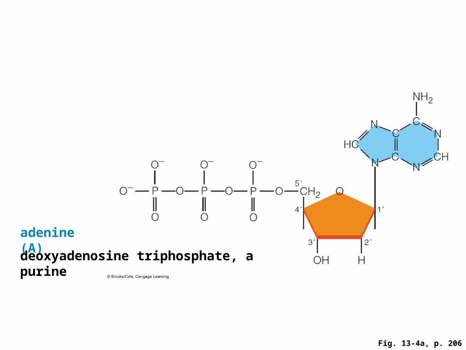
Fig. 13-4a, p. 206
adenine (A)
deoxyadenosine triphosphate, a purine

Fig. 13-4b, p. 206
guanine (G)
deoxyguanosine triphosphate, a purine
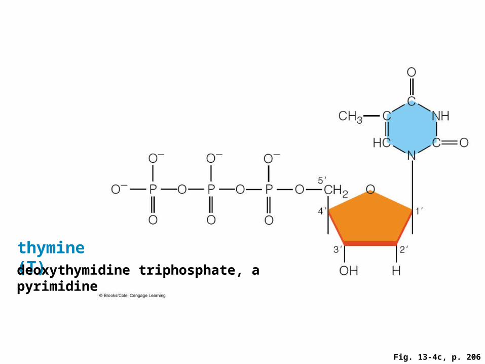
Fig. 13-4c, p. 206
thymine (T)deoxythymidine triphosphate, a pyrimidine

Fig. 13-4d, p. 206
cytosine (C)deoxycytidine triphosphate, a pyrimidine

Chargaff’s Rules
The amounts of thymine and adenine in DNA are the same, and the amounts of cytosine and guanine are the same: A = T and G = C
The proportion of adenine and guanine differs among species

Franklin, Watson and Crick
Rosalind Franklin’s research in x-ray crystallography revealed the dimensions and shape of the DNA molecule: an alpha helix
This was the final piece of information Watson and Crick needed to build their model of DNA
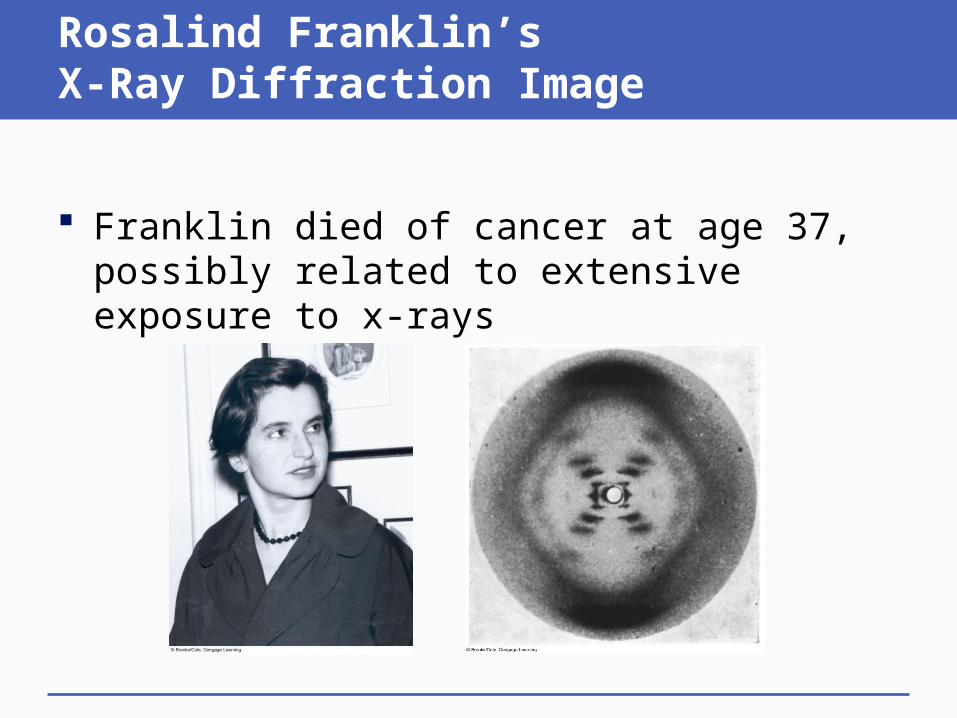
Rosalind Franklin’s X-Ray Diffraction Image
Franklin died of cancer at age 37, possibly related to extensive exposure to x-rays

Watson and Crick’s DNA Model
A DNA molecule consists of two nucleotide chains (strands), running in opposite directions and coiled into a double helix
Base pairs form on the inside of the helix, held together by hydrogen bonds (A-T and G-C)

Patterns of Base Pairing
Bases in DNA strands can pair in only one way• A always pairs with T; G always pairs with C
The sequence of bases is the genetic code• Variation in base sequences gives life diversity
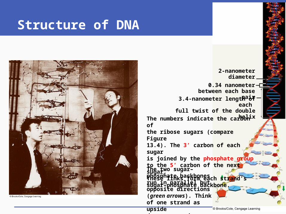
Structure of DNA
2-nanometer diameter
0.34 nanometer between each base pair
3.4-nanometer length of each full twist of the double helix
The numbers indicate the carbon of the ribose sugars (compare Figure 13.4). The 3’ carbon of each sugar is joined by the phosphate group to the 5’ carbon of the next sugar. These links form each strand’s sugar–phosphate backbone.
The two sugar–phosphate backbones run in parallel but opposite directions (green arrows). Think of one strand as upside down compared with the other.
13.3 DNA Replication and Repair
A cell copies its DNA before mitosis or meiosis I
DNA repair mechanisms and proofreading correct most replication errors
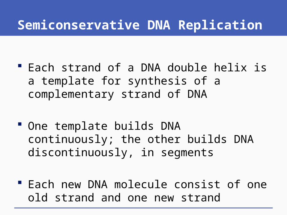
Semiconservative DNA Replication
Each strand of a DNA double helix is a template for synthesis of a complementary strand of DNA
One template builds DNA continuously; the other builds DNA discontinuously, in segments
Each new DNA molecule consist of one old strand and one new strand

Enzymes of DNA Replication
引子酶課本未強調,但是合成岡崎片段的重要酶
DNA helicase (解旋酶 )• Breaks hydrogen bonds between DNA strands
DNA polymerase (DNA聚合酶 )• Joins free nucleotides into a new strand of DNA• DNA 聚合的能量由核苷酸的高能磷酸鍵提供• 核苷酸聚合不但不耗能,甚至還放能!
DNA ligase (DNA連接酶 )• Joins DNA segments on discontinuous strand
DNA Replication

Stepped ArtFig. 13-6, p. 208
D DNA ligase seals any gaps that remain between bases of the “new” DNA, so a continuous strand forms. The base sequence of each half-old, half-new DNA molecule is identical to that of the parent DNA molecule.
C Each of the two parent strands serves as a template for assembly of a new DNA strand from free nucleotides, according to base-pairing rules (G to C, T to A). Thus, the two new DNA strands are complementary in sequence to the parental strands.
B As replication starts, the two strands of DNA are unwound. In cells, the unwinding occurs simul- taneously at many sites along the length of each double helix.
A A DNA molecule is double-stranded. The two strands of DNA stay zippered up together because they are complementary: their nucleotides match up according to base-pairing rules (G to C, T to A).
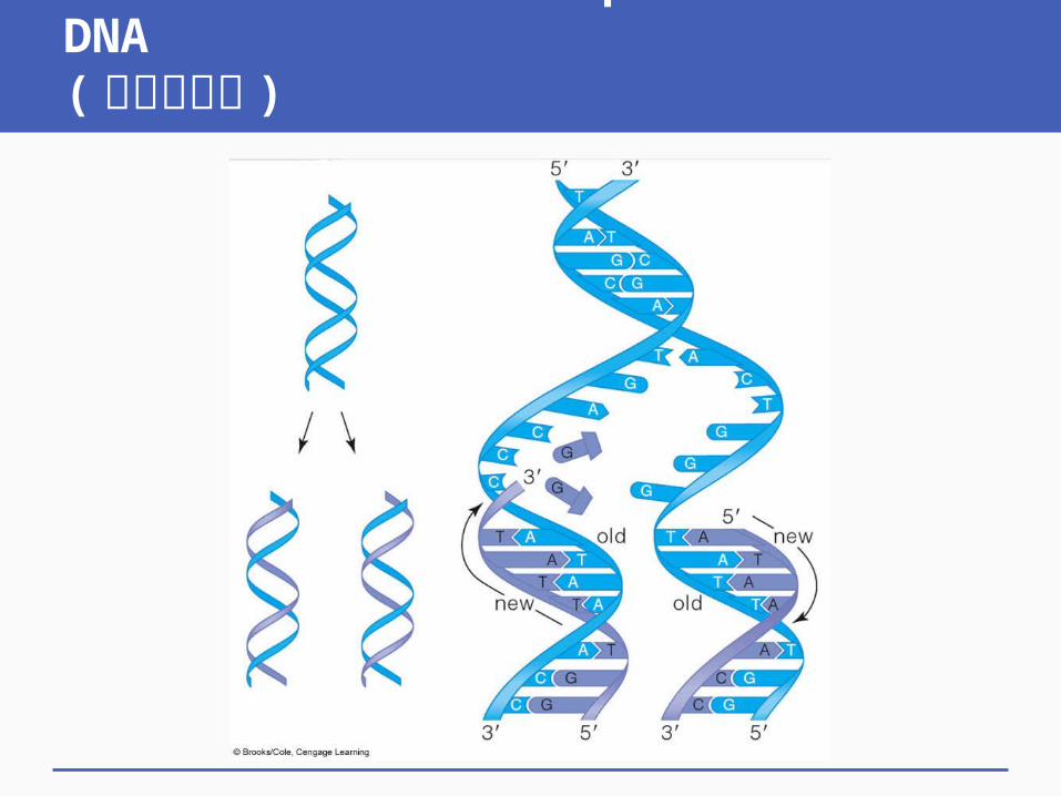
Semiconservative Replication of DNA(半保留複製 )

Discontinuous Synthesis of DNA

Checking for Mistakes
DNA repair mechanisms • DNA polymerases proofread DNA sequences
during DNA replication and repair damaged DNA• DNA 聚合酶本身效率極快 (1000bases/sec) ,發
生錯誤在所難免,因此有此修正機制
When proofreading and repair mechanisms fail, an error becomes a mutation – a permanent change in the DNA sequence

13.4 Using DNA to Duplicate Existing Mammals
Reproductive cloning is a reproductive intervention that results in an exact genetic copy of an adult individual

Cloning
Clones• Exact copies of a molecule, cell, or individual• Occur in nature by asexual reproduction or
embryo splitting (identical twins) Reproductive cloning technologies produce
an exact copy (clone) of an individual ( 注意是人為的、非自然的,用於複製相同個體,因此可以是人或動物 )
Therapeutic cloning 使用人類胚胎幹細胞做研究 ( 只會使用人類胚胎,因為是治療目的,不可能用其他物種 )
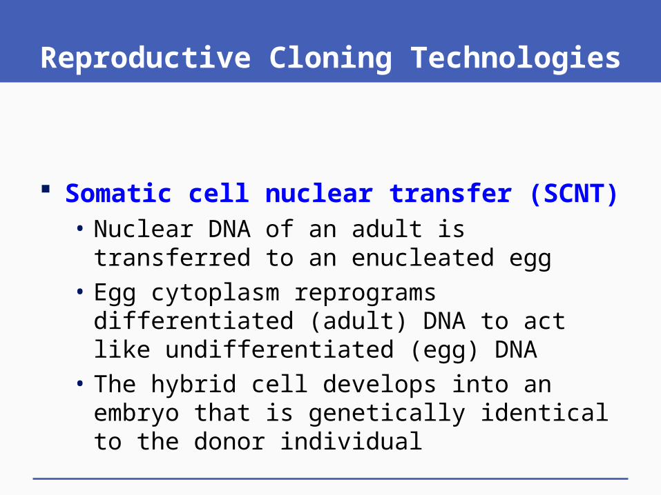
Reproductive Cloning Technologies
Somatic cell nuclear transfer (SCNT)• Nuclear DNA of an adult is transferred to an
enucleated egg• Egg cytoplasm reprograms differentiated (adult)
DNA to act like undifferentiated (egg) DNA• The hybrid cell develops into an embryo that is
genetically identical to the donor individual
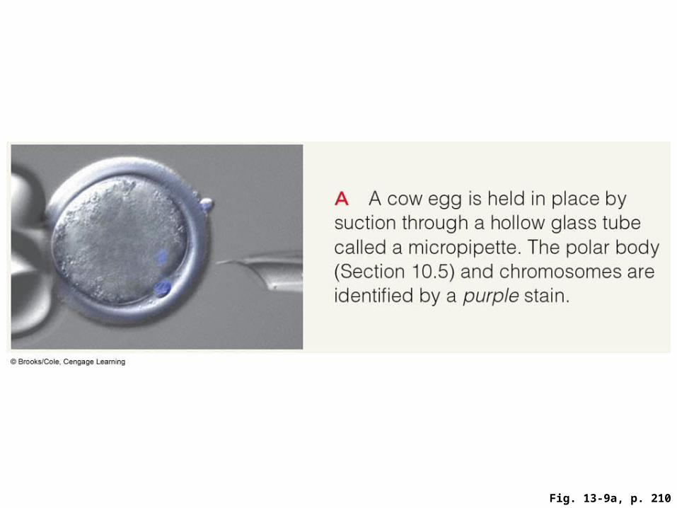
Fig. 13-9a, p. 210

Fig. 13-9b, p. 210

Fig. 13-9c, p. 210
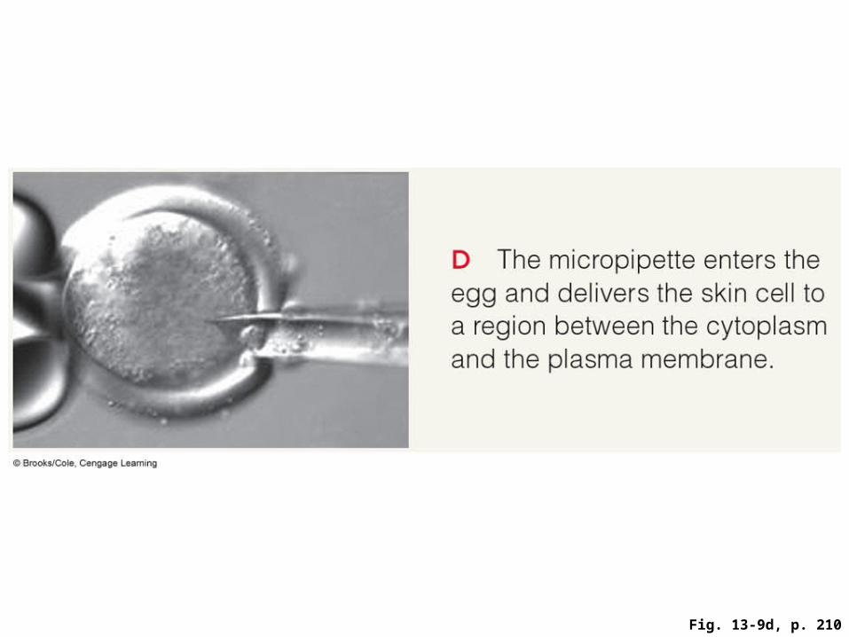
Fig. 13-9d, p. 210

Fig. 13-9e, p. 210

Fig. 13-9f, p. 210
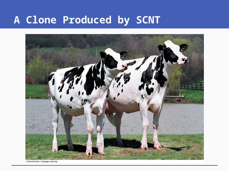
A Clone Produced by SCNT

Animation: How Dolly was created
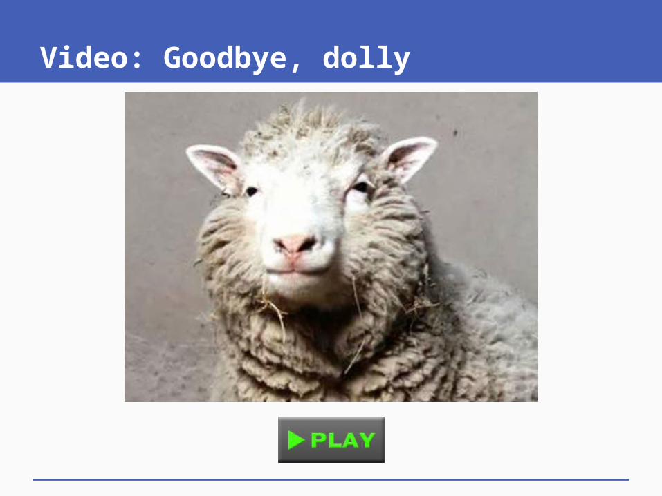
Video: Goodbye, dolly
Related Documents