DNA methylation does not stably lock gene expression but instead serves as a molecular mark for gene silencing memory Noël J.-M. Raynal 1,2 , Jiali Si 1 , Rodolphe F. Taby 1 , Vazganush Gharibyan 1 , Saira Ahmed 1 , Jaroslav Jelinek 1,2 , Marcos R.H. Estécio 1 , and Jean-Pierre J. Issa 1,2,* 1 Department of Leukemia, The University of Texas MD Anderson Cancer Center, 1515 Holcombe Blvd., Houston, TX 77030, USA 2 Fels Institute for Cancer Research and Molecular Biology, Temple University, Philadelphia, PA 19140, USA Abstract DNA methylation is commonly thought of as a "molecular lock" that leads to permanent gene silencing. To investigate this notion, we tested 24 different HDAC inhibitors (HDACi) on colon cancer cells that harbor a GFP locus stably integrated and silenced by a hypermethylated CMV promoter. We found that HDACi efficiently reactivated expression of GFP and many other endogenous genes silenced by DNA hypermethylation. After treatment, all promoters were marked with active chromatin, yet DNA hypermethylation did not change. Thus, DNA methylation could not prevent gene reactivation by drug-induced resetting of the chromatin state. In evaluating the relative contribution of DNA methylation and histone modifications to stable gene silencing, we followed expression levels of GFP and other genes silenced by DNA hypermethylation over time after treatment with HDACi or DNA demethylating drugs. Reactivation of methylated loci by HDACi was detectable for only 2 weeks, whereas DNA demethylating drugs induced permanent epigenetic reprogramming. Therefore, DNA methylation cannot be considered as a lock for gene expression, but rather as a memory signal for long-term maintenance of gene silencing. These findings define chromatin as an important druggable target for cancer epigenetic therapy and suggest that removal of DNA methylation signals is required to achieve long-term gene reactivation. Introduction Epigenetic marks such as histone modifications and DNA methylation are involved in cell memory expression patterns which are transmitted through cell division (1). Chromatin modifications are required in all organisms while DNA methylation is not present in some lower organisms like worms and flies, suggesting that chromatin has a broader epigenetic function in gene regulation (1). Histone acetylation is associated with open chromatin and gene expression while removal of acetyl groups by histone deacetylases is observed in inactive chromatin. In higher organisms, DNA methylation plays an important role in several physiological processes, including X chromosome inactivation, genomic imprinting, silencing of germ cell specific genes and repetitive elements (2). In cancer, tumor suppressor genes (TSG) are silenced by both DNA hypermethylation and chromatin repressive marks (2, 3). * Correspondance: Jean-Pierre J. Issa, Fels Institute for Cancer Research and Molecular Biology, Temple University, Philadelphia, PA 19140, USA. [email protected]. Phone: 215-707-1454. NIH Public Access Author Manuscript Cancer Res. Author manuscript; available in PMC 2013 March 1. Published in final edited form as: Cancer Res. 2012 March 1; 72(5): 1170–1181. doi:10.1158/0008-5472.CAN-11-3248. NIH-PA Author Manuscript NIH-PA Author Manuscript NIH-PA Author Manuscript

Welcome message from author
This document is posted to help you gain knowledge. Please leave a comment to let me know what you think about it! Share it to your friends and learn new things together.
Transcript

DNA methylation does not stably lock gene expression butinstead serves as a molecular mark for gene silencing memory
Noël J.-M. Raynal1,2, Jiali Si1, Rodolphe F. Taby1, Vazganush Gharibyan1, Saira Ahmed1,Jaroslav Jelinek1,2, Marcos R.H. Estécio1, and Jean-Pierre J. Issa1,2,*
1Department of Leukemia, The University of Texas MD Anderson Cancer Center, 1515 HolcombeBlvd., Houston, TX 77030, USA2Fels Institute for Cancer Research and Molecular Biology, Temple University, Philadelphia, PA19140, USA
AbstractDNA methylation is commonly thought of as a "molecular lock" that leads to permanent genesilencing. To investigate this notion, we tested 24 different HDAC inhibitors (HDACi) on coloncancer cells that harbor a GFP locus stably integrated and silenced by a hypermethylated CMVpromoter. We found that HDACi efficiently reactivated expression of GFP and many otherendogenous genes silenced by DNA hypermethylation. After treatment, all promoters weremarked with active chromatin, yet DNA hypermethylation did not change. Thus, DNAmethylation could not prevent gene reactivation by drug-induced resetting of the chromatin state.In evaluating the relative contribution of DNA methylation and histone modifications to stablegene silencing, we followed expression levels of GFP and other genes silenced by DNAhypermethylation over time after treatment with HDACi or DNA demethylating drugs.Reactivation of methylated loci by HDACi was detectable for only 2 weeks, whereas DNAdemethylating drugs induced permanent epigenetic reprogramming. Therefore, DNA methylationcannot be considered as a lock for gene expression, but rather as a memory signal for long-termmaintenance of gene silencing. These findings define chromatin as an important druggable targetfor cancer epigenetic therapy and suggest that removal of DNA methylation signals is required toachieve long-term gene reactivation.
IntroductionEpigenetic marks such as histone modifications and DNA methylation are involved in cellmemory expression patterns which are transmitted through cell division (1). Chromatinmodifications are required in all organisms while DNA methylation is not present in somelower organisms like worms and flies, suggesting that chromatin has a broader epigeneticfunction in gene regulation (1). Histone acetylation is associated with open chromatin andgene expression while removal of acetyl groups by histone deacetylases is observed ininactive chromatin. In higher organisms, DNA methylation plays an important role inseveral physiological processes, including X chromosome inactivation, genomic imprinting,silencing of germ cell specific genes and repetitive elements (2). In cancer, tumor suppressorgenes (TSG) are silenced by both DNA hypermethylation and chromatin repressive marks(2, 3).
*Correspondance: Jean-Pierre J. Issa, Fels Institute for Cancer Research and Molecular Biology, Temple University, Philadelphia, PA19140, USA. [email protected]. Phone: 215-707-1454.
NIH Public AccessAuthor ManuscriptCancer Res. Author manuscript; available in PMC 2013 March 1.
Published in final edited form as:Cancer Res. 2012 March 1; 72(5): 1170–1181. doi:10.1158/0008-5472.CAN-11-3248.
NIH
-PA Author Manuscript
NIH
-PA Author Manuscript
NIH
-PA Author Manuscript

A common hypothesis is that DNA methylation serves as a «molecular lock» that preventsreprogramming and is responsible for stable gene silencing (1, 4, 5). This concept was builton indirect observations whereby hypermethylated genes in cancer cells could be reactivatedonly after removal of promoter DNA hypermethylation using hypomethylating drugs such asdecitabine (5-aza-2’-deoxycytidine, 5-AZA-CdR). By contrast chromatin acetylationinduced by histone deacetylase inhibitors (HDACi) such as Trichostatin A (TSA) could notreactivate hypermethylated genes in cancer cells (6–8). However, more recently somereports have shown that HDACi such as TSA and depsipeptide (Depsi) produce genereactivation from hypermethylated promoters without any changes in DNA methylation atthe promoter level (9–12). Since these reports were against the current paradigm, a moredetailed look at this issue was needed.
One of the problems in studying DNA methylation-associated silencing of TSG is thatreactivation of these genes can impair cellular growth and be difficult to detect andquantitate. A selectable system was recently described to overcome this issue. YB5 cells arederived from the SW48 colon cancer cell line transfected with a green fluorescent protein(GFP) driven by a hypermethylated cytomegalovirus (CMV) promoter packaged intoinactive chromatin. YB5 carries a single copy of CMV/GFP stably integrated inchromosome 1. GFP can be reactivated in YB5 cells by treatment with 5-AZA-CdR when itspromoter region is demethylated and also marked by active chromatin signals characterizedby H3K9 acetylation, low level of H3K27 trimethylation and low nucleosome density (13).
In this paper, we use YB5 cells (and 5 other cancer cell lines) to show that the vast majorityof HDACi tested can reactivate genes silenced by promoter hypermethylation withoutdetectable changes in DNA methylation. We further show that while DNA methylationcannot prevent gene activation by chromatin reprogramming, it is essential for long-termgene silencing.
Materials and MethodsCell culture and drug treatments
All cell lines were obtained from American Type Culture Collection. Cell lines wereauthenticated at MD Anderson Cancer Center with short tandem repeat PCR method. YB5cell line is a colon cancer cell line generated from SW48 as previously described (13). YB5cell line was cultured in L-15 medium while MCF-7, K562, MDA-MD-231, and PC-3 cellswere cultured in RPMI 1640. Both cell culture media contained 10% fetal bovine serum and100 µg/mL Penicillin-Streptomycin solution. Cells growing in log phase were treated withdecitabine (5-AZA-CdR) at 50 nM for 72h. Drug and medium were replaced every day.Cells were cultured an additional 24h without drug prior to analysis. HDAC inhibitors(HDACi) were dissolved either in DMSO, ethanol or PBS according the manufacturers’recommendations. HDACi were added for 24h at various concentrations prior to analysis.
Histone extraction and Western blotsTotal protein extracts were prepared using RIPA buffer as described previously (14) in thepresence of sodium butyrate (5 mM) to avoid in vitro histone deacetylation and resolved on15% SDS-polyacrylamide gels. Antibodies used were H3K9-acetylation (07–352 Millipore),H4K16-acetylation (39168, Active motif) and total H3 (ab1791 Abcam).
Fluorescence-activated cell sorting analysis and cell sortingYB5 cells were trypsinized and stained with propidium iodide (PI). GFP and PI fluorescencewere measured by Gallios flow cytometer (Beckman Coulter). Data were analyzed using
Raynal et al. Page 2
Cancer Res. Author manuscript; available in PMC 2013 March 1.
NIH
-PA Author Manuscript
NIH
-PA Author Manuscript
NIH
-PA Author Manuscript
Kaluza® software. GFP cell sorting was performed using BD FACSAriaII. GFPfluorescence of samples was analyzed post-sorting to assess the purity of the sorted cells.
RNA extraction, cDNA synthesis, quantitative real-time PCRTotal RNA (2 µg) was extracted using Trizol (Invitrogen) and cDNA was synthesized usingHigh-Capacity cDNA Kit (Applied Biosystems). Quantification of cDNA was done byqPCR with the Universal PCR Master Mix (Bio-Rad) using ABI Prism 7900HT. Resultswere obtained from at least three independent experiments where each sample was analyzedin duplicate. 18S was used as a reference gene. cDNA synthesis used the same amount ofRNA after treatment with different drugs. All primers, except GFP primers that weredescribed previously (13), are listed in supplemental Table S1. 5′ Rapid amplification ofcDNA ends (5′-RACE) was done as previously described (13).
DNA extraction and DNA methylation analysisDNA extraction and bisulfite conversion, pyrosequencing and bisulfite cloning/sequencingwere carried out as previously described (13). All primers are listed in supplemental TableS1, except for all GFP primers that were described previously (13).
Chromatin immunoprecipitation (ChIP)ChIP was performed as described previously (14). Antibodies used were anti–histone H3(ab1791, Abcam), anti–histone H3K9 acetylation (07–352, Millipore), anti–histone H3K27trimethylation (07–449, Millipore), anti-histone H3K4 trimethylation (17–614, Millipore),anti-histone H3K36 trimethylation (ab9050, Abcam) and anti-IgG (ab46540, Abcam).Quantification of ChIP DNA was done by qPCR, and primers/probes are listed insupplemental Table S1. Each ChIP assay was validated using targets for the variousmodifications (ACTB for active histone mark and LINE-1 for inactive histone mark). Thevalue of each histone modification was determined by H3 and IgG normalization using theequation: Fold enrichment = 2^-[Ct(Ab) − Ct(H3)] – 2^-[Ct(IgG) − Ct(H3)].
Gene expression and DNA methylation microarraysGene expression analysis was performed using the Agilent whole genome array (G4112F)that was scanned using the Agilent G2505B scanner. Data represents the average expressionlevel of two independent experiments. DNA methylation analysis using high-throughputmethylation profiling by MCA coupled to CpG island microarray (MCAM) was performedas described previously (15). After analysis, genes with M values more than 1.3 wereconsidered methylated. Microarray data sets were deposited in the Gene ExpressionOmnibus (GEO) database with the accession number GSE34077.
Statistical analysisDifferences between groups were assessed using a t-test. A 2-sided P value of 0.05 wasconsidered statistically significant.
ResultsHDAC inhibitors induce GFP reactivation from a DNA hypermethylated promoter
To examine the effects of HDACi on gene silencing by DNA methylation, YB5 cells weretreated with 24 different HDACi that belong to 8 different chemical classes in a wide rangeof concentrations (Table 1) and 17 of them reactivated GFP. Although GFP reactivation wasdetected in a wide range of concentrations (even at high doses, Table 1), the subsequentexperiments were performed at doses where the percentage of dead cells after treatment wasless than 30% and where GFP reactivation was the highest as detected by FACS analysis.
Raynal et al. Page 3
Cancer Res. Author manuscript; available in PMC 2013 March 1.
NIH
-PA Author Manuscript
NIH
-PA Author Manuscript
NIH
-PA Author Manuscript

Following treatment with depsipeptide (Depsi, for 24h at 20 nM), GFP expression wasdetectable in ~50% of cells as seen by fluorescent microscopy (Fig. 1A), quantified byFACS analysis (Fig. 1B), and associated with extensive global histone acetylation (Fig. 1C).HDACi produced GFP mRNA and GFP fluorescence as early as 12h after treatment (FigureS1 A–B). GFP reactivation was not specific to a molecular structure or chemical class ofthese epigenetic drugs since the vast majority of HDACi tested activated thishypermethylated locus (Fig. 1D and Table 1). Moreover, GFP fluorescence and mRNAlevels were stronger after a 24h treatment with HDACi than after a 72h treatment with 5-AZA-CdR (Fig. 1D–E). We confirmed by 5’RACE experiments that GFP mRNA originatedfrom its own promoter and not from an alternative transcription start site (data not shown).
It has previously been suggested that HDACi can lead to DNA demethylation (16–18). Totest this, DNA methylation levels were measured after treatment with 7 different HDACiand 5-AZA-CdR was used as a control for DNA hypomethylation. Analyses were performedby bisulfite pyrosequencing and cloning/sequencing at the GFP promoter (respectively Fig.2A–B). No changes were detected after treatment with any of the HDACi tested after 24htreatment. Similarly, there were no effects on global DNA methylation assessed by bisulfitepyrosequencing of LINE-1 methylation after 24h treatment (Fig. 2C) or 10 days followingtreatment (Fig. 2D). Only treatment with 5-AZA-CdR reduced DNA methylation levels.These results and others (19) clearly show that HDACi do not alter DNA methylation levelsof cancer cells. Thus, HDACi can induce gene reactivation through a DNA hypermethylatedpromoter without any change in DNA methylation levels. These results do not support thelock hypothesis and are in agreement with more recent findings demonstrating that HDACican reactivate hypermethylated genes.
HDACi induce gene reactivation of endogenous genes silenced by promoter DNAhypermethylation
Since these data are not in agreement with other studies on gene reactivation induced byHDACi (6, 7), we asked whether this effect was specific to the GFP locus or could beobserved in other methylated genes in various cancer cell lines. First, we analyzed in YB5cells gene reactivation of other hypermethylated genes in response to Depsi (Fig. 3A) andother HDACi (Fig. S2A–B). For this, we selected 7 TSG silenced by DNAhypermethylation in YB5 cells (85–100% methylation as detected by bisulfitepyrosequencing). These play roles in mediating cell adhesion (CDH13), metastasis(TIMP-3), cell cycle (P16), DNA mismatch repair (MLH1), Wnt pathway signaling (WIF-1),MAP kinase signaling (DOK5), and cell differentiation (RAR-β). Among these, all but one(RAR-β) are driven by promoter CpG Islands (CGI) (20). These genes are epigeneticallyinactivated in many cancers. 24h treatment with Depsi and other HDACi reactivated allthese hypermethylated genes as detected by qPCR (Fig. 3A and Fig. S2A–B), while DNAmethylation levels did not change as compared to untreated cells (Fig. 3B). These resultswere extended to four other cancer cell lines (K562, a CML cell line; MCF-7 and MDA-MB-231, breast cancer cell lines; and PC-3, a prostate cancer cell line) with six differentgenes (CDH1, MGMT, NPM2, RASSF1A, DOK5, and PDLIM4; Fig. S3 A–D) whosepromoter methylation levels vary between 65 and 100% methylation as detected bypyrosequencing. Most of them showed reactivation after HDACi treatment. The induction ofp21, a cyclin-dependent kinase inhibitor, by HDACi was used as a positive control forHDACi activity since it is considered as a hallmark of the effect of HDACi on geneexpression. Our data are in agreement with previous reports on single genes that HDACisuch as TSA, phenylbutyrate and LBH589 (9, 11, 12, 21, 22) or SIRT1 inhibition (10) caninduce gene reactivation of hypermethylated genes without alteration in DNA methylation.Our findings on numerous genes and in different cancer cell lines demonstrate thatchromatin acetylation induced by HDACi overcomes DNA hypermethylation silencing and
Raynal et al. Page 4
Cancer Res. Author manuscript; available in PMC 2013 March 1.
NIH
-PA Author Manuscript
NIH
-PA Author Manuscript
NIH
-PA Author Manuscript

induces gene reactivation. All together, these data demonstrate that chromatin remodelingallows a subset of TSG silenced by DNA hypermethylation to be reactivated in response toHDACi.
Chromatin remodeling despite DNA hypermethylationSince HDACi-induced gene reactivation was not associated with DNA demethylation, weinvestigated the effect of the treatment on chromatin modifications at the promoter regionsof these hypermethylated genes. We performed chromatin immunoprecipitation assays(ChIP) coupled with qPCR analysis for H3K9-Ac, H3K4-me3, H3K36-me3 (marksassociated with gene activity), and H3K27-me3 (modification associated with generepression) in YB5 cells untreated and treated with Depsi for 24h at 20 nM (Fig. 4A–D).Interestingly, we recently reported that regardless of DNA methylation status, genereactivation is associated with a promoter region marked by active chromatin marks and lownucleosome density (13). Following Depsi treatment, all promoter regions of GFP, CDH13,MLH1, and WIF-1 showed an increase in activating marks (1.5 to 24.5 fold) such as H3K9-Ac (Fig. 4A), H3K4-me3 (Fig. 4B), and H3K36-me3 (Fig. 4C). By contrast, H3K27-me3, asurrogate for chromatin inactivated by polycomb, was reduced after Depsi treatment by 1.4to 4 fold (Fig. 4D). Interestingly, Depsi treatment did not seem to have modified nucleosomedensity (measured by ChIP-PCR of H3/input) on the promoter region of these genes (datanot shown). To rule out indirect effects through other chromatin regulators, we measured theexpression of DNMT1, DNMT3a, DNMT3b, DNMT3L, DROSHA, DICER, TET1, TET2, andEZH2. We found that most did not change significantly after Depsi treatment (Fig. 3A).These data show that HDACi directly induced chromatin remodeling and this was associatedwith gene reactivation from DNA hypermethylated promoters.
Genome wide effects of Depsipeptide on hypermethylated genesWe next investigated on a genomic scale the effect of Depsi on gene reactivation ofhypermethylated genes. We combined the data of two independent gene expressionmicroarrays of untreated and Depsi-treated YB5 cells (Fig. S4A) with whole genome DNAmethylation data using Methylation CpG island microarray (MCAM, Fig. S4B) (15). Aspreviously reported by other groups (23, 24), we found that HDACi increased the expressionof 11% of the genes with the same amount of genes being repressed (11%; Fig. S4D).Whole genome methylation data showed that this colon cancer cell line has more than 330detectable hypermethylated promoter CGIs (i.e, 6% of the gene promoters on the array; Fig.S4B). When combining the data of both gene expression and DNA methylation microarrays,we could analyze gene expression and DNA methylation of more than 4,300 genes (Fig.S4C). In this gene list, 258 genes showed promoter DNA hypermethylation and 8% (21genes) of these were reactivated following HDACi treatment (Fig. S4E–F). Considering that80–90% of hypermethylated genes in cancer are not expressed in normal tissues and thuslack the appropriate transcription factor for activation, our data suggest that the majority of‘inducible’ genes are actually induced by HDACi (25). By contrast 14% of the unmethylatedgenes could be reactivated by treatment with Depsi.
DNA methylation is a long-term memory signal for gene silencingThe data described so far demonstrate that rapid activation of a DNA hypermethylatedpromoter is possible with strong drug-induced chromatin acetylation. These results raise thequestion of the importance of DNA methylation in gene silencing. To study the relativecontribution of DNA methylation versus chromatin modifications in gene silencing, wecompared the long-term effects of Depsi and 5-AZA-CdR treatment on gene expression andDNA methylation. YB5 cells were treated with Depsi (24h) or 5-AZA-CdR (72h) and werethen subjected to cell sorting to obtain enriched GFP+ cell populations (purity ~70% of GFP+ cells for each treatment conditions). GFP positive cells were cultured post-sorting without
Raynal et al. Page 5
Cancer Res. Author manuscript; available in PMC 2013 March 1.
NIH
-PA Author Manuscript
NIH
-PA Author Manuscript
NIH
-PA Author Manuscript

drugs and GFP expression was followed for more than 3 months by FACS analysis (Fig.5A). During the first week post-sorting, the population of Depsi-treated YB5 cells wasmostly GFP+. Ten days post-sorting, approximately 60% of the cells treated with Depsi lostGFP expression and 2 weeks post-treatment, GFP expression was rare among these cells.These data were confirmed with other HDACi such as VPA, Apicidin, Cay 10398, and TSA(data not shown). GFP expression was undetectable 25 days following Depsi-treatment andwas similar to untreated cells for the rest of the experiment.
Results obtained with YB5 cells treated with 5-AZA-CdR exhibited a different pattern (Fig.5A). For the first week, the vast majority of the cells exhibited GFP fluorescence. Then, thepercentage of cells showing GFP fluorescence decreased to 50% and 35% after 10 days and25 days post-treatment, respectively. The number of GFP+ decreased slowly thereafter andafter 3 months, 3% of YB5 cells treated with 5-AZA-CdR still exhibited GFP fluorescence(Fig. 5A). We similarly investigated the RNA expression of several hypermethylated genesincluding GFP (Fig. 5B), MLH1 (Fig. 5C), CDH13 (Fig. S5A), WIF-1 (Fig. S5B), andTIMP-3 (Fig. S5C) and found that gene expression was activated immediately by treatmentwith either epigenetic drug, but expression was silenced 2 weeks after Depsi treatment whileit was maintained following treatment with 5-AZA-CdR for up to 9 weeks. Interestingly,other chromatin modifiers such as histone methyltransferase inhibitors were previouslyshown to induce transient gene activation which returned to the original state upon drugremoval (26). As previously mentioned, after Depsi treatment, DNA methylation in thepromoter regions of these hypermethylated genes did not change. By contrast, after 5-AZA-CdR treatment, methylation levels decreased significantly at GFP (Fig. 5D), MLH1 (Fig.5E), CDH13 (Fig. S5D), WIF-1 (Fig. S5E), TIMP-3 (Fig. S5F), and LINE-1 (Fig. S5G).
The gradual loss of GFP expression after 5-AZA-CdR withdrawal was coincided withgradual remethylation of the CMV promoter. Although it has been reported that thep16CDKN2A/INK4 locus was remethylated after 5-AZA-CdR removal, it has been suggestedthat the apparent remethylation was due to clonal replacement by a subset of cancer cellsthat were not affected by 5-AZA-CdR (19, 27). Cells treated with hypomethylating agentstend to have a longer cell cycle because of the reexpression of growth regulatory signals.Therefore, hypomethylated cell populations can be easily replaced by more rapidly growingmethylated populations, which can bias the measurement of DNA methylation (19). Toaddress this issue, we performed two series of cell sorting experiments using YB5 cellscultured 9 weeks without drug following initial 5-AZA-CdR treatment (Fig. 6A). The purityof the sorted GFP+ cells exceeded 90% while the GFP− cells contained only 0.2% GFPpositive cells. Gene expression and DNA methylation analysis of GFP+ and GFP− cells at 9weeks post-treatment revealed that gene reactivation in GFP+ (Fig. 6B) was at this late timepoint associated with an unmethylated promoter region with an average methylation of 15%(Fig. 6C). GFP− cells exhibited 1000 times less GFP mRNA and a promoter closer to themethylation level of untreated cells with an average methylation of 66% (Fig. 6B–C). Thisgene expression pattern was also observed for other TSG such as TIMP-3, CDH13, andMLH1 while their DNA methylation level was reduced in the GFP+ and GFP− cells (FigureS6A–F). Global DNA methylation measured by the LINE-1 assay did not changesignificantly between untreated cells, GFP+ and GFP− cells (Figure S6G).
In order to eliminate the effect of clonal replacement, we performed cell sorting and singlecloning experiments (Fig. 6D). After clonal expansion of these single clones to obtainenough cells (~2 weeks), we monitored their GFP fluorescence over time (black bars, Fig.6D) as compared to sorted cells obtained after 5-AZA-CdR treatment when the purity was~70% (white bars, Fig. 6D) and 9 weeks after treatment when the purity exceeded 90%(grey bars, Fig. 6D). Single cell clones of these GFP+ YB5 cells obtained 9 weeks after 5-AZA-CdR without drugs revealed that 92–97% stably express GFP for up to 6 months post-
Raynal et al. Page 6
Cancer Res. Author manuscript; available in PMC 2013 March 1.
NIH
-PA Author Manuscript
NIH
-PA Author Manuscript
NIH
-PA Author Manuscript

treatment demonstrating stable epigenetic reprogramming (Fig. 6D). Interestingly, the GFPpromoter region was completely demethylated in these GFP+ clones (Fig. 6C). These resultsclearly show that DNA methylation is the molecular mechanism responsible for long-termgene silencing. Thus, full epigenetic reprogramming and switching from the silent to theexpressed state can be accomplished by complete promoter demethylation which iscorrelated with RNA pol II occupancy (28).
DiscussionEarly studies have reported that TSG silenced by promoter DNA hypermethylation could bereactivated only after the removal of methylation marks. In these studies, treatment withTSA, an HDACi, could not produce gene reactivation of genes silenced by promoter DNAhypermethylation (6, 7). Based on these indirect observations, the function of promoterDNA methylation, as a signal for gene silencing, has been considered as a ‘lock’ for geneexpression. However, other studies have reported that hypermethylated genes can bereactivated by TSA and other HDACi without any loss in promoter DNA methylation (9–12). These reports put in jeopardy the lock hypothesis which has been the paradigm for morethan a decade. Hence, we chose to investigate this issue by looking at the effects of morethan 20 different HDACi on reactivation of genes silenced by promoter DNA methylation.
Using the well-characterized YB5 system and other cancer cell lines, we discovered thatmost of the HDACi-tested could reactivate hypermethylated genes in a dose-dependentpattern regarding their chemical class and HDAC affinity. DNA methylation analysisrevealed that gene reactivation was generated without any loss of promoter DNAmethylation. Methylation levels were carefully assessed before and after treatment bypyrosequencing and bisulfite cloning sequencing since it was reported that HDACi couldpotentially reduce DNA methylation levels by non-specific mechanisms (16–18). However,our study, as well as others (19) show that methylation levels did not change 24h afterHDACi exposure or several days post-treatment. Therefore, these data confirm that HDACican reactivate gene expression through hypermethylated promoters, which demonstrates thatDNA methylation does not lock gene expression in that it does not prevent reactivation bychromatin remodeling. It is not clear why previous studies reported that HDACi do notreactivate the expression of hypermethylated genes, though it may relate to the use of lowdoses of HDACi for short periods of time, and the use of insensitive methods for geneexpression analysis. Alternately, it is possible that some genes/cell lines are resistant to thiseffect, though we observed it for most genes and most cell lines tested.
The fact that DNA methylation does not lock gene expression raises the question of therelative contribution of DNA methylation and chromatin modifications to gene silencing.The YB5 system was particularly suitable to investigate this issue since after treatment witheither Depsi or with 5-AZA-CdR, we were able to sort the GFP-expressing cells and monitorGFP fluorescence for several months. We discovered that a treatment with HDACi cantransiently reactivate hypermethylated genes (GFP and other TSG) for up to 2 weekswithout any changes in DNA methylation level in their promoter regions. On the other hand,treatment with 5-AZA-CdR leads to gene reactivation of GFP and other TSG for severalmonths. Moreover, the decline in GFP-expressing cells after 5-AZA-CdR, thought largely tobe due to remethylation, is in fact attributable in part to clonal replacement by YB5 cells thatare methylated and do not express GFP. Indeed, cell sorting and single cell cloning 9 weeksafter drug removal led to clones where the promoter region was completely demethylated,and expression permanently on. Thus, efficient demethylation leads to permanent genereactivation, showing that DNA methylation provides a memory signal for the silent state.
Raynal et al. Page 7
Cancer Res. Author manuscript; available in PMC 2013 March 1.
NIH
-PA Author Manuscript
NIH
-PA Author Manuscript
NIH
-PA Author Manuscript
Our data show that the respective roles of DNA methylation and chromatin remodeling canbe completely separated using the YB5 selectable system. The chromatin state determinesthe immediate gene expression potential, while DNA methylation provides a long-termmemory for gene silencing. Thus, DNA methylation does not provide a ‘lock’ function aspreviously thought, because gene expression can be restored by drug-induced chromatinmodifications without any DNA demethylation (i.e. without breaking the lock). Rather,DNA methylation provides a ‘spring’ function, which does not suppress gene expression butbrings back silencing, presumably through the previously defined order of events: methyl-binding protein recruitment, histone deacetylation, histone methylation, HP1 binding and soon (3). This explains why physiologically, DNA methylation at promoter CGIs is onlyinvolved when very long-term silencing is required, and why it provides such a selectiveadvantage to cancer cells when TSG are silenced by this mechanism (3). Interestingly, aftertreatment with HDACi, gene expression is not sufficient to lead to permanent expressionand DNA demethylation. It is possible that gene reactivation induced by HDACi may becaused by either i) bypassing transcription factors, whereby histone acetylation will directlytrigger RNA pol II activation leading to reactivation or ii) transient binding of transcriptionfactors to promoter regions, with gene silencing rapidly restored by repressive signalsarising from DNA methylation. Restoring a silenced state is likely when histones arereplaced during cell divisions in the face of persistent DNA methylation. Importantly afterthe treatment with hypomethylating drugs, it has been previously demonstrated that removalof DNA methylation marks will allow the binding of transcription factors leading topermanent epigenetic resetting promoting the emergence of stably reactivated clones. Thiswas shown by 5-AZA-CdR-induced DNA demethylation in YB5 cells where the CREBtranscription factor bound only the CMV promoter only when it was hypomethylated (13).
These data have implications for therapeutic intervention. We show that genes silenced byDNA hypermethylation in cancer can be significantly but transiently reactivated throughchromatin remodeling without any changes in DNA methylation, and this may be part of theclinical mechanisms of action of HDACi. Moreover, our results provide a molecularexplanation for the synergy between decitabine and HDACi (6, 29) in which thecombination induces more complete epigenetic reprogramming. Finally, while thesefindings validate chromatin as a key target for therapeutic intervention in cancer, they alsosuggest that stable reprogramming may require the removal of DNA methylation signals.
Supplementary MaterialRefer to Web version on PubMed Central for supplementary material.
AcknowledgmentsThis work was supported by National Institutes of Health grants CA100632, CA121104, and a grant from the StandUp to Cancer foundation. JPI is an American Cancer Society Clinical Research professor supported by a generousgift from the F. M. Kirby Foundation. DNA sequencing and Flow Cytometry in the respective cores at the M. D.Anderson Cancer Center is supported by Core Grant CA16672 from the National Institutes of Health.
References1. Bird A. DNA methylation patterns and epigenetic memory. Genes & evelopment. 2002; 16:6–21.2. Sharma S, Kelly TK, Jones PA. Epigenetics in cancer. Carcinogenesis. 2010; 31:27–36. [PubMed:
19752007]3. Taby R, Issa JP. Cancer Epigenetics. CA: a cancer journal for clinicians. 20104. Siegfried Z, Cedar H. DNA methylation: a molecular lock. Curr Biol. 1997; 7:R305–R307.
[PubMed: 9115385]
Raynal et al. Page 8
Cancer Res. Author manuscript; available in PMC 2013 March 1.
NIH
-PA Author Manuscript
NIH
-PA Author Manuscript
NIH
-PA Author Manuscript

5. Cedar H, Bergman Y. Linking DNA methylation and histone modification: patterns and paradigms.Nat Rev Genet. 2009; 10:295–304. [PubMed: 19308066]
6. Cameron EE, Bachman KE, Myohanen S, Herman JG, Baylin SB. Synergy of demethylation andhistone deacetylase inhibition in the re-expression of genes silenced in cancer. Nature genetics.1999; 21:103–107. [PubMed: 9916800]
7. Suzuki H, Gabrielson E, Chen W, et al. A genomic screen for genes upregulated by demethylationand histone deacetylase inhibition in human colorectal cancer. Nature genetics. 2002; 31:141–149.[PubMed: 11992124]
8. Meng CF, Zhu XJ, Peng G, Dai DQ. Promoter histone H3 lysine 9 di-methylation is associated withDNA methylation and aberrant expression of p16 in gastric cancer cells. Oncology reports. 2009;22:1221–1227. [PubMed: 19787243]
9. Fujii S, Luo RZ, Yuan J, et al. Reactivation of the silenced and imprinted alleles of ARHI isassociated with increased histone H3 acetylation and decreased histone H3 lysine 9 methylation.Hum Mol Genet. 2003; 12:1791–1800. [PubMed: 12874100]
10. Pruitt K, Zinn RL, Ohm JE, et al. Inhibition of SIRT1 reactivates silenced cancer genes withoutloss of promoter DNA hypermethylation. PLoS genetics. 2006; 2:e40. [PubMed: 16596166]
11. Bovenzi V, Momparler RL. Antineoplastic action of 5-aza-2'-deoxycytidine and histonedeacetylase inhibitor and their effect on the expression of retinoic acid receptor beta and estrogenreceptor alpha genes in breast carcinoma cells. Cancer chemotherapy and pharmacology. 2001;48:71–76. [PubMed: 11488527]
12. Yang X, Ferguson AT, Nass SJ, et al. Transcriptional activation of estrogen receptor alpha inhuman breast cancer cells by histone deacetylase inhibition. Cancer research. 2000; 60:6890–6894.[PubMed: 11156387]
13. Si J, Boumber YA, Shu J, et al. Chromatin remodeling is required for gene reactivation afterdecitabine-mediated DNA hypomethylation. Cancer research. 2010; 70:6968–6977. [PubMed:20713525]
14. Kondo Y, Shen L, Cheng AS, et al. Gene silencing in cancer by histone H3 lysine 27trimethylation independent of promoter DNA methylation. Nature genetics. 2008; 40:741–750.[PubMed: 18488029]
15. Estecio MR, Yan PS, Ibrahim AE, et al. High-throughput methylation profiling by MCA coupledto CpG island microarray. Genome research. 2007; 17:1529–1536. [PubMed: 17785535]
16. Xiong Y, Dowdy SC, Podratz KC, et al. Histone deacetylase inhibitors decrease DNAmethyltransferase-3B messenger RNA stability and down-regulate de novo DNAmethyltransferase activity in human endometrial cells. Cancer research. 2005; 65:2684–2689.[PubMed: 15805266]
17. Wu LP, Wang X, Li L, et al. Histone deacetylase inhibitor depsipeptide activates silenced genesthrough decreasing both CpG and H3K9 methylation on the promoter. Molecular and cellularbiology. 2008; 28:3219–3235. [PubMed: 18332107]
18. You JS, Kang JK, Lee EK, et al. Histone deacetylase inhibitor apicidin downregulates DNAmethyltransferase 1 expression and induces repressive histone modifications via recruitment ofcorepressor complex to promoter region in human cervix cancer cells. Oncogene. 2008; 27:1376–1386. [PubMed: 17828306]
19. Bender CM, Pao MM, Jones PA. Inhibition of DNA methylation by 5-aza-2'-deoxycytidinesuppresses the growth of human tumor cell lines. Cancer research. 1998; 58:95–101. [PubMed:9426064]
20. Takai D, Jones PA. Comprehensive analysis of CpG islands in human chromosomes 21 and 22.Proceedings of the National Academy of Sciences of the United States of America. 2002;99:3740–3745. [PubMed: 11891299]
21. Sirchia SM, Ren M, Pili R, et al. Endogenous reactivation of the RARbeta2 tumor suppressor geneepigenetically silenced in breast cancer. Cancer research. 2002; 62:2455–2461. [PubMed:11980632]
22. Zhou Q, Atadja P, Davidson NE. Histone deacetylase inhibitor LBH589 reactivates silencedestrogen receptor alpha (ER) gene expression without loss of DNA hypermethylation. Cancerbiology & herapy. 2007; 6:64–69.
Raynal et al. Page 9
Cancer Res. Author manuscript; available in PMC 2013 March 1.
NIH
-PA Author Manuscript
NIH
-PA Author Manuscript
NIH
-PA Author Manuscript
23. Drummond DC, Noble CO, Kirpotin DB, Guo Z, Scott GK, Benz CC. Clinical development ofhistone deacetylase inhibitors as anticancer agents. Annual review of pharmacology andtoxicology. 2005; 45:495–528.
24. Peart MJ, Smyth GK, van Laar RK, et al. Identification and functional significance of genesregulated by structurally different histone deacetylase inhibitors. Proceedings of the NationalAcademy of Sciences of the United States of America. 2005; 102:3697–3702. [PubMed:15738394]
25. Sproul D, Nestor C, Culley J, et al. Transcriptionally repressed genes become aberrantlymethylated and distinguish tumors of different lineages in breast cancer. Proceedings of theNational Academy of Sciences of the United States of America. 2011; 108:4364–4369. [PubMed:21368160]
26. Miranda TB, Cortez CC, Yoo CB, et al. DZNep is a global histone methylation inhibitor thatreactivates developmental genes not silenced by DNA methylation. Molecular cancer therapeutics.2009; 8:1579–1588. [PubMed: 19509260]
27. Egger G, Aparicio AM, Escobar SG, Jones PA. Inhibition of histone deacetylation does not blockresilencing of p16 after 5-aza-2'-deoxycytidine treatment. Cancer research. 2007; 67:346–353.[PubMed: 17210717]
28. Kagey JD, Kapoor-Vazirani P, McCabe MT, Powell DR, Vertino PM. Long-term stability ofdemethylation after transient exposure to 5-aza-2'-deoxycytidine correlates with sustained RNApolymerase II occupancy. Mol Cancer Res. 2010; 8:1048–1059. [PubMed: 20587535]
29. Belinsky SA, Klinge DM, Stidley CA, et al. Inhibition of DNA methylation and histonedeacetylation prevents murine lung cancer. Cancer research. 2003; 63:7089–7093. [PubMed:14612500]
Raynal et al. Page 10
Cancer Res. Author manuscript; available in PMC 2013 March 1.
NIH
-PA Author Manuscript
NIH
-PA Author Manuscript
NIH
-PA Author Manuscript

Figure 1.Various HDAC inhibitors (HDACi) induce significant expression of genes silenced bypromoter DNA hypermethylation. A, Representative pictures by fluorescent microscopy andB, FACS analysis of untreated YB5 cells and treated with the HDACi depsipeptide (Depsi)at 20 nM for 24h. On the FACS scatter plot, the x-axis and the y-axis represent respectivelyGFP and propidium iodide (PI) fluorescence. C, Western blots show H3K9 and H4K16acetylation after a 24h treatment with sodium butyrate (SB) at 20 mM and Depsi at 20 nM.D, GFP protein expression detected by FACS analysis and E, GFP mRNA expressiondetected by qPCR after a 24h treatment using 12 different HDACi (n>3). HDACi thatbelong to the hydroxamic acid group are shown in white bars whereas the short-chain fattyacid and the cyclic peptide HDACi are respectively shown in grey and black bars. GFPexpression is also obtained after a treatment with 5-AZA-CdR, at 50 nM daily for 72hfollowed by 24h without drug, as a positive control. Each experiment was done threeseparate times: shown is the mean ± SEM.
Raynal et al. Page 11
Cancer Res. Author manuscript; available in PMC 2013 March 1.
NIH
-PA Author Manuscript
NIH
-PA Author Manuscript
NIH
-PA Author Manuscript
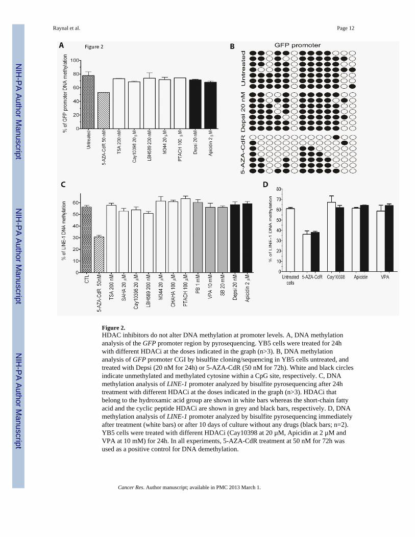
Figure 2.HDAC inhibitors do not alter DNA methylation at promoter levels. A, DNA methylationanalysis of the GFP promoter region by pyrosequencing. YB5 cells were treated for 24hwith different HDACi at the doses indicated in the graph (n>3). B, DNA methylationanalysis of GFP promoter CGI by bisulfite cloning/sequencing in YB5 cells untreated, andtreated with Depsi (20 nM for 24h) or 5-AZA-CdR (50 nM for 72h). White and black circlesindicate unmethylated and methylated cytosine within a CpG site, respectively. C, DNAmethylation analysis of LINE-1 promoter analyzed by bisulfite pyrosequencing after 24htreatment with different HDACi at the doses indicated in the graph (n>3). HDACi thatbelong to the hydroxamic acid group are shown in white bars whereas the short-chain fattyacid and the cyclic peptide HDACi are shown in grey and black bars, respectively. D, DNAmethylation analysis of LINE-1 promoter analyzed by bisulfite pyrosequencing immediatelyafter treatment (white bars) or after 10 days of culture without any drugs (black bars; n=2).YB5 cells were treated with different HDACi (Cay10398 at 20 µM, Apicidin at 2 µM andVPA at 10 mM) for 24h. In all experiments, 5-AZA-CdR treatment at 50 nM for 72h wasused as a positive control for DNA demethylation.
Raynal et al. Page 12
Cancer Res. Author manuscript; available in PMC 2013 March 1.
NIH
-PA Author Manuscript
NIH
-PA Author Manuscript
NIH
-PA Author Manuscript

Figure 3.HDACi reactivates endogenous hypermethylated genes in YB5 cell line. A, Gene expressionwas detected by qPCR of untreated (white bars) and treated YB5 cells with Depsi at 20 nMfor 24h (black bars). Tested genes were divided in three categories: control genes,hypermethylated genes and epigenetic regulators. A star indicates a significant differencebetween untreated and treated cells (p<0.05). Each experiment was done three separatetimes: shown is the mean ± SEM. B, DNA methylation analysis of TIMP-3, WIF-1 andCDH13 promoters analyzed by pyrosequencing. YB5 cells were treated withhypomethylating drug 5-AZA-CdR or with HDACi Depsi (n>3).
Raynal et al. Page 13
Cancer Res. Author manuscript; available in PMC 2013 March 1.
NIH
-PA Author Manuscript
NIH
-PA Author Manuscript
NIH
-PA Author Manuscript

Figure 4.Depsipeptide induces chromatin modifications on promoters of DNA hypermethylatedgenes. Chromatin-immunoprecipitation analysis of A, H3K9-Ac; B, H3K4-me3; C, H3K36-me3, and D, H3K27-me3 were performed in YB5 cells untreated (white bars) and treatedwith Depsi at 20 nM for 24h (black bars). Chromatin status at the promoter of GFP, CDH13,MLH1, P16 and WIF-1 was quantified by qPCR and expressed as fold enrichment, using thefollowing equation 2^-[Ct(Ab) – Ct(H3)] – 2^-[Ct(IgG) – Ct(H3)]. Histone marks at ACTBand LINE-1 promoters were used as controls for active and inactive genes, respectively.Each experiment was done three separate times: shown is the mean ± SEM. A star indicatesa significant difference between untreated and treated cells (p<0.05).
Raynal et al. Page 14
Cancer Res. Author manuscript; available in PMC 2013 March 1.
NIH
-PA Author Manuscript
NIH
-PA Author Manuscript
NIH
-PA Author Manuscript
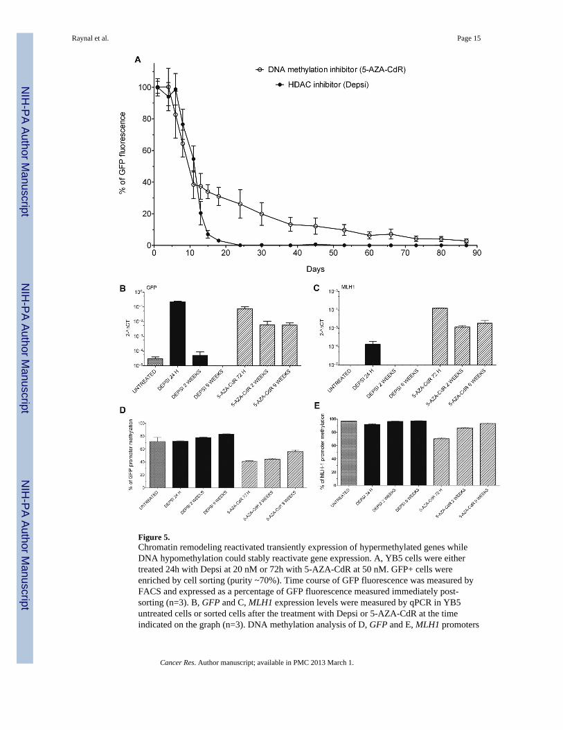
Figure 5.Chromatin remodeling reactivated transiently expression of hypermethylated genes whileDNA hypomethylation could stably reactivate gene expression. A, YB5 cells were eithertreated 24h with Depsi at 20 nM or 72h with 5-AZA-CdR at 50 nM. GFP+ cells wereenriched by cell sorting (purity ~70%). Time course of GFP fluorescence was measured byFACS and expressed as a percentage of GFP fluorescence measured immediately post-sorting (n=3). B, GFP and C, MLH1 expression levels were measured by qPCR in YB5untreated cells or sorted cells after the treatment with Depsi or 5-AZA-CdR at the timeindicated on the graph (n=3). DNA methylation analysis of D, GFP and E, MLH1 promoters
Raynal et al. Page 15
Cancer Res. Author manuscript; available in PMC 2013 March 1.
NIH
-PA Author Manuscript
NIH
-PA Author Manuscript
NIH
-PA Author Manuscript

by pyrosequencing of YB5 untreated cells or sorted cells treated with Depsi or 5-AZA-CdRat the time indicated on the graph (n=3).
Raynal et al. Page 16
Cancer Res. Author manuscript; available in PMC 2013 March 1.
NIH
-PA Author Manuscript
NIH
-PA Author Manuscript
NIH
-PA Author Manuscript
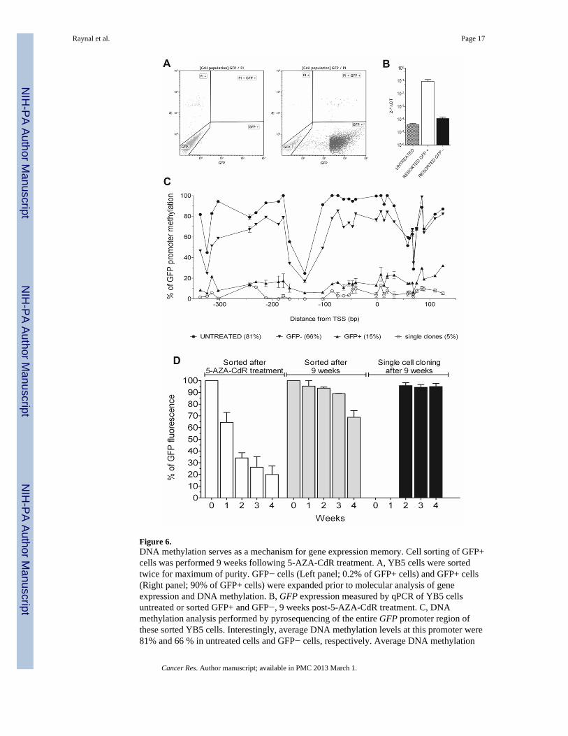
Figure 6.DNA methylation serves as a mechanism for gene expression memory. Cell sorting of GFP+cells was performed 9 weeks following 5-AZA-CdR treatment. A, YB5 cells were sortedtwice for maximum of purity. GFP− cells (Left panel; 0.2% of GFP+ cells) and GFP+ cells(Right panel; 90% of GFP+ cells) were expanded prior to molecular analysis of geneexpression and DNA methylation. B, GFP expression measured by qPCR of YB5 cellsuntreated or sorted GFP+ and GFP−, 9 weeks post-5-AZA-CdR treatment. C, DNAmethylation analysis performed by pyrosequencing of the entire GFP promoter region ofthese sorted YB5 cells. Interestingly, average DNA methylation levels at this promoter were81% and 66 % in untreated cells and GFP− cells, respectively. Average DNA methylation
Raynal et al. Page 17
Cancer Res. Author manuscript; available in PMC 2013 March 1.
NIH
-PA Author Manuscript
NIH
-PA Author Manuscript
NIH
-PA Author Manuscript
levels in this locus was only 15 % in GFP+ cells. Moreover, methylation level of singleclones isolated by cell sorting, 9 weeks following 5-AZA-CdR treatment, showed 5 %methylation levels which is similar to the background of the assay. D, Time-dependent GFPexpression measured by FACS analysis on YB5 cells sorted immediately following 5-AZA-CdR treatment at 70 % purity of GFP+ cells (left), 9 weeks post-5-AZA-CdR treatment after2 additional rounds of cell sorting, at 90 % purity of GFP+ cells (middle), and after singlecell cloning for GFP+ cells performed 9 weeks after the initial 5-AZA-CdR treatment(right).
Raynal et al. Page 18
Cancer Res. Author manuscript; available in PMC 2013 March 1.
NIH
-PA Author Manuscript
NIH
-PA Author Manuscript
NIH
-PA Author Manuscript

NIH
-PA Author Manuscript
NIH
-PA Author Manuscript
NIH
-PA Author Manuscript
Raynal et al. Page 19
Table 1
Effects of various HDAC inhibitors (HDACi) on GFP expression in YB5 cells detected by FACS analysisafter a 24h treatment. HDACi targets (specific isoforms or entire HDAC class) are shown in the table.Concentration (Conc.) range tested on the YB5 system is indicated in the table.
Drugs Structural class Target HDAC Conc. tested GFPexpression
SAHA Hydroxamic acid I, IIa, IIb, IV 0.3–20 µM Yes
TSA Hydroxamic acid I, II 50–200 nM Yes
Oxamiflatin Hydroxamic acid HDAC 3, 6 100–400 nM Yes
APHA8 Hydroxamic acid HDAC 1 1–25 µM Yes
Cay 10398 Hydroxamic acid HDAC 1 10–40 µM Yes
M344 Hydroxamic acid HDAC 1, 6 1–4 µM Yes
CHAHA Hydromaxic acid HDAC 6 0.2–1.5 µM Yes
LBH-589 Hydromaxic acid Pan-HDACi 0.02–20 µM Yes
Depudicin TSA-like but non hydroxamate HDAC 1 5–25 µM Yes
PTACH TSA-like but non hydroxamate --- 1–10 µM Yes
Valproic acid Short-chain fatty acid HDAC 2 0,25–10 mM Yes
Phenylbutyrate Short-chain fatty acid I 1 mM Yes
Sodium butyrate Short-chain fatty acid I, IIa 5–20 mM Yes
Depsipeptide Cyclic peptide HDAC 1, 2 1 nM-2 µM Yes
Apicidin Cyclic peptide I, II 0.4–40 µM Yes
HC-Toxin Cyclic peptide HDAC 1, 2, 3, 8 25–100 nM Yes
Sirtinol Hydroxy-naphthaldehyde Sirt 2 1.25–100 µM Yes
Splitomicin Naphthalenes Sirt 2 0.1–75 µM No
Ex527 Tetrahydrocarbazoles Sirt 1 20–100 µM No
CHIC-35 ND Sirt 1 0.1–1 µM No
Cambinol β-naphthol Sirt1, 2 2.5–25 µM No
PCI-34051 Hydroxamic acid HDAC 8 1–10 µM No
BATCP ND HDAC 6 1–10 µM No
Scriptaid Hydroxamic acid HDAC 1, 2, 8 0.05– 1 µM No
ND: not determined.
Cancer Res. Author manuscript; available in PMC 2013 March 1.
Related Documents