UNCORRECTED PROOF Please cite this article in press as: Rodr´ ıguez, J. L., et al., Transcription of the MAT2A gene, coding for methionine adeno- syltransferase, is up-regulated by E2F and Sp1 at a chromatin level during proliferation of liver cells, International Journal of Biochemistry and Cell Biology (2007), doi:10.1016/j.biocel.2007.01.009 ARTICLE IN PRESS +Model BC 2357 1–9 The International Journal of Biochemistry & Cell Biology xxx (2007) xxx–xxx Transcription of the MAT2A gene, coding for methionine adenosyltransferase, is up-regulated by E2F and Sp1 at a chromatin level during proliferation of liver cells 3 4 5 Jos´ e L. Rodr´ ıguez a , Abdelhalim Boukaba a , Juan Sandoval a , Elena I. Georgieva a,1 , M. Ujue Latasa d , Elena R. Garc´ ıa-Trevijano a , Gaetano Serviddio b,2 , Toshikazu Nakamura c , Mat´ ıas A. ´ Avila d , Juan Sastre b , Luis Torres a , Jos´ e M. Mato d , Gerardo L ´ opez-Rodas a,∗ 6 7 8 9 a Department of Biochemistry and Molecular Biology, University of Valencia, Spain 10 b Department of Physiology, University of Valencia, Spain 11 c Division of Molecular Regenerative Medicine of Biochemistry and Molecular Biology, Osaka University Graduate School of Medicine, Japan 12 13 d Division of Hepatology and Gene Therapy, CIMA, University of Navarra, Spain 14 Received 16 October 2006; received in revised form 28 December 2006; accepted 8 January 2007 15 Abstract 16 Methionine adenosyltransferase (MAT) is an essential enzyme because it catalyzes the formation of S-adenosylmethionine, the main methyl donor. Two MAT-encoding genes (MAT1A, MAT2A) are found in mammals. The latter is expressed in proliferating liver, dedifferentiation and cancer, whereas MAT1A is expressed in adult quiescent hepatocytes. Here, we report studies on the molecular mechanisms controlling the induction of MAT2A in regenerating rat liver and in proliferating hepatocytes. The MAT2A is up-regulated at two discrete moments during liver regeneration, as confirmed by RNApol-ChIP analysis. The first one coincides with hepatocyte priming (i.e. G 0 –G 1 transition), while the second one takes place at the G 1 –S interface. Electrophoretic mobility shift assays showed that a putative E2F sequence present in MAT2A promoter binds this factor and ChIP assays confirmed that E2F1, E2F3 and E2F4, as well as the pocket protein p130, are bound to the promoter in quiescent liver. MAT2A activation is accompanied by changes in the binding of histone-modifying enzymes to the promoter. Interestingly, p130 is not displaced from MAT2A promoter during hepatocyte priming, but it is in the late expression of the gene at the G 1 –S transition. Finally, the transcription factor Sp1 seems to play a decisive role in MAT2A induction, as it binds the promoter when the gene is being actively transcribed. In summary, the present work shows that the molecular mechanism of MAT2A expression is different during G 0 –G 1 or G 1 –S transition and this may be related to the distinct requirements of S-adenosylmethionine during liver regeneration. 17 18 19 20 21 22 23 24 25 26 27 28 29 © 2007 Published by Elsevier Ltd. 30 Keywords: Histone acetylation; Chromatin immunoprecipitation; ChIP; RNApol ChIP; Liver regeneration 31 32 ∗ Corresponding author. Tel.: +34 96 3544867; fax: +34 96 3544635. E-mail address: [email protected] (G. L ´ opez-Rodas). 1 Permanent address: Department of Molecular Genetics, Institute of Genetics, Bulgarian Academy of Sciences, Sofia, Bulgaria. 2 Present address: Dipartimento di Scienze Mediche e del Lavoro, Universit` a di Foggia, Foggia, Italy. 1 1357-2725/$ – see front matter © 2007 Published by Elsevier Ltd. 2 doi:10.1016/j.biocel.2007.01.009

Welcome message from author
This document is posted to help you gain knowledge. Please leave a comment to let me know what you think about it! Share it to your friends and learn new things together.
Transcript
B
3
4
5
6
7
8
9
10
11
12
13
14
15
A16
mlmiwsEbdstm
17
18
19
20
21
22
23
24
25
26
27
28
29
©30
K31
32
1 12 d
EC
TED
PR
OO
F
ARTICLE IN PRESS+ModelC 2357 1–9
The International Journal of Biochemistry & Cell Biology xxx (2007) xxx–xxx
Transcription of the MAT2A gene, coding for methionineadenosyltransferase, is up-regulated by E2F and Sp1 at
a chromatin level during proliferation of liver cells
Jose L. Rodrıguez a, Abdelhalim Boukaba a, Juan Sandoval a, Elena I. Georgieva a,1,M. Ujue Latasa d, Elena R. Garcıa-Trevijano a, Gaetano Serviddio b,2,Toshikazu Nakamura c, Matıas A. Avila d, Juan Sastre b, Luis Torres a,
Jose M. Mato d, Gerardo Lopez-Rodas a,∗a Department of Biochemistry and Molecular Biology, University of Valencia, Spain
b Department of Physiology, University of Valencia, Spainc Division of Molecular Regenerative Medicine of Biochemistry and Molecular Biology,
Osaka University Graduate School of Medicine, Japand Division of Hepatology and Gene Therapy, CIMA, University of Navarra, Spain
Received 16 October 2006; received in revised form 28 December 2006; accepted 8 January 2007
bstract
Methionine adenosyltransferase (MAT) is an essential enzyme because it catalyzes the formation of S-adenosylmethionine, theain methyl donor. Two MAT-encoding genes (MAT1A, MAT2A) are found in mammals. The latter is expressed in proliferating
iver, dedifferentiation and cancer, whereas MAT1A is expressed in adult quiescent hepatocytes. Here, we report studies on theolecular mechanisms controlling the induction of MAT2A in regenerating rat liver and in proliferating hepatocytes. The MAT2A
s up-regulated at two discrete moments during liver regeneration, as confirmed by RNApol-ChIP analysis. The first one coincidesith hepatocyte priming (i.e. G0–G1 transition), while the second one takes place at the G1–S interface. Electrophoretic mobility
hift assays showed that a putative E2F sequence present in MAT2A promoter binds this factor and ChIP assays confirmed that E2F1,2F3 and E2F4, as well as the pocket protein p130, are bound to the promoter in quiescent liver. MAT2A activation is accompanied
Ry changes in the binding of histone-modifying enzymes to the promoter. Interestingly, p130 is not displaced from MAT2A promoteruring hepatocyte priming, but it is in the late expression of the gene at the G1–S transition. Finally, the transcription factor Sp1eems to play a decisive role in MAT2A induction, as it binds the promoter when the gene is being actively transcribed. In summary,he present work shows that the molecular mechanism of MAT2A expression is different during G0–G1 or G1–S transition and thisionine regeneration.
Ray be related to the distinct requirements of S-adenosylmethUN
CO
Please cite this article in press as: Rodrıguez, J. L., et al., Transcsyltransferase, is up-regulated by E2F and Sp1 at a chromatin leveBiochemistry and Cell Biology (2007), doi:10.1016/j.biocel.2007.0
2007 Published by Elsevier Ltd.
eywords: Histone acetylation; Chromatin immunoprecipitation; ChIP; RNA
∗ Corresponding author. Tel.: +34 96 3544867; fax: +34 96 3544635.E-mail address: [email protected] (G. Lopez-Rodas).
1 Permanent address: Department of Molecular Genetics, Institute of Gene2 Present address: Dipartimento di Scienze Mediche e del Lavoro, Universi
357-2725/$ – see front matter © 2007 Published by Elsevier Ltd.oi:10.1016/j.biocel.2007.01.009
during liver
ription of the MAT2A gene, coding for methionine adeno-l during proliferation of liver cells, International Journal of1.009
pol ChIP; Liver regeneration
tics, Bulgarian Academy of Sciences, Sofia, Bulgaria.ta di Foggia, Foggia, Italy.
José Luis
Tachado
José Luis
Texto insertado
RNAPol-ChIP
José Luis
Tachado
José Luis
Texto insertado
T
IN+Model
al of Bio
1
2
3
4
5
6
7
8
9
10
11
12
13
14
15
16
17
18
19
20
21
22
23
24
25
26
27
28
29
30
31
32
33
34
35
36
37
38
39
40
41
42
43
44
45
46
47
48
49
50
51
52
53
54
55
56
57
58
59
60
61
62
63
64
65
66
67
68
69
70
71
72
73
74
75
76
77
78
79
80
81
82
83
84
85
86
87
88
89
90
91
92
93
94
95
96
97
98
99
NC
OR
RE
C
ARTICLEBC 2357 1–9
2 J.L. Rodrıguez et al. / The International Journ
1. Introduction
The unique capacity of the liver to regenerate uponpartial hepatectomy (PH) to restore its functional massprovides an attractive model to study cell proliferation,because the hepatocytes, which rarely divide, pass froma quiescent G0 state to a quickly dividing one very soonafter the surgical removal of as much as two thirds ofthe organ (Higgins & Anderson, 1931). In the processof liver regeneration, Fausto has emphasized that twomain distinct steps occur, namely the transition of thequiescent hepatocytes into the cell cycle (priming) andthe progression beyond the restriction point in the Glphase of the cycle (Fausto, Campbell, & Riehle, 2006).These two steps seem to be controlled by different sig-nals, and it has been proposed that priming is mainlyunder the control of TNF� and IL-6 family cytokines(Koniaris, McKillop, Schwartz, & Zimmers, 2003; Yanget al., 2003), while the cell cycle progression occurs inresponse to growth factors HGF and TGF� (Fausto et al.,2006; Koniaris et al., 2003). The priming, the G0–Gl tran-sition occurs very early after partial hepatectomy. In rats,for instance, the entrance into the Gl phase is completedby 4 h and it is accompanied by the expression of severalimmediate-early genes. The proto-oncogene c-myc, c-jun and c-fos were first described as falling into this group(Alcorn, Feitelberg, & Brenner, 1990; Morello, Lavenu,& Babinet, 1990; Thompson et al., 1986), but the applica-tion of more powerful methods has allowed other authorsto identify around 100 of putative immediate-early genesinvolved in liver regeneration (Haber, Mohn, Diamond,& Taub, 1993; Su, Guidotti, Pezacki, Chisari, & Schultz,2002; White, Brestelli, Kaestner, & Greenbaum, 2005).
In the rat model of liver regeneration, the transcriptionof immediate-early genes is followed by that of ‘delayed’genes and, at the end of G1, by the transcription of the‘cell cycle’ genes that allow the cells to pass over therestriction point to enter the S phase and progress throughthe cell cycle (Fausto, 2002; Fausto et al., 2006). In someinstances, the expression of immediate-early genes thatoccurs during priming is resumed in the G1–S transition.This bimodal expression pattern was first described forc-myc (Thompson et al., 1986). As commented above,priming and the transition beyond the restriction pointare controlled by different mechanisms. It is thereforelikely that the activating mechanisms of the genes show-ing a bimodal expression pattern differ in some way inboth peaks of their expression.
UPlease cite this article in press as: Rodrıguez, J. L., et al., Transcsyltransferase, is up-regulated by E2F and Sp1 at a chromatin leveBiochemistry and Cell Biology (2007), doi:10.1016/j.biocel.2007.0
The role of pocket proteins in the control of cell prolif-eration beyond the restriction point is well known. Theyinhibit the expression of S phase genes either by block-ing the ability of E2F factors to activate transcription
ED
PR
OO
F
PRESSchemistry & Cell Biology xxx (2007) xxx–xxx
or by an active repressing mechanism of their com-plexes with E2Fs (for reviews, see references Harbour& Dean, 2000 and Trimarchi & Lees, 2002). The activerepression is a complex phenomenon and it involveschanges in chromatin structure. Pocket proteins recruithistone deacetylases complexes to the promoters of thetarget genes (Luo, Postigo, & Dean, 1998; Magnaghi-Jaulin et al., 1998), which results in the deacetylation ofhistone H3 and eventually in the acquisition of a repres-sive chromatin structure (Trimarchi & Lees, 2002). Theformation of repressive complexes between E2Fs andpocket proteins depends upon the phosphorylation of thelatters. Hyperphosphorylation of pocket proteins, whichis achieved by different cyclin-dependent kinases, resultsin their release from E2Fs and in the relieving of theinhibitory potential of their complexes. For instance, ithas been suggested that the repressive p130-E2F4 com-plex is disassembled upon phosphorylation of 8 serinesand one threonine of p130 by cdk2 and cdk4/6 andthat the phosphorylation driven by cyclin D-cdk4/6 is akey event in that disassembling (Farkas, Hansen, Holm,Lukas, & Bartek, 2002). As cyclin D accumulates dur-ing G1, it is reasonable to think that all these events arecrucial for progressing through the G1 restriction point,but not for entering the cell cycle from G0.
We have previously found that MAT2A, the geneencoding the �2 catalytic subunit of the MAT II isozymeof methionine adenosyltransferase (MAT), which is up-regulated during liver regeneration (Huang, Mao, Cai,& Lu, 1998), is also induced in primary hepatocyte cul-tures by HGF (Latasa et al., 2001). This induction istriggered by the autophosphoryation of the MET recep-tor for HGF and it is accompanied by an increase of theacetylation of histone H4 in MAT2A promoter (Latasa etal., 2001), indicating that chromatin structure is involvedin the process. As primary hepatocyte cultures respondto growth factors, cultured hepatocytes could be consid-ered to be already primed for proliferation (Loyer et al.,1996; Nelson, 2002). This assumption is reinforced bythe fact that treatment of primary hepatocytes with HGFor transforming growth factor � (TGF�) induced DNAsynthesis (Garcıa-Trevijano, Martınez-Chantal, Latasa,Mato, & Avila, 2002; Latasa et al., 2001). In other words,when the observed HGF-induced expression of MAT2Aoccurs, the cells are at or beyond G1/S. On the other hand,MAT2A expression is also required for cell proliferationin cultured H35 hepatoma cells (Paneda et al., 2002).
It would be interesting, therefore, to study in more
ription of the MAT2A gene, coding for methionine adeno-l during proliferation of liver cells, International Journal of1.009
detail the expression pattern of MAT2A during liver 100
regeneration. This was the main aim of the present 101
work in which we describe that MAT2A is regulated 102
by E2F and Sp1 and it is not only expressed at the 103
CT
IN+ModelB
l of Bio
e104
d105
e106
r107
s108
p109
i110
p111
2112
2113
114
w115
c116
a117
h118
(119
a120
s121
a122
o123
w124
c125
g126
1127
4128
H129
c130
t131
T132
i133
b134
2135
136
(137
e138
c139
d140
m141
a142
G143
r144
p145
r146
a147
(148
a149
m
150
151
152
153
154
155
156
157
158
159
160
161
162
163
164
165
166
167
168
169
170
171
172
173
174
175
176
177
178
179
180
181
182
183
184
185
186
187
188
189
190
191
192
193
194
NC
OR
RE
ARTICLEC 2357 1–9
J.L. Rodrıguez et al. / The International Journa
nd of G1, but also a sharp peak of gene expression isetected during hepatocyte priming. While the secondxpression peak involves the phosphorylation-drivenemoval of p130, the first peak of MAT2A expres-ion during priming does not require the release of theocket protein, but rather, is accompanied by changesn the binding of histone-modifying enzymes in theromoter.
. Materials and methods
.1. Animal care and rat liver experiments
Male pathogen-free Wistar rats weighing 220–260 gere maintained at 22 ◦C with a 12 h light:12 h dark
ycle and fed ad libitum with free access to waternd standard laboratory diet. Animals were cared andandled in conformance with the European regulationsCouncil Directive 86/609/EEC) and the studies werepproved by the Research Committee of the Univer-ity of Valencia. Partial hepatectomy was carried outccording to Higgins and Anderson (1931). At vari-us times after PH, animals were sacrificed and liversere treated with 1% formaldehyde during 12 min to
rosslink the chromatin. The liver tissue was disaggre-ated with a Dounce homogenizer and centrifuged at500 × g for 5 min. The cell pellet was resuspended inml of cell lysis buffer (85 mM KCl, 0.5% NP40, 5 mMEPES pH 8.0) supplemented with protease inhibitor
ocktail (Sigma), incubated on ice for 15 min and cen-rifuged at 3500 × g for 5 min to pellet the nuclei.he nuclei were used for chromatin immunoprecip-
tation (ChIP) and RNApol-ChIP assay as describedelow.
.2. Isolation and culture of rat hepatocytes
Hepatocytes were isolated from rats by collagenaseGibco-BRL) perfusion as previously described (Latasat al., 2001). Cells were plated onto 60 mm collagen-oated culture dishes at a density of 3 × 10⁶ cells perish. Cultures were maintained in MEM medium supple-ented with 10% fetal calf serum (FCS), nonessential
mino acids, 2 mM glutamine, and antibiotics (all fromibco-BRL). After 6 h, the incubation medium was
emoved, and cells were refed with the same mediumlus 0.5% FCS. After a period of 18 h, the medium wasemoved and fresh culture medium without serum was
UPlease cite this article in press as: Rodrıguez, J. L., et al., Transcsyltransferase, is up-regulated by E2F and Sp1 at a chromatin leveBiochemistry and Cell Biology (2007), doi:10.1016/j.biocel.2007.0
dded. For HGF stimulation, human recombinant HGFNakamura et al., 1989) was added two hours later to hep-tocyte cultures at a concentration of 25 ng/ml of cultureedium.
ED
PR
OO
F
PRESSchemistry & Cell Biology xxx (2007) xxx–xxx 3
2.3. Chromatin immunoprecipitation assay,RNApol-ChIP and determination of steady-statelevels of mRNA
ChIP and RNApol-ChIP procedures were per-formed according to Sandoval et al. (2004). Briefly,isolated nuclei from formaldehyde-crosslinked liversor primary hepatocyte cultures were lysed and thecrosslinked chromatin was sonicated to yield fragmentsof ∼500 bp. Diluted soluble chromatin fragments wereprecleared with blocked protein A/G Sepharose, todiscard non-specifically-bound chromatin fragments.Immunofractionation of complexes was carried out byadding 2 �g of the corresponding antibodies (SantaCruz, E2F1 sc-193, E2F2 sc-9967, E2F3 sc-878,E2F4 sc-866, E2F5 sc-999, p130 sc-317, mSin3Asc-994, CBP sc-369, p300 sc-584, Sp1 sc-59 andRNApol II sc-899 and Pharmingen, pRB 554136) toaliquots containing 50 �g DNA each. The immuno-complexes were recovered by centrifugation afteradding blocked protein A/G Sepharose and extensivelywashed. Immunoselected chromatin was eluted andthe formaldehyde crosslinking was reverted. TheDNA from all samples was purified with a PCRpurification kit (Qiagen) and used for PCR analysis,which was done with the following primers: MAT2A(promoter): 5′-TCGATAGAATGGTCCAGCCC-3′ and5′-AGGAGGGTCGCTTCAACTCTC-3′; MAT2A (cod-ing region): 5′-GATTGGTCAGGCGGTTAAGAGT-3′and 5′-TTGTGCGATGGTGTTCAACTG-3′; cdc2(promoter): 5′-CGACATTGGAAGGAAAGCTGA-3′and 5′-GACGTTCAAAGGAGCCAATCA-3′; c-fos(coding region): 5′-TGGACTTGACTGGGGGTCTG-3′ and 5′-CAGGTCCACATCTGGCACAG-3′; cycE(coding region): 5′-TCCTCGTTGGAGTTGATGCA-3′and 5′-CCCATCTCCCGGATAACCAT-3′; �-actin(promoter): 5′AGGGACTCTAGTGCCCAACACC-3′and 5′CCCACCTCCACCCTACCTGC; �-actin (codingregion): 5′-AGGATTCCTACGTGGGCGAC-3′ and 5′-TAGAGAGACAGCACCGCCTG-3′; �-actin (codingregion): 5′-AGAGCAAGAGAGGCATCCTG-3′ and5′-GGGTCATCTTTTCACGGTTGG-3′.
2.4. Electrophoretic mobility shift assays (EMSA)
Total rat liver nuclear extracts were prepared fromnuclei obtained as described by Karagyozov, Stoyanova,and Hadjiolov (1980) except that phosphatase inhibitor
ription of the MAT2A gene, coding for methionine adeno-l during proliferation of liver cells, International Journal of1.009
cocktails were added to all buffers. Nuclei were then 195
suspended 100 �l of lysis buffer (450 KCl, 0.1% NP- 196
40, 1 mM EGTA, 1 mM DDT, 10% glycerol, 6 mM 197
MgCl2, 5 �l of mammalian protease inhibitors cock- 198
José Luis
Tachado
José Luis
Texto insertado
RNAPol-ChIP
José Luis
Tachado
José Luis
Texto insertado
RNAPol-ChIP
José Luis
Tachado
José Luis
Texto insertado
RNAPol-ChIP
T
IN+Model
al of Bio
199
200
201
202
203
204
205
206
207
208
209
210
211
212
213
214
215
216
217
218
219
220
221
222
223
224
225
226
227
228
229
230
231
232
233
234
235
236
237
238
239
240
241
242
243
244
245
246
247
248
249
250
251
252
253
254
255
256
257
258
259
ARTICLEBC 2357 1–9
4 J.L. Rodrıguez et al. / The International Journ
tail (Sigma), 50 mM HEPES, pH 7.9) and incubatedon ice for 30 min. After centrifugation at 14,000 rpmfor 20 min, the supernatant was diluted with the samebuffer except that KCl was omitted. Oligonucleotidesequence encompassing the putative MAT2A E2F site(underlined) and spanning from−145 to−113, as well asthe mutated version was as follows: 5′-GCCCGTGGC-GCATTGGCGCGCGACGTCGAGGGG-3′; 5′-GCCC-GTGGCGCATTGGatCGCGACGTCGAGGGG-3′. Thedouble-stranded oligonucleotides containing the abovesequences and their complementary ones were radioac-tively labelled with 32P, by using T4 polynucleotidekinase (Roche) following the manufacturer’s instruc-tions. The DNA–protein complexes were separated ona 5% native polyacrylamide gel in 0.25 × TBE, dryedunder vacuum and analyzed by electronic autoradiogra-phy (FLA3000 PhosphoImager, Fujifilm).
3. Results
3.1. MAT2A displays bimodal expression behaviourduring liver regeneration
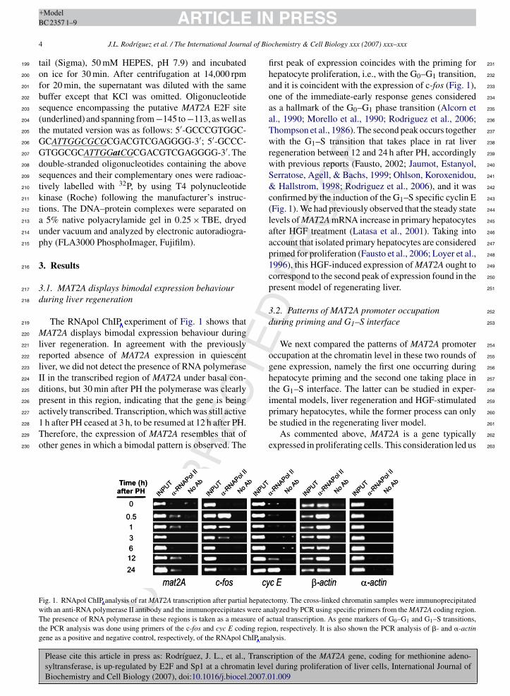
The RNApol ChIP experiment of Fig. 1 shows thatMAT2A displays bimodal expression behaviour duringliver regeneration. In agreement with the previouslyreported absence of MAT2A expression in quiescentliver, we did not detect the presence of RNA polymeraseII in the transcribed region of MAT2A under basal con-ditions, but 30 min after PH the polymerase was clearlypresent in this region, indicating that the gene is being
UN
CO
RR
EC
Please cite this article in press as: Rodrıguez, J. L., et al., Transcsyltransferase, is up-regulated by E2F and Sp1 at a chromatin leveBiochemistry and Cell Biology (2007), doi:10.1016/j.biocel.2007.0
actively transcribed. Transcription, which was still active1 h after PH ceased at 3 h, to be resumed at 12 h after PH.Therefore, the expression of MAT2A resembles that ofother genes in which a bimodal pattern is observed. The
Fig. 1. RNApol ChIP analysis of rat MAT2A transcription after partial hepatewith an anti-RNA polymerase II antibody and the immunoprecipitates were anThe presence of RNA polymerase in these regions is taken as a measure of athe PCR analysis was done using primers of the c-fos and cyc E coding regigene as a positive and negative control, respectively, of the RNApol ChIP ana
ED
PR
OO
F
PRESSchemistry & Cell Biology xxx (2007) xxx–xxx
first peak of expression coincides with the priming forhepatocyte proliferation, i.e., with the G0–G1 transition,and it is coincident with the expression of c-fos (Fig. 1),one of the immediate-early response genes consideredas a hallmark of the G0–G1 phase transition (Alcorn etal., 1990; Morello et al., 1990; Rodriguez et al., 2006;Thompson et al., 1986). The second peak occurs togetherwith the G1–S transition that takes place in rat liverregeneration between 12 and 24 h after PH, accordinglywith previous reports (Fausto, 2002; Jaumot, Estanyol,Serratose, Agell, & Bachs, 1999; Ohlson, Koroxenidou,& Hallstrom, 1998; Rodriguez et al., 2006), and it wasconfirmed by the induction of the G1–S specific cyclin E(Fig. 1). We had previously observed that the steady statelevels of MAT2A mRNA increase in primary hepatocytesafter HGF treatment (Latasa et al., 2001). Taking intoaccount that isolated primary hepatocytes are consideredprimed for proliferation (Fausto et al., 2006; Loyer et al.,1996), this HGF-induced expression of MAT2A ought tocorrespond to the second peak of expression found in thepresent model of regenerating liver.
3.2. Patterns of MAT2A promoter occupationduring priming and G1–S interface
We next compared the patterns of MAT2A promoteroccupation at the chromatin level in these two rounds ofgene expression, namely the first one occurring duringhepatocyte priming and the second one taking place inthe G1–S interface. The latter can be studied in exper-imental models, liver regeneration and HGF-stimulated
ription of the MAT2A gene, coding for methionine adeno-l during proliferation of liver cells, International Journal of1.009
primary hepatocytes, while the former process can only 260
be studied in the regenerating liver model. 261
As commented above, MAT2A is a gene typically 262
expressed in proliferating cells. This consideration led us 263
ctomy. The cross-linked chromatin samples were immunoprecipitatedalyzed by PCR using specific primers from the MAT2A coding region.ctual transcription. As gene markers of G0–G1 and G1–S transitions,on, respectively. It is also shown the PCR analysis of �- and �-actinlysis.
José Luis
Tachado
José Luis
Texto insertado
RNAPol-ChIP
José Luis
Tachado
José Luis
Texto insertado
RNAPol-ChIP
José Luis
Tachado
José Luis
Texto insertado
RNAPol-ChIP
CT
ARTICLE IN+ModelBC 2357 1–9
J.L. Rodrıguez et al. / The International Journal of Bio
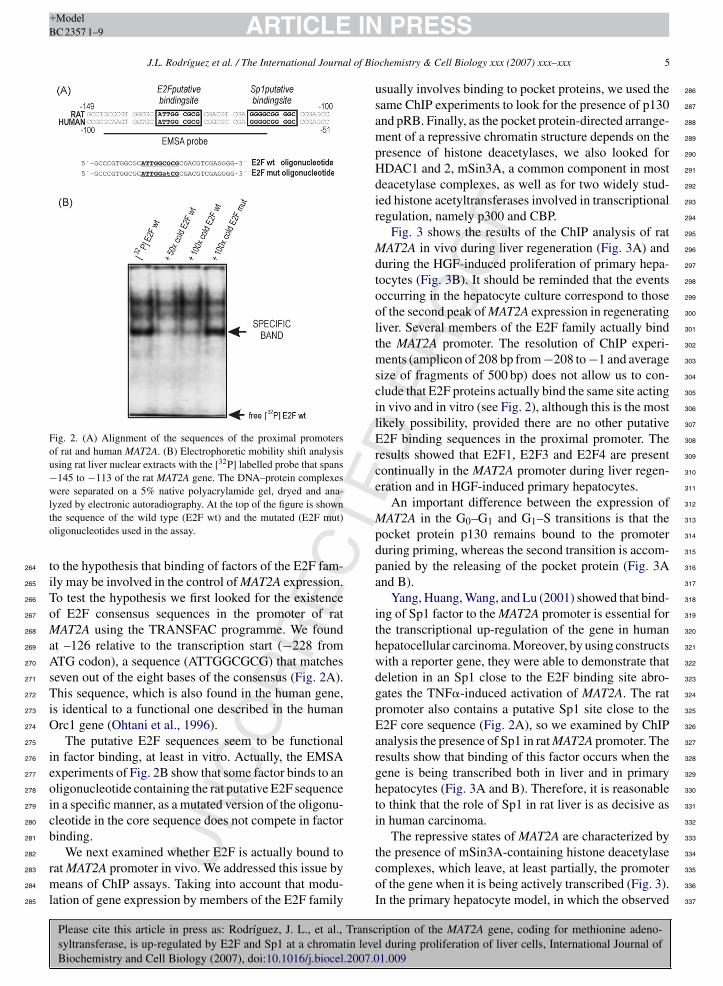
Fig. 2. (A) Alignment of the sequences of the proximal promotersof rat and human MAT2A. (B) Electrophoretic mobility shift analysisusing rat liver nuclear extracts with the [32P] labelled probe that spans−145 to −113 of the rat MAT2A gene. The DNA–protein complexeswere separated on a 5% native polyacrylamide gel, dryed and ana-lto
t264
i265
T266
o267
M268
a269
A270
s271
T272
i273
O274
275
i276
e277
o278
i279
c280
b281
282
r283
m284
l285
286
287
288
289
290
291
292
293
294
295
296
297
298
299
300
301
302
303
304
305
306
307
308
309
310
311
312
313
314
315
316
317
318
319
320
321
322
323
324
325
326
327
328
329
330
331
332
333
NC
OR
RE
yzed by electronic autoradiography. At the top of the figure is shownhe sequence of the wild type (E2F wt) and the mutated (E2F mut)ligonucleotides used in the assay.
o the hypothesis that binding of factors of the E2F fam-ly may be involved in the control of MAT2A expression.o test the hypothesis we first looked for the existencef E2F consensus sequences in the promoter of ratAT2A using the TRANSFAC programme. We found
t –126 relative to the transcription start (−228 fromTG codon), a sequence (ATTGGCGCG) that matcheseven out of the eight bases of the consensus (Fig. 2A).his sequence, which is also found in the human gene,
s identical to a functional one described in the humanrc1 gene (Ohtani et al., 1996).The putative E2F sequences seem to be functional
n factor binding, at least in vitro. Actually, the EMSAxperiments of Fig. 2B show that some factor binds to anligonucleotide containing the rat putative E2F sequencen a specific manner, as a mutated version of the oligonu-leotide in the core sequence does not compete in factorinding.
UPlease cite this article in press as: Rodrıguez, J. L., et al., Transcsyltransferase, is up-regulated by E2F and Sp1 at a chromatin leveBiochemistry and Cell Biology (2007), doi:10.1016/j.biocel.2007.0
We next examined whether E2F is actually bound toat MAT2A promoter in vivo. We addressed this issue byeans of ChIP assays. Taking into account that modu-
ation of gene expression by members of the E2F family
ED
PR
OO
F
PRESSchemistry & Cell Biology xxx (2007) xxx–xxx 5
usually involves binding to pocket proteins, we used thesame ChIP experiments to look for the presence of p130and pRB. Finally, as the pocket protein-directed arrange-ment of a repressive chromatin structure depends on thepresence of histone deacetylases, we also looked forHDAC1 and 2, mSin3A, a common component in mostdeacetylase complexes, as well as for two widely stud-ied histone acetyltransferases involved in transcriptionalregulation, namely p300 and CBP.
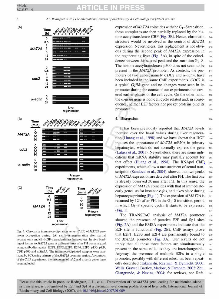
Fig. 3 shows the results of the ChIP analysis of ratMAT2A in vivo during liver regeneration (Fig. 3A) andduring the HGF-induced proliferation of primary hepa-tocytes (Fig. 3B). It should be reminded that the eventsoccurring in the hepatocyte culture correspond to thoseof the second peak of MAT2A expression in regeneratingliver. Several members of the E2F family actually bindthe MAT2A promoter. The resolution of ChIP experi-ments (amplicon of 208 bp from −208 to −1 and averagesize of fragments of 500 bp) does not allow us to con-clude that E2F proteins actually bind the same site actingin vivo and in vitro (see Fig. 2), although this is the mostlikely possibility, provided there are no other putativeE2F binding sequences in the proximal promoter. Theresults showed that E2F1, E2F3 and E2F4 are presentcontinually in the MAT2A promoter during liver regen-eration and in HGF-induced primary hepatocytes.
An important difference between the expression ofMAT2A in the G0–G1 and G1–S transitions is that thepocket protein p130 remains bound to the promoterduring priming, whereas the second transition is accom-panied by the releasing of the pocket protein (Fig. 3Aand B).
Yang, Huang, Wang, and Lu (2001) showed that bind-ing of Sp1 factor to the MAT2A promoter is essential forthe transcriptional up-regulation of the gene in humanhepatocellular carcinoma. Moreover, by using constructswith a reporter gene, they were able to demonstrate thatdeletion in an Sp1 close to the E2F binding site abro-gates the TNF�-induced activation of MAT2A. The ratpromoter also contains a putative Sp1 site close to theE2F core sequence (Fig. 2A), so we examined by ChIPanalysis the presence of Sp1 in rat MAT2A promoter. Theresults show that binding of this factor occurs when thegene is being transcribed both in liver and in primaryhepatocytes (Fig. 3A and B). Therefore, it is reasonableto think that the role of Sp1 in rat liver is as decisive asin human carcinoma.
The repressive states of MAT2A are characterized by
ription of the MAT2A gene, coding for methionine adeno-l during proliferation of liver cells, International Journal of1.009
the presence of mSin3A-containing histone deacetylase 334
complexes, which leave, at least partially, the promoter 335
of the gene when it is being actively transcribed (Fig. 3). 336
In the primary hepatocyte model, in which the observed 337
UN
CO
RR
EC
T
Please cite this article in press as: Rodrıguez, J. L., et al., Transcsyltransferase, is up-regulated by E2F and Sp1 at a chromatin leveBiochemistry and Cell Biology (2007), doi:10.1016/j.biocel.2007.0
ARTICLE IN+ModelBC 2357 1–9
6 J.L. Rodrıguez et al. / The International Journal of Bio
Fig. 3. Chromatin immunoprecipitation assay (ChIP) of MAT2A pro-moter occupation during: (A) rat liver regeneration after partialhepatectomy and (B) HGF-treated primary hepatocytes. In vivo bind-ing of factors to MAT2A gene at different times after PH was analyzedusing antibodies against E2F1, E2F2, E2F3, E2F4, E2F5, p130, pRB,CBP, p300 and mSin3A. The immunoprecipitated samples were ana-lyzed by PCR using primers of the MAT2A promoter region. As controlsof the ChIP experiment, the promoters of cdc2 and �-actin genes havebeen included.
338
339
340
341
342
343
344
345
346
347
348
349
350
351
352
353
354
355
356
357
358
359
360
361
362
363
364
365
366
367
368
369
370
371
372
373
374
375
376
377
378
379
380
381
382
383
ED
PR
OO
F
PRESSchemistry & Cell Biology xxx (2007) xxx–xxx
expression of MAT2A coincides with the G1–S transition,these complexes are then partially replaced by the his-tone acetyltransferase CBP (Fig. 3B). Hence, chromatinstructure would be involved in the control of MAT2Aexpression. Nevertheless, this replacement is not obvi-ous during the second peak of MAT2A expression inthe regenerating liver (Fig. 3A), in spite of the coinci-dence between this second peak and the transition G1–S.The histone acetyltransferase p300 does not seem to bepresent in the MAT2A promoter. As controls, the pro-moters of two genes, namely CDC2 and �-actin, havebeen included in the same ChIP experiments. CDC2 isa typical G2/M gene and no changes were seen in itspromoter during the course of our experiments that cov-ered earlier phases of the cell cycle. On the other hand,the �-actin gene is non-cell cycle related and, in conse-quence, neither E2F factors nor pocket proteins bind itspromoter.
4. Discussion
It has been previously reported that MAT2A levelsincrease over the basal values during liver regenera-tion (Huang et al., 1998) and we have shown that HGFinduces the appearance of MAT2A mRNA in primaryhepatocytes, which do not normally express the gene(Latasa et al., 2001). Nevertheless, there are some indi-cations that mRNA stability may partially account forthat effect (Huang et al., 1998). The RNApol ChIPexperiments, which allow a measurement of actual tran-scription (Sandoval et al., 2004), showed that two peaksof MAT2A expression are detected after PH. The first oneis already observed 30 min after PH. In this sense, theexpression of MAT2A coincides with that of immediate-early genes, as for instance c-fos, and takes place duringhepatocyte priming (Fig. 1). The expression of MAT2A isresumed by 12 h after PH, in the G1–S transition, periodin which G1–S specific cyclin E starts to be expressed(Fig. 1).
The TRANSFAC analysis of MAT2A promotershowed the presence of putative E2F and Sp1 sites(Fig. 2A) and the EMSA experiments indicate that theE2F site is functional (Fig. 2B). ChIP assays provethat E2F1, E2F3 and E2F4 are permanently bound tothe MAT2A promoter (Fig. 3A). Our results do notimply that all these three factors are simultaneouslypresent in the same cells, as they are interchangeable.Anyway, the presence of multiple E2Fs in a single
ription of the MAT2A gene, coding for methionine adeno-l during proliferation of liver cells, International Journal of1.009
promoter, possibly with different roles, has been repeat- 384
edly described (Takahashi, Rayman, & Dynlacht, 2000; 385
Wells, Graveel, Bartley, Madore, & Farnham, 2002; Zhu, 386
Giangrande, & Nevins, 2004; for reviews, see Refs. 387
José Luis
Tachado
José Luis
Texto insertado
RNAPol-ChIP
CT
IN+ModelB
l of Bio
G388
T389
t390
w391
S392
t393
t394
g395
h396
s397
t398
i399
2400
b401
f402
a403
s404
t405
g406
E407
o408
o409
p410
a411
a412
g413
m414
F415
416
b417
m418
s419
h420
p421
w422
i423
H424
s425
d426
s427
s428
h429
i430
t431
g432
a433
s434
a435
t436
a437
o438
C439
440
441
442
443
444
445
446
447
448
449
450
451
452
453
454
455
456
457
458
459
460
461
462
463
464
465
466
467
468
469
470
471
472
473
474
475
476
477
478
479
480
481
482
483
484
485
NC
OR
RE
ARTICLEC 2357 1–9
J.L. Rodrıguez et al. / The International Journa
iacinti & Giordano, 2006 and Stevaux & Dyson, 2002).he binding of Sp1 to MAT2A promoter follows a pat-
ern different from that of E2Fs, as it is present onlyhen the gene is being transcribed (compare the lanesp1 in Fig. 3 with Fig. 1). This observation, valid for the
wo waves of MAT2A expression, suggests an impor-ant role for Sp1 as a transcriptional factor for theene, in coincidence with the results showed in humanepatocellular carcinoma (Yang et al., 2001). This tran-criptional factor may cooperate with E2F in activatinghe MAT2A promoter, as shown for other genes (see, fornstance, Kramps, Strieder, Sapetschnig, Suske, & Lutz,004). This cooperation implies the physical interactionetween Sp1 and E2Fs, which has been demonstratedor E2F1, E2F2 and E2F3, but not for E2F4 (Lin etl., 1996). The proximity of the putative E2F and Sp1ites in the MAT2A promoter (see Fig. 2) would allowhis interaction between the factors bound to their tar-et sites. In several instances, the cooperation between2F and Sp1 results in both, repressive and active statesf the promoter, depending upon the further recruitmentf either histone deacetylase-containing repressor com-lexes (Chang, Illenye, & Heintz, 2001; Doetzlhofer etl., 1999) or histone acetyltransferases (Doetzlhofer etl., 1999). In the present case, Sp1 is not involved inene repression, as the factor is not bound to the pro-oter in the absence of active transcription (compareigs. 1 and 3).
It is noteworthy that E2F and Sp1 are involved inoth waves of MAT2A transcription. Nevertheless, theechanisms responsible for these two peaks of tran-
cription are different. The first one, taking place duringepatocyte priming, is independent of pocket protein dis-lacement (Fig. 3A), while the second one, concomitantith the G1 ≡ S transition, involves the releasing of p130
n both the models used, namely liver regeneration andGF-stimulation of primary hepatocytes. Chromatin
tructure, as revealed by the alternating presence of theifferent histone-modifying complexes (Fig. 3A), alsoeems to play a role in the regulation of MAT2A expres-ion. We have already shown that HGF induces histoneyperacetylation in hepatocytes concomitantly with thenduction of MAT2A and that these effects depend onhe tyrosine kinase activity of the Met receptor for therowth factor (Latasa et al., 2001). We found that, ingreement with the results of Paneda et al. (2002), theignal elicited by HGF is transduced towards MAT2Activation through the MAP kinase pathway and that
UPlease cite this article in press as: Rodrıguez, J. L., et al., Transcsyltransferase, is up-regulated by E2F and Sp1 at a chromatin leveBiochemistry and Cell Biology (2007), doi:10.1016/j.biocel.2007.0
he phosphatidylinositol 3-phosphate pathway is prob-bly involved as well (data not shown). In this context,ur present data show that the histone acetyltransferaseBP replaces the mSin3A-containing deacetylase com-
ED
PR
OO
F
PRESSchemistry & Cell Biology xxx (2007) xxx–xxx 7
plex in MAT2A promoter during hepatocyte priming afterPH (Fig. 3A) and during the HGF-induced stimulation ofprimary hepatocytes (Fig. 3B), but not in the second peakof gene expression after PH. Probably, the broad speci-ficity of CBP (Grant & Berger, 1999) is responsible forthe HGF-induced histone hyperacetylation in primaryhepatocytes (Latasa et al., 2001), but once the MAT2Achromatin has been made competent for transcription ina process involving histone hyperacetylation, a more spe-cific histone acetyltransferase is required for the secondwave of gene expression.
Finally, we wish to comment that our results show forthe first time that MAT2A ranks among the genes that areexpressed immediately after PH. This fact reinforces thecurrent view that this gene plays a key role in liver cellproliferation. It was originally reported by Cai, Mao,Hwang, and Lu (1998), that interference with MAT2Agene expression in human hepatocellular carcinomacells results in a reduced basal growth rate. Subse-quently, Paneda et al. (2002) showed that the proliferativeresponse elicited by HGF in H35 rat hepatoma cells wasalso dependent on MAT2A expression. Nevertheless, thesituation in the normal hepatocyte may be apparentlydifferent, since these cells constitutively express anothergene, MAT1A, whose product, MATI/III, also catalyzesthe formation of S-adenosylmethionine (Mato, Corrales,Lu, & Avila, 2002). This differential response of the twoenzymes may be relevant during liver regeneration, sinceit is known that the production of these reactive speciesis enhanced in the regenerating liver (Hortelano, Dewez,Genaro, Diaz-Guerra, & Bosca, 1995). Taken together,these observations suggest that the up-regulation ofMAT2A after partial hepatectomy would be necessaryto provide the hepatocyte with the S-adenosylmethioninerequired for the progress of cell proliferation during liverregeneration (Chen et al., 2004).
Acknowledgements
This work was supported by Grants BFU2004-03616from Ministerio de Educacion y Ciencia and GruposACOMP 06/132 from Conselleria de Empresa, Universi-tat i Ciencia to G. Lopez-Rodas; Grants NIH AA12677,AA13847 and AT1576, PN I + D 2005-00855, and Redde Centros FIS C03/02 to J.M. Mato; Grant RCMN(C03/08) from Instituto de Salud Carlos III, to L. Tor-res; Grants C03/02 and G03/015 from Instituto de SaludCarlos III, CP04/00123 and PI051098 from Ministerio
ription of the MAT2A gene, coding for methionine adeno-l during proliferation of liver cells, International Journal of1.009
de Sanidad y Consumo and SAF 2004-03538 from Min- 486
isterio de Educacion y Ciencia to M.A. Avila. 487
We are in debt with Prof. L. Franco for his priceless 488
help during all steps of the research and for manuscript 489
José Luis
Tachado
José Luis
Texto insertado
-
T
IN+Model
al of Bio
490
491
492
493
494
495
496
497
498
499
500
501
502
503
504
505
506
507
508
509
510
511
512
513
514
515
516
517
518
519
520
521
522
523
524
525
526
527
528
529
530
531
532
533
534
535
536
537
538
539
540
541
542
543
544
545
546
547
548
549
550
551
552
553
554
555
556
557
558
559
560
561
562
563
564
565
566
567
568
569
570
571
572
573
574
575
576
577
578
579
580
581
582
583
584
585
586
587
588
589
590
591
592
593
594
595
596
597
598
599
600
601
602
603
604
NCOR
RE
C
ARTICLEBC 2357 1–9
8 J.L. Rodrıguez et al. / The International Journ
comments. E.I. Georgieva is a recipient of a researchgrant from the Consellerıa de Empresa, Universitat iCiencia and from Ministerio de Educacion y Ciencia.M.U. Latasa is a fellow of the Juan de la Cierva Programfrom Ministerio de Educacion, Spain.
References
Alcorn, J. A., Feitelberg, S. P., & Brenner, D. A. (1990). Transientinduction of c-jun during hepatic regeneration. Hepatology, 11,909–915.
Brehm, A., Miska, E. A., McCance, D. J., Reid, J. L., Bannister, A.J., & Kouzarides, T. (1998). Retinoblastoma protein recruits his-tone deacetylase to repress transcription. Nature (London), 391,597–601.
Cai, J., Mao, Z., Hwang, J. J., & Lu, S. C. (1998). Differential expres-sion of methionine adenosyltransferase genes influences the rateof growth of human hepatocellular carcinoma cells. Cancer Res.,58, 1444–1450.
Chang, Y. C., Illenye, S., & Heintz, N. H. (2001). Cooperation of E2F-p130 and Sp1-pRb complexes in repression of the Chinese hamsterdhfr gene. Mol. Cell Biol., 21, 1121–1131.
Chen, L., Zeng, Y., Yang, H., Lee, T. D., French, S. W., Corrales, F. J., etal. (2004). Impaired liver regeneration in mice lacking methionineadenosyltransferase 1A. FASEB J., 18, 914–916.
Doetzlhofer, A., Rotheneder, H., Lagger, G., Koranda, M., Kurtev,V., Brosch, G., et al. (1999). Histone deacetylase 1 can represstranscription by binding to Sp1. Mol. Cell Biol., 19, 5504–5511.
Farkas, T., Hansen, K., Holm, K., Lukas, J., & Bartek, J. (2002). Dis-tinct phosphorylation events regulate p130- and p107-mediatedrepression of E2F-4. J. Biol. Chem., 277, 26741–32652.
Fausto, N. (2000). Liver regeneration. J. Hepatol., 32, 19–31.Fausto, N., Campbell, J. S., & Riehle, K. J. (2006). Liver regeneration.
Hepatology, 43, S45–S53.Garcıa-Trevijano, E. R., Martınez-Chantal, M. L., Latasa, M. U.,
Mato, J. M., & Avila, M. A. (2002). NO sensitizes rat hepato-cytes to proliferation by modifying S-adenosylmethionine levels.Gastroenterology, 122, 1355–1363.
Giacinti, C., & Giordano, A. (2006). RB and cell cycle progression.Oncogene, 25, 5220–5227.
Grant, P. A., & Berger, S. L. (1999). Histone acetyltransferase com-plexes. Semin. Cell Dev. Biol., 10, 169–177.
Guerrieri, F., Vendemiale, G., Grattagliano, I., Cocco, T., Pellechia,G., & Altomare, E. (1999). Mitochondrial oxidative alterationsfollowing partial hepatectomy. Free Radic. Biol. Med., 26, 34–41.
Haber, B. A., Mohn, K. L., Diamond, R. H., & Taub, R. (1993).Induction patterns of 70 genes during nine days after hepatectomydefine the temporal course of liver regeneration. J. Clin. Invest.,91, 1319–1326.
Harbour, J. W., & Dean, D. C. (2000). The Rb/E2F pathway: expandingroles and emerging paradigms. Genes. Dev., 14, 2393–2409.
Higgins, G. M., & Anderson, R. M. (1931). Experimental pathologyof the liver I: Restoration of the liver of the white rat followingsurgical removal. Arch. Path., 12, 186–202.
Hortelano, S., Dewez, B., Genaro, A. M., Diaz-Guerra, M. J. M., &
U
Please cite this article in press as: Rodrıguez, J. L., et al., Transcsyltransferase, is up-regulated by E2F and Sp1 at a chromatin leveBiochemistry and Cell Biology (2007), doi:10.1016/j.biocel.2007.0
Bosca, L. (1995). Nitric oxide is released in regenerating liver afterpartial hepatectomy. Hepatology, 21, 776–786.
Huang, Z. Z., Mao, Z., Cai, J., & Lu, S. C. (1998). Changes in methio-nine adenosyltransferase during liver regeneration in the rat. Am.J. Physiol., 275, G14–G21.
ED
PR
OO
F
PRESSchemistry & Cell Biology xxx (2007) xxx–xxx
Jaumot, M., Estanyol, J. M., Serratose, J., Agell, N., & Bachs, O.(1999). Activation of Cdk4 and Cdk2 during rat liver regenera-tion is associated with intranuclear rearrangements of Cyclin-Cdkcomplexes. Hepatology, 29, 385–395.
Karagyozov, L. K., Stoyanova, B. B., & Hadjiolov, A. A. (1980). Effectof cycloheximide on the in vivo and in vitro synthesis of ribosomalRNA in rat liver. Biochim. Biophys. Acta, 607, 295–303.
Koniaris, L. G., McKillop, I. H., Schwartz, S. I., & Zimmers, T. A.(2003). Liver regeneration. J. Am. Coll. Surg., 197, 634–659.
Kramps, C., Strieder, V., Sapetschnig, A., Suske, G., & Lutz, W.(2004). E2F and Sp1/Sp3 Synergize but are not sufficient to acti-vate the MYCN gene in neuroblastomas. J. Biol. Chem., 279,5110–5117.
Latasa, M. U., Boukaba, A., Garcia-Trevijano, E. R., Torres,L., Rodriguez, J. L., Caballeria, J., et al. (2001). Hepatocytegrowth factor induces MAT2A expression and histone acetyla-tion in rat hepatocytes: role in liver regeneration. FASEB J., 15,1248–1250.
Lin, S. Y., Black, A. R., Kostic, D., Pajovic, S., Hoover, C. N., &Azizkhan, J. C. (1996). Cell cycle-regulated association of E2F1and Sp1 is related to their functional interaction. Mol. Cell. Biol.,16, 1668–1675.
Loyer, P., Cariou, S., Glaise, D., Bilodeau, M., Baffet, G., & Guguen-Guillouzo, C. (1996). Growth factor dependence of progressionthrough G1 and S phases of adult rat hepatocytes in vitro. Evidenceof a mitogen restriction point in mid-late G1. J. Biol. Chem., 271,11484–11492.
Luo, R. X., Postigo, A. A., & Dean, D. C. (1998). Rb interacts withhistone deacetylase to repress transcription. Cell, 92, 463–473.
Magnaghi-Jaulin, L., Groisman, R., Naguibneva, I., Robin, P., Lorain,S., Le Villain, J. P., et al. (1998). Retinoblastoma protein repressestranscription by recruiting a histone deacetylase. Nature (London),391, 601–605.
Mato, J. M., Corrales, F. J., Lu, S. C., & Avila, M. A. (2002). S-Adenosylmethionine: A control switch that regulates liver function.FASEB J., 16, 15–26.
Morello, D., Lavenu, A., & Babinet, C. (1990). Differential regulationand expression of c-jun, c-fos and c-myc proto-oncogenes duringmouse liver regeneration and after inhibition of protein synthesis.Oncogene, 5, 1511–1519.
Nakamura, T., Nishizawa, T., Hagiya, M., Seki, T., Shimonishi, M.,Sugimura, A., et al. (1989). Molecular cloning and expression ofhuman hepatocyte growth factor. Nature (London), 342, 440–443.
Ohtani, K., DeGregori, J., Leone, G., Herendeen, D. R., Kelly, T. J., &Nevins, J. R. (1996). Expression of the HsOrc1 gene, a humanORC1 homolog, is regulated by cell proliferation via the E2Ftranscription factor. Mol. Cell Biol., 16, 6977–6984.
Ohlson, L. C., Koroxenidou, L., & Hallstrom, I. P. (1998). Inhibition ofin vivo rat liver regeneration by 2-acetylaminofluorene affects theregulation of cell cycle-related proteins. Hepatology, 27, 691–696.
Paneda, C., Gorospe, I., Herrera, B., Nakamura, T., Fabregat, I., &Varela-Nieto, I. (2002). Liver cell proliferation requires methion-ine adenosyltransferase 2A mRNA up-regulation. Hepatology, 35,1381–1391.
Rodriguez, J. L., Sandoval, J., Serviddio, G., Sastre, J., Morante, M.,Perrelli, M. G., et al. (2006). Id2 leaves the chromatin of the E2F4-p130-controlled c-myc promoter during hepatocyte priming for
ription of the MAT2A gene, coding for methionine adeno-l during proliferation of liver cells, International Journal of1.009
liver regeneration. Biochem. J., 398, 431–437. 605
Sandoval, J., Rodriguez, J. L., Tur, G., Serviddio, G., Pereda, J., 606
Boukaba, A., et al. (2004). RNAPol-ChIP: A novel application of 607
chromatin immunoprecipitation to the analysis of real-time gene 608
transcription. Nucleic Acids Res., 32, e88. 609
IN+ModelB
l of Bio
S610
611
612
S613
614
615
616
T617
618
619
620
T621
622
623
T624
625
626
627
628
629
630
631
632
633
634
635
636
637
ARTICLEC 2357 1–9
J.L. Rodrıguez et al. / The International Journa
tevaux, O., & Dyson, N. J. (2002). A revised picture of the E2Ftranscriptional network and RB function. Curr. Opin. Cell Biol.,14, 684–691.
u, A. I., Guidotti, L. G., Pezacki, J. P., Chisari, F. V., & Schultz,P. G. (2002). Gene expression during the priming phase of liverregeneration after partial hepatectomy in mice. Proc. Natl. Acad.Sci. U.S.A., 99, 11181–11186.
akahashi, Y., Rayman, J. B., & Dynlacht, B. D. (2000). Analysis ofpromoter binding by the E2F and pRB families in vivo: DistinctE2F proteins mediate activation and repression. Genes Dev., 14,804–816.
hompson, N. L., Mead, J. E., Braun, L., Goyette, M., Shank, P. R.,
UN
CO
RR
EC
T
Please cite this article in press as: Rodrıguez, J. L., et al., Transcsyltransferase, is up-regulated by E2F and Sp1 at a chromatin leveBiochemistry and Cell Biology (2007), doi:10.1016/j.biocel.2007.0
& Fausto, N. (1986). Sequential protooncogene expression duringrat liver regeneration. Cancer Res., 46, 3111–3117.
ogo, S., Makino, H., Kobayashi, T., Morita, T., Shimizu, T., Kubota,T., et al. (2004). Mechanism of liver regeneration after partial hepa-tectomy using mouse cDNA microarray. J. Hepatol., 40, 464–471.
F
PRESSchemistry & Cell Biology xxx (2007) xxx–xxx 9
Trimarchi, J. M., & Lees, J. A. (2002). Sibling rivalry in the E2F family.Nat. Rev. Mol. Cell Biol., 3, 11–20.
Wells, J., Graveel, C. R., Bartley, S. M., Madore, S. J., & Farnham, P.J. (2002). The identification of E2F1-specific target genes. Proc.Natl. Acad. Sci. U.S.A., 99, 3890–3895.
White, P., Brestelli, J. E., Kaestner, K. H., & Greenbaum, L. E. (2005).Identification of transcriptional networks during liver regeneration.J. Biol. Chem., 280, 3715–3722.
Yang, H., Huang, Z. Z., Wang, J., & Lu, S. C. (2001). The role of c-Myband Sp1 in the up-regulation of methionine adenosyltransferase 2Agene expression in human hepatocellular carcinoma. FASEB J., 15,1507–1516.
ED
PR
OO
ription of the MAT2A gene, coding for methionine adeno-l during proliferation of liver cells, International Journal of1.009
Yang, H., Sadda, M. R., Yu, V., Zeng, Y., Lee, T. D., Ou, X., et al. 638
(2003). Induction of methionine adenosyltransferase 2A expres- 639
sion by tumor necrosis factor �. J. Biol. Chem., 278, 50887–50896. 640
Zhu, W., Giangrande, P. H., & Nevins, J. R. (2004). E2Fs link the 641
control of G1/S and G2/M transcription. EMBO J., 23, 4615–4626. 642
Related Documents


![Dietary supplementation with free methionine or methionine … · 2019. 6. 27. · with MHA or DL-methionine in heat stress-exposed broilers [23, 24]. In this study, we hypothesize](https://static.cupdf.com/doc/110x72/60e337666b3f9a31a45a96d1/dietary-supplementation-with-free-methionine-or-methionine-2019-6-27-with-mha.jpg)