DNA cleavage reaction induced by dimeric copper(II) complexes of N-substituted thiazole sulfonamides R. Cejudo a , G. Alzuet a , M. Gonza ´lez-A ´ lvarez a , J.L. Garcı ´a-Gimenez a , J. Borra ´s a, * , M. Liu-Gonza ´lez b a Departament de Quı ´mica Inorga ` nica, Universitat de Vale `ncia, Avda. Vicent Estelle ´s s/n, 46100-Burjassot, Spain b SCSIE RX, Universitat de Vale `ncia, Dr. Moliner 50 46100-Burjassot, Spain Received 13 July 2005; received in revised form 4 October 2005; accepted 17 October 2005 Available online 28 November 2005 Abstract A new dinuclear copper(II) complex has been synthesised and structurally characterised: [Cu 2 (tz-ben) 4 ] (Htz-ben = N-thiazol-2-yl- benzenesulfonamide). Its crystal structure, magnetic properties and electronic paramagnetic resonance (EPR) spectra were studied in detail. In the compound the metal centres are bridged by four non-linear triatomic NCN groups. The coordination geometry of the cop- per ions in the dinuclear entity is distorted square pyramidal (4 + 1). Two thiazole N and two sulfonamido N atoms occupy the equa- torial positions and one sulfonamido O atom is in the axial position. Magnetic susceptibility data show a strong antiferromagnetic coupling, 2J = 114.1 cm 1 . The EPR spectra of a polycrystalline sample of compound has been obtained at the X- and Q-band fre- quencies at different temperatures. Above 20 K the spectra are characteristic of S = 1 species with a zero field splitting parameter D = 0.4 cm 1 . The EPR parameters are discussed in terms of the known binuclear structures. The chemical nuclease ability of the title complex and that of the related [Cu 2 (tz-tol) 4 ] compound (Htz-tol = N-thiazol-2-yl-toluenesulfonamide) is reported. The participation of hydroxyl radicals and a singlet oxygen-like entity in the DNA cleavage reaction has been deduced from the assays with radical oxygen scavengers. Ó 2005 Elsevier Inc. All rights reserved. Keywords: Copper dinuclear complex; Sulfonamides; Magnetic properties; DNA cleavage reaction 1. Introduction Dinuclear copper(II) complexes have been extensively used to derive magneto-structural correlations to under- stand the nature of the spin–spin coupling phenomena in different structural arrangements and as precursors in the synthesis of high nuclearity clusters showing novel supra- molecular features [1–4]. Non-linear triatomic bidentate groups are able to function as binucleating ligands. Among them, carboxylate anions such as acetate and formiate or ligands with NCN groups such as adenine and phenyltria- zene are described. DNA damage is perhaps one of the most crucial events in the cytotoxicity of reactive oxygen species (ROS). DNA lesions resulting from exposure to ROS include modified bases, abasic sites, single and double strand breaks, and DNA–protein crosslinks [5–7]. Such damage is of potential pathobiologic significance, because many ROS-induced base modifications are promutagenic [8–13]. ROS-induced DNA damage has been linked to cancer and aging [14–17]. The transition metal ion-catalyzed reduction of hydrogen peroxide has served as useful model reaction for generating ROS. In recent years, the interaction of transition metal com- plexes with nucleic acids has gained prominence, because of their relevance in the development of new reagents for biotechnology and medicine. There has been substantial interest in the rational design of novel transition metal 0162-0134/$ - see front matter Ó 2005 Elsevier Inc. All rights reserved. doi:10.1016/j.jinorgbio.2005.10.004 * Corresponding author. Tel.: +34 963544530; fax: +34 963544960. E-mail address: [email protected] (J. Borra ´s). www.elsevier.com/locate/jinorgbio Journal of Inorganic Biochemistry 100 (2006) 70–79 JOURNAL OF Inorganic Biochemistry

Welcome message from author
This document is posted to help you gain knowledge. Please leave a comment to let me know what you think about it! Share it to your friends and learn new things together.
Transcript
JOURNAL OF
www.elsevier.com/locate/jinorgbio
Journal of Inorganic Biochemistry 100 (2006) 70–79
InorganicBiochemistry
DNA cleavage reaction induced by dimeric copper(II) complexesof N-substituted thiazole sulfonamides
R. Cejudo a, G. Alzuet a, M. Gonzalez-Alvarez a, J.L. Garcıa-Gimenez a,J. Borras a,*, M. Liu-Gonzalez b
a Departament de Quımica Inorganica, Universitat de Valencia, Avda. Vicent Estelles s/n, 46100-Burjassot, Spainb SCSIE RX, Universitat de Valencia, Dr. Moliner 50 46100-Burjassot, Spain
Received 13 July 2005; received in revised form 4 October 2005; accepted 17 October 2005Available online 28 November 2005
Abstract
A new dinuclear copper(II) complex has been synthesised and structurally characterised: [Cu2(tz-ben)4] (Htz-ben = N-thiazol-2-yl-benzenesulfonamide). Its crystal structure, magnetic properties and electronic paramagnetic resonance (EPR) spectra were studied indetail. In the compound the metal centres are bridged by four non-linear triatomic NCN groups. The coordination geometry of the cop-per ions in the dinuclear entity is distorted square pyramidal (4 + 1). Two thiazole N and two sulfonamido N atoms occupy the equa-torial positions and one sulfonamido O atom is in the axial position. Magnetic susceptibility data show a strong antiferromagneticcoupling, �2J = 114.1 cm�1. The EPR spectra of a polycrystalline sample of compound has been obtained at the X- and Q-band fre-quencies at different temperatures. Above 20 K the spectra are characteristic of S = 1 species with a zero field splitting parameterD = 0.4 cm�1. The EPR parameters are discussed in terms of the known binuclear structures. The chemical nuclease ability of the titlecomplex and that of the related [Cu2(tz-tol)4] compound (Htz-tol = N-thiazol-2-yl-toluenesulfonamide) is reported. The participation ofhydroxyl radicals and a singlet oxygen-like entity in the DNA cleavage reaction has been deduced from the assays with radical oxygenscavengers.� 2005 Elsevier Inc. All rights reserved.
Keywords: Copper dinuclear complex; Sulfonamides; Magnetic properties; DNA cleavage reaction
1. Introduction
Dinuclear copper(II) complexes have been extensivelyused to derive magneto-structural correlations to under-stand the nature of the spin–spin coupling phenomena indifferent structural arrangements and as precursors in thesynthesis of high nuclearity clusters showing novel supra-molecular features [1–4]. Non-linear triatomic bidentategroups are able to function as binucleating ligands. Amongthem, carboxylate anions such as acetate and formiate orligands with NCN groups such as adenine and phenyltria-zene are described.
0162-0134/$ - see front matter � 2005 Elsevier Inc. All rights reserved.
doi:10.1016/j.jinorgbio.2005.10.004
* Corresponding author. Tel.: +34 963544530; fax: +34 963544960.E-mail address: [email protected] (J. Borras).
DNA damage is perhaps one of the most crucial eventsin the cytotoxicity of reactive oxygen species (ROS). DNAlesions resulting from exposure to ROS include modifiedbases, abasic sites, single and double strand breaks, andDNA–protein crosslinks [5–7]. Such damage is of potentialpathobiologic significance, because many ROS-inducedbase modifications are promutagenic [8–13]. ROS-inducedDNA damage has been linked to cancer and aging[14–17]. The transition metal ion-catalyzed reduction ofhydrogen peroxide has served as useful model reactionfor generating ROS.
In recent years, the interaction of transition metal com-plexes with nucleic acids has gained prominence, because oftheir relevance in the development of new reagents forbiotechnology and medicine. There has been substantialinterest in the rational design of novel transition metal
R. Cejudo et al. / Journal of Inorganic Biochemistry 100 (2006) 70–79 71
complexes which bind and cleave duplex DNA with highsequence or structure selectivity [18–20]. Indeed, there isalready a considerable literature involving the practicaluse of transition metal complexes as chemical nucleases[21,22]. Redox active copper complexes are efficient artifi-cial nucleases [23–25]. Recently, Karlin et al. [26,27] haveshown that the nuclearity is an important factor in the oxi-dative DNA cleavage, the synergy between the two metalions contributes to its high nucleolytic efficiency. We havestudied the nuclease activity of several copper(II) com-plexes, mononuclear and dinuclear that confirms the Kar-lin�s proposition [28].
In the present paper, we describe the synthesis, struc-tural determination and properties of a new dimer cop-per(II) complex of N-thiazol-2-yl-benzenesulfonamidateanion. The nuclease activity of the title compound and thatof the related copper(II) dimer complex of N-thiazol-2-yl-toluenesulfonamidate anion previously described [29] isreported.
2. Experimental
2.1. Materials and physical measurements
All reagents and chemicals were purchased from com-mercial sources and used without further purification. Ele-mental Analyses (C, H, N, S) were carried out at themicroanalytical laboratory of the Instituto Tecnologicode Quımica (Universidad Politecnica de Valencia, Spain).The electrospray mass spectra of the compounds dissolvedin acetone and acetonitrile were obtained from anESQUIRE 3000 Plus (Bruker) ion trap spectrometer.The infrared spectra (m = 400–4000 cm�1) were obtainedon KBr pellets using a Mattson Satellite FT-IR spectro-photometer. Electronic spectra were recorded on samplesdispersed in Nujol mulls (solid) using a Shimadzu 2101PC and on acetonitrile and acetone solutions using a HP8453 spectrophotometer. Electrochemical measurementswere made using a Princeton Applied Research Model273A potentiostat/galvanostat. Cyclic voltammogramswere obtained for acetonitrile or acetone solutions of thecomplexes containing tetrabutylammonium perchlorateas the supporting electrolyte under argon atmosphere.The electrochemical cell employed was of a standard threeelectrode configuration: platinum working electrode, plat-inum wire counter electrode and Ag–AgCl reference elec-trode. Electronic paramagnetic resonance (EPR)measurements on ground crystals were carried out at var-iable temperature on a Bruker ELEXYS spectrometer,operating at X- and Q-band frequencies. The variabletemperature magnetic susceptibility measurements onmicrocrystalline samples were taken with a QuantumDesign MPMS2 SQUID susceptometer equipped with a55 kG magnet, operating at 10 kG in the range of 1.8–400 K. The susceptometer was calibrated with(NH4)2Mn(SO4)2 Æ 12H2O. Corrections for the diamagne-tism were estimated from Pascal constants.
2.2. Synthesis
2.2.1. N-(thiazol-2-yl)benzenesulfonamide (Htz-ben)
A solution containing 2 g of 2-aminothiazole and 4.55 gof benzenesulfonyl chloride in 12 ml of pyridine was heatedat reflux at 60 �C for 1 h. The solution was added to 20 mlof cold water and then was stirred for a few min. The prod-uct was crystallised in ethanol. The solid was filtered off,washed with ethanol and dried until constant weight. Anal.Htz-ben: Calc. for C9H7N2S2O2: C, 47.24; H, 3,94; N,11.02; S, 25.20. Found: C, 47.81; H, 3.95; N, 10.81; S,24.78%. IR(KBr): mmax/cm
�1 1541 (thiazole ring); 1329and 1147 (SO2); 934 (S–N).
2.2.2. [Cu2[tz-ben)4]
An ethanolic solution (100 ml) containing 1 mmol ofHtz-ben (0.24 g) was added to 50 ml of an ethanolicsolution containing 0.75 mmol of Cu(CF3COO)2(0.15 g). The resulting yellow mixture was left to standat room temperature. After about 1 month, well-shapeddark red crystals suitable for X-ray diffraction wereformed. Cu2C36H28N8S8O8 (1084.29). Anal. Calc.: C,39.88; H, 2.60; N, 10.33; S, 23.66. Found: C, 39.75; H,2.58; N, 9.94; S, 23.64%. Electrospray ionization (ESI)mass spectra: m/z+ 1122.7 (acetonitrile), m/z+ 1178 and1120 (acetone). IR (KBr) (mmax/cm
�1): 1468 (thiazolering); 1302–1290, 1143 (SO2); 950 (S–N). Solid state elec-tronic spectra (kmax, nm): 506 (LF). UV–Vis (kmax nm):acetone, 493 (e = 1532 M�1 cm�1); acetonitrile, 493(e = 1715 M�1 cm�1).
2.3. Crystallography
A dark red crystal, size 0.25 · 0.20 · 0.15 mm. Mono-clinic, Space group P21 (determined from the systematicabsences) was used. Data collection was performed at293 K on a Nonius Kappa2000 single crystal diffractome-ter, using Cu Ka radiation (k = 1.54184 A). Crystal-detec-tor distance was fixed at 35 mm, and a total of 204 imageswere collected using the oscillation method, with scan angleper frame 2� oscillation and 60 s exposure time per image.Data collection strategy was calculated with the programCollect [30]. Data reduction and cell refinement were per-formed with the programs HKL Denzo and Scalepack[31]. Unit cell dimensions were determined from 2008reflections between h = 0.999� and 56.925�.
Unit cell parameters: a = 13.5130(6) A, b =10.9550(5) A, c = 14.4840(5) A, b = 91.960(3)�. V =2142.88(16), Z = 4, Dx = 1.680 mg/m3, l = 5.392 mm�1,multiple observations were averaged, Rmerge = 0.000,resulting in 3719 unique reflections of which 2869 wereobserved with I > 2r(I). Final mosaicity was 0.5. All datacompleteness was 84.7%.
Crystal structure was solved by direct methods, usingthe program SIR-97 [32]. Anisotropic least-squares refine-ment was carried out with SHELXL-97 [33]. All non-hydro-gen atoms were anisotropically refined. Hydrogen atoms
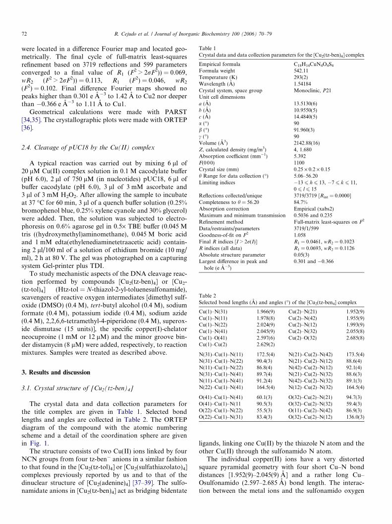
Table 1Crystal data and data collection parameters for the [Cu2(tz-ben)4] complex
Empirical formula C18H14CuN4O4S4Formula weight 542.11Temperature (K) 293(2)Wavelength (A) 1.54184Crystal system, space group Monoclinic, P21Unit cell dimensionsa (A) 13.5130(6)b (A) 10.9550(5)c (A) 14.4840(5)a (�) 90b (�) 91.960(3)
72 R. Cejudo et al. / Journal of Inorganic Biochemistry 100 (2006) 70–79
were located in a difference Fourier map and located geo-metrically. The final cycle of full-matrix least-squaresrefinement based on 3719 reflections and 599 parametersconverged to a final value of R1 (F2 > 2rF2)) = 0.069,wR2 (F2 > 2rF2)) = 0.113, R1 (F2) = 0.046, wR2
(F2) = 0.102. Final difference Fourier maps showed nopeaks higher than 0.301 e A�3 to 1.42 A to Cu2 nor deeperthan �0.366 e A�3 to 1.11 A to Cu1.
Geometrical calculations were made with PARST[34,35]. The crystallographic plots were made with ORTEP[36].
c (�) 90Volume (A3) 2142.88(16)Z, calculated density (mg/m3) 4, 1.680Absorption coefficient (mm�1) 5.392F(000) 1100Crystal size (mm) 0.25 · 0.2 · 0.15h Range for data collection (�) 5.06–56.20Limiting indices �13 6 h 6 13, �7 6 k 6 11,
0 6 l 6 15Reflections collected/unique 3719/3719 [Rint = 0.0000]Completeness to h = 56.20 84.7%Absorption correction Empirical (xabs2)Maximum and minimum transmission 0.5036 and 0.235Refinement method Full-matrix least-squares on F2
Data/restraints/parameters 3719/1/599Goodness-of-fit on F2 1.058Final R indices [I > 2r(I)] R1 = 0.0461, wR2 = 0.1023R indices (all data) R1 = 0.0693, wR2 = 0.1126Absolute structure parameter 0.05(3)Largest difference in peak andhole (e A�3)
0.301 and �0.366
Table 2Selected bond lengths (A) and angles (�) of the [Cu2(tz-ben4] complex
Cu(1)–N(31) 1.966(9) Cu(2)–N(21) 1.952(9)Cu(1)–N(11) 1.978(8) Cu(2)–N(42) 1.955(9)Cu(1)–N(22) 2.024(9) Cu(2)–N(12) 1.993(9)Cu(1)–N(41) 2.045(9) Cu(2)–N(32) 2.055(8)Cu(1)–O(41) 2.597(6) Cu(2)–O(32) 2.685(8)Cu(1)–Cu(2) 2.629(2)
N(31)–Cu(1)–N(11) 172.5(4) N(21)–Cu(2)–N(42) 173.5(4)
2.4. Cleavage of pUC18 by the Cu(II) complex
A typical reaction was carried out by mixing 6 ll of20 lM Cu(II) complex solution in 0.1 M cacodylate buffer(pH 6.0), 2 ll of 750 lM (in nucleotides) pUC18, 6 ll ofbuffer cacodylate (pH 6.0), 3 ll of 3 mM ascorbate and3 ll of 3 mM H2O2. After allowing the sample to incubateat 37 �C for 60 min, 3 ll of a quench buffer solution (0.25%bromophenol blue, 0.25% xylene cyanole and 30% glycerol)were added. Then, the solution was subjected to electro-phoresis on 0.6% agarose gel in 0.5· TBE buffer (0.045 Mtris ((hydroxymethyl)aminomethane), 0.045 M boric acidand 1 mM edta(ethylenediaminetetraacetic acid) contain-ing 2 ll/100 ml of a solution of ethidium bromide (10 mg/ml), 2 h at 80 V. The gel was photographed on a capturingsystem Gel-printer plus TDI.
To study mechanistic aspects of the DNA cleavage reac-tion performed by compounds [Cu2(tz-ben)4] or [Cu2-(tz-tol)4] (Htz-tol = N-thiazol-2-yl-toluenesulfonamide),scavengers of reactive oxygen intermediates [dimethyl sulf-oxide (DMSO) (0.4 M), tert-butyl alcohol (0.4 M), sodiumformate (0.4 M), potassium iodide (0.4 M), sodium azide(0.4 M), 2,2,6,6-tetramethyl-4-piperidone (0.4 M), superox-ide dismutase (15 units)], the specific copper(I)-chelatorneocuproine (1 mM or 12 lM) and the minor groove bin-der distamycin (8 lM) were added, respectively, to reactionmixtures. Samples were treated as described above.
N(31)–Cu(1)–N(22) 90.4(3) N(21)–Cu(2)–N(12) 88.6(4)N(11)–Cu(1)–N(22) 86.8(4) N(42)–Cu(2)–N(12) 92.1(4)N(31)–Cu(1)–N(41) 89.7(4) N(21)–Cu(2)–N(32) 88.6(3)N(11)–Cu(1)–N(41) 91.2(4) N(42)–Cu(2)–N(32) 89.1(3)N(22)–Cu(1)–N(41) 164.5(4) N(12)–Cu(2)–N(32) 164.5(4)
O(41)–Cu(1)–N(41) 60.1(3) O(32)–Cu(2)–N(21) 94.7(3)O(41)–Cu(1)–N(11 90.5(3) O(32)–Cu(2)–N(32) 59.4(3)O(22)–Cu(1)–N(22) 55.5(3) O(11)–Cu(2)–N(42) 86.9(3)O(22)–Cu(1)–N(31) 83.4(3) O(32)–Cu(2)–N(12) 136.0(3)
3. Results and discussion
3.1. Crystal structure of [Cu2(tz-ben)4]
The crystal data and data collection parameters forthe title complex are given in Table 1. Selected bondlengths and angles are collected in Table 2. The ORTEPdiagram of the compound with the atomic numberingscheme and a detail of the coordination sphere are givenin Fig. 1.
The structure consists of two Cu(II) ions linked by fourNCN groups from four tz-ben� anions in a similar fashionto that found in the [Cu2(tz-tol)4] or [Cu2(sulfathiazolato)4]complexes previously reported by us and to that of thedinuclear structure of [Cu2(adenine)4] [37–39]. The sulfo-namidate anions in [Cu2(tz-ben)4] act as bridging bidentate
ligands, linking one Cu(II) by the thiazole N atom and theother Cu(II) through the sulfonamido N atom.
The individual copper(II) ions have a very distortedsquare pyramidal geometry with four short Cu–N bonddistances [1.952(9)–2.045(9) A] and a rather long Cu–Osulfonamido (2.597–2.685 A) bond length. The interac-tion between the metal ions and the sulfonamido oxygen
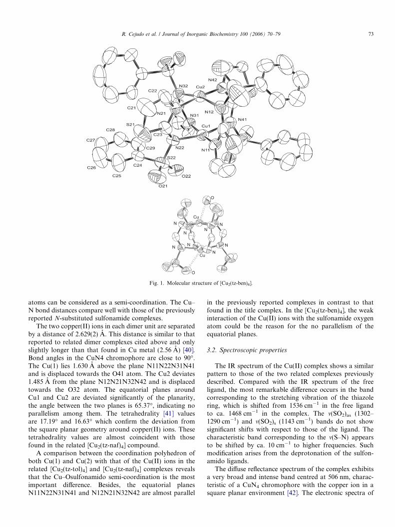
Fig. 1. Molecular structure of [Cu2(tz-ben)4].
R. Cejudo et al. / Journal of Inorganic Biochemistry 100 (2006) 70–79 73
atoms can be considered as a semi-coordination. The Cu–N bond distances compare well with those of the previouslyreported N-substituted sulfonamide complexes.
The two copper(II) ions in each dimer unit are separatedby a distance of 2.629(2) A. This distance is similar to thatreported to related dimer complexes cited above and onlyslightly longer than that found in Cu metal (2.56 A) [40].Bond angles in the CuN4 chromophore are close to 90�.The Cu(1) lies 1.630 A above the plane N11N22N31N41and is displaced towards the O41 atom. The Cu2 deviates1.485 A from the plane N12N21N32N42 and is displacedtowards the O32 atom. The equatorial planes aroundCu1 and Cu2 are deviated significantly of the planarity,the angle between the two planes is 65.37�, indicating noparallelism among them. The tetrahedrality [41] valuesare 17.19� and 16.63� which confirm the deviation fromthe square planar geometry around copper(II) ions. Thesetetrahedrality values are almost coincident with thosefound in the related [Cu2(tz-naf)4] compound.
A comparison between the coordination polyhedron ofboth Cu(1) and Cu(2) with that of the Cu(II) ions in therelated [Cu2(tz-tol)4] and [Cu2(tz-naf)4] complexes revealsthat the Cu–Osulfonamido semi-coordination is the mostimportant difference. Besides, the equatorial planesN11N22N31N41 and N12N21N32N42 are almost parallel
in the previously reported complexes in contrast to thatfound in the title complex. In the [Cu2(tz-ben)4], the weakinteraction of the Cu(II) ions with the sulfonamide oxygenatom could be the reason for the no parallelism of theequatorial planes.
3.2. Spectroscopic properties
The IR spectrum of the Cu(II) complex shows a similarpattern to those of the two related complexes previouslydescribed. Compared with the IR spectrum of the freeligand, the most remarkable difference occurs in the bandcorresponding to the stretching vibration of the thiazolering, which is shifted from 1536 cm�1 in the free ligandto ca. 1468 cm�1 in the complex. The m(SO2)as (1302–1290 cm�1) and m(SO2)s (1143 cm�1) bands do not showsignificant shifts with respect to those of the ligand. Thecharacteristic band corresponding to the m(S–N) appearsto be shifted by ca. 10 cm�1 to higher frequencies. Suchmodification arises from the deprotonation of the sulfon-amido ligands.
The diffuse reflectance spectrum of the complex exhibitsa very broad and intense band centred at 506 nm, charac-teristic of a CuN4 chromophore with the copper ion in asquare planar environment [42]. The electronic spectra of
0
0.001
0.002
0.003
0.004
0.005
0
0.2
0.4
0.6
0.8
1
0 50 100 150 200 250 300
χ M/c
m3
mol
-1
χM
T/cm
3K
mol -1
T / K
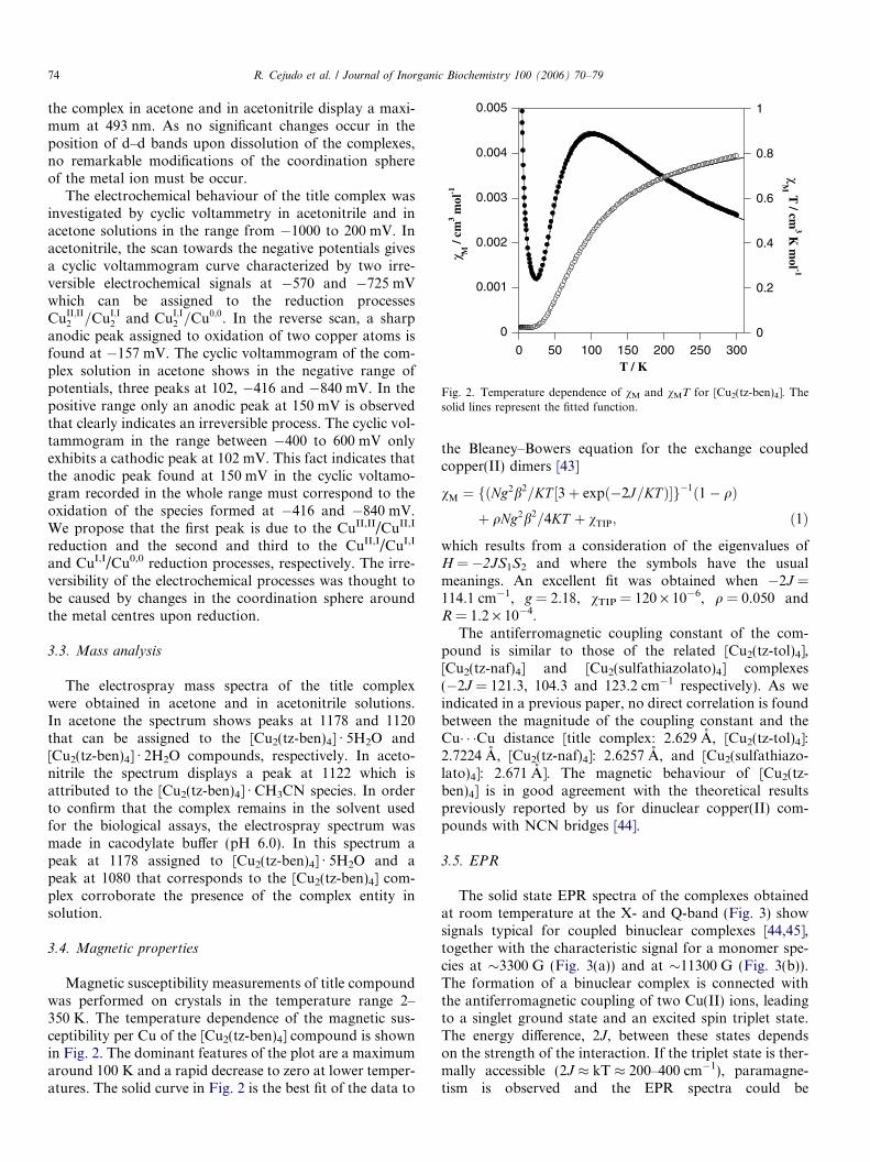
Fig. 2. Temperature dependence of vM and vMT for [Cu2(tz-ben)4]. Thesolid lines represent the fitted function.
74 R. Cejudo et al. / Journal of Inorganic Biochemistry 100 (2006) 70–79
the complex in acetone and in acetonitrile display a maxi-mum at 493 nm. As no significant changes occur in theposition of d–d bands upon dissolution of the complexes,no remarkable modifications of the coordination sphereof the metal ion must be occur.
The electrochemical behaviour of the title complex wasinvestigated by cyclic voltammetry in acetonitrile and inacetone solutions in the range from �1000 to 200 mV. Inacetonitrile, the scan towards the negative potentials givesa cyclic voltammogram curve characterized by two irre-versible electrochemical signals at �570 and �725 mVwhich can be assigned to the reduction processesCuII;II
2 =CuI;I2 and CuI;I
2 =Cu0;0. In the reverse scan, a sharpanodic peak assigned to oxidation of two copper atoms isfound at �157 mV. The cyclic voltammogram of the com-plex solution in acetone shows in the negative range ofpotentials, three peaks at 102, �416 and �840 mV. In thepositive range only an anodic peak at 150 mV is observedthat clearly indicates an irreversible process. The cyclic vol-tammogram in the range between �400 to 600 mV onlyexhibits a cathodic peak at 102 mV. This fact indicates thatthe anodic peak found at 150 mV in the cyclic voltamo-gram recorded in the whole range must correspond to theoxidation of the species formed at �416 and �840 mV.We propose that the first peak is due to the CuII,II/CuII,I
reduction and the second and third to the CuII,I/CuI,I
and CuI,I/Cu0,0 reduction processes, respectively. The irre-versibility of the electrochemical processes was thought tobe caused by changes in the coordination sphere aroundthe metal centres upon reduction.
3.3. Mass analysis
The electrospray mass spectra of the title complexwere obtained in acetone and in acetonitrile solutions.In acetone the spectrum shows peaks at 1178 and 1120that can be assigned to the [Cu2(tz-ben)4] Æ 5H2O and[Cu2(tz-ben)4] Æ 2H2O compounds, respectively. In aceto-nitrile the spectrum displays a peak at 1122 which isattributed to the [Cu2(tz-ben)4] Æ CH3CN species. In orderto confirm that the complex remains in the solvent usedfor the biological assays, the electrospray spectrum wasmade in cacodylate buffer (pH 6.0). In this spectrum apeak at 1178 assigned to [Cu2(tz-ben)4] Æ 5H2O and apeak at 1080 that corresponds to the [Cu2(tz-ben)4] com-plex corroborate the presence of the complex entity insolution.
3.4. Magnetic properties
Magnetic susceptibility measurements of title compoundwas performed on crystals in the temperature range 2–350 K. The temperature dependence of the magnetic sus-ceptibility per Cu of the [Cu2(tz-ben)4] compound is shownin Fig. 2. The dominant features of the plot are a maximumaround 100 K and a rapid decrease to zero at lower temper-atures. The solid curve in Fig. 2 is the best fit of the data to
the Bleaney–Bowers equation for the exchange coupledcopper(II) dimers [43]
vM ¼ fðNg2b2=KT ½3þ expð�2J=KT Þ�g�1ð1� qÞþ qNg2b2=4KT þ vTIP; ð1Þ
which results from a consideration of the eigenvalues ofH = �2JS1S2 and where the symbols have the usualmeanings. An excellent fit was obtained when �2J =114.1 cm�1, g = 2.18, vTIP = 120 · 10�6, q = 0.050 andR = 1.2 · 10�4.
The antiferromagnetic coupling constant of the com-pound is similar to those of the related [Cu2(tz-tol)4],[Cu2(tz-naf)4] and [Cu2(sulfathiazolato)4] complexes(�2J = 121.3, 104.3 and 123.2 cm�1 respectively). As weindicated in a previous paper, no direct correlation is foundbetween the magnitude of the coupling constant and theCu� � �Cu distance [title complex: 2.629 A, [Cu2(tz-tol)4]:2.7224 A, [Cu2(tz-naf)4]: 2.6257 A, and [Cu2(sulfathiazo-lato)4]: 2.671 A]. The magnetic behaviour of [Cu2(tz-ben)4] is in good agreement with the theoretical resultspreviously reported by us for dinuclear copper(II) com-pounds with NCN bridges [44].
3.5. EPR
The solid state EPR spectra of the complexes obtainedat room temperature at the X- and Q-band (Fig. 3) showsignals typical for coupled binuclear complexes [44,45],together with the characteristic signal for a monomer spe-cies at �3300 G (Fig. 3(a)) and at �11300 G (Fig. 3(b)).The formation of a binuclear complex is connected withthe antiferromagnetic coupling of two Cu(II) ions, leadingto a singlet ground state and an excited spin triplet state.The energy difference, 2J, between these states dependson the strength of the interaction. If the triplet state is ther-mally accessible (2J � kT � 200–400 cm�1), paramagne-tism is observed and the EPR spectra could be
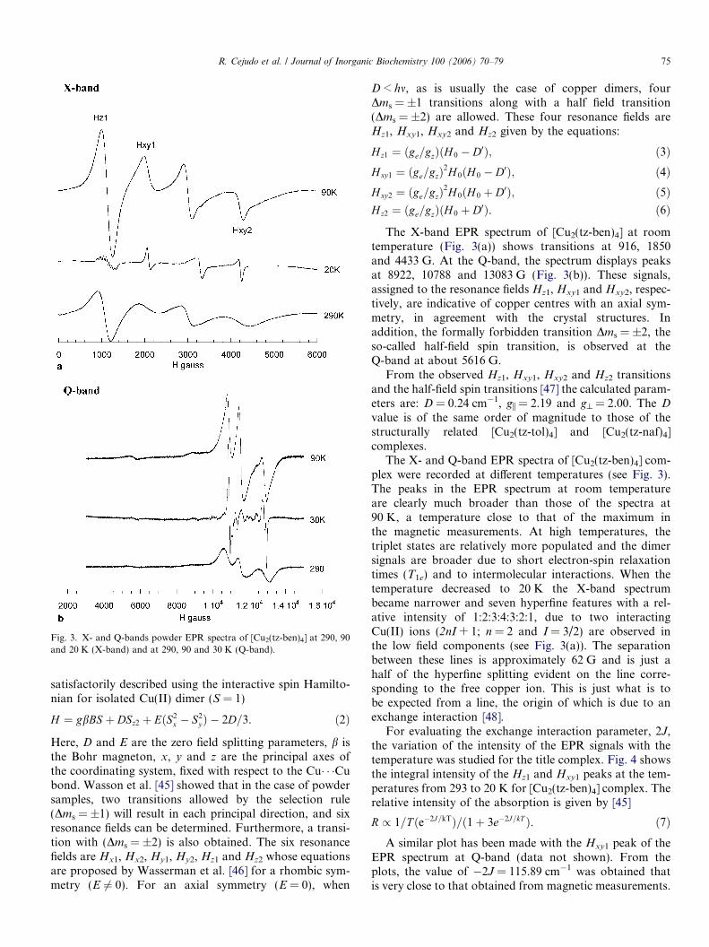
Fig. 3. X- and Q-bands powder EPR spectra of [Cu2(tz-ben)4] at 290, 90and 20 K (X-band) and at 290, 90 and 30 K (Q-band).
R. Cejudo et al. / Journal of Inorganic Biochemistry 100 (2006) 70–79 75
satisfactorily described using the interactive spin Hamilto-nian for isolated Cu(II) dimer (S = 1)
H ¼ gbBS þ DSz2 þ EðS2x � S2
yÞ � 2D=3. ð2Þ
Here, D and E are the zero field splitting parameters, b isthe Bohr magneton, x, y and z are the principal axes ofthe coordinating system, fixed with respect to the Cu� � �Cubond. Wasson et al. [45] showed that in the case of powdersamples, two transitions allowed by the selection rule(Dms = ±1) will result in each principal direction, and sixresonance fields can be determined. Furthermore, a transi-tion with (Dms = ±2) is also obtained. The six resonancefields are Hx1, Hx2, Hy1, Hy2, Hz1 and Hz2 whose equationsare proposed by Wasserman et al. [46] for a rhombic sym-metry (E 6¼ 0). For an axial symmetry (E = 0), when
D < hm, as is usually the case of copper dimers, fourDms = ±1 transitions along with a half field transition(Dms = ±2) are allowed. These four resonance fields areHz1, Hxy1, Hxy2 and Hz2 given by the equations:
Hz1 ¼ ðge=gzÞðH 0 � D0Þ; ð3ÞHxy1 ¼ ðge=gzÞ
2H 0ðH 0 � D0Þ; ð4ÞHxy2 ¼ ðge=gzÞ
2H 0ðH 0 þ D0Þ; ð5ÞHz2 ¼ ðge=gzÞðH 0 þ D0Þ. ð6Þ
The X-band EPR spectrum of [Cu2(tz-ben)4] at roomtemperature (Fig. 3(a)) shows transitions at 916, 1850and 4433 G. At the Q-band, the spectrum displays peaksat 8922, 10788 and 13083 G (Fig. 3(b)). These signals,assigned to the resonance fields Hz1, Hxy1 and Hxy2, respec-tively, are indicative of copper centres with an axial sym-metry, in agreement with the crystal structures. Inaddition, the formally forbidden transition Dms = ±2, theso-called half-field spin transition, is observed at theQ-band at about 5616 G.
From the observed Hz1, Hxy1, Hxy2 and Hz2 transitionsand the half-field spin transitions [47] the calculated param-eters are: D = 0.24 cm�1, gi = 2.19 and g^ = 2.00. The D
value is of the same order of magnitude to those of thestructurally related [Cu2(tz-tol)4] and [Cu2(tz-naf)4]complexes.
The X- and Q-band EPR spectra of [Cu2(tz-ben)4] com-plex were recorded at different temperatures (see Fig. 3).The peaks in the EPR spectrum at room temperatureare clearly much broader than those of the spectra at90 K, a temperature close to that of the maximum inthe magnetic measurements. At high temperatures, thetriplet states are relatively more populated and the dimersignals are broader due to short electron-spin relaxationtimes (T1e) and to intermolecular interactions. When thetemperature decreased to 20 K the X-band spectrumbecame narrower and seven hyperfine features with a rel-ative intensity of 1:2:3:4:3:2:1, due to two interactingCu(II) ions (2nI + 1; n = 2 and I = 3/2) are observed inthe low field components (see Fig. 3(a)). The separationbetween these lines is approximately 62 G and is just ahalf of the hyperfine splitting evident on the line corre-sponding to the free copper ion. This is just what is tobe expected from a line, the origin of which is due to anexchange interaction [48].
For evaluating the exchange interaction parameter, 2J,the variation of the intensity of the EPR signals with thetemperature was studied for the title complex. Fig. 4 showsthe integral intensity of the Hz1 and Hxy1 peaks at the tem-peratures from 293 to 20 K for [Cu2(tz-ben)4] complex. Therelative intensity of the absorption is given by [45]
R / 1=T ðe�2J=kTÞ=ð1þ 3e�2J=kT Þ. ð7ÞA similar plot has been made with the Hxy1 peak of the
EPR spectrum at Q-band (data not shown). From theplots, the value of �2J = 115.89 cm�1 was obtained thatis very close to that obtained from magnetic measurements.
0
1
2
3
4
5
0 50 100 150 200 250 300
Inte
nsity
, var
iabl
e
T/K
Hz1
Hxy1
Fig. 4. Temperature dependence of the intensity of the Hz1 and Hxy1 EPRsignals for [Cu2(tz-ben)4] complex.
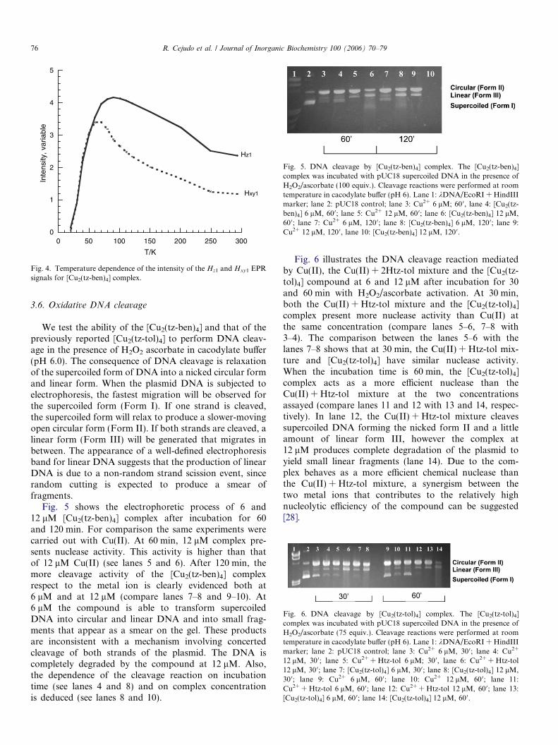
Fig. 5. DNA cleavage by [Cu2(tz-ben)4] complex. The [Cu2(tz-ben)4]complex was incubated with pUC18 supercoiled DNA in the presence ofH2O2/ascorbate (100 equiv.). Cleavage reactions were performed at roomtemperature in cacodylate buffer (pH 6). Lane 1: kDNA/EcoRI + HindIIImarker; lane 2: pUC18 control; lane 3: Cu2+ 6 lM; 60 0, lane 4: [Cu2(tz-ben)4] 6 lM, 600; lane 5: Cu2+ 12 lM, 60 0; lane 6: [Cu2(tz-ben)4] 12 lM,60 0; lane 7: Cu2+ 6 lM, 120 0; lane 8: [Cu2(tz-ben)4] 6 lM, 120 0; lane 9:Cu2+ 12 lM, 120 0, lane 10: [Cu2(tz-ben)4] 12 lM, 120 0.
Fig. 6. DNA cleavage by [Cu2(tz-tol)4] complex. The [Cu2(tz-tol)4]complex was incubated with pUC18 supercoiled DNA in the presence ofH2O2/ascorbate (75 equiv.). Cleavage reactions were performed at roomtemperature in cacodylate buffer (pH 6). Lane 1: kDNA/EcoRI + HindIIImarker; lane 2: pUC18 control; lane 3: Cu2+ 6 lM, 300; lane 4: Cu2+
12 lM, 300; lane 5: Cu2+ + Htz-tol 6 lM; 30 0, lane 6: Cu2+ + Htz-tol12 lM, 30 0; lane 7: [Cu2(tz-tol)4] 6 lM, 300; lane 8: [Cu2(tz-tol)4] 12 lM,30 0; lane 9: Cu2+ 6 lM, 600; lane 10: Cu2+ 12 lM, 60 0; lane 11:Cu2+ + Htz-tol 6 lM, 600; lane 12: Cu2+ + Htz-tol 12 lM, 60 0; lane 13:[Cu2(tz-tol)4] 6 lM, 60 0; lane 14: [Cu2(tz-tol)4] 12 lM, 600.
76 R. Cejudo et al. / Journal of Inorganic Biochemistry 100 (2006) 70–79
3.6. Oxidative DNA cleavage
We test the ability of the [Cu2(tz-ben)4] and that of thepreviously reported [Cu2(tz-tol)4] to perform DNA cleav-age in the presence of H2O2 ascorbate in cacodylate buffer(pH 6.0). The consequence of DNA cleavage is relaxationof the supercoiled form of DNA into a nicked circular formand linear form. When the plasmid DNA is subjected toelectrophoresis, the fastest migration will be observed forthe supercoiled form (Form I). If one strand is cleaved,the supercoiled form will relax to produce a slower-movingopen circular form (Form II). If both strands are cleaved, alinear form (Form III) will be generated that migrates inbetween. The appearance of a well-defined electrophoresisband for linear DNA suggests that the production of linearDNA is due to a non-random strand scission event, sincerandom cutting is expected to produce a smear offragments.
Fig. 5 shows the electrophoretic process of 6 and12 lM [Cu2(tz-ben)4] complex after incubation for 60and 120 min. For comparison the same experiments werecarried out with Cu(II). At 60 min, 12 lM complex pre-sents nuclease activity. This activity is higher than thatof 12 lM Cu(II) (see lanes 5 and 6). After 120 min, themore cleavage activity of the [Cu2(tz-ben)4] complexrespect to the metal ion is clearly evidenced both at6 lM and at 12 lM (compare lanes 7–8 and 9–10). At6 lM the compound is able to transform supercoiledDNA into circular and linear DNA and into small frag-ments that appear as a smear on the gel. These productsare inconsistent with a mechanism involving concertedcleavage of both strands of the plasmid. The DNA iscompletely degraded by the compound at 12 lM. Also,the dependence of the cleavage reaction on incubationtime (see lanes 4 and 8) and on complex concentrationis deduced (see lanes 8 and 10).
Fig. 6 illustrates the DNA cleavage reaction mediatedby Cu(II), the Cu(II) + 2Htz-tol mixture and the [Cu2(tz-tol)4] compound at 6 and 12 lM after incubation for 30and 60 min with H2O2/ascorbate activation. At 30 min,both the Cu(II) + Htz-tol mixture and the [Cu2(tz-tol)4]complex present more nuclease activity than Cu(II) atthe same concentration (compare lanes 5–6, 7–8 with3–4). The comparison between the lanes 5–6 with thelanes 7–8 shows that at 30 min, the Cu(II) + Htz-tol mix-ture and [Cu2(tz-tol)4] have similar nuclease activity.When the incubation time is 60 min, the [Cu2(tz-tol)4]complex acts as a more efficient nuclease than theCu(II) + Htz-tol mixture at the two concentrationsassayed (compare lanes 11 and 12 with 13 and 14, respec-tively). In lane 12, the Cu(II) + Htz-tol mixture cleavessupercoiled DNA forming the nicked form II and a littleamount of linear form III, however the complex at12 lM produces complete degradation of the plasmid toyield small linear fragments (lane 14). Due to the com-plex behaves as a more efficient chemical nuclease thanthe Cu(II) + Htz-tol mixture, a synergism between thetwo metal ions that contributes to the relatively highnucleolytic efficiency of the compound can be suggested[28].
R. Cejudo et al. / Journal of Inorganic Biochemistry 100 (2006) 70–79 77
A comparison of lane 14 in Fig. 6 and lane 6 in Fig. 5indicates that the activity of the [Cu2(tz-tol)4] compoundis higher than the activity of the [Cu2(tz-ben)4].
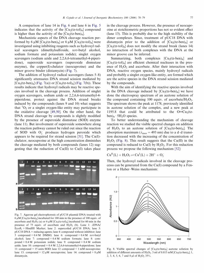
Mechanistic aspects of the DNA cleavage reaction per-formed by 6 lM [Cu2(tz-ben)4] or 6 lM [Cu2(tz-tol)4] wereinvestigated using inhibiting reagents such as hydroxyl rad-ical scavengers (dimethylsulfoxide, tert-butyl alcohol,sodium formate and potassium iodide); singlet oxygenscavengers (sodium azide and 2,2,6,6-tetramethyl-4-piperi-done), superoxide scavengers (superoxide dismutaseenzyme), the copper(I)-chelator (neocuproine) and theminor groove binder (distamycin) (Fig. 7).
The addition of hydroxyl radical scavengers (lanes 5–8)significantly attenuates DNA strand scission mediated by[Cu2(tz-ben)4] (Fig. 7(a)) or [Cu2(tz-tol)4] (Fig. 7(b)). Theseresults indicate that hydroxyl radicals may be reactive spe-cies involved in the cleavage process. Addition of singletoxygen scavengers, sodium azide or 2,2,6,6-tetramethyl-4-piperidone, protect against the DNA strand breaksinduced by the compounds (lanes 9 and 10) what suggeststhat 1O2 or a singlet oxygen-like entity may participate inthe oxidative cleavage [49,50]. On the other hand, theDNA strand cleavage by compounds is slightly modifiedby the presence of superoxide dismutase (SOD) enzyme(lane 11). But involvement of superoxide somewhere alongthe reaction pathway cannot be ruled out since the reactionof SOD with O�
2 produces hydrogen peroxide whichappears to be required for strand scission [51]. The Cu(I)-chelator, neocuproine at the high concentration diminishesthe cleavage mediated by both compounds (lanes 12) sug-gesting that the reduction of Cu(II) to Cu(I) takes place
Fig. 7. Agarose gel electrophoresis of pUC18 plasmid DNA treated with6 lM [Cu2(tz-ben)4] incubated for 180 min in the presence of 100 equiv. ofascorbate and H2O2 (a) or 6 lM [Cu2(tz-tol)4] incubated for 60 min in thepresence of 75 equiv. of ascorbate and H2O2 (b). Lane 1: kDNA/EcoR1 + HindIII Marker; lane 2: supercoiled pUC18 DNA; lane 3:pUC18 DNA + reducing agents; lane 4: compound without inhibitor; lane5 compound + 0.4 M DMSO; lane 6: compound + 0.4 M tert-butylalcohol; lane 7: compound + 0.4 M sodium formate; lane 8: com-pound + 0.4 M potassium iodide; lane 9: compound + 0.4 M sodiumazide; lane 10: compound + 0.4 M 2,2,6,6-tetramethyl-4-piperidone; lane11: compound + 15 units SOD; lane 12: compound + 1 mM neocuproine;lane 13: compound + 12 lM neocuproine; lane 14: compound + 8 lMdistamycin.
in the cleavage process. However, the presence of neocupr-oine at stoichiometric proportions has not so evident effect(lane 13). This is probably due to the high stability of thedimer complexes. Since, treatment of pUC18 DNA withdistamycin prior to the addition of [Cu2(tz-ben)4] or[Cu2(tz-tol)4] does not modify the strand break (lanes 14)no interaction of both complexes with the DNA at theminor groove can be inferred.
Summarizing, both complexes [Cu2(tz-ben)4] and[Cu2(tz-tol)4] are efficient chemical nucleases in the pres-ence of H2O2 and ascorbate. After their interaction withDNA, reactive oxygen species, mainly hydroxyl radicalsand probably a singlet oxygen-like entity, are formed whichare the active species in the DNA strand scission mediatedby the compounds.
With the aim of identifying the reactive species involvedin the DNA cleavage induced by [Cu2(tz-ben)4] we havedone the electrospray spectrum of an acetone solution ofthe compound containing 100 equiv. of ascorbate/H2O2.The spectrum shows the peak at 1178, previously identifiedin acetone solution of the complex, and a new peak at1195.8 that could be attributed to the O@Cu2(tz-ben)4 Æ 5H2O species.
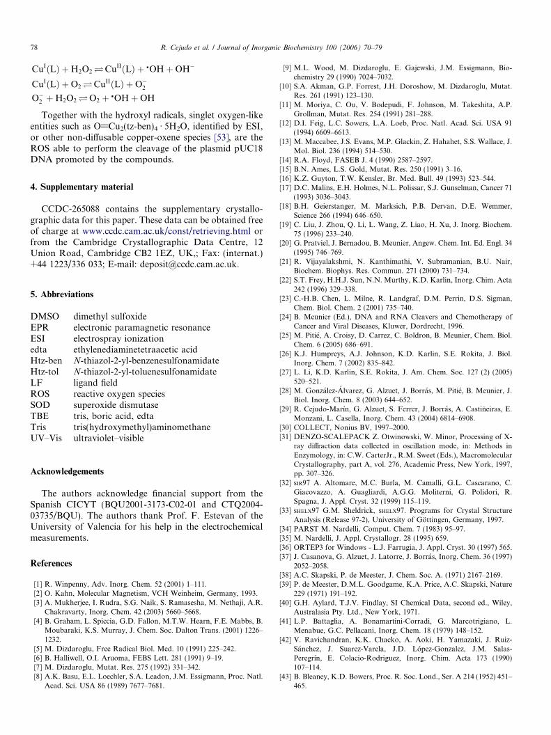
To better understanding the mechanism of cleavagereaction we studied the visible spectral changes on additionof H2O2 to an acetone solution of [Cu2(tz-ben)4]. Theabsorption maximum (kmax = 493 nm) due to a d–d transi-tion decreased with the increasing of the concentration ofH2O2 (Fig. 8). This result suggests that the Cu(II) in thecompound is reduced to Cu(I) by H2O2. For this reductionprocess we propose the following mechanism [52]:
CuIIðLÞ þH2O2 �CuIðLÞ þ 2Hþ þO�2
Then, the hydroxyl radicals involved in the cleavage pro-cesss can be generated from the Cu(I) compound by a Fen-ton or a Haber–Weiss mechanism:
0
0.5
1
1.5
2
350 400 450 500 550 600 650 700
Abs
orba
nce
(AU
)
Wavelength (nm)
0
1
2
3
4
5
67
8
Fig. 8. Visible spectral changes of [Cu2(tz-ben)4] acetone solution byaddition of different amounts of H2O2. 3 ml of 0.815 mM [Cu2(tz-ben)4]; 1,2, 3, 4, 5, 6, 7 and 8 ll of H2O2 35%.
78 R. Cejudo et al. / Journal of Inorganic Biochemistry 100 (2006) 70–79
CuIðLÞ þH2O2 �CuIIðLÞ þ �OHþOH�
CuIðLÞ þO2 �CuIIðLÞ þO�2
O�2 þH2O2 �O2 þ �OHþOH
Together with the hydroxyl radicals, singlet oxygen-likeentities such as O@Cu2(tz-ben)4 Æ 5H2O, identified by ESI,or other non-diffusable copper-oxene species [53], are theROS able to perform the cleavage of the plasmid pUC18DNA promoted by the compounds.
4. Supplementary material
CCDC-265088 contains the supplementary crystallo-graphic data for this paper. These data can be obtained freeof charge at www.ccdc.cam.ac.uk/const/retrieving.html orfrom the Cambridge Crystallographic Data Centre, 12Union Road, Cambridge CB2 1EZ, UK,; Fax: (internat.)+44 1223/336 033; E-mail: [email protected].
5. Abbreviations
DMSO dimethyl sulfoxide
EPR electronic paramagnetic resonance ESI electrospray ionization edta ethylenediaminetetraacetic acid Htz-ben N-thiazol-2-yl-benzenesulfonamidate Htz-tol N-thiazol-2-yl-toluenesulfonamidate LF ligand field ROS reactive oxygen species SOD superoxide dismutase TBE tris, boric acid, edta Tris tris(hydroxymethyl)aminomethane UV–Vis ultraviolet–visibleAcknowledgements
The authors acknowledge financial support from theSpanish CICYT (BQU2001-3173-C02-01 and CTQ2004-03735/BQU). The authors thank Prof. F. Estevan of theUniversity of Valencia for his help in the electrochemicalmeasurements.
References
[1] R. Winpenny, Adv. Inorg. Chem. 52 (2001) 1–111.[2] O. Kahn, Molecular Magnetism, VCH Weinheim, Germany, 1993.[3] A. Mukherjee, I. Rudra, S.G. Naik, S. Ramasesha, M. Nethaji, A.R.
Chakravarty, Inorg. Chem. 42 (2003) 5660–5668.[4] B. Graham, L. Spiccia, G.D. Fallon, M.T.W. Hearn, F.E. Mabbs, B.
Moubaraki, K.S. Murray, J. Chem. Soc. Dalton Trans. (2001) 1226–1232.
[5] M. Dizdaroglu, Free Radical Biol. Med. 10 (1991) 225–242.[6] B. Halliwell, O.I. Aruoma, FEBS Lett. 281 (1991) 9–19.[7] M. Dizdaroglu, Mutat. Res. 275 (1992) 331–342.[8] A.K. Basu, E.L. Loechler, S.A. Leadon, J.M. Essigmann, Proc. Natl.
Acad. Sci. USA 86 (1989) 7677–7681.
[9] M.L. Wood, M. Dizdaroglu, E. Gajewski, J.M. Essigmann, Bio-chemistry 29 (1990) 7024–7032.
[10] S.A. Akman, G.P. Forrest, J.H. Doroshow, M. Dizdaroglu, Mutat.Res. 261 (1991) 123–130.
[11] M. Moriya, C. Ou, V. Bodepudi, F. Johnson, M. Takeshita, A.P.Grollman, Mutat. Res. 254 (1991) 281–288.
[12] D.I. Feig, L.C. Sowers, L.A. Loeb, Proc. Natl. Acad. Sci. USA 91(1994) 6609–6613.
[13] M. Maccabee, J.S. Evans, M.P. Glackin, Z. Hahahet, S.S. Wallace, J.Mol. Biol. 236 (1994) 514–530.
[14] R.A. Floyd, FASEB J. 4 (1990) 2587–2597.[15] B.N. Ames, L.S. Gold, Mutat. Res. 250 (1991) 3–16.[16] K.Z. Guyton, T.W. Kensler, Br. Med. Bull. 49 (1993) 523–544.[17] D.C. Malins, E.H. Holmes, N.L. Polissar, S.J. Gunselman, Cancer 71
(1993) 3036–3043.[18] B.H. Geierstanger, M. Marksich, P.B. Dervan, D.E. Wemmer,
Science 266 (1994) 646–650.[19] C. Liu, J. Zhou, Q. Li, L. Wang, Z. Liao, H. Xu, J. Inorg. Biochem.
75 (1996) 233–240.[20] G. Pratviel, J. Bernadou, B. Meunier, Angew. Chem. Int. Ed. Engl. 34
(1995) 746–769.[21] R. Vijayalakshmi, N. Kanthimathi, V. Subramanian, B.U. Nair,
Biochem. Biophys. Res. Commun. 271 (2000) 731–734.[22] S.T. Frey, H.H.J. Sun, N.N. Murthy, K.D. Karlin, Inorg. Chim. Acta
242 (1996) 329–338.[23] C.-H.B. Chen, L. Milne, R. Landgraf, D.M. Perrin, D.S. Sigman,
Chem. Biol. Chem. 2 (2001) 735–740.[24] B. Meunier (Ed.), DNA and RNA Cleavers and Chemotherapy of
Cancer and Viral Diseases, Kluwer, Dordrecht, 1996.[25] M. Pitie, A. Croisy, D. Carrez, C. Boldron, B. Meunier, Chem. Biol.
Chem. 6 (2005) 686–691.[26] K.J. Humpreys, A.J. Johnson, K.D. Karlin, S.E. Rokita, J. Biol.
Inorg. Chem. 7 (2002) 835–842.[27] L. Li, K.D. Karlin, S.E. Rokita, J. Am. Chem. Soc. 127 (2) (2005)
520–521.[28] M. Gonzalez-Alvarez, G. Alzuet, J. Borras, M. Pitie, B. Meunier, J.
Biol. Inorg. Chem. 8 (2003) 644–652.[29] R. Cejudo-Marın, G. Alzuet, S. Ferrer, J. Borras, A. Castineiras, E.
Monzani, L. Casella, Inorg. Chem. 43 (2004) 6814–6908.[30] COLLECT, Nonius BV, 1997–2000.[31] DENZO-SCALEPACK Z. Otwinowski, W. Minor, Processing of X-
ray diffraction data collected in oscillation mode, in: Methods inEnzymology, in: C.W. CarterJr., R.M. Sweet (Eds.), MacromolecularCrystallography, part A, vol. 276, Academic Press, New York, 1997,pp. 307–326.
[32] SIR97 A. Altomare, M.C. Burla, M. Camalli, G.L. Cascarano, C.Giacovazzo, A. Guagliardi, A.G.G. Moliterni, G. Polidori, R.Spagna, J. Appl. Cryst. 32 (1999) 115–119.
[33] SHELX97 G.M. Sheldrick, SHELX97. Programs for Crystal StructureAnalysis (Release 97-2), University of Gottingen, Germany, 1997.
[34] PARST M. Nardelli, Comput. Chem. 7 (1983) 95–97.[35] M. Nardelli, J. Appl. Crystallogr. 28 (1995) 659.[36] ORTEP3 for Windows - L.J. Farrugia, J. Appl. Cryst. 30 (1997) 565.[37] J. Casanova, G. Alzuet, J. Latorre, J. Borras, Inorg. Chem. 36 (1997)
2052–2058.[38] A.C. Skapski, P. de Meester, J. Chem. Soc. A. (1971) 2167–2169.[39] P. de Meester, D.M.L. Goodgame, K.A. Price, A.C. Skapski, Nature
229 (1971) 191–192.[40] G.H. Aylard, T.J.V. Findlay, SI Chemical Data, second ed., Wiley,
Australasia Pty. Ltd., New York, 1971.[41] L.P. Battaglia, A. Bonamartini-Corradi, G. Marcotrigiano, L.
Menabue, G.C. Pellacani, Inorg. Chem. 18 (1979) 148–152.[42] V. Ravichandran, K.K. Chacko, A. Aoki, H. Yamazaki, J. Ruiz-
Sanchez, J. Suarez-Varela, J.D. Lopez-Gonzalez, J.M. Salas-Peregrın, E. Colacio-Rodriguez, Inorg. Chim. Acta 173 (1990)107–114.
[43] B. Bleaney, K.D. Bowers, Proc. R. Soc. Lond., Ser. A 214 (1952) 451–465.
R. Cejudo et al. / Journal of Inorganic Biochemistry 100 (2006) 70–79 79
[44] L. Gutierrez, G. Alzuet, J. Borras, A. Castineiras, A. Rodriguez-Fortea, E. Ruiz, Inorg. Chem. 40 (2001) 3089–3096.
[45] J.R. Wasson, S. Chin-I, C. Trapp, Inorg. Chem. 7 (1968) 469–473.[46] E. Wasserman, L.C. Snyder, W.A. Yager, J. Chem. Phys. 41 (1964)
1763–1772.[47] S.A. Eaton, K.M. More, B.M. Sawant, G.R. Eaton, J. Am. Chem.
Soc. 105 (1983) 6560–6567.[48] C.P. Slichter, Phys. Rev. 99 (1955) 479–480.
[49] Y. Li, M.A. Trush, Carcinogenesis 14 (1993) 1303–1311.[50] S. Frelon, T. Douki, A. Favier, J. Cadet, Chem. Res. Toxicol. 16
(2003) 191–197.[51] S.T. Frey, H.H.J. Sun, N.N. Murthy, K.D. Karlin, Inorg. Chim. Acta
242 (1996) 329–338.[52] T. Ozawa, A. Hanaki, K. Onodera, Polyhedron 11 (1992) 735–
738.[53] D.S. Sigman, C.H.B. Chen, ACS Symp. Ser. 402 (1989) 24–49.
Related Documents