CURRICULUM FOR 2 YEARS DIPLOMA PROGRAMME IN MEDICAL RADIOLOGY-THERAPEUTICS (DMRT) 2007 UINVERSITY OF HEALTH SECIENCES LAHORE, PAKISTAN

Welcome message from author
This document is posted to help you gain knowledge. Please leave a comment to let me know what you think about it! Share it to your friends and learn new things together.
Transcript
CURRICULUM
FOR
2 YEARS DIPLOMA PROGRAMME IN
MEDICAL RADIOLOGY-THERAPEUTICS
(DMRT)
2007
UINVERSITY OF HEALTH SECIENCES LAHORE, PAKISTAN
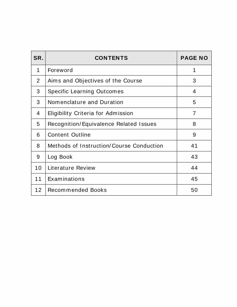
SR. CONTENTS PAGE NO
1 Foreword 1
2 Aims and Objectives of the Course 3
3 Specific Learning Outcomes 4
3 Nomenclature and Duration 5
4 Eligibility Criteria for Admission 7
5 Recognition/Equivalence Related Issues 8
6 Content Outline 9
8 Methods of Instruction/Course Conduction 41
9 Log Book 43
10 Literature Review 44
11 Examinations 45
12 Recommended Books 50

1
FOREWORD
University of Health Sciences (UHS) Lahore was inaugurated by the President of Pakistan on the 3rd of October 2002 with the vision to explicitly address academic and research needs in the field of health sciences and allied disciplines and to uplift their existing level to bring them at par with the international standards.
The mission of the University is to develop an intellectual nexus to provide excellence, innovation, and research in medical education in order to;
• Impart knowledge and skills to health care providers to enhance their competence in providing community oriented and multi-disciplinary patient-centered care
• Train and produce researchers and specialists in basic and clinical medical sciences
• Establish and maintain continuing professional development programmes for the faculty
• Provide trained professionals and scientists/researchers for the field of Electro Medical/Bio-Medical disciplines
• Assure quality in health education and to promote research at all levels
A university is the zenith of knowledge that imparts quality education and awards degrees for extensive educational attainments in various disciplines with attendant advancement for the development of intellectual community. Protection of traditional knowledge, making exploration about it and obtaining deep understanding of modern technology and research techniques are some of the responsibilities of any university.
UHS is running a number of courses in the field of health sciences in Punjab. The list extends from undergraduate level courses to the doctorate level courses in basic, clinical and allied health sciences.
Since its inception, certain vital tasks were taken into serious consideration by UHS, for instance, curricula development and their up-gradation were among the most important ones besides introduction of contemporary educational programmes.
UHS has revised and finalized curricula for undergraduate Medical/ Dental Education, B Sc Nursing, and Allied Health Sciences.
2
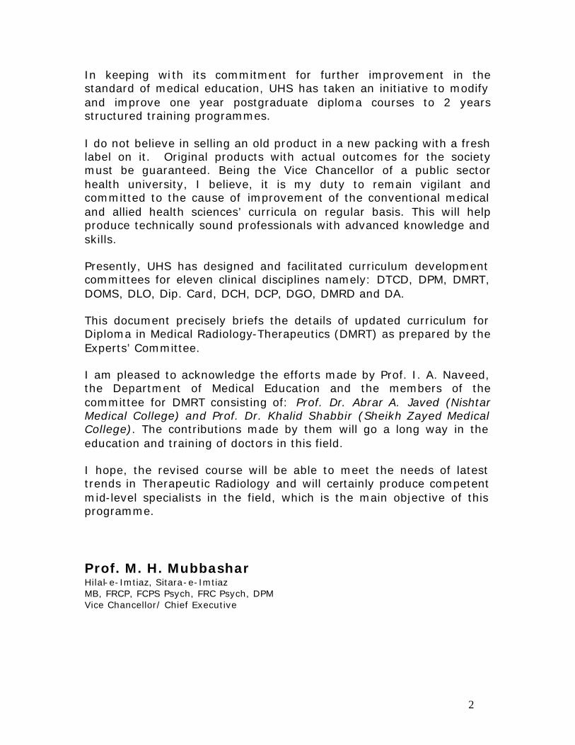
In keeping with its commitment for further improvement in the standard of medical education, UHS has taken an initiative to modify and improve one year postgraduate diploma courses to 2 years structured training programmes.
I do not believe in selling an old product in a new packing with a fresh label on it. Original products with actual outcomes for the society must be guaranteed. Being the Vice Chancellor of a public sector health university, I believe, it is my duty to remain vigilant and committed to the cause of improvement of the conventional medical and allied health sciences’ curricula on regular basis. This will help produce technically sound professionals with advanced knowledge and skills.
Presently, UHS has designed and facilitated curriculum development committees for eleven clinical disciplines namely: DTCD, DPM, DMRT, DOMS, DLO, Dip. Card, DCH, DCP, DGO, DMRD and DA.
This document precisely briefs the details of updated curriculum for Diploma in Medical Radiology-Therapeutics (DMRT) as prepared by the Experts’ Committee.
I am pleased to acknowledge the efforts made by Prof. I. A. Naveed, the Department of Medical Education and the members of the committee for DMRT consisting of: Prof. Dr. Abrar A. Javed (Nishtar Medical College) and Prof. Dr. Khalid Shabbir (Sheikh Zayed Medical College). The contributions made by them will go a long way in the education and training of doctors in this field.
I hope, the revised course will be able to meet the needs of latest trends in Therapeutic Radiology and will certainly produce competent mid-level specialists in the field, which is the main objective of this programme.
Prof. M. H. Mubbashar Hilal-e-Imtiaz, Sitara-e-Imtiaz MB, FRCP, FCPS Psych, FRC Psych, DPM Vice Chancellor/ Chief Executive

3
AIMS AND OBJECTIVES OF THE COURSE AIM The aim of two years diploma programme in Medical Radiology-Therapeutics is to equip medical graduates with relevant professional knowledge, skills and ethical values to enable them to apply their acquired expertise at primary and secondary health care organizations as non-academic consultants. OBJECTIVES At the end of training in DMRT, a trainee doctor should be able to: 1. Take a comprehensive and pertinent history of patients coming for
management of cancer diseases 2. Take proper informed consent before commencing any procedure
and ensuring confidentiality and appropriate environment for procedures and treatment processes involving unusual exposure
3. Explain all procedures to patients or to their relatives in patient’s preferred language in elective and emergency situations good communication skills, empathy and empowerment to patients
4. Satisfactorily address fears, concerns and expectations of the patients
5. Discuss problems of patient, outcome and after effects of treatment 6. Exhibit emotional maturity, stability, integrity, ethical values,
professional approach and sense of responsibility in day-to-day professional activities
7. Act as an independent specialist at Community/Tehsil and District Headquarter Hospital
8. Show initiative and become self-directed learner tapping on resources including clinical material, faculty, internet, library and on-line learning programmes
9. Independently manage all cancer malignancies with special concern about the safety of the patient during all the procedures to be undertaken for the treatment of patients
4
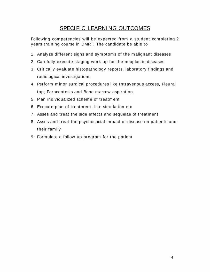
SPECIFIC LEARNING OUTCOMES Following competencies will be expected from a student completing 2 years training course in DMRT. The candidate be able to
1. Analyze different signs and symptoms of the malignant diseases
2. Carefully execute staging work up for the neoplastic diseases
3. Critically evaluate histopathology reports, laboratory findings and
radiological investigations
4. Perform minor surgical procedures like Intravenous access, Pleural
tap, Paracentesis and Bone marrow aspiration.
5. Plan individualized scheme of treatment
6. Execute plan of treatment, like simulation etc
7. Asses and treat the side effects and sequelae of treatment
8. Asses and treat the psychosocial impact of disease on patients and
their family
9. Formulate a follow up program for the patient
5
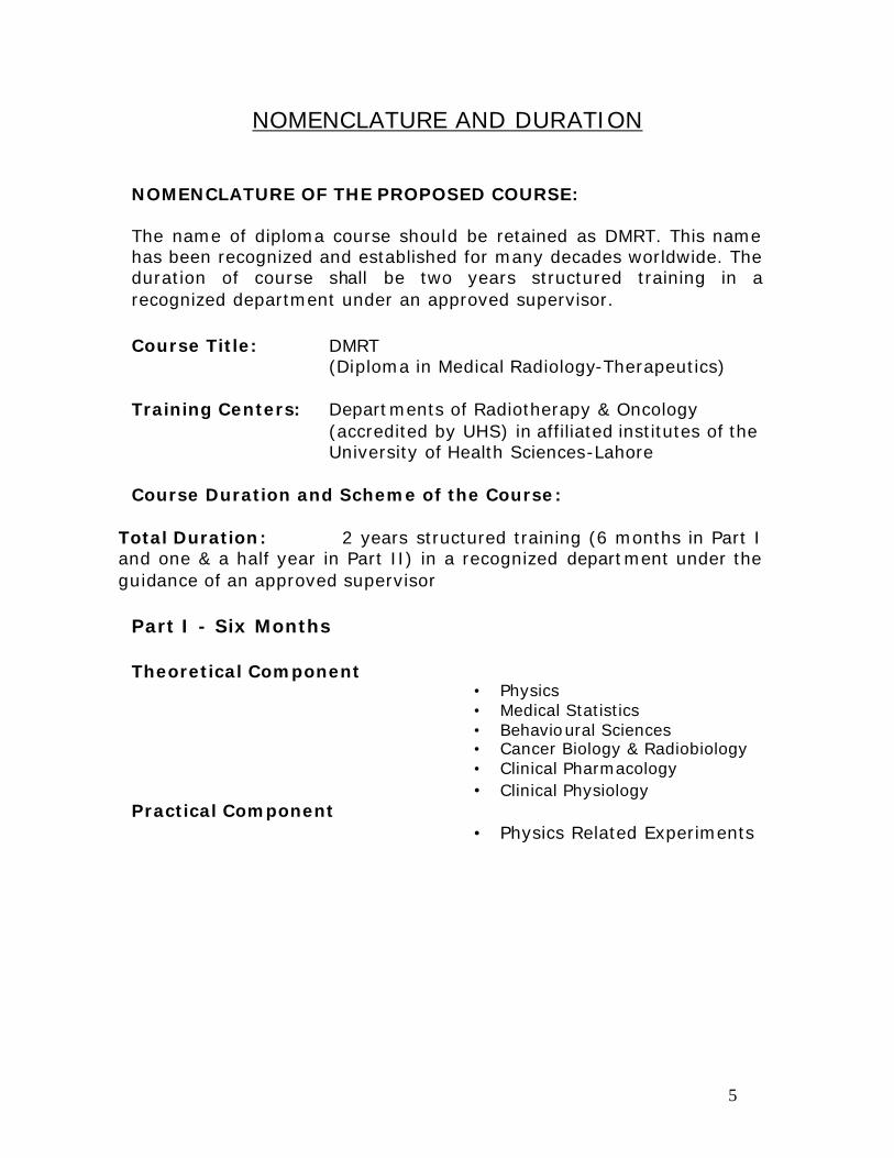
NOMENCLATURE AND DURATION
NOMENCLATURE OF THE PROPOSED COURSE: The name of diploma course should be retained as DMRT. This name has been recognized and established for many decades worldwide. The duration of course shall be two years structured training in a recognized department under an approved supervisor. Course Title: DMRT
(Diploma in Medical Radiology-Therapeutics) Training Centers: Departments of Radiotherapy & Oncology
(accredited by UHS) in affiliated institutes of the University of Health Sciences-Lahore
Course Duration and Scheme of the Course:
Total Duration: 2 years structured training (6 months in Part I and one & a half year in Part II) in a recognized department under the guidance of an approved supervisor
Part I - Six Months Theoretical Component
• Physics • Medical Statistics • Behavioural Sciences • Cancer Biology & Radiobiology • Clinical Pharmacology • Clinical Physiology
Practical Component • Physics Related Experiments
6
Part II - Year and a half Theoretical Component
INTRODUCTION
Final Examination expects candidates to have a wide knowledge of malignant disease and their management. The main emphasis is on radiotherapy and drug therapy, but a good knowledge of medicine, surgery, radiographic imaging, gynaecology and allied specialities is expected.
KNOWLEDGE
Candidates for the Final Examination need to have a broad knowledge relating to all aspects of investigation and management of patients with cancer.
1. Prevention
• A broad knowledge of the environmental causes of cancer and possible strategies for prevention
2. Screening
• Detail knowledge of screening programmes for cervical, breast and colorectal cancers
3. Genetics
• An understanding of the familial aspect of cancers, the management of high risk families & the genetic counselling is required.
Clinical Component
• Regular duties to learn Radiotherapy techniques in Simulator & Treatment Planning
• Regular indoor, outdoor and emergency duties
7
ELIGIBILITY CRITERIA FOR ADMISSION
DOCUMENTS REQUIRED FOR THE ADMISSION
1. Completed DMRT application form 2. Copy of MBBS degree with mark sheets of professional
examinations and certificate of number of attempts in each professional examinations
3. Copy of valid PMDC registration certificate 4. Three latest passport size photographs 5. Reference letters from two consultants, with whom the applicant
has worked 6. Certificates of experience from the institutional head
GENERAL REQUIREMENTS
Candidates eligible for admission should have MBBS or equivalent qualification, registered with PMDC and shall fulfill one of the following criteria: a. One year experience in Radiotherapy as house officer/medical
officer from a recognized institution b. Six months experience in Radiotherapy and six months in General
Medicine/General Surgery as house officer/medical officer
SPECIAL REQUIREMENTS
1. Obtaining pass percentage in the entry test conducted by University of Health Sciences, Lahore
2. Qualifying the interview successfully 3. Having up to the mark credentials as determined by the UHS
rules (no. of attempts in each professional, any gold medals or distinctions, relevant work experience, research experience from a recognized institution, original research work published in a national or an international journal)
REGISTRATION AND ENROLLMENT
• The total number of students enrolled for the course must not exceed 8 per accredited department
• UHS Lahore will approve supervisors for diploma courses • Candidates selected for the courses will be registered with relevant
supervisors and enrolled at UHS
8
RECOGNITION/EQUIVALENCE OF THE DEGREE AND INSTITUTION
After two years training course, and passing the university examination, the candidate will be given status of mid-level specialist equivalent to any other similar qualification. ACCREDITAT ION RELATED ISSUES OF THE INSTITUTION: 1. Faculty
Properly qualified teaching staff in accordance with the requirements of Pakistan Medical and Dental Council (PMDC)
2. Building
Adequate space including class-rooms (with audiovisual aids), computer lab, Radiation Therapy Equipment, Simulator, Treatment Planning System, Indoor and out door facilities be provided
3. Library
Departmental library should have latest editions of recommended books for DMRT, reference books and latest journals (one national and two international)
9
CURRICULUM OF DIPLOMA IN MEDICAL RADIOLOGY-THERAPEUTICS
(DMRT) Part-I Physics Cancer biology & radiobiology Clinical pharmacology Clinical physiology General Pathology Behavioural sciences Biostatistics & research
PHYSICS A. THEORY B. PRACTICAL
A. THEORY 1. Basic Physics relevant to Radiotherapy
• Atomic structure, atomic and mass numbers • Electron shells and energy levels • Nuclear forces and nuclear energy levels • Particle radiation • Electromagnetic radiation, wave and quantum model • Electromagnetic spectrum • Energy quantitization • Relationship between wavelength, frequency and energy • Description of an x- or gamma-ray beam (quality, energy,
intensity, size) • Basics of production of gamma-rays • Nuclear Reactions and Nuclear Reactors • Basics of production of x-rays, including the X-ray tube
and the basic X-ray circuit • Continuous and discrete spectra • Attenuation, absorption, scattering of x-rays • Attenuation coefficient, Energy Transfer and Energy
Absorption coefficients, half value layer

10
2. Electromagnetic Radiation and its interaction with Matter, Relative importance of various types of interactions
• Ionization • Coherent scattering • Photoelectric effect • Compton effect • Pair production • Photonuclear interactions • Auger effect • Scattered radiation • Secondary electrons • Range versus energy • Linear energy transfer
3. Interaction of sub atomic particles with matter • Ionization and excitation due to charged particles • Electrons
§ collision loss § radiative loss § stopping power due to each and total stopping power § particle range § Bragg peak
• Bremsstrahlung • Neutrons: elastic and inelastic collisions • Protons, ionization profile • Elementary knowledge of pions and heavy ions
4. Radiation Dosimetry • Concept of absorbed dose • Definitions and units • Variation of absorbed dose in different tissues and
materials • Concept of exposure and KERMA • Simple introduction to the relationship between exposure,
KERMA and absorbed dose • Ionization in gases • The physical principles underlying radiation dose
measurement • Concepts and practice of dose measurement • Relationship between measurement of ionization and
derived measurement of dose • Measurement of exposure • Free air ionization chamber • Methods of measurement • Elemental knowledge of the construction, advantages and
disadvantages of the following:

11
§ ionization methods (ionization chamber, Geiger counter, diodes)
§ chemical methods, primarily films § thermo luminescence (TLD) § scintillation counters § calorimetry
• Calibration methods § intercomparisons § standards (local and national) § corrections (température, pressure, beam direction etc) § constancy checks
• Practical dose measurements § introduction to the derivation of isodose curves § central axis depth dose profiles
5. Teletherapy beams physics (x-rays) • X-rays beams used in clinical practice • Energy ranges • Build up and skin sparing for x-rays • Isodose curves for x-rays • Fixed FSD and isocentric approaches • Principles of wedges • Wedge angle • Trays • Output factors • Beam geometry
§ Magnification, field size definition and penumbra 6. Electron Beam Physics
• Electron beams used in clinical practice • Energy ranges , energy specification and measurement • Electron dosimetry • Percentage depth dose • Factors affecting depth dose • Build up and skin sparing for electrons • Isodose curves for electrons • Effects of surface obliquity and in homogeneities on dose
distributions • Field shaping ,External shielding ,Internal shielding • Principals of Electron Arc Therapy and Total Skin
Irradiation. 7. Radiotherapy treatment planning
• Data required for treatment planning • Immobilization (techniques and accuracy) • Effects and minimization of patient and organ movement • Tumour localization: direct visual, simulator, CT, MRI,
12
ultrasound • Separation and contour information (uniplanar, multiplanar) • Transposition of patient data: magnification, target
volumes, sensitive structures, dose modifying structures • Structure and use of a simulator • Use of a CT scanner in radiotherapy planning • CT simulator • Fixed FSD v isocentric planning • Coplanar planning in a uniform medium • Isodose distributions in each of the following situations,
their uses and critical assessment: § Single field § isodose summation § multifield planning § weighting
• Principles of conformal therapy • Principles of arc and rotational therapy • Principles of non-coplanar planning • Principles of stereo tactic localization • Tissue compensators • Surface obliquity • Inhomogeneous media • Volume definition (various methods including ICRU 50, 62) • Dose prescription (various methods including ICRU 50, 62) • Basics of dose calculations in the presence of extensive
shielding (eg sector or Clarkson integration) • Field matching • TBI (Total Body Irradiation) • Principles of CT treatment planning
§ acquisition of data and data transfer § image manipulation and image fusion § defining the volume, growing tools § beam placement using beam's eye view § plan verification and evaluation using isodose
display, dose volume histograms (DVH cumulative and frequency) and digitally reconstructed radiographs (DRR)
§ elements of inverse planning § elements of intensity modulated radiotherapy
13
8. Beam Therapy Equipment • Principles of superficial and orthovoltage x-ray production • Principles of the linear accelerator • Basics of the following:
§ microwave production § wave guide construction § electron beam production § x-ray production, beam control and stability
• Basics of the linear accelerator head construction • Betatron • Microtron • Cyclotron • Basic construction of a cobalt machine • Output • Concept and definition of the isocentre
§ source size § defining the beam geometry: collimators,
applicators, multileaf collimators, cast blocks, penumbra, factors influencing penumbra
§ defining the beam quality § wedges and applicators: types, construction, action,
use and effect on depth dose § shielding: techniques, materials, transmission,
scatter, doses under shields • Irradiating the target
§ the treatment couch § positioning the patient § lasers § pointers § light fields § monitoring radiation output § control of the accelerator
• Multileaf collimators: edge definition, leaf leakage, influence of leaf size
• Stereo tactic equipment 9. Quality Assurance in Radiotherapy
• Definition of quality assurance and quality control • Writing and implementing the radiotherapy prescription • The role of computer verification • Manual checking • Monitoring accuracy of treated volume: verification films
and mega-voltage imaging • Monitoring accuracy of positioning (laser, light-fields,
mechanical pointers, tolerances)

14
• Monitoring accuracy of radiation output: symmetry and field flatness (tolerances)
• Legal requirements 10. Radioactive sources
• Basics of radioactivity, including § types of radiation and radioactive decay § isotopes § definitions and units of activity and half-life. § characteristics of radiation § parent and daughter decay series § radioactive equilibrium § sealed and unsealed sources
• Types of sources and their construction (wires, hairpins, seeds, tubes, needles, ovoids, etc)
• Requirement for clinical sealed sources • Specific forms of sources (198Au, 192Ir, 137Cs, 125I, 90Sr) • Inverse square law • Specifications of source strength, air KERMA rate • Calculation of absorbed dose from a source • Dose distributions around standard sources • Hazards with sealed sources • Control and testing of sealed sources • Measurement of activity • Storage and movement control • Source handling, issue • Leak testing, inspection • Safety devices
11. Brachytherapy • Principles of clinical use • Distribution rules and dose calculation basis for Paris
system • Gynaecological intracavitary brachytherapy systems,
source and dose distributions • Dose specification • Principles of after loading • Types of after loading (manual, remote, low, intermediate
and high dose rate) 12. Unsealed sources
• Isotopes • Stability, shelf life • Physical vs. biological half life • Radio pharmaceuticals • Use in imaging and therapy • Clinical applications and dose calculations
15
13. Radiation Protection • Radiation risks • Stochastic and non-stochastic processes • Quality factors and dose equivalent • Statutory framework • Background radiation • Low level exposure effects • Radiation limits • Classification of staff, designated areas • IRR 1999 • Guidance Notes • IR(ME)R 2000 • Local Rules • Dose limits • Controlled areas and screening • Protection mechanisms: time, distance, shielding • Design of treatment rooms • Primary/secondary barriers • Transmission through barriers, elementary calculations • Mazes, doors and interlocks • Leakage and scattered radiation • Design of sealed sources • Monitoring of personnel: construction and operating of film
badge, TLD badge, direct reading dosimeter • Dose reporting mechanisms and dose levels

16
B - Practical
The following practical will be conducted to illustrate and clarify the different concepts outlined in theory above. 1. To draw the plateau of a Geiger-Muller counter and determine it’s
operating voltage.
2. To measure the "dead" time of a Geiger-Muller counter.
3. To study the effect of filament current on the dose-rate of an x-ray
machine.
4. To study the effect of kilo-voltage on the dose-rate of x-ray machine.
5. To note the effect of filtration on the dose-rate of an x-ray machine.
6. To observe the effect of distance on the dose-rate of x-ray machine.
7. To measure the half value thickness of an x-ray beam.
8. To perform radiation survey of controlled and uncontrolled areas in a
radiation treatment facility.
9. To obtain a port film on a simulator.
10. To find the magnification factor in radiographic films.
11. To check the gantry angles of radiotherapy equipment.
12. To use optical range finder of radiotherapy equipment.
13. To align the light field with radiation field in a radiotherapy machine.
14. To perform collimator field size calibration on a radiotherapy
equipment.
15. To locate the radiation iso-center in a radiotherapy machine.
16. To define the depth of maximum dose buildup in a given radiotherapy
machine.
17. To define the field flatness and symmetry of high-energy x-ray
machine.
18. Measurement of penumbra at treatment depth in a radiotherapy
machine.
19. To calculate the dose-rate and output in a cobalt 60 machine.
20. To check the leakage radiation in a cobalt 60 machine.
17
CANCER BIOLOGY AND RADIOBIOLOGY
1. General principles of tumor biology • Definitions of and distinctions between different types of
growth disorder, dysphasia and carcinoma in situ • The cell cycle, basic cell kinetics, including parameters
associated with cell cycle times. • Mechanisms of spread, local invasion/migration,
metastasis • Effects of tumors: local (e.g pressure), distant (metastatic
and non-metastatic) • Tumor vasculature and angiogenesis
2. General principles of radiobiology • Cellular systems (hierarchical, flexible) and their response
to radiation • Parallel and linear systems • Radiation biology models (monolayer, spheroids, animal
(normal and transgenic), regrowth curves, clonogenic assay, MTT
• LET and its relevance to cellular damage • Radiation damage at the cellular level (membrane,
cytoplasmic, nuclear) 3. Techniques in molecular biology
Principles and use of technique only, not details of execution • Nucleic acid analyses including electrophoresis,
hybridisation, blotting, PCR, sequencing, transfection • Micro array techniques • Transgenic models
4. The genetics of normal and malignant cells • Normal chromosomal structure and function, normal gene
transcription and its control • Normal DNA repair mechanisms • Polymorphisms, mini and microsatellites • Chromatin structure and function • Methylation, hypomethylation and methylation reversal • Chromosomal and genetic changes in malignancy, point
mutations, translocations, deletions, gene amplification and over-expression
• Oncogenes, proto-oncogenes, tumour suppressor genes (a knowledge of well established examples in each class is expected)
• Protein-protein interactions 5. Molecular biology of radiation damage and repair
• The basics of experimental molecular radiobiology
18
• Molecular processes involved in radiation damage and repair
• Time course of repair • Molecular biology of chemotherapy drug resistance
6. Normal and aberrant mechanisms of cell growth control • Control of normal cell growth and behaviour • Autocrine, paracrine and endocrine growth factors • Altered expression, function and control of these
mechanisms in malignancy • Signal transduction (MAP kinases) • The role of cyclin kinases • Gene promoters and their activity in normal and malignant
cells 7. Normal tissue radiobiology
• Normal tissue damage (early and late) • The concept of normal tissue tolerance • Factors influencing tolerance • Effects of radiation on different tissues and organs • Tolerance levels for different tissues and organs • Organ tolerance to retreatment with radiation • Schemes for reporting normal tissue damage
8. Population radiobiology • Production of the cell survival curve • Descriptive models, e.g. linear quadratic model • The concept of damage (lethal, sub lethal, potentially
lethal) • Concept of repair (early and late) • Effect of cell cycle on radiation sensitivity • Repopulation • The cell survival curve as a basis for fractionation • Terms describing cellular sensitivity (SF2, a, b, mean
inactivation dose) • a/b ratio and its relevance to acute and late responding
tissues • Isoeffect curves (various forms) and formulae, including
BED • Fractionation and its influence on outcome with varying
a/b ratio • Hyperfractionation, accelerated fractionation and
hypofractionation • Influence of gaps in radiotherapy and their management • Influence of time on radiation response, including dose
rate effects • Relative biological effect (RBE) and relation to LET
19
• Influence of oxygen on radiosensitivity, including oxygen enhancement ratio (OER)
• Reoxygenation • Relationship between OER and LET • Methods of identifying hypoxia experimentally • Hypoxic cell sensitisers and cytotoxins • Use of high LET radiation • Radiation protectors
9. Interaction between radiation and other agents • Chemotherapy (before, during or following radiation) • Basic principles of hyperthermia
10. Causation of human cancers • Environmental factors and influences • Carcinogenesis in vitro and in vivo • Viral carcinogenesis
§ viruses firmly associated with cancer (HPV, EBV etc) • Radiation carcinogenesis
§ ionising and non-ionising radiation associated with carcinogenesis
§ DNA damage and repair (differing effects with various radiation types)
§ Nucleotide excision repair § Genes and products associated with repair
11. Cancer genetics • Familial cancer syndromes: • Other conditions with increased predisposition for
development of cancer: • Genes conferring susceptibility to cancer • Mechanisms whereby such genes can be associated with
neoplasia • Linkage analysis • Principles of genetic counselling
12. The immune system • Cellular involvement in the immune system • Antigen recognition and processing • Dendritic cells • Clonal expansion of lymphoid cells in response to
stimulation • Immunological surveillance • Tumour immunology
20
CLINICAL PHARMACOLOGY
1. Mode of action of cytotoxic drugs
• Mechanisms of action • Phase specific and cycle specific drugs • Mechanisms of cell death • Mechanisms of drug resistance • Drug resistance modifiers
2. Principal actions of anti cancer therapies • Anti-tumor alkylating agents • Cisplatin and its analogues • Anti-metabolites • Topoisomerase interactive agents • Anti-micro tubule agents • Interferons • Interleukin-II • Hormonal Agents • Differentiation agents • Monoclonal antibodies • Anti-angiogenesis agents • Miscellaneous agents
§ Bleomycin § L-Asparaginase § Amifostane
3. Drug design and development • Novel therapeutic targets • New drug discovery and development • Preclinical assessment of candidate compounds • Clinical studies (Phase I, II, III, IV)
4. Pharmacokinetics and pharmacodynamics • General principles of pharmacokinetics • Route and timing of administration • Plasma concentration and its relationship to drug actions • AUC • Drug activation, metabolism and clearance • Protein and tissue binding • Drug concentration at target site
5. Principles of clinical use • Dose response curves • Dose intensity • Single agent and combination therapy • Adjuvant and neo-adjuvant therapy • High-dose chemotherapy
21
• Regional therapy • Targeting of drugs • Modification of drug resistance • The clinical pharmacology of continuous infusion • The clinical pharmacology of intrathecal treatment
6. Toxicity of chemotherapy • Dose limiting and common toxicities • Common toxicities • Dose-related and idiosyncratic toxicity • Early, intermediate and late toxicity • Mechanisms of toxicity • Chemical and other factors modifying drug toxicity • Safe handling of cytotoxic drugs
7. The clinical pharmacology of analgesics • Morphine and derivatives • Drug combinations • Different formulations, eg slow release and patch
formulations 8. The clinical pharmacology of steroids and anti-emetics 9. Drug interactions in cancer treatment
• Common or important interactions between drugs used in cancer therapy and other commonly used agents, eg increased toxicity in patients receiving methotrexate who are taking NSAIDs
10. Endocrine therapy • Mechanisms of action • Mechanism of resistance • Common side-effects • Combination with other therapies
11. Biological and novel therapies • Biological therapies, their mechanism of action, their
combination with standard therapy • The mode of action of Interferons, interleukins, growth
factors, antibody therapy, gene therapy and immunotherapy
• Novel targets for anti-cancer drugs, including vasculature, cell signal control and oncogene products
• Bioreductive drugs • Cancer vaccines
12. The basic principles of high-dose therapy • The clinical pharmacology and rationale of high-dose
therapy • Methods for protection/rescue of stem cells • Unusual toxicities, eg veno-occlusive disease etc
22
APPLIED PHYSIOLOGY
1. The physiology of haemopoiesis
• Marrow structure and organisation • The haemopoietic microenvironment • Cell lineages and hierarchies • Control mechanisms in normal haemopoiesis
2. Calcium metabolism and the physiology of bone
• Calcium and phosphorus metabolism • Factors affecting calcium and phosphorus Homeostasis • Basic bone structure,osteoblasts &osteoclasts • Factors affecting osteoblasts and osteoclasts • Bone growth, Bone formation and resorption
3. Endocrinology and reproductive physiology
• Effects of thyroid hormones and regulation of thyroid hormone secretion
• Basic function of hormones secreted by pancreas • Effects of adrenal medullary hormones • Effects of glucocorticoids and mineralocorticoids • Effects of adrenal androgens and estrogens • Effects of pituitary hormones • Endocrine function of kidneys • • Endocrine function of testes and ovaries • Gametogenesis
GENERAL PATHOLOGY
Cell Injury and adaptation Cell Injury • Reversible and Irreversible Injury • Fatty change, Pigmentation, Pathologic calcification • Necrosis and Gangrene Cellular adaptation • Atrophy, Hypertrophy, • Hyperplasia, Metaplasia, Aplasia Inflammation
23
• Acute inflammation --- Vascular changes, Chemotaxis, Opsonization and Phagocytosis
• Enlist the cellular components and chemical mediators of acute inflammation
• Differentiate between exudates and transudate • Chronic inflammation • Etiological factors, Granuloma Cell repair and wound healing • Regeneration and Repair • Healing--- steps of wound healing by first and second
intention • Factors affecting healing • Enlist the complications of wound healing Haemodynamic disorders • Define and classify the terms Edema, Haemorrhage,
Thrombosis, Embolism, Infarction & Hyperaemia • Define and classify Shock with causes of each. • Describe the compensatory mechanisms involved in shock • Describe the pathogenesis and possible consequences of
thrombosis • Describe the difference between arterial and venous
emboli Neoplasia • Dysplasia and Neoplasia • Differences between benign and malignant neoplasms • Enlist the common etiological factors for neoplasia • Define and discuss the different modes of metastasis • TNM staging system and tumor grade Immunity and Hypersensitivity Urinary system: Effect of injury and disease
BIOSTATISTICS & RESEARCH
1. Types of data • Presenting and summarizing individual variables • Categorical data (nominal, ordinal) • Numerical data (discrete and continuous) • The Normal distribution and transformation to Normality. • Bar charts and histograms • Measures of central tendency and spread
2. Sampling and sampling techniques
24
• Concept of a source population • Probability and non- probability sampling • Simple random sampling, stratified sampling, systematic
sampling etc • Sampling distribution • Estimation of population parameters • Standard error of a sample mean and of a proportion, and their
differences • Confidence intervals • Reference ranges
3. Principles of statistical inference • Hypothesis testing and estimation • Type I and II errors, • Interpretation of p-values and confidence intervals • Statistical and clinical significance
4. Comparing 2 or more groups • T-tests / ANOVA • Contingency table analysis / Chi square
5. Measures and tests of association between variables • Correlation and regression • Scatter plots • Simple / multiple linear regression • Logistic regression models • Screening • Sensitivity • Specificity • Positive and negative predictive value
6. Survival analysis
• Types of time-to-event data (survival data, recurrence data)
• Presentation of survival data /Life tables • Kaplan-Meier and actuarial survival curves • Summarizing survival data • Comparing groups
§ Log rank test for two or more groups, including ordered groups
§ Use of Cox's proportional hazards regression model § Hazard ratios and their interpretation
7. Clinical trials • Phases I-IV of clinical trials • Randomization
§ Need for randomization § Problems with non-randomized studies and historical
25
controls § Methods of randomization (simple, block, stratified
minimization) § Blinding/masking
• Designs: parallel group, cross-over, factorial, quasi experimental
• Contents of a trial protocol • Ethics and informed consent • Measures of response
§ Tumor regression § Quality of life § Morbidity § Local and regional recurrence § Distant metastases § Death
• Principles of sample size calculation • Interim analyses • Intent-to-treat analysis • Role and basic principles of meta-analysis
8. Epidemiology • Epidemiological methods • Scope of descriptive epidemiology • Scope of analytic epidemiology • Design and interpretation of retrospective (case control) and
prospective (cohort) studies • Odds ratios and relative risks • Mortality rates and standardized mortality rates • Cancer registration and follow-up • Trends in cancer incidence and mortality for major cancers
26
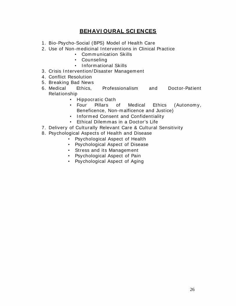
BEHAVIOURAL SCIENCES
1. Bio-Psycho-Social (BPS) Model of Health Care 2. Use of Non-medicinal Interventions in Clinical Practice
• Communication Skills • Counseling • Informational Skills
3. Crisis Intervention/Disaster Management 4. Conflict Resolution 5. Breaking Bad News 6. Medical Ethics, Professionalism and Doctor-Patient
Relationship • Hippocratic Oath • Four Pillars of Medical Ethics (Autonomy,
Beneficence, Non-malficence and Justice) • Informed Consent and Confidentiality • Ethical Dilemmas in a Doctor’s Life
7. Delivery of Culturally Relevant Care & Cultural Sensitivity 8. Psychological Aspects of Health and Disease
• Psychological Aspect of Health • Psychological Aspect of Disease • Stress and its Management • Psychological Aspect of Pain • Psychological Aspect of Aging
27
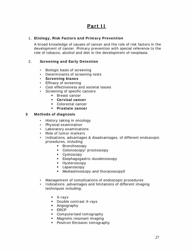
Part II
1. Etiology, Risk Factors and Primary Prevention
A broad knowledge of causes of cancer and the role of risk factors in the development of cancer. Primary prevention with special reference to the role of tobacco, alcohol and diet in the development of neoplasia.
2. Screening and Early Detection
• Biologic basis of screening • Determinants of screening tests • Screening biases • Efficacy of screening • Cost effectiveness and societal issues • Screening of specific cancers
§ Breast cancer § Cervical cancer § Colorectal cancer § Prostate cancer
3 Methods of diagnosis
• History taking in oncology • Physical examination • Laboratory examinations • Role of tumor markers • Indications, advantages & disadvantages, of different endoscopic
procedures, including § Bronchoscopy § Colonoscopy/ proctoscopy § Cystoscopy § Esophagogastro-duodenoscopy § Hysteroscopy § Laparoscopy § Mediastinoscopy and thoracoscopy0
• Management of complications of endoscopic procedures • Indications ,advantages and limitations of different imaging
techniques including;
§ X-rays § Double contrast X-rays § Angiography § ERCP § Computerised tomography § Magnetic resonant imaging § Positron Emission tomography
28
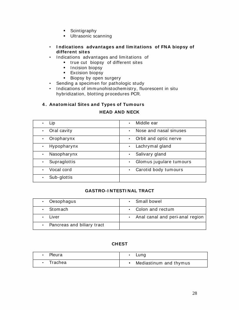
§ Scintigraphy § Ultrasonic scanning
• Indications advantages and limitations of FNA biopsy of different sites
• Indications advantages and limitations of § true cut biopsy of different sites § Incision biopsy § Excision biopsy § Biopsy by open surgery
• Sending a specimen for pathologic study • Indications of immunohistochemistry, fluorescent in situ
hybridization, blotting procedures PCR.
4. Anatomical Sites and Types of Tumours
HEAD AND NECK
• Lip • Middle ear
• Oral cavity • Nose and nasal sinuses
• Oropharynx • Orbit and optic nerve
• Hypopharynx • Lachrymal gland
• Nasopharynx • Salivary gland
• Supraglottis • Glomus jugulare tumours
• Vocal cord • Carotid body tumours
• Sub-glottis
GASTRO-INTESTINAL TRACT
• Oesophagus • Small bowel
• Stomach • Colon and rectum
• Liver • Anal canal and peri-anal region
• Pancreas and biliary tract
CHEST
• Pleura • Lung
• Trachea • Mediastinum and thymus
29
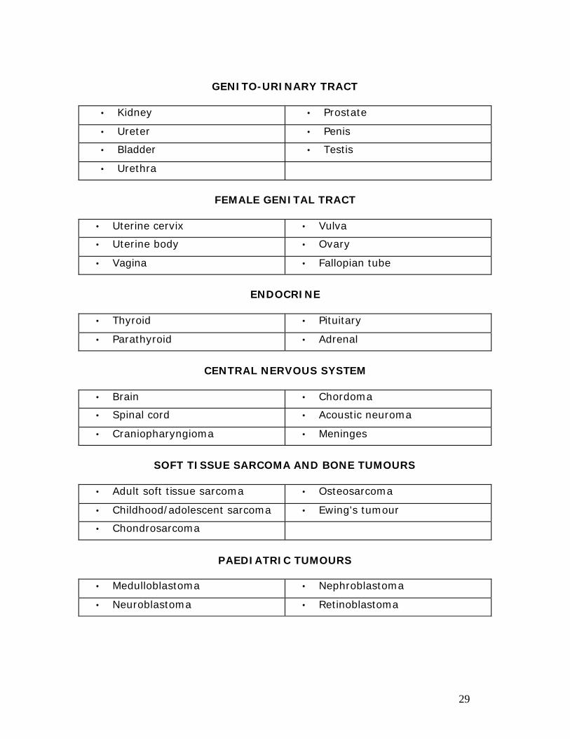
GENITO-URINARY TRACT
• Kidney • Prostate
• Ureter • Penis
• Bladder • Testis
• Urethra
FEMALE GENITAL TRACT
• Uterine cervix • Vulva
• Uterine body • Ovary
• Vagina • Fallopian tube
ENDOCRINE
• Thyroid • Pituitary
• Parathyroid • Adrenal
CENTRAL NERVOUS SYSTEM
• Brain • Chordoma
• Spinal cord • Acoustic neuroma
• Craniopharyngioma • Meninges
SOFT TISSUE SARCOMA AND BONE TUMOURS
• Adult soft tissue sarcoma • Osteosarcoma
• Childhood/adolescent sarcoma • Ewing's tumour
• Chondrosarcoma
PAEDIATRIC TUMOURS
• Medulloblastoma • Nephroblastoma
• Neuroblastoma • Retinoblastoma

30
LYMPHOPROLIFERATIVE AND MYELOPROLIFERATIVE DISORDERS
• Hodgkin's lymphoma • Plasma cell malignancies
• Non-Hodgkin's lymphomas • Acute and chronic leukaemias
MISCELANEOUS TUMOURS
• Breast • Malignant melanoma
• Basal cell carcinoma • Cutaneous lymphoma
• Squamous cell carcinoma • Kaposi's sarcoma
For each of the tumour types and sites listed at paragraph 4:
Management
• Initial staging investigations including imaging and tumour markers
• Relevant prognostic factors
• Assessment for treatment
• Role of surgery
• A management plan, or, where applicable, a range of such plans
• Ionising Radiation Regulations
• Roles of surgery, radiotherapy and cytotoxic chemotherapy in multimodality approaches to cancer treatment
Pathology • The range of tumours that can occur
• Their aetiology, incidence and epidemiology
• A brief morphology of the common tumours
• The natural history of the disease including likely presentation, characteristic growth and metastatic pattern.
• Staging classifications, e.g. TNM, FIGO
• Use of tumour markers in diagnosis and treatment of tumours
• Use of specialised pathology techniques, e.g. immunohistochemistry
• Interpretation of clinico-pathological data in the tumour site specialised multidisciplinary approach to patient management

31
Radiotherapy • The role of irradiation in radical and palliative management
• Where radical radiotherapy is a treatment option:
§ Staging investigations
§ A definition of tumour volume and target volume boundaries
§ ICRU reports 50 and 62
§ An acceptable radio therapeutic technique, or, where applicable, a range of such techniques
§ The correct treatment position
§ Details of the target volume localisation process
§ Use of CT axial images, 3D planning
§ Verification techniques such as laser alignment, skin tattoos, orthogonal and portal films
§ The approximate dose distributions for the chosen technique
§ An appropriate dose/fractionation regime
§ Relevant dose modifying factors (changes in fractionation, age, target volume, intercurrent infections, previous therapies)
§ Details of the set-up instructions for radiographers
§ Appropriate responses to changes of patient parameters or interruptions during treatment
§ The possible acute and late side effects of the irradiation
§ Radiation dose modifying factors, chemotherapy timing in all forms of chemo radiation schedules
Drug Therapy
• The role of cytotoxic, hormonal and biological drugs therapies in radical and palliative management
• Radical and palliative regiment in common use including dosage, scheduling, toxicities and outcome
• The techniques of stem cell mobilisation and the procedures for stem cell and bone marrow harvesting
• The timing of total body irradiation, the re-infusion of bone marrow or stem cells and patient support during the engraftment
32
Outcomes • The expected outcomes of treatment
5. Drug Therapy
. Cytotoxic Chemotherapy
• A basic knowledge of the pharmacokinetics, therapeutic uses, dose ranges and toxicities of the currently used cytotoxic agents
• Where applicable, a range of multiagent chemotherapy regimens and details of their administration
a. Hormone Therapy
• A basic knowledge of the therapeutic use and toxicities of currently used hormone therapy
b. Biological Therapies
• A basic knowledge of the clinical uses of currently used biological therapies including interferons, colony stimulating factors, other growth factors and preparations such as Herceptin
6. Oncological Emergencies
• The management of the following complications when they are related to cancer:
§ Ureteric obstruction
§ Spinal cord compression
§ Haemorrhage
§ Mediastinal obstruction
7. Radiotherapy for Benign Disease
• The indications for radiotherapy in the treatment of benign conditions, including suitable techniques and dosage schedules, and likely benefits and risks
8. Complications of Treatment
• The acute and late complications of oncological treatment and their management including:
§ Skin reactions
§ Nausea and vomiting
§ Diarrhoea
§ Oedema
§ Bone marrow toxicity
§ Neutropenic sepsis
33
§ Drug reactions
§ Cytotoxic extravasation
§ Alopecia
§ Cataract
§ Skin atrophy and ulceration
§ Colitis, proctitis, gut strictures and perforation
§ Renal effects
§ Cardiac effects
§ Pulmonary effects
§ Fibrosis and lymphoedema
§ Endocrine effects (thyroid, pituitary and salivary gland)
§ Effects on fertility
§ Incidence of second and radiation induced cancers
9. Symptom Control and Continuing Care
• The available medical and surgical techniques for the control of pain, nausea, vomiting and malignant effusions
• Treatment of various cancer related conditions and paraneoplastic syndromes including
§ Hypercalcaemia
§ Ectopic hormone production
§ Raised intra cranial pressure
§ Anaemia
10. Current Research and Literature
• Current major research in progress in the form of multicentre trials
• Recent major publications in oncology journals
34
SKILLS AND CLINICAL EXPERIENCE
Candidates need to have gained a wide range of experience in the areas of patient investigation, diagnosis, treatment with radiation, chemotherapy, hormonal therapy, biological therapy and in palliative and supportive care and to have gained the practical experience detailed below.
1. Radiotherapy – Basic Techniques
a. Positioning the Patient
• Setting up of a patient in each of the three basic treatment positions (supine, prone and lateral) to allow the patient to be planned and treated effectively and without discomfort
• Setting up the source skin distance for fixed FSD, and extended FSD treatment
• Setting up patients using laser beam alignment
• Making temporary and permanent marks on the patient for field positions (Gentian violet, tattoo)
b. Immobilisation Techniques
• Application of some of the following immobilisation techniques: head-clamp, Velcro strap, polystyrene beads, vacuum bag, breast armrest
• The construction of thermoplastic beam direction shell
c. Methods of Target Volume Determination
• Performance of planning
§ using direct vision of the tumour (eg skin tumours) § from surface landmarks (eg the parotid bed, breast
tumours) § with direct screening using simulator (eg lung
tumours, bone metastases), including opacification techniques (eg barium swallow, cystogram)
§ by volume transfer to orthogonal radiographs (eg head and neck tumours, brain tumours)
• Volume determination from planning CT scans for creating a central axis plan and for 3-dimensional CT planning
d. Outline Techniques
• Use of manual techniques (flexi-curves, plaster of Paris bandage) and CT derived outlines
e. Basic Field Arrangements
• Planning of treatments (under supervision where necessary) using the following field arrangements:
35
§ Single direct field
§ Opposed pair of fields using equal and unequal weightings
§ Opposed pair using wedges
§ Wedged right-angled pair
§ Wedged oblique pair
§ Plans using 3 and 4 fields
§ Total body irradiation
f. Tissue Compensation
• Planning of patients requiring tissue compensation using bolus, wedges and remote tissue compensators
g. Shielding
• Planning of patients using lead cut outs and lead masks for simple superficial tumours
• Knowledge of the thickness of lead required for superficial, orthovoltage and electron treatments at various energies
• Prescription and insertion of eye shields
h. Megavoltage Techniques
• Planning of patients incorporating simple lead blocking techniques using standard blocks and cast blocks from templates
i. Electrons
• The indications for, and planning of, electron treatments, including the selection of electron energy
• A technique for total skin electron therapy and experience of its use
j. Dose Calculation
• Proficiency in the use of equivalent square tables
• Performance of depth dose calculations for single fields and opposed fields using various energies
• The principles applied to convert dose to machine units for a range of machines
• The principles of computer based treatment planning
k. Radiotherapy Prescriptions
36
• Writing radiotherapy prescriptions (countersigned where necessary) for all the field arrangements mentioned above
• Understanding of dose specification as in ICRU50 and 62
l. Radiotherapy Machines
• Planning of patients for treatment on a full spectrum of equipment, including superficial x-ray therapy, megavoltage x-ray therapy and megavoltage electron therapy (also orthovoltage x-ray therapy and cobalt?60 therapy, if available)
m. Quality Assurance in External Beam Therapy
• Requesting portal imaging and interpreted their appearance satisfactorily in all sites
• Principles of in vivo dosimetry and interpretation of results
n. Brachytherapy
• The insertion and removal of radioactive sources manually or using an appropriate after-loading device
• Interpretation of subsequent check films
• Interpretation of the corresponding dose calculation and writing of an appropriate prescription
• Removal of live sources and the after?loading device
• The placement of implants
• Principles of oral and intravenous radionuclide therapy
o. Radiation Safety
• The role of the radiation safety and radiation protection supervisor
• The meaning of and requirements for controlled and supervised areas and their location
• The procedure to be adopted in the case of a spill of radioactive material
• Quality assurance practices in radiotherapy and the procedures for dealing with errors in treatment delivery
2. Radiotherapy Assessment and the Care of Patients on Treatment
a. Treatment Review Clinics
• Regular weekly treatment review clinics
37
b. Treatment Checks
• Assessment of patient position and treatment field placement(s) in relation to the target volume at the start of treatment
• Performance of checks during the course of treatment on the implementation of the treatment plan, position of shielding for critical normal structures and the use of portal imaging
• Assessment of changes occurring in patient parameters during treatment and resultant modification of treatment when appropriate
• Assessment of normal tissue reactions to radiotherapy
• Use of dose volume histograms and in vivo radiation dosimetry techniques
c. Symptom Control
• Giving advice on skin care during radiation treatment and on the management of skin reactions, including desquamation
• Managing mucosal reactions in oral cavity, oropharynx, nasopharynx, trachea, oesophagus, anus and vagina
• Giving dietary advice during abdominal radiotherapy
• Managing radiation induced nausea and vomiting, diarrhoea, dysphagia, xerostomia and cystitis
• Giving prophylaxis for radiation induced cerebral oedema
• Giving advice on timing and extent of hair loss with respect to radiation dose
d. Follow-up
• Managing acute and chronic radiation sequelae, such as pneumonitis, cystitis, chronic bowel complications, gynaecological sequelae (vaginal stenosis, vaginal dryness, infertility and dyspareunia)
3. Drug Therapy
a. Access Technique for Sampling and Delivery
• Insertion and maintenance of intravenous lines for both sampling of blood and delivery of chemotherapy, including temporary sharp cannulation (butterfly type) and intermediate term flexible cannulation (venflon type)
• Arranging the insertion of Hickman or temporary long lines and subcutaneous implanted lines (portacath type),
38
their use for sampling (where possible) and the delivery of chemotherapy and maintenance for protracted used
• Principles, regulations and guidelines for the delivery of intrathecal chemotherapy
b. Drug Delivery
• The indications and eligibility of a wide variety of cytotoxic agents, and their side-effect profiles as single agents and in combinations
• Prescription and delivery of drugs in current usage
c. Support Techniques
• Pre hydration and maintenance of urine flow and modification of urine pH during chemotherapy delivery
• Prescription of protective agents, eg folinic acid (oral and iv) with MTX level monitoring or Mesna with appropriate chemotherapy regimens
• The use of anti-emetics
• The appropriate use of colony stimulating factors
• The appropriate use of bone marrow reconstitution techniques after high dose chemotherapy/TBI
d. Management of Acute Complications
• Managing extravasation reactions caused by vesicant drugs
• Managing patients with chemotherapy induced neutropenia, with and without pyrexia
• Managing chemotherapy induced thrombocytopenia, including the use of platelet transfusions
e. Treatment with Hormonal Therapy
• Implementation of hormone treatment for breast and prostate cancer
f. Treatment with Biological Therapies
4. Supportive and Palliative Care
a. Pain Relief
• Drug treatment
§ A wide range analgesic techniques, including simple analgesics, mild and strong opioids, given by a variety of routes
39
§ Management of the complications of analgesics, including constipation, nausea, gastro intestinal discomfort and analgesic intolerance
• Mechanical methods
§ Prescription, siting and evaluation of TENS analgesia
§ Referral of patients with refractory pain for procedures such as a nerve block, intrathecal analgesia, rhizotomy or orthopaedic stabilisation
• Radiotherapy
§ Use of radiation to treat painful metastatic disease with single fractions, multiple fractions and hemi body radiotherapy
b. Nausea and Vomiting
• Treatment of nausea and vomiting arising in advanced illness using anti-emetics
• Palliative management of sub-acute intestinal obstruction
c. Anorexia and Dysphagia
• Management, where appropriate, with corticosteroids, progestogens and nasal gastric feeding
d. Pleural Effusions and Ascites
• Drainage of pleural effusions and ascites
• Other treatments, such as talc pleurodesis
e. Depression and Anxiety
• Treatment of depression at all stages of cancer management, using counselling and drug techniques with anti?depressants
• Treatment of anxiety with counselling, anxiolytics and major tranquillisers
f. Hospice Care
• Awareness of local hospice facilities
• A one week (at least) attachment to a hospice or palliative care team
g. Counselling
• Counselling of patients and relatives at all stages of the disease
40
5. Investigational Techniques
a. Laboratory Investigations
• Interpretation of the results of haematological, biochemical and radio-immune assay investigations
b. Radiology
• Attendance at regular radiological review sessions involving a consultant clinical radiologist for the examination of plain x?rays, CT scans, magnetic resonance imaging and ultrasound covering the whole spectrum of cancer radiology
• Current indications and techniques in interventional procedures
c. Pathology
• Attendance at regular pathological review sessions involving a consultant pathologist
d. Other Procedures
• Indirect layrngoscopy
• Lumbar puncture
• Skin biopsy
• Fibre optic naso-endoscopy
• Pelvic EUA and cystoscopy
6. Site or Disease Specific Procedures
• Assessment, treatment and follow-up, in detail, for each of the anatomical sites and types of tumour listed at paragraph 4 of the "Knowledge" section above
• Presentation and assessment of patients discussed at multidisciplinary team meeting
• Staging
• Radiotherapy – adjuvant, radical and palliative
• Chemotherapy – adjuvant, radical and palliative
• Hormone and biological therapy
• Palliative care
• Appropriate follow up
• Acute and late side effects of treatment
7. Clinical Trials, Literature and Research
• The aims and format of Phase I to IV clinical trials
41
• Obtaining informed consent, following study protocols and using data forms
• Research programmes (although research experience is not a prerequisite)
• Major areas of current research and of recent important publications
• Submission of a research project to an ethics committee
• Structure and functioning of local and national clinical and research cancer networks
8. Communication and Publication
• Effective communication with colleagues, patients and their carers
• Giving clear and comprehensive descriptions of disease processes, investigations and treatment
• Clear expression in English and production of legible script
• Preparing work for publication
9. Outpatient and Joint Clinics
• Participation in joint consultative clinics and regular general oncology outpatient sessions
• Seeing review and new patients and planning their overall management
10.Resource Management and Quality Assurance
• Introduction to the resource management and quality assurance of an oncology service, so as to be able to develop these skills at a later stage
42
METHODS OF INSTRUCTION/COURSE CONDUCTION
As a policy, active participation & self learning of students at all levels will be encouraged. Following teaching modalities will be employed:
1. Lectures, Seminar Presentation and Journal Club Presentations 2. Group Discussions 3. Grand Rounds 4. SEQ as assignments on the content areas 5. Hands-on training experience in Radiation Therapy Techniques 6. Active participation in the National Cancer Conference 7. Indoor and outdoor clinics 8. Long and short case presentations 9. Preparation of teaching case files
In addition to the conventional teaching methodologies following interactive strategies will also be introduced to improve both communication and clinical skills in the upcoming consultants: 1.1. Monthly Student Meetings Each affiliated department approved to conduct training for DMRT will provide a room for student meetings/discussions such as
a. Journal Club Meeting b. Core Curriculum Meetings c. Skill Development
a. Journal Club Meeting Two hours per month should be allocated to the presentation and discussion of a recent Journal article related to Radiation Oncology. The article should be critically evaluated and its applicable results should be highlighted which can be incorporated in clinical practice. Record of all such articles should be maintained in the relevant department of each medical college. Students of different medical colleges may be given an opportunity to share all such interesting articles with each other.
b. Core Curriculum Meetings All the core topics of DMRT should be thoroughly discussed during these sessions. The duration of each session should be at least two hours once a month. It should be chaired by the chief student (elected by the students of the relevant diploma). Each student should be given an opportunity to brainstorm all topics included in the course and to generate new ideas regarding the improvement of the course structure.
43
c. Skill Development
Two hours twice a month should be assigned for learning and practicing clinical skills. List of skills to be learnt during these sessions is as follows:
1. Communication skills 2. Interpretation of various imaging findings 3. Practical skills 4. Presentation skills: Power point, lectures, small group
discussions, article presentation etc. 5. Research and scientific paper writing 6. Acquisition of procedural skills like Paracentesis, Intravenous
access, Aspiration, Bone Marrow Examination and other minor procedures should be availed
1.2 Annual Grand Meeting
Once a year all students enrolled for DMRT shall be invited to the Annual meeting at University of Health Sciences Lahore. One full day will be allocated to this event. All the chief students will present their annual reports. Issues and concerns related to their relevant diploma courses will be discussed. Feedback may be collected and also suggestions can be formulated in order to involve students in decision making. The research work and the literary work of students may also be displayed. In the evening an informal get together and dinner should be arranged. This will help in creating a sense of belonging and ownership among students and the faculty.

44
LOG BOOK The trainees must maintain a log book and get it signed regularly by the supervisor. A complete and duly certified log book should be part of the requirement to sit for the DMRT examination. Log book should include adequate number of procedures, routine and emergency management of patients, case presentations in Clinical Pathological Conference, journal club meetings and literature review. Proposed Format of Log Book is as follows: Candidate’s Name: ______________________Roll No. _____ PROCEDURES:
1. Pleural Tap 2. Ascitic Tap 3. Intravenous Access 4. Urinary Bladder Catheterization 5. Chest Tube Insertion 6. Bone Marrow Examination 7. Pelvic Examination 8. Chemotherapy with different routes of administration 9. Treatment Planning 10. Simulation
Sr.# Date Name of Patient, Age, Sex & Admission No.
Diagnosis Procedure Performed
Supervisor’s Signature
1
Case Presented Sr.# Date Name of Patient, Age,
Sex & Admission No. Case Presented Supervisor’s
Signature 1
Seminar/Journal Club Presentation Sr.# Date Topic Supervisor’s signature
1
Evaluation Record (Excellent, Good, Adequate, Inadequate, Poor) Sr.#
Date
Method of Evaluation (Oral, Practical, Theory)
Rating
Supervisor’s Signature
1

45
LITERATURE REVIEW Students will be assigned a clinical problem; most commonly encountered in the relevant specialty and will be specifically trained to review literature in the pertinent field and write a ‘Review of an Article’ comprising of:
• Topic • Introduction • Discussion of the reviewed literature • Conclusion • References
46
EXAMINATIONS Assessment It will consist of action and professional growth oriented student-centered integrated assessment with an additional component of informal internal assessment, formative assessment and measurement-based summative assessment. Student-Centered Integrated Assessment It views students as decision-makers in need of information about their own performance. Integrated Assessment is meant to give students responsibility for deciding what to evaluate, as well as how to evaluate it, encourages students to ‘own’ the evaluation and to use it as a basis for self-improvement. Therefore, it tends to be growth-oriented, student-controlled, collaborative, dynamic, contextualized, informal, flexible and action-oriented. In the proposed curriculum, it will be based on:
• Self Assessment by the student • Peer Assessment • Informal Internal Assessment by the Faculty
Self Assessment by the Student Each student will be provided with a pre-designed self-assessment form to evaluate his/her level of comfort and competency in dealing with different relevant clinical situations. It will be the responsibility of the student to correctly identify his/her areas of weakness and to take appropriate measures to address those weaknesses. Peer Assessment The students will also be expected to evaluate their peers after the monthly small group meeting. These should be followed by a constructive feedback according to the prescribed guidelines and should be non-judgmental in nature. This will enable students to become good mentors in future.
47
Informal Internal Assessment by the Faculty There will be no formal allocation of marks for the component of Internal Assessment so that students are willing to confront their weaknesses rather than hiding them from their instructors. It will include:
a. Punctuality b. Work during different postings c. Monthly assessment (written tests to indicate particular
areas of weaknesses) d. Participation in interactive sessions
Formative Assessment Will help to improve the existing instructional methods and the curriculum in use Feedback to the faculty by the students: After every three months students will be providing a written feedback regarding their course components and teaching methods. This will help to identify strengths and weaknesses of the relevant course, faculty members and to ascertain areas for further improvement. Summative Assessment It will be carried out at the end of the programme to empirically evaluate cognitive, psychomotor and affective domains in order to award diplomas for successful completion of courses. Eligibility to Appear in Final Examination
- Only those candidates will be eligible to take final examination,
who have passed Part 1 examination (after 6 months of education) and have completed two years of structured/supervised training programme.
- Students who have completed their log books and hold certificate of 75% attendance may be allowed to sit for the exam
- The application for the final examination will be forwarded on the recommendations of the supervisor
- Candidates who qualify in theory will be called for the clinical examination
48
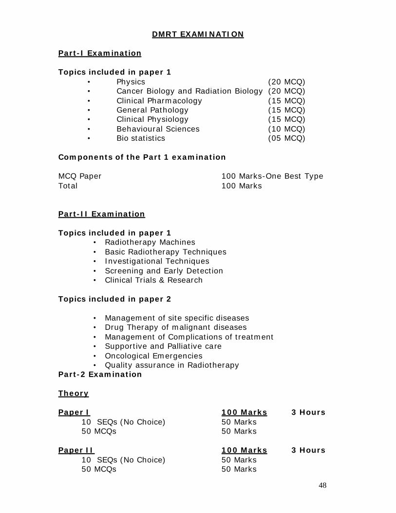
DMRT EXAMINATION Part-I Examination Topics included in paper 1
• Physics (20 MCQ) • Cancer Biology and Radiation Biology (20 MCQ) • Clinical Pharmacology (15 MCQ) • General Pathology (15 MCQ) • Clinical Physiology (15 MCQ) • Behavioural Sciences (10 MCQ) • Bio statistics (05 MCQ)
Components of the Part 1 examination
MCQ Paper 100 Marks-One Best Type Total 100 Marks Part-II Examination Topics included in paper 1
• Radiotherapy Machines • Basic Radiotherapy Techniques • Investigational Techniques • Screening and Early Detection • Clinical Trials & Research
Topics included in paper 2
• Management of site specific diseases • Drug Therapy of malignant diseases • Management of Complications of treatment • Supportive and Palliative care • Oncological Emergencies • Quality assurance in Radiotherapy
Part-2 Examination Theory Paper I 100 Marks 3 Hours 10 SEQs (No Choice) 50 Marks 50 MCQs 50 Marks Paper II 100 Marks 3 Hours 10 SEQs (No Choice) 50 Marks 50 MCQs 50 Marks

49
The candidate who passes theory papers, will be called for the clinical & viva voce examination. OSCE 90 Marks 10 stations each carrying 9 marks of 10 minutes duration; each evaluating performance based assessment with five of them interactive. Clinical 90 Marks Four short cases each carrying 15 marks and one long case of 30 marks. Components of the Part 2 examination Theory paper 1 100 marks Theory paper 2 100 marks Clinical/OSCE 180 marks Log Book 20 marks Total 400 Marks A panel of four examiners (Two internal and two external) will be appointed for practical examination. Each component of practical examination will be assessed by two examiners awarding marks simultaneously and independently. The final score awarded will be an average score, as agreed by both examiners. Pass Percentage and Other Regulations Regarding Examination
• Criterion referenced assessment principles will be used • Twenty marks for the log book will be included in the
OSCE/Clinical component • Sixty percent marks will be a pass score in each component • Candidates failing in any component will have to re-sit for the
entire examination • A maximum of 5 attempts in three calendar years are allowed to
pass the examination.
• Re-admission to DMRT course is not permissible. • The results will be announced according to the rules and
regulations set by the Examination Branch of University of Health Sciences, Lahore.
50
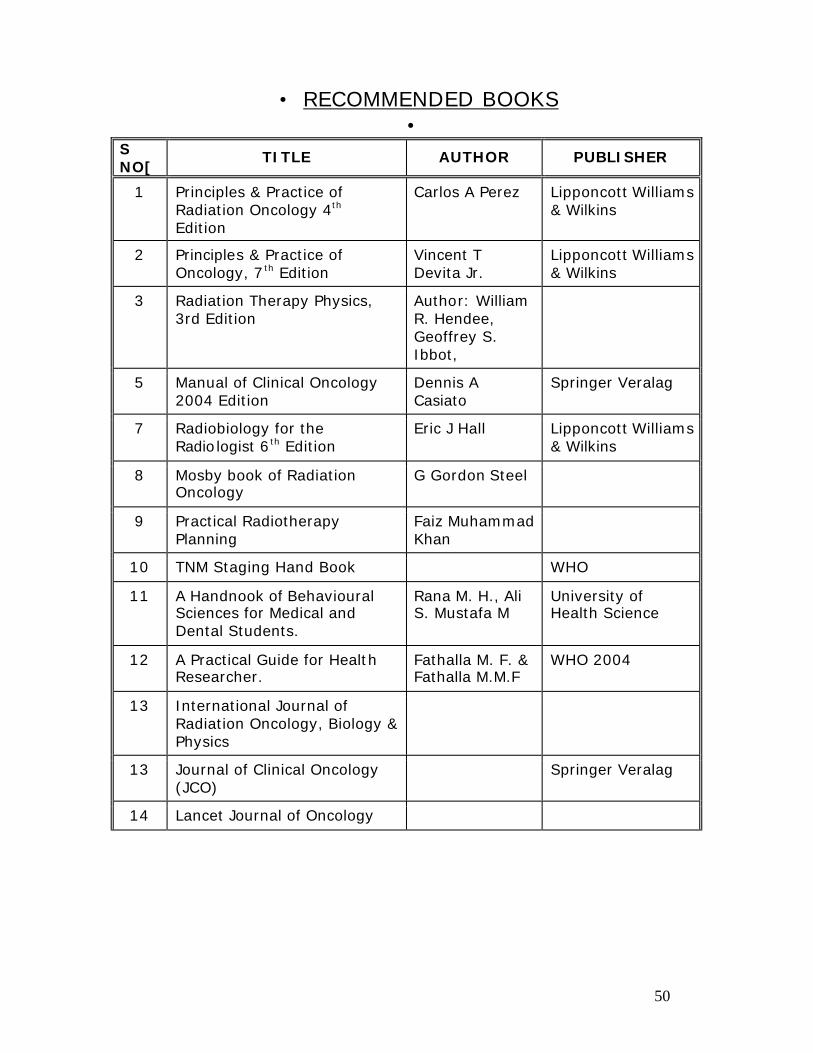
• RECOMMENDED BOOKS
• S NO[
TITLE AUTHOR PUBLISHER
1 Principles & Practice of Radiation Oncology 4th Edition
Carlos A Perez Lipponcott Williams & Wilkins
2 Principles & Practice of Oncology, 7th Edition
Vincent T Devita Jr.
Lipponcott Williams & Wilkins
3 Radiation Therapy Physics, 3rd Edition
Author: William R. Hendee, Geoffrey S. Ibbot,
5 Manual of Clinical Oncology 2004 Edition
Dennis A Casiato
Springer Veralag
7 Radiobiology for the Radiologist 6th Edition
Eric J Hall Lipponcott Williams & Wilkins
8 Mosby book of Radiation Oncology
G Gordon Steel
9 Practical Radiotherapy Planning
Faiz Muhammad Khan
10 TNM Staging Hand Book WHO
11 A Handnook of Behavioural Sciences for Medical and Dental Students.
Rana M. H., Ali S. Mustafa M
University of Health Science
12 A Practical Guide for Health Researcher.
Fathalla M. F. & Fathalla M.M.F
WHO 2004
13 International Journal of Radiation Oncology, Biology & Physics
13 Journal of Clinical Oncology (JCO)
Springer Veralag
14 Lancet Journal of Oncology
Related Documents