DMH1, a Novel BMP Small Molecule Inhibitor, Increases Cardiomyocyte Progenitors and Promotes Cardiac Differentiation in Mouse Embryonic Stem Cells Ada Ao 1 *, Jijun Hao 1 , Corey R. Hopkins 2 , Charles C. Hong 1,3,4,5 * 1 Division of Cardiovascular Medicine, Department of Medicine, Vanderbilt University School of Medicine, Nashville, Tennessee, United States of America, 2 Vanderbilt Institute of Chemical Biology, Department of Pharmacology, Center for Neuroscience Drug Discovery, Vanderbilt University School of Medicine, Nashville, Tennessee, United States of America, 3 Vanderbilt Institute of Chemical Biology, Department of Pharmacology, Vanderbilt University School of Medicine, Nashville, Tennessee, United States of America, 4 Department of Cell and Developmental Biology, Vanderbilt University School of Medicine, Nashville, Tennessee, United States of America, 5 Research Medicine, Veterans Affairs Tennessee Valley Healthcare System, Nashville, Tennessee, United States of America Abstract The possibility of using cell-based therapeutics to treat cardiac failure has generated significant interest since the initial introduction of stem cell-based technologies. However, the methods to quickly and robustly direct stem cell differentiation towards cardiac cell types have been limited by a reliance on recombinant growth factors to provide necessary biological cues. We report here the use of dorsomorphin homologue 1 (DMH1), a second-generation small molecule BMP inhibitor based on dorsomorphin, to efficiently induce beating cardiomyocyte formation in mouse embryonic stem cells (ESCs) and to specifically upregulate canonical transcriptional markers associated with cardiac development. DMH1 differs significantly from its predecessor by its ability to enrich for pro-cardiac progenitor cells that respond to late-stage Wnt inhibition using XAV939 and produce secondary beating cardiomyocytes. Our study demonstrates the utility of small molecules to complement existing in vitro cardiac differentiation protocols and highlights the role of transient BMP inhibition in cardiomyogenesis. Citation: Ao A, Hao J, Hopkins CR, Hong CC (2012) DMH1, a Novel BMP Small Molecule Inhibitor, Increases Cardiomyocyte Progenitors and Promotes Cardiac Differentiation in Mouse Embryonic Stem Cells. PLoS ONE 7(7): e41627. doi:10.1371/journal.pone.0041627 Editor: Rajesh Gopalrao Katare, University of Otago, New Zealand Received May 14, 2012; Accepted June 22, 2012; Published July 27, 2012 Copyright: ß 2012 Ao et al. This is an open-access article distributed under the terms of the Creative Commons Attribution License, which permits unrestricted use, distribution, and reproduction in any medium, provided the original author and source are credited. Funding: The project is supported by grant number 5R01HL104040-02 from National Heart, Lung, and Blood Institute, Veterans Affair, and International Fibrodysplasia Ossificans Progressiva Association. AA is supported in part by United States Public Health Service training grant number 1T32HL105334-1A1. The funders had no role in study design, data collection and analysis, decision to publish, or preparation of the manuscript. Competing Interests: The authors have declared that no competing interests exist. * E-mail: [email protected] (AA); [email protected] (CCH) Introduction The irreversible loss of cardiomyocytes following myocardial infarction causes the clinical features of heart failure, marked by regional contractile dysfunction manifesting mainly in the ventricular chamber. The advent of stem cell biology and regenerative medicine offer enticing prospects for manufacturing specific cell types from pluripotent cells, which may be used to replace or repair damaged tissues. This is preferable to traditional organ transplants because donor availability and compatibility constitutes a significant barrier. However, the expected cell-based therapeutics have not matured as the directed differentiation process is inefficient, resulting in a heterogeneous cell population that risks further complications if implanted into patients. There are also technical hurdles against the large-scale production of clinical grade products because current protocols rely on the use of animal-derived growth factors, which may introduce batch-to- batch variability that constitutes additional safety concerns for humans [1–3]. Therefore, there is an urgent need to develop tools for directed differentiation that are both xeno-free and have robust biological effects. Insights from developmental biology studies have uncovered key molecular pathways that guide mammalian cardiac differentiation. The process of cardiomyocyte development from mesoderm progenitors requires coordinated changes in BMP signaling along with other mitogenic pathways including Activin, FGF, and Wnt signaling [4–8]. Previous studies have shown that the simple presence of BMP ligands is insufficient to initiate cardiac differentiation [6,9], and BMP signaling in mesoderm is sequen- tially and locally controlled by antagonists secreted from the surrounding ectoderm and endoderm during cardiac morphogen- esis [10–12]. Recent studies also suggested that the timing and the duration of BMP signaling in pluripotent cells may influence atrial and ventricular lineage commitment of multipotent cardiac progenitors [13–15]. An overall picture emerges in which early BMP signaling modulation is not only necessary to specify the cardiac progenitor pool, but also to temporally regulate cardiac chamber development. Small molecules have emerged as an adaptable tool that take advantage of insights borrowed from developmental biology. They have been used for directing differentiation and have demonstrat- ed their advantages over the use of recombinant proteins in many aspects of regenerative medicine [16–18]. Our previous study, which described the use of dorsomorphin (DM) to mimic the function of endogenous BMP inhibitor Noggin for directing cardiomyocyte formation in mouse embryonic stem cells, demon- strated that the timely application of a single chemical can be a viable strategy for directed cardiac differentiation [19]. However, PLoS ONE | www.plosone.org 1 July 2012 | Volume 7 | Issue 7 | e41627

Welcome message from author
This document is posted to help you gain knowledge. Please leave a comment to let me know what you think about it! Share it to your friends and learn new things together.
Transcript

DMH1, a Novel BMP Small Molecule Inhibitor, IncreasesCardiomyocyte Progenitors and Promotes CardiacDifferentiation in Mouse Embryonic Stem CellsAda Ao1*, Jijun Hao1, Corey R. Hopkins2, Charles C. Hong1,3,4,5*
1 Division of Cardiovascular Medicine, Department of Medicine, Vanderbilt University School of Medicine, Nashville, Tennessee, United States of America, 2 Vanderbilt
Institute of Chemical Biology, Department of Pharmacology, Center for Neuroscience Drug Discovery, Vanderbilt University School of Medicine, Nashville, Tennessee,
United States of America, 3 Vanderbilt Institute of Chemical Biology, Department of Pharmacology, Vanderbilt University School of Medicine, Nashville, Tennessee, United
States of America, 4 Department of Cell and Developmental Biology, Vanderbilt University School of Medicine, Nashville, Tennessee, United States of America, 5 Research
Medicine, Veterans Affairs Tennessee Valley Healthcare System, Nashville, Tennessee, United States of America
Abstract
The possibility of using cell-based therapeutics to treat cardiac failure has generated significant interest since the initialintroduction of stem cell-based technologies. However, the methods to quickly and robustly direct stem cell differentiationtowards cardiac cell types have been limited by a reliance on recombinant growth factors to provide necessary biologicalcues. We report here the use of dorsomorphin homologue 1 (DMH1), a second-generation small molecule BMP inhibitorbased on dorsomorphin, to efficiently induce beating cardiomyocyte formation in mouse embryonic stem cells (ESCs) andto specifically upregulate canonical transcriptional markers associated with cardiac development. DMH1 differs significantlyfrom its predecessor by its ability to enrich for pro-cardiac progenitor cells that respond to late-stage Wnt inhibition usingXAV939 and produce secondary beating cardiomyocytes. Our study demonstrates the utility of small molecules tocomplement existing in vitro cardiac differentiation protocols and highlights the role of transient BMP inhibition incardiomyogenesis.
Citation: Ao A, Hao J, Hopkins CR, Hong CC (2012) DMH1, a Novel BMP Small Molecule Inhibitor, Increases Cardiomyocyte Progenitors and Promotes CardiacDifferentiation in Mouse Embryonic Stem Cells. PLoS ONE 7(7): e41627. doi:10.1371/journal.pone.0041627
Editor: Rajesh Gopalrao Katare, University of Otago, New Zealand
Received May 14, 2012; Accepted June 22, 2012; Published July 27, 2012
Copyright: � 2012 Ao et al. This is an open-access article distributed under the terms of the Creative Commons Attribution License, which permits unrestricteduse, distribution, and reproduction in any medium, provided the original author and source are credited.
Funding: The project is supported by grant number 5R01HL104040-02 from National Heart, Lung, and Blood Institute, Veterans Affair, and InternationalFibrodysplasia Ossificans Progressiva Association. AA is supported in part by United States Public Health Service training grant number 1T32HL105334-1A1. Thefunders had no role in study design, data collection and analysis, decision to publish, or preparation of the manuscript.
Competing Interests: The authors have declared that no competing interests exist.
* E-mail: [email protected] (AA); [email protected] (CCH)
Introduction
The irreversible loss of cardiomyocytes following myocardial
infarction causes the clinical features of heart failure, marked by
regional contractile dysfunction manifesting mainly in the
ventricular chamber. The advent of stem cell biology and
regenerative medicine offer enticing prospects for manufacturing
specific cell types from pluripotent cells, which may be used to
replace or repair damaged tissues. This is preferable to traditional
organ transplants because donor availability and compatibility
constitutes a significant barrier. However, the expected cell-based
therapeutics have not matured as the directed differentiation
process is inefficient, resulting in a heterogeneous cell population
that risks further complications if implanted into patients. There
are also technical hurdles against the large-scale production of
clinical grade products because current protocols rely on the use of
animal-derived growth factors, which may introduce batch-to-
batch variability that constitutes additional safety concerns for
humans [1–3]. Therefore, there is an urgent need to develop tools
for directed differentiation that are both xeno-free and have robust
biological effects.
Insights from developmental biology studies have uncovered key
molecular pathways that guide mammalian cardiac differentiation.
The process of cardiomyocyte development from mesoderm
progenitors requires coordinated changes in BMP signaling along
with other mitogenic pathways including Activin, FGF, and Wnt
signaling [4–8]. Previous studies have shown that the simple
presence of BMP ligands is insufficient to initiate cardiac
differentiation [6,9], and BMP signaling in mesoderm is sequen-
tially and locally controlled by antagonists secreted from the
surrounding ectoderm and endoderm during cardiac morphogen-
esis [10–12]. Recent studies also suggested that the timing and the
duration of BMP signaling in pluripotent cells may influence atrial
and ventricular lineage commitment of multipotent cardiac
progenitors [13–15]. An overall picture emerges in which early
BMP signaling modulation is not only necessary to specify the
cardiac progenitor pool, but also to temporally regulate cardiac
chamber development.
Small molecules have emerged as an adaptable tool that take
advantage of insights borrowed from developmental biology. They
have been used for directing differentiation and have demonstrat-
ed their advantages over the use of recombinant proteins in many
aspects of regenerative medicine [16–18]. Our previous study,
which described the use of dorsomorphin (DM) to mimic the
function of endogenous BMP inhibitor Noggin for directing
cardiomyocyte formation in mouse embryonic stem cells, demon-
strated that the timely application of a single chemical can be a
viable strategy for directed cardiac differentiation [19]. However,
PLoS ONE | www.plosone.org 1 July 2012 | Volume 7 | Issue 7 | e41627

DM was later shown to target not only Smad-dependent signaling,
but it also targeted AMP-kinase (AMPK) and receptor tyrosine
kinases for PDGF and VEGF signaling [20–22]. Hao et al. [19]
speculated that non-BMP signaling may have induced cardio-
myogenesis and may also account for the delayed or limited
induction of early cardiac differentiation markers in that study.
Therefore, this study proposes to investigate the cardiomyogenic
molecular profile using a second-generation small molecule BMP
inhibitor, dorsomorphin homologue 1 (DMH1), which was
synthesized and characterized in a large-scale in vivo structure-
activity relationship (SAR) study [21]. DMH1 was shown to be a
far more selective inhibitor of BMP Type 1 receptors than DM
and LDN-193189 [23,24] and did not possess inhibitory activity
for p38 MAPK phosphorylation, Activin A-induced Smad2
phosphorylation, or VEGF-induced Flk1 phosphorylation [21].
We report here a detailed comparison of DM and DMH1 in the
context of cardiomyogenic induction in mouse embryonic stem
cells. In doing so, we uncovered additional advantages presented
by DMH1 and its ability to affect early cell fate commitment that
can contribute to late-stage cardiomyogenesis.
Materials and Methods
Mouse Embryonic Cell Lines and MaintenanceCGR8 mouse embryonic cells were kindly provided by Antonis
Hatzopoulos (Vanderbilt University), which were first described in
[25]. The cells were maintained on 0.2% gelatin-coated dishes in
maintenance media composed of GMEM (Sigma) supplemented
with 10% HI-FBS (Gibco), 2 mM L-glutamine (Sigma), 0.5 M 2-
Mercaptoethanol (Sigma), and 200 U/mL mLIF (Millipore).
Feeder-dependent R1 and BryT-GFP cells were kind gifts from
Eric Adler (Oregon Health Science Center) and were previously
described [26]. The cells were maintained on mitomycin C-
inactivated SNL cells (gift from Kevin Ess at Vanderbilt), which
were first described in [27]. They were plated onto 0.1% gelatin-
coated dishes, in High Glucose DMEM (Gibco #11960)
supplemented with 15% HI-FBS (Gibco), 2 mM L-glutamine
(Sigma), 1X nonessential amino acids (Sigma), 1% Pen-Strep
(Gibco), 0.05 mM 2-mercaptoethanol (Sigma), 1 mM sodium
pyruvate (Sigma), and 200 U/mL mLIF (Millipore). The media
was changed daily for all mouse embryonic cell lines prior to
differentiation induction.
Small MoleculesThe synthesis and characterization of dorsomorphin (DM),
dorsomorphin homologue 1 (DMH1), dorsomorphin homologue 4
(DMH4), and Wnt inhibitor XAV939 were described previously
[21,28,29]. All experiments were performed using 2 mM DM,
0.5 mM DMH1, 2 mM DMH4, or 1 mM XAV939 diluted in
DMSO.
Cardiac DifferentiationFor CGR8 differentiation, the media was composed of IMDM
(Gibco) supplemented with 20% HI-FBS (Gibco), 2 mM L-
glutamine (Sigma), 1X nonessential amino acids (Sigma), and
0.1 mM 2-mercaptoethanol (Sigma).
For R1 and BryT-GFP differentiation, the cells were passaged
at least twice onto 0.2% gelatin-coated dishes to remove feeder
cells, and embryonic culture media as described above supple-
mented with an additional 1000 U per mL mLIF. To initiate
differentiation, the cells were dissociated with 0.5% trypsin-EDTA
and resuspended in differentiation media composed of IMDM
(Gibco) supplemented with 20% HI-FBS (Gibco), 2 mM L-
glutamine (Sigma), 1X nonessential amino acids (Sigma), 0.1 mM
2-mercaptoethanol (Sigma), and 1 mM sodium pyruvate (Gibco).
Differentiation was initiated using the hanging-drop method.
Briefly, mouse embryonic cells were trypsinized and resuspended
in differentiation media at 25,000 cells per mL. Next, 20 mL drops
were placed onto an inverted 15-cm petri dish lid under
humidified conditions, and cells were allowed to aggregate in the
droplet for 1–2 days to form embryonic bodies (EBs). The media
was changed 2 days after induction by washing the EBs with fresh
media, and then continuing suspension culture in petri dishes. The
EBs were transferred onto 0.2% gelatin-coated culture vessels
4 days after induction to allow attachment. The media was
changed every 2 days thereafter.
RNA Extraction and cDNA SynthesisCell samples were homogenized using QIAshredder (Qiagen)
according to manufacturer’s instructions. RNA extraction was
performed using RNeasy Mini Kit (Qiagen) according to
manufacturer’s instructions. Extracted total RNA (1–2 mL) was
immediately used for cDNA synthesis. cDNA synthesis was
performed using SuperScript III (Invitrogen) according to
manufacturer’s protocol with 50 ng random hexamer primers in
a 20 mL reaction. The resulting cDNA was diluted 1:5 in ddH2O
prior to real-time PCR.
Taqman Real-time PCR (rt-PCR)One microliter of diluted cDNA was used in each 20 mL
reaction, along with 1 mL Taqman probe (Applied Biosystems,
ABI), and 10 mL 2X Taqman Universal PCR Master Mix (ABI).
The Taqman probed used to evaluate gene expression were as
follows: mGAPDH (Mm99999915_g1), mGATA1 (Mm0048467
8_m1), mMesp1 (Mm00801883_g1), mMyh11 (Mm0044301
3_m1), mNkx2.5 (Mm00657783_m1), mBryT (Mm0043687
7_m1), mTbx18 (Mm00470177_m1), mIsl1 (Mm00627860_m1),
mKdr (Mm00440099_m1), mFoxA2 (Mm00839704_mH),
mSox17x (Mm00488363_m1), and mNestin (Mm00450205_m1).
Each cDNA sample was analyzed in triplicate in a 384-well plate.
The expression levels shown are normalized to Day 0 and
represents mRNA increases that are above those on Day 0. The
results were averaged from three independent experiments and
displayed as mean 6 standard error of the mean (S.E.M.). The p-
value was calculated using two-tailed Student’s t-test and compares
DMSO control treatments vs. DM or DMH1; p-values of ,0.05
are considered to be significant. Treatments that induced greater
than 2-fold relative expression changes when compared with
DMSO are considered to be specific.
FACSAfter 4 days of cardiac induction, EBs formed from BryT-GFP
cells as hanging drops were collected in 15-mL conical tubes and
allowed to settle by gravity. The supernatant was removed, and
the EBs were washed 1X in PBS, and allowed to settle again by
gravity. The PBS wash was removed, and the collected EBs were
resuspended in 1 mL 0.05% trypsin-EDTA and incubated at 37uCfor 3–5 minutes. Four milliliters of differentiation media was
added to quench the digestion, and then the EBs were manually
dissociated by repeat pipetting using a 5-mL pipette. The
dissociated cells were centrifuged and the supernatant was
removed. The cell pellet was washed twice in 1X Hank’s Balanced
Salt Solution (1X HBSS, Sigma) before resuspension in 250–
300 mL of cold cell sorting buffer (1X HBSS with 5% HI-FBS) and
kept on ice until sorting. R1 cells were cultured and induced in
parallel with BryT-GFP cells and served as GFP-negative gating
control.
DMH1 Induced Cardiomyocyte Formation in mESCs
PLoS ONE | www.plosone.org 2 July 2012 | Volume 7 | Issue 7 | e41627

Sorted cells are collected in differentiation media. The sorted
samples are centrifuged and resuspended to a final cell density of
100,000 cells per mL in differentiation media. Small molecules or
vehicle were added to the cell suspension, as appropriate. The
samples were then replated at 100 mL per well in U-shaped, 96-
well low attachment plates (Corning), and allowed to reaggregate
for 2 days. The media was changed every 2 days thereafter,
samples for RNA extraction were collect, and beating aggregates
were manually scored.
Beating Embryonic Bodies (EB) QuantificationThe protocol for beating EB quantification was described
previously [30]. Briefly, mouse embryonic cells were induced
under cardiogenic conditions, and resuspended at either
5,000 cells per mL for CGR8 induction, or 100,000 cells per
mL for FACS-sorted cells. The cell suspensions were plated at
100 mL per well in U-shaped, 96-well low attachment plates
(Corning), and cultured for 2 days so that each well contains a
single aggregated EB. The media was changed every 2 days
thereafter. The wells were observed from Day 7 to Day 13 after
initial induction. Wells were scored manually for beating EB
activity and the percentage was calculated for each condition
according to the number of wells containing beating EBs and the
total number of EBs plated. The results are averaged from three
independent experiments and presented as mean 6 standard
deviation (SD) unless otherwise noted.
Western Blot AnalysisInduced CGR8 EBs were homogenized in CelLytic M (Sigma)
according to manufacturer’s instructions. Whole cell lysate was
separated on a 10% SDS-PAGE gel and transferred onto
nitrocellulose membrane. The membrane was blocked in Odyssey
Blocking Solution (Li-Cor Biosciences), and probed using
antibodies diluted in the blocking solution. The antibodies used
were myosin heavy chain 7 (Myl7, clone B-10) from Santa Cruz
Biotechnologies at a dilution of 1:100, and alpha-tubulin (Clone
11H10, Cell Signaling Technologies) at a dilution of 1:5000.
Relative intensities of the observed bands were analyzed using
ImageJ software (http://rsb.info.nih.gov/ij/).
TUNEL AssayCGR8 cells were seeded at low density onto 1% gelatin-coated
chamber glass slides for 2 days in GMEM-based maintenance
media as described above. To begin the experiment, the culture
media was changed to CGR8 differentiation media (IMDM-
based) containing DMH1, DM, or vehicle at experimental
concentrations. The cells were incubated for 1–2 days before
cellular apoptosis was assayed using In Situ Cell Death Detection
Kit–Fluorescein (Roche) according to manufacturer’s instructions.
Results
Treatment with DMH1 is Sufficient to InduceCardiomyogenesis
To gauge the cardiogenic efficiency of DMH1, we quantify
contracting embryonic bodies (EBs) by aggregating CGR8 mouse
embryonic stem cells in low-attachment U-shaped 96-well plates as
previously described [30] to permit scoring after chemical
treatments. We rely on the beating EB assay in place of protein-
based assays for cardiomyocyte identification because the presence
of cardiac-specific proteins does not necessarily correlate with
cardiomyocyte contractility. In contrast, the ability to contract
indicates that the necessary cardiac-specific proteins are present to
allow function. As shown in Figure 1, contracting EBs began to
appear on Day 7 after DMH1 induction at about 20% efficiency
and peaked at about 70%–75% efficiency for both DM and
DMH1 induction on Days 9 to 10. Both DM-and DMH1-treated
EBs showed a consistent tendency to significantly increase beating
Figure 1. DMH1 induces cardiomyogenesis in mouse ES cells. The data show both DM and DMH1 treatment can induce beating EB formationbeginning on Day 7 and that the percentage of beating EBs increases over time. Both compounds have comparable levels of induction efficiency thatare significantly higher than DMSO control. Results are presented as the average of three independent experiments. Error bars denote the S.E.M.(standard error of the mean). P-value is calculated using two-tailed Student’s t-test. DMSO is the vehicle control. DM is dorsomorphin treatment.DMH1 is dorsomorphin homologue 1.doi:10.1371/journal.pone.0041627.g001
DMH1 Induced Cardiomyocyte Formation in mESCs
PLoS ONE | www.plosone.org 3 July 2012 | Volume 7 | Issue 7 | e41627
EB formation compared with DMSO vehicle control, which
remained at ,10% efficiency for all observed time points.
When DMH1 is compared with DM, we observed that DMH1-
induced EBs began to beat a day earlier than DM (Figure 1).
However, this difference is transitory as the percentage of DM-
induced beating EBs approached those formed by DMH1
induction on Day 8, and the two percentages were very similar
on Days 9 to 10. The earlier appearance of beating EBs in DMH1-
treated samples may be an indication of the compound’s greater
BMP inhibition specificity.
DMH1 Treatment Induced a Cardiogenic MolecularExpression Profile that Differs from DM Treatment
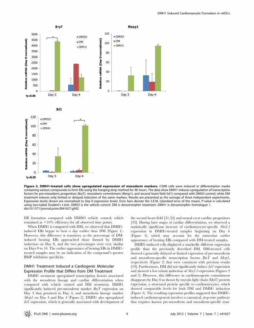
DMH1 treatment upregulated transcription factors associated
with the mesoderm lineage and cardiac differentiation when
compared with vehicle control and DM treatment. DMH1
significantly induced pre-mesoderm marker BryT expression on
Day 3 that persisted to Day 4, and mesoderm lineage marker
Mesp1 on Day 3 and Day 4 (Figure 2). DMH1 also upregulated
Isl1 expression, which is generally associated with development of
the second heart field [31,32] and neural crest cardiac progenitors
[33]. During later stages of cardiac differentiation, we observed a
statistically significant increase of cardiomyocyte-specific Nkx2.5
expression in DMH1-treated samples beginning on Day 6
(Figure 3), which may account for the somewhat earlier
appearance of beating EBs compared with DM-treated samples.
DMH1-induced cells displayed a markedly different expression
profile than the previously described DM. DM-treated cells
showed a generally delayed or limited expression of pre-mesoderm
and mesoderm-specific transcription factors BryT and Mesp1,
respectively (Figure 2) that were consistent with previous results
[19]. Furthermore, DM did not significantly induce Isl1 expression
and showed a less robust induction of Nkx2.5 expression (Figures 2
and 3). However, this difference in cardiomyogenic commitment
disappears by Day 8 as shown by myosin light chain (Myl7) protein
expression, a structural protein specific to cardiomyocytes, which
showed comparable levels for both DM and DMH1 induction
(Figure 3). The resulting expression profiles suggested that DMH1-
induced cardiomyogenesis involves a canonical, step-wise pathway
that requires known pre-mesoderm and mesoderm-specific tran-
Figure 2. DMH1-treated cells show upregulated expression of mesoderm markers. CGR8 cells were induced in differentiation mediacontaining various compounds to form EBs using the hanging-drop method for 48 hours. The data show DMH1 induces upregulation of transcriptionfactors for pre-mesoderm progenitors (BryT), mesoderm commitment (Mesp1), and second heart field (Isl1) compared with DMSO control; while DMtreatment induces only limited or delayed induction of the same markers. Results are presented as the average of three independent experiments.Expression levels shown are normalized to Day 0 expression levels. Error bars denote the S.E.M. (standard error of the mean). P-value is calculatedusing two-tailed Student’s t-test. DMSO is the vehicle control. DM is dorsomorphin treatment. DMH1 is dorsomorphin homologue 1.doi:10.1371/journal.pone.0041627.g002
DMH1 Induced Cardiomyocyte Formation in mESCs
PLoS ONE | www.plosone.org 4 July 2012 | Volume 7 | Issue 7 | e41627

scription factors to regulate cardiomyocyte commitment. The
profiles also confirmed previously published data that DM induced
limited or delayed expression of mesoderm-associated factors, and
suggested that DM-induced cardiomyocytes developed from a
non-canonical pathway that remains unknown.
DMH1 does not Specifically Induce Other Mesoderm-associated Lineage Markers
BryT-committed cells are known to be multipotent cells capable
of producing hematopoietic progenitors [34] and other mesoderm-
associated lineages such as smooth muscle cells and endocardial
cells [35]. Therefore, we further probed the DMH1-induced gene
expression profile for signs of hematopoiesis and non-cardiomy-
ocyte development. In Figure 4, transcriptional profiling of
DMH1-induced EBs indicated that, similar to DM [19], DMH1
did not specifically induced the expression of multipotent
progenitor marker Kdr/Flk-1 or hematopoiesis (Gata1) compared
with DMSO induction, which stimulates spontaneous and non-
specific differentiation. Similarly, DMH1 treatment did not
induced specific Myh11 expression, a marker for smooth muscle
cells, relative to non-specific DMSO induction as the two
expression levels are comparable.
However, DMH1 supported a modest 2.3-fold increase in the
expression of endocardial marker VE-CAM on Day 10 (Figure 4)
when compared with DMSO, but it is unclear if this modest
increase contributes to further endocardial lineage development.
Overall, DMH1 treatment did not specifically increase Kdr/Flk-
1, Gata1, or Myh11 expression levels relative to non-specific
DMSO induction. This is unlike DM treatment, which showed a
notable decrease in the expression of those three genes relative to
DMSO. This pattern suggested that DM may specifically repress
their expression and is consistent with previously published results
[19].
DMH1 does not Specifically Induce Endoderm orEctoderm Markers
DMH1-induced EBs were also examined for endoderm or
ectoderm commitment to confirm mesoderm induction specificity.
As shown in Figure 5, DMH1 did not specifically upregulate
endoderm markers Sox17 or FoxA2 expression in a statistically
significant manner when compared with DMSO from Day 3 to
Day 6. In addition, the increases observed for DMH1 treatments
were less than 2-fold, which is within the margin of error for rt-
PCR analysis and therefore were not considered a meaningful
increase. There was also sufficient variability between each
independent experiment to indicate that the observed Sox17 or
FoxA2 expression level may be either randomly, or minimally,
induced by DMH1. The ectoderm marker Nestin was also
minimally affected during the early stages of DMH1-induced
differentiation. We did not observe statically significant Nestin
induction by DMH1 relative to DMSO. We concluded that the
influence of DMH1 on directed differentiation during the earliest
stages of induction is restricted to the mesoderm linage.
DM treatment appeared to delay the expression of endoderm
markers Sox17 and FoxA2 at the time points examined (Figure 5).
But like DMH1, the observed increases in expression appeared to
be non-specific and are not statistically significant changes. The
expression level of Nestin was also not significantly affected by DM.
DMH1 Treatment Enriched for Pro-cardiac Progenitorsthat Responded to Wnt Inhibition and Progressed toForm Secondary Cardiomyocytes
The marked difference in the pro-cardiac gene expression
profiles between DM and DMH1 prompted us to further
investigate their respective developmental potential at the pre-
mesoderm and early mesoderm stages following treatment. We
used a mouse embryonic stem cell line containing a recombinant
BryT-GFP reporter that was described previously [26,36] to isolate
BryT-GFP positive (GFP+) and negative (GFP–) cell fractions
4 days after induction. The Day 4 time point was chosen to obtain
the maximum number of GFP+ cells from all treatments for
further experiments, as Day 3 FACS resulted in too few cells for
additional studies. Figure 6 shows the average percentage of GFP+cells for each treatment. The data showed that DMSO treatment
yielded a greater percentage of GFP+ cells than DMH1 treatment.
This observation may be due to differences in temporal BryT
regulation by the two treatments (Figure 2). Since the cells were
sorted on Day 4, the GFP signal in DMH1-treated samples may be
Figure 3. DMH1-treated cells showed increased cardiomyocyte lineage commitment. (Left) DMH1 induces significant upregulation oftranscription factor for cardiomyocyte commitment (Nkx2.5) compared with DMSO control. DM also induces Nkx2.5 expression, but the induction isless robust than DMH1. Results are presented as the average of three independent experiments. Expression levels shown are normalized to Day 0expression levels. Error bars denote the S.E.M. (standard error of the mean). P-value is calculated using two-tailed Student’s t-test. (Right) Western blotshowing increased Myl7 expression (,19 kDa), a structural protein specific to cardiomyocytes, in CGR8 cells 8 days after DM or DMH1 induction. NoMyl7 expression was observed after DMSO treatment. Alpha-tubulin (,50 kDa) is shown as loading control. The relative intensities of Myl7 bands arenormalized to alpha-tubulin and are shown at the bottom.doi:10.1371/journal.pone.0041627.g003
DMH1 Induced Cardiomyocyte Formation in mESCs
PLoS ONE | www.plosone.org 5 July 2012 | Volume 7 | Issue 7 | e41627

residual signal from Day 3, whereas the signal from DMSO
treatment may be nascent. There is also a large degree of
experimental variation and each cell sorting yields a unique
percentage of BryT-GFP positive cells. BryT expression was
confirmed in the GFP+ fractions using rt-PCR and the data
showed that there is no significant BryT expression leakage in the
GFP–fractions (Figure 6). The GFP+ fractions also retained cells
with upregulated Mesp1 and Isl1 expression levels. Their expres-
sion levels were highly variable, which suggested the GFP+ cells
were transitioning to the mesoderm lineage at the time of sorting.
Both Mesp1 and Isl1 expression were at barely detectable levels in
the GFP– fractions and indicated they had not undergone
mesoderm commitment.
We decided to track the cardiac development potential of all
sorted cell fractions as an indirect measurement of pro-cardio-
myogenic progenitor enrichment following DM and DMH1
treatment. We hypothesized that the DM-or DMH1-treated
GFP+ fractions will generate beating cardiomyocytes, whereas
the DMSO-treated fractions and the DM/DMH1-treated GFP–
fractions will not because they lack pro-cardiac cells. Wnt signaling
Figure 4. DMH1 treatment does not specifically induce other mesoderm-associated lineage markers. CGR8 induction and geneexpression analysis were performed as previously described. The data show DM or DMH1 treatment does not specifically induce hematopoieticmarkers Gata1 and Flk-1/Kdr, or smooth muscle marker Myh11 when compared with DMSO control. Both compounds show a modest induction ofvascular endothelial marker VE-CAM but the differences are not statistically significant. Results are presented as the average of three independentexperiments. Expression levels shown are normalized to Day 0 expression levels. Error bars denote the S.E.M. DMSO is the vehicle control. DM isdorsomorphin treatment. DMH1 is dorsomorphin homologue 1.doi:10.1371/journal.pone.0041627.g004
DMH1 Induced Cardiomyocyte Formation in mESCs
PLoS ONE | www.plosone.org 6 July 2012 | Volume 7 | Issue 7 | e41627
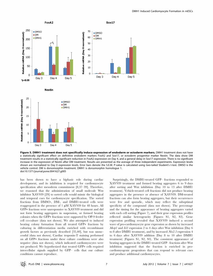
has been shown to have a biphasic role during cardiac
development, and its inhibition is required for cardiomyocyte
specification after mesoderm commitment [8,37–39]. Therefore,
we reasoned that the administration of small molecule Wnt
inhibitor XAV939 [29] to sorted cells would mimic the biological
and temporal cues for cardiomyocyte specification. The sorted
fractions from DMSO-, DM-, and DMH1-treated cells were
reaggregated in the presence of 1 mM XAV939 for 48 hours. All
GFP+ fractions were unresponsive to XAV939 treatment and did
not form beating aggregates in suspension, or formed beating
colonies when the GFP+ fractions were supported by OP-9 feeder
cell co-culture (data not shown). We also attempted to induced
beating colony formation from all treated GFP+ fractions by
culturing in differentiation media enriched with recombinant
growth factors as previously described [35,40], but was unsuc-
cessful (data not shown). Cardiac troponin-T staining performed
on all GFP+ fractions under these culture conditions were also
negative (data not shown), which indicated cardiomyocytes were
not produced. We hypothesized that treated GFP+ cells required
intercellular signals supplied by GFP– cells that our culture
conditions cannot reproduce.
Surprisingly, the DMH1-treated GFP– fractions responded to
XAV939 treatment and formed beating aggregates 6 to 9 days
after sorting and Wnt inhibition (Day 10 to 13 after DMH1
treatment). Vehicle-treated cell fractions did not produce beating
aggregates in the presence or absence of XAV939. DM-treated
fractions can also form beating aggregates, but their occurrences
were few and sporadic, which may reflect the suboptimal
specificity of the compound (data not shown). The percentage
and the timing for the appearance of beating aggregates varied
with each cell sorting (Figure 7), and their gene expression profiles
reflected similar heterogeneity (Figures S1, S2, S3). Gene
expression profiling revealed that XAV939 induced a second
wave of pro-cardiomyocyte gene expression as shown by increased
Mesp1 and Isl1 expression 2 to 4 days after Wnt inhibition (Day 6
to 8 after DMH1 treatment), and by increased Nkx2.5 expression 4
to 6 days after XAV939 addition (Day 8 to 10 after DMH1
treatment) (Figures S1, S2, S3). The consistent appearance of
beating aggregates in the DMH1-treated GFP– fractions after Wnt
inhibition suggested that the fraction is enriched in pro-
cardiomyogenic progenitors that can respond to relevant cues
and produce additional cardiomyocytes.
Figure 5. DMH1 treatment does not specifically induce expression of endoderm or ectoderm markers. DMH1 treatment does not havea statistically significant effect on definitive endoderm markers FoxA2 and Sox17, or ectoderm progenitor marker Nestin. The data show DMtreatment results in a statistically significant reduction in FoxA2 expression on Day 4, and a general delay in Sox17 expression. There is no significantincrease in the expression of Nestin after DM treatment. Results are presented as the average of three independent experiments. Expression levelsshown are normalized to Day 0 expression levels. Error bars denote the S.E.M. P-value is calculated using two-tailed Student’s t-test. DMSO is thevehicle control. DM is dorsomorphin treatment. DMH1 is dorsomorphin homologue 1.doi:10.1371/journal.pone.0041627.g005
DMH1 Induced Cardiomyocyte Formation in mESCs
PLoS ONE | www.plosone.org 7 July 2012 | Volume 7 | Issue 7 | e41627
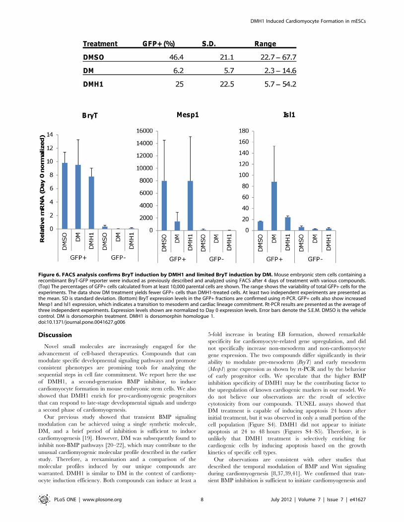
Discussion
Novel small molecules are increasingly engaged for the
advancement of cell-based therapeutics. Compounds that can
modulate specific developmental signaling pathways and promote
consistent phenotypes are promising tools for analyzing the
sequential steps in cell fate commitment. We report here the use
of DMH1, a second-generation BMP inhibitor, to induce
cardiomyocyte formation in mouse embryonic stem cells. We also
showed that DMH1 enrich for pro-cardiomyogenic progenitors
that can respond to late-stage developmental signals and undergo
a second phase of cardiomyogenesis.
Our previous study showed that transient BMP signaling
modulation can be achieved using a single synthetic molecule,
DM, and a brief period of inhibition is sufficient to induce
cardiomyogenesis [19]. However, DM was subsequently found to
inhibit non-BMP pathways [20–22], which may contribute to the
unusual cardiomyogenic molecular profile described in the earlier
study. Therefore, a reexamination and a comparison of the
molecular profiles induced by our unique compounds are
warranted. DMH1 is similar to DM in the context of cardiomy-
ocyte induction efficiency. Both compounds can induce at least a
5-fold increase in beating EB formation, showed remarkable
specificity for cardiomyocyte-related gene upregulation, and did
not specifically increase non-mesoderm and non-cardiomyocyte
gene expression. The two compounds differ significantly in their
ability to modulate pre-mesoderm (BryT) and early mesoderm
(Mesp1) gene expression as shown by rt-PCR and by the behavior
of early progenitor cells. We speculate that the higher BMP
inhibition specificity of DMH1 may be the contributing factor to
the upregulation of known cardiogenic markers in our model. We
do not believe our observations are the result of selective
cytotoxicity from our compounds. TUNEL assays showed that
DM treatment is capable of inducing apoptosis 24 hours after
initial treatment, but it was observed in only a small portion of the
cell population (Figure S4). DMH1 did not appear to initiate
apoptosis at 24 to 48 hours (Figures S4–S5). Therefore, it is
unlikely that DMH1 treatment is selectively enriching for
cardiogenic cells by inducing apoptosis based on the growth
kinetics of specific cell types.
Our observations are consistent with other studies that
described the temporal modulation of BMP and Wnt signaling
during cardiomyogenesis [8,37,39,41]. We confirmed that tran-
sient BMP inhibition is sufficient to initiate cardiomyogenesis and
Figure 6. FACS analysis confirms BryT induction by DMH1 and limited BryT induction by DM. Mouse embryonic stem cells containing arecombinant BryT-GFP reporter were induced as previously described and analyzed using FACS after 4 days of treatment with various compounds.(Top) The percentages of GFP+ cells calculated from at least 10,000 parental cells are shown. The range shows the variability of total GFP+ cells for theexperiments. The data show DM treatment yields fewer GFP+ cells than DMH1-treated cells. At least two independent experiments are presented asthe mean. SD is standard deviation. (Bottom) BryT expression levels in the GFP+ fractions are confirmed using rt-PCR. GFP+ cells also show increasedMesp1 and Isl1 expression, which indicates a transition to mesoderm and cardiac lineage commitment. Rt-PCR results are presented as the average ofthree independent experiments. Expression levels shown are normalized to Day 0 expression levels. Error bars denote the S.E.M. DMSO is the vehiclecontrol. DM is dorsomorphin treatment. DMH1 is dorsomorphin homologue 1.doi:10.1371/journal.pone.0041627.g006
DMH1 Induced Cardiomyocyte Formation in mESCs
PLoS ONE | www.plosone.org 8 July 2012 | Volume 7 | Issue 7 | e41627

small molecules can serve as synthetic tools for this function
[19,41]. We also showed that Wnt inhibition at a later stage can
initiate cardiomyogenesis in our low BryT-expressing cell fractions
[8,37,38]. This effect is reminiscent of the second heart field, in
which a second wave of cardiovascular development occurs that
contributes to the formation of the outflow tract and the right
ventricle, and Wnt signaling modulation has been shown to
regulate its development [8]. Our model cannot determine the
specific progenitor population Wnt inhibition is targeting. The
beating EBs may have derived from residual mesoderm-commit-
ted progenitors in the GFP– fractions, or they may be multipotent
cells that initiated mesoderm commitment and cardiomyocyte
differentiation after Wnt inhibition. Based on the upregulation of
Mesp1 and Isl1 expression 2 to 4 days after XAV939 treatment
(Figures S1, S2, S3), we suspect that the secondary cardiomyocytes
were derived from pro-cardiac multipotent progenitors in the
GFP– fractions that were directed by late-stage Wnt inhibition
towards cardiac lineage commitment. Our results are consistent
with the sequential nature of directed differentiation and that
specific signaling modulation during early development can
produce a more restricted progenitor lineage that favors particular
cell types.
Our data suggests that selective inhibition of BMP Type 1
receptors during early development is critical for increasing the
proportion of cardiomyogenic progenitor cells and maximizing the
overall cardiomyocyte induction potential. When the gene
expression profiles of DM-or DMH1-treated cells were compared
with those treated with DMH4, which is a selective VEGF-
signaling inhibitor with no BMP inhibition activity [21], we
observed no cardiomyogenic gene upregulation by DMH4 (Figure
S6). Instead, DMH4 treatment appeared to favor vascular
development as shown by the upregulation of smooth muscle
marker Myh11 and endocardial marker VE-CAM (Figure S6).
Furthermore, other DM homologues that are structurally similar
to DMH1 and are also selective inhibitors of BMP Type 1
receptors can initiate beating EB formation ([21], unpublished
data). This observation suggests that modulating only a small
subset of BMP Type 1 receptors is sufficient to initiate
cardiomyogenesis.
In summary, our study presents evidence that our second-
generation BMP inhibitor DMH1 has more specific cardiomyo-
genic properties than dorsomorphin in the context of in vitro
directed differentiation. DMH1 has a greater effect on early
cardiac development than dorsomorphin and may enrich for pro-
cardiac multipotent progenitors that can respond to late-stage Wnt
inhibition and initiate a second phase of cardiomyogenic
commitment. We also postulate that modulation of a small subset
of BMP Type 1 receptors is central for this phenomenon. We
believe DMH1 can be part of a multi-step strategy to increase
cardiomyocyte induction from pluripotent cells.
Supporting Information
Figure S1 Gene expression analysis of individual ex-periments for mesoderm and cardiogenic markers forexperiment No.1. Induction of mesoderm and cardiac specific
markers in the GFP– fractions after Wnt inhibition were analyzed
using rt-PCR and shown as individual experiments in each figure.
XAV939 (1 mM) upregulated Mesp1 expression 2 to 4 days after
treatment in the sample initially induced using DMH1. The
DMH1-treated fraction shows increased Isl1 expression 4 days
after XAV939 treatment (Day 8 post-DMH1 induction). The
expression of cardiomyocyte transcription factor Nkx2.5 increases
4 to 6 days after XAV939 treatment. Expression levels shown are
normalized to Day 0 expression levels. DMSO+V is DMSO
induction with no XAV939 after FACS. DMSO+XAV is DMSO
induction plus XAV939 addition after FACS. DM+V is DM
induction with no XAV939 after FACS. DM+XAV is DM
induction plus XAV939 addition after FACS. DMH1+V is
DMH1 induction with no XAV939 after FACS. DM+XAV is
DMH1 induction plus XAV939 addition after FACS.
(TIF)
Figure S2 Gene expression analysis of individual ex-periments for mesoderm and cardiogenic markers forexperiment No.2. Induction of mesoderm and cardiac specific
markers in the GFP– fractions after Wnt inhibition were analyzed
using rt-PCR and shown as individual experiments in each figure.
XAV939 (1 mM) upregulated Mesp1 expression 2 to 4 days after
treatment in the sample initially induced using DMH1. The
DMH1-treated fraction shows increased Isl1 expression 4 days
after XAV939 treatment (Day 8 post-DMH1 induction). The
expression of cardiomyocyte transcription factor Nkx2.5 increases
4 to 6 days after XAV939 treatment. Expression levels shown are
normalized to Day 0 expression levels. DMSO+V is DMSO
induction with no XAV939 after FACS. DMSO+XAV is DMSO
induction plus XAV939 addition after FACS. DM+V is DM
induction with no XAV939 after FACS. DM+XAV is DM
induction plus XAV939 addition after FACS. DMH1+V is
DMH1 induction with no XAV939 after FACS. DM+XAV is
DMH1 induction plus XAV939 addition after FACS.
(TIF)
Figure 7. Low BryT-expressing fraction produces beating EBafter Wnt inhibition. Each table is data from a single experimentshowing the percentage of beating aggregates that formed after GFP–fractions were treated with either DMSO control or 1 mM XAV939 toinhibit Wnt signaling. The DMH1-treated GFP– fractions typically formbeating aggregates 6 to 8 days after XAV939 addition (Day 10 to Day 12after initial induction). The percentages increase over time and theefficiency of beating aggregate formation varies with each FACS. Thepercentages were calculated from the number of beating aggregatesand the number of total aggregates for each condition.doi:10.1371/journal.pone.0041627.g007
DMH1 Induced Cardiomyocyte Formation in mESCs
PLoS ONE | www.plosone.org 9 July 2012 | Volume 7 | Issue 7 | e41627

Figure S3 Gene expression analysis of individual ex-periments for mesoderm and cardiogenic markers forexperiment No.3. Induction of mesoderm and cardiac specific
markers in the GFP– fractions after Wnt inhibition were analyzed
using rt-PCR and shown as individual experiments in each figure.
XAV939 (1 mM) upregulated Mesp1 expression 2 to 4 days after
treatment in the sample initially induced using DMH1. The
DMH1-treated fraction shows increased Isl1 expression 4 days
after XAV939 treatment (Day 8 post-DMH1 induction). The
expression of cardiomyocyte transcription factor Nkx2.5 increases
4 to 6 days after XAV939 treatment. Expression levels shown are
normalized to Day 0 expression levels. DMSO+V is DMSO
induction with no XAV939 after FACS. DMSO+XAV is DMSO
induction plus XAV939 addition after FACS. DM+V is DM
induction with no XAV939 after FACS. DM+XAV is DM
induction plus XAV939 addition after FACS. DMH1+V is
DMH1 induction with no XAV939 after FACS. DM+XAV is
DMH1 induction plus XAV939 addition after FACS.
(TIF)
Figure S4 DMH1 does not induce apoptosis 24 h aftertreatment. CGR8 cells were incubated in differentiation media
with or without compounds for 24 h prior to TUNEL assay.
DNase-treated cells were used as positive control. Cells incubated
with the labeling solution alone and without enzyme served as
negative control. The results show DM can cause cellular
apoptosis 24 h after treatment in one visual field, and the results
are not statistically significant. No positive TUNEL staining was
observed after DMH1 treatment.
(TIF)
Figure S5 DMH1 does not induce apoptosis 48 h aftertreatment. CGR8 cells were incubated in differentiation media
with or without compounds for 48 h prior to TUNEL assay.
DNase-treated cells were used as positive control. Cells incubated
with the labeling solution alone and without enzyme served as
negative control. The results show neither DM nor DMH1 causes
cellular apoptosis 48 h after treatment.
(TIF)
Figure S6 Early transient BMP inhibition is essentialfor cardiomyogenesis. Gene expression profiling was per-
formed after treatment with DM, DMH1, or DMH4. DMH4 is a
VEGF-specific inhibitor with no antagonist effects on BMP
signaling [21]. The data show BMP inhibition is required to
upregulate cardiomyogenic gene expression, while VEGF-inhibi-
tion appeared to yield an expression profile consistent with
vascular development by upregulating smooth muscle marker
Myh11 and endocardial marker VE-CAM. Results are presented
as the average of three independent experiments. Expression levels
shown are normalized to Day 0 expression levels. Error bars
denote the S.E.M. DMSO is the vehicle control. DM is
dorsomorphin treatment. DMH1 is dorsomorphin homologue 1.
DMH4 is dorsomorphin homologue 4.
(TIF)
Acknowledgments
We thank Eric Alder for providing the BryT reporter cell line. We also
thank Kevin Ess for giving us SNL feeder cells for culture maintenance,
and Antonis Hatzopoulos for providing CGR8 cells and technical advice.
Author Contributions
Conceived and designed the experiments: AA JH CCH. Performed the
experiments: AA JH. Analyzed the data: AA JH CCH. Contributed
reagents/materials/analysis tools: CRH. Wrote the paper: AA. Reviewed
the manuscript: JH CCH.
References
1. Puceat M (2008) Protocols for cardiac differentiation of embryonic stem cells.
Methods 45: 168–171. doi:10.1016/j.ymeth.2008.03.003.
2. Fuegemann CJ, Samraj AK, Walsh S, Fleischmann BK, Jovinge S, et al. (2010)Differentiation of mouse embryonic stem cells into cardiomyocytes via the
hanging-drop and mass culture methods. Curr Protoc Stem Cell Biol Chapter 1:Unit 1F.11. doi:10.1002/9780470151808.sc01f11s15.
3. Mummery C, van der Heyden MAG, de Boer TP, Passier R, Ward D, et al.
(2007) Cardiomyocytes from human and mouse embryonic stem cells. MethodsMol Med 140: 249–272.
4. Kattman SJ, Witty AD, Gagliardi M, Dubois NC, Niapour M, et al. (2011)
Stage-specific optimization of activin/nodal and BMP signaling promotes
cardiac differentiation of mouse and human pluripotent stem cell lines. CellStem Cell 8: 228–240. doi:10.1016/j.stem.2010.12.008.
5. Paige SL, Osugi T, Afanasiev OK, Pabon L, Reinecke H, et al. (2010)
Endogenous Wnt/beta-catenin signaling is required for cardiac differentiation inhuman embryonic stem cells. PLoS ONE 5: e11134. doi:10.1371/journal.-
pone.0011134.
6. Barron M, Gao M, Lough J (2000) Requirement for BMP and FGF signalingduring cardiogenic induction in non-precardiac mesoderm is specific, transient,
and cooperative. Dev Dyn 218: 383–393. doi:10.1002/(SICI)1097-0177(200006)218:2,383:: AID-DVDY11.3.0.CO;2-P.
7. Johansson BM, Wiles MV (1995) Evidence for involvement of activin A and
bone morphogenetic protein 4 in mammalian mesoderm and hematopoietic
development. Mol Cell Biol 15: 141–151.
8. Klaus A, Saga Y, Taketo MM, Tzahor E, Birchmeier W (2007) Distinct roles ofWnt/beta-catenin and Bmp signaling during early cardiogenesis. Proc Natl Acad
Sci USA 104: 18531–18536. doi:10.1073/pnas.0703113104.
9. Lough J, Barron M, Brogley M, Sugi Y, Bolender DL, et al. (1996) CombinedBMP-2 and FGF-4, but neither factor alone, induces cardiogenesis in non-
precardiac embryonic mesoderm. Dev Biol 178: 198–202. doi:10.1006/dbio.1996.0211.
10. Schultheiss TM, Burch JB, Lassar AB (1997) A Role for Bone Morphogenetic
Proteins in the Induction of Cardiac Myogenesis. Genes Dev 11: 451–462.doi:10.1101/gad.11.4.451.
11. Mine N, Anderson RM, Klingensmith J (2008) BMP antagonism is required in
both the node and lateral plate mesoderm for mammalian left-right axis
establishment. Development 135: 2425–2434. doi:10.1242/dev.018986.
12. Liu W, Foley AC (2011) Signaling pathways in early cardiac development. Wiley
Interdiscip Rev Syst Biol Med 3: 191–205. doi:10.1002/wsbm.112.
13. Marques SR, Yelon D (2009) Differential requirement for BMP signaling in
atrial and ventricular lineages establishes cardiac chamber proportionality. DevBiol 328: 472–482. doi:10.1016/j.ydbio.2009.02.010.
14. Hutson MR, Zeng XL, Kim AJ, Antoon E, Harward S, et al. (2010) Arterial
pole progenitors interpret opposing FGF/BMP signals to proliferate ordifferentiate. Development 137: 3001–3011. doi:10.1242/dev.051565.
15. Tirosh-Finkel L, Zeisel A, Brodt-Ivenshitz M, Shamai A, Yao Z, et al. (2010)
BMP-mediated inhibition of FGF signaling promotes cardiomyocyte differen-tiation of anterior heart field progenitors. Development 137: 2989–3000.
doi:10.1242/dev.051649.
16. Ao A, Hao J, Hong CC (2011) Regenerative Chemical Biology: CurrentChallenges and Future Potential. Chemistry & Biology 18: 413–424.
doi:10.1016/j.chembiol.2011.03.011.
17. Emre N, Coleman R, Ding S (2007) A chemical approach to stem cell biology.
Curr Opin Chem Biol 11: 252–258. doi:10.1016/j.cbpa.2007.04.024.
18. Li W, Ding S (2010) Small molecules that modulate embryonic stem cell fate andsomatic cell reprogramming. Trends Pharmacol Sci 31: 36–45. doi:10.1016/
j.tips.2009.10.002.
19. Hao J, Daleo MA, Murphy CK, Yu PB, Ho JN, et al. (2008) Dorsomorphin, aselective small molecule inhibitor of BMP signaling, promotes cardiomyogenesis
in embryonic stem cells. PLoS One 3: e2904. doi:10.1371/journal.pone.0002904.
20. Boergermann JH, Kopf J, Yu PB, Knaus P (2010) Dorsomorphin and LDN-
193189 inhibit BMP-mediated Smad, p38 and Akt signalling in C2C12 cells.The International Journal of Biochemistry & Cell Biology 42: 1802–1807.
doi:10.1016/j.biocel.2010.07.018.
21. Hao J, Ho JN, Lewis JA, Karim KA, Daniels RN, et al. (2010) In VivoStructure2Activity Relationship Study of Dorsomorphin Analogues Identifies
Selective VEGF and BMP Inhibitors. ACS Chemical Biology 5: 245–253.doi:10.1021/cb9002865.
22. Yu PB, Deng DY, Lai CS, Hong CC, Cuny GD, et al. (2008) BMP type I
receptor inhibition reduces heterotopic [corrected] ossification. Nat Med 14:1363–1369. doi:10.1038/nm.1888.
23. Cross EE, Thomason RT, Martinez M, Hopkins CR, Hong CC, et al. (2011)Application of small organic molecules reveals cooperative TGFb and BMP
DMH1 Induced Cardiomyocyte Formation in mESCs
PLoS ONE | www.plosone.org 10 July 2012 | Volume 7 | Issue 7 | e41627
regulation of mesothelial cell behaviors. ACS Chem Biol 6: 952–961.
doi:10.1021/cb200205z.24. Hill CR, Sanchez NS, Love JD, Arrieta JA, Hong CC, et al. (2012) BMP2
signals loss of epithelial character in epicardial cells but requires the Type III
TGFb receptor to promote invasion. Cell Signal 24: 1012–1022. doi:10.1016/j.cellsig.2011.12.022.
25. Nichols J, Evans EP, Smith AG (1990) Establishment of germ-line-competentembryonic stem (ES) cells using differentiation inhibiting activity. Development
110: 1341–1348.
26. Fehling HJ, Lacaud G, Kubo A, Kennedy M, Robertson S, et al. (2003)Tracking mesoderm induction and its specification to the hemangioblast during
embryonic stem cell differentiation. Development 130: 4217–4227.27. McMahon AP, Bradley A (1990) The Wnt-1 (int-1) proto-oncogene is required
for development of a large region of the mouse brain. Cell 62: 1073–1085.28. Yu PB, Hong CC, Sachidanandan C, Babitt JL, Deng DY, et al. (2008)
Dorsomorphin inhibits BMP signals required for embryogenesis and iron
metabolism. Nat Chem Biol 4: 33–41. doi:10.1038/nchembio.2007.54.29. Wang H, Hao J, Hong CC (2011) Cardiac induction of embryonic stem cells by
a small molecule inhibitor of Wnt/b-catenin signaling. ACS Chem Biol 6: 192–197. doi:10.1021/cb100323z.
30. Ao A, Williams CH, Hao J, Hong CC (2011) Modified Mouse Embryonic Stem
Cell based Assay for Quantifying Cardiogenic Induction Efficiency. J Vis Exp.Available:http://www.ncbi.nlm.nih.gov/pubmed/21540823. Accessed 5 May
2011.31. Moretti A, Caron L, Nakano A, Lam JT, Bernshausen A, et al. (2006)
Multipotent embryonic isl1+ progenitor cells lead to cardiac, smooth muscle,and endothelial cell diversification. Cell 127: 1151–1165. doi:10.1016/
j.cell.2006.10.029.
32. Qyang Y, Martin-Puig S, Chiravuri M, Chen S, Xu H, et al. (2007) The renewaland differentiation of Isl1+ cardiovascular progenitors are controlled by a Wnt/
beta-catenin pathway. Cell Stem Cell 1: 165–179. doi:10.1016/j.stem.2007.05.018.
33. Engleka KA, Manderfield LJ, Brust RD, Li L, Cohen A, et al. (2012) Islet1
Derivatives in the Heart Are of Both Neural Crest and Second Heart Field
Origin. Circulation Research. Available:http://www.ncbi.nlm.nih.gov/
pubmed/22394517. Accessed 22 March 2012.
34. Kabrun N, Buhring HJ, Choi K, Ullrich A, Risau W, et al. (1997) Flk-1
expression defines a population of early embryonic hematopoietic precursors.
Development 124: 2039–2048.
35. Kattman SJ, Huber TL, Keller GM (2006) Multipotent flk-1+ cardiovascular
progenitor cells give rise to the cardiomyocyte, endothelial, and vascular smooth
muscle lineages. Dev Cell 11: 723–732. doi:10.1016/j.devcel.2006.10.002.
36. Kubo A, Shinozaki K, Shannon JM, Kouskoff V, Kennedy M, et al. (2004)
Development of definitive endoderm from embryonic stem cells in culture.
Development 131: 1651–1662. doi:10.1242/dev.01044.
37. Naito AT, Shiojima I, Akazawa H, Hidaka K, Morisaki T, et al. (2006)
Developmental stage-specific biphasic roles of Wnt/beta-catenin signaling in
cardiomyogenesis and hematopoiesis. Proc Natl Acad Sci USA 103: 19812–
19817. doi:10.1073/pnas.0605768103.
38. Ueno S, Weidinger G, Osugi T, Kohn AD, Golob JL, et al. (2007) Biphasic Role
for Wnt/B-Catenin Signaling in Cardiac Specification in Zebrafish and
Embryonic Stem Cells. PNAS 104: 9685–9690. doi:10.1073/pnas.0702859104.
39. David R, Brenner C, Stieber J, Schwarz F, Brunner S, et al. (2008) MesP1 drives
vertebrate cardiovascular differentiation through Dkk-1-mediated blockade of
Wnt-signalling. Nature Cell Biology 10: 338–345. doi:10.1038/ncb1696.
40. Misfeldt AM, Boyle SC, Tompkins KL, Bautch VL, Labosky PA, et al. (2009)
Endocardial cells are a distinct endothelial lineage derived from Flk1+multipotent cardiovascular progenitors. Dev Biol 333: 78–89. doi:10.1016/
j.ydbio.2009.06.033.
41. Yuasa S, Itabashi Y, Koshimizu U, Tanaka T, Sugimura K, et al. (2005)
Transient inhibition of BMP signaling by Noggin induces cardiomyocyte
differentiation of mouse embryonic stem cells. Nat Biotechnol 23: 607–611.
doi:10.1038/nbt1093.
DMH1 Induced Cardiomyocyte Formation in mESCs
PLoS ONE | www.plosone.org 11 July 2012 | Volume 7 | Issue 7 | e41627
Related Documents