DKT1, a novel K þ channel from carrot, forms functional heteromeric channels with KDC1 Elide Formentin a, * , Serena Varotto b , Alex Costa a , Patrick Downey c , Monica Bregante d , Alessia Naso d , Cristiana Picco d , Franco Gambale d , Fiorella Lo Schiavo a a CRIBI, Centro Interdipartimentale per le Biotecnologie Innovative, Universit a di Padova, Viale G. Colombo 3, 35131 Padova, Italy b Dipartimento di Agronomia Ambientale e Produzioni Vegetali, Universit a di Padova, Agripolis, Viale dell’Universit a 16, 35020 Legnaro (Padova), Italy c Schering-Plough Research Institute, San Raffaele Biomedical Science Park, Via Olgettina 58, 20132 Milan, Italy d Istituto di Biofisica-CNR, Via DeMarini 6, 16149 Genova, Italy Received 13 May 2004; revised 21 July 2004; accepted 21 July 2004 Available online 29 July 2004 Edited by Ulf-Ingo Fl€ ugge Abstract We report the isolation and characterisation of DKT1, a new carrot K þ channel a-subunit belonging to the Shaker-like family. DKT1 is expressed in many tissues of the adult plant, suggesting that it may play important roles in both nutrition and other important physiological processes. During embryo development, DKT1 is expressed at later phases implying the involvement of K þ in embryo maturation. When co-expressed with KDC1 in Xenopus oocytes, DKT1 is able to form functional, heteromeric channels, suggesting that possible interactions between these two ion channels in plant tissues may modulate K þ uptake. Ó 2004 Federation of European Biochemical Societies. Pub- lished by Elsevier B.V. All rights reserved. Keywords: K þ channel; Heteromeric channel; Shaker family; Daucus carota 1. Introduction K þ is involved in several key cellular functions such as the maintenance of membrane potential, enzyme activation, sto- matal movements and tropisms [1–4]. Over the past decade, reverse genetics and electrophysiological studies have revealed the presence of several families of K þ channels. Potassium channels of the Shaker family have been isolated from several plant species where they appear to be involved in several steps which are crucial for K þ absorption [5]. Based on DNA sequence analysis, and functional charac- teristics, these channels can be divided into five groups. Groups I, II and IV contain inward rectifier channels, group III weakly inward rectifier channels, while group V consists of outward rectifying channels [6]. In particular, group IV rep- resents inward rectifier channels, such as Arabidopsis AtKC1 and carrot KDC1, whose functions in planta have yet to be determined. KDC1, the first inward potassium channel iso- lated from carrot [7], is expressed in roots and root hairs and its Arabidopsis homologue AtKC1 has been shown to have a very similar expression pattern [8]. In contrast to AtKC1, which is unable to form functional channels in heterologous systems, KDC1 has been functionally expressed in CHO cells where an initial characterisation of the homomeric channel was performed [7]. A more detailed study was conducted in Xenopus oocytes where KDC1 does not form functional ho- momeric channels alone, however, when co-injected with the Arabidopsis KAT1 a-subunit, it forms heteromeric channels. KDC1:KAT1 heteromers show current activation which is significantly different from those of the homomeric KAT1 channel, demonstrating the modulator capability of KDC1 on other channels [9,10]. Heteromerization is a widespread mechanism present in both plants and animals that participates in modulation of potassium permeability [11,12]. In Arabidopsis plants, AtKC1 forms heteromeric channels with AKT1 in root hairs, where it acts as a regulatory subunit [8]. In fact, while the akt1 loss- of-function mutants do not express functional inward rectifier channels, the atkc1 knock-out mutants still express channels showing different properties with respect to wild-type plants. To further investigate the mechanism of action of KDC1 as a modulator subunit, we focused on its possible partners in vivo. To this end, we isolated a cDNA encoding a novel Shaker-like inward rectifier potassium channel from carrot roots and determined its expression pattern, which was found to partly overlap that of KDC1. Using co-expression tech- niques in Xenopus oocytes, we demonstrate that the two proteins can indeed form functional heteromeric potassium channels. 2. Materials and methods 2.1. Plant materials Seeds of Daucus carota (L.), cv. S. Valery, were sterilized and in- cubated in Petri dishes, with Gamborg’s B5 basal medium (Sigma, Italy) [13] and allowed to germinate in the dark in a growth chamber at 24 °C. Shoot apex and cotyledons were collected 5 days after germi- nation. Leaves, stems and roots were collected 3 weeks after trans- plantation of germinated seeds in plastic boxes containing Gamborg’s B5 medium with 0.5% phytoagar (Micropoli, Italy). * Corresponding author. Fax: +39-49-827-6159. E-mail address: [email protected] (E. Formentin). Abbreviations: KDC1, K þ Daucus carota 1; RACE, rapid amplification of cDNA ends; RT-PCR, reverse transcriptase-polymerase chain reaction; SAM, shoot apical meristem; S.D., standard deviation; S.E., standard error 0014-5793/$22.00 Ó 2004 Federation of European Biochemical Societies. Published by Elsevier B.V. All rights reserved. doi:10.1016/j.febslet.2004.07.052 FEBS 28705 FEBS Letters 573 (2004) 61–67

Welcome message from author
This document is posted to help you gain knowledge. Please leave a comment to let me know what you think about it! Share it to your friends and learn new things together.
Transcript

FEBS 28705 FEBS Letters 573 (2004) 61–67
DKT1, a novel Kþ channel
from carrot, forms functionalheteromeric channels with KDC1Elide Formentina,*, Serena Varottob, Alex Costaa, Patrick Downeyc, Monica Breganted,Alessia Nasod, Cristiana Piccod, Franco Gambaled, Fiorella Lo Schiavoa
aCRIBI, Centro Interdipartimentale per le Biotecnologie Innovative, Universit�a di Padova, Viale G. Colombo 3, 35131 Padova, ItalybDipartimento di Agronomia Ambientale e Produzioni Vegetali, Universit�a di Padova, Agripolis, Viale dell’Universit�a 16,
35020 Legnaro (Padova), ItalycSchering-Plough Research Institute, San Raffaele Biomedical Science Park, Via Olgettina 58, 20132 Milan, Italy
dIstituto di Biofisica-CNR, Via DeMarini 6, 16149 Genova, Italy
Received 13 May 2004; revised 21 July 2004; accepted 21 July 2004
Available online 29 July 2004
Edited by Ulf-Ingo Fl€ugge
Abstract We report the isolation and characterisation ofDKT1, a new carrot Kþ channel a-subunit belonging to theShaker-like family. DKT1 is expressed in many tissues of theadult plant, suggesting that it may play important roles in bothnutrition and other important physiological processes. Duringembryo development, DKT1 is expressed at later phasesimplying the involvement of Kþ in embryo maturation. Whenco-expressed with KDC1 in Xenopus oocytes, DKT1 is able toform functional, heteromeric channels, suggesting that possibleinteractions between these two ion channels in plant tissues maymodulate Kþ uptake.� 2004 Federation of European Biochemical Societies. Pub-lished by Elsevier B.V. All rights reserved.
Keywords: Kþ channel; Heteromeric channel; Shaker family;Daucus carota
1. Introduction
Kþ is involved in several key cellular functions such as the
maintenance of membrane potential, enzyme activation, sto-
matal movements and tropisms [1–4]. Over the past decade,
reverse genetics and electrophysiological studies have revealed
the presence of several families of Kþ channels. Potassium
channels of the Shaker family have been isolated from several
plant species where they appear to be involved in several steps
which are crucial for Kþ absorption [5].
Based on DNA sequence analysis, and functional charac-
teristics, these channels can be divided into five groups.
Groups I, II and IV contain inward rectifier channels, group
III weakly inward rectifier channels, while group V consists of
outward rectifying channels [6]. In particular, group IV rep-
resents inward rectifier channels, such as Arabidopsis AtKC1
and carrot KDC1, whose functions in planta have yet to be
* Corresponding author. Fax: +39-49-827-6159.E-mail address: [email protected] (E. Formentin).
Abbreviations:KDC1, Kþ Daucus carota 1; RACE, rapid amplificationof cDNA ends; RT-PCR, reverse transcriptase-polymerase chainreaction; SAM, shoot apical meristem; S.D., standard deviation;S.E., standard error
0014-5793/$22.00 � 2004 Federation of European Biochemical Societies. Pu
doi:10.1016/j.febslet.2004.07.052
determined. KDC1, the first inward potassium channel iso-
lated from carrot [7], is expressed in roots and root hairs and
its Arabidopsis homologue AtKC1 has been shown to have a
very similar expression pattern [8]. In contrast to AtKC1,
which is unable to form functional channels in heterologous
systems, KDC1 has been functionally expressed in CHO cells
where an initial characterisation of the homomeric channel
was performed [7]. A more detailed study was conducted in
Xenopus oocytes where KDC1 does not form functional ho-
momeric channels alone, however, when co-injected with the
Arabidopsis KAT1 a-subunit, it forms heteromeric channels.
KDC1:KAT1 heteromers show current activation which is
significantly different from those of the homomeric KAT1
channel, demonstrating the modulator capability of KDC1 on
other channels [9,10].
Heteromerization is a widespread mechanism present in
both plants and animals that participates in modulation of
potassium permeability [11,12]. In Arabidopsis plants, AtKC1
forms heteromeric channels with AKT1 in root hairs, where
it acts as a regulatory subunit [8]. In fact, while the akt1 loss-
of-function mutants do not express functional inward rectifier
channels, the atkc1 knock-out mutants still express channels
showing different properties with respect to wild-type plants.
To further investigate the mechanism of action of KDC1 as a
modulator subunit, we focused on its possible partners in
vivo. To this end, we isolated a cDNA encoding a novel
Shaker-like inward rectifier potassium channel from carrot
roots and determined its expression pattern, which was found
to partly overlap that of KDC1. Using co-expression tech-
niques in Xenopus oocytes, we demonstrate that the two
proteins can indeed form functional heteromeric potassium
channels.
2. Materials and methods
2.1. Plant materialsSeeds of Daucus carota (L.), cv. S. Valery, were sterilized and in-
cubated in Petri dishes, with Gamborg’s B5 basal medium (Sigma,Italy) [13] and allowed to germinate in the dark in a growth chamber at24 �C. Shoot apex and cotyledons were collected 5 days after germi-nation. Leaves, stems and roots were collected 3 weeks after trans-plantation of germinated seeds in plastic boxes containing Gamborg’sB5 medium with 0.5% phytoagar (Micropoli, Italy).
blished by Elsevier B.V. All rights reserved.
62 E. Formentin et al. / FEBS Letters 573 (2004) 61–67
In order to initiate embryogenesis, seven-day old carrot cell cultureswere filtered to standardize clump size, and resuspended at low densityin basal medium. Embryos of each stage were isolated from thesedifferentiating cultures yielding mixed populations of different em-bryonic stages as previously described [14].
2.2. RNA extraction and DKT1 full-length cDNA isolation10–100 mg of frozen material from carrot was crushed to powder in
liquid nitrogen and used for each preparation. Total RNA extractionwas performed using the TriZol reagent (Gibco brl, Germany). AfterDNAseI treatment (Ambion Ltd, UK), first strand cDNA was syn-thesized starting from 5 lg of total RNA using the PowerScriptTM
Reverse Transcriptase (Clontech, USA) and diluted 1:5.DKT1 partial cDNA was first isolated by PCR using degenerate
primers designed against Shaker-like potassium channels conservedregions. The full-length cDNA was obtained by 50 and 30 RACEperformed on root total RNA following the manufacturer’s instruc-tions (SMART RACE cDNA Amplification Kit, Clontech, USA).This cDNA was cloned into vectors and completely sequenced in bothstrands.
2.3. Relative-quantitative RT-PCRRelative-quantitative RT-PCR was carried out with 5 ll of first-
strand cDNA, using the 18S rRNA as an internal standard(QuantumRNA 18S Internal Standards Kit, Ambion Ltd, UK). The18S primers:competimers ratio was established as 1:9.The DKT1 amplicon (500 bp) was obtained using the following
forward primer 50-TTCAACTGCCCGAGGAAAAACAC-30 and re-verse primer 50-GAGTTCGGCGGCAAAATTGAAAC-30. The cy-cling parameters were as follows: 20 s at 94 �C, 1 min at 68 �C. Thenumber of cycles (27) was determined as described in theQuantumRNA protocol. Densitometric analysis of ethidium bromidestained agarose gels (0.5 lg/ml) was performed using Quantity Onesoftware (Bio-Rad). The relative abundance of the transcript withinthe samples was calculated as the ratio of the intensities of the DKT1amplicon relative to the 18S rRNA amplicon.
2.4. Sequence analysisThe protein coding region in the DKT1 cDNA sequence was de-
termined using the Translate tool of the Swiss Institute of Bioinfor-matics (SIB) (www.expasy.ch). Phylogenetic and molecularevolutionary analyses were conducted using MEGA version 2.1 [15] onthe basis of the alignment obtained using DAMBE [16]. The sequenceswere from GenBank database and the accession numbers were thefollowing: KAT1 (NP_199436); KAT2 (NP_193563); SKOR(NP_186934); AtKC1 (CAB79982); AKT2 (NP_567651); AKT5(NP_194976); SPIK (NP_180131); AKT1 (NP_180233); GORK(NP_198566); PTK2 (CAC0548); PTORK (CAC05488); KPT1(CAC87141); SIRK (AAL24466); VvSOR (CAD35400); MKT1P(AAF81249); SKT1 (T07651); KST1 (S55349); SKT2 (CAA70870);SPICK1 (AAD16278); SPICK2 (AAD39492); SPORK1 (CAC10514);LKT1 (CAA65254); NpKT1 (BAA84085); EcKT1-1 (AAL25648);EcKT1-2 (AAL25649); VKC1 (T12177); KZM1 (CAD18901); KZM2(CAD90161); ZMK1 (CAA68912); ZMK2 (CAB54856); ZMK2.1(AAR21352); TaAKT1 (AAF36832); OsAKT1 (BAC05546); OsK3(BAB90143); OsK4 (BAB68056); OsK5 (BAA96150); KDC1(CAB62555); DKT1 (AJ697979). Sequence similarity searches wereperformed using the Blast algorithm [17] at NCBI (www.ncbi.nlm.-nih.gov). Pattern and profile searches were conducted by mean of Pfamat Sanger Institute (www.sanger.ac.uk). Secondary structure predictionwas performed by using PSIPRED [18] (bioinf.cs.ucl.ac.uk/psipred/).
2.5. In situ hybridisationPlant materials were fixed in 4% paraformaldehyde, 0.2% glutaral-
dehyde in 0.1 M phosphate buffer (pH 7.2) for 16 h at 4 �C and em-bedded in Paraplast Plus (Sigma–Aldrich, Italy). Sections (10 lm) werecut using a microtome (RM 2135 Leica, Germany) and collected inxylane-coated slides. Slides were deparaffinized, treated with 5 lg/mlproteinase K and hybridised with sense and antisense riboprobes in50% formamide at 45 �C overnight. A 500 bp 30 specific cDNA frag-ment of DKT1 was amplified by PCR, using the primers listed below,cloned in pCRII-TOPO vector (Invitrogen, UK) and transcribed invitro to obtain DIG-UTP (Roche, Germany) labelled RNA sense andantisense probes using T7 and Sp6 polymerases. After hybridisationthe slides were extensively washed in 2X SSC at 45 �C and treated with
20 lg/ml RNaseA (Roche, Germany). DIG detection and signal vi-sualization were carried out using NBT and BCIP (Roche) followingthe manufacturer’s instructions. Images were acquired using a LeicaDC 300F camera.
2.6. Expression in Xenopus laevis oocytes and electrophysiologyInjection of RNA in oocytes. Xenopus laevis oocytes were isolated [19]
and injected with DKT1 and KDC1 mRNAs (0.24–5 lg/ll) using aDrumond ‘‘Nanoject’’ microinjector (50 nl/oocyte). RNA concentra-tion was quantified by absorbance spectroscopy. All the experimentspresented were performed using a single preparation of the two RNAs(KDC1 and DKT1), although similar results were obtained using otherbatches. Current recordings were made 2–5 days after injection. In thecase of co-expression, KDC1 and DKT1 were co-injected at 1:1 molarratio.Voltage-clamp recordings. Whole cell Kþ currents were measured
with a two microelectrode home-made voltage-clamp amplifier [10],using 0.2–0.4 Mohm pipettes filled with 3 M KCl. Unless otherwiseindicated the following bath standard solution was used (in mM): 100KCl, 2 MgCl2, 1 CaCl2, 10 MES/TRIS (pH 5.6). Each data pointrepresents the mean� S.E. obtained from at least three different ex-periments. Holding potential was 0 mV and currents were typicallyfiltered with a cut-off frequency of 3 kHz.
3. Results
3.1. Cloning of DKT1 cDNA from carrot roots
In order to isolate new Shaker-like potassium channels, RT-
PCR experiments were performed using degenerate primers
designed on the highly conserved pore region. Starting with
root RNA, we isolated a full-length cDNA of 2827 bp which
contains an open reading frame of 2622 bp encoding a pre-
dicted protein of 873 amino acids (MW¼ 99 kDa; pI 7.06).
Sequence similarity searches revealed that this putative protein
has a strong identity (82%) to Arabidopsis Shaker-like potas-
sium channel AKT1 [20] and to potassium channels of the
AKT1 family cloned from other plant species, such as tomato
LKT1 (85%) [21]. The high degree of homology between the
cloned protein and AKT1 allowed its attribution to the AKT1
group of Shaker-like potassium channels [6] as shown in Fig. 1.
We named this second carrot potassium channel gene DKT1,
according to its homology with AKT1.
The hydrophobicity profile and the PSIPRED output for
this putative protein revealed the presence of six transmem-
brane elements (Fig. 2), which is a typical membrane topology
for potassium channels belonging to the Shaker-like family.
Other domains were predicted by using Pfam, namely a cyclic
nucleotide binding domain (cNBD) between amino acids 390
and 482, six ankyrin tandem repeats (ANK1–ANK6) between
residues 520 and 721, a hydrophilic C-terminal domain (KHA),
and the TxxTxGYGD consensus sequence for Kþ selectivity
[22] in the pore region (Fig. 2).
3.2. Expression pattern profile of the DKT1 gene
DKT1 is a single copy gene in the carrot genome (data not
shown), and its expression pattern was investigated in different
mature plant tissues and stages of embryo development by RT-
PCR and in situ hybridization.
Analysis in mature plants. Relative-quantitative RT-PCR
was performed on total RNA extracted from several plant
tissues. These experiments detected the highest levels of the
DKT1 transcript in leaves and in shoot apex but expression
was also detected in roots, cotyledons and stems (Fig. 3). In
order to determine the cellular localisation of the DKT1
transcript in plant roots, in situ hybridisation experiments were

Fig. 1. Phylogenetic tree showing that DKT1 belongs to the AKT1group of plant Shaker-like potassium channels.
Fig. 3. Expression pattern analysis of DKT1 in different carrot tissuesby relative-quantitative RT-PCR. In the upper panel the relativeabundance of DKT1 transcript is shown. Data are reported as meanvalues�S.D.; n ¼ 3. In the lower panel, an ethidium bromide stainedgel of the RT-PCR products showing the typical pattern obtained forthis transcript is presented.
E. Formentin et al. / FEBS Letters 573 (2004) 61–67 63
performed on seedling root sections. In plantlet roots, DKT1
showed low and constitutive expression. Comparing root sec-
tions hybridised with the sense (Fig. 4A) and antisense (Figs.
4B and C) riboprobes after three days incubation in the
staining solution, the signal was detectable in the epidermis,
cortex, endodermis, pericycle and vascular cylinder. A weak
hybridisation signal was also present in root hairs (Fig. 4C). In
both young and mature leaves DKT1 was expressed mainly in
the lower and upper epidermis and around the veins (Fig. 4E).
Fig. 2. Predicted amino acid sequence of DKT1. The predicted six transmemselectivity consensus sequence (boxed), the cNBD, the six ankyrin repeats (A
A hybridization signal was also detected in leaf trichomes
(Fig. 4F) and stomatal cells (Fig. 4G). Five days after germi-
nation, carrot seedlings were embedded in order to obtain
sections through the SAM. The expression of DKT1 in SAM
was localised in the L1 and L2 layers, which will generate leaf
epidermis and mesophyll, in leaf primordia and in developing
leaves (Fig. 4H).
Analysis during embryo development. As carrot is a model
system for cell cultures and somatic embryogenesis, we anal-
ysed the pattern of expression of DKT1 during embryogenesis.
The transcript was not found in any of the purified somatic
embryo forms (globular, heart, torpedo) as analysed by rela-
tive-quantitative RT-PCR (Fig. 3). This is in agreement with in
situ hybridization experiments carried out in carrot ovary
during seed maturation. During zygotic embryogenesis the
hybridisation signal was not detectable in early phases of
brane domains (S1–S6), the pore forming region (H5) leading the Kþ
NK1–6) and the C-terminal KHA domain are underlined.
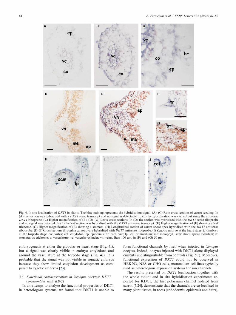
Fig. 4. In situ localisation of DKT1 in plants. The blue staining represents the hybridisation signal. (A)–(C) Root cross sections of carrot seedling. In(A) the section was hybridised with a DKT1 sense transcript and no signal is detectable. In (B) the hybridisation was carried out using the antisenseDKT1 riboprobe. (C) Higher magnification of (B). (D)–(G) Leave cross sections. In (D) the section was hybridised with the DKT1 sense riboprobeand no signal was detected. In (E) the leaf section was hybridised with the DKT1 antisense transcript. (F) Higher magnification of (E) showing a leaftrichome. (G) Higher magnification of (E) showing a stomata. (H) Longitudinal section of carrot shoot apex hybridised with the DKT1 antisenseriboprobe. (I)–(J) Cross sections through a carrot ovary hybridised with DKT1 antisense riboprobe. (I) Zygotic embryo at the heart stage. (J) Embryoat the torpedo stage. co: cortex; cot: cotyledon; ep: epidermis; hr: root hair; lp: leaf primordium; me: mesophyll; sam: shoot apical meristem; st:stomata; tr: trichome; v: vasculature; vc: vascular cylinder; vn: veins. Bars 100 lm, in (F) and (G) 50 lm.
64 E. Formentin et al. / FEBS Letters 573 (2004) 61–67
embryogenesis at either the globular or heart stage (Fig. 4I),
but a signal was clearly visible in embryo cotyledons and
around the vasculature at the torpedo stage (Fig. 4J). It is
probable that the signal was not visible in somatic embryos
because they show limited cotyledon development as com-
pared to zygotic embryos [23].
3.3. Functional characterization in Xenopus oocytes: DKT1
co-assembles with KDC1
In an attempt to analyse the functional properties of DKT1
in heterologous systems, we found that DKT1 is unable to
form functional channels by itself when injected in Xenopus
oocytes. Indeed, oocytes injected with DKT1 alone displayed
currents undistinguishable from controls (Fig. 5C). Moreover,
functional expression of DKT1 could not be observed in
HEK293, N2A or CHO cells, mammalian cell lines typically
used as heterologous expression systems for ion channels.
The results presented on DKT1 localization together with
the whole mount and in situ hybridisation experiments re-
ported for KDC1, the first potassium channel isolated from
carrot [7,24], demonstrate that the channels are co-localised in
many plant tissues, in roots (endodermis, epidermis and hairs),

Table 1Overlapping expression pattern between KDC1 and DKT1
Tissue or organ KDC1 DKT1
Somatic embryo(protoderm)
Globular stage + )Heart stage + )Torpedo stage + )
Zygotic embryo Torpedo stage (cotyledon) + +
Plantlet Root epidermis (root hair) + +Root endodermis + +Root cortex ) +Cotyledon ) +Stem ) +SAM + +Leaf ) +
List of plant tissues and embryonic stages where the expression patternof KDC1 [24] and DKT1 was analysed by whole mount or in situhybridisation. The ‘+’ and ‘)’ symbols indicate the presence or theabsence of the transcript.
Fig. 5. Functional co-expression of KDC1 and DKT1 in oocytes. (A)Voltage steps (from )60 to )180 mV) were applied in 10 mV incre-ments. (B) Ionic currents in uninjected (control) oocytes. (C) currentsin oocytes injected with DKT1 and (D) in oocytes co-injected withKDC1:DKT1. (E) Current voltage characteristics of the family ofcurrents shown in (B, C, D) obtained by plotting the mean value of thecurrent during the last 50 ms in each record as a function of the appliedmembrane potential.
E. Formentin et al. / FEBS Letters 573 (2004) 61–67 65
SAM and embryo cotyledons (summed up in Table 1), there-
fore suggesting a possible interaction between the two chan-
nels. It has been widely demonstrated that plant Kþ channels
can co-assemble [8–11,25], and we have previously shown that
KDC1 can act as a modulator subunit when co-expressed with
KAT1 [9,10], we thus wanted to see if KDC1 can form func-
tional heteromeric channels with DKT1. Interestingly, we
obtained exceptionally robust and stable negative inwardly-
rectifying ionic currents in almost all oocytes which were co-
injected with KDC1 and DKT1. Fig. 5D shows typical
KDC1:DKT1 currents, which activate in a time dependent
manner at membrane potentials more negative than )120 mV
(Fig. 5E). This suggests the formation of heteromultimeric
channels between KDC1 and DKT1 subunits. In line with the
results obtained with KDC1, which forms heteromeric potas-
sium channels with KAT1 [9], the ability of DKT1 to form
heteromultimeric channels with KAT1 was also verified by the
co-expression in Xenopus oocytes. The DKT1:KAT1 channel
displayed large inward rectifying currents with modified
characteristics with respect to homomeric KAT1 (data not
shown).
The heteromeric KDC1:DKT1 channels were potassium-
selective. Indeed, KDC1:DKT1 current amplitude depends on
Kþ concentrations, as demonstrated by experiments where
external media contained different concentrations of KCl and
NaCl to a total salt concentration of 100 mM (Fig. 6A). Upon
variation of the Kþ concentration in the bath, the reversal
potentials determined by tail current experiments shifted by
(55� 3) mV for a 10-fold change in the Kþ gradient, indicating
the high selectivity of the channel for potassium over sodium.
Fig. 6B shows an example of tail currents recorded at an ex-
ternal potassium concentration equal to 30 mM. In Fig. 6C the
reversal potentials are plotted against the potassium activity.
Moreover, the inward rectifying KDC1:DKT1 channel was
blocked by millimolar concentrations of external caesium as
shown in Fig. 6D, where a typical decrease of the current
following the addition of CsCl in the bath solution is shown.
4. Discussion
This paper describes the isolation of a new transcript en-
coding a Kþ inward rectifier channel from carrot, namely
DKT1. This channel belongs to the AKT1 group of plant
Shaker channels [6], showing high sequence homology and
structural similarities with the other AKT1-like channels (Figs.
1 and 2).
The analysis of the gene expression (Figs. 3 and 4) reveals
similar pattern to that of AKT1: both genes are expressed in
roots and root hairs [26,27] where they may have a nutritional
role [28]. In leaves, DKT1 shows a wider expression profile
than its Arabidopsis homologue [26,29], and its presence in
guard cells suggests an involvement in stomatal movements.
The temporal expression of DKT1 in zygotic cotyledons, sug-
gests a role of DKT1 in cotyledon expansion during late
phases of embryo maturation. Concerning hormonal regula-
tion, neither AKT1 [30] nor DKT1 are regulated by auxin. In
our system, young seedlings and mature plants were isolated
and incubated with auxin and the expression pattern of the
gene was analysed by semi-quantitative RT-PCR in different
tissues (root, stem, leaf, shoot apex and cotyledons) (data not

Fig. 6. The heteromeric KDC1:DKT1 channel is potassium-selective.(A) Average normalized current–voltage characteristics are shown fortwo different potassium concentrations. Currents were elicited by 2 shyperpolarizing pulses from )60 to )160 mV in )10 mV steps. Inset:normalized current at )150 mV as a function of potassium activities2.3, 7.7, 23.1, 77 mM. (B) Representative tail currents elicited by a tailpulse from )10 to )50 mV (at )10 mV decrements) following a con-ditioning prepulse to )150 mV. (C) Reversal potential Vrev of thecurrents carried by the heteromeric channels upon variation of theexternal Kþ activity. The continuous line represents the best fit ofexperimental data points, revealing a shift in Vrev of 55 mV per 10-foldchange in the Kþ gradient. (D) KDC1:DKT1 current was reversiblyinhibited by 5 mM Csþ. Voltage steps to )150 mV.
66 E. Formentin et al. / FEBS Letters 573 (2004) 61–67
shown). In contrast, in the monocotyledonous Zea mais, the
AKT1-like channel ZMK1 is under the control of auxin, es-
pecially in the coleoptile tissue [31,32], suggesting a different
auxin response in mono- and dicotyledonous plant species.
The electrophysiological experiments conducted in Xenopus
oocytes demonstrated that DKT1, like KDC1, injected indi-
vidually does not form a functional channel. In contrast, we
demonstrated the formation of heteromeric KDC1:DKT1
channels that are able to mediate robust and stable inward
currents that activated at hyperpolarizing membrane poten-
tials. These currents are mediated by typical potassium selec-
tive channels, as demonstrated by the shift of the reversal
potential with Kþ concentration and their block by external
caesium (Figs. 5 and 6). These results together with ones re-
ported by Duby et al. [33] might open the possibility for the
identification and characterisation of new mechanisms con-
trolling channel targeting to the cell membrane. In addition,
KDC1 is an excellent model subunit owing to its peculiar
histidine composition and ‘‘tolerance’’ to metals [9,10] while
more studies are needed to identify a more precise functional
fingerprinting of DKT1.
In conclusion, we report, for the first time, evidence that a-subunits belonging to the same species and present in the same
tissues (see Table 1) can cooperate to form functional hetero-
meric plant Kþ channels. We suggest that a different behaviour
in Kþ absorption could be determined by a differential ex-
pression of these two (and possibly other) subunits. It seems
reasonable that plants may have adopted this modulator
strategy to respond to different environmental conditions and
external stresses. However, we cannot exclude that in tissues
where KDC1 is not present (e.g. guard cells, see Table 1),
DKT1 may form homomeric channels or interact with other as
of yet unidentified subunits. The study of DKT1 in transgenic
plants and a complete characterisation of the heteromultimeric
KDC1:DKT1 channel should be forthcoming.
Acknowledgements: We thank Dr. Armando Carpaneto, Dr. Ildik�oSzab�o and Prof. Mario Terzi for a critical reading of the manuscript.This research was supported by the Ministero dell’Istruzione e dellaRicerca, Fondi per gli Investimenti della Ricerca di Base, Project N-RBAUO183A9 and by a contribution from Consorzio Interuniversi-tario Biotecnologie (CIB) to FLS.
References
[1] Br€uggemann, L., Dietrich, P., Becker, D., Dreyer, I., Palme, K.and Hedrich, R. (1999) Proc. Natl. Acad. Sci. USA 96, 3298–3302.
[2] Clarkson, D.T. and Hanson, J.B. (1980) Annu. Rev. PlantPhysiol. 31, 239–298.
[3] Schroeder, J.I. and Hedrich, R. (1989) Trends Biochem. Sci. USA87, 9305–9309.
[4] Mouline, K., V�ery, A.A., Gaymard, F., Boucherez, J., Pilot, G.,Devic, M., Bouchez, D., Thibaud, J.B. and Sentenac, H. (2002)Genes Dev. 16, 339–350.
[5] V�ery, A.A. and Sentenac, H. (2003) Annu. Rev. Plant Physiol.Plant Mol. Biol. 54, 575–603.
[6] Pilot, G., Pratelli, R., Gaymard, F., Meyer, Y. and Sentenac, H.(2003) J. Mol. Evol. 56, 418–434.
[7] Downey, P., Szab�o, I., Ivashikina, N., Negro, A., Guzzo, F.,Ache, P., Hedrich, R., Terzi, M. and Lo Schiavo, F. (2000) J. Biol.Chem. 275, 39420–39426.
[8] Reintanz, B., Szyroki, A., Ivashikina, N., Ache, P., Godde, M.,Becker, D., Palme, K. and Hedrich, R. (2002) Proc. Natl. Acad.Sci. USA 99, 4079–4084.
[9] Paganetto, A., Bregante, M., Downey, P., Lo Schiavo, F., Hoth,S., Hedrich, R. and Gambale, F. (2001) J. Bioenereg. Biomembr.33, 63–71.
[10] Picco, C., Bregante, M., Naso, A., Gavazzo, P., Costa, A.,Formentin, E., Downey, P., Lo Schiavo, F. and Gambale, F.(2004) Biophys. J. 86, 224–234.
[11] Dreyer, I., Antunes, S., Toshinori, H., M€uller–R€ober, B., Palme,K., Pongs, O., Reintanz, B. and Hedrich, R. (1997) Biophys. J. 72,2143–2150.
[12] Finn, J.T., Krautwurst, D., Schroeder, J.E., Chen, T.Y., Reed,R.R. and Yau, K.W. (1998) Biophys. J. 74, 1333–1345.
[13] Gamborg, O.L., Miller, R.A. and Ojima, K. (1968) Exp. Cell Res.50, 151–158.
E. Formentin et al. / FEBS Letters 573 (2004) 61–67 67
[14] Giuliano, G., Rosellini, D. and Terzi, M. (1983) Plant Cell. Rep.2, 216–218.
[15] Kumar, S., Tamura, K., Jakobsen, I.B. and Nei, M. (2001)Bioinformatics 17, 1244–1245.
[16] Xia, X. and Xie, Z. (2001) J. Heredity 92, 371–373.[17] Altschul, S.F., Gish, W., Miller, W., Myers, E.W. and Lipman,
D.J. (1990) J. Mol. Biol. 215, 403–410.[18] Jones, D.T. (1999) J. Mol. Biol. 292, 195–202.[19] Hedrich, R., Moran, O., Conti, F., Bush, H., Becker, D.,
Gambale, F., Dreyer, I., Kuch, A., Neuwinger, K. and Palme,K. (1995) Eur. Biophys. J. 24, 107–115.
[20] Sentenac, H., Bonneaud, N., Minet, M., Lacroute, F., Salmon,J.M., Gaymard, F. and Grignon, C. (1992) Science 256, 663–665.
[21] Hartje, S., Zimmermann, S., Klonus, D. and M€uller-R€ober, B.(2000) Planta 210, 723–731.
[22] Schachtman, D.P. (2000) Biochem. Biophys. Acta 1465, 127–139.[23] Steeves, T.A. and Sussex, I.M. (1989) Patterns in plant develop-
ment. Cambridge University Press, Cambridge, MA.[24] Costa, A., Carpaneto, A., Varotto, S., Formentin, E., Marin, O.,
Barizza, E., Terzi, M., Gambale, F., and Lo Schiavo, F. (in press)Plant Mol. Biol. Available from: www.kluwer.com/issn/0167-4412.
[25] Pilot, G., Gaymard, F., Mouline, K., Ch�erel, I. and Sentenac, H.(2003) Plant Mol. Biol. 51, 773–787.
[26] Lagarde, D., Basset, M., Lepetit, M., Conejero, G., Gaymard, F.,Astruc, S. and Grignon, C. (1996) Plant J. 9, 195–203.
[27] Ivashikina, N., Becker, D., Ache, P., Meyerhoff, O., Felle, H.H.and Hedrich, R. (2001) FEBS Lett. 508, 463–469.
[28] Dennison, K.L., Robertson, W.R., Lewis, B.D., Hirsch, R.E.,Sussman, M.R. and Spalding, E.P. (2001) Plant Physiol. 127,1012–1019.
[29] Szyroki, A., Ivashikina, N., Dietrich, P., Roelfsema, M.R.G.,Ache, P., Reintanz, B., Deeken, R., Godde, M., Felle, H.,Steinmeyer, R., Palme, K. and Hedrich, R. (2001) Proc. Natl.Acad. Sci. USA 98, 2917–2921.
[30] Philippar, K., Ivashikina, N., Ache, P., Christian, M., L€uthen, H.,Palme, K. and Hedrich, R. (2004) Plant J. 37, 815–827.
[31] Philippar, K., Fuchs, I., L€uthen, H., Hoth, S., Bauer, C.S., Haga,K., Thiel, G., Ljung, K., Sandberg, G., Bottger, M., Becker, D.and Hedrich, R. (1999) Proc. Natl. Acad. Sci. USA 96, 12186–12191.
[32] Fuchs, I., Philippar, K., Ljung, K., Sandberg, G. and Hedrich, R.(2003) Proc. Natl. Acad. Sci. USA 100, 11795–11800.
[33] Duby, G., Hosy, E., Broekgaarden, C., Costa, A., Vavasseur, A.,Mouline, K., Thibaud, J.B., and Sentenac, H. (2004) In: 13thInternational Workshop on Plant Membrane Biology, Montpel-lier, July 6–10. Oral session T4-3, p. 34.
Related Documents
![Functions of Heteromeric and Homomeric Isoamylase-Type ... · Functions of Heteromeric and Homomeric Isoamylase-Type Starch-Debranching Enzymes in Developing Maize Endosperm1[W][OA]](https://static.cupdf.com/doc/110x72/600557e5a427f91eee0161c8/functions-of-heteromeric-and-homomeric-isoamylase-type-functions-of-heteromeric.jpg)