©FUNPEC-RP www.funpecrp.com.br Genetics and Molecular Research 13 (3): 4918-4931 (2014) Diversity and potential application of endophytic bacteria in ginger T. Chen 1 , Z. Chen 1 , G.H. Ma 2 , B.H. Du 1 , B. Shen 1 , Y.Q. Ding 1 and K. Xu 3 1 College of Life Science, Taian, Shandong Province, China 2 Anqiu City Agricultural Bureau, Anqiu, Shandong Province, China 3 College of Horticulture Science and Engineering, Taian, Shandong Province, China Corresponding authors: Y.Q. Ding / K. Xu E-mail: [email protected] / [email protected] Genet. Mol. Res. 13 (3): 4918-4931 (2014) Received April 10, 2013 Accepted November 21, 2013 Published July 4, 2014 DOI http://dx.doi.org/10.4238/2014.July.4.6 ABSTRACT. Here, 248 endophytic bacterial strains were isolated to assess the distribution and population diversity of endophytic bacteria in ginger plants. A total of 10.4 x 10 4 to 20.2 x 10 4 CFU/g fresh weight endophytic bacteria of different growth stages were isolated. Maximum bacterium numbers were obtained at the seedling stage. A total of 107 functional strains were screened, including 17 antibacterial strains and 90 indole acetic acid-producing strains. Based on 16S rDNA sequence restriction fragment length polymorphism and 16S rDNA sequences, these 107 strains were mapped and grouped into 16 genera. Bacillus and Pseudomonas were the dominant genera; however, the bacteria belonged to a tremendous range of genera, with the highest species richness being observed at the seedling stage. Sixteen strains exhibited antimicrobial activity against Pythium myriotylum Drechsler, while 7 strains exhibited antimicrobial activity against Phyllosticta zingiberi Hori. Bacillus was the dominant antibacterial strain. Pseudomonas fluorescens, B. megaterium, and Enterobacter ludwigii produced remarkably high levels of IAA. Only a few endophytic bacterial

Welcome message from author
This document is posted to help you gain knowledge. Please leave a comment to let me know what you think about it! Share it to your friends and learn new things together.
Transcript

©FUNPEC-RP www.funpecrp.com.brGenetics and Molecular Research 13 (3): 4918-4931 (2014)
Diversity and potential application of endophytic bacteria in ginger
T. Chen1, Z. Chen1, G.H. Ma2, B.H. Du1, B. Shen1, Y.Q. Ding1 and K. Xu3
1College of Life Science, Taian, Shandong Province, China2 Anqiu City Agricultural Bureau, Anqiu, Shandong Province, China3College of Horticulture Science and Engineering, Taian, Shandong Province, China
Corresponding authors: Y.Q. Ding / K. XuE-mail: [email protected] / [email protected]
Genet. Mol. Res. 13 (3): 4918-4931 (2014)Received April 10, 2013Accepted November 21, 2013Published July 4, 2014DOI http://dx.doi.org/10.4238/2014.July.4.6
ABSTRACT. Here, 248 endophytic bacterial strains were isolated to assess the distribution and population diversity of endophytic bacteria in ginger plants. A total of 10.4 x 10⁴ to 20.2 x 10⁴ CFU/g fresh weight endophytic bacteria of different growth stages were isolated. Maximum bacterium numbers were obtained at the seedling stage. A total of 107 functional strains were screened, including 17 antibacterial strains and 90 indole acetic acid-producing strains. Based on 16S rDNA sequence restriction fragment length polymorphism and 16S rDNA sequences, these 107 strains were mapped and grouped into 16 genera. Bacillus and Pseudomonas were the dominant genera; however, the bacteria belonged to a tremendous range of genera, with the highest species richness being observed at the seedling stage. Sixteen strains exhibited antimicrobial activity against Pythium myriotylum Drechsler, while 7 strains exhibited antimicrobial activity against Phyllosticta zingiberi Hori. Bacillus was the dominant antibacterial strain. Pseudomonas fluorescens, B. megaterium, and Enterobacter ludwigii produced remarkably high levels of IAA. Only a few endophytic bacterial

4919
©FUNPEC-RP www.funpecrp.com.brGenetics and Molecular Research 13 (3): 4918-4931 (2014)
Diversity and potential application of endophytic bacteria
strains were inhibited in fresh ginger juice. Most of these strains were present during seedling stage, including Roseateles depolymerans, Chryseobacterium taiwanense, E. ludwigii, Agrobacterium larrymoorei, P. fluorescens, and Bacillus amyloliquefaciens. This study indicates that the community of endophytic bacteria in ginger changes with the synthesis of antibacterial substances.
Key words: Endophytic bacteria; IAA; Ginger; Antifungal activity
INTRODUCTION
Ginger is the rhizomes of Zingiber officinale Roscoe (Zingiberaceae), and has been widely used as a spice and condiment in a range of forms. In addition to its food additive function, ginger has a long history of medicinal use in the treatment of a variety of human diseases, including common colds, fever, rheumatic disorders, gastrointestinal complications, motion sickness, diabetes, and cancer, among others (Kundu et al., 2009). Because of the high yields and economic benefits of ginger, Shandong Province has been developed into the larg-est ginger planting base in China. Ginger contains many nutrients, including saccharides, fats, proteins, carotenes, vitamin C, and a large number of microelements. Ginger also secretes various substances, such as gingerol, shogaol, zingerone, and ginger essential oil (Singh et al., 2003). Gingerol and ginger essential oil have antibacterial effects against Bacillus subti-lis, Escherichia coli, Saccharomyces, Staphylococcus aureus, Aspergillus niger, and Rhizopus (Zhao, 2008).
Endophytic bacteria play an important role in plant growth. Endophytes colonize plant tissues, and are able to interact among themselves and with invaders (e.g., pathogens); thus, influencing plant development. Evolutionarily, endophytes also appear to form an intermediate group between saprophytic bacteria and plant pathogens. Endophytic bacteria have been isolated from a variety of plants, because they ubiquitously inhabit most plant species, including sugar beet, several potato varieties, and poplar trees (Sessitsch et al., 2002; Dent et al., 2004; Taghavi et al., 2009). Many studies have shown that endophytes are widely present in plant tissues, such as the roots, stems, leaves, and flowers (Kobayashi and Palumbo, 2000). For instance, several genera have been isolated from legume tissues, including Aerobacter, Aeromonas, Agrobacterium, Bacillus, Chryseomonas, Curtobacterium, Enterobacter, Erwinia, Flavimonas, Pseudomonas, and Sphingomonas (Sturz et al., 1997; Elvira-Recuenco and van Vuurde, 2000; Oehrle et al., 2000).
Many researchers have studied the breeding and cultivation characteristics of ginger. However, investigations about the endophytic bacteria that this plant harbors remain limited. Some endophytic bacteria have been screened in ginger, and were found to promote plant growth (Xie et al., 2009). Rajan et al. (2000) studied the effects of 4 isolates of endophytic bacteria on the growth of a ginger cultivar (cv.). They also assessed the suppressive activity of endophytic bacteria against Phythium sp, Fusarium oxysporum, and Pratylenchus coffeae in a pot experiment. Chu et al. (2011) analyzed the diversity of endophytes isolated from the ginger tuber.
Endophytes influence plant growth via several processes, including the production of plant hormones. One such hormone is indole-3-acetic acid (IAA), which is an essential phy-tohormone that is involved in different plant developmental processes (Liu et al., 2010). The
4920
©FUNPEC-RP www.funpecrp.com.brGenetics and Molecular Research 13 (3): 4918-4931 (2014)
T. Chen et al.
production of IAA is widely distributed among plant-associated bacteria. While the combined actions of several rhizobacteria seem to result in the promotion of plant growth, bacterial phytohormone production, particularly IAA, is still considered the primary mechanism that enhances the growth and yield of plants (Arkhipova et al., 2005). Another important function of plant growth-promoting bacteria is their antagonistic action against pathogenic microorgan-isms. Endophytic bacteria interact collaboratively with plant hosts, and are easy to cultivate in vivo. Thus, the investigation of endophytic bacterial functions and subsequent development of their use in plants is of importance to microbiologists and plant protection experts.
Because ginger secretes gingerol and other antibacterial substances during growth, changes in the endogenous bacterial population of ginger are expected to differ compared to bacterial populations in other crops. So the bacterial strains isolating from ginger were used in ginger, which are not only biocontrol or promoting-growth function, but also should be adaptation to different ginger growth stages (Policegoudra et al., 2007). This paper studies the ginger endophytic bacterial population change rule at ginger different growth stages, and provides strain resources and a theoretical basis for bacterial agents that promote the growth and bio-control of ginger. The current study is the first report about the evolutionary changes of endophytic bacteria throughout the different growth stages of ginger.
MATERIAL AND METHODS
Sampling
Healthy ginger plants were sampled at 3 different growth stages; specifically, the seed-ling stage, the stem and leaf vigorous growth stage, and the rhizome enlargement stage. Sam-ples were collected from the same field plot in Qiujiadian, Taian, Shandong Province, China.
Surface sterilization of plants and isolation of endophytic bacteria
Total endophytic bacteria were isolated from the roots, stems, tubers, and leaves of gin-ger. Whole plants were first washed with tap water to remove attached clay. Ten grams of tissue from each of the specified plant parts was cut with a sterile surgical knife for surface sterilization. The collected plant materials were immersed in 75% ethanol for 2.5 min, rinsed with 3% sodium hypochlorite (NaClO) for 2 min, dipped in 75% ethanol for 30 s, and finally washed 5 times with sterile distilled water. To determine whether the sterilization process was successful, 100 µL water from the third rinsing was plated on R2A medium (0.05% proteose peptone, 0.05% starch, 0.05% glucose, 0.05% yeast extract, 0.05% casein hydrolysate, 0.03% dipotassium phosphate, 0.03% sodium pyruvate, 0.0024% magnesium sulfate anhydrous, 2% w/v agar, pH 7.2 ± 0.2), which was also the medium used for the isolation and purification of endophytic bacteria (Kawai et al., 2002). The plates were incubated at 28°C for 1-3 days to determine surface sterilization ef-ficacy. If bacterial colonies were not observed on the plates, the sterilization process was consid-ered successful. All surface-sterilized samples were placed in a sterilized mortar, and thoroughly ground after the addition of 10 mL sterile distilled water. The resulting suspension was diluted 10-fold with sterile distilled water, and about 100 μL of each dilution was spread onto R2A me-dium. Bacterial isolates were obtained after incubation at 28°C for 2-3 days. Glycerol was then added, and the purified isolates were kept frozen at -80°C until use.
4921
©FUNPEC-RP www.funpecrp.com.brGenetics and Molecular Research 13 (3): 4918-4931 (2014)
Diversity and potential application of endophytic bacteria
Total DNA extraction and 16S rDNA PCR amplification
Total bacterial DNA isolation was completed according to the procedure of Mur-ray and Thompson (1980). 16S rDNA PCR amplification was carried out with the universal primers 27f (5'-AGAGTTTGATCCTGGCTCAG-3') and 1492r (5'-TACGGCTACCTTGTTA CGACTTCACCCC-3') by using a (Biometra TGRADIENT) thermocycler. The PCR condi-tions were as follows: initial denaturation at 95°C for 4 min, followed by 32 cycles of denatur-ation at 94°C for 45 s, annealing at 56°C for 1 min, extension at 72°C for 1.5 min, and a final extension at 72°C for 10 min. Products of about 1500 bp long were monitored by electropho-resis on 1% (w/v) agarose gel at 110 V for 30 min in 1X Tris-boric acid-EDTA (TBE) buffer. PCR products were viewed by ethidium bromide (EB) staining and UV transillumination.
ARDRA analysis of 16S rRNA gene sequences and evolutionary analysis
A 3S Spin PCR Product Purification Kit (Shenergy Biocolor Bioscience and Technol-ogy Company, China) was used for PCR product purification following manufacturer proto-cols. Purified PCR products were digested using 3 restriction enzymes, AluI, HaeI, and MspI (TaKaRa Biotechnology Co., Ltd., Dalian, China), in separate reactions. The selection of these restriction enzymes was based on the study of Laguerre et al. (1994). Digestion reactions were performed for 4 h at 37°C in a reaction volume of 10 μL containing 5 μL purified PCR products, 1 μL commercially supplied 10X incubation buffer, 3.5 μL water, and 0.5 μL (10 U/μL) restriction enzyme. Reaction products (10 μL) were run on 2.5% (w/v) agarose gel in 1X TBE buffer for 2.5 h at 120 V under refrigeration. Agarose gels were stained, visualized, and digitalized as previously described.
Visible bands greater than 100 bp were used for dendrogram construction. From the banding patterns generated by each of the restriction enzymes, a binary data matrix was constructed based on the presence or absence of each band (coded as 1 or 0, respectively). To obtain a single pattern for each isolate, the banding patterns obtained from each of the enzymes were combined. The patterns were then used to construct a dendrogram using the unweighted pair group method with arithmetic averages (UPGMA) by a clustering algorithm that makes use of the Sorensen’s coefficient along with fine optimization options, which was incorporated into the MultiVariate Statistical Package (MVSP) version 3.13h (GeoMem, Blairgowrice, UK).
16S rRNA gene sequencing and sequence analysis
Purified PCR products obtained from the isolates of each ARDRA group were se-quenced. The NCBI (National Center for Biotechnology Information database) nucleotide-nucleotide BLAST (BLASTn) tool (http://blast.ncbi.nlm.nih.gov/Blast.cgi) was used to locate closely identical sequences for the 16S rRNA gene sequences that were determined. High-similarity sequences, as well as 16S rRNA gene sequences of the type strains, were retrieved from the Ribosomal Database Project. The sequences were aligned using the CLUSTAL X software version 1.8. Evolutionary distances were calculated using the package TREECON software version 1.3b. Construction of a neighbor-joining tree and bootstrap analysis (1000 replicates) was also performed using TREECON (Tamura et al., 2007).

4922
©FUNPEC-RP www.funpecrp.com.brGenetics and Molecular Research 13 (3): 4918-4931 (2014)
T. Chen et al.
In vitro antagonistic spectrum bioassay
An antagonistic spectrum bioassay of the endophytic bacteria was performed with Pythium myriotylum Drechsler and Phyllosticta zingiberi Hori. Bacterial isolates were inocu-lated on the margin of the fungal colony with sterile toothpicks, and incubated at 28°C for 2-4 days. Growth inhibition was calculated using the formula R1/R2, where R1 is the maximum radius of the fungal colony away from the bacterial colony and R2 is the average radius of the bacterial colony.
Screening of IAA-producing bacterial isolates and quantitative analysis
IAA-producing bacteria were screened by growing the bacterial isolates on R2A medi-um modified with L-tryptophan (200 mg/L) at 28°C and 180 rpm for 3 days. Fifty microliters each of the bacterial liquid cultures and Salkowski reagent were mixed and viewed on a white board background. After being kept in the dark for 30 min, IAA-producing bacteria were iden-tified by a change in color from pink to red. The concentration of IAA-positive strain cultures was measured at 600 nm before centrifugation at 10,000 rpm for 10 min. The procedures of Glickmann and Dessaux (1995) were used to evaluate IAA production in the supernatant, and a standard curve was generated using pure IAA.
Detection of the bacteriostatic effect of fresh ginger juice
Peeled ginger was immersed in 75% ethanol for 2.5 min, and then rinsed with 3% NaClO for 2 min. The samples were then dipped in 75% ethanol for 30 s, and washed 5 times with sterile distilled water. The fresh ginger juice, obtained using a disinfected juicer, was used to produce 4 concentrations of ginger solution: 100, 50, 25, and 10%. Endophytic bacterial strains were suspended in physiological saline at concentrations ranging from 105 to 106. Sterilized filter papers (6 mm) were immersed in various concentrations of ginger juice for 12 h, and then dried at 28°C. The endophytic bacterial suspensions were then inoculated on the R2A culture plates at a volume of 500 mL. The filter papers that had been immersed in ginger juice were placed on the culture plates containing endophytic bacteria. Each plate contained two of the filter papers for each concentration level, and experiments were performed in triplicate. The plates were incubated at 28°C for 1 day. The diameter of the observed inhibition zone was measured for each setup, and the data were recorded. Filter papers immersed in 0.5% CuSO4 were used as controls.
RESULTS
Endophytic isolates of ginger plants
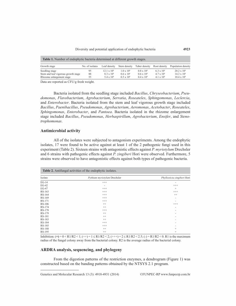
Based on phenotypic characterization, a total of 248 isolates were obtained (Table 1). Bacterial densities from different ginger growth stages were counted, with densities ranging from 10.4 x 10⁴ to 20.2 x 10⁴ CFU/g fresh weight. These data produced significant period differences. The highest density was observed at the seedling stage, whereas the lowest density was observed at the rhizome enlargement stage. We also found that ginger leaves have the largest number of en-dogenous bacteria with the lowest bacterial numbers being observed in stems and tubers (Table 1).
4923
©FUNPEC-RP www.funpecrp.com.brGenetics and Molecular Research 13 (3): 4918-4931 (2014)
Diversity and potential application of endophytic bacteria
Bacteria isolated from the seedling stage included Bacillus, Chryseobacterium, Pseu-domonas, Flavobacterium, Agrobacterium, Serratia, Roseateles, Sphingomonas, Leclercia, and Enterobacter. Bacteria isolated from the stem and leaf vigorous growth stage included Bacillus, Paenibacillus, Pseudomonas, Agrobacterium, Aeromonas, Acetobacter, Roseateles, Sphingomonas, Enterobacter, and Pantoea. Bacteria isolated in the rhizome enlargement stage included Bacillus, Pseudomonas, Herbaspirillum, Agrobacterium, Ensifer, and Steno-trophomonas.
Antimicrobial activity
All of the isolates were subjected to antagonism experiments. Among the endophytic isolates, 17 were found to be active against at least 1 of the 2 pathogenic fungi used in this experiment (Table 2). Sixteen strains with antagonistic effects against P. myriotylum Drechsler and 6 strains with pathogenic effects against P. zingiberi Hori were observed. Furthermore, 5 strains were observed to have antagonistic effects against both types of pathogenic bacteria.
Isolate Pythium myriotylum Drechsler Phyllosticta zingiberi Hori
EG-14 +++ -EG-42 - +++EG-47 +++ +RS-163 +++ +++RS-164 +++ ++RS-169 +++ -RS-171 +++ -RS-106 ++ +++RS-174 ++ -RS-178 +++ -RS-179 ++ -RS-181 ++ -RS-183 ++ -RS-184 +++ -RS-185 +++ -RS-188 ++ +RS-195 ++ +
Inhibition: (+) = 0 < R1/R2 < 1; (++) = 1 ≤ R1/R2 < 2; (+++) = 2 ≤ R1/R2 < 2.5; (˗) = R1/R2 < 0. R1 is the maximum radius of the fungal colony away from the bacterial colony. R2 is the average radius of the bacterial colony.
Table 2. Antifungal activities of the endophytic isolates.
ARDRA analysis, sequencing, and phylogeny
From the digestion patterns of the restriction enzymes, a dendrogram (Figure 1) was constructed based on the banding patterns obtained by the NTSYS 2.1 program.
Growth stage No. of isolates Leaf density Stem density Tuber density Root density Population density
Seedling stage 95 12.1 x 10⁴ 1.0 x 10⁴ 0.8 x 10⁴ 6.3 x 10⁴ 20.2 x 10⁴
Stem and leaf vigorous growth stage 88 8.3 x 10⁴ 0.6 x 10⁴ 0.6 x 10⁴ 4.7 x 10⁴ 14.2 x 10⁴
Rhizome enlargement stage 55 5.4 x 10⁴ 0.5 x 10⁴ 0.4 x 10⁴ 4.1 x 10⁴ 10.4 x 10⁴
Table 1. Number of endophytic bacteria determined at different growth stages.
Data are reported as CFU/g fresh weight.

4924
©FUNPEC-RP www.funpecrp.com.brGenetics and Molecular Research 13 (3): 4918-4931 (2014)
T. Chen et al.
Figure 1. Dendrogram showing the relationships of 107 endophytic bacteria based on 16S-RFLP fingerprints obtained using cluster analysis.
4925
©FUNPEC-RP www.funpecrp.com.brGenetics and Molecular Research 13 (3): 4918-4931 (2014)
Diversity and potential application of endophytic bacteria
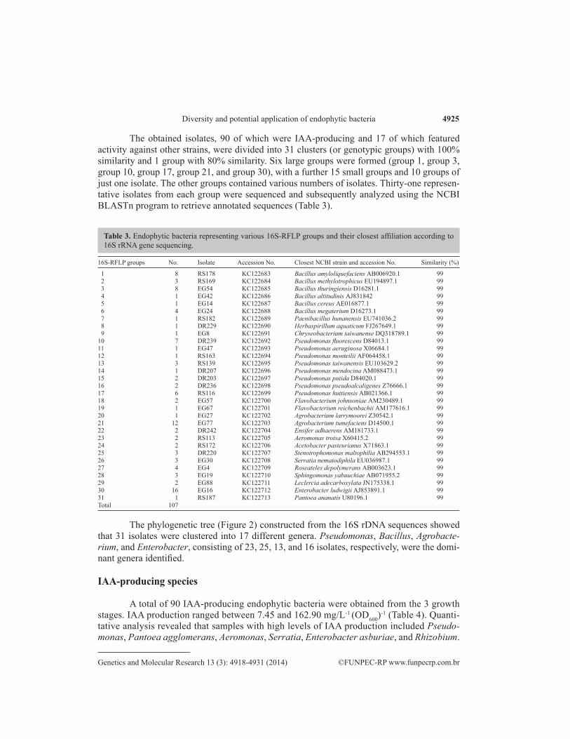
The obtained isolates, 90 of which were IAA-producing and 17 of which featured activity against other strains, were divided into 31 clusters (or genotypic groups) with 100% similarity and 1 group with 80% similarity. Six large groups were formed (group 1, group 3, group 10, group 17, group 21, and group 30), with a further 15 small groups and 10 groups of just one isolate. The other groups contained various numbers of isolates. Thirty-one represen-tative isolates from each group were sequenced and subsequently analyzed using the NCBI BLASTn program to retrieve annotated sequences (Table 3).
16S-RFLP groups No. Isolate Accession No. Closest NCBI strain and accession No. Similarity (%)
1 8 RS178 KC122683 Bacillus amyloliquefaciens AB006920.1 99 2 3 RS169 KC122684 Bacillus methylotrophicus EU194897.1 99 3 8 EG54 KC122685 Bacillus thuringiensis D16281.1 99 4 1 EG42 KC122686 Bacillus altitudinis AJ831842 99 5 1 EG14 KC122687 Bacillus cereus AE016877.1 99 6 4 EG24 KC122688 Bacillus megaterium D16273.1 99 7 1 RS182 KC122689 Paenibacillus hunanensis EU741036.2 99 8 1 DR229 KC122690 Herbaspirillum aquaticum FJ267649.1 99 9 1 EG8 KC122691 Chryseobacterium taiwanense DQ318789.1 9910 7 DR239 KC122692 Pseudomonas fluorescens D84013.1 9911 1 EG47 KC122693 Pseudomonas aeruginosa X06684.1 9912 1 RS163 KC122694 Pseudomonas monteilii AF064458.1 9913 3 RS139 KC122695 Pseudomonas taiwanensis EU103629.2 9914 1 DR207 KC122696 Pseudomonas mendocina AM088473.1 9915 2 DR203 KC122697 Pseudomonas putida D84020.1 9916 2 DR236 KC122698 Pseudomonas pseudoalcaligenes Z76666.1 9917 6 RS116 KC122699 Pseudomonas huttiensis AB021366.1 9918 2 EG57 KC122700 Flavobacterium johnsoniae AM230489.1 9919 1 EG67 KC122701 Flavobacterium reichenbachii AM177616.1 9920 1 EG27 KC122702 Agrobacterium larrymoorei Z30542.1 9921 12 EG77 KC122703 Agrobacterium tumefaciens D14500.1 9922 2 DR242 KC122704 Ensifer adhaerens AM181733.1 9923 2 RS113 KC122705 Aeromonas trotsa X60415.2 9924 2 RS172 KC122706 Acetobacter pasteurianus X71863.1 9925 3 DR220 KC122707 Stenotrophomonas maltophilia AB294553.1 9926 3 EG30 KC122708 Serratia nematodiphila EU036987.1 9927 4 EG4 KC122709 Roseateles depolymerans AB003623.1 9928 3 EG19 KC122710 Sphingomonas yabuuchiae AB071955.2 9929 2 EG88 KC122711 Leclercia adecarboxylata JN175338.1 9930 16 EG16 KC122712 Enterobacter ludwigii AJ853891.1 9931 1 RS187 KC122713 Pantoea ananatis U80196.1 99Total 107
Table 3. Endophytic bacteria representing various 16S-RFLP groups and their closest affiliation according to 16S rRNA gene sequencing.
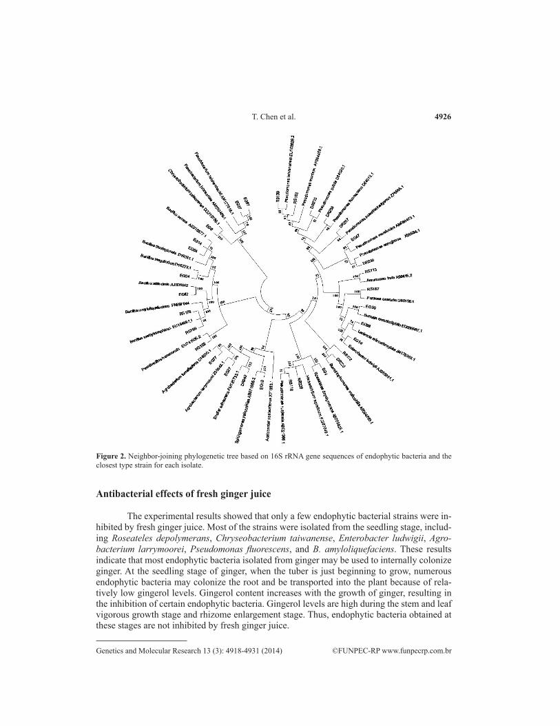
The phylogenetic tree (Figure 2) constructed from the 16S rDNA sequences showed that 31 isolates were clustered into 17 different genera. Pseudomonas, Bacillus, Agrobacte-rium, and Enterobacter, consisting of 23, 25, 13, and 16 isolates, respectively, were the domi-nant genera identified.
IAA-producing species
A total of 90 IAA-producing endophytic bacteria were obtained from the 3 growth stages. IAA production ranged between 7.45 and 162.90 mg/L-1 (OD600)
-1 (Table 4). Quanti-tative analysis revealed that samples with high levels of IAA production included Pseudo-monas, Pantoea agglomerans, Aeromonas, Serratia, Enterobacter asburiae, and Rhizobium.
4926
©FUNPEC-RP www.funpecrp.com.brGenetics and Molecular Research 13 (3): 4918-4931 (2014)
T. Chen et al.
Figure 2. Neighbor-joining phylogenetic tree based on 16S rRNA gene sequences of endophytic bacteria and the closest type strain for each isolate.
Antibacterial effects of fresh ginger juice
The experimental results showed that only a few endophytic bacterial strains were in-hibited by fresh ginger juice. Most of the strains were isolated from the seedling stage, includ-ing Roseateles depolymerans, Chryseobacterium taiwanense, Enterobacter ludwigii, Agro-bacterium larrymoorei, Pseudomonas fluorescens, and B. amyloliquefaciens. These results indicate that most endophytic bacteria isolated from ginger may be used to internally colonize ginger. At the seedling stage of ginger, when the tuber is just beginning to grow, numerous endophytic bacteria may colonize the root and be transported into the plant because of rela-tively low gingerol levels. Gingerol content increases with the growth of ginger, resulting in the inhibition of certain endophytic bacteria. Gingerol levels are high during the stem and leaf vigorous growth stage and rhizome enlargement stage. Thus, endophytic bacteria obtained at these stages are not inhibited by fresh ginger juice.

4927
©FUNPEC-RP www.funpecrp.com.brGenetics and Molecular Research 13 (3): 4918-4931 (2014)
Diversity and potential application of endophytic bacteria
DISCUSSION
Endophytic bacteria are important constituents of the plant micro-ecological system, providing resources that promote crop growth and plant disease prevention (Forchetti et al., 2010). In this study, a large number of endophytic bacterium species from a diverse range of genera were found in ginger plants. Based on the clustering results of ARDRA and 16S rRNA gene sequencing, 107 isolates were separated into 16 different genera and at least 31 differ-ent species, representing a wide variety of endophytic bacteria. The results indicate that the numbers and types of endophytic bacteria present decrease as the host ginger grows, which supported the results obtained from the colony count and 16S rDNA sequencing data.
Isolates IAA production Isolates IAA production [mg·L-1·(OD600)
-1] [mg·L-1·(OD600)-1]
P. fluorescens EG81 88.96 ± 0.2 A. tumefaciens EG1 16.14 ± 0.3 DR239 81.62 ± 0.4 EG3 9.10 ± 0.2 EG91 92.33 ± 0.7 EG6 11.00 ± 0.3 RS95 80.83 ± 0.2 EG28 12.23 ± 0.3 RS133 86.09 ± 0.3 EG48 12.46 ± 0.4 DR224 99.07 ± 0.2 EG49 9.22 ± 0.2 DR246 81.63 ± 0.3 EG62 15.86 ± 0.3P. taiwanensis RS139 28.15 ± 0.3 EG77 13.28 ± 0.3 RS142 26.02 ± 0.4 EG79 18.33 ± 0.4 DR216 27.58 ± 0.5 EG90 11.45 ± 0.6P. putida DR203 26.71 ± 0.4 RS193 5.64 ± 0.4 DR208 27.11 ± 0.3 DR234 9.27 ± 0.5P. mendocina DR207 60.87 ± 0.2 E. adhaerens DR241 45.19 ± 0.4P. pseudoalcaligenes DR236 45.99 ± 0.3 DR242 40.20 ± 0.5 DR238 46.81 ± 0.3 S. yabuuchiae EG19 46.62 ± 0.3E. ludwigii EG16 96.87 ± 0.4 RS157 48.91 ± 0.2 EG51 114.52 ± 0.6 DR213 47.51 ± 0.3 EG55 90.12 ± 0.2 P. huttiensis EG36 45.05 ± 0.2 EG76 99.21 ± 0.5 EG45 30.94 ± 0.2 EG86 97.99 ± 0.3 RS116 37.56 ± 0.1 EG87 94.68 ± 0.5 RS125 37.64 ± 0.2 RS98 93.18 ± 0.4 DR240 52.15 ± 0.2 RS101 104.49 ± 0.5 DR248 41.50 ± 0.1 RS103 121.93 ± 0.3 H. aquaticum DR229 85.55 ± 0.2 RS108 105.16 ± 0.2 R. depolymerans EG4 12.45 ± 0.2 RS109 96.80 ± 0.3 EG7 15.18 ± 0.3 RS111 162.90 ± 0.3 RS114 10.97 ± 0.2 RS115 111.19 ± 0.2 RS162 17.75 ± 0.3 RS132 103.92 ± 0.3 F. reichenbachii EG67 27.25 ± 0.2 RS153 123.40 ± 0.2 F. johnsoniae EG57 72.50 ± 0.2 RS161 148.48 ± 0.3 EG61 63.23 ± 0.2L. adecarboxylata EG88 77.84 ± 0.2 C. taiwanense EG8 76.41 ± 0.3 EG92 72.78 ± 0.3 B. megaterium EG24 126.84 ± 0.2S. nematodiphila EG30 77.10 ± 0.2 RS136 120.80 ± 0.2 EG31 73.64 ± 0.2 RS167 122.02 ± 0.2 RS144 75.82 ± 0.2 RS168 123.46 ± 0.4Pantoea ananatis RS187 78.85 ± 0.2 B. thuringiensis EG54 8.88 ± 0.2A. pasteurianus RS172 18.50 ± 0.2 EG82 3.95 ± 0.3 RS175 20.11 ± 0.1 EG93 2.58 ± 0.2S. maltophilia DR220 15.45 ± 0.2 RS201 4.97 ± 0.3 DR211 10.31 ± 0.2 DR202 8.76 ± 0.2 DR212 10.21 ± 0.2 DR206 2.26 ± 0.1A. trotsa RS113 86.04 ± 0.2 DR209 3.62 ± 0.2 RS150 91.69 ± 0.3 DR214 3.97 ± 0.4A. larrymoorei EG27 80.78 ± 0.2 P. hunanensis DR182 17.32 ± 0.2
Data are reported as means ± SE from triplicate samples.
Table 4. IAA production by endophytic isolates.
4928
©FUNPEC-RP www.funpecrp.com.brGenetics and Molecular Research 13 (3): 4918-4931 (2014)
T. Chen et al.
The most dominant genera were Bacillus and Pseudomonas, both of which were found in all 3 growth stages. Chryseobacterium, Flavobacterium, Serratia, and Leclercia were only found in the seedling stage. Paenibacillus, Aeromonas, Acetobacter, and Pantoea were only found in the stem and leaf vigorous growth stage. Herbaspirillum, Ensifer, and Steno-trophomonas were only found in the rhizome enlargement stage. The seedling stage yielded the highest number of endophyte species, whereas the rhizome enlargement stage yielded the least. Colony counting showed that the maximum density of endophytic bacteria appears dur-ing the seedling stage. In contrast, the minimum density of bacteria appeared in the rhizome enlargement stage. Chu et al. (2011) isolated 23 endophytes from a ginger tuber cultivar, 8 of which were sequenced and analyzed. In their study, the strains fell into the following genera: Pseudomonas spp, Bacillus spp, Brachybacterium spp, Stenotrophomonas spp, and Rahnella spp. In comparison, 23 different genera were identified in the current study. Distinct changes in the quantity of the endophytic population, with a peak occurring at the stem and leaf vigorous growth stage, were observed. This observation proves that colony counts are not consistent across different stages of growth. We also found that ginger leaves have the largest number of endogenous bacteria, followed by the roots, stems, and tubers. Fluctuations in the number of retrieved endophytic microorganisms seem to be influenced by both the host plant and the surrounding environment.
Ninety IAA-producing isolates and 17 antagonistic strains were isolated and screened from the ginger plants during the 3 growth stages. A significant change in bacterial diversity was observed among the growth stages. Six Bacillus spp (Table 4) were isolated in this study, with their sequences aligning with those of B. amyloliquefaciens, B. methylotrophicus, B. thuringiensis, B. altitudinis, B. cereus, and B. megaterium. The results showed that B. ce-reus, B. methylotrophicus, and B. amyloliquefaciens exhibit antimicrobial activities against P. myriotylum Drechsler. B. altitudinis, which also has a broad antibacterial spectrum, exhibited antimicrobial activity against P. zingiberi Hori (Xu et al., 2012). B. thuringiensis, which is extensively used in the biological control of insects, exhibited the lowest IAA production in this study (Vilas-Bôas et al., 2007). The most active IAA producer was B. megaterium, which produced 126.84 mg·L-1· (OD600)
-1 IAA. This result supports a previous report by Ali et al. (2009), in which B. megaterium was observed to promote the growth of red clover plants, either individually or in combination, with Rhizobium leguminosarum.
P. aeruginosa and P. monteilii exhibited antimicrobial activity against P. myriotylum Drechsler and P. zingiberi Hori. Of these 2 bacteria, P. monteilii exhibited a higher level of activ-ity. Based on published literature, we found that P. aeruginosa and P. monteilii also have broad antibacterial spectra. P. monteilii has antimicrobial activities against Cylindrocarpon destructans of ginseng, black spot of Dioscorea nipponica Makino, and P. myriotylum Drechsler of Schisan-dra chinensis (Zhai et al., 2012). P. aeruginosa exhibits antimicrobial activities against Ralstonia solanacearum of tobacco (Dong et al., 2011). Other Pseudomonas isolates from the present study, including P. fluorescens, P. taiwanensis, P. mendocina, P. putida, P. pseudoalcaligenes, and P. huttiensis, also produce IAA at varying levels. P. putida and P. fluorescens exhibit antago-nistic effects on the tobacco mosaic virus (Wu et al., 2008; Guo et al., 2011).
Previous studies have reported that some IAA-producing endophytic bacteria, such as R. depolymerans, Herbaspirillum aquaticum, Sphingomonas yabuuchiae, and Agrobacterium tumefaciens, have associated nitrogen-fixation functions (Kanvinde and Sastry, 1990; An et al., 1999; Hu et al., 2007; Liu et al., 2011). We also screened E. ludwigii in ginger, which produced both IAA but also ACC deaminase, and showed antimicrobial activity against E. coli and B.
4929
©FUNPEC-RP www.funpecrp.com.brGenetics and Molecular Research 13 (3): 4918-4931 (2014)
Diversity and potential application of endophytic bacteria
subtilis (Gong et al., 2011). Endogenetic bacteria with ACC deaminase play important roles in alleviating plants subject to various stressors, such as drought, water logging, salt, heavy metal, and pressure (Zahir et al., 2008). Other studies have also reported that E. ludwigii in vetiver grass has associated nitrogen-fixation functions, while E. ludwigii in cotton and tobacco exhib-its antimicrobial activity against Fusarium and Verticillium (Li et al., 2009; Zhao et al., 2009).
Other endophytic bacteria in ginger that also produce IAA include C. taiwanense, Fla-vobacterium johnsoniae, F. reichenbachii, A. larrymoorei, Aeromonas trotsa, Acetobacter pas-teurianus, Stenotrophomonas maltophilia, Serratia nematodiphila, Leclercia adecarboxylata, and Pantoea ananatis. Members of the genus Chryseobacterium are relatively common, and have been reported to be present in asparagus lettuce, wheat, and ramie, among other plants (Mc-Spadden Gardener and Weller, 2001; Young et al., 2005; Shao et al., 2010). The present study is the first to report Chryseobacterium as an endophytic bacterium of ginger. F. johnsoniae is found ubiquitously in nature including inhabiting plants, soil, and water, and plays a significant role in the natural material circulation process. They can degrade various bio-macromolecules, such as chitin, glucose, and protein, and play an important role in the natural recycling of substances.
The roles that the endophytic bacteria identified in ginger play toward promoting plant growth have yet to be determined; hence, further experiments are required. This paper re-vealed changes in endophytic bacterium density and distribution at different growth stages of ginger, in addition to analyzing the antistatic ability of these bacterial groups and their ability to produce IAA. This study serves as a reference for strain resources for the recorded biologi-cal agents, and provides a theoretical basis for their subsequent use.
ACKNOWLEDGMENTS
Research supported by the Special Fund for Agro-Scientific Research in the Public Interest of China (Grant #200903018).
REFERENCES
Ali B, Sabri AN, Ljung K and Hasnain S (2009). Quantification of indole-3-acetic acid from plant associated Bacillus spp. and their phytostimulatory effect on Vigna radiata (L.). World J. Microbiol. Biotechnol. 25: 519-526.
An QL and Li JD (1999). Endophytic diazotroph. Plant Physiol. Commun. 35: 265-272.Arkhipova TN, Veselov SU, Melentiev AI, Martynenko EV, et al. (2005). Ability of bacterium Bacillus subtilis to produce
cytokinins and to influence the growth and endogenous hormone content of lettuce plants. Plant Soil 272: 201-209.Chu M, Zhang Z, Wang W, Song S, et al. (2011). The diversity of endophytes in ginger and screening of the antagonism.
Xinjiang Agr. Sci. 48: 2061-2066.Dent KC, Stephen JR and Finch-Savage WE (2004). Molecular profiling of microbial communities associated with seeds
of Beta vulgaris subsp. Vulgaris (sugar beet). J. Microbiol. Methods 56: 17-26.Dong XW, Miao L, Jin CL, Dong KM, et al. (2011). Isolation and identification of a soil bacterial strain efficiently
inhibiting Ralstonia solanacearum in tobacco field. Acta Agr. Jiangxi 23: 30-33.Elvira-Recuenco M and van Vuurde JW (2000). Natural incidence of endophytic bacteria in pea cultivars under field
conditions. Can. J. Microbiol. 46: 1036-1041.Forchetti G, Masciarelli O, Izaguirre MJ, Alemano S, et al. (2010). Endophytic bacteria improve seedling growth of sunflower
under water stress, produce salicylic acid, and inhibit growth of pathogenic fungi. Curr. Microbiol. 61: 485-493.Gong FJ, Borrathybay E, Zhang YF and Nazierbieke W (2011). Isolation and antibacterial activity of ACC deaminase-
containing endophytic bacteria from Eucommia ulmoides Oliver. Microbiol. China 38: 1526-1532.Glickmann E and Dessaux Y (1995). A critical examination of the specificity of the salkowski reagent for indolic
compounds produced by phytopathogenic bacteria. Appl. Environ. Microbiol. 61: 793-796.

4930
©FUNPEC-RP www.funpecrp.com.brGenetics and Molecular Research 13 (3): 4918-4931 (2014)
T. Chen et al.
Guo C, Yang JG, Shen LL, Qian YM, et al. (2011). Screening and identification of Pseudomonas putida strain against TMV. J. South China Agric. Univ. 32: 57-60.
Hu J, He XH, Li DP and Liu Q (2007). Progress in research of Sphingomonas. Chin. J. Appl. Environ. Biol. 13: 431-437.Kanvinde L and Sastry GRK (1990). Agrobacterium tumefaciens is a diazotrophic bacterium. Appl. Environ. Microbiol.
56: 2087-2092.Kawai M, Matsutera E, Kanda H, Yamaguchi N, et al. (2002). 16S ribosomal DNA-based analysis of bacterial diversity in
purified water used in pharmaceutical manufacturing processes by PCR and denaturing gradient gel electrophoresis. Appl. Environ. Microbiol. 68: 699-704.
Kobayashi DY and Palumbo JD (2000). Bacterial Endophytes and their Effects on Plants and Uses in Agriculture. Micro Endophytes, New York, 199-233.
Kundu JK, Na HK and Surh YJ (2009). Ginger-derived phenolic substances with cancer preventive and therapeutic potential. Forum Nutr. 61: 182-192.
Laguerre G, Allard MR, Revoy F and Amarger N (1994). Rapid identification of rhizobia by restriction fragment length polymorphism analysis of PCR-amplified 16S rRNA genes. Appl. Environ. Microbiol. 60: 56-63.
Li CH, Deng YY, Zhao MW and Tang CM (2009). Population dynamics and antagonism toward Fusarium oxysporium F. sp. vasimfectum and Verticillium dahliae Kleb of endophytic bacteria from cotton. Acta Microbiol. Sin. 49: 1196-1202.
Liu L, Sun L, Zhang RY, Yao N, et al. (2010). Diversity of IAA-producing endophytic bacteria isolated from the roots of Cymbidium goeringii. Biodivers. Sci. 18: 182-187.
Liu Y, Zuo S, Zou YY, Wang JH, et al. (2011). Diversity of endophytic bacterial communities in seeds of hybrid maize (Zea mays L., Nongda 108) and their parental lines. China Agr. Sci. 44: 4763-4771.
McSpadden Gardener BB and Weller DM (2001). Changes in populations of rhizosphere bacteria associated with take-all disease of wheat. Appl. Environ. Microbiol. 67: 4414-4425.
Murray MG and Thompson WF (1980). Rapid isolation of high molecular weight plant DNA. Nucleic Acids Res. 8: 4321-4325.
Oehrle NW, Karr DB, Kremer RJ and Emerich DW (2000). Enhanced attachment of Bradyrhizobium japonicum to soybean through reduced root colonization of internally seedborne microorganisms. Can. J. Microbiol 46: 600-606.
Policegoudra RS, Abiraj K, Channe GD and Aradhya SM (2007). Isolation and characterization of antioxidant and antibacterial compound from mango ginger (Curcuma amada Roxb.) rhizome. J. Chromatogr. B Analyt. Technol. Biomed. Life Sci. 852: 40-48.
Rajan PP, Gupta SR, Sarma YR and Jackson GVH (2000). Endophytic Bacteria: Its Disease Suppressive and Growth Promotive Activities in Ginger (Zingiber officinale Rosc.). Centennial Conference on Spices and Aromatic Plants, Calicut, 254-258.
Sessitsch A, Reiter B, Pfeifer U and Wilhelm E (2002). Cultivation-independent population analysis of bacterial endophytes in three potato varieties based on eubacterial and actinomycetes-specific PCR of 16S rRNA genes. FEMS Microbiol. Ecol. 39: 23-32.
Shao JH, Yao XW and He SJ (2010). Isolating Cd resistant bacteria from ramie rhizosphere and their effects on the growth and Cd absorptional characters of ramie. J. Agr. Environ. Sci. 29: 487-491.
Singh A, Singh B, Singh A and Singh P (2003). Response of ginger (Zingiber officinale) to metholds of planting and levels of phosphorus in a rehabilitated forest developed on sodic land. J. Spices Aromat. Crop 12: 63-66.
Sturz AV, Christie BR, Matheson BG and Nowak J (1997). Biodiversity of endophytic bacteria that colonized red clover nodules, roots, stems and foliage and their influence on host growth. Biol. Fertil. Soils 25: 13-19.
Taghavi S, Garafola C, Monchy S, Newman L, et al. (2009). Genome survey and characterization of endophytic bacteria exhibiting a beneficial effect on growth and development of poplar trees. Appl. Environ. Microbiol. 75: 748-757.
Tamura K, Dudley J, Nei M and Kumar S (2007). MEGA4: Molecular evolutionary genetics analysis (MEGA) software version 4.0. Mol. Biol. Evol. 24: 1596-1599.
Vilas-Bôas GT, Peruca AP and Arantes OM (2007). Biology and taxonomy of Bacillus cereus, Bacillus anthracis, and Bacillus thuringiensis. Can. J. Microbiol. 53: 673-687.
Wu HH, Wang FG, Shen LL, Chen DX, et al. (2008). Primary studies on the control mechanism of TMV with an antiviral protein. Plant Prot. 34: 74-77.
Xie YF, Liang YL, Yang X, Wang L, et al. (2009). Activities of antibacterial protein form Zingiber officinale rose endophyte. Food Res. Dev. 9: 43-45.
Xu YY, Du BH, Yao LT, Jin FL, et al. (2012). Diversity of antagonistic bacteria isolated from rhizosphere of several cash crops. Ying Yong Sheng Tai Xue Bao 23: 511-518.
Young CC, Kampfer P, Shen FT, Lai WA, et al. (2005). Chryseobacterium formosense sp. nov., isolated from the rhizosphere of Lactuca sativa L. (garden lettuce). Int. J. Syst. Evol. Microbiol. 55: 423-426.
4931
©FUNPEC-RP www.funpecrp.com.brGenetics and Molecular Research 13 (3): 4918-4931 (2014)
Diversity and potential application of endophytic bacteria
Zahir ZA, Munir A, Asghar HN, Shaharoona B, et al. (2008). Effectiveness of rhizobacteria containing ACC deaminase for growth promotion of peas (Pisum sativum) under drought conditions. J. Microbiol. Biotechnol. 18: 958-963.
Zhai XL, Yang JG, Shen LL, Qian YM, et al. (2012). Selection and identification of a biocontrol bacteria strain with inhibitory activity against TMV and PVY. China Agr. Sci. 45: 2180-2188.
Zhao J (2008). Studies on the Extraction of Gingerol from Ginger and its Functionality. Master’s dissertation, Southwest Jiaotong University, Chengdu.
Zhao X, Javed CH, He Y, Zhang Z, et al. (2009). Diversity of associated nitrogen-fixing bacteria isolated from the pioneer plants - Vetiver zizanioides. Wei Sheng Wu Xue Bao 49: 1430-1437.
Related Documents