Diversity and biogeography of deep-ocean sea anemones (Cnidaria: Anthozoa: Actiniaria) A Senior Honors Thesis Presented in Partial Fulfillment of the Requirements for graduation with research distinction in Evolution and Ecology in the undergraduate colleges of The Ohio State University by Christopher N. Castorani The Ohio State University June 2008 Project Advisor: Professor Marymegan Daly, Department of Evolution, Ecology, and Organismal Biology 1

Welcome message from author
This document is posted to help you gain knowledge. Please leave a comment to let me know what you think about it! Share it to your friends and learn new things together.
Transcript
Diversity and biogeography of deep-ocean sea anemones (Cnidaria: Anthozoa: Actiniaria)
A Senior Honors Thesis
Presented in Partial Fulfillment of the Requirements for graduation with
research distinction in Evolution and Ecology in the undergraduate colleges of The Ohio State University
by
Christopher N. Castorani
The Ohio State University
June 2008
Project Advisor: Professor Marymegan Daly, Department of Evolution, Ecology, and Organismal Biology
1
Index Abstract……………………………………………………………………...........………......3 Part 1: Biogeographic review of sea anemones (Cnidaria: Anthozoa: Actiniaria) endemic to the deep Pacific Ocean and their relationship to major sites of hydrothermal vents...….....4-16 Part 2: Morphological phylogeny of family Actinostolidae (Anthozoa: Actiniaria) with a description of a new genus and species of hydrothermal vent sea anemone redefining family Actinoscyphiidae……….………………………………………………………………...17-53
2
Abstract
The deep sea and its fauna have been surveyed for over a century, but the ecosystems
within had not been explored until the recent advent of maneuverable submersible vessels
capable of deep diving. Historically, deep sea animals were blindly collected, poorly
preserved, and under-described, leaving modern scientists little information on their attributes
or ecology. We wanted to examine the relationship between deep ocean sea anemones and
sites of hydrothermal activity. Specifically, we sought to identify taxa as potential vent fauna
based on their geographic location, especially those collected without knowledge of their
benthic environment. Using modern information on benthic topography and geology, we
identify eight confirmed vent species and seven potential vent species from among forty-
seven species of sea anemones in the deep Pacific Ocean. All of the confirmed vent species
are known from a single vent or vent system, and all belong to different genera. Given this
striking degree of endemicity, exploration of the vents and vent systems from which sea
anemone diversity is undocumented is likely to be fruitful in terms of the discovery of new
species and genera of Actiniaria.
Alvinactis reu gen., sp. nov is a sea anemone which exemplifies the wealth of deep-
ocean species to be discovered. We describe this novel genus and species from recent
collections that targeted the diversity of fauna at the deep sea hydrothermal vents of the
eastern North Pacific Ocean. The combination of characters in Alvinactis reu is unique
among currently known genera of Mesomyaria; most notable among its external features is a
belt of verrucae and cinclides in the distal column. We assess the placement of Alvinactis
and evaluate taxonomic features used to distinguish groups within Actinostolidae Carlgren,
1893 and Actinoscyphiidae Stephenson, 1920 with a cladistic analysis of morphological
characters. Phylogenetic analysis reveals that Alvinactis and several genera previously
ascribed to Actinostolidae belong in Actinoscyphiidae. Morphological evidence fails to
support monophyly of Actinostolidae, but does support monophyly of the previously
proposed subfamily Actinostolinae.
3
Biogeographic review of sea anemones (Cnidaria: Anthozoa: Actiniaria) endemic to the deep Pacific Ocean and their relationship to major sites of hydrothermal vents Christopher CastoraniA, B, Marymegan DalyA, Estefanía RodríguezA
ADepartment of Evolution, Ecology, & Organismal Biology, The Ohio State University, Columbus, OH 43210 BCorresponding author. Email [email protected] Introduction
Hydrothermal vents are a relatively recently discovered habitat, first described by
geologists in 1976. Hydrothermal vent fields are usually found at seafloor spreading centers,
which occur at mid-ocean ridges. At these locations, volcanic activity between neighboring
tectonic plates forms new oceanic crust. The Pacific Ocean contains several plates, which
produce hydrothermal vents where they come in contact. At these oceanic fissures,
geothermally-heated seawater, infused with dissolved minerals and heavy metals, emerges in
concentrated plumes at up to 400° C (Van Dover et al. 2006). More fascinating than the
finding of the geological processes of vents was the discovery of unique vent communities.
Dense communities of organisms adapted to the extreme temperature and high mineral
content were found surrounding the vents. Hydrothermal vent communities are unusual in
that organisms there depend on primary production by chemosynthetic autotrophs, such as
sulfur and methane-fixing bacteria, instead of photosynthetic autotrophs. Energy derives not
from sunlight, but instead from the oxidation of reduced compounds. Chemosynthetic
bacteria provide energy that allows for high diversity and abundance at vents, when
compared to the deep seafloor in general. The discovery of vents showed that diverse,
complex ecosystems containing macroscopic multicellular animals could be supported by
microbial chemosynthetic primary production (Van Dover et al. 2006).
Hydrothermal vents present unique challenges to their inhabitants in terms of
reproduction and dispersion. Although a vent field may be active for a long period of time,
4
individual vents are often short-lived phenomena. The movement of molten rock beneath the
surface can divert hydrothermal circulation without overflowing onto the seafloor (Van
Dover et al. 2006). In such an event, a vent may lose its hydrothermal connection and cease
active production, resulting in the death of all its inhabitants. At the same time, flow may
emerge elsewhere, creating new vent habitats. It is believed that vents are only active for a
number of months to years (Van Dover 2000). Therefore, it is important for vent taxa to
transmit progeny to another hydrothermal vent within a short period of time. Most vent
species do not broadcast spawn, but their larvae may be competent for long periods,
increasing the chances of successful colonization (Tyler and Young 2001). These
reproductive pressures are even greater for sessile organisms, such as sea anemones, which
cannot migrate as adults. In addition to the difficulty of migration to suitable habitats,
relatively low larval dispersal may contribute to high endemicity (Tyler and Young 2001).
Over the past 150 years, with collections being most intense from about 1880 to 1930,
the benthic marine fauna was collected during oceanographic expeditions that blindly
dredged and trawled the deep sea. These naturalists did not have detailed knowledge about
the submarine environments which they surveyed, let alone an awareness of the existence of
hydrothermal vents. Therefore, descriptions of animals lacked important ecological
information that is only knowable through visual exploration of their habitats. Since the
discovery of vents in 1976, scientists have explored a limited number of them, often
collecting organisms using maneuverable deep-submersible vessels. Yet a mere 1.6% of the
total identified vent species are sea anemones, indicating a severe lack of information
regarding this group of organisms (López-González 2007). Sea anemones are usually found
at vents, but are often not studied because of the lack of a specialist (Daly and Rodríguez,
personal observation). Although data are limited, it is believed that most vent anemones are
5
part of a single evolutionary radiation, as they constitute a monophyletic group (Rodríguez et
al. 2008).
Since 1976, scientists have located at least ninety-eight major hydrothermal vent
fields, across the Atlantic, Indian, and Pacific Oceans, and the Mediterranean Sea. Seventy-
nine of these are located in the Pacific Ocean (Fig. 1), where surveying has been most
intense. Still, our knowledge of hydrothermal vent locations is limited, since surveying of
the deep ocean benthos is a costly undertaking. In light of recent advances, it is possible to
compare historical information on deep ocean sea anemones and modern knowledge of the
location and nature of hydrothermal vents. We wanted to examine the relationship between
deep ocean sea anemones and sites of hydrothermal activity. Specifically, we sought to
identify taxa as potential vent fauna based on their geographic location, especially those
collected without knowledge of their benthic environment.
Materials and Methods
Taxonomic, distributional, and bathymetric information on all deep ocean (at least
1000 m in depth) Pacific sea anemones were gathered primarily from an online database that
contains a catalogue, bibliography, and distribution map for all extant sea anemone species.
This database, known as Hexacorallians of the World (Fautin 2007), is a compilation of
information on all extant hexacorallians, including taxonomy, taxon synonymy, taxonomic
status, nomenclature, type specimens, type locations, published geographic distribution, and
bibliographic references for all known species. It also includes interactive world maps,
arranging specimens by latitude and longitude coordinates. The database includes cnidarians
of the orders Actiniaria, Antipatharia, Ceriantharia, Corallimorpharia, Ptychodactiaria,
Scleractinia, and Zoanthidea. For this study we were focused on order Actiniaria, sea
anemones in the strictest sense.
6

For information that could not be elicited from Hexacorallians of the World, primary
(e.g. – original species descriptions) and secondary (e.g. – López-González 2007) literature
were consulted. The locations of hydrothermal vents were gathered from the literature (e.g. –
Desbruyéres 2007).
Our search was limited to species found in the Pacific Ocean. To the north,
boundaries included Alaska and eastern Russia; we did not consider anything from above the
Arctic Circle (66° 33’39”), since the land bridge prevents circulation and no hydrothermal
systems are known in this region. The eastern boundaries were defined as the western coast
of America, and 60° W in the south (roughly from the Falkland Islands south to the tip of the
Antarctic Peninsula). The western boundaries were defined as the eastern coast of the Asian
continent. In Southeast Asia, the border ran from the Malay Peninsula through the middle of
the Indonesian archipelago (along the eastern and northern borders of the Indonesian islands
of Sumatra and Java). The southwest was bounded by 115° E (roughly the west coast of
Australia and southward to Antarctica). However, we did not consider the waters north of
Australia between 115° E and 130° E (roughly between 10° S and 20° S). This decision was
made because this body of water circulates more readily with the Indian Ocean than the
Pacific Ocean, due to the presence of the Indonesian land masses. This information is
summarized in Fig. 1.
We generated a database to incorporate and organize all biogeographic information
on Pacific deep-ocean sea anemones, as well as known hydrothermal vents. To test whether
these distributions were associated, an interactive digital map of sea anemones and vent/seep
locations was created using GoogleEarth. GoogleMaps was utilized to create a Mercator
projection and measure distances between vents and sea anemones.
7
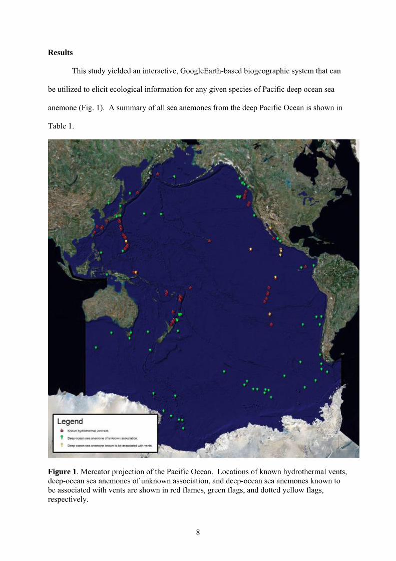
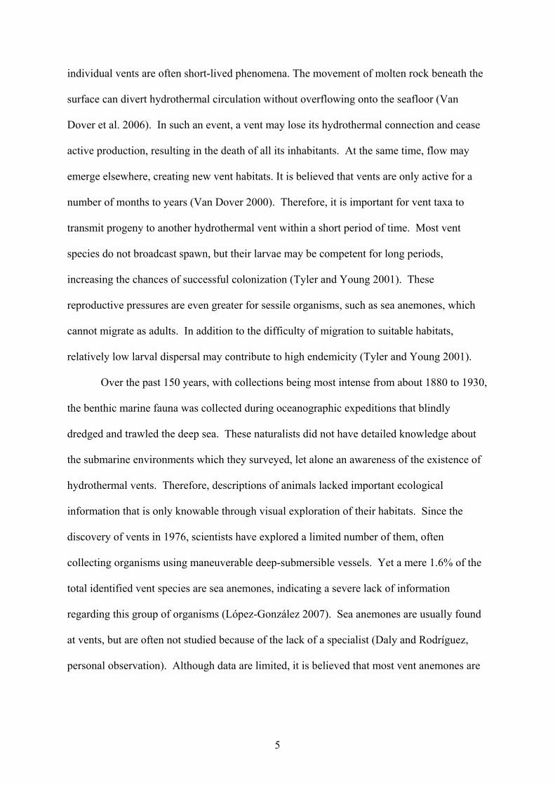
Results
This study yielded an interactive, GoogleEarth-based biogeographic system that can
be utilized to elicit ecological information for any given species of Pacific deep ocean sea
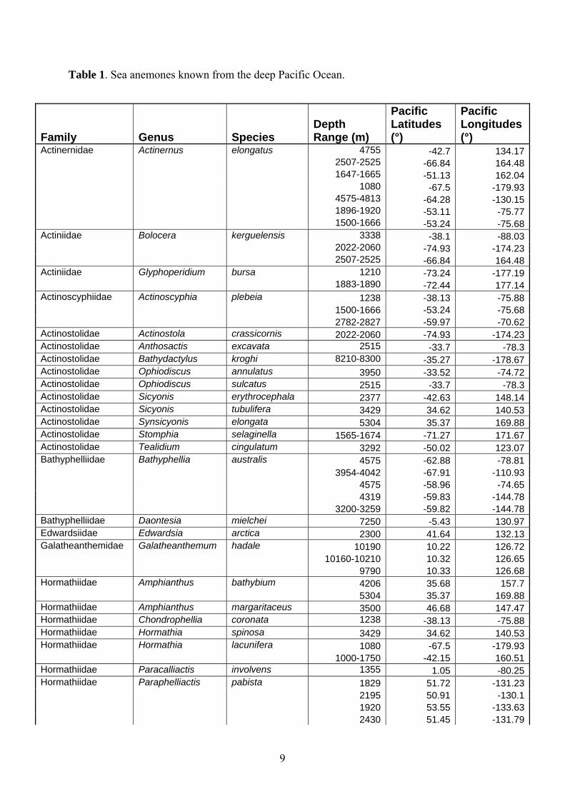
anemone (Fig. 1). A summary of all sea anemones from the deep Pacific Ocean is shown in
Table 1.
Figure 1. Mercator projection of the Pacific Ocean. Locations of known hydrothermal vents, deep-ocean sea anemones of unknown association, and deep-ocean sea anemones known to be associated with vents are shown in red flames, green flags, and dotted yellow flags, respectively.
8
9
Table 1. Sea anemones known from the deep Pacific Ocean.
Family Genus Species Depth Range (m)
Pacific Latitudes (°)
Pacific Longitudes (°)
Actinernidae Actinernus elongatus 4755 -42.7 134.17 2507-2525 -66.84 164.48 1647-1665 -51.13 162.04 1080 -67.5 -179.93 4575-4813 -64.28 -130.15 1896-1920 -53.11 -75.77 1500-1666 -53.24 -75.68Actiniidae Bolocera kerguelensis 3338 -38.1 -88.03 2022-2060 -74.93 -174.23 2507-2525 -66.84 164.48Actiniidae Glyphoperidium bursa 1210 -73.24 -177.19 1883-1890 -72.44 177.14Actinoscyphiidae Actinoscyphia plebeia 1238 -38.13 -75.88 1500-1666 -53.24 -75.68 2782-2827 -59.97 -70.62Actinostolidae Actinostola crassicornis 2022-2060 -74.93 -174.23Actinostolidae Anthosactis excavata 2515 -33.7 -78.3Actinostolidae Bathydactylus kroghi 8210-8300 -35.27 -178.67Actinostolidae Ophiodiscus annulatus 3950 -33.52 -74.72Actinostolidae Ophiodiscus sulcatus 2515 -33.7 -78.3Actinostolidae Sicyonis erythrocephala 2377 -42.63 148.14Actinostolidae Sicyonis tubulifera 3429 34.62 140.53Actinostolidae Synsicyonis elongata 5304 35.37 169.88Actinostolidae Stomphia selaginella 1565-1674 -71.27 171.67Actinostolidae Tealidium cingulatum 3292 -50.02 123.07Bathyphelliidae Bathyphellia australis 4575 -62.88 -78.81 3954-4042 -67.91 -110.93 4575 -58.96 -74.65 4319 -59.83 -144.78 3200-3259 -59.82 -144.78Bathyphelliidae Daontesia mielchei 7250 -5.43 130.97Edwardsiidae Edwardsia arctica 2300 41.64 132.13Galatheanthemidae Galatheanthemum hadale 10190 10.22 126.72 10160-10210 10.32 126.65 9790 10.33 126.68Hormathiidae Amphianthus bathybium 4206 35.68 157.7 5304 35.37 169.88Hormathiidae Amphianthus margaritaceus 3500 46.68 147.47Hormathiidae Chondrophellia coronata 1238 -38.13 -75.88Hormathiidae Hormathia spinosa 3429 34.62 140.53Hormathiidae Hormathia lacunifera 1080 -67.5 -179.93 1000-1750 -42.15 160.51Hormathiidae Paracalliactis involvens 1355 1.05 -80.25Hormathiidae Paraphelliactis pabista 1829 51.72 -131.23 2195 50.91 -130.1 1920 53.55 -133.63 2430 51.45 -131.79
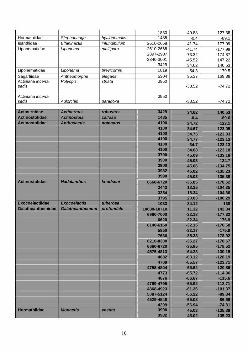
1830 49.88 -127.38Hormathiidae Stephanauge hyalonematis 1485 -0.4 -89.1Isanthidae Eltaninactis infundibulum 2610-2668 -41.74 -177.99Liponematidae Liponema multipora 2610-2668 -41.74 -177.99 2897-2907 -73.32 -174.87 2840-3001 -45.52 147.22 3429 34.62 140.53Liponematidae Liponema brevicornis 1019 54.3 179.5
Antheomorphe elegans 5304 35.37 169.88Sagartiidae Actiniaria incerta sedis
Polyopis striata 3950-33.52 -74.72
Actiniaria incerta sedis Aulorchis paradoxa
3950-33.52 -74.72
Actinernidae Actinernus robustus 3429 34.62 140.53Actinostolidae Actinostola callosa 1485 -0.4 -89.6Actinostolidae Anthosactis nomados 4100 34.72 -123.1 4100 34.67 -123.05 4100 34.75 -123.03 4100 34.77 -123.13 4100 34.7 -123.13 4100 34.68 -123.18 3700 45.09 -133.18 3900 45.03 -134.7 3900 45.06 -134.75 3932 45.02 -135.23 3990 45.03 -135.39Actinostolidae Hadalanthus knudseni 6660-6720 -35.85 -178.52 3443 18.35 -104.35 3354 18.34 -104.36 3795 20.03 -106.29Exocoelactiidae Exocoelactis tuberosa 1033 34.12 138Galatheanthemidae Galatheanthemum profundale 10630-10710 11.32 142.34 6960-7000 -32.18 -177.32 6620 -32.34 -176.9 6140-6160 -32.15 -176.58 5850 -32.17 -175.9 7630 -35.33 -178.92 8210-8300 -35.27 -178.67 6660-6720 -35.85 -178.52 4575-4813 -64.28 -130.15 4682 -63.12 -128.19 4709 -65.57 -123.71 4758-4804 -65.62 -120.86 4773 -65.72 -114.96 4676 -66.67 -115.6 4789-4795 -65.92 -112.71 4868-4923 -61.36 -101.37 5087-5124 -56.22 -89.84 4529-4548 -65.08 -86.66 4209 -56.94 -74.81Hormathiidae Monactis vestita 3990 45.03 -135.39 3932 45.02 -135.23
10
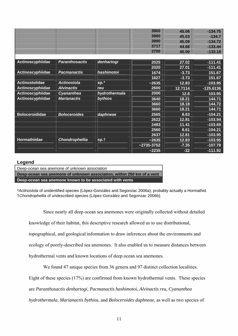
Since nearly all deep ocean sea anemones were originally collected without detailed
knowledge of their habitat, this descriptive research allowed us to use distributional,
topographical, and geological information to draw inferences about the environments and
ecology of poorly-described sea anemones. It also enabled us to measure distances between
hydrothermal vents and known locations of deep ocean sea anemones.
We found 47 unique species from 36 genera and 97 distinct collection localities.
Eight of these species (17%) are confirmed from known hydrothermal vents. These species
are Paranthosactis denhartogi, Pacmanactis hashimotoi, Alvinactis reu, Cyananthea
hydrothermala, Marianactis bythios, and Boloceroides daphneae, as well as two species of
3900 45.06 -134.75 3900 45.03 -134.7 3900 45.09 -134.72 3717 44.68 -133.44 3700 45.09 -133.18 Actinoscyphiidae Paranthosactis denhartogi 2025 27.02 -111.41 2020 27.01 -111.41Actinoscyphiidae Pacmanactis hashimotoi 1674 -3.73 151.67 1627 -3.73 151.67Actinostolidae Actinostola sp.* ~2635 12.83 -103.95Actinoscyphiidae Alvinactis reu 2600 12.7114 -125.6136Actinoscyphiidae Cyananthea hydrothermala 2000 12.8 103.95Actinoscyphiidae Marianactis bythios 3640 18.21 144.71 3660 18.18 144.72 3660 18.21 144.71Boloceroididae Boloceroides daphneae 2565 8.63 -104.21 2622 12.81 -103.94 2483 11.41 -103.69 2560 8.61 -104.21 2637 12.81 -103.95Hormathiidae Chondrophellia sp.† ~2635 12.83 -103.95 ~2735-3752 -7.35 -107.78 ~2235 -32 -111.92 Legend Deep-ocean sea anemone of unknown association Deep-ocean sea anemone of unknown association, within 250 km of a vent Deep-ocean sea anemone known to be associated with vents *Actinostola of unidentified species (López-González and Segonzac 2006a); probably actually a Hormathid. †Chondrophellia of undescribed species (López-González and Segonzac 2006b).
11
the genera Actinostola and Chondrophellia which are unidentified and undescribed,
respectively. Six of these species (75%) were collected from a single vent and two of these
species (25%) collected from a single ridge/vent system. Five of these species (63%) belong
to genera found only at vents (vent-endemic).
Thirty-nine species (83%) were of unknown association. Of these species, seven
(18%) have been found within 250 kilometers of a known vent. These species are Actinernus
robustus, Actinostola callosa, Anthosactis nomados, Hadalanthus knudseni, Exocoelactis
tuberosa, Galatheanthemum profundale, and Monactis vestita.
Discussion
The vast majority of described deep-ocean sea anemone species are of unknown
association (83%). Sea anemone species that are found at vents are known from either a
single vent or a single ridge/vent system. This indicates that identified vent sites are under-
surveyed, and suggests a high level of endemicity for sea anemones at vents. Further support
for high endemicity is the fact that all recognized vent species are from different genera.
We do not agree with López-González and Segonzac (2006a) on their identification
of Actinostola sp. based on a photograph from a vent at the East Pacific Rise. Species of
Actinostola have a smooth column (Carlgren 1949), whereas the animal in their photograph
has a column bearing cuticle. This specimen more likely belongs to family Hormathiidae
(Daly and Rodríguez, personal observation), whose members often bear cuticle on the
column and are common in the deep sea (Carlgren 1949; Fautin and Barber 1989). The
species identified as Chondrophellia sp. is a currently undescribed species (López-González
and Segonzac 2006b), likely to belong to a new taxon (Daly and Rodríguez, personal
observation).
12

We identified seven deep ocean sea anemone species from within 250 kilometers of a
known vent. Given the relative proximity to vents, these species are potential vent
organisms. The specific locations of hydrothermal vents can shift over time as the geological
dynamics of mid-oceanic ridges change in both latitude and longitude, and bathymetry (Van
Dover 2000). Furthermore, charting by early naturalists was not as precise as modern,
satellite-based global positioning systems. These seven species are candidates for further
investigation of distribution, ecology, life-history, and morphology to ascertain whether they
are actually vent taxa. It would be interesting to return to original dredge and trawl locations
and explore the benthos using maneuverable deep submersible vehicles to search for the
presence of extant hydrothermal vents or remnants of extinct vents.
If not found to be from vents themselves, these candidate species may be from the
vent periphery. Vents contribute inorganic chemicals, such as sulfide, and organic carbon to
nearby regions (and indeed the entire global oceanic system). Particulate organic matter
(POM) has been documented to spread at least 2 kilometers from vents (Roth and Dymond
1989). However, the impact zone of hydrothermal vent fields has yet to be quantified and the
specifics of vent to non-vent benthic coupling are still unclear (Van Dover 2000).
This map facilitates visualization and identification of unsurveyed ocean-floor
locations and regions that seem to be lacking Actiniarian fauna. An extremely small portion
of the ocean floor has been extensively sampled; the vast majority has yet to be well
characterized. For regions of seafloor that have been sampled and clearly lack sea anemones,
specific attributes of the habitat may prevent colonization. Furthermore, though a number of
hydrothermal vents have been identified, the biodiversity of the great majority of them has
not been sampled (Van Dover 2000).
With knowledge of both surface and deep ocean currents, this study has important
implications for studies of reproductive distribution. Finally, the methodology of this
13
research could easily be applied to other groups of deep-sea organisms or to other marine
habitats. Cold seeps, another variety of chemosynthetic environment, are a similar habitat to
hydrothermal vents. There is a significant amount of taxonomic overlap between the two
habitats and some propose that they are “stepping-stone” ecosystems from one site to another
in terms of colonization and evolution (Van Dover et al. 2002). Therefore, interesting results
could arise from the incorporation of cold seeps into this study. The Atlantic and Indian
Oceans, too, bear chemosynthetic environments. These sites could be incorporated into work
from the Pacific Ocean. The Atlantic Ocean, in particular, is geologically much younger than
the Pacific and therefore we hypothesize that vent fauna in the Atlantic Ocean should be
evolutionarily derived from the Pacific and Indian Oceans.
Acknowledgements
We would like to thank the National Science Foundation Research Experience for
Undergraduates (REU) program, which provided funding that made this project possible.
The College of Biological Science’s Dean’s Undergraduate Research Fund also extended
generous support. The Pressey Honors Endowment Grant afforded funding for travel to the
Evolution 2008 Meeting to present this research in July 2008. Finally, members of our lab
group, Annie Lindgren, Esprit Heestand, Luciana Gusmão, Abby Reft, Anthony D’Orazio,
and Alpana Chaudhuri, have been encouraging and invaluable to the research.
References
Carlgren, O. 1949. A survey of the Ptychodactiaria, Corallimorpharia and Actiniaria.
Kungliga Svenska Vetenskaps-Akademiens Handlingar 1(1): 1-121.
Desbruyéres, D. 2007. Major known deep-sea hydrothermal vent fields. In Desbruyéres, D.,
M. Segonzac, and M. Bright (Eds.) Handbook of Deep-Sea Hydrothermal Vent
14
Fauna. Densia, Linz, pp. 513-517.
Fautin, D.G. 2007. Hexacorallians of the World.
http://geoportal.kgs.ku.edu/hexacoral/anemone2/index.cfm
Fautin, D.G., and Hessler, R.R. (1989). Marianactis bythios, a new genus and species of
actinostolid sea anemone (Coelenterata: Actiniaria) from the Mariana vents.
Proceedings of the Biological Society of Washington 102, 815–25.
López-González, P. 2007. Cnidaria, Anthozoa, Actiniaria. In Desbruyéres, D., M. Segonzac,
and M. Bright (Eds.) Handbook of Deep-Sea Hydrothermal Vent Fauna. Densia, Linz,
pp. 65-73.
López-González, P. and M. Segonzac. 2006a. Actinostola (Verrill, 1883). In Desbruyéres, D.,
M. Segonzac, and M. Bright (Eds.) Handbook of Deep-Sea Hydrothermal Vent
Fauna. Densia, Linz, p. 66.
López-González, P. and M. Segonzac. 2006b. Chondrophellia cf. coronata (Verrill, 1883). In
Desbruyéres, D., M. Segonzac, and M. Bright (Eds.) Handbook of Deep-Sea
Hydrothermal Vent Fauna. Densia, Linz, p. 73.
Rodríguez, E., C. Castorani, and M. Daly. 2008. Morphological phylogeny of family
Actinostolidae (Anthozoa: Actiniaria) with a description of a new genus and species
of hydrothermal vent sea anemone redefining family Actinoscyphiidae. Invertebrate
Systematics (in press).
Roth, S.E., and J. Dymond. 1989. Transport and settling of organic material in a deep-sea
hydrothermal plume: Evidence from particle flux measurements. Deep Sea Research.
36:1237-1254.
Tyler, P.A. and C.M. Young. 2001. Reproduction and dispersal at vents and cold seeps.
Journal of the Marine Biological Association of the UK 79: 193-208.
Van Dover, C.L. 2000. The Ecology of Deep-Sea Hydrothermal Vents. Princeton University
15
Press, Princeton.
Van Dover, C.L., M. Biscoito, A. Gebruk, J. Hashimoto, V. Tunnicliffe, P. Tyler, and D.
Desbruyéres. 2006. In Desbruyéres, D., M. Segonzac, and M. Bright (Eds.) Handbook
of Deep-Sea Hydrothermal Vent Fauna. Densia, Linz, pp. 13-25.
Van Dover, C.L., C.R. German, K.G. Speer, L.M. Parson, R.C. Vrijenhoek. 2002. Evolution
and biogeography of deep-sea vent and seep invertebrates. Science 295(5558): 1253-
1257.
16
Morphological phylogeny of family Actinostolidae (Anthozoa: Actiniaria) with a description of a new genus and species of hydrothermal vent sea anemone redefining family Actinoscyphiidae Estefanía RodríguezA, B, Christopher N. CastoraniA, Marymegan DalyA
ADepartment of Evolution, Ecology, & Organismal Biology, The Ohio State University, Columbus, OH 43210 BCorresponding author. Email [email protected]
Introduction
Sea anemones attributed to the family Actinostolidae dominate in the deep sea and
polar waters (Carlgren 1949; Fautin and Barber 1999) and at hydrothermal vents (López-
González and Segonzac 2006). The majority of the genera currently placed in it are
monotypic (Fautin 2007), suggesting that the taxonomic characters traditionally used to
differentiate genera need to be re-assessed. The descriptions of several new monotypic
genera in recent decades (Doumenc and Van Praët 1988; Fautin and Hessler 1989; Fautin and
Barber 1999; López-González et al. 2003, 2005) demonstrate the difficulty of
accommodating new taxa in narrowly-defined existing groups, and further argue for a re-
evaluation of the family. Furthermore, a synthetic, phylogenetic assessment of Actinostolidae
would clarify the relationship between the monotypic genera and large, heterogeneous groups
such as the type genus, Actinostola Verrill, 1883. However, such an assessment is difficult
because the family is likely to comprise a paraphyletic grade or a polyphyletic assemblage
rather than a monophyletic group.
Actinostolidae has a long and complex taxonomic history (Table 1). Several members
of Actinostolidae were first grouped together by Hertwig (1882) in family Paractidae, which
he defined as comprising “Hexactiniae with numerous perfect septa and with very contractile
moderately long tentacles, which can be completely covered; circular muscle very strong,
mesodermal”. In this family, he included Antholoba Hertwig, 1882, Dysactis Milne Edwards,
1857, Ophiodiscus Hertwig, 1882, Tealidium Hertwig, 1882, and taxa no longer considered
17
valid, such as its type genus Paractis Milne Edwards & Haime, 1851. Andres (1883) used the
name Paractidae for a sub-family of his Actininae, and placed in this group Paranthus
Andres, 1883, Paractinia Andres, 1883, and Paractis. Hertwig’s (1882) use of the name has
priority.
Carlgren (1893) redefined Paractidae and transferred its previous diagnosis to a new
family, Actinostolidae, into which he placed Actinostola and Stomphia Gosse, 1859.
Carlgren (1893) defined Actinostolidae as “Actiniaria with pedal disc, with very contractile
and moderate long tentacles and usually numerous perfect mesenteries. Pairs of mesenteries
of the last cycles (third and forth cycles) irregularly developed, so the mesentery, which
retractor muscles are facing the next cycle, is more developed than the other. Radial muscles
of oral disc and longitudinal tentacle muscles generally mesogleal. Sphincter mesogleal
usually well developed. No acontia or cinclides”. Carlgren (1893) also provided a new
diagnosis for Paractidae: “Actiniaria with pedal disc, with moderate long tentacles and
usually numerous perfect mesenteries. Mesenteries of the same pair regularly developed.
Radial muscles of oral disc and longitudinal tentacle muscles generally mesogleal. Sphincter
mesogleal usually well developed. No acontia or cinclides”. His distinction between the two
was based on the development of pairs of mesenteries: in Actinostolidae, the two members of
a pair are not identical in size and morphology; in Paractidae, the two members of a pair are
identical. Carlgren (1899) subsequently reclassified Actinostolidae and Paractidae as
subfamilies of family Paractidae, later adding a third subfamily, Polysiphoniinae Carlgren,
1918. Polysiphoniinae was later removed from Paractidae and reclassified as Exocoelactidae
Carlgren, 1925.
Although he used Carlgren’s subfamilies, Stephenson (1921) was not sure that the
distinctions between them were clear, and did not think that any of them merited the rank of
family. In particular, Stephenson (1921) considered Actinostolinae and Paractinae a single,
18
difficult-to-subdivide group. Carlgren (1927) was unable to determine a valid diagnosis for
the type genus Paractis, and later (Carlgren 1932) resurrected the family name
Actinostolidae for some members of Paractidae.
Recent works by Riemann-Zürneck (1978a) and Fautin and Hessler (1989) changed
the definition of the family and reconsidered some features used to differentiate its members.
Riemann-Zürneck (1978a) revised the mesomyarian family Actinoscyphiidae Stephenson,
1920, clarifying the distinctions between this group and Actinostolidae. Fautin and Hessler
(1989) amended Carlgren’s (1949) key to the genera of Actinostolidae, correcting his errors
and incorporating new species. In their revised key, Fautin and Hessler (1989) omitted
Cyananthea Doumenc & Van Praët, 1988 because the sole account of its type species was too
fragmentary to evaluate many of the critical features. This genus has been recently re-
described and placed in the family Actinoscyphiidae based on its cnidom (Sanamyan and
Sanamyan 2007). This redescription of Cyananthea highlights the confusion that remains
about the circumscription of Actinoscyphiidae and Actinostolidae: Sanamyan and Sanamyan
(2007) point out that additional genera that had been described as Actinostolidae are likely to
be more appropriately placed in Actinoscyphiidae, but they fail to fully address this issue or
formally reassign genera.
We describe Alvinactis reu gen., sp. nov. from the East Pacific Rise of the North
Pacific Ocean. This new genus has a mesogleal sphincter and lacks acontia, and thus belongs
to Mesomyaria. To assess the distinctiveness of Alvinactis gen. nov. and to evaluate whether
it belongs to Actinostolidae or Actinoscyphiidae, we generated a data matrix of
morphological features of genera of Actinostolidae and Actinoscyphiidae. Although
morphological attributes may be subject to convergence, preservation artefacts, or other
sources of systematic error, these are the only data available for many of these taxa, because
most are known only from formalin-fixed museum material. Phylogenetic analysis of this
19
matrix is used to explore the consistency and information content of various taxonomic
features used in classification of Actinostolidae and Actinoscyphiidae, test the monophyly of
each family, and identify potentially monophyletic groups within Actinostolidae. This is the
first cladistic analysis for members of the actiniarian superfamily Mesomyaria.
Materials and Methods
Specimens were collected during a cruise of the Woods Hole Oceanographic
Institution research vessel “Atlantis” using the Deep Submergence Vessel “Alvin”. All
specimens came from one collection during dive 3941, on 26 November 2003, in the North
Pacific Ocean: East Pacific Rise, 12°42.680’N, 103°54.462’W, depth 2600 m. Specimens
were collected using Alvin’s manipulator arm; at the surface, specimens were placed in
chilled water and allowed to relax before being anaesthetized with isotonic magnesium
chloride. Pieces of some specimens were fixed immediately in 95% ethanol. The remaining
specimens were fixed in 10% seawater formalin and later transferred to 70% ethanol for
long-term storage. All specimens were deposited at the Field Museum of Natural History
(FMNH).
Preserved specimens were examined whole, in dissection, and as serial sections.
Serial sections were prepared using standard paraffin techniques. Histological slides were
stained in Masson’s trichrome (Presnell and Schreibman 1997). Small pieces of tissue from
tentacles, column, pedal disc, mesenterial filaments, and actinopharynx were smeared on a
slide; nematocysts in these smears were examined using DIC at 100X magnification. Cnidae
terminology follows Mariscal (1974).
The phylogenetic analysis of genera of Actinostolidae is based on a matrix of
characters scored from direct observation or descriptions of type species. The characters are
those traditionally used to recognize taxa within Actinostolidae, including those features
20

identified by Carlgren (1949) in his key to the family. Some of these features (e.g.,
bathymetric range, habitat) are not strictly morphological, but can be interpreted as proxies
for physiological attributes. All characters are treated as unordered and weighted equally.
Outgroups include four genera classified in more distant groups: the endomyarian Epiactis
Verrill, 1869 and the acontiarians Bathyphellia Carlgren, 1932, Hormathia Gosse, 1859, and
Kadosactis Danielssen, 1890. These species span the diversity of Actiniaria and thus provide
a strong test of monophyly of Actinostolidae. We included the mesomyarian Actinoscyphia
Stephenson, 1920 because it was once included in Actinostolidae (Table 1), and because
several taxa originally assigned to Actinostolidae have been hypothesized to be closely
related to this genus (Riemann-Zurneck 1978a; Sanamyan and Sanamyan 2007). The
character states attributed to the generic exemplars in the analysis were evaluated from direct
observation or literature reports of type species, except in the case of Bathydactylus Carlgren,
1928. We considered Bathydactylus krogni Carlgren, 1956 rather than Bathydactylus
valdiviae Carlgren, 1928, because the type species of the genus is known only from a single,
poorly-preserved specimen. We included three species of Anthosactis Danielssen, 1890
because the great heterogeneity of the genus (White et al. 1999; Daly and Gusmão 2007)
raises concern that the group is not monophyletic. Riemann-Zurneck (1978b) synonymized
Paractinostola Carlgren, 1928 with Actinostola, but recognized that the latter was likely to be
a paraphyletic group. We included the type species of the former Paractinostola,
Paractinostola bulbosa Carlgren, 1928, in recognition of the heterogeneity in Actinostola.
The initial assessment of nematocyst types in the tentacles of Paranthosactis was equivocal
(López-González et al. 2003); upon reconsideration of their material and photographs, we
find that the nematocysts called microbasic b-mastigophores by López-González et al. (2003)
are holotrichs similar in size and morphology to those seen in the tentacles of Alvinactis gen.
nov. Other comparative material examined includes Marianatis bythios Fautin & Hessler,
21
1989 deposited at the US National Museum of Natural History (USNM 84401, 84402),
Bathydactylus krogni and Epiparactis dubia Carlgren, 1928 deposited at Zoological Museum
in Copenhagen, and Anthosactis pearseae Daly & Gusmão, 2007 deposited at the California
Academy of Sciences (CAS 174323-174325) and the US National Museum of Natural
History (USNM 1096705, 1096706).
The final matrix of 41 characters (Appendix 1 and 2) was analyzed in NONA
(Goloboff 1999), using Winclada (Nixon 1999) to initiate 50 rounds of TBR branch
swapping. Further rounds of swapping were not recommended by the results of the initial
searches. We present the strict consensus of the equally parsimonious trees with Bremer
support (Bremer 1994) calculated for all clades appearing in the consensus. The character
optimizations discussed are those features that can be placed unambiguously at a particular
node. Numbers in the text, on Fig. 1, and in Appendix 2 refer to the characters of Appendix
1.
Carlgren (1949) used the ranks “tribe” and “subtribe” to refer to groups between
suborders and families. We have corrected this misapplication of ranks in our treatment of
the taxonomy of Alvinactis reu gen., sp. nov. We have based our diagnoses of higher taxa on
those of Carlgren (1949) and Riemann-Zürneck (1978a), altering them to be parallel and
telegraphic; more substantive changes are indicated in italics.
Results
Phylogenetic analysis recovered 22 trees of L=166 (CI=0.30, RI=0.59). The strict
consensus of these (Fig. 1) includes two main clades. One of these is a large clade that
includes Actinostola, Antholoba, Anthosactis janmayeni Danielssen, 1890, Cnidanthus
Carlgren, 1927, Hormosoma Stephenson, 1918, Ophiodiscus, Paractinostola, Parasicyonis
Carlgren, 1921, Pycnanthus McMurrich, 1893, Sicyonis Hertwig, 1882, Stomphia,
22
Synsicyonis Carlgren, 1921, and Tealidium; this roughly corresponds to Carlgren’s subfamily
Actinostolinae. Henceforth, we refer to this clade as “Actinostolina”.
The other main clade includes the remaining genera previously attributed to
Actinostolidae, Actinoscyphia, and the outgroups Bathyphellia, Hormathia, and Kadosactis,
which nest among members of Actinostolidae. This clade comprises two smaller clades: one
includes the acontiate outgroups together with Bathydactylus and Hadalanthus Carlgren,
1956; the second includes Actinoscyphia, Epiparactis, and the taxa from chemosynthetic
habitats (Fig. 1). The membership of this second clade corresponds closely to
Actinoscyphiidae sensu Sanamyan and Sanamyan (2007); we refer these taxa to this family.
All taxa from hydrothermal vents and cold seeps (Alvinactis gen. nov., Cyananthea,
Maractis, Marianactis, Pacmanactis López-González et al. 2005, and Paranthosactis) form a
clade without consistent internal resolution. This chemosynthetic habitat clade, hereafter
called Chemosynthina, is strongly supported by six morphological characters (#s 5, 6, 9, 17,
26, 31) and two additional ones referring to the habitat (#s 38, 39). The three species of
Anthosactis do not group together.
Order Actiniaria Hertwig, 1882
Suborder Nynantheae Carlgren, 1899
Superfamily Mesomyaria Stephenson, 1921
Family Actinostolidae Carlgren, 1893
Diagnosis. Nynantheae with basilar muscles and mesogleal marginal sphincter; column
commonly smooth, rarely tuberculate or with papillae. Tentacles regularly arranged, their
aboral sides sometimes with nematocysts batteries, sometimes thickened. Mesenteries not
divisible into macro- and micro-cnemes. Younger mesenteries not bilaterally arranged.
23
Retractor muscles diffuse, rarely circumscribed. No acontia. Cnidom: Gracile spirocysts,
basitrichs, and microbasic b- and p-mastigophores. (Modified from Carlgren 1949).
Remarks. Carlgren (1949) listed the authorship of Actinostolidae as Carlgren, 1932 but the
family was erected by him in 1893 (Table 1).
Included genera. Actinostola; Antholoba; Anthosactis; Antiparactis Verrill, 1899;
Bathydactylus; Cnidanthea Carlgren, 1956; Cnidanthus; Hadalanthus; Hormosoma;
Ophiodiscus; Paranthus; Parasicyonis; Pseudoparactis Stephenson, 1920; Pycnanthus;
Sicyonis; Stomphia; Synsicyonis; Tealidium.
Family Actinoscyphiidae Stephenson, 1920
Diagnosis. Nynantheae with basilar muscles and mesogleal marginal sphincter. Pedal disc
flat, sometimes small, grasping. Column commonly smooth, often with distal row of cinclides
and sometimes verrucae. Tentacles usually marginal on wide oral disc, their aboral sides
sometimes thickened. Oral disc sometimes lobed. Mesenteries not divisible into macro- and
micro-cnemes. At least six pairs of perfect and fertile mesenteries. Retractor muscles diffuse
and weak. Longitudinal muscles of the tentacles ectodermal. No acontia. Cnidom: Robust
and gracile spirocysts, basitrichs, holotrichs, and microbasic p-mastigophores. (Modified
from Riemann-Zürneck 1978a).
Remarks. Riemann-Zürneck (1978a) resurrected Actinoscyphiidae primarily based on
Schmidt’s (1972) classification of types of cnidae. Thus, Riemann-Zürneck (1978a)
characterizes Actinoscyphiidae as having “p-rhadoids B” and lacking “p-rhadoids A”.
Schmidt’s distinction between the categories “p-rhadoids A/p-rhadoids B” roughly
corresponds with Mariscal’s distinction between “microbasic p-
mastigophores/amastigophores”. Nomenclatural issues aside, although these types are
24
certainly different in ultrastructure (shaft and tubule spination), accurate recognition of their
distinctiveness requires observing them in a discharged state under SEM. To use all of
Schmidt’s subdivisions of p-mastigophores is necessary to observe the fine details of spine
length, density and angle of attachment which are important characters in this system
(England 1991; Östman 2000). Using the ultrastructure of p-mastigophores, Schmidt (1972,
1974) grouped mesomyarian families into “Early” and “Late” Mesomyaria). However, his
distinction was based on examination of relatively few species; these types of nematocysts
have not been distinguished for most of the genera. Furthermore, many actiniarian families
are polyphyletic (Daly et al. 2008), making combining them into groups especially
problematic. The phylogenetic interpretation of morphological differences among nematocyst
types is not clear. Because molecular evidence does not support Schmidt’s (1972, 1974)
distinction between “Early” and “Late” Mesomyaria (Daly et al. 2008), attributing high
phylogenetic significance to the distinction between A or B p-mastigophores may be
unwarranted. Given the current lack of clarity about the generality and applicability of this
character to many taxa, and its dubious value as a phylogenetic feature, we prefer not to
include these differences in the definition of the families.
Included genera. Actinoscyphia, Alvinactis gen. nov., Cyananthea; Epiparactis Carlgren,
1921; Maractis Fautin & Barber, 1999; Marianactis Fautin & Hessler, 1989; Pacmanactis;
Paranthosactis.
Genus Alvinactis
Diagnosis. Pedal disc well developed. Column smooth, not divisible into scapus and
scapulus, with distal row of verrucae and cinclides. Distal margin of column distinctly
marked as marginal ring. Tentacles of uniform thickness along entire length, those of inner
cycle longer than those of outer cycle. Longitudinal muscles of tentacles ectodermal, equally
25

developed. Mesenteries arranged in four cycles, only first cycle perfect. Same number of
mesenteries proximally and distally. All mesenteries except those of youngest cycle fertile.
Two well developed siphonoglyphs each attached to pair of directives. Retractor muscles
diffuse; parietobasilar muscles not differentiated. Cnidom: Robust and gracile spirocysts,
basitrichs, holotrichs, microbasic p-mastigophores.
Types species. Alvinactis reu sp. nov.
Etymology. The name Alvinactis combines the name of the submersible “Alvin” and “–actis”
a common suffix for actiniarians, referring to their rayed or star-like external morphology.
Alvinactis reu sp. nov.
(Figs 2-5, Table 2)
Diagnosis. Column of preserved specimens cylindrical, not divisible into scapus and
scapulus, with more or less distinct marginal ring. Column smooth except for distal belt of
small, round, perforate verrucae. Mesenteries hexamerously arranged in four cycles, all larger
ones fertile, only those of first cycle perfect. Tentacles with numerous spirocysts and
basitrichs; holotrichs in tips of tentacles of most specimens. Pedal disc diameter 14-59 mm,
column height 6-34 mm (contracted and preserved specimens).
Material examined. FMNH 1150*, holotype; FMNH 11504, 3 paratypes.
Base and column. Column stout, of approximately equal diameter throughout in preserved
specimens, encircled by distal belt of 24 small, hollow outgrowths of all three layers of
column, perforate verrucae (Figs 2B, D, 4D, E). Verrucae inside crease beneath sphincter,
associated with endocoelic spaces of stronger mesenteries, likely adherent. No fosse,
although distal edge of column may extend over base of tentacles in contracted specimens
26
(Figs 2A, B). Column of preserved specimens uniform brownish-pink. In life, column
trumpet shaped, flaring slightly from base (Fig. 3); column, tentacles, and oral disc of living
specimens uniform translucent grayish-green. Strong columnar circular musculature and
mesogleal sphincter; sphincter spans distal quarter of column, reticulated, lies closer to
epidermis than gastrodermis, tapers more distally than proximally (Fig. 4E). Mesoglea of
distal column with small, darkly-staining inclusions; these are especially abundant near
marginal sphincter.
Base flat or slightly withdrawn inside column. Pedal disc adherent, muscular, same
color as column in preserved material, approximately equal or slightly wider in diameter than
oral disc in preserved specimens (Fig. 2A). Pedal disc circular in smaller specimens; oval in
largest specimen.
Oral disc and tentacles. Tentacles marginal, approximately 100 in five cycles; those of outer
cycle markedly shorter; those of inner cycles obscure oral disc in contracted specimens (Figs
2A, B). Specimens with more than 96 tentacles do not have additional mesenteries,
suggesting tentacle regeneration rather than additional cycles of mesenteries at the distal
column. Tips of tentacles perforated. Inner tentacles moderate in length, to 21 mm long,
longitudinally sulcated in preserved specimens (Fig. 2B). In life, tentacles conical,
approximately equal in length or longer than column. Oral disc flat, mouth oval; two
prominent siphonoglyphs. Tentacles, oral disc, lips, actinopharynx, and siphonoglyphs same
color as column.
Mesenteries and internal anatomy. Mesenteries arranged hexamerously in four cycles,
those of first cycle perfect; two pairs of directives, each attached to a well developed
siphonoglyph. All mesenteries of first, second, and third cycles (including directives) bear
filaments and gametogenic tissue; those of fourth cycle weak, lacking filaments and
gametogenic tissue (Fig. 4C). Species gonochoric; all specimens collected in late November
27
sexually mature, with either oocytes or spermatic vesicles (48-234 and 31-120 μm in
diameter, respectively; Figs 4B, G).
Longitudinal muscles of mesenteries diffuse (Figs 4G, H). Pennon of parietobasilar
muscles not differentiated (Fig. 4H). Basilar muscles present, equally developed (Fig. 4A).
Cnidom. Robust and gracile spirocysts, basitrichs, holotrichs, microbasic p-mastigophores
(Fig. 5). See Table 2 for size and distribution.
Habitat and biology. All specimens living on and among oxidized clumps of the tubeworm
Tevnia (Fig. 3). Multiple individuals co-occur on single clump, but specimens typically not
close enough to touch one another.
Etymology. The specific name “reu” honors the NSF Research Experience for
Undergraduates program, which supported CNC’s participation in this project. The species
epithet should be considered an undeclinable Latin noun.
Discussion
Comparison of Alvinactis reu gen., sp. nov. with other genera
As is true of other sea anemones described from chemosynthetic environments (e.g.,
Fautin and Hessler 1989; Fautin and Barber 1999; López-González et al. 2003, 2005),
Alvinactis gen. nov. presents an unusual combination of characters that make it incompatible
with the diagnoses of other genera. It has four cycles of mesenteries; although only those of
the first cycle are perfect, all of the larger mesenteries are fertile. It has a belt of perforate
verrucae encircling the distal column, and a strong circumferential marginal ring.
Our phylogenetic analysis demonstrates that Alvinactis gen. nov. is clearly part of the
Actinosyphiidae, and lies within the Chemosynthina, but its relationship to other genera is
unclear. In some of the primary trees, Alvinactis and Paranthosactis together are the sister
28
clade to a clade of Cyananthea and Pacmanactis because all have a marginal ring (#26). The
marginal ring optimizes elsewhere on the tree, as a synapomorphy for Hormosoma,
Anthosactis janmayeni, and Tealidium, and in Bathydactylus and in the outgroup Kadosactis.
In other primary trees, Alvinactis and Paranthosactis are together (but not resolved) as the
sister to Maractis and Marianactis, based on an imperfect second cycle of mesenteries (#12).
Alvinactis gen. nov. is also associated with Maractis and Pacmanactis individually: as sister
to Maractis because both lack microbasic p-mastigophores in the tentacles (#31), or as sister
to Pacmanactis because both have a distal row of verrucae (#4).
The primary anatomical difference between Alvinactis gen. nov., Maractis,
Marianactis, and Paranthosactis is the distal belt of verrucae in Alvinactis gen. nov.
Verrucae are hollow outgrowths of all three layers of the column; the ectodermal musculature
and epidermis of verrucae differ from that of the surrounding column (Stephenson 1928, den
Hartog 1987). These are most commonly seen in endomyarian sea anemones, in members of
the family Actiniidae in particular (Stephenson 1928, Carlgren 1949, den Hartog 1987). The
columnar outgrowths of acontiarian and mesomyarian anemones are typically called
“suckers” or “tenaculi”; these structures are solid rather than hollow (Stephenson 1921). As
the columnar structures of Alvinactis gen. nov. are hollow (Figs 4C, E), and are identical in
form to verrucae of actiniid anemones (see, e.g., den Hartog 1987; Daly 2004), we consider
them verrucae rather than suckers. Although there is no material adhering to the verrucae of
Alvinactis reu gen., sp. nov., this is frequently the case in preserved specimens of species
known to bear verrucae (MD, pers. obs.)
Alvinactis gen. nov. further differs from Maractis because the latter lacks a marginal
ring, and from Paranthosactis because Alvinactis gen. nov. lacks microbasic p-mastigophores
in the tentacles. In other actiniarians (e.g., Actiniidae, Edwardsiidae, Isanthidae, etc), having
specializations like verrucae, tenaculi, or vesicles on the column is of generic significance
29
(Carlgren 1949). It is possible that it is of lesser significance among mesomyarians, and that
Alvinactis gen. nov., Maractis, and Paranthosactis belong in the same genus. However,
because cladistic analysis of morphological data (including all the aforementioned
similarities) did not consistently recover sister group relationships among these taxa, we have
no objective basis for synonymizing them.
Alvinactis gen. nov., Cyananthea, Pacmanactis, and Marianactis are all known from
chemosynthetically active habitats in the Pacific Ocean, but these three genera are clearly
distinct. Like Pacmanactis, Alvinactis gen. nov. has distal perforate verrucae, although the
distal structures are not histologically defined in Pacmanactis (López-González et al. 2005);
they differ in that Alvinactis gen. nov. lacks microbasic p-mastigophores in the tentacles, and
lacks microbasic b-mastigophores in the column margin and in the tentacles. Both
Pacmanactis and Cyananthea have two cycles of perfect mesenteries, whereas Alvinactis
gen. nov. has only one perfect cycle. Furthermore, Cyananthea has a distal belt of cinclides
in the distal column but not verrucae. Marianactis has a distal belt of cinclides in the column
and only one cycle of perfect mesenteries, characteristics seen in Alvinactis gen. nov.
Nevertheless, Marianactis lacks both verrucae and a marginal ridge, lacks holotrichs in the
distal column or tentacles, and has a differentiated pennon on the parietobasilar muscles.
Marianactis also has microbasic amastigophores rather than microbasic p-mastigophores in
the tentacles, but this distinction is of less value because of the difficulty of distinguishing
between these nematocysts when undischarged (Östman 2000).
Phylogenetic relationships of Actinostolidae and Actinoscyphiidae
Our phylogenetic analysis of morphological data highlights problems with the
taxonomy and organization of Actinostolidae in its old sense. Neither the strict consensus tree
(Fig. 1) nor any of the primary trees support monophyly of Actinostolidae, suggesting that it
30
is a grade rather than a clade. Phylogenetic analysis of a more diverse assemblage of
actiniarians, including representatives of Actinostola, Actinoscyphia, Anthosactis,
Hormosoma, Stomphia, and the taxa used here as outgroups recovers a pattern of
relationships compatible with the morphological evidence (Daly et al. 2008).
The sole feature shared by all members of Actinostolidae is a mesogleal marginal
sphincter, an attribute common to many other actiniarian families. The remaining diagnostic
features are absences: lack of the nematocyst-dense threads called acontia and of
microcnemic mesenteries. The lack of resolution and the inclusion of Actinoscyphia and the
outgroups Bathyphellia, Hormathia, and Kadosactis among the ingroup taxa suggest that
some members of Actinostolidae are not closely related to one another; Bathyphellia,
Hormathia, and Kadosactis belong to the superfamily Acontiaria. Molecular evidence
suggests that all Acontiaria belong to a monophyletic group, although this clade also includes
taxa without acontia (Daly et al. 2008). It is likely that at least some members of the family
will need to be transferred to other families or placed in new families.
Nevertheless, our phylogenetic analysis indicates that some genera share uniquely
derived attributes (Fig. 1). Our clade Actinostolina contains many of the taxa Carlgren (1899)
included in his original description of the subfamily Actinostolinae, including the type genus
Actinostola. Two synapomorphies for Actinostolina are characteristics Carlgren (1899)
ascribed to the subfamily Actinostolinae, including mesogleal longitudinal muscles in
tentacles (#23) and microbasic b-mastigophores in the tentacles (#30). The third feature,
microbasic p-mastigophores in the tentacles (#31), has also been used to distinguish
actinostolid genera (Carlgren 1949). Internal brooding of offspring (#35) is seen outside of
Actinostolina only in Anthosactis pearseae; as Anthosactis is a polyphyletic assemblage, the
interpretation of this character is unclear. Perfect mesenteries in the second and third cycles
(#s 12, 13) characterize most members of the Actinostolina, except A. janmayeni, Tealidium,
31

and Ophiodiscus; in these three taxa, none of the mesenteries of the third cycle are perfect
(#13).
Carlgren (1949) used the dissimilar morphology of mesenteries of a pair (#s 7, 8) to
divide the actinostolids (groups I and II, see Carlgren 1949). These features are a
synapomorphy for a clade within Actinostolina that encompasses most of the taxa Carlgren
(1949) placed in group I (Fig.1). However, at least two genera (Antholoba and Pycnanthus)
with similar mesenteries group with the clade of taxa with dissimilar mesenteries (Fig. 1).
The tree provides no support for the monophyly of the taxa Carlgren (1949) placed in group
II, although this is not surprising, as having paired mesenteries of similar morphology is
common to most Actiniaria.
The genus Anthosactis is very heterogeneous, and previous authors have suggested
that it may be a polyphyletic assemblage rather than monophyletic clade (e.g., Riemann-
Zürneck 1997; White et al. 1999; Daly and Gusmão 2007). Our results bolster this
interpretation: the three species of Anthosactis in our analysis did not group together, being
widely dispersed through the tree. The type species A. janmeyeni groups with Hormosoma
and Tealidium as the sister clade to Actinostolina (Fig. 1). A close relationship between
Tealidium and Anthosactis has been proposed previously (Riemann-Zürneck 1997). Batteries
of microbasic b-mastigophores in the aboral bases of the tentacles (#29) and the sphincter
forming a marginal ring (#26) group Hormosoma and the clade comprised of A. janmayeni
and Tealidim. The two other species of Anthosactis, A. nomados and A. pearseae, are in the
other main clade: A. pearseae is the sister group to Chemosynthina, and A. nomados is the
sister group to the crown clade consisting of Actinoscyphiidae and its sister clade.
In the consensus tree, Epiparactis, Actinoscyphia, and Anthosactis pearseae are sister
to the clade we call Chemosynthina, and this clade is the sister to a group composed of
Hadalanthus, Bathydactylus, and the acontiate outgroups. Although the clustering of
32
outgroup and ingroup taxa points to problems in the circumscription of these groups, some
components of this tree have been advocated by other authors. In the discussion that
accompanied her resurrection of family Actinoscyphiidae, Riemann-Zürneck (1978a)
hypothesized a close relationship between Epiparactis and Actinoscyphia. Following
Schmidt’s (1972, 1974) subdivision of mesomyarians in “Early” and “Late” groups based on
attributes of the cnidae, Rieman-Zürneck (1978a) further hypothesized that members of
Actinoscyphiidae had lost acontia. Stephenson (1920) expressed a similar idea by including
Lilliella Stephenson, 1918 and Isoparactis Stephenson, 1920 in Actinoscyphiidae; these
genera have since been synonymized with the acontiate genera Hormathia and
Acraspedanthus Carlgren, 1924, respectively. Sanamyan and Sanamyan (2007) considered
Cyananthea and Epiparactis within Actinoscyphiidae, following the diagnosis given by
Riemann-Zürneck (1978a). They also noted that the ring of cinclides in the distal column of
Cyananthea strongly recalls Kadosactis, thereby relating Cyananthea to acontiarians. Finally,
Sanamyan and Sanamyan (2007) pointed out the similarities between Pacmanactis and
Cyananthea (both only differing in the presence of verrucae and the number of mesenteries
distally and proximally), and highlighted the similarities in the cnidom of Marianactis and
Cyananthea. Based on these comparisons, they proposed Pacmanactis and Marianactis be
transferred to Actinoscyphiidae but they did not make the change (see Sanamyan and
Sanamyan 2007).
In our consensus tree, most of the taxa Sanamyan and Sanamyan (2007) include in
Actinoscyphiidae group together (Fig. 1). Epiparactis is basal to the rest of the genera of this
clade. It lacks holotrichs in the tentacles (#33), a feature shared by all other taxa except
Marianactis, and has three rather than four complete cycles of mesenteries. Most members of
Actinoscyphiidae have a marginal sphincter situated closer to the epidermis (#27), and four
cycles of mesenteries (#10). The Actinoscyphiidae is the sister to a clade that includes the
33
acontiarian outgroups plus Hadalanthus and Bathydactylus. Thus, this analysis suggests a
close relationship between Actinoscyphiidae and some Acontiaria.
Within Actinoscyphiidae is Chemosynthina, the clade containing the genera reported
from hydrothermal vents and cold seeps. Anthosactis pearseae, known from whalefalls, is the
sister group to Chemosynthina in some but not all primary trees. Monophyly of
Chemosynthina is supported by a mosaic of characters: the presence of cinclides (#5),
relatively robust or thick column walls (#6, except Pacmanactis and Marianactis), equal
numbers of mesenteries proximally and distally (#9, except Cyananthea), fertile mesenteries
in the third cycle (#17, except Pacmanactis), a strong sphincter (#25, except Pacmanactis), a
marginal ring (# 26, absent in Maractis and Marianactis), and microbasic p-mastigophores in
the tentacles (# 31, except Maractis and Alvinactis gen. nov.). In our re-examination of the
type material of Marianactis, we found a belt of small cinclides in the distal column; the
cinclides are very small and are inconspicuous in preserved material, and are therefore easily
overlooked. Their presence may also have been overlooked in Maractis or Paranthosactis.
Based on these results, we accept Sanamyan and Sanamyan’s (2007) circumscription
of Actinoscyphiidae, and add to it Marianactis and Pacmanactis, Alvinactis gen. nov.,
Maractis, and Paranthosactis. Actinostolidae in a new sense includes the genera in
Actinostolina, plus an assemblage of taxa that are basal to Actinostolina or Actinosyphiidae,
including Anthosactis, Antiparactis, Cnidanthea, Cnidanthus, Hormosoma, Paranthus,
Pseudoparactis, and Tealidium. Actinostolidae in its new sense is not monophyletic. Re-
organizing it to reflect monophyly will require dense sampling across Actiniaria, and should
include molecular as well as morphological data. Anthosactis is polyphyletic, with some
members more closely related to genera in Actinoscyphiidae than to those in Actinostolidae.
Because the type species A. janmayeni lies within Actinostolidae, pending a comprehensive
species-level revision of Anthosactis, we leave it in Actinostolidae.
34
In addition to identifying potential synapomorphies for Actinoscyphiidae,
Actinostolidae, and their subgroups, our analysis highlights characters that seem to have little
ability to group taxa. The number of distal and proximal mesenteries (#9) varies widely in the
family, and has not been assessed for many taxa. Similarly, having the sphincter form a
marginal ring (#26) occurs quite broadly across the tree, as do broad bathymetric ranges
(#37). As with the number of mesenteries, these features may have been scored inconsistently
by some authors, making them appear less informative than they actually are. The relative
thickness of the column wall (#6) is often not recorded and is very subjective, varying with
degree of contraction and preservation state.
The types of nematocysts in the tentacles have been used as a generic character in
Actinostolidae (Carlgren 1949; Fautin and Hessler 1989). The presence of microbasic b-
mastigophores is a potential synapomorphy of Actinostolina; their presence and arrangement
in batteries distinguishes Tealidium, Hormosoma, and Anthosactis (see Carlgren 1949).
However, the use of these features as taxonomic characters has been challenged in recent
studies (see López-González et al. 2003). Similarly, although microbasic p-mastigophores or
amastigophores have been used to differentiate actinostolid genera, the phylogenetic value of
these characters is far from clear because these types are difficult to distinguish with light
microscopy (Östman 2000). Holotrichs in the tentacles are inducible in some species (e.g.,
Fautin 1988; Edmands and Fautin 1991), rendering them suspect as a taxonomic or
phylogenetic feature. Nonetheless, holotrichs in the tentacles is phylogentically useful in this
analysis, grouping Chemosynthina and its allies.
35
Acknowledgements
Specimens were collected by Janet Voight, with the assistance of the scientific party
of cruise AT 11-03, the crew of the R/V Atlantis and the crew of the DSV Atlantis. J. Gerber
of the FMNH helped in the accession and loan of the specimens. Collection of specimens was
funded through NSF DEB-0072695 to J. Voight; their description was funded through NSF
EF-0531763 to MD and an OSU CBS Dean’s Undergraduate Research award to CNC. J.
Voight of the FMNH, S. Cairns of the USNM, R. Van Syoc of the CAS, and O. Tendal of the
ZMUC provided comparative material. J. Wenzel gave advice on coding characters and
identifying networks within the tree. A. Reft is thanked for her helpful advice and comments
on cnidae.
36
References
Andres, A. (1883). ‘Le Attinie (Monografía)’. (Coi Tipi der Salviucci: Roma).
Bremer, K. (1994). Branch support and tree stability. Cladistics 10, 295–304.
Carlgren, O. (1893). Studien über Nordische Actinien. Kungliga Svenska Vetenskaps-
Akademiens Handlingar 25, 1–148.
Carlgren, O. (1899). Zoantharien. Hamburger Magalhaensische Sammelreise 4(1), 1–48.
Carlgren, O. (1918). Die Mesenterienanordnung der Halcuriiden. Kungliga Fysiografiska
Sällskapets Handlingar 29(29), 1–37.
Carlgren, O. (1921). Actiniaria. I. Danish Ingolf-Expedition 5(9), 1–241.
Carlgren, O. (1924). Actiniaria from New Zealand and its Subantarctic Islands.
Videnskabelige Meddelelser fra Dansk Naturhistorisk Forening (Copenhagen) 77,
179-261
Carlgren, O. (1925). Zur Kenntnis der Hexacorallen. Zoologischer Anzeiger 65, 87–99.
Carlgren, O. (1927). Actiniaria and Zoantharia. Further Zoological Results of the Swedish
Antarctic Expedition 1901-1903 2(3), 1–102.
Carlgren O. (1928). Actiniaria der Deutschen Tiefsee-Expedition. Wissenschaftliche
Ergebnisse der Deutschen Tiefsee-Expedition auf dem Dampfer "Valdivia" 1898-1899
4(22), 125–266 [reprint 1–144].
Carlgren, O. (1932). Die Ceriantharien, Zoantharien und Actiniarien des arktischen Gebietes.
In ‘Eine Zusammenstellung der arktischen Tierformen mit besonderer
Berücksichtigung des Spitzbergen-Gebietes auf Grund der Ergebnisse der Deutschen
Expedition in das Nördliche Eismeer im Jahre 1898. Vol 6.’ (Eds F. Römer, F.
Schaudinn, A. Brauer, and W. Arndt.) pp. 255–266. (Gustav Fischer: Jena).
Carlgren, O. (1949). A survey of the Ptychodactiaria, Corallimorpharia and Actiniaria.
Kunglia Svenska Vetenskapsakadamiens Handlingar 1, 1–121.
37
Carlgren, O. (1956). Actiniaria from depths exceeding 6000 meters. Galathea Reports 2, 9–
16.
Daly, M. (2004). Anatomy and taxonomy of three species of sea anemones (Cnidaria:
Anthozoa: Actiniidae) from the Gulf of California, including Isoaulactina
hespervolita Daly, n. sp. Pacific Science 58, 377–390.
Daly, M., and Gusmão, L. (2007). The first sea anemone (Cnidaria, Anthozoa) from a whale
fall. Journal of Natural History 41, 1–11.
Daly, M., Chaudhuri, A., Gusmão, L., and Rodríguez, E. (2008). Phylogenetic Relationships
among sea anemones (Cnidaria: Anthozoa: Actiniaria). Molecular Phylogenetics and
Evolution, DOI: 10.1016/j.ympev.2008.02.022.
Danielssen, D. C. (1890). Actinida. In ‘Den Norske Nordhavs-Expedition 1876-1878.
Zoology.’ Pp. 184. (Grøndahl and Søn: Christiania).
Doumenc, D., and Van-Präet, M. (1988.) Actinies abyssales d'un site hydrothermal du
Pacifique oriental. Oceanologica Acta 8, 61–68.
Edmands, S., and Fautin, D. G. (1991). Redescription of Aulactinia veratra n. comb.
(=Cnidopus veratra) (Coelenterata: Actiniaria) from Australia. Records of the
Western Australian Museum (Perth) 15(1), 59–68.
England, K. W. (1991). Nematocysts of sea anemones (Actiniaria, Ceriantharia, and
Corallimorpharia: Cnidaria): nomenclature. Hydrobiologia 216/217, 691–697.
Fautin, D. G. (1988). Importance of nematocysts to Actinian taxonomy. In ‘The Biology of
Nematocysts.’ (Eds D. A. Hessinger and H. M. Lenhoff). Pp. 487–500. (Academic
Press: San Diego).
Fautin, D. G., and Barber, B. R. (1999). Maractis rimicarivora, a new genus and species of
sea anemone (Cnidaria: Anthozoa: Actiniaria: Actinostolidae) from an Atlantic
hydrothermal vent. Proceedings of the Biological Society of Washington 112, 624–31.
38
Fautin, D. G., and Hessler, R. R. (1989). Marianactis bythios, a new genus and species of
actinostolid sea anemone (Coelenterata: Actiniaria) from the Mariana vents.
Proceedings of the Biological Society of Washington 102, 815–25.
Goloboff, P. A. (1999). NONA (software and documentation). (Available online at
www.cladistics.com).
Gosse, P. H. (1859). Characters and descriptions of some new British sea anemones. Annals
and Magazine of Natural History 3(13), 46–50.
den Hartog, J. C. (1987). A redescription of the sea anemone Bunodosoma biscayensis
(Fischer, 1874) (Actiniaria, Actiniidae). Zoologische Mededelingen 61, 533–559.
Hertwig, R. (1882). ‘Die Actinien der Challenger expedition’. Pp. 119. (Gustav Fischer:
Jena).
López-González, P. J., and Segonzac, M. (2006). Cnidaria: Anthozoa: Actiniaria:
Actinostolidae. In ‘Handbook of Deep-Sea Hydrothermal Vent Fauna’. (Eds D.
Desbruyeres, M. Segonzac, and M. Bright). Pp. 544. (Denisa: Linz, Austria).
López-González, P. J., Rodríguez, E., Gili J. M., and Segonzac, M. (2003). New records on
sea anemones (Anthozoa: Actiniaria) from hydrothermal vents and cold seeps.
Zoologische Verhandelingen Leiden 345, 215–43.
López-González, P. J, Rodríguez, E., and Segonzac, M. (2005). A new species of sea
anemone (Cnidaria: Anthozoa: Actiniaria) from Manus Basin Hydrothermal vents,
South-western Pacific. Marine Biology Research 1, 326–37.
Mariscal, R. N. (1974). Nematocysts. In ‘Coelenterate biology: reviews and new
perspectives.’ (Eds L. Muscatine and H. M. Lenhoff.) pp. 129–178. (Academic Press:
New York).
McMurrich, J. P. (1893). Report on the Actiniæ collected by the United States Fish
Commission Steamer Albatross during the winter of 1887-1888. Proceedings of the
39
United States National Museum 16, 119–216.
Milne Edwards, H. (1857). ‘Histoire Naturelle des Coralliaires ou Polypes Proprement Dits.
Vol. 1’. Pp. 326. (Librairie Encyclopédique de Roret: Paris).
Milne Edwards, H., and Haime, J. (1851). Reserches sur les polypiers; septième mémoire.
Monographie des poritides. Annales des Sciences Naturelles 16(3), 21–70.
Nixon, K. C. (1999). ‘Winclada, version 0.9.9’. (Available online at www.cladistics.com).
Östman, C. (2000). A guideline to nematocyst nomenclature and classification, and some
note on the systematic value of nematocysts. Scientia Marina 64(1), 31–46.
Presnell J. K., and Schreibman, M. P. (1997). ‘Humason’s animal tissue techniques.’ Pp. 572.
(The Johns Hopkins University Press: Baltimore).
Riemann-Zürneck, K. (1978a). Tiefsee-Aktinien der familie Actinoscyphiidae aus dem
Nordatlantik (Actiniaria, Mesomyaria). Zoologica Scripta 7, 145-153
Riemann-Zürneck, K. (1978b). Actiniaria des Südwestatlantik IV. Actinostola crassicornis
(Hertwig, 1882) mit einer Diskussion verwandter Arten. Veröffentlichungen des
Institutes für Meeresforschung Bremerhaven 17, 65–85.
Riemann-Zürneck, K. (1997). Anthosactis janmayeni Danielssen, 1890, a rare high-arctic sea
anemone. Polar Biology 17, 487–491.
Sanamyan, N. P., and Sanamyan, K. E. (2007). Deep-water Actiniaria from East Pacific
hydrothermal vents and cold seeps. Invertebrate Zoology 4(1), 83–102.
Schmidt, H. (1972). Die Nesselkapseln der Anthozoen und ihre Bedeutung fur die
phylogenetische Systematik. Helgoländer Wissenschaftliche Meeresuntersuchungen
23, 422-458.
Schmidt, H. (1974). On evolution in the Anthozoa. Proceedings of the Second International
Coral Reef Symposium 1, 533-560.
Stephenson, T. A. (1918). Coelenterata. I. Actiniaria. Natural History Reports on British
40
Antarctic ("Terra Nova") Expedition 1910 5(1), 1–68.
Stephenson, T. A. (1920). On the classification of Actiniaria. Part I. Forms with acontia and
forms with a mesogleal sphincter. Quarterly Journal of Microscopical Science 64,
425–574.
Stephenson, T. A. (1921). On the classification of Actiniaria. Part II. Consideration of the
whole group and its relationships, with special reference to forms not treated in Part I.
Quarterly Journal of Microscopical Science 65, 493–576.
Stephenson, T.A. (1928). ‘The British sea anemones, Volume 1’. Pp. 148. (The Ray Society:
London).
Verrill, A. E. (1869). Review of the corals and polyps of the west coast of America.
Transactions of the Connecticut Academy of Arts and Sciences 1(6), 377–558.
Verrill, A. E. (1883). Reports on the Anthozoa, and on some additional species dredged by
the "Blake" in 1877-1879, and by the U. S. Fish Commission Steamer "Fish Hawk" in
1880-82. Bulletin of the Museum of Comparative Zoology (Harvard University)
11(1), 1–72.
Verrill, A. E. (1899). Descriptions of imperfectly known and new Actinians, with critical
notes on other species, IV. American Journal of Science and Arts 7(4), 205–218.
White, T. R., Wakefield Pagels, A. K., and Fautin, D. G. (1999). Abyssal sea anemones
(Cnidaria: Actiniaria) of the northeast Pacific symbiotic with molluscs: Anthosactis
nomados, a new species, and Monactis vestita (Gravier, 1918). Proceedings of the
Biological Society of Washington 112(4), 637–651.
Williams, R. B. (1998). Measurements of cnidae from sea anemones (Cnidaria: Actiniaria),
II: further studies of differences amongst sample means and their taxonomic
relevance. Scientia Marina 62, 361–372.
41
42
Williams, R. B. (2000). Measurements of cnidae from sea anemones (Cnidaria: Actiniaria),
III: ranges and other measures of statistical dispersion, their interrelations and
taxonomic relevance. Scientia Marina 64, 49–68.
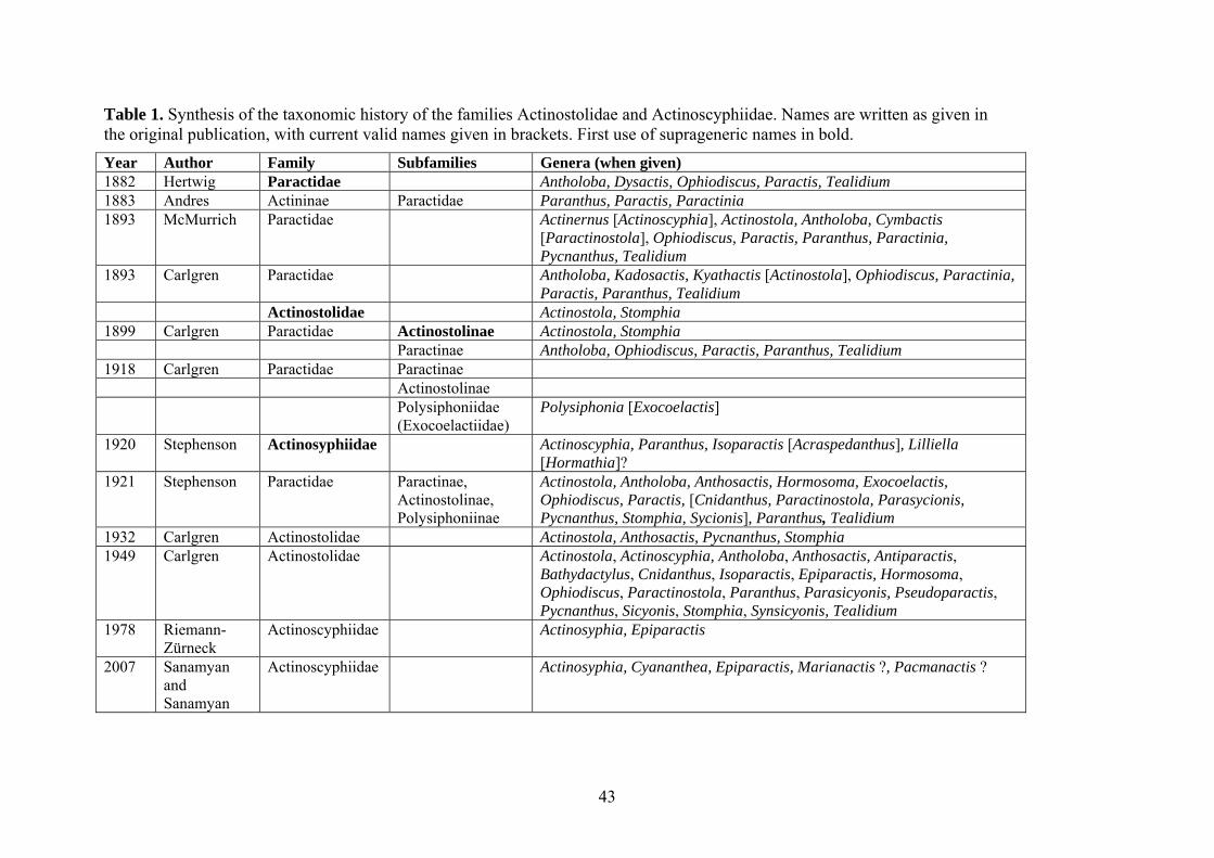
Table 1. Synthesis of the taxonomic history of the families Actinostolidae and Actinoscyphiidae. Names are written as given in the original publication, with current valid names given in brackets. First use of suprageneric names in bold. Year Author Family Subfamilies Genera (when given) 1882 Hertwig Paractidae
43
Antholoba, Dysactis, Ophiodiscus, Paractis, Tealidium 1883 Andres Actininae Paractidae Paranthus, Paractis, Paractinia 1893 McMurrich Paractidae Actinernus [Actinoscyphia], Actinostola, Antholoba, Cymbactis
[Paractinostola], Ophiodiscus, Paractis, Paranthus, Paractinia, Pycnanthus, Tealidium
1893 Carlgren Paractidae Antholoba, Kadosactis, Kyathactis [Actinostola], Ophiodiscus, Paractinia, Paractis, Paranthus, Tealidium
Actinostolidae Actinostola, Stomphia 1899 Carlgren Paractidae Actinostolinae Actinostola, Stomphia Paractinae Antholoba, Ophiodiscus, Paractis, Paranthus, Tealidium 1918 Carlgren Paractidae Paractinae Actinostolinae Polysiphoniidae
(Exocoelactiidae) Polysiphonia [Exocoelactis]
1920 Stephenson Actinosyphiidae Actinoscyphia, Paranthus, Isoparactis [Acraspedanthus], Lilliella [Hormathia]?
1921 Stephenson Paractidae Paractinae, Actinostolinae, Polysiphoniinae
Actinostola, Antholoba, Anthosactis, Hormosoma, Exocoelactis, Ophiodiscus, Paractis, [Cnidanthus, Paractinostola, Parasycionis, Pycnanthus, Stomphia, Sycionis], Paranthus, Tealidium
1932 Carlgren Actinostolidae Actinostola, Anthosactis, Pycnanthus, Stomphia 1949 Carlgren Actinostolidae Actinostola, Actinoscyphia, Antholoba, Anthosactis, Antiparactis,
Bathydactylus, Cnidanthus, Isoparactis, Epiparactis, Hormosoma, Ophiodiscus, Paractinostola, Paranthus, Parasicyonis, Pseudoparactis, Pycnanthus, Sicyonis, Stomphia, Synsicyonis, Tealidium
1978 Riemann-Zürneck
Actinoscyphiidae Actinosyphia, Epiparactis
2007 Sanamyan and Sanamyan
Actinoscyphiidae Actinosyphia, Cyananthea, Epiparactis, Marianactis ?, Pacmanactis ?
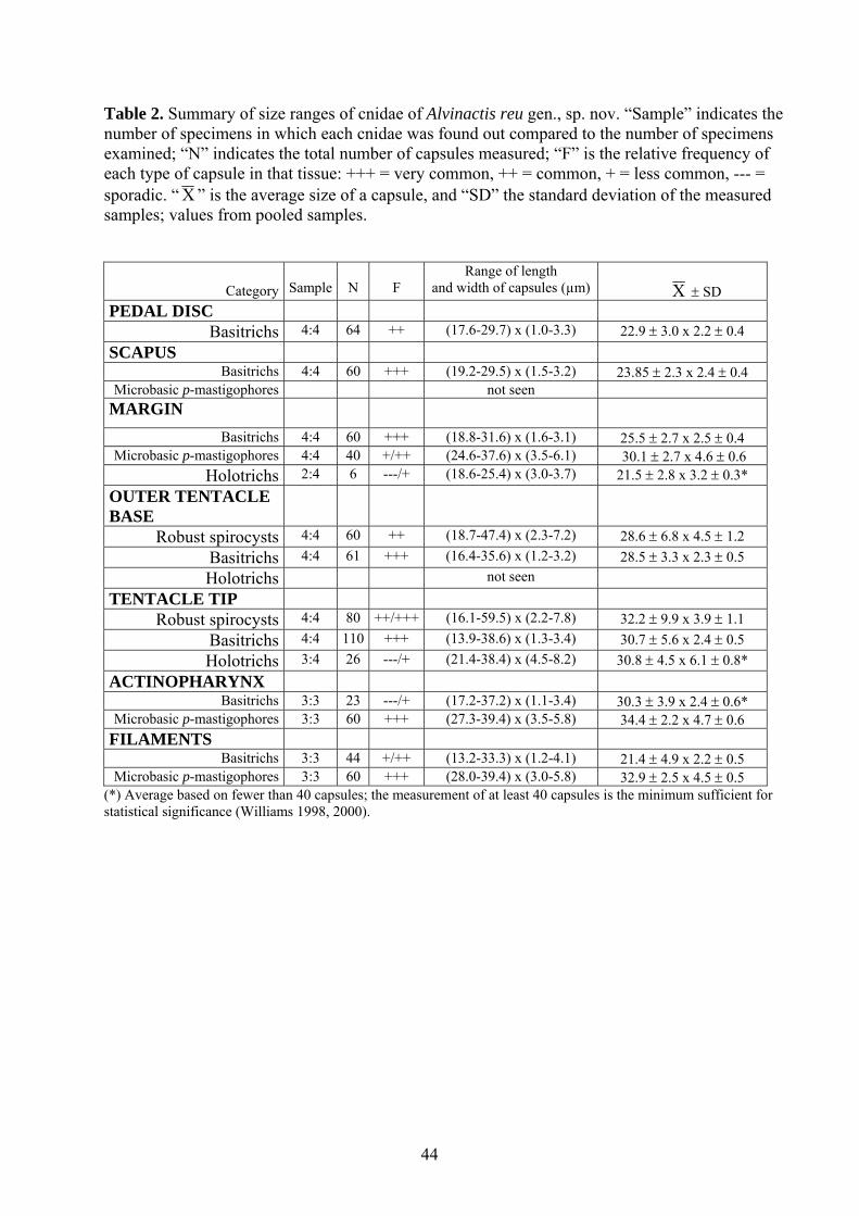
Table 2. Summary of size ranges of cnidae of Alvinactis reu gen., sp. nov. “Sample” indicates the number of specimens in which each cnidae was found out compared to the number of specimens examined; “N” indicates the total number of capsules measured; “F” is the relative frequency of each type of capsule in that tissue: +++ = very common, ++ = common, + = less common, --- = sporadic. “Χ ” is the average size of a capsule, and “SD” the standard deviation of the measured samples; values from pooled samples.
Category
Sample
N
F
Range of length and width of capsules (µm)
Χ ± SD
PEDAL DISC Basitrichs 4:4 64 ++ (17.6-29.7) x (1.0-3.3) 22.9 ± 3.0 x 2.2 ± 0.4
SCAPUS Basitrichs 4:4 60 +++ (19.2-29.5) x (1.5-3.2) 23.85 ± 2.3 x 2.4 ± 0.4
Microbasic p-mastigophores not seen MARGIN
Basitrichs 4:4 60 +++ (18.8-31.6) x (1.6-3.1) 25.5 ± 2.7 x 2.5 ± 0.4 Microbasic p-mastigophores 4:4 40 +/++ (24.6-37.6) x (3.5-6.1) 30.1 ± 2.7 x 4.6 ± 0.6
Holotrichs 2:4 6 ---/+ (18.6-25.4) x (3.0-3.7) 21.5 ± 2.8 x 3.2 ± 0.3* OUTER TENTACLE BASE
Robust spirocysts 4:4 60 ++ (18.7-47.4) x (2.3-7.2) 28.6 ± 6.8 x 4.5 ± 1.2 Basitrichs 4:4 61 +++ (16.4-35.6) x (1.2-3.2) 28.5 ± 3.3 x 2.3 ± 0.5 Holotrichs not seen
TENTACLE TIP Robust spirocysts 4:4 80 ++/+++ (16.1-59.5) x (2.2-7.8) 32.2 ± 9.9 x 3.9 ± 1.1
Basitrichs 4:4 110 +++ (13.9-38.6) x (1.3-3.4) 30.7 ± 5.6 x 2.4 ± 0.5 Holotrichs 3:4 26 ---/+ (21.4-38.4) x (4.5-8.2) 30.8 ± 4.5 x 6.1 ± 0.8*
ACTINOPHARYNX Basitrichs 3:3 23 ---/+ (17.2-37.2) x (1.1-3.4) 30.3 ± 3.9 x 2.4 ± 0.6*
Microbasic p-mastigophores 3:3 60 +++ (27.3-39.4) x (3.5-5.8) 34.4 ± 2.2 x 4.7 ± 0.6 FILAMENTS
Basitrichs 3:3 44 +/++ (13.2-33.3) x (1.2-4.1) 21.4 ± 4.9 x 2.2 ± 0.5 Microbasic p-mastigophores 3:3 60 +++ (28.0-39.4) x (3.0-5.8) 32.9 ± 2.5 x 4.5 ± 0.5
(*) Average based on fewer than 40 capsules; the measurement of at least 40 capsules is the minimum sufficient for statistical significance (Williams 1998, 2000).
44
Figure Legend Fig. 1. Strict consensus of 22 equally parsimonious trees (L=167, CI=0.29, RI=0.59) recovered from analysis of morphological data (Appendix 2). Numbers above the branches are Bremer support values. Characters supporting Actinostolina, Actinoscyphiidae, and Chemosynthina are indicated; numbers refer to Appendix 1. Fig. 2. External anatomy, preserved specimens Alvinactis reu gen., sp. nov. Scale in mm. A. Lateral view. B. Close up of column margin. Note verrucae inside crease in distal column (arrows). C. Top view. D. Perforate verrucae. Fig. 3. External anatomy and habitat, living Alvinactis reu gen., sp. nov. Fig. 4. Internal anatomy and histology, Alvinactis reu gen., sp. nov. Scale in μm. A. Basilar muscles. B. Cross-section through a tentacle of a contracted individual. Note gametogenic tissue in coelenteric space of tentacle. C. Longitudinal section through verrucae in distal column. D. Cross section through mesenteries below actinopharynx, showing size dimorphism of mesenteries if the first and third cycles. E. Longitudinal section through distal column, showing mesogleal sphincter and verruca (arrow). The space separating the distal and proximal portions of the sphincter is not present in all specimens or all sections from a single specimen. F. Maturing oocyte with trophonema (arrow). G. Cross section through parietal muscle of larger mesentery. H. Cross section through mesenteries below actinopharynx, showing diffuse retractor musculature. Abbreviations: ep, epidermis; ga, gastrodermis. Fig. 5. Cnidae of Alvinactis reu gen., sp. nov. A. Basitrich. B. Basitrich. C. Basitrich. D. Microbasic p-mastigophore. E. Gracile spirocyst. F. Basitrich. G. Holotrich. H. Robust spirocyst. I. Basitrich. J. Basitrich. K. Microbasic p-mastigophore. L. Basitrich. M. Microbasic p-mastigophore.
45
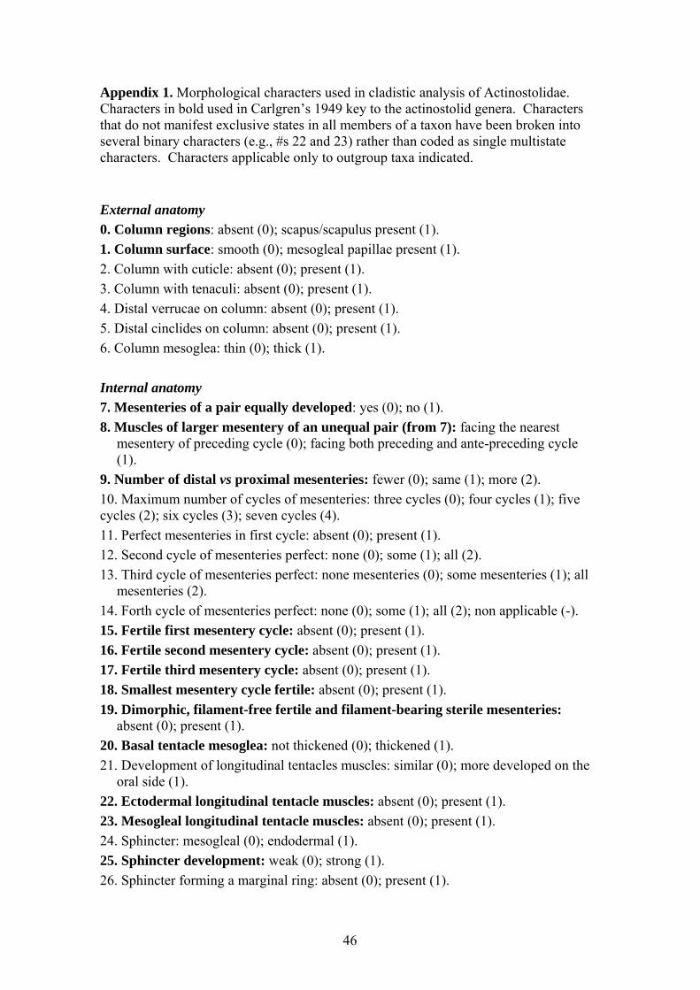
Appendix 1. Morphological characters used in cladistic analysis of Actinostolidae. Characters in bold used in Carlgren’s 1949 key to the actinostolid genera. Characters that do not manifest exclusive states in all members of a taxon have been broken into several binary characters (e.g., #s 22 and 23) rather than coded as single multistate characters. Characters applicable only to outgroup taxa indicated. External anatomy 0. Column regions: absent (0); scapus/scapulus present (1). 1. Column surface: smooth (0); mesogleal papillae present (1). 2. Column with cuticle: absent (0); present (1). 3. Column with tenaculi: absent (0); present (1). 4. Distal verrucae on column: absent (0); present (1). 5. Distal cinclides on column: absent (0); present (1). 6. Column mesoglea: thin (0); thick (1). Internal anatomy 7. Mesenteries of a pair equally developed: yes (0); no (1). 8. Muscles of larger mesentery of an unequal pair (from 7): facing the nearest
mesentery of preceding cycle (0); facing both preceding and ante-preceding cycle (1).
9. Number of distal vs proximal mesenteries: fewer (0); same (1); more (2). 10. Maximum number of cycles of mesenteries: three cycles (0); four cycles (1); five cycles (2); six cycles (3); seven cycles (4). 11. Perfect mesenteries in first cycle: absent (0); present (1). 12. Second cycle of mesenteries perfect: none (0); some (1); all (2). 13. Third cycle of mesenteries perfect: none mesenteries (0); some mesenteries (1); all
mesenteries (2). 14. Forth cycle of mesenteries perfect: none (0); some (1); all (2); non applicable (-). 15. Fertile first mesentery cycle: absent (0); present (1). 16. Fertile second mesentery cycle: absent (0); present (1). 17. Fertile third mesentery cycle: absent (0); present (1). 18. Smallest mesentery cycle fertile: absent (0); present (1). 19. Dimorphic, filament-free fertile and filament-bearing sterile mesenteries:
absent (0); present (1). 20. Basal tentacle mesoglea: not thickened (0); thickened (1). 21. Development of longitudinal tentacles muscles: similar (0); more developed on the
oral side (1). 22. Ectodermal longitudinal tentacle muscles: absent (0); present (1). 23. Mesogleal longitudinal tentacle muscles: absent (0); present (1). 24. Sphincter: mesogleal (0); endodermal (1). 25. Sphincter development: weak (0); strong (1). 26. Sphincter forming a marginal ring: absent (0); present (1).
46
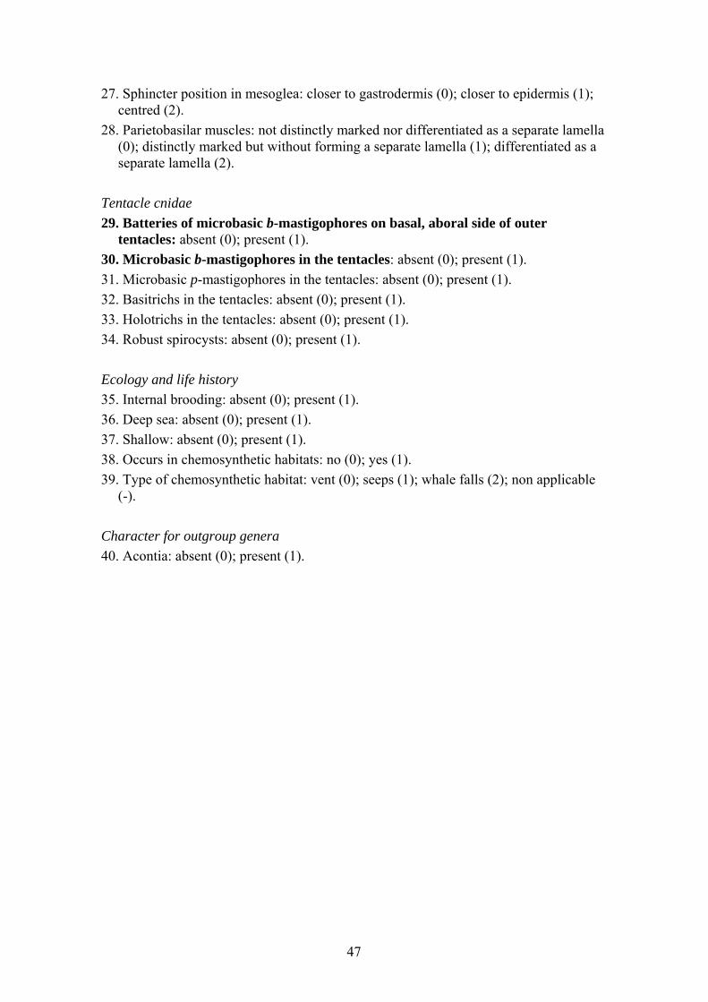
27. Sphincter position in mesoglea: closer to gastrodermis (0); closer to epidermis (1); centred (2).
28. Parietobasilar muscles: not distinctly marked nor differentiated as a separate lamella (0); distinctly marked but without forming a separate lamella (1); differentiated as a separate lamella (2).
Tentacle cnidae 29. Batteries of microbasic b-mastigophores on basal, aboral side of outer
tentacles: absent (0); present (1). 30. Microbasic b-mastigophores in the tentacles: absent (0); present (1). 31. Microbasic p-mastigophores in the tentacles: absent (0); present (1). 32. Basitrichs in the tentacles: absent (0); present (1). 33. Holotrichs in the tentacles: absent (0); present (1). 34. Robust spirocysts: absent (0); present (1). Ecology and life history 35. Internal brooding: absent (0); present (1). 36. Deep sea: absent (0); present (1). 37. Shallow: absent (0); present (1). 38. Occurs in chemosynthetic habitats: no (0); yes (1). 39. Type of chemosynthetic habitat: vent (0); seeps (1); whale falls (2); non applicable
(-). Character for outgroup genera 40. Acontia: absent (0); present (1).
47
48
Appendix 2: Morphological character state distributions for outgroups and ingroups genera. Dash indicates that the corresponding state is unknown or inapplicable. Outgroup genera in bold. See appendix 1 for character list.
0 1234 5 6 7 8 9 10
11
12 13
14
15
16
17
18
19
20
21
22
23
24
25
26
27
28
29
30
31
32
33
34
35
36
37
38
39
40
Actinoscyphia 0 0000 0 10 - 1 2 1 2 0 0 1 1 0 0 0 1 0 1 1 0 0 0 0 0 0 0 0 1 1 1 0 1 0 0 - 0Bathyphellia 1 1110 0 - 0 - 0 0 1 0 0 - 1 0 0 0 0 0 0 1 0 0 1 0 2 2 0 0 0 1 0 1 0 1 0 0 - 1Epiactis 0 1000 0 0 0 - 0 2 1 2 2 0 1 1 1 0 0 0 0 1 0 1 1 0 - 2 0 0 1 0 0 0 1 1 1 0 - 0Hormathia 1 1100 0 1 0 - 1 1 1 0 0 0 0 1 1 1 0 0 0 1 0 0 0 0 0 1 0 0 0 1 0 1 0 1 1 0 - 1Kadosactis 1 1110 1 1 0 - 1 0 1 2 0 - 1 1 0 0 0 1 - 1 0 0 1 1 0 1 0 0 1 1 0 1 0 1 0 0 - 1Actinostola 0 0000 0 1 1 0 0 4 1 2 1 0 0 0 1 1 0 1 0 0 1 0 0 0 0 0 0 1 1 1 0 0 1 1 1 0 - 0Alvinactis 0 0001 1 0 0 - 1 1 1 0 0 0 1 1 1 0 0 0 0 1 0 0 1 1 0 0 0 0 0 1 1 1 0 1 0 1 0 0Antholoba 0 0000 0 1 0 - 2 4 1 2 2 1 0 0 0 1 0 0 0 1 1 0 0 0 0 0 0 0 0 1 0 0 1 0 1 0 - 0Anthosactis janmayeni
0 0000 0 1 0 - - 1 1 1 0 0 1 1 1 1 0 - 1 1 0 0 1 1 1 0 1 1 0 1 0 0 0 1 1 0 - 0
Anthosactis nomados
0 0000 0 0 0 - 0 0 1 2 0 - 1 1 0 0 0 0 - 1 0 0 1 0 2 - - 1 0 1 0 1 0 1 0 0 - 0
Anthosactis pearseae
0 0000 0 1 0 - 0 1 1 0 0 0 1 1 0 0 0 0 0 1 0 0 0 0 0 0 0 0 0 1 1 1 1 1 0 1 2 0
Antiparactis 0 0000 0 0 0 - 0 1 1 0 0 0 0 1 1 0 0 - 0 1 0 0 1 0 2 0 0 - - - - 0 0 0 1 0 - 0Bathydactylus 1 0000 1 1 0 - 0 0 1 0 0 - 1 1 0 0 0 0 0 1 0 0 1 1 - 1 0 0 0 1 0 0 0 1 0 - - 0Cnidanthea 0 1000 0 - 0 - 1 0 1 0 0 - 1 1 0 0 0 0 0 1 0 0 1 0 2 0 0 0 1 1 0 - 0 0 1 0 - 0Cnidanthus 0 0000 0 0 0 - 0 1 1 2 1 0 1 1 1 0 0 0 0 0 1 0 1 0 0 0 0 1 0 1 0 - 1 0 1 0 - 0Cyananthea 0 0000 1 0 0 - 2 2 1 2 0 0 1 1 1 0 0 0 0 1 0 0 1 1 0 0 0 0 1 1 1 1 0 1 0 1 0 0Epiparactis 0 0000 0 1 0 - - 2 1 0 0 0 - - - - 0 0 0 1 0 0 0 0 2 0 0 0 0 1 0 1 0 1 0 0 - 0Hadalanthus 1 1110 0 0 0 - 1 1 1 0 0 0 1 1 - - 0 0 0 1 0 0 1 0 0 0 0 0 1 1 0 - 0 1 0 - - 0Hormosoma 0 0000 0 1 0 - 1 1 1 2 2 0 1 1 1 1 0 0 0 0 1 0 1 1 1 2 1 1 0 0 0 0 1 0 1 0 - 0Maractis 0 0000 0 0 0 - 1 1 1 0 0 0 1 1 1 1 0 0 0 1 0 0 1 0 0 1 0 0 0 1 1 1 0 1 0 1 0 0Marianactis 0 0000 1 1 0 - 1 1 1 0 0 0 1 1 1 1 0 0 0 1 0 0 1 0 0 2 0 0 1 1 0 1 0 1 0 1 0 0Ophiodiscus 0 0000 0 1 1 0 - 1 1 2 0 0 0 0 0 1 1 1 1 0 1 0 0 0 - 0 - - - - - - 0 1 0 - - 0Pacmanactis 0 0001 1 1 0 - 1 1 1 2 0 0 1 1 0 0 0 0 0 1 0 0 0 1 1 0 1 1 1 1 1 1 0 1 0 1 0 0Paractinostola 0 0000 0 1 1 0 2 4 1 2 2 1 0 0 1 1 0 1 1 0 1 0 0 0 0 0 0 1 0 1 0 - 0 1 1 0 - 0Paranthosactis 0 0000 - 0 0 - 1 1 1 0 0 0 1 1 1 0 0 0 0 1 0 0 1 1 0 0 0 0 1 1 1 1 0 1 0 1 0 0Paranthus 0 0001 0 - 0 - 2 1 1 2 0 0 1 1 0 0 0 - - 1 0 0 0 0 0 0 0 0 1 1 0 0 0 0 1 0 - 0Parasicyonis 0 0000 0 1 1 1 0 2 1 2 2 1 0 0 0 1 0 0 1 0 1 0 1 0 0 0 0 0 0 1 0 - 0 1 0 0 - 0Pseudoparactis 1 0000 0 - 0 - - 1 - - - - 0 1 1 - 0 - - 1 0 0 0 0 2 0 - - - - - - 0 0 1 0 - 0Pycnanthus 0 0000 0 1 0 - 0 2 1 2 1 0 0 0 1 1 0 1 1 0 1 0 1 0 0 1 0 1 1 1 0 0 1 1 0 0 - 0Sicyonis 0 0000 0 1 1 1 0 3 1 2 1 0 0 0 0 1 0 1 1 0 1 0 0 0 0 1 0 0 0 1 0 0 0 1 1 0 - 0Stomphia 0 0000 0 1 1 0 0 1 1 2 1 0 0 0 1 1 0 0 0 1 1 0 0 0 0 0 1 1 0 1 0 0 1 1 1 0 - 0Synsicyonis 0 0000 0 1 1 1 2 1 1 2 1 0 0 0 1 1 0 1 1 0 1 0 0 0 - - - - - - - - 0 1 0 0 - 0
Tealidium 1 1000 0 1 0 - - 1 1 1 0 0 1 1 1 1 0 1 1 1 0 0 1 1 2 0 1 1 0 1 0 - 0 1 0 0 - 0
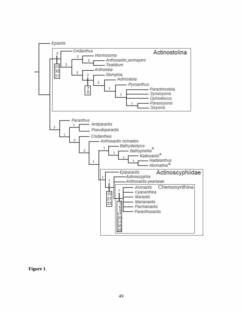
Figure 1.
49
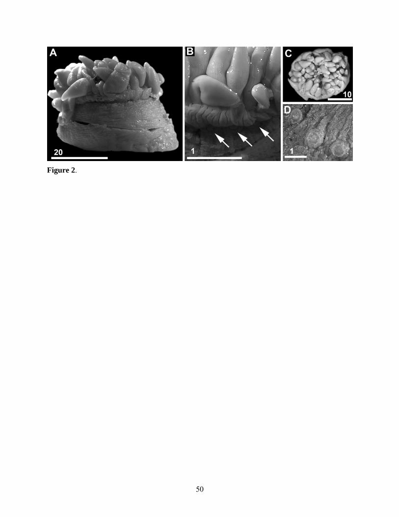
Figure 2.
50
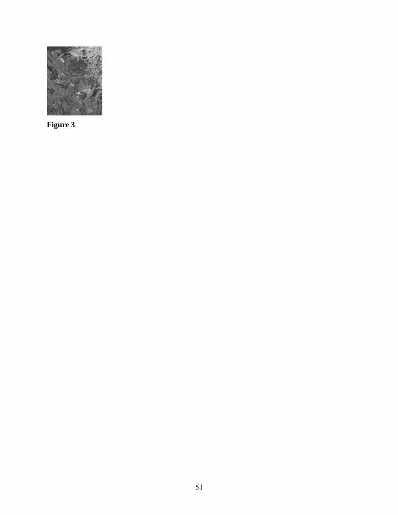
Figure 3.
51
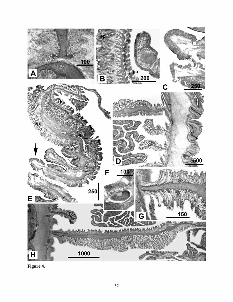
Figure 4.
52
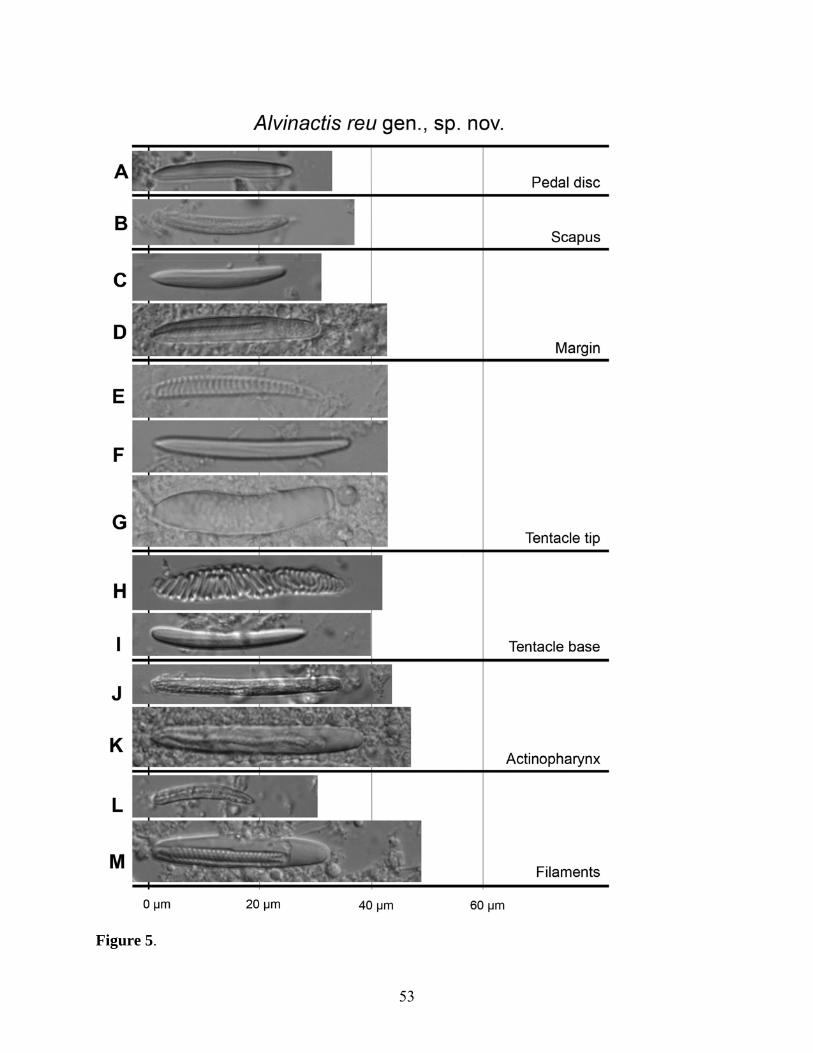
Figure 5.
53
Related Documents