TESIS DOCTORAL INTERNACIONAL Distribución mutacional y correlación genotipo-fenotipo del síndrome de Charcot-Marie-Tooth en la Comunidad Valenciana. Doctorando: Rafael Sivera Mascaró Directora de tesis: Maria Teresa Sevilla Mantecón. PROGRAMA DE DOCTORADO EN MEDICINA DEPARTAMENTO DE MEDICINA UNIVERSIDAD DE VALENCIA 2015

Welcome message from author
This document is posted to help you gain knowledge. Please leave a comment to let me know what you think about it! Share it to your friends and learn new things together.
Transcript

TESIS DOCTORAL INTERNACIONAL
Distribución mutacional y correlación genotipo-fenotipo del síndrome de
Charcot-Marie-Tooth en la Comunidad Valenciana.
Doctorando: Rafael Sivera Mascaró
Directora de tesis: Maria Teresa Sevilla Mantecón.
PROGRAMA DE DOCTORADO EN MEDICINA
DEPARTAMENTO DE MEDICINA
UNIVERSIDAD DE VALENCIA
2015


AGRADECIMIENTOS
A Teresa por todo el camino que me ha ayudado a recorrer durante estos años, y porque
sencillamente sin ella esta tesis no hubiese sido posible.
A Juan porque aprender neuromuscular de él es un lujo del que siempre estaré agradecido.
Al resto del servicio de Neurología de La Fe, entré esperando compañeros y me llevo más
de un amigo.
A Carmina y Paco por su trabajo y dedicación y por ser capaces de poner cordura en
momentos de estrés.
Al resto de equipo del CIPF porque hacen que la medicina translacional sea un placer.
A mis amigos de Godella y de medicina por ser expertos oficiosos en CMT luego de la
brasa que les he dado.
A mis padres y hermana, por ser uno de mis grandes apoyos y por ser en parte
responsables del ‘gusanillo’ de querer ser un doctor sin comillas.
A Rafa y Marina por ser mi alegría y motivación, por poner esa cara seria cuando le
explicaba que papa tenía que trabajar en la tesis.
A María, por compartir mi vida y mis preocupaciones, por quererme. Por todas las horas
delante de ordenador y cuidando a los peques mientras yo estaba enfrascado.


ÍNDICE:
1) Introducción página 3
a. El nervio periférico
i. Estructura
ii. Conducción nerviosa
b. Neuropatía periférica
c. Polineuropatía hereditaria sensitiva y motora (enfermedad de Charcot-
Marie-Tooth o CMT)
i. Clasificaciones
ii. Mecanismos biológicos
iii. Características clínicas; correlación genotipo-fenotipo
2) Hipótesis de trabajo y objetivos página 77
a. Hipótesis de trabajo
b. Objetivos
i. Objetivo general
ii. Objetivos específicos
3) Material y métodos página 81
a. Población
b. Evaluación clínica
c. Protocolo neurofisiológico
d. Estudios anatomopatológicos

e. Resonancia magnética muscular
f. Análisis genéticos
g. Aspectos éticos
4) Resultados página 91
a. Artículo I
b. Artículo II
c. Artículo III
d. Artículo IV
e. Algoritmos diagnósticos
5) Discusión página 137
a. Artículo I
i. Resumen de resultados
ii. Discusión
b. Artículo II
i. Resumen de resultados
ii. Discusión
c. Artículo III
i. Resumen de resultados
ii. Discusión
d. Artículo IV
i. Resumen de resultados
ii. Discusión
e. Algoritmos diagnósticos

6) Conclusiones página 151
a. En castellano
b. En inglés
7) Bibliografía página 157
8) Apéndice página 191
a. Protocolo clínico del Hospital La Fe para pacientes con CMT


ÍNDICE TABLAS Y FIGURAS:
Tablas:
1) Clasificación de los tipos de fibras nerviosas. página 5
2) Clasificación etiológica de las polineuropatías. página 10
3) Neuropatías hereditarias en el contexto de enfermedades
multisistema. página 12
4) Clasificación de CMT axonales y desmielinizantes. página 15
5) Clasificación de CMT intermedias. página 16
6) Distribución mutacional en series clínicas representativas. página 27
Figuras
1) Estructura macroscópica del nervio periférico. página 4
2) Potencial de membrana. página 6
3) Potencial de acción muscular. página 7
4) Propagación del potencial de acción. página 8
5) Esquema de las proteínas patógenas implicadas en CMT y la
diversidad de funciones. página 26
6) Paciente afecto de CMT1A. página 29
7) Anatomía patológica de nervio sural de un paciente con CMT1A. página 30
8) Atrofia de predominio en la eminencia tenar en un paciente varón
con CMTX1 página 33

9) Corte semifino de un paciente con un neuropatía hipomielinizante
congénita por mutación en MPZ. página 36
10) Paciente con mutaciones AR en GDAP1. página 39
11) Mujer joven con un fenotipo leve ocasionado por una mutación
AD en GDAP1 . página 40
12) Corte semifino de un paciente con una mutción en cl gen NEFL. página 43
13) Algoritmo para el diagnóstico genético en CMT desmielinizante. página 131
14) Algoritmo para el diagnóstico genético en CMT intermedio. página 132
15) Algoritmo para el diagnóstico genético en CMT axonal. página 133

ABREVIATURAS EMPLEADAS:
AD: autosómico dominante
ADN: ácido desoxirribonucleico
AED: atrofia espinal distal
AR: autosómico recesivo
cDNA: ADN complementario
CIDP: polineuropatía inflamatoria desmielinizante crónica
CK: creatinquinasa
CMAP: potencial de acción muscular compuesto
CMT: enfermedad de Charcot-Marie-Tooth
CMTNS: CMT Neuropathy Score
CMTNS2: CMT Neuropathy Score segunda version
dHMN: neuropatía hereditaria motora distal
DHPLC: Denaturing High Performance Liquid Chromatography
D/I: derecha/izquierda
EMG: electromiograma
FDS: Functional Disability Scale
HC: historia clínica
H-H: transmisión hombre-hombre
HSAN: neuropatía hereditaria sensitiva y autonómica
HTA: hipertensión arterial
LCR: líquido cefalorraquídeo

MID: miembro inferior derecho
MII: miembro inferior izquierdo
MMII: miembros inferiores
MMSS: miembros superiores
MCR: Medical Research Council
MLPA: Multiplex Ligation-dependent Probe Amplification
MSD: miembro superior derecho
MSI: miembro superior izquierdo
NHPP: neuropatía hereditaria con predisposición a parálisis por compresión
NARP: neuropatía, ataxia y retinitis pigmentosa
ORF: open reading frame
PCR: Polymerase Chain Reaction
PESS: potenciales evocados somatosensoriales
R. aquíleo: reflejo aquíleo
RCP: reflejo cutáneoplantar
RM: resonancia magnética
R. rotuliano: reflejo rotuliano
SNC: sistema nervioso central
STIR: Short T1 Inversion Recovery
VC: velocidad de conducción
VCMM: velocidad de conducción motora del nervio mediano
vHIT: video impulse test

1) INTRODUCCIÓN


3
a) El nervio periférico
El sistema nervioso está constituido por el sistema nervioso central (SNC) (encéfalo y
médula espinal) y el sistema nervioso periférico (raíces nerviosas, nervios periféricos,
unión neuromuscular y músculos). Los nervios periféricos son las estructuras
nerviosas que conducen el estímulo eléctrico entre los órganos periféricos y el sistema
nervioso central. Dicho estímulo puede ser aferente, si viaja de la periferia al encéfalo
(por ej. estímulos sensitivos) o eferente, si viaja desde el sistema nervioso central al
órgano efector (por ej. estímulos motores). La mayoría de nervios periféricos
contienen fibras nerviosas motoras, sensitivas y vegetativas y, por tanto, conducen
tanto estímulos aferentes como eferentes.
i. Estructura:
Los nervios periféricos son estructuras cordonales compuestas por un conjunto de
fibras nerviosas agrupadas en fascículos y rodeadas por una membrana llamada
epineuro. Los fascículos nerviosos están delimitados por una membrana denominada
perineuro y están compuestos por fibras nerviosas y tejido conectivo (endoneuro). Las
fibras nerviosas están formadas por el axón de la neurona (motora, sensitiva o
vegetativa) y la célula de Schwann. Se distinguen fibras nerviosas mielínicas si el
axón está envuelto por prolongaciones de células de Schwann formado vainas de
mielina y amielínicas si las células de Schwann están en contacto con varios axones
sin llegar a formar mielina (fig. 1). La vaina de mielina proporciona aislamiento al
axón del exterior, protegiéndolo de agentes nocivos y siendo importante también en el
proceso de regeneración nerviosa tras una lesión (1).

4
Figura 1: Estructura macroscópica del nervio periférico. Imagen adaptada
de Texto y Atlas de Anatomía Prometheus, volumen 3. Versión:
http://www.med.ufro.cl.
Las fibras nerviosas pueden clasificarse según la función que desempeñan,
la presencia de mielina, el diámetro axonal o la velocidad de conducción
(2) (tabla 1).

5
Tabla 1: Clasificación de los tipos de fibras nerviosas.
TIPO DE FIBRA FUNCIÓN DIÁMETRO
(μm)
VELOCIDAD
(m/s)
MIELÍNICAS
A IA α Motor somático
Propiocepción
(husos
musculares)
12-20 70-120
IB α Propiocepción
(órganos de
Golgi)
5-12 30-70
II β
γ
Tacto, presión
Motor (husos
musculares)
5-12
3-6
30-70
15-30
III δ Dolor (rápido),
frío, tacto
2-5
12-30
B Preganglionares
autonómicas
<3 3-15
AMIELÍNICAS
C IV Raíz dorsal
Vegetativas
Dolor (lento),
mecanoceptores,
temperatura
Postganglionares
autonómicas
0.4-1.2
0.3-1.3
0.5-2
0.7-2.3

6
ii. Conducción nerviosa:
La conducción nerviosa es la transmisión de un estímulo eléctrico a lo largo de una
fibra nerviosa. La misma se basa en que el axón nervioso está polarizado, es decir,
mantiene una diferencia de potencial entre el interior y el exterior celular, y es
excitable. Dicha diferencia de potencial se produce por distintas concentraciones
iónicas extra e intracelulares (fig. 2). Ante el estímulo adecuado se produce un cambio
en las concentraciones iónicas provocando la despolarización de la célula.
Figura 2: Potencial de membrana. Extraído de Manual de Electromiografía
clínica. Prous science, S.A., 2000.

7
Si la despolarización de la membrana alcanza un umbral determinado (alrededor de -45
mV) se produce un potencial de acción, que provoca una despolarización de hasta + 45
mV (fig. 3).
Figura 3: Potencial de acción muscular.
Dicha despolarización ejerce un estímulo sobre la membrana celular colindante,
produciéndose una propagación del potencial de acción por la fibra nerviosa (fig. 4). La
velocidad de propagación del potencial de acción por la fibra nerviosa se conoce como
velocidad de conducción y depende, mayormente, del diámetro axonal y de la
mielinización de la fibra nerviosa. En las fibras mielínicas el potencial de acción se
propaga de una manera ‘saltatoria’, despolarizándose únicamente los espacios entre las
vainas de mielina (nodos de Ranvier), siendo por tanto más rápida la conducción
nerviosa. En cualquier caso, en ambos tipos de fibras, a mayor diámetro axonal, mayor
velocidad de conducción.

8
Figura 4: Propagación del potencial de acción y conducción nerviosa en fibras
mielínicas y amielínicas. Extraído de Manual de Electromiografía clínica. Prous
science, S.A., 2000.
b) Neuropatía periférica
El término neuropatía periférica se refiere a una alteración de la función o de la
estructura de las neuronas periféricas que componen la fibra nerviosa. Se trata de un
grupo de enfermedades relativamente frecuentes con una incidencia anual estimada
Nodo de Ranvier
Axón mielínico
Axón amielínico

9
entre 710-2384/100.000 habitantes (4, 5) y una prevalencia en España de cerca del 2-
8% (6). La lesión puede producirse de una manera focal en un único nervio
(mononeuropatía), indicando la presencia de un proceso local causante. También
puede producirse un daño en distintas localizaciones de nervios no contiguos de una
manera secuencial o simultánea. En este caso se conoce como mononeuropatía
múltiple y la práctica totalidad de los casos tienen una etiología adquirida,
habitualmente, con una fisiopatología inflamatoria (sistémica o local), infecciosa, o
infiltrativa (7).
Cuando la lesión del nervio periférico se produce de una manera bilateral y simétrica,
se conoce como polineuropatía. Clínicamente, cursa con déficits motores, sensitivos
y/o vegetativos simétricos con mayor afectación distal que proximal. El abanico
etiológico en este tipo de enfermedades es extremadamente amplio (tabla 2) y para
tratar de acotarlo es importante tener en cuenta la rapidez de instauración y determinar
si el daño inicial se produce en el axón nervioso o en la mielina circundante
(polineuropatías axonales y desmielinizantes respectivamente) (8). Para esto último es
necesaria la realización de estudios electrofisiológicos de conducción nerviosa y, en
ocasiones, electromiografía de aguja (9, 10).

10
AXONAL DESMIELINIZANTE
AGUDA
Inflamatorias: Formas axonales del
Síndrome de Guillain Barré
Porfirias
Tóxicos: Talio, organofosforados, taxol, etc.
Fármacos: Vincristina, inhibidores de la
transcriptasa inversa, etc.
Inflamatoria: Síndrome Guillain-Barré
Infecciosa: Difteria
Tóxicos: Arsénico, suramida, etc.
CRÓNICA
Diabetes Mellitus
Alcohol
Sistémicas: uremia, hepatopatía,
hipotiroidismo
Infecciones: VIH, lepra, enf. De Lyme
Tóxicos: Plomo, mercurio, arsénico,
organofosforados, talio, acrilamida, etc.
Fármacos: isoniacida, amiodarona, fenitoína,
vincristina, metronidazol, talidomida, etc.
Deficiencias de vits. B1, B6, B12, E
Asociadas a disproteinemia
Amiloidosis primaria
Hereditarias
Paraneoplásicas
Idiopáticas
Inflamatoria: Polineuropatía desmielinizante
inflamatoria crónica (CIDP), neuropatía motora
multifocal
Asociadas a disproteinemia
Amiloidosis primaria
Fármacos: amiodarona
Hereditarias
Paraneoplásicas
Hepatopatía crónica
Diabetes mellitus
Hipotiroidismo
Tabla 2: Clasificación etiológica de las polineuropatías.

11
En las polineuropatías hereditarias el agente causal que provoca la aparición de la
enfermedad es un defecto genético. A medida que se ha desarrollado la genética
molecular, el conocimiento sobre las mismas se ha ido multiplicando y el número de
genes y mutaciones causales descritas ha aumentado exponencialmente. En cualquier
caso, cabe diferenciar las neuropatías hereditarias en las que la neuropatía es la única o
principal manifestación de la enfermedad, de aquellas en las que forma parte de una
constelación más amplia de manifestaciones neurológicas o sistémicas (tabla 3). En el
primero de estos grupos se distinguen neuropatías hereditarias con afectación sensitiva y
motora (enfermedad de Charcot-Marie-Tooth o CMT), otras con afectación
exclusivamente motora (neuropatía hereditaria motora distal o dHMN), y aquellas con
afectación exclusivamente sensitiva y autonómica (neuropatía hereditaria sensitiva y
autonómica o HSAN). Aparte, existen 2 entidades con un curso recurrente o paroxístico:
neuropatía hereditaria con predisposición a las parálisis por compresión (NHPP) y la
neuralgia amiotrófica hereditaria (11). El segundo grupo está resumido en la tabla 3.

12
Polineuropatía amiloidótica familiar Asociada a ataxia cerebelosa
– Por mutaciones en el gen de la transtirretina
(Tipo 1 y 2)
– Tipo 3 (Van Allen)
– Tipo 4 (Finlandesa)
– Con herencia autosómico recesiva
(ataxia de Friedrich, ataxia con apraxia
óptica tipo 1 y 2, ataxia asociada a déficit
de vitamina E, Ataxia espástica AR de
Charlevoix Saguenay…)
– Con herencia autosómico dominante
Enfermedad del mecanismo lipídico Porfiria
– Leucodistrofias (leucodistrofia
metacromática, adrenomieloneuropatía)
– Alteraciones metabólicas del ácido fitánico
(enf. Refsum)
– Deficiencias de lipoproteínas
– Deficiencia de -galactosidasa (enf. Fabry)
– Alteración del metabolismo del colestanol
– Aguda intermitente
– Coproporfiria
– Variegata
– Deficiencia de ALA deshidrogenasa
Alteración de la reparación del ADN Misceláneo
– Ataxia telangiectasia
– Xeroderma pigmentosum
– Síndrome de Cockayne
– Neuroacantocitosis
Asociadas con enfermedades mitocondriales
Tabla 3: Neuropatías hereditarias en el contexto de enfermedades
multisistema.

13
a. Polineuropatía hereditaria sensitiva y motora (enfermedad de Charcot-
Marie-Tooth)
El síndrome de Charcot-Marie-Tooth (CMT) es el epónimo con el que se conoce a las
polineuropatías hereditarias sensitivas y motoras. Es la forma de neuropatía hereditaria
más frecuente, con una prevalencia global descrita entre 15,2-40 casos/100.000 habitantes
(12, 13). En España, la prevalencia estimada es de 28.2 casos/100.000 habitantes gracias a
un estudio epidemiológico realizado en la región de Cantabria (14).
Se trata de un conjunto heterogéneo de enfermedades. En estos momentos ya se conocen
mutaciones causales en más de 70 genes o loci y este número está constantemente
ampliándose (http://www.molgen.ua.ac.be/CMTMutations/,
http://neuromuscular.wustl.edu/). La herencia de la enfermedad puede ser autosómico
dominante (AD), autosómico recesiva (AR) o ligada al cromosoma X (dominante o
recesiva). Incluso hay genes, como el GDAP1, en el que determinadas mutaciones se
heredan de forma AD y otras de forma AR (15, 16).
El curso clínico es variable y depende tanto de la mutación subyacente como de otras
variables epigenéticas y/o ambientales. Como conjunto, la enfermedad suele tener un
inicio infantil o juvenil, apareciendo debilidad e hipoestesia distal progresiva en las
extremidades, disminución o abolición de los reflejos profundos y ciertas deformidades
óseas distales (pies cavos, dedos en martillo, etc.) (17).

14
i. Clasificaciones:
Desde su descubrimiento, el enfoque de esta enfermedad ha ido evolucionando
sustancialmente, sobre todo con el desarrollo del conocimiento de la medicina molecular.
En cualquier caso, la clasificación actual parte de los estudios de Dyck y Lambert, y
posteriormente de Harding y Thomas, que se basaban en las características clínicas,
neurofisiológicas e histológicas (18,19). Así se diferencian 2 grandes grupos, que siguen
estando vigentes:
1) CMT desmielinizante (CMT1 si tiene una herencia AD y CMT4 si es AR): Existe un
enlentecimiento de las velocidades de conducción (VC) en los estudios de conducción
nerviosa con amplitudes normales y/o en la biopsia de nervio sural se observa
desmielinización y proliferación de células de Schwann formando estructuras
concéntricas denominadas bulbos de cebolla.
2) CMT axonal (CMT2): Existe una caída de la amplitud de potenciales, sin
enlentecimiento de las velocidades de conducción y/o la patología muestra hallazgos de
degeneración y regeneración axonal.
Se acordó que el límite de separación entre ambos grupos podía establecerse
considerando la velocidad de conducción nerviosa motora del nervio mediano (VCMM).
Si esta es menor a 38 m/s se considera CMT desmielinizante y si es > a 38 m/s se
considera una variante axonal (19).
3) CMT intermedia: Se trata de un término útil a nivel práctico pero difícil de definir con
exactitud. Generalmente, se considera que una familia con CMT es intermedia si sus
miembros tienen una VCMM entre 30-45 m/s (20). Los hallazgos patológicos son
variables pudiendo mostrar características axonales y/o desmielinizantes.

15
El desarrollo de las técnicas de genética molecular ha revolucionado el campo de las
neuropatías hereditarias, descubriéndose en los últimos 20 años la gran heterogeneidad
genética que subyace a este síndrome. A día de hoy, se han descrito más de 70 genes cuya
disfunción puede causar CMT y la lista continúa creciendo. Por este motivo, el
diagnóstico molecular se incorporó a la clasificación de la enfermedad, utilizando letras
consecutivas que corresponden con los distintos genes o locus responsables (21). Dicha
clasificación actualizada puede observarse en las tablas 4 y 5 adaptadas de (22).
CMT1/CMT4
Subtipo Gen Herencia OMIM
CMT2
Subtipo Gen Herencia OMIMCMT1A PMP22 AD 601097 CMT2A1 KIF1B AD 605995 CMT1B MPZ AD 159440 CMT2A2 MFN2 AD 608507 CMT1C LITAF AD 603795 CMT2B RAB7 AD 602298 CMT1D EGR2 AD 129010 CMT2B1 LMNA AR 150330 CMT1E PMP22 AD 601097 CMT2B2 MED25 AR 610197 CMT1F NEFL AD 162280 CMT2C TRPV4 AD 605427
FBLN5 AD 614434 CMT2D GARS AD 600287 CMT2E NEFL AD 162280
CMT4A GDAP1 AR 606598 CMT2F HSPB1 AD 602195 CMT4B1 MTMR2 AR 603557 CMT2G 12q-q13.2 CMT4B2 MTMR13 AR 607697 CMT2H GDAP1 AR 606598 CMT4B3 MTMR5 AR 603560 CMT2I/J MPZ AD 159440 CMT4C SH3TC2 AR 608206 CMT2K GDAP1 AD 606598 CMT4D NDRG1 AR 605262 CMT2L HSPB8 AD 608014 CMT4E ERG2 AR 129010 CMT2M DNM2 AD 602378 CMT4E MPZ AR 159440 CMT2N AARS AD 601065 CMT4F PRX1 AR 605725 CMT2O DYNC1H1 AD 600112 CMT4G HK1 AR 142600 CMT2P LRSAM1 AD/AR 610933 CMT4H FGD4 AR 611104 CMT2Q DHTKD1 AD/AR 610933 CMT4J FIG4 AR 609390 TFG AD 602498
MARS AD 156560 HARS AD 142810 HINT1 AR 601314 TRIM2 AR 614141 MT-ATP6 Mitoc

16
Tablas 4 y 5: Clasificación actualizada de CMT con genotipo subyacente y OMIM.
La complejidad genética del CMT no sólo radica en el número de genes implicados, sino
también en la variabilidad fenotípica de muchos de ellos y el solapamiento con otras
enfermedades. De hecho, hay varios genes (MPZ, NEFL, etc.) en los que existen
mutaciones que provocan fenotipo axonal y otras desmielinizante (23). En otros, como el
GDAP1, existen mutaciones con herencia AD y otras con herencia AR, con fenotipos
muy diferenciados (15, 16). Por último, hay genes que están implicados en otros tipos de
neuropatías hereditarias, como el GARS cuya disfunción también puede producir una
neuropatía hereditaria motora distal (24) o incluso con otras enfermedades hereditarias
neurológicas como las paraparesias espásticas (25).
Dichos descubrimientos han provocado un aumento del espectro de mecanismos
biológicos que subyacen a estas enfermedades, así como una continua renovación del
conocimiento sobre las características clínicas, y la correlación genotipo-fenotipo.
ii. Mecanismos biológicos:
Los mecanismos biológicos causantes de CMT son una gran fuente de conocimiento
sobre el funcionamiento de la fibra nerviosa. La heterogeneidad genética subyacente a la
Formas intermedias
HerenciaAD
Subtipo Gen OMIM
Herencialigada al
X
Subtipo Gen OMIMCMT-DIA 1q24.1-25.1 CMTX1 GJB1 304040 CMT-DIB DNM2 602378 CMTX2 Xp22.2 302801 CMT-DIC YARS 603623 CMTX3 Xq26-28 302802 CMT-DID MPZ 159440 CMTX4 AIFM1 310490 CMT-DIE INF2 610982 CMTX5 PRPS1 311850 CMT-DIF GNB4 610982 CMTX6 PDK3 300906
HerenciaAR
CMT-RIB KARS 601421 CMT-RIC PLEKHG5 611101

17
enfermedad implica que existe una gran variedad de mecanismos que pueden alterar la
transmisión nerviosa y provocar enfermedad. Se podría pensar que los genes implicados
en CMT desmielinizante codificarán exclusivamente para proteínas integradas en la
mielina, y que los que causan CMT2 codificarán proteínas axonales, pero realmente el
escenario es mucho más complejo (26). Los mecanismos se pueden agrupar en:
1. Estructurales de la mielina:
Se han descrito alteraciones en 3 genes que codifican para proteínas con funciones
estructurales mielínicas, siendo lo más frecuente las mutaciones en PMP22 (CMT1A,
CMT1E y NHPP). Dicho gen codifica una proteína de 22 KD que compone el 2-5% de la
mielina compacta de la fibra nerviosa. Se produce principalmente en las células de
Schwann y aunque su función exacta no se conoce parece ejercer su papel en el
crecimiento y diferenciación de la mielina (27). Lo que está claro es que tanto el exceso
como el defecto de la producción de dicha proteína ejerce un efecto deletéreo sobre la
estructura mielínica. La presencia de una duplicación en 17p11.2 que contiene el gen
PMP22 causa el subtipo CMT1A, la variante de CMT más frecuente (28). Habitualmente
la duplicación genética es de 1.5 Mb, si bien se han descrito duplicaciones con un menor
número de pares de bases y, recientemente, se ha demostrado que un pequeño porcentaje
se debe a una triplicación (29, 30). En cualquier caso, el exceso de producción de proteína
PMP22 en las células de Schwann causa una acumulación de agregados proteicos, lo que
provoca un aumento del estrés en el retículo endoplásmico y, finalmente, la apoptosis de
la célula (31).
Pese a que la alteración inicialmente aparece en la mielina, a medida que avanza la
enfermedad se produce un daño secundario del axón subyacente y una degeneración
axonal de la fibra nerviosa. Este proceso secundario pone de manifiesto la importancia de

18
la interacción célula de Schwann-axón y es el responsable de la disminución de la fuerza
y sensibilidad propias de esta enfermedad (32).
Si, en vez de una duplicación, existe una deleción de una región del cromosoma 17p11.2
que incluye el gen PMP22, se produce un cuadro de neuropatía hereditaria con
predisposición a las parálisis por compresión (NHPP) (33). En este caso, la deleción
genética se traduce en una menor expresión en la proteína PMP 22, lo cual produce
anormalidades estructurales mielínicas (34). Se postula que esto produce una alteración
de la excitabilidad nerviosa y una predisposición a desarrollar bloqueos de conducción
ante estímulos mecánicos (35). Un pequeño porcentaje de NHPP se debe a mutaciones
puntuales en PMP22, especialmente aquellas que causan un codón stop (36). El resto de
mutaciones puntuales en PMP22 que causan CMT se agrupan bajo el epígrafe CMT1E,
las cuales son poco frecuentes y tienen un fenotipo algo variable (37).
Las mutaciones en el gen MPZ producen una alteración de la trascripción de la proteína
mayor 0 (MP0), que compone cerca del 50% de las proteínas mielínicas y es esencial para
la estructura y la función de la mielina (38).
Ya se han descrito más de 150 mutaciones que pueden provocar la enfermedad, y estas se
pueden agrupar en aquellas que asocian un fenotipo con una desmielinización evidente
desde la infancia (CMT1B) y en las que aparece una neuropatía de inicio tardío en la que
predomina la degeneración axonal secundaria a la alteración mielínica (CMT-DID,
CMT2I/J) (39). Los mecanismos moleculares subyacentes a las diversas mutaciones son
variados y, recientemente, se ha postulado que las variaciones fenotípicas en estos
pacientes se podrían deber a un efecto de ‘dosis genética’, como también ocurría en el
PMP22 (40).
Las mutaciones en el gen GJB1 del cromosoma X producen una alteración de la
transcripción de conexina 32 (Cx32), una proteína de unión tipo ‘gap junction’ importante

19
en la mielina no compacta, y causan CMTX1 (41). La Cx32 es esencial en esa
localización para formar vías de comunicación radiales a través de las distintas vainas de
mielina que son fundamentales para la correcta interacción e intercambio de moléculas
con el axón. Existen más de 300 mutaciones causantes de CMTX1, con muy diversos
mecanismos de acción, pero todos ellos producen una alteración de la función normal de
la proteína (42). Se trata del segundo subtipo genético más frecuente y uno de los que
tiene mayor variabilidad fenotípica. Pese a ello, aún no se ha podido establecer con
exactitud una correlación consistente entre el tipo o localización de la mutación y el
fenotipo.
2. Estructura y transporte axonal:
Las alteraciones de proteínas estructurales axonales y del transporte de moléculas a lo
largo del axón pueden causar CMT. Una de las proteínas citoesqueléticas más relevantes
para el mantenimiento de la estructura celular y transporte axonal son los neurofilamentos
citoplásmicos. Las mutaciones en el gen neurofilament light chain (NEFL), que codifica
para una proteína estructural de los neurofilamentos intermedios, se asocian con varios
subtipos de CMT, fundamentalmente axonales (CMT2E) (43). Curiosamente, se han
descrito varios casos con mutaciones en este gen en el que la neuropatía tiene signos
desmielinizantes neurofisiológicos y anatomopatológicos (CMT1F) (44). En estos casos,
los signos de alteración de la mielina pueden deberse tanto a una disrupción de la
interacción célula de Schwann-axón, como a que la alteración del citoesqueleto axonal se
traduce en axones más finos y, por tanto, con menor velocidad de conducción (45).
Las heat shock proteins (HSP) son unas macromoléculas de tipo chaperonas que, sin ser
proteínas estructurales, sí que actúan sobre los neurofilamentos axonales. Las mutaciones
en HSPB8 y HSPB1, que codifican para HSP22 y HSP27 respectivamente se han

20
asociado a 2 tipos de CMT axonales (CMT2L y CMT2F) y también a neuropatía
hereditaria motora distal (46, 47). En ambos casos, la alteración en la afinidad de la HSP
por los neurofilamentos intermedios axonales causa una disrupción del ensamblaje de los
mismos, un fallo en el transporte axonal y una susceptibilidad a padecer daños por
agentes externos (48).
Las dineinas son unas proteínas importantes para el transporte axonal retrógrado y,
recientemente, se ha descubierto que las mutaciones en DYNC1H1, que codifica la cadena
pesada 1 de la dineina, pueden producir CMT axonal (CTM2O), así como una variante de
atrofia muscular espinal (49, 50). Por otro lado, las kinesinas son unas proteínas motoras
que ejercen su función en el transporte axonal anterógrado, y las mutaciones en KIF1 se
han descrito como causantes de CMT2A1 (51).
3. Dinámica mitocondrial:
La producción energética en las mitocondrias es esencial para el funcionamiento de la
fibra nerviosa por su extraordinaria longitud y requerimientos energéticos. Las
mutaciones en el gen MFN2, cuyo transcripto es una GTP-asa de la pared externa
mitocondrial, causan la forma axonal más frecuente: CMT2A2 (52). En estos pacientes,
se ha observado una variabilidad mutacional muy amplia que condiciona una disfunción
en la fusión mitocondrial, alterando la interacción de las mitocondrias con el retículo
endoplásmico y el trasporte axonal de las propias mitocondrias (53).
Las mutaciones en el gen GDAP1 también provocan una alteración en la dinámica y
función mitocondrial en la fibra nerviosa. Se han descrito mutaciones en todos los exones
del gen que pueden heredarse de manera AD o AR con fenotipo claramente diferenciado
(15, 16). La proteína GDAP1 es una enzima glutatión S-transferasa que se encuentra en la
membrana externa mitocondrial localizada en el axón y, en menor medida, en la célula de

21
Schwann, y su alteración provoca una disfunción del proceso de fisión mitocondrial (54).
La mayoría de los pacientes presentan un fenotipo axonal con una herencia AR o AD
(CMT2H) aunque, habitualmente, se encuentra alguna alteración mielínica
anatomopatológica, y se han descrito casos con fenotipo desmielinizante y herencia AR
(CMT4A) (55).
Recientemente, se ha demostrado que las mutaciones en el gen MT-ATP6 del ADN
mitocondrial pueden ser una causa poco común de CMT2 (56). Este gen codifica para la
subunidad ATP6 de la ATP sintetasa mitocondrial y, en estos pacientes, se produce una
alteración de la función del complejo V, y una disminución en la producción mitocondrial
de ATP. Las mutaciones en dicho gen pueden tener un fenotipo muy diverso, que va
desde el síndrome de Leigh, a parálisis periódicas, o necrosis estriatal (57).
4. Síntesis, distribución y degradación de proteínas:
El proceso de síntesis, distribución y degradación de proteínas debe estar perfectamente
regulado para asegurar la integridad y función de los componentes membranosos de la
mielina y el axón (58). Por ello, no es de extrañar que mutaciones en genes encargados de
regular estos procesos puedan provocar distintos subtipos de CMT.
Se han descrito varios genes implicados en CMT y estrechamente ligados a la síntesis
proteica. En muchas ocasiones codifican para una aminoacil t-RNA sintetasa, enzimas
que cargan aminoácidos al t-RNA en el primer proceso de trascripción proteica. Pese a
estar localizadas en todo el organismo, las mutaciones de los genes GARS, YARS, AARS,
KARS, MARS y HARS tienen una predilección por afectar el axón nervioso y causar CMT.
Por ejemplo, las mutaciones en GARS afectan a una glicil t-RNA sintetasa, causando
CMT2N o una neuropatía hereditaria motora distal (24), las mutaciones en YARS afectan

22
a una tirosil t-RNA sintetasa ocasionando CMT-DIC (59), y las mutaciones en AARS
afectan a una alanin t-RNA sintetasa y producen CMT2N (60).
Existe una serie de genes implicados en la degradación y distribución proteica que son
una causa rara de CMT. Las mutaciones en el gen LITAF, que codifica para la ubiquitin
ligasa SIMPLE, pueden provocar CMT1C (61). La función de la proteína no se conoce
con exactitud pero, aparentemente, regula la distribución y el tráfico endosoma-lisosoma
proteico sobre todo en la célula de Schwann (62).
Las mutaciones en los genes MTMR2, MTMR13 (SBF2) y MTMR5 (SBF1) pueden
provocar CMT4B1, CMT4B2 y CMT4B3, respectivamente (63, 64, 65, 66). Dichos genes
codifican para tres subtipos de miotubularina, una fosfatasa localizada principalmente en
la célula de Schwann que ejerce su función en la endocitosis, distribución y degradación
proteica. Las mutaciones en dichos genes se heredan de manera recesiva y causan una
variante grave con plegamientos anómalos mielínicos. Las mutaciones en el gen FIG4,
que codifica para otra fosfatasa implicada en la regulación de la endocitosis, distribución
y degradación proteica, también se ha descrito como causa de CMT4J (67). De hecho, se
cree que la función de estos dos últimos genes está relacionada (68).
Por otro lado, se han descrito mutaciones causales en tres genes que codifican para GTP-
asas relevantes en los primeros pasos de la endocitosis y la distribución proteica. El gen
DNM2 codifica para una GTP-asa grande llamada dinamina 2 que realiza su función en
las primeras fases de la endocitosis. Las mutaciones en dicho gen pueden causar CMT
con un fenotipo intermedio (CMT-DIB) o axonal (CMT2M), y también una miopatía
centronuclear dominante, según la localización y tipo de la mutación (69, 70). Las
mutaciones en el gen RAB7 se relacionan con CMT2B por la alteración de la función de
su transcrito, que forma parte de la familia de las GTP-asas pequeñas (71). La proteína
Ras associated protein-7 es ubicua en el organismo, y ejerce su función en el tráfico de

23
membranas, especialmente en los endosomas tempranos y en el transporte endosoma-
lisosoma (72). Las mutaciones en FGD4 son una cusa poco frecuente de CMT
desmielinizante recesivo (CMT4H) (73). En este caso, el gen codifica para FRABIN, que
actúa sobre Cdc42, una Rho GTP-asa importante en la endocitosis de la célula de
Schwann, causando una alteración de la mielinización de los nervios en los primeros años
de vida (74). La mielinización temprana también se ve afectada en los casos de CMT4C,
por mutaciones recesivas en SH3TC2 (75). Pese a que el mecanismo biológico no se
conoce con exactitud, se ha visto que la proteína se localiza en los endosomas e
interacciona con la GTPasa Rab 11 (76).
Más recientemente se han descrito mutaciones causales de CMT en el gen LRSAM1, que
pueden heredarse de manera AD o AR y producen un fenotipo axonal (CMT2P) (77).
Pese a que la función de la proteína no es conocida con exactitud, se trata de una E3
ubiquitin ligasa que, posiblemente, ejerce su función en el proceso de distribución
proteica (78).
5. Integridad de membranas:
La integridad de las membranas intracelulares de la célula de Schwann o del axón puede
verse afectada en algunos subtipos de CMT. Un ejemplo de ello es el CMT4F, causado
por mutaciones autosómicas recesivas en el gen PRX (79, 80). Las dos isoformas de
periaxina que son transcritas por este gen se localizan en la célula de Schwann y se
encargan del anclaje entre la membrana basal y el citoesqueleto, lo cual es esencial para
una correcta mielinización (81). Por otro lado, las mutaciones en el gen LMNA, que
codifican para la lamina A/C, pueden causar CMT2B1 (82). Las laminas son una
proteínas integrales de la membrana nuclear que se expresan en todas las células, y su
alteración puede conducir a una alteración de la función nuclear y del anclaje

24
citoplasmático al mismo. Dado que el mecanismo de acción y la distribución de la lamina
es tan amplia, no es de extrañar que se hayan descrito fenotipos de CMT axonal, pero
también de miopatía tipo Emery Dreifuss, cardiomiopatía, lipodistrofia, etc. (83).
6. Otros:
Por último, existe una serie de variantes de CMT en las que el mecanismo biológico
subyacente no se puede clasificar en los anteriores epígrafes, bien por ser distinto o
desconocido en este momento.
El gen EGR2 codifica para un factor transcripcional con una función sobre la
mielinización, vía la regulación de otros genes importantes en la mielinización como
MPZ. Por ello, las mutaciones dominantes en dicho gen pueden provocar CMT1D (84,
85). Aparte, se ha descrito una única mutación causal en el gen MED25, correspondiente
con un complejo coactivador transcripcional con una función patógena poco dilucidada
(86). Es la mutación responsable del CMT2B2 y se hereda de forma AR. Las mutaciones
en el gen TRPV4, que codifica para una proteína que conforma un canal catiónico no
selectivo, causan CMT2C. Este canal se encuentra en una concentración bastante
reducida en las neuronas, pero se postula que los canales mutantes alteran el flujo de
calcio a través de la membrana axónica, causando así una degeneración axonal (87).
También se han descrito mutaciones en el gen PRPS1, que pueden causar CMTX5, así
como otra serie de fenotipos como sordera neurosensorial, síndrome de Arts, diabetes
insípida, retinopatía, etc. (88). Dicho gen codifica para la fosforibosil-pirofosfato sintetasa
1, una enzima que cataliza el primer paso de la síntesis nucleotídica y del metabolismo de
las purinas.
Las mutaciones en los genes NDGR1 y HK1 causan CMT4D y CMT4G (89, 90). Son dos
subtipos de CMT desmielinizante de herencia recesiva que aparecen en pacientes de etnia

25
gitana y se conocen como neuropatía tipo Lom y Russe, respectivamente. El mecanismo
biológico de ambas neuropatías es desconocido, si bien se ha demostrado que la proteína
NDGR1 se expresa de manera preferente en las células de Schwann afectando la
mielinización temprana (91).
En los últimos años se han descrito mutaciones causales en varios genes que no se
conocían previamente y cuya patogenia no está del todo aclarada. Por ejemplo, las
mutaciones dominantes en el gen INF2 provocan CMT-DIE en el que se combina
neuropatía y glomerulopatía. Dicho gen codifica para la formina-2, que actúa sobre la
actina citoesquelética de la célula de Schwann e interacciona con la Rho-GTPasa Cdc42 y
MAL, que intervienen en la mielinización (92, 93). Otro ejemplo son las mutaciones en
el gen PDK3, que se han relacionado recientemente con el desarrollo de CMTX6 (94).
Dicho gen codifica para la piruvato deshidrogenasa kinasa isoenzima 3, que ejerce su
función en el ciclo bioenergético de la glicolisis, catalizando la descarboxilación de
piruvato en acetil CoA. Lo que no se conoce con exactitud es como esa alteración
bioenergética afecta de manera casi exclusiva al axón nervioso.

26
Figura 5: Esquema de las proteínas patógenas implicadas en CMT y la
diversidad de funciones. Para esa figura se ha utilizado Servier
Medical Art.
iii. Características clínicas; correlación genotipo-fenotipo:
La miríada de características clínicas que componen el fenotipo de CMT debe abordarse
siempre teniendo en cuenta el genotipo subyacente. Desde que se descubrió el defecto
genético causante del CMT1A, los estudios de correlación genotipo-fenotipo se han
RRAABB77 DDYYNNCC11HH11
FFGGDD44 FFIIGG44 MMTTMMRR LLIITTAAFF
GGAARRSS YYAARRSS AAAARRSS DDNNMM22
LLMMNNAA
PPRRXX11

27
sucedido y actualizado sin cesar. Asimismo se ha ido revisando la frecuencia relativa de
las distintas mutaciones en las sucesivas series clínicas.
En estos momentos se conocen las frecuencias de mutaciones en los distintos genes
causales en grandes series clínicas anglosajonas (95, 96), pero falta por comprobar si
resultan extrapolables a otras poblaciones. La dificultad radica en establecer
comparaciones directas entre series clínicas con distinta metodología y distinto número de
genes estudiados (ver tabla 6).
En cualquier caso, siguiendo aproximadamente el orden de frecuencia relativa, vamos a
analizar las características fenotípicas más relevantes de los distintos genes causales.
Tabla 6: Distribución mutacional en series clínicas representativas.
Gen Saportaet al. (95)
Murphyet al. (96)
Gess et al.(97)
Benedetti et al. (98)
Østern etal. (99)
PMP 22 (dup) 290 168 180 14 26
GJB1 80 46 47 6 20 MPZ 45 13 21 8 11 MFN2 21 12 12 4 13 PMP22 (p. mut) 5 6 4 GDAP1 6 2 2 NEFL 4 2 1 2 SH3TC2 3 5 HSPB1 2 2 TRPV4 3 GARS1 3 2 PRX1 1 LITAF 5 4 MTMR2 1 GAN1 1 BSCL2 1 FIG4 2 FGD4 2 EGR2 1 NDRG1 2 Pacientes CMT 739 425 506 94 435

28
1. PMP 22 (duplicación): CMT1A.
Es la forma más frecuente de CMT en todas las series publicadas, afectando al 37-40% de
los pacientes con CMT, y supone cerca del 70% de las formas desmielinizantes (95, 96).
Las características fenotípicas son bastante constantes pero existe una sustancial
variabilidad intra e interfamiliar con respecto a la gravedad de los síntomas. La edad de
inicio suele ser en las primeras 2 décadas, si bien hay casos de inicio neonatal y otros en
la edad adulta (19). El cuadro clínico habitualmente comienza con dificultad para correr o
caminar por debilidad en la musculatura distal de miembros inferiores (MMII), o con la
detección de una deformidad en los pies (pies cavos, dedos en martillo). A lo largo de las
siguientes décadas la debilidad muscular y la atrofia van ascendiendo en los MMII y
posteriormente aparecen también en miembros superiores (MMSS) (100, 101). La pérdida
de sensibilidad puede afectar a todas las modalidades, especialmente la algésica y
vibratoria, y tiene un patrón en guante y calcetín. Los síntomas sensitivos positivos y,
fundamentalmente, el dolor son más frecuentes de lo que se pensaba inicialmente (102).
Habitualmente en la exploración se demuestra debilidad y atrofia de la musculatura del
pie y distal de la pantorrilla, provocando la típica imagen de ‘piernas de cigüeña’ o ‘de
botella de champan invertida’ (figura 6). Los pies cavos son una de las manifestaciones
cardinales de la enfermedad, aparecen en el 40-90% de los casos y son más evidentes a
medida que avanza la enfermedad. Su desarrollo probablemente esté relacionado con la
denervación y consecuente debilidad de la musculatura intrínseca del pie, principalmente
de los músculos lumbricales, provocando una redistribución de las cargas mecánicas del
antepie (103). Aparte de los pies cavos hay otra serie de deformidades óseas que pueden
aparecer, aunque con menor frecuencia: dedos en martillo, retracción aquílea, manos en
garra, escoliosis, etc. (19).

29
Figura 6: Atrofia de la musculatura distal de pantorrillas, ocasionando un aspecto
de piernas de cigüeña, pies cavos y dedos en martillo en un paciente afecto de
CMT1A.
Entre las manifestaciones menos frecuentes del CMT1A destaca la hipertrofia de nervios,
que puede ser evidente clínicamente o sólo mediante estudios de imagen o
anatomopatológicos (104). Recientemente se ha observado que algo menos del 40% de
los pacientes también presenta alteración del sueño de tipo apnea obstructiva del sueño o
síndrome de piernas inquietas (105). Asimismo pueden padecer hipoacusia neurosensorial
escasamente progresiva o alteración de la comprensión en ambientes ruidosos (106, 107).
Los estudios de conducción nerviosa muestran un enlentecimiento homogéneo de las
velocidades de conducción en todos los nervios explorados. Dichos hallazgos pueden
objetivarse desde la infancia y, a medida que avanza la enfermedad, las VC permanecen
bastante constantes mientras que las amplitudes motoras van disminuyendo por el daño
axonal secundario a la desmielinización mantenida (108). La VC motora en estos

30
pacientes es bastante homogénea, de hecho, recientemente se demostró que el 89% de los
pacientes con CMT, fenotipo compatible y VC motora en MMSS entre 15 y 35 m/s
tenían un subtipo CMT1A (95).
Los hallazgos anatomopatológicos en las biopsias de nervio sural en estos pacientes
muestran un descenso en la densidad de fibras mielinizadas, con adelgazamiento de la
mielina y frecuentes cambios hipertróficos. El hallazgo más característico es la aparición
de bulbos de cebolla, que corresponden a proliferaciones concéntricas de células de
Schwann que se organizan en torno a un axón. Estos fenómenos de desmielinización y
remielinización anómala predominan en las fases iniciales de la enfermedad, mientras que
en pacientes con mayor tiempo de evolución de la enfermedad y un fenotipo más grave,
predomina la pérdida de fibras nerviosas y degeneración axonal (100) (figura 7).
Figura 7: Anatomía patológica de nervio sural de un paciente con
CMT1A, destacando la presencia de abundantes bulbos de cebolla
(flechas) rodeando a fibras con diverso grado de mielinización.

31
La progresión de la enfermedad es lenta y existe bastante variabilidad entre las distintas
series publicadas. La demostración objetiva de dicha progresión puede ser bastante
dificultosa y se han utilizado distintas herramientas para ello. La más extendida es la
escala CMT Neuropathy Score (CMTNS) y su segunda versión (CMTNS2) (109, 110),
que agrupan una serie de parámetros clínicos y neurofisiológicos. El empeoramiento de la
CMTNS en los pacientes con CMT1A es aproximadamente de 0,25-0,7 puntos por año
(111, 112, 113), aunque en determinadas series se ha visto estabilidad o incluso leve
mejoría de la escala en un periodo de un año (114). Aparte de dicha escala hay otra serie
de herramientas clínicas que pueden ser útiles para objetivar la progresión, incluyendo la
escala funcional de discapacidad, dinamometría, el 9-hole peg test, el test de Lunge, etc.,
así como ciertos parámetros neurofisiológicos o, incluso, en imágenes de resonancia
magnética (RM) muscular de miembros inferiores (17, 111-115). En cualquier caso cabe
destacar que existe una importante variabilidad tanto intra como interfamiliar e incluso en
un mismo paciente puede existir un curso clínico prácticamente quiescente que en un
momento determinado se acelera por razones desconocidas (116).
El conocimiento sobre la progresión clínica de esta enfermedad y la manera en la que
puede ser objetivable es esencial para poder diseñar con fiabilidad ensayos clínicos
terapéuticos. De momento no existen fármacos conocidos que modifiquen la historia
natural de la enfermedad, aunque ya se han realizado varios ensayos clínicos
aleatorizados y doble ciego con distintas dosis de ácido ascórbico, pero no alcanzaron los
objetivos establecidos (113, 114). A nivel experimental existe una serie de compuestos
que reducen la expresión el gen PMP22 y alguno de ellos ya han sido probados en
ratones, como la onapristona (antagonista del receptor de la progesterona) o la
neurotrofina 3 (117, 118). Aunque no existan fármacos que modifiquen el curso de la

32
enfermedad, no debemos obviar en estos pacientes el tratamiento de soporte y
sintomático. El enfoque terapéutico de estos pacientes debe ser multidisciplinar e incluir
expertos en rehabilitación, fisioterapia, cirujanos ortopédicos, terapeutas ocupacionales y
psicólogos. El ejercicio físico y la rehabilitación motora son beneficiosos, aunque los
programas de ejercicio específicos deben ser comprobados aún mediante ensayos
aleatorizados (119, 120). Las ortesis pueden ser útiles para contrarrestar la debilidad distal
en tobillos y, en menor medida, en muñecas, mejorando la capacidad funcional de los
pacientes (121, 122). La corrección quirúrgica de las deformidades esqueléticas también
es una opción, especialmente de los pies cavos y los dedos en martillo, aunque debe ser
indicada de forma individualizada y realizada por un equipo experto (123). Por último, no
debemos olvidar la adaptación del medio del paciente a la enfermedad, y el apoyo para
desarrollar las armas psicológicas necesarias para poder confrontarla (124).
2. GJB1: CMTX1.
Las mutaciones en el gen GJB1 son la segunda causa más frecuente de CMT en todas las
series. Las características clínicas y especialmente la gravedad del fenotipo varían según
el sexo, siendo más homogéneo y grave en varones. En ellos la clínica suele aparecer
antes de la segunda década, principalmente con torpeza para caminar, debilidad y atrofia
de la musculatura distal de miembros inferiores (42, 125). En pocos años la debilidad y la
atrofia también aparecen en la musculatura de las manos, característicamente en los
músculos inervados por el nervio mediano y en la mano dominante (126) (figura 8). El
cuadro se acompaña de hipoestesia distal en extremidades y habitualmente de arreflexia.
El cuadro puede ser indistinguible de un paciente con CMT1A, si bien hay una serie de
características clínicas que son más frecuentes en los pacientes con CMTX1: la atrofia

33
muscular (principalmente de la musculatura tenar), los síntomas positivos sensitivos y la
hipoestesia (127).
En las mujeres el fenotipo tiene unas características bastante similares, pero existe una
gran variabilidad en cuanto a la gravedad. Aproximadamente dos tercios de las mujeres
tienen un fenotipo leve que no se modifica casi con la edad, e incluso existiendo un
pequeño porcentaje completamente asintomáticas. El otro tercio de mujeres, en cambio,
tienen manifestaciones clínicas superponibles a las de los varones, con un fenotipo más
grave y progresivo (128). Se ha postulado que esta variabilidad se debe, en parte, a la
inactivación aleatoria de uno de los cromosomas X que se produce en la embriogénesis, si
bien esto no se ha podido demostrar fehacientemente (129).
Figura 8: Atrofia de predominio en la eminencia tenar en un paciente
varón con CMTX1.
Un porcentaje importante de pacientes con CMTX1 desarrollan síntomas o signos
compatibles con afectación de la mielina del SNC. Estos pueden ser simplemente un
enlentecimiento de las latencias de los potenciales evocados troncoencefálicos, un
mantenimiento de los reflejos osteotendinosos, o hiperreflexia (130). Asimismo se ha

34
descrito un pequeño número de pacientes en los que se desarrolla un cuadro agudo, tipo
encefalopático, con lesiones en sustancia blanca evidentes en la RM cerebral, que se
resuelve sin tratamiento alguno (131).
Los estudios de conducción nerviosa son bastante característicos, habitualmente los
varones tienen velocidades de conducción motoras en el rango intermedio (30-40 m/s en
el nervio mediano), mientras que las mujeres pueden tener velocidades algo más
conservadas (30-50 m/s) (42, 125, 127). Aparte, existe una caída de amplitud
proporcional a la gravedad del fenotipo. Estos hallazgos son menos uniformes que en los
pacientes con CMT1A, existiendo diferencias entre distintos nervios del mismo paciente
y mayor dispersión temporal (132, 133). En MMSS, parece existir una mayor afectación
del nervio mediano que del nervio cubital, como también lo existe clínicamente (126).
A nivel anatomopatológico las biopsias de nervio sural de pacientes con CMTX1 son
bastante homogéneas y muestran pérdida de fibras mielínicas, algún cluster regenerativo,
adelgazamiento de las vainas de mielina y muy ocasionales estructuras tipo bulbo de
cebolla, pero menos organizadas y frecuentes que en CMT1A (125, 132, 133).
3. MPZ: CMT1B, CMT4E, CMT2I/J, CMT-DID.
Las mutaciones en el gen MPZ pueden causar un amplio abanico de fenotipos.
Habitualmente se distinguen dos grupos de pacientes, unos con una neuropatía
desmielinizante grave de inicio muy precoz, y otros con una enfermedad de inicio en el
adulto y curso más leve, con características neurofisiológicas axonales o intermedias (39).
En el primer grupo de pacientes, el inicio sintomático puede ser desde el nacimiento
(hipotonía neonatal) o aparecer a los pocos meses de vida, con un retraso en la
adquisición de habilidades motoras. Se trata de neuropatías graves con debilidad
importante, que puede desembocar en problemas para la respiración o deglución y pueden

35
incluso causar la muerte en la infancia (23). También hay pacientes en los que, aunque el
inicio sea muy temprano, el curso es escasamente progresivo o, incluso, pueden mejorar
discretamente con el tiempo. Electrofisiológicamente, estos pacientes se caracterizan por
velocidades de conducción nerviosa muy enlentecidas (VCMM < 15 m/s), con amplitudes
del potencial mantenidas. En la anatomía patológica habitualmente se puede observar una
ausencia completa o disminución muy importante de la mielina de las fibras nerviosas,
con preservación de los axones (figura 9). Se ha descrito alguna estructura tipo bulbo de
cebolla atípica formada por láminas basales, así como fenómenos focales de
desmielinización y remielinización (134). Gracias a los estudios de microscopía
electrónica se ha visto que hay pacientes con zonas de mielina no compactada y otros en
los que está anormalmente plegada formando tomáculas (135).
El grupo de pacientes con un inicio más tardío son mucho más numerosos y más
heterogéneos, tanto clínicamente como mutacionalmente. Como conjunto, se trata de
pacientes con un inicio en la edad adulta, apareciendo debilidad distal e hipoestesia en
miembros inferiores que progresa lentamente sin causar gran discapacidad. En cualquier
caso, hay ciertas mutaciones en los que la progresión puede ser más marcada, incluso
llegando a una situación de imposibilidad para la deambulación autónoma (136). Existe
una serie de manifestaciones clínicas asociadas a algunas mutaciones puntuales concretas,
siendo la más frecuente las anormalidades pupilares, pero también se ha descrito
hipoacusia neurosensorial o alteraciones de la sustancia blanca del SNC (136, 137). Los
estudios neurofisiológicos también son variables según la mutación subyacente, la
mayoría de familias tienen velocidades de conducción motora de >30 m/s en miembros
superiores, estando claramente en rango axonal en algunos pacientes (138). En cualquier
caso, en la mayoría de los estudios se evidencia una caída de la amplitud de los
potenciales motores y sensitivos, en concordancia con la degeneración axonal subyacente.

36
Dichos hallazgos suelen ser homogéneos en los distintos nervios, si bien se han descrito
casos con bloqueos de conducción (139). Las imágenes anatomopatólogicas de este grupo
de pacientes son claramente distintas de las del grupo de pacientes con un inicio precoz.
En ellas predomina la degeneración y regeneración axonal, aunque también aparece una
disminución en el número de fibras mielínicas y, ocasionalmente, signos de
desmielinización. Los fenómenos de descompactación de la mielina o la aparición de
tomáculas son muy poco frecuentes (135, 136, 138).
Figura 9: Corte semifino de un paciente con un neuropatía
hipomielinizante congénita por mutación en MPZ, con una ausencia casi
completa de fibras mielinizadas.
4. MFN2: CMT2A2.
Las mutaciones en el gen MFN2 son la causa más frecuente de CMT axonal en la mayoría
de series publicadas. Provocan un cuadro polineuropático con una variabilidad bastante
amplia en cuanto a la gravedad. El grupo más numeroso de pacientes son aquellos que
padecen un fenotipo grave, con una edad de inicio temprana (primeros 10 años), retraso

37
en el desarrollo motor, debilidad distal, caídas, etc. La progresión de la debilidad e
hipoestesia es muy rápida, apareciendo debilidad proximal en pocos años, y necesitando
una silla de ruedas antes de los 20 años en la mayoría de los casos (140, 141). Todos
presentan deformidades óseas distales (dedos en martillo, pies cavos...) y en algún
paciente se ha descrito escoliosis, pérdida de visión por una neuropatía óptica, parálisis
de cuerdas vocales o hipoacusia neurosensorial (142). Puede ser una característica de
estos pacientes la asimetría entre las distintas extremidades, y la afectación precoz de
musculatura proximal. El resto de pacientes tienen un inicio algo más tardío y oscilan
entre pacientes con un fenotipo moderado, y otros con un inicio mucho más tardío (hasta
los 50 años) y un fenotipo más leve. A este grupo de pacientes es a los que se les llama un
fenotipo CMT2 ‘clásico’, que se caracteriza por un inicio en la edad adulta con debilidad
e hipoestesia distal de miembros inferiores, que van ascendiendo por las piernas y
apareciendo en las manos tras años de evolución, sin llegar a causar una discapacidad
importante (143). Estos pacientes más leves desarrollan mucho menos frecuentemente
neuropatía óptica o parálisis de cuerdas vocales.
Neurofisiológicamente, estos pacientes tienen una polineuropatía claramente axonal, con
velocidades de conducción mantenidas y amplitudes de potenciales motores y sensitivos
que van cayendo conforme progresa la enfermedad. En casos graves es muy frecuente que
la caída de la amplitud sea tal que el potencial no se pueda registrar en las zonas más
distales (141). Se han descrito RM cerebrales con hiperintensidades de sustancia blanca,
habitualmente sin ningún signo clínico de afectación del SNC y recientemente atrofia de
la médula espinal en un pequeño número de casos (142). Los hallazgos
anatomopatológicos son compatibles con una degeneración axonal primaria, siendo
evidente la caída en el número de fibras mielínicas proporcional a la gravedad del cuadro,
así como signos de degeneración y regeneración axonal. Con microscopía electrónica se

38
ha podido confirmar la degeneración axonal y las anormalidades de las mitocondrias
axonales, que aparecen redondeadas y anormalmente agregadas (144, 145).
5. GDAP1: CMT4A, CMT2H, CMT2K.
Las mutaciones en el gen GDAP1 suelen ser una causa menos frecuente de CMT que las
anteriores, aunque hay series en las que explica hasta el 14% de los pacientes con CMT
axonal (146). El fenotipo en estos pacientes está claramente diferenciado según el modo
de herencia de la mutación causal.
Las mutaciones con herencia AR provocan un fenotipo muy grave, con un inicio precoz.
Cerca de la mitad de los pacientes desarrollan hipotonía neonatal y el resto suele tener
síntomas en el primer año de vida. La debilidad e hipoestesia es predominantemente distal
inicialmente, pero en pocos años afecta a musculatura proximal, siempre en mayor
medida en miembros inferiores. Con los años, la polineuropatía acaba ocasionando una
discapacidad muy importante, necesitando silla de ruedas para desplazarse antes de los 20
años. Aparte de las deformidades óseas distales, un pequeño porcentaje puede asociar
escoliosis y la gran mayoría acaban desarrollando parálisis de cuerdas vocales y
diafragmática (147). Pese a que los hallazgos neurofisiológicos de estos pacientes han
sido interpretados como desmielinizantes, axonales o intermedios, las características de
los mismos son bastante homogéneas (15, 55, 148). Tanto las latencias como las
velocidades de conducción nerviosa registradas sobre musculatura no atrófica suelen estar
conservadas (> 40 m/s). Aparte, se observa una disminución precoz de las amplitudes
motoras en la musculatura distal en concordancia con la debilidad y atrofia tan manifiesta,
y una menor disminución de la amplitud sensitiva (147). A nivel histológico existe una
disminución de las fibras mielínicas, con agrupamientos de axones amielínicos (clusters
regenerativos) y escasas formaciones tipo bulbo de cebolla rodeando habitualmente a

39
clusters regenerativos. Con microscopio electrónico se detecta una mielina con
plegamientos anómalos y alguna tomácula aislada (149).
Figura 10: Paciente con mutaciones AR en GDAP1 con debilidad proximal y distal que
asocia parálisis de cuerdas vocales con atrofia de las mismas.
Cuando la mutación causal se hereda de forma AD, las características clínicas son muy
distintas apareciendo un fenotipo leve con inicio en adolescencia o en la edad adulta.
Estos pacientes suelen debutar con tropiezos, o debilidad distal en miembros inferiores,
desarrollando un fenotipo CMT2 ‘clásico’ lentamente progresivo. A pesar de que en la
mayoría de pacientes la progresión es lenta y no es muy incapacitante, se han descrito
algunos pacientes con una progresión rápida pese a debutar en la edad adulta (16, 150).
Neurofisiológicamente existe una caída de amplitudes motoras y sensitivas con
velocidades de conducción claramente preservadas. Los estudios histológicos en estos
pacientes son escasos y se describe una pérdida de fibras mielínicas mucho menor que en

40
pacientes con herencia AR, con prominente degeneración y regeneración axonal y alguna
estructura mielínica tipo bulbo de cebolla atípico. En la microscopía electrónica se
evidencia la prominente degeneración axonal, así como una agregación anómala de
mitocondrias (150).
Figura 11: Mujer joven con un fenotipo leve ocasionado por una mutación
AD en GDAP1. Infiltración grasa y leve edema muscular en musculatura
del grupo posterior de las pantorrillas. Corte semifino a pequeños
aumentos del nervio sural observando una pérdida de fibras mielínicas con
leves anormalidades en la mielina.

41
6. PMP22 (mutaciones puntuales): CMT1E.
Las mutaciones puntuales en el gen PMP22 pueden causar un fenotipo variable, que
oscila entre pacientes con un cuadro desmielinizante grave y otros con clínica de
neuropatía hereditaria con predisposición a las parálisis por compresión. Solamente el
primer grupo se considera CMT1E como tal, aunque hay ocasiones en el que el fenotipo
puede estar entremezclado (151). Es una causa poco frecuente de CMT (<1%) y
generalmente son pacientes con un inicio sintomático muy precoz (en la primera década),
incluso con retraso en la adquisición de habilidades motoras, y una progresión rápida
hasta acumular una gran discapacidad (37). En estos pacientes se han descrito
deformidades óseas tanto proximales (escoliosis) como distales, así como otras
características menos frecuentes como sordera (152). A nivel neurofisiológico existe una
reducción muy llamativa en las velocidades de conducción con VCMM inferiores a 10m/s
y latencias distales muy prolongadas (37). La anatomía patológica del nervio sural tiene
características similares al CMT1A, con pérdida de fibras mielinizadas proporcional a la
gravedad y abundantes bulbos de cebolla (153). En los casos en los que el fenotipo está
entremezclado con una neuropatía hereditaria con predisposición a las parálisis por
compresión, también se pueden observar tomáculas.
En unos pocos pacientes el fenotipo es más leve, con una progresión parecida al CMT1A.
De manera excepcional se ha descrito una familia con una polineuropatía muy leve de
inicio en el adulto, progresión lenta y hallazgos neurofisiológicos y anatomopatológicos
concordantes con una neuropatía axonal (154).

42
7. NEFL: CMT1F, CMT2E.
Las mutaciones en el gen NEFL son una causa poco frecuente de CMT con una
variabilidad tanto fenotípica como genotípica muy importante. La edad de inicio suele ser
en las dos primeras décadas, pero existe una variabilidad muy manifiesta entre pacientes
con distintas mutaciones e incluso entre pacientes con un mismo genotipo (45, 155).
Inicialmente aparece torpeza y debilidad distal en miembros inferiores que progresa
rápidamente en la mayoría de casos, con debilidad proximal y necesidad de silla de
ruedas. La hipoestesia también es bastante prominente y afecta todas las modalidades con
un gradiente distal>proximal (156). Un pequeño porcentaje de pacientes presentan
además síntomas de afectación del SNC, principalmente ataxia cerebelosa, aunque
también se han descrito hipoacusia o enlentecimiento de potenciales visuales (156, 157,
44). Los estudios neurofisiológicos también muestran una importante variabilidad, con
velocidades de conducción que pueden estar enlentecidas o ser normales, y amplitudes
disminuidas de manera proporcional a la gravedad. Por este motivo no es de extrañar que
estos pacientes hayan sido clasificados como CMT1, CMT2, o CMT intermedia (44, 45).
En las biopsias de nervio sural generalmente predominan los hallazgos de degeneración
axonal pero también son frecuentes las alteraciones de la mielina. Asimismo se ha
descrito pérdida de fibras mielínicas, fibras con mielina delgada, degeneración axonal con
clusters regenerativos, estructuras tipo bulbo de cebolla y, en algún caso, axones gigantes
(44, 99, 158). En los estudios de microscopía electrónica se puede detectar las
anormalidades de los neurofilamentos propias, con acúmulos de los mismos y
degeneración axonal secundaria (159).

43
Figura 12: Corte semifino a pequeños aumentos observando
una pérdida de fibras mielínicas y adelgazamiento de la misma.
8. LITAF: CMT1C.
Las mutaciones en el gen LITAF son una causa poco frecuente de CMT. Habitualmente el
fenotipo es difícilmente distinguible del de los pacientes con CMT1A. Presentan una edad
de inicio variable, generalmente en las primeras dos décadas, debutando con pies cavos,
torpeza y/o tropiezos al caminar. Esto se sigue de una debilidad e hipoestesia distal
lentamente progresiva que no suele condicionar una discapacidad muy importante (160,
161). Ocasionalmente se ha observado hipertrofia de nervio retroauricular o hipoacusia
neurosensorial (162). Neurofisiológicamente todos los pacientes presentan velocidades de
conducción nerviosa enlentecidas en rango desmielinizante, con retraso en latencias
distales, salvo una única familia con hallazgos consistentes con un subtipo axonal
(VCMM conservada y descenso de amplitudes) (162). No es raro encontrar bloqueos de
conducción nerviosa en estos pacientes o incluso dispersión temporal lo cual es poco
habitual en CMT1A (163, 164, 165). A nivel anatomopatológico los hallazgos son muy

44
similares a CMT1A, con pérdida de fibras mielínicas y abundantes bulbos de cebolla,
salvo en la familia con un fenotipo axonal en el que la histología es concordante con el
mismo (162, 165).
9. EGR2: CMT1D, CMT4E.
Las mutaciones en el gen EGR2 son una causa muy poco frecuente de CMT
desmielinizante y pueden heredarse de manera AR o AD. Pese a que se han descrito
apenas 14 mutaciones patogénicas el espectro fenotípico es amplio (84). Un grupo
importante de estos pacientes presentan un fenotipo grave con inicio muy precoz, retraso
en la adquisición de hitos motores, debilidad distal y proximal progresiva importante.
Estos pacientes frecuentemente asocian afectación de nervios craneales,
fundamentalmente debilidad facial aunque también anormalidades pupilares, de los
movimientos oculares o hipoacusia (166, 167). En un pequeño porcentaje la debilidad
llega a afectar al diafragma ocasionando insuficiencia respiratoria e incluso la muerte a
una edad temprana (168). En el otro extremo se han descrito un grupo de pacientes con
una edad de inicio más tardía (hasta 59 años) y un fenotipo similar al CMT1A, con
debilidad e hipoestesia distal lentamente progresivas, pies cavos, etc. (84, 169).
Neurofisiológicamente los hallazgos también son variables y concordantes con la
gravedad del fenotipo. Las velocidades de conducción motora en los pacientes con
fenotipo precoz y grave suelen estar muy disminuidas (< 10 m/s), mientras que en los
pacientes con inicio más tardío y fenotipo más leve suelen estar menos disminuidas (15-
40 m/s), pero siempre acorde a un CMT desmielinizante o intermedio (84, 167).
Histológicamente, en los pacientes con un fenotipo grave, se puede observar una ausencia
completa de mielina en todas las fibras (neuropatía hipomielinizante congénita) o una
disminución importante de la misma, con abundantes bulbos de cebolla (fenotipo

45
Dejerine-Sottas) (84, 168). Los pacientes con un fenotipo más leve pueden tener biopsias
indistinguibles de pacientes con CMT1A (169).
10. SH3TC2: CMT4C.
Las mutaciones en SH3TC2 son probablemente la causa más frecuente de CMT4. Es una
causa muy frecuente de CMT en la población de etnia gitana, pero también se han
descrito pacientes de otras etnias (170). El fenotipo es indistinguible entre pacientes de
distintas etnias y genotipos. El comienzo sintomático suele ser en la infancia pero la
gravedad y progresión posterior es bastante variable.
Inicialmente los pacientes refieren tropiezos, inestabilidad y deformidades óseas tanto
distales (pies cavos), como proximales (escoliosis). La aparición de escoliosis es
característicamente precoz en estos pacientes y mucho más frecuente que en otros tipos
de CMT (171). La debilidad suele ser inicialmente distal, pero en cerca de la mitad de los
pacientes puede existir una cierta debilidad proximal desde el principio (172). Asimismo
la hipoestesia es prominente e incluye todos los tipos de sensibilidad, causando
inestabilidad y ataxia sensitiva. Frecuentemente existe afectación de nervios craneales
con hipoacusia, neuropatía vestibular, debilidad facial, atrofia lingual, o problemas para la
deglución (172, 173, 174). Pese a que el inicio es precoz, habitualmente la progresión es
muy lenta, aunque hay casos en los que se desarrolla una gran discapacidad funcional.
Neurofisiológicamente las velocidades de conducción motora están en rango
desmielinizante o intermedio y existe una mayor disminución de las amplitudes del
potencial nervioso sensitivo que motor (174). Puede existir asimetría entre los distintos
nervios explorados, así como dispersión temporal o bloqueos de conducción incompletos
(172). Los hallazgos anatomopatológicos habitualmente son específicos e incluyen una
pérdida de fibras mielínicas acompañada de estructuras similares a los bulbos de cebolla

46
compuestos por láminas de membrana basal y células de Schwann con procesos
anormalmente largos y atenuados (173, 175). En una familia aislada también se ha
descrito la presencia de axones gigantes (171).
11. NDRG1: CMT4D.
Las mutaciones en NDRG1 son una causa de CMT4 que sólo se ha descrito en enfermos
de etnia gitana. Se conoce también como neuropatía tipo Lom por el pueblo balcánico en
el que se descubrió la primera familia. Se trata de una neuropatía bastante grave,
debutando en la primera década con tropiezos, inestabilidad y debilidad distal en
miembros inferiores. Aparece también debilidad en manos antes de los 15 años y una
inestabilidad manifiesta, por afectación de todas las modalidades sensitivas. De manera
característica estos pacientes desarrollan hipoacusia neurosensorial antes de los 20 años
que puede desembocar en sordera (176). Ocasionalmente asocian síntomas de debilidad
en musculatura bulbar como disartria, disfagia o atrofia de la lengua (177, 178).
Neurofisiológicamente los pacientes con este tipo de neuropatía presentan conducciones
motoras enlentecidas tanto en nervios proximales como distales (VCMM < 15 m/s) y a
medida que progresa la enfermedad pueden enlentecerse un poco más (179). Las
conducciones sensitivas habitualmente están ausentes desde edades tempranas y existen
signos de denervación distal muy importante en los estudios electromiográficos. A nivel
anatomopatológico existe una depleción de fibras mielínicas con adelgazamiento de las
vainas de mielina y estructuras tipo bulbo de cebolla de pequeño tamaño y mal definidos
que tienden a desaparecer con la progresión de la enfermedad (176). Ocasionalmente se
ha descrito descompactación de la mielina o inclusiones axónicas curvilíneas (180).

47
12. HK1: CMT4G.
Las mutaciones en el gen HK1 son una causa poco frecuente de CMT4 que sólo ha sido
descrita en pacientes de etnia gitana. El fenotipo es similar a la neuropatía tipo Lom, pero
aparece con menor frecuencia la afectación de pares craneales. Los síntomas se inician en
la primera década e incluyen tropiezos, inestabilidad y/o debilidad distal en miembros
inferiores. Las deformidades óseas son frecuentes, tanto distales (pies cavos, dedos en
martillo) como proximales (escoliosis en cerca del 50% de los pacientes) (181, 182). La
debilidad afecta a miembros superiores de una manera más tardía y es infrecuente la
aparición de sordera u otros síntomas de afectación de pares craneales. La alteración
sensitiva incluye todas las modalidades, causando inestabilidad y ataxia sensitiva. La
progresión en la enfermedad suele ser bastante lenta pero el espectro de discapacidad es
bastante amplio. Los hallazgos neurofisiológicos son bastante homogéneos e incluyen
unas conducciones motoras con enlentecimiento leve en rango desmielinizante o
intermedio (VCMM 20-32 m/s) con caídas de amplitud proporcionales a la debilidad y
atrofia (mayor distalmente y en miembros inferiores). Las conducciones sensitivas están
más afectadas que las motoras, siendo inexcitables en la gran mayoría de los pacientes
(182). Histológicamente los hallazgos son poco específicos e incluyen pérdida de fibras
mielínicas, adelgazamiento de las vainas de mielina y signos de regeneración mielínica
(181).
13. PRX1: CMT4F.
Las mutaciones en el gen PRX1 son una causa poco frecuente de CMT4 que aparece
primordialmente en el sudeste asiático. Las mismas causan una neuropatía grave con un
inicio precoz en la gran mayoría de los casos, aunque se han descrito pacientes aislados
con inicio en la edad adulta (183, 184). Más de la mitad de los pacientes debutan con un

48
retraso en la adquisición de hitos motores, apareciendo un trastorno de la marcha con
inestabilidad franca y debilidad distal. Posteriormente la progresión es bastante lenta,
incluso casi quiescente en algunos pacientes (185). Clínicamente predomina la
hipoestesia de todas las modalidades y la consecuente inestabilidad por ataxia sensitiva.
La debilidad habitualmente permanece circunscrita a la musculatura distal (miembros
inferiores > superiores) y son muy frecuentes las deformidades óseas distales y la
escoliosis (81). Los estudios de conducción nerviosa también son congruentes con el
predominio sensitivo, apareciendo una ausencia de potenciales sensitivos en la mayoría
de pacientes. Las conducciones motoras están muy enlentecidas (VCMM <20 m/s), con
retraso en las latencias distales y signos de una pérdida distal axonal precoz (81).
Histológicamente aparece una pérdida de fibras mielínicas con adelgazamiento de la
misma, así como bulbos de cebolla típicos en todos los pacientes. En ocasiones se
observan bulbos de cebolla compuestos por membrana basal, plegamientos anómalos de
mielina, tomáculas, o anormalidades en la mielina del paranodo (81, 183).
14. FGD4: CMT4H.
Las mutaciones en el gen FGD4 son una causa rara de CMT4 descrita inicialmente en
familias consanguíneas de África oriental. Estas familias padecían una neuropatía de
inicio en los primeros dos años, con retraso en el desarrollo psicomotor y una progresión
lenta pero que condicionaba una gran discapacidad (186). Posteriormente el fenotipo se
ha ido ampliando, de manera que se conocen pacientes con un inicio algo más tardío y
que permanecen ambulantes (187). En cualquier caso, parece que en todos los casos
existe un predominio de la hipoestesia y la ataxia sensitiva sobre la debilidad, que suele
ser distal y de miembros inferiores. Frecuentemente aparecen escoliosis y deformidades
en los pies pero la afectación de pares craneales es muy rara. Los hallazgos

49
neurofisiológicos son bastante homogéneos e incluyen una ausencia de potenciales
sensitivos registrables y conducciones motoras con una velocidad de conducción muy
reducida (VCMM < 12 m/s), latencias prolongadas y frecuente disminución de amplitud
(186, 187). Histológicamente hay una pérdida de fibras mielínicas, con adelgazamiento de
la mielina de las fibras restantes. Se encuentran ocasionales bulbos de cebolla, pero el
hallazgo más característico son los pliegues anómalos y protrusiones de las vainas de
mielina de las pocas fibras mielinizadas (186, 188).
15. FIG4: CMT4J.
Las mutaciones en FIG4 pueden causar CMT4 con un fenotipo bastante característico. La
edad de inicio es muy variable, desde la infancia tardía hasta la sexta década y
habitualmente refirieren problemas para caminar o atrofia muscular. La progresión de la
debilidad es muy diversa, pero de manera característica es asimétrica y afecta muy
precozmente a la musculatura proximal (189). De hecho, es frecuente que estos pacientes
desarrollen de una manera subaguda (en unos meses) una debilidad en una extremidad
hasta quedar prácticamente inmóvil, pudiendo confundirse con una neuropatía adquirida o
una enfermedad de motoneurona (190). En cualquier caso, en todos los pacientes la
debilidad y la atrofia muscular predominan claramente sobre los síntomas sensitivos, que
son escasos y de predominio distal. Rara vez se ha descrito afectación de pares craneales
del tipo de atrofia lingual o debilidad en la motilidad ocular (191). La progresión de la
enfermedad es variable incluso en un mismo paciente, pudiendo desembocar en una
parálisis casi completa de la musculatura o en una discapacidad mucho más leve. Los
estudios neurofisiológicos revelan unas conducciones nerviosas sensitivas levemente más
preservadas que las motoras. En estas últimas existe una caída importante de la amplitud,
con velocidades de conducción disminuidas (VCMM 20-40 m/s, con amplitudes > 1 mV)

50
y latencias distales prolongadas (191). Es muy característica la presencia de signos de
denervación en musculatura proximal y distal mediante la electromiografía de fibra fina.
Anatomopatológicamente existe una pérdida de fibras mielínicas, principalmente de gran
tamaño, con adelgazamiento de la mielina y alguna estructura tipo bulbo de cebolla (189).
Con microscopía electrónica se observan signos de degeneración mielínica (191).
16. MTMR2/MTMR13/MTMR5: CMT4B1, CMT4B2, CMT4B3.
Las mutaciones en distintos genes de la familia de las miotubularinas son una causa poco
frecuente de CMT4. El fenotipo de las distintas formas tiene elementos en común pese a
tener algún aspecto diferenciador.
Las mutaciones en el gen MTMR2 fueron las primeras en descubrirse y de momento son
el tipo más frecuente de los tres. Habitualmente se trata de una neuropatía grave, con un
inicio en la infancia temprana, bien con retraso en la adquisición de hitos motores o a los
pocos años de vida. La debilidad es inicialmente distal en miembros inferiores, pero
progresa hasta musculatura proximal y de miembros superiores, necesitando una silla de
ruedas para la deambulación en la 2ª-3ª década e incluso llegando a causar la muerte en
alguna ocasión (63, 192). Las alteraciones sensitivas son mucho menos pronunciadas. En
alguna ocasión existe afectación de musculatura facial, estridor, disfagia e insuficiencia
respiratoria (193). Recientemente se ha descrito una paciente japonesa con un cuadro
clínico más leve, inicio en la segunda década y debilidad exclusivamente distal de
predominio en miembros inferiores (194). Neurofisiológicamente los pacientes presentan
conducciones motoras enlentecidas (<20m/s en MMSS), con caída precoz de amplitudes
y conducciones sensitivas que habitualmente no son registrables (194). Histológicamente
existe una pérdida de fibras mielínicas y las fibras restantes están hipomielinizadas. Éstas
presentan zonas de engrosamiento focal o plegamientos anómalos de la mielina, con

51
protrusiones de mielina redundante que improntan sobre el axón. Esto último es un
hallazgo muy característico en estos pacientes, pese a que no es exclusivo de este
genotipo (63, 194).
Las mutaciones en el gen MTMR13/SBF2 se han descrito en familias de África
nororiental, Italia y Japón (64, 65, 195, 196). Clínicamente el cuadro es similar al causado
por mutaciones en MTMR2, con un inicio entre los 5-15 años con debilidad distal y una
progresión a musculatura proximal marginalmente más lenta. Existe hipoestesia asociada,
pero no predomina en el cuadro clínico. Las deformidades óseas son distales y no se ha
descrito afectación de pares craneales salvo hipoacusia neurosensorial (196). En cambio
lo que sí es característico de algunas familias es que asocian la aparición de glaucoma
congénito (65, 195). Asimismo la familia japonesa descrita tiene un aumento de las
proteínas en LCR (195). Neurofisiológicamente hay un enlentecimiento discreto de las
velocidades de conducción motora (en torno a 20 m/s en nervio mediano) con caída de
amplitud asociada y las conducciones sensitivas no son registrables. Histológicamente los
hallazgos son muy similares al CMT4B1, siendo característico los plegamientos
anómalos de mielina, con protrusiones de mielina redundante que improntan sobre el
axón (64, 65, 195, 196).
Las mutaciones en el gen MTMR5/SBF1 fueron descritas como causa de CMT4 en 2013 y
de momento sólo se han encontrado en 2 familias en Korea y Arabia Saudi (66, 197). El
fenotipo aún debe ser confirmado pero aparentemente es similar a los anteriores, con un
inicio en las 2 primeras décadas y un cuadro progresivo con mayor afectación motora que
sensitiva. Asocian deformidades óseas distales y en alguna ocasión escoliosis (66). No se
ha descrito afectación de pares craneales ni glaucoma, pero la familia de Arabia presenta
estrabismo, sindactilia y microcefalia aunque no está claro si son atribuibles a la misma
enfermedad (197). Los estudios de conducción nerviosa revelan un enlentecimiento de

52
conducciones motoras menor que en las anteriores (30-40 m/s en MMSS) con descenso
de amplitudes y denervación precoz. Las conducciones sensitivas no eran registrables
(66). Los hallazgos anatomopatológicos son superponibles a los de CMT4B1 y CMT4B2,
con la única diferencia notable siendo que la pérdida de fibras mielínicas parece algo
menor (66).
17. KIF1B: CMT2A1.
Las mutaciones en el gen KIF1B se describieron en una única familia japonesa antes del
descubrimiento de las mutaciones en MFN2 y se denominaron CMT2A. Desde entonces
no se ha vuelto a encontrar mutaciones en dicho gen pero la patogenicidad de en esta
familia está bien argumentada, con modelo animal inclusive (51). Clínicamente estos
pacientes desarrollan un fenotipo leve iniciando en la adolescencia con debilidad y/o
deformidades óseas distales de miembros inferiores. La progresión es muy lenta,
afectando posteriormente a musculatura de pantorrilla (tanto del compartimento
anterolateral como posterior) pero causando una escasa discapacidad. Los hallazgos
sensitivos son mínimos y no existe afectación de pares craneales, escoliosis u otras
características clínicas diferenciadoras (198). Neurofisiológicamente existe una caída de
amplitudes motoras y sensitivas de predominio en miembros inferiores con velocidades
de conducción mantenidas salvo en nervios con una caída importante de amplitud. No
existen datos anatomopatológicos.
18. RAB7: CMT2B.
Las mutaciones en RAB7 son una causa poco frecuente de CMT2 con un fenotipo
bastante diferenciado. Se trata de una neuropatía con claro predominio sensitivo, incluso
existen familias que pueden encuadrarse dentro de una neuropatía hereditaria sensitiva y
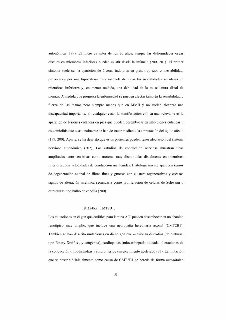
53
autonómica (199). El inicio es antes de los 30 años, aunque las deformidades óseas
distales en miembros inferiores pueden existir desde la infancia (200, 201). El primer
síntoma suele ser la aparición de úlceras indoloras en pies, tropiezos o inestabilidad,
provocados por una hipoestesia muy marcada de todas las modalidades sensitivas en
miembros inferiores y, en menor medida, una debilidad de la musculatura distal de
piernas. A medida que progresa la enfermedad se pueden afectar también la sensibilidad y
fuerza de las manos pero siempre menos que en MMII y no suelen alcanzar una
discapacidad importante. En cualquier caso, la manifestación clínica más relevante es la
aparición de lesiones cutáneas en pies que pueden desembocar en infecciones cutáneas u
osteomielitis que ocasionalmente se han de tratar mediante la amputación del tejido afecto
(199, 200). Aparte, se ha descrito que estos pacientes pueden tener afectación del sistema
nervioso autonómico (202). Los estudios de conducción nerviosa muestran unas
amplitudes tanto sensitivas como motoras muy disminuidas distalmente en miembros
inferiores, con velocidades de conducción mantenidas. Histológicamente aparecen signos
de degeneración axonal de fibras finas y gruesas con clusters regenerativos y escasos
signos de alteración mielínica secundaria como proliferación de células de Schwann o
estructuras tipo bulbo de cebolla (200).
19. LMNA: CMT2B1.
Las mutaciones en el gen que codifica para lamina A/C pueden desembocar en un abanico
fenotípico muy amplio, que incluye una neuropatía hereditaria axonal (CMT2B1).
También se han descrito mutaciones en dicho gen que ocasionan distrofias (de cinturas,
tipo Emery-Dreifuss, y congénita), cardiopatías (miocardiopatía dilatada, alteraciones de
la conducción), lipodistrofias y síndromes de envejecimiento acelerado (83). La mutación
que se describió inicialmente como causa de CMT2B1 se hereda de forma autosómico

54
recesiva y se ha descrito en familias del norte de África donde existe un efecto fundador
(82). En dichos pacientes la edad de inicio es variable, pero habitualmente aparece en las
2 primeras décadas. Suelen debutar con debilidad distal en miembros inferiores,
progresando a miembros superiores y a musculatura proximal de miembros inferiores en
un tiempo variable. La escoliosis es frecuente, así como las deformidades óseas distales, y
los déficits sensitivos son algo menos prominentes que los motores (82, 203, 204).
Neurofisiológicamente los hallazgos corresponden con una variante axonal, las
velocidades de conducción y latencias distales son normales y las amplitudes nerviosas
sensitivas y motoras están disminuidas. La anatomía patológica muestra una pérdida de
fibras nerviosas, sobre todo de gran diámetro. Es posible observar signos de degeneración
axonal y clusters regenerativos sin anormalidades mielínicas asociadas (203, 204).
Aparte de los pacientes con herencia recesiva y un fenotipo puramente neuropático, cada
vez existen más casos de fenotipo combinado (83, 205, 206). De hecho, la presencia de
una neuropatía axonal se ha encontrado en pacientes con distrofia muscular,
cardiomiopatía, leuconiquia o una combinación de los mismos (207, 208). En estos casos,
la variabilidad en la misma familia puede ser muy importante, incluso hay casos en los
que cada miembro de la familia desarrolla un tipo de afectación. Generalmente la
neuropatía en esto casos combinados es algo menos grave que la que aparece en los
pacientes del norte de África.
20. MED25: CMT2B2.
La mutación Ala335Val en el gen MED25 se ha descrito en homocigosis en una única
familia de Costa Rica que padece CMT (86). Estos pacientes presentan una neuropatía
axonal menos grave que otras formas axonales recesivas (LMNA, GDAP1, etc.). El debut
suele ser en la edad adulta (media de 34 años) con debilidad distal en miembros inferiores

55
y una progresión lenta. Llega a afectar a la musculatura distal de miembros superiores en
cerca de la mitad de los pacientes y asocian deformidades óseas distales pero no
escoliosis ni afectación de pares craneales. Los hallazgos sensitivos son congruentes con
los motores. Neurofisiológicamente existe un mantenimiento de las velocidades de
conducción motoras (> 40 m/s en MMSS) con descenso de las amplitudes y
habitualmente no se puede registrar las conducciones sensitivas en miembros inferiores.
No existen datos histológicos de la familia descrita (209, 210).
21. TRPV4: CMT2C.
Las mutaciones en el gen TRPV4 causan CMT2C, una neuropatía axonal con un fenotipo
bastante característico, pero también se han descrito en pacientes con atrofia muscular
espinal (congénita y escapuloperoneal), neuropatía hereditaria motora distal y displasias
esqueléticas (87, 211, 212, 213). Los pacientes con CMT2C padecen una neuropatía con
una variabilidad muy importante, pudiendo ser muy grave en algunos pacientes o
asintomática en otros (214). Se caracteriza por un inicio precoz, aunque puede ser tardío y
una frecuente afectación de las cuerdas vocales y, en menor medida, de la musculatura
respiratoria. De hecho, cerca de la mitad de los pacientes debutan con estridor o ronquera,
mientras que los otros lo hacen con debilidad distal en miembros inferiores. En todos los
casos la neuropatía es de predominio motor, afectando a musculatura proximal a lo largo
de la evolución de la misma. Las deformidades óseas proximales como escoliosis o
incluso escápulas aladas son frecuentes. La parálisis de cuerdas vocales aparece de una
manera precoz y puede acabar desembocando en una traqueostomía para poder respirar
(87, 212). Aparte se han descrito afectación de otros pares craneales causando parálisis
diafragmática, hipoacusia neurosensorial o alteración en la motilidad ocular, así como
incontinencia urinaria. Ocasionalmente se ha asociado la neuropatía a una displasia

56
esquelética o simplemente a estatura baja (215). Los estudios de conducción nerviosa
revelan una neuropatía axonal de predominio motor, con caída en las amplitudes motoras
y velocidades de conducción bastante preservadas. La histología es poco específica y se
encuentra una pérdida de fibras mielínicas con ocasionales axones en proceso de
degeneración (87).
22. GARS: CMT2D.
Las mutaciones en el gen GARS causan una neuropatía de predominio motor que puede
ser clasificada como CMT2D o neuropatía hereditaria motora distal o atrofia muscular
espinal distal, según si existe o no un componente sensitivo (24, 216). En cualquier caso,
el fenotipo es común e incluso en determinadas familias existen pacientes diagnosticados
con CMT2D y otros como neuropatía hereditaria motora distal. El inicio es en la 2ª-3ª
década, aunque hay casos con inicio infantil (217). Pese a que se han descrito pacientes
con inicio en miembros inferiores, de manera característica los primeros síntomas
aparecen en las manos con debilidad y atrofia de la eminencia tenar y primer interóseo
dorsal. El inicio puede ser unilateral, pero en poco tiempo de progresión la enfermedad se
hace bilateral y unos tres años después aparecen síntomas distales motores en miembros
inferiores. Los hallazgos sensitivos siempre son discretos y hay pacientes que no los
desarrollan a lo largo de todo el curso de la enfermedad (217). Las deformidades óseas
distales son habituales pero la escoliosis es muy poco frecuente. No se ha descrito
afectación de pares craneales. La progresión es lenta y no suele conllevar una gran
discapacidad (24, 217). Electrofisiológicamente los pacientes presentan unas
conducciones motoras con caída de amplitud en miembros superiores (sobre todo en
nervio mediano) y en miembros inferiores, con velocidades de conducción preservadas.
Las conducciones sensitivas pueden reflejar una leve caída de amplitud principalmente en

57
nervio sural, pero siempre son registrables (217). Histológicamente se observa una
pérdida de fibras mielínicas, especialmente de las de pequeño y mediano calibre. En
muchas de ellas se encontraron hallazgos sugestivos de degeneración axonal con
múltiples clusters regenerativos. La mielina estaba intacta pese a estar adelgazada en
algunas fibras y asociar ocasionales estructuras tipo bulbo de cebolla (217).
23. HSPB1: CMT2F.
Las mutaciones en el gen HSPB1 causan una neuropatía de predominio motor que puede
clasificarse como CMT o neuropatía hereditaria motora distal (46, 218). Tal y como
ocurría con las mutaciones en GARS, incluso hay familias en las que miembros se
clasifican como CMT2 y otros como neuropatía hereditaria motora distal (219). En
cualquier caso el fenotipo es común, con una edad de debut sintomático variable (desde la
infancia hasta pasados los 50 años). Los primeros síntomas suelen ser en miembros
inferiores, desarrollando debilidad y atrofia distal en pies y pantorrillas antes que en
manos. Los síntomas motores son claramente predominantes sobre los sensitivos pero a
medida que progresa la enfermedad, habitualmente aparece hipoestesia distal de las
distintas modalidades (220, 221). Suelen existir deformidades óseas distales pero es
infrecuente la escoliosis y no se ha descrito afectación de pares craneales.
Ocasionalmente pueden encontrarse reflejos vivos pero suelen estar disminuidos o
ausentes (221). La progresión es muy variable incluso en una misma familia, pudiendo
ocasionar una pérdida de la deambulación independiente o simplemente una debilidad
distal poco discapacitante (220). Electrofisiológicamente existe una caída de amplitudes
con velocidades de conducción mantenidas de predominio motor y de miembros
inferiores. No existen datos histológicos de pacientes con CMT2F.

58
24. HSPB8: CMT2L.
Las mutaciones en HSPB8 son una causa de CMT2 o neuropatía hereditaria motora distal
mucho más infrecuente que las mutaciones en HSPB1 (47, 222). La edad de inicio suele
ser en las 3 primeras décadas con debilidad distal en miembros inferiores. Los síntomas
motores predominan sobre los sensitivos y, pese a que pueden llegar a afectar a
musculatura proximal, la progresión suele ser lenta. Las deformidades óseas en pies son
muy frecuentes y la escoliosis aparece en cerca del 20% de los casos (223, 224). No se ha
descrito afectación de pares craneales. Los estudios electrofisiológicos revelan los
hallazgos típicos de CMT2 (descenso de amplitudes motoras y sensitivas con velocidades
de conducción y latencia conservadas), y en la RM muscular también se encuentra una
atrofia muscular longitud dependiente sin asimetrías (223, 224). Histológicamente hay
una pérdida de fibras mielínicas principalmente de gran tamaño, con signos de
degeneración axonal y clusters regenerativos (223, 224). Con microscopía electrónica se
puede comprobar la degeneración axonal al detectarse vacuolización de los axones, así
como acúmulos de material granular en axoplasma o disolución de organelas (224).
25. DNM2: CMT2M, CMT-DIB.
Las mutaciones en el gen DNM2 pueden causar una neuropatía hereditaria que puede
clasificarse como axonal o intermedia (225). La edad de inicio varía entre la primera
década y los 50 años pero suele debutar entre los 15-20 años. Los primeros síntomas
aparecen en miembros inferiores con debilidad distal, atrofia y deformidades óseas. La
progresión es variable, siendo generalmente leve o moderada, pero hay casos descritos
con pérdida de deambulación (225, 226). Las deformidades óseas distales son constantes
pero no asocian escoliosis ni afectación de pares craneales. En cambio sí que se han
descrito pacientes con alteraciones hematológicas (neutropenia) y/o cataratas asociadas

59
(225). Las cataratas eran de inicio precoz, o incluso congénitas en una familia, que
también asociaba ptosis y oftalmoparesia (225, 227). Los hallazgos neurofisiológicos en
estos pacientes son bastante variables y de ahí que se hayan clasificado como CMT2 o
CMT intermedio. Las conducciones nerviosas motoras o sensitivas pueden estar
moderadamente enlentecidas (hasta 26 m/s en MMSS) o ser normales y las amplitudes
suelen estar levemente disminuidas (225). La electromiografía revela una denervación
distal, hallazgo que se confirma con la RM muscular, donde se observa una atrofia
longitud dependiente en musculatura de pie y pantorrilla (228). Anatomopatológicamente
también hay hallazgos compatibles con una afectación combinada axonal y mielínica.
Existe una pérdida de fibras mielínicas de gran tamaño, con escasos signos de
degeneración axonal, pero frecuentes clusters regenerativos. Aparte, existen fibras
nerviosas con pliegues anómalos de mielina y con aumento del tamaño de la hendidura de
Schmidt-Lanterman (225). Muy ocasionalmente se han encontrado estructuras tipo bulbo
de cebolla con desmielinización segmentaria (229).
26. AARS: CMT2N.
Las mutaciones en el gen AARS son una causa infrecuente de CMT2 y de neuropatía
hereditaria motora distal (60, 230). El fenotipo de la misma aún debe ser confirmado y
clarificado pero aparentemente son pacientes con un debut sintomático variable (5años -
edad adulta) e incluso hay pacientes asintomáticos. La sintomatología inicial es motora de
miembros inferiores y la progresión de la misma suele ser bastante lenta, raramente
afectando a musculatura proximal. Los hallazgos sensitivos son proporcionales a los
motores y las deformidades óseas distales son constantes (60, 231, 232). Como
características añadidas cabe destacar que se han descrito varios pacientes con calambres
y fasciculaciones y, en menor medida, sordera neurosensorial (231). Los estudios

60
neurofisiológicos revelan unas velocidades de conducción en rango axonal o intermedio
(VCMM 32-50 m/s) con reducción de amplitudes (60). No existen datos histológicos de
momento.
27. DYNC1H1: CMT2O.
Las mutaciones en DYNC1H1 se han descrito recientemente como causa de CMT axonal
(CTM2O), atrofia muscular espinal o, incluso, retraso mental y alteraciones en la
migración neuronal (49, 50, 233). La neuropatía sólo se ha descrito en una familia inglesa
por lo que el fenotipo aún debe ser confirmado. Aparentemente existe una variabilidad
importante y puede existir cierto solapamiento con los otros fenotipos descritos en este
gen. La edad de inicio suele ser precoz, con retraso en la adquisición de habilidades
motoras en más de la mitad de los pacientes. Aparte, un tercio de ellos presentaban
distintos grados de retraso mental o del aprendizaje (49). La debilidad es inicialmente
distal de miembros inferiores, pero pueden acabar desarrollando debilidad proximal.
Incluso hay un paciente descrito en que el inicio fue proximal, asemejándose a un
fenotipo de atrofia muscular espinal (49). La pérdida sensitiva es variable y suele afectar
a todas las modalidades, siendo frecuentes las parestesias y el dolor neuropático.
Neurofisiológicamente se describen unas velocidades de conducción mantenidas con
amplitudes algo disminuidas, sobre todo en las conducciones motoras de MMII.
Anatomopatológicamente hay una pérdida de fibras mielínicas, con signos de
degeneración y regeneración axonal (49).

61
28. LRSAM1: CMT2P.
Las mutaciones en LRSAM1 recientemente se han descubierto como causantes de CMT2
(77). Las mismas se pueden heredar de manera recesiva o dominante, pero el fenotipo es
indistinguible (78, 234, 235). Habitualmente se produce un inicio sintomático después de
los 20 años con debilidad distal en miembros inferiores o calambres y fasciculaciones.
Posteriormente el curso es bastante lento con progresión de la debilidad a manos y rara
vez a musculatura proximal. Los síntomas sensitivos están presentes pero no predominan
sobre los motores salvo en una familia en que el fenotipo fue predominantemente
sensitivo (235). Suelen existir deformidades óseas en pies, pero no proximales ni
afectación de pares craneales. En algún caso aislado sí se ha descrito afectación
autonómica del tipo de disfunción eréctil (234). Aparentemente en todos los casos existe
una elevación leve de la creatinquinasa (CK) sanguínea. Los estudios neurofisiológicos
revelan unas conducciones motoras con velocidades y latencias mantenidas y amplitudes
que pueden estar disminuidas o ser normales. Las conducciones sensitivas
ocasionalmente no son registrables y, en cualquier caso, siempre hay una caída
importante de la amplitud. Histológicamente se describe una pérdida leve de fibras
mielínicas con ocasionales fibras con signos de degeneración axonal y clusters
regenerativos (235).
29. DHTKD1: CMT2Q.
Las mutaciones en el gen DHTKD1 se han descrito recientemente como causales de
CMT2, así como de varios tipos de aciduria (236, 237). De momento el cuadro sólo se ha
encontrado en una familia de origen chino con un fenotipo CMT2 ‘clásico’. El inicio es
entre los 15-25 años con debilidad y atrofia distal en miembros inferiores y la progresión
es lenta, afectando a musculatura distal de miembros superiores y escasamente a

62
musculatura proximal. Asocian pies cavos e hipoestesia de todas las modalidades, pero no
escoliosis ni afectación de pares craneales. Las conducciones nerviosas reflejan
velocidades de conducción mantenidas y amplitudes motoras y sensitivas disminuidas.
Sólo existe información histológica sobre una biopsia de músculo que demuestra fibras
anguladas y signos de denervación (236).
30. YARS: CMT-DIC.
Las mutaciones en el gen YARS son una causa poco frecuente de CMT de características
intermedias y existe escasa información sobre el fenotipo (59). Suele debutar en las
primeras dos décadas, pero hay pacientes con inicio en la edad adulta. El cuadro es de
predominio motor y de miembros inferiores, pero existen signos de pérdida sensitiva en
todos los pacientes (238). Las deformidades óseas distales son frecuentes pero no existe
información sobre la afectación de pares craneales o de escoliosis. Neurofisiológicamente
las velocidades de conducción motora oscilan entre 30 m/s y normales en miembros
superiores. Histológicamente se describe una reducción en el número de fibras y en el
grosor de la mielina sin apreciarse bulbos de cebolla (238).
31. INF2: CMT-DIE.
Las mutaciones en el gen INF2 son una causa de CMT intermedio asociado a
glomerulopatía, que fue descubierta recientemente (92). Los pacientes debutan en las dos
primeras décadas con síntomas provocados por la debilidad distal en miembros inferiores
(tropiezos, deformidades óseas, etc.). Aproximadamente al mismo tiempo, aparece la
nefropatía asociada (glomeruloesclerosis focal segmentaria), que debuta con proteinuria y
en 3-5 años acaba desembocando en una insuficiencia renal terminal. Neurológicamente
la debilidad progresa a musculatura distal de miembros superiores y, en menor medida, a

63
musculatura proximal (92, 239). Los síntomas sensitivos son proporcionales a los
motores. Como características asociadas se han descrito ocasionalmente sordera
neurosensorial, retraso psicomotor o Schwanomas cutáneos (92, 239, 240).
Neurofisiológicamente existe una variabilidad importante con velocidades de conducción
motora que oscilan entre 20 m/s y valores normales, y amplitudes que generalmente están
algo disminuidas. Las conducciones sensitivas pueden estar ausentes o existir un
enlentecimiento moderado de las velocidades con amplitudes claramente disminuidas. En
la RM cerebral pueden observarse hiperintensidades de sustancia blanca periventricular,
con un aumento del tamaño ventricular (92, 239). Mediante una RM muscular de
miembros inferiores se puede observar una sustitución grasa de la musculatura del pie y
de la pantorrilla (principalmente del grupo anteroexterno) con un patrón longitud
dependiente (241). Histológicamente existe una pérdida de fibras mielínicas con
abundantes estructuras tipo bulbo de cebolla (92).
32. GNB4: CMT-DIF .
Las mutaciones en GNB4 son una causa reciente de CMT intermedio (242). De momento
se ha descrito una familia y un paciente aislado, todos ellos de origen chino. El debut
sintomático suele ser en las dos primeras décadas, con tropiezos y debilidad distal en
miembros inferiores, afectando progresivamente a musculatura distal de miembros
superiores. Asocian disminución de todas las modalidades sensitivas y pies cavos, pero no
escoliosis ni disfunción de pares craneales. En la familia descrita las mujeres tenían un
fenotipo algo menos grave que los hombres, pero debe confirmarse en otros pedigrís
(242). Electrofisiológicamente las conducciones motoras presentan un enlentecimiento
variable (VCMM 17-46 m/s) con amplitudes levemente disminuidas, y las conducciones
sensitivas tienen latencias distales preservadas y amplitudes disminuidas o ausentes en

64
miembros inferiores (242, 243). Histológicamente se describe una pérdida de fibras
mielínicas principalmente las de gran tamaño, con clusters regenerativos axonales y
ocasionales bulbos de cebolla (243).
33. KARS: CMT-RIB .
Las mutaciones en KARS se han descrito en un único paciente heterocigoto compuesto
(244). Se trata de un fenotipo complejo que incluye retraso mental, conductas autolesivas,
dismorfia facial, schwannomas vestibulares y neuropatía. La misma es moderada-grave,
con predominio distal. Electrofisiológicamente hay un enlentecimiento moderado de las
velocidades de conducción (cerca de 40 m/s en MMSS) con descenso importante de las
amplitudes motoras (244).
34. PLEKHG5: CMT-RIC.
Las mutaciones en PLEKHG5 se han descrito en tres familias con una variante de CMT
intermedia y de herencia recesiva (245, 246). Previamente se habían encontrado
mutaciones en este gen en paciente con atrofia muscular espinal distal (247). Los
pacientes con estas mutaciones debutan sintomáticamente entre los 7-45 años con
debilidad y atrofia distal de miembros inferiores. La progresión es variable pero llega a
afectar musculatura distal de miembros superiores y, de forma poco frecuente, a la
musculatura proximal de miembros inferiores. Asocian hipoestesia de todas las
modalidades, pies cavos y ocasionalmente escoliosis (245, 246). Los estudios
electrofisiológicos revelan un enlentecimiento de velocidades de conducción motoras en
rangos desmielinizantes o intermedios (VCMM 25-39 m/s) con amplitudes motoras
levemente disminuidas y mayor afectación en miembros inferiores. Las conducciones
sensitivas también están moderadamente enlentecidas con amplitudes disminuidas. La

65
RM muscular de miembros inferiores muestra una atrofia muscular con infiltración grasa
con un patrón longitud dependiente y mayor afectación del compartimento anterior y
lateral de la pantorrilla que el posterior (246). Histológicamente existe una pérdida
moderada de fibras mielínicas de gran tamaño con escasos clusters regenerativos y
ausencia de bulbos de cebolla. Con microscopía electrónica, aparte de los hallazgos
anteriores, se describen escasos plegamientos mielínicos anómalos (246).
35. AIFM1: CMTX4.
Recientemente se ha descubierto que la base genética del CMTX4 (síndrome de
Cowchoc) son las mutaciones en AIFM1 (248). Se trata de una neuropatía grave descrita
en una única familia ítalo-americana que sólo es sintomática en varones. Se inicia en los
primeros años de vida y con una debilidad prominente distal > proximal y de predominio
en miembros inferiores. Existe hipoestesia de todas las modalidades y deformidades óseas
distales. Como características asociadas pueden existir hipoacusia neurosensorial y
retraso mental (249). Los estudios de conducción nerviosa revelan velocidades
mantenidas en miembros superiores (>50 m/s) y levemente enlentecidas en miembros
inferiores con amplitudes disminuidas. Histológicamente se describe una pérdida de
fibras mielínicas importante, principalmente de las de gran tamaño (249).
36. PRPS1: CMTX5.
Las mutaciones en PRPS1 se han descrito recientemente como la causa de CMTX5 (88).
El fenotipo de dichas mutaciones se ha ido expandiendo en los últimos años,
describiéndose también pacientes con síndrome de Arts, sordera neurosensorial no
sindrómica, retraso del desarrollo, distrofia retiniana y diabetes insípida (250, 251).
Inicialmente se creía que dichos fenotipos estaban claramente diferenciados pero

66
realmente el espectro fenotípico de la enfermedad es continuo. En la descripción inicial
de pacientes con CMTX5 existe una polineuropatía axonal asociada a sordera
neurosensorial y atrofia óptica (252, 253). Sólo son sintomáticos los varones y el inicio
ocurre en los primeros años con sordera neurosensorial prelingual. Posteriormente, sobre
los 10 años, aparecen los síntomas motores y visuales, con debilidad distal de miembros
inferiores y disminución de agudeza visual. Los síntomas sensitivos suelen ser menores
que los motores y están ausentes en alguno de estos pacientes. También se han descrito
pacientes con polineuropatía y sordera pero sin atrofia óptica, u otros pacientes en los que
hay un solapamiento con el síndrome de Arts (ataxia, retraso mental, hiperuricemia,
inmunodeficiencia, etc.) (254, 255). Neurofisiológicamente las velocidades de
conducción motora están levemente enlentecidas o preservadas (>40 m/s en miembros
superiores) con caída de amplitudes en miembros inferiores. Las conducciones sensitivas
pueden estar ausentes o con amplitudes disminuidas (253, 254). Anatomopatológicamente
existe una pérdida de fibras mielínicas de todos los tamaños, adelgazamiento de la
mielina y escasos bulbos de cebolla (253).
37. PDK3: CMTX6.
Las mutaciones en el gen PDK3 se han descubierto en una única familia australiana con
un fenotipo axonal y una herencia ligada al X (94). En este caso el fenotipo estaba
claramente diferenciado según el sexo. Los pacientes varones comenzaron habitualmente
en la primera década con problemas para caminar y debilidad distal en piernas. De una
manera muy lentamente progresiva se desarrolla debilidad y atrofia en pies y pantorrillas
y en musculatura intrínseca de la mano. En cualquier caso, todos los adultos de dicha
familia permanecen ambulantes y no tienen debilidad proximal. También se encuentra
hipoestesia de todas las modalidades, deformidades óseas en pies y, ocasionalmente,

67
hipoacusia. Las mujeres pueden ser asintomáticas (cerca del 50%) pero todas tienen
alteraciones mínimas en la exploración, fundamentalmente una leve debilidad y atrofia en
pies y manos, torpeza en los movimientos o temblor de miembros superiores. Los
estudios de conducción nerviosa en varones revelan unas velocidades de conducción
motora mantenidas, con descenso de amplitudes sobre todo en miembros inferiores y
conducciones sensitivas no registrables. En las mujeres los hallazgos son muy discretos,
con leve descenso de amplitudes o incluso normalidad en los estudios de conducción pero
signos de denervación en electromiografía (94).
38. FBLN5.
Las mutaciones en FBLN5 son una causa poco frecuente de CMT1, pero también se han
descrito en pacientes con degeneración macular senil y cutis laxa (256, 257, 258). De
hecho, puede existir un solapamiento entre los tres fenotipos (256). Estos pacientes
generalmente tienen un inicio sintomático tardío (> 25 años en todos los pacientes salvo
una familia) con debilidad distal de miembros inferiores. La progresión suele ser lenta,
afectando sólo a la musculatura distal de extremidades, aunque hay pacientes en los que sí
llega a afectarse la musculatura proximal y desarrollan una discapacidad importante.
Existen deformidades óseas distales, los hallazgos sensitivos son proporcionales a los
motores y parece existir una frecuencia importante de parestesias en miembros superiores
más que en inferiores (256, 259).
Como hallazgos asociados puede existir hiperelasticidad cutánea y en un paciente
también se encontró degeneración macular (256). Neurofisiológicamente hay un
enlentecimiento de las velocidades de conducción en rango desmielinizante (VCMM 15-
33 m/s) con alargamiento de latencias distales y amplitudes inicialmente mantenidas
(259).

68
39. TFG.
Las mutaciones en TGF se han descrito recientemente en familias con CMT y fenotipos
claramente diferenciados (260, 261, 262). Entre 2012 y 2013 se describió como la causa
genética subyacente a un subtipo concreto de CMT (neuropatía hereditaria sensitiva y
motora con afectación proximal dominante) conocida en pacientes japoneses de la isla de
Okinawa y en una familia coreana (260, 261). Estos pacientes tienen un debut sintomático
en la edad adulta con fasciculaciones, calambres y/o debilidad proximal en miembros
inferiores. En todos ellos predomina la debilidad proximal sobre la distal y la evolución
es variable, pudiendo llegar a perder la deambulación autónoma. Asocian atrofia
prominente de muslos y arreflexia, pero no existen deformidades óseas distales y no
suelen aparecer síntomas bulbares. La hipoestesia aparece con la evolución de la
enfermedad y afecta a todas las modalidades, aunque puede predominar la hipopalestesia.
Neurofisiológicamente existe un descenso de amplitudes motoras con velocidades de
conducción mantenidas y conducciones sensitivas que frecuentemente no son registrables.
Con la electromiografía se puede detectar frecuentes fasciculaciones y fibrilaciones, con
potenciales de características neuropáticas. Mediante RM muscular de miembros
inferiores se puede comprobar que los primeros músculos afectos son el glúteo menor y
mediano, seguido de la musculatura del muslo (principalmente el semitendinoso) y de la
pierna (261). Histológicamente se describe en el nervio periférico una pérdida de fibras
mielínicas de gran tamaño, con algunas anormalidades del grosor de la mielina y clusters
regenerativos. También se ha observado pérdida neuronal en el asta anterior medular,
núcleo dorsal y cordones posteriores (263).
En 2014 se describió una familia taiwanesa con una mutación en este gen y un fenotipo
CMT2 ‘clásico’, totalmente distinta a las descritas previamente (262). La edad de inicio
es entre los 28 y 40 años con debilidad distal en miembros inferiores y la progresión es

69
bastante lenta, con escasa discapacidad. Existen pies cavos e hipoestesia de todas las
modalidades pero no escoliosis ni afectación de pares craneales. Las conducciones
nerviosas muestran una neuropatía axonal con caída de amplitud y mantenimiento de
velocidades de conducción.
La RM muscular revela una atrofia muscular con sustitución grasa de predominio distal
. Histológicamente hay una pérdida de fibras de gran tamaño sin ninguna anormalidad
mielínica (262).
40. MARS.
Las mutaciones en MARS se han encontrado recientemente como causa de CMT2 en una
única familia (264). El fenotipo debe ser confirmado pero aparentemente hay una
penetrancia incompleta, existiendo una paciente asintomática y dos sintomáticos. Los dos
pacientes debutaron con síntomas en la edad adulta (50-67 años), que inicialmente eran de
predominio sensitivo (dolor neuropático e hipoestesia distal). Prácticamente desde el
inicio existía debilidad discreta tanto distal como proximal en miembros inferiores y
debilidad distal en miembros superiores. Tanto la debilidad como la hipoestesia
progresaron lentamente, necesitando uno de los pacientes apoyo bilateral para caminar.
Los estudios de conducción nerviosa revelan una ausencia o reducción de las amplitudes
sensitivas y en menor medida motoras, de predominio en miembros inferiores. Las
velocidades de conducción son normales. Los hallazgos de la biopsia del nervio sural son
compatibles con una neuropatía axonal (264).
41. HARS.
Las mutaciones en HARS se han descrito recientemente en un paciente esporádico con
CMT2 y en uno asintomático, por lo que aún se debe de confirmar que sean una causa de

70
CMT2 (265). El paciente afecto desarrolló síntomas a los 50 años, siendo inicialmente
hipoestesia y posteriormente dolor neuropático. Los síntomas motores eran mínimos y no
había atrofia ni pies cavos. Neurofisiológicamente existe un descenso de las amplitudes
sensitivas y motoras de predominio en miembros inferiores, con normalidad de las
velocidades de conducción (265).
42. HINT1.
Las mutaciones recesivas en HINT1 son una causa de CMT axonal o neuropatía
hereditaria motora distal frecuentemente asociada a neuromiotonía (266, 267). El debut
sintomático suele ser en la primera década con problemas para caminar por debilidad
distal de miembros inferiores o calambres y rigidez muscular. Desde el inicio existe
debilidad distal en piernas, así como dificultad para la relajación muscular en manos tras
una flexión intensa (miotonía de acción). Habitualmente no se encuentra miotonía de
percusión, pero si calambres ocasionales. La debilidad va progresando con el tiempo
afectando a musculatura de miembros superiores e incluso a musculatura proximal (266,
268). Los síntomas sensitivos sólo están presentes en cerca de la mitad de los pacientes y
siempre en menor medida que los motores. Neurofisiológicamente se describe un
descenso de las amplitudes motoras principalmente en miembros inferiores con
velocidades de conducción normales. Las conducciones sensitivas pueden ser normales o
asociar un descenso de amplitud moderado. Mediante la electromiografía se pueden
observar potenciales de denervación y frecuentes descargas miotónicas o mioquimias en
musculatura proximal y distal (neuromiotonía). Los hallazgos histológicos son
compatibles con una neuropatía axonal y se describen incluso en pacientes sin
anormalidades sensitivas clínicas o neurofisiológicas (266).

71
43. TRIM2.
Las mutaciones en TRIM2 se han descrito en una única paciente con CMT2 recesivo
(269). Es una paciente finlandesa que debutó con leve retraso en la adquisición de
habilidades motoras e hipotrofia muscular. Progresivamente apareció debilidad distal en
extremidades, hipoestesia de todas las modalidades y deformidades óseas distales. No se
describe disfunción de funciones superiores, pares craneales o escoliosis. Los estudios de
conducción nerviosa revelan una ausencia de potenciales registrables en miembros
inferiores y amplitudes disminuidas con velocidades de conducción normales o levemente
enlentecidas en miembros superiores. Histológicamente existe una reducción en el
número de fibras mielínicas principalmente de gran tamaño, así como signos
inespecíficos de degeneración axonal. Con microscopía electrónica se identifican algunas
fibras con un diámetro aumentado y acúmulo de neurofilamentos en el axoplasma (269).
44. MT-ATP6.
Las mutaciones en MT-ATP6 se han descrito recientemente en pacientes con CMT2 (56).
También pueden encontrarse en pacientes con diversos fenotipos de enfermedad
mitocondrial como el síndrome de Leigh, NARP (neuropatía, ataxia y retinitis
pigmentosa), paraparesia espástica, necrosis estriatal, etc. (57). De momento se conocen
dos mutaciones que pueden cursar con un fenotipo predominantemente o exclusivamente
neuropático (56, 270). Estos pacientes presentan una amplia variabilidad clínica, lo cual
se ve reflejado en la edad de inicio, pudiendo ser desde la primera década hasta pacientes
adultos asintomáticos. Habitualmente debutan con anormalidades en la marcha por
debilidad distal de miembros inferiores. La progresión de la debilidad es muy variable,
pudiendo afectar a musculatura proximal de miembros inferiores y a manos. Hay
pacientes que desarrollan una discapacidad muy importante, quedando confinados a una

72
silla de ruedas desde la tercera década, mientras que hay otros pacientes asintomáticos.
Generalmente asocian pies cavos e hipoestesia distal (en menos de la mitad) y
ocasionalmente piramidalismo u otros síntomas de afectación mitocondrial como
empeoramiento de los síntomas con la fiebre, migraña, problemas de aprendizaje, ataxia,
diabetes mellitus, cataratas, etc. (56, 270). Los estudios neurofisiológicos revelan
hallazgos compatibles con una polineuropatía sensitivo motora axonal en la mayoría de
pacientes (descenso de amplitudes con velocidades de conducción conservadas), pero hay
casos con normalidad en las conducciones sensitivas. Histológicamente se describe una
pérdida de fibras mielínicas con abundantes bulbos de cebolla (270).
45. CMT2G.
El termino CMT2G se utiliza para designar una familia de origen español descrita en
1986 y cuyo defecto genético se desconoce pero su locus es 12q12–q13.3 (271, 272). Se
trata de una familia extensa con una neuropatía bastante leve que se inicia entre los 9 y
los 76 años (media de 38). Comienza con debilidad distal en piernas y deformidades
óseas y progresa muy lentamente, afectando muy rara vez a musculatura de manos pero
no a musculatura proximal. Asocia hipoestesia distal de predominio en miembros
inferiores pero no afectación de pares craneales ni escoliosis. Neurofisiológicamente las
conducciones nerviosas son normales o muy levemente enlentecidas y las amplitudes
motoras y sensitivas de miembros inferiores generalmente están disminuidas.
Histológicamente se describe una pérdida de fibras mielínicas con gradiente distal >
proximal, escasas fibras con adelgazamiento de la mielina, signos de degeneración axonal
y clusters regenerativos (271, 272).

73
46. CMT-DIA.
Se conoce como CMT-DIA a una familia italiana con una variante intermedia de la
enfermedad cuya causa genética no se conoce, pero su locus es 10q24.1-q25.1 (273, 274).
El debut sintomático se produce habitualmente en la primera década con problemas para
caminar y dificultad para ponerse de talones. La debilidad va progresando muy
lentamente, afectando a musculatura de miembros superiores en la 4ª-5ª década, y pueden
llegar a necesitar apoyo para la deambulación. Asocian pies cavos, hipoestesia distal,
temblor en manos, pero no afectación de pares craneales (salvo disartria en 2 pacientes),
escoliosis ni otras alteraciones. Neurofisiológicamente existe un enlentecimiento de las
velocidades de conducción motora en rango intermedio (VCMM 25-45 m/s) con
amplitudes disminuidas principalmente en miembros inferiores. Las conducciones
sensitivas muestran hallazgos similares con caída de amplitudes y de velocidades.
Histológicamente existe una pérdida de fibras mielínicas con signos de degeneración y
regeneración axonal (clusters), pero también áreas en las que la mielina está
anormalmente gruesa o se evidencian bulbos de cebolla (273, 274).
47. CMTX2.
Se conoce como CMTX2 a una familia descrita con herencia recesiva ligada al X y cuyo
defecto genético se ha relacionado con la región Xp22.2. Únicamente están afectados los
varones, que debutan sintomáticamente en la primera década con atrofia y debilidad distal
en miembros inferiores. El 40% de los casos asocian retraso mental y no asocian otras
características diferenciadoras. Los estudios neurofisiológicos se describen como un
enlentecimiento moderado de las velocidades de conducción con signos de denervación
(275).

74
48. CMTX3.
Se conoce como CMTX3 a dos familias descritas con herencia recesiva ligada al X y
cuyo defecto genético se ha relacionado con la región Xq26-q28. Sólo están afectados los
varones, que debutan sintomáticamente en la edad adulta con atrofia y debilidad distal en
miembros inferiores y/o en miembros superiores. En una de las dos familias los pacientes
presentan reflejos vivos y signo de Babinski. No asocian retraso mental ni otra
característica diferenciadora, pero si hipoestesia de todas las modalidades y pies cavos
. Los estudios neurofisiológicos se describen como un enlentecimiento moderado de las
velocidades de conducción con signos de denervación (275).

2) HIPÓTESIS Y OBJETIVOS


77
a. Hipótesis de trabajo
La distribución mutacional de pacientes afectos de enfermedad de Charcot-Marie-Tooth
evaluados en el Hospital Universitari i Politècnic La Fe (centro de referencia para esta
patología en la Comunidad Valenciana) tiene características diferenciadoras con series
clínicas descritas en otras regiones.

78
b. Objetivos
i. Objetivo general:
Describir la distribución mutacional de pacientes con CMT evaluados en el Hospital
Universitari i Politècnic La Fe durante el periodo de 1990-2012 y compararla con otras
poblaciones. Aportar una descripción clínica y neurofisiológica exhaustiva de los
pacientes con diagnóstico genético para poder establecer una correlación genotipo-
fenotipo.
ii. Objetivos específicos:
1. Profundizar en la caracterización del fenotipo de pacientes con mutaciones que
aporten rasgos diferenciadores con respecto a otras series.
2. Proponer algoritmos diagnósticos para poder orientar el cribaje mutacional de
pacientes afectos de enfermedad de Charcot-Marie-Tooth en la Comunidad
Valenciana.

3) MATERIAL Y MÉTODOS


81
a. Población
Se incluyeron a todos los pacientes con el diagnóstico de enfermedad de Charcot-Marie-
Tooth evaluados en el Hospital Universitari i Politècnic La Fe de Valencia durante el
periodo de 1990-2012. Se consideró que padecen CMT los pacientes con neuropatía
sensitiva y motora en los que concurría al menos una de las siguientes características:
Identificación del defecto genético causal.
Otros miembros de la familia con características clínicas similares.
Pacientes con neuropatía sensitiva y motora con historia natural, clínica y
neurofisiología compatible con CMT en los que se han excluido otras
causas de neuropatía.
Se excluyeron del estudio aquellos pacientes afectos de neuropatías hereditarias
exclusivamente motoras (neuropatía hereditaria motora distal), o exclusivamente
sensitivas y autonómicas (neuropatía hereditaria sensitiva y autonómica), así como los
pacientes con neuropatía hereditaria con predisposición a las parálisis por compresión
(NHPP), neuralgia amiotrófica hereditaria o aquellos síndromes complejos en los que la
neuropatía no era la característica clínica predominante (amiloidosis, ataxia recesiva de
Charleroix-Sauvignon, etc.).
Los pacientes se clasificaron según el patrón de herencia en autosómico dominante,
autosómico recesiva, ligada al cromosoma X o esporádico; y según la velocidad de
conducción motora del nervio mediano en CMT1 o desmielinizante (VCMM < 38m/s) o
CMT2 o axonales (VCMM > 38m/s). Si el potencial de acción motor (CMAP) estaba
abolido o con una amplitud muy disminuida (<0,5mV) se clasificaron según los
parámetros neurofisiológicos de la conducción nerviosa a músculos más proximales o
según las características anatomopatológicas.

82
b. Evaluación clínica
En el Hospital Universitari i Politècnic La Fe de Valencia la evaluación clínica de los
pacientes con neuropatías hereditarias está protocolizada, de manera que incluye un
cuestionario cerrado sobre los síntomas y signos referidos por el paciente y una
exploración física detallada (apéndice). Estas evaluaciones se realizan al menos una vez
al año e incluyen una exploración neurológica y física general, seguido de una evaluación
de:
- Función motora: compuesta por una gradación de la fuerza de 20 grupos
musculares utilizando la escala estandarizada del Medical Research Council
(MRC) y la apreciación de atrofias musculares en extremidades.
- Función sensitiva: incluye la exploración de la sensibilidad táctil fina,
algésica, vibratoria y posicional de las extremidades.
- Reflejos profundos en las 4 extremidades.
- Deformidades asociadas: Tanto distales (pie cavo, dedos en martillo,
retracción aquílea, manos en garra, etc.) como proximales (escoliosis, etc.).
Aparte de la exploración pormenorizada, desde 2006 se utiliza la escala global de
neuropatías hereditarias CMT neuropathy score (CMTNS) que incluye elementos clínicos
(síntomas y signos) y neurofisiológicos y fue diseñada justamente para analizar la historia
natural de esta enfermedad (109). Para evaluar la situación funcional se usa la functional
disability scale (FDS) desde 2009. Asimismo se han tratado de extrapolar los datos
previos a dichas escalas (17).

83
c. Protocolo neurofisiológico
Los estudios neurofisiológicos se realizaron a todos los probandos y a la mayoría de
familiares en al menos una ocasión e incluyeron un estudio de conducciones nerviosas
sensitivas y motoras registradas con electrodos de superficie, y una electromiografía con
aguja concéntrica. Se ejecutaron siempre por la misma neurofisióloga experta, en equipos
similares y siguiendo el mismo protocolo. Las conducciones motoras se registraron en los
nervios mediano, cubital, radial, peroneal, tibial posterior y axilar obteniendo la amplitud,
latencia distal y velocidad de conducción. Si la amplitud de la conducción nerviosa
motora a músculos distales fue < 0,5 mV, se midieron también los mismos parámetros en
músculos más proximales (palmar largo, flexor cubital del carpo, etc.). Asimismo, en esas
ocasiones se midió la latencia y amplitud de la conducción nerviosa sobre el diafragma
mediante la estimulación en el cuello. Las conducciones sensitivas se exploraron en los
nervios mediano, cubital, radial, peroneal y sural, de manera ortodrómica en todos ellos
salvo en el nervio sural, en que se realizó de manera antidrómica, registrándose en todos
ellos la amplitud y la velocidad de conducción nerviosa sensitiva. El estudio
electromiográfico incluyó un registro de la actividad muscular en reposo, con contracción
sostenida y con esfuerzo máximo en al menos dos grupos musculares de cada extremidad.
d. Estudios anatomopatológicos
Los estudios anatomopatológicos de nervio sural no se realizaron de manera rutinaria ni
con fines investigacionales, sino solamente cuando existían dudas razonables sobre el
diagnóstico del paciente. La biopsia de nervio sural se realizó en quirófano con anestesia
local, obteniendo una muestra de 2-3 cm a nivel del tobillo. Luego se fijó inicialmente en
gluteraldehido al 2,5% y paraformaldehido al 1% en 0,1M de cacodilato sódico y, en

84
segunda instancia, en tetraóxido de osmio al 1%. Posteriormente, se deshidrató e
introdujo en resina epoxi para realizar cortes semifinos teñidos con azul de toluidina y
cortes ultrafinos contrastados con acetato de uranilo y citrato de plomo. Los cortes
semifinos se analizaron a distintos aumentos con microscopía óptica y mediante técnicas
morfométricas con el software Scion image analyst software
(http://www.scioncorp.com), determinándose la densidad de fibras nerviosas, grosor de la
vaina de mielina, g-ratio, la presencia de alteraciones de la compactación de mielina,
bulbos de cebolla o clusters de regeneración axonal, etc. Los cortes ultrafinos fueron
estudiados con microscopía electrónica para observar la ultraestructura del axón, así como
de vaina de mielina que lo rodea.
e. Resonancia magnética muscular de miembros inferiores
La resonancia magnética muscular de miembros inferiores no se realizó de manera
rutinaria, sino que se seleccionaron aquellos pacientes en los que podía aportar
información importante para definir el fenotipo. El protocolo que se utilizó fue diseñado
expresamente para pacientes con neuropatías e incluye cortes axiales y longitudinales de
pies, piernas y, ocasionalmente, muslos. Se utilizó un aparato de 1.5-Teslas MR platform
Siemens Avanto (Erlangen, Alemania), realizando secuencias T1 y STIR (Short T1
Inversion Recovery) en al menos un plano, y una secuencia T1 fat-sat antes y después de
la inyección de contraste paramagnético. Se evaluó la alteración en la intensidad de señal
muscular para determinar la presencia de edema muscular, realce anormal de contraste
y/o infiltración grasa, graduando esta última según una escala semicuantitativa de 0-4
(276). Se valoraron por separado la musculatura de pies, los cuatro compartimentos de la
pierna (compartimento anterior: músculos tibial anterior, extensor hallucis longus, y
extensor largo de los dedos; compartimento lateral: músculos peroneo largo y corto;

85
compartimento superficial posterior: músculos soleo y gastrocnemius y compartimento
profundo posterior: músculos tibial posterior, flexor largo de los dedos y flexor hallucis
longus) y los muslos.
f. Análisis Genéticos
Los análisis genéticos se han podido realizar gracias a la colaboración con el
departamento de genética y genómica de enfermedades neuromusculares perteneciente al
programa de genética y enfermedades raras del Centro de Investigación Príncipe Felipe
(previamente el departamento de Genética y Medicina Molecular del Instituto de
Biomedicina de Valencia, Consejo Superior de Investigaciones Científicas). Dicho grupo
está liderado por el Dr. Francesc Palau, e integrado por la Dra. Carmen Espinos y otros
colaboradores que han realizado el trabajo de secuenciación y diagnóstico genético de los
pacientes. El protocolo utilizado ha ido evolucionando a medida que se ha ido ampliando
el conocimiento de la enfermedad y han aparecido nuevas técnicas de secuenciación
masiva. En cualquier caso, el ADN de cada uno de los pacientes y sus familiares se
extrajo mediante métodos estándar tras la firma de un consentimiento informado para el
estudio genético. En todos los probandos, independientemente del fenotipo, se procedió al
estudio de la duplicación del gen PMP22 (CMT1A) mediante MLPA (Multiplex
Ligation- dependent Probe Amplification) empleando el kit comercial P033B (MRC-
Holland) en un analizador genético ABI Prism 3130xl (Applied Biosystem). Una vez
descartada la duplicación CMT1A se procedió al análisis mutacional de los genes
implicados en CMT. Para ello:

86
1) En algunos casos se dio prioridad al análisis de un gen de acuerdo con las
características clínicas, neurofisiológicas y patrón de herencia.
2) Si no era posible, se procedió al análisis sistemático de genes implicados en
CMT según el orden de prevalencia de estos genes en series de pacientes CMT
estudiadas en otras poblaciones europeas.
3) Finalizado este análisis se estudiaron otros genes poco frecuentes implicados
en la neuropatía de CMT.
De esta manera en los pacientes caucásicos con CMT desmielinizante se estudiaron los
genes: PMP22, GJB1, MPZ, GDAP1, SH3TC2, FGD4, NEFL, LITAF, FIG4, ERG2,
PRX1, MTMR2, MTMR13, PRPS1, DNM2, YARS y SOX10. En los pacientes caucásicos
con CMT axonal, se estudiaron los genes: MFN2, GJB1, MPZ, GDAP1, HSPB1, HSPB8,
LITAF, NEFL, DNM2, GARS, AARS1, KARS, YARS, TRPV4, RAB7, MED25 (sólo se
estudió la mutación fundadora p.A335V), LMNA (sólo se estudió la mutación fundadora
p.R298C) y RSAM. Por último en la población gitana desmielinizante se analizaron
exclusivamente las mutaciones fundadoras asociadas a esta etnia: p.R148X en el gen
NDRG1, p.R1109X y p.C737_P738delinsX en el gen SH3TC2, y g.9712G>C en el gen
HK1.
Cada gen se analizó tras una amplificación mediante PCR (Polymerase Chain Reaction)
de cada uno de los exones codificantes y sus regiones intrónicas flanqueantes. Los
cebadores empleados se diseñaron y revisaron mediante el software Gene Runner vs. 3.05
(http://www.generunner.net). El análisis mutacional de cada uno de los genes estudiados
se llevó a cabo mediante DHPLC (Denaturing High Performance Liquid
Chromatography, Transgenomic WAVE® System) y los patrones anómalos detectados se
secuenciaron en un analizador genético ABI Prism 3130xl (Applied Biosystem). Los

87
electroforegramas se visualizaron con la herramienta informática Chromas 2.33 y las
secuencias se analizaron mediante alineamiento con la secuencia salvaje empleando el
programa BLAST (http://www.ncbi.nlm.nih.gov/blast/Blast.cgi).
Adicionalmente se analizó la región 5’UTR y promotora (hasta -900 pb desde el codón de
inicio de transcripción) del gen GJB1 y en los genes MPZ y GJB1 se analizó la existencia
de posibles cambios debidos a grandes deleciones y/o duplicaciones mediante MLPA
empleando los kits comerciales P143 y P129(MRC-Holland) en un analizador genético
ABI Prism3130xl (Applied Biosystem).
Cada cambio nucleotídico se nombró de acuerdo con la pauta de lectura abierta (Open
Reading Frame, ORF) de las secuencias de referencia de ADN complementario (cDNA)
utilizadas. Las variaciones identificadas se investigaron mediante análisis de segregación
si se disponía de muestra de familiares y si el cambio era novel se compararon con 200
cromosomas de individuos sanos de ancestro español (Banco Nacional de ADN).
Para investigar la posible patogénesis de los cambios noveles se emplearon los software
SIFT (http://blocks.fhcrc.org/sift/SIFT.html) y Polyphen
(http://genetics.bwh.harvard.edu/pph) en el caso de cambios de aminoácido y en el caso
de tratarse de un cambio que afectara a una región de procesado de RNA los software
NNSPLICE (http://fruitfly.org:9005/seq_tools/splice.html) y Splice View
(http://bioinfo.itb.cnr.it/oriel/splice-view.html).
g. Aspectos éticos
El proyecto incluye dos aspectos claramente diferenciados, la caracterización fenotípica y
los análisis genéticos. El primero se realiza en el contexto de un seguimiento clínico
habitual, sólo que de una manera más concienzuda y protocolizada, no suponiendo

88
perjuicio alguno para el paciente. Las pruebas complementarias invasivas (estudios
neurofisiológicos, biopsia del nervio sural, etc.) sólo se realizaron si era indispensable
para el diagnóstico y nunca cuando ya existía un diagnóstico genético o sólo para
seguimiento. En cualquier caso, a los pacientes se les pidió que firmasen un
consentimiento informado aprobado por el comité ético del Hospital Universitari i
Politècnic La Fe para poder utilizar sus datos clínicos en una base de datos propia y para
poder incorporarlos a una base de datos de ámbito nacional
(http://www.pubgle.com/ciber/neuropatiashereditarias).
Los estudios genéticos se realizaron en pacientes y ocasionalmente en familiares
aparentemente no afectos, siempre tras informar de la naturaleza del diagnóstico genético
así como de sus consecuencias tanto personales como familiares. Asimismo se les explicó
claramente que en estos momentos no existe ningún tratamiento aprobado para la
enfermedad de Charcot-Marie-Tooth. Previa a la extracción de sangre los pacientes
firmaron un consentimiento informado explicativo y se reiteró que en cualquier momento
podían retirarlo si lo creían conveniente.

4) RESULTADOS


91
a. Artículo I:
Charcot-Marie-Tooth disease: genetic and clinical spectrum in a Spanish
clinical series.
Sivera R, Sevilla T, Vílchez JJ, Martínez-Rubio D, Chumillas MJ, Vázquez
JF, Muelas N, Bataller L, Millán JM, Palau F, Espinós C.
Neurology. 2013 Oct 29;81(18):1617-25.


Rafael Sivera, MD*Teresa Sevilla, MD, PhD*Juan Jesús Vílchez, MD,
PhDDolores Martínez-Rubio,
MBChBMaría José Chumillas,
MDJuan Francisco Vázquez,
MDNuria Muelas, MD, PhDLuis Bataller, MD, PhDJosé María Millán, PhDFancesc Palau, MD, PhDCarmen Espinós, PhD
Correspondence toDr. Sevilla:[email protected]
Charcot-Marie-Tooth diseaseGenetic and clinical spectrum in a Spanish clinical series
ABSTRACT
Objectives: To determine the genetic distribution and the phenotypic correlation of an extensiveseries of patients with Charcot-Marie-Tooth disease in a geographically well-defined Mediterra-nean area.
Methods: A thorough genetic screening, including most of the known genes involved in thisdisease, was performed and analyzed in this longitudinal descriptive study. Clinical data wereanalyzed and compared among the genetic subgroups.
Results: Molecular diagnosis was accomplished in 365 of 438 patients (83.3%), with a highersuccess rate in demyelinating forms of the disease. The CMT1A duplication (PMP22 gene) wasthe most frequent genetic diagnosis (50.4%), followed by mutations in the GJB1 gene (15.3%),and in theGDAP1 gene (11.5%). Mutations in 13 other genes were identified, but were much lessfrequent. Sixteen novel mutations were detected and characterized phenotypically.
Conclusions: The relatively high frequency of GDAP1 mutations, coupled with the scarceness ofMFN2 mutations (1.1%) and the high proportion of recessive inheritance (11.6%) in this seriesexemplify the particularity of the genetic distribution of Charcot-Marie-Tooth disease in thisregion. Neurology� 2013;81:1–9
GLOSSARYAD 5 autosomal dominant; AR 5 autosomal recessive; CMT 5 Charcot-Marie-Tooth; MMNCV 5 median motor nerve con-duction velocity.
Charcot-Marie-Tooth (CMT) disease refers to the genetically heterogeneous group of hereditarymotor and sensory neuropathies. It is one of the most common inherited neurologic disorders,with a prevalence of 15.2 to 40 cases per 100,000.1–3 Molecular studies have provided an ever-growing list of more than 40 involved genes and loci (http://www.molgen.ua.ac.be/CMTMutations/, http://neuromuscular.wustl.edu/, both accessed June 24, 2013). Most of thepatients with CMT disease have autosomal dominant (AD) inheritance, but many have X-linkedor autosomal recessive (AR) inheritance. CMT disease can be classified according to clinical,electrophysiologic, and nerve pathology findings into demyelinating forms (CMT1, CMT4),with a median motor nerve conduction velocity (MMNCV) of ,38 m/s and pathologicevidence of nerve fiber demyelination; and axonal forms (CMT2), with preserved conductionvelocities (MMNCV.38m/s) and pathologic signs of axonal degeneration and regeneration.4 Anintermediate type (CMT-I) is accepted in which MMNCV lies between 25 and 45 m/s and nervepathology shows axonal and/or demyelinating features.5
Clinically, the most frequent CMT phenotype is characterized by progressive distal weaknessand sensory loss appearing toward the second decade, with foot deformities and absent reflexes.However, other patients develop a much more severe form with onset in infancy or early
*These authors contributed equally to this work.
From the Departments of Neurology (R.S., T.S., J.J.V., J.F.V., N.M., L.B.), Clinical Neurophysiology (M.J.C.), and Genetics (J.M.M.), HospitalUnivesitari i Politècnic La Fe, Valencia; Centro de Investigación Biomédica en Red de Enfermedades Neurodegenerativas (T.S., J.J.V., M.J.C., N.M., L.B.), Valencia; Departments of Medicine (T.S., J.J.V.) and Genetics (C.E.), University of Valencia; Program in Rare and Genetic Diseases (D.M.-R., F.P., C.E.), Centro de Investigación Príncipe Felipe (CIPF), Valencia; Centro de Investigación Biomédica en Red de Enfermedades Raras(D.M.-R., J.M.M., F.P., C.E.), Valencia; IBV-CSIC Associated Unit at CIPF (D.M.-R., F.P., C.E.), Valencia; and School of Medicine (F.P.),University of Castilla-La Mancha, Ciudad Real, Spain.
Go to Neurology.org for full disclosures. Funding information and disclosures deemed relevant by the authors, if any, are provided at the end of the article.
© 2013 American Academy of Neurology 1
ª 2013 American Academy of Neurology. Unauthorized reproduction of this article is prohibited.
Published Ahead of Print on September 27, 2013 as 10.1212/WNL.0b013e3182a9f56a

childhood and great disability within a fewyears, or a milder course with few symptomsuntil adulthood. This clinical heterogeneity,coupled with the expanding genetic diversity,is the complex scenario of the inherited neu-ropathies. Comprehensive clinical series, inwhich at least the most frequent genes havebeen studied, are needed to shed light onthe populational genetic distribution andgenotype-phenotype correlation in CMT dis-ease.6,7 Herein, we present the genetic distribu-tion and phenotypic characterization of anextensive series of CMT disease after an exhaus-tive genetic screening in the Region of Valencia, ageographically well-defined Mediterranean area.
METHODS Subjects. This is a longitudinal descriptive study,
which includes all of the patients with the diagnosis of CMT disease
and evaluated at the inherited neuropathy clinic of Hospital Uni-
versitari i Politècnic La Fe in Valencia from 2000 to 2012. Patients
with sensory-motor neuropathy were considered to have CMT
disease if a) a causative genetic defect was determined, b) family
members with similar characteristics were detected, or c) sporadic
cases were included if their medical history, examination, and neu-
rophysiology were compatible with CMT disease, and other known
causes of acquired neuropathy were reasonably discarded. Patients
with inherited neuropathies with exclusive motor (distal hereditary
motor neuropathies) or sensory and autonomic (hereditary sensory
and autonomic neuropathies) signs were excluded from this study,
as well as those with hereditary neuropathy with liability to pressure
palsies, and those with complex disorders in which neuropathy was
not the most predominant phenotypic feature. Patients were sub-
classified with demyelinating or axonal CMT disease according to
MMNCVs of the proband, except when the amplitudes of median
compound motor action potentials were reduced .90%. In those
cases, the conduction velocities to nerves innervating proximal
muscles were measured (palmaris longus for the median nerve,
flexor carpi ulnaris for the ulnar nerve, etc.), and occasionally laten-
cies of other proximal nerves such as the axillary nerve, or patho-
logic evidence were considered.
Standard protocol approvals, registrations, and patientconsents. This study protocol was approved by the Institutional
Review Board of the Hospital Univesitari i Politècnic La Fe.
Written informed consents were obtained from all of the mem-
bers included in this study.
Clinical assessments. The clinical assessment included strength,
muscular atrophy, sensory loss, reflexes, foot deformities, as well as
a general and neurologic examination. Muscle strength was graded
using the standard Medical Research Council scale. CMT neuropa-
thy score was recorded in all patients followed since 2006,8 and the
Functional Disability Scale score in those after 20009; previous clin-
ical data were extrapolated to CMT neuropathy score and Functional
Disability Scale score when possible. Comprehensive electrophysio-
logic studies were performed in 401 of 438 patients (91.6%), and
were not performed only when the genetic diagnosis of another
family member was already available. Lower limb muscle MRI and
sural nerve biopsy were performed only when there were reasonable
doubts regarding the clinical diagnosis or for investigational purposes,
and followed the protocols described previously.10
Mutational analysis. Blood samples were drawn and genomic
DNA was obtained by standard methods from peripheral white
blood cells. In all of the probands, the CMT1A duplication was
analyzed by MLPA (Multiplex Ligation–dependent Probe Ampli-
fication, SALSA kit P033 CMT1; MRC-Holland, Amsterdam, the
Netherlands) in a genetic analyzer ABI Prism 3130xl (Applied
Biosystems, Foster City, CA). Once the CMT1A duplication was
discarded, a mutational screening of genes involved in CMT disease
was performed taking into account the ethnicity of the proband and
the phenotype. In patients with Gypsy ethnicity, the genetic testing
strategy was planned as described previously.11 In Caucasian pa-
tients, the mutational screening was clinically oriented, and
included the genes detailed in table 1 until the causative mutation
was identified or all of the genes had been analyzed.
The mutational screening was performed by amplification of
all exons and their intronic flanking sequences, except in the
GJB1 gene in which the promoter sequence has also been ana-
lyzed. The Gene Runner version 3.05 software was used for
designing primers (available on request). The PCR products were
analyzed by using denaturing high-performance liquid chroma-
tography (WAVE System; Transgenomic Inc., Omaha, NE), and
the anomalous patterns were investigated by Sanger sequencing
(ABI Prism 3130xl). Finally, in both the MPZ and the GJB1genes, large deletions and/or duplications were investigated by
Table 1 Genes analyzed in the mutationalscreening
CMT1
CMT2Caucasian Gypsy
PMP22 SH3TC2a MFN2
GJB1 NDRG1a GJB1
MPZ HK1a MPZ
GDAP1 GDAP1
SH3TC2 HSPB1
FGD4 HSPB8
NEFL LITAF
LITAF NEFL
GAN1 DNM2b
BSCL2 GARS
FIG4 AARS
ERG2 KARS
PRXb YARS
MTMR2 TRPV4
MTMR13 RAB7
PRPS1 MED25a
DNM2b LMNAa
YARS LRSAM1
SOX10
Abbreviation: CMT 5 Charcot-Marie-Tooth.aOnly founder mutations were analyzed: SH3TC2p.C737_P738delinsX, SH3TC2 p.R1109X, NDRG1p.R148X, HK1 g.9712G.C, MED25 p.A335V, and LMNAp.R298C.bMore than one sequence reference was used because ofthe presence of isoforms.
2 Neurology 81 October 29, 2013
ª 2013 American Academy of Neurology. Unauthorized reproduction of this article is prohibited.

MLPA using the SALSA kits P143 and P129 (MRC-Holland) in
an ABI Prism 3130xl autoanalyzer. We did not screenMT-ATP6,PDK3, DHTKD1, GNB4, or TRIM2 genes because they had not
been described when this project was concluded.12–16
When possible, segregation analyses within the families were
performed, and novel mutations were analyzed in 200 chromo-
somes from healthy controls of Spanish ancestry. The biological rel-
evance of the amino acid changes was studied using both SIFT
(http://blocks.fhcrc.org/sift/SIFT.html, accessed June 24, 2013)
and PolyPhen (http://genetics.bwh.harvard.edu/pph, accessed
June 24, 2013) programs. When the detected alteration modified
a splicing sequence, we used the NNSPLICE (http://fruitfly.org:
9005/seq_tools/splice.html, accessed June 24, 2013) and the Splice
View (http://zeus2.itb.cnr.it/;webgene/wwwspliceview_ex.html,
accessed June 24, 2013) software.
RESULTS A total of 1,009 patients were evaluated atour inherited neuropathy clinic during the timeframe2000 to 2012; 438 of them were considered to haveCMT disease and met our inclusion criteria. All wereSpanish, and 401 of them (91.6%) were currently liv-ing or had ancestral roots in the Region of Valencia, inthe Western Mediterranean area. Initially, 275(62.8%) were classified as demyelinating CMT, and163 (37.2%) as axonal CMT. Regarding the inheri-tance pattern, 242 (55.3%) were considered as AD,51 (11.6%) were AR, 52 (11.9%) were X-linked,and 93 (21.2%) were considered sporadic. Geneticdiagnosis was achieved in 365 of 438 patients(83.3%), with a higher success rate in the demyelinat-ing forms (263/275; 95.6%) over the axonal forms(102/163; 62.6%). The causative mutations were de-tected in 214 of 242 patients (88.4%) with AD inher-itance, 45 of 51 (88.2%) with AR inheritance, 52 of 52(100%) with X-linked inheritance, and in only 54 of93 (58.1%) with a sporadic presentation. In table 2,the detailed genetic diagnosis can be analyzed andcompared with the latest published data, and in thefigure, the distribution according to CMT subtype isshown. All of the genetic and clinical information hasalso been recorded in a readily accessible mutationdatabase (http://www.treat-cmt.es/db, accessed June24, 2013).
Patients with demyelinating CMT disease. Of the 275 pa-tients with demyelinating CMT disease, 241 were ofCaucasian ethnicity and 34 were of Gypsy origin. Ofthe Caucasian patients with the demyelinating form,184 (76.3%) carried the CMT1A duplication, whichis the most frequent cause of CMT disease. In the re-maining 57 Caucasian patients, the disease causingmutation was identified in 45 with the following distri-bution: 25 mutations in GJB1, 9 in MPZ, 4 in PRX, 2point mutations in PMP22, 2 in FGD4, 2 in SH3TC2,and 1 in NEFL. Six novel mutations were detected indemyelinating CMT (table 3). Once the genetic screen-ing was performed, the causative change remainedunknown in 12 patients (4.9%). No mutations were
identified in any of the following genes: LITAF, EGR2,GDAP1, MTMR2, MTMR13, FIG4, PRPS1, DNM2,YARS, and SOX10. In the Gypsy population, the dis-ease-causing mutation was identified in all cases, andconsisted exclusively of founder mutations related toCMT disease in the Gypsy population.11
Table 4 shows the relevant clinical features associ-ated with AR forms of demyelinating CMT disease(CMT4). These forms have certain common charac-teristics such as early onset, delayed motor develop-ment, and severe disability, but other features differbetween the CMT4 subtypes.
Patients with axonal CMT disease. The mutationalscreening detailed in table 1 led to identification ofthe disease-causing mutation in 102 of 163 patientswith axonal CMT disease (62.6%). In this set of pa-tients, there is a marked genetic heterogeneity, withmutations in the GDAP1 and GJB1 genes being the2 most frequent causes of axonal CMT disease. Muta-tions in the GDAP1 gene correspond to 24 patients
Table 2 Genetic distribution and comparison toother series
Gene
No. of patients (frequency, %)
Present study Saporta et al.6 Murphy et al.7
PMP22a 184 (48.8) 290 (55) 168 (63.2)
GJB1 56 (14.9) 80 (15.2) 46 (17.3)
GDAP1 42 (11.1) 6 (1.2) 2 (0.8)
SH3TC2 27 (7.2) 3 (0.6) 5 (1.9)
MPZ 19 (5.0) 45 (8.5) 13 (4.9)
NDRG1 7 (1.9)
HSPB1 7 (1.9) 2 (0.8)
MFN2 6 (1.6) 21 (4.0) 12 (4.5)
HK1 5 (1.3)
NEFL 4 (1.1) 4 (0.8) 2 (0.8)
GARS 4 (1.1) 3 (0.6)
PRX 4 (1.1) 1 (0.2)
HSPB8 3 (0.8)
PMP22b 2 (0.5) 5 (1.0) 6 (2.3)
FGD4 2 (0.5)
KARS 1 (0.3)
YARS 1 (0.3)
TRPV4 1 (0.3) 3 (1.1)
LITAF 5 (1.0) 4 (1.5)
MTMR2 1 (0.4)
GAN 1 (0.4)
BSCL2 1 (0.4)
FIG4 2 (0.4)
aCarriers of the CMT1A duplication.bCarriers of point mutations in the PMP22 gene.
Neurology 81 October 29, 2013 3
ª 2013 American Academy of Neurology. Unauthorized reproduction of this article is prohibited.

(14.7% of CMT2) with AD inheritance (caused by thep.R120W mutation in all cases except one) and 18patients (11.0%) with AR inheritance and diverse gen-otype. All of our patients with GDAP1 mutations weredefined as CMT2 because the neurophysiologic find-ings were clearly axonal, although the pathologyincluded both axonal features (fiber loss, axonal degen-eration, few regenerative clusters, etc.) and myelinabnormalities (thin myelin sheaths, abnormal myelinfolding, occasional onion bulb–like formations). Pa-tients with AR inheritance developed a severe pheno-type with important disability, vocal cord, anddiaphragmatic palsies whereas patients with domi-nant GDAP1 mutations presented with a mild tomoderate phenotype with certain clinical and MRIparticularities reported previously.10
Mutations in the GJB1 gene were detected in 31patients with axonal CMT disease (19.0%). It is inter-esting to note that although the patients were classifiedas having demyelinating or axonal CMT disease ac-cording to the MMNCVs of the proband, more than80% of these families would be classified as havingintermediate forms of CMT disease.
The remaining mutations were actually quite rare,accounting for only 29 cases (17.8%), and are distributed
among several genes: 10 patients with mutations inMPZ, 7 in HSPB1, 4 in MFN2, 3 in HSPB8, 3 inNEFL, 1 in GARS, and 1 in KARS. In the aggregate,25 different mutations were identified in the CMT2series and 10 of them were novel (table 3). Oncethe mutational screening was performed, the disease-causing mutation remained unknown in 61 patients(37.4%). No change was identified in the followinggenes: RAB7, DNM2, YARS, AARS, LRSAM1, andTRPV4, nor the founder mutations MED25 p.A335Vor LMNA p.R298C.
DISCUSSION A thorough genetic screening has beenperformed in an extensive clinical series of patients withCMT disease in aWesternMediterranean area. Overall,a molecular diagnosis was achieved in 83.3%, with ahigher success rate in demyelinating than in axonalCMT disease. In demyelinating patients, these ratesare comparable to the other series in which a compre-hensive genetic screening was performed (table 2),6,7,17
suggesting that few genes involved in this form of CMTdisease remain undiscovered. However, in CMT2,although the success rate is higher than in other series,37.4% of patients remain without genetic diagnosis.The mutational distribution described confirms the
Figure Genetic characterization of CMT disease subtypes
Patients evaluated at the inherited neuropathy clinic during the timeframe 2000–2012. a Carriers of the CMT1A duplica-tion. b Carriers of point mutations in the PMP22 gene. AD 5 autosomal dominant; AR 5 autosomal recessive; CMT 5
Charcot-Marie-Tooth.
4 Neurology 81 October 29, 2013
ª 2013 American Academy of Neurology. Unauthorized reproduction of this article is prohibited.

extensive heterogeneity intrinsic to this disease; 56 dif-ferent mutations have been detected in this series, and16 had not been described previously. This comprehen-sive study depicts the genetic distribution of a largeCMT series in the Mediterranean basin, and there arecertain distinctive features compared with other geo-graphic areas.
The CMT1A duplication is by far the most com-mon mutation detected, and all patients were classifiedas demyelinating CMT; in fact, none had MMNCV.30 m/s. CMT1A accounts for 66.9% of the demy-elinating forms, which is somewhat lower than other
series that report slightly more than 70%.18 Actually,these results are biased by the presence of 34 Gypsypatients affected by demyelinating CMT disease whoharbored the previously described founder mutationsassociated with the Gypsy population as we have pre-viously reported.11,19 These 34 Gypsy patients and 8others of Caucasian ethnicity (4 with mutations inPRX, 2 in SH3TC2, and 2 in FGD4; table 4) comprisethe 11.6% of demyelinating CMT with an AR inher-itance (CMT4). The percentage of patients with AR orsporadic presentation is in fact greater than in otherseries6 andmay reflect certain populational peculiarities,
Table 3 Novel mutations with detailed assessments and conduction velocities of the probands, and phenotypic peculiarities
Gene
Mutation
PresentationNo. ofpatients
Onset,y
Age at exam,y CMTNS FDS
MMNCV(m/s) Phenotypic characteristicsNucleotide Amino acid (aa)
GJB1 c.44_45delinsTT p.R15L X-linked 3 20 59 14 3 36.1 Early distal upper limb atrophyand weakness. Intrafamilyvariability regarding severity.
c.529G.A p.V177M Sporadic 1 18 34 14 1 30 Early distal upper limb atrophyand weakness.
c.-540C.G No aa change X-linked 10 26 36 15 2 31 Lower limb distal weaknessearlier and more prominent thanupper limb. Includes 2asymptomatic women.
c.484dupA p.M162NfsX81 X-linked 2 15 34 13 2 40 Early distal upper limb atrophyand weakness.
c.141_143dupGAA p.K48_S49insK X-linked 4 24 44 12 2 41 Early distal upper limb atrophyand weakness. Brisk reflexes onlyin proband.
SH3TC2 c.3305delA (hom)a p.H1102LfsX14a Sporadic 1 9 43 15 3 27 Early sensory ataxia, scoliosis.Lower . upper limb distalweakness and atrophy. Nohearing loss.
PRX c.589G.T;c.642insC
p.E197X;p.R215QfsX8
AR 2 2 42 27 8 4 Early onset, sensory ataxia,scoliosis. Refractory trigeminalneuralgia in 1/2. Few motor signs.
FGD4 c.1886delGAAA(hom)
p.K630NfsX5 AR 2 3 34 14 4 11 Early onset but slow progression.Sensory ataxia. Lower . upperlimb distal weakness and atrophy.Spinal syringomyelia in 1/2.
GDAP1 c.1031T.G;c.487C.Tb
p.L344R; p.Q163Xb Sporadic 1 12 49 12 2 57 Mild phenotype for a recessivemutation. Distal lower limbweakness, no vocal cord ordiaphragmatic palsy.
MFN2 c.306dupT p.G103WfsX41 AD 2 22 40 11 2 52 Classic CMT2 phenotype,moderate instability.
c.752C.T p.P251L Sporadic 1 25 47 13 2 51 Classic CMT2 phenotype.
MPZ c.21_26dupTGGGGG p.P9_A10dup AD 2 30 39 9 1 54 Proband with mild phenotype andhis father is mostlyasymptomatic. Upper limbreflexes are present.
NEFL c.293A.C p.N98T Sporadic 1 3 54 26 8 44 Early onset, severe phenotype.Hearing loss. Wheelchair bound inthe 4th decade, death at 58 y.
c.1315T.A p.F439I Sporadic 1 23 41 8 2 45 Early distal upper limb atrophyand weakness. Brisk reflexes.
GARS c.1171C.T p.R391C Sporadic 1 18 39 10 1 53 Early distal upper limb atrophyand weakness. Brisk reflexes.Motor . sensory involvement.
Abbreviations: AD 5 autosomal dominant; AR 5 autosomal recessive; CMT 5 Charcot-Marie-Tooth; CMTNS 5 CMT neuropathy score; FDS 5 FunctionalDisability Scale; hom 5 homozygous; MMNCV 5 median motor nerve conduction velocity (normal values in our laboratory .51.6 m/s).aWe cited this mutation in Lupo et al.,36 but clinical features were not included.b This mutation has been widely described; we have included it because this patient is a compound heterozygote for a novel mutation.
Neurology 81 October 29, 2013 5
ª 2013 American Academy of Neurology. Unauthorized reproduction of this article is prohibited.

Tab
le4
Gen
otyp
e-phe
noty
peco
rrelat
ionof
these
ries
ofpat
ient
swithau
toso
mal
rece
ssivedem
yelin
atingCMTdisea
se
Gen
eMut
ations
Pat
ient
s/fa
milies
Ons
et,
yAgeat
exam
,yWea
knes
sSen
sory
loss
Foo
tdef
ormity,
%Sco
liosis,
%Cra
nial
nerv
esa
CMTNS;
FDS
MMNCV,m
/s;
CMAP,m
Vb
SNAP,m
V
SH3TC2
p.R1109X
(hom
)21/1
13.2
23.4
LL.
UL;
prox
imal
38%
Prominen
t;vibr
atory5
pinp
rick
;atax
ia100%
100
91
V-trige
minal
neuralgia
(5%
);VIII
(48%
)16.8;3
.724.6;4
.20.7;N
R52%
p.R1109X/
p.C737_P738de
linsX
5/3
4.1
20.3
LL.
UL;
prox
imal
40%
Sam
e100
100
VIII
(40%
)15.6;4
.122.7;4
.80.3;N
R80%
p.H1102LfsX
14
(hom
)1/1
930
LL.
UL;
distal
.pr
oxim
alSam
eYes
Yes
No
15;2
18;8
.7NR
p.R529Q
(hom
)1/1
843
LL.
UL;
only
distal
Sam
eYes
Yes
,mild
VIII
10;2
28;9
.6NR
HK1
g.9712G.C
(hom
)6/3
4.8
24.2
LL.
UL;
prox
imal
33%
Prominen
t;vibr
atory.
pinp
rick
;atax
ia100%
100
50
VIII
(33%
)14.1;3
26.3;5
.11.9;N
R17%
NDRG1
p.R148X
(hom
)2/2
3.8
18.1
LL.
UL;
prox
imal
50%
Prominen
t;vibr
atory.
pinp
rick
;atax
ia100%
100
100
VIII
(50%
)16.3;3
.116.7;6
.20.9;N
R50%
PRX
p.E197X/
p.R215QfsX8
3/1
2.7
25.7
LL.
UL;
only
distal
Prominen
t;vibr
atory.
pinp
rick
;atax
ia100%
100
100
V-trige
minal
neuralgia
(33%
)22.7;5
.34.9;1
.2NR
100%
p.E113fsX3
(hom
)1/1
112
LL.
UL;
only
distal
Prominen
t;vibr
atory.
pinp
rick
;atax
iaYes
Yes
No
18;3
5.8;0
.5NR
FGD4c
p.K630NfsX5
(hom
)2/1
2.5
32
LL.
UL;
only
distal
Prominen
t;vibr
atory.
pinp
rick
;atax
ia100%
Yes
Yes
No
12;3
11.5;5
.2NR
100%
Abb
reviations
:CMAP5
compo
undmus
cleac
tion
potentialo
fthemed
ianne
rve(normal
values
.9.3
mV);CMT5
Cha
rcot-M
arie-Too
th;C
MTN
S5
CMTne
urop
athy
score;
FDS5
Fun
ctiona
lDisab
ility
Sca
le;h
om5
homoz
ygou
s;LL
5lower
limbs
;MMNCV5
med
ianmotor
nerveco
nduc
tion
velocity
(normal
values
inou
rlabo
ratory
.51.6
m/s);NR5
notreco
rdab
le(exp
ress
edin
%of
thepa
tien
ts);SNAP5
sens
oryne
rveac
tion
potentialinmed
ianne
rve(normal
values
.16.5
mV);UL5
uppe
rlim
bs.
Ifmorethan
oneca
se,the
numeric
values
aremea
nsan
dthepe
rcen
tage
s,arerelative
freq
uenc
yof
ach
arac
teristic.
aVIII
nervewas
cons
idered
affected
whe
nthepa
tien
trepo
rted
releva
nthy
poac
usia
orthehe
aringloss
was
confirmed
withau
diom
etry.
bNerve
cond
uction
stud
iesof
med
ianne
rvene
ares
tto
themom
entof
phys
ical
exam
ination.
cTh
e2
patien
tswithmutations
intheFGD4
gene
hadan
earlyon
setan
dmod
eratedisa
bilityfrom
infanc
y,bu
tve
ryslow
prog
ress
iontherea
fter.
6 Neurology 81 October 29, 2013
ª 2013 American Academy of Neurology. Unauthorized reproduction of this article is prohibited.

as the Region of Valencia hosts a numerous Gypsypopulation (more than 50,000), and certain isolatedareas have a high consanguinity rate.
Mutations in GJB1 were the second most commongenetic diagnosis after CMT1A, accounting for 12.8%of the CMT series. These patients were classified ac-cording to the MMNCV of the proband, but clinicallyall patients had a consistent phenotype that was not somuch influenced by conduction velocities, as by sex.20,21
Only 5 patients (9%) had signs of CNS involvement(brisk reflexes and Babinski sign in 2 of them) withnormal encephalic and spinal MRI. It is worth notingthat in 2 of these patients after a long follow-up (.20years), the pyramidal signs became less prominent as theneuropathy progressed, becoming overshadowed by theneuropathic signs. More than 300 mutations have beendescribed in the GJB1 gene, throughout the codingregion and exceptionally, in the 5’-UTR (untranslatedregion). A very extensive family of our series was foundto be carriers of a novel c.-540C.G mutation in thisregion. Its pathogenicity was demonstrated by a lucif-erase assay (data not shown).
Mutations in MPZ were detected in only 4.3% ofthe series; 9 were classified as demyelinating CMTand 10 as axonal CMT. In this case, there was impor-tant phenotypical variability, as has been reported inthis gene.22,23 Except for one family, demyelinatingpatients were more severely affected, with earlier dis-ease onset (first decade), prominent sensory loss, andmoderate to severe disability with progression. One ofthese patients, carrier of the MPZ p.S121F mutation,developed a severe congenital hypomyelinating neu-ropathy.24 Other genes were actually quite scarcelyaffected in our CMT1 series (NEFL, point mutationsin PMP22, PRX, SH3TC2, and FGD4).
There is a great genetic diversity in axonal forms ofCMT disease, as 25 different mutations were detectedin 9 genes. The success rate of our series in these patients(62.6%) is one of the highest that has been published,probably because of the ample genetic screening thathas been performed, and the high relative frequencyof GDAP1. The genetic distribution in CMT2 showsthat the 2 most frequent causes of axonal CMT diseasewere mutations in the GDAP1 and GJB1 genes, whichcombined accounted for 44.8% of patients who hadaxonal CMT disease. However, 37.4% of the patientswith CMT2 remained undiagnosed, and this consti-tutes a great challenge for the near future.
Our series of 42 patients with mutations in theGDAP1 gene is to date the most extensive one publishedand all of them presented neurophysiologic features ofaxonal CMT disease. Patients with apparently demye-linating or intermediate nerve conduction studies havebeen reported,25,26 but in our patients, the only oneswith slow conduction velocities were those in whichcompound motor action potential was ,0.5 mV, and
nerve conduction velocity was clearly normal if mea-sured to nerves innervating proximal muscles. Althoughthe neurophysiologic findings in these patients wereunequivocally axonal, the pathology included both axo-nal degeneration andmyelin abnormalities.10,27 Eighteenpatients with recessiveGDAP1mutations were detected,with an early disease onset and rapid progression, andwere wheelchair-bound in the second or third decade inall cases except 2 (associated with p.L344R/p.Q163Xcompound heterozygote, and p.R282C/p.R282Chomozygote genotypes) who had a relatively milder phe-notype.27 Twenty-four of 25 patients with dominantGDAP1 mutations carried the p.R120W substitution,which is to date the most frequent dominant mutationdetected in the GDAP1 gene. Although this mutationhas been described in families with different geographicorigins,28–30 the GDAP1 p.R120W probably has afounder effect in our population, and presents with amild to moderate phenotype.10
Apart from the high prevalence of GDAP1 muta-tions, the other notable factor in the axonal CMTseries is the low number of cases with mutations inthe MFN2 gene (2.5%). MFN2 has been identified asthe most common gene in axonal CMT disease inmany series,7,8 accounting consistently for 10% to33%31–33 of this CMT form, even in other SpanishMediterranean areas.34 Certain other European serieshave described even lower frequencies35 than our own,suggesting that the distribution of MFN2 mutationsmay be quite heterogeneous within Europe. The re-maining mutations identified in axonal patients wereeven less frequent, including MPZ, HSPB1, NEFL,GARS, HSPB8, and YARS genes (15.3% of theCMT2 series).
The knowledge derived from thoroughly screenedCMT series is essential to comprehend the global pic-ture of this disease, as there may be relevant changes inthe genetic distribution of different areas. A clear exam-ple of this is the relatively high prevalence of recessiveforms and the predominance ofGDAP1 overMFN2 inthis clinical series. More information about the geneticdistribution in other Spanish or Mediterranean areas isneeded to discern whether this is only a local charac-teristic, or can be extrapolated to other areas.
AUTHOR CONTRIBUTIONSDr. Sivera: acquisition of data, analysis and interpretation, initial manu-
script elaboration. Dr. Sevilla: study concept and design, initial manu-
script elaboration. Dr. Vílchez: critical revision of the manuscript for
important intellectual content. Ms. Martínez-Rubio: genetic studies,
acquisition of data. Dr. Chumillas: nerve conduction studies, acquisition
of data. Dr. Vázquez: acquisition of data, analysis and interpretation.
Dr. Muelas and Dr. Bataller: critical revision of the manuscript for
important intellectual content. Dr. Millán: genetic studies (CMT1A
duplication), acquisition of data. Dr. Palau: study concept and design,
critical revision of the manuscript for important intellectual content.
Dr. Espinós: study concept and design, study supervision, genetic
screening.
Neurology 81 October 29, 2013 7
ª 2013 American Academy of Neurology. Unauthorized reproduction of this article is prohibited.

ACKNOWLEDGMENTThe authors are grateful to Paula Sancho for helping to generate the data-
base of CMT mutations in the Spanish population, to Susana Rovira for
performing the study of the GJB1 gene promoter region, and to Itziar
Llopis for the sample management.
STUDY FUNDINGThis collaborative joint project is awarded by IRDiRC and funded by the
Instituto de Salud Carlos III (ISCIII)–Subdirección General de Evalua-
ción y Fomento de la Investigación within the framework of the National
R1D1I Plan (grants IR11/TREAT-CMT, PI08/90857, PI08/0889,
CP08/00053, PI12/00453, and PI12/0946), cofunded with FEDER
funds, and the Generalitat Valenciana (grant Prometeo/2009/051). The
Centro de Investigación Biomédica en Red de Enfermedades Neurode-
generativas (CIBERNED) and the Centro de Investigación Biomédica en
Red de Enfermedades Raras (CIBERER) are initiatives from the ISCIII.
DISCLOSURER. Sivera reports no disclosures. T. Sevilla is funded by grants from the
ISCIII (PI12/0946, PI08/0889) and IRDiRC (IR11/TREAT-CMT).
J. Vílchez received research support from the CIBERNED. D. Martínez-
Rubio, M. Chumillas, J. Vázquez, N. Muelas, L. Bataller, and J. Millán
report no disclosures. F. Palau is funded by grants from Fondo de Inves-
tigación Sanitaria (PI08/90857), the Generalitat Valenciana (Prometeo/
2009/051), the IRDiRC (IR11/TREAT-CMT), and the CIBERER.
C. Espinós is funded by a grant from the ISCIII (PI12/00453) and
IRDiRC (IR11/TREAT-CMT). Dr. Espinós has a “Miguel Servet” contract
funded by the ISCIII and the CIBERER (CP08/00053). Go to Neurology.
org for full disclosures.
Received May 10, 2013. Accepted in final form July 30, 2013.
REFERENCES1. Combarros O, Calleja J, Polo JM, Berciano J. Prevalence
of hereditary motor and sensory neuropathy in Cantabria.
Acta Neurol Scand 1987;75:9–12.
2. Foley C, Schofield I, Eglon G, Bailey G, Chinnery PF,
Horvath R. Charcot-Marie-Tooth disease in Northern Eng-
land. J Neurol Neurosurg Psychiatry 2012;83:572–573.
3. Skre H. Genetic and clinical aspects of Charcot Marie
Tooth’s disease. Clin Genet 1974;6:98–118.
4. Dyck PJ, Lambert EH. Lower motor and primary sensory
neuron diseases with peroneal muscular atrophy. I. Neu-
rologic, genetic, and electrophysiologic findings in hered-
itary polyneuropathies. Arch Neurol 1968;18:603–618.
5. Payreson D, Marchesi C. Diagnosis, natural history, and
management of Charcot-Marie-Tooth disease. Lancet
Neurol 2009;8:654–667.
6. Saporta AS, Sottile SL, Miller LJ, Feely SM, Siskind CE,
Shy ME. Charcot-Marie-Tooth disease subtypes and
genetic testing strategies. Ann Neurol 2011;69:22–33.
7. Murphy SM, Laura M, Fawcett K, et al. Charcot-Marie-
Tooth disease: frequency of genetic subtypes and guide-
lines for genetic testing. J Neurol Neurosurg Psychiatry
2012;83:706–710.
8. Shy ME, Blake J, Krajewski K, et al. Reliability and valid-
ity of the CMT neuropathy score as a measure of disabil-
ity. Neurology 2005;64:1209–1214.
9. Birouk N, Gouider R, Le Guern E, et al. Charcot–Marie–
Tooth disease type 1A with 17p11.2 duplication: clinical and
electrophysiological phenotype study and factors influencing
disease severity in 119 cases. Brain 1997;120:813–823.
10. Sivera R, Espinós C, Vílchez JJ, et al. Phenotypical features of
the p.R120W mutation in the GDAP1 gene causing auto-
somal dominant Charcot-Marie-Tooth disease. J Peripher
Nerv Syst 2010;15:334–344.
11. Sevilla T, Martínez-Rubio D, Márquez C, et al. Genetics
of the Charcot-Marie-Tooth disease in the Spanish Gypsy
population: the hereditary motor and sensory neuropathy–
Russe in depth. Clin Genet 2013;83:565–570.
12. Pitceathly R, Murphy SM, Cottenie E, et al. Genetic dys-
function of MT-ATP6 causes axonal Charcot-Marie-Tooth
disease. Neurology 2012;79:1145–1154.
13. Kennerson ML, Yiu EM, Chuang DT, et al. A new locus for
X-linked dominant Charcot-Marie-Tooth disease (CMTX6)
is caused by mutations in the pyruvate dehydrogenase kinase
isoenzyme 3 (PDK3) gene. Hum Mol Genet 2013;22:
1404–1416.
14. Xu W, Gu MM, Sun LH, et al. A nonsense mutation in
DHTKD1 causes Charcot-Marie-Tooth disease type 2 in
a large Chinese pedigree. Am J Hum Genet 2012;91:
1088–1094.
15. Soong BW, Huang YH, Tsai PC, et al. Exome sequencing
identifies GNB4 mutations as a cause of dominant inter-
mediate Charcot-Marie-Tooth disease. Am J Hum Genet
2013;92:422–430.
16. Ylikallio E, Pöyhönen R, Zimon M, et al. Deficiency of
the E3 ubiquitin ligase TRIM2 in early-onset axonal neu-
ropathy. Hum Mol Genet 2013;22:2975–2983.
17. Lin KP, Soong BW, Yang CC, et al. The mutational spec-
trum in a cohort of Charcot-Marie-Tooth disease type 2
among the Han Chinese in Taiwan. PLoS One 2011;6:
e29393.
18. Nelis E, Van Broeckhoven C, De Jonghe P, et al. Estima-
tion of the mutation frequencies in Charcot-Marie-Tooth
disease type 1 and hereditary neuropathy with liability to
pressure palsies: a European collaborative study. Eur J
Hum Genet 1996;4:25–33.
19. Claramunt R, Sevilla T, Lupo V, et al. The p.R1109X
mutation in SH3TC2 gene is predominant in Spanish Gyp-
sies with Charcot-Marie-Tooth disease type 4. Clin Genet
2007;71:343–349.
20. Shy ME, Siskind C, Swan ER, et al. CMT1X phenotypes
represent loss of GJB1 gene function. Neurology 2007;68:
849–855.
21. Siskind CE, Murphy SM, Ovens R, Polke J, Reilly MM,
Shy ME. Phenotype expression in women with CMT1X.
J Peripher Nerv Syst 2011;16:102–107.
22. Warner L, Hilz M, Appel S, et al. Clinical phenotypes of
different MPZ (P0) mutations may include Charcot–Marie–
Tooth type 1B, Dejerine–Sottas, and congenital hypomye-
lination. Neuron 1996;17:451–460.
23. Shy ME, Jani A, Krajewski K, et al. Phenotypic clustering
in MPZ mutations. Brain 2004;127:371–384.
24. Sevilla T, Lupo V, Sivera R, et al. Congenital hypomyeli-
nating neuropathy due to a novel MPZ mutation.
J Peripher Nerv Syst 2011;16:347–352.
25. Baxter RV, Ben Othmane K, Rochelle JM, et al. Ganglioside-
induced differentiation-associated protein-1 is mutant in
Charcot-Marie-Tooth disease type 4A/8q21. Nat Genet
2002;30:21–22.
26. Senderek J, Bergmann C, Ramaekers VT, et al. Mutations
in the ganglioside-induced-differentiation-associated pro-
tein-1 (GDAP1) gene in intermediate type autosomal
recessive Charcot-Marie-Tooth neuropathy. Brain 2003;
126:642–649.
27. Sevilla T, Jaijo T, Nauffal D, et al. Vocal cord paresis and
diaphragmatic dysfunction are severe and frequent symp-
toms of GDAP1-associated neuropathy. Brain 2008;131:
3051–3061.
8 Neurology 81 October 29, 2013
ª 2013 American Academy of Neurology. Unauthorized reproduction of this article is prohibited.

28. Claramunt R, Pedrola L, Sevilla T, et al. Genetics of
Charcot-Marie-Tooth disease type 4A: mutations, inheri-
tance, phenotypic variability, and founder effect. J Med
Genet 2005;42:358–365.
29. Zimo�n M, Baets J, Fabrizi GM, Jaakkola E, et al. Dom-
inant GDAP1 mutations cause predominantly mild CMT
phenotypes. Neurology 2011;77:540–548.
30. Ammar N, Nelis E, Merlini L, Barisic N, et al. Identifi-
cation of novel GDAP mutations causing autosomal reces-
sive Charcot-Marie-Tooth disease. Neuromuscul Disord
2003;13:720–728.
31. Verhoeven K, Claeys KG, Züchner S, et al. MFN2 muta-
tion distribution and genotype/phenotype correlation in
Charcot-Marie-Tooth type 2. Brain 2006;129:2093–2102.
32. Calvo J, Funalot B, Ouvrier RA, et al. Genotype-phenotype
correlations in Charcot-Marie-Tooth disease type 2
caused by mitofusin 2 mutations. Arch Neurol 2009;
66:1511–1516.
33. Auranen M, Ylikallio E, Toppila J, Somer M, Kiuru-Enari S,
Tyynismaa H. Dominant GDAP1 founder mutations is a
common cause of axonal Charcot-Marie-Tooth disease in
Finland. Neurogenetics 2013;14:123–132.
34. Casasnovas C, Banchs I, Cassereau J, et al. Phenotypic
spectrum of MFN2 mutations in the Spanish population.
J Med Genet 2010;47:249–256.
35. Braathen GJ. Genetic epidemiology of Charcot-Marie-
Tooth disease. Acta Neurol Scand Suppl 2012;126:1–22.
36. Lupo V, Galindo MI, Martínez-Rubio D, et al. Missense
mutations in the SH3TC2 protein causing Charcot-Marie-
Tooth disease type 4C affect its localization in the plasma
membrane and endocytic pathway. Hum Mol Genet 2009;
18:4603–4614.
Neurology 81 October 29, 2013 9
ª 2013 American Academy of Neurology. Unauthorized reproduction of this article is prohibited.


103
b. Artículo II:
Phenotypical features of the p.R120W mutation in the GDAP1 gene
causing autosomal dominant Charcot-Marie-Tooth disease.
Sivera R, Espinós C, Vílchez JJ, Mas F, Martínez-Rubio D, Chumillas MJ,
Mayordomo F, Muelas N, Bataller L, Palau F, Sevilla T.
J Peripher Nerv Syst. 2010 Dec;15(4):334-44.


Journal of the Peripheral Nervous System 15:334–344 (2010)
RESEARCH REPORT
Phenotypical features of the p.R120W mutationin the GDAP1 gene causing autosomal dominant
Charcot-Marie-Tooth disease
Rafael Sivera1, Carmen Espinos2, Juan J. Vılchez1,3, Fernando Mas3,4, DoloresMartınez-Rubio2,5, Marıa Jose Chumillas3,6, Fernando Mayordomo3, Nuria Muelas1,3,
Luis Bataller1,3, Francesc Palau2,5, and Teresa Sevilla1,3
1Department of Neurology, University Hospital Universitari La Fe, Valencia; 2Centro de Investigacion Biomedica en Red deEnfermedades Raras (CIBERER), Valencia; 3Centro de Investigacion Biomedica en Red de Enfermedades
Neurodegenerativas (CIBERNED), Valencia; 4Department of Radiology, University Hospital Universitari La Fe, Valencia;5Laboratory of Genetics and Molecular Medicine, Instituto de Biomedicina de Valencia-CSIC, Valencia; and 6Clinical
Neurophysiology, University Hospital Universitari La Fe, Valencia, Spain
Abstract Mutations in the ganglioside-induced-differentiation-associated protein 1 gene(GDAP1) can cause Charcot-Marie-Tooth (CMT) disease with demyelinating (CMT4A) oraxonal forms (CMT2K and ARCMT2K). Most of these mutations present a recessiveinheritance, but few autosomal dominant GDAP1 mutations have also been reported. Weperformed a GDAP1 gene screening in a clinically well-characterized series of 81 indexcases with axonal CMT neuropathy, identifying 17 patients belonging to 4 unrelated familiesin whom the heterozygous p.R120W was found to be the only disease-causing mutation.The main objective was to fully characterize the neuropathy caused by this mutation.The clinical picture included a mild–moderate phenotype with onset around adolescence,but great variability. Consistently, ankle dorsiflexion and plantar flexion were impairedto a similar degree. Nerve conduction studies revealed an axonal neuropathy. Musclemagnetic resonance imaging studies demonstrated selective involvement of intrinsicfoot muscles in all patients and a uniform pattern of fatty infiltration in the calf, withdistal and superficial posterior predominance. Pathological abnormalities included depletionof myelinated fibers, regenerative clusters and features of axonal degeneration withmitochondrial aggregates. Our findings highlight the relevance of dominantly transmittedp.R120W GDAP1 gene mutations which can cause an axonal CMT with a wide clinicalprofile.
Key words: ADCMT, Charcot-Marie-Tooth disease, CMT2K, GDAP1 mutations
IntroductionCharcot-Marie-Tooth disease (CMT) is a genet-
ically heterogeneous group of inherited motor andsensory neuropathies. Molecular studies have shown
Address correspondence to: Dr. Teresa Sevilla, Department ofNeurology, Hospital Universitari La Fe, Avda. Campanar, 21, 46009Valencia, Spain. Tel: +3496-3862761; Fax: +3496-1973290; E-mail:sevilla [email protected]
extensive genetic heterogeneity in CMT neuropathieswith an ever-growing list of causative mutationsand loci (Pareyson and Marchesi, 2009). Mutationsin the ganglioside-induced-differentiation-associatedprotein 1 (GDAP1; MIM 606598) gene 8q21 havebeen reported in CMT patients with demyelinating(CMT4A; MIM 214400) (Baxter et al., 2002) and axonalforms (CMT2K and ARCMT2K; MIM 607831) of thedisease (Cuesta et al., 2002). Inheritance in most CMT
© 2010 Peripheral Nerve Society 334

Sivera et al. Journal of the Peripheral Nervous System 15:334–344 (2010)
causative GDAP1 mutations is autosomal recessive(Boerkoel et al., 2003; Sevilla et al., 2003) characterizedby a severe phenotype with early disease onset andrapid progression to important disability in the secondor third decade. In recent reports, certain mutationshave been shown to segregate as an autosomal dom-inant (AD) trait with a later disease onset and a mildphenotype (Claramunt et al., 2005; Chung et al., 2008a;Cassereau et al., 2009), the p.R120W missense muta-tion being the most prevalent one (Claramunt et al.,2005; Cavallaro et al., 2009).
In a previous study we reported a series of autoso-mal recessive or sporadic GDAP1-related neuropathies(Sevilla et al., 2008). The extension of the screeningfor mutations in the GDAP1 gene to AD families withaxonal CMT has allowed us to recognize 17 patientsbelonging to 4 unrelated families in whom the onlydetected mutation was GDAP1 p.R120W in heterozy-gosis, 2 of them were only known by history andgenetics. The main objective of the present studywas to characterize clinically, electrophysiologicallyand pathologically this form of CMT neuropathy, des-ignated CMT2K. Additionally, a systematic magneticresonance imaging (MRI) investigation was performedin these patients so as to identify specific patterns ofmuscle involvement, as has been described in othertypes of CMT disease (Gallardo et al., 2006; Chunget al., 2008b).
Material and MethodsStudy subjects
We investigated a clinically well-characterizedseries of 81 patients who presented with axonal CMT.A mutational screening of the more frequent axonalCMT genes, MFN2, GJB1, GDAP1, and MPZ, andsome of the rarer ones, NEFL, HSP22, and HSP27,has been carried out. Causative mutations have beenrecognized in 34 probands and in 4 of them, theGDAP1 p.R120W mutation was the only one present.We have identified 17 patients belonging to thesefour families with the GDAP1 p.R120W (c.358C>T)mutation in which disease was inherited as an ADtrait. The pedigrees are displayed in Fig. 1; family Bwas previously reported (Claramunt et al., 2005). Thefamilies were unrelated, and do not originate fromthe same Spanish region. All protocols performed inthis study complied with the ethics guidelines of theinstitutions involved. All patients and relatives wereaware of the investigative nature of the studies andgave their consent.
Genetic analyses
Blood samples were drawn from the patients andrelatives after informed consent and in accordance
with the Helsinki declaration. Genomic DNA wasobtained by standard methods from peripheral whiteblood cells. Mutation analysis of the GDAP1 gene wasperformed by amplification of the six exons and theirintronic flanking sequences using primers previouslydescribed (Cuesta et al., 2002). The polymerasechain reaction (PCR) products were analyzed byDHPLC (Denaturing High Liquid Chromatography,Transgenomic WAVE® System) and the anomalouspatterns were investigated by automated sequencing(ABI Prism 3130xl, Applied Biosystems, Foster, CA).When possible, segregation analyses were performed.
Linkage analysis was carried out under theassumption of AD inheritance, full penetrance, andequal frequency of marker alleles. Pairwise LOD scoreswere calculated using the MLINK program version 5.1of the FASTLINK package 2.1 (Lathrop and Laouel,1984). Haplotype analyses at the GDAP1 locus wascarried out on the basis of cen D8S279-D8S286 −D8S551-c.507T>G−D8S1474-D8S1829-D8S84 tel aspreviously described (Claramunt et al., 2005).
Clinical and electrophysiological assessments
All probands and individuals at risk wereexamined except patients B-II2 and B-III2. Theclinical assessment included strength, muscle atrophy,sensory loss, reflexes, foot deformities as well as ageneral and neurologic examination. Muscle strengthwas graded using the standard Medical ResearchCouncil (MRC) scale. CMT neuropathy score (CMTNS)was applied to determine neurological impairment:mild (CMTNS ≤10 points), moderate (CMTNS 11–20),and severe (CMTNS 21–36) (Shy et al., 2005). Thefunctional disability scale (FDS) was used to measurethe disability status (Birouk et al., 1997). Data fromfamily B were updated except for patients B-II2 andB-III2, who were not available for this study.
Electrophysiological studies were performed in 14of the patients following the same protocol that wasdescribed previously (Sevilla et al., 2003).
Magnetic resonance imaging
MRI was performed on the feet and distallegs of eight patients in a supine position usinga 1.5-T MR platform (Siemens Avanto, Erlangen,Germany). The following protocol was used in allpatients: axial and coronal T1-weighted TSE (turbospin echo), Short T1 Inversion Recovery (STIR), and T1fat-saturation images of both legs and feet beforeand after Gadolinum-DTPA (Magnevist, Schering,Germany) administration were obtained. Only oneimaging plane was acquired in each region aftercontrast administration.
The four classic anatomical compartments wereused to evaluate calf muscles: anterior compartment
335

Sivera et al. Journal of the Peripheral Nervous System 15:334–344 (2010)
Figure 1. Pedigrees of the four affected families. Squares = males, circles = females, shaded symbols = affected,dotted circle = affected by history. Family B has been previously reported (Claramunt et al., 2005) and includes two individuals(*) who have not been clinically assessed in this moment.
(tibialis anterior, extensor hallucis longus, and extensordigitorum longus), lateral compartment (peronei longusand brevis), superficial posterior compartment (soleusand gastrocnemius), and deep posterior compartment(tibialis posterior, flexor digitorum longus, and flexorhallucis longus). In axial MR images of lower limbs,fatty infiltration was graded from 0 to 4 as follows: 0,no fat signal in muscle; 1, some fatty streaks; 2, fatoccupying a minor part of muscle; 3, similar amount offat and muscle tissue; 4, fat occupying the greater partof muscle (Chung et al., 2008b).
Nerve biopsies
Sural nerve biopsy was performed in two cases(patients A-II1 and C-II2) when they were 47 and31 years old, respectively, and compared to a 27-year-old multi-organ donor without neuropathic or systemicdisease history. Semi-thin sections stained with tolu-idine blue were prepared for evaluation under a light
microscope following the same protocol as describedpreviously (Sevilla et al., 2003). Morphometry of myeli-nated fibers was performed on high-resolution micro-graphic images obtained with a Polaroid DMC digitalcamera and analyzed by means of Scion image anal-ysis software (http://www.scioncorp.com). Ultra-thincut samples were contrasted with uranyl acetate andlead citrate for ultrastructural study.
ResultsGenetic analyses
Seventeen patients from four unrelated familiescarrying the GDAP1 p.R120W mutation in a het-erozygous state were identified. No other pathogenicmutations were detected in other exons or in theirflanking intron regions. AD inheritance was confirmedby co-segregation of the GDAP1 p.R120W mutation
336

Sivera et al. Journal of the Peripheral Nervous System 15:334–344 (2010)
with disease in families A, B, and C (Fig. 1). In familyD, only sample from the proposita was available andtherefore a segregation analysis was not possible.Next, we performed a linkage analysis for the GDAP1p.R120W mutation in the three multiplex families. Theobtained cumulative maximum LOD score (Zmax) was2.83 (θ = 0.00). This Zmax ∼3 reinforces that theGDAP1 p.R120W presents an AD inheritance. Wehave constructed haplotypes by analysis of six flankingmicrosatellite markers, which span ∼2.83 Mb aroundthe GDAP1 locus, and one intragenic single nucleotidepolymorphism (SNP). All the chromosomes carryingthe GDAP1 p.R120W mutation share the same haplo-type 3-5-G-5-6-6 on the basis of cen D8S286-D8S551-c.507T>G-D8S1474-D8S1829-D8S84 tel, suggestinga common origin.
Clinical findings
The clinical characteristics of 15 patients from thefour families with the p.R120W missense mutationin the GDAP1 gene are summarized in Table 1. Theage in which patients experienced the first symptomranged between 9 and 65 years (median 17 years),with disease duration between 5 and 48 years. Therewas no delay in the acquisition of motor milestones.Four patients (A-II2, A-II3, B-III1, and C-I2) aged 20,38, 33, and 72 did not complain of any symptom butclinical examination revealed minor signs. Patient A-II2had mild weakness in toe extension, hyporreflexia andhypoesthesia in lower limbs, patient A-II3 absent anklereflexes and distal hypoesthesia, patient B-III1 pescavus, and patient C-I2 absent knee and ankle jerks.
The disease started in the distal lower limbs. Onlytwo patients had mild proximal involvement in thelower limbs (A-I1 and D-II1), being quite disabling inone case. All symptomatic cases presented weak-ness in ankle plantar flexion (EHL) and toe extension.Weakness in ankle dorsiflexors was present to thesame degree as plantar flexors in most symptomaticpatients, being the impairment of heel and toe walkquite analogous. Distal upper limb weakness appearedlater in the course of the disease and involved theintrinsic hand muscles predominately. At the time ofthe examination, 12 patients had motor deficits in dis-tal lower limbs, seven patients in both the distal lowerand upper limbs. In one patient (C-II2), the distributionof atrophy and weakness in the lower limbs was asym-metric. All patients except two had preserved reflexesin the upper limbs, but hypo/arreflexia in lower limbsand different degrees of lower limb distal atrophy.Foot deformities, including pes cavus and Achilles ten-don shortening, were also very common. Sensory lossproportional to the motor deficit was present in allsymptomatic patients, especially pinprick and vibration T
ab
le1.C
linic
ald
ata
of
the
seri
es.
Pat
ien
tO
nse
t(y
ears
)/A
ge
atex
amC
MT
NS
Pro
xim
alLL
An
kle
DF
An
kle
PF
TE
IHM
Hee
l/to
ew
alk
UL
DT
RK
nee
DT
RA
nkl
eD
TR
Pes
cavu
sP
inp
rick
sen
sory
loss
Vib
rato
ryse
nso
rylo
ssFD
S
A-I
1>
40/6
721
42
20
4I/I
+−
−M
od
erat
eK
nee
/elb
ow
An
kle
5A
-II1
9/43
145
33
04+
I/I++
+−
Mo
der
ate
Kn
ee/e
lbo
wA
nkl
e3
A-I
I2A
/40
55
55
4+5
N/D
++++
+M
ildT
oes
No
ne
0A
-II3
35/3
87
55
55
5N
/N++
++−
Mild
An
kle
To
es0
B-I
465
/80
11∗
52
22
4I/I
−−
−N
oA
nkl
eA
nkl
e4
B-I
I115
/52
105
44
34
I/I++
++−
Mild
An
kle
No
ne
3B
-III1
A/2
00
55
55
5N
/N++
++++
Mild
No
ne
No
ne
0B
-II2
16/4
04
54
54
5D
/D++
+−
Mild
An
kle
To
es1
B-I
I417
/38
125
44
34
D/D
+++
−M
od
erat
eA
nkl
eT
oes
3C
-I1
18/6
29
55
43
5D
/D++
−−
Mo
der
ate
An
kle/
fin
ger
An
kle
2C
-II1
20/3
36
55
55
5D
/N++
−−
Mild
An
kle
No
ne
1C
-II2
14/3
015
53
2R/4
L1
4I/I
++−
−M
od
erat
eA
nkl
eA
nkl
e2
C-I
I312
/24
75
44
35
D/D
+++
−M
od
erat
eA
nkl
eN
on
e2
C-I
2A
/72
0∗5
55
55
N/N
++−
−N
oN
on
eN
on
e0
D-I
I125
/75
254+
00
04
I/I−
−−
Mild
Kn
ee/fi
ng
erA
nkl
e5
A,a
sym
pto
mat
ic;
D,d
ifficu
lty;
DF,
do
rsifl
exio
n;D
TR
,dee
pte
nd
on
refl
exes
;I,
imp
oss
ible
;IH
M,i
ntr
insi
ch
and
mu
scle
s;L,l
eft;
LL,l
ow
erlim
bs;
N,n
orm
al;P
F,p
lan
tar
flex
ion
;R,r
igh
t;T
E,
toe
exte
nsi
on
;UL,
up
per
limb
s.−
rep
rese
nts
abse
nt
refl
ex,+
mild
lyd
epre
ssed
refl
ex,a
nd
++n
orm
alre
flex
.∗ P
atie
nts
inw
hic
ho
nly
the
clin
ical
par
amet
ers
wer
eu
sed
toca
lcu
late
the
Ch
arco
t-M
arie
-To
oth
neu
rop
ath
ysc
ore
(no
elec
tro
ph
ysio
log
ical
dat
aav
aila
ble
).
337

Sivera et al. Journal of the Peripheral Nervous System 15:334–344 (2010)
in distal lower limbs. Patient A-II2 received chemother-apy for breast cancer 10 years ago, but during a 24-yearfollow-up the disability has followed a progressivecourse.
The CMTNS scores in the series reflected thewide phenotypic spectrum, ranging from 0 to 25. Therewere only two patients who were catalogued in theCMTNS severe category, both of them had a longdisease evolution. Nine patients (60%) were in themild category and four in the moderate. All patients, todate, have an FDS equal or less than four except twopatients, patient A-I1 who is mostly wheelchair boundand patient D-II1 who needs a cane or crutches towalk. Disease progression was slow in the majority ofcases but two patients (A-I1 and B-I4) had onset after40, but developed a relevant functional impairment.
Electrophysiological studies
Table 2 summarizes the nerve conduction andelectromyography studies performed. Motor nerveconduction velocities, distal latencies, and F-waveswere normal in all tested nerves (median MNCV>54 m/s in all patients). Ulnar and median CMAP werereduced only in two individuals, while peroneal CMAPwas reduced in most affected individuals. Sensorynerve conduction studies showed a reduction of SNAPin all tested patients, but conduction velocities anddistal latencies were preserved in nerves with SNAP>0.5 μV. In the whole series, even in asymptomaticpatients, needle electromyography revealed motor unitaction potentials (MUAPs) increased in amplitude,duration, and polyphasic incidence. Positive sharpwaves and fibrillation potentials were not present.
MRI studies
All patients had detectable abnormalities in theMRI consisting of fatty infiltration and/or muscleedema (Table 2).
Intrinsic musculature of both feet showedconsistent and bilateral fatty infiltration of the footmuscles in all patients, even in asymptomatic ones(Fig. 2A). The changes were present in all intrinsicfoot muscles and were more pronounced in severelyaffected patients. All patients had evidence of varyingdegrees of fatty substitution in the muscles of the calfin concordance with the severity of the phenotype. Inany case there was a common pattern: a predominanceof fatty substitution distally and in the posteriorcompartment over the anterolateral one.
Mild cases showed distinct abnormalities in thedistal muscles of the calf; high signal intensity onT1-weighted and STIR images, corresponding withfatty substitution and muscle edema, respectively. Thefirst muscle affected and the one in which the findingswere more prominent was the gastrocnemius, and to
a slightly lesser degree the soleus (Fig. 2B); the rest ofthe muscles in the calf were completely preserved inmost mild cases.
In more severe cases the fatty substitutioninvolved all muscle compartments of the calf andmuscle edema was no longer present. The poste-rior compartment was always affected to a greaterdegree than the anterolateral one with a mean gradeof 3.1 vs. 1.5 (p < 0.05) (Fig. 2C). MRI of the thigh wasperformed in only two patients (A-II1 and D-II1) witha moderate–severe phenotype, existing fatty substitu-tion distally in all the muscles.
Pathologic findings
The two sural nerve biopsies performed revealedsimilar pathological findings. Semi-thin sectionsshowed a pronounced depletion of myelinated fibersin both nerves (fiber density was 3202/mm2 in patientA-II1, 5863/mm2 in patient C-II2, and 9095/mm2 in thecontrol subject). The histograms representing myeli-nated fiber size were ‘‘shifted to the left,’’ showing amarked reduction of large-diameter fibers, especiallyin patient A-II1 (fibers >6 μV = 12.3% in A-II1, 31.5%in C-II2, and 41.7% in control). Morphometric data alsorevealed a considerable proportion of thinly myelinatedfibers with a g-ratio greater than 0.7 (18.2% in A-II1,20.9% in C-II2, and 5% in control).
Rather frequent regenerative clusters and occa-sional onion bulb formations were also present(Fig. 3A). In detailed electron-microscope views, onionbulbs were made up of concentric layers of Schwanncell processes adopting a crescent shape and enclos-ing a central core composed of a regenerative cluster,or less frequently a hypomyelinated or normally myeli-nated fiber (Fig. 3B). These formations had a limitednumber of folds (pseudo-bulbs) not reaching the sizeobserved in CMT1A. The regenerative clusters werecomposed of a group of small myelinated fibers, alarge bundle of unmyelinated axons, or a combinationof the two (Fig. 3E).
On high magnification, myelin compaction alwaysappeared normal. However, axons often presenteddegenerative features consisting of axolemma retrac-tion with partial or total detachment (Fig. 3C), aggrega-tion of normal and abnormal mitochondria mixed withempty vacuoles, and cytoskeleton dissolution withearly disappearance of microtubules (Fig. 3D). Degen-eration involved both myelinated and unmyelinatedaxons with preference for those included in regenera-tive clusters (Fig. 3F).
DiscussionAD GDAP1 mutations are exceedingly rare in
most published CMT series. Here we present the
338

Sivera et al. Journal of the Peripheral Nervous System 15:334–344 (2010)
Tab
le2.N
erve
con
du
ctio
ns
and
MR
Idat
a.
Ner
veco
nd
uct
ion
stu
die
s
Med
ian
Uln
arP
ero
nea
lM
edia
nS
ura
lM
RI
Pat
ien
tC
MA
P(m
V)
MC
V(m
/s)
CM
AP
(mV
)M
CV
(m/s
)C
MA
P(m
V)
MC
V(m
/s)
SN
AP
(μV
)S
CV
(m/s
)S
NA
P(μ
V)
SC
V(m
/s)
EM
G.
neu
rog
enic
chan
ges
IFM
So
leu
sG
astr
ocn
emiu
sD
PC
AC
LCT
hig
h
A-I
19.
454
.86.
153
.50.
640
.31.
636
.3N
RN
RY
esA
-II1
7.5
69N
PN
P1.
348
444
329
Yes
44
42
33
3A
-II2
9.5
6012
60.3
5.4
4212
57.1
1.8
50Y
es3
23
00
1N
PA
-II3
11.5
6015
.764
.97.
350
.810
54.7
9.1
NP
Yes
21
10
00
NP
B-I
I111
.269
.111
.767
.94
53.7
6.5
53.3
6.2
55.6
Yes
B-I
II117
.261
.220
.669
.311
.945
2450
1565
.9Y
esB
-II2
12.1
60.3
14.4
52.3
443
.826
609.
948
.2Y
esB
-II3
9.1
59N
PN
P1.
847
8.5
490.
541
Yes
B-I
I411
.756
.513
.657
.52.
140
.82.
847
.46.
633
.3Y
esC
-I1
NP
NP
NP
NP
7.9
42.5
NR
NR
1.1
37.6
NP
43
40
11
NP
C-I
I118
.557
NP
NP
1539
4.5
NP
0.7
NR
Yes
C-I
I215
56.3
13.7
59.2
0.5
NR
5.8
40.7
4.5
38.2
Yes
44
41
22
NP
C-I
I38
56N
PN
P5.
245
1.6
NR
NR
NR
NP
44
41
11
NP
C-I
23
12
00
1N
PD
-II1
6.6
53.8
5.6
50N
RN
RN
RN
R0.
737
.1Y
es4
44
34
43
AC
,an
teri
or
com
par
tmen
t;C
MA
P,
com
po
un
dm
usc
leac
tio
np
ote
nti
al;
DP
C,
dee
pp
ost
erio
rco
mp
artm
ent;
IFM
,in
trin
sic
foo
tm
usc
les;
LC,
late
ral
com
par
tmen
t;M
CV
,m
oto
rn
erve
con
du
ctio
nve
loci
ty;M
RI,
mag
net
icre
son
ance
imag
ing
;NP
,no
tp
erfo
rmed
;NR
,no
resp
on
se;S
CV
,sen
sory
con
du
ctio
nve
loci
ty;S
NA
P,c
om
po
un
dse
nso
ryn
erve
acti
on
po
ten
tial
.0
=n
ofa
tsi
gn
alin
mu
scle
,1=
som
efa
tty
stre
aks,
2=
fat
occ
up
yin
ga
min
or
par
to
fm
usc
le,3
=si
mila
ram
ou
nt
of
fat
and
mu
scle
tiss
ue,
4=
fat
occ
up
yin
gth
eg
reat
erp
art
of
mu
scle
.
339

Sivera et al. Journal of the Peripheral Nervous System 15:334–344 (2010)
Figure 2. Magnetic resonance imaging of the foot and calf muscles in mild and severe phenotypes. (A) Axial T1 weightedimages showing fatty A infiltration in the intrinsic foot muscles from left to right of a control subject, an asymptomatic patient(patient A-II2) and a moderately severe patient (patient C-II2). (B) Axial STIR (above) and T1 weighted (below) images of thecalf of patients with a mild phenotype (patient C-I2 and B-II1, respectively). There is muscle edema and fatty infiltration in thesuperficial posterior compartment of the calf (arrows). (C) Axial T1 weighted images of the calf (left) and thigh (right) of patientswith a severe phenotype (patient A-I1 and D-II1, respectively) showing fatty substitution in all the muscle compartments ofthe calf (posterior > anterolateral) and distal thigh.
clinical records belonging to 15 carriers of the GDAP1p.R120W mutation from four families. They represent5% of our series of axonal CMT index patients.Whether this indicates a genetic drift in our populationor an under-representation in other series is a pendingquestion.
It is also noteworthy that all our families shareda common haplotype, which is probably the conse-quence of a founder effect, like other reported GDAP1mutations (Claramunt et al., 2005). This mutation hasalso been detected in two unrelated cases in Belgiumand Italy (Ammar et al., 2003; Cavallaro et al., 2009).It would be very interesting to construct haplotypes in
these two families so as to ascertain if all of them sharethe same haplotype and, therefore, a unique origincould be postulated for the GDAP1 p.R120W mutation.
The clinical picture comprehends a mild–moderatephenotype with great clinical variability. Disease onsetvaried, and duration is not clearly related to phenotypicseverity. The identification of four practically asymp-tomatic mutation carriers, one of whom was alreadyof age 72, suggests that the p.R120W mutation mayhave incomplete penetrance. Weakness was first man-ifested distally in the lower limbs with the peculiaritythat ankle dorsi and plantar flexors were impairedto similar degrees, as were tiptoe and heel walking.
340

Sivera et al. Journal of the Peripheral Nervous System 15:334–344 (2010)
Figure 3. Semithin and electron microscope views of the sural nerve biopsy. (A) Semithin transverse section of patient A-II1showing a pronounced depletion of large myelinated fibers. Note as well thinly myelinated fibers, regenerative clusters, andfew onion bulb formations. Plates B–F display distinct electron microscope views. (B) Onion bulb formation surrounding athinly myelinated axon. (C) Axonal atrophy with axolemma detachment from myelin sheath. (D) Normally myelinated axonwith focal accumulates of abnormal mitochondria and paucity of microtubules. (E) Bulb formation encircling a regeneratingcluster of unmyelinated axons. (F) Regenerative cluster composed of a bundle of axons, one of them with a tiny myelinsheath, showing axoplasmic degenerative features. Bar = 10 mm in B, 2 mm in C, D, F, G, 1 mm in E.
This picture contrasts with the typical CMT1A (MIM118220) patients, which usually begin with foot dropdue to weakness of ankle dorsiflexors (Birouk et al.,1997), being more similar to patients with late onsetMFN2 (mitofusin 2) mutations (CMT2A; MIM 609260)in which there may exist a predominance of ankle plan-tar flexion weakness and greater difficulty in toe thanin heel walking (Chung et al., 2008b).
Comparing the distribution of motor weaknessin AD patients to patients carrying recessive GDAP1mutations is quite a difficult task due to the severity
and the rapid disease progression in the latter. In ourseries with recessive GDAP1 mutations (Sevilla et al.,2008), characterization of the distal distribution weak-ness has been possible only in two (in one data notpublished) who had a slightly more indolent course.In these patients the motor weakness in ankle plantarand dorsiflexion was analogous, as was the impairmentof toe and heel walking, findings quite similar to thedominant forms. None of the dominant patients hadstridor or voice hoarseness, a common characteristicin recessive forms (Azzedine et al., 2003; Senderek
341

Sivera et al. Journal of the Peripheral Nervous System 15:334–344 (2010)
et al., 2003; Moroni et al., 2009). Proximal muscleswere involved only late in the course of the diseasein some patients, causing impairment of independentambulation.
Nerve conduction studies in our series revealedmotor velocities in the axonal range. Needle elec-tromyography exposed giant motor units even inasymptomatic patients. This finding was very con-sistent in our series and has been already describedin another family with the p.C240Y dominantly inher-ited mutation in GDAP1 (Cassereau et al., 2009). Thephysiopathology of this remains unclear, but is consis-tent with the slowly progressive nature of the disease,permitting significant collateral sprouting and hencemotor unit remodeling.
The pathologic study of the two sural nerve biop-sies performed was quite homogeneous and verysimilar to those described in the recessive GDAP1mutations, although the fiber loss was clearly moreprominent in the latter (Sevilla et al., 2003). The mainabnormalities were loss of myelinated fibers andaxonal degenerative features. The presence of regen-erative clusters was prominent, and may representa true reparative process of sprouting after axonaldamage, or an inadequate development of myelinatedaxons. Whatever be the origin, these sprouted fiberscould account for the high proportion of hypomyeli-nated fibers reported in the morphometric data. Thepresence of the small onion bulb formations is notyet explained, but probably do not correspond to apurely demyelinating phenomenon, as the nerve con-duction velocities are clearly in the axonal range. Thesefindings have also been reported in other axonal neu-ropathies like those related to MFN2 mutations (Chunget al., 2006).
In our series, the pattern of muscle abnormalitiesin MRI was quite homogeneous and concordant withdisease severity. The main findings described werefatty substitution of affected muscles, atrophy, andoccasionally edema in subacute muscle denervation(Fleckenstein et al., 1993; May et al., 2000). Thesewere consistent and present to a greater or lesserdegree in all tested patients, even in patients like C-I2who was asymptomatic, and had no abnormalities inexamination except absent lower limb reflexes. Thefirst affected muscles were the intrinsic foot and dis-tal calf muscles, with a clear predominance of theposterior over the anterolateral compartment. Fattyinfiltration in the calf sequentially extended from gas-trocnemius to soleus muscles and in time to theanterolateral compartment muscles (Table 2, Fig. 2Band 2C). This pattern is quite similar to that reportedin late-onset CMT2A (Chung et al., 2008b) but in thistype the soleus muscle in the superficial posterior com-partment was the earliest and most severely affected
muscle. On the other hand, the pattern is quite dif-ferent to that in CMT1A (Gallardo et al., 2006), wherethere is a predominance of fatty substitution in theanterolateral compartment of the calf. These differ-ences in the pattern of lower leg muscle involvement indiverse types of CMT are one of the more solid reasonsto consider MRI as an important tool for phenotypicCMT characterization. In any case further studies areneeded to confirm if the preferential involvement ofthe posterior superficial compartment beginning withthe gastrocnemius is specific for GDAP1 mutations.
Most of the GDAP1 mutations co-segregate withCMT in an autosomal recessive manner, whereas ADGDAP1 mutations are rare. Mutations causative ofan axonal CMT neuropathy with both dominant andrecessive patterns of inheritance have been reportedin three other genes: NEFL (Abe et al., 2009; Yumet al., 2009), HSP27 (Houlden et al., 2009), and MFN2(Nicholson et al., 2008; Calvo et al., 2009). To date, sixGDAP1 missense mutations with AD inheritance pat-tern have been reported: p.R120W, p.T157P, p.Q218E,p.C240Y, p.P274L, and p.H123R (Claramunt et al.,2005; Chung et al., 2008a; Cassereau et al., 2009; Cav-allaro et al., 2009). Each of these mutations has beendescribed in only one family except the p.R120W andthe p.H123R changes: p.H123R has been identified inCMT patients from two unrelated families (Cavallaroet al., 2009) and p.R120W in six unrelated familiesincluding those of the present work (Claramunt et al.,2005; Cavallaro et al., 2009). The increasing numberof dominant missense mutations in the GDAP1 gene,mainly the p.R120W, undoubtedly shows that someGDAP1 mutations by themselves cause a mild CMTphenotype.
The mechanism by which a dominantly inheritedmutation in the GDAP1 gene causes disease is largelyunknown, although one explanation could be that thesedominant mutations could have a negative effect.GDAP1 is a mitochondrial fission protein localized in themitochondrial outer membrane, functioning as a tail-anchored protein (Niemann et al., 2005; Pedrola et al.,2005; 2008; Wagner et al., 2009) that promotes fis-sion without increasing the risk of apoptosis (Wagneret al., 2009). The overexpression of GDAP1 carryingmissense mutations including the dominant p.R120Wone leads to the fragmentation of the mitochondrialnetwork (Pedrola et al., 2008). Different malfunction inmitochondrial dynamics have been postulated accord-ing to the mode of inheritance: recessive GDAP1mutations seem to lead to a reduction of fission activ-ity, whereas dominant GDAP1 mutations may impairmitochondrial fusion and cause mitochondrial aggre-gation. This latter mechanism may be similar to somepathogenic MFN2 mutations (CMT2A) in which mito-chondrial fusion activity is not overly affected, but there
342

Sivera et al. Journal of the Peripheral Nervous System 15:334–344 (2010)
is excessive mitochondrial aggregation and impairmentof mitochondrial transport (Baloh et al., 2007; Detmerand Chan, 2007; Niemann et al., 2009). This observa-tion emphasizes that both GDAP1 and MFN2 may beinvolved in the same pathway of axonal CMT patho-physiology, explaining the clinical and neuroimagingsimilarities.
AcknowledgementsWe are grateful to the propositi and their relatives
for their kind collaboration. We also want to thankI. Llopis and M. Escutia for their help with sample man-agement. This work was supported by the Instituto deSalud Carlos III [PI08/90857, PI08/0889, CP08/00053and PS09/00095], the Fundacion para la Investigaciondel Hospital Universitari La Fe [CM06/00154], theSpanish Ministry Science and Innovation [grant numberSAF2006-01047], and the Generalitat Valenciana [grantno. Prometeo/2009/05]. Dr. C. Espinos has a ‘‘MiguelServet’’ contract funded by the Fondo de InvestigacionSanitaria. Both Centro de Investigacion Biomedica enRed de Enfermedades Raras (CIBERER) and Centrode Investigacion Biomedica en Red de EnfermedadesNeurodegenerativas (CIBERNED) are initiatives fromthe Instituto de Salud Carlos III.
ReferencesAbe A, Numakura C, Saito K, Koide H, Oka N, Honma A,
Kishikawa Y, Hayasaka K (2009). Neurofilament light chainpolypeptide gene mutations in Charcot-Marie-Tooth disease:nonsense mutation probably causes a recessive phenotype.J Hum Genet 54:94–97.
Ammar N, Neils E, Merlini L, Barisic N, Amouri R, Ceuterick C,Martin JJ, Timmerman V, Hentati F, De Jonghe P (2003).Identification of novel GDAP1 mutations causing autosomalrecessive Charcot-Marie-Tooth disease. Neuromuscul Disord13:720–728.
Azzedine H, Ruberg M, Ente D, Gilardeau C, Perie S,Wechsler B, Brice A, LeGuern E, Dubourg O (2003). Variabil-ity of disease progression in a family with autosomal reces-sive CMT associated with a S194X and new R310Q mutationin the GDAP1 gene. Neuromuscul Disord 13:341–346.
Baloh RH, Schmidt RE, Pestronk A, Milbrandt J (2007). Alteredaxonal mitochondrial transport in the pathogenesis ofCharcot-Marie-Tooth disease from mitofusin 2 mutations.J Neurosci 27:422–430.
Baxter RV, Ben Othmane K, Rochelle JM, Stajich JE, Hulette C,Dew-Knight S, Hentati F, Ben Hamida M, Bel S, Stenger JE,Gilbert JR, Pericak-Vance MA, Vance JM (2002). Ganglioside-induced differentiation-associated protein-1 is mutant inCharcot-Marie-Tooth disease type 4A/8q21. Nat Genet30:21–22.
Birouk N, Gouider R, Le Guern E, Gugenheim M, Tardieu S,Maisonobe T, Le Forestier N, Agid Y, Brice A, Bouche P(1997). Charcot-Marie-Tooth disease type 1A with 17p11.2
duplication. Clinical and electrophysiological phenotype studyand factors influencing disease severity in 119 cases. Brain120:813–823.
Boerkoel CF, Takashima H, Nakagawa M, Izumo S, Armstrong D,Butler I, Mancias P, Papasozomenos SC, Stern LZ, Lupski JR(2003). CMT4A: identification of a Hispanic GDAP1 foundermutation. Ann Neurol 53:400–405.
Calvo J, Funalot B, Ouvrier RA, Lazaro L, Toutain A,De Mas P, Bouche P, Gilbert-Dussardier B, Arne-Bes MC,Carriere JP, Journel H, Minot-Myhie MC, Guillou C, Ghorab K,Magy L, Sturtz F, Vallat JM, Magdelaine C (2009). Genotype-phenotype correlations in Charcot-Marie-Tooth disease type 2caused by mitofusin 2 mutations. Arch Neurol 66:1511–1516.
Cassereau J, Chevrollier A, Gueguen N, Malinge MC,Letournel F, Nicolas G, Richard L, Ferre M, Verny C, Dubas F,Procaccio V, Amati-Bonneau P, Bonneau D, Reynier P (2009).Mitochondrial complex I deficiency in GDAP1-relatedautosomal dominant Charcot-Marie-Tooth disease (CMT2K).Neurogenetics 10:145–150.
Cavallaro T, Ferrarini M, Taioli F, Zimon M, De Jonghe P,Jordanova A, Fabrizi GM (2009). Autosomal dominantCharcot-Marie-Tooth disease type 2 associated with GDAP1gene. Third International Charcot-Marie-Tooth ConsortiumMeeting, Antwerpen, Belgium.
Claramunt R, Pedrola L, Sevilla T, Lopez de Munain A,Berciano J, Cuesta A, Sanchez-Navarro B, Millan JM, SaifiGM, Lupski JR, Vilchez JJ, Espinos C, Palau F (2005). Genet-ics of Charcot-Marie-Tooth disease type 4A: mutations,inheritance, phenotypic variability, and founder effect. J MedGenet 42:358–365.
Chung KW, Kim SB, Park KD, Choi KG, Lee JH, Eun HW,Suh JS, Hwang JH, Kim WK, Seo BC, Kim SH, Son IH,Kim SM, Sunwoo IN, Choi BO (2006). Early onset severe andlate-onset mild Charcot-Marie-Tooth disease with mitofusin2 (MFN2) mutations. Brain 129:2103–2118.
Chung KW, Kim SM, Sunwoo IN, Cho SY, Hwang SJ, Kim J,Kang SH, Park KD, Choi KG, Choi IS, Choi BO (2008a). Anovel GDAP1 Q218E mutation in autosomal dominantCharcot-Marie-Tooth disease. J Hum Genet 53:360–364.
Chung KW, Suh BC, Shy ME, Cho SY, Yoo JH, Park SW,Moon H, Park KD, Choi KG, Kim S, Kim SB, Shim DS,Kim SM, Sunwoo IN, Choi BO (2008b). Different clinical andmagnetic resonance imaging features between Charcot-Marie-Tooth disease type 1A and 2A. Neuromuscul Disord18:610–618.
Cuesta A, Pedrola L, Sevilla T, Garcia-Planells J, Chumillas MJ,Mayordomo F, LeGuern E, Marin I, Vilchez JJ, Palau F (2002).The gene encoding ganglioside-induced differentiation-associated protein 1 is mutated in axonal Charcot-Marie-Tooth type 4A disease. Nat Genet 30:22–25.
Detmer SA, Chan DC (2007). Complementation between mouseMfn1 and Mfn2 protects mitochondrial fusion defects causedby CMT2A disease mutations. J Cell Biol 176:405–414.
Fleckenstein JL, Watumull D, Conner KE, Ezaki M, Greenlee RGJr, Bryan WW, Chason DP, Parkey RW, Peshock RM,Purdy PD (1993). Denervated human skeletal muscle: MRimaging evaluation. Radiology 187:213–218.
Gallardo E, Garcia A, Combarros O, Berciano J (2006). Charcot-Marie-Tooth disease type 1A duplication: spectrum of clinicaland magnetic resonance imaging features in leg and footmuscles. Brain 129:426–437.
Houlden H, Laura M, Ginsberg L, Jungbluth H, Robb SA,Blake J, Robinson S, King RH, Reilly MM (2009). The
343

Sivera et al. Journal of the Peripheral Nervous System 15:334–344 (2010)
phenotype of Charcot-Marie-Tooth disease type 4C dueto SH3TC2 mutations and possible predisposition toan inflammatory neuropathy. Neuromuscul Disord 19:264–269.
Lathrop GM, Laouel JM (1984). Easy calculations of lod scoresand genetic risks on small computers. Am J Hum Genet36:460–465.
May DA, Disler DG, Jones EA, Balkissoon AA, Manaster BJ(2000). Abnormal signal intensity in skeletal muscle at MRimaging: patterns, pearls, and pitfalls. Radiographics 20. SpecNo:S295-315.
Moroni I, Morbin M, Milani M, Ciano C, Bugiani M, Pagliano E,Cavallaro T, Pareyson D, Taroni F (2009). Novel mutationsin the GDAP1 gene in patients affected with early-onsetaxonal Charcot-Marie-Tooth type 4A. Neuromuscul Disord19:476–480.
Nicholson GA, Magdelaine C, Zhu D, Grew S, Ryan MM,Sturtz F, Vallat JM, Ouvrier RA (2008). Severe early-onsetaxonal neuropathy with homozygous and compoundheterozygous MFN2 mutations. Neurology 70:1678–1681.
Niemann A, Ruegg M, La Padula V, Schenone A, Suter U (2005).Ganglioside-induced differentiation associated protein 1 is aregulator of the mitochondrial network: new implications forCharcot-Marie-Tooth disease. J Cell Biol 170:1067–1078.
Niemann A, Wagner KM, Ruegg M, Suter U (2009). GDAP1mutations differ in their effects on mitochondrial dynamicsand apoptosis depending on the mode of inheritance.Neurobiol Dis 36:509–520.
Pareyson D, Marchesi C (2009). Diagnosis, natural history, andmanagement of Charcot-Marie-Tooth disease. Lancet Neurol8:654–667.
Pedrola L, Espert A, Wu X, Claramunt R, Shy ME, Palau F(2005). GDAP1, the protein causing Charcot-Marie-Tooth
disease type 4A, is expressed in neurons and is associatedwith mitochondria. Hum Mol Genet 14:1087–1094.
Pedrola L, Espert A, Valdes-Sanchez T, Sanchez-Piris M,Sirkowski EE, Scherer SS, Farinas I, Palau F (2008). Cellexpression of GDAP1 in the nervous system and patho-genesis of Charcot-Marie-Tooth type 4A disease. J Cell MolMed 12:679–689.
Senderek J, Bergmann C, Ramaekers VT, Nelis E, Bernert G,Makowski A, Zuchner S, De Jonghe P, Rudnik-Schoneborn S,Zerres K, Schroder JM (2003). Mutations in the ganglioside-induced differentiation-associated protein-1 (GDAP1) gene inintermediate type autosomal recessive Charcot-Marie-Toothneuropathy. Brain 126:642–649.
Sevilla T, Cuesta A, Chumillas MJ, Mayordomo F, Pedrola L,Palau F, Vilchez JJ (2003). Clinical, electrophysiological andmorphological findings of Charcot-Marie-Tooth neuropathywith vocal cord palsy and mutations in the GDAP1 gene.Brain 126:2023–2033.
Sevilla T, Jaijo T, Nauffal D, Collado D, Chumillas MJ, Vilchez JJ,Muelas N, Bataller L, Domenech R, Espinos C, Palau F(2008). Vocal cord paresis and diaphragmatic dysfunctionare severe and frequent symptoms of GDAP1-associatedneuropathy. Brain 131:3051–3061.
Shy ME, Blake J, Krajewski K, Fuerst DR, Laura M, Hahn AF,Li J, Lewis RA, Reilly M (2005). Reliability and validity of theCMT neuropathy score as a measure of disability. Neurology64:1209–1214.
Wagner KM, Ruegg M, Niemann A, Suter U (2009). Targetingand function of the mitochondrial fission factor GDAP1 aredependent on its tail-anchor. PLoS One 4:e5160.
Yum SW, Zhang J, Mo K, Li J, Scherer SS (2009). A novelrecessive Nefl mutation causes a severe, early-onset axonalneuropathy. Ann Neurol 66:759–770.
344


117
c. Artículo III:
Vestibular impairment in Charcot Marie Tooth disease type 4C.
Pérez-Garrigues H, Sivera R, Vílchez JJ, Espinós C, Palau F, Sevilla T.
J Neurol Neurosurg Psychiatry. 2014 Jul;85(7):824-27.


Vestibular impairment inCharcot-Marie-Tooth diseasetype 4C
ABSTRACTCharcot-Marie-Tooth disease type 4C (CMT4C)is a hereditary neuropathy with prominentunsteadiness. The objective of the currentstudy is to determine whether the imbalancein CMT4C is caused only by reducedproprioceptive input or if vestibular nerveinvolvement is an additional factor. Weselected 10 CMT4C patients and 10 age-matched and sex-matched controls. Weperformed a comprehensive evaluation of thevestibular system, including video HeadImpulse Test, bithermal caloric test, galvanicstimulation test and skull vibration-inducednystagmus test. None of the patientsexperienced dizziness, spontaneous or gaze-evoked nystagmus, but all had significant
824 J Neurol Neurosurg Psychiatry July 2014 Vol 85 No 7
PostScript
group.bmj.com on October 19, 2014 - Published by jnnp.bmj.comDownloaded from

vestibular impairment when tested whencompared to controls. Seven had completelyunexcitable vestibular systems and abnormalvestibuloocular reflex. There was no correlationbetween the degree of vestibulopathy and ageor clinical severity. Significant vestibularimpairment is a consistent finding in CMT4Cand is present early in disease evolution. Theprofound imbalance that is so disabling inthese patients may result from a combinationof proprioceptive loss and vestibularneuropathy, and this would modify therecommended rehabilitation strategies.
INTRODUCTIONRecessive mutations of the SH3TC2 genecause a demyelinating polyneuropathy(CMT4C, Charcot-Marie-Tooth diseasetype 4C) which can be quite disabling. It isusually an early-onset disease characterisedby unsteadiness, distal weakness, occa-sional cranial nerve involvement (hearingloss, pupillary abnormalities and/or tongueatrophy), foot and spinal deformities.1
Postural imbalance is frequent inpatients with polyneuropathy and hasbeen attributed to a reduced somatosen-sory input, but the damage of the vestibu-lar nerve in the neuropathic process couldalso be a factor. Distinguishing clinicalsymptoms of vestibular dysfunction inthese patients can be quite challenging asthey may present without prominentvertigo or dizziness, and comprehensivevestibular testing is complex and time-consuming. Vestibular neuropathy hasrarely been described in acquired neuro-pathies, peroneal muscle atrophies andfew forms of CMT.2 Recently, Porettiet al3 found that vestibular loss waspresent in 60–75% of patients with differ-ent subtypes of CMT.
In our series of patients with CMT4C,we observed a profound postural imbal-ance exceeding what was expected for theseverity of the peripheral neuropathy, andthus decided to perform an exhaustiveclinical and instrumental vestibular testing.
METHODSPatientsWe selected ten patients with geneticallyconfirmed CMT4C and ten age-matchedand sex-matched controls. The studyprotocol was approved by the InstitutionalReview Board of the Hospital U. i P. La Fe.Written informed consents were obtainedfrom all participants.
Clinical evaluationSubjects were questioned regarding generalneuropathic and vestibular symptomsincluding age of independent walking,
symptom onset, dizziness, unsteadinessworsened by darkness and instability ofthe visual environment. The neurologicassessments involved a complete neuromus-cular evaluation and the CMT neuropathyscore (CMTNS). The neurotological exam-ination included the vestibule ocular reflex(VOR), evaluation of positional nystagmuswith Hallpike manoeuvre and clinicalexamination of posture and gait withRomberg and Unterberger tests. Themost recent neurophysiological data wasrecorded retrospectively.
Neurotological studyThe studies performed were the videoHead Impulse Test (vHIT), bithermalcaloric test (BCT), galvanic stimulationtest (GST) and skull vibration-inducednystagmus test (SVINT). The BCT, GSTand SVINT were recorded with videonys-tagmography in a video based system,SYNAPSIS.
The vHIT was performed with a videosystem (GN Otometrics). The parametersevaluated were the VOR mean gain andthe appearance of refixation saccades (RS),being abnormal if gain was <0.8 or therewere RS. The BCT was performed byirrigating the external ear canal with cold(30°C), warm (44°C) and ice water, thenrecording the maximum velocity of theslow-phase component of nystagmus. TheGSTwas performed with a galvanic stimu-lator device built by Maastrich Instrumentsand the SVINTusing the V.VIB 3F stimula-tor (Synapsys, France). A response wasconsidered when stimulation produced areproducible, sustained nystagmus.
Agreeing with Zingler et al,4 we consid-ered: complete bilateral vestibulopathy ifpathological vHIT and bilateral absence ofcaloric responses, and incomplete bilateralvestibulopathy if reduced BCT responses(<5o/sec) and/or bilateral pathological vHIT.
Statistical analysisStatistical analysis was performed withSPSS V.19 and included unpaired two-tailed t test for case–control mean ana-lysis, and linear regression to compare thedegree of vestibulopathy with age or clin-ical severity. A level of significance of 0.05was adopted.
RESULTSThe most relevant genetic, clinical andelectrophysiological features are sum-marised in table 1. The series consisted ofsix women and four men with a mean ageof 31 years, and a mean duration of thedisease of 15.7 years. All were of Gypsyethnicity except patients 4 and 8 whowere Caucasian. Three subjects admitted
hearing loss, none reported dizziness, butall described unsteadiness worsened bydarkness. None presented spontaneous orgaze-evoked nystagmus, but correctingsaccades following Halmagyi-Curthoyshead impulses were observed in allpatients except two. All were extremelyunstable and would have fallen (exceptpatient 9) with feet together and closedeyes. None were taking drugs whichmight interfere with the tests.
Neurotological studyThe results of the neurotologic tests arerecorded in table 2. Taking all into account7/10 patients suffered from a completebilateral vestibular loss and the 3 remainingpatients exhibited incomplete vestibulopa-thy. All the controls had normal vestibularfunction. The comparison between thegroup of CMT4C patients and controlsrevealed statistically significant differencesin all the vestibular tests employed. All cor-relation analyses between vestibular dys-function and age or clinical severity wereclearly non-significant.
DISCUSSIONOne of the hallmarks of CMT4C is thepresence of unsteadiness and gait instabil-ity, which represent early and disablingsymptoms. These features have generallybeen explained by the important sensoryloss inherent to this type of CMT, but thepresence and relevance of vestibular dys-function remain unidentified.
Neuropathies of the vestibular nervehave been described in very few patientswith diverse subtypes of CMT, and this isthe first in depth characterisation of vesti-bulopathy in a CMT subtype. Our resultsconfirm the presence of significant vestibu-lar impairment in all CMT4C patientstested when compared to age-matched andsex-matched controls. The degree of vesti-bulopathy is strikingly profound in mostcases, but ranges from unexcitable vestibu-lar systems (70%), to a dysfunctionexpressed exclusively by the presence ofrefixation saccades in vHIT and theabsence of response to SVINT (patient 5).The extent of vestibular dysfunction doesnot seem to correlate with age and appearsearly in the disease evolution. We couldnot find correlation between vestibulopa-thy and severity expressed by CMTNS;however, this may be partly due to limita-tions regarding the scale. No clinical orinstrumental difference between the Gypsyand the Caucasian patients was noted.
None of our patients had positive ves-tibular signs (dizziness, nystagmus, etc),and only two referred difficulty in visualfixation. This may be the result of the early
J Neurol Neurosurg Psychiatry July 2014 Vol 85 No 7 825
PostScript
group.bmj.com on October 19, 2014 - Published by jnnp.bmj.comDownloaded from

Table1
Clinicalcharacteristicsandnerveconductionstudies
No(sex)/Age
Gen
otype
Inde
p.walking
(mon
ths)
Onset
(years)
Sensoryloss
Motor
UL/LL
Cran
ialn
erves
CMTN
S
Nerve
cond
uctio
nstud
ies
MMNCV
(age
)CM
AP
UL/LL
SNAP
UL/LL
1(M)4
5p.R1109X
/p.R1109X
20De
layedwalking
All
+/++
Hearingloss
2331
m/s(24y)
7/0
0.6/0
2(F)12
p.R1109X
/p.R1109X
36De
layedwalking
All
+/+++
–18*
17m/s(3y)
NP/NP
NP
3(F)20
p.R1109X
/p.C737_P738delinsX
18De
layedwalking
P,V
+/++
–19
23m/s(12y)
10.6/0.5
1.9/0
4(M)5
8p.R529Q/p
.R529Q
?Un
stable10
yP,V
+/+++
Hearingloss
2427
m/s(39y)
2,2/0
1/0
5(F)33
p.R1109X
/p.R1109X
22De
layedwalking
All
+/+++
–23
19m/s(20y)
4/0
0/0
6(M)5
6p.R1109X
/p.R1109X
18De
layedwalking
All
+/++
Hearingloss
Trigem
inalneuralgia
2025,8m/s(53y)
4,3/0
0/0
7(F)20
p.R1109X
/p.R1109X
30De
layedwalking
All
+/+
–16
33m/s(10y)
10/0.1
0.8/0
8(M)3
7p.H1
102LfsX1
4/p.H1
102LfsX1
4/?
Unstable12
yAll
+/++
–16
33m/s(30y)
5,9/0
1.5/0
9(F)27
p.R1109X
/p.C737_P738delinsX
14Pescavus9y
P,V
+/++
–17
34m/s(27y)
4.2/0
0/0
10(F)2
4p.R1109X
/p.C737_P738delinsX
15Falls
2yAll
++/+++
–23
36m/s(24y)
4.1/0
0/0
*Score
forpatient
2areCM
TESas
thenerveconductionstudieswereincomplete.Patients1and5,
and9and10
belong
tothesamefamily.
CMAP
,com
poundmuscleactionpotential,data
oftheUL,m
ediannerve(normalvalues
>13.8mV);C
MTN
S,CM
TNeuropathyscore;LL,low
erlim
b;LL,p
eronealnerve
(normalvalues
>7.4mV);LL,suralnerve
(normalvalues
>18
μV);MMNCV
,median
motor
nerveconductionvelocity(normalvalues
>51.6m/s);NP,notperfo
rmed;P,p
inprick;SN
AP,sensory
nerveactionpotential,data
oftheUL:m
ediannerve(normalvalues
>16.5μV
);UL,u
pper
limb;
V,vibratory.
Table2
Neurotologicalstudies
inpatientsandcomparison
tocontrolgroup
Rombe
rgCa
loric
-left
Caloric
-right
Galvanic
Vibratory
vHIT
No
Age
OC
3044
IW30
44IW
LR
LR
Gain-L
Gain-R
RS
145
FF
00
0.7
00
0.7
00
00
0.69
0.79
B2
12F
F0.4
00
0.5
00
00
00
0.47
0.61
B3
19I
F0
00
00
0NP
NP
NP
NP
NP
NP
NP
458
IF
00
00
00
00
00
0.51
0.75
B5
32F
F10.5
9.5
357
1428
8.4
4.9
00
0.80
0.86
B6
56F
F0
00
00
0NP
NP
00
0.45
0.39
B7
20I
F0
00
00
00
00
00.86
0.72
B8
37I
F0
00
00
03
3.1
00
0.54
0.56
B9
27N
I7.9
12NP
9.2
21.5
NP
13.3
13.8
00
0.73
0.67
B10
24I
F4
4.7
284.9
027.3
3.9
4.4
00
0.64
0.42
BControls
15.7
NN
20.1
p<0.001
27.8
p<0.001
*24.0
p<0.001
23.7
p<0.001
*6.7
p<0.05
6.4
p<0.05
0.4
p<0.001
0.5
p<0.001
0.99
p<0.005
1.01
p<0.005
A
Results
forcontro
lsaremeans
andpvalues
forthecomparison
with
thecase
group.
*Ice
water
testwas
notperfo
rmed
inthecontrolg
roup
asitprovokes
asevere
vertigo
inpatientswith
novestibulopathy.
B,bilateral;C,
closed
eyes;F,fall;I,instability;IW,ice
water,C
aloric,g
alvanicandvibratorytests:expressedindegreespersecond;L,left;N,n
ormal;N
P,notperfo
rmed;O
,openeyes;R
,right;R
S,refixationsaccades;vHIT,videohead
impulse
test.
826 J Neurol Neurosurg Psychiatry July 2014 Vol 85 No 7
PostScript
group.bmj.com on October 19, 2014 - Published by jnnp.bmj.comDownloaded from

and gradual occurrence of the disorder,allowing for the development of mechan-isms that compensate the altered vestibularinputs. In any case vestibulopathy is a con-sistent phenotypic feature of CMT4C inour series, and in keeping with the basicdisease process is most likely due to a neur-opathy of the vestibular nerve.
Proprioceptive loss was also quite strik-ing in all our patients, and we thereforespeculate that the instability typical inCMT4C may result from a combination ofproprioceptive loss, vestibular neuropathyand to a lesser degree distal weakness. Forclinical neurologists this information isquite relevant as certain vestibular rehabili-tation strategies focused in the visualsystem can be offered to these patients.5
Herminio Pérez-Garrigues,1 Rafael Sivera,2
Juan Jesús Vílchez,2,3,4 Carmen Espinós,5,6,7
Francesc Palau,5,6,8 Teresa Sevilla2,3,4
1Departments of Otology, Valencia, Spain2Departments of Neurology, Hospital Universitari iPolitècnic La Fe, Valencia, Spain3Centro de Investigación Biomédica en Red deEnfermedades Neurodegenerativas (CIBERNED),Valencia, Spain4Department of Medicine, Ciudad Real, Spain5Centro de Investigación Biomédica en Red deEnfermedades Raras (CIBERER), Valencia, Spain6Program on Rare and Genetic Diseases, Centro deInvestigación Príncipe Felipe (CIPF), Valencia, Spain7Department of Genetics, University of Valencia, CiudadReal, Spain8School of Medicine, University of Castilla-La Mancha,Ciudad Real, Spain
Correspondence to Rafael Sivera, Hospital U. i P.La Fe, Bulevar Sur s/n, 46024-Valencia, Spain;[email protected]
Acknowledgements We thank would like to thankItziar Llopis and Ana Anton for their collaboration.
Contributors HPG: acquisition, analysis andinterpretation of data, manuscript revision. RS: analysisand interpretation of data, manuscript elaboration. JJV:critical revision of the manuscript for important intellectualcontent. CE: genetic screening. FP: critical revision of themanuscript for important intellectual content. TS: studyconcept and design, manuscript revision.
Funding This collaborative joint project was awardedby IRDiRC and funded by the Instituto de Salud CarlosIII (ISCIII) Subdirección General de Evaluación yFomento de la Investigación within the framework ofthe National R+D+I Plan [Grants no IR11/TREAT-CMT,PI12/00946 and P12/00453], co-funded with FEDERfunds and the Generalitat Valenciana [grant no.Prometeo/2009/051]. The Centro de InvestigaciónBiomédica en Red de EnfermedadesNeurodegenerativas (CIBERNED) and the Centro deInvestigación Biomédica en Red de Enfermedades Raras(CIBERER) are initiatives from the ISCIII.
Competing interests JJV received research supportfrom the CIBERNED. FP is funded by grants from theGeneralitat Valenciana (Prometeo/2009/051), theIRDiRC (IR11/TREAT-CMT) and the CIBERER. CE has a“Miguel Servet” contract funded by the ISCIII and theCIBERER and is funded by grants from the ISCIII (P12/00453) and IRDiRC (IR11/TREAT-CMT). TS is funded bygrants from the IRDiRC (IR11/TREAT-CMT) and Fondode Investigacion Sanitaria (PI12/00946).
Patient consent Obtained.
Ethics approval Institutional Review Board of theHospital Univesitari i Politécnic La Fe, Valencia.
Provenance and peer review Not commissioned;externally peer reviewed.
HP-G and RS contributed equally to this work.
To cite Pérez-Garrigues H, Sivera R, Vílchez J J, et al.J Neurol Neurosurg Psychiatry 2014;85:824–827.
Received 8 December 2013Revised 26 January 2014Accepted 27 January 2014Published Online First 10 March 2014
J Neurol Neurosurg Psychiatry 2014;85:824–827.doi:10.1136/jnnp-2013-307421
REFERENCES1 Claramunt R, Sevilla T, Lupo V, et al. The p.R1109X
mutation in SH3TC2 gene is predominant in SpanishGypsies with Charcot-Marie-Tooth disease type 4. ClinGenet 2007;71:343–9.
2 Palla A, Schmid-Priscoveanu A, Studer A, et al. Deficienthigh-acceleration vestibular function in patients withpolyneuropathy. Neurology 2009;72:2009–13.
3 Poretti A, Palla A, Tarnutzer AA, et al. Vestibularimpairment in patients with Charcot-Marie-Toothdisease. Neurology 2013;80:2099–105.
4 Zingler VC, Cnyrim C, Jahn K, et al. Causative factorsand epidemiology of bilateral vestibulopathy in 255patients. Ann Neurol 2007;61:524–32.
5 Hillier SL, McDonnell M. Vestibular rehabilitation forunilateral peripheral vestibular dysfunction. CochraneDatabase Syst Rev 2011;2:CD005397.
J Neurol Neurosurg Psychiatry July 2014 Vol 85 No 7 827
PostScript
group.bmj.com on October 19, 2014 - Published by jnnp.bmj.comDownloaded from

123
d. Artículo IV:
Congenital hypomyelinating neuropathy due to a novel MPZ mutation.
Sevilla T, Lupo V, Sivera R, Marco-Marín C, Martínez-Rubio D, Rivas E,
Hernández A, Palau F, Espinós C.
J Peripher Nerv Syst. 2011 Dec;16(4):347-52.


Journal of the Peripheral Nervous System 16:347–352 (2011)
CASE REPORT
Congenital hypomyelinating neuropathy due to a novelMPZ mutation
Teresa Sevilla1,2, Vincenzo Lupo3, Rafael Sivera1,2, Clara Marco-Marın3,4,Dolores Martınez-Rubio3,4, Eloy Rivas5, Arturo Hernandez6, Francesc Palau3,4,
and Carmen Espinos3
1Department of Neurology, Hospital Universitari i Politecnic La Fe, Valencia; 2Centro de Investigacion Biomedica en Red deEnfermedades Neurodegenerativas (CIBERNED), Valencia; 3Centro de Investigacion Biomedica en Red de Enfermedades
Raras (CIBERER), Valencia; 4Genetics and Molecular Unit, Instituto de Biomedicina de Valencia (IBV), CSIC, Valencia;5Department of Pathology, Hospital Universitario Virgen del Rocıo, Sevilla; and 6Department of Pediatrics, Hospital
Universitario Puerta del Mar, Cadiz, Spain
Abstract Congenital hypomyelinating neuropathy (CHN) is a severe inheritedneuropathy with neonatal or early infancy onset, reduced nerve conduction velocity,and pathological evidence of hypomyelination. We describe a case of CHN that presentedwith neonatal hypotonia and a progressive downhill clinical course, developing cranial nervedysfunction, and respiratory failure. The nerve conduction velocities were severely slowedand sural nerve biopsy revealed non-myelinated and poorly myelinated axons, with notypical onion bulbs. The mutational screening showed that our proband harbored a novelmissense mutation, p.S121F, in the MPZ gene. In silico analyses and molecular modelingpredicted that the replacement of a serine by a phenylalanine is a non-tolerated changeand may affect the folding and the stability of the protein. Subcellular location studieswere performed and revealed that the mutant protein loses its correct location on the cellmembrane surface and is mainly expressed in the cytosol, reducing its adhesive properties.This case illustrates the clinical heterogeneity that exists in neuropathies associated withMPZ mutations and highlights that in patients with mild hypotonia in the first monthsthat develop a very severe demyelinating neuropathy, the MPZ gene must be taken intoaccount.
Key words: Charcot-Marie-Tooth, congenital hypomyelinating neuropathy, Dejerine-Sottassyndrome, MPZ gene, nerve biopsy
IntroductionCongenital hypomyelinating neuropathy (CHN;
MIM 605253) is a hereditary demyelinating neuropathycharacterized by neonatal or early infancy onset,hypotonia, areflexia, and severe slowing of nerveconduction velocity. There are two clinically distinct
Address correspondence to: Dr. Teresa Sevilla, Department ofNeurology, Hospital Universitari i Politecnic La Fe, Bulevar sur s/n,46026 Valencia, Spain. Tel: +3496-3862761; Fax: +3496-1973290;E-mail: [email protected]
groups of patients with CHN (Phillips et al., 1999).Some patients present in the neonatal period withsevere hypotonia, weakness, and frequently developrespiratory failure, while a second group presentbeyond the neonatal period with hypotonia, delayedmotor development, and generally have a milderprognosis. In any case, the sural nerve pathologyis quite consistent and shows an almost total lackof myelin sheaths with good preservation of axons.Mutations in several genes encoding for proteinsinvolved in peripheral nerve myelination (MPZ, PMP22,EGR2, MTMR2, and SOX10) have been described
© 2011 Peripheral Nerve Society 347

Sevilla et al. Journal of the Peripheral Nervous System 16:347–352 (2011)
Table 1. Electrophysiological findings.
NerveDistal latency
(ms)
Compoundmuscle actionpotential (μV)
Motor nerveconduction
velocity (m/s)
Sensory nerveevoked
potential
Median 13.7 559 2.9 NRUlnar 12.3 99 3.3 –Posterior tibial 21.7 143 3.6 –Sural – – – NR
NR, no response.
in patients who suffer from CHN, being de novomutations in the MPZ gene the most frequentcause. The clinical phenotypes associated with MPZmutations range from severe CHN and Dejerine-Sottas syndrome, to demyelinating Charcot-Marie-Tooth (CMT1) or late onset axonal CMT2. Correlationbetween specific mutations and the phenotype hasbeen studied (Shy et al., 2004), although furtherstudies are necessary to fully characterize themolecular basis underlying the clinical heterogeneityof MPZ-associated neuropathies.
Case ReportThe patient is a 4-year-old boy who was caesarean
born after an uneventful pregnancy. At birth Apgarscores were 10 after 1 and 5 min, and physicalexamination was normal. Hypotonia was first detectedat 4 months of age, but was not studied until hewas 6 months old and had not gained weight in thelast 3 months. On clinical examination the patient hadgood visual contact, weak crying and very prominenthypotonia, especially in the axial muscles. He wasunable to turn in bed, raise his head or put hisfeet in the mouth, but could raise his hands inthe air and touch one with the other. Deep tendonreflexes were absent. During his instay he developeda respiratory insufficiency that needed ventilatorysupport and intubation in the critical care unit.Cerebrospinal fluid analysis showed protein levels of87 mg/dl (normal 15–45 mg/dl). Magnetic resonanceimaging of the brain was normal. Nerve conductionstudies (Table 1) and sural nerve biopsy (Fig. 1A) at theage of 7 months revealed a severe hypomyelinatingneuropathy. Electron microscopy discovered scatteredatypical onion bulbs formed by redundant and re-duplicated basal lamina, but no typical onion bulbs(Fig. 1B). Muscle biopsy showed fiber size variation,but no indirect signs of denervation like group atrophy(Figs. 1C and 1D).
At 11 months the patient required gastrostomybecause of malnutrition. From then on the course hasbeen clearly progressive; the patient is now 4 years
old, requires assisted ventilation and enteral feedingand is unable to perform any voluntary movement atall except with the eyes.
Methods and ResultsAll protocols performed in this study complied with
the ethics guidelines of the institutions involved. Thepatient’s parents were aware of the nature of thestudies and gave their consent.
Mutations in the codified regions of thegenes PMP22 (NM_000304.2) included the CMT1Aduplication by Multiplex Ligation-dependent ProbeAmplification (SALSA kit P033 CMT1, MRC Holland),EGR2 (NM_000399.3), and SOX10 (NM_006941.3)were discarded. The analysis of the MPZ gene(NM_000530.4) revealed a novel mutation, c.362C>T(p.S121F; NP_000521.2), in heterozygosis (Fig. 1E).His healthy parents did not carry this change, whichwas neither identified in 318 chromosomes fromhealthy controls of Spanish ancestry, suggesting apathogenic effect for the MPZ p.S121F.
We investigated in silico the biological relevanceof the MPZ p.S121F mutation as previously described(Espinos et al., 2009). The residue S121 is anevolutionary conserved amino acid, invariant acrossmore than 100 different species (data not shown).Computational analyses performed with the SIFT andPolyPhen algorithms predicted that the MPZ p.S121Fmutation was probably damaging. Visualization of thestructure and of the consequences of the mutationson the 3D structure of the MPZ extracellular domain(Protein Data Bank; entry 1NEU) (Shapiro et al.,1996) was carried out using the program Coot(Emsley and Cowtan, 2004). The structure of theextracellular domain of MPZ showed that the sidechain hydroxyl group of S121 forms a hydrogen bondwith T44 (Fig. 1F). The p.S121F mutation implies areplacement of a small polar serine with a large andaromatic highly hydrophobic residue of phenylalaninewhich would prevent the hydrogen bond and wouldelicit the misfolding of the protein and so alter itsstability.
348

Sevilla et al. Journal of the Peripheral Nervous System 16:347–352 (2011)
S121
T44
S121F
T44
MPZ-HA p.S121F-HA
B
DC
CONTROL
c.362C>T (p.S121F)
FE
G
A
Figure 1. Sural and muscle nerve biopsy, electrophoregram, cellular and structural findings related to the MPZ p.S121Fmutation. Sural nerve biopsy shows (A) severe reduction of myelinated fibers in semi-thin sections with a few scatteredthinly myelinated axons (arrows), (epoxy section, toluidine blue stain, ×100) and (B) re-duplicated basal lamina around thinmyelinated fiber (arrows), (electron microscopy). Muscle biopsy (C) shows fiber size variation (HE, ×40) and (D) type I fiberhypotrophy and predominance; no signs of neurogenic atrophy were observed (ATPase pH9.4, ×40). (E) Electrophoregramof the c.362C>T mutation identified in the MPZ gene in heterozygosis (arrow). (F) Detail of the MPZ structure to show theinteractions mediated by S121 (left panel) and the effect of p.S121F mutation (right panel). (G) Subcellular localization of thewild MPZ protein on the cellular surface (left panel) and of the p.S121F protein in the cytoplasm (right panel).
349

Sevilla et al. Journal of the Peripheral Nervous System 16:347–352 (2011)T
ab
le2
.M
uta
tio
ns
inM
PZw
ith
ph
eno
typ
eo
fco
ng
enit
alh
ypo
mye
linat
ing
neu
rop
ath
y.
Ph
eno
typ
eS
Ran
dM
NC
VB
iop
syM
uta
tio
nR
efer
ence
s
Hyp
oto
nia
at10
mo
nth
s,w
alks
afe
wst
eps
un
assi
sted
at3.
5ye
ars.
Sco
liosi
sat
age
3ye
ars.
Last
tim
evi
site
d8
year
s.
SR
:ab
sen
tM
NC
V:6
m/s
(10
m)
Th
inm
yelin
shea
ths,
rare
rud
imen
tary
OB
wit
hE
M.
Cer
ebro
spin
alfl
uid
:75
mg
%
p.Q
215X
(War
ner
etal
.,19
96)
Hyp
oto
nia
at6
mo
nth
s,th
enst
abili
zati
on
.D
ow
nh
illcl
inic
alco
urs
efr
om
10m
on
war
ds.
Dea
th22
m.
SR
:no
tav
aila
ble
MN
CV
:8.5
m/s
Th
inm
yelin
shea
ths,
un
com
pac
ted
mye
linp
.R69
C(M
eije
rink
etal
.,19
96)
Hyp
oto
nia
at5
mo
nth
s,w
alke
dw
ith
bra
ces
at2.
5ye
ars.
At
5ye
ars
Go
wer
sm
aneu
ver.
Ata
xic
gai
t.
SR
:ab
sen
t(2
year
s)M
NC
V:9
m/s
Sev
ere
hyp
om
yelin
atio
nw
ith
bas
alla
min
aO
Bp
.R69
C(P
hilli
pset
al.,
1999
)
At
12m
:del
ayed
mo
tor
mile
sto
nes
,h
ypo
ton
ia,a
nd
sco
liosi
s.A
t7
year
sd
ista
lmu
scle
was
tin
gan
dse
nso
ryat
axia
.
SR
:ab
sen
tM
NC
V:4
.2m
/sLo
sso
fm
yelin
fib
ers,
thin
mye
linsh
eath
s.A
typ
ical
OB
p.Q
215X
(Man
dich
etal
.,19
99)
Hyp
oto
nia
and
artr
og
ryp
osi
sat
bir
thw
ith
resp
irat
ory
failu
re.
NS
Vu
nti
l31
m.A
tth
isti
me
she
was
able
tocr
awla
nd
knee
l.
SR
:ab
sen
tM
NC
V:1
1m
/sA
xon
sw
ith
littl
eto
no
com
pac
tm
yelin
and
few
bas
alla
min
aO
B.
c.55
0_55
2del
insG
(Szi
geti
etal
.,20
03)
Flo
pp
yin
fan
tan
dd
elay
edm
oto
rm
ilest
on
es.A
t7
year
sab
leto
wal
kw
ith
sup
po
rt.S
colio
sis
and
ches
td
efo
rmat
ion
.At
12ye
ars
un
able
tow
alk.
SR
:ab
sen
t(7
year
s)M
NC
V:3
m/s
Lack
of
no
rmal
lym
yelin
ated
fib
ers.
Bas
alla
min
aO
B.
p.T
124K
(Now
akow
skia
ndK
ocha
nski
,200
4)
Hyp
oto
nia
,art
rog
ryp
osi
s,d
ifficu
lty
insw
allo
win
g,a
nd
pn
eum
on
ia.
Res
pir
ato
rysu
pp
ort
un
til3
m.
Fath
er:2
7ye
ars.
Infa
ncy
:h
ypo
ton
ia,s
wal
low
ing
,an
dre
spir
ato
ryd
ifficu
ltie
s.S
colio
sis.
Wal
ksw
ith
aid
at4
year
s,n
ot
pro
gre
ssiv
e.P
artl
yw
hee
lch
air
bo
un
d.
SR
:no
per
form
ed(1
0d
ays)
MN
CV
:4m
/sM
NC
V:n
ore
spo
nse
.N
om
yelin
ated
fib
ers.
No
OB
.
p.L
184A
fsX
51
p.L
184A
fsX
51
(Sm
itet
al.,
2008
)
(Sm
itet
al.,
2008
)
5m
.Hyp
oto
nia
and
failu
reto
thri
ve
3ye
ars.
Hyp
oto
nia
del
ayed
mo
tor
adq
uis
itio
ns.
Wal
k2.
5ye
ars
No
SR
or
MN
CV
No
SR
or
MN
CV
Nea
rly
com
ple
teab
sen
ceo
fm
yelin
atio
nD
emye
linat
ion
.Occ
asio
nal
OB
-lik
efo
rmat
ion
s
c.36
8_38
2del
p.N
131K
(McM
illan
etal
.,20
10)
(McM
illan
etal
.,20
10)
EM
,ele
ctro
nm
icro
sco
py;
MN
CV
,mo
tor
ner
veco
nd
uct
ion
velo
city
;NV
S,n
oct
urn
alve
nti
lato
rysu
pp
ort
;OB
,on
ion
bu
lb;S
R,s
enso
ryre
spo
nse
.
350

Sevilla et al. Journal of the Peripheral Nervous System 16:347–352 (2011)
The human full-length cDNA of MPZ was obtainedusing the human MGC Verified FL cDNA clone(ID: 3926008; Open Biosystems), which was sub-cloned in-frame into the mammalian expressionvector pcDNA3-HA, to produce the MPZ-HA con-struct. The construct p.S121F-HA was generated withspecific primers containing the nucleotide changeusing the QuickChange� Site-Directed Mutagenesiskit (Stratagene). HeLa cells were grown, transientlytransfected with either MPZ-HA or p.S121F-HA con-struct, and further analyzed as described elsewhere(Lupo et al., 2009). The subcellular localization stud-ies showed that the MPZ-HA construct was cor-rectly expressed on the cellular surface, and incontrast, the p.S121F-HA construct presented a cyto-plasmatic expression (Fig. 1G). The mutant proteintherefore lost its correct localization on the cell sur-face.
DiscussionThe proband suffered from a CHN caused by a de
novo MPZ p.S121F mutation, which has deleteriouseffects on the protein function according to the insilico and molecular modeling analyses performed.Moreover, the subcellular localization studies showedthat the mutant protein is retained in the cytoplasmand potentially decreases the MPZ-mediated adhesion.Other MPZ mutants retained in the cytoplasm havebeen demonstrated to provoke unfolded proteinresponse and apoptosis (Khajavi et al., 2005; Wrabetzet al., 2006; Pennuto et al., 2008), possibly relatedto the accumulation of excessive improperly foldedproteins (Harding et al., 2002). Independently of themolecular pathomechanism of MPZ mutant proteinstrapped within the cytosol, the reduction of adhesivefunctions has been demonstrated in some of themand usually leads to early onset demyelinatingneuropathies (Grandis et al., 2008; Lee et al., 2008).
Mutations in MPZ result in a wide spectrum ofclinical phenotypes, which is probably determined bythe location and type of the pathogenic mutation.Several arguments support this hypothesis. First, thesame mutation has been reported in different subjectswith identical clinical phenotype as the sporadicp.Q215X (Warner et al., 1996; Mandich et al., 1999)or familial p.L184AfsX51 (Smit et al., 2008). Second,several mutations, such as the p.F64del (Ikegami et al.,1996) and p.V102del (Pareyson et al., 1999) deletions,have been described in homozygous and heterozygousstates with different severity. The heterozygousindividuals present with a mild CMT1 phenotype whilethe homozygous carriers have a severe DS phenotype.Third, certain missense mutations in the MPZ gene
affect the same codon but with a different amino acidchange resulting in completely different phenotypes.The p.T124K mutation causes CHN (Nowakowski andKochanski, 2004) while the p.T124M mutation causesa milder CMT2 phenotype (De Jonghe et al., 1999).In fact, the p.S121C mutation which affects the sameresidue as in our patient has been reported associatedwith a CMT1 phenotype (Mandich et al., 2009).
For a concise diagnosis of CHN, compatible nervepathology is mandatory, because the differentiationfrom DS on clinical grounds alone can be quitedifficult. This has resulted in some inconsistencyin the nosology of the literature, and after acomprehensive review of the available pathologicalphenotypes, nine MPZ mutations actually seem tobe associated with CHN (Table 2). The conceptof hypomyelination implies a congenital onset andnon-progressive or slowly progressive course unlessaxon degeneration intervenes. Clinically, most casesshow a slow sustained improvement over timeinstead of progressive decline, but some have avery severe phenotype, with neonatal hypotonia,progressive weakness, ventilatory support, andsometimes resulting in death.
The muscle biopsy in our case revealed noclassic features of denervation, and good fibertype differentiation. In a patient reported with theMPZ c.550_552delinsG mutation, there was nodifferentiation of the fiber type (Szigeti et al., 2003).This could be related to the different ages at time ofbiopsy or to the severity of clinical findings at birth. Inour patient there was probably an acceptable postnatalinnervation, in fact clinical features went unnoticedduring the first 3 months, while the patient withoutfiber type differentiation was born with artrogriposisand severe respiratory failure.
The propositus presented has a phenotypic pecu-liarity because he had normal development until theage of 3 months, followed by 3–4 months of stabi-lization and from then on a progressive decline untilhe developed complete paralysis of all voluntary mus-cles except for the eyes, thus resembling a locked-insyndrome. A similar clinical course was described in apatient with the MPZ p.R69C mutation reported as aDS phenotype, but the clinical course and pathologicalfindings seem to be consistent with CHN (Meijerinket al., 1996).
In summary, the patient reported suffered a severerapidly progressive CHN neuropathy caused by a pre-viously unreported MPZ p.S121F mutation. The spec-trum of phenotypes and MPZ mutations is broad andmust be taken into account in patients with mild hypo-tonia in the first months that develop a very severedemyelinating neuropathy with complete paralysis.
351

Sevilla et al. Journal of the Peripheral Nervous System 16:347–352 (2011)
AcknowledgementsWe would like to thank the propositus’ parents
for their kind collaboration. We are thankful to Dr.D. Barettino (Instituto de Biomedicina de Valencia,CSIC), who kindly provided us the vector pcDNA3-HAto perform the subcellular location studies. This workwas supported by the Instituto de Salud Carlos III(Grants number PI08/90857, PI08/0889, CP08/00053,and PS09/00095) co-funded with FEDER funds. C. E.has a ‘‘Miguel Servet’’ contract funded by the Institutode Salud Carlos III. The CIBERNED and the CIBERERare initiatives of the Instituto de Salud Carlos III.
ReferencesDe Jonghe P, Timmerman V, Ceuterick C, Nelis E, De Vriendt E,
Lofgren A, Vercruyssen A, Verellen C, Van Maldergem L,Martin JJ, Van Broeckhoven C (1999). The Thr124Metmutation in the peripheral myelin protein zero (MPZ) geneis associated with a clinically distinct Charcot-Marie-Toothphenotype. Brain 122:281–290.
Emsley P, Cowtan K (2004). Coot: model-building tools formolecular graphics. Acta Crystallogr D Biol Crystallogr60:2126–2132.
Espinos C, Pineda M, Martinez-Rubio D, Lupo V, Ormazabal A,Vilaseca MA, Spaapen LJ, Palau F, Artuch R (2009). Muta-tions in the urocanase gene UROC1 are associated withurocanic aciduria. J Med Genet 46:407–411.
Grandis M, Vigo T, Passalacqua M, Jain M, Scazzola S, LaPadula V, Brucal M, Benvenuto F, Nobbio L, Cadoni A,Mancardi GL, Kamholz J, Shy ME, Schenone A (2008).Different cellular and molecular mechanisms for early andlate-onset myelin protein zero mutations. Hum Mol Genet17:1877–1189.
Harding HP, Calfon M, Urano F, Novoa I, Ron D (2002).Transcriptional and translational control in the mammalianunfolded protein response. Annu Rev Cell Dev Biol18:575–599.
Ikegami T, Nicholson G, Ikeda H, Ishida A, Johnston H, Wise G,Ouvrier R, Hayasaka K (1996). A novel homozygous mutationof the myelin Po gene producing Dejerine-Sottas disease(hereditary motor and sensory neuropathy type III). BiochemBiophys Res Commun 222:107–110.
Khajavi M, Inoue K, Wiszniewski W, Ohyama T, Snipes GJ, Lup-ski JR (2005). Curcumin treatment abrogates endoplas-mic reticulum retention and aggregation-induced apoptosisassociated with neuropathy-causing myelin protein zero-truncating mutants. Am J Hum Genet 77:841–850.
Lee YC, Yu CT, Lin KP, Chang MH, Hsu SL, Liu YF, Lu YC,Soong BW (2008). MPZ mutation G123S characterization:evidence for a complex pathogenesis in CMT disease.Neurology 70:273–277.
Lupo V, Galindo MI, Martinez-Rubio D, Sevilla T, VilchezJJ, Palau F, Espinos C (2009). Missense mutations in theSH3TC2 protein causing Charcot-Marie-Tooth disease type4C affect its localization in the plasma membrane andendocytic pathway. Hum Mol Genet 18:4603–4614.
Mandich P, Mancardi GL, Varese A, Soriani S, Di Maria E,Bellone E, Bado M, Gross L, Windebank AJ, Ajmar F,Schenone A (1999). Congenital hypomyelination due tomyelin protein zero Q215X mutation. Ann Neurol45:676–678.
Mandich P, Fossa P, Capponi S, Geroldi A, Acquaviva M, Gulli R,Ciotti P, Manganelli F, Grandis M, Bellone E (2009). Clinicalfeatures and molecular modelling of novel MPZ mutationsin demyelinating and axonal neuropathies. Eur J Hum Genet17:1129–1134.
McMillan HJ, Santagata S, Shapiro F, Batish SD, Couchon L,Donnelly S, Kang PB (2010). Novel MPZ mutations andcongenital hypomyelinating neuropathy. Neuromuscul Disord725–729.
Meijerink PH, Hoogendijk JE, Gabreels-Festen AA, Zorn I, Veld-man H, Baas F, de Visser M, Bolhuis PA (1996). Clinicallydistinct codon 69 mutations in major myelin protein zero indemyelinating neuropathies. Ann Neurol 40:672–675.
Nowakowski A, Kochanski A (2004). Screening of the myelinprotein zero gene in patients with Charcot-Marie-Toothdisease. Acta Biochim Pol 51:273–280.
Pareyson D, Sghirlanzoni A, Bolti S, Ciano C, Fallica E, Mora M,Taroni F (1999). Charcot-Marie-Tooth disease type 2 and P0gene mutations. Neurology 52:1110–1111.
Pennuto M, Tinelli E, Malaguti M, Del Carro U, D’Antonio M,Ron D, Quattrini A, Feltri ML, Wrabetz L (2008). Ablationof the UPR-mediator CHOP restores motor function andreduces demyelination in Charcot-Marie-Tooth 1B mice.Neuron 57:393–405.
Phillips JP, Warner LE, Lupski JR, Garg BP (1999). Congenitalhypomyelinating neuropathy: two patients with long-termfollow-up. Pediatr Neurol 20:226–232.
Shapiro L, Doyle JP, Hensley P, Colman DR, Hendrickson WA(1996). Crystal structure of the extracellular domain fromP0, the major structural protein of peripheral nerve myelin.Neuron 17:435–449.
Shy ME, Jani A, Krajewski K, Grandis M, Lewis RA, Li J,Shy RR, Balsamo J, Lilien J, Garbern JY, Kamholz J (2004).Phenotypic clustering in MPZ mutations. Brain 127:371–384.
Smit LS, Roofthooft D, van Ruissen F, Baas F, van Doorn PA(2008). Congenital hypomyelinating neuropathy, a long termfollow-up study in an affected family. Neuromuscul Disord18:59–62.
Szigeti K, Saifi GM, Armstrong D, Belmont JW, Miller G,Lupski JR (2003). Disturbance of muscle fiber differentiationin congenital hypomyelinating neuropathy caused by a novelmyelin protein zero mutation. Ann Neurol 54:398–402.
Warner LE, Hilz MJ, Appel SH, Killian JM, Kolodry EH, Karpati G,Carpenter S, Watters GV, Wheeler C, Witt D, Bodell A,Nelis E, Van Broeckhoven C, Lupski JR (1996). Clinicalphenotypes of different MPZ (P0) mutations may includeCharcot-Marie-Tooth type 1B, Dejerine-Sottas, and congenitalhypomyelination. Neuron 17:451–460.
Wrabetz L, D’Antonio M, Pennuto M, Dati G, Tinelli E, Fratta P,Previtali S, Imperiale D, Zielasek J, Toyka K, Avila RL,Kirschner DA, Messing A, Feltri ML, Quattrini A (2006).Different intracellular pathomechanisms produce diverseMyelin Protein Zero neuropathies in transgenic mice. JNeurosci 26:2358–2368.
352

131
e. Algoritmos diagnósticos:
Con la información clínica recopilada durante estos años, y teniendo en cuenta las
frecuencias relativas tanto en nuestra serie de pacientes como en la literatura, se diseñaron
una serie de algoritmos para tratar de realizar un estudio genético racional y adaptado a la
población de la Comunidad Valenciana.
Figura 13: Algoritmo para el diagnóstico genético en CMT desmielinizante.
CMT desmielinizante
Herencia AR Herencia AD o esporádico
Etnia Gitana Etnia Caucásica
SH3TC2HK1
NDRG1
PRX1SH3TC2FGD4
GDAP1*
FIG4MTMR2EGR2MPZ
PMP22 duplicación
VCMM < 15 m/s VCMM 15-35 m/s
MPZ GJB1**NEFLMPZ
PMP22 (mut. puntuales)
EGR2SIMPLE

132
*: Los pacientes con mutaciones GDAP1 recesivas se han descrito ocasionalmente como
desmielinizantes aunque en nuestra serie las velocidades de conducción a músculos
proximales siempre estaban en rango axonal.
**: Siempre que no exista transmisión varón-varón.
Los pasos marcados en rojo pueden sustituirse por la utilización de paneles de genes o
técnicas de secuenciación exómica.
Figura 14: Algoritmo para el diagnóstico genético en CMT intermedio.
*: Siempre que no exista transmisión varón-varón.
Los pasos marcados en rojo pueden sustituirse por la utilización de paneles de genes o
técnicas de secuenciación exómica.
CMT intermedio
GJB1*
MPZDNM2YARS
INF2GNB4Otros

133
Figura 15: Algoritmo para el diagnóstico genético en CMT axonal.
*: Siempre que no exista transmisión varón-varón.
Los pasos marcados en rojo pueden sustituirse por la utilización de paneles de genes o
técnicas de secuenciación exómica.
CMT axonal
Herencia AR Herencia AD o esporádico
GJB1*GDAP1MFN2MPZ
GDAP1
LMNANEFL
MED25LRSAM1HINT1TRIM2
NEFLGARSHSPB1HSPB8DNM2Otros


5) DISCUSIÓN


137
a. Artículo I:
i. Resumen de resultados:
Se trata de un estudio longitudinal descriptivo sobre las características clínicas y la
distribución mutacional de todos los pacientes con diagnóstico de CMT evaluados en el
Hospital Universitari i Politècnic La Fe durante los años 2000-2012. De los 438 pacientes
con CMT, 62.8% se clasificaron como desmielinizantes y 37,8% como axonales. Tras el
cribaje mutacional extenso se consiguió el diagnóstico genético en 365 pacientes (83.3%),
siendo mayor el porcentaje en pacientes con fenotipo desmielinizante (95.6%) que axonal
(62,6%).
El diagnóstico genético más frecuente en pacientes con fenotipo desmielinizante fue la
duplicación en el gen PMP22 correspondiente con CMT1A (76.3%), seguido de las
mutaciones en el gen GJB1, que se encontraron tanto en pacientes clasificados como
CMT desmielinizante o axonal. Existía un grupo importante de pacientes de etnia gitana
con fenotipo desmielinizante y herencia AR, y en todos ellos se encontraron mutaciones
fundadoras asociadas a dicha etnia en los genes SH3TC2, NDRG1 y HK1. El resto de
diagnósticos genéticos en pacientes con CMT1/CMT4 fueron mucho menos frecuentes (9
en MPZ, 4 en PRX, 2 mutaciones puntuales en PMP22, 2 en FGD4, 2 en SH3TC2, y 1 en
NEFL) e incluía 3 mutaciones noveles.
En los pacientes con un fenotipo axonal la heterogeneidad genética fue mucho más
marcada, siendo la causa más frecuente las mutaciones en GDAP1 (tanto AR como AD),
seguidas de las mutaciones en GJB1. Las mutaciones en otros genes fueron mucho menos
frecuentes (10 en MPZ, 7 en HSPB1, 4 en MFN2, 3 en HSPB8, 3 en NEFL, 1 en GARS y
1 en KARS). 10 de ellas eran mutaciones noveles.

138
ii. Discusión:
La descripción fenotípica y genotípica de pacientes con CMT en distintas regiones es
fundamental para conocer la distribución mutacional global de la enfermedad. En este
caso se trata de la serie más numerosa y con un cibaje mutacional más amplio de España
y de toda la región mediterránea. Aparte, se describe el fenotipo de 13 mutaciones que no
se habían descrito previamente en la literatura, ampliando el conocimiento de la
correlación genotipo-fenotipo en dichos genes. Cuando se compara con series
anglosajonas publicadas con una sistemática similar se puede observar que existen una
serie de características diferenciadoras.
Una de ellas es que existe una mayor frecuencia de pacientes con herencia AR, en parte
debido a la población de etnia gitana y a que existen áreas geográficas en la Comunidad
Valenciana donde existe una elevada tasa de consanguinidad.
La otra es que en la serie de pacientes con CMT2 la causa más frecuente son las
mutaciones en el gen GDAP1, mientras que estas mutaciones son muy poco frecuentes en
la población anglosajona. De hecho, se postula que esto se debe a que la mutación
p.R120W en dicho gen, que ocasiona la gran mayoría de pacientes con herencia
dominante en nuestra serie, debe tener un efecto fundador en el oeste europeo.
Asimismo, cabe destacar que las mutaciones en el gen MFN2 son la causa más frecuente
de CMT2 en múltiples series (habitualmente del 10-33%), incluso algunas españolas,
mientras que en nuestra población y en otras series publicadas es una causa poco
frecuente (2,5%). Esto subraya la variabilidad de la distribución genética en las distintas
áreas y la necesidad de información regional sobre las determinadas peculiaridades
genotípicas.

139
b. Artículo II:
i. Resumen de resultados:
Se trata de un estudio descriptivo sobre las características clínicas, electrofisiológicas,
anatomopatológicas y de RM muscular de una serie de pacientes con CMT debido a
mutaciones dominantes en el gen GDAP1 evaluados en el Hospital Universitari i
Politècnic La Fe. Se encontraron 17 pacientes pertenecientes a 4 familias no
emparentadas y todos ellos presentaban la mutación p.R120W. Clínicamente existía una
variabilidad importante en cuanto a la gravedad, existiendo 4 pacientes asintomáticos
(uno con 72 años) y otros pacientes con gran discapacidad. Además, en varios pacientes
se observó como pasaban de una situación clínica prácticamente quiescente a progresar
rápidamente en pocos años. En cuanto a las manifestaciones clínicas, generalmente
debutaban sintomáticamente con debilidad distal en miembros inferiores que ocasionaba
tropiezos y caídas frecuentes. Habitualmente la debilidad en la flexión y en la extensión
del tobillo era bastante análoga, a diferencia de otros subtipos de CMT. En 7 pacientes
existía debilidad distal en miembros superiores y sólo en 2 se encontró debilidad proximal
en miembros inferiores. Asociaban deformidades óseas en pies, disminución de reflejos e
hipoestesia proporcional al déficit motor sin otras características asociadas.
Neurofisiológicamente, se encontraron velocidades de conducción, ondas F y latencias
distales normales con reducción de amplitudes en miembros inferiores y, en menor
medida, en miembros superiores, así como signos de denervación en músculos distales de
miembros inferiores.
En la RM muscular de miembros inferiores se observaron alteraciones en todos los
pacientes, incluso en los asintomáticos. El hallazgo primordial fue una atrofia con
sustitución grasa en los vientres musculares con un gradiente distal-proximal.

140
En la pantorrilla, además, existía un patrón de atrofia y sustitución grasa particular, ya
que se infiltraba el compartimento posterior más que el anterolateral, a diferencia de la
mayoría de subtipos, inclusive el CMT1A.
En algún paciente con fenotipo leve se encontró edema muscular en pantorrillas, pero no
fue un hallazgo consistente.
Histológicamente, existía una reducción de fibras mielínicas, sobre todo de gran tamaño
con reducción en el grosor de la mielina. Aparte, existían signos de degeneración axonal,
con clusters regenerativos y estructuras tipo bulbos de cebolla. Cuando se observaron con
microscopía electrónica dichas estructuras tenían menos capas que los bulbos de cebolla
clásicos y rodean a clusters regenerativos, fibras hipomielinizadas o rara vez fibras
mielinizadas. La compactación de mielina parecía normal pero sí se diferenciaban signos
de degeneración axonal en algunas fibras (retracción del axolema, agregación anormal de
mitocondrias, disolución de microtúbulos, etc.).
ii. Discusión:
Las mutaciones dominantes en el gen GDAP1 son una causa poco frecuente de CMT en
otras series, por lo que la descripción exhaustiva del fenotipo de estos 17 pacientes tiene
gran relevancia clínica. Cabe destacar que todos los pacientes tienen la misma mutación
(p.R120W), que también se ha descrito en pacientes en Bélgica a Italia, existiendo un
probable efecto fundador del oeste europeo. Clínicamente se describe un fenotipo CMT2
‘clásico’, pero con una serie de peculiaridades relevantes. La primera es la variabilidad
clínica tanto intra como interfamiliar, probablemente asociada a factores modificadores
tanto genéticos como epigenéticos. También cabe destacar el hecho de que se afecte la
dorsiflexión el tobillo y la flexión plantar del mismo en el mismo grado inicialmente.

141
Esto contrasta con los pacientes con CMT1A en que primero existe una debilidad de la
dorsiflexión (pie caído) por afectación de la musculatura anterolateral de la pantorrilla.
Dichos hallazgos se confirman mediante la RM muscular donde se observa que, aparte
del patrón distal>proximal propio de pacientes con CMT, en la pantorrilla predomina la
sustitución grasa del compartimento posterior (soleo, gastrocnemius) sobre el
anterolateral. Este patrón de RM muscular en pantorrilla no es específico porque se ha
descrito en otros subtipos de CMT (CMT2A, etc.) pero sí parece ser altamente sugestivo
de esta enfermedad.
El otro hallazgo destacable son los hallazgos de las dos biopsias de nervio sural que
muestran como una polineuropatía inicialmente axonal (pérdida de fibras, clusters
regenerativos, agregación mitocondrial y degeneración de microtúbulos del axoplasma)
puede asociar signos de alteración de la mielina (adelgazamiento mielínico, pseudo
bulbos de cebolla). De hecho, en los pacientes con mutaciones en el gen GDAP1 y
herencia AR aún existe cierta controversia sobre si deben clasificarse como CMT tipo
desmielinizante o axonal. En nuestra serie de pacientes tanto recesivos como dominantes
los estudios de conducción nerviosa son claramente axonales, pero histológicamente sí
que existen alteraciones mielínicas asociadas que podrían ser secundarias a la axonopatía.
c. Artículo III:
i. Resumen de resultados:
Se trata de un estudio de casos y controles en el que se comparó la función vestibular de
10 pacientes con CMT debido a mutaciones recesivas en SH3TC2 con 10 controles
ajustados por sexo y edad. El objetivo de dicha comparación era ver si la inestabilidad
propia de este subtipo de CMT estaba influenciada por una afectación vestibular asociada.
Clínicamente no se encontró nistagmus espontáneo, pero sí sacadas correctoras tras

142
impulsos cefálicos laterales bruscos, e inestabilidad prominente, siendo imposible
mantener la bipedestación con pies juntos y ojos cerrados en todos los pacientes salvo
uno. En todas las pruebas complementarias que se utilizaron para medir la función
vestibular se encontró una diferencia estadísticamente significativa entre los pacientes y
los controles. De hecho, 7 de los 10 pacientes presentaban una vestibulopatía bilateral
completa, que refleja un sistema vestibular completamente inexcitable. Los otros 3 tenían
una alteración en las pruebas vestibulares, pero en menor grado que los anteriores,
efectivamente había una paciente que sólo se demostró la vestibulopatía en la prueba de
vHIT (video head impulse test) y con la estimulación vibratoria.
ii. Discusión:
Los pacientes con mutaciones en SH3TC2 pueden asociar afectación de pares craneales
como parte del fenotipo, pero la existencia de una afectación vestibular tan prominente no
era conocida previamente. En consonancia con la fisiopatología de la enfermedad,
creemos que dicha vestibulopatía corresponde con una neuropatía del nervio vestibular
(afectación del VIII par craneal).
Todos los pacientes explorados tenían una afectación vestibular importante, y el 70% un
sistema vestibular completamente inexcitable. El grado de afectación vestibular no se
pudo correlacionar con la edad ni con las escalas empleadas para definir la gravedad, pero
esto puede deberse en parte al escaso número de pacientes, y a las limitaciones de las
escalas empleadas. Por tanto, no podemos asegurar el grado en el que influye la
afectación vestibular sobre la inestabilidad, que también puede estar causada por
hipoestesia propioceptiva y en menor medida por la debilidad distal en miembros
inferiores.

143
En cualquier caso, la existencia de una vestibulopatía tan prominente puede modificar el
planteamiento de las terapias rehabilitadoras, ya que se pueden emplear tratamientos
centrados en la fijación visual.
d. Artículo IV:
i. Resumen de resultados:
Se trata de un estudio descriptivo centrado en un caso clínico de un paciente con un
fenotipo de neuropatía hipomielinizante congénita asociado a una mutación novel en el
gen MPZ. El paciente debutó sintomáticamente a los 4 meses con hipotonía y escasa
ganancia ponderal. Durante los siguientes 4 años el cuadro fue rápidamente progresivo,
desarrollando una parálisis completa de toda la musculatura salvo los movimientos
oculares y necesitando de una gastrostomía para asegurar la alimentación y ventilación
asistida. Electrofisiológicamente, hay un enlentecimiento importante de las velocidades
de conducción (< 3,5 m/s) con alargamiento de las latencias distales y caída de
amplitudes. Se realizó una biopsia de nervio sural que demostró una ausencia casi
completa de fibras mielinizadas, quedando fibras amielínicas o escasamente mielinizadas,
así como bulbos de cebolla compuestos por plegamientos de membrana basal.
En la secuenciación de MPZ se encontró un cambio novel en heterocigosis que no estaba
presente en los padres. Se estudió in silico la patogenicidad de la mutación y se determinó
que la sustitución de fenilalanina por serina en dicha localización probablemente impedía
un enlace de hidrógeno que se demostró esencial para el plegamiento de la proteína
transcrita. También se realizó un estudio de localización subcelular en células HeLa y se
observó que la proteína mutante perdía la localización en la superficie celular presente en
la ‘wild type’ y se localizaba en el citoplasma, reduciendo las propiedades adhesivas.

144
ii. Discusión:
Las mutaciones en el gen MPZ se asocian a una variabilidad fenotípica muy amplia que
va desde neuropatías hipomielinizantes congénitas a neuropatías de inicio en el adulto y
de carácter axonal o intermedio. Este caso subraya dicha variabilidad fenotípica, ya que
es una mutación novel que se une a las 9 mutaciones en MPZ descritas que pueden
ocasionar un fenotipo similar. Clínicamente lo más llamativo del caso es la gravedad del
mismo y que el desarrollo durante los primeros 3-4 meses fue normal. Las pruebas
complementarias certifican que en efecto se trata de una neuropatía hipomielinizante
congénita, donde la mielinización no se llega a producir o se afecta en estadios muy
precoces de la misma.
Los estudios in silico y los de localización subcelular confirman la patogenicidad de una
mutación novel y aportan información sobre el posible mecanismo por el cual dicha
mutación afecta el plegamiento y por tanto la estructura proteica.
e. Algoritmos diagnósticos:
Los algoritmos diagnósticos son importantes para tratar de orientar el abordaje de
enfermedades como el CMT, donde existe una gran heterogeneidad clínica y genética.
El objetivo de los mismos es servir de ayuda para los profesionales que se encuentren
ante un paciente con CMT en la Comunidad Valenciana.
Deben tomarse simplemente como una herramienta de apoyo, y no pueden sustituir a una
evaluación fenotípica detallada, ya que esta puede ser indicativa de un diagnóstico
genético concreto.
Aparte, hay que tener en cuenta que los algoritmos diagnósticos no deben ser estáticos en
enfermedades donde continuamente aparece nueva información que debe ser considerada.

145
Por ejemplo, durante este último año se han descrito nuevos genes implicados en CMT,
características fenotípicas no conocidas previamente y ha continuado el avance de las
técnicas de biología molecular, de manera que el estudio mediante paneles de genes o
estudio exómico completo ya está a la orden del día. Por estos motivos, el estudio de esta
enfermedad puede ser iniciado localmente, pero si no se encuentran los subtipos genéticos
más comunes, los pacientes deben ser evaluados en un centro de referencia que tenga una
amplia experiencia clínica y acceso a tecnología avanzada.
La división inicial del algoritmo se basa en la agrupación de pacientes con CMT en
subtipos axonal, desmielinizante e intermedio. Pese a sus limitaciones y posibles
solapamientos, sigue siendo un punto de partida muy útil para orientar el diagnóstico
genético.
En los pacientes con CMT desmielinizante, el siguiente punto a considerar es
el modo de herencia. Si la sospecha es que sea una herencia AR, tenemos que considerar
la etnia del paciente, ya que existen un gran número de pacientes de etnia gitana con
CMT4 y en ellos debemos buscar inicialmente las mutaciones fundadoras gitanas en
SH3TC2, HK1 y NDRG1. En los pacientes de etnia caucásica, la heterogeneidad genética
es más amplia y el fenotipo debe ser analizado detalladamente por si orienta a un
diagnóstico genético concreto. Según nuestros datos las mutaciones en los genes PRX1,
SH3TC2 y FGD4 son algo más frecuentes que otras posibilidades (FIG4, MTMR, EGR2,
MPZ…), pero aun así debe prevalecer la orientación fenotípica, pudiendo incluso utilizar
tecnología genética de nueva generación como paneles de genes o secuenciación exótica.
Mención aparte merece el gen GDAP1, ya que se han descrito pacientes con CMT4 por
mutaciones recesivas en este gen, aunque en nuestra serie todos los pacientes con dichas
mutaciones tienen un fenotipo electrofisiológico axonal. En cualquier caso, hay que tener
en cuenta que en pacientes con un fenotipo tan grave, las conducciones nerviosas distales

146
habitualmente presentan amplitudes muy disminuidas, con velocidades de conducción
poco valorables por este motivo. En nuestra serie las conducciones a músculos
proximales, que están más preservados, revelan velocidades de conducción normales con
caídas leve de amplitud. Pese a ello, histológicamente sí que existen anormalidades en la
mielina junto con una importante degeneración axonal. Por tanto, es un diagnóstico
genético que puede considerarse en estos pacientes.
En los pacientes con CMT desmielinizante y herencia AD o desconocida se debe
descartar inicialmente la duplicación del gen PMP22 causante de CMT1A. Si esta es
negativa, se utiliza la velocidad de conducción motora del nervio mediano para orientar el
siguiente paso. Si esta resulta menor de 15 m/s el primer gen a considerar es MPZ,
mientras que si es mayor a 15 m/s, podríamos seguir teniendo en cuenta al gen MPZ, pero
también NEFL y sobre todo GJB1. Sobre este último cabe destacar que la herencia es
ligada al X, por lo que la existencia de transmisión varón-varón imposibilita que sea la
causa genética subyacente. Si llegados a este punto sigue sin conocerse la mutación
causal se deben estudiar las posibles causas más raras de CMT1 (mutaciones puntuales de
PMP22, EGR2, SIMPLE, FBLN5, etc.) bien de manera secuencial o con tecnología
avanzada.
El término de CMT intermedio utilizado en los algoritmos se debe interpretar en el
contexto de una familia (no un único paciente) cuyos miembros tienen velocidades de
conducción variables en el rango 25-45 m/s. En esos casos el primer gen a considerar es
GJB1, siempre que no exista transmisión varón-varón en la familia. Si existe este tipo de
transmisión o la secuenciación de GJB1 no revela ninguna mutación causal, los siguientes
genes a tener en cuenta podrían ser MPZ, DNM2 o YARS. En cualquier caso, la frecuencia
de los mismos no es mucho mayor que la de otros genes más raros (INF2, GNB4, KARS,
PLEKHG5, AIFM1, PRPS1, PDK3, etc.).

147
Por este motivo es esencial buscar las características fenotípicas que nos pueden orientar
a un diagnóstico genético concreto (por ejemplo la presencia de glomerulopatía es
sugestivo de una mutación causal en IFN2) y plantearnos la utilización de paneles de
genes o secuenciación exómica.
El estudio genético de pacientes con CMT axonal debe orientarse según el patrón de
herencia. De esta manera, en los pacientes con CMT2 y una herencia AR el primer gen
que se debe secuenciar sería GDAP1, mayormente teniendo en cuenta las características
poblacionales descritas. Si este es negativo se pueden considerar genes algo menos
frecuentes (LMNA, NEFL, MED25, LRSAM1, HINT1, TRIM2, etc.), apoyándonos
siempre en el fenotipo.
En los pacientes con una herencia dominante o desconocida existe una gran
heterogeneidad genética. Teniendo en cuenta los datos de nuestra serie, los genes más
frecuentemente implicados serían GJB1 (siempre que no exista transmisión varón-varón)
y GDAP1. Pese a ello, hay que tener en cuenta que en la gran mayoría de estudios
publicados el gen más frecuentemente afectado es MFN2, que en nuestra serie es una
causa testimonial. Por este motivo estos 3 genes, así como MPZ, deben ser los primeros
en ser estudiados. Si en estos 4 genes no se encuentra la mutación causal las posibilidades
aumentan exponencialmente, y se debe buscar mutaciones en genes menos frecuentes
(NEFL, GARS, HSPB1, HSPB8, DNM2, AARS, MARS, HARS, LRSAM1, TRPV4, RAB7,
etc.). En estas situaciones es donde entran en juego tanto la orientación fenotípica (por
ejemplo inicio en miembros superiores y GARS), como las nuevas tecnologías, ya que es
un escenario propicio para la utilización de paneles de genes o de secuenciación exómica.


6) CONCLUSIONES


151
a. En castellano:
i. Objetivo general: Describir la distribución mutacional de pacientes con
CMT evaluados en el Hospital Universitari i Politècnic La Fe durante
el periodo de 1990-2012 y compararla con otras poblaciones
El análisis de la distribución mutacional de pacientes con CMT seguidos en el Hospital
Universitari i Politècnic La Fe (centro de referencia para esta patología en la Comunidad
Valenciana) confirma que la misma tiene características diferenciadoras con otras series
publicadas. Entre ellas destaca la elevada frecuencia de formas con herencia autosómica
recesiva y de mutaciones causales en el gen GDAP1.
La frecuencia de pacientes con herencia recesiva es explicable en parte por la existencia
de un grupo de pacientes con mutaciones recesivas en el gen GDAP1 y principalmente
por una numerosa población de etnia gitana, que presentan mutaciones fundadoras
causales de CMT4. La elevada frecuencia de mutaciones en el gen GDAP1 en los
pacientes con un subtipo axonal probablemente esté en relación con un presumible efecto
fundador de la mutación p.R120W en el oeste de Europa.
ii. Objetivo específico: Profundizar en la caracterización del fenotipo de
pacientes con mutaciones que aporten rasgos diferenciadores con
respecto a otras series.
Para ello se trató de ampliar el conocimiento fenotípico de los pacientes con mutaciones
dominantes en GDAP1, recesivas en SH3TC2, así como de las mutaciones noveles que se
detectaron.
La elevada frecuencia de pacientes con la mutación p.R120W en GDAP1 en nuestra serie
clínica motivó que se tratase de definir con exactitud el fenotipo de dichos pacientes. Se
observó un cuadro leve-moderado con amplia variabilidad clínica, una afectación

152
equivalente de la flexión y extensión del tobillo y un patrón de RM muscular de
pantorrillas característico.
Las mutaciones recesivas en SH3TC2 se estudiaron mediante la realización de un estudio
vestibular, observando una disfunción vestibular objetivable en todos los pacientes y muy
marcada en el 70% de los mismos. Dicha vestibulopatía probablemente influye
deletéreamente en la estabilidad del paciente, y puede modificar las estrategias
rehabilitadoras empleadas.
Aparte, se aporta información fenotípica detallada de las 13 mutaciones que no habían
sido descritas previamente. Entre ellas destaca uno de los pocos casos descritos de una
neuropatía hipomielinizante congénita debida a una mutación novel en el gen MPZ.
iii. Objetivo específico: Proponer algoritmos diagnósticos para poder
orientar el cribaje mutacional de pacientes afectos de enfermedad de
Charcot-Marie-Tooth en la Comunidad Valenciana..
Gracias a la distribución mutacional encontrada y a los estudios fenotípicos realizados, y
teniendo en cuenta el conocimiento previo acumulado, se proponen una serie de
algoritmos diagnósticos para orientar el cribaje mutacional de futuros pacientes con CMT
en la Comunidad Valenciana. Hasta qué punto dichos algoritmos son extrapolables a otras
poblaciones cercanas debe ser valorado en años venideros, mediante la realización de
cribajes mutacionales comparables en otras áreas de España o de países mediterráneos.

153
b. En inglés:
i. General aim: To describe the mutational distribution of patients with
CMT followed during 1990-2012 in Hospital Universitari i Politècnic
La Fe and compare it with other published series
The analysis of the mutational distribution of the CMT patients followed in Hospital
Universitari i Politècnic La Fe, the referral center for this disease in the Valencian
Region, confirms that it has differentiating characteristics when compared to those of
other populations. The two most remarkable are the high frequency of patients with
autosomal recessive inheritance, and of causal mutations in the GDAP1 gene.
The first can be explained partly by of the presence of a group of patients with AR
mutations in GDAP1, and especially because of the high proportion of patients with
Gypsy ethnicity in our population. In these patients the most common genetic defect was
recessive mutations in SH3TC2, which caused a severe neuropathy with great postural
imbalance. The second is in part due to the high number of patients with the dominant
p.R120W mutation in GDAP1, which probably has a founder effect in Western Europe.
ii. Specific aim: To describe in depth the clinical characteristics of
patients with mutations that provide differentiating features with other
clinical series
The studies performed to achieve this aim were in patients with dominant GDAP1
mutations, recesive SH3TC2 mutations, and novel mutations in other genes.
The dominant p.R120W mutation in GDAP1 causes a mild-moderate phenotype with
variable severity, analogous weakness in ankle dorsi and plantar flexion, and
characteristic findings in calf MRI.

154
In patients with recesive SH3TC2 mutations we decided to study the vestibular system,
finding important vestibulopathy in all patients and completely unexcitable vestibular
systems in 70%. This vestibular dysfunction negatively influences stability, and may
modify the rehabilitation strategies employed.
The description of the CMT series includes the phenotypic characterization of 13 novel
mutations, including one of the few cases described with congenital hypomyelinating
neuropathy due to mutations in MPZ.
iii. Specific aim: To propose specific diagnostic algorithms for rational
mutational screening in patients with CMT in the Valencian Region.
Based on the mutational distribution, the phenotypic descriptions, and taking into account
previous knowledge, we elaborated a series of diagnostic algorithms to rationalize the
genetic screening of future CMT patients in the Valencian Region. Further studies are
needed to see if these algorithms are applicable to other areas of Spain or Mediterranean
countries.

7) BIBLIOGRAFÍA


157
1. Sherman DL, Brophy PJ. Mechanisms of axon ensheathment and myelin growth. Nat
Rev Neurosci. 2005;6:683-90.
2. Berthold CH. Morphology of normal peripheral axons. Waxman SGed. Physiology and
pathobiology of axons. New York: Raven Press;1978:3-63.
3. Guyton AC, Hall JE. Membrane Potentials and Action Potentials. Guyton and Hall-
Textbook of Medical Physiology, 12th edition. Philadelphia, Pennsylvania: Elsevier
Saunders;2006:57-71.
4. Quet F, Preux PM, Huerta M, et al. Determining the burden of neurological disorders
in populations living in tropical areas: who would be questioned? Lessons from a
Mexican rural community. Neuroepidemiology. 2011;36:194-203.
5. Bharucha NE, Bharucha AE, Bharucha EP. Prevalence of peripheral neuropathy in the
Parsi community of Bombay. Neurology. 1991;41:1315-17.
6. Pardo J, Gutiérrez-Rivas E, Guijarro M. Polineuropatías. Julio Pascual Gomez, Tratado
de Neurología. Madrid: Luzan 5 S.A;2012:949-86.
7. Parry GJ. Mononeuropathy multiplex. Muscle Nerve. 1985;8:493-98.
8. Hughes R. Peripheral nerve diseases: the bare essentials. Pract Neurol. 2008;8:396-
405.
9. England JD, Asbury AK. Peripheral neuropathy. Lancet. 2004;363:2151-61.
10. Wein TH, Albers JW. Electrodiagnostic approach to the patient with suspected
peripheral polyneuropathy. Neurol Clin. 2002;20:503-26.
11. Reilly MM. Sorting out the inherited neuropathies. Pract Neurol. 2007;7:93-105.
12. Foley C, Schofield I, Eglon G, et al. Charcot-Marie-Tooth disease in Northern
England. J Neurol Neurosurg Psychiatry. 2012;83:572-73.

158
13. Skre H. Genetic and clinical aspects of Charcot Marie Tooth’s disease. Clin Genet.
1974;6:98-118.
14. Combarros O, Calleja J, Polo JM, Berciano J. Prevalence of hereditary motor and
sensory neuropathy in Cantabria. Acta Neurol Scand. 1987;75:9-12.
15. Cuesta A, Pedrola L, Sevilla T, et al. The gene encoding ganglioside-induced
differentiation-associated protein 1 is mutated in axonal Charcot-Marie-Tooth type
4A disease. Nat Genet. 2002;30:22-25.
16. Zimon M, Baets J, Fabrizi GM, et al. Dominant GDAP1 mutations cause
predominantly mild CMT phenotypes. Neurology. 2011;77:540-48.
17. Birouk N, Gouider R, Le Guern E, et al. Charcot–Marie–Tooth disease type 1A with
17p11.2 duplication. Clinical and electrophysiological phenotype study and factors
influencing disease severity in 119 cases. Brain. 1997;120:813-23.
18. Dyck PJ, Lambert EH. Lower motor and primary sensory neuron diseases with
peroneal muscular atrophy II. Neurologic, genetic, and electrophysiologic findings in
various neuronal degenerations. Arch Neurol. 1968b;18:619-25.
19. Harding AE, Thomas PK. The clinical features of hereditary motor and sensory
neuropathy types I and II. Brain. 1980;103:259-80.
20. Szigeti K, Lupski JR. Charcot-Marie-Tooth disease. Eur J Hum Genet. 2009;17:703-
10.
21. Pareyson D, Marchesi C. Diagnosis, natural history, and management of Charcot-
Marie-Tooth disease. Lancet Neurol. 2009;8:654-67.
22. Azzedine H, Senderek J, Rivolta C, Chrast R. Molecular Genetics of Charcot-Marie-
Tooth Disease: From Genes to Genomes. Mol Syndromol. 2012;3:204-14.

159
23. Warner LE, Hilz MJ, Appel SH, et al. Clinical phenotypes of different MPZ
mutations may include Charcot Marie tooth type 1B, Dejerinne Sottas, and congenital
hypomyelination. Neuron. 1996;17:451-60.
24. Antonellis A, Ellsworth RE, Sambuughin N, et al. Glycyl tRNA synthetase mutations
in Charcot-Marie-Tooth disease type 2D and distal spinal muscular atrophy type V.
Am J Hum Genet. 2003;72:1293-99.
25. Timmerman V, Clowes VE, Reid E. Overlapping molecular pathological themes link
Charcot-Marie-Tooth neuropathies and hereditary spastic paraplegias. Exp Neurol.
2013;246:14-25.
26. Berciano J, Sevilla T, Casasnovas C, et al. Guidelines for molecular diagnosis of
Charcot-Marie-Tooth disease. Neurología. 2012;27:169-78.
27. Snipes GJ, Suter U, Welcher AA, Shooter EM. Characterization of a novel peripheral
nervous system myelin protein (PMP-22/SR13). J Cell Biol. 1992;117:225-38.
28. Timmerman V, Raeymaekers P, de Jonghe P, et al. Assignment of the Charcot-Marie-
Tooth neuropathy type 1 (CMT 1A) gene to 17p11.2-p12. Am J Hum Genet.
1990;47:680-85.
29. Valentijn LJ, Baas F, Zorn I, et al. Alternatively sized duplication in Charcot-Marie-
Tooth disease type 1A. Hum Mol Genet. 1993;2:2143-46.
30. Liu P, Gelowani V, Zhang F, et al. Mechanism, prevalence, and more severe
neuropathy phenotype of the Charcot-Marie-Tooth type 1A triplication. Am J Hum
Genet. 2014;94:462-69.
31. Lupski JR, Wise CA, Kuwano A, et al. Gene dosage is a mechanism for Charcot-
Marie-Tooth disease type 1A. Nat Genet.1992;1:29-33.

160
32. Krajewski KM, Lewis RA, Fuerst DR et al. Neurological dysfunction and axonal
degeneration in Charcot-Marie-Tooth disease type 1A. Brain. 2000;123,1516-27.
33. Chance PF, Alderson MK, Leppig KA, et al. DNA deletion associated with hereditary
neuropathy with liability to pressure palsies. Cell. 1993;72:143-51.
34. Gabriel JM, Erne B, Pareyson D, et al. Gene dosage effects in hereditary peripheral
neuropathy. Expression of peripheral myelin protein 22 in Charcot-Marie-Tooth
disease type 1A and hereditary neuropathy with liability to pressure palsies nerve
biopsies. Neurology. 1997;49:1635-40.
35. Bai Y, Zhang X, Katona I, et al. Conduction block in PMP22 deficiency. J Neurosci.
2010;30:600-08.
36. Nicholson GA, Valentijn LJ, Cherryson AK, et al. A frame shift mutation in the
PMP22 gene in hereditary neuropathy with liability to pressure palsies. Nat Genet.
1994;6:263-66.
37. Russo M, Laura M, Polke JM, et al. Variable phenotypes are associated with PMP22
missense mutations. Neuromuscul Disord. 2011;21:106-14.
38. Hayasaka K, Himoro M, Sato W, et al. Charcot-Marie-Tooth neuropathy type 1B is
associated with mutations of the myelin P0 gene. Nat Genet. 1993;5:31-34.
39. Shy ME, Jani A, Krajewski K, et al. Phenotypic clustering in MPZ mutations. Brain.
2004;127:371-84.
40. Maeda MH, Mitsui J, Soong BW, et al. Increased gene dosage of myelin protein zero
causes Charcot-Marie-Tooth disease. Ann Neurol. 2012:71:84-92.
41. Bergoffen J, Scherer SS, Wang S, et al. Connexin mutations in X-linked Charcot-
Marie-Tooth disease. Science. 1993;262:2039-42.

161
42. Shy ME, Siskind C, Swan ER, et al. CMT1X phenotypes represent loss of GJB1 gene
function. Neurology. 2007;68:849-55.
43. Mersiyanova IV, Perepelov AV, Polyakov AV, et al. A new variant of Charcot-
Marie-Tooth disease type 2 is probably the result of a mutation in the neurofilament-
light gene. Am J Hum Genet. 2000;67:185-86.
44. Yum SW, Zhang J, Mo K, et al. A novel recessive NEFL mutation causes a severe,
early-onset axonal neuropathy. Ann Neurol. 2009;66:759-70.
45. Jordonova A, De Jonghe P, Boerkel CF, et al. Mutations in the neurofilament light
chain gene (NEFL) cause early onset severe Charcot-Marie-Tooth disease. Brain.
2003;126:590-97.
46. Evgrafov OV, Mersiyanova I, Irobi J, et al. Mutant small heat-shock protein 27
causes axonal Charcot-Marie-Tooth disease and distal hereditary motor neuropathy.
Nat Genet. 2004;36:602-06.
47. Tang BS, Zhao GH, Luo W, et al. Small heat-shock protein 22 mutated in autosomal
dominant Charcot-Marie-Tooth disease type 2L. Hum Genet. 2005;116:222-24.
48. Almeida-Souza L, Asselbergh B, d'Ydewalle C, et al. Small heat-shock protein
HSPB1 mutants stabilize microtubules in Charcot-Marie-Tooth neuropathy. J
Neurosci. 2011;31:15320-28.
49. Weedon MN, Hastings R, Caswell R, et al. Exome sequencing identifies a DYNC1H1
mutation in a large pedigree with dominant axonal Charcot-Marie-Tooth disease. Am
J Hum Genet. 2011;89:308-12.
50. Harms MB, Ori-McKenney KM, Scoto M, et al. Mutations in the tail domain of
DYNC1H1 cause dominant spinal muscle atrophy. Neurology. 2012;78:1714-20.

162
51. Zhao C, Takita J, Tanaka Y, et al. Charcot-Marie-Tooth disease type 2A caused by
mutation in a microtubule motor KIF1Bbeta. Cell. 2001;105:587-97.
52. Zuchner S, Mersiyanova IV, Muglia M, et al. Mutations in the mitochondrial GTPase
mitofusin2 cause Charcot-Marie-Tooth neuropathy type 2A. Nat Genet. 2004;36:449-
51.
53. Misko A, Jiang S, Wegorzewska I, et al. Mitofusin 2 is necessary for transport of
axonal mitochondria and interacts with the Miro/Milton complex. J Neurosci.
2010;30:4232-40.
54. Pedrola L, Espert A, Valdés-Sánchez T, et al. Cell expression of GDAP1 in the
nervous system and pathogenesis of Charcot-Marie-Tooth type 4A disease. J Cell Mol
Med. 2008;12:679-89.
55. Baxter RV, Othmane KB, Rochelle JM, et al. Ganglioside-induced differentiation-
associated protein-1 is mutant in Charcot-Marie-Tooth disease type 4A/8q21. Nat
Genet. 2002;30:21-22.
56. Pitceathly R, Murphy S, Cottenie E, et al. Genetic dysfunction of MT-ATP6 causes
axonal Charcot-Marie-Tooth disease. Neurology. 2012;79:1145-54.
57. Lopez-Gallardo E, Emperador S, Solano A, et al. Expanding the clinical phenotypes
of MT-ATP6 mutations. Hum Mol Genet. 2014, ;23:6191-200.
58. Niemann A, Berger P, Suter U. Pathomechanisms of mutant proteins in Charcot-
Marie-Tooth disease. Neuromolecular Med. 2006;8:217-42.
59. Jordanova A, Irobi J, Thomas FP, et al. Disrupted function and axonal distribution of
mutant tyrosyl-tRNA-synthethase in dominant intermediate Charcot-Marie-Tooth
neuropathy. Nat Genet. 2006;38:197-202.

163
60. Latour P, Thauvin-Robinet C, Baudelet-Mery C, et al. A major determinant for
binding and aminoacyation of t-RNA (Ala) in cytoplasmic Alanyl-tRNA synthethase
is mutated in dominant axonal Charcot-Marie-Tooth disease. Am J Hum Genet.
2010;86:77-82.
61. Street V, Goldy J, Golden A, et al. Mapping of Charcot-Marie-Tooth disease type 1C
to chromosome 16p identifies a novel locus for demyelinating neuropathies. Am J
Hum Genet. 2002;70:244-50.
62. Lee SM, Chin LS, Li L. Charcot-Marie-Tooth disease-linked protein SIMPLE
functions with the ESCRT machinery in endosomal trafficking. J Cell Biol.
2012;199:799-816.
63. Bolino A, Muglia M, Conforti FL, et al. Charcot-Marie-Tooth type 4B is caused by
mutations in the gene encoding myotubularin-related protein-2. Nat Genet.
2000;25:17-19.
64. Senderek J, Bergmann C, Weber S, et al. Mutation of the SBF2 gene, encoding a
novel member of the myotubularin family, in Charcot-Marie-Tooth neuropathy type
4B2/11p15. Hum Mol Genet. 2003:12:349-56.
65. Azzedine H, Bolino A, Taieb T, et al. Mutations in MTMR13, a new
pseudophosphatase homologue of MTMR2 and Sbf1, in two families with an
autosomal recessive demyelinating form of Charcot-Marie-Tooth disease associated
with early onset glaucoma. Am J Hum Genet. 2003;72:41-53.
66. Nakhro K, Park JM, Hong YB, et al. SET binging factor 1 (SBF1) mutation causes
Charcot-Marie-Tooth disease type 4B3. Neurology. 2013;81:165-73.
67. Chow CY, Zhang Y, Dowling JJ, et al. Mutation of FIG4 causes neurodegeneration in
pale tremor mouse and patients with CMT4J. Nature. 2007;448:68-72.

164
68. Vaccari I, Dina G, Thonchére H, et al. Genetic interaction between MTMR2 and FIG4
phospholipid phosphatases involved in Charcot-Marie-Tooth neuropathies. PLos
Genet. 2011;7:e1002319.
69. Züchner S, Noureddine M, Kennerson M, et al. Mutations in the pleckstrin homology
domain of dynamin 2 cause dominant intermediate Charcot-Marie-Tooth disease. Nat
Genet. 2005;37:289-94.
70. Bitoun M, Maugenre S, Jeannet PY, et al. Mutations in dynamin 2 cause dominant
centronuclear myopathy. Nat Genet. 2005;37:1207-09.
71. Verhoeven K, De Jonghe P, Coen K, et al. Mutations in the small GTP-ase late
endosomal protein RAB7 cause Charcot-Marie-Tooth type 2B neuropathy. Am J Hum
Genet. 2003;72:722-27.
72. Liu TT, Gomez TS, Sacekey BK, et al. Rab GTP-ase regulation of retromer-mediated
cargo export during endosome maturation. Mol Biol Cell. 2012;23:2505-15.
73. Delague V, Jacquier A, Hamadouche T, et al. Mutations in FGD4 encoding the Rho
GDP/GTP exchange factor FRABIN cause autosomal recessive Charcot-Marie-Tooth
type 4H. Am J Hum Genet. 2007;81:1-16.
74. Horn M, Baumann R, Pereira JA, et al. Myelin is dependent on the Charcot-Marie-
Tooth type 4H disease culprit protein FRABIN/FGD4 in Schwann cells. Brain.
2012;135:3567-83.
75. Senderek J, Bergmann C, Stendel C, et al. Mutations in a gene encoding a novel
SH3/TPR domain protein cause autosomal recessive Charcot-Marie-Tooth type 4C
neuropathy. Am J Hum Genet. 2003;73:1106-19.

165
76. Lupo V, Galindo MI, Martínez-Rubio D, et al. Missense mutations in the SH3TC2
protein causing Charcot-Marie-Tooth disease type 4C affect its localization in the
plasma membrane and endocytic pathway. Hum Mol Genet. 2009;23:4603-14.
77. Guersney DL, Jiang H, Bedard K, et al. Mutation in the gene encoding ubiquitin
ligase LRSAM1 in patients with Charcot-Marie-Tooth disease. PLoS Genet.
2010;6:e1001081.
78. Weterman MAJ, Sorrentino V, Kasher PR, et al. A frameshift mutation in LRSAM1 is
responsible for a dominant hereditary polyneuropathy. Hum Mol Genet. 2012;21:358-
70.
79. Boerkoel CF, Takashima H, Stankiewicz P, et al. Periaxin mutations cause recessive
Dejerine-Sottas neuropathy. Am J Hum Genet. 2001;68:325-33.
80. Guilbot A, Williams A, Ravise N, et al. A mutation in periaxin is responsible for
CMT4F, an autosomal recessive form of Charcot-Marie-Tooth disease. Hum Mol
Genet. 2001;10:415-21.
81. Marchesi C, Milani M, Morbin M, et al. Four novel cases of periaxin-related
neuropathy and review of the literature. Neurology. 2010;75:1830-38.
82. De Sandre-Giovannoli A, Chaouch M, Kozlov S, et al. Homozygous defects in
LMNA, encoding lamin A/C nuclear-envelope proteins, cause autosomal recessive
axonal neuropathy in human (Charcot-Marie-Tooth disorder type 2) and mouse. Am J
Hum Genet. 2002;70:726-36.
83. Schreiber KH, Kennedy BK. When lamins go bad: nuclear structure and disease. Cell.
2013;152:1365-75.

166
84. Warner LE, Mancias P, Butler IJ, et al. Mutations in the early growth response 2
(EGR2) gene are associated with hereditary myelinopathies. Nat Genet. 1998;18:382-
84.
85. LeBlanc SE, Ward RM, Svaren J. Neuropathy-associated EGR2 mutants disrupt
cooperative ectivation of myelin protein zero by EGR2 and SOX10. Mol Cell Biol.
2007;27:3521-29.
86. Leal A, Huehne K, Bauer F, et al. Identification of the variant Ala335Val of MED25
as responsible for CMT2B2: molecular data, functional studies of the SH3 recognition
motif and correlation between wild-type MED25 and PMP22 RNA levels in CMT1A
animal models. Neurogenetics. 2009;10:275-87.
87. Landoure G, Zdebik AA, Martinez TL, et al. Mutations in TRPV4 cause Charcot-
Marie-Tooth disease type 2C. Nat Genet. 2010;42:170-74.
88. Kim HJ, Sohn KM, Shy ME, et al. Mutations in PRPS1, which encodes the
phosphoribosyl pyrophosphate synthetase enzyme critical for nucleotide biosynthesis,
cause hereditary peripheral neuropathy with hearing loss and optic neuropathy
(CMTX5). Am J Hum Genet. 2007;81:552-58.
89. Kalaydjieva L, Gresham D, Gooding R, et al. N-myc downstream-regulated gene 1 is
mutated in hereditary motor and sensory neuropathy-Lom. Am J Hum Genet.
2000;67:47-58.
90. Rogers T, Chandler D, Angelicheva D, et al. A novel locus for autosomal recessive
peripheral neuropathy in the EGR2 region on 10q23. Am J Hum Genet. 2000;67:664-
67.
91. King RH, Chandler D, Lopaticki S, et al. NDRG1 in development and maintenance of
the myelin sheath. Neurobiol Dis. 2011;42:368-80.

167
92. Boyer O, Nevo F, Plaisier E, et al. INF2 mutations in Charcot–Marie–Tooth disease
with glomerulopathy. N Engl J Med. 2011;365:2377-88.
93. Mathis S, Funalot B, Boyer D, et al. Neuropathologic characterization of INF2-related
Charcot-Marie-Tooth disease: evidence for a Schwann cell actinopathy. J Neuropathol
Exp Neurol. 2014;73:223-33.
94. Kennerson ML, Yiu EM, Chuang DT, et al. A new locus for X-linked dominant
Charcot-Marie-Tooth disease (CMTX6) is caused by mutations in the pyruvate
dehydrogenase kinase isoenzyme 3 (PDK3) gene. Hum Mol Genet. 2013;22:1404-16.
95. Saporta AS, Sottile SL, Miller LJ, et al. Charcot-Marie-Tooth disease subtypes and
genetic testing strategies. Ann Neurol. 2011;69:22-33.
96. Murphy SM, Laura M, Fawcett K, et al. Charcot-Marie-Tooth disease: frequency of
genetic subtypes and guidelines for genetic testing. J Neurol Neurosurg Psychiatry.
2012;83:706-10.
97. Gess B, Schirmacher A, Boentert M, Young P. Charcot-Marie-Tooth disease:
frequency of genetic subtypes in a German neuromuscular center population.
Neuromuscul Disord. 2013;23:647-51.
98. Benedetti S, Previtali SC, Coviello S, et al. Analyzing Histopathological Features of
Rare Charcot-Marie-Tooth Neuropathies to Unravel Their Pathogenesis. Arch Neurol.
2010;67:1498-505.
99. Østern R, Fagerheim T, Hjellnes H, et al. Diagnostic laboratory testing for Charcot
Marie Tooth disease (CMT): the spectrum of gene defects in Norwegian patients with
CMT and its implications for future genetic test strategies. BMC Med Genet.
2013;14:94.

168
100. Thomas PK, Marques W, Davis MB, et al. The phenotypic manifestations of
chromosome 17p11.2 duplication. Brain. 1997;120:465-478.
101. Van Paassen BW, van der Kooi AJ, van Spaendonck-Zwarts KY, et al. PMP22
related neuropathies: Charcot-Marie-Tooth disease type 1A and Hereditary
Neuropathy with liability to Pressure Palsies. Orphanet J Rare Dis. 2014;9:38.
102. Ribiere C, Bernardin M, Sacconi S, et al. Pain assessment in Charcot-Marie-Tooth
(CMT) disease. Ann Phys Rehabil Med. 2012;55:160-173.
103. Berciano J, Gallardo E, García A, et al. New insights into the pathophysiology of pes
cavus in Charcot-Marie-Tooth disease type 1A duplication. J Neurol. 2011;258:1594-
602.
104. Carvalho AA, Vital A, Ferrer X, et al. Charcot-Marie-Tooth disease type 1A:
clinicopathological correlations in 24 patients. J Peripher Nerv Syst. 2005;10:85-92.
105. Boentert M1, Knop K, Schuhmacher C, et al. Sleep disorders in Charcot-Marie-
Tooth disease type 1. J Neurol Neurosurg Psychiatry. 2014;85:319-25.
106. Verhagen WI, Huygen PL, Gabreels-Festen AA, et al. Sensorineural hearing
impairment in patients with PMP22 duplication, deletion, and frameshift mutations.
Otol Neurotol. 2005;26:405-14.
107. Rance G, Ryan MM, Bayliss K, et al. Auditory function in children with Charcot-
Marie-Tooth disease. Brain. 2012;135:1412-22.
108. Kaku DA, Parry GJ, Malamut R, et al. Nerve conduction studies in Charcot-Marie-
Tooth polyneuropathy associated with a segmental duplication of chromosome 17.
Neurology. 1993:43:1806-08.

169
109. Shy ME, Blake J, Krajewski K, et al. Reliability and validity of the CMT neuropathy
score as a measure of disability. Neurology. 2005;64:1209-14.
110. Murphy SM, Herrmann DN, Mc Dermott MP, et al. Reliability of the CMT
neuropathy score (second version) in Charcot-Marie.Tooth disease. J Peripher Nerv
Syst. 2011;16:191-98.
111. Shy ME, Chen L, Swan ER, et al. Neuropathy progression in Charcot-Marie-Tooth
disease type 1A. Neurology. 2008;70;378-83.
112. Verhamme C, van Schaik IN, Koelman J, et al. The natural history of Charcot–
Marie-Tooth type 1A in adults: a 5-year follow-up study. Brain. 2009;132:3252-62.
113. Pareyson D, Reilly MM, Schenone A, et al. Ascorbic acid in Charcot-Marie-Tooth
disease type 1A (CMT-TRIAAL and CMT-TRAUK): a double-blind randomised
trial. Lancet Neurol. 2011;10:320-28.
114. Lewis RA, McDermott MP, Herrmann DN, et al. High-dosage ascorbic acid
treatment in Charcot-Marie-Tooth disease type 1A: results of a randomized, double-
masked, controlled trial. JAMA Neurol. 2013;70:981-87.
115. Pelayo-Negro AL, Gallardo E, García A, et al. Evolution of Charcot-Marie-Tooth
disease type 1A duplication: a 2-year clinico-electrophysiological and lower-limb
muscle MRI longitudinal study. J Neurol. 2014;261:675-85.
116. Berciano J, Gallardo E, García A, et al. Clinical progression in Charcot-Marie-Tooth
disease type 1A duplication: clinico-electrophysiological and MRI longitudinal study
of a family. J Neurol. 2010;257:1633-41.
117. Sereda MW, Meyer Z, Suter U, et al. Therapeutic administration of progesterone
antagonist in a model of Charcot-Marie- Tooth disease (CMT-1A). Nat Med.
2003;9:1533-37.

170
118. Sahenk Z, Nagaraja HN, McCracken BS, et al. NT-3 promotes nerve regeneration
and sensory improvement in CMT1A mouse models and in patients. Neurology.
2005;65:681-89.
119. Sman AD, Raymond J, Refshauge KM, et al. Randomised controlled trial protocol
of foot and ankle exercise for children with Charcot-Marie-Tooth disease. J
Physiother. 2014;60:55.
120. Piscosquito G, Reilly MM, Schenone A, et al. for the CMT-TRIAAL & CMT-
TRAUK Group. Is overwork weakness relevant in Charcot-Marie-Tooth disease? J
Neurol Neurosurg Psychiatry. 2014;85:1354-58.
121. Ramdharry GM, Day BL, Reilly MM, Marsden JF. Foot drop splints improve
proximal as well as distal leg control during gait in Charcot-Marie-Tooth disease.
Muscle Nerve. 2012;46:512-19.
122. Videler A, Eijffinger E, Nollet F, Beelen A. A thumb opposition splint to improve
manual dexterity and upper-limb functioning in Charcot-Marie-Tooth disease. J
Rehabil Med. 2012;44:249-53.
123. Ward CM, Dolan LA, Bennett DL, et al. Long-term results of reconstruction for
treatment of a flexible cavovarus foot in Charcot-Marie-Tooth disease. J Bone Joint
Surg Am. 2008;90:2631-42.
124. Matyjasik-Liggett M, Wittman P. The utilization of occupational therapy services for
persons with Charcot-Marie-Tooth disease. Occup Ther Health Care. 2013;27:228-37.
125. Birouk N, LeGuern E, Maisonobe T, et al. X-linked Charcot-Marie-Tooth disease
with connexin 32 mutations: clinical and electrophysiologic study. Neurology.
1998;50:1074-82.

171
126. Arthur-Farraj PJ, Murphy SM, Laura M, et al. Hand weakness in Charcot-Marie-
Tooth disease 1X. Neuromuscul Disord. 2012;22:622-26.
127. Scherer SS, Kleopa KA. X-linked Charcot-Marie-Tooth disease. J Peripher Nerv
Syst. 2012;17:9-13.
128. Siskind CE, Murphy SM, Ovens R, et al. Phenotype expression in women with
CMT1X. J Peripher Nerv Syst. 2011;16:102-07.
129. Murphy SM, Ovens R, Polke J, et al. X inactivation in females with X-linked
Charcot-Marie-Tooth disease. Neuromuscul Disord. 2012;22:617-21.
130. Nicholson G, Corbett A. Slowing of central conduction in X-linked Charcot-Marie-
Tooth neuropathy shown by brain stem auditory evoked responses. J Neurol
Neurosurg Psychiatry. 1996;61:43-46.
131. Schelhaas HJ, Van Engelen BG, Gabreëls-Festen AA, et al. Transient cerebral
white matter lesions in a patient with connexin 32 missense mutation. Neurology.
2002;14:2007-08.
132. Tabaraud F, Lagrange E, Sindou P, et al. Demyelinating X-linked Charcot-Marie-
Tooth disease: unusual electrophysiological findings. Muscle Nerve. 1999;22:1442-
47.
133. Gutierrez A, England JD, Sumner AJ, et al. Unusual electrophysiological findings in
X-linked dominant Charcot-Marie-Tooth disease. Muscle Nerve. 2000;23:182-88.
134. Szigeti K, Saifi GM, Armstrong D, et al. Disturbance of muscle fiber differentiation
in congenital hypomyelinating neuropathy caused by a novel myelin protein zero
mutation. Ann Neurol. 2003;54:398-402.

172
135. Gabreels-Festen AA, Hoogendijk JE, Meijerink PH, et al. Two divergent types of
nerve pathology in patients with different P0 mutations in Charcot-Marie-Tooth
disease. Neurology. 1996;47:761-65.
136. De Jonghe P, Timmerman V, Ceuterick C, et al. The Thr124Met mutation in the
peripheral myelin protein zero (MPZ) gene is associated with a clinically distinct
Charcot-Marie-Tooth phenotype. Brain. 1999;122:281-90.
137. Reyes-Marin K, Jimenez-Pancho J, Pozo L, et al. A novel myelin protein zero
(V136G) homozygous mutation causing late onset demyelinating polyneuropathy
with brain white matter lesions. Clin Neurol Neurosurg. 2011;113:243-44.
138. Marrosu MG, Vaccargiu S, Marrosu G, et al. Charcot-Marie-Tooth disease type 2
associated with mutation of the myelin protein zero gene. Neurology. 1998;50:1397-
1401.
139. De Angelis MV, Di Muzio A, Capasso M, et al. Segmental conduction abnormalities
and myelin thickenings in Val102/fs null mutation of MPZ gene. Neurology.
2004;63:2180-83.
140. Verhoeven K, Claeys KG, Züchner S, et al. MFN2 mutation distribution and
genotype/phenotype correlation in Charcot-Marie-Tooth type 2. Brain.
2006;129:2093-102.
141. Feely SM, Laura M, Siskind CE, et al. MFN2 mutations cause severe phenotypes in
most patients with CMT2A. Neurology. 2011;76:1690-96.
142. Bombelli F, Stojkovic T, Dubourg O, et al. Charcot-Marie-Tooth Disease Type 2A:
From Typical to Rare Phenotypic and Genotypic Features. JAMA Neurol.
2014;71:1036-42.

173
143. Calvo J, Funalot B, Ouvrier RA, et al. Genotype-phenotype correlations in Charcot-
Marie-Tooth disease type 2 caused by mitofusin 2 mutations. Arch Neurol.
2009;66:1511-16.
144. Chung KW, Kim SB, Park KD, et al. Early onset severe and late-onset mild
Charcot-Marie-Tooth disease with mitofusin 2 (MFN2) mutations. Brain.
2006;129:2103-18.
145. Vallat JM, Ouvrier RA, Pollard JD, et al. Histopathological findings in hereditary
motor and sensory neuropathy of axonal type with onset in early childhood associated
with mitofusin 2 mutations. J Neuropathol Exp Neurol. 2008;67:1097-1102.
146. Auranen M, Ylikallio E, Toppila J, et al. Dominant GDAP1 founder mutation is a
common cause of axonal Charcot-Marie-Tooth disease in Finland. Neurogenetics.
2013;14:123-32.
147. Sevilla T, Jaijo T, Nauffal D, et al. Vocal cord paresis and diaphragmatic
dysfunction are severe and frequent symptoms of GDAP1-associated neuropathy.
Brain. 2008;131:3051-61.
148. Senderek J, Bergmann C, Ramaekers VT, et al. Mutations in the ganglioside-
induced differentiation-associated protein-1 (GDAP1) gene in intermediate type
autosomal recessive Charcot-Marie-Tooth neuropathy. Brain. 2003;126:642-49.
149. Sevilla T, Cuesta A, Chumillas MJ, et al. Clinical, electrophysiological and
morphological findings of Charcot-Marie-Tooth neuropathy with vocal cord palsy and
mutations in the GDAP1 gene. Brain. 2003;126:2023-33.
150. Sivera R, Espinos C, Vilchez JJ, et al. Phenotypical features of the p.R120W
mutation in the GDAP1 gene causing autosomal dominant Charcot-Marie-Tooth
disease. J Peripher Nerv Syst. 2010;15:334-44.

174
151. De Vries SD, Verhamme C, Van Ruissen F, et al. The phenotype of the
Gly94fsX222 PMP22 insertion. J Peripher Nerv Syst. 2011;16:113-18.
152. Luigetti M, Zollino M, Conti G, et al. Inherited neuropathies and deafness caused by
a PMP22 point mutation: a case report and review of the literature. Neurol Sci.
2013;34:1705-07.
153. Hanemann CO, D'Urso D, Gabreels-Festen AA, Muller HW. Mutation-dependent
alteration in cellular distribution of peripheral myelin protein 22 in nerve biopsies
from Charcot-Marie-Tooth type 1A. Brain. 2000;123:1001-06.
154. Gess B, Jeibmann A, Schirmacher A, et al. Report of a novel mutation in the PMP22
gene causing axonal neuropathy. Muscle Nerve. 2011;43:605-09.
155. Eibracht M, Senderek J, Schara U, et al. Clinical and morphological variability of
the E396K mutation in the neurofilament light chain gene in patients with Charcot-
Marie-Tooth disease type 2E. Clin Neuropathol. 2014;33:335-43.
156. Miltenberger-Miltenyi G, Janecke AR, Wanschitz JV, et al. Clinical and
electrophysiological features in Charcot-Marie-Tooth disease with mutation in the
NEFL gene. Arch Neurol. 2007;64:966-70.
157. Zuchner S, Vorgerd M, Sindern E, Schroder JM. The novel neurofilament light
(NEFL) mutation Glu397Lys is associated with a clinically and morphologically
heterogeneous type of Charcot-Marie-Tooth neuropathy. Neuromuscul Disord.
2004;14:147-57.
158. Fabrizi GM, Cavallaro T, Angiari C, et al. Giant axon and neurofilament
accumulation in Charcot-Marie-Tooth disease type 2E. Neurology. 2004;62:1429-31.
159. Fabrizi GM, Cavallaro T, Angiari C, et al. Charcot-Marie-Tooth disease type 2E, a
disorder of the cytoeskeleton. Brain. 2007;130:394-403.

175
160. Street VA, Bennett CL, Goldy JD, et al. Mutation of a putative protein degradation
gene LITAF/SIMPLE in Charcot-Marie-Tooth disease 1C. Neurology. 2003;60:22-
26.
161. Latour P, Gonnaud PM, Ollagnon E, et al. SIMPLE mutation analysis in dominant
demyelinating Charcot-Marie-Tooth disease: three novel mutations. J Peripher Nerv
Syst. 2006;11:148-55.
162. Saifi GM, Szigeti K, Wiszniewski W, et al. SIMPLE mutations in Charcot-Marie-
Tooth disease and the potential role of its protein product in protein degradation. Hum
Mutat. 2005;25:372-83.
163. Ciotti P, Luigetti M, Geroldi A. A novel LITAF/SIMPLE mutation within a family
with a demyelinating form of Charcot-Marie-Tooth disease. J Neurol Sci. 15;243:183-
86.
164. Gerding WM, Koeting J, Epplen JT, Neusch C. Hereditary motor and sensory
neuropathy caused by a novel mutation in LITAF. Neuromuscul Disord. 2009;19:701-
03.
165. Bennett CL, Shirk AJ, Huynh HM, et al. SIMPLE mutation in demyelinating
neuropathy and distribution in sciatic nerve. Ann Neurol. 2004;55:713-20.
166. Pareyson D, Taroni F, Botti S, et al. Cranial nerve involvement in CMT disease type
1 due to early growth response 2 gene mutation. Neurology. 2000;54:1696-98.
167. Szigeti K, Wiszniewski W, Saifi GM, et al. Functional, histopathologic, and natural
history study of neuropathy associated with EGR2 mutations. Neurogenetics.
2007;8:257-62.

176
168. Timmerman V, De Jonghe P, Ceuterick C, et al. Novel missense mutation in the
early growth response 2 gene associated with a Dejerine-Sottas syndrome phenotype.
Neurology. 1999;52:1827-32.
169. Yoshihara T, Kanda F, Yamamoto M, et al. A novel missense mutation in the early
growth response 2 gene associated with late-onset Charcot-Marie-Tooth disease type
1. J Neurol Sci. 2001;184:149-53.
170. Claramunt R, Sevilla T, Lupo V, et al. The p.R1109X mutation in SH3TC2 gene is
predominant in Spanish Gypsies with Charcot–Marie–Tooth disease type 4. Clin
Genet. 2007;71:343-49.
171. Azzedine H, Ravise N, Verny C, et al. Spine deformities in Charcot-Marie-Tooth 4C
caused by SH3TC2 gene mutations. Neurology. 2006;67:602-06.
172. Yger M, Stojkovic T, Tardieu S, et al. Characteristics of clinical and
electrophysiological pattern of Charcot-Marie-Tooth 4C. J Peripher Nerv Syst.
2012;17:112-22.
173. Houlden H, Laura M, Ginsberg L, et al. The phenotype of Charcot-Marie-Tooth
disease type 4C due to SH3TC2 mutations and possible predisposition to an
inflammatory neuropathy. Neuromuscul Disord. 2009;19:264-69.
174. Perez-Garrigues H, Sivera R, Vílchez JJ, et al. Vestibular impairment in Charcot-
Marie-Tooth disease type 4C. J Neurol Neurosurg Psychiatry. 2014;85:824-27.
175. Gabreëls-Festen A, van Beersum S, Eshuis L, et al. Study on the gene and
phenotypic characterization of autosomal recessive demyelinating motor and sensory
neuropathy (Charcot-Marie-Tooth disease) with a gene locus on chromosome 5q23-
q33. J Neurol Neurosurg Psychiatry. 1999;66:569-74.

177
176. Kalaydjieva L, Nikolova A, Tournev I, et al. Hereditary motor and sensory
neuropathy - Lom, a novel demyelinating neuropathy associated with deafness in
Gypsies: clinical, electrophysiological and nerve biopsy findings. Brain.
1998;121:399-408.
177. J. Dacković, M. Keckarević-Marković, Z. Komazec et al. Hereditary motor and
sensory neuropathy Lom type in a Serbian family. Acta Myol. 2008;27:59-62.
178. Colomer J, Iturriaga J, Kalaydjieva L, et al. Hereditary motor and sensory
neuropathy-Lom (HMSNL) in a Spanish family: clinical, electrophysiological,
pathological and genetic studies. Neuromuscul Disord. 2000;10:578-83.
179. Ishpekova BA, Christova LG, Alexandrov AS, Thomas PK. The
electrophysiological profile of hereditary motor and sensory neuropathy–Lom. J
Neurol Neurosurg Psychiatry. 2005;76:875-78.
180. King RH, Tournev I, Colomer J, et al. Ultrastructural changes in peripheral nerve in
hereditary motor and sensory neuropathy-Lom. Neuropathol Appl Neurobiol.
1999;25:306-12.
181. Thomas PK, Kalaydijeva L, Youl B, et al. Hereditary motor and senory neuropathy-
Russe: a new autosomal recessive neuropathy in Balkan Gypsies. Ann Neurol.
2001;50:452-57.
182. Sevilla T, Martinez-Rubio D, Marquez C, et al. Genetics of the Charcot-Marie-
Tooth disease in the Spanish Gypsy population: the hereditary motor and sensory
neuropathy Russe in depth. Clin Genet. 2013;83:565-70.
183. Takashima H, Boerkoel CF, De Jonghe P, et al. Periaxin mutations cause a broad
spectrum of demyelinating neuropathies. Ann Neurol. 2002;51:709-15.

178
184. Tokunaga S, Hashiguchi A, Yoshimura A, et al. Late-onset Charcot-Marie-Tooth
disease 4F caused by periaxin gene mutation. Neurogenetics. 2012;13:356-65.
185. Kijima K, Numakura C, Shirahata E, et al. Periaxin mutations causes early-onset but
slow progressive Charcot-Marie-Tooth disease. J Hum Genet. 2004;49:376-79.
186. De Sandre-Giovannoli A, Delague V, Hamadouche T, et al. Homozygosity mapping
of autosomal recessive demyelinating Charcot-Marie-Tooth neuropathy (CMT4H) to
a novel locus on chromosome 12p11.21-q13.11. J Med Genet. 2005;42:260-65.
187. Houlden H, Hammans S, Katifi H, et al. A novel frabin (FGD4) nonsense mutation
p.R275X associated with phenotypic variability in CMT4H. Neurology. 2009;72:617-
20.
188. Fabrizi GM, Taioli F, Cavallaro T, et al. Further evidence that mutations in
FGD4/frabin cause Charcot-Marie-Tooth disease type 4H. Neurology. 2009;72:1160-
64.
189. Zhang X, Chow CY, Sahenk Z, et al. Mutation of FIG4 causes a rapidly progressive,
asymmetric neuronal degeneration. Brain. 2008;131:1990-2001.
190. Cottenie E, Menezes MP, Rossor AM, et al. Rapidly progressive asymmetrical
weakness in Charcot–Marie–Tooth disease type 4J resembles chronic inflammatory
demyelinating polyneuropathy Neuromuscul Disord. 2013;23:399-403.
191. Nicholson G, Lenk GM, Reddel SW, et al. Distinctive genetic and clinical features
of CMT4J: a severe neuropathy caused by mutations in the PI(3,5)P phosphatase
FIG4. Brain. 2011;134:1959-71.
192. Quattrone A, Gambardella A, Bono F, et al. Autosomal recessive hereditary motor
and sensory neuropathy with focally folded myelin sheaths: clinical,

179
electrophysiologic, and genetic aspects of a large family. Neurology. 1996;46:1318-
24.
193. Houlden H, King RH, Wood NW, et al. Mutations in the 5′ region of the
myotubularin-related protein 2 (MTMR2) gene in autosomal recessive hereditary
neuropathy with focally folded myelin. Brain. 2001;124:907-15.
194. Murakamia T, Kutokua Y, Nishimurab H, et al. Mild phenotype of Charcot–Marie–
Tooth disease type 4B1. J Neurol Sci. 2013;334:176-79.
195. Hirano R, Takashima H, Umehara F, et al. SET binding factor 2 (SBF2) mutation
causes CMT4B with juvenile-onset glaucoma. Neurology. 2004;63:577-80.
196. Conforti FL, Muglia M, Mazzei R, et al. A new SBF2 mutation in a family with
recessive demyelinating Charcot-Marie-Tooth disease. Neurology. 2004;63:1327-28.
197. Alazami AM, Alzahrani F, Bohlega S, et al. SET binging factor 1 (SBF1) mutation
causes Charcot-Marie-Tooth disease type 4B3. Neurology. 2014;82:1665-66.
198. Saito M, Hayashi Y, Suzuki T, et al. Linkage mapping of the gene for Charcot-
Marie-Tooth disease type 2 to chromosome 1p (CMT2A) and the clinical features of
CMT2A. Neurology. 1997;49:1630-35.
199. Houlden H, King RH, Muddle JR, et al. A novel RAB7 mutation associated with
ulcero-mutilating neuropathy. Ann Neurol. 2004;56:586-90.
200. Auer-Grumbach M, De Jonghe P, Wagner K, et al. Phenotype-genotype correlations
in a CMT2B family with refined 3q13-q22 locus. Neurology. 2000;55:1552-57.
201. Meggouh F, Bienfait HME, Weterman MAJ, et al. Charcot-Marie-Tooth disease due
to a de novo mutation of the RAB7 gene. Neurology. 2006;67:1476-78.

180
202. Manganelli F, Pisciotta C, Provitera V, et al. Autonomic nervous system
involvement in a new CMT2B family. J Peripher Nerv Syst. 2012;17:361-64.
203. Chaouch M, Allal Y, De Sandre-Giovannoli A, et al. The phenotypic manifestations
of autosomal recessive axonal Charcot-Marie-Tooth due to a mutation in Lamin A/C
gene. Neuromuscul Disord. 2003;13:60-67.
204. Bouhouche A, Birouk N, Azzedine H, et al. Autosomal recessive axonal Charcot-
Marie-Tooth disease (ARCMT2): phenotype -genotype correlations in 13 Moroccan
families. Brain. 2007;130:1062-75.
205. Carboni N, Politano L, Floris M, et al. Overlapping syndromes in laminopathies: a
meta-analysis of the reported literature. Acta Myol. 2013;32:7-17.
206. Politano L, Carboni N, Madej-Pilarczy A, et al. Advances in basic and clinical
research in laminopathies. Acta Myol. 2013;32:18-22.
207. Benedetti S, Bertini E, Iannaccone S, et al. Dominant LMNA mutations can cause
combined muscular dystrophy and peripheral neuropathy. J Neurol Neurosurg
Psychiatry. 2005;76:1019-21.
208. Goizet C, Yaou RB, Demay L, et al. A new mutation of the lamin A/C gene leading
to autosomal dominant axonal neuropathy muscular dystrophy, cardiac disease and
leuconychia. J Med Genet. 2004;41:e29.
209. Leal A, Morera B, Del Valle G, et al. A second locus for an axonal form of
autosomal recessive Charcot-Marie-Tooth disease maps to chromosome 19q13.3. Am
J Hum Genet. 2001;68:269-74.
210. Berghoff C, Berghoff M, Leal A, et al. Clinical and electrophysiological
characteristics of autosomal recessive axonal Charcot-Marie-Tooth disease

181
(ARCMT2B) that maps to chromosome 19q13.3. Neuromuscul Disord. 2004;14:301-
06.
211. Dyck PJ, Litchy WJ, Minnerath S, et al. Hereditary motor and sensory neuropathy
with diaphragm and vocal cord paresis. Ann Neurol. 1994;35:608-15.
212. Auer-Grumbach M, Olschewski A, Papic L, et al. Alterations in the ankyrin domain
of TRPV4 cause congenital distal SMA, scapuloperoneal SMA and HMSN2C. Nat
Genet. 2010;42:160-64.
213. Andreucci E, Aftimos S, Alcausin M, et al. TRPV4 related skeletal dysplasias: a
phenotypic spectrum highlighted byclinical, radiographic, and molecular studies in 21
new families. Orphanet J Rare Dis. 2011;6:37.
214. Zimon M, Baets J, Auer-Grumbach M, et al. Dominant mutations in the cation
channel gene transient receptor potential vanilloid 4 cause an unusual spectrum of
neuropathies. Brain. 2010;133:1798-1809.
215. Chen DH, Sul Y, Weiss M, et al. CMT2C with vocal cord paresis associated with
short stature and mutations in the TRPV4 gene. Neurology. 2010;75:1968-75.
216. James PA, Cader MZ, Muntoni F, et al. Severe childhood SMA and axonal CMT
due to anticodon binding domain mutations in the GARS gene. Neurology.
2006;67:1710-12.
217. Sivakumar K, Kyriakides T, Puls I, et al. Phenotypic spectrum of disorders
associated with glycyl-tRNA synthetase mutations. Brain. 2005;128:2304-14.
218. Houlden H, Laura M, Wavrant-De Vrièze F, et al. Mutations in the HSP27 (HSPB1)
gene cause dominant, recessive, and sporadic distal HMN/CMT type 2. Neurology.
2008;71;1660-68.

182
219. Solla P, Vanelli A, Bolino A, et al. Heat shock protein 27 R127W mutation:
evidence of a continuum between axonal Charcot-Marie-Tooth and distal hereditary
motor neuropathy. J Neurol Neurosurg Psychiatry. 2010;81:958-62.
220. Rossor AM, Davidson GL, Blake J, et al. A novel p.Glu175X premature stop
mutation in the C-terminal end of HSP27 is a cause of CMT2. J Peripher Nerv Syst.
2012;17:201-05.
221. Capponi S, Geroldi A, Fossa P, et al. HSPB1 and HSPB8 in inherited neuropathies:
study of an Italian cohort of dHMN and CMT2 patients. J Peripher Nerv Syst.
2011;16:287-94.
222. Irobi J, Van Impe K, Seeman P, et al. Hot-spot residue in small heat-shock protein
22 causes distal motor neuropathy. Nat Genet. 2004;36:597-601.
223. Tang BS, Luo W, Xia K, et al. A new locus for autosomal dominant Charcot-Marie-
Tooth disease type 2 (CMT2L) maps to chromosome 12q24. Hum Genet.
2004;114:527-33.
224. Nakhro K, Park JM, Kim YJ, et al. A novel Lys141Thr mutation in small heat shock
protein 22 (HSPB8) gene in Charcot-Marie-Tooth disease type 2L. Neuromuscul
Disord. 2013;23:656-63.
225. Claeys KG, Züchner S, Kennerson M, et al. Phenotypic spectrum of dynamin 2
mutations in Charcot-Marie-Tooth neuropathy. Brain. 2009;132:1741-52.
226. Fabrizi GM, Ferrarini M, Cavallaro T, et al. Two novel mutations in dynamin-2
cause axonal Charcot-Marie-Toothdisease. Neurology. 2007;69:291-95.
227. Bitoun M, Stojkovic T, Prudhon B, et al. A novel mutation in the dynamin 2 gene in
a Charcot-Marie-Tooth type 2 patient: clinical and pathological findings.
Neuromuscul Disord. 2008;18:334-38.

183
228. Gallardo E, Claeys KG, Nelis E, et al. Magnetic resonance imaging findings of leg
musculature in Charcot-Marie-Tooth disease type 2 due to dynamin 2 mutation. J
Neurol. 2008;255:986-92.
229. Kennerson ML, Zhu D, Gardner RJ, et al. Dominant intermediate Charcot-Marie-
Tooth neuropathy maps to chromosome 19p12-p13.2. Am J Hum Genet.
2001;69:883-88.
230. Zhao Z, Hashiguchi A, Hu J, et al. Alanyl-tRNA synthetase mutation in a family
with dominant distal hereditary motor neuropathy. Neurology. 2012;78:1644-49.
231. McLaughlin HM, Sakaguchi R, Giblin W, et al. A recurrent loss-of-function alanyl-
tRNA synthetase (AARS) mutation in patients with Charcot-Marie-Tooth disease type
2N (CMT2N). Hum Mutat. 2012;33:244-53.
232. Lin KP1, Soong BW, Yang CC, et al. The mutational spectrum in a cohort of
Charcot-Marie-Tooth disease type 2 among the Han Chinese in Taiwan. PLoS One.
2011;6:e29393.
233. Willemsen MH, Vissers LE, Willemsen MA, et al. Mutations in DYNC1H1 cause
severe intellectual disability with neuronal migration defects. J Med Genet.
2012;49:179-83.
234. Nicolaou P, Cianchetti C, Minaidou A, et al. A novel LRSAM1 mutation is
associated with autosomal dominant axonal charcot-marie-tooth disease. Eur J Hum
Genet. 2013;21:190-94.
235. Engelholm M, Sekler J, Schöndorf DC, et al. A novel mutation in LRSAM1 causes
axonal Charcot-Marie-Tooth disease with dominant inheritance. BMC Neurol.
2014;14:118-25.

184
236. Xu WY, Gu MM, Sun LH, et al. A nonsense mutation in DHTKD1 causes Charcot-
Marie-Tooth disease type 2 in a large Chinese pedigree. Am J Hum Genet.
2012;91:1088-94.
237. Danhauser K, Sauer SW, Haack TB, et al. DHTKD1 mutations cause 2-aminoadipic
and 2-oxoadipic aciduria. Am J Hum Genet. 2012;91:1082-87.
238. Jordanova A, Thomas FP, Guergueltcheva V, et al. Dominant intermediate Charcot-
Marie-Tooth type C maps to chromosome 1p34-p35. Am J Hum Genet.
2003;73:1423-30.
239. Mademan I, Deconinck T, Dinopoulos A, et al. De novo INF2 mutations expand the
genetic spectrum of hereditary neuropathy with glomerulopathy. Neurology.
2013;81:1953-58.
240. Toyota K, Ogino D, Hayashi M, et al. INF2 mutations in Charcot-Marie-Tooth
disease complicated with focal segmental glomerulosclerosis. J Peripher Nerv Syst.
2013;18:97-98.
241. Park HJ, Kim HJ, Hong YB, et al. A novel INF2 mutation in a Korean family with
autosomal dominant intermediate Charcot-Marie-Tooth disease and focal segmental
glomerulosclerosis. J Peripher Nerv Syst. 2014;19:175-79.
242. Soong BW, Huang YH, Tsai PC, et al. Exome sequencing identifies GNB4
mutations as a cause of dominant intermediate Charcot-Marie-Tooth disease. Am J
Hum Genet. 2013;92:422-30.
243. Lee YC, Lee TC, Lin KP, et al. Clinical characterization and genetic analysis of a
possible novel type of dominant Charcot-Marie-Tooth disease. Neuromuscul Disord.
2010;20:534-39.

185
244. McLaughlin HM, Sakaguchi R, Liu C, et al. Compound heterozygosity for loss-of-
function lysyl-tRNA synthetase mutations in a patient with peripheral neuropathy.
Am J Hum Genet. 2010;87:560-66.
245. Azzedine H, Zavadakova P, Planté-Bordeneuve V, et al. PLEKHG5 deficiency leads
to an intermediate form of autosomal-recessive Charcot-Marie-Tooth disease. Hum
Mol Genet. 2013;22:4224-32.
246. Kim HJ, Hong YB, Park JM, et al. Mutations in the PLEKHG5 gene is relevant with
autosomal recessive intermediate Charcot-Marie-Tooth disease. Orphanet J Rare Dis.
2013;8:104.
247. Maystadt I, Rezsohazy R, Barkats M, et al. The nuclear factor kappaB-activator
gene PLEKHG5 is mutated in a form of autosomal recessive lower motor neuron
disease with childhood onset. Am J Hum Genet. 2007;81:67–76.
248. Rinaldi C, Grunseich C, Sevrioukova IF, et al. Cowchock syndrome is associated
with a mutation in apoptosis-inducing factor. Am J Hum Genet. 2012;91:1095-1102.
249. Cowchock FS, Duckett SW, Streletz LJ, et al. X-linked motor-sensory neuropathy
type-II with deafness and mental retardation: a new disorder. Am J Med Genet.
1985;20:307-15.
250. de Brouwer AP, van Bokhoven H, Nabuurs SB, et al. PRPS1 mutations: four
distinct syndromes and potential treatment. Am J Hum Genet. 2010;86:506-18.
251. Al-Maawali A, Dupuis L, Blaser S et al. Prenatal growth restriction, retinal
dystrophy, diabetes insipidus and white matter disease: expanding the spectrum of
PRPS1-related disorders. Eur J Hum Genet. 2014;Jun 25:doi:10.1038 (ahead of print).
252. Rosenberg RN, Chutorian A. Familial opticoacoustic nerve degeneration and
polyneuropathy. Neurology. 1967;17:827-32.

186
253. Kim HJ, Hong SH, Ki CS, et al. A novel locus for X-linked recessive CMT with
deafness and optic neuropathy maps to Xq21.32-q24. Neurology. 2005;64:1964-67.
254. Park J, Hyun YS, Kim YJ, et al. Exome Sequencing Reveals a Novel PRPS1
mutation in a Family with CMTX5 without Optic Atrophy. J Clin Neurol.
2013;9:283-38.
255. Synofzik M, Müller vom Hagen J, Haack TB, et al. X-linked Charcot-Marie-Tooth
disease, Arts syndrome, and prelingual non-syndromic deafness form a disease
continuum: evidence from a family with a novel PRPS1 mutation. Orphanet J Rare
Dis. 2014;9:24.
256. Auer-Grumbach M, Weger M, Fink-Puches R, et al. Fibulin-5 mutations link
inherited neuropathies, age-related macular degeneration and hyperelastic skin. Brain.
2011;134:1839-52.
257. Loeys B, Van Maldergem L, Mortier G, et al. Homozygosity for a missense
mutation in fibulin-5 (FBLN5) results in a severe form of cutis laxa. Hum Mol Genet.
2002;11:2113-18.
258. Stone EM, Braun TA, Russell SR, et al. Missense variations in the fibulin 5 gene
and age-related macular degeneration. N Engl J Med. 2004;351:346-53.
259. Brozková DS, Lassuthová P, Neupauerová J, et al. Czech family confirms the link
between FBLN5 and Charcot–Marie–Tooth type 1 neuropathy. Brain. 2013;136:1-4.
260. Ishiura H, Sako W, Yoshida M, et al. The TRK-fused gene is mutated in hereditary
motor and sensory neuropathy with proximal dominant involvement. Am J Hum
Genet. 2012;91:320-29.

187
261. Lee SS, Lee HJ, Park JM, et al. Proximal dominant hereditary motor and sensory
neuropathy with proximal dominance association with mutation in the TRK-fused
gene. JAMA Neurol. 2013;70:607-15.
262. Tsai PC, Huang YH, Guo YC, et al. A novel TFG mutation causes Charcot-Marie-
Tooth disease type 2 and impairs TFG function. Neurology. 2014;83:903-12.
263. Takashima H, Nakagawa M, Nakahara K, et al. A new type of hereditary motor and
sensory neuropathy linked to chromosome 3. Ann. Neurol. 1997;41:771-80.
264. Gonzalez M, McLaughlin H, Houlden H, et al. Inherited Neuropathy Consortium.
Exome sequencing identifies a significant variant in methionyl-tRNA synthetase
(MARS) in a family with late-onset CMT2. J Neurol Neurosurg Psychiatry.
2013;84:1247-49.
265. Vester A, Velez-Ruiz G, McLaughlin HM, et al. A loss-of-function variant in the
human histidyl-tRNA synthetase (HARS) gene is neurotoxic in vivo. Hum Mutat.
2013;34:191-99.
266. Zimon M, Baets J, Almeida-Souza L, et al. Loss-of-function mutations in HINT1
cause axonal neuropathy with neuromyotonia. Nat Genet. 2012;44:1080-83.
267. Zhao H, Race V, Matthijs G, et al. Exome sequencing reveals HINT1 mutations as a
cause of distal hereditary motor neuropathy. Eur J Hum Genet. 2014;22:847-50.
268. Caetano JS, Costa C, Baets J, et al. Autosomal recessive axonal neuropathy with
neuromyotonia: a rare entity. Pediatr Neurol. 2014;50:104-07.
269. Ylikallio E, Pöyhönen R, Zimon M, et al. Deficiency of the E3 ubiquitin ligase
TRIM2 in early-onset axonal neuropathy. Hum Mol Genet. 2013;22:2975-83.

188
270. Synofzik M, Schicks J, Wilhelm C, et al. Charcot-Marie-Tooth hereditary
neuropathy due to a mitochondrial ATP6 mutation. Eur J Neurol. 2012;19:e114-16.
271. Berciano J, Combarros O, Figols J, et al. Hereditary motor and sensory neuropathy
type II. Clinicopathological study of a family. Brain. 1986;109:897-914.
272. Nelis E, Berciano J, Verpoorten N, et al. Autosomal dominant axonal Charcot-
Marie-Tooth disease type 2 (CMT2G) maps to chromosome 12q12-q13.3. J Med
Genet. 2004;41:193-97.
273. Rossi A, Paradiso C, Cioni R, et al. Charcot-Marie-Tooth disease: study of a large
kinship with an intermediate form. J Neurol. 1985;232:91-98.
274. Verhoeven K, Villanova M, Rossi A, et al. Localization of the gene for the
intermediate form of Charcot-Marie-Tooth to chromosome 10q24.1-q25.1. Am J Hum
Genet. 2001;69:889-94.
275. lonasescu VV, Trofatter J, Haines JL, et al. Heterogeneity in X-linked recessive
Charcot-Marie-Tooth neuropathy. Am J Hum Genet. 1991;48:1075-83.
276. Chung KW, Suh BC, Shy ME, et al. Different clinical and magnetic resonance
imaging features between Charcot-Marie-Tooth disease type 1A and 2A.
Neuromuscul Disord. 2008;18:610-18.

8) APÉNDICE


191
a. Protocolo clínico del Hospital La Fe para pacientes con CMT.
DATOS GENERALES:
HC: (7 dígitos)..................................................................................................... Centro:................................................................................................................. Nombre y apellidos:............................................................................................ Dirección:........................................................................................................... Ciudad Provincia:............................................................................................... Teléfono:............................................................................................................. Fecha y lugar de nacimiento:............................................................................. Lugar de nacimiento padres:.............................................................................. Abuelos paternos:................................................................................................ Abuelos maternos:............................................................................................... Sexo: 1- hombre 2- mujer
Paciente (código):.............................................................................................. Tipo: 1- desmielinizante
2- axonal 3- intermedio 4- AED
Modo de herencia: 1- AD (transmisión H-H)2- Dominante (no transmisión H-H)3- Ligada X 4- AR (ambos padres examinados) 5- AR (padres no examinados o solo uno) 6- Esporádico (ambos padres examinados) 7- Esporádico (padres no examinados o solo uno)
ANTECEDENTES PERSONALES: Alergias a fármacos Si
HTA Si
Diabetes No Si
Otras Si
Intervenciones Si
ANTECEDENTES FAMILIARES Otras enfermedades Si
CARACTERÍSTICAS GENERALES DE LA NEUROPATÍA Inicio de la marcha:........................................................................................... Síntomas: Si
Inicio de los síntomas: 1- nacimiento /primeros meses 2- retraso desarrollo motor (anda >18m) 3- de 2-10 años 4- de 11-20 años 5- de 21-30 años 6- de 31-40 años 7- mayores de 40 años 8- incapaz de precisar

192
SÍNTOMAS NEUROPÁTICOS
1. SENSITIVOS Si
1.1. TIPO
1.1.1 Positivos
a) Dolor
b) Parestesias
1.1.2 Negativos
- Falta de tacto o acorchamiento
- Menor percepción de estímulos dolorosos
- Alteración en la percepción de la temperatura
- Sensación de caminar sobre algodones
- Inestabilidad para caminar
1.2. LOCALIZACIÓN
Miembros inferiores Miembros superiores
2. SÍNTOMAS AUTONÓMICOS
3. SÍNTOMAS MOTORES
- debilidad manos
- pérdida masa muscular
- dedos martillo
- tropiezos y caídas frecuentes
- dificultad subir escaleras
- dificultad levantarse silla
- debilidad tobillos (esguinces de tobillo frecuentes)
- pie caído
- pie cavo
- torpeza general
- poco equilibrio
- dificultad para caminar
- calambres
- otros
Examen (fecha): Facial D/I: …../…..Separación hombros D/I: …../…..Flexión codo D/I: …../….. Extensión codo D/I: …../…..Extensión muñeca D/I: …../….. Flexión muñeca D/I: …../…..Flexión dedos D/I: …../….. Extensión dedos D/I: …../…..Oposición pulgar D/I: …../….. Ms intrínseca mano: D/I ....../......
Flexión cadera D/I: …../….. Extensión cadera D/I: …../…..Flexión rodilla D/I: …../….. Extensión rodilla D/I: …../…..

193
Flexión tobillo D/I: …../….. Extensión tobillo D/I: …../…..Eversión D/I: …../….. Inversión D/I: …../…..Flexión dedos D/I: …../….. Extensión dedos D/I: …../…..Extensión dedo gordo D/I: …../…..
Atrofia MSD: 0-no hay 1-ligera 2-moderada 3-importante Atrofia MSI: 0-no hay 1-ligera 2-moderada 3-importante Atrofia MID: 0-no hay 1-ligera 2-moderada 3-importante Atrofia MII: 0-no hay 1-ligera 2-moderada 3-importante
Temblor-ataxia MSD: 0-no hay 1-ligera 2-moderada 3-importante Temblor-ataxia MSI: 0-no hay 1-ligera 2-moderada 3-importante Temblor-ataxia MID: 0-no hay 1-ligera 2-moderada 3-importante Temblor-ataxia MII: 0-no hay 1-ligera 2-moderada 3-importante
Reflejos: (0= ausente, 1=reducido, 2=normal, 3=vivos) RCP (0=extensor, 1=equívoco, 2=flexor)
Reflejos MSD/MSI: …../…..R. rotuliano D/I: …../…..R. aquíleo D/I: …../…..RCP: …./…..
Sensibilidad: (según modalidad afecta) 1- tacto 6- dolor + vibratoria + tacto 2- dolor 7- dolor + vibratoria + tacto + posición 3- normal 8- dolor + vibratoria + posición 4- vibratoria 9- dolor + tacto 5- dolor y vibratoria 0- no figura
Sensibilidad MSD/MSI: …../…..Sensibilidad MID/MII: …../…..
Deformidades: Retracción aquilea: 1-sí 2- noPies cavos: 0- no 1- ligero 2- moderado 3- grave Mano en garra: 1-sí 2- noEscoliosis: 1-sí 2- noHipoacusia: 1-sí 2- noDisfonía: 1-sí 2- noOtros (especificar): (Atrofia óptica, pupilas, disartria, nistagmus, RCP, Romberg)
Escala funcional: 0= movilidad normal 1= normal, pero calambres o fatiga 2= movilidad anormal, incapaz correr 3= andar difícil pero posible sin ayuda (ortesis, bastón) 4= capaz de caminar con ortesis tobillo o zapatos ortopédicos 5 = andar con bastón 6= andar con muletas 7= andar con andador 8= silla de ruedas 9= encamado
Escala Neuropática CMTNS 1: CMTNS 2:
9-hole peg-test: Estudio neurofisiológico (fecha):.................................................................................

194
EMG: Actividad espontánea: Signos reinervación: Máximo esfuerzo:
CONDUCCIONES SENSITIVAS: N. MedianoAmplitud Velocidad de conducción N. Cubital Amplitud Velocidad de conducción
N. Sural Amplitud Velocidad de conducción
N. Peroneal Superficial Amplitud Velocidad de conducción
Otros:
CONDUCCIONES MOTORAS: N. Mediano Amplitud Latencia distal Velocidad de conducción Bloqueo conducción
N. Cubital Amplitud Latencia distal Velocidad de conducción Bloqueo conducción
N. Axilar Amplitud Latencia distal Velocidad de conducción
N. Frénico Amplitud Latencia distal Velocidad de conducción
N. Peroneal Amplitud Latencia distal Velocidad de conducción Bloqueo conducción
Otros nervios:
PESS:
Analítica (fecha): Hemograma:

195
Bioquímica: CK: (valores)
RM (fecha):Cabeza Espinal Muscular
Biopsia nervio (fecha): 1- sí 2- no
Biopsia piel (fecha): 1-sí 2-no
Genética molecular (fecha):
Protocolo estudio escoliosis (fecha):

Related Documents