Distinct Role of Long 3′UTR BDNF mRNA in Spine Morphology and Synaptic Plasticity in Hippocampal Neurons Juan Ji An 1 , Kusumika Gharami 1,6 , Guey-Ying Liao 1,6 , Newton H. Woo 2 , Anthony G. Lau 3 , Filip Vanevski 1 , Enrique R. Torre 4 , Kevin R. Jones 5 , Yue Feng 3 , Bai Lu 2 , and Baoji Xu 1,7 1Department of Pharmacology, Georgetown University, Washington, DC 20057, USA 2Section on Neural Development and Plasticity, National Institute of Child Health and Human Development, Bethesda, MD 20892, USA 3Department of Pharmacology, Emory University, Atlanta, GA 30322, USA 4Department of Neurology, Emory University, Atlanta, GA 30322, USA 5Department of Molecular, Cellular and Developmental Biology, University of Colorado, Boulder, CO 80309, USA SUMMARY The brain produces two brain-derived neurotrophic factor (BDNF) transcripts, with either short or long 3′ untranslated regions (3′UTR). The physiological significance of the two forms of mRNAs encoding the same protein is unknown. Here we show that the short and long 3′UTR BDNF mRNAs are involved in different cellular functions. The short 3′UTR mRNAs are restricted to somata whereas the long 3′UTR mRNAs are also localized in dendrites. In a mouse mutant where the long 3′UTR is truncated, dendritic targeting of BDNF mRNAs is impaired. There is little BDNF in hippocampal dendrites despite normal levels of total BDNF protein. This mutant exhibits deficits in pruning and enlargement of dendritic spines, as well as selective impairment in long-term potentiation in dendrites, but not somata, of hippocampal neurons. These results provide insights into local and dendritic actions of BDNF and reveal a mechanism for differential regulation of subcellular functions of proteins. INTRODUCTION It has become increasingly clear that the generation of multiple transcripts from the same gene through alternative splicing is a general rule rather than an exception. It is relatively easy to appreciate the increase in functional diversity afforded by alternative splicing that produces mRNAs encoding different proteins. However, in many cases multiple transcripts encode exactly the same protein. Such is the case for brain-derived neurotrophic factor (BDNF), where the Bdnf gene is transcribed from at least 6 promoters, each of which drives transcription of a short 5′ exon alternatively spliced onto a common 3′ exon encoding the BDNF protein (Liu et al., 2006). A plausible explanation for having multiple promoters driving the expression of the same protein is that different transcripts are regulated by different signaling pathways (Lu, 7Correspondence: [email protected]. 6 These authors contributed equally to this work. Publisher's Disclaimer: This is a PDF file of an unedited manuscript that has been accepted for publication. As a service to our customers we are providing this early version of the manuscript. The manuscript will undergo copyediting, typesetting, and review of the resulting proof before it is published in its final citable form. Please note that during the production process errors may be discovered which could affect the content, and all legal disclaimers that apply to the journal pertain. NIH Public Access Author Manuscript Cell. Author manuscript; available in PMC 2009 July 11. Published in final edited form as: Cell. 2008 July 11; 134(1): 175–187. doi:10.1016/j.cell.2008.05.045. NIH-PA Author Manuscript NIH-PA Author Manuscript NIH-PA Author Manuscript

Welcome message from author
This document is posted to help you gain knowledge. Please leave a comment to let me know what you think about it! Share it to your friends and learn new things together.
Transcript
Distinct Role of Long 3′UTR BDNF mRNA in Spine Morphology andSynaptic Plasticity in Hippocampal Neurons
Juan Ji An1, Kusumika Gharami1,6, Guey-Ying Liao1,6, Newton H. Woo2, Anthony G. Lau3,Filip Vanevski1, Enrique R. Torre4, Kevin R. Jones5, Yue Feng3, Bai Lu2, and Baoji Xu1,7
1Department of Pharmacology, Georgetown University, Washington, DC 20057, USA
2Section on Neural Development and Plasticity, National Institute of Child Health and Human Development,Bethesda, MD 20892, USA
3Department of Pharmacology, Emory University, Atlanta, GA 30322, USA
4Department of Neurology, Emory University, Atlanta, GA 30322, USA
5Department of Molecular, Cellular and Developmental Biology, University of Colorado, Boulder, CO80309, USA
SUMMARYThe brain produces two brain-derived neurotrophic factor (BDNF) transcripts, with either short orlong 3′ untranslated regions (3′UTR). The physiological significance of the two forms of mRNAsencoding the same protein is unknown. Here we show that the short and long 3′UTR BDNF mRNAsare involved in different cellular functions. The short 3′UTR mRNAs are restricted to somata whereasthe long 3′UTR mRNAs are also localized in dendrites. In a mouse mutant where the long 3′UTR istruncated, dendritic targeting of BDNF mRNAs is impaired. There is little BDNF in hippocampaldendrites despite normal levels of total BDNF protein. This mutant exhibits deficits in pruning andenlargement of dendritic spines, as well as selective impairment in long-term potentiation indendrites, but not somata, of hippocampal neurons. These results provide insights into local anddendritic actions of BDNF and reveal a mechanism for differential regulation of subcellular functionsof proteins.
INTRODUCTIONIt has become increasingly clear that the generation of multiple transcripts from the same genethrough alternative splicing is a general rule rather than an exception. It is relatively easy toappreciate the increase in functional diversity afforded by alternative splicing that producesmRNAs encoding different proteins. However, in many cases multiple transcripts encodeexactly the same protein. Such is the case for brain-derived neurotrophic factor (BDNF), wherethe Bdnf gene is transcribed from at least 6 promoters, each of which drives transcription of ashort 5′ exon alternatively spliced onto a common 3′ exon encoding the BDNF protein (Liu etal., 2006). A plausible explanation for having multiple promoters driving the expression of thesame protein is that different transcripts are regulated by different signaling pathways (Lu,
7Correspondence: [email protected] authors contributed equally to this work.Publisher's Disclaimer: This is a PDF file of an unedited manuscript that has been accepted for publication. As a service to our customerswe are providing this early version of the manuscript. The manuscript will undergo copyediting, typesetting, and review of the resultingproof before it is published in its final citable form. Please note that during the production process errors may be discovered which couldaffect the content, and all legal disclaimers that apply to the journal pertain.
NIH Public AccessAuthor ManuscriptCell. Author manuscript; available in PMC 2009 July 11.
Published in final edited form as:Cell. 2008 July 11; 134(1): 175–187. doi:10.1016/j.cell.2008.05.045.
NIH
-PA Author Manuscript
NIH
-PA Author Manuscript
NIH
-PA Author Manuscript
2003). A more puzzling finding is that BDNF mRNAs are polyadenylated at either of twoalternative sites, leading to distinct populations of mRNAs: those with a short 3′UTR and thosewith a long 3′UTR (Ghosh et al., 1994; Timmusk et al., 1993). It is not clear, however, why aneuron needs two species of BDNF mRNAs if they encode the same protein.
The 3′UTRs of some mRNAs, such as those for the α-subunit of Ca2+/calmodulin-dependentprotein kinase II (CaMKIIα) and activity-regulated cytoskeleton-associated protein (Arc), havebeen shown to target transcripts to dendrites (Kobayashi et al., 2005; Rook et al., 2000), whichcan then serve as templates for local translation in response to synaptic activity (Bramham andWells, 2007). BDNF mRNA is also localized in dendrites (Tongiorgi et al., 1997; Tongiorgiet al., 2004), although whether its 3′UTRs are involved in dendritic trafficking is unclear.Unlike CaMKIIα and Arc mRNAs that have a single dominant 3′UTR, the two BDNF mRNAspecies are found in comparable abundance in the cortex (Timmusk et al., 1993). Wehypothesize that the two BDNF mRNA species may have different subcellular distributions inneurons, one in somata and the other in dendrites. In formulating this hypothesis we consideredtwo unique features of the BDNF protein. First, BDNF elicits diverse cellular functions in thecentral nervous system (CNS), ranging from neuronal survival and morphologicaldifferentiation to synapse formation and plasticity (Reichardt, 2006). Second, the secretion ofBDNF is primarily activity-dependent and its diffusion is relatively limited (Lu, 2003).Targeting a fraction of BDNF mRNAs to dendrites for local translation would facilitatedifferential regulation of BDNF functions in dendrites and somata.
In this work we present evidence for a role of the long 3′UTR, but not the short 3′UTR, intargeting BDNF mRNA to dendrites. By testing a mouse mutant that produces a truncated longBDNF 3′UTR, we have revealed unexpected roles for the long 3′UTR in controlling theabundance of dendritic BDNF protein and regulating pruning and enlargement of dendriticspines. Furthermore, we show a selective impairment in LTP at dendritic synapses, but notsomatic synapses, in CA1 pyramidal neurons lacking dendritic BDNF mRNA. These resultsdemonstrate the importance of the long 3′UTR in BDNF mRNA trafficking and dendriticfunctioning in CNS neurons.
RESULTSDifferential localization of short and long BDNF mRNAs in somata and dendrites
The short (0.35 kb) and long (2.85 kb) BDNF 3′UTRs arise from alternative polyadenylation(Fig. 1A and S1). Northern blot analyses of total RNA revealed that both long and short BDNFmRNAs were present in all examined brain regions (Fig. 1B). Interestingly, the ratio of longto short BDNF mRNAs (L/S ratio) varied greatly in different brain regions, with the highestratio (1.3) in the cortex and the lowest ratio (0.5) in the brainstem. To examine whethertranscription through specific promoters also affects selection between the two alternativepolyadenylation sites, we performed Northern blot analyses of hippocampal RNA using exon2, exon 4, or exon 6 of the Bdnf gene as probes (Fig. 1C and S1B). The L/S ratio for exon 2-containing mRNA was significantly higher than those for exon 4- and exon 6-containingmRNAs.
BDNF mRNA was detected in apical dendrites of hippocampal neurons after epileptogenicstimulation (Tongiorgi et al., 2004). To investigate whether BDNF mRNA is also present indendrites in resting animals, we performed highly sensitive fluorescent in situ hybridization(FISH) on brain sections of untreated wild-type (WT) mice. In this experiment, we useddendritically targeted CaMKIIα mRNA as a positive control and soma-restricted β-tubulinmRNA as a negative control (Paradies and Steward, 1997). Hippocampal CA1 pyramidalneurons packed in the stratum pyramidale (s.p.) send their apical dendrites to the stratumradiatum (s.r.) and basal dendrites to the stratum oriens (s.o.). We detected CaMKIIα mRNA
An et al. Page 2
Cell. Author manuscript; available in PMC 2009 July 11.
NIH
-PA Author Manuscript
NIH
-PA Author Manuscript
NIH
-PA Author Manuscript
in the s.p. as well as in the s.r. and s.o. areas, but β-tubulin mRNA only in the s.p. (Fig. 1D).Similar to CaMKIIα mRNA, BDNF mRNA was also present in the s.r. and s.o. areas.Furthermore, BDNF mRNA and CaMKIIα mRNA were localized in dendrites of corticalneurons (Fig. 1D). In contrast, β-tubulin mRNA was restricted to the cell bodies of theseneurons. Thus, BDNF mRNA is targeted to dendrites under physiological conditions.
To determine if either the long or short BDNF mRNA is preferentially targeted to dendrites,we separated cell bodies (soma fraction) from pre- and post-synaptic components (dendritefraction) of cultured rat cortical neurons (Fig. S2). Two sets of primers were usedsimultaneously in semi-quantitative reverse transcription-polymerase chain reactions (RT-PCR) to measure the relative abundance of the BDNF coding sequence and the long 3′UTR inRNA samples isolated from the two fractions (Fig. 1E). The ratio of long 3′UTR to codingsequence was ∼7 fold higher in the dendrite fraction than in the soma fraction (Fig. 1F). Thisobservation suggests that the long BDNF mRNA is preferentially targeted to dendrites. Tocorroborate this biochemical evidence, we carried out FISH on cultured rat cortical neuronsusing RNA probes derived from the BDNF coding region and a 1.9-kb cDNA fragmentcorresponding to the 3′ end of the long BDNF 3′UTR (Fig. 1G). The ratio of the FISH signalin the initial 50-μm segment of dendrites to the somatic FISH signal from the 3′UTR probewas ∼4 fold higher than that from the coding region probe (Fig. 1H), supporting the conclusionthat the long BDNF mRNA is preferentially targeted to dendrites.
The long BDNF 3′UTR is sufficient for targeting mRNA to dendrites for local translationWe cloned the sequences encoding the two BDNF 3′UTRs from mouse genomic DNA andtermed the sequence between the stop codon and the 1st polyadenylation site as “A” and thesequence between the 1st polyadenylation site and the 2nd polyadenylation site as “B”. Fourconstructs were generated by fusing the cDNA for green fluorescence protein (GFP) to asequence encoding the 3′UTR for bovine growth hormone (BGH), sequence A (encoding theshort 3′UTR), sequence AB (encoding the long 3′UTR), or sequence B (Fig. 2A). Fusiontranscripts were expressed in cultured rat hippocampal neurons and examined using FISH withantisense probes corresponding to the GFP coding sequence. As expected, FISH signals wereonly detectable in proximal dendrites of neurons transfected with the GFP-BGH construct (Fig.2B). Similarly, the short BDNF 3′UTR could not target GFP mRNA to distal dendrites (Fig.2C). In contrast, GFP mRNA was present in distal dendrites in neurons transfected with eitherthe GFP-AB or GFP-B construct (Fig. 2D, E).
We then measured levels of GFP mRNA along one major dendrite of each transfected neuron.Levels of GFP mRNA at any 10-μm dendritic segment were significantly higher in neuronsexpressing GFP-AB or GFP-B than in neurons expressing GFP-A (Fig. 2F). GFP mRNA atdendrites 20 μm or more away from cell bodies was barely detectable in neurons transfectedwith either GFP-BGH or GFP-A, whereas its levels remained high in neurons expressing GFP-AB or GFP-B (Fig. 2F). There was no significant difference between neurons expressing GFP-BGH and GFP-A in levels of GFP mRNA along the main dendrite. These results indicate thatneither the BGH 3′UTR nor the short BDNF 3′UTR is capable of targeting mRNA to distaldendrites. Although the difference was not statistically significant, neurons expressing GFP-B displayed a trend towards higher levels of GFP mRNA in dendrites than neurons expressingGFP-AB (Fig. 2F), which is likely due to the fact that GFP-AB generates two GFP mRNAs,one with the short BDNF 3′UTR and one with the long BDNF 3′UTR. Taken together, theseresults show that the BDNF 3′UTR sequence between the two polyadenylation sites is sufficientto target GFP mRNA to dendrites.
To test whether the long 3′UTR mRNA targeted to dendrites is translated locally, we generatedtwo protein synthesis reporter constructs by attaching the myr-d1GFP coding sequence tosequence A (myr-d1GFP-A) or sequence A*B (myr-d1GFP-A*B, where the first
An et al. Page 3
Cell. Author manuscript; available in PMC 2009 July 11.
NIH
-PA Author Manuscript
NIH
-PA Author Manuscript
NIH
-PA Author Manuscript
polyadenylation signal of sequence AB was mutated). The myristoylation peptide (myr) andthe short half life of the destabilized d1GFP protein impede GFP synthesized in somata fromdiffusing to distal dendrites, so that GFP in distal dendrites reflects local protein synthesis(Aakalu et al., 2001). Blockade of action potentials with tetrodotoxin (TTX) has been shownto decrease dendritic local protein synthesis in hippocampal neurons (Sutton et al., 2004). Wefound that TTX significantly decreased dendritic expression of myr-d1GFP-A*B but not myr-d1GFP-A at distal dendrites 100-200 μm away from somata (Fig. 2G and H). Furthermore,TTX increased the amount of GFP in cell bodies transfected by myr-d1GFP-A*B (100 ± 15%for control vs. 155 ± 17% for TTX, p=0.024), but not by myr-d1GFP-A (100 ± 9% for controlvs. 96 ± 9% for TTX, p=0.759). These results support the notion that the long BDNF 3′UTRis able to direct dendritic local protein synthesis and that spontaneous action potentials enhancelocal BDNF synthesis at least in part by stimulating dendritic targeting of the long 3′UTRBDNF mRNA.
Requirement of the long 3′UTR for dendritic targeting of BDNF mRNA in vivoTo examine dendritic targeting of BDNF mRNA in vivo, we took advantage of a previouslygenerated Bdnf knockin strain. In this strain, a loxP site was inserted into the small 5′UTRwithin exon 8 and another loxP site into a BssHII site 936 bp downstream of the stop codon(Gorski et al., 2003). In addition, three tandem SV40 polyadenylation signal sequences wereinserted immediately upstream of the second loxP site, which is followed by a lacZ gene (Fig.3A). In this Bdnf allele, termed Bdnfklox here, insertion of the three tandem SV40polyadenylation signal sequences results in truncation of the long BDNF 3′UTR. Indeed, inBdnfklox/klox mice, the short BDNF mRNA was not affected, whereas the long BDNF mRNAwas absent (Fig. 3B). One new species of BDNF mRNAs appeared in Bdnfklox/klox mice. It waslonger than the short BDNF mRNA, presumably derived from polyadenylation at the threeSV40 polyadenylation sites. However, the total amount of hippocampal BDNF mRNA inBdnfklox/klox mice was equivalent to that in WT mice (100 ± 15% for WT mice vs. 105 ± 17%for Bdnfklox/klox mice, n=3 pairs of mice, p=0.833). These results show that Bdnfklox/klox micehave a negligible amount of the long 3′UTR BDNF mRNA, although their total amount ofBDNF mRNAs remains similar.
We next performed in situ hybridization to determine if the truncation of the long 3′UTRimpairs dendritic localization of BDNF mRNA. Strikingly, virtually all cortical neurons inBdnfklox/klox mice lacked BDNF mRNA in apical dendrites (Fig. 3D, F). This was in markedcontrast to what was observed in WT cortical neurons (Fig. 3C, E). Similarly, BDNF mRNAswere rarely detected in the dendrites of hippocampal CA1 neurons in Bdnfklox/klox mice (Fig.3G-J). Quantitative analysis indicates that levels of BDNF mRNA in the s.r. at various distancesaway from the s.p. were greatly reduced in Bdnfklox/klox mice as compared to WT mice (Fig.3K). In contrast, Bdnfklox/klox mice appeared to have elevated levels of BDNF mRNA in somataof both cortical and CA1 pyramidal neurons (Fig. 3C-J). These results demonstrate theimportance of the long 3′UTR in dendritic targeting of BDNF mRNA in vivo.
Deficits in dendritic BDNF protein localization in neurons lacking long 3′UTR mRNATo identify BDNF proteins on immunoblots, we compared cortical protein extracts preparedfrom WT mice and Bdnf transgenic (BTg) mice that overexpress BDNF in the dorsal forebrain(Huang et al., 1999). Two proteins of 15 kDa and 32 kDa were detected in the WT extract andtheir abundance was increased in the BTg extract (Fig. 4A), indicating that the two bandscorrespond to the precursor (pro-BDNF) and protease-cleaved mature BDNF (Lee et al.,2001). Using the same antibody, we detected no alterations in levels of pro-BDNF and totalBDNF (pro-BDNF plus mature BDNF) in the cortex, hippocampus, and striatum ofBdnfklox/klox mice (Fig. 4B, C). These results indicate that Bdnfklox/klox mice synthesize thesame amount of BDNF in the brain.
An et al. Page 4
Cell. Author manuscript; available in PMC 2009 July 11.
NIH
-PA Author Manuscript
NIH
-PA Author Manuscript
NIH
-PA Author Manuscript
A deficiency in dendritic targeting of BDNF mRNA may or may not lead to alterations indendritic localization of BDNF protein, since BDNF synthesized in somata may be transportedto dendrites. It is therefore important to examine the localization of BDNF protein inBdnfklox/klox dendrites. Immunostaining of many hippocampal sections showed that BDNFlevels were higher in the s.p. and lower in the s.r. in the Bdnfklox/klox hippocampus, as comparedto those in the WT hippocampus (Fig. 4D). Quantification of immunostaining in brain sectionsis difficult. Moreover, the difference in dendritic and somatic BDNF contents between WTand Bdnfklox/klox mice may be reduced by BDNF-containing afferents projecting to the CA1region. To directly examine BDNF levels in dendrites and somata, we used culturedhippocampal neurons isolated from WT and Bdnfklox/klox pups at postnatal day 1 or 2. After 10days in vitro (DIV), neurons were stained with antibodies to MAP2 and BDNF. In WT neurons,BDNF protein could be detected in distal dendrites. In Bdnfklox/klox neurons, however, BDNFprotein was only detectable in proximal dendrites and its level in dendrites was lower incomparison with WT neurons (Fig. 4E). Although the amount of BDNF protein in the first 10μm of dendrites was similar for WT neurons and Bdnfklox/klox neurons, immunofluorescencedeclined from proximal to distal dendrites in a much steeper fashion in Bdnfklox/klox neuronsas compared to WT neurons (Fig. 4F). In addition, BDNF immunofluorescence in somata was44% higher in Bdnfklox/klox neurons than in WT neurons (Fig. 4G). Thus, impairment ofdendritic targeting of BDNF mRNA seen in Bdnfklox/klox mice is associated with more BDNFprotein in cell bodies and less BDNF protein in dendrites, without changes in the total amountof BDNF protein in the brain. It is believed that activity-dependent BDNF secretion occursprimarily in dendritic spines (Lu, 2003). In support of this view, we found that regulated BDNFsecretion was impaired in hippocampal neurons isolated from Bdnfklox/klox mice (Fig. 4H).
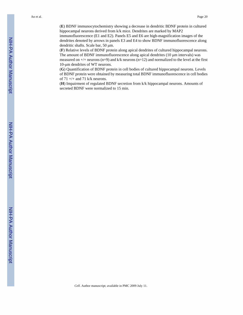
Dysmorphogenesis of dendritic spines in neurons lacking the long 3′ UTR mRNATo test whether the lack of dendritic BDNF mRNA in Bdnfklox/klox neurons affects dendriticgrowth, dendrites of Golgi-stained CA1 pyramidal neurons from 2-month old mice were tracedusing the Neurolucida software. Dendritic arbors of reconstructed WT CA1 pyramidal neuronsdid not appear different from those of Bdnfklox/klox neurons (Fig. 5A). Numbers of primary andsecondary basal dendrites were also similar between genotypes (Fig. 5B). In agreement withthese observations, Bdnfklox/klox mice had normal cytoarchitecture in the cortex and thehippocampus (Fig. S3).
In contrast to the relatively normal dendritic arbors, spines on distal apical dendrites of CA1pyramidal neurons were noticeably altered in Bdnfklox/klox mice at 2 months of age. Dendriticspines on higher orders of Golgi-stained apical dendrites in Bdnfklox/klox mice appeared thinnerand more numerous as compared to those in WT mice (Fig. 5C). The average spine headdiameter in Bdnfklox/klox mice was 20% smaller than that in WT mice (Fig. 5D). There was aleft-shift in cumulative distribution of spine head diameter, indicating that all classes ofdendritic spines have smaller heads in Bdnfklox/klox neurons (Fig. 5E). Lack of the long 3′UTRBDNF mRNA also led to more spines in apical dendrites. Quantitative analysis revealed a 54%increase in spine density in Bdnfklox/klox neurons (Fig. 5F).
The spine phenotypes could result from either lack of dendritic BDNF synthesis or increasedlevels of BDNF in cell bodies in Bdnfklox/klox CA1 pyramidal neurons. To distinguish betweenthese two possibilities, we used BTg mice that express 2-3 fold higher levels of BDNF mRNAin the hippocampus (Huang et al., 1999). Importantly, BDNF mRNA derived from thistransgene contains an SV40 3′UTR and therefore should stay in the soma. Indeed, whereas thein situ hybridization signals for BDNF mRNA in the s.p. were higher in BTg neurons ascompared to WT neurons (Fig. S4A), the distribution of BDNF mRNA along the apicaldendrites was similar between WT and BTg mice (Fig. S4B). Unlike Bdnfklox/klox neurons, theaverage spine head diameter in BTg neurons was not reduced (Fig. 5D, p=0.143). The spine
An et al. Page 5
Cell. Author manuscript; available in PMC 2009 July 11.
NIH
-PA Author Manuscript
NIH
-PA Author Manuscript
NIH
-PA Author Manuscript
density in BTg mice was only slightly increased (18% over WT, p=0.022), and much lowerthan that in Bdnfklox/klox mice (Fig. 5F, p=0.0021). These results argue that it is the decreasein dendritic BDNF, rather than the increase in somatic BDNF, that caused spinedysmorphogenesis.
We further investigated whether the increase in spine density in Bdnfklox/klox neurons is due tospine overproduction or lack of spine pruning. Spine pruning occurs after the third postnatalweek in mice (Zuo et al., 2005). Consistent with this, we found that the spine density was muchhigher in CA1 neurons in P21 juvenile WT mice than that in adult WT mice (3.43 ± 0.39 spines/μm at P21 vs. 1.96 ± 0.08 spines/μm at 2 months of age). If the lack of dendritic BDNF mRNAleads to spine overproduction, an increase in spine density is expected before spine pruningoccurs. However, the spine density on distal apical dendrites of CA1 pyramidal neurons wassimilar between WT and Bdnfklox/klox mice at P21 (Fig. 6A, p=0.517). In addition, we foundthat dendritic spines in WT and Bdnfklox/klox mice at P21 had similar sizes (Fig. 6B) anddistributions (Fig. 6C) of spine head diameter. These results suggest that BDNF mRNAs withthe long 3′UTR are not essential for the formation and initial growth of dendritic spines butare required for their pruning and enlargement.
Selective impairment of hippocampal LTP at apical dendrites in Bdnfklox/klox miceDeletion of the Bdnf gene results in marked impairment in LTP at hippocampal CA1 synapses(Korte et al., 1995; Patterson et al., 1996). It is not known, however, whether local BDNFsynthesis in apical dendrites of CA1 pyramidal neurons is important for CA1 LTP. To addressthis issue, we performed electrophysiological recording using hippocampal slices derived fromBdnfklox/klox mice and WT littermates. Robust LTP was induced in the apical dendrite regionof CA1 pyramidal neurons by applying tetanic stimulation (HFS, 2 × 100 Hz, 1 sec) to theSchaeffer collaterals in slices derived from WT animals (Fig. 7A). In contrast, slices derivedfrom mutant mice did not show persistent LTP (Fig. 7A). Mean field EPSP (fEPSP) slopes at60 min after HFS were 145 ± 4% and 113 ± 8% for WT and Bdnfklox/klox slices, respectively.In similar experiments, LTP induced by theta-burst stimulation (TBS) at CA1 synapses in thes.r. was also impaired in mutant mice as compared to WT mice (Fig. 7B). Thus, the long 3′UTR BDNF mRNA appears to be important for LTP expression in the dendritic region. As acontrol, input-output curves obtained by plotting the fEPSP slopes against fiber volleyamplitudes (proportional to stimulation intensities) were indistinguishable between thegenotypes, suggesting normal basal synaptic transmission (Fig. S5A). In addition, synapticresponses to a prolonged train of low-frequency repetitive stimulation (LFS), which results ina continuous decline of fEPSP slopes over time, also showed no differences between WT andBdnfklox/klox slices (Fig. S5B).
In addition to the LTP deficit, conventional Bdnf knockout mice exhibit impairments in pairedpulse facilitation (PPF) and synaptic response to high frequency stimulation (HFS), both ofwhich reflect changes in the properties of presynaptic terminals from CA3 neurons (Pozzo-Miller et al., 1999). We next assessed whether these forms of short-term presynaptic plasticityrequire BDNF derived from the apical dendrites of CA1 neurons. Surprisingly, synapticresponse to a brief HFS (100 Hz, 1 sec) was completely normal in Bdnfklox/klox slices (Fig. 7C).Additionally, PPF ratios were also the same at all tested intervals in Bdnfklox/klox and WT slices(Fig. 7D). Taken together, these results suggest that BDNF derived from the long 3′UTRmRNA may not be important for the regulation of the readily releasable pool of synapticvesicles.
Concomitant with the lack of dendritic BDNF mRNA, there is an increase in BDNF proteinin the cell bodies of CA1 pyramidal neurons. To examine whether synaptic plasticity may bedifferentially regulated in the somata and apical dendrites of Bdnfklox/klox neurons, we recordedLTP in cell bodies. Evoked population spikes were monitored by placing a field recording
An et al. Page 6
Cell. Author manuscript; available in PMC 2009 July 11.
NIH
-PA Author Manuscript
NIH
-PA Author Manuscript
NIH
-PA Author Manuscript
electrode in the s.p. and stimulating the proximal area of the CA1 region. HFS induced robustLTP in WT animals (Fig. 7E). In contrast to the LTP deficit in the dendritic region, themagnitude and duration of LTP induced in the cell body region were completely normal inBdnfklox/klox slices. The population spike amplitudes at 60 min after HFS were 155 ± 4% and154 ± 5% for WT and Bdnfklox/klox slices, respectively. To further investigate whether anincrease in BDNF protein in somata could alter neuronal excitability, we performed whole cellpatch clamp recordings of CA1 neurons in hippocampal slices while all synaptic transmissionwas pharmacologically inhibited (Fig. S6A). The number of action potentials increased,paralleling the increasing depolarization steps, but no difference was found between WT andBdnfklox/klox neurons (Fig. 7F). Additionally, the threshold, amplitude and half width of thefirst recorded action potentials were identical in both genotypes (Table S1). Finally, spikeaccommodation during a prolonged depolarization, defined by the ratio of 1st and 9th interspikeinterval (ISI), was similar between the two genotypes (Fig. S6B). Collectively, lack of the longBDNF mRNA altered neither synaptic function nor neuronal excitability in the soma regionof CA1 pyramidal neurons.
DISCUSSIONDifferential localization of mRNAs with short and long 3′UTRs
Our study provides an example where mRNAs containing the same coding sequence butdistinct 3′UTRs can have distinct physiological functions due to their selective subcellularlocalization and translation. The control of translation through differential use of the two 3′UTRs may be important for key regulatory proteins such as BDNF, which regulates manyaspects of neuronal structure and function. Translation of the long BDNF mRNA in dendritesmay regulate synaptic structure and function locally, whereas that of the short BDNF mRNAin the soma may produce BDNF protein required for neuronal survival and maintenance.Furthermore, dendritic local BDNF synthesis may offer a mechanism by which a diffusiblemolecule such as BDNF achieves local and synapse-specific modulation.
Transcripts with different lengths of 3′UTRs encoding the same protein appear to be a generalphenomenon. For example, the trkB gene encodes one full-length receptor (TrkB-F) and onetruncated receptor (TrkB-T) that lacks the tyrosine kinase domain (Klein et al., 1990). TrkBmRNA has been detected in dendrites of cultured hippocampal neurons (Tongiorgi et al.,1997). While both TrkB-F and TrkB-T mRNAs have short and long 3′UTRs, it remains to bedemonstrated whether they are targeted to different subcellular compartments. CaMKIIαmRNA also has a short 3′UTR and a long 3′UTR, although the long mRNA is much moreabundant. The sequence required for dendritic targeting has been mapped downstream of thefirst polyadenylation site (Blichenberg et al., 2001), suggesting that the short CaMKIIα mRNAstays in somata while the long CaMKIIα mRNA is transported to dendrites. Thus, the two-3′UTR strategy may be commonly employed for genes that have important functions in bothsomata and dendrites.
A unique system to study dendritic BDNF mRNA in vivoA growing number of mRNAs have been localized to neuronal dendrites (Steward andSchuman, 2003). It is generally believed that dendritic local protein synthesis is important inlong-lasting synaptic plasticity (Huber et al., 2000; Kang and Schuman, 1996; Miller et al.,2002). However, physiological functions of dendritically localized mRNAs remainundetermined due to the technical challenge of selectively abolishing dendritic local proteinsynthesis without affecting the total protein level in a neuron. For example, a mouse mutantwhere the native 3′UTR of CaMKIIα mRNA is replaced with the BGH 3′UTR exhibitsimpairment in dendritic localization of CaMKIIα mRNA as well as a significant reduction in
An et al. Page 7
Cell. Author manuscript; available in PMC 2009 July 11.
NIH
-PA Author Manuscript
NIH
-PA Author Manuscript
NIH
-PA Author Manuscript
overall expression of CaMKIIα (Miller et al., 2002). Thus, it is difficult to attribute behavioralabnormalities of the mutant to the lack of local CaMKIIα synthesis.
In Bdnfklox/klox mice, the long 3′UTR mRNA is truncated, and thus all BDNF transcripts areconverted to short 3′UTR mRNAs, leading to decreased dendritic BDNF mRNA. Importantly,levels of total BDNF mRNA and protein in the brain are unchanged, making it an ideal systemto examine the physiological function of dendritically localized mRNAs. Using this uniquemodel, we provide evidence that dendritically localized BDNF mRNAs are required formaintenance of a physiological level of BDNF protein in distal dendrites, maturation ofdendritic spines, and plasticity of synapses located on dendrites.
Local protein synthesis and pruning of dendritic spinesSpine pruning is as important as spine growth in controlling the number and location offunctional synapses. The pruning process is dependent on sensory experience (Zuo et al.,2005) and has been implicated in activity-dependent refinement of synaptic connections. Weshow here that Bdnfklox/klox mice have the same spine density on distal dendrites of hippocampalneurons as WT mice at P21 but do not show a reduction in spine density with maturation. Theseresults suggest that Bdnfklox/klox mice have deficits in spine pruning and implicate BDNF inthe control of spine pruning. Whether BDNF is truly involved in spine pruning requiresvalidation by future experiments using in vivo time-lapse imaging.
The deficit in spine pruning in distal dendrites of Bdnfklox/klox neurons appears to be due todecreased BDNF in dendrites, rather than an increase in the soma. This phenotype is verydifferent than the small increase seen in the number of spines for BTg neurons. In the lattercase, there is a global increase in BDNF levels in the entire dendritic field due to an increasein BDNF transported to dendrites from cell bodies, which leads to a small increase in spineformation. This is consistent with the published observation that application of BDNF tocultured neurons stimulates spine formation (Ji et al., 2005). In addition to the different timingsfor spine formation and pruning, our findings underscore the differential effects of globalincreases in BDNF signaling and local changes in BDNF concentration in dendrites.
How does locally synthesized BDNF regulate spine morphology and synaptic plasticity?Pro-BDNF is secreted and has unique biological activities in neurons (Lee et al., 2001; Wooet al., 2005), although the amount of secreted pro-BDNF remains controversial (Lee et al.,2001; Matsumoto et al., 2008). Since the majority of dendrites lack Golgi-like organelles toprocess secreted proteins (Horton et al., 2005), a significant portion of dendritically translatedBDNF is likely secreted as pro-BDNF, which can be converted to mature BDNF extracellularlyby proteases such as tPA (Pang et al., 2004). Since tPA is only secreted from stimulated spines(Lochner et al., 2006), the action of mature BDNF is limited to the stimulated spines byactivating TrkB in an autocrine manner. We speculate that TrkB signaling induces localsynthesis of proteins associated with spine growth and plasticity within and/or underneath thestimulated spines. These locally synthesized proteins promote actin dynamics and AMPAreceptor trafficking in spines, which leads to growth of the spine head and formation of stableLTP. In parallel, pro-BDNF diffused nearby should promote pruning of unstimulated spines,which lack tPA, by binding to the sortilin/p75NTR receptor complex with high affinity (Lee etal., 2001). This model is based on several previous observations in addition to our findingsdescribed here. First, the activation of TrkB-mediated signaling cascades has been shown toinduce local synthesis of several proteins, including Arc, CaMKIIα, Homer2, and LIMK1(Kang and Schuman, 1996; Schratt et al., 2004; Yin et al., 2002). While CaMKIIα is a keyregulator of AMPA receptor trafficking at synapses (Derkach et al., 2007), the other threeproteins promote actin polymerization required for LTP consolidation (Bramham and Wells,2007) and the maturation and enlargement of spine heads (Meng et al., 2002; Sala et al.,
An et al. Page 8
Cell. Author manuscript; available in PMC 2009 July 11.
NIH
-PA Author Manuscript
NIH
-PA Author Manuscript
NIH
-PA Author Manuscript
2001). Second, the gradual phase of spine enlargement induced by synaptic stimulation isdependent on protein synthesis and BDNF action (Tanaka et al., 2008). Finally, the p75NTR
receptor is localized in postsynaptic sites in the CA1 area (Woo et al., 2005) and deletion ofthe gene for the receptor results in increased spine density on apical dendrites of hippocampalneurons (Zagrebelsky et al., 2005). Our model predicts that selective inhibition of dendriticBDNF synthesis would reduce the size of spine heads but increase spine number, thephenotypes that we observed in Bdnfklox/klox mice.
While application of exogenous BDNF has been shown to increase the size of EPSPs (Kangand Schuman, 1995), deletion of the Bdnf gene or inhibition of TrkB signaling generallyimpairs only LTP without affecting basal synaptic transmission (Figurov et al., 1996; Pattersonet al., 1996; Xu et al., 2000). Here we show that inhibition of dendritic local BDNF synthesisalso affects early-phase LTP but not basal transmission in apical dendrites. The selectiveimpairment in dendritic LTP is consistent with the size reduction in spine heads observed onapical dendrites of CA1 neurons in Bdnfklox/klox mice. The increase in spine density in themutant mice may not alter synaptic transmission and plasticity, as these thin spines may notform functional connections with presynaptic terminals.
Given that early-phase LTP does not require newly synthesized proteins during its inductionand expression (Kelleher et al., 2004), it is unlikely that the LTP impairment in theBdnfklox/klox synapses is due to a failure in stimulation-induced BDNF synthesis in dendrites.Rather, a reduced basal level of BDNF in dendrites due to deficits in local BDNF synthesismay lead to accumulated deficiencies in LTP-generating signaling cascades at CA1 synapsesprior to LTP recording, which impair LTP expression in Bdnfklox/klox hippocampal slices.Moreover, the LTP deficits are less severe as compared to those in Bdnf knockout mice. Severalfactors could contribute to the difference between the two types of Bdnf mutant mice. First,despite lack of locally synthesized BDNF, there is a small amount of soma-derived BDNF inBdnfklox/klox dendrites, which could alleviate some of the LTP deficit. Second, the LTPphenotype in Bdnf knockout mice may results from both pre- and post-synaptic deficits(Gartner et al., 2006; Kovalchuk et al., 2002; Xu et al., 2000). However, Bdnfklox/klox miceexhibit normal PPF and response to HFS. Taken together, our data demonstrate the distinctroles of BDNF synthesized in somata vs. dendrites in synaptic plasticity.
EXPERIMENTAL PROCEDURESAnimals
Mice were maintained on standard mouse chow (Purina diet 5001) at 22 °C on a 12 h light/12h dark cycle. Both Bdnfklox/klox and BTg mouse strains were in the C57BL6/J geneticbackground. All animal procedures were approved by the Georgetown University Animal Careand Use Committee.
Culture and transfection of primary neuronsHippocampal and cortical neurons were isolated and cultured according to a previouslydescribed procedure (Sala et al., 2000) from E18.5 rat embryos or mouse pups at P0-P2. Weused lipofectamine 2000 or lipofectamine LTX to transfect cultured neurons.
In situ hybridizationIn situ hybridization of brain sections and cultured neurons was performed using DIG-labeledriboprobes and the TSA Plus Fluorescein System (PerkinElmer, Waltham, MA) according topreviously described procedures (Marz et al., 1998; Muddashetty et al., 2007) withmodifications. Brain sections at 40 μm were obtained with a sliding microtome from brainsfixed in 4% paraformaldehyde-20% sucrose for 3 days. Sections were digested with 10-30
An et al. Page 9
Cell. Author manuscript; available in PMC 2009 July 11.
NIH
-PA Author Manuscript
NIH
-PA Author Manuscript
NIH
-PA Author Manuscript
μg/ml proteinase K in PBS plus 0.1% Tween 20 for 20 min at room temperature to exposemRNAs before hybridization. After hybridization, sections or cells were treated with RNaseA at 37 °C to remove un-annealed probes.
GFP constructs and local protein synthesis assayThe EGFP coding sequence from the pEGFPN1 vector (BD Bioscience, San Jose, CA) wasinserted into pcDNA3.1(+) (Invitrogen, Carlsbad, CA) to generate the GFP-BGH construct.The mouse genomic sequences encoding 3′UTRs A, B and AB were obtained by PCR and usedto replace the BGH 3′UTR for generation of GFP-A, GFP-B, and GFP-AB constructs.
A sequence encoding the Src myristoylation peptide was added to the 5′-end of a PCR-amplified d1EGFP insert from plasmid pd1EGFP-N1, which was inserted into a plasmiddownstream the human synapsin promoter, generating phSYN-myr-d1GFP. The mousesequences A and A*B (where the first polyadenylation signal AATAAA was changed toTTTTTT) were cloned into phSYN-myr-d1GFP, generating phSYN-myr-d1GFP-A (myr-d1GFP-A) and phSYN-myr-d1GFP-A*B (myr-d1GFP-A*B). These two constructs weretransfected into cultured rat hippocampal neurons at 13 DIV. One day after transfection, thecultured neurons were treated with 1 μM TTX or vehicle for 6 hours and then fixed. The longestdendrites of transfected neurons were analyzed by quantifying the fluorescent intensity of aline drawn through the center of the dendrite, and mean intensity values for each conditionwere calculated using 50 μm bins.
Golgi impregnationGolgi staining was performed using the FD Rapid GolgiStain Kit (FD NeuroTechnologies,Ellicott City, MD). We used the Neurolucida software (MicroBrightField, Williston, VT) totrace dendrites. To measure spine density, we took images of distal dendrites with similardiameters in the stratum radiatum, enlarged the images with Photoshop software (AdobeSystems, San Jose, CA), and counted spines on printouts. All small protrusions on dendriteswere considered as spines. Spine density for each animal was obtained from many distaldendrites with a total length of 1000-2000 μm. Dendritic images from each mouse wererandomly used for measurement of spine size. The densely stained area at the tip of a spinewas considered as a spine head.
Immunohistochemistry, Northern blots, and ImmunoblottingImmunohistochemistry of brain sections and cultured neurons was carried out as previouslydescribed (Gharami et al., 2008). The antibody to BDNF (Santa Cruz Biotechnology, SantaCruz, CA) was used at 1:2,000 for brain sections, at 1:500 for cultured neurons, and at 1:1,000for immunoblotting. The mouse monoclonal antibody to MAP2 (1:500 dilution) was purchasedfrom Chemicon International (Temecula, CA). The probes for Northern blots were labeledusing the Rediprime II DNA labeling system (Amersham Bioscience, Piscataway, NJ).
BDNF secretionHippocampal neurons were seeded at a density of 5 × 10⁵ cells per well in a 12-well plate. Formeasurement of constitutive BDNF secretion, cells at 10 DIV were incubated with Neurobasalmedium supplemented with 0.1 μg/ml bovine serum albumin, 15 μM TTX, 20 μM D-2-amino-5-phosphonovaleric acid, and 20 μM 6-cyano-7-nitroquinoxaline-2,3-dione for 16hours. For measurement of regulated BDNF secretion, cells at 10 DIV were treated withNeurobasal medium supplemented with 0.1 μg/ml bovine serum albumin and 50 mM KCl for15 min. The amount of BDNF in each medium sample was determined using the BDNFEmax ImmunoAssay System (Promega, Madison, WI).
An et al. Page 10
Cell. Author manuscript; available in PMC 2009 July 11.
NIH
-PA Author Manuscript
NIH
-PA Author Manuscript
NIH
-PA Author Manuscript
Electrophysiological recordingTransverse hippocampal slices (400 μm) were prepared from Bdnfklox/klox and age-matchedWT mice (8-10 weeks old). Extracellular and whole cell recordings were performed aspreviously described (Xu et al., 2000).
Statistical analysisAll data are expressed as mean ± SEM. Data were analyzed using an unpaired Student's t-test;*, p<0.05; **, p<0.01; ***, p<0.001.
SUPPLEMENTAL DATARefer to Web version on PubMed Central for supplementary material.
AcknowledgementsWe thank S. Tonegawa for BTg mice, G. Bassell for sharing FISH protocols, Y. Chang for the pd1EGFP-N1 plasmid,D. Pak for sharing rat neurons, and L. Reichardt for critical reading of the manuscript. This work was supported bythe grants from the NIH (NS050596), the American Heart Association, the Whitehall Foundation, and the AmericanDiabetes Association to BX.
ReferencesAakalu G, Smith WB, Nguyen N, Jiang C, Schuman EM. Dynamic visualization of local protein synthesis
in hippocampal neurons. Neuron 2001;30:489–502. [PubMed: 11395009]Blichenberg A, Rehbein M, Muller R, Garner CC, Richter D, Kindler S. Identification of a cis-acting
dendritic targeting element in the mRNA encoding the alpha subunit of Ca2+/calmodulin-dependentprotein kinase II. Eur J Neurosci 2001;13:1881–1888. [PubMed: 11403681]
Bramham CR, Wells DG. Dendritic mRNA: transport, translation and function. Nat Rev Neurosci2007;8:776–789. [PubMed: 17848965]
Derkach VA, Oh MC, Guire ES, Soderling TR. Regulatory mechanisms of AMPA receptors in synapticplasticity. Nat Rev Neurosci 2007;8:101–113. [PubMed: 17237803]
Figurov A, Pozzo-Miller LD, Olafsson P, Wang T, Lu B. Regulation of synaptic responses to high-frequency stimulation and LTP by neurotrophins in the hippocampus. Nature 1996;381:706–709.[PubMed: 8649517]
Gartner A, Polnau DG, Staiger V, Sciarretta C, Minichiello L, Thoenen H, Bonhoeffer T, Korte M.Hippocampal long-term potentiation is supported by presynaptic and postsynaptic tyrosine receptorkinase B-mediated phospholipase Cgamma signaling. J Neurosci 2006;26:3496–3504. [PubMed:16571757]
Gharami K, Xie Y, An JJ, Tonegawa S, Xu B. Brain-derived neurotrophic factor over-expression in theforebrain ameliorates Huntington's disease phenotypes in mice. J Neurochem 2008;105:369–379.[PubMed: 18086127]
Ghosh A, Carnahan J, Greenberg ME. Requirement for BDNF in activity-dependent survival of corticalneurons. Science 1994;263:1618–1623. [PubMed: 7907431]
Gorski JA, Zeiler SR, Tamowski S, Jones KR. Brain-derived neurotrophic factor is required for themaintenance of cortical dendrites. J Neurosci 2003;23:6856–6865. [PubMed: 12890780]
Horton AC, Racz B, Monson EE, Lin AL, Weinberg RJ, Ehlers MD. Polarized secretory traffickingdirects cargo for asymmetric dendrite growth and morphogenesis. Neuron 2005;48:757–771.[PubMed: 16337914]
Huang ZJ, Kirkwood A, Pizzorusso T, Porciatti V, Morales B, Bear MF, Maffei L, Tonegawa S. BDNFregulates the maturation of inhibition and the critical period of plasticity in mouse visual cortex. Cell1999;98:739–755. [PubMed: 10499792]
Huber KM, Kayser MS, Bear MF. Role for rapid dendritic protein synthesis in hippocampal mGluR-dependent long-term depression. Science 2000;288:1254–1257. [PubMed: 10818003]
An et al. Page 11
Cell. Author manuscript; available in PMC 2009 July 11.
NIH
-PA Author Manuscript
NIH
-PA Author Manuscript
NIH
-PA Author Manuscript

Ji Y, Pang PT, Feng L, Lu B. Cyclic AMP controls BDNF-induced TrkB phosphorylation and dendriticspine formation in mature hippocampal neurons. Nat Neurosci 2005;8:164–172. [PubMed:15665879]
Kang H, Schuman EM. Long-lasting neurotrophin-induced enhancement of synaptic transmission in theadult hippocampus. Science 1995;267:1658–1662. [PubMed: 7886457]
Kang H, Schuman EM. A requirement for local protein synthesis in neurotrophin-induced hippocampalsynaptic plasticity. Science 1996;273:1402–1406. [PubMed: 8703078]
Kelleher RJ 3rd, Govindarajan A, Tonegawa S. Translational regulatory mechanisms in persistent formsof synaptic plasticity. Neuron 2004;44:59–73. [PubMed: 15450160]
Klein R, Conway D, Parada LF, Barbacid M. The trkB tyrosine protein kinase gene codes for a secondneurogenic receptor that lacks the catalytic kinase domain. Cell 1990;61:647–656. [PubMed:2160854]
Kobayashi H, Yamamoto S, Maruo T, Murakami F. Identification of a cis-acting element required fordendritic targeting of activity-regulated cytoskeleton-associated protein mRNA. Eur J Neurosci2005;22:2977–2984. [PubMed: 16367764]
Korte M, Carroll P, Wolf E, Brem G, Thoenen H, Bonhoeffer T. Hippocampal long-term potentiation isimpaired in mice lacking brain-derived neurotrophic factor. Proc Natl Acad Sci U S A 1995;92:8856–8860. [PubMed: 7568031]
Kovalchuk Y, Hanse E, Kafitz KW, Konnerth A. Postsynaptic induction of BDNF-mediated long-termpotentiation. Science 2002;295:1729–1734. [PubMed: 11872844]
Lee R, Kermani P, Teng KK, Hempstead BL. Regulation of cell survival by secreted proneurotrophins.Science 2001;294:1945–1948. [PubMed: 11729324]
Liu QR, Lu L, Zhu XG, Gong JP, Shaham Y, Uhl GR. Rodent BDNF genes, novel promoters, novelsplice variants, and regulation by cocaine. Brain Res 2006;1067:1–12. [PubMed: 16376315]
Lochner JE, Honigman LS, Grant WF, Gessford SK, Hansen AB, Silverman MA, Scalettar BA. Activity-dependent release of tissue plasminogen activator from the dendritic spines of hippocampal neuronsrevealed by live-cell imaging. J Neurobiol 2006;66:564–577. [PubMed: 16555239]
Lu B. BDNF and activity-dependent synaptic modulation. Learn Mem 2003;10:86–98. [PubMed:12663747]
Marz P, Cheng JG, Gadient RA, Patterson PH, Stoyan T, Otten U, Rose-John S. Sympathetic neuronscan produce and respond to interleukin 6. Proc Natl Acad Sci U S A 1998;95:3251–3256. [PubMed:9501249]
Matsumoto T, Rauskolb S, Polack M, Klose J, Kolbeck R, Korte M, Barde YA. Biosynthesis andprocessing of endogenous BDNF: CNS neurons store and secrete BDNF, not pro-BDNF. NatNeurosci 2008;11:131–133. [PubMed: 18204444]
Meng Y, Zhang Y, Tregoubov V, Janus C, Cruz L, Jackson M, Lu WY, MacDonald JF, Wang JY, FallsDL, et al. Abnormal spine morphology and enhanced LTP in LIMK-1 knockout mice. Neuron2002;35:121–133. [PubMed: 12123613]
Miller S, Yasuda M, Coats JK, Jones Y, Martone ME, Mayford M. Disruption of dendritic translation ofCaMKIIalpha impairs stabilization of synaptic plasticity and memory consolidation. Neuron2002;36:507–519. [PubMed: 12408852]
Muddashetty RS, Kelic S, Gross C, Xu M, Bassell GJ. Dysregulated metabotropic glutamate receptor-dependent translation of AMPA receptor and postsynaptic density-95 mRNAs at synapses in a mousemodel of fragile X syndrome. J Neurosci 2007;27:5338–5348. [PubMed: 17507556]
Pang PT, Teng HK, Zaitsev E, Woo NT, Sakata K, Zhen S, Teng KK, Yung WH, Hempstead BL, Lu B.Cleavage of proBDNF by tPA/plasmin is essential for long-term hippocampal plasticity. Science2004;306:487–491. [PubMed: 15486301]
Paradies MA, Steward O. Multiple subcellular mRNA distribution patterns in neurons: a nonisotopic insitu hybridization analysis. J Neurobiol 1997;33:473–493. [PubMed: 9322162]
Patterson SL, Abel T, Deuel TA, Martin KC, Rose JC, Kandel ER. Recombinant BDNF rescues deficitsin basal synaptic transmission and hippocampal LTP in BDNF knockout mice. Neuron1996;16:1137–1145. [PubMed: 8663990]
Pozzo-Miller LD, Gottschalk W, Zhang L, McDermott K, Du J, Gopalakrishnan R, Oho C, Sheng ZH,Lu B. Impairments in high-frequency transmission, synaptic vesicle docking, and synaptic protein
An et al. Page 12
Cell. Author manuscript; available in PMC 2009 July 11.
NIH
-PA Author Manuscript
NIH
-PA Author Manuscript
NIH
-PA Author Manuscript
distribution in the hippocampus of BDNF knockout mice. J Neurosci 1999;19:4972–4983. [PubMed:10366630]
Reichardt LF. Neurotrophin-regulated signalling pathways. Philos Trans R Soc Lond B Biol Sci2006;361:1545–1564. [PubMed: 16939974]
Rook MS, Lu M, Kosik KS. CaMKIIalpha 3′ untranslated region-directed mRNA translocation in livingneurons: visualization by GFP linkage. J Neurosci 2000;20:6385–6393. [PubMed: 10964944]
Sala C, Piech V, Wilson NR, Passafaro M, Liu G, Sheng M. Regulation of dendritic spine morphologyand synaptic function by Shank and Homer. Neuron 2001;31:115–130. [PubMed: 11498055]
Sala C, Rudolph-Correia S, Sheng M. Developmentally regulated NMDA receptor-dependentdephosphorylation of cAMP response element-binding protein (CREB) in hippocampal neurons. JNeurosci 2000;20:3529–3536. [PubMed: 10804193]
Schratt GM, Nigh EA, Chen WG, Hu L, Greenberg ME. BDNF regulates the translation of a select groupof mRNAs by a mammalian target of rapamycin-phosphatidylinositol 3-kinase-dependent pathwayduring neuronal development. J Neurosci 2004;24:7366–7377. [PubMed: 15317862]
Steward O, Schuman EM. Compartmentalized synthesis and degradation of proteins in neurons. Neuron2003;40:347–359. [PubMed: 14556713]
Sutton MA, Wall NR, Aakalu GN, Schuman EM. Regulation of dendritic protein synthesis by miniaturesynaptic events. Science 2004;304:1979–1983. [PubMed: 15218151]
Tanaka J, Horiike Y, Matsuzaki M, Miyazaki T, Ellis-Davies GC, Kasai H. Protein synthesis andneurotrophin-dependent structural plasticity of single dendritic spines. Science 2008;319:1683–1687. [PubMed: 18309046]
Timmusk T, Palm K, Metsis M, Reintam T, Paalme V, Saarma M, Persson H. Multiple promoters directtissue-specific expression of the rat BDNF gene. Neuron 1993;10:475–489. [PubMed: 8461137]
Tongiorgi E, Armellin M, Giulianini PG, Bregola G, Zucchini S, Paradiso B, Steward O, Cattaneo A,Simonato M. Brain-derived neurotrophic factor mRNA and protein are targeted to discrete dendriticlaminas by events that trigger epileptogenesis. J Neurosci 2004;24:6842–6852. [PubMed: 15282290]
Tongiorgi E, Righi M, Cattaneo A. Activity-dependent dendritic targeting of BDNF and TrkB mRNAsin hippocampal neurons. J Neurosci 1997;17:9492–9505. [PubMed: 9391005]
Woo NH, Teng HK, Siao CJ, Chiaruttini C, Pang PT, Milner TA, Hempstead BL, Lu B. Activation ofp75NTR by proBDNF facilitates hippocampal long-term depression. Nat Neurosci 2005;8:1069–1077. [PubMed: 16025106]
Xu B, Gottschalk W, Chow A, Wilson RI, Schnell E, Zang K, Wang D, Nicoll RA, Lu B, Reichardt LF.The role of brain-derived neurotrophic factor receptors in the mature hippocampus: modulation oflong-term potentiation through a presynaptic mechanism involving TrkB. J Neurosci 2000;20:6888–6897. [PubMed: 10995833]
Yin Y, Edelman GM, Vanderklish PW. The brain-derived neurotrophic factor enhances synthesis of Arcin synaptoneurosomes. Proc Natl Acad Sci U S A 2002;99:2368–2373. [PubMed: 11842217]
Zagrebelsky M, Holz A, Dechant G, Barde YA, Bonhoeffer T, Korte M. The p75 neurotrophin receptornegatively modulates dendrite complexity and spine density in hippocampal neurons. J Neurosci2005;25:9989–9999. [PubMed: 16251447]
Zuo Y, Yang G, Kwon E, Gan WB. Long-term sensory deprivation prevents dendritic spine loss inprimary somatosensory cortex. Nature 2005;436:261–265. [PubMed: 16015331]
An et al. Page 13
Cell. Author manuscript; available in PMC 2009 July 11.
NIH
-PA Author Manuscript
NIH
-PA Author Manuscript
NIH
-PA Author Manuscript
Figure 1. Localization of long 3′UTR BDNF mRNA to dendrites(A) Diagram of the mouse Bdnf gene depicting two alternative polyadenylation (pA) sites inexon 8 (arrows). Curved lines linking boxes (exons) indicate alternative splicing from the firstseven exons to exon 8. The filled box within exon 8 represents the coding sequence.(B) Long and short 3′UTR BDNF mRNAs in different brain regions. BDNF mRNAs with ashort 3′UTR (BDNF-S) or a long 3′UTR (BDNF-L) were present in the cortex (Ctx),hippocampus (Hip), striatum (Stm), hypothalamus (Hyp), cerebellum (Cbl), and brainstem(BS). Lower panel: 18S rRNA as a loading control.(C) Ratios of long and short BDNF mRNAs (L/S) derived from different Bdnf promoters. Thesame blot was used for all probes (n=4 mice).(D) Localization of BDNF mRNAs in dendrites of hippocampal CA1 and cortical neurons.Riboprobes were derived from the coding regions for CaMKIIα, BDNF, and β-tubulin.CaMKIIα and BDNF mRNAs were detected in dendrites (arrows). Abbreviations in this andall other figures: s.o., stratum oriens; s.p., stratum pyramidale; s.r., stratum radiatum. Scalebars, 50 μm.(E) Relative abundance of the long 3′UTR BDNF mRNA in the soma and dendrite fractions.
An et al. Page 14
Cell. Author manuscript; available in PMC 2009 July 11.
NIH
-PA Author Manuscript
NIH
-PA Author Manuscript
NIH
-PA Author Manuscript

(F) Ratio of the long 3′UTR signal to the coding region signal in the soma and dendrite fractions(n=3).(G) Distribution of BDNF mRNA in cell bodies and dendrites of cultured rat cortical neurons.MAP2 immunostaining marks cell bodies and dendrites. Scale bar, 20 μm.(H) Ratio of FISH signals in dendrites to those in cell bodies.
An et al. Page 15
Cell. Author manuscript; available in PMC 2009 July 11.
NIH
-PA Author Manuscript
NIH
-PA Author Manuscript
NIH
-PA Author Manuscript
Figure 2. Cis-acting sequence in the 3′UTR required for the dendritic localization(A) Schematic of four GFP constructs with different 3′UTRs.(B-E) Localization of GFP mRNA in neurons transfected with GFP-BGH (B), GFP-A (C),GFP-AB (D), or GFP-B (E). Top panels: FISH of cultured neurons with a GFP antisenseriboprobe; Bottom panels: MAP2 immunocytochemistry; *, an untransfected neuron; Scalebar, 25 μm.(F) Levels of GFP mRNAs along the main apical dendrites of transfected neurons. Data wereobtained from 15-23 transfected neurons grown on 3 coverslips for each construct andpresented in arbitrary unit (a.u.). GFP-A was compared with the other three constructs.(G) Whole cell images of cultured hippocampal neurons expressing either myr-d1GFP-A ormyr-d1GFP-A*B. The images were taken with a fluorescent microscope using a GFP filter.Scale bar, 50 μm.(H) Quantification of myr-d1GFP fluorescence in dendrites. Fluorescence intensities on distaldendrites (100-150 μm and 150-200 μm away from somata) were measured and normalized tocontrol levels. The number of neurons for each condition is indicated inside bars.
An et al. Page 16
Cell. Author manuscript; available in PMC 2009 July 11.
NIH
-PA Author Manuscript
NIH
-PA Author Manuscript
NIH
-PA Author Manuscript
Figure 3. Impairment of dendritic targeting of BDNF mRNA in 3′UTR mutant mice(A) The Bdnfklox allele generated by insertion of a lox P sequence into the 5′UTR within exon8 and into a site 3′ to the first polyadenylation site of a sequence containing three tandem SV40polyadenylation signals and a lacZ gene.(B) Northern blot analysis of BDNF mRNA from the cortex of WT (+/+) and Bdnfklox/klox (k/k) mice. Total RNA (10 μg) was loaded onto each lane. The asterisk denotes a new BDNFmRNA species. Lower panel: 18S rRNA as a loading control.(C-J) In situ hybridization revealing defects in dendritic targeting of BDNF mRNA in k/k mice.In layer 5 of the cortex (C, D) and the hippocampal CA1 region (G, H), there is a markedreduction in levels of dendritic BDNF mRNA in k/k mice as compared to +/+ mice. Arrows
An et al. Page 17
Cell. Author manuscript; available in PMC 2009 July 11.
NIH
-PA Author Manuscript
NIH
-PA Author Manuscript
NIH
-PA Author Manuscript

denote representative dendrites containing BDNF mRNA. Also note many small punctacontaining BDNF mRNA in G. The sense probe did not produce significant signals in thecortex (E, F) or hippocampal CA1 region (I, J). Scale bar, 50 μm.(K) Quantification of BDNF mRNA in situ hybridization signals in the s.r. of +/+ and k/k mice(n=4 mice each; 3 sections per mouse).
An et al. Page 18
Cell. Author manuscript; available in PMC 2009 July 11.
NIH
-PA Author Manuscript
NIH
-PA Author Manuscript
NIH
-PA Author Manuscript
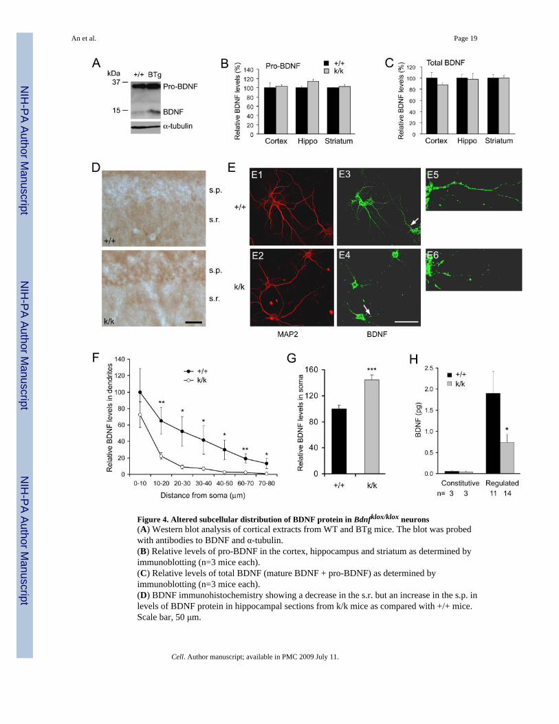
Figure 4. Altered subcellular distribution of BDNF protein in Bdnfklox/klox neurons(A) Western blot analysis of cortical extracts from WT and BTg mice. The blot was probedwith antibodies to BDNF and α-tubulin.(B) Relative levels of pro-BDNF in the cortex, hippocampus and striatum as determined byimmunoblotting (n=3 mice each).(C) Relative levels of total BDNF (mature BDNF + pro-BDNF) as determined byimmunoblotting (n=3 mice each).(D) BDNF immunohistochemistry showing a decrease in the s.r. but an increase in the s.p. inlevels of BDNF protein in hippocampal sections from k/k mice as compared with +/+ mice.Scale bar, 50 μm.
An et al. Page 19
Cell. Author manuscript; available in PMC 2009 July 11.
NIH
-PA Author Manuscript
NIH
-PA Author Manuscript
NIH
-PA Author Manuscript
(E) BDNF immunocytochemistry showing a decrease in dendritic BDNF protein in culturedhippocampal neurons derived from k/k mice. Dendrites are marked by MAP2immunofluorescence (E1 and E2). Panels E5 and E6 are high-magnification images of thedendrites denoted by arrows in panels E3 and E4 to show BDNF immunofluorescence alongdendritic shafts. Scale bar, 50 μm.(F) Relative levels of BDNF protein along apical dendrites of cultured hippocampal neurons.The amount of BDNF immunofluorescence along apical dendrites (10 μm intervals) wasmeasured on +/+ neurons (n=9) and k/k neurons (n=12) and normalized to the level at the first10-μm dendrites of WT neurons.(G) Quantification of BDNF protein in cell bodies of cultured hippocampal neurons. Levelsof BDNF protein were obtained by measuring total BDNF immunofluorescence in cell bodiesof 71 +/+ and 71 k/k neurons.(H) Impairment of regulated BDNF secretion from k/k hippocampal neurons. Amounts ofsecreted BDNF were normalized to 15 min.
An et al. Page 20
Cell. Author manuscript; available in PMC 2009 July 11.
NIH
-PA Author Manuscript
NIH
-PA Author Manuscript
NIH
-PA Author Manuscript
Figure 5. Dysmorphogenesis of dendritic spines in Bdnfklox/klox hippocampal neurons at 2 monthsof age(A) Representative dendritic arbors of CA1 pyramidal neurons, reconstructed with theNeurolucida software from Golgi-impregnated brain sections of WT (+/+) and Bdnfklox/klox (k/k) mice.(B) Similar dendritic numbers in CA1 pyramidal neurons from +/+ and k/k mice. Primary andsecondary basal dendrites from 12 +/+ neurons and 13 k/k neurons were traced with theNeurolucida software and counted (n=4 mice for each genotype). p=0.12 for primary dendrites;p=0.19 for secondary dendrites.(C) Representative dendritic spines on distal apical dendrites of +/+ and k/k mice. Scale bar,5 μm.
An et al. Page 21
Cell. Author manuscript; available in PMC 2009 July 11.
NIH
-PA Author Manuscript
NIH
-PA Author Manuscript
NIH
-PA Author Manuscript

(D) Reduction in spine size in k/k neurons. Average spine head diameter was calculated frommeasurements on 355 +/+ spines (3 mice), 355 k/k spines (3 mice), and 343 BTg spines (3mice).(E) Cumulative distribution curves for spine head diameter, using the data in D.(F) Spine density on distal apical dendrites of CA1 pyramidal neurons (n=3 mice each).
An et al. Page 22
Cell. Author manuscript; available in PMC 2009 July 11.
NIH
-PA Author Manuscript
NIH
-PA Author Manuscript
NIH
-PA Author Manuscript
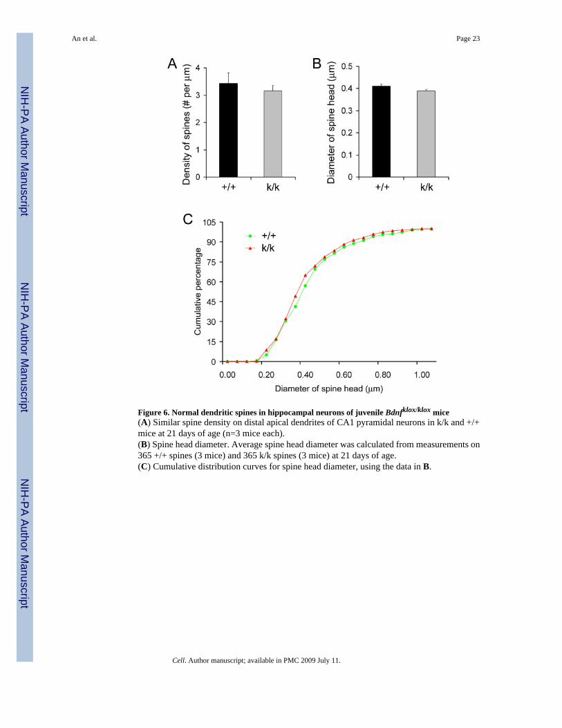
Figure 6. Normal dendritic spines in hippocampal neurons of juvenile Bdnfklox/klox mice(A) Similar spine density on distal apical dendrites of CA1 pyramidal neurons in k/k and +/+mice at 21 days of age (n=3 mice each).(B) Spine head diameter. Average spine head diameter was calculated from measurements on365 +/+ spines (3 mice) and 365 k/k spines (3 mice) at 21 days of age.(C) Cumulative distribution curves for spine head diameter, using the data in B.
An et al. Page 23
Cell. Author manuscript; available in PMC 2009 July 11.
NIH
-PA Author Manuscript
NIH
-PA Author Manuscript
NIH
-PA Author Manuscript
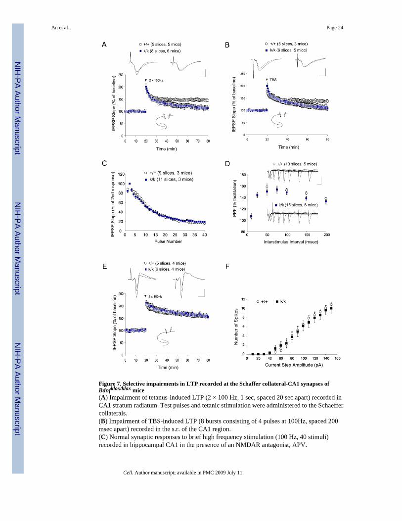
Figure 7. Selective impairments in LTP recorded at the Schaffer collateral-CA1 synapses ofBdnfklox/klox mice(A) Impairment of tetanus-induced LTP (2 × 100 Hz, 1 sec, spaced 20 sec apart) recorded inCA1 stratum radiatum. Test pulses and tetanic stimulation were administered to the Schaeffercollaterals.(B) Impairment of TBS-induced LTP (8 bursts consisting of 4 pulses at 100Hz, spaced 200msec apart) recorded in the s.r. of the CA1 region.(C) Normal synaptic responses to brief high frequency stimulation (100 Hz, 40 stimuli)recorded in hippocampal CA1 in the presence of an NMDAR antagonist, APV.
An et al. Page 24
Cell. Author manuscript; available in PMC 2009 July 11.
NIH
-PA Author Manuscript
NIH
-PA Author Manuscript
NIH
-PA Author Manuscript
(D) Normal paired pulse facilitation in k/k mice. No significant difference was observed whenthe PPF ratio (second fEPSP slope / first fEPSP * 100) was plotted against different inter-stimulus intervals.(E) Normal tetanus-induced LTP was recorded in the s.p. area. Test pulses and tetanicstimulation were administered to the Schaeffer collaterals.(F) Normal excitability of CA1 pyramidal neurons in k/k mice. Spike number in response todepolarizing current steps was not significantly different between +/+ and k/k mice at all currentsteps tested.
An et al. Page 25
Cell. Author manuscript; available in PMC 2009 July 11.
NIH
-PA Author Manuscript
NIH
-PA Author Manuscript
NIH
-PA Author Manuscript
Related Documents