doi: 10.1136/ard.2009.128207 published online May 24, 2010 Ann Rheum Dis L van Bon, C Popa, R Huijbens, et al. and diffuse cutaneous systemic sclerosis cell cytokine secretion in patients with limited Distinct evolution of TLR-mediated dendritic http://ard.bmj.com/content/early/2010/05/21/ard.2009.128207.full.html Updated information and services can be found at: These include: References http://ard.bmj.com/content/early/2010/05/21/ard.2009.128207.full.html#ref-list-1 This article cites 31 articles, 12 of which can be accessed free at: P<P Published online May 24, 2010 in advance of the print journal. service Email alerting box at the top right corner of the online article. Receive free email alerts when new articles cite this article. Sign up in the Notes articles must include the digital object identifier (DOIs) and date of initial publication. priority; they are indexed by PubMed from initial publication. Citations to Advance online prior to final publication). Advance online articles are citable and establish publication yet appeared in the paper journal (edited, typeset versions may be posted when available Advance online articles have been peer reviewed and accepted for publication but have not http://ard.bmj.com/cgi/reprintform To order reprints of this article go to: http://ard.bmj.com/subscriptions go to: Annals of the Rheumatic Diseases To subscribe to group.bmj.com on June 15, 2010 - Published by ard.bmj.com Downloaded from

Welcome message from author
This document is posted to help you gain knowledge. Please leave a comment to let me know what you think about it! Share it to your friends and learn new things together.
Transcript

doi: 10.1136/ard.2009.128207 published online May 24, 2010Ann Rheum Dis
L van Bon, C Popa, R Huijbens, et al. and diffuse cutaneous systemic sclerosiscell cytokine secretion in patients with limited Distinct evolution of TLR-mediated dendritic
http://ard.bmj.com/content/early/2010/05/21/ard.2009.128207.full.htmlUpdated information and services can be found at:
These include:
References http://ard.bmj.com/content/early/2010/05/21/ard.2009.128207.full.html#ref-list-1
This article cites 31 articles, 12 of which can be accessed free at:
P<P Published online May 24, 2010 in advance of the print journal.
serviceEmail alerting
box at the top right corner of the online article.Receive free email alerts when new articles cite this article. Sign up in the
Notes
articles must include the digital object identifier (DOIs) and date of initial publication. priority; they are indexed by PubMed from initial publication. Citations to Advance online prior to final publication). Advance online articles are citable and establish publicationyet appeared in the paper journal (edited, typeset versions may be posted when available Advance online articles have been peer reviewed and accepted for publication but have not
http://ard.bmj.com/cgi/reprintformTo order reprints of this article go to:
http://ard.bmj.com/subscriptions go to: Annals of the Rheumatic DiseasesTo subscribe to
group.bmj.com on June 15, 2010 - Published by ard.bmj.comDownloaded from
Extended report
van Bon L, Popa C, Huijbens R, et al. Ann Rheum Dis (2010). doi:10.1136/ard.2009.128207 1 of 9
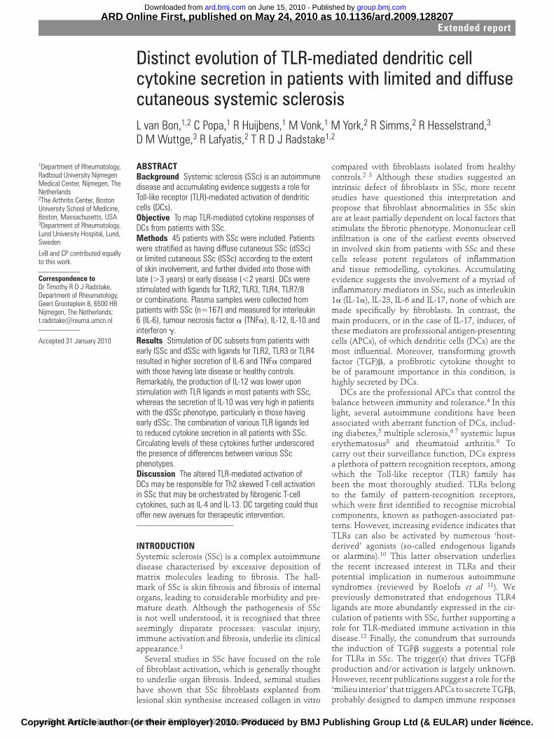
ABSTRACTBackground Systemic sclerosis (SSc) is an autoimmune
disease and accumulating evidence suggests a role for
Toll-like receptor (TLR)-mediated activation of dendritic
cells (DCs).
Objective To map TLR-mediated cytokine responses of
DCs from patients with SSc.
Methods 45 patients with SSc were included. Patients
were stratifi ed as having diffuse cutaneous SSc (dSSc)
or limited cutaneous SSc (lSSc) according to the extent
of skin involvement, and further divided into those with
late (>3 years) or early disease (<2 years). DCs were
stimulated with ligands for TLR2, TLR3, TLR4, TLR7/8
or combinations. Plasma samples were collected from
patients with SSc (n=167) and measured for interleukin
6 (IL-6), tumour necrosis factor α (TNFα), IL-12, IL-10 and
interferon γ.
Results Stimulation of DC subsets from patients with
early lSSc and dSSc with ligands for TLR2, TLR3 or TLR4
resulted in higher secretion of IL-6 and TNFα compared
with those having late disease or healthy controls.
Remarkably, the production of IL-12 was lower upon
stimulation with TLR ligands in most patients with SSc,
whereas the secretion of IL-10 was very high in patients
with the dSSc phenotype, particularly in those having
early dSSc. The combination of various TLR ligands led
to reduced cytokine secretion in all patients with SSc.
Circulating levels of these cytokines further underscored
the presence of differences between various SSc
phenotypes.
Discussion The altered TLR-mediated activation of
DCs may be responsible for Th2 skewed T-cell activation
in SSc that may be orchestrated by fi brogenic T-cell
cytokines, such as IL-4 and IL-13. DC targeting could thus
offer new avenues for therapeutic intervention.
INTRODUCTIONSystemic sclerosis (SSc) is a complex autoimmune disease characterised by excessive deposition of matrix molecules leading to fi brosis. The hall-mark of SSc is skin fi brosis and fi brosis of internal organs, leading to considerable morbidity and pre-mature death. Although the pathogenesis of SSc is not well understood, it is recognised that three seemingly disparate processes: vascular injury, immune activation and fi brosis, underlie its clinical appearance.1
Several studies in SSc have focused on the role of fi broblast activation, which is generally thought to underlie organ fi brosis. Indeed, seminal studies have shown that SSc fi broblasts explanted from lesional skin synthesise increased collagen in vitro
Distinct evolution of TLR-mediated dendritic cell cytokine secretion in patients with limited and diffuse cutaneous systemic sclerosisL van Bon,1,2 C Popa,1 R Huijbens,1 M Vonk,1 M York,2 R Simms,2 R Hesselstrand,3
D M Wuttge,3 R Lafyatis,2 T R D J Radstake1,2
1Department of Rheumatology, Radboud University Nijmegen Medical Center, Nijmegen, The Netherlands2The Arthritis Center, Boston University School of Medicine, Boston, Massachusetts, USA3Department of Rheumatology, Lund University Hospital, Lund, Sweden
LvB and CP contributed equally to this work.
Correspondence to Dr Timothy R D J Radstake, Department of Rheumatology, Geert Grooteplein 8, 6500 HB Nijmegen, The Netherlands; [email protected]
Accepted 31 January 2010
compared with fi broblasts isolated from healthy controls.2 3 Although these studies suggested an intrinsic defect of fi broblasts in SSc, more recent studies have questioned this interpretation and propose that fi broblast abnormalities in SSc skin are at least partially dependent on local factors that stimulate the fi brotic phenotype. Mononuclear cell infi ltration is one of the earliest events observed in involved skin from patients with SSc and these cells release potent regulators of infl ammation and tissue remodelling, cytokines. Accumulating evidence suggests the involvement of a myriad of infl ammatory mediators in SSc, such as interleukin 1α (IL-1α), IL-23, IL-6 and IL-17, none of which are made specifi cally by fi broblasts. In contrast, the main producers, or in the case of IL-17, inducer, of these mediators are professional antigen-presenting cells (APCs), of which dendritic cells (DCs) are the most infl uential. Moreover, transforming growth factor (TGF)β, a profi brotic cytokine thought to be of paramount importance in this condition, is highly secreted by DCs.
DCs are the professional APCs that control the balance between immunity and tolerance.4 In this light, several autoimmune conditions have been associated with aberrant function of DCs, includ-ing diabetes,5 multiple sclerosis,6 7 systemic lupus erythematosus8 and rheumatoid arthritis.9 To carry out their surveillance function, DCs express a plethora of pattern recognition receptors, among which the Toll-like receptor (TLR) family has been the most thoroughly studied. TLRs belong to the family of pattern-recognition receptors, which were fi rst identifi ed to recognise microbial components, known as pathogen-associated pat-terns. However, increasing evidence indicates that TLRs can also be activated by numerous ‘host-derived’ agonists (so-called endogenous ligands or alarmins).10 This latter observation underlies the recent increased interest in TLRs and their potential implication in numerous autoimmune syndromes (reviewed by Roelofs et al 11). We previously demonstrated that endogenous TLR4 ligands are more abundantly expressed in the cir-culation of patients with SSc, further supporting a role for TLR-mediated immune activation in this disease.12 Finally, the conundrum that surrounds the induction of TGFβ suggests a potential role for TLRs in SSc. The trigger(s) that drives TGFβ production and/or activation is largely unknown. However, recent publications suggest a role for the ‘milieu interior’ that triggers APCs to secrete TGFβ, probably designed to dampen immune responses
annrheumdis128207.indd 1annrheumdis128207.indd 1 5/17/2010 12:19:10 PM5/17/2010 12:19:10 PM
ARD Online First, published on May 24, 2010 as 10.1136/ard.2009.128207
Copyright Article author (or their employer) 2010. Produced by BMJ Publishing Group Ltd (& EULAR) under licence.
group.bmj.com on June 15, 2010 - Published by ard.bmj.comDownloaded from
Extended report
van Bon L, Popa C, Huijbens R, et al. Ann Rheum Dis (2010). doi:10.1136/ard.2009.1282072 of 9
and limit tissue damage. For example, zymosan, a ligand for TLR2 was shown to induce IL-10 and TGFβ-expressing macro-phages that could induce immunological tolerance in vitro.13 In addition, TLR4 has been shown to be crucial in the devel-opment of hepatic fi brosis.14 Thus, while the emerging picture of the pathogenesis of SSc is one of enormous complexities; many observations suggest a role for DCs and TLR-mediated DC activation in this condition.
We show here that the stimulation of DCs from patients with SSc with single TLR ligands results in the increased production of various cytokines compared with that seen by DCs from healthy controls. Interestingly, TLR-stimulated IL-12 secretion was lower in patients with SSc with early disease, whereas IL-10 production was markedly increased. Stimulation with combi-nations of TLR agonists resulted in a much lower secretion of infl ammatory mediators by DCs from patients with SSc com-pared with that in healthy controls. Again, IL-10 secretion was higher in DCs from patients with SSc, suggesting that TLR ago-nists in patients with SSc might stimulate strong Th2 skewing. These observed aberrant TLR responses may have an important role in the production of profi brotic mediators such as IL-13.
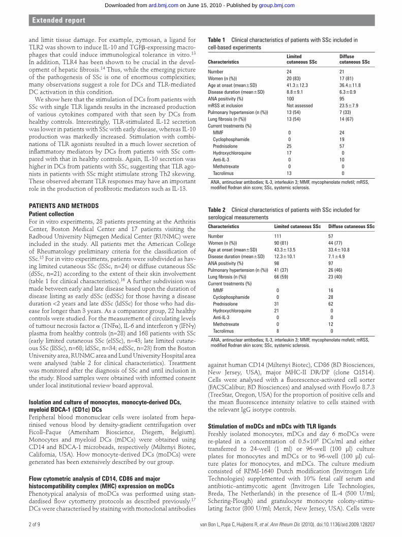
PATIENTS AND METHODSPatient collectionFor in vitro experiments, 28 patients presenting at the Arthritis Center, Boston Medical Center and 17 patients visiting the Radboud University Nijmegen Medical Center (RUNMC) were included in the study. All patients met the American College of Rheumatology preliminary criteria for the classifi cation of SSc.15 For in vitro experiments, patients were subdivided as hav-ing limited cutaneous SSc (lSSc, n=24) or diffuse cutaneous SSc (dSSc, n=21) according to the extent of their skin involvement (table 1 for clinical characteristics).16 A further subdivision was made between early and late disease based upon the duration of disease listing as early dSSc (edSSc) for those having a disease duration <2 years and late dSSc (ldSSc) for those who had dis-ease for longer than 3 years. As a comparator group, 22 healthy controls were studied. For the measurement of circulating levels of tumour necrosis factor α (TNFα), IL-6 and interferon γ (IFNγ) plasma from healthy controls (n=28) and 168 patients with SSc (early limited cutaneous SSc (elSSc), n=43; late limited cutane-ous SSc (llSSc), n=68; ldSSc, n=34; edSSc, n=23) from the Boston University area, RUNMC area and Lund University Hospital area were analysed (table 2 for clinical characteristics). Treatment was monitored after the diagnosis of SSc and until inclusion in the study. Blood samples were obtained with informed consent under local institutional review board approval.
Isolation and culture of monocytes, monocyte-derived DCs, myeloid BDCA-1 (CD1c) DCsPeripheral blood mononuclear cells were isolated from hepa-rinised venous blood by density-gradient centrifugation over Ficoll–Paque (Amersham Bioscience, Diegem, Belgium). Monocytes and myeloid DCs (mDCs) were obtained using CD14 and BDCA-1 microbeads, respectively (Miltenyi Biotec, California, USA). How monocyte-derived DCs (moDCs) were generated has been extensively described by our group.
Flow cytometric analysis of CD14, CD86 and major histocompatibility complex (MHC) expression on moDCsPhenotypical analysis of moDCs was performed using stan-dardised fl ow cytometry protocols as described previously.17 DCs were characterised by staining with monoclonal antibodies
against human CD14 (Miltenyi Biotec), CD86 (BD Biosciences, New Jersey, USA), major MHC-II DR/DP (clone Q1514). Cells were analysed with a fl uorescence-activated cell sorter (FACSCalibur; BD Biosciences) and analysed with FlowJo 8.7.3 (TreeStar, Oregon, USA) for the proportion of positive cells and the mean fl uorescence intensity relative to cells stained with the relevant IgG isotype controls.
Stimulation of moDCs and mDCs with TLR ligandsFreshly isolated monocytes, mDCs and day 6 moDCs were re-plated in a concentration of 0.5×10⁶ DCs/ml and either transferred to 24-well (1 ml) or 96-well (100 μl) culture plates for monocytes and mDCs or to 96-well (100 μl) cul-ture plates for monocytes, and mDCs. The culture medium consisted of RPMI-1640 Dutch modifi cation (Invitrogen Life Technologies) supplemented with 10% fetal calf serum and antibiotic–antimycotic agent (Invitrogen Life Technologies, Breda, The Netherlands) in the presence of IL-4 (500 U/ml; Schering-Plough) and granulocyte monocyte colony-stimu-lating factor (800 U/ml; Merck, New Jersey, USA). Cells were
Table 1 Clinical characteristics of patients with SSc included in cell-based experiments
CharacteristicsLimited cutaneous SSc
Diffuse cutaneous SSc
Number 24 21Women (n (%)) 20 (83) 17 (81)Age at onset (mean±SD) 41.3±12.3 36.4±11.8Disease duration (mean±SD) 8.8±9.1 6.3±0.9ANA positivity (%) 100 95mRSS at inclusion Not assessed 23.5±7.9Pulmonary hypertension (n (%)) 13 (54) 7 (33)Lung fi brosis (n (%)) 13 (54) 14 (67)Current treatments (%) MMF 0 24 Cyclophosphamide 0 19 Prednisolone 25 57 Hydroxychloroquine 17 0 Anti-IL-3 0 10 Methotrexate 0 0 Tacrolimus 13 0
ANA, antinuclear antibodies; IL-3, interleukin 3; MMF, mycophenolate mofetil; mRSS, modifi ed Rodnan skin score; SSc, systemic sclerosis.
Table 2 Clinical characteristics of patients with SSc included for serological measurementsCharacteristics Limited cutaneous SSc Diffuse cutaneous SSc
Number 111 57Women (n (%)) 90 (81) 44 (77)Age at onset (mean±SD) 43.3±13.5 33.4±10.8Disease duration (mean±SD) 12.3±10.1 7.1±4.9ANA positivity (%) 98 97Pulmonary hypertension (n (%)) 41 (37) 26 (46)Lung fi brosis (n (%)) 66 (59) 23 (40)Current treatments (%) MMF 0 16 Cyclophosphamide 0 28 Prednisolone 31 62 Hydroxychloroquine 21 0 Anti-IL-3 0 0 Methotrexate 0 12 Tacrolimus 8 0
ANA, antinuclear antibodies; IL-3, interleukin 3; MMF, mycophenolate mofetil; mRSS, modifi ed Rodnan skin score; SSc, systemic sclerosis.
annrheumdis128207.indd 2annrheumdis128207.indd 2 5/17/2010 12:19:11 PM5/17/2010 12:19:11 PM
group.bmj.com on June 15, 2010 - Published by ard.bmj.comDownloaded from

Extended report
van Bon L, Popa C, Huijbens R, et al. Ann Rheum Dis (2010). doi:10.1136/ard.2009.128207 3 of 9
between these disease subsets, we stimulated moDCs from these four SSc subgroups and healthy controls with ligands for TLR2 (Pam3Cys), TLR3 (poly-IC), TLR4 (LPS) and TLR7/8 (R848). After 24 h of co-incubation, secretion of the infl am-matory mediators TNFα, IL-6, IL-12 and IL-10 was measured in culture supernatants. Interestingly, DCs from patients with edSSc produced signifi cantly more TNFα and IL-6 upon stim-ulation with TLR2, TLR3 or TLR4 compared with those DCs from all other SSc phenotypes or healthy controls (fi gure 1A,B). Although DCs from patients with elSSc also showed a trend towards increased production of these infl ammatory mediators upon stimulation with TLR2 and three ligands, no statistical sig-nifi cance was reached.
In contrast to the augmented TNFα and IL-6 secretion upon TLR2, 3 and 4 ligands by DCs from patients with edSSc, the secretion of IL-12 upon stimulation with TLR4 or TLR7/8 was signifi cantly lower than for controls (fi gure 1C). DCs from patients with elSSc also showed decreased IL-12 secretion upon TLR4 or TLR7/8 stimulation. Notably, DCs obtained from patients with llSSc or ldSSc secreted comparable levels of IL-12 production to healthy donors.
The secretion of IL-10 by DCs from patients with dSSc, both those having early as well as late disease, was mark-edly increased upon stimulation with TLR2 and TLR4 ligands (fi gure 1D). However, DCs from patients with ldSSc secreted consistently less IL-10 than DCs from edSSc upon TLR2 or TLR4 stimulation. In addition, TLR3 ligand also induced some IL-10 production in edSSc. Together these data clearly indicate that the secretion of several key immunoregulatory cytokines is dif-ferently regulated in DCs derived from patients with lSSc com-pared with dSSc and also depends upon disease duration.
Stimulation of SSc moDCs with a combination of TLR2/3 and TLR3/4 ligands leads to a shift in classical Th1/Th2 cytokine productionIt is likely that multiple TLR ligands are recognised simulta-neously in vivo, emphasising the importance of studying the effects of combinations of TLR agonists. To further explore this in SSc, we stimulated moDCs with combinations of TLR2 and TLR4 (TLR2/4), TLR2 and TLR3 (TLR2/3), TLR2 and TLR7/8 (TLR2/7/8) and TLR3 and TLR4 (TLR3/4) agonists. Consistent with the recent literature, TLR2 agonists inhibited the secretion of IL-6 and TNFα induced by TLR4 from DCs from healthy con-trols (p values <0.001) (Gerosa et al18 and unpublished data). In contrast, the addition of TLR2 ligands did not markedly inhibit the TLR4-stimulated secretion of IL-6 and TNFα in patients with elSSc and edSSc, resulting in markedly higher levels in these patients than in those with llSSc and ldSSc disease and healthy controls (fi gure 2A,B). Although the combination of TLR2/4 ligands in patients with elSSc and edSSc led to TNFα and IL-6 secretion levels that were higher then those seen in DC superna-tants from healthy controls, all other TLR ligand combinations led to a markedly blunted production of IL-6, TNFα in all patients with SSc (p values all <0.001) (fi gure 2A,B). The secretion of IL-12 closely followed the pattern of IL-6 and TNFα with the only dif-ference that the combination of TLR2 and TLR4 agonist resulted in similar levels in all patients tested (fi gure 2C). Intriguingly, and in contrast with these latter observations, combinations of TLR2/4, TLR2/3 and TLR3/4 ligands led to a markedly higher production of IL-10 in patients with the dSSc phenotype, espe-cially those having early disease (fi gure 2D). Notably, the com-bination of TLR2 and TLR7/8 ligands did not induce cytokine production in any of the investigated groups.
then stimulated with TLR agonists for 24 h for the collection of supernatants. TLR agonists were used at the following con-centrations unless otherwise described: pLPS (TLR4, 100 ng/ml, Escherichia coli; Sigma-Aldrich, The Netherlands), R848 (TLR7/8, 2 μg/ml; InvivoGen, France), Pam3Cys (TLR2, 5 μg/ml; EMC Microcollections, Germany) and poly(IC) (TLR3, 25 μg/ml; InvivoGen).12 17 E coli lipopolysaccharide was double purifi ed at our laboratory according to the phenol–water extraction method to remove any contaminating proteins before use.
Measurement of cytokinesLevels of IL-10, TNFα, IL-12p70 and IL-6, were measured in the supernatants using commercially available kits (Bio-Rad, Veenendaal, The Netherlands), according to the manufacturer’s instructions. Cytokine levels were measured and analysed with the Bio-Plex system (Bio-Rad). The sensitivity of the cytokine assay was <5 pg/ml for all cytokines measured. IL-6, TNFα, IL-12, IL-10 and IFNγ were measured in the plasma of patients with SSc and healthy controls using the Bio-Plex system.
Statistical analysisValues are shown throughout the paper as mean±SEM. Proportions of lymphocyte subpopulations were tested for nor-mal distribution using the Shapiro–Wilk W test and compared using statistical analyses appropriate for normal (Student t test) or non-normally distributed results (Mann–Whitney U test) where appropriate. Differences were considered signifi cant for *p<0.05, **p<0.01 and ***p<0.001. All statistical analyses were performed using Graphpad Prism (GraphPad Prism 4.0 by Graph Pad soft-ware, La Jolla, California, USA). Notably, monocytes, moDCs and mDCs from patients with SSc showed a very low and com-parable level of spontaneous production of the cytokines tested. Therefore, in all fi gures, the data are corrected for spontaneous production (basic culture conditions without stimulation).
RESULTSTLR-mediated stimulation of freshly isolated monocytes from patients with SSc indicates an altered response compared with healthy controlsMonocytes provide an easy means to screen for potential altera-tions in the myeloid lineage because they are easily accessible in relatively large quantities. To test for dysregulated monocyte signalling in SSc, we positively selected monocytes using mag-netic bead isolation and stimulated them with specifi c agonists for TLR2 (Pam3Cys), TLR3 (poly-IC) or TLR4 (LPS) for 24 h and measured the proinfl ammatory mediators IL-1β, IL-6 and TNFα. Monocytes from patients with SSc (n=6) secreted sig-nifi cantly higher levels (pg/ml) of IL-6 then those from healthy controls (n=6) upon stimulation with Pam3Cys (8518±1050 vs 4134±1425, p=0.01 (mean±SD)), LPS (9176±480 vs 7606±937, p=0.03) and poly-IC (320±50 vs 189±40, p=0.06). Likewise, the production of IL-1β, TNFα and IL-10 after TLR-mediated stimulation showed similar trends, although small sample sizes precluded defi nitive conclusions (data not shown). Altogether, these data suggested that TLR responses might be different between patients with SSc and healthy controls and prompted us to defi ne these differences in DCs more carefully.
moDCs from different SSc phenotypes display an augmented but differential infl ammatory response towards TLR ligandsTo investigate whether alterations in innate immunity and DC function might underlie some of the clinical distinctions
annrheumdis128207.indd 3annrheumdis128207.indd 3 5/17/2010 12:19:11 PM5/17/2010 12:19:11 PM
group.bmj.com on June 15, 2010 - Published by ard.bmj.comDownloaded from

Extended report
van Bon L, Popa C, Huijbens R, et al. Ann Rheum Dis (2010). doi:10.1136/ard.2009.1282074 of 9
Phenotypical analysis shows no differences between moDCs from healthy controls and patients with SScThe level of maturation of moDCs is refl ected by the cyto-kine profi le and by the expression of various surface mark-ers. Since we observed a markedly different cytokine pattern between moDCs from SSc clinical subsets upon TLR-mediated
stimulation, we investigated whether these observations might be the result of an altered moDC phenotype in SSc subsets. To this aim, the expression of CD14, CD86 and MHC-II was inves-tigated on moDCs from healthy controls (n=10), and patients with elSSc (n=5), llSSc (n=6), edSSc (n=6) and ldSSc (n=7) using fl ow cytometry (data not shown). We saw no clear difference in
Figure 1 Single Toll-like receptor (TLR) ligand-mediated stimulation of monocyte-derived dendritic cells (moDCs) results in an aberrant cytokine pattern between patients with systemic sclerosis (SSc) in comparison with healthy controls. In these experiments moDCs were cultured from healthy controls (n=22), patients with early limited cutaneous SSc (elSSc, n=10), late limited cutaneous SSc (llSSc, n=14), early diffuse cutaneous SSc (edSSc, n=11) and late diffuse cutaneous SSc (ldSSc, n=10) and subsequently stimulated with ligands specifi c for TLR2 (Pam3Cys), TLR4 (LPS), TLR3 (poly-IC) and TLR7/8 (R848). After 24 h of incubation, interleukin 6 (IL-6) (A), tumour necrosis factor α (TNFα) (B), IL-12 (C) and IL-10 (D) were measured in the collected supernatants.
annrheumdis128207.indd 4annrheumdis128207.indd 4 5/17/2010 12:19:11 PM5/17/2010 12:19:11 PM
group.bmj.com on June 15, 2010 - Published by ard.bmj.comDownloaded from

Extended report
van Bon L, Popa C, Huijbens R, et al. Ann Rheum Dis (2010). doi:10.1136/ard.2009.128207 5 of 9
the expression of these markers between groups or on the num-ber of positive cells or on the mean fl uorescence intensity, sug-gesting that the altered phenotype of moDCs from patients with SSc cannot be detected using these markers of DC maturity.
TLR-mediated stimulation of mDCsSince controversy exists in the literature about the relevance of in vitro cultured DCs, we also studied cytokine secretion from freshly isolated mDCs. Consistent with our observations for in
Figure 2 Simultaneous stimulation of monocyte-derived dendritic cells (moDCs) with multiple Toll-like receptor (TLR) ligands potentiates cytokine secretion in healthy controls but inhibits this response in patients with systemic sclerosis (SSc). In these experiments moDCs were cultured from healthy controls (n=22), patients with early limited cutaneous SSc (elSSc, n=10), late limited cutaneous SSc (llSSc, n=14), early diffuse cutaneous SSc (edSSc, n=11) and late diffuse cutaneous SSc (ldSSc, n=10) and subsequently stimulated with ligands specifi c for combinations of TLR2/4, TLR2/3, TLR2/7/8 and TLR3/4. After 24 h of incubation, interleukin 6 (IL-6) (A), tumour necrosis factor α (TNFα) (B), IL-12 (C) and IL-10 (D) were measured in the collected supernatants.
annrheumdis128207.indd 5annrheumdis128207.indd 5 5/17/2010 12:19:12 PM5/17/2010 12:19:12 PM
group.bmj.com on June 15, 2010 - Published by ard.bmj.comDownloaded from

Extended report
van Bon L, Popa C, Huijbens R, et al. Ann Rheum Dis (2010). doi:10.1136/ard.2009.1282076 of 9
vitro-generated DCs, mDCs from patients with elSSc and edSSc produced signifi cantly more IL-6 and TNFα upon TLR2-, TLR3-, TLR4- and TLR7/8-mediated stimulation compared with those from healthy controls and patients with llSSc and ldSSc. This observation closely parallels the fi ndings for moDCs (compare fi gure 3A,B with fi gure 1A,B). In addition TLR2-, TLR3- and TLR7/8-stimulated mDCs from all groups (including controls) secreted relatively lower levels of TNF than in vitro matured moDCs (fi gure 4B).
In accordance with the results seen for moDCs, mDCs from patients with elSSc and edSSc secreted less IL-12 upon TLR4 stimulation than with healthy controls, or patients with llSSc or ldSSc (compare fi gure 3C with fi gure 1C). The striking stim-ulation of IL-12 by TLR7/8 seen in moDCs was not seen in fresh mDCs from healthy controls or any of the patient groups (fi gure 4C).Thus, we could not confi rm the striking decrease in IL-12 secretion seen upon TLR7/8 by moDC from patients with elSSc and edSSc (fi gure 1C).
Notably, as seen in moDCs, mDCs from patients with dSSc produced signifi cantly more IL-10 upon TLR2- or TLR4-mediated stimulation (compare fi gure 3D with fi gure 1D). This difference reached statistical signifi cance in both early and late dSSc but was more pronounced in those patients with the edSSc phe-notype (fi gure 3D). mDCs stimulated with TLR3 and TLR7/8 ligands produced little IL-10 and was similar between all groups investigated.
Circulating cytokine levels refl ect TLR-mediated cytokine secretion by DCs in SSc phenotypesTo test the relationship between in vitro TLR-stimulated cytokine secretion and in vivo cytokine production, we tested the circulating levels of IL-6, TNFα, IL-12, IL-10 and IFNγ in a large set of patients with SSc in comparison with healthy con-trols. Both IL-6 and TNFα levels were signifi cantly increased in SSc (all clinical phenotypes, p<0.001) compared with healthy controls. Cytokine levels observed in elSSc and edSSc were higher (p<0.01) compared with those with disease dura-tion >2 and >3 years for diffuse and limited SSc, respectively (fi gure 4A,B). In line with our observations regarding the pro-duction of IL-12 and IL-10 by moDCs and mDCs from patients with SSc, the production of IL-12 was higher in longstanding disease than in those having early disease, whereas the levels of IL-10 were markedly higher in those patients with early diffuse (p<0.0001) and late diffuse (p<0.001) disease than in all other patients tested (fi gure 4C,D). As a hallmark cytokine of the Th1 responses, we also measured IFNγ levels in patients with SSc. IFNγ was increased in patients with lSSc—elSSc (p=0.003) and even higher in patients with llSSc phenotype (p<0.0001)—but not in those with dSSc (fi gure 4E). Thus, the increase of circulating several levels of the cytokines upon TLR stimulation of DCs further suggests the presence of Th1/Th2 difference in SSc phenotypes and mirrors the pattern from moDCs and mDCs derived from comparable SSc phenotypes (fi gure 4F).
DISCUSSIONAlthough a role for TLRs has been examined in many infl am-matory conditions including autoimmune diseases, their role in SSc has not been subjected to similar scrutiny. Here, we show that monocytes, moDCs and mDCs and from patients with SSc display an aberrant cytokine pattern compared with those from healthy controls. Interestingly, patients with early
progressive disease had a more pronounced production of IL-6 and TNFα upon single-ligand TLR stimulation, whereas their counterparts with longstanding disease showed a simi-lar production to that of healthy controls. Together with the observation that patients with early SSc produce much less IL-12—especially those with edSSc—and produce markedly higher levels of IL-10, this suggests that early in the disease an augmented infl ammatory response exists that is directed towards the Th2 axis.
Many studies have previously shown prominent Th2 immune skewing in SSc. For instance, plasma levels of IL-4, IL-13 and IL-10 were increased in SSc.19 20 Likewise, several studies have shown that T cells from affected sites such as skin21 22 and bron-choalveolar lavage fl uid23 24 from patients with SSc have higher Th2-type cytokines. More recently, the existence of a skewed Th2 response in SSc was further substantiated by a study from Boin et al, in which specifi c T-cell surface markers were analysed displaying a Th2 phenotype.25 The pathways that underlie such a Th2 skewing have thus far not been identifi ed. T-cell prim-ing is orchestrated by DCs and is dependent upon the cytokines released by these DCs. Whereas IL-12 is a potent inducer of Th1 response, IL-10 in the absence of IL-12 primes Th2 responses. Our fi nding of comparable levels of circulating IL-12 levels early in disease followed by increased levels in late disease is con-sistent with an earlier report.26 The observation that DCs from patients with SSc, especially those obtained from patients with SSc having early disease, secrete less IL-12 and more IL-10 upon TLR-mediated activation indicates that altered TLR signalling and DC activation might at least partly explain these fi ndings. Since some of the T-cell-derived cytokines released as a result of T-cell skewing, such as IL-13, can contribute to fi brosis, these results suggest that DC dysfunction may have a key role in SSc pathogenesis.
During normal life we constantly encounter various micro-organisms that are fought by an effective innate and adaptive immune response. In addition to exogenous TLR ligands, mul-tiple endogenous TLR ligands are likely to be released after various modes of cell stress such as trivial trauma or exercise, for example. Thus, it is generally accepted that both endoge-nous and exogenous ligands bind TLRs, subsequently driving immune responses.27 28 In systemic lupus erythematosus endog-enous TLR ligands are well-documented, including nucleic acid components against which autoantibody complexes form. Autoantibodies directed against DNA- and RNA-binding pro-teins also occur in SSc, and DNA- and RNA-binding proteins might in some cases act as TLR7 and or TLR9 ligands.29 Aside from these autoantibody-containing immune complexes, endog-enous ligands have only been identifi ed for TLR4, but may also exist for other TLRs. Recently, our group observed increased circulating TLR4 ligands in patients with SSc.12 30 Although the study size did not allow an analysis between patients with lim-ited and diffuse SSc, these data at least suggest that increased TLR4 signalling may activate DCs in patients with SSc.
Although stimulation with single TLR ligands produced results that were generally consistent with observed alterations in circulating cytokine levels, ligand combinations indicated the complexity of the effects of TLR activation on DC biol-ogy. Stimulation of DCs from all clinical phenotypes with TLR ligand combinations including TLR2 resulted in unchanged production of IL-6, TNFα and IL-12, which was present in all SSc clinical phenotypes. On the contrary, the combination of TLR3 and TLR4 resulted in synergistically enhanced cytokine production especially in the healthy controls but unexpectedly
annrheumdis128207.indd 6annrheumdis128207.indd 6 5/17/2010 12:19:14 PM5/17/2010 12:19:14 PM
group.bmj.com on June 15, 2010 - Published by ard.bmj.comDownloaded from

Extended report
van Bon L, Popa C, Huijbens R, et al. Ann Rheum Dis (2010). doi:10.1136/ard.2009.128207 7 of 9
with SSc, in general, have a diminished Th1 response upon the recognition of endogenous and exogenous TLR ligands and that patients with edSSc have a marked skewing of the immune response to Th2.
not in early patient subgroups, as recently described.12 31 In contrast and most importantly, IL-10 secretion remained signif-icantly increased in patients with the early diffuse SSc pheno-type. These latter observations strongly suggest that patients
Figure 3 Freshly isolated myeloid-derived dendritic cells (mDCs) stimulated with Toll-like receptors (TLRs) display a similar secretion pattern to that of moDCs. mDCs were positively isolated with MACS bead selection after which they were cultured for 24 h. Immediately after isolation, the mDCs were stimulated with ligands specifi c for TLR2 (Pam3Cys), TLR4 (LPS), TLR3 (poly-IC) and TLR7/8 (R848). In these experiments, mDCs were cultured from healthy controls (n=10), patients with early limited cutaneous systemic sclerosis (elSSc, n=8), late limited cutaneous SSc (llSSc, n=6), early diffuse cutaneous SSc (edSSc, n=8) and late diffuse cutaneous SSc (ldSSc, n=9). After 24 h of culture, interleukin 6 (IL-6) (A), tumour necrosis factor α (TNFα) (B), IL-12 (C) and IL-10 (D) were measured in the collected supernatants using Bio-Plex assays.
annrheumdis128207.indd 7annrheumdis128207.indd 7 5/17/2010 12:19:14 PM5/17/2010 12:19:14 PM
group.bmj.com on June 15, 2010 - Published by ard.bmj.comDownloaded from

Extended report
van Bon L, Popa C, Huijbens R, et al. Ann Rheum Dis (2010). doi:10.1136/ard.2009.1282078 of 9
REFERENCES 1. Parel Y, Aurrand-Lions M, Scheja A, et al. Presence of CD4+CD8+ double-positive
T cells with very high interleukin-4 production potential in lesional skin of patients with
systemic sclerosis. Arthritis Rheum 2007;56:3459–67.
2. LeRoy EC. Connective tissue synthesis by scleroderma skin fi broblasts in cell culture.
J Exp Med 1972;135:1351–62.
3. LeRoy EC. Increased collagen synthesis by scleroderma skin fi broblasts in vitro: a
possible defect in the regulation or activation of the scleroderma fi broblast. J Clin
Invest 1974;54:880–9.
4. Steinman RM, Nussenzweig MC. Avoiding horror autotoxicus: the importance of
dendritic cells in peripheral T cell tolerance. Proc Natl Acad Sci USA 2002;99:351–8.
5. Saxena V, Ondr JK, Magnusen AF, et al. The countervailing actions of myeloid and
plasmacytoid dendritic cells control autoimmune diabetes in the nonobese diabetic
mouse. J Immunol 2007;179:5041–53.
6. Cudrici C, Ito T, Zafranskaia E, et al. Dendritic cells are abundant in non-lesional gray
matter in multiple sclerosis. Exp Mol Pathol 2007;83:198–206.
7. Gottenberg JE, Chiocchia G. Dendritic cells and interferon-mediated autoimmunity.
Biochimie 2007;89:856–71.
8. Monrad S, Kaplan MJ. Dendritic cells and the immunopathogenesis of systemic
lupus erythematosus. Immunol Res 2007;37:135–45.
9. Radstake TR, van Lieshout AW, van Riel PL, et al. Dendritic cells, Fc{gamma}
receptors, and Toll-like receptors: potential allies in the battle against rheumatoid
arthritis. Ann Rheum Dis 2005;64:1532–8.
10. Matzinger P. The danger model: a renewed sense of self. Science
2002;296:301–5.
11. Roelofs MF, Abdollahi-Roodsaz S, Joosten LA, et al. The orchestra of Toll-like
receptors and their potential role in frequently occurring rheumatic conditions. Arthritis
Rheum 2008;58:338–48.
Altogether, the unrelenting and destructive progression of the fi brotic process in SSc remains a major medical chal-lenge for which treatments are desperately needed. Here we provide novel insights into the different TLR responses by moDCs and mDCs from different SSc phenotypes. These observations provide clear impetus for pursuing studies directed at further elucidating the effects of different TLR ligands and DC-mediated activation of immune responses in this condition.
Acknowledgements We are indebted to Anila Hussaini who carefully selected all the patients who participated in the study.
Funding CP was funded by an unrestricted grant from ‘Het Landelijk Katholiek Reuma Centrum (LKRC)’. The Post-doctoral programme (TRDJR) was funded by the Termeulen Stipendium from the KNAW, Netherlands. The work presented here was partly funded by the VIDI laureate (TRDJR) from the Dutch Organisation of Research (NWO). Support was also provided by grants to RL from the National Institutes of Health (NIAMS U01AR055063) and an unrestricted grant from the American Society for Scleroderma Research.
Competing interests None.
Ethics approval This study was conducted with the approval of the local ethical committees University Nijmegen and Boston.
Provenance and peer review Not commissioned; externally peer reviewed.
Patient consent Obtained.
Figure 4 Circulating cytokine patterns suggest a different M1/M2 profi le in systemic sclerosis (SSc) phenotypes. Circulating levels of interleukin 6 (IL-6) (A), tumour necrosis factor α (TNFα) (B), IL-12 (C), IL-10 (D), interferon γ (IFNγ) (E) and IL-13 (F) were measured in plasma samples from healthy controls (n=28), patients with early limited cutaneous SSc (elSSc, n=43), late limited cutaneous SSc (llSSc, n=68), early diffuse cutaneous SSc (edSSc, n=34) and late diffuse cutaneous SSc (ldSSc, n=23) originating from three independent international cohorts.
annrheumdis128207.indd 8annrheumdis128207.indd 8 5/17/2010 12:19:15 PM5/17/2010 12:19:15 PM
group.bmj.com on June 15, 2010 - Published by ard.bmj.comDownloaded from
Extended report
van Bon L, Popa C, Huijbens R, et al. Ann Rheum Dis (2010). doi:10.1136/ard.2009.128207 9 of 9
21. Mavalia C, Scaletti C, Romagnani P, et al. Type 2 helper T-cell predominance and high
CD30 expression in systemic sclerosis. Am J Pathol 1997;151:1751–8.
22. Salmon-Ehr V, Serpier H, Nawrocki B, et al. Expression of interleukin-4 in
scleroderma skin specimens and scleroderma fi broblast cultures. Potential role in
fi brosis. Arch Dermatol 1996;132:802–6.
23. Atamas SP, White B. Interleukin 4 in systemic sclerosis: not just an increase.
Clin Diagn Lab Immunol 1999;6:658–9.
24. Atamas SP, Luzina IG, Choi J, et al. Pulmonary and activation-regulated chemokine
stimulates collagen production in lung fi broblasts. Am J Respir Cell Mol Biol
2003;29:743–9.
25. Boin F, De Fanis U, Bartlett SJ, et al. T cell polarization identifi es distinct clinical
phenotypes in scleroderma lung disease. Arthritis Rheum 2008;58:1165–74.
26. Matsushita T, Hasegawa M, Hamaguchi Y, et al. Longitudinal analysis of serum
cytokine concentrations in systemic sclerosis: association of interleukin 12 elevation
with spontaneous regression of skin sclerosis. J Rheumatol 2006;33:275–84.
27. Akira S. Toll receptor families: structure and function. Semin Immunol
2004;16:1–2.
28. Matzinger P. An innate sense of danger. Ann N Y Acad Sci 2002;961:341–2.
29. Kim D, Peck A, Santer D, et al. Induction of interferon-alpha by scleroderma sera
containing autoantibodies to topoisomerase I: association of higher interferon-alpha
activity with lung fi brosis. Arthritis Rheum 2008;58:2163–73.
30. van Lieshout AW, van der Voort R, le Blanc LM, et al. Novel insights in the regulation
of CCL18 secretion by monocytes and dendritic cells via cytokines, toll-like receptors
and rheumatoid synovial fl uid. BMC Immunol 2006;7:23.
31. Napolitani G, Rinaldi A, Bertoni F, et al. Selected Toll-like receptor agonist
combinations synergistically trigger a T helper type 1-polarizing program in dendritic
cells. Nat Immunol 2005;6:769–76.
12. Roelofs MF, Joosten LA, Abdollahi-Roodsaz S, et al. The expression of toll-like
receptors 3 and 7 in rheumatoid arthritis synovium is increased and costimulation of
toll-like receptors 3, 4, and 7/8 results in synergistic cytokine production by dendritic
cells. Arthritis Rheum 2005;52:2313–22.
13. Dillon S, Agrawal S, Banerjee K, et al. Yeast zymosan, a stimulus for TLR2 and
dectin-1, induces regulatory antigen-presenting cells and immunological tolerance.
J Clin Invest 2006;116:916–28.
14. Seki E, De Minicis S, Osterreicher CH, et al. TLR4 enhances TGF-beta signaling and
hepatic fi brosis. Nat Med 2007;13:1324–32.
15. Masi A, Rodnan G, Medsger T. Preliminary criteria for the classifi cation of systemic
sclerosis (scleroderma). Subcommittee for scleroderma criteria of the American
Rheumatism Association Diagnostic and Therapeutic Criteria Committee. Arthritis
Rheum 1980;23:581–90.
16. LeRoy EC, Medsger TA Jr. Criteria for the classifi cation of early systemic sclerosis.
J Rheumatol 2001;28:1573–6.
17. Roelofs MF, Boelens WC, Joosten LA, et al. Identifi cation of small heat shock protein
B8 (HSP22) as a novel TLR4 ligand and potential involvement in the pathogenesis of
rheumatoid arthritis. J Immunol 2006;176:7021–7.
18. Gerosa F, Baldani-Guerra B, Lyakh LA, et al. Differential regulation of
interleukin 12 and interleukin 23 production in human dendritic cells. J Exp Med
2008;205:1447–61.
19. Hasegawa M, Fujimoto M, Kikuchi K, et al. Elevated serum levels of interleukin
4 (IL--4), IL--10, and IL--13 in patients with systemic sclerosis. J Rheumatol
1997;24:328–32.
20. Needleman BW, Wigley FM, Stair RW. Interleukin-1, interleukin-2, interleukin-4,
interleukin-6, tumor necrosis factor alpha, and interferon-gamma levels in sera from
patients with scleroderma. Arthritis Rheum 1992;35:67–72.
annrheumdis128207.indd 9annrheumdis128207.indd 9 5/17/2010 12:19:15 PM5/17/2010 12:19:15 PM
group.bmj.com on June 15, 2010 - Published by ard.bmj.comDownloaded from
Related Documents