Core Curriculum V5 Distal Radius Fractures Hans P. Van Lancker MD FRCSC FAAOS

Welcome message from author
This document is posted to help you gain knowledge. Please leave a comment to let me know what you think about it! Share it to your friends and learn new things together.
Transcript

Core Curriculum V5
Distal Radius Fractures
Hans P. Van Lancker MD FRCSC FAAOS

Core Curriculum V5
Impact of Distal Radius Fractures
• Common injury: 650,000+/yr in USA, ~17% of all fractures
• Increasing as population ages• High potential for functional impairment and
complications

Core Curriculum V5
Introduction• Fractures through the distal metaphysis of the radius• May involve articular surface (50%)• Often involve the ulnar styloid• Often result from a fall on the outstretched hand
• forced extension of the carpus • impact loading of the distal radius
• Bimodal old low energy, younger high energy• Associated injuries may accompany distal radius fractures
• TFCC, DRUJ, SLL, LTL.

Core Curriculum V5
Diagnosis: History• Mechanism of injury• Handedness • Type of work the patient does• Pre-existing carpal tunnel symptoms • Assessment of pain
• Compartment syndrome is a possibility in high energy injuries

Core Curriculum V5
Acute Compartment Syndrome• Diagnosis of physical exam
• Pain on passive stretch and out of proportion with injury are key symptoms not to miss
• Also assess for acute carpal tunnel syndrome• Compartment pressure needle assessment in unconscious or otherwise
difficult to examine patient with concern (high energy or swollen arm)• positive finding of pressure delta between compartment and diastolic
<30• Pallor, pulselessness and paresthesias are late symptoms

Core Curriculum V5
Diagnosis: Physical Exam• Visible deformity of the wrist is usually noted
• hand most commonly displaced in the dorsal direction
• Movement of the hand and wrist are painful• Adequate assessment of the neurovascular status
of the hand is imperative• concomitant acute carpal tunnel syndrome is
possible and should be addressed • Evaluation of the injured joint and joints above and
below
REMOVE ASAP
Image courtesy of Khare Wilson PA-C

Core Curriculum V5
Diagnosis: Diagnostic Tests and Examination
• Radiographs of the injured wrist
• Radiographs of other areas, if symptoms warrant.
• CT scan of the distal radius in cases with complex intra-articular injury or unclear DRUJ involvement

Core Curriculum V5
Treatment Goals• Preserve hand and wrist function
• Length, alignment and rotation of normal osseous anatomy • Articular congruency• DRUJ alignment
• Achieve complete bony healing
• Early active finger and elbow ROM

Core Curriculum V5
Osseous Anatomy• Distal radius – 80% of axial load
• Scaphoid fossa• Lunate fossa
• Distal ulna – 20% axial load
• Sigmoid notch – DRUJ

Core Curriculum V5
Anatomy • Scaphoid and lunate fossa
• small ridge normally exists between these two
• Sigmoid notch: second important articular surface
• Triangular fibrocartilage complex(TFCC): distal edge of radius to base of ulnar styloid

Core Curriculum V5
Radiographic alignment• Radial inclination = 22°• Both radial length and volar tilt ~11°
• Radial length (terms “height” and “length” used interchangeably)
• 11-12mm height of radial styloid• ulnar neutral
• Volar (palmar) tilt = 11-14°• Scapho-lunate angle
• 47° +/- 15°

Core Curriculum V5
Measurement of Radial Length and Inclination
Inclination = 22-23°

Core Curriculum V5
Measurement of Volar(palmar)-Dorsal Tilt

Core Curriculum V5
Assessment of x-rays• Assess articular involvement
• Comminution of dorsal rim• Fracture of volar rim
• Look for compression (die-punch) lesions of the scaphoid or lunate fossa
• Assess shortening• Look for DRUJ involvement• Decide need for CT assessment

Core Curriculum V5
Dorsal (apex volar) angulation and comminution

Core Curriculum V5
Volar subluxation of carpus with fracture fragment

Core Curriculum V5
3: Line along axis of scaphoid
Scapholunate angle measured between lines 2 and 3
(normal 47 ± 15 degrees)1: Line connecting dorsal and volar tip of lunate2: Line perpendicular to lunate

Core Curriculum V5
Signs of possible DRUJ injury on x-ray
• Fracture at base of ulnar styloid• Widening of DRUJ space on PA
x-ray• >20° of dorsal angulation• >5mm of proximal
displacement of the distal part of the radius

Core Curriculum V5
Indications for Obtaining CT Scan:
• Intra-articular fxs with multiple fragments• Articular impaction• DRUJ incongruity

Core Curriculum V5
Classification of Distal Radius FracturesClassified by:•Intra-articular involvement•degree of comminution•dorsal vs. volar displacement•involvement of the distal
radioulnar joint.
Treatment decided by:• Type of injury• Severity Evaluation• Patient Discussion• Surgical Options• Prognosis

Core Curriculum V5
Common Classifications
• Weber (AO/ASIF) – comprehensive but complex
• Frykman – based on joint involvement, styloid involvement
• Melone – divides intra-articular fractures into 4 types based on displacement
• Fernandez – based on mechanism of injury

Core Curriculum V5
EponymsDie-Punch
FractureDepressed fracture
of lunate fossa of articular distal radius
Barton’s Fracture
Fracture dislocation of radial carpal joint
involving volar or dorsal lip
Chauffer’s Fracture
Radial Styloid Fracture
Colles’ Fracture
Low energy dorsally displaced
Smith’s Fracture
Low energy volarly displaced
Die-Punch Colles’ Chauffer’s

Core Curriculum V5
Radio-carpal fracture dislocation• Not your typical distal radius
fracture• High energy
• Needs early attention and reduction
• Acute carpal tunnel• Stiffness
Images courtesy of Khare Wilson PA-C

Core Curriculum V5
Options for TreatmentCasting/Splinting
• Long arm vs. short arm• Sugar-tong or volar/dorsal splint
External Fixation• Joint-spanning• Non bridging
Percutaneous pinningInternal Fixation
• Dorsal plating• Volar plating• Combined dorsal/volar plating• Dorsal bridge plating • Focal (fracture specific) plating
Images from: https://otaonline.org/book/2573/chapter/207609774/principles-of-nonoperative-management-of-fractures#tornetta9-tornetta-9-tornetta-ch009-topic018
Presenter
Presentation Notes
Insert xray

Core Curriculum V5
Predictors of Instability:• Depends on assessment of fracture stability• LaFontaine Criteria https://pubmed.ncbi.nlm.nih.gov/2592094/• Indicators of instability are:
• Patient age • Metaphyseal comminution• Shortening: ulnar variance
• Consider patient level of function and medical comorbidities• https://www.trauma.co.uk/wristcalc
Mackenney, McQueen, Elbton, JBJS 2006 Sep;88(9):1944-51., Prediction of instability in distal radial fractures

Core Curriculum V5
Standard of Care• Trial of nonop management after
adequate reduction • ORIF if reduction is lost
• Early collapse often predicts ongoing collapse
Images courtesy of Nicholas Elisseou MD

Core Curriculum V5
Malunion• Treatment depends on patient factors and alignment• Malunion can be functional and tolerated in some patient
• > age 65, low demand • Poorly tolerated in young patients
• Assess risk with McQueen/LaFontaine criteria https://pubmed.ncbi.nlm.nih.gov/2592094/

Core Curriculum V5
LaFontaine Criteria• Dorsal Angulation > 20• Dorsal Comminution > 50%
• Palmar comminution• Intra-articular Comminution
• Initial Displacement >1cm• Radial shortening >5mm• Associated ulnar fracture• Severe Osteoporosis

Core Curriculum V5
AAOS Guidelines• https://www.aaos.org/globalassets/quality-and-practice-
resources/distal-radius/drfcpg.pdf
• AAOS Appropriate Use Criteria Calculator• https://www.orthoguidelines.org/go/auc/auc.cfm?auc_id=224789

Core Curriculum V5
Indications for Closed Treatment• Low-energy fracture• Low-demand patient• Medical co-morbidities• Minimal displacement = acceptable alignment• Match treatment to demands of the patient
https://otaonline.org/video-library/45036/procedures-and-techniques/multimedia/16731335/closed-reduction-of-distal-radius-fractures
Core Curriculum V5
Closed Treatment of Distal Radial FracturesGoal is to obtain and maintain an acceptable reduction
Immobilization: • initially maintained with splinting or a split cast to accommodate
for swelling and prevent cast compartment syndrome• Citation: Management of Limb Injuries, during disasters and conflicts, Editor: Harold
Veen, 2016, AO Foundation• short arm cast or splint often adequate• long arm (cast or sugar-tong) for high demand patients
Frequent follow-up necessary in order to diagnose re-displacement.• weekly for first 3-4 weeks to ensure maintained reduction

Core Curriculum V5
Technique of Closed ReductionAnesthesia
• Hematoma block• Intravenous sedation (ketamine + propofol, versed + fentanyl)• Bier block
Traction: finger traps and weightsReduction Maneuver (dorsally angulated fracture):
• hyperextension of the distal fragment (in direction of deformity) • Maintain traction and reduce the distal to the proximal fragment with 3 point
moulding applied to the distal radiusApply well-molded volar/dorsal splint or cast, with wrist in neutral to slight flexion.https://otaonline.org/video-library/45036/procedures-and-techniques/multimedia/16731335/closed-reduction-of-distal-radius-fracturesAvoid extreme positions of splinting Perform neurovascular exam after reduction and splinting

Core Curriculum V5
After-ReductionWatch for median nerve symptoms
• parasthesias can occur with blocks but should diminish over next few hours
• If pain or parenthesis persist- release pressure on cast, take wrist out of flexion
• Acute carpal tunnel: if symptoms progress; ORIF and CTR requiredFollow-up x-rays needed in 1 week to evaluate reduction.Transition to short-arm cast after 2-3 weeks, continue until fracture healing (est. 6 weeks for most)

Core Curriculum V5
Management of Redisplacement• Repeat reduction and casting
• high rate of failureSchermann H, Kadar A, Dolkart O, Atlan F, Rosenblatt Y, Pritsch T. Repeated closed reduction attempts of distal
radius fractures in the emergency department. Arch Orthop Trauma Surg. 2018 Apr;138(4):591-596. doi: 10.1007/s00402-018-2904-2. Epub 2018 Feb 16. PMID: 29453642.
• Repeat reduction and percutaneous pinning• External Fixation• ORIF• Discussion with patient regarding outcomes and risks of
surgery vs nonop tx

Core Curriculum V5
Factors Affecting Functional Outcome
McQueen (1996): carpal alignment after distal radius fractures is the main influence on final outcome
•malalignment = negative effect on function•failure to restore volar tilt predisposes to carpal
collapse and carpal malalignment
Presenter
Presentation Notes
Include xrays This slide should be moved up in the talk where you are discussing indications

Core Curriculum V5
Indications for Surgical Treatment
• High-energy injury with instability• Open injury• Radial shortening >3mm• Articular step-off, or gap > 2mm• Dorsal angulation > 10 °• DRUJ incongruity• Carpal mal-alignmentAmerican Academy of Orthopaedic Surgeons. The treatment of distal radius fractures. Guideline and evidence report. Rosemont, IL: American Academy of Orthopaedic Surgeons, 2020. https://www.aaos.org/globalassets/quality-and-practice-resources/distal-radius/drfcpg.pdf

Core Curriculum V5
Operative Managementof Distal Radius Fractures

Core Curriculum V5
OR setup• Position- Supine • Table- Jackson or standard table with radiolucent hand table• Height - Sitting or Standing
• Standing when addressing other injuries as well• Fluoroscopy - Mini or full size c-arm
• Pay attention to radiation dose with either• Tourniquet
• WALANT technique shown to be effective• Blood loss same with and without tourniquet
• Ahmad AA, Yi LM, Ahmad AR. Plating of Distal Radius Fracture Using the Wide-Awake Anesthesia Technique. J Hand Surg Am. 2018 Nov;43(11):1045.e1-1045.e5. doi: 10.1016/j.jhsa.2018.03.033. Epub 2018 Jun 14. PMID: 29866390.
• Possible relationship between micro-vascular injury and CRPS• Coderre TJ, Bennett GJ. A hypothesis for the cause of complex regional pain syndrome-type I (reflex sympathetic dystrophy): pain due to
deep-tissue microvascular pathology. Pain Med. 2010;11(8):1224-1238. doi:10.1111/j.1526-4637.2010.00911.x

Core Curriculum V5
External Fixation• An option for distal radius fractures with metaphyseal displacement
but a congruous joint• Observed better functional, clinical and radiographic outcomes
when treated with immediate ex-fix and optional k-wires vs casting
• https://otaonline.org/video-library/45036/procedures-and-techniques/multimedia/16776579/external-fixation-of-the-distal-radius
Presenter
Presentation Notes
Might delete this slide. Might be true but doesn’t come up that often.

Core Curriculum V5
External Fixation• Good option for open, contaminated injuries,
medically unstable patients, poly-trauma stabilization. • Relies on ligamentotaxis for stability.• Good for restoring length but not angulation or
articular surface. • Can be supplemented with pins• Pitfalls
• Overdistraction, assoc. with CRPS• Extensor Tendon Adhesion
• Kaempfe FA, Wheeler DR, Peimer CA, Hvisdak KS, Ceravolo J, Senall J. Severe fractures of the distal radius: effect of amount and duration of external fixation distraction on outcome. J Hand Surg. 1993;18A:33-41.

Core Curriculum V5
Spanning Platei.e. the "Internal Ex Fix"

Core Curriculum V5
Indications for Wrist Spanning Plate:
• High energy comminuted fractures• Radio-carpal fracture dislocation• Pilon/Impaction fractures
• fragment elevation/grafting• ICU patients or others where perc pins are
undesirable• Patients that will not tolerate an external
fixator
Sarah Lewis, Amir Mostofi, Milan Stevanovic, Alidad Ghiassi,Risk of Tendon Entrapment Under a Dorsal Bridge Plate in a Distal Radius Fracture Model,The Journal of Hand Surgery, Volume 40, Issue 3, 2015, Pages 500-504,ISSN 0363-5023,https://doi.org/10.1016/j.jhsa.2014.11.020.(https://www.sciencedirect.com/science/article/pii/S0363502314016086)

Core Curriculum V5

Core Curriculum V5
Plate out at 3-4 months
Core Curriculum V5
Percutaneous Pinning - Methods
• Various techniques described• Most common radial styloid pinning + dorsal-ulnar corner of
radius pinning• Supplemental immobilization with cast, splint• In conjunction with external fixation (Augmented external
fixation)• Kapandji = Pinning through fracture site to aid reductionStrohm PC, Müller CA, Boll T, Pfister U. Two procedures for Kirschner wire osteosynthesis of distal radial fractures. A randomized trial. J Bone Joint Surg Am. 2004 Dec;86(12):2621-8. doi: 10.2106/00004623-200412000-00006. PMID: 15590845.

Core Curriculum V5
Percutaneous Pins

Core Curriculum V5
Percutaneous Pins

Core Curriculum V5
Open Reduction and Internal Fixation of Distal Radius Fractures• Better for elevation of depressed articular fragments• Required if articular fragments can not be adequately reduced
with percutaneous methods• Volar approach is most common• *Primary means of treating displaced and unstable distal radius
fractures

Core Curriculum V5
ORIF vs ExFix• Plate fixation is better than external fixation combined with perc
pinning for the treatment of intra-articular distal radius fractures• ORIF yields better functional outcomes, DASH, supination, bony
anatomy, less infection• Ex-Fix results in better grip strength and flexion

Core Curriculum V5
Selection of Approach• Based on location of fracture and displacement• Volar approach (most common) for volar rim
fractures and comminuted fractures that can be reduced
• Radial styloid approach for buttressing of styloid• Dorsal approach
• Occasionally for dorsally displaced fractures that cannot be reduced or maintained from volar approach
• Combined approaches needed for high-energy fractures with significant axial impaction

Core Curriculum V5
-
3rd DC –EPL(extensile)
1-2nd DC
Dorsal Approach
• Interval used is best chosen based on what portion of the radius you are looking to see
• 1st and 2nd for radial styloid• 3rd for metaphysis• 5th and 6th for ulnar styloid
Copyright AO Foundation

Core Curriculum V5
1. Classical Henry approach 2. Extended carpal tunnel approach
Useful for:
volar ulnar corner fragment
Fxs associated with CTS
Volar Approaches
Copyright AO Foundation

Core Curriculum V5
Distal Radius- volar Barton
35 y.o. M, MVC

Core Curriculum V5Carpal subluxation

Core Curriculum V5
With volar Barton’s fracture, it is critical to place the buttress fixation ulnar enough to maintain located reduction of fragment and carpus.

Core Curriculum V5
Volar Plating• Workhorse for ORIF of distal radius
fractures• Reliable, reproducible outcomes• Lower complication rates when
compared to other surgical options• Alter, Todd H. BS1; Ilyas, Asif M. MD1,a Complications Associated with Volar
Locking Plate Fixation of Distal Radial Fractures, JBJS Reviews: October 2018 -Volume 6 - Issue 10 - p e7 doi: 10.2106/JBJS.RVW.18.00004
• Good soft tissue coverage, low profile
• Most plates offer variable angle locking with 15° spread
https://otaonline.org/video-library/45036/procedures-and-techniques/multimedia/16776671/volar-locked-plating-of-the-distal-radius

Core Curriculum V5
• Less tendon irritation than dorsal plating
• Indirect reduction
• Better tolerated than Ex fix
Volar Plating for Dorsal Fractures

Core Curriculum V5
Capture of dorsal fragments enabled by locked screws or pegs
https://otaonline.org/video-library/45036/procedures-and-techniques/multimedia/16776647/dvr-plating

Core Curriculum V5
Dorsal Plating • Select cases with specific dorsal fractures/instability
• Issues with tendon irritation and stiffness
• Tendon rupture is possible complication
https://otaonline.org/video-library/45036/procedures-and-techniques/multimedia/18007831/dorsal-approach-for-dorsal-impaction-distal-radius
Credit: Jacqueline Geissler, MD
Core Curriculum V5
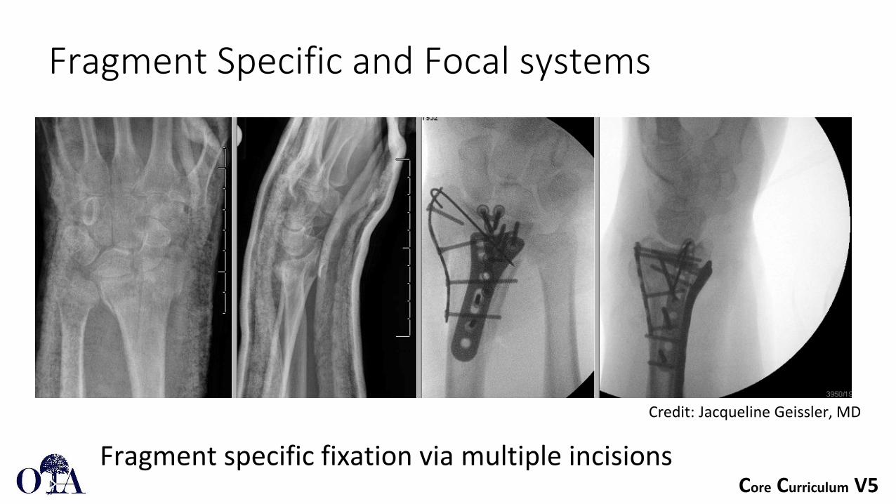
Fragment Specific and Focal systems
Fragment specific fixation via multiple incisions
Credit: Jacqueline Geissler, MD

Core Curriculum V5
Plating Pearls
• Check SL, DRUJ• Xrays
• AP xray with elbow elevated in relation to the wrist
• Lateral xray with wrist elevated in relation to the elbow
• Tangential notch view to assess screw tips
• Semi-supinated for volar ulnar corner, semi-pronated for dorsal ulnar corner

Core Curriculum V5
Plating pitfalls• Avoid plate proximal to watershed line
• https://www.ncbi.nlm.nih.gov/pmc/articles/PMC3828490/• Unicortical screws distal, Bicortical proximal
Example of distal bicortical screws
Examples of plates distal to watershed line
Image courtesy of J Orbay MD

Core Curriculum V5
Based on Three Column TheoryRadial Column
Lateral side of radiusIntermediate Column
Ulnar side of radiusUlnar Column
Distal ulna
Radial column
Intermediate column
Ulnar column

Core Curriculum V5
Intra-articular Fracture
Multi-fragmentary intra-articular fracture

Core Curriculum V5
Focal plating

Core Curriculum V5
Combined dorsal and volar plating• Reserved for very complex fractures
• Second operation for implant removal is common• Increased risk of tendon rupture Credit: Jacqueline Geissler, MD

Core Curriculum V5
Anchor Repair of Radiolunate Ligaments for: radiocarpal dislocation cases to restore stability
Kyle C. Bohm, Jacqueline A. Geissler, Christina M. Ward, Volar Radiocarpal Ligament Repair with Suture Anchors for Radiocarpal Fracture Dislocations: Case Series, J Wrist Surg , DOI: 10.1055/s-0040-1718915, Publication Date: 04 November 2020 (online)https://www.thieme-connect.com/products/ejournals/abstract/10.1055/s-0040-1718915
Credit: Jacqueline Geissler, MD

Core Curriculum V5
Advanced TechniquesArthroscopy-assisted• Reduce articular incongruities• Diagnose and treat associated soft tissue lesions• Minimally invasive
Presenter
Presentation Notes
Minimally invasive vs. adjunct to usual techniques.

Core Curriculum V5
Malunion of Distal Radius Fractures
Changes load-bearing patterns on the distal radius and load sharing between the radius and ulnaOften leads to arthrosis
Diego L. Fernandez, Reconstructive Procedures for Malunion and Traumatic Arthritis, Orthopedic Clinics of North America, Volume 24, Issue 2,1993,Pages 341-363,ISSN 0030-5898,
https://doi.org/10.1016/S0030-5898(21)00022-5.
https://www.sciencedirect.com/science/article/pii/S0030589821000225

Core Curriculum V5
Malunion of Distal Radius Fractures• Not a considerable issue in older patients >60y/o
• Functional outcomes not significantly different
• May require osteotomy and revision fixation (younger patients)• Plating depending on malunion
• +/- bone graft

Core Curriculum V5
Nonunion of Distal Radius Fractures• Rare• Internal Factors
• Poor reduction or fixation• External Factors
• Nutrition• Patient compliance
• Prommersberger, Karl-Josef MD*; Fernandez, Diego L MD† Nonunion of Distal Radius Fractures, Clinical Orthopaedics and Related Research: February 2004 - Volume 419 - Issue - p 51-56

Core Curriculum V5
Smoking
• Important part of the patient history• Best if patients can quit smoking• Low intensity ultrasound (bone stim) can accelerate
healing in smokers

Core Curriculum V5
Specific complications of distal radius fracture ORIF
• Tendon rupture• Extensor or flexor, most common EPL or FPL• Screw tips or volar plate within 3mm of articular surface
• Nerve injury• Median or superficial radial
• CRPS/RSD• Vitamin C may not be of benefit, though still AAOS guidelineÖzkan, Sezai MD; Teunis, Teun MD, PhD; Ring, David C. MD, PhD; Chen, Neal C. MD What Is the Effect of Vitamin C on Finger Stiffness After Distal Radius Fracture? A Double-blind, Placebo-controlled Randomized Trial, Clinical Orthopaedics and Related Research: October 2019 - Volume 477 -Issue 10 - p 2278-2286 doi: 10.1097/CORR.0000000000000807

Core Curriculum V5
ConclusionsMany options for treatment of distal radius fracturesPatient factors must be included when considering treatment optionsImportant to maintain proficiency with and understand all of them as many have specific applications Continuously evolving

Core Curriculum V5
ConclusionsPlating techniques allow for more accurate and rigid fixation of fragments with more reliable outcomes.
Plating allows for early wrist ROM
Volar, smaller and more anatomic plates are better tolerated
Combination treatment and wrist spanning options can be useful in specific instances.
If you would like to volunteer as an author for the Resident Slide Project or recommend updates to any of the following slides, please send an e-mail to [email protected]

Core Curriculum V5
Bibliography
1: Ruch DS, Ginn TA, Yang CC, Smith BP, Rushing J, Hanel DP. Use of a distraction plate for distal radial fractures with metaphyseal and diaphyseal comminution. J Bone Joint Surg Am. 2005 May;87(5):945-54.
2: Sammer DM, Shah HM, Shauver MJ, Chung KC. The effect of ulnar styloid fractures on patient-rated outcomes after volar lockingplating of distal radius fractures. J Hand Surg Am. 2009 Nov;34(9):1595-602.
3: Liporace FA, Adams MR, Capo JT, Koval KJ. Distal radius fractures. J Orthop Trauma. 2009 Nov-Dec;23(10):739-48.
4: Koenig KM, Davis GC, Grove MR, Tosteson AN, Koval KJ. Is early internal fixation preferred to cast treatment for well-reducedunstable distal radial fractures? J Bone Joint Surg Am. 2009 Sep;91(9):2086-93.
5: Rozental TD, Blazar PE, Franko OI, Chacko AT, Earp BE, Day CS. Functional outcomes for unstable distal radial fractures treated with open reduction and internal fixation or closed reduction and percutaneous fixation. A prospective randomized trial. J Bone Joint Surg Am. 2009 Aug;91(8):1837-46.
6: Wei DH, Raizman NM, Bottino CJ, Jobin CM, Strauch RJ, Rosenwasser MP. Unstable distal radial fractures treated with external fixation, a radial column plate, or a volar plate. A prospective randomized trial. J Bone Joint Surg Am. 2009 Jul;91(7):1568-77.

Core Curriculum V5
7: Berglund LM, Messer TM. Complications of volar plate fixation for managing distal radius fractures. J Am Acad Orthop Surg. 2009 Jun;17(6):369-77.
8: Casaletto JA, Machin D, Leung R, Brown DJ. Flexor pollicis longus tendon ruptures after palmar plate fixation of fractures of the distal radius. J Hand Surg Eur Vol. 2009 Aug;34(4):471-4.
9: Mirza A, Jupiter JB, Reinhart MK, Meyer P. Fractures of the distal radius treated with cross-pin fixation and a nonbridging external fixator, the CPX system: a preliminary report. J Hand Surg Am. 2009 Apr;34(4):603-16.
10: Souer JS, Ring D, Matschke S, Audige L, Marent-Huber M, Jupiter JB; AOCID Prospective ORIF Distal Radius Study Group. Effect of an unrepaired fracture of the ulnar styloid base on outcome after plate-and-screw fixation of a distal radial fracture. J Bone Joint Surg Am. 2009 Apr;91(4):830-8
11: Arora R, Gabl M, Gschwentner M, Deml C, Krappinger D, Lutz M. A comparative study of clinical and radiologic outcomes of unstable colles type distal radius fractures in patients older than 70 years: nonoperative treatment versus volar locking plating. J Orthop Trauma. 2009 Apr;23(4):237-42
12: Capo JT, Rossy W, Henry P, Maurer RJ, Naidu S, Chen L. External fixation of distal radius fractures: effect of distraction and duration. J Hand Surg Am. 2009 Nov;34(9):1605-11.
13: Jupiter JB, Marent-Huber M; LCP Study Group. Operative management of distal radial fractures with 2.4-millimeter locking plates. A multicenter prospective case series. J Bone Joint Surg Am. 2009 Jan;91(1):55-65.

Core Curriculum V5
14: Thomas AD, Greenberg JA. Use of fluoroscopy in determining screw overshoot in the dorsal distal radius: a cadaveric study. J Hand Surg Am. 2009 Feb;34(2):258-61.
15: Soong M, Got C, Katarincic J, Akelman E. Fluoroscopic evaluation of intra-articular screw placement during locked volar plating of the distal radius: a cadaveric study. J Hand Surg Am. 2008 Dec;33(10):1720-3.
16: Kreder HJ, Hanel DP, Agel J, McKee M, Schemitsch EH, Trumble TE, Stephen D. Indirect reduction and percutaneous fixation versus open reduction and internal fixation for displaced intra-articular fractures of the distal radius: a randomised, controlled trial. J Bone Joint Surg Br. 2005 Jun;87(6):829-36.
17: Orbay JL, Fernandez DL. Volar fixed-angle plate fixation for unstable distal radius fractures in the elderly patient. J Hand Surg Am. 2004 Jan;29(1):96-102.
18: Orbay JL, Fernandez DL. Volar fixation for dorsally displaced fractures of the distal radius: a preliminary report. J Hand Surg Am. 2002 Mar;27(2):205-15.
19: Kreder HJ, Agel J, McKee MD, Schemitsch EH, Stephen D, Hanel DP. A randomized, controlled trial of distal radius fractures with metaphyseal displacement but without joint incongruity: closed reduction and casting versus closed reduction, spanning external fixation, and optional percutaneous K-wires. J Orthop Trauma. 2006 Feb;20(2):115-21.
Related Documents