Neurocase (2005) 11, 114–128 Copyright © Taylor & Francis Inc. ISSN: 1355-4795 print DOI: 10.1080/13554790590922513 Neurocase Dissociation of emotional processes in response to visual and olfactory stimuli following frontotemporal damages ROBERT SOUSSIGNAN 1 , NATHALIE EHRLÉ 2 , AUDREY HENRY 1 , BENOIST SCHAAL 3 , and SERGE BAKCHINE 2 1 Laboratoire Vulnérabilité, Adaptation, Psychopathologie, Hôpital de la Salpêtrière, Paris, France, 2 Service de Neurologie, CHU de, Reims, Hôpital Maison Blanche, Reims, France, and 3 Centre Européen des Sciences du Goût, Université de Bourgogne, Dijon, France Contemporary neuropsychological studies have stressed the widely distributed and multicomponential nature of human affective processes. Here, we examined facial electromyographic (EMG) (zygomaticus and corrugator muscle activity), autonomic (skin conductance and heart rate) and subjective measures of affective valence and arousal in patient TG, a 30 year-old man with left anterior mediotemporal and left orbitofrontal lesions resulting from a traumatic brain injury. Both TG and a normal control group were exposed to hedonically valenced visual and olfactory stimuli. Contrarily to control subjects, facial EMG and electrodermal activity in TG did not differentiate between pleasant, unpleasant and neutral pictures. In addition, the controls reacted spontaneously with larger corrugator EMG activity and higher skin conductance to unpleasant odors. By contrast, the subjective feeling states (pleasure and arousal ratings) remained preserved in TG. The covariation between facial and self-report measures of negative valence was also a function of the nature of the olfactory task in the patient only. Taken together, the data suggest a functional dissociation between brain substrates supporting generation of emotion and those supporting representation of emotion. Introduction A large body of research supports the view that the fundamen- tal structure of emotion is mainly explained in terms of affec- tive valence and arousal (Russell, 1980; Bradley, 1994). This dimensional conception of emotion is thought to reflect the evolutionary foundation of a basic motivational organization system underlying approach-withdrawal or appetitive-aversive behaviour (Lang et al., 1998; Davidson and Irwin, 1999). Although the anatomical substrates of affective processes related to this superordinate division are not entirely eluci- dated, animal and human research has clearly implicated ante- rior mediotemporal lobe structures (e.g., amygdaloid complex) and the orbitofrontal cortex (OFC) as intimately connected regions of a distributed neural network playing a critical role in various aspects of approach- and withdrawal-related emotion and motivation (Damasio, 1994; Rolls, 2000; Lane and Nadel, 2000; Davidson, 2002). Accumulating evidence has estab- lished that these brain regions are involved in the processing of the reinforcing value of stimuli in relation to decision-making and goal-directed behaviors, in the encoding and consolidation of implicit or explicit learning of emotionally salient events, and in the evaluation of the affective significance of stimuli (e.g., Rolls, 2000; Adolphs and Damasio, 2001; Hamann, 2001; Davidson, 2002; Anderson et al., 2003; Holland and Gallagher, 2004; Izquierdo and Murray, 2004). While the current findings suggest that the amygdala and the OFC are part of an integrated neural system subserving critical functions in reinforcement mechanisms, decision- making, and affective memory, their role in various aspects of emotion (e.g., perception, appraisal, physiology, expression, and subjective experience) is not clearly understood in humans. Students of emotion provided evidence that affective valence and arousal are multicomponent processes, comprising expressive (i.e., facial and vocal expressions) and autonomic reactivity, and their subjective or experiential counterparts (Cacioppo et al., 1992; Lang et al., 1993; Soussignan, 2002). Autonomic responses and spontaneous facial actions consti- tute rapid changes in somatic and neurophysiological activity presumed to reflect emotional states, whereas the subjective experience of emotion is viewed as a mental representation of emotional states based on conscious awareness, recall of past affective experience, and language (Lane, 2000; Dolan, 2002). Such a distinction is well emphasized in current con- ceptualizations under the terms of emotion and feeling. For instance, according to Damasio (1995, 1998) emotion refers to “dispositional” responses to the perception of stimuli pro- ducing changes within the body itself (externalized aspects), whereas feeling designates a subject’s perception of bodily changes induced by responses or by mental states resulting from emotional state (internal experience). Whether distinct, non-overlapping neural pathways are involved in physiological emotional responses and self-reported Received on 29 April 2004; accepted 16 December 2004. We thank TG and the subjects of the control group for their participation in this research. Correspondance to: R. Soussignan, Laboratoire Vulnérabilité, Adaptation, Psychopathologie, CNRS UMR 7593. Hôpital de la Salpêtrière, Pavillon Clérambault, 47 Bd de l’Hôpital, 75013 Paris. Tel. : 01.55.52.02.74 E-mail: [email protected]

Welcome message from author
This document is posted to help you gain knowledge. Please leave a comment to let me know what you think about it! Share it to your friends and learn new things together.
Transcript

Neurocase (2005) 11, 114–128 Copyright © Taylor & Francis Inc. ISSN: 1355-4795 print DOI: 10.1080/13554790590922513
Neurocase
Dissociation of emotional processes in response to visual and olfactory stimuli following frontotemporal damages
ROBERT SOUSSIGNAN1, NATHALIE EHRLÉ2, AUDREY HENRY1, BENOIST SCHAAL3, and SERGE BAKCHINE2
1Laboratoire Vulnérabilité, Adaptation, Psychopathologie, Hôpital de la Salpêtrière, Paris, France, 2Service de Neurologie, CHU de, Reims, Hôpital Maison Blanche, Reims, France, and 3Centre Européen des Sciences du Goût, Université de Bourgogne, Dijon, France
Contemporary neuropsychological studies have stressed the widely distributed and multicomponential nature of human affective processes.Here, we examined facial electromyographic (EMG) (zygomaticus and corrugator muscle activity), autonomic (skin conductance and heartrate) and subjective measures of affective valence and arousal in patient TG, a 30 year-old man with left anterior mediotemporal and leftorbitofrontal lesions resulting from a traumatic brain injury. Both TG and a normal control group were exposed to hedonically valencedvisual and olfactory stimuli. Contrarily to control subjects, facial EMG and electrodermal activity in TG did not differentiate betweenpleasant, unpleasant and neutral pictures. In addition, the controls reacted spontaneously with larger corrugator EMG activity and higherskin conductance to unpleasant odors. By contrast, the subjective feeling states (pleasure and arousal ratings) remained preserved in TG.The covariation between facial and self-report measures of negative valence was also a function of the nature of the olfactory task in thepatient only. Taken together, the data suggest a functional dissociation between brain substrates supporting generation of emotion and thosesupporting representation of emotion.
Introduction
A large body of research supports the view that the fundamen-tal structure of emotion is mainly explained in terms of affec-tive valence and arousal (Russell, 1980; Bradley, 1994). Thisdimensional conception of emotion is thought to reflect theevolutionary foundation of a basic motivational organizationsystem underlying approach-withdrawal or appetitive-aversivebehaviour (Lang et al., 1998; Davidson and Irwin, 1999).Although the anatomical substrates of affective processesrelated to this superordinate division are not entirely eluci-dated, animal and human research has clearly implicated ante-rior mediotemporal lobe structures (e.g., amygdaloid complex)and the orbitofrontal cortex (OFC) as intimately connectedregions of a distributed neural network playing a critical role invarious aspects of approach- and withdrawal-related emotionand motivation (Damasio, 1994; Rolls, 2000; Lane and Nadel,2000; Davidson, 2002). Accumulating evidence has estab-lished that these brain regions are involved in the processing ofthe reinforcing value of stimuli in relation to decision-makingand goal-directed behaviors, in the encoding and consolidationof implicit or explicit learning of emotionally salient events,
and in the evaluation of the affective significance of stimuli(e.g., Rolls, 2000; Adolphs and Damasio, 2001; Hamann,2001; Davidson, 2002; Anderson et al., 2003; Holland andGallagher, 2004; Izquierdo and Murray, 2004).
While the current findings suggest that the amygdala andthe OFC are part of an integrated neural system subservingcritical functions in reinforcement mechanisms, decision-making, and affective memory, their role in various aspectsof emotion (e.g., perception, appraisal, physiology, expression,and subjective experience) is not clearly understood inhumans. Students of emotion provided evidence that affectivevalence and arousal are multicomponent processes, comprisingexpressive (i.e., facial and vocal expressions) and autonomicreactivity, and their subjective or experiential counterparts(Cacioppo et al., 1992; Lang et al., 1993; Soussignan, 2002).Autonomic responses and spontaneous facial actions consti-tute rapid changes in somatic and neurophysiological activitypresumed to reflect emotional states, whereas the subjectiveexperience of emotion is viewed as a mental representation ofemotional states based on conscious awareness, recall of pastaffective experience, and language (Lane, 2000; Dolan,2002). Such a distinction is well emphasized in current con-ceptualizations under the terms of emotion and feeling. Forinstance, according to Damasio (1995, 1998) emotion refersto “dispositional” responses to the perception of stimuli pro-ducing changes within the body itself (externalized aspects),whereas feeling designates a subject’s perception of bodilychanges induced by responses or by mental states resultingfrom emotional state (internal experience).
Whether distinct, non-overlapping neural pathways areinvolved in physiological emotional responses and self-reported
Received on 29 April 2004; accepted 16 December 2004.We thank TG and the subjects of the control group for their
participation in this research.Correspondance to: R. Soussignan, Laboratoire Vulnérabilité,
Adaptation, Psychopathologie, CNRS UMR 7593. Hôpital de laSalpêtrière, Pavillon Clérambault, 47 Bd de l’Hôpital, 75013 Paris.Tel. : 01.55.52.02.74 E-mail: [email protected]

115 Soussignan et al.
feeling states remains debated. On the one hand, in humans,the amygdala and the OFC exert modulating influences onautonomic responses probably through their projections tothe hypothalamus and lower brainstem (Mangina and Beuzeron-Mangina, 1996; Williams et al., 2001; Critchley, 2002), andsubjective experience of emotion often engages neural struc-tures not necessarily linked either to the OFC or amygdala(Lane, 2000). For instance, neuroimaging studies haveshown that attention directed to one’s own affective experi-ence under various emotionally-laden conditions (films,recall, slides) increased activity preferentially within theanterior cingulate cortex, and the medial prefrontal cortex,but not in the amygdala or OFC (Georges et al., 1995; Laneet al., 1997; Lane et al., 1998; Lane 2000). Furthermore, theamygdala recruitment in the conscious experience of emo-tion does not seem necessary because a lack of impairmentof self-reported affective states was found in patients withunilateral or bilateral amygdala lesions (Adolphs et al.,1997; Anderson and Phelps, 2002). On the other hand, func-tional neuroimaging studies have also shown that the activa-tion of the amygdaloid complex or the OFC was associatedwith subjective ratings of emotionally valenced stimuli (Zaldand Pardo, 1997) or with self-report of sadness during recallof emotionally-loaded situations (Pardo et al., 1993).Finally, a number of studies demonstrated that neural struc-tures hypothesized to be involved in the conscious experi-ence of emotion (cingulate cortex, medial prefrontal cortex,insula, and somatosensory cortices) may be co-activatedwith the amygdala (Zald et al., 1998; Liberzon et al., 2000;Royet et al., 2000) and/or the OFC (Schneider et al., 1997;Paradiso et al., 1999; Dougherty et al., 1999; Damasio et al.,2000).
Although functional neuroimaging techniques are power-ful to illuminate the widely distributed nature of affectiveprocesses, they cannot directly specify the contribution ofparticular brain regions to specific emotional responses, andthe associations detected between neural activity and subjec-tive experience in a number of studies may reflect otheraspects of emotional processing correlated with feeling states(e.g., physiological arousal, facial efference). A complimen-tary approach for exploring the contribution of specific neuralstructures to distinct emotional responses bears on the conse-quences of focal brain lesions. More specifically, patientswith discrete brain damages afford the opportunity to investi-gate whether the areas identified by imaging are critical in thecontrol or the modulation of multiple output components ofemotion.
In the present study, we examined facial EMG, autonomicand self-reported verbal measures of affective valence andarousal in a male patient with left anterior temporal and leftorbitofrontal damages while exposed to hedonically valencedvisual and olfactory stimuli. We expected to evidence thefollowing dissociation between these distinct components ofemotion: the patient with frontotemporal damages should bemore impaired in his facial and autonomic responses (mea-sures of implicit processing) but less in his self-reported
affective ratings (a measure of conscious experience), aswould be consistent with the role of these brain regions in themediation of automatic and unconscious bodily changes(Damasio, 1994). Facial EMG activity over the muscleregions responsible for frowning (corrugator supercilii) andsmiling (zygomaticus major) was targeted because electricalactivity from these muscles appeared to be a reliable correlateof the processing of the affective valence of the stimulus(Cacioppo et al., 1992; Lang et al., 1993) and may reflectautomatically operating affect programs (Dimberg et al.,2000). For instance, it was found that looking at or imaginingpleasant scenes increased zygomatic activity, whereas view-ing or recalling unpleasant scenes elevated corrugator activity(Schwartz et al., 1980; Dimberg, 1990). Autonomic measuressuch as heart rate (HR) and skin conductance (SC) wererecorded because they may index processes linked to orient-ing response (HR) or emotional arousal (SC) (Lang et al.,1993) and reflect implicit processing (Kubota et al., 2000;Öhman et al., 2000).
A second purpose was to investigate whether affectivereports, facial and autonomic responses to hedonicallyvalenced stimuli depend on the sensory modality. Althoughneuroimaging studies have shown that both visual and olfac-tory stimuli can activate the amygdala and the OFC, the per-ception of odors is dominated by a hedonic dimension(Soussignan and Schaal, 1996; Royet et al., 2000; Roubyand Bensafi, 2002) and may produce autonomic and expres-sive changes as early as the first days of life (Soussignan etal., 1997). Anatomically, the primary olfactory cortex iscontinuous with the anterior portion of the amygdala andprojects directly to the amygdala and posterior orbitofrontalcortex (Carmichael et al., 1994; Zald and Pardo, 2000). Inaddition, functional brain imaging and lesion studies haveconsistently found that the exposure to odorants involvedboth the OFC and interconnected regions (anteriormediotemporal lobe and/or hypothalamus) (Eslinger et al.,1982; Zatorre et al., 1992; Zald and Pardo, 1997; 2000;Zatorre et al., 2000; Royet et aregional cerebral blood flow(rCBF) recording during the
l., 2000; 2001). For instance,
presentation of emotionally valenced olfactory, visual, andauditory stimuli have shown increased rCBF in the left OFCfor each sensory modality, whereas only odors inducedrCBF increases in both the amygdala and the OFC (Royet etal., 2000). Based on evidence cited above, we addressed theissue of whether psychophysiological and experiential corre-lates of affective processing would be more impaired forolfactory than visual stimuli in the patient with frontotemporaldamages.
A final purpose of the study was to examine whether affectiveresponsiveness following damages of frontotemporal structuresis specifically related to the nature of the olfactory task.Because neural and autonomic responses to odors were found tobe task-dependent (Royet et al., 2001; Bensafi et al., 2002a;Royet et al., 1999, 2003), facial EMG and autonomic corre-lates of hedonic processing and arousal were compared duringtasks of affective judgment (i.e., explicit task of affective

Emotion and frontotemporal damages 116
processing) and of odor identification (i.e., implicit task of affec-tive processing). We hypothesized that frontotemporal damageswould produce a deficit in emotional physiological reactivityduring the implicit task of affective processing of odors. Thishypothesis is consistent with findings from functional imagingresearch which indicate that anterior mediotemporal structuresand OFC can be activated by emotional stimuli even withoutawareness after parietal damage (Vuilleumier et al., 2002).
Patient report: Case TG
Medical history and personal information
TG is a 30-year-old, right-handed male. He was 19-years-oldwhen he sustained a severe closed head injury during a carcrash. Before, he was attempting a second year of superiordegree and was socially integrated with a lot of friends and agirlfriend. Following the head trauma, he presented a nonre-active coma. He had an initial Glasgow Coma Scale of 9, and amild right brachio-facial hemiparesia. At this time, a CT-scanwas done, but did not reveal any hematoma. He was admittedto an intensive care unit because he needed assisted ventila-tion. The coma lasted about four days. After the comatosephase and during the following years, TG did not present anyepileptic seizure. An EEG was done and indicated only smallbursts of theta waves in the left frontotemporal lobe. His con-dition improved gradually so that he could be dischargedfrom hospital after one month. TG was considered as cogni-tively not impaired, except for a mild anterograde amnesia,with a retrograde amnesia for a 3 month period. TG followeda mnemonic training and a physical therapy program during 6months as an outpatient in a rehabilitation centre. He took uphis everyday life apparently on a normal basis. However, thesubsequent evolution was marked by many difficulties. TGleft his girlfriend without clear motives. He tried to startagain his studies, but gave up after 2 years of successive fail-ing. His family reported that he was no longer able to orga-nize conveniently his time to study or to develop learningstrategies. All these facts constituted a clear change in TG’shabits. During the 6 following years, TG returned to his par-ent’s house. He manifested very little personal involvementin the search for new studies or for a job. TG’s parentsincreased their commitment in this task. Finally, under thepressure of his parents, TG planned to open a small sandwichshop. Both these projects failed each time because he was notable to manage the required organizational tasks. For instance,he concentrated on the office furnishing while neglecting thelegal paperwork. After these two failures, TG disappearedsuddenly from his house during one year. His presence wasoccasionally mentioned in bars with suspect frequentations.He returned home twice, for brief periods and then disap-peared again for one year. During this period he becamehomeless after having spent the totality of the compensa-tion he had obtained for his trauma. Finally, his parents foundhim in a park and took him home. TG was neither able to
explain his behavior during his wandering period, nor did heexpress any regret or remorse. Strictly coached by his family,TG was engaged in a sandwich shop managed by his cousin.He is reported to work well, but under strict supervision. Still,he disappeared again two or three times, but for very shortperiods. In February 2001, TG was addressed to our neuro-logical department for an expert report at the request of hisparents. At this examination, TG’s parents reported that hisbehavior was still markedly impaired with a strong need forhelp from his family.
Neurological examination and neuropsychological assessment
A three-dimensional acquisition magnetic resonance imaging(MRI) was performed, including T1-weighted and T2-weighted sequences, fluid-attenuated inversion recovery(FLAIR) and T2 echo planar sequences. Figure 1 illustratessome examples of structural MRI scans in this patient. AxialFLAIR images through the frontal and temporal lobes indi-cated abnormal signal intensity in the left orbitofrontal cor-tex, in left anterior mediotemporal structures (anteriortemporal pole, periamygdalar region), and in the left posteriorinsula. Most posterior regions of the temporal lobes appearednormal (e.g., hippocampus). It can be noted that there was noevidence of mass lesion and that the abnormal signals reflectedmild aspects of sequelae of cerebral contusion (cortical
Fig. 1. Axial fluid-attenuated inversion recovery (FLAIR) images(A, B, C) through the frontal and temporal lobes of TG’s brain.There is abnormal signal intensity in the left anteromedial temporallobe (anterior temporal pole, periamygdalar region), in the leftorbitofrontal cortex, and in the left posterior insula (see the arrows).A sagittal T1-weighted image (D) indicated a thinning of theanterior portion of the corpus callosum (see the arrow), but nodamage in the anterior cingulate cortex and other frontal structures.Note that there was no evidence of mass lesion. The left hemisphereis depicted in the right side of each image.

117 Soussignan et al.
deposits of hemosiderin and white matter T2 hyperintensi-ties). A sagittal T1-weighted MRI also revealed a thinning ofthe anterior portion of the corpus callosum and a small lesion
of the splenium. However, there is no evidence of damage inthe anterior cingulate cortex and other frontal structures.
A single-photon emission computerized tomography (SPECT)scan was also performed indicating marked hypoperfusion inboth the anterior part of the left anterior temporal region (peri-amygdalar portion) and the left orbitofrontal cortex (Figure 2).
Fig. 1. Continued.
Fig. 2. SPECT images of TG‘s brain. Horizontal (a) and coronal(b) sections show respectively a blood flow reduction in the leftorbitofrontal cortex and in the anterior part of the left temporal lobeinvolving the periamygdalar region.

Emotion and frontotemporal damages 118
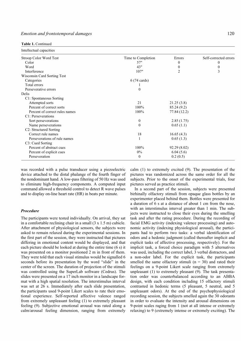
The patient was assessed in our department 11 years afterthe injury. His cognitive performances are presented inTable 1. At this time, he was correctly oriented and presented asuperior efficiency (GIQ: 120, shortened form of the WechslerAdult Intelligence Scale-Revised, WAIS-R), without any sig-nificant dissociation between verbal (VIQ: 117) and perfor-mance IQ (PIQ: 120). Among the subtests used in theshortened form of the WAIS-R, only Digit Symbol subtestrevealed abnormal slowing. His working and episodic mem-ory performances were in the normal range, for both verbal(digit backward span; Rey Auditory Verbal Learning Test) andvisual stimuli (localization backward span; Rey-OsterriethTest). The examination of language did not demonstrate anynaming, reading, writing, comprehension or fluency deficit(French version of the Boston Naming Test, Chapman-CookSpeed of Reading Test, Token test). His spontaneous expres-sion was considered both formally and semantically rich. Nosign of apraxia or visuo-spatial trouble was observed and theexecutive control was excellent, as assessed by the Trailmakingtest, the Wisconsin Card Sorting Test, and the Stroop andDelis tests. In summary, TG seems to show a preservation ofhis whole cognitive functioning with performances often inthe superior mean.
Method
Subjects
Patient TG’s responses to a set of stimuli chosen for theirarousing impact and their affective valence were comparedto those of a normal control group. This latter group com-prised 10 subjects matched to TG for gender and age (M =26.4-years-old, SD = 4.87). Additional criteria for selectionwere: (a) full right handedness as defined by the EdinburghLaterality Inventory (Oldfield, 1971); (b) 2 years college edu-cation; (c) absence of olfactory disturbances and respiratoryallergies, and (d) absence of smoking habits. All the subjectsparticipating in the study provided informed consent indicat-ing their acceptance of the procedure.
Stimulus selection
The emotional material comprised visual and olfactory stimuli.First, 90 pictures were selected from the International Affec-tive Picture System (IAPS) (Lang et al., 2001). These pic-tures consisted of three 30-item sets of pleasant, neutral andunpleasant CD-ROM slides for which normative ratings onvalence and arousal value were obtained from prior valida-tion studies in male subjects. Unpleasant slides (valence: M =2.46, SD = 0.96) included themes such as mutilated and deadbodies, skin disease, frightening animals, human violence,disgusting scenes, starving child, and so on. Neutral slides(valence: M = 4.97, SD = 0.21) showed items such as house-hold objects, neutral faces, plants, and so on. Pleasant slides(valence: M = 7.37, SD = 0.42) showed human and animal
babies, nature scenes, erotic females, sport events, socialscenes, and so on.
Second, the odor stimuli were composed of pleasantly andunpleasantly valenced compounds (n=10 each) purchased fromdifferent aroma and chemical companies (Euracli, Chasse surRhône, and Sigma-Aldrich, Saint Quentin-Fallavier, France).Part of the pleasant and unpleasant stimuli was selected on thebasis of previous studies on hedonic judgments of odorousstimuli (e.g., Royet et al., 1999, 2001). The set of pleasantstimuli included odors of flower (rose, lavender, violet),fruits and comestible plants (banana, apple, coconut, vanilla,mint, anise, and caramel). Unpleasant stimuli consisted ofpure chemical or mixtures evoking food, body and environ-mental odors. They included adoxal (rotten egg), butyric acid(cheesy), cod liver oil, triethylamine (fishy), synthetic sweat,skatole (faeces), castoreum (animal), pyridine (chemicalindustry), tetrahydrothiophene (THT: gas), and fuel oil.These odorants were diluted in mineral oil to produce 5 ml ofsolution (concentration: 1% v/v) that was absorbed onpolypropylene placed in 50-ml brown glass bottles. The con-centration of the compounds with high stimulative potency(THT, pyridine) was limited to 1‰. The neutral stimuli con-sisted of 10 50-ml bottles of the odorless solvent (mineral oil)absorbed on polypropylene.
Psychophysiological recording
Facial EMG activity and autonomic measures (heart rate,skin conductance) were recorded using a 4-channel PowerLab system (model 4SP, ADInstruments Pty Ltd), which wasconnected to a PC. The bioelectric signals were filtered andamplified before being fed into the analog input connector ofthe PowerLab unit, and were sampled at a rate of 40 points/sunder the on-line control of an application program (Chart forWindows v 4.0.4).
Muscle activity was recorded over the left corrugatorsupercilii and zygomaticus major regions using miniatureAg/AgCl surface electrodes, filled with electrode pasteand placed bipolarly following the guidelines proposed byFridlund and Cacioppo (1986). Before the electrodes wereattached, the target sites of the skin were cleaned with alcoholand slightly rubbed in order to reduce inter-electrode imped-ance. To minimize demand characteristics of the EMG exper-iments (Fridlund and Cacioppo, 1986), a cover story wasgiven to the subjects by telling them that the facial electrodeswere placed in order to record face temperature. The EMGsignals were fed into electronic amplifiers and were band-passfiltered from 10 Hz to 250 Hz. The raw EMG was rectifiedand smoothed with a time constant set at 500 ms.
Skin conductance (SC) was transduced with Ag/AgCl elec-trodes filled with a conducting Biogel and attached with aVelcro strap on the volar surface of the distal phalanges ofthe second and third fingers of the nondominant (i.e., left)hand. A UFI Bioderm skin conductance coupler provided aconstant 0.5 V current across the electrodes. Heart activity

119 Soussignan et al.
Table 1. Results of TG’s neuropsychological testing
Intellectual capacities
WAIS-R ScoresFull Scale Score 117Verbal Score 120Performance Score 117
MemoryDigit spans
Forward 7Backward 6
Localization spansForward 7Backward 7
Rey Auditory Verbal Learning TestImmediate Recalls 8-13-12-13-11/15Total 57/75Interference 5/15Retention after Interference 10/15Delayed Recall 11/15Recognition 12/15
Rey-Osterrieth TestImmediate Recall 22/36Delayed Recall 22/36
OrientationMEM-III (French version of the WMS-III)Information and Orientation Subtest 14/14
Visuo-spatial testsBells Test 35/35Copy of Rey Complex Figure 32/36
LanguageNaming
DO80 test (French version of the Boston Naming Test) 79/80Reading
Words (Simple, Complex, Irregular) 16/16Non-Words 6/6
WritingWords (Simple, Complex, Irregular) 16/16Non-Words 6/6
Semantic Fluency (2 minutes each) WordsAnimals 30Fruits 22Jobs 22
Letter Fluency (2 minutes each) WordsP 22R 24V 22
Comprehension ScoreChapman-Cook Speed of Reading Test 15 paragraphsToken Test 163/163
Executive functioningTrailmaking Test Time to Completion Errors
Part A 26" 0Part B 43" 0
Continued
Emotion and frontotemporal damages 120
was recorded with a pulse transducer using a piezoelectricdevice attached to the distal phalange of the fourth finger ofthe nondominant hand. A low-pass filtering of 50 Hz was usedto eliminate high-frequency components. A computed inputcommand allowed a threshold control to detect R wave pulsesand to display on-line heart rate (HR) in beats per minute.
Procedure
The participants were tested individually. On arrival, they satin a comfortable reclining chair in a small (3 x 1.5 m) cubicle.After attachment of physiological sensors, the subjects wereasked to remain relaxed during the experimental sessions. Inthe first part of the session, they were instructed that picturesdiffering in emotional content would be displayed, and thateach picture should be looked at during the entire time (6 s) itwas presented on a monitor positioned 2 m in front of them.They were told that each visual stimulus would be signalled 6seconds before its presentation by the word “slide” in thecenter of the screen. The duration of projection of the stimuliwas controlled using the SuperLab software (Cedrus). Theslides were presented on a 17 inch monitor in a landscape for-mat with a high spatial resolution. The interstimulus intervalwas set at 26 s. Immediately after each slide presentation,the participants used 9-point Likert scales to rate their emo-tional experience. Self-reported affective valence rangedfrom extremely unpleasant feeling (1) to extremely pleasantfeeling (9). Subjective emotional arousal was rated along acalm/arousal feeling dimension, ranging from extremely
calm (1) to extremely excited (9). The presentation of thepictures was randomized across the same order for all thesubjects. Prior to the onset of the experimental trials, fourpictures served as practice stimuli.
In a second part of the session, subjects were presentedbirhinally olfactory stimuli from opaque glass bottles by anexperimenter placed behind them. Bottles were presented fora duration of 6 s at a distance of about 1 cm from the nose,with an interstimulus interval greater than 1 min. The sub-jects were instructed to close their eyes during the smellingtask and after the rating procedure. During the recording offacial EMG activity (indexing valence processing) and auto-nomic activity (indexing physiological arousal), the partici-pants had to perform two tasks: a verbal identification ofodors and a hedonic judgment (called thereafter implicit andexplicit tasks of affective processing, respectively). For theimplicit task, a forced choice paradigm with 5 alternativeswas used, including the correct label, 3 verbal distractors, anda non-odor label. For the explicit task, the participantssmelled the same olfactory stimuli (n = 30) and rated theirfeelings on a 9-point Likert scale ranging from extremelyunpleasant (1) to extremely pleasant (9). The task presenta-tion order was counterbalanced according to an ABBAdesign, with each condition including 15 olfactory stimulicontrasted in hedonic terms (5 pleasant, 5 neutral, and 5unpleasant odors). At the end of the psychophysiologicalrecording session, the subjects smelled again the 30 odorantsin order to evaluate the intensity and arousal dimensions on9-point scales raging from 1 (not at all intense or extremelyrelaxing) to 9 (extremely intense or extremely exciting). The
Table 1. Continued
Intellectual capacities
Stroop Color Word Test Time to Completion Errors Self-corrected errorsColor 57" 0 0Word 43" 0 0Interference 107" 2 3
Wisconsin Card Sorting TestCategories 6 (74 cards)Total errors 1Perseverative errors 0
DelisC1: Spontaneous Sorting
Attempted sorts 21 21.25 (3.8)Percent of correct sorts 100% 85.24 (9.2)Percent of correct rules names 100% 77.84 (12.2)
C1: PerseverationsSort perseverations 0 2.85 (1.75)Name perseverations 0 0.65 (1.1)
C2: Structured SortingCorrect rule names 18 16.65 (4.3)Perseverations of rule names 1 0.65 (1.3)
C3: Cued SortingPercent of abstract cues 100% 92.29 (8.02)Percent of explicit cues 0% 6.04 (5.6)Perseveration 0 0.2 (0.5)

121 Soussignan et al.
intensity of a stimulus was defined as the degree of its“potency” regardless of its hedonic valence. Before testing,the subjects were trained to rate differences in subjectiveintensity perception between stimuli using two odorants ofdistinct intensity levels.
Physiological data reduction and analysis
EMG and HR data were averaged off-line for each of the 6 speriods immediately preceding (baseline period) and follow-ing (stimulus period) the stimulus onset. In accordance withstandard statistical practice (Fridlund, 1991), mean EMG datawere log transformed [log10(EMG + 1)] to minimize skew-ness and heteroscedasticity. The EMG transformed baselinescore (in log-µV) and the HR prestimulus levels (in bpm)were subtracted from the EMG transformed score and the HRvalue during stimulus presentation to extract mean changesfor each physiological measure. Skin conductance response(SCR) was defined as changes in the amplitude with onsetoccurring 1–4 s after stimulus presentation. SCR change (inµSiemens) was calculated by subtracting the 2-s SC levelimmediately preceding stimulus onset from the largest valueaveraged in the 2-s window after stimulation1.
For the control subjects, within-subjects comparisons ofthe facial, autonomic, and subjective rating data were per-formed using multivariate analysis of variance (MANOVA),as suggested by Jennings et al. (1987), the resulting Wilks’lambda statistics were referred to the F distribution. Bonferronit tests were used for pairwise comparisons of means to controlfamilywise error rate. These statistical analyses in a norma-tive sample tested whether the distinct dependent variablesdiscriminated between affectively valenced stimuli.
The comparison between TG and the normal subjects wasinvestigated by calculating difference measures (pleasant –neutral, unpleasant – neutral) to control for the possible dif-ferential responsiveness to neutral stimuli between the patientand the normal subjects, and then by combining the datameans of the trials for each subject in the control group toprovide the reference data. Then, a one-sample-t-test wascomputed to see whether the patient’s mean performancefalls outside the reference distribution at P < 0.05.
Results
Facial muscle responses
A first question is whether the electrophysiological responsesof facial muscles indexing the dimension of affective valenceare reduced in a patient with damages of brain structures pre-
sumed to be involved in emotion. The facial-EMG meansduring the projection of slides for both the control subjectsand TG are illustrated in Figure 3. The control subjectsshowed stronger reaction of the corrugator supercilii muscleregion to unpleasant pictures (Figure 3a), compared to pleasantand neutral pictures [F(2, 297) = 20.83, P < 0.0001; Bonferronitests, all P < 0.0001]. Furthermore, the controls exhibited onaverage higher EMG reactivity over the brow region than TGduring projection of negative slides only, t(29) = 5.44, P <0.0001. The zygomatic activity of controls also discriminatedthe affective valence of the pictures [F(2, 238) = 4.81, P =0.009], particularly between pleasant and neutral conditions(P = 0.02), whereas for TG, zygomatic EMG activity did notvary as a function of picture valence, t(29), all P > 0.05 (Fig. 3b).However, the control subjects did not reveal higher zygo-matic responses over the cheek region during the presentationof pleasant slides compared to TG, t(29) = 0.3, P > 0.05.
For the olfactory experiment, in the control group, a two-way MANOVA was used with type of task (implicit vs.explicit affective processing) and odor valence (unpleasant,neutral, and pleasant) as repeated measures factors. Concern-ing corrugator activity, significant main effects weredetected for the type of task, [F(1, 99) = 3.86, P = 0.05], andthe valence of the odor, [F(2, 98) = 28.47, P < 0.0001]. Nor-mal subjects exhibited greater corrugator activity during theexplicit (M = 0.06, SD = 0.09) than during the implicit task ofaffective processing of odors (M = 0.04, SD = 0.07), [F(1, 99 =3.86, P = 0.05]. They also showed stronger corrugator activ-ity to unpleasant odors than to neutral or pleasant odors(Bonferroni tests, all P < 0.0001) (see Figure 4). Concerningzygomatic activity, control subjects exhibited a higher activitywhile smelling pleasant odors compared to neutral olfactorycondition, however the effect was only marginally significant[F(2, 82) = 2.70, P = 0.07]. In contrast, TG’s facial reactivityfor both corrugator and zygomaticus muscles did not differ-entiate between the hedonically valenced odors (one-sample-t-tests, all P > 0.05). One-sample t-tests also indicated thatthe control group displayed higher EMG corrugator activitythan did TG while smelling unpleasant odors during both theimplicit and explicit tasks of affective processing [t(9) = 2.25and 7.15, P = 0.05 and < 0.001, respectively], whereas no sig-nificant difference was detected for zygomatic activitybetween TG and the controls while smelling pleasant odors.
Autonomic tone
The autonomic measures were recorded to index bothphysiological arousal (SC) and attention (HR). We testedspecifically whether emotional arousal is reduced in thepatient.
Skin conductance
During the projection of slides, greater changes in SC weredetected in controls while viewing unpleasant as compared toneutral pictures [F(2, 259) = 4.11, P = 0.017; Bonferroni test,P = 0.02]. TG also exhibited larger SC responses to unpleasant
1Because of the presence of artefacts and equipment problemsduring the recording of zygomatic EMG muscle activity and elec-trodermal activity, data from one (for SC) or two (for zygomaticEMG) participants had to be excluded.

Emotion and frontotemporal damages 122 2
relative to neutral pictures, however the difference failed toreach significance, t(29) = 1.94, P = 0.06. More interestingly,SC changes were larger in the controls than in TG, whileviewing both unpleasant, t(29) = 3.96, P < 0.0001, and pleas-ant pictures, t(29) = 2.26, P = 0.03 (Figure 5a). The smellingof unpleasant odorants induced greater SC responses than didthe pleasant or neutral olfactory stimuli in the controls(F(2,82) = 9.29, P < 0.0001; Bonferroni tests, all P < 0.0001)(see Figure 5b). Smelling unpleasant odorants also generatedhigher SC changes in the controls than in TG both during theimplicit, t(9) = 2.93, P = 0.02, and explicit tasks of affectivejudgment, t(9) = 8.17, P < 0.0001.
Heart rate
During the projection of slides, the control subjects showedlarger cardiac decelerative response to unpleasant picturesthan to neutral or positive pictures, F(2, 295) = 3.98, P = 0.02(see Figure 6), but HR did not change as a function of valenceor type of task when they smelled the odors. No statisticaldifference was detected for HR data between TG and the con-trol subjects (one-sample t-tests, all P > 0.05) for both thevisual and olfactory stimuli.
Subjective experienceWe examined the hypothesis of a dissociation between sub-jective experience of emotion (feeling) and emotionalsomatic and autonomic reactivity by testing whether the self-reported affective ratings (pleasure, arousal) are impaired inTG. Intensity ratings of odors were also assessed to verify
Fig. 3. Facial electromyographic (EMG) responses to pictures as a function of affective valence in controls and patient TG: (a) Meanresponses of the corrugator supercilii muscle region; (b) Mean responses of the zygomaticus major muscle region. Error bars representstandard errors (SE).
Figure 4. Mean responses of the corrugator supercilii muscleregion to odors as a function of affective valence in controls andpatient TG. Error bars represent SE.
-0.02
-0.01
0
0.01
0.02
0.03
0.04
Corrugator R
esponse (∆log-µV)
Controls TG
*
Unpleasant Neutral Pleasant
a
-0.04
-0.035
-0.03
-0.025
-0.02
-0.015
-0.01
-0.005
0
0.005
0.01
Zygom
atic Response (∆log-µV
)
Controls TG
Unpleasant Neutral Pleasant
b
0
0.02
0.04
0.06
0.08
0.1
0.12
0.14
0.16
0.18
0.2
Unpleasant Neutral Pleasant
Corrugator R
esponse (∆log-µV)
Controls TG

123 Soussignan et al.
that the perception of this chemical dimension was notaffected in the patient and thus, was not a confoundingvariable contributing to the subjective experience of affect-related to olfaction.
Affective ratings
Both TG and the controls reported more pleasurable experi-ence while viewing pleasant pictures, and more displeasurewhile viewing aversive pictures compared with the neutralcondition (Figure 7a), [F(2, 298) = 812.6, P < 0.0001 andt(29), all P < 0.0001, for controls and TG respectively]. Moreinterestingly, no significant differences emerged between thecontrols and TG while viewing aversive pictures, t(29) =1.85, P > .05. Furthermore, TG rated pleasant pictures asmore pleasurable than did the controls, t(29) = 2.27, P = 0.03.All the subjects also rated the unpleasant pictures as morearousing than either neutral or pleasant pictures [F(2, 298) =210.03, P < 0.001, and t(29), all P < 0.0001 for controls andTG respectively]. Finally, TG rated the pleasant [t(29) = 4.48,P < 0.001] and unpleasant [t (29) = 2.4, P = 0.02] pictures asmore arousing than the normal subjects.
The affective ratings of both the control group and TGwhile smelling the odors also differed with respect to valence[controls: F(2,98) = 196.68, P < 0.0001; TG: t(29), all P <0.05]. As can be seen in Figure 7b, the controls reported moreexperience of displeasure in response to unpleasant odors,and more pleasure in response to pleasant odors, comparedwith the neutral condition (all P < 0.0001). For TG, unpleasant
Fig. 6. Heart rate changes to pictures as a function of affectivevalence in controls and patient TG. Error bars represent SE.
Fig. 5. Mean changes of skin conductance response to pictures (a) and odors (b) as a function of affective valence in controls and patientTG. Error bars represent SE.
-3.5
-3
-2.5
-2
-1.5
-1
-0.5
0
0.5
1
Heart R
ate (∆bpm)
Controls TG
Unpleasant Neutral Pleasant
Pictures
-0.1
0
0.1
0.2
0.3
0.4
0.5
0.6
0.7
0.8
SCR
Magnitude (∆µS)
Controls TG
Unpleasant Neutral Pleasant
a Odors
0
0.2
0.4
0.6
0.8
1
1.2
Unpleasant Neutral Pleasant
SCR
Magnitude (∆µS)
Controls TGb

Emotion and frontotemporal damages 124
odors were also experienced as more negative than the neutralolfactory condition (P < 0.05). However, TG’s pleasure rat-ings did not differ from control subjects for both unpleasant[t (9) = 0.58, P > 0.05] and pleasant odors [t(9) = 2.01, P =0.07]. Controls and TG rated unpleasant odors as more arous-ing than either pleasant (all P < 0.0001) or neutral odors (allP < 0.0001). However, TG’s subjective arousal did not differfrom the controls for both pleasant [t(9) = 0.92, P > 0.05] andunpleasant [t(9) = 1.62, P> 0.05] odors.
Intensity ratings
The rating of subjective intensity of olfactory stimuli alsovaried with affective valence for the controls [F(2, 98) =648.38, P < 0.0001]. All the subjects rated unpleasant odorsas more intense than pleasant odors (all P s < 0.0001), andpleasant odors as more intense than the neutral odors (all P s< 0.0001). However, no significant difference was detectedbetween the controls and patient TG within either emotionally-valenced condition, t(9), all P > 0.05.
Covariation between subjective ratings and psychophysiological measures according to the olfactory task
For the control subjects, negative correlations were detectedbetween corrugator EMG activity and pleasure ratingsregardless of the nature of the olfactory task [explicit task:r(28) = -0.36, P = 0.05; implicit task: r(28) = -0.43, P < 0.05].
When intensity rating of odors was entered as a covariate, thenegative correlations between corrugator EMG activity andpleasure ratings reached also significance (partial correla-tions, all P < 0.05). Skin conductance responses were alsonegatively correlated with pleasure ratings of odors [explicittask: r (28) = -0.45, P < 0.05; implicit task: r (28) = -0.36, P =0.05], and positively correlated with arousal during theexplicit task of affective processing, r (28) = 0.39, P = 0.03.After controlling for intensity, the correlations between theSC responses and pleasure ratings remained significant (par-tial correlations, all P < 0.05).
For TG, the corrugator EMG activity during odor smellingwas significantly related to pleasure ratings only during theexplicit task of affective judgement (explicit task: r (28) = -0.59,P = 0.001; implicit task: r (28) = -0.31, P = 0.09). Aftercontrolling for intensity, the negative correlation betweencorrugator EMG activity and pleasure ratings during theimplicit task of hedonic processing remained nonsignificant(explicit task: r = - 0.48, P = 0.007; implicit task: r = - 0.15,P = 0.44). Finally, SC responses exhibited by TG did notcovary with affective or intensity ratings (all P > 0.05).
Identification of odors
To verify that the head injury in our patient did not reduce thekeenness of smell, his ability to identify odors was assessed(olfactory identification performance is highly correlatedwith detection of odors, Cain, 1979). Patient TG and the con-trols provided accurate identification of verbal labels of odors
Fig. 7. Ratings of pleasure experience of pictures (a) and odors (b) as a function of affective valence in controls and patient TG. Error barsrepresent SE
Pictures
0
1
2
3
4
5
6
7
8
9
Unpleasant Neutral Pleasant
Mean Pleasure R
ating
Controls TGa b
0
1
2
3
4
5
6
7
8
Unpleasant Neutral Pleasant
Mean Pleasure R
ating
Controls TGOdors

125 Soussignan et al.
more often than could be expected by chance (20%) duringthe task of forced-choice judgments. For TG, the level ofaccurate recognition reached 53.3% (16/30), whereas for thecontrols the percentage of accurate recognition of olfactorystimuli ranged from 53.3% to 76.66% (M = 64.66%). Whenonly the pleasant and unpleasant stimuli were considered, thepercentage of accurate recognition was 50% for TG, whereasfor the controls the percentage varied between 50% and 70%(M = 62%). Chi-square tests comparing the individual correctresponses of the controls with the accurate responses pro-vided by TG revealed no significant difference between thelatter and each subject of the control group (all P > 0.05).Thus, it can be concluded that the head trauma had not dis-turbed TG’s ability to smell and identify odors.
Discussion
We undertook recording of multiple measures of emotionprocesses (facial muscles activity, indices of autonomicarousal, and self-reported subjective experience) to explorethe proposed dissociation of these components in a patientrevealing frontotemporal damages. Case TG provides evidencethat damages altering the functioning of anterior temporaland orbitofrontal structures strongly affect facial EMG (cor-rugator activity being more reduced than zygomatic activity)and electrodermal responses to hedonically-valenced picturesor odors, whereas the subjective feeling states seem to bepreserved2. The impairment in corrugator muscle regionreactivity to unpleasant stimuli in TG suggests that the neuralpathway involving the anterior temporal structures and/or theOFC likely plays a critical role in the processing of negativevalence of stimuli. A similar alteration of facial EMG reactivityto negative pictures has been reported in a patient with a rightorbitofrontal lesion (Angrilli et al., 1999). Unlike controlsubjects, this patient did not show a high level of corrugatoractivity in response to either unpleasant pictures or unpleas-ant imagery scripts. In the case of olfaction, highly aversiveodorant was found to produce strong rCBF increases in bothamygdala and in the left OFC, whereas less aversive odorantproduced rCBF in the OFC only (Zald and Pardo, 1997).Interestingly, a recent neuroimaging study, aimed at manipu-lating independently valence and intensity, demonstrated thatorbitofrontal cortex activity was specifically related to thevalence, but not the intensity of odors, whereas activity in theamygdala correlated with the intensity of odors, suggestingthat the orbitofrontal cortex is the most promising candidatefor exploring the neural basis of valence processing (Andersonet al., 2003). These findings corroborate the relevance of ourcase study for studying the brain substrates of basic dimensions
of the affective space. It is not clear, in the present study, whyactivity in the zygomatic region to pleasant pictures or pleas-ant odors did not differentiate between the brain-damagedpatient and the control subjects. This result could be due to aselection of weakly evocative pleasant stimuli which onlyinduced a slight increase of EMG activity over the cheekmuscle region in our control subjects (see Cacioppo et al.,1992). Furthermore, because zygomatic activity (smile mus-cle) reflects communicative intentions, future investigationsneed to consider the relevance of pleasant social stimuli inthe processing of positive valence in our patient. As much asaffective valence, physiological, but not subjective, correlatesof arousal differentiated reliably between the brain-damagedpatient and the control subjects. Electrodermal reactivity, asan index of sympathetic nervous system activation, wasfound to be higher in controls while being exposed to pleas-ant and unpleasant pictures or to unpleasant odors. By con-trast, the self-reported scores of arousal discriminated thecontent of pictures and the quality of odors in both TG andthe controls, suggesting that autonomic and experiential com-ponents of emotional arousal are likely mediated by distinctbrain systems. Our finding of a lower electrodermal responseto emotional stimulations confirms the critical role of theanterior temporal region (e.g., amygdala) and/or the orbito-frontal cortex in physiological arousal. Attenuation of skinconductance responses to unpleasant IAPS slides was foundin patients with lesions of the amygdala or the orbitofrontalcortex (Angrilli et al., 1999; Kubota et al., 2000; Glascherand Adolphs, 2003). In humans, the direct electric stimula-tion of the amygdala increased skin conductance (Manginaand Beuzeron-Mangina, 1996), and the simultaneous record-ing of brain and electrodermal activity while subjects viewedaversive pictures indicated positive associations betweenamygdala activation and skin conductance responses suggest-ing a key role of this region in the modulation of autonomicarousal linked to perception (Liberzon et al., 2000; Williamset al., 2001). The slight heart rate deceleration in the controlsand TG for pleasant and unpleasant pictures in our study canbe interpreted as an indication of an orienting response(Graham and Clifton, 1966). However, the lack of significantdifference between the patient and the control subjects suggestthat cardiac responses in TG are consistent with non-alteredattention to emotional pictures during the testing session.
Taken as a whole our findings support the view of a func-tional segregation between brain substrates supporting theneuromuscular, autonomic and experiential processing ofaffective valence and arousal effect of stimuli. The anteriortemporal structures and the orbitofrontal cortex, probablyfunctioning in tandem, mediate the elicitation of rapidsomatic and autonomic changes to emotionally salient stim-uli, while other brain regions, receiving inputs from the inter-nal milieu, viscera and musculoskeletal structures (brainstemnuclei, anterior cingulate cortex, somatosensory cortices andinsula) participate in the subjective experience of emotionalstates (Lane, 2000; Adolphs and Damasio, 2001; Dolan,2002; Bechara and Naqvi, 2004). It can be noted that our
2TG’s average rating of the pleasant pictures, although higher thanthat reported by the control subjects, is very close to that provided bynormal subjects in other studies (see stimuli selection section); thus it isreasonable to assume that TG’s pleasure score is normally distributed.

Emotion and frontotemporal damages 126
patient had also minor damage in the left posterior insula,which does not appear to contribute strongly to emotionalfeelings. Accumulating evidence, based on functional mag-netic resonance imaging (fMRI), pointed out that the anteriorinsular cortex, and predominantly the right portion, isinvolved in the conscious representation of visceral responses(i.e., interoceptive feelings) or in self-induced, and internallygenerated recalled emotion (Pardo et al., 1993; Lane et al.,1997; Damasio et al., 2000; Critchley et al., 2004; Craig,2004). Other investigations further highlighted that emotionaland feelings states are mediated by distinct brain regions. Forinstance, patients with unilateral or bilateral lesions ofamygdala reported normal affective experiences in everydaylife (Anderson and Phelps, 2002). Furthermore, fRMI experi-ments in patients with peripheral autonomic denervation sug-gested that the mental representation of bodily states ofemotional arousal involved the anterior cingulate and/or insular-somatosensory cortices, but not the amygdala (Critchley, 2002;Critchley et al., 2002). Finally, subjects with alexithymia,which are impaired in their ability to explicitly recognize orexperience emotions, but not in physiological arousal (Laneet al., 1997; Luminet et al., 2004), revealed differences inactivity in the anterior cingular and mediofrontaling emotional stimuli processing, but not in limbic structures
cortices dur-
(e.g., the amygdala, and the hypothalamus), or the orbitofron-tal cortex (Berthoz et al., 2002).
The fact that a patient showing blunted facial and auto-nomic reactivity to emotional stimuli was able to report eithersimilar (pleasantness ratings of unpleasant pictures and ofodors) or higher (pleasantness ratings of pleasant pictures)subjective affective experience than normal subjects is notentirely consistent with a neo-Jamesian perspective of emo-tional consciousness (e.g., Damasio, 1995, 1998). Accordingto this view, feelings are mental images arising from the rep-resentation of the changes that have just occurred in thebody-proper and are being signalled to body-representingstructures in the central nervous system (brainstem nuclei,somatosensory cortices, insula, cingulate cortex). Our resultsare rather consistent with emotion theories stating that feed-back from the viscera (i.e., interoceptive cues) or facialexpressions (i.e., proprioceptive/cutaneous cues from the stri-ated muscles of the face) may contribute to the experience ofemotion, but is not necessary to produce emotional experi-ence (e.g., de Bonis, 1996; Keillor et al. 2002; Soussignan,2004). It can be noted, however, that the findings of our studyare in line with the elaborate “as-if-body-loop” mechanismproposed by Damasio (1995). Briefly, as a consequence ofprevious experience throughout ontogenesis, some brainregions (e.g., somatosensory structures/insular cortices) mighthave learnt to simulate an emotional body state and form rep-resentations of affective states, and later in life, might createa fainter image of an emotional state independently of actualbody signalling. Although plausible, this hypothesis shouldbe tested empirically in future studies using fMRI experi-ments in brain-damaged subjects displaying blunted emo-tional reactions.
An alternative interpretation of the observed dissociationbetween the physiological and experiential components ofemotion processes have to be considered as well. Appraisaltheorists claimed that emotional feelings result not only fromthe representation of current bodily states (i.e., bodily feel-ings or “feelings of”), but also from cognitive appraisalsinvolving value judgments and semantic knowledge (i.e.,“feelings about”) (e.g., Averill, 1994). In this perspective,visual scenes and odors are semantically rich stimuli thatcontribute to emotional feelings with respect to standards orvalues allowing people to judge and experience them as beingpleasant or unpleasant, even if they do not bodily react tothem. For instance, although our patient displayed bluntedbodily reactivity to unpleasant pictures, his conscious experi-ence of unconscious emotional processing might be furtherrelated to the representation of the value of the stimuli ratherthan to the awareness of his bodily sensations.
A second purpose of the study was to examine whetheraffective reports and physiological responses to emotionallyvalenced stimuli are modality-specific. This purpose wasguided by a previous neuroimaging study demonstrating thatolfactory stimuli, as compared to other sensory stimuli, inducedhigher activation in both amygdala and orbitofrontal cortex(Royet et al. 2000). Furthermore, because of the extensiveolfactory projections onto the anterior temporal regions andorbitofrontal cortex, and the greater potency of odors toevoke autonomic and subjective correlates of hedonic pro-cessing (Bensafi et al., 2002b; Anderson et al., 2003), agreater impairment of physiological responsiveness and ver-bal reports in our patient was expected for olfaction thanvision. Our findings do not strongly support this hypothesissince facial and autonomic reactivity was similarly reduced toboth pictures and odors, and TG’s affective ratings of odorsdid not appear to be altered. However, it can be noted that weused a more limited number of odorant stimuli than picturesin our study, and given the wide variability of hedonic judg-ments for the pleasant dimension of odors reported in normativestudies (Schaal et al., 1997, 1998), further research is requiredto examine this issue with a larger sample of olfactory stimuli.
A final issue addressed in this study was the influence ofthe olfactory task on emotional processing, when the subjectswere asked either to identify odors (implicit processing ofaffective valence) or to evaluate their hedonic valence(explicit processing of affective valence). Our results suggestthat the pattern of psychophysiological activation depends inpart on specific requirements of each task. More specifically,EMG reactivity of the corrugator to odors was higher in con-trols when they were instructed to process the hedonic mean-ing of the stimuli than when they were asked to identify them.This finding suggests that the explicit processing of affectivestimuli by focusing attention on unpleasant features of odorsincreased facial muscle responsiveness in control subjects.This likely reflects the facilitating influence of top-down pro-cesses on facial efference. More interestingly, while corruga-tor activity, electrodermal activity and displeasure ratingswere correlated in the controls regardless of the nature of the

127 Soussignan et al.
task, the facial and self-report measures of affective valencewere related only during the hedonic judgement of unpleasantodors in TG (after controlling for intensity). That is, whileunpleasant smells elicited negative facial responses for bothimplicit and explicit tasks in controls, TG did not appear torespond negatively while identifying unpleasant odors. Thisfinding suggests that valence processing is not automaticallyactivated when anterior temporal and orbitofrontal structuresare damaged. In contrast, because feeling states seem to bepreserved in TG, activity within structures presumed to sup-port these feeling states (anterior cingulate, insula and soma-tosensory cortices) could be correlated with valenceprocessing during the explicit task of affective judgement.
To our knowledge, this is the first study to examine facial,autonomic and subjective correlates of emotional processingto olfactory and visual stimuli in a patient with frontotempo-ral damages. In light of the present results and growing accu-mulative evidence, it appears that multiple components ofemotion are probably mediated by distinct brain systems.More particularly, our findings provided evidence on the dis-sociability of the neural bases of subjective and physiologicalemotional responses. Thus, they suggest segregation betweenregions supporting generation of emotion and those supportingrepresentation of emotion. Further experiments are requiredfrom both lesion and neuroimaging studies using implicit andexplicit tasks with a number of psychophysiological and self-report measures in order to investigate the brain substratesmediating automatic processing and subjective awareness ofaffective valence and emotional arousal in humans.
References
Adolphs R, Cahill L, Schul R, Babinsky R. Impaired declarative memoryfor emotional material following bilateral amygdala damage inhumans. Learn. Mem. 1997; 4:291–300.
Adolphs R, Damasio RA. The interaction of affect and cognition: A neuro-biological perspective. In: Forgas JP, editor. Handbook of affect andsocial cognition. Mahwah, NJ: Erlbaum, 2001: 27–49.
Anderson AK, Phelps EA. Is human amygdala critical for the subjectiveexperience of emotion? Evidence of intact dispositional affect inpatients with amygdala lesions. J. Cogn. Neurosci. 2002; 14: 1–12.
Anderson AK, Christoff K, Stappen I, Panitz D, Ghahremani DG, Glover G,Gabrieli JDE, Sobel N. Dissociated neural representations of intensityand valence in human olfaction. Nat. Neurosci. 2003; 6: 196–202.
Angrilli A, Palomba D, Cantagallo A, Maietti A, Stegagno L. Emotionalimpairment after right orbitofrontal lesion in a patient without cogni-tive deficits. NeuroReport 1999; 10:1741–46.
Averill JR. I feel, therefore I am- I think? In: Ekman P, Davidson RJ, edi-tors. The nature of emotion. New York: Oxford University Press,1994: 379–85.
Bechara A., Naqvi N. Listening to your heart: Interoceptive awareness as agateway of feeling. Nature Neurosci. 2004; 7: 102–3.
Bensafi M, Rouby C, Farget V, Bertrand B, Vigouroux M, Holley A. Influ-ence of affective and cognitive judgments on autonomic parametersduring inhalation of pleasant and unpleasant odors in humans. Neuro-sci. Lett. 2002a; 319: 162–6.
Bensafi M, Rouby C, Farget V, Bertrand B, Vigouroux M, Holley A. Auto-nomic nervous systems responses to odors: the role of pleasantnessand arousal. Chem. Senses 2002b; 27: 703–9.
Berthoz, S, Artiges, ED, Van de Moortele PF, Poline JB, Rouquette S,Consoli SM, Martinot JL. Effect of impaired recognition and expres-sion of emotions on frontocingulate cortices: An fMRI study of menwith alexithymia. Am. J. Psychiatry 2002; 159: 961–7.
Bradley, MM. Emotional memory: A dimensional analysis. In: Van GoozenSHM, Van de Poll NE, Sergeant JA. Essays on emotion theory.Hillsdale, NJ: Erlbaum, 1994: 97–134.
Cacioppo JT, Bush LK, Tassinary LG. Microexpressive facial actions as afunction of affective stimuli: Replication and extension. Pers. Psychol.Soc. Bull. 1992; 18: 515–26.
Cain WS. To know with the nose: Keys to odor identification. Science1979; 203: 467–70.
Carmichael ST, Clugnet MC, Price JL. Central olfactory connections in themacaque monkey. J. Comp. Neurol. 1994; 346: 403–34.
Craig AD. Human feelings: why are some more aware than others? TrendsCogn. Sci. 2004; 8: 239–41.
Critchley HD. Electrodermal responses: what happens in the brain. Neuro-scientist 2002; 8:132–42.
Critchley HD, Mathias CJ, Dolan RJ. Fear conditioning in humans: Theinfluence of awareness and autonomic arousal on functional neuro-anatomy. Neuron 2002; 33:653–63.
Critchley HD, Wiens S, Rotshtein P, Öhman A, Dolan RJ. Neural sys-tems supporting interoceptive awareness. Nature Neurosci. 2004;7: 189–95.
Damasio AR. Descartes’ error: Emotion, reason and the human brain. NewYork: Grosset/Putnam, 1994.
Damasio AR. Toward a neurobiology of emotion and feeling: Operationalconcepts and hypotheses. Neuroscientist 1995; 1: 19–25.
Damasio AR. Emotion in the perspective of an integrated nervous system.Brain Res. Rev. 1998; 26: 83–6.
Damasio AR, Grabowski TJ, Bechara A, Damasio H, Ponto LLB, Parvizi Jet al. Subcortical and cortical brain activity during the feeling of self-generated emotions. Nature Neurosci. 2000; 3: 1049–56.
Davidson RJ, Irwin W. The functional neuroanatomy of emotion and affec-tive style. T. Cogn. Sci. 1999; 3: 11–21.
Davidson, RJ. Anxiety and affective style: Role of prefrontal cortex andamygdala. Biol. Psychiatry 2002; 51: 68–80.
De Bonis M. Connaître les émotions humaines. Bruxelles: Mardaga, 1996.Dimberg U. Facial electromyography and emotional reactions. Psychophys-
iology 1990; 27: 481–94.Dimberg U, Thunberg M., Elmehed K. Unconscious facial reactions to
emotional facial expressions. Psychol. Sci. 2000; 11: 86–9.Dolan RJ. Emotion, cognition, and behavior. Science 2002; 298: 1191–4.Dougherty DD, Shin LM, Rauch SL. Anger in healthy men: A PET study
using script-driven imagery. Biol. Psychiatry 1999; 46: 466–72.Eslinger PJ, Damasio AR, Van Hoesen, GW. Olfactory dysfunction in
man: Anatomical and behavioral aspects. Brain Cogn. 1982; 1:259–85.
Fridlund AJ. Sociality of solitary smiling: Potentiation by an implicit audi-ence. J. Pers. Soc. Psychol. 1991; 60: 229–40.
Fridlund AJ, Cacioppo JT. Guidelines for human electromyographicresearch. Psychophysiology 1986; 23: 567–89.
Georges MS, Ketter TA, Parekh BA, Horwitz B, Herscovitch P, Post RM.Brain activity during transient sadness and happiness in healthywomen. Am. J. Psychiatry 1995; 152: 341–51.
Glascher J, Adolphs RJ. Processing of the arousal of subliminal and supral-iminal emotional stimuli by the human amygdala. J. Neurosci. 2003;23: 10274–82.
Graham FK, Clifton RK. Heart rate change as a component of the orientingresponse. Psychol. Bull. 1966; 65: 305–20.
Hamann S. Cognitive and neural mechanisms of emotional memory.T. Cogn. Sci. 2001; 5: 394–400.
Holland PC, Gallagher M. Amygdala-frontal interactions and rewardexpectancy. Curr. Opin. Neurobiol. 2004; 14: 1–8.
Izquierdo, A, Murray EA. Combined unilateral lesions of the amygdala andorbital prefrontal cortex impair affective processing in Rhesus mon-keys. J. Neurophysiol. 2004; 91: 2023–39.

Emotion and frontotemporal damages 128
Jennings JR, Cohen MJ, Ruchkin DS, Fridlund AJ. Editorial policy on analysesof variance with repeated measures. Psychophysiology 1987; 24: 474–9.
Keillor JM, Barrett AM, Crucian GP, Kortenkamp S, Heilman KM. Emo-tional experience and perception in the absence of facial feedback. J.Int. Neuropsychol. Soc. 2002; 8: 130–5.
Kubota Y, Sato W, Murai T, Toichi M, Ikeda A, Sengoku A Emotional cog-nition without awareness after unilateral temporal lobectomy inhumans. J. Neurosci. 2000; 20: 1–5.
Lane RD. Neural correlates of conscious emotional experience. In: LaneRD, Nadel L, editors. cognitive neuroscience of emotion. Oxford:Oxford University Press, 2000: 345–67.
Lane RD, Ahern GL, Schwartz GE, Kaszniak AW. Is alexithymia the emo-tional equivalent of blindsight? Biol. Psychiatry 1997; 42: 834–44.
Lane RD, Fink GR, Chau PML, Dolan RJ. Neural activation during selec-tive attention to subjective emotional responses. Neuroreport, 1997; 8:3969–72.
Lane RD, Nadel L. Cognitive neuroscience of emotion. Oxford: OxfordUniversity Press, 2000.
Lane RD, Reiman EM, Axelrod B, Yun LS, Holmes A, Schwartz GE.Neural correlates of levels of emotional awareness: Evidence of aninteraction between emotion and attention in the anterior cingulatecortex. J. Cogn. Neurosci. 1998; 10: 525–35.
Lang PJ, Bradley MM, Cuthbert BN. Emotion, motivation, and anxiety:Brain mechanisms and psychophysiology. Biol. Psychiatry 1998;44:1246–63.
Lang PJ, Bradley MM, Cuthbert BN. The International Affective PictureSystem (IAPS): Instruction manual and affective ratings. The Centerfor Research in Psychophysiology, University of Florida, 2001.
Lang PJ, Greenwald MK, Bradley MM, Hamm AO. Looking at pictures:Affective, facial, visceral, and behavioural reactions. Psychophysiol-ogy 1993; 30: 261–73.
Liberzon I, Taylor SF, Lorraine M, Fig Ch.B, Decker LR, Koeppe RA Limbicactivation and psychophysiological responses to aversive visual stimuli:Interactions with cognitive task. Neuropsychopharmacology 2000; 23:508–16.
Luminet O, Rimé B, Bagby RM, Taylor GJ. A multimodal investigation ofemotional responding in alexithymia. Cogn. Emot. 2004 ; 6 : 741–66.
Mangina CA, Beuzeron-Mangina JH. Direct electrical stimulation ofspecific human brain structures and bilateral electrodermal activity. Int.J. Psychophysiol. 1996; 22: 1–8.
Öhman A, Flykt A, Lundqvist D. Unconscious emotion: Evolutionary per-spectives, psychophysiological data, and neuropsychological mecha-nisms. In: Lane R, Nadel L, editors. Cognitive neuroscience ofemotion. New York: Oxford University Press, 2000: 296–327.
Oldfield RC. The assessment and analysis of handedness: The Edinburghinventory; Neuropsychologia 1971; 9: 97–113.
Paradiso, S, Johnson DL, Andreasen NC, O’Leary DS, Watkins GL, BolesPonto LL Cerebral blood flow changes associated with attribution ofemotional valence to pleasant, unpleasant, and neutral visual stimuli ina PET study of normal subjects. Am. J. Psychiatry1999; 156: 1618–29.
Pardo JV, Pardo PJ, Raichle ME. Neural correlates of self-induced dyspho-ria. Am. J. Psychiatry 1993; 150: 713–9.
Rolls ET. The brain and emotion. Behav. Brain Sci. 2000; 23: 177–234.Rouby C, Bensafi M. Is there a hedonic dimension to odors? In: Rouby C,
Schaal B, Dubois D, Gervais R, Holley A, editors. Olfaction, taste andcognition. Cambridge: Cambridge University Press, 2002: 140–59.
Royet JP, Koenig O, Gregoire MC, Cinotti L, Lavenne F, Lebars D Func-tional anatomy of perceptual and semantic processing for odors.J. Cogn. Neurosci. 1999; 11: 94–109.
Royet JP, Zald D, Versace R, Costes N, Lavenne F, Koenig O Emotionalresponses to pleasant and unpleasant olfactory, visual, and auditorystimuli: a positron emission tomography study. J. Neurosci. 2000; 15:7752–9.
Royet JP, Hudry J, Zald DH, Godinot D, Gregoire MC, Lavenne F Func-tional neuroanatomy of different olfactory judgments. NeuroImage2001; 13: 506–19.
Royet JP, Plailly J, Delon-Martin C, Kareken DA, Segebarth C. fMRI ofemotional responses to odors: influence of hedonic valence and judg-ment, handedness, and gender. NeuroImage 2003; 20: 713–28
Russell JA. A circumplex model of affect. J. Pers. Soc. Psychol. 1980; 39:1161–78.
Schaal B, Soussignan R, Marlier, L, Kontar F, Karima, IS., Tremblay RE.Variability and invariants in early odor preferences: Comparativedata from children belonging to three cultures. Chem. Senses 1997;22: 212.
Schaal B, Rouby C, Marlier L, Soussignan R, Kontar F, Tremblay RE. Vari-abilité et universaux au sein de l’espace perçu des odeurs: approchesinter-culturelles de l’hédonisme olfactif. In: Dulau R, Pitte JR, editors.Géographie des odeurs. Paris: L’Harmattan, 1998: 25–47.
Schneider F, Grodd W, Weiss U, Klose U, Mayer KR, Nägele T et al. Func-tional MRI reveals left amygdala activation during emotion.Psychiatry Res. 1997; 76: 75–82.
Schwartz G, Brown S, Ahern G. Facial muscle patterning and subjectiveexperience during affective imagery: Sex differences. Psychophysiol-ogy 1980; 17: 75–82.
Soussignan, R. Duchenne smile, emotional experience and autonomicreactivity: A test of the facial feedback hypothesis. Emotion 2002; 2:52–74.
Soussignan, R. Regulatory function of facial actions in emotion processes.Adv. Psychol. Res. 2004; 31: 171–96.
Soussignan R, Schaal B. Children’s facial responsiveness to odors: Influ-ences of hedonic valence of odor, gender, age and social presence.Dev. Psychol. 1996; 32: 367–79.
Soussignan R, Schaal B, Marlier L, Jiang T. Facial and autonomicresponses to biological and artificial olfactory stimuli in human neo-nates: Re-examining early hedonic discrimination of odors. Physiol.Behav. 1997; 62: 745–58.
Vuilleumier P, Armony JL, Clarke K, Husain M, Driver, Dolan RJ. Neuralresponse to emotional faces with and without awareness: Event-relatedfMRI in a parietal patient with visual extinction and spatial neglect.Neuropsychologia 2002; 40: 2156–66.
Williams LM, Philips ML, Brammer MJ, Skerrett D, Lagopoulos J, RennieC Arousal dissociates amygdala and hippocampal fear responses:Evidence from simultaneous fMRI and skin conductance recording.Neuroimage 2001; 14: 1070–9.
Zald DH, Lee JT, Fluegel KW, Pardo JV. Aversive gustatory stimulationactivates limbic circuits in humans. Brain 1998; 121: 1143–54.
Zald DH, Pardo JV. Emotion, olfaction, and the human amygdala:Amygdala activation during aversive olfactory stimulation. Proc. Natl.Acad. Sci. USA 1997; 94: 4119–24.
Zald DH, Pardo JV. Functional neuroimaging of the olfactory system inhumans. Int. J. Psychophysiol. 2000; 36: 165–81.
Zatorre RJ, Jones-Gotman M, Evans AC, Meyer E. Functional localiza-tion and lateralization of human olfactory cortex. Nature 1992; 360:339–40.
Zatorre RJ, Jones-Gotman M, Rouby C. Neural mechanisms involved inodor pleasantness and intensity judgments. Neuroreport 2000; 11:2711–16.
Related Documents


