2006, 80(12):6072. DOI: 10.1128/JVI.02495-05. J. Virol. Damien Vitour, Eliane Meurs and John Hiscott Long Yang, Suzanne Paz, Peter Wilkinson, Ilkka Julkunen, Rongtuan Lin, Judith Lacoste, Peyman Nakhaei, Qiang Sun, Proteolytic Cleavage Membrane by Hepatitis C Virus NS3-4A Complex from the Mitochondrial Outer Molecular ε MAVS/IPS-1/VISA/Cardif-IKK Dissociation of a http://jvi.asm.org/content/80/12/6072 Updated information and services can be found at: These include: SUPPLEMENTAL MATERIAL Supplemental material REFERENCES http://jvi.asm.org/content/80/12/6072#ref-list-1 at: This article cites 43 articles, 22 of which can be accessed free CONTENT ALERTS more» articles cite this article), Receive: RSS Feeds, eTOCs, free email alerts (when new http://journals.asm.org/site/misc/reprints.xhtml Information about commercial reprint orders: http://journals.asm.org/site/subscriptions/ To subscribe to to another ASM Journal go to: on October 3, 2014 by guest http://jvi.asm.org/ Downloaded from on October 3, 2014 by guest http://jvi.asm.org/ Downloaded from

Welcome message from author
This document is posted to help you gain knowledge. Please leave a comment to let me know what you think about it! Share it to your friends and learn new things together.
Transcript
2006, 80(12):6072. DOI: 10.1128/JVI.02495-05. J. Virol.
Damien Vitour, Eliane Meurs and John HiscottLong Yang, Suzanne Paz, Peter Wilkinson, Ilkka Julkunen, Rongtuan Lin, Judith Lacoste, Peyman Nakhaei, Qiang Sun, Proteolytic CleavageMembrane by Hepatitis C Virus NS3-4AComplex from the Mitochondrial Outer
MolecularεMAVS/IPS-1/VISA/Cardif-IKKDissociation of a
http://jvi.asm.org/content/80/12/6072Updated information and services can be found at:
These include:
SUPPLEMENTAL MATERIAL Supplemental material
REFERENCEShttp://jvi.asm.org/content/80/12/6072#ref-list-1at:
This article cites 43 articles, 22 of which can be accessed free
CONTENT ALERTS more»articles cite this article),
Receive: RSS Feeds, eTOCs, free email alerts (when new
http://journals.asm.org/site/misc/reprints.xhtmlInformation about commercial reprint orders: http://journals.asm.org/site/subscriptions/To subscribe to to another ASM Journal go to:
on October 3, 2014 by guest
http://jvi.asm.org/
Dow
nloaded from
on October 3, 2014 by guest
http://jvi.asm.org/
Dow
nloaded from
JOURNAL OF VIROLOGY, June 2006, p. 6072–6083 Vol. 80, No. 120022-538X/06/$08.00�0 doi:10.1128/JVI.02495-05Copyright © 2006, American Society for Microbiology. All Rights Reserved.
Dissociation of a MAVS/IPS-1/VISA/Cardif-IKKε Molecular Complexfrom the Mitochondrial Outer Membrane by Hepatitis C Virus
NS3-4A Proteolytic Cleavage†Rongtuan Lin,1,3* Judith Lacoste,1 Peyman Nakhaei,1,2 Qiang Sun,1 Long Yang,1,3 Suzanne Paz,1,2
Peter Wilkinson,1 Ilkka Julkunen,4 Damien Vitour,5 Eliane Meurs,5 and John Hiscott1,2,3*Terry Fox Molecular Oncology Group, Lady Davis Institute for Medical Research,1 and Departments of Microbiology and
Immunology2 and Medicine,3 McGill University, Montreal, Canada; National Public Health Institute andUniversity of Helsinki, Helsinki, Finland4; and Department of Virology, Pasteur Institute, Paris, France5
Received 29 November 2005/Accepted 4 April 2006
Intracellular RNA virus infection is detected by the cytoplasmic RNA helicase RIG-I that plays an essentialrole in signaling to the host antiviral response. Recently, the adapter molecule that links RIG-I sensing ofincoming viral RNA to downstream signaling and gene activation events was characterized by four differentgroups; MAVS/IPS-1-1/VISA/Cardif contains an amino-terminal CARD domain and a carboxyl-terminal mi-tochondrial transmembrane sequence that localizes to the mitochondrial membrane. Furthermore, the hepa-titis C virus NS3-4A protease complex specifically targets MAVS/IPS-1/VISA/Cardif for cleavage as part of itsimmune evasion strategy. With a novel search program written in python, we also identified an uncharacterizedprotein, KIAA1271 (K1271), containing a single CARD-like domain at the N terminus and a Leu-Val-rich Cterminus that is identical to that of MAVS/IPS-1/VISA/Cardif. Using a combination of biochemical analysis,subcellular fractionation, and confocal microscopy, we now demonstrate that NS3-4A cleavage of MAVS/IPS-1/VISA/Cardif/K1271 results in its dissociation from the mitochondrial membrane and disrupts signaling tothe antiviral immune response. Furthermore, virus-induced IKK� kinase, but not TBK1, colocalized stronglywith MAVS at the mitochondrial membrane, and the localization of both molecules was disrupted by NS3-4Aexpression. Mutation of the critical cysteine 508 to alanine was sufficient to maintain mitochondrial localiza-tion of MAVS/IPS-1/VISA/Cardif and IKK� in the presence of NS3-4A. These observations provide an outlineof the mechanism by which hepatitis C virus evades the interferon antiviral response.
The hepatitis C virus (HCV) is an important cause of humanchronic liver diseases (10, 21) and is a major public healthproblem. More than 170 million people worldwide are infectedwith HCV (38), a virus that is often associated with significantliver disease, including chronic active hepatitis, cirrhosis, andhepatocellular carcinoma (3). HCV is an enveloped virus clas-sified in the Flaviviridae family (33). The positive-stranded viralRNA genome encodes a single polyprotein precursor that isprocessed into structural proteins (core, envelope protein 1,and envelope protein 2) and nonstructural proteins (p7, NS2,NS3, NS4A, NS4B, NS5A, and NS5B) by host and viral pro-teases (reviewed in references 32 and 39).
HCV and many viral infections are detected by the host cellthrough the presence of viral nucleic acids, such as single- anddouble-stranded RNA (dsRNA), triggering the production ofinterferons (IFN) and other cytokines that in turn stimulateinnate and adaptive immune responses (16). Extracellular viraldsRNA is recognized by the Toll-like receptor 3 (TLR3) (1, 2),whereas intracellular viral dsRNA is detected by two recentlycharacterized RNA helicases, RIG-I (41) and/or Mda5 (4, 18).The importance of the RIG-I pathway was confirmed with the
generation of RIG-I-deficient mice (19), which revealed thatRIG-I and not the TLR system played an essential role in theIFN antiviral response in most cell types, including fibroblastic,epithelial, and conventional dendritic cells. In contrast, plas-macytoid dendritic cells utilize TLR-mediated signaling inpreference to RIG-I.
Upon dsRNA recognition and binding by its RNA helicaseactivity, RIG-I dimerizes and undergoes conformational alter-ations that enable the N-terminal CARD domain to interactwith another downstream adapter protein(s). RIG-I signalingultimately engages the IKK kinase complex and the stress-activated kinases, as well as the IKK-related kinases TBK1 andIKKε, leading to the phosphorylation and activation of NF-�B,ATF-2/c-jun, and interferon regulatory factor 3 (IRF-3) tran-scription factors (27). Coordinated activation of these factorsresults in the formation of a transcriptionally competent en-hanceosome that triggers IFN-� production (30).
How HCV deals with the immediate host intracellular re-sponse to virus has been an area of intense study. Recentstudies demonstrated that the HCV gene product NS3-4Aprotease complex, a multifunctional serine protease, efficientlyblocked the RIG-I signaling pathway and contributed to viruspersistence by enabling HCV to escape the IFN antiviral re-sponse. Nevertheless, RIG-I was not a direct target of NS3-4A,and likewise, the kinases TBK1 and IKKε were not subject toproteolytic cleavage by NS3-4A (8, 14, 37). Interestingly, theNS3-4A protease appears to target the TRIF/TICAM adapterof the TLR3 pathway and causes specific proteolytic cleavage
* Corresponding author. Mailing address: Lady Davis Institute forMedical Research, 3755 Cote Ste. Catherine, Montreal H3T 1E2,Quebec, Canada. Phone: (514) 340-8222, ext. 5272. Fax: (514) 340-7576. E-mail for Rongtuan Lin: [email protected]. E-mail forJohn Hiscott: [email protected].
† Supplemental material for this article may be found at http://jvi.asm.org/.
6072
on October 3, 2014 by guest
http://jvi.asm.org/
Dow
nloaded from
of TRIF, although this pathway has a minimal role in triggeringthe IFN antiviral response (22a). Additional evidence for theimportance of RIG-I comes from studies demonstrating thatpermissiveness for HCV RNA replication in Huh7.5 (6) cells isdue to mutational inactivation of the RIG-I protein (37). Thus,RIG-I signaling appears to be an essential pathway regulatingcellular permissiveness to HCV replication.
The adapter molecule that links RIG-I sensing of incomingviral RNA and downstream activation events was recently elu-cidated by four independent groups (20, 29, 35, 40). MAVS/IPS-1/VISA/Cardif contains an amino-terminal CARD do-main and a carboxyl-terminal mitochondrial transmembranesequence that localizes this protein to the mitochondrial mem-brane, thus suggesting a novel role for mitochondrial signalingin the cellular innate response (35). Under the name of Cardif,this protein was described by Meylan et al. to interact withRIG-I and recruits IKK�, IKK�, and IKKε kinases through itsC-terminal region. Importantly, Cardif was cleaved at its C-terminal end, adjacent to the mitochondrion-targeting domain,by the NS3-4A protease of hepatitis C virus (29). Li et al.subsequently demonstrated that NS3-4A cleavage of MAVS/IPS-1/VISA/Cardif resulted in its dissociation from the mito-chondrial membrane and disruption of signaling to the antivi-ral immune response (23).
Using a combination of biochemical analysis, subcellularfractionation, and confocal microscopy, we demonstrate thatthe virus-inducible IKKε, but not TBK1, was strongly recruitedto the mitochondria via MAVS/IPS-1/VISA/Cardif. Further-more, stable expression of the NS3-4A complex in Huh8 HCVreplicon cells or transient expression in Cos-7 cells disruptedthe molecular complex between the MAVS/IPS-1/VISA/Cardifadapter and IKKε, resulting in relocation from the mitochon-drial membrane to the cytosolic fraction and ablation of sig-naling to the antiviral immune response. These observationsprovide the outline of the mechanism for HCV evasion of theIFN signaling pathway.
MATERIALS AND METHODS
Plasmid constructions and mutagenesis. Plasmids encoding IKKε and TBK1,P2(2)-TK pGL3, IFNB/pGL3, IFNA14/pGL3, and pRLTK, have been describedpreviously (25, 36). The cDNA encoding HCV NS3-4A was amplified fromNS3-4A pcDNA3 plasmid (provided by M. Gale) and cloned into FlagpcDNA3.1/Zeo (Flag–NS3-4A). The cDNA encoding full-length human K1271was amplified from a T-cell cDNA library and cloned into MYC pcDNA3.1/Zeo(Myc-KIAA1271) or Flag pcDNA3.1/Zeo (Flag-K1271). The point mutant A508was generated by overlap PCR-mediated mutagenesis. The K1271 deletion mu-tants, including amino acids (aa) 1 to 508 [K1271(aa 1–150)], aa 1 to 467, aa 151to 540, and aa 1 to 150 and a deletion of aa 150 to 467 (�150–467) and aa 150to 502 (�150–502), were generated by PCR. DNA sequencing was performed forconfirmation of mutations.
Cell culture, transfections, and luciferase assays. Human hepatoma Huh7cells have been previously described (8). They were cultured in Dulbecco’smodified Eagle’s medium (DMEM) supplemented with 10% fetal bovine serum(FBS), 50 �g/ml gentamicin, and nonessential amino acids (1�). The samemedium, also containing 500 �g/ml neomycin as a selection marker, was used toculture Huh8 cells, which are derived from Huh7 cells and stably express theHCV replicon. Human lung carcinoma A549 cells were cultured in F12K mediasupplemented with 10% FBS and 50 �g/ml gentamicin. Where indicated, A549cells were infected with 10 multiplicities of infection of vesicular stomatitis virus(VSV) for 1 hour. Transfections for the luciferase assay were carried out inhuman embryonic kidney 293 (HEK293) cells grown in DMEM (GIBCO-BRL)supplemented with 10% fetal bovine serum, glutamine, and antibiotics. Subcon-fluent HEK293 cells were transfected by the calcium phosphate coprecipitationmethod with 100 ng of pRLTK reporter (Renilla luciferase for internal control),
100 ng of pGL-3 reporter (firefly luciferase, experimental reporter), 200 or 1,000ng of �RIG-I, K1271, IKKε, or TBK1 expression plasmid, and 125, 500, or 2,000ng of pcDNA3, Flag NS3-4A pcDNA3 zeo, or Flag A20 pcDNA3 zeo plasmid asindicated. The reporter plasmids were IFNB pGL3, ISRE-luc, P2(2)-TK pGL3,and IFNA14 pGL-3 reporter genes; the transfection procedures were previouslydescribed (24). At 24 h after transfection, reporter gene activity was measured bythe Dual-Luciferase reporter assay, according to the manufacturer’s instructions(Promega). Where indicated, cells were treated with Sendai virus (40 hemagglu-tinating units/ml) for the indicated time or 15 h for luciferase assays. Transfec-tions for microscopy analyses were performed in African green monkey kidneycells (COS-7) cultured in DMEM containing 10% FBS and 50 �g/ml gentamicin.Briefly, cells were seeded on glass coverslips (12-mm diameter) and grownovernight to 50% confluence. At that point, cells were transfected with FuGENE6 (Roche Diagnostics, Indianapolis, IN), using equal amounts (total of 0.4 �gDNA) of K1271-myc, IKKε-myc, and Flag–NS3-4A expression plasmids; as in-dicated, in the individual experiments, coverslips were harvested and processedfor immunofluorescence staining 18 h later.
Generation of NS3-4A-expressing cell lines. Plasmids pcDNA3 zeo and Flag–NS3-4A pcDNA3 zeo were introduced into HEK293T cells by the calciumphosphate method. Cells were selected beginning at 48 h for approximately 3weeks in DMEM containing 10% heat-inactivated calf serum, glutamine, anti-biotics, and 100 �g/ml zeocin (Invitrogen).
Antibodies. K1271(aa 1–150) was expressed in Escherichia coli as a glutathioneS-transferase fusion protein and purified by glutathione-Sepharose column chro-matography. The recombinant proteins were injected into rabbits or guinea pigsto produce antisera against KIAA1271(aa 1–150). Similarly GST-K1271(aa 157–540) fusion proteins were injected into rabbits to produce antisera againstKIAA1271(aa 157–540). Mouse anti-IKKε antibody was obtained from BD Bio-Sciences (Mountain View, CA). Rabbit anti-TBK1 antibody was obtained fromUpstate Biotech Inc. (Lake Placid, NY). The green (donkey anti-rabbit or anti-mouse immunoglobin [Ig] conjugated with AlexaFluor 488) and the red (donkeyanti-guinea pig Ig conjugated with AlexaFluor 546) fluorochromes were obtainedfrom Invitrogen/Molecular Probes.
Coimmunoprecipitation and Western blot analysis. Transient transfection,coimmunoprecipitation, and Western blot analysis were performed as previousdescribed (26). In some experiments, mitochondria were isolated using thereagents and protocols of the mitochondria isolation kit (Pierce, Brockville,Canada).
Immunofluorescence staining and confocal microscopy. Cells were seeded onglass coverslips and grown overnight to 50% confluence. Staining of mitochon-dria was achieved by placing the cells in 25 nM MitoTracker Orange CMTMRos(MTO; Invitrogen/Molecular Probes, Eugene, OR), a red fluorochrome, for 30min in a CO2 incubator. Excess MTO was removed by further culturing the cellsin fresh media for another 30 min. Coverslips were washed twice in warm culturemedia, fixed (37°C for 15 min) in a warm buffered fixation solution (3.7%paraformaldehyde and 10% FBS in phosphate-buffered saline [PBS]), andwashed three times in PBS. Further steps were carried out at room temperature.Cells were permeabilized for 30 min in PBS containing 0.2% Triton X-100 and3% IgG-free bovine serum albumin (BSA; Jackson ImmunoResearch, WestGrove, PA). From this point on, all steps were done in PBS containing 0.5%IgG-free BSA. Coverslips were washed twice before being exposed to the pri-mary antibodies for 60 min. Anti-K1271 antibody was diluted at 1:200, anti-IKKεantibody was used at 1 �g/ml, and anti-TBK1 antibody was diluted at 1:500. Theprimary antibody was washed off twice, before the coverslips were in the presenceof fluorochromes (2 �g/ml, red and/or green, as indicated) for 60 min. Finally,coverslips were washed twice in BSA-PBS, washed once in water, and mountedon slides using ImmuMount (Thermo Electron Corporation, Pittsburgh, PA).Samples were analyzed on an inverted Axiovert 200 M Zeiss microscopeequipped with an LSM 5-Pa confocal imaging system (Carl Zeiss Canada, Mon-treal, Quebec, Canada). Confocal images (0.3- to 0.5-�m slices) were acquiredwith a Plan-Apochromat �63 oil objective, using the argon and HeNe laser lines(488 nm and 543 nm, respectively).
RESULTS
K1271 activates NF-�B and IRF-3/IRF-7 to induce inter-feron antiviral response. The observations that RIG-I, TBK1,and IKKε are not proteolytic substrates of NS3-4A indicatedthat an unidentified adapter(s) between RIG-I and the kinasesmay be a target for NS3-4A cleavage (8, 14). To this end,mda-5 and RIG-I protein sequences were separated into do-
VOL. 80, 2006 MAVS CLEAVAGE BY HCV NS3-4A 6073
on October 3, 2014 by guest
http://jvi.asm.org/
Dow
nloaded from
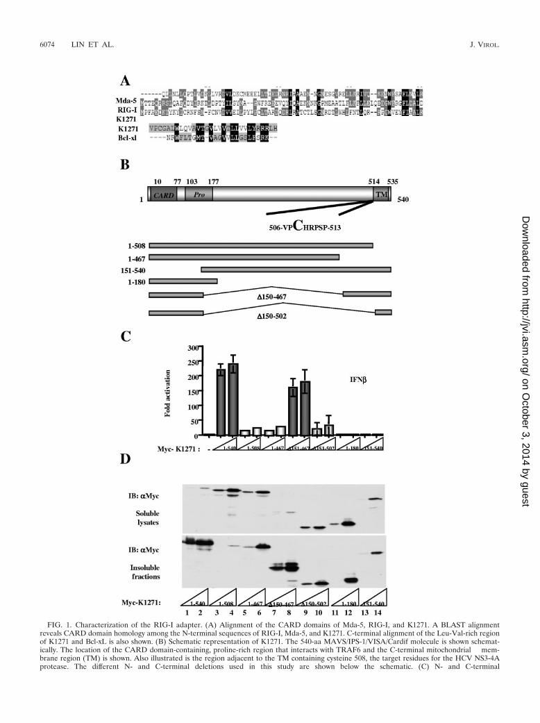
FIG. 1. Characterization of the RIG-I adapter. (A) Alignment of the CARD domains of Mda-5, RIG-I, and K1271. A BLAST alignmentreveals CARD domain homology among the N-terminal sequences of RIG-I, Mda-5, and K1271. C-terminal alignment of the Leu-Val-rich regionof K1271 and Bcl-xL is also shown. (B) Schematic representation of K1271. The 540-aa MAVS/IPS-1/VISA/Cardif molecule is shown schemat-ically. The location of the CARD domain-containing, proline-rich region that interacts with TRAF6 and the C-terminal mitochondrial mem-brane region (TM) is shown. Also illustrated is the region adjacent to the TM containing cysteine 508, the target residues for the HCV NS3-4Aprotease. The different N- and C-terminal deletions used in this study are shown below the schematic. (C) N- and C-terminal
6074 LIN ET AL. J. VIROL.
on October 3, 2014 by guest
http://jvi.asm.org/
Dow
nloaded from
mains, and the domains were analyzed separately, creating aregular expression that represents the patterns of the domain.Next, a search program was written in python language (http://www.biopython.org); a database search was performed thatidentified an uncharacterized protein, KIAA1271(K1271),containing a single CARD-like domain at the N terminus anda Leu-Val-rich C terminus (Fig. 1A). Interestingly, analysis ofthe C terminus of K1271 revealed high homology with the Cterminus of the antiapoptotic protein Bcl-xl. Following PCRamplification and subcloning, a series of K1271 deletion con-structs were generated and tested for the capacity to stimulatethe IFN-� promoter (Fig. 1B). Coexpression of K1271 stronglyactivated the IFN-�-dependent promoter and appeared to bethe adapter linking RIG-I sensing to downstream signalingevents. Both the N and C termini were essential for transacti-vation function, since deletion of either end of the proteineliminated transactivation (Fig. 1C). Interestingly, an expres-sion construct that contained the N- and C-terminal domainsbut lacked the region between aa 150 and aa 467 also stimu-lated the IFN-� promoter (Fig. 1C), whereas a similar internaldeletion construct removing the region between aa 150 and aa502 was inactive, indicating that the region of K1271 betweenaa 467 and aa 502 was important for transactivation function.A direct correlation between downstream activation of theIFN-� promoter and subcellular localization was observedwhen the different K1271 deletion constructs were evaluated;wtK1271 and the active internal deletion �150–467 were local-ized to the insoluble fraction of cytoplasmic extracts (Fig. 1D,lanes 1, 2, 7 and 8), whereas the C-terminal truncation of aa 1 to508 was localized exclusively in the soluble cytoplasmic extract(Fig. 1D, lanes 3 and 4). Other constructs, including the inactiveinternal deletion �150–502 and the deletions of aa 1 to 467, aa 1to 180, and aa 151 to 540, redistributed to both the soluble andinsoluble cytoplasmic fractions (Fig. 1D, lanes 5, 6, and 9 to 14).
Four independent groups reported that the same protein,alternately named MAVS/VISA/IPS-1-1/Cardif, acted down-stream of RIG-I and stimulated the expression of IFN-�through the activation of NF-�B and IRF-3 (20, 29, 35, 40).The C-terminal Leu-Val domain was recognized as a mito-chondrial transmembrane domain, and localization to the mi-tochondrial outer membrane was required for its function (35).Thus, MAVS/VISA/IPS-1-1/Cardif/K1271 is a critical adapterof the RIG-I pathway that links the “sensing” of incoming viralparticles by RIG-I with downstream TBK1/IKKε kinases.
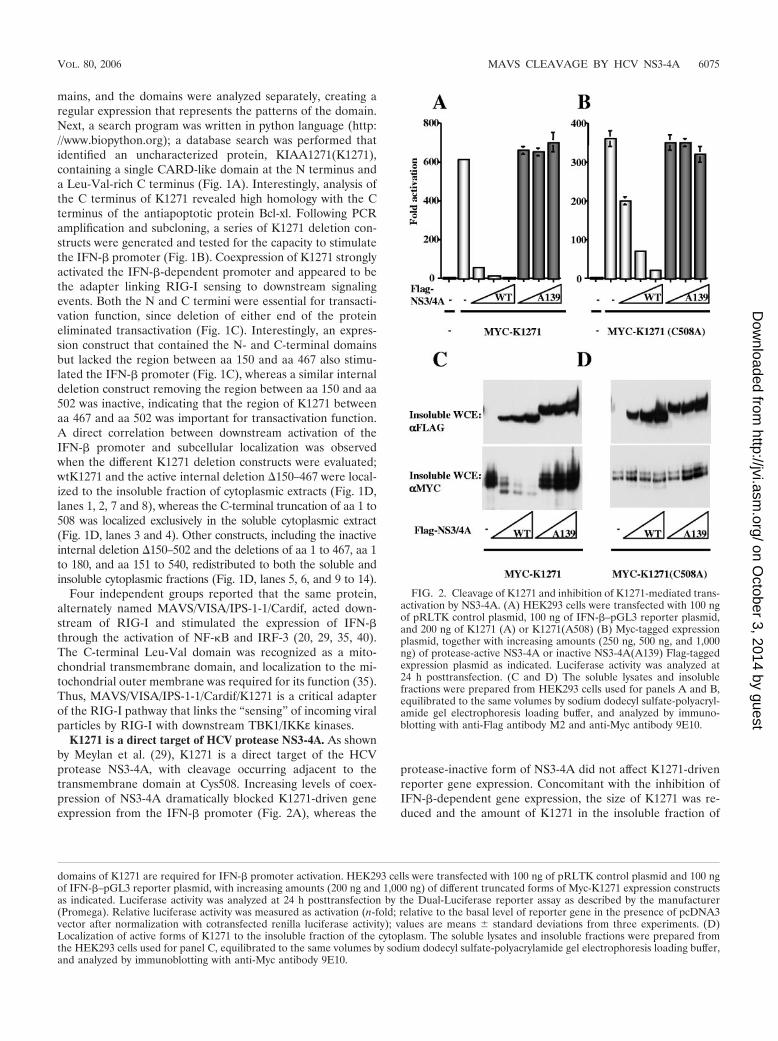
K1271 is a direct target of HCV protease NS3-4A. As shownby Meylan et al. (29), K1271 is a direct target of the HCVprotease NS3-4A, with cleavage occurring adjacent to thetransmembrane domain at Cys508. Increasing levels of coex-pression of NS3-4A dramatically blocked K1271-driven geneexpression from the IFN-� promoter (Fig. 2A), whereas the
protease-inactive form of NS3-4A did not affect K1271-drivenreporter gene expression. Concomitant with the inhibition ofIFN-�-dependent gene expression, the size of K1271 was re-duced and the amount of K1271 in the insoluble fraction of
domains of K1271 are required for IFN-� promoter activation. HEK293 cells were transfected with 100 ng of pRLTK control plasmid and 100 ngof IFN-�–pGL3 reporter plasmid, with increasing amounts (200 ng and 1,000 ng) of different truncated forms of Myc-K1271 expression constructsas indicated. Luciferase activity was analyzed at 24 h posttransfection by the Dual-Luciferase reporter assay as described by the manufacturer(Promega). Relative luciferase activity was measured as activation (n-fold; relative to the basal level of reporter gene in the presence of pcDNA3vector after normalization with cotransfected renilla luciferase activity); values are means � standard deviations from three experiments. (D)Localization of active forms of K1271 to the insoluble fraction of the cytoplasm. The soluble lysates and insoluble fractions were prepared fromthe HEK293 cells used for panel C, equilibrated to the same volumes by sodium dodecyl sulfate-polyacrylamide gel electrophoresis loading buffer,and analyzed by immunoblotting with anti-Myc antibody 9E10.
FIG. 2. Cleavage of K1271 and inhibition of K1271-mediated trans-activation by NS3-4A. (A) HEK293 cells were transfected with 100 ngof pRLTK control plasmid, 100 ng of IFN-�–pGL3 reporter plasmid,and 200 ng of K1271 (A) or K1271(A508) (B) Myc-tagged expressionplasmid, together with increasing amounts (250 ng, 500 ng, and 1,000ng) of protease-active NS3-4A or inactive NS3-4A(A139) Flag-taggedexpression plasmid as indicated. Luciferase activity was analyzed at24 h posttransfection. (C and D) The soluble lysates and insolublefractions were prepared from HEK293 cells used for panels A and B,equilibrated to the same volumes by sodium dodecyl sulfate-polyacryl-amide gel electrophoresis loading buffer, and analyzed by immuno-blotting with anti-Flag antibody M2 and anti-Myc antibody 9E10.
VOL. 80, 2006 MAVS CLEAVAGE BY HCV NS3-4A 6075
on October 3, 2014 by guest
http://jvi.asm.org/
Dow
nloaded from
cytoplasmic extracts was decreased (Fig. 2C), whereas expres-sion of the protease-inactive point mutant A139 did not alterthe mobility or the amount of K1271. In contrast, the K1271(C508A) point mutation remained localized in the insoluble cy-toplasmic fraction, thus illustrating that K1271(C508A) was notcleaved by NS3-4A (Fig. 2D). However, K1271(C508A) IFN-�-driven gene activity was still decreased when NS3-4A was ex-pressed, although with about 10-fold decreased efficiency, sug-gesting that NS3-4A may also target an undefined proteindownstream of K1271.
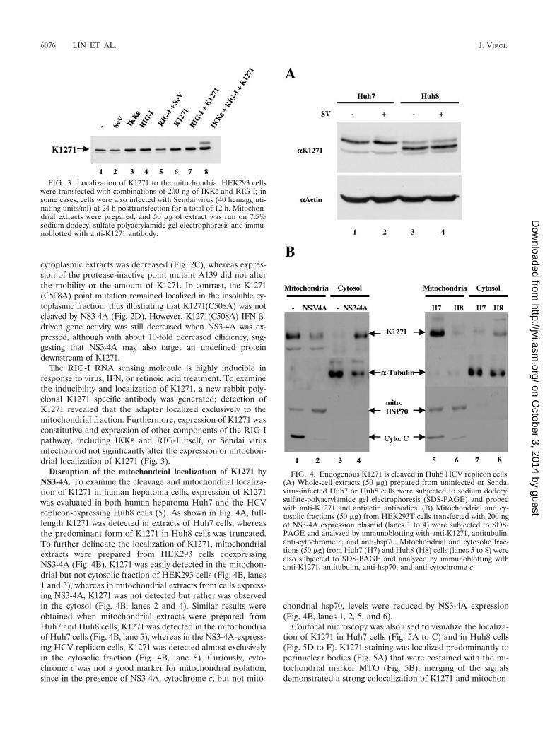
The RIG-I RNA sensing molecule is highly inducible inresponse to virus, IFN, or retinoic acid treatment. To examinethe inducibility and localization of K1271, a new rabbit poly-clonal K1271 specific antibody was generated; detection ofK1271 revealed that the adapter localized exclusively to themitochondrial fraction. Furthermore, expression of K1271 wasconstitutive and expression of other components of the RIG-Ipathway, including IKKε and RIG-I itself, or Sendai virusinfection did not significantly alter the expression or mitochon-drial localization of K1271 (Fig. 3).
Disruption of the mitochondrial localization of K1271 byNS3-4A. To examine the cleavage and mitochondrial localiza-tion of K1271 in human hepatoma cells, expression of K1271was evaluated in both human hepatoma Huh7 and the HCVreplicon-expressing Huh8 cells (5). As shown in Fig. 4A, full-length K1271 was detected in extracts of Huh7 cells, whereasthe predominant form of K1271 in Huh8 cells was truncated.To further delineate the localization of K1271, mitochondrialextracts were prepared from HEK293 cells coexpressingNS3-4A (Fig. 4B). K1271 was easily detected in the mitochon-drial but not cytosolic fraction of HEK293 cells (Fig. 4B, lanes1 and 3), whereas in mitochondrial extracts from cells express-ing NS3-4A, K1271 was not detected but rather was observedin the cytosol (Fig. 4B, lanes 2 and 4). Similar results wereobtained when mitochondrial extracts were prepared fromHuh7 and Huh8 cells; K1271 was detected in the mitochondriaof Huh7 cells (Fig. 4B, lane 5), whereas in the NS3-4A-express-ing HCV replicon cells, K1271 was detected almost exclusivelyin the cytosolic fraction (Fig. 4B, lane 8). Curiously, cyto-chrome c was not a good marker for mitochondrial isolation,since in the presence of NS3-4A, cytochrome c, but not mito-
chondrial hsp70, levels were reduced by NS3-4A expression(Fig. 4B, lanes 1, 2, 5, and 6).
Confocal microscopy was also used to visualize the localiza-tion of K1271 in Huh7 cells (Fig. 5A to C) and in Huh8 cells(Fig. 5D to F). K1271 staining was localized predominantly toperinuclear bodies (Fig. 5A) that were costained with the mi-tochondrial marker MTO (Fig. 5B); merging of the signalsdemonstrated a strong colocalization of K1271 and mitochon-
FIG. 3. Localization of K1271 to the mitochondria. HEK293 cellswere transfected with combinations of 200 ng of IKKε and RIG-I; insome cases, cells were also infected with Sendai virus (40 hemaggluti-nating units/ml) at 24 h posttransfection for a total of 12 h. Mitochon-drial extracts were prepared, and 50 �g of extract was run on 7.5%sodium dodecyl sulfate-polyacrylamide gel electrophoresis and immu-noblotted with anti-K1271 antibody.
FIG. 4. Endogenous K1271 is cleaved in Huh8 HCV replicon cells.(A) Whole-cell extracts (50 �g) prepared from uninfected or Sendaivirus-infected Huh7 or Huh8 cells were subjected to sodium dodecylsulfate-polyacrylamide gel electrophoresis (SDS-PAGE) and probedwith anti-K1271 and antiactin antibodies. (B) Mitochondrial and cy-tosolic fractions (50 �g) from HEK293T cells transfected with 200 ngof NS3-4A expression plasmid (lanes 1 to 4) were subjected to SDS-PAGE and analyzed by immunoblotting with anti-K1271, antitubulin,anti-cytochrome c, and anti-hsp70. Mitochondrial and cytosolic frac-tions (50 �g) from Huh7 (H7) and Huh8 (H8) cells (lanes 5 to 8) werealso subjected to SDS-PAGE and analyzed by immunoblotting withanti-K1271, antitubulin, anti-hsp70, and anti-cytochrome c.
6076 LIN ET AL. J. VIROL.
on October 3, 2014 by guest
http://jvi.asm.org/
Dow
nloaded from
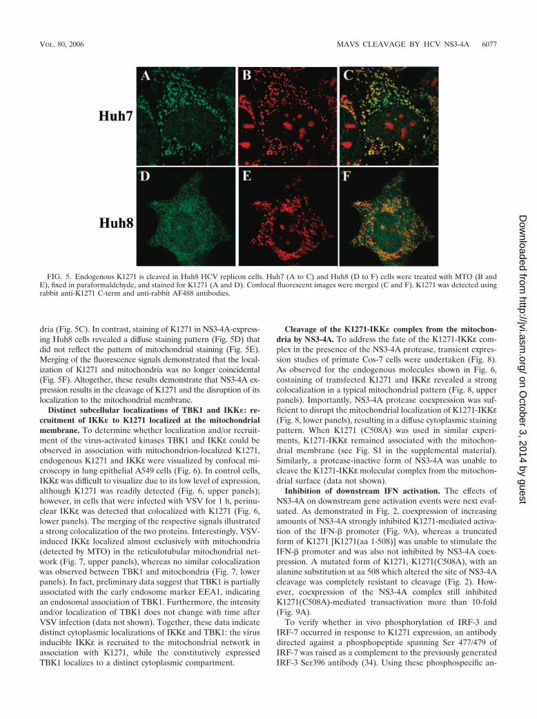
dria (Fig. 5C). In contrast, staining of K1271 in NS3-4A-express-ing Huh8 cells revealed a diffuse staining pattern (Fig. 5D) thatdid not reflect the pattern of mitochondrial staining (Fig. 5E).Merging of the fluorescence signals demonstrated that the local-ization of K1271 and mitochondria was no longer coincidental(Fig. 5F). Altogether, these results demonstrate that NS3-4A ex-pression results in the cleavage of K1271 and the disruption of itslocalization to the mitochondrial membrane.
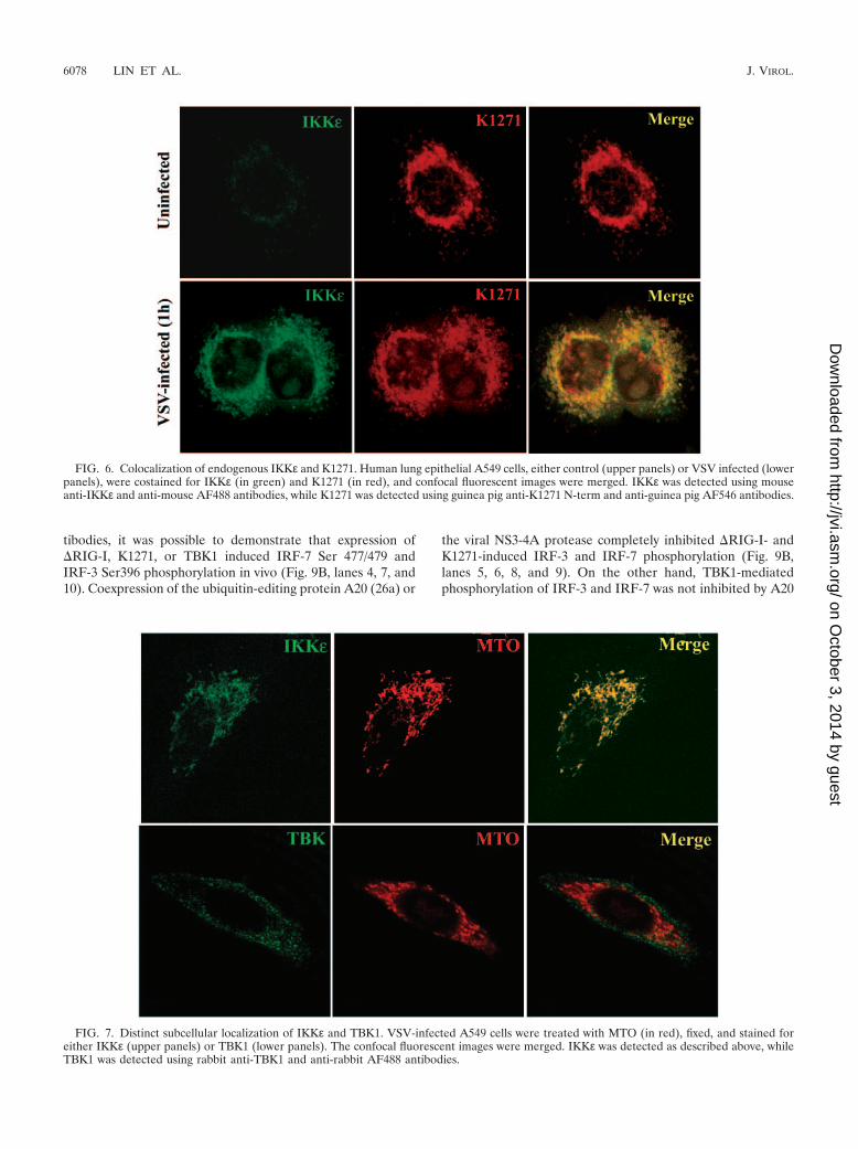
Distinct subcellular localizations of TBK1 and IKK�: re-cruitment of IKK� to K1271 localized at the mitochondrialmembrane. To determine whether localization and/or recruit-ment of the virus-activated kinases TBK1 and IKKε could beobserved in association with mitochondrion-localized K1271,endogenous K1271 and IKKε were visualized by confocal mi-croscopy in lung epithelial A549 cells (Fig. 6). In control cells,IKKε was difficult to visualize due to its low level of expression,although K1271 was readily detected (Fig. 6, upper panels);however, in cells that were infected with VSV for 1 h, perinu-clear IKKε was detected that colocalized with K1271 (Fig. 6,lower panels). The merging of the respective signals illustrateda strong colocalization of the two proteins. Interestingly, VSV-induced IKKε localized almost exclusively with mitochondria(detected by MTO) in the reticulotubular mitochondrial net-work (Fig. 7, upper panels), whereas no similar colocalizationwas observed between TBK1 and mitochondria (Fig. 7, lowerpanels). In fact, preliminary data suggest that TBK1 is partiallyassociated with the early endosome marker EEA1, indicatingan endosomal association of TBK1. Furthermore, the intensityand/or localization of TBK1 does not change with time afterVSV infection (data not shown). Together, these data indicatedistinct cytoplasmic localizations of IKKε and TBK1: the virusinducible IKKε is recruited to the mitochondrial network inassociation with K1271, while the constitutively expressedTBK1 localizes to a distinct cytoplasmic compartment.
Cleavage of the K1271-IKK� complex from the mitochon-dria by NS3-4A. To address the fate of the K1271-IKKε com-plex in the presence of the NS3-4A protease, transient expres-sion studies of primate Cos-7 cells were undertaken (Fig. 8).As observed for the endogenous molecules shown in Fig. 6,costaining of transfected K1271 and IKKε revealed a strongcolocalization in a typical mitochondrial pattern (Fig. 8, upperpanels). Importantly, NS3-4A protease coexpression was suf-ficient to disrupt the mitochondrial localization of K1271-IKKε(Fig. 8, lower panels), resulting in a diffuse cytoplasmic stainingpattern. When K1271 (C508A) was used in similar experi-ments, K1271-IKKε remained associated with the mitochon-drial membrane (see Fig. S1 in the supplemental material).Similarly, a protease-inactive form of NS3-4A was unable tocleave the K1271-IKKε molecular complex from the mitochon-drial surface (data not shown).
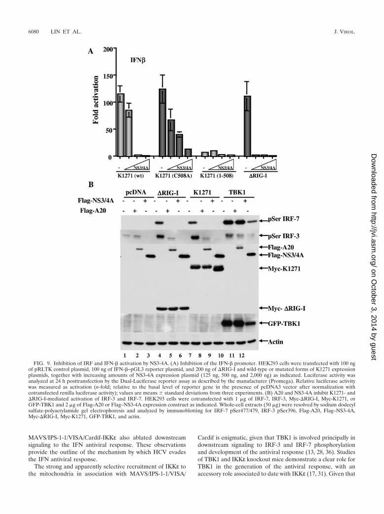
Inhibition of downstream IFN activation. The effects ofNS3-4A on downstream gene activation events were next eval-uated. As demonstrated in Fig. 2, coexpression of increasingamounts of NS3-4A strongly inhibited K1271-mediated activa-tion of the IFN-� promoter (Fig. 9A), whereas a truncatedform of K1271 [K1271(aa 1-508)] was unable to stimulate theIFN-� promoter and was also not inhibited by NS3-4A coex-pression. A mutated form of K1271, K1271(C508A), with analanine substitution at aa 508 which altered the site of NS3-4Acleavage was completely resistant to cleavage (Fig. 2). How-ever, coexpression of the NS3-4A complex still inhibitedK1271(C508A)-mediated transactivation more than 10-fold(Fig. 9A).
To verify whether in vivo phosphorylation of IRF-3 andIRF-7 occurred in response to K1271 expression, an antibodydirected against a phosphopeptide spanning Ser 477/479 ofIRF-7 was raised as a complement to the previously generatedIRF-3 Ser396 antibody (34). Using these phosphospecific an-
FIG. 5. Endogenous K1271 is cleaved in Huh8 HCV replicon cells. Huh7 (A to C) and Huh8 (D to F) cells were treated with MTO (B andE), fixed in paraformaldehyde, and stained for K1271 (A and D). Confocal fluorescent images were merged (C and F). K1271 was detected usingrabbit anti-K1271 C-term and anti-rabbit AF488 antibodies.
VOL. 80, 2006 MAVS CLEAVAGE BY HCV NS3-4A 6077
on October 3, 2014 by guest
http://jvi.asm.org/
Dow
nloaded from
tibodies, it was possible to demonstrate that expression of�RIG-I, K1271, or TBK1 induced IRF-7 Ser 477/479 andIRF-3 Ser396 phosphorylation in vivo (Fig. 9B, lanes 4, 7, and10). Coexpression of the ubiquitin-editing protein A20 (26a) or
the viral NS3-4A protease completely inhibited �RIG-I- andK1271-induced IRF-3 and IRF-7 phosphorylation (Fig. 9B,lanes 5, 6, 8, and 9). On the other hand, TBK1-mediatedphosphorylation of IRF-3 and IRF-7 was not inhibited by A20
FIG. 6. Colocalization of endogenous IKKε and K1271. Human lung epithelial A549 cells, either control (upper panels) or VSV infected (lowerpanels), were costained for IKKε (in green) and K1271 (in red), and confocal fluorescent images were merged. IKKε was detected using mouseanti-IKKε and anti-mouse AF488 antibodies, while K1271 was detected using guinea pig anti-K1271 N-term and anti-guinea pig AF546 antibodies.
FIG. 7. Distinct subcellular localization of IKKε and TBK1. VSV-infected A549 cells were treated with MTO (in red), fixed, and stained foreither IKKε (upper panels) or TBK1 (lower panels). The confocal fluorescent images were merged. IKKε was detected as described above, whileTBK1 was detected using rabbit anti-TBK1 and anti-rabbit AF488 antibodies.
6078 LIN ET AL. J. VIROL.
on October 3, 2014 by guest
http://jvi.asm.org/
Dow
nloaded from

and NS3-4A, indicating that both of these inhibitory activitiesacted upstream of TBK1 (Fig. 9B, lanes 10 to 12) and confirm-ing previous findings that NS3-4A has no effect on the ability ofTBK-1 to phosphorylate IRF-3 (8).
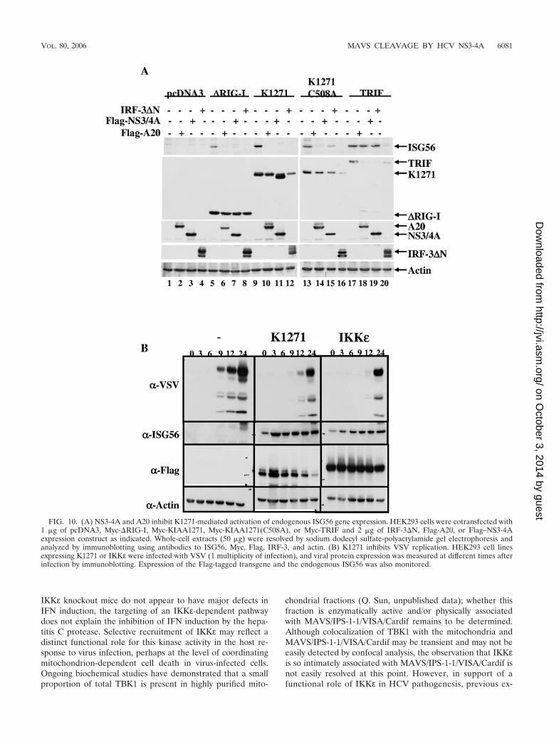
K1271 activates endogenous ISG expression and the antivi-ral state. As a measure of the induction of the antiviral state,the constitutively active form of RIG-I, �RIG-I, K1271, or theTRIF adapter was used to assess the induction of endogenousIFN-stimulated gene 56 (ISG56) expression. Coexpression of�RIG-I, K1271, or TRIF resulted in a strong induction ofendogenous ISG56 expression (Fig. 10A, lanes 5, 9, and 17).Expression of the point-mutated K1271(C508A) also inducedISG56 expression (Fig. 10A, lane 13). The cellular A20 proteincompletely inhibited RIG-I- and K1271-induced ISG56 expres-sion (Fig. 10A, lanes 6, 10, and 14) but did not affect TRIF-induced ISG56 expression (Fig. 10A, lane 18). NS3-4A alsototally blocked �RIG-I- and K1271-mediated induction ofISG56 (Fig. 10A, lanes 7 and 11); however, NS3-4A onlyweakly interfered with K1271(C508A)-mediated induction ofISG56 (Fig. 10A, lane 15) because NS3-4A failed to cleaveK1271(C508A). In contrast, molecular weight reduction ofK1271 as a result of NS3-4A cleavage was easily detected (Fig.10A, lane 11). NS3-4A also failed to inhibit TRIF-mediatedISG56 expression. In all cases, expression of the dominant-negative form of IRF3, IRF3�N, inhibited ISG56 expression(Fig. 10A, lanes 8, 12, 16, and 20), demonstrating that ISG56induction occurred through an IRF3-dependent pathway. As afurther confirmation of the generation of an antiviral state,HEK293 cells expressing K1271 or IKKε were infected withVSV and the kinetics of viral protein expression was measured.Viral protein production was significantly inhibited from 6 to9 h in control cells to 12 to 24 h in cells expressing K1271 orIKKε protein; furthermore, ISG56 expression was constitu-tively detected in K1271- or IKKε-expressing cells (Fig. 10B),
thus demonstrating the generation of an antiviral state byK1271 that blocked VSV replication.
DISCUSSION
It has quickly become clear that the RNA helicase RIG-Ipathway plays an essential role in the sensing of incoming virusinfection and directly relays regulatory signals to the host an-tiviral response. The adapter molecule providing a link be-tween RIG-I sensing of incoming viral RNA and downstreamactivation events was recently elucidated; four independentgroups used high-throughput screening and/or database searchanalyses to identify the new signaling component, which hasalternatively been termed MAVS/IPS-1-1/VISA/Cardif (20, 29,35, 40) and is termed K1271 in this study. The fact that MAVS/IPS-1-1/VISA/Cardif localizes to the mitochondrial membranesuggests linkage among recognition of viral infection, the de-velopment of innate immunity, and mitochondrial function(35). The IKK-related kinases TBK1 and IKKε are criticaldownstream components of the activation of the interferonantiviral response through their ability to phosphorylate theC-terminal domains of IRF-3 and IRF-7 (13, 28, 36). In thepresent study, we demonstrate that VSV infection of lungepithelial A549 cells results in the induction and recruitment ofIKKε to the mitochondrial membrane, in association withK1271 (MAVS/IPS-1-1/VISA/Cardif). Furthermore, the hepa-titis C virus NS3-4A protease activity specifically cleaves theMAVS/IPS-1-1/VISA/Cardif-IKKε molecular complex fromthe mitochondria as part of its immune evasion strategy. Pointmutation of Cys508 to Ala, the target residue of NS3-4A,created a MAVS/IPS-1-1/VISA/Cardif protein that not onlywas resistant to cleavage and subcellular relocation but alsomaintained the association with IKKε in the presence of pro-tease activity. Disruption of the mitochondrial localization of
FIG. 8. Disruption of the IKKε-K1271 complex localization by expression of NS3-4A. COS-7 cells transfected with IKKε and K1271 expressionplasmids, without (upper panels) or with (lower panels) a construct encoding NS3-4A, were costained for IKKε (in green) and K1271 (in red), andconfocal fluorescent images were merged. IKKε and K1271 were detected as described above.
VOL. 80, 2006 MAVS CLEAVAGE BY HCV NS3-4A 6079
on October 3, 2014 by guest
http://jvi.asm.org/
Dow
nloaded from
MAVS/IPS-1-1/VISA/Cardif-IKKε also ablated downstreamsignaling to the IFN antiviral response. These observationsprovide the outline of the mechanism by which HCV evadesthe IFN antiviral response.
The strong and apparently selective recruitment of IKKε tothe mitochondria in association with MAVS/IPS-1-1/VISA/
Cardif is enigmatic, given that TBK1 is involved principally indownstream signaling to IRF-3 and IRF-7 phosphorylationand development of the antiviral response (13, 28, 36). Studiesof TBK1 and IKKε knockout mice demonstrate a clear role forTBK1 in the generation of the antiviral response, with anaccessory role associated to date with IKKε (17, 31). Given that
FIG. 9. Inhibition of IRF and IFN-� activation by NS3-4A. (A) Inhibition of the IFN-� promoter. HEK293 cells were transfected with 100 ngof pRLTK control plasmid, 100 ng of IFN-�–pGL3 reporter plasmid, and 200 ng of �RIG-I and wild-type or mutated forms of K1271 expressionplasmids, together with increasing amounts of NS3-4A expression plasmid (125 ng, 500 ng, and 2,000 ng) as indicated. Luciferase activity wasanalyzed at 24 h posttransfection by the Dual-Luciferase reporter assay as described by the manufacturer (Promega). Relative luciferase activitywas measured as activation (n-fold; relative to the basal level of reporter gene in the presence of pcDNA3 vector after normalization withcotransfected renilla luciferase activity); values are means � standard deviations from three experiments. (B) A20 and NS3-4A inhibit K1271- and�RIG-I-mediated activation of IRF-3 and IRF-7. HEK293 cells were cotransfected with 1 �g of IRF-7, IRF-3, Myc-�RIG-I, Myc-K1271, orGFP-TBK1 and 2 �g of Flag-A20 or Flag–NS3-4A expression construct as indicated. Whole-cell extracts (50 �g) were resolved by sodium dodecylsulfate-polyacrylamide gel electrophoresis and analyzed by immunoblotting for IRF-7 pSer477/479, IRF-3 pSer396, Flag-A20, Flag–NS3-4A,Myc-�RIG-I, Myc-K1271, GFP-TBK1, and actin.
6080 LIN ET AL. J. VIROL.
on October 3, 2014 by guest
http://jvi.asm.org/
Dow
nloaded from
IKKε knockout mice do not appear to have major defects inIFN induction, the targeting of an IKKε-dependent pathwaydoes not explain the inhibition of IFN induction by the hepa-titis C protease. Selective recruitment of IKKε may reflect adistinct functional role for this kinase activity in the host re-sponse to virus infection, perhaps at the level of coordinatingmitochondrion-dependent cell death in virus-infected cells.Ongoing biochemical studies have demonstrated that a smallproportion of total TBK1 is present in highly purified mito-
chondrial fractions (Q. Sun, unpublished data); whether thisfraction is enzymatically active and/or physically associatedwith MAVS/IPS-1-1/VISA/Cardif remains to be determined.Although colocalization of TBK1 with the mitochondria andMAVS/IPS-1-1/VISA/Cardif may be transient and may not beeasily detected by confocal analysis, the observation that IKKεis so intimately associated with MAVS/IPS-1-1/VISA/Cardif isnot easily resolved at this point. However, in support of afunctional role of IKKε in HCV pathogenesis, previous ex-
FIG. 10. (A) NS3-4A and A20 inhibit K1271-mediated activation of endogenous ISG56 gene expression. HEK293 cells were cotransfected with1 �g of pcDNA3, Myc-�RIG-I, Myc-KIAA1271, Myc-KIAA1271(C508A), or Myc-TRIF and 2 �g of IRF-3�N, Flag-A20, or Flag–NS3-4Aexpression construct as indicated. Whole-cell extracts (50 �g) were resolved by sodium dodecyl sulfate-polyacrylamide gel electrophoresis andanalyzed by immunoblotting using antibodies to ISG56, Myc, Flag, IRF-3, and actin. (B) K1271 inhibits VSV replication. HEK293 cell linesexpressing K1271 or IKKε were infected with VSV (1 multiplicity of infection), and viral protein expression was measured at different times afterinfection by immunoblotting. Expression of the Flag-tagged transgene and the endogenous ISG56 was also monitored.
VOL. 80, 2006 MAVS CLEAVAGE BY HCV NS3-4A 6081
on October 3, 2014 by guest
http://jvi.asm.org/
Dow
nloaded from
periments have demonstrated that IKKε overexpression, butnot the expression of TBK1 or other signaling adapters,partially reversed HCV protease-mediated inhibition of IFNinduction (8).
The mechanism of recruitment of IKKε to the mitochondriaremains unclear at present. However, given that NS3-4A is stillable to block IFN-� promoter activity when the Cys508-to-Alapoint-mutated MAVS/IPS-1-1/VISA/Cardif protein is ex-pressed, albeit with a 10-fold-reduced efficiency, it is possiblethat another adapter downstream of MAVS/IPS-1-1/VISA/Cardif may be targeted by NS3-4A. Alternatively, an additionalmechanism involved in the recruitment of IKKε to the MAVS/IPS-1-1/VISA/Cardif mitochondrial reticulotubular networkmay be affected by the HCV protease. We have also identifiedsignificant changes in mitochondrial reticulotubular architec-ture following VSV infection and the recruitment of IKKε tothe mitochondria, suggesting that IKKε may be contributing tovirus-induced apoptosis.
Seth et al. identified the localization of MAVS to the mito-chondrial membrane and also showed that MAVS moved intoa detergent-resistant mitochondrial fraction upon viral infec-tion (35). The fact that MAVS functionality requires mito-chondrial association suggests linkage among recognition ofviral infection, the development of innate immunity, and virus-induced mitochondrion-dependent cell death. In fact, knock-down of MAVS gene expression by small interfering RNAsincreased apoptosis, possibly hinting at a protective role forMAVS during the early stages of viral infection. Potentially,the activation of other components of the mitochondrial mem-brane could also contribute to signaling to the antiviral re-sponse. In support of this idea, MAVS colocalizes in the samedetergent-insoluble fraction as the antiapoptotic protein Bcl-xL. Among the many CARD-containing proteins with roles inapoptosis and immunity (Apaf1, NOD1, NOD2 RIP2, andRIG-I), MAVS is unique (7, 11, 12). The localization of thisCARD domain-containing adapter to the mitochondrial mem-brane is highly strategic and may help the host cell to senseincoming viral challenge and coordinate an immune or apop-totic response, depending on the pathogen. Many viruses rep-licate in intracellular organelles, such as the endoplasmic re-ticulum; a good example is HCV, which replicates in themembranous web that connects the endoplasmic reticulum tothe mitochondria. dsRNA structures, possibly within replicat-ing ribonucleoprotein complexes, may be recognized by RIG-Iand/or Mda-5, resulting in downstream signaling throughMAVS. Mitochondria may be at the center of a delicate bal-ancing act between the host immune response and virus-in-duced apoptosis. In the case of HCV infection, cleavage ofMAVS by the NS3-4A protease appears to tip the balance,resulting in disruption of innate immune responses and theestablishment of chronic HCV persistence (15).
The identification of MAVS/IPS-1/VISA/Cardif and its rolein innate signaling and its characterization as the physiologi-cally relevant target of the NS3-4A protease are importantsteps in the complete understanding of the mechanisms bywhich HCV evades the early host response. The essential lo-calization of this CARD domain-containing adapter to themitochondria furthermore suggests an important function inthe coordination of the innate immune and apoptotic re-sponses. The implications for the study of HCV pathogenesis
are particularly profound, given that experimental compounds,such as BILN2061 and VX-950, that block NS3-4A proteaseactivity may accomplish two goals: inhibition of virus multipli-cation and processing and restoration of the early innate im-mune response that is critical to the development of a robustadaptive response in patients (9, 22).
ACKNOWLEDGMENTS
We thank Hong-Bing Shu, Zhijian Chen, Ganes Sen, Wen-Zhe Ho,and Michael Gale for reagents used in this study and members of theMolecular Oncology Group, Lady Davis Institute, for helpful discus-sions.
This research was supported by grants from the Canadian Institutesof Health Research (J.H. and R.L.), CANVAC, and the CanadianNetwork for Vaccines and Immunotherapeutics (J.H.) and by theNational Cancer Institute of Canada, with the support of the CanadianCancer Society (J.H.). D.V. was supported by l’Agence Nationale de laRecherche sur le Sida (ANRS). R.L. was supported by an FRSQChercheur-boursier and J.H. by a CIHR Senior Investigator award.
REFERENCES
1. Akira, S., and K. Takeda. 2004. Toll-like receptor signalling. Nat. Rev.Immunol. 4:499–511.
2. Alexopoulou, L., A. C. Holt, R. Medzhitov, and R. A. Flavell. 2001. Recog-nition of double-stranded RNA and activation of NF-kappaB by Toll-likereceptor 3. Nature 413:732–738.
3. Alter, H. J., and L. B. Seeff. 2000. Recovery, persistence, and sequelae inhepatitis C virus infection: a perspective on long-term outcome. Semin. LiverDis. 20:17–35.
4. Andrejeva, J., K. S. Childs, D. F. Young, T. S. Carlos, N. Stock, S. Good-bourn, and R. E. Randall. 2004. The V proteins of paramyxoviruses bind theIFN-inducible RNA helicase, mda-5, and inhibit its activation of the IFN-beta promoter. Proc. Natl. Acad. Sci. USA 101:17264–17269.
5. Blight, K. J., A. A. Kolykhalov, and C. M. Rice. 2000. Efficient initiation ofHCV RNA replication in cell culture. Science 290:1972–1974.
6. Blight, K. J., J. A. McKeating, J. Marcotrigiano, and C. M. Rice. 2003.Efficient replication of hepatitis C virus genotype 1a RNAs in cell culture.J. Virol. 77:3181–3190.
7. Bouchier-Hayes, L., and S. J. Martin. 2002. CARD games in apoptosis andimmunity. EMBO Rep. 3:616–621.
8. Breiman, A., N. Grandvaux, R. Lin, C. Ottone, S. Akira, M. Yoneyama, T.Fujita, J. Hiscott, and E. F. Meurs. 2005. Inhibition of RIG-I-dependentsignaling to the interferon pathway during hepatitis C virus expression andrestoration of signaling by IKKε. J. Virol. 79:3969–3978.
9. Chen, S. H., and S. L. Tan. 2005. Discovery of small-molecule inhibitors ofHCV NS3-4A protease as potential therapeutic agents against HCV infec-tion. Curr. Med. Chem. 12:2317–2342.
10. Choo, Q. L., G. Kuo, A. J. Weiner, L. R. Overby, D. W. Bradley, and M.Houghton. 1989. Isolation of a cDNA clone derived from a blood-bornenon-A, non-B viral hepatitis genome. Science 244:359–362.
11. Damiano, J. S., R. M. Newman, and J. C. Reed. 2004. Multiple roles ofCLAN (caspase-associated recruitment domain, leucine-rich repeat, andNAIP CIIA HET-E, and TP1-containing protein) in the mammalian innateimmune response. J. Immunol. 173:6338–6345.
12. Damiano, J. S., and J. C. Reed. 2004. CARD proteins as therapeutic targetsin cancer. Curr. Drug Targets 5:367–374.
13. Fitzgerald, K. A., S. M. McWhirter, K. L. Faia, D. C. Rowe, E. Latz, D. T.Golenbock, A. J. Coyle, S. M. Liao, and T. Maniatis. 2003. IKKepsilon andTBK1 are essential components of the IRF3 signaling pathway. Nat. Immu-nol. 4:491–496.
14. Foy, E., K. Li, R. Sumpter, Jr., Y. M. Loo, C. L. Johnson, C. Wang, P. M.Fish, M. Yoneyama, T. Fujita, S. M. Lemon, and M. Gale, Jr. 2005. Controlof antiviral defenses through hepatitis C virus disruption of retinoic acid-inducible gene-I signaling. Proc. Natl. Acad. Sci. USA 102:2986–2991.
15. Gale, M., Jr., and E. M. Foy. 2005. Evasion of intracellular host defence byhepatitis C virus. Nature 436:939–945.
16. He, Y., and M. G. Katze. 2002. To interfere and to anti-interfere: theinterplay between hepatitis C virus and interferon. Viral Immunol. 15:95–119.
17. Hemmi, H., O. Takeuchi, S. Sato, M. Yamamoto, T. Kaisho, H. Sanjo, T.Kawai, K. Hoshino, K. Takeda, and S. Akira. 2004. The roles of two IkappaBkinase-related kinases in lipopolysaccharide and double stranded RNA sig-naling and viral infection. J. Exp. Med. 199:1641–1650.
18. Kang, D. C., R. V. Gopalkrishnan, Q. Wu, E. Jankowsky, A. M. Pyle, andP. B. Fisher. 2002. mda-5: an interferon-inducible putative RNA helicasewith double-stranded RNA-dependent ATPase activity and melanomagrowth-suppressive properties. Proc. Natl. Acad. Sci. USA 99:637–642.
6082 LIN ET AL. J. VIROL.
on October 3, 2014 by guest
http://jvi.asm.org/
Dow
nloaded from
19. Kato, H., S. Sato, M. Yoneyama, M. Yamamoto, S. Uematsu, K. Matsui, T.Tsujimura, K. Takeda, T. Fujita, O. Takeuchi, and S. Akira. 2005. Celltype-specific involvement of RIG-I in antiviral response. Immunity 23:19–28.
20. Kawai, T., K. Takahashi, S. Sato, C. Coban, H. Kumar, H. Kato, K. J. Ishii,O. Takeuchi, and S. Akira. 2005. IPS-1, an adaptor triggering RIG-I- andMda5-mediated type I interferon induction. Nat. Immunol. 6:981–988.
21. Kiyosawa, K., T. Sodeyama, E. Tanaka, Y. Gibo, K. Yoshizawa, Y. Nakano,S. Furuta, Y. Akahane, K. Nishioka, R. H. Purcell, et al. 1990. Interrelation-ship of blood transfusion, non-A, non-B hepatitis and hepatocellular carci-noma: analysis by detection of antibody to hepatitis C virus. Hepatology12:671–675.
22. Lamarre, D., P. C. Anderson, M. Bailey, P. Beaulieu, G. Bolger, P. Bonneau,M. Bos, D. R. Cameron, M. Cartier, M. G. Cordingley, A. M. Faucher, N.Goudreau, S. H. Kawai, G. Kukolj, L. Lagace, S. R. LaPlante, H. Narjes,M. A. Poupart, J. Rancourt, R. E. Sentjens, R. St. George, B. Simoneau, G.Steinmann, D. Thibeault, Y. S. Tsantrizos, S. M. Weldon, C. L. Yong, and M.Llinas-Brunet. 2003. An NS3 protease inhibitor with antiviral effects inhumans infected with hepatitis C virus. Nature 426:186–189.
22a.Li, K., E. Foy, J. C. Ferreon, M. Nakamura, A. C. Ferreon, M. Ikeda, S. C.Ray, M. Gale, Jr., and S. M. Lemon. 2005. Immune evasion by hepatitis Cvirus NS3/4A protease-mediated cleavage of the Toll-like receptor 3 adaptorprotein TRIF. Proc. Natl. Acad. Sci. USA 102:2992–2997.
23. Li, X. D., L. Sun, R. B. Seth, G. Pineda, and Z. J. Chen. 2005. Hepatitis Cvirus protease NS3/4A cleaves mitochondrial antiviral signaling protein offthe mitochondria to evade innate immunity. Proc. Natl. Acad. Sci. USA102:17717–17722.
24. Lin, R., P. Beauparlant, C. Makris, S. Meloche, and J. Hiscott. 1996. Phos-phorylation of I�B� in the C-terminal PEST domain by casein kinase IIaffects intrinsic protein stability. Mol. Cell. Biol. 16:1401–1409.
25. Lin, R., P. Genin, Y. Mamane, and J. Hiscott. 2000. Selective DNA bindingand association with the CREB binding protein coactivator contribute todifferential activation of alpha/beta interferon genes by interferon regulatoryfactors 3 and 7. Mol. Cell. Biol. 20:6342–6353.
26. Lin, R., C. Heylbroeck, P. M. Pitha, and J. Hiscott. 1998. Virus-dependentphosphorylation of the IRF-3 transcription factor regulates nuclear translo-cation, transactivation potential, and proteasome-mediated degradation.Mol. Cell. Biol. 18:2986–2996.
26a.Lin, R., L. Yang, P. Nakhaei, Q. Sun, E. Sharif-Askari, I. Julkunen, and J.Hiscott. 2006. Negative regulation of the retinoic acid-inducible gene I-in-duced antiviral state by the ubiquitin-editing protein A20. J. Biol. Chem.281:2095–2103.
27. Maniatis, T., J. V. Falvo, T. H. Kim, T. K. Kim, C. H. Lin, B. S. Parekh, andM. G. Wathelet. 1998. Structure and function of the interferon-beta enhanceo-some. Cold Spring Harbor Symp. Quant. Biol. 63:609–620.
28. McWhirter, S. M., K. A. Fitzgerald, J. Rosains, D. C. Rowe, D. T. Golenbock,and T. Maniatis. 2004. IFN-regulatory factor 3-dependent gene expression isdefective in Tbk1-deficient mouse embryonic fibroblasts. Proc. Natl. Acad.Sci. USA 101:233–238.
29. Meylan, E., J. Curran, K. Hofmann, D. Moradpour, M. Binder, R. Barten-schlager, and J. Tschopp. 2005. Cardif is an adaptor protein in the RIG-Iantiviral pathway and is targeted by hepatitis C virus. Nature 437:1167–1172.
30. Munshi, N., J. Yie, M. Merika, K. Senger, S. Lomvardas, T. Agalioti, and D.Thanos. 1999. The IFN-beta enhancer: a paradigm for understanding acti-vation and repression of inducible gene expression. Cold Spring HarborSymp. Quant. Biol. 64:149–159.
31. Perry, A. K., E. K. Chow, J. B. Goodnough, W. C. Yeh, and G. Cheng. 2004.Differential requirement for TANK-binding kinase-1 in type I interferonresponses to Toll-like receptor activation and viral infection. J. Exp. Med.199:1651–1658.
32. Rehermann, B., and M. Nascimbeni. 2005. Immunology of hepatitis B virusand hepatitis C virus infection. Nat. Rev. Immunol. 5:215–229.
33. Rosenberg, S. 2001. Recent advances in the molecular biology of hepatitis Cvirus. J. Mol. Biol. 313:451–464.
34. Servant, M. J., N. Grandvaux, B. R. tenOever, D. Duguay, R. Lin, and J.Hiscott. 2003. Identification of the minimal phosphoacceptor site requiredfor in vivo activation of interferon regulatory factor 3 in response to virus anddouble-stranded RNA. J. Biol. Chem. 278:9441–9447.
35. Seth, R. B., L. Sun, C. K. Ea, and Z. J. Chen. 2005. Identification andcharacterization of MAVS, a mitochondrial antiviral signaling protein thatactivates NF-kappaB and IRF 3. Cell 122:669–682.
36. Sharma, S., B. R. tenOever, N. Grandvaux, G. P. Zhou, R. Lin, and J.Hiscott. 2003. Triggering the interferon antiviral response through an IKK-related pathway. Science 300:1148–1151.
37. Sumpter, R., Jr., Y. M. Loo, E. Foy, K. Li, M. Yoneyama, T. Fujita, S. M.Lemon, and M. Gale, Jr. 2005. Regulating intracellular antiviral defense andpermissiveness to hepatitis C virus RNA replication through a cellular RNAhelicase, RIG-I. J. Virol. 79:2689–2699.
38. Wasley, A., and M. J. Alter. 2000. Epidemiology of hepatitis C: geographicdifferences and temporal trends. Semin. Liver Dis. 20:1–16.
39. Wieland, S. F., and F. V. Chisari. 2005. Stealth and cunning: hepatitis B andhepatitis C viruses. J. Virol. 79:9369–9380.
40. Xu, L. G., Y. Y. Wang, K. J. Han, L. Y. Li, Z. Zhai, and H. B. Shu. 2005.VISA is an adapter protein required for virus-triggered IFN-beta signaling.Mol. Cell 19:727–740.
41. Yoneyama, M., M. Kikuchi, T. Natsukawa, N. Shinobu, T. Imaizumi, M.Miyagishi, K. Taira, S. Akira, and T. Fujita. 2004. The RNA helicase RIG-Ihas an essential function in double-stranded RNA-induced innate antiviralresponses. Nat. Immunol. 5:730–737.
VOL. 80, 2006 MAVS CLEAVAGE BY HCV NS3-4A 6083
on October 3, 2014 by guest
http://jvi.asm.org/
Dow
nloaded from
Related Documents


![Journal of Molecular and Cellular Cardiology€¦ · Cardif, and VISA) adapter protein and promote MAVS oligomerization [42,44,78,101,102]. MAVS localizes primarily to the outer mitochon-drial](https://static.cupdf.com/doc/110x72/5ff8125ed446ec04280eefb4/journal-of-molecular-and-cellular-cardiology-cardif-and-visa-adapter-protein-and.jpg)


