Dissociable effects of prefrontal and anterior temporal cortical lesions on stereotypical gender attitudes Marta Gozzi a,b , Vanessa Raymont a,c,d , Jeffrey Solomon e , Michael Koenigs a,f , and Jordan Grafman a,* a Cognitive Neuroscience Section, National Institute of Neurological Disorders and Stroke, National Institutes of Health, Bethesda, MD, 20892−1440, USA b Dipartimento di Psicologia, Università di Milano Bicocca, 20126 Milano, Italy c Vietnam Head Injury Study, Henry M. Jackson Foundation, National Naval Medical Center, Bethesda, MD, 20889, USA d Department of Radiology, Johns Hopkins University, Baltimore, MD, 21287, USA e Medical Numerics, Inc., Germantown, MD, 20876, USA f Department of Psychiatry, University of Wisconsin-Madison, Madison, WI, 53719, USA Abstract Clinical observations of patients with ventral frontal and anterior temporal cortical lesions reveal marked abnormalities in social attitudes. A previous study in seven patients with ventral prefrontal lesions provided the first direct experimental evidence for abnormalities in social attitudes using a well-established measure of gender stereotypes, the Implicit Association Test (IAT). Here, we were able to test whether these first findings could be reproduced in a larger sample of 154 patients with penetrating head injuries, and to determine the differential effects of ventromedial prefrontal (vmPFC) and ventrolateral prefrontal (vlPFC) cortical lesions on IAT performance. In addition, we investigated the role of the superior anterior temporal lobe (aTL), recently shown to represent conceptual social knowledge. First, we used a linear regression model to identify the role of each of the three regions, while controlling for the extent of damage to other regions. We found that larger lesions in either the vmPFC or the superior aTL were associated with increased stereotypical attitudes, whereas larger lesions in the vlPFC were associated with decreased stereotypical attitudes. Second, in a confirmatory analysis, we grouped patients by lesion location and compared their performance on the IAT with that of healthy volunteers. Compared to controls, patients with lesions in either the vmPFC or the superior aTL showed increased stereotypical attitudes, whereas patients with lesions in the vlPFC showed decreased stereotypical attitudes. The functional contributions of these regions in social attitudes are discussed. Keywords Ventromedial prefrontal cortex; Stereotypes; Implicit Association Test * Corresponding author at: Cognitive Neuroscience Section, National Institute of Neurological Disorders and Stroke, National Institutes of Health, Building 10, Room 7D43, MSC 1440, Bethesda, MD, 20892−1440, USA. Tel.: +1 301 496 0220; fax: +1 301 480 2909. E- mail address: E-mail: [email protected] (J. Grafman).. Disclaimer: The views expressed in this article are those of the authors and do not necessarily reflect the official policy or position of the Department of the Navy, Department of Defense, nor the U.S. Government. NIH Public Access Author Manuscript Neuropsychologia. Author manuscript; available in PMC 2009 August 1. Published in final edited form as: Neuropsychologia. 2009 August ; 47(10): 2125–2132. doi:10.1016/j.neuropsychologia.2009.04.002. NIH-PA Author Manuscript NIH-PA Author Manuscript NIH-PA Author Manuscript

Welcome message from author
This document is posted to help you gain knowledge. Please leave a comment to let me know what you think about it! Share it to your friends and learn new things together.
Transcript
Dissociable effects of prefrontal and anterior temporal corticallesions on stereotypical gender attitudes
Marta Gozzia,b, Vanessa Raymonta,c,d, Jeffrey Solomone, Michael Koenigsa,f, and JordanGrafmana,*a Cognitive Neuroscience Section, National Institute of Neurological Disorders and Stroke, National Institutesof Health, Bethesda, MD, 20892−1440, USA
b Dipartimento di Psicologia, Università di Milano Bicocca, 20126 Milano, Italy
c Vietnam Head Injury Study, Henry M. Jackson Foundation, National Naval Medical Center, Bethesda, MD,20889, USA
d Department of Radiology, Johns Hopkins University, Baltimore, MD, 21287, USA
e Medical Numerics, Inc., Germantown, MD, 20876, USA
f Department of Psychiatry, University of Wisconsin-Madison, Madison, WI, 53719, USA
AbstractClinical observations of patients with ventral frontal and anterior temporal cortical lesions revealmarked abnormalities in social attitudes. A previous study in seven patients with ventral prefrontallesions provided the first direct experimental evidence for abnormalities in social attitudes using awell-established measure of gender stereotypes, the Implicit Association Test (IAT). Here, we wereable to test whether these first findings could be reproduced in a larger sample of 154 patients withpenetrating head injuries, and to determine the differential effects of ventromedial prefrontal(vmPFC) and ventrolateral prefrontal (vlPFC) cortical lesions on IAT performance. In addition, weinvestigated the role of the superior anterior temporal lobe (aTL), recently shown to representconceptual social knowledge. First, we used a linear regression model to identify the role of each ofthe three regions, while controlling for the extent of damage to other regions. We found that largerlesions in either the vmPFC or the superior aTL were associated with increased stereotypicalattitudes, whereas larger lesions in the vlPFC were associated with decreased stereotypical attitudes.Second, in a confirmatory analysis, we grouped patients by lesion location and compared theirperformance on the IAT with that of healthy volunteers. Compared to controls, patients with lesionsin either the vmPFC or the superior aTL showed increased stereotypical attitudes, whereas patientswith lesions in the vlPFC showed decreased stereotypical attitudes. The functional contributions ofthese regions in social attitudes are discussed.
KeywordsVentromedial prefrontal cortex; Stereotypes; Implicit Association Test
* Corresponding author at: Cognitive Neuroscience Section, National Institute of Neurological Disorders and Stroke, National Institutesof Health, Building 10, Room 7D43, MSC 1440, Bethesda, MD, 20892−1440, USA. Tel.: +1 301 496 0220; fax: +1 301 480 2909. E-mail address: E-mail: [email protected] (J. Grafman)..Disclaimer: The views expressed in this article are those of the authors and do not necessarily reflect the official policy or position ofthe Department of the Navy, Department of Defense, nor the U.S. Government.
NIH Public AccessAuthor ManuscriptNeuropsychologia. Author manuscript; available in PMC 2009 August 1.
Published in final edited form as:Neuropsychologia. 2009 August ; 47(10): 2125–2132. doi:10.1016/j.neuropsychologia.2009.04.002.
NIH
-PA Author Manuscript
NIH
-PA Author Manuscript
NIH
-PA Author Manuscript
1. IntroductionStereotypes refer to “socially shared sets of beliefs about traits that are characteristic ofmembers of a social category” (Greenwald & Banaji, 1995, p. 14). Although overgeneralizedand resistant to new information, stereotypes represent an important, often implicit, componentof adaptive behavior, serving as shortcuts that enable fast predictions of other people's behavior.Stereotypes may be considered a specialized instance of a social attitude and, as such, theyinfluence our decision making and behavior in social situations (Wood, 2003). Using theImplicit Association Test (IAT; Greenwald, McGhee, & Schwartz, 1998), stereotypicalattitudes have been extensively explored in personality and social psychology (e.g., Asendorpf,Banse, & Mucke, 2002; Rudman, Greenwald, & McGhee, 2001), yet their neural substratesare still unclear.
The first evidence on the neural basis of social attitudes came from clinical observations ofmarked changes in social, religious or moral attitudes after focal lesions of the ventral frontallobes (Kleist, 1922). These changes in social attitudes have been observed in frontotemporaldementia patients with focal neurodegeneration of the frontal lobes (Miller et al., 2001) andpatients with focal atrophy of the anterior temporal lobes (aTL; Edwards-Lee et al., 1997).
Experimental probes to directly investigate the neural basis of social attitudes have mostlyrelied on functional magnetic resonance imaging (fMRI) in healthy populations (Cunningham,Johnson, Gatenby, Gore, & Banaji, 2003; Cunningham & Zelazo, 2007; Knutson, Mah, Manly,& Grafman, 2007; Phelps et al., 2000). However, anterior temporal and ventral frontalactivations in fMRI are difficult to detect without specific sequence optimization (Knutson etal., 2007). Furthermore, functional neuroimaging in healthy subjects cannot demonstratewhether activated brain regions are necessary for task performance (Price, Mummery, Moore,Frakowiak, & Friston, 1999). The study of patients with brain lesions is thus indispensable toelucidate the neural basis of social attitudes.
In a recent study using an experimental probe of social attitudes in patients with brain lesions,Milne and Grafman (2001) found that stereotypical attitudes about gender were diminished inseven patients with penetrating head injuries to the ventral prefrontal cortex (PFC) whencompared with healthy volunteers and patients with damage to the dorsolateral PFC (dlPFC).The authors attributed the decrease in stereotypical attitudes to damage to the medial sector ofthe ventral PFC. In that study, however, the patients with ventral lesions had damage to lateralas well as medial sectors of the ventral PFC, and it is believed that these sectors may havedifferent functional roles (Elliott, Dolan, & Frith, 2000; Kringelbach, 2005; Kringelbach &Rolls, 2004; Wood & Grafman, 2003). In addition, the number of subjects studied was small.Here, we tested whether decreased strength of stereotypical attitudes can be reproduced in alarger sample of patients, and whether this decrease is caused by damage to the ventromedialPFC (vmPFC) or the ventrolateral PFC (vlPFC).
Further, we investigated the contribution of the superior aTL to stereotypical gender attitudes.As mentioned above, neuropsychological studies have reported attitudinal changes andabnormalities of social behavior in frontotemporal dementia patients with aTL atrophy(Edwards-Lee et al., 1997; Liu et al., 2004). Two recent fMRI studies have shown that thesuperior aTL selectively represents abstract conceptual social knowledge (Zahn et al., 2007),and that conceptual social knowledge representations within the superior aTL are activatedimplicitly when evaluating social behavior (Zahn et al., 2009). Based on these studies, wesuggest that the aTL may be important for stereotyping. In particular, we see two possibleconsequences of aTL damage. One possibility is that stereotyping depends critically on socialknowledge representations within superior aTL. If this is the case, then damage to the superioraTL would decrease stereotypical attitudes. Alternatively, there might be an inverse
Gozzi et al. Page 2
Neuropsychologia. Author manuscript; available in PMC 2009 August 1.
NIH
-PA Author Manuscript
NIH
-PA Author Manuscript
NIH
-PA Author Manuscript
relationship between the level of detail of representations in this region and stereotyping. Thatis, overgeneralized, stereotypical attitudes may be more prevalent in patients with degradedaccess to detailed and specific social conceptual knowledge. In this case, damage to the superioraTL would increase the strength of stereotypical attitudes.
2. Materials and methods2.1. Participants
We selected subjects from the Vietnam Head Injury Study (VHIS) Phase 3, which wasconducted between April 2003 and November 2006 at the Bethesda National Naval MedicalCenter. The VHIS (Phase 3) includes 199 Vietnam veterans with penetrating head injuriessustained during combat and 55 veterans with combat exposure, but no brain injury. Patientswere evaluated using the Structured Clinical Interview for DSM-IV-TR Axis I disorders,nonpatient edition (SCID-I/NP; First, Spitzer, Gibbon, & Williams, 2001) and excluded fromthe present study if they had psychotic symptoms or met the criteria for bipolar disorder, majordepression, alcohol/substance dependence or abuse. In addition, three patients were excludedbecause of physical inability to perform our test of interest and one patient was excludedbecause of technical problems, leaving a sample size of N = 154 patients (all males; mean age58 years, SD = 2.4; mean education 14.8 years, SD = 2.5). Using the same exclusion criteria,we obtained a total of 43 controls (all males; mean age 58.7 years, SD = 2.1; mean education15.3 years, SD = 2.5). Patients and comparison subjects did not differ in age (P = .10) or numberof years of education (P = .24). All participants gave informed written consent, which wasobtained according to the Declaration of Helsinki (BMJ 1991; 302: 1194). The work wasapproved by the Institutional Review Boards at the National Naval Medical Center and theNational Institute of Neurological Disorders and Stroke/National Institutes of Health.
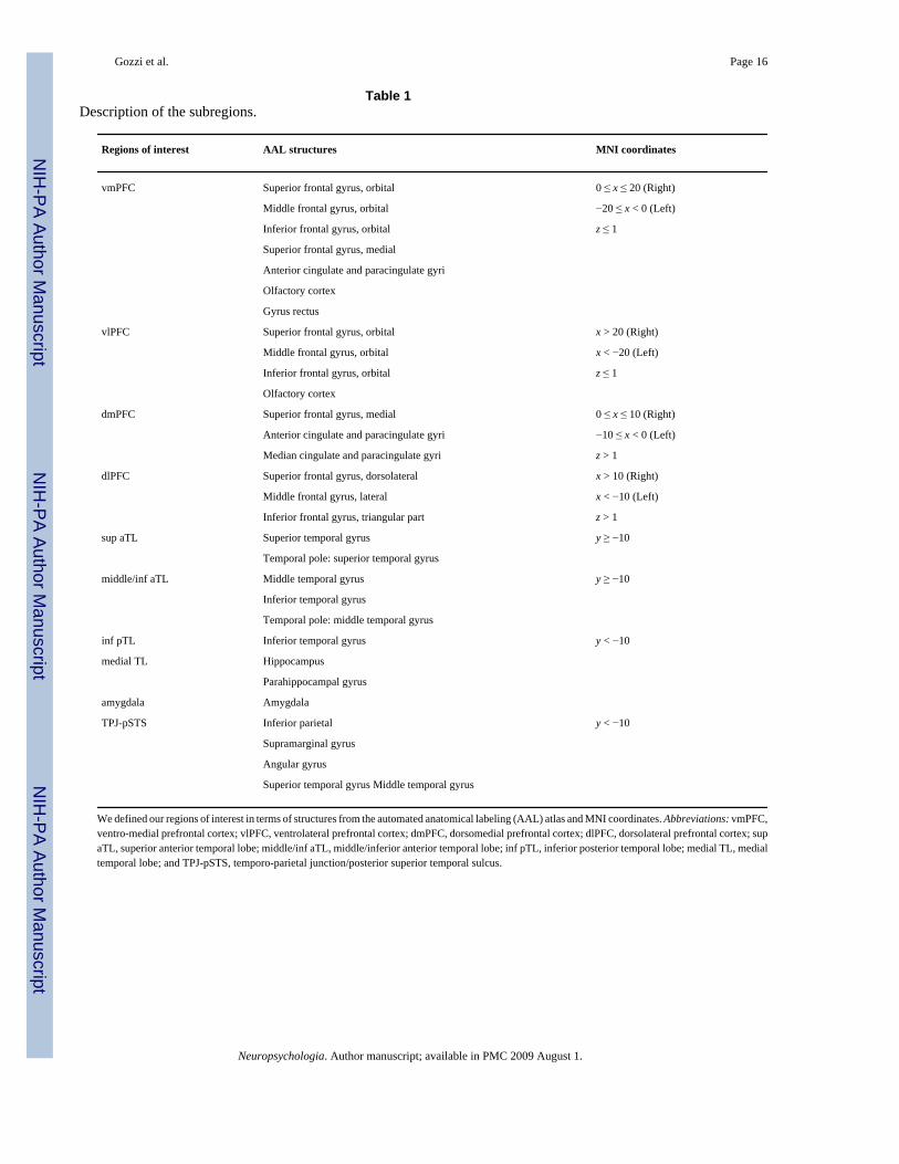
2.2. Lesion analysisWe acquired axial computed tomography (CT) scans (MRI scans could not be obtained in mostpatients because they still had metal fragments in the brain as a result of their injury) withoutcontrast at the Bethesda Naval Hospital on a General Electric Medical Systems Light SpeedPlus CT scanner in helical mode. We reconstructed the images with an in-plane voxel size of0.4 mm × 0.4 mm, an overlapping slice thickness of 2.5 mm and a 1-mm slice interval. Brainlesions were evaluated using the Analysis of Brain Lesions (ABLe) software (Makale et al.,2002; Solomon, Raymont, Braun, Butman, & Grafman, 2007) contained in MEDx v3.44(Medical Numerics, Germantown, MD). For each patient, the brain lesion was manually tracedon each slice by V.R. (a psychiatrist with clinical experience of reading CT scans), andreviewed by J.G., who was blind to the results of the neuropsychological testing. The totalvolume of the lesion was calculated by summing the traced areas and multiplying by slicethickness. The skull and scalp components of the CT volume were then removed, a processknown as de-skulling (Solomon et al., 2007). Each volume was spatially normalized to a de-skulled CT scan, which was previously spatially normalized to match the shape of the T1 MNIbrain (standard of the International Consortium for Brain Mapping). The ABLe program thatwas used for lesion analysis has the option of excluding the manually delineated lesion fromthe spatial normalization process, thus improving registration accuracy. Spatial normalizationwas performed using the automated image registration (AIR) algorithm from Woods, Grafton,Watson, Sicotte, and Mazziotta (1998) using a 12-parameter affine model on de-skulled CTscans. Accuracy of registration was assessed qualitatively based on image fusion of theregistration scan with the template image and quantitatively by computing an overlap of thesetwo images. Based on these measures, all registrations were considered accurate. Finally, wedefined regions of interest in terms of structures from the automated anatomical labeling (AAL)atlas (Tzourio-Mazoyer et al., 2002) and MNI coordinates (see Table 1) and quantified regional
Gozzi et al. Page 3
Neuropsychologia. Author manuscript; available in PMC 2009 August 1.
NIH
-PA Author Manuscript
NIH
-PA Author Manuscript
NIH
-PA Author Manuscript
damage by analyzing the overlap of the spatially normalized lesion image with the AAL atlasimage.
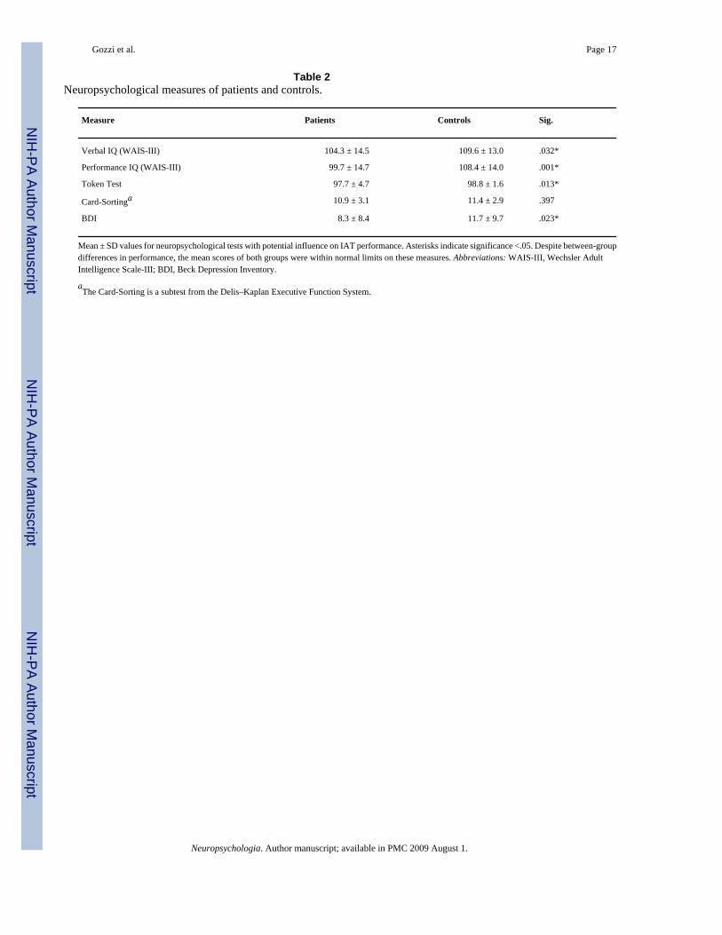
2.3. Neuropsychological assessmentAs part of the VHIS (Phase 3), both patients and controls were administered an extensiveclinical and experimental neuropsychological test battery. From that battery we used thefollowing tests to measure neuropsychological functions that might influence performance onthe IAT: the Wechsler Adult Intelligence Scale-III (Wechsler, 1997) to measure intellectualability; the Token Test (McNeil & Prescott, 1994) to measure verbal comprehension; the Card-Sorting subtest from the Delis–Kaplan Executive Function System (Delis, Kaplan, & Kramer,2001) to measure executive functions; and the Beck Depression Inventory-II (Beck, Steer, &Brown, 1996) to measure depression symptomatology. The neuropsychological test results arereported in Table 2.
2.4. Implicit Association Test (IAT)To measure the strength of stereotypical gender attitudes, we used the IAT, a computer-administered response time task. The IAT is a widely validated implicit measure of attitudes(Greenwald, Nosek, & Banaji, 2003; Hofmann, Gawronski, Gschwendner, Le, & Schmitt,2005; Rudman et al., 2001) and has recently been applied in neuroimaging studies (e.g., Chee,Sriram, Soon, & Lee, 2000; Knutson, Wood, Spampinato, & Grafman, 2006; Luo et al.,2006). We chose this task because participants are generally unaware that their stereotypes arebeing assessed, hence their answers are free of social desirability concerns (Fazio & Olson,2003). The IAT task design is depicted in Fig. 1. Stimuli were female names (e.g., “Jennifer”,“Mary”), male names (e.g., “Andrew”, “Paul”), words related to strength (e.g., “dominant”,“powerful”), and words related to weakness (e.g., “delicate”, “fragile”). The complete list ofstimuli can be obtained from the authors. There were 20 stimuli for each category that wereselected from previous gender IAT studies (Knutson et al., 2007; Rudman et al., 2001). Stimuliwere presented in the centre of the visual field in random order using SuperLab Pro 1.75 (CedrusCorporation, San Pedro, CA) on Macintosh G3 and G4 computers. Participants were asked tocategorize names as female or male and words as strong or weak by pressing one of two keyson the computer keyboard. The categories for the classification were shown on the left andright sides of the screen. Each stimulus was shown on the screen until a response key waspressed, followed by a 500 ms blank screen. Participants were instructed to respond as quicklyand as accurately as possible.
The IAT procedure consisted of five blocks. In Block 1 participants were asked to classify thestimuli as male or female names and in Block 2 as weak or strong words. In Block 3, thecategorizations were combined such that participants had to press one response key for femalenames or strong words, and another response key for male names or weak words. This is knownas the incongruent condition, since subjects are required to press the same response key forconcepts that are not usually associated. In Block 4, participants were asked to categorize maleand female names again, but this time the key assignment was switched as compared to Block1. Finally, in Block 5, the categorizations were combined such that participants pressed oneresponse key for male names or strong words and another response key for female names orweak words. This is known as the congruent condition, since subjects press the same responsekey for stereotypically associated concepts. The number of trials was 80 for each of the twocritical blocks (Blocks 3 and 5), and 40 for each of the remaining blocks. All participantsperformed practice trials before each block. The stimuli used in the practice blocks wereadditional names and words that were not included in the test trials. The order of the blockswas counterbalanced across participants (the position of Blocks 1 and 3 were switched withthose of Blocks 4 and 5, respectively).
Gozzi et al. Page 4
Neuropsychologia. Author manuscript; available in PMC 2009 August 1.
NIH
-PA Author Manuscript
NIH
-PA Author Manuscript
NIH
-PA Author Manuscript
The difference in response latencies between the congruent and incongruent conditionsprovides a measure of gender attitudes (“IAT effect”). If subjects associate women withweakness and men with strength, their response times will be faster in the congruent condition(for example, when they press the same response key for female names and weak words) thanin the incongruent condition (when they press the same response key for female names andstrong words). In the present study, this difference was computed as a D score in accordancewith the recent scoring algorithm outlined by Greenwald et al. (2003, p. 214). Outliers (i.e.,D scores exceeding ±2.5 standard deviations from the mean) were excluded from the analyses(N = 4). This was done to ensure that the results were not driven by outliers. The results didnot differ when outliers were included.
2.5. Comparison with other measuresTo test whether the observed IAT effects were specific for implicit gender stereotyping ascompared to implicit associations in general, we asked participants to complete a second IATwhich was identical in structure to the gender IAT except that positive or negative words werepaired with words related to Vietnam or hockey. To test whether our findings were specificfor implicit gender stereotyping as compared to explicit gender stereotyping, we askedparticipants to perform a short version of the Attitudes toward Women Scale (AWS; Spence& Helmreich, 1978). The AWS provides an explicit measurement of gender-related attitudes.Participants are shown 15 statements about traditional gender-roles beliefs (e.g., “Theintellectual leadership of a community should be largely in the hands of men”). The degree ofagreement to each statement is measured on a 5-point scale ranging from 1 (agree strongly)to5(disagree strongly). A total score is derived by summing the responses after reverse scoringthe egalitarian statements. High scores indicate a profeminist, egalitarian attitude while lowscores indicate more traditional or sexist attitudes. The AWS is administered as a paper-and-pencil questionnaire.
2.6. Statistical analysisTo identify the effects of vmPFC, vlPFC and superior aTL lesions on stereotypical genderattitudes, we employed two analyses: (i) we entered percentages of regional damage into amultiple regression model and (ii) in a confirmatory analysis, we grouped patients by lesionlocation and compared their performance on the IAT with that of the controls.
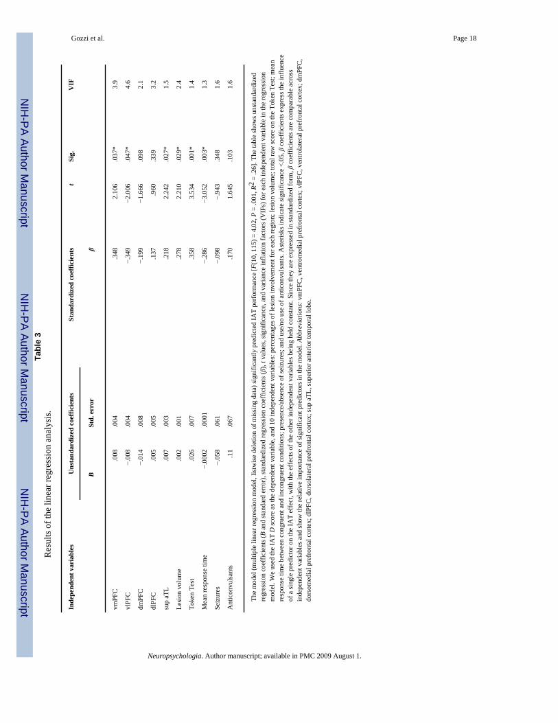
We performed a multiple regression analysis to identify the independent contribution of eachregion of interest (vmPFC, vlPFC and superior aTL), while controlling for the other two anda number of confounding variables. We used the IAT D score as the dependent variable, andthe percentages of lesion involvement in each of the regions as the independent variables. Foreach region, the percentage of lesion reported in the model was the average for the left andright hemisphere. To control for their potential confounding effects, we included in the modelpercentages of lesion involvement in the dorsomedial PFC (dmPFC) and dlPFC, total volumeof the lesion, presence of seizures, and use of anticonvulsants. In addition, we checked whetherthe IAT effect correlated with demographic characteristics (age and education), mean responsetimes on the IAT, and performance on the neuropsychological tests described earlier. Sincetwo variables correlated with the IAT effect (mean response times on the IAT, r = −.260, P = .001, and performance on the Token Test, r = .229, P = .006), we included them in the modelas well. All variables were assessed for multicollinearity (i.e., presence of high correlationsbetween the variables in the regression equation), which may bias the estimates of theregression coefficients (Cohen, Cohen, West, & Aiken, 2003). Typically, variance inflationfactors (VIFs) of 10 or more provide evidence of serious multicollinearity. In the present study,VIFs were never greater than 4.6 (see Table 3).
Gozzi et al. Page 5
Neuropsychologia. Author manuscript; available in PMC 2009 August 1.
NIH
-PA Author Manuscript
NIH
-PA Author Manuscript
NIH
-PA Author Manuscript
In a confirmatory analysis, we grouped patients by lesion location and examined whether lesiongroups performed significantly differently than controls in the directions predicted by theregression analysis. Lesion groups were defined for each region that significantly predictedIAT performance in the regression analysis. Based on previous studies (Koenigs et al., 2008;Tranel, Damasio, Denburg, & Bechara, 2005), a patient was assigned to a lesion group if hislesion occupied at least 15% of that region. Patients with damage to more than one region wereclassified based on the region with the greatest percentage of damage. This procedure wasadopted to ensure that patients in a lesion group not only had damage to that region, but alsohad greater damage to that region than to the other regions under investigation (see Mah,Arnold, & Grafman, 2004 for a similar procedure). Patients whose damage was outside theregions of interest or <15% were not included in this confirmatory analysis. We performedplanned comparisons between the control group and each lesion group using t tests for linearcontrasts in a one-way analysis of variance (ANOVA). These planned comparisons were onetailed because we had a priori hypotheses on the direction of the effects that were derived fromthe regression analysis. Finally, data on the non-gender IAT and the AWS were analyzed usingone-way ANOVAs to determine between-group differences. All statistical analyses werecarried out using SPSS 15.0 (SPSS Inc., Chicago, IL). The significance level was set at P = .05.
3. ResultsTable 3 shows the model derived from the multiple linear regression analysis, with standardizedregression coefficients (β) and corresponding t values as well as significance for eachindependent variable. This model indicates that lesions in vmPFC, vlPFC, and superior aTLindependently contributed to IAT performance, after controlling for each other and the rest ofthe confounding variables. Larger lesions in either the vmPFC or the superior aTL wereassociated with a higher IAT effect (i.e., stronger stereotypical associations). On the other hand,larger lesions in the vlPFC were associated with a lower IAT effect (i.e., weaker stereotypicalassociations). Lesions in the dmPFC and dlPFC did not significantly predict IAT performance.We computed additional multiple regression analyses that examined whether other temporalor parietal regions (not predicted a priori) significantly influenced IAT performance (includingmiddle/inferior aTL, posterior inferior temporal lobe, medial temporal lobe, and temporo-parietal junction/posterior superior temporal sulcus). We found that only lesions in middle/inferior aTL (β = .206, P = .024), but not lesions in the other regions, significantly predictedIAT performance.
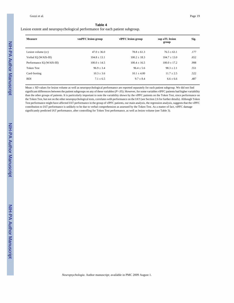
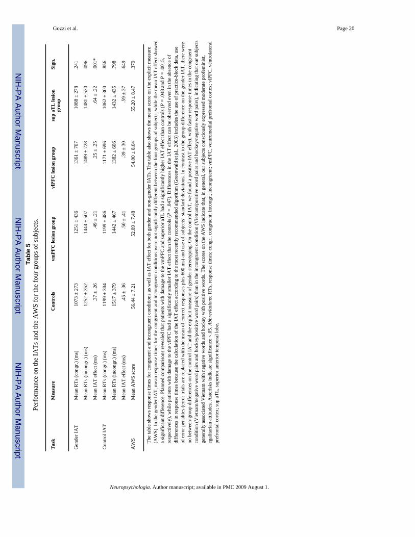
Fig. 2 shows the lesion overlaps for patients with damage to vmPFC (N = 18), vlPFC (N = 15),and superior aTL (N = 10). Table 4 provides information about lesion extent andneuropsychological performance for each patient subgroup. Table 5 reports performance onthe gender and non-gender IATs and the AWS for the three patient subgroups and the controlgroup. On the gender IAT, a one-way ANOVA of the mean IAT effect between the four groupsshowed a significant difference. Planned comparisons revealed that patients with damage tothe vmPFC and superior aTL had a significantly higher IAT effect than controls, while patientswith damage to the vlPFC had a significantly smaller IAT effect than the controls. In contrastto the group differences on the gender IAT, we found no significant differences between groupson the non-gender IAT and the explicit measure of gender stereotyping.
4. DiscussionThis study examined dissociable contributions of prefrontal and temporal cortical subregionsto stereotypical gender attitudes in 154 patients with penetrating head injuries. Detailed lesionanalysis of brain images allowed us to quantify the extent of damage to specific subregions.To evaluate gender stereotypes, we used the IAT, an implicit task which reveals attitudes and
Gozzi et al. Page 6
Neuropsychologia. Author manuscript; available in PMC 2009 August 1.
NIH
-PA Author Manuscript
NIH
-PA Author Manuscript
NIH
-PA Author Manuscript
beliefs even when they are not explicitly endorsed (Greenwald et al., 2002; Hofmann et al.,2005; Nosek, Banaji, & Greenwald, 2002). Using a multiple regression model, we found thatlarger lesions in either the vmPFC or the superior aTL were associated with increasedstereotypical attitudes, whereas larger lesions in the vlPFC were associated with decreasedstereotypical attitudes. In a confirmatory analysis, we divided the patients into lesion groupsand compared their performance on the IAT with that of healthy volunteers. The lesion groupsperformed significantly differently than controls in the directions predicted by the regressionanalysis.
Our first main result was that lesions of the vmPFC and vlPFC had opposite effects onstereotypical attitudes about gender. Milne and Grafman (2001) found decreased stereotypingin patients with lesions in the ventral PFC. Here, by quantifying the extent of damage to eachregion and performing a multiple regression analysis, we were able to analyze subdivisions ofthe ventral PFC and detect functional differences between the medial and lateral sectors. Thisfinding is in line with previous studies reporting a dissociation between medial and lateralaspects of the ventral PFC (e.g., Elliott et al., 2000; Kringelbach, 2005; Kringelbach & Rolls,2004; O'Doherty, Kringelbach, Rolls, Hornak, & Andrews, 2001; Wood & Grafman, 2003).
On the one hand, we found that vmPFC damage was associated with a stronger IAT effect,which indicates increased stereotyping. This finding is in line with a new study (Quadflieg etal., in press) that examined the neural correlates of gender stereotyping using fMRI. Consistentwith the current neuropsychological data, Quadflieg et al. found activation within medial PFCfor stereotypic versus nonstereotypic judgments. More research is needed to understand thenature of this finding. One potential explanation may be related to the role of the vmPFC inthe modulation of the expression of social and emotional behavior (e.g., Grafman & Litvan,1999; Pietrini, Guazzelli, Basso, Jaffe, & Grafman, 2000). It has been suggested that lesionsof the vmPFC disrupt inhibitory and emotional mechanisms, leading to impulsive and sociallyinappropriate behavior and decision making (Anderson, Bechara, Damasio, Tranel, &Damasio, 1999; Bechara, Damasio, Damasio, & Anderson, 1994). Indeed, lesions of thevmPFC have been associated with abnormal autonomic responses to socially meaningfulstimuli (Damasio, Tranel, & Damasio, 1990), with “acquired sociopathy” (Eslinger &Damasio, 1985) and with the expression of aggressive and violent behavior (Blair & Cipolotti,2000; Grafman et al., 1996). Notably, vmPFC patients may present inappropriate socialbehavior despite preserved capacities for general intelligence, logical reasoning, anddeclarative knowledge of social and moral norms (Eslinger & Damasio, 1985). These patientscan access some types of social knowledge (Saver & Damasio, 1991) and describe social normsof behavior with a stranger (Beer, John, Scabini, & Knight, 2006). Based on this literature, wespeculate that our finding of increased stereotyping associated with vmPFC damage may berelated to the inability to suppress the expression of inappropriate social behavior, such asstereotyping. It is important to note that in this study we did not distinguish the contributionsof orbital (Brodmann's Area [BA] 11), subgenual cingulate (BA 25, 32), and dorsal parts ofthe anterior vmPFC (BA 10, 32). From a phylogenetic and anatomical perspective, however,it is unlikely that these different areas are functionally heterogeneous (Kringelbach & Rolls,2004).
On the other hand, we found that vlPFC damage was associated with a diminished IAT effect,which indicates decreased stereotyping. A recent fMRI study using a race IAT has suggestedthat the vlPFC might play a role in stereotyping (Beer et al., 2008). The involvement of thevlPFC in IAT performance has been also reported in two fMRI investigations, one assessingthe association of visually depicted legal and illegal behaviors with positive animals (e.g.,puppies) and negative animals (e.g., snake) (Luo et al., 2006), and the other assessing theassociation of flowers and insects with pleasant and unpleasant words (Chee et al., 2000). Theseprevious imaging studies, coupled with the current results, suggest that the vlPFC may play a
Gozzi et al. Page 7
Neuropsychologia. Author manuscript; available in PMC 2009 August 1.
NIH
-PA Author Manuscript
NIH
-PA Author Manuscript
NIH
-PA Author Manuscript
more general role in mediating implicit attitudes, rather than in gender stereotyping per se.Alternatively, the role of vlPFC in IAT performance may have less to do with attitudes thanwith the executive demands of the task, which include speeded responses and also, potentially,some degree of response inhibition. This interpretation is supported by previous studiesdemonstrating vlPFC involvement in go/no-go tasks, which require subjects to performspeeded responses on “go” trials and to inhibit their response on “no-go” trials (Aron, Fletcher,Bullmore, Sahakian, & Robbins, 2003; Casey et al., 2001). The response variability in ourvlPFC patients (see Table 5) may suggest that the vlPFC plays a role in the coordination ofexecutive task demands unrelated to stereotype processing. Response variability in the vlPFCgroup may also originate from the fact that this group of patients was more heterogeneous thanthe other two (see Table 4). While the current results demonstrate the importance of vlPFC fornormal performance on the IAT for gender stereotypes, future studies will be needed to clarifythe role of vlPFC in the mediation of implicit (social or otherwise) attitudes versus generalresponse selection processes. Another interesting question for future studies would be whetherthere is an effect of laterality in vlPFC function. Previous response inhibition findings primarilyconcerned the right vlPFC (Aron et al., 2003; Aron, Robbins, & Poldrack, 2004; Horn, Dolan,Elliott, Deakin, & Woodruff, 2003). In the present study we did not distinguish between left-and right-sided lesions.
In addition to the medial and lateral aspects of the ventral PFC, we also found a criticalinvolvement of the superior aTL in IAT performance. The contribution of the aTL is unlikelyto reflect general language-related difficulties. First, as reported in Table 4, performance onthe verbal WAIS test and the Token Test was not different among the subgroups of patients.Second, performance on the verbal IQ did not correlate with performance on the IAT. Sinceperformance on the Token Test correlated with performance on the IAT, this variable wasentered in the regression model as a covariate. The regression analysis showed that aTL damagesignificantly predicted IAT performance, after controlling for Token Test performance (seeTable 3). Instead, we suggest that the aTL may play a role in IAT performance by providingrepresentations of conceptual social knowledge. As mentioned in Section 1, a recentneuroimaging study has shown that the superior aTL selectively represents conceptual socialknowledge (Zahn et al., 2007). Selective activation in the superior aTL was found for socialconcepts (i.e., concepts describing social behavior: e.g., ‘polite’, ‘stingy’) as compared withconcepts describing animal behavior (animal function concepts: e.g., ‘trainable’, ‘nutritious’).Remarkably, activity in the superior aTL increased with higher levels of social conceptualdetail. Thus, representations within the superior aTL are recruited, particularly, when finedistinctions of the conceptual quality of social behaviors are required (e.g., whether somebodybehaved “tactlessly” or “stingily”). Further it has been demonstrated that representations ofconceptual qualities of social behavior in the superior aTL are independent of context of actionsand emotions (Zahn et al., 2009). Based on these fMRI findings, we propose that damage tothe superior aTL may lead to reduce social conceptual detail. This might in turn lead to greaterreliance on stereotypes, which are characterized by a lack of conceptual detail. To ourknowledge, this is the first study that suggests the potential importance of conceptual socialknowledge impairments due to superior aTL damage as contributing to stereotypical socialattitudes.
Because most superior aTL lesions extended into the middle/inferior aTL, we were unable totest whether the demonstrated effect was specifically due to superior or middle/inferior aTLdamage. However, we excluded the contribution of adjacent regions (posterior inferiortemporal lobe, medial temporal lobe, temporo-parietal junction/posterior superior temporalcortex, and vlPFC).
Given the proximity of superior aTL and amygdala, it is reasonable to ask whether amygdaladamage could account for the observed effects in the superior aTL. Indeed, the amygdala is
Gozzi et al. Page 8
Neuropsychologia. Author manuscript; available in PMC 2009 August 1.
NIH
-PA Author Manuscript
NIH
-PA Author Manuscript
NIH
-PA Author Manuscript
involved in the recognition of emotion in facial expressions (Adolphs, Tranel, Damasio, &Damasio, 1994) and in the perception of faces of a different race (Hart et al., 2000; Phelps etal., 2000). A neuropsychological study indicated that the amygdala is not critical for normalperformance on the IAT (Phelps, Cannistraci, & Cunningham, 2003). Here, we were able toexclude a prominent role of the amygdala for the observed effects in the superior aTL. Only 2subjects in the superior aTL group had significant damage to the amygdala, and their IAT effectwas lower than the mean IAT effect of the aTL group. Further, since there are direct connectionsof the amygdala with the vmPFC via the medial forebrain bundle (Nieuwenhuys, 1982) whichdo not depend on connections with the lateral temporal pole, a disconnection syndrome ofamygdalo-frontal fibers is unlikely to explain the impact of lateral aTL lesions on socialattitudes.
The lack of group differences on the non-gender IAT supports the claim that the vlPFC, vmPFC,and aTL are critically involved in implicit gender stereotyping, rather than playing a moregeneral role in implicit associations or IAT task performance. However, we realize that furtherstudies will be required to more precisely characterize the specific roles of these brain areas inmediating social stereotypes. Previous lesion and neuroimaging studies suggest that superioraTL and ventral PFC may play a specific role in social cognition. As mentioned earlier, recentstudies corroborate the hypothesis that the aTL selectively represents social concepts versusconcepts describing animal behavior (Zahn et al., 2007). In addition, previous literature hasconfirmed the involvement of the ventral PFC in gender (Quadflieg et al., in press) as well asrace (Beer et al., 2008) stereotyping. In this respect, it is important to note that much of whatis currently known about the neural circuitry supporting stereotyping has been gathered fromstudies exploring racial attitudes (see Eberhardt, 2005 for a review of neuroimaging studies ofrace), and specifically the perception of outgroup faces (e.g., Cunningham et al., 2004; Golby,Gabrieli, Chiao, & Eberhardt, 2001; Hart et al., 2000; Phelps et al., 2000; Richeson et al.,2003).
To test whether our findings were specific for implicit gender stereotyping as compared toexplicit gender stereotyping, we compared performance on the gender IAT with performanceon an explicit measure of gender attitudes. The results showed no between-group differenceson the explicit measure of gender stereotyping, supporting the conclusion that vmPFC, vlPFC,and aTL are critically involved in implicit but not explicit gender stereotyping. Our finding isin line with previous lesion and neuroimaging studies. For example, Milne and Grafman(2001) reported between-group differences on the IAT but not on the scales measuring explicitgender stereotyping. Phelps et al. (2000) found that brain activity during race stereotyping wascorrelated with the strength of participants’ implicit attitudes but not their explicit beliefs(although see Quadflieg et al., in press).
Our patients’ abnormalities on the IAT cannot be attributed to impairments in otherneuropsychological domains. The patients’ performance on the IAT did not correlate with age,education, or performance on neuropsychological tests measuring intellectual ability,executive functions, and symptoms of depression (verbal IQ, performance IQ, performance onthe Card-Sorting Test, and score on the BDI-II). Since the IAT D score correlated with meanresponse times on the IAT and performance on the Token Test, we controlled for theconfounding effect of these variables by entering them as covariates into the multiple regressionmodel. Additionally, our results resist general explanations related to lesion volume, becausethis variable was also entered in the regression model.
In conclusion, in this study we investigated the critical involvement of vmPFC, vlPFC andsuperior aTL in stereotypical attitudes about gender using the IAT. We found that damage toeither the vmPFC or the superior aTL was associated with a greater IAT effect (i.e., increasedstereotyping), whereas lesions of the vlPFC were associated with a diminished IAT effect (i.e.,
Gozzi et al. Page 9
Neuropsychologia. Author manuscript; available in PMC 2009 August 1.
NIH
-PA Author Manuscript
NIH
-PA Author Manuscript
NIH
-PA Author Manuscript
decreased stereotyping). These findings reveal the potential involvement of ventral frontal andanterior temporal brain regions in social attitudes.
AcknowledgmentsWe thank R. Zahn for insightful comments and fruitful discussions related to the data analysis and earlier versions ofthis manuscript, L. Mah for task programming, K. Reding for data management, and F. Krueger, J. Hassenplug, andN. Ruesch for their helpful comments on the manuscript. We thank the veterans for their participation in the study.This work was supported by the US National Institute of Neurological Disorders and Stroke intramural researchprogram and a project grant form the United Sates Army Medical Research and Material Command administered bythe Henry M. Jackson Foundation (Vietnam Head Injury Study Phase III: A 30 Year Post-Injury Follow-Up Study,Grant number DAMD17-01-1-0675).
ReferencesAdolphs R, Tranel D, Damasio H, Damasio A. Impaired recognition of emotion in facial expressions
following bilateral damage to the human amygdala. Nature 1994;372(6507):669–672. [PubMed:7990957]
Anderson SW, Bechara A, Damasio H, Tranel D, Damasio AR. Impairment of social and moral behaviorrelated to early damage in human prefrontal cortex. Nature 1999;2:1032–1037.
Aron AR, Fletcher PC, Bullmore ET, Sahakian BJ, Robbins TW. Stop-signal inhibition disrupted bydamage to right inferior frontal gyrus in humans. Nature Neuroscience 2003;6(2):115–116.
Aron AR, Robbins TW, Poldrack RA. Inhibition and the right inferior frontal cortex. Trends in CognitiveSciences 2004;8(4):170–177. [PubMed: 15050513]
Asendorpf JB, Banse R, Mucke D. Double dissociation between implicit and explicit personality self-concept: The case of shy behavior. Journal of Personality and Social Psychology 2002;83(2):380–393.[PubMed: 12150235]
Bechara A, Damasio AR, Damasio H, Anderson SW. Insensitivity to future consequences followingdamage to human prefrontal cortex. Cognition 1994;50:7–15. [PubMed: 8039375]
Beck, AT.; Steer, RA.; Brown, GK. Beck depression inventory. Vol. second ed.. PsychologicalCorporation; San Antonio, TX: 1996.
Beer JS, John OP, Scabini D, Knight RT. Orbitofrontal cortex and social behavior: Integrating self-monitoring and emotion–cognition interactions. Journal of Cognitive Neuroscience 2006;18(6):871–879. [PubMed: 16839295]
Beer JS, Stallen M, Lombardo MV, Gonsalkorale K, Cunningham WA, Sherman JW. The QuadrupleProcess model approach to examining the neural underpinnings of prejudice. NeuroImage 2008;43(4):775–783. [PubMed: 18809502]
Blair RJR, Cipolotti L. Impaired social response reversal: A case of ‘acquired sociopathy’. Brain2000;123:1122–1141. [PubMed: 10825352]
Casey BJ, Forman SD, Franzen P, Berkowitz A, Braver TS, Nystrom LE, et al. Sensitivity of prefrontalcortex to changes in target probability: A functional MRI study. Human Brain Mapping 2001;13(1):26–33. [PubMed: 11284044]
Chee MWL, Sriram N, Soon CS, Lee KM. Dorsolateral prefrontal cortex and the implicit association ofconcepts and attributes. NeuroReport 2000;11:135–140. [PubMed: 10683845]
Cohen, J.; Cohen, P.; West, SG.; Aiken, LS. Applied multiple regression/correlation analysis for thebehavioral sciences. Lawrence Erlbaum Associates; Mahwah, NJ: 2003.
Cunningham WA, Johnson MK, Gatenby JC, Gore JC, Banaji MR. Neural components of socialevaluation. Journal of Personality & Social Psychology 2003;85(4):639–649. [PubMed: 14561118]
Cunningham WA, Zelazo PD. Attitudes and evaluations: A social cognitive neuroscience perspective.Trends in Cognitive Sciences 2007;11(3):97–104. [PubMed: 17276131]
Cunningham WA, Johnson MK, Raye CL, Chris Gatenby J, Gore JC, Banaji MR. Separable neuralcomponents in the processing of black and white faces. Psychological Science 2004;15(12):806–813.[PubMed: 15563325]
Damasio AR, Tranel D, Damasio H. Individuals with sociopathic behavior caused by frontal damage failto respond autonomically to social stimuli. Behavioral Brain Research 1990;41(2):81–94.
Gozzi et al. Page 10
Neuropsychologia. Author manuscript; available in PMC 2009 August 1.
NIH
-PA Author Manuscript
NIH
-PA Author Manuscript
NIH
-PA Author Manuscript
Delis, Kaplan; Kramer. Delis–Kaplan executive function system. The Psychological Corporation; SanAntonio, TX: 2001.
Eberhardt JL. Imaging race. American Psychologist 2005;60(2):181–190. [PubMed: 15740450]Edwards-Lee T, Miller BL, Benson DF, Cummings JL, Russell GL, Boone K, et al. The temporal variant
of frontotemporal dementia. Brain 1997;120(Pt 6):1027–1040. [PubMed: 9217686]Elliott R, Dolan RJ, Frith CD. Dissociable functions in the medial and lateral orbitofrontal cortex:
Evidence from human neuroimaging studies. Cerebral Cortex 2000;10:308–317. [PubMed:10731225]
Eslinger PJ, Damasio AR. Severe disturbance of higher cognition after bilateral frontal lobe ablation:Patient EVR. Neurology 1985;35(12):1731–1741. [PubMed: 4069365]
Fazio RH, Olson MA. Implicit measures in social cognition research: Their meaning and use. AnnualReview of Psychology 2003;54:297–327.
First, MB.; Spitzer, RL.; Gibbon, M.; Williams, JBW. Structured clinical interview for DSM-IV-TR axisI disorders, non-patient edition (SCID-I/NP). Biometrics Research, New York State PsychiatricInstitute; New York: 2001.
Golby AJ, Gabrieli JD, Chiao JY, Eberhardt JL. Differential responses in the fusiform region to same-race and other-race faces. Nature Neuroscience 2001;4(8):845–850.
Grafman J, Litvan I. Importance of deficits in executive functions. Lancet 1999;354(9194):1921–1923.[PubMed: 10622291]
Grafman J, Schwab K, Warden D, Pridgen A, Brown HR, Salazar A. Frontal lobe injuries, violence, andaggression: A report of the Vietnam Head Injury Study. Neurology 1996;46:1231–1238. [PubMed:8628458]
Greenwald AG, Banaji MR. Implicit social cognition: Attitudes, self-esteem, and stereotypes.Psychological Review 1995;102:4–27. [PubMed: 7878162]
Greenwald AG, Banaji MR, Rudman LA, Farnham SD, Nosek BA, Mellott DS. A unified theory ofimplicit attitudes, stereotypes, self-esteem, and self-concept. Psychological Review 2002;109(1):3–25. [PubMed: 11863040]
Greenwald AG, McGhee DE, Schwartz JLK. Measuring individual differences in implicit cognition: TheImplicit Association Test. Journal of Personality and Social Psychology 1998;74:1464–1480.[PubMed: 9654756]
Greenwald AG, Nosek BA, Banaji MR. Understanding and using the Implicit Association Test. I. Animproved scoring algorithm. Journal of Personality and Social Psychology 2003;85(2):197–216.[PubMed: 12916565]
Hart AJ, Whalen PJ, Shin LM, McInerney SC, Fischer H, Rauch SL. Differential response in the humanamygdala to racial outgroup vs ingroup face stimuli. NeuroReport 2000;11:2351–2355. [PubMed:10943684]
Hofmann, Gawronski; Gschwendner, Le; Schmitt. A meta-analysis on the correlation between theImplicit Association Test and explicit self-report measures. Personality and Social PsychologyBulletin 2005;31(10):1369–1385. [PubMed: 16143669]
Horn NR, Dolan M, Elliott R, Deakin JF, Woodruff PW. Response inhibition and impulsivity: An fMRIstudy. Neuropsychologia 2003;41(14):1959–1966. [PubMed: 14572528]
Kleist, K. Geistes und nervenkrankheiten. Verlag von Johann Ambrosius Barth; Leipzig: 1922.Knutson KM, Mah L, Manly CF, Grafman J. Neural correlates of automatic beliefs about gender and
race. Human Brain Mapping 2007;28(10):915–930. [PubMed: 17133388]Knutson KM, Wood JN, Spampinato MV, Grafman J. Politics on the brain: An fMRI investigation. Social
Neuroscience 2006;1:25–40. [PubMed: 17372621]Koenigs M, Huey ED, Raymont V, Cheon B, Solomon J, Wassermann EM, et al. Focal brain damage
protects against post-traumatic stress disorder in combat veterans. Nature Neuroscience 2008;11(2):232–237.
Kringelbach ML. The human orbitofrontal cortex: Linking reward to hedonic experience. Nature ReviewsNeuroscience 2005;6(9):691–702.
Gozzi et al. Page 11
Neuropsychologia. Author manuscript; available in PMC 2009 August 1.
NIH
-PA Author Manuscript
NIH
-PA Author Manuscript
NIH
-PA Author Manuscript
Kringelbach ML, Rolls ET. The functional neuroanatomy of the human orbitofrontal cortex: Evidencefrom neuroimaging and neuropsychology. Progress in Neurobiology 2004;72(5):341–372. [PubMed:15157726]
Liu W, Miller BL, Kramer JH, Rankin K, Wyss-Coray C, Gearhart R, et al. Behavioral disorders in thefrontal and temporal variants of frontotemporal dementia. Neurology 2004;62(5):742–748.[PubMed: 15007124]
Luo Q, Nakic M, Wheatley T, Richell R, Martin A, Blair RJ. The neural basis of implicit moral attitude—an IAT study using event-related fMRI. NeuroImage 2006;30(4):1449–1457. [PubMed:16418007]
Mah L, Arnold MC, Grafman J. Impairment of social perception associated with lesions of the prefrontalcortex. American Journal of Psychiatry 2004;161(7):1247–1255. [PubMed: 15229058]
Makale M, Solomon J, Patronas NJ, Danek A, Butman JA, Grafman J. Quantification of brain lesionsusing Interactive Automated Software. Behavior Research Methods, Instruments, and Computers2002;34(1):6–18.
McNeil, MM.; Prescott, TE. Revised Token Test. Western Psychological Services; Los Angeles, CA:1994.
Miller BL, Seeley WW, Mychack P, Rosen HJ, Mena I, Boone K. Neuroanatomy of the self: Evidencefrom patients with frontotemporal dementia. Neurology 2001;57(5):817–821. [PubMed: 11552010]
Milne E, Grafman J. Ventromedial prefrontal cortex lesions in humans eliminate implicit genderstereotyping. Journal of Neuroscience 2001;21(RC150):151–156.
Nieuwenhuys, G. Veening The medial forebrain bundle of the rat. I. General introduction. Journal ofComparative Neurology 1982;206:49–81. [PubMed: 6124562]
Nosek BA, Banaji MR, Greenwald AG. Math = male, me = female, therefore math not = me. Journal ofPersonality and Social Psychology 2002;83(1):44–59. [PubMed: 12088131]
O'Doherty J, Kringelbach ML, Rolls ET, Hornak J, Andrews C. Abstract reward and punishmentrepresentations in the human orbitofrontal cortex. Nature Neuroscience 2001;4:95–102.
Phelps EA, Cannistraci CJ, Cunningham WA. Intact performance on an indirect measure of race biasfollowing amygdala damage. Neuropsychologia 2003;41:203–208. [PubMed: 12459218]
Phelps EA, O'Connor KJ, Cunningham WA, Funayama ES, Gatenby JC, Gore JC, et al. Performance onindirect measures of race evaluation predicts amygdala activation. Journal of Cognitive Neuroscience2000;12:729–738. [PubMed: 11054916]
Pietrini P, Guazzelli M, Basso G, Jaffe K, Grafman J. Neural correlates of imaginal aggressive behaviorassessed by positron emission tomography. American Journal of Psychiatry 2000;157:1772–1781.[PubMed: 11058474]
Price CJ, Mummery CJ, Moore CJ, Frakowiak RS, Friston KJ. Delineating necessary and sufficient neuralsystems with functional imaging studies of neuropsychological patients. Journal of CognitiveNeuroscience 1999;11(4):371–382. [PubMed: 10471846]
Quadflieg S, Turk DJ, Waiter GD, Mitchell JP, Jenkins AC, Macrae CN. Exploring the neural correlatesof social stereotyping. Journal of Cognitive Neuroscience. (in press)
Richeson JA, Baird AA, Gordon HL, Heatherton TF, Wyland CL, Trawalter S, et al. An fMRIinvestigation of the impact of interracial contact on executive function. Nature Neuroscience 2003;6(12):1323–1328.
Rudman LA, Greenwald AG, McGhee DE. Implicit self-concept and evaluative implicit genderstereotypes: Self and ingroup share desirable traits. Personality and Social Psychology Bulletin2001;27(9):1164–1178.
Saver JL, Damasio AR. Preserved access and processing of social knowledge in a patient with acquiredsociopathy due to ventromedial frontal damage. Neuropsychologia 1991;29(12):1241–1249.[PubMed: 1791934]
Solomon J, Raymont V, Braun A, Butman JA, Grafman J. User-friendly software for the analysis of brainlesions (ABLe). Computer Methods and Programs in Biomedicine 2007;86(3):245–254. [PubMed:17408802]
Spence, JT.; Helmreich, RL. Masculinity and femininity: Their psychological dimensions, correlates andantecedents. University of Texas Press; Austin, TX: 1978.
Gozzi et al. Page 12
Neuropsychologia. Author manuscript; available in PMC 2009 August 1.
NIH
-PA Author Manuscript
NIH
-PA Author Manuscript
NIH
-PA Author Manuscript
Tranel D, Damasio H, Denburg NL, Bechara A. Does gender play a role in functional asymmetry ofventromedial prefrontal cortex? Brain 2005;128(Pt 12):2872–2881. [PubMed: 16195242]
Tzourio-Mazoyer N, Landeau B, Papathanassiou D, Crivello F, Etard O, Delcroix N, et al. Automatedanatomical labeling of activations in SPM using a macroscopic anatomical parcellation of the MNIMRI single-subject brain. NeuroImage 2002;15(1):273–289. [PubMed: 11771995]
Wechsler, D. Wechsler adult intelligence scale. Vol. third ed.. The Psychological Corporation; SanAntonio, TX: 1997.
Wood JN. Social cognition and the prefrontal cortex. Behavioral and Cognitive Neuroscience Reviews2003;2:97–114. [PubMed: 13678518]
Wood JN, Grafman J. Human prefrontal cortex: Processing and representational perspectives. NatureReviews Neuroscience 2003;4(2):139–147.
Woods RP, Grafton ST, Watson JD, Sicotte NL, Mazziotta JC. Automated image registration. II.Intersubject validation of linear and nonlinear models. Journal of Computer Assisted Tomography1998;22(1):153–165. [PubMed: 9448780]
Zahn R, Moll J, Krueger F, Huey ED, Garrido G, Grafman J. Social concepts are represented in thesuperior anterior temporal cortex. Proceedings of the National Academy of Sciences, USA 2007;104(15):6430–6435.
Zahn R, Moll J, Paiva M, Garrido G, Krueger F, Huey ED, et al. The neural basis of human social values:Evidence from functional MRI. Cerebral Cortex 2009;19(2):276–283. [PubMed: 18502730]
Gozzi et al. Page 13
Neuropsychologia. Author manuscript; available in PMC 2009 August 1.
NIH
-PA Author Manuscript
NIH
-PA Author Manuscript
NIH
-PA Author Manuscript
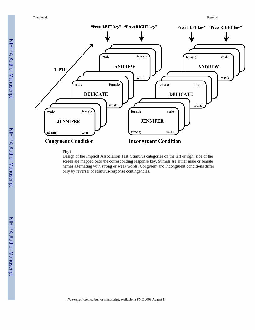
Fig. 1.Design of the Implicit Association Test. Stimulus categories on the left or right side of thescreen are mapped onto the corresponding response key. Stimuli are either male or femalenames alternating with strong or weak words. Congruent and incongruent conditions differonly by reversal of stimulus-response contingencies.
Gozzi et al. Page 14
Neuropsychologia. Author manuscript; available in PMC 2009 August 1.
NIH
-PA Author Manuscript
NIH
-PA Author Manuscript
NIH
-PA Author Manuscript
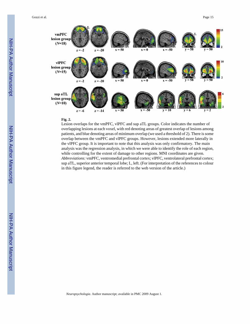
Fig. 2.Lesion overlaps for the vmPFC, vlPFC and sup aTL groups. Color indicates the number ofoverlapping lesions at each voxel, with red denoting areas of greatest overlap of lesions amongpatients, and blue denoting areas of minimum overlap (we used a threshold of 2). There is someoverlap between the vmPFC and vlPFC groups. However, lesions extended more laterally inthe vlPFC group. It is important to note that this analysis was only confirmatory. The mainanalysis was the regression analysis, in which we were able to identify the role of each region,while controlling for the extent of damage to other regions. MNI coordinates are given.Abbreviations: vmPFC, ventromedial prefrontal cortex; vlPFC, ventrolateral prefrontal cortex;sup aTL, superior anterior temporal lobe; L, left. (For interpretation of the references to colourin this figure legend, the reader is referred to the web version of the article.)
Gozzi et al. Page 15
Neuropsychologia. Author manuscript; available in PMC 2009 August 1.
NIH
-PA Author Manuscript
NIH
-PA Author Manuscript
NIH
-PA Author Manuscript
NIH
-PA Author Manuscript
NIH
-PA Author Manuscript
NIH
-PA Author Manuscript
Gozzi et al. Page 16
Table 1Description of the subregions.
Regions of interest AAL structures MNI coordinates
vmPFC Superior frontal gyrus, orbital 0 ≤ x ≤ 20 (Right)
Middle frontal gyrus, orbital −20 ≤ x < 0 (Left)
Inferior frontal gyrus, orbital z ≤ 1
Superior frontal gyrus, medial
Anterior cingulate and paracingulate gyri
Olfactory cortex
Gyrus rectus
vlPFC Superior frontal gyrus, orbital x > 20 (Right)
Middle frontal gyrus, orbital x < −20 (Left)
Inferior frontal gyrus, orbital z ≤ 1
Olfactory cortex
dmPFC Superior frontal gyrus, medial 0 ≤ x ≤ 10 (Right)
Anterior cingulate and paracingulate gyri −10 ≤ x < 0 (Left)
Median cingulate and paracingulate gyri z > 1
dlPFC Superior frontal gyrus, dorsolateral x > 10 (Right)
Middle frontal gyrus, lateral x < −10 (Left)
Inferior frontal gyrus, triangular part z > 1
sup aTL Superior temporal gyrus y ≥ −10
Temporal pole: superior temporal gyrus
middle/inf aTL Middle temporal gyrus y ≥ −10
Inferior temporal gyrus
Temporal pole: middle temporal gyrus
inf pTL Inferior temporal gyrus y < −10
medial TL Hippocampus
Parahippocampal gyrus
amygdala Amygdala
TPJ-pSTS Inferior parietal y < −10
Supramarginal gyrus
Angular gyrus
Superior temporal gyrus Middle temporal gyrus
We defined our regions of interest in terms of structures from the automated anatomical labeling (AAL) atlas and MNI coordinates. Abbreviations: vmPFC,ventro-medial prefrontal cortex; vlPFC, ventrolateral prefrontal cortex; dmPFC, dorsomedial prefrontal cortex; dlPFC, dorsolateral prefrontal cortex; supaTL, superior anterior temporal lobe; middle/inf aTL, middle/inferior anterior temporal lobe; inf pTL, inferior posterior temporal lobe; medial TL, medialtemporal lobe; and TPJ-pSTS, temporo-parietal junction/posterior superior temporal sulcus.
Neuropsychologia. Author manuscript; available in PMC 2009 August 1.
NIH
-PA Author Manuscript
NIH
-PA Author Manuscript
NIH
-PA Author Manuscript
Gozzi et al. Page 17
Table 2Neuropsychological measures of patients and controls.
Measure Patients Controls Sig.
Verbal IQ (WAIS-III) 104.3 ± 14.5 109.6 ± 13.0 .032*
Performance IQ (WAIS-III) 99.7 ± 14.7 108.4 ± 14.0 .001*
Token Test 97.7 ± 4.7 98.8 ± 1.6 .013*
Card-Sortinga 10.9 ± 3.1 11.4 ± 2.9 .397
BDI 8.3 ± 8.4 11.7 ± 9.7 .023*
Mean ± SD values for neuropsychological tests with potential influence on IAT performance. Asterisks indicate significance <.05. Despite between-groupdifferences in performance, the mean scores of both groups were within normal limits on these measures. Abbreviations: WAIS-III, Wechsler AdultIntelligence Scale-III; BDI, Beck Depression Inventory.
aThe Card-Sorting is a subtest from the Delis–Kaplan Executive Function System.
Neuropsychologia. Author manuscript; available in PMC 2009 August 1.
NIH
-PA Author Manuscript
NIH
-PA Author Manuscript
NIH
-PA Author Manuscript
Gozzi et al. Page 18Ta
ble
3R
esul
ts o
f the
line
ar re
gres
sion
ana
lysi
s.
Inde
pend
ent v
aria
bles
Uns
tand
ardi
zed
coef
ficie
nts
Stan
dard
ized
coe
ffici
ents
tSi
g.V
IF
BSt
d. e
rror
β
vmPF
C.0
08.0
04.3
482.
106
.037
*3.
9
vlPF
C−.
008
.004
−.34
9−2
.006
.047
*4.
6
dmPF
C−.
014
.008
−.19
9−1
.666
.098
2.1
dlPF
C.0
05.0
05.1
37.9
60.3
393.
2
sup
aTL
.007
.003
.218
2.24
2.0
27*
1.5
Lesi
on v
olum
e.0
02.0
01.2
782.
210
.029
*2.
4
Toke
n Te
st.0
26.0
07.3
583.
534
.001
*1.
4
Mea
n re
spon
se ti
me
−.00
02.0
001
−.28
6−3
.052
.003
*1.
3
Seiz
ures
−.05
8.0
61−.
098
−.94
3.3
481.
6
Ant
icon
vuls
ants
.11
.067
.170
1.64
5.1
031.
6
The
mod
el (m
ultip
le li
near
regr
essi
on m
odel
, lis
twis
e de
letio
n of
mis
sing
dat
a) si
gnifi
cant
ly p
redi
cted
IAT
perf
orm
ance
[F(1
0, 1
15) =
4.0
2, P
= .0
01, R
2 =
.26]
. The
tabl
e sh
ows u
nsta
ndar
dize
dre
gres
sion
coe
ffic
ient
s (B
and
stan
dard
err
or),
stan
dard
ized
regr
essi
on c
oeff
icie
nts (β)
, t v
alue
s, si
gnifi
canc
e, a
nd v
aria
nce
infla
tion
fact
ors (
VIF
s) fo
r eac
h in
depe
nden
t var
iabl
e in
the
regr
essi
onm
odel
. We
used
the
IAT
D sc
ore
as th
e de
pend
ent v
aria
ble,
and
10
inde
pend
ent v
aria
bles
: per
cent
ages
of l
esio
n in
volv
emen
t for
eac
h re
gion
; les
ion
volu
me;
tota
l raw
scor
e on
the
Toke
n Te
st; m
ean
resp
onse
tim
e be
twee
n co
ngru
ent a
nd in
cong
ruen
t con
ditio
ns; p
rese
nce/
abse
nce
of se
izur
es; a
nd u
se/n
o us
e of
ant
icon
vuls
ants
. Ast
eris
ks in
dica
te si
gnifi
canc
e <.
05. β
coe
ffic
ient
s exp
ress
the
influ
ence
of a
sing
le p
redi
ctor
on
the
IAT
effe
ct, w
ith th
e ef
fect
s of t
he o
ther
inde
pend
ent v
aria
bles
bei
ng h
eld
cons
tant
. Sin
ce th
ey a
re e
xpre
ssed
in st
anda
rdiz
ed fo
rm, β
coe
ffic
ient
s are
com
para
ble
acro
ssin
depe
nden
t var
iabl
es a
nd sh
ow th
e re
lativ
e im
porta
nce
of si
gnifi
cant
pre
dict
ors i
n th
e m
odel
. Abb
revi
atio
ns: v
mPF
C, v
entro
med
ial p
refr
onta
l cor
tex;
vlP
FC, v
entro
late
ral p
refr
onta
l cor
tex;
dm
PFC
,do
rsom
edia
l pre
fron
tal c
orte
x; d
lPFC
, dor
sola
tera
l pre
fron
tal c
orte
x; su
p aT
L, su
perio
r ant
erio
r tem
pora
l lob
e.
Neuropsychologia. Author manuscript; available in PMC 2009 August 1.
NIH
-PA Author Manuscript
NIH
-PA Author Manuscript
NIH
-PA Author Manuscript
Gozzi et al. Page 19
Table 4Lesion extent and neuropsychological performance for each patient subgroup.
Measure vmPFC lesion group vlPFC lesion group sup aTL lesiongroup
Sig.
Lesion volume (cc) 47.0 ± 36.0 78.8 ± 61.3 76.5 ± 63.1 .177
Verbal IQ (WAIS-III) 104.8 ± 13.1 100.2 ± 18.3 104.7 ± 13.0 .652
Performance IQ (WAIS-III) 100.0 ± 14.5 100.4 ± 16.5 100.0 ± 17.2 .998
Token Test 96.9 ± 3.4 96.4 ± 5.6 98.3 ± 2.1 .551
Card-Sorting 10.3 ± 3.6 10.1 ± 4.00 11.7 ± 2.5 .522
BDI 7.1 ± 6.5 9.7 ± 8.4 6.6 ± 6.6 .487
Mean ± SD values for lesion volume as well as neuropsychological performance are reported separately for each patient subgroup. We did not findsignificant differences between the patient subgroups on any of these variables (P>.05). However, for some variables vlPFC patients had higher variabilitythan the other groups of patients. It is particularly important to note the variability shown by the vlPFC patients on the Token Test, since performance onthe Token Test, but not on the other neuropsychological tests, correlates with performance on the IAT (see Section 2.6 for further details). Although TokenTest performance might have affected IAT performance in the group of vlPFC patients, our main analysis, the regression analysis, suggests that the vlPFCcontribution to IAT performance is unlikely to be due to verbal comprehension as assessed by the Token Test. As a matter of fact, vlPFC damagesignificantly predicted IAT performance, after controlling for Token Test performance, as well as lesion volume (see Table 3).
Neuropsychologia. Author manuscript; available in PMC 2009 August 1.
NIH
-PA Author Manuscript
NIH
-PA Author Manuscript
NIH
-PA Author Manuscript
Gozzi et al. Page 20Ta
ble
5Pe
rfor
man
ce o
n th
e IA
Ts a
nd th
e A
WS
for t
he fo
ur g
roup
s of s
ubje
cts.
Tas
kM
easu
reC
ontr
ols
vmPF
C le
sion
gro
upvl
PFC
lesi
on g
roup
sup
aTL
lesi
ongr
oup
Sign
.
Gen
der I
AT
Mea
n R
Ts (c
ongr
.) (m
s)10
73 ±
273
1251
± 4
3613
61 ±
707
1088
± 2
78.2
41
Mea
n R
Ts (i
ncon
gr.)
(ms)
1252
± 3
5214
44 ±
507
1489
± 7
2814
81 ±
530
.096
Mea
n IA
T ef
fect
(ms)
.37
± .2
6.4
9 ±
.21
.25
± .2
5.6
4 ±
.22
.001
*
Con
trol I
AT
Mea
n R
Ts (c
ongr
.) (m
s)11
99 ±
304
1199
± 4
8611
71 ±
696
1062
± 3
00.8
56
Mea
n R
Ts (i
ncon
gr.)
(ms)
1517
± 3
7914
42 ±
467
1382
± 6
0614
32 ±
435
.798
Mea
n IA
T ef
fect
(ms)
.45
± .3
6.5
0 ±
.41
.39
± 30
.59
± 37
.649
AW
SM
ean
AW
S sc
ore
56.4
4 ±
7.21
52.8
9 ±
7.48
54.0
0 ±
8.64
55.2
0 ±
8.47
.379
The
tabl
e sh
ows r
espo
nse
times
for c
ongr
uent
and
inco
ngru
ent c
ondi
tions
as w
ell a
s IA
T ef
fect
for b
oth
gend
er a
nd n
on-g
ende
r IA
Ts. T
he ta
ble
also
show
s the
mea
n sc
ore
on th
e ex
plic
it m
easu
re(A
WS)
. In
the
gend
er IA
T, m
ean
resp
onse
tim
es fo
r the
con
grue
nt a
nd in
cong
ruen
t con
ditio
ns w
ere
not s
igni
fican
tly d
iffer
ent b
etw
een
the
four
gro
ups o
f sub
ject
s, w
hile
the
mea
n IA
T ef
fect
show
eda
sign
ifica
nt d
iffer
ence
. Pla
nned
com
paris
ons r
evea
led
that
pat
ient
s with
dam
age
to th
e vm
PFC
and
supe
rior a
TL h
ad a
sign
ifica
ntly
hig
her I
AT
effe
ct th
an c
ontro
ls (P
= .0
48 a
nd P
= .0
015,
resp
ectiv
ely)
, whi
le p
atie
nts w
ith d
amag
e to
the
vlPF
C h
ad a
sign
ifica
ntly
smal
ler I
AT
effe
ct th
an th
e co
ntro
ls (P
= .0
47).
Diff
eren
ces i
n th
e IA
T ef
fect
can
be
obse
rved
eve
n in
the
abse
nce
ofdi
ffer
ence
s in
resp
onse
tim
es b
ecau
se th
e ca
lcul
atio
n of
the
IAT
effe
ct a
ccor
ding
to th
e m
ost r
ecen
tly re
com
men
ded
algo
rithm
(Gre
enw
ald
et a
l., 2
003)
incl
udes
the
use
of p
ract
ice-
bloc
k da
ta, u
seof
err
or p
enal
ties (
erro
r tria
ls a
re re
plac
ed w
ith th
e m
ean
of c
orre
ct re
spon
ses p
lus 6
00 m
s) a
nd u
se o
f sub
ject
s’ st
anda
rd d
evia
tions
. In
cont
rast
to th
e gr
oup
diff
eren
ce o
n th
e ge
nder
IAT,
ther
e w
ere
no b
etw
een-
grou
p di
ffer
ence
s on
the
cont
rol I
AT
and
the
expl
icit
mea
sure
of g
ende
r ste
reot
ypin
g. O
n th
e co
ntro
l IA
T, w
e fo
und
a po
sitiv
e IA
T ef
fect
, with
fast
er re
spon
se ti
mes
in th
e co
ngru
ent
cond
ition
(Vie
tnam
/neg
ativ
e w
ord
pairs
and
hoc
key/
posi
tive
wor
d pa
irs) t
han
in th
e in
cong
ruen
t con
ditio
n (V
ietn
am/p
ositi
ve w
ord
pairs
and
hoc
key/
nega
tive
wor
d pa
irs),
indi
catin
g th
at o
ur su
bjec
tsge
nera
lly a
ssoc
iate
d V
ietn
am w
ith n
egat
ive
wor
ds a
nd h
ocke
y w
ith p
ositi
ve w
ords
. The
scor
es o
n th
e A
WS
indi
cate
that
, in
gene
ral,
our s
ubje
cts c
onsc
ious
ly e
xpre
ssed
mod
erat
e pr
ofem
inis
t,eg
alita
rian
attit
udes
. Ast
eris
ks in
dica
te si
gnifi
canc
e <.
05. A
bbre
viat
ions
: RTs
, res
pons
e tim
es; c
ongr
., co
ngru
ent;
inco
ngr.,
inco
ngru
ent;
vmPF
C, v
entro
med
ial p
refr
onta
l cor
tex;
vlP
FC, v
entro
late
ral
pref
ront
al c
orte
x; su
p aT
L, su
perio
r ant
erio
r tem
pora
l lob
e.
Neuropsychologia. Author manuscript; available in PMC 2009 August 1.
Related Documents