Dissimilar processing of emotional facial expressions in human and monkey temporal cortex Qi Zhu, Koen Nelissen, Jan Van den Stock, Franc ¸ois-Laurent De Win- ter, Karl Pauwels, Beatrice de Gelder, Wim Vanduffel, Mathieu Vandenbulcke PII: S1053-8119(12)01087-7 DOI: doi: 10.1016/j.neuroimage.2012.10.083 Reference: YNIMG 9927 To appear in: NeuroImage Accepted date: 30 October 2012 Please cite this article as: Zhu, Qi, Nelissen, Koen, den Stock, Jan Van, De Winter, Fran¸ cois-Laurent, Pauwels, Karl, de Gelder, Beatrice, Vanduffel, Wim, Vandenbulcke, Mathieu, Dissimilar processing of emotional facial expressions in human and monkey temporal cortex, NeuroImage (2012), doi: 10.1016/j.neuroimage.2012.10.083 This is a PDF file of an unedited manuscript that has been accepted for publication. As a service to our customers we are providing this early version of the manuscript. The manuscript will undergo copyediting, typesetting, and review of the resulting proof before it is published in its final form. Please note that during the production process errors may be discovered which could affect the content, and all legal disclaimers that apply to the journal pertain.

Welcome message from author
This document is posted to help you gain knowledge. Please leave a comment to let me know what you think about it! Share it to your friends and learn new things together.
Transcript
�������� ����� ��
Dissimilar processing of emotional facial expressions in human and monkeytemporal cortex
Qi Zhu, Koen Nelissen, Jan Van den Stock, Francois-Laurent De Win-ter, Karl Pauwels, Beatrice de Gelder, Wim Vanduffel, Mathieu Vandenbulcke
PII: S1053-8119(12)01087-7DOI: doi: 10.1016/j.neuroimage.2012.10.083Reference: YNIMG 9927
To appear in: NeuroImage
Accepted date: 30 October 2012
Please cite this article as: Zhu, Qi, Nelissen, Koen, den Stock, Jan Van, De Winter,Francois-Laurent, Pauwels, Karl, de Gelder, Beatrice, Vanduffel, Wim, Vandenbulcke,Mathieu, Dissimilar processing of emotional facial expressions in human and monkeytemporal cortex, NeuroImage (2012), doi: 10.1016/j.neuroimage.2012.10.083
This is a PDF file of an unedited manuscript that has been accepted for publication.As a service to our customers we are providing this early version of the manuscript.The manuscript will undergo copyediting, typesetting, and review of the resulting proofbefore it is published in its final form. Please note that during the production processerrors may be discovered which could affect the content, and all legal disclaimers thatapply to the journal pertain.
ACC
EPTE
D M
ANU
SCR
IPT
ACCEPTED MANUSCRIPT
Title: Dissimilar processing of emotional facial expressions in human and monkey temporal
cortex.
Authors: Qi Zhu1*
, Koen Nelissen1,2*
, Jan Van den Stock3,4*
, François-Laurent De Winter4,
Karl Pauwels1, Beatrice de Gelder
2,3,4, Wim Vanduffel
1,2†, Mathieu Vandenbulcke
4
Affiliation: 1Laboratory for Neuro- and Psychophysiology, KU Leuven, Leuven, Belgium;
2Athinoula A. Martinos Center for Biomedical Imaging, Massachusetts General Hospital,
Harvard Medical School, Charlestown, Massachusetts, USA.; 3Cognitive and Affective
Neuroscience Laboratory, Tilburg University, Tilburg, The Netherlands; 4Brain and Emotion
Laboratory Leuven (BELL), Division of Psychiatry, Department of Neuroscience, KU
Leuven, Leuven, Belgium. *These authors contributed equally to this work.
Corresponding author: †Correspondence should be addressed to Wim Vanduffel, Bldg 149,
13th Street, Charlestown, MA 02129, United States. Tel.: 6176174175; fax: 6177267422.
Email: [email protected].
Highlights:
Responses to emotional expressions in human STS and monkey IT are dissimilar.
Human right posterior STS is emotion-responsive independent of species.
Human right middle STS responds selectively to conspecific emotional expressions.
Keywords: fMRI; emotions; facial expressions; monkey; human; STS
ACC
EPTE
D M
ANU
SCR
IPT
ACCEPTED MANUSCRIPT
ABSTRACT
Emotional facial expressions play an important role in social communication across primates.
Despite major progress made in our understanding of categorical information processing such
as for objects and faces, little is known, however, about how the primate brain evolved to
process emotional cues. In this study, we used functional magnetic resonance imaging (fMRI)
to compare the processing of emotional facial expressions between monkeys and humans. We
used a 2 x 2 x 2 factorial design with species (human and monkey), expression (fear and
chewing) and configuration (intact versus scrambled) as factors. At the whole brain level,
selective neural responses to conspecific emotional expressions were anatomically confined to
the superior temporal sulcus (STS) in humans. Within the human STS, we found functional
subdivisions with a face-selective right posterior STS area that also responded selectively to
emotional expressions of other species and a more anterior area in the right middle STS that
responded specifically to human emotions. Hence, we argue that the latter region does not
show a mere emotion-dependent modulation of activity but is primarily driven by human
emotional facial expressions. Conversely, in monkeys, emotional responses appeared in
earlier visual cortex and outside face-selective regions in inferior temporal cortex that
responded also to multiple visual categories. Within monkey IT, we also found areas that
were more responsive to conspecific than to non-conspecific emotional expressions but these
responses were not as specific as in human middle STS. Overall, our results indicate that
ACC
EPTE
D M
ANU
SCR
IPT
ACCEPTED MANUSCRIPT
human STS may have developed unique properties to deal with social cues such as emotional
expressions.
INTRODUCTION
Research on emotional facial expressions in non-human primates has often attracted scientists
because it opens an evolutionary window on emotions and social perception in humans (de
Gelder, 2010; de Waal, 2011; Parr et al., 2005; Parr et al., 2008; Parr and Heintz, 2009). Since
the advent of functional neuroimaging, facial expressions have been the favorite stimulus
class for studying emotion processing in the human brain and insights from animal research
have strongly influenced the interpretation of findings in humans. However, in contrast with
the large literature of comparative studies on the processing of categorical information (Bell et
al., 2009; Pinsk et al., 2009; Rajimehr et al., 2009; Tsao et al., 2003; Tsao et al., 2008a), a
direct comparison of processing emotional expressions between species has not been reported
yet and it remains largely speculative how the primate brain evolved to deal with emotional
cues (Ghazanfar and Santos, 2004). During evolution the repertoire of facial displays evolved
in parallel with species-specific social interactions (Burrows et al., 2009; Parr et al., 2005).
Hence, although many aspects of processing emotional expressions may be conserved across
primate species, the differences between humans and monkeys may primarily be reflected in
ACC
EPTE
D M
ANU
SCR
IPT
ACCEPTED MANUSCRIPT
neural pathways involved in social cognitive processes such as attributing meaning to other’s
mental states (Brothers, 1989; Joffe and Dunbar, 1997; Parr et al., 2005).
Neural correlates of emotional facial expressions have been reported in humans and monkeys
separately. However, the limited numbers of studies in monkeys hampers a comparison based
on the existing neuroimaging literature. Emotion effects in monkeys include activation of face
selective ventral prefrontal areas (Tsao et al., 2008b), amygdala (Hoffman et al., 2007), and
modulatory effects in non-face-selective inferotemporal cortex (Hadj-Bouziane et al., 2008).
In humans, orbitofrontal cortex and amygdala also respond to emotional expressions and are
thought to be involved in more basic species-independent emotion operations such as control
processes and decoding valence or saliency (Dolan, 2002; Rolls, 2004). Similar to the effects
in monkey IT, emotion-dependent activity changes in human ventral temporal occipital face
areas are generally interpreted as modulatory effects, as supported by lesion studies of the
amygdala (Vuilleumier et al., 2004). In addition, human neuroimaging studies repeatedly
documented emotion effects in the superior temporal sulcus (STS). The human STS is not
only implicated in processing visual information, including variable facial information such as
gaze or expressions (Graham and LaBar, 2012), but also in modality-independent higher order
social cognitive functions (Allison et al., 2000; Hein and Knight, 2008; Kujala et al., 2009).
Given its proposed role as an interface between perception and more complex social cognitive
ACC
EPTE
D M
ANU
SCR
IPT
ACCEPTED MANUSCRIPT
processes, we considered the STS as a candidate region for human-specific facial emotion
effects.
To compare directly the processing of facial emotion cues between species, we used event-
related fMRI in monkeys (Vanduffel et al., 2001) and humans with an identical 2x2x2
factorial design with dynamic facial expression (fear and chewing), species (human and
monkey) and configuration (intact versus mosaic scrambled) as factors (Fig. 1). To stay as
close as possible to naturalistic conditions, we used dynamic faces. We chose fear as
emotional condition because this is the most widely-studied expression in neuroimaging
studies of each species separately. Videos of chewing faces served as neutral controls and
videos of scrambled faces were used to control for the low-level effects such as motion (Puce
et al., 1998). Because the interpretation of emotional expressions is largely species-specific
(Hebb, 1946), we took advantage of our factorial design to study which areas responded
preferentially to conspecific emotional expressions by contrasting them with heterospecific
expressions in both species. Furthermore, to relate our findings anatomically to face-selective
regions, an independent localizer experiment was also conducted in both species.
METHODS
Subjects
ACC
EPTE
D M
ANU
SCR
IPT
ACCEPTED MANUSCRIPT
Three healthy male rhesus monkeys (M18, M19 and M20; 5~7 kg, 4~5 years old) and twenty-
three normal human volunteers (11 male, 24~34 years old, all right-handed and had normal or
corrected-to-normal visual acuity) were scanned for the dynamic facial expression
experiment. Two of the three monkeys and seven human volunteers (3 male, all right-handed,
23~32 years old) were scanned in the separate localizer experiment. All human participants
gave written informed consent in accordance with the Declaration of Helsinki. The ethical
committee of the University of Leuven Medical School approved the experiments.
Stimuli
Twenty-four movie clips, acquired from six unfamiliar professional male human actors and
six male monkeys, were used for each type of expressions (twelve for each species) in the
dynamic facial expression experiment. All dynamic facial expression stimuli were frontal
view color movie clips, with the external face contour removed and the mean luminance (9
cd/m2) equalized (Fig. 1A). The expressions were all gaze-averted but with heads fixed. We
chose averted gaze, because unlike similar grimaces in humans, the direct-gaze, teeth-baring
expressions of rhesus macaques signal submission towards the observer (de Waal and Luttrell,
1985; Maestripieri and Wallen, 1997). To control for the eye-gaze direction, head orientation
and movement asymmetries, the mirror-reversed version of each movie clip was also created.
The spatiotemporally scrambled control stimuli were generated from each dynamic face
video, by applying the temporally scrambled flow field of that movie clip to the mosaic-
ACC
EPTE
D M
ANU
SCR
IPT
ACCEPTED MANUSCRIPT
scrambled start image of the original sequence (Fig. 1A) (for further details on stimulus
construction and selection, see supplementary method). The mosaic scrambling was
accomplished by dividing the image into a 32 x 32 grid and shuffling the positions of the grid
elements. The flow field of the original movie clips was calculated using an optic flow
estimation algorithm developed by Papenberg et al. (2006), then temporally scrambled by
spatially dividing the flow field into an 8 x 8 grid (as shown by the grid lines presented in
both the intact and scrambled stimuli, Fig. 1A) and shuffling the frames differently for each
grid across temporal blocks with five frames for each block. This way of temporal shuffling
completely destroyed the facial expression actions, but kept as much the low-level motion
information in the scrambled stimuli as in the original videos, except that the maximum range
of motion was restricted to the size of each grid in the scrambled stimuli. It needs to be noted
that without temporal shuffling, human subjects clearly recognized the type of expressions in
the scrambled videos, hence we chose not to control for the maximum range of motion.
For the localizer experiment, ten object categories, each containing 20 static monochrome
images, were presented to both humans and monkeys during the scanning (supplementary Fig.
1). These categories include human and monkey faces, headless human and monkey bodies,
inanimate objects with two different aspect ratios (3.09 for objects H and 1.55 for objects M,
supplementary Fig. 1), animals, birds, fruits and sculptures. All stimuli were matched in area
ACC
EPTE
D M
ANU
SCR
IPT
ACCEPTED MANUSCRIPT
and mean luminance and were embedded in a random-noise background with the noise being
of the same spatial frequency, power spectrum, and mean luminance as the images.
Experimental design and procedure
Dynamic facial expression experiment. We used an event-related design, and stimuli were
presented in ten different orders (each run was 550 s long). In each order, every movie clip
was presented once for 2s, followed by a 2.5 s to 3.5 s inter-stimulus interval displaying only
the grid (Fig. 1B). Twelve null trials with the grid presented for 4.5 s to 5.5 s were randomly
interspersed. All stimuli were presented at a size of 7 x 7 degrees of visual angle for both
species. A central fixation point (8’) was continuously presented and a passive fixation task
was performed. Monkeys received liquid rewards for maintaining fixation within a virtual 2 x
2 degree window. Before scanning, only a few movie clips were shown to human participants
for practice, and another set of static object images, unrelated to the present experiment, were
used in monkeys for training. After scanning, 19 human subjects participated in three
behavioral experiments to assess the emotional significance of the stimuli presented in the
fMRI session. In each of the behavioral experiments, a trial consisted of the presentation of a
fixation cross of variable duration (1-3 s), followed by a stimulus (2 s) after which a question
mark appeared until the response. In the first experiment, participants were instructed to
categorize the emotion expressed in the stimulus in a 6-alternative, forced-choice task (anger,
disgust, fear, happy, neutral or sad). In the second and third experiment, participants were
ACC
EPTE
D M
ANU
SCR
IPT
ACCEPTED MANUSCRIPT
instructed to indicate separately the arousal and valence of each stimulus, using the Self-
Assessment Manikin test (Bradley and Lang, 1994). Monkey eye positions and pupil
diameters were monitored during the fMRI scans using a pupil-corneal reflection tracking
system (120 HZ, Iscan). Pupil diameter, considered a viable psychophysiological measure of
fear (Sturgeon et al., 1989), was used as an index of behavioral significance of the stimuli in
monkeys.
Localizer experiment. Stimuli were presented in an event-related fashion, with each stimulus
presented for 500 ms, followed by a 2.5 s to 3.5 s interstimulus interval displaying only the
noise background. Between successive trials, the noise background was changed to avoid
adaptation to the background. A central fixation point (8’) was continuously presented and a
passive fixation task was performed by both humans and monkeys. Monkeys received liquid
rewards for maintaining fixation, and the reward frequency was increased as the duration of
fixation increased. The stimulus sequences were generated using the M-sequences (Buracas
and Boynton, 2002), to counterbalance the order of stimulus presentation. Different sequences
were randomly selected from 100 pre-generated sequences and used for different runs in both
humans and monkeys. Each run lasted 400 s in both species.
fMRI acquisition
ACC
EPTE
D M
ANU
SCR
IPT
ACCEPTED MANUSCRIPT
Monkeys were scanned on a 3T Siemens Trio scanner following standard procedures
(Ekstrom et al., 2008; Nelissen et al., 2006; Vanduffel et al., 2001), using an 8-channel
monkey coil (TR 2 s, TE 17 ms, flip angle 75°, 40 slices, no gap, 1.25 mm isotropic). Before
each scanning session, a contrast agent (MION, or Feraheme 8-11 mg/kg) was injected into
the monkey femoral/saphenous vein. The use of the contrast agent improves the contrast–
noise ratio approximately threefold at 3Tesla (Leite et al., 2002; Vanduffel et al., 2001) and
enhanced spatial selectivity of the MR signal changes (Zhao et al., 2006), compared with
blood oxygenation level-dependent (BOLD) measurements. For the dynamic facial expression
experiment, a total of 163, 112 and 102 runs from 5, 5 and 4 sessions were collected, and 144,
103 and 98 runs were analyzed for monkeys M18, M19 and M20 respectively. Only runs in
which the monkeys maintained fixation less than 85% of the time, or runs without papillary
records were excluded from the analysis. For the localizer experiment, a total of 106 and 95
runs from four sessions were collected and analyzed for monkey M18 and M19. High-
resolution anatomical images were acquired for each monkey during a separate session under
Ketamine/Xylazine anesthesia, using a single radial transmit–receive surface coil and a
MPRAGE sequence (TR 2200 ms, TE 4.05 ms, flip angle 13°, 208 slices, 0.4 mm isotropic).
Humans were scanned in a 3T Philips scanner using an 8-channel head coil and a standard
EPI-sequence (TR 2 s, TE 30 ms, flip angle 90°, 40 slices, 2.75 x 2.75 x 3.5 mm3
voxel size).
For the dynamic facial expression experiment, a total of 6 runs were obtained in all except
ACC
EPTE
D M
ANU
SCR
IPT
ACCEPTED MANUSCRIPT
three subjects, from whom 1 to 2 runs were omitted due to technical problems. A high-
resolution anatomical volume for each subject was acquired in the middle of each scanning
session using a MPRAGE sequence (TR 9.6 ms, TE 4.6 ms, flip angle 8°, 182 slices, 0.98 x
0.98 x 1.2 mm voxel size). For the localizer experiment, data from four sessions each
containing 8 runs were acquired from each of the human subjects.
Data analysis
Monkey pupillary response. The horizontal and vertical eye position records were first
analyzed using ILAB (Gitelman, 2002) and customized Matlab scripts to determine the
periods of stable central gaze. Specifically, eye blinks were detected by ILAB and removed
from each eye trace prior to analysis. The same methodology as described by Bair and
O’Keefe (1998) was then adopted for detecting and extracting the periods of stable central
gaze (within a 2.25 x 2.25 deg window – which differed from the 2 x 2 deg fixation window
during the fMRI experiments). The velocity threshold for detecting saccades was set to 50
deg/s. For each trial, analysis of the pupil size was restricted to a time window from 500 ms
before to 4.5 s after the stimulus onset and only the recordings within the aforementioned
central gaze periods were considered as valid. Trials having a proportion of valid recordings
lower than 75% were excluded from further analysis. Less than 5% of the trials were excluded
on average from each run for each subject based on this criterion. For each session, the
percent pupil diameter changes relative to baseline (the average pupil diameter over the 500
ACC
EPTE
D M
ANU
SCR
IPT
ACCEPTED MANUSCRIPT
ms preceding stimulus onset) was calculated for each condition and then averaged across
sessions. To control for different degrees of initial pupillary light reflex after stimulus onset
across conditions, the average degree of pupil constriction was calculated from a time window
between 375 ms and 425 ms after stimulus onset for each condition, and then subtracted from
the pupillary data. This initial pupil constriction time window was determined based on the
group data across all the monkey and sessions, centered at the peak of the pupil constriction.
The pupillary response to the movie content was calculated within a window from 375 ms to
2 s after picture onset, for each scan session first, and then values from all the sessions were
submitted to the second level group analysis across sessions.
fMRI data analysis. Data were analyzed using Freesurfer and FS-FAST
(http://surfer.nmr.mgh.harvard.edu/). The human and monkey data were preprocessed in the
same way before submitted to the GLM analysis, except that the slice-time correction was
only conducted in humans, and different FWHM values for spatial smoothing were used in
humans (5.5 mm) and monkeys (2.4 mm). For GLM analysis, each condition was modeled by
convolving a Gamma function (delta = 2.25, tau = 1.25 and exponent = 2 for humans; delta =
0, tau = 8 and exponent = 0.3 for monkeys) at each trial onset over the duration of 2 s
reflecting the length of one trial. Trials during which monkeys aborted the fixation were
treated as the fixation condition and two extra covariates that were generated from the eye
movement traces and the reward schedules were used in monkeys as regressors-of-no-interest.
ACC
EPTE
D M
ANU
SCR
IPT
ACCEPTED MANUSCRIPT
For group analysis, individual human data were resampled to Talairach space using the
standard linear Talairach transformation (Fischl et al., 1999), and individual session monkey
data were warped to M18’s anatomical space using a non-linear transformation in JIP
software (http://www.nitrc.org/projects/jip) (Mandeville et al., 2011). A random-effect group
analyses (across subjects for humans, and across sessions for monkeys) for the dynamic facial
expression experiment was performed. A fixed-effect group analysis for the localizer
experiment was conducted in both species (with a cluster-wise correction for multiple
comparisons, 10,000 Monte Carlo simulations). The significance maps from the group
analysis were projected onto the flattened cortical surface of fsaverage in humans and the
M18’s surface in monkeys for display.
To plot the profiles of the activated regions, ROIs were selected based on the group activation
maps from the dynamic facial expression experiment (p < 0.05, corrected), and then projected
back, for each subject in humans, or for each session in monkeys. For the dynamic facial
expression experiment, the profiles of these regions are only shown for illustrative purposes to
show the amplitude of the fMRI responses in the local maximum. The middle part of the right
STS (rSTSm) in humans and area TE and rML in monkeys were defined based on the 3-way
interaction between species, expression and configuration. The left anterior inferior temporal
cortex (lAIT) ROI in monkeys was defined based on the activation for monkey fearful
expressions (fear versus chewing controlled for the activations for scrambled faces). All these
ACC
EPTE
D M
ANU
SCR
IPT
ACCEPTED MANUSCRIPT
ROIs were defined as a cubic volume (3 x 3 x 3 voxels) around the peak activation of each
region. The posterior portion of the right superior temporal sulcus (rSTSp) in humans was
defined based on the overlap between the emotion effect of human and monkey fearful faces
(compared to chewing and controlled for the activations for scrambled faces), therefore to
avoid a bias to either of the human or monkey emotion effect, we delineated a same size (27
voxels) cubic volume ROI around the geometric center of the conjoined activation. For the
amygdala, ROIs were defined based on the constrast faces versus scrambled face, and
included all the activated voxels at a threshold of p < 0.0005, uncorrected for multiple
comparisons. The percent signal change was calculated relative to fixation and averaged
across all voxels within each ROI for each subject in humans or each session in monkeys
separately, and then submitted to a second-level random-effect group analysis. For the
responses to dynamic facial expressions, within-subject ANOVAs were performed. For the
responses to object categories in the localizer experiment, a Wilcoxon signed rank test was
performed due to small number of subjects. To facilitate comparisons with BOLD, the sign of
the MION percent signal changes was reversed.
RESULTS
Behavioral results
ACC
EPTE
D M
ANU
SCR
IPT
ACCEPTED MANUSCRIPT
For human subjects, fearful faces of both species were more arousing and their valence was
rated more negatively than chewing faces (Ps < 0.02, paired t-test). A direct comparison of
human and monkey fearful faces revealed that human fearful faces were experienced as more
arousing (paired t-test, t(18) = 4.11, p < 0.001) and the valence was perceived more
negatively than monkey fearful faces (paired t-test, t(18) = 3.76, p < 0.001). Furthermore, we
found a two-way interaction between species and expression: human fearful faces relative to
chewing were more arousing and more negative than monkey fearful faces relative to chewing
(ANOVA, arousal: F(1, 18) = 36.92, p < 0.001; valence: F(1, 18) = 21.47, p < 0.001) (Fig.
2B). Human subjects categorized human fearful faces accurately, but experienced difficulties
in distinguishing between fear and anger when rating monkey fearful faces (Fig. 2A).
In monkeys, after an initial phase of pupil constriction in response to stimulus onset, monkey
pupils were significantly more dilated in response to fearful faces relative to chewing faces of
both monkeys and humans. There was no significant interaction between the pupillary
response to human and monkey fearful expression relative to chewing (Fig. 2D). Also no
difference was found in fixation performance between different types of faces and expressions
(supplementary Fig. 2).
fMRI results
ACC
EPTE
D M
ANU
SCR
IPT
ACCEPTED MANUSCRIPT
First, we determined all areas in monkeys and humans that responded1 to dynamic facial
expressions of humans and monkeys irrespective of the expression (single main effect of
configuration), then we compared the neural processing of emotional expressions. To relate
our findings anatomically to face-selective regions, we used black outlines in Figure 3 and 4
to label those areas responding more strongly (p < 0.05, uncorrected) to all static faces
(human and monkey faces) than control objects (objects H and objects M) in the independent
localizer experiment.
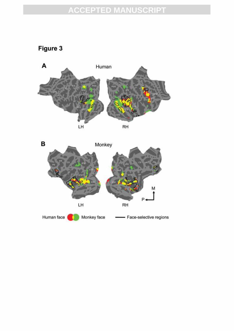
Neural processing of dynamic faces
In humans, conspecific and heterospecific dynamic facial expressions (red and green,
respectively in Fig. 3A), relative to their spatially and temporally scrambled versions,
activated a largely overlapping distributed network (yellow in Fig. 3A). Besides face-selective
areas (as defined by the contrast between static faces and control objects), the network also
included neighboring occipito-temporal cortex, right inferior frontal gyrus, inferior parietal
cortex (Fig. 3A) and bilateral amygdala. Stereotactic coordinates of all reported activations in
the human brain are listed in supplementary Table 1. In monkeys, the same contrasts,
activated bilateral face-selective areas in the upper and lower bank of the STS, with the
1 ‘Selective’ and ‘responsive’ are terms quite intensively used in this paper. We make a clear
distinction between them: 'selective' refers to differences between expression conditions (fear
and chewing) in the dynamic facial expression experiment, or between faces and objects in
the localizer experiment (in this case ‘face-selective’ is used), while ‘responsive’ refers to any
response compared to scrambled faces or fixation.
ACC
EPTE
D M
ANU
SCR
IPT
ACCEPTED MANUSCRIPT
activity extending posteriorly into TEO and extrastriate areas such as V2, V3 and V4 (Fig.
3B). In addition, prefrontal cortex and left amygdala were face-responsive as well. The right
amygdala was responsive to both human and monkey faces at p< 0.005, uncorrected for
multiple comparisons. Most of this dynamic-face responsive system was conjointly activated
by both human and monkey faces (shown in yellow in Fig. 3B).
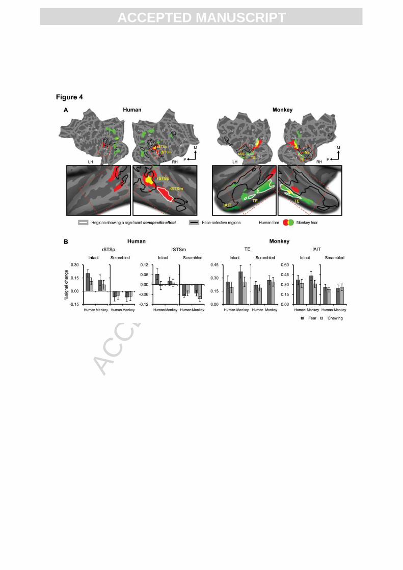
Neural processing of emotional expressions
We first compared in each species separately the areas that responded selectively to either
human or monkey emotional facial expressions, by conducting a two-way interaction between
expression and configuration for human faces and monkey faces separately. In humans,
human fearful expressions, relative to chewing (controlled for low-level effects such as
motion, by using scrambled versions) activated the middle and posterior part of the right STS
and upper and lower bank of the posterior part of the left STS (shown in red and yellow in Fig.
4A). Although a similar effect was found in the anterior part of the fusiform gyrus bilaterally,
this was relatively weak and only survived when a threshold uncorrected for multiple
comparisons was used (supplementary Fig. 3A), or when a direct comparison between fearful
and chewing expressions was performed, omitting the scrambled controls (supplementary Fig.
3B). The right posterior STS was the only face-selective region that showed a clear effect of
emotional expression (supplementary Fig. 4) and was also the only region that responded to
emotional expressions of both species (shown in yellow in Fig. 4A).
ACC
EPTE
D M
ANU
SCR
IPT
ACCEPTED MANUSCRIPT
In monkeys, bilateral infero-temporal cortex (specifically the convexity of the inferior
temporal gyrus) was selective to the fearful expressions of monkeys (shown in green and
yellow in Fig. 4A). This effect of emotional expression fell mainly outside the face-selective
areas (black outline in Fig. 4A) (activity profiles showing the emotion effect from all face-
selective areas can be checked in supplementary Fig. 4). In monkeys, we found that activity in
early visual cortex (mainly restricted to the lunate sulcus) was modulated by emotion for both
species (but more extensively for human faces). Given the absence of changes in early visual
activity in response to emotional expressions in the human brain, it is unlikely that this effect
in monkeys is due to low-level stimulus characteristics. To examine whether specialization for
processing conspecific emotional expressions exists in both species, we performed a three-
way interaction between species, expression and configuration. In humans, the middle part of
the right STS (rSTSm) was specifically activated by human fearful expression (white outline
in rSTS in Fig. 4A) and showed no differential activation between monkey fearful expressions
and monkey chewing (paired t-test, t(22) = 0.71, p = 0.48, Fig. 4B profiles). This was the only
area in the human brain showing a conspecific-specific response to emotional expressions. In
monkeys, we also found conspecific-specific responses bilaterally in posterior TE (white
outline in green labeled regions in Fig. 4A) and left lunate sulcus (left V4d). However, in
strong contrast with the conspecific responses in human rSTSm, human fear also increased
ACC
EPTE
D M
ANU
SCR
IPT
ACCEPTED MANUSCRIPT
activity in monkey TE in comparison to chewing (paired t-test, t(13) = 2.31, p < 0.05, Fig. 4B
profiles).
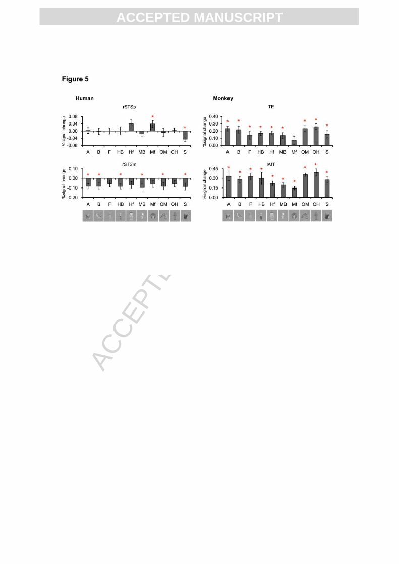
Although both human STS and monkey IT responded selectively to emotional expressions,
we found differences in properties between these regions that make it unlikely that they fulfill
the same function in both species. First, monkey IT responded to all dynamic stimuli,
including the scrambled displays whereas rSTSp responded only to dynamic facial
expressions and rSTSm only to human emotional expressions. Furthermore, our independent
localizer experiment with static stimuli showed that monkey IT responded to all non-facial
categories tested (Ps < 0.05, Wilcoxon signed rank test), whereas human rSTSp only
responded to faces (p < 0.05, Wilcoxon signed rank test) and human rSTSm was not activated
at all by any of the visual categories presented (Fig. 5), consistent with the selective response
to human emotional expressions. In the monkey, we also investigated whether other areas that
responded to conspecific emotional expressions without necessarily a conspecific-specific
effect, such as the left anterior inferior temporal cortex (left AIT) (Fig. 4A), would respond
solely to conspecific emotional expressions and not to other visual categories, but the answer
was negative (Fig. 5).
Although not the primary aim of this study, we found that other species’ emotional
expressions elicited more distributed and mainly posterior effects in humans, including
ventral occipito-temporal and dorsal occipito-parietal cortex, superior parietal lobule, right
ACC
EPTE
D M
ANU
SCR
IPT
ACCEPTED MANUSCRIPT
posterior STS, temporo-parietal junction, but also premotor cortex (shown in green and
yellow in Fig. 4A). In monkeys, viewing human fearful faces relative to chewing, was
associated with posterior early visual effects and an activation of a face-selective area that has
been labeled ML (Moeller et al., 2008). Based on the properties of ML, we can rule out that
this activation for human emotional expressions in monkeys corresponds with our conspecific
emotion effect in rSTSm in humans: in contrast with rSTSm, area rML was face selective and
responded to all visual categories tested (supplementary Fig. 5).
Finally, the emotion effect in human but not monkey STS was not a matter of differences in
responsiveness to dynamic facial stimuli: human and monkey dynamic face stimuli compared
to the scrambled ones (single main effect of configuration) activated a largely overlapping
distributed network including face-selective areas of STS in both species (Fig. 3).
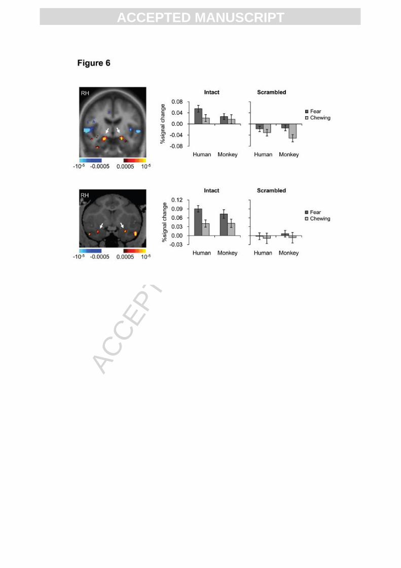
Effects of emotional expressions in the amygdala
Given the evidence favoring amygdala involvement in fear processing across species (Dolan
and Morris, 2000; Emery and Amaral, 2000; Phelps and LeDoux, 2005), we specifically
looked at the activity profiles in the face-responsive (faces vs. scrambled faces irrespective of
the species) parts of the amygdala (Fig. 6). In humans, amygdala responded more strongly to
human, but not monkey fearful faces, compared to chewing (paired t-test. Human fear vs.
chewing: t(22) = 2.79, p < 0.01; Monkey fear vs. chewing: t(22) = 0.73, p = 0.47). There was
ACC
EPTE
D M
ANU
SCR
IPT
ACCEPTED MANUSCRIPT
a significant three-way interaction between species, expression and configuration (ANOVA,
F(1, 22) = 5.76, p < 0.05). However, when studying the two-way species x emotion
interaction, leaving out the scrambled versions, the effect was not significant (ANOVA, F(1,
22) = 2.38, p = 0.14). In monkeys, amygdala responded more strongly to both human and
monkey fearful faces than to chewing faces (paired t-test. Human fear vs. chewing: t(13) =
4.82, p < 0.001; Monkey fear vs. chewing: t(13) = 2.33, p < 0.05), and there was no
significant three-way interaction between species, expression and configuration (ANOVA,
F(1, 13) = 0.70, p = 0.42), or two-way interaction between species and expression when the
scrambles were left out (ANOVA, F(1, 13) = 1.20, p = 0.29).
DISCUSSION
Our data reveal differences in neural processing of emotional facial expressions between
humans and monkeys, and argue for a more unique role of human STS in facial emotion
perception than previously documented. Although human and monkey STS are both
responsive to dynamic faces, we found that human but not monkey STS shows significant
activity differences between emotional and non-emotional dynamic facial expressions.
Second, we provide evidence for further functional specialization within human STS along a
posterior to anterior axis. Posterior STS responded selectively to emotional expressions
independent of species and the emotion effect in rSTSp fell within a face-selective region. In
ACC
EPTE
D M
ANU
SCR
IPT
ACCEPTED MANUSCRIPT
contrast, the response in rSTSm, anterior to rSTSp, was highly selective for the emotional cue
of human faces and appeared outside face-selective areas.
In monkeys, we observed effects of monkey emotional expressions mainly in bilateral
inferotemporal cortex but also in early visual cortex. In posterior TE, the activity was
significantly higher for conspecific than for human emotional expressions. The emotional
effects in monkey IT, appearing outside or at the edge of face-selective areas, confirm earlier
findings using static facial expressions (Hadj-Bouziane et al., 2008) and extend those
observations to demonstrate that the posterior part of IT responds particularly to conspecific
emotional expressions.
Although tempting to speculate on similarities between human STS and monkey IT in
processing emotion cues from dynamic faces, we found important differences in functional
properties between these regions with monkey IT being responsive to all visual stimuli
presented and human STS being selective for faces (rSTSp) and human emotions (rSTSm).
Our interpretation of the data is that human STS developed a high degree of neural
specialization for emotional expressions as socially meaningful stimuli (Peelen et al., 2010),
whereas emotion effects in monkey IT constitute mainly modulatory responses in the visual
processing stream (Hadj-Bouziane et al., 2008). Such modulatory effects in IT have been
covered before and are hypothesized to originate from limbic structures, mainly the amygdala
(Emery and Amaral, 2000). Support for this hypothesis also comes from the observation that
ACC
EPTE
D M
ANU
SCR
IPT
ACCEPTED MANUSCRIPT
different aspects of facial information are encoded at different latencies during single cell
recordings in IT (Sugase et al., 1999). Global information such as species is encoded by an
early transient discharge whereas fine information such as emotional expressions is conveyed
by a later sustained discharge. The time delay likely reflects feedback from other areas.
Furthermore, in agreement with our findings, IT contains neurons that respond at a much
higher level to monkey than to human expressions (Sugase et al., 1999). In this study, activity
changes to emotional expressions also occurred in early visual cortex of monkeys. Early
visual responses have been reported in human studies showing that attention to stimuli that
contain emotional information enhances responses in early visual cortex (Pessoa et al., 2002;
West et al., 2011) and is consistent with anatomical studies in monkeys that show feedback
projections from the amygdala terminating in TE and in V1 (Freese and Amaral, 2005; Freese
and Amaral, 2006). It should be noted that the lack of emotion effects in monkey STS in our
study does not mean that monkey STS is not involved in processing emotional expressions.
Neurons with preferential responses to emotional expressions in macaque STS have been
documented before (Hasselmo et al., 1989; Perrett et al., 1984; Rolls, 2007). However, our
findings show that – in contrast with human STS – fMRI response, a measure of averaged
regional brain activity, is not significantly higher for emotional compared to non-emotional
expressions in monkey STS.
ACC
EPTE
D M
ANU
SCR
IPT
ACCEPTED MANUSCRIPT
There is growing evidence for an important role of the human STS in the perception of facial
emotional expressions (Adolphs, 2002; Allison et al., 2000; Calder and Young, 2005; Engell
and Haxby, 2007; Furl et al., 2007; Haxby et al., 2002; Kret et al., 2011; LaBar et al., 2003;
Narumoto et al., 2001; Said et al., 2010; Winston et al., 2004), as well as in other aspects of
social perception from faces including gaze perception, lip-reading and other types of
meaningful biological motion (Allison et al., 2000; de Gelder, 2006). In line with our
hypothesis that STS activation in humans fulfills a social function and is involved in
attributing meaning to the expression, there is growing evidence that the posterior STS is
implicated in the understanding of others’ mental states (Gallagher et al., 2000; Gobbini et al.,
2007; Redcay et al., 2010) and encodes supramodal representations of perceived emotions
(Peelen et al., 2010). Furthermore, dysfunction of the human STS in clinical populations, such
as autistic subjects, leads to complex impairment of social perception (Redcay, 2008;
Zilbovicius et al., 2006). The emergence of neural specialization for processing human-
specific emotional and social information from faces in middle and anterior parts of the
human temporal lobe, especially rSTSm, is not surprising. An important extra-allometric
expansion of this part of the brain has occurred in the course of anthropoid evolution (Rilling
and Seligman, 2002), which is, at least on the phylogenetic time scale, correlated with
increasing social demands (Joffe and Dunbar, 1997). A higher degree of specialization for
extracting dynamic information from faces in anterior compared to posterior human STS was
ACC
EPTE
D M
ANU
SCR
IPT
ACCEPTED MANUSCRIPT
recently reported (Pitcher et al., 2011). Another study reported specialization for human facial
motion compared to hand motion in right middle STS (Thompson et al., 2007) and fMRI
adaptation studies confirm functional specialization within human right STS with sensitivity
for human emotional expressions in more anterior parts (Winston et al., 2004). Furthermore,
electrical stimulation of human right middle STS disturbs labeling of facial emotions (Fried et
al., 1982). Also neurodegeneration of the right anterior temporal cortex leads to severe
emotion recognition deficits in patients with frontotemporal dementia (Rosen et al., 2002).
Although the heterospecific faces were primarily meant as controls to study whether emotion
effects were specific for the own species, we were surprised to find so little overlap between
the effects of conspecific and heterospecific emotional expressions, especially in humans.
This contrasts with the important overlap of face-responsive regions in both species (Fig. 3),
supporting that face processing in general is largely species-independent whereas processing
of emotional cues is much more species-dependent. More posterior, parietal and occipito-
temporal, responses to heterospecific expressions have been reported before though in
humans (Buccino et al., 2004), but it is not exactly clear what they mean. It is unlikely that
these posterior activations were caused by low-level stimulus differences since we control for
it by the interaction with the scrambled stimuli. Moreover, activation in the early occipito-
temporal cortex was found only in monkeys for human fearful faces (compared to chewing),
but not in humans. If it was a low-level effect we should have observed it in both humans and
ACC
EPTE
D M
ANU
SCR
IPT
ACCEPTED MANUSCRIPT
monkeys. Differences in arousal could be another possibility, as dynamic monkey faces
(certainly emotional monkey faces) may be more arousing for humans than dynamic human
faces are for monkeys. However, the behavioral data presented in Fig. 2B show that this is
very unlikely: the degree of arousal for humans is larger for human faces compared to
monkey faces. Aspects that are harder to control for are differences in selective spatial
attention across stimulus types, which are known to drive portions of parietal cortex and
modulate activity in occipital areas. Hence, although speculative, a more parsimonious
explanation is that humans pay more attention to the monkey fearful faces than to the human
fearful faces. Even so a stronger homospecific (compared to heterospecific) effect was still
observed in higher order cortex (rSTSm) in humans, which further strengthens the unique role
of STS in dealing with social cues such as emotional expression.
It should also be noted that differences in familiarity may have contributed to the conspecific
effect in our results. However, our study design was conceptualized to minimize familiarity
effects in monkeys and novelty effects in humans, by contrasting emotional heterospecific
with non-emotional heterospecific faces and thereby subtracting the familiarity or novelty
effects of heterospecific faces in monkeys and humans respectively.
To conclude, our data suggest that human STS evolved towards an expertise in processing
emotional expressions that is not present to a comparable degree in monkeys. More generally,
our data underscore the importance of cross-species comparisons (Mantini et al., 2012) to gain
ACC
EPTE
D M
ANU
SCR
IPT
ACCEPTED MANUSCRIPT
insight in the species-typical neural basis of social interactions (Ghazanfar and Santos, 2004).
Further comparative studies with species-specific social cues are certainly needed to support
our claims and to elucidate what is typically human about our so-called ‘social brain’.
Acknowledgements: We thank C. Fransen, C. Van Eupen and A. Coeman for animal training
and care; H. Kolster, W. Depuydt, G. Meulemans, P. Kayenbergh, M. De Paep, S. Verstraeten,
M. Docx, and I. Puttemans for technical assistance. In addition, we thank I. Popivanov, R.
Vogels, J. Jastorff and N. Caspari for their help with the localizer experiment. This work was
supported by the Fund for Scientific Research (Flanders) G.0746.09, G.0622.08, and
G.0831.11, Hercules II funding, Inter University Attraction Pole 6/29, the National Institute of
Neurological Disorders and Stroke (R21NS064432), and the National Science Foundation
(BCS-0745436) in the USA, Programme Financing PFV/10/008, Geconcerteerde Onderzoeks
Actie 10/19. Q.Z., K.N. and J.V.d.S. are postdoctoral fellows of the FWO Vlaanderen.
ACC
EPTE
D M
ANU
SCR
IPT
ACCEPTED MANUSCRIPT
Figure Legend
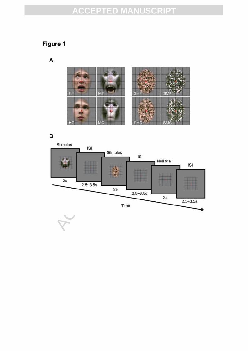
Fig. 1. Stimuli and experimental paradigm. (A) Upper row left panels: intact human fearful
(HF) and monkey fearful (MF) expressions; upper row right: scrambled versions of HF (SHF)
and MF (SMF). Lower row left panels: intact human chewing (HC) and monkey chewing
(MC); lower row right panels: scrambled versions of HC (SHC) and MC (SMC). Examples of
dynamic displays are provided in supplementary Video 1 to 8. (B) Event-related experimental
design. Trials consisted of 2 s stimulus presentation followed by a variable interstimulus
interval (ISI) between 2.5 and 3.5 seconds.
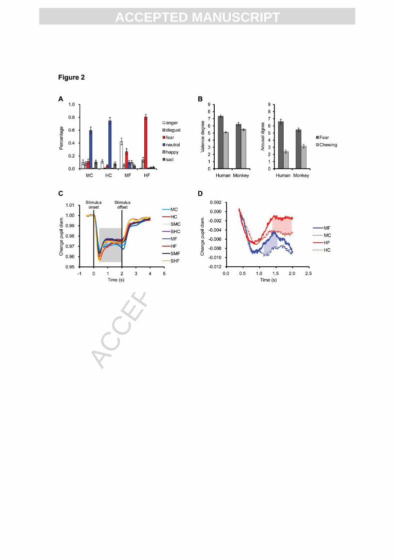
Fig. 2. Behavioral results in humans (A, B) and pupil data in monkeys (C, D). (A) Forced
choice categorisation of facial expressions (same abbreviations as Fig. 1). Y-axis shows
percentage (mean ± s.e.m.) of choices for each category per facial expression. (B) Valence
and arousal ratings of facial expressions (mean ± s.e.m.). Scores ranging from 1 to 9
corresponding with highly positive to highly negative valence or lowest to highest arousal.
(C) Pupil diameter change (percentage change relative to baseline) per trial for each condition
for monkeys. (D) Pupil diameter change between 375 ms and 2000 ms after stimulus-onset
(transparent grey time window shown in C) for intact faces relative to scrambled versions.
Significant differences between fear and chewing conditions are indicated by blue (monkey)
or red (human) shadow (p < 0.01).
ACC
EPTE
D M
ANU
SCR
IPT
ACCEPTED MANUSCRIPT
Fig. 3. Neural system responsive to dynamic facial expressions in humans and monkeys.
Group-level significance maps of human (red) and monkey (green) dynamic faces compared
to their scrambled versions (voxel-level p < 0.001, cluster-level corrected p < 0.05),
irrespective of expression, are shown in humans (A) and monkeys (B). Areas that are
activated by both human and monkey faces are shown in yellow. Black outlines represent
face-selective areas in both species. LH, left hemisphere; RH, right hemisphere; M, medial; P,
posterior. Abbreviations of sulci names: SFS, superior frontal sulcus; IFS, inferior frontal
sulcus; PreCS, precentral sulcus; CS, central sulcus; IPS, intraparietal sulcus; POS, parieto-
occipital sulcus; SF, sylvian fissure; STS, superior temporal sulcus; COS, collateral sulcus;
PS, principal sulcus; AS, arcuate sulcus; CS, central sulcus; LuS, Lunate sulcus; IOS, inferior
occipital sulcus; OTS, occipitotemporal sulcus.
Fig. 4. Areas selective to emotional facial expressions in humans and monkeys. (A)
Color-coded surface maps show regions of significant two-way emotion x configuration
interaction: significance maps (group-level) of human (red) and monkey (green) fearful faces
(relative to scrambled fearful faces) compared to chewing faces (relative to scrambled
chewing faces) (voxel-level p < 0.01, cluster-level corrected p < 0.05). Areas that are
activated by both human and monkey fearful faces are shown in yellow. Regions of
ACC
EPTE
D M
ANU
SCR
IPT
ACCEPTED MANUSCRIPT
significant conspecific effect (three-way species x emotion x configuration interaction, same
threshold as two-way interaction) are labeled on the surface maps using white outlines
(Human as well as monkey specific emotion responsive areas). Black outlines represent face-
selective areas in both species. (B) Response to dynamic facial expressions in areas
responsive to conspecific emotional expressions in humans and monkeys. Activity profiles
(mean ± s.e.m.) show percent signal change relative to fixation (Y-axis) for each of the 8
conditions (X-axis). LH, left hemisphere; RH, right hemisphere; M, medial; P, posterior.
Fig. 5. Response to object categories in areas responsive to conspecific emotional
expressions in humans and monkeys. Activity profiles (mean ± s.e.m.) show percent signal
change relative to fixation (Y-axis) for each category (X-axis) in the posterior and middle part
of the right superior temporal sulcus (rSTSm) in humans, and area TE and left anterior
inferior temporal cortex (lAIT) in monkeys. ROIs were defined the same way as in Fig. 4B.
Abbreviations for object categories: human faces (Hf), monkey faces (Mf), human bodies
(HB), monkey bodies (MB), objects with two different aspect ratios (OH and OB), animals
(A), birds (B), fruits (F) and sculptures (S). *: p < 0.05 (each category vs. fixation, Wilcoxon
signed rank test, uncorrected).
ACC
EPTE
D M
ANU
SCR
IPT
ACCEPTED MANUSCRIPT
Fig. 6. Responses in Amygdala to facial expressions. Activity profiles based on all face-
responsive voxels (faces vs. scrambled faces irrespective of the species) in human and
monkey amygdala (as shown by the white arrows). RH, right hemisphere.
References
Adolphs, R., 2002. Neural systems for recognizing emotion. Curr. Opin. Neurobiol. 12, 169-
177.
Allison, T., Puce, A., McCarthy, G., 2000. Social perception from visual cues: role of the STS
region. Trends Cogn. Sci. 4, 267-278.
Bair, W., O'Keefe, L.P., 1998. The influence of fixational eye movements on the response of
neurons in area MT of the macaque. Vis. Neurosci. 15, 779-786.
Bell, A.H., Hadj-Bouziane, F., Frihauf, J.B., Tootell, R.B., Ungerleider, L.G., 2009. Object
representations in the temporal cortex of monkeys and humans as revealed by
functional magnetic resonance imaging. J. Neurophysiol. 101, 688-700.
Bradley, M.M., Lang, P.J., 1994. Measuring emotion: the Self-Assessment Manikin and the
Semantic Differential. J. Behav. Ther. Exp. Psychiatry 25, 49-59.
Brothers, L., 1989. A biological perspective on empathy. Am. J. Psychiatry 146, 10-19.
Buccino, G., Lui, F., Canessa, N., Patteri, I., Lagravinese, G., Benuzzi, F., Porro, C.A.,
Rizzolatti, G., 2004. Neural circuits involved in the recognition of actions performed
by nonconspecifics: an FMRI study. J. Cogn. Neurosci. 16, 114-126.
Buracas, G.T., Boynton, G.M., 2002. Efficient design of event-related fMRI experiments
using M-sequences. Neuroimage 16, 801-813.
Burrows, A.M., Waller, B.M., Parr, L.A., 2009. Facial musculature in the rhesus macaque
(Macaca mulatta): evolutionary and functional contexts with comparisons to
chimpanzees and humans. J. Anat. 215, 320-334.
ACC
EPTE
D M
ANU
SCR
IPT
ACCEPTED MANUSCRIPT
Calder, A.J., Young, A.W., 2005. Understanding the recognition of facial identity and facial
expression. Nat. Rev. Neurosci. 6, 641-651.
de Gelder, B., 2006. Towards the neurobiology of emotional body language. Nat. Rev.
Neurosci. 7, 242-249.
de Gelder, B., 2010. The grand challenge for Frontiers in Emotion Science. Front. Psychol. 1,
1-4.
de Waal, F.B., 2011. What is an animal emotion? Ann. N. Y. Acad. Sci. 1224, 191-206.
de Waal, F.B.M., Luttrell, L.M., 1985. The Formal Hierarchy of Rhesus Macaques - An
Investigation of the Bared-Teeth Display. Am. J. Primatol. 9, 73-85.
Dolan, R.J., 2002. Emotion, cognition, and behavior. Science 298, 1191-1194.
Dolan, R.J., Morris, J.S., 2000. The Functional Anatomy of Innate and Acquired Fear:
Perspectives from Neuroimaging. In: Lane, R.D., Nadel, L. (Eds.), Cognitive
Neuroscience of Emotion. Oxford University Press, Inc., New York, New York, pp.
225-241.
Ekstrom, L.B., Roelfsema, P.R., Arsenault, J.T., Bonmassar, G., Vanduffel, W., 2008.
Bottom-up dependent gating of frontal signals in early visual cortex. Science 321,
414-417.
Emery, N.J., Amaral, D.G., 2000. The Role of the Amygdala in Primate Social Cognition. In:
Lane, R.D., Nadel, L. (Eds.), Cognitive Neuroscience of Emotion. Oxford University
Press, Inc., New York, New York, pp. 156-191.
Engell, A.D., Haxby, J.V., 2007. Facial expression and gaze-direction in human superior
temporal sulcus. Neuropsychologia 45, 3234-3241.
Fischl, B., Sereno, M.I., Tootell, R.B., Dale, A.M., 1999. High-resolution intersubject
averaging and a coordinate system for the cortical surface. Hum. Brain Mapp. 8, 272-
284.
Freese, J.L., Amaral, D.G., 2005. The organization of projections from the amygdala to visual
cortical areas TE and V1 in the macaque monkey. J. Comp. Neurol. 486, 295-317.
Freese, J.L., Amaral, D.G., 2006. Synaptic organization of projections from the amygdala to
visual cortical areas TE and V1 in the macaque monkey. J. Comp. Neurol. 496, 655-
667.
ACC
EPTE
D M
ANU
SCR
IPT
ACCEPTED MANUSCRIPT
Fried, I., Mateer, C., Ojemann, G., Wohns, R., Fedio, P., 1982. Organization of visuospatial
functions in human cortex. Evidence from electrical stimulation. Brain 105, 349-371.
Furl, N., van Rijsbergen, N.J., Treves, A., Friston, K.J., Dolan, R.J., 2007. Experience-
dependent coding of facial expression in superior temporal sulcus. Proc. Natl. Acad.
Sci. U. S. A. 104, 13485-13489.
Gallagher, H.L., Happe, F., Brunswick, N., Fletcher, P.C., Frith, U., Frith, C.D., 2000.
Reading the mind in cartoons and stories: an fMRI study of 'theory of mind' in verbal
and nonverbal tasks. Neuropsychologia 38, 11-21.
Ghazanfar, A.A., Santos, L.R., 2004. Primate brains in the wild: the sensory bases for social
interactions. Nat. Rev. Neurosci. 5, 603-616.
Gitelman, D.R., 2002. ILAB: a program for postexperimental eye movement analysis. Behav.
Res. Methods Instrum. Comput. 34, 605-612.
Gobbini, M.I., Koralek, A.C., Bryan, R.E., Montgomery, K.J., Haxby, J.V., 2007. Two takes
on the social brain: a comparison of theory of mind tasks. J. Cogn. Neurosci. 19, 1803-
1814.
Graham, R., Labar, K.S., 2012. Neurocognitive mechanisms of gaze-expression interactions
in face processing and social attention. Neuropsychologia 50, 553-566.
Hadj-Bouziane, F., Bell, A.H., Knusten, T.A., Ungerleider, L.G., Tootell, R.B., 2008.
Perception of emotional expressions is independent of face selectivity in monkey
inferior temporal cortex. Proc. Natl. Acad. Sci. U. S. A. 105, 5591-5596.
Hasselmo, M.E., Rolls, E.T., Baylis, G.C., 1989. The role of expression and identity in the
face-selective responses of neurons in the temporal visual cortex of the monkey.
Behav. Brain Res. 32, 203-218.
Haxby, J.V., Hoffman, E.A., Gobbini, M.I., 2002. Human neural systems for face recognition
and social communication. Biol. Psychiatry 51, 59-67.
Hebb, D.O., 1946. Emotion in man and animal: an analysis of the intuitive processes of
recognition. Psychol. Rev. 53, 88-106.
Hein, G., Knight, R.T., 2008. Superior Temporal Sulcus--It's My Area: Or Is It? J. Cogn.
Neurosci. 20, 2125-2136.
Hoffman, K.L., Gothard, K.M., Schmid, M.C., Logothetis, N.K., 2007. Facial-expression and
gaze-selective responses in the monkey amygdala. Curr. Biol. 17, 766-772.
ACC
EPTE
D M
ANU
SCR
IPT
ACCEPTED MANUSCRIPT
Joffe, T.H., Dunbar, R.I., 1997. Visual and socio-cognitive information processing in primate
brain evolution. Proc. Biol. Sci. 264, 1303-1307.
Kret, M.E., Pichon, S., Grezes, J., de Gelder, B., 2011. Similarities and differences in
perceiving threat from dynamic faces and bodies. An fMRI study. Neuroimage. 54,
1755-1762.
Kujala, M.V., Tanskanen, T., Parkkonen, L., Hari, R., 2009. Facial Expressions of Pain
Modulate Observer's Long-Latency Responses in Superior Temporal Sulcus. Hum.
Brain Mapp. 30, 3910-3923.
LaBar, K.S., Crupain, M.J., Voyvodic, J.T., McCarthy, G., 2003. Dynamic perception of
facial affect and identity in the human brain. Cereb. Cortex 13, 1023-1033.
Leite, F.P., Tsao, D., Vanduffel, W., Fize, D., Sasaki, Y., Wald, L.L., Dale, A.M., Kwong,
K.K., Orban, G.A., Rosen, B.R., Tootell, R.B.H., Mandeville, J.B., 2002. Repeated
fMRI using iron oxide contrast agent in awake, behaving macaques at 3 Tesla.
Neuroimage 16, 283-294.
Maestripieri, D., Wallen, K., 1997. Affiliative and submissive communication in rhesus
macaques. Primates 38, 127-138.
Mandeville, J.B., Choi, J.K., Jarraya, B., Rosen, B.R., Jenkins, B.G., Vanduffel, W., 2011.
fMRI of Cocaine Self-Administration in Macaques Reveals Functional Inhibition of
Basal Ganglia. Neuropsychopharmacology.
Mantini, D., Hasson, U., Betti, V., Perrucci, M.G., Romani, G.L., Corbetta, M., Orban, G.A.,
Vanduffel, W., 2012. Interspecies activity correlations reveal functional
correspondence between monkey and human brain areas. Nat. Methods 9, 277-282.
Moeller, S., Freiwald, W.A., Tsao, D.Y., 2008. Patches with links: a unified system for
processing faces in the macaque temporal lobe. Science 320, 1355-1359.
Narumoto, J., Okada, T., Sadato, N., Fukui, K., Yonekura, Y., 2001. Attention to emotion
modulates fMRI activity in human right superior temporal sulcus. Brain Res. Cogn.
Brain Res. 12, 225-231.
Nelissen, K., Vanduffel, W., Orban, G.A., 2006. Charting the lower superior temporal region,
a new motion-sensitive region in monkey superior temporal sulcus. J. Neurosci. 26,
5929-5947.
ACC
EPTE
D M
ANU
SCR
IPT
ACCEPTED MANUSCRIPT
Papenberg, N., Bruhn, A., Brox, T., Didas, S., Weickert, J., 2006. Highly accurate optic flow
computation with theoretically justified warping. Int. J. Comput. Vision 67, 141-158.
Parr, L.A., Heintz, M., 2009. Facial expression recognition in rhesus monkeys, Macaca
mulatta. Anim. Behav. 77, 1507-1513.
Parr, L.A., Waller, B.M., Fugate, J., 2005. Emotional communication in primates:
implications for neurobiology. Curr. Opin. Neurobiol. 15, 716-720.
Parr, L.A., Waller, B.M., Heintz, M., 2008. Facial expression categorization by chimpanzees
using standardized stimuli. Emotion. 8, 216-231.
Peelen, M.V., Atkinson, A.P., Vuilleumier, P., 2010. Supramodal representations of perceived
emotions in the human brain. J. Neurosci. 30, 10127-10134.
Perrett, D.I., Smith, P.A., Potter, D.D., Mistlin, A.J., Head, A.S., Milner, A.D., Jeeves, M.A.,
1984. Neurones responsive to faces in the temporal cortex: studies of functional
organization, sensitivity to identity and relation to perception. Hum. Neurobiol. 3,
197-208.
Pessoa, L., McKenna, M., Gutierrez, E., Ungerleider, L.G., 2002. Neural processing of
emotional faces requires attention. Proc. Natl. Acad. Sci. U. S. A. 99, 11458-11463.
Phelps, E.A., LeDoux, J.E., 2005. Contributions of the amygdala to emotion processing: From
animal models to human behavior. Neuron 48, 175-187.
Pinsk, M.A., Arcaro, M., Weiner, K.S., Kalkus, J.F., Inati, S.J., Gross, C.G., Kastner, S., 2009.
Neural representations of faces and body parts in macaque and human cortex: a
comparative FMRI study. J. Neurophysiol. 101, 2581-2600.
Pitcher, D., Dilks, D.D., Saxe, R.R., Triantafyllou, C., Kanwisher, N., 2011. Differential
selectivity for dynamic versus static information in face-selective cortical regions.
Neuroimage. 56, 2356-2363.
Puce, A., Allison, T., Bentin, S., Gore, J.C., McCarthy, G., 1998. Temporal cortex activation
in humans viewing eye and mouth movements. J. Neurosci. 18, 2188-2199.
Rajimehr, R., Young, J.C., Tootell, R.B., 2009. An anterior temporal face patch in human
cortex, predicted by macaque maps. Proc. Natl. Acad. Sci. U. S. A. 106, 1995-2000.
Redcay, E., 2008. The superior temporal sulcus performs a common function for social and
speech perception: implications for the emergence of autism. Neurosci. Biobehav. Rev.
32, 123-142.
ACC
EPTE
D M
ANU
SCR
IPT
ACCEPTED MANUSCRIPT
Redcay, E., Dodell-Feder, D., Pearrow, M.J., Mavros, P.L., Kleiner, M., Gabrieli, J.D., Saxe,
R., 2010. Live face-to-face interaction during fMRI: a new tool for social cognitive
neuroscience. Neuroimage. 50, 1639-1647.
Rilling, J.K., Seligman, R.A., 2002. A quantitative morphometric comparative analysis of the
primate temporal lobe. J. Hum. Evol. 42, 505-533.
Rolls, E.T., 2004. The functions of the orbitofrontal cortex. Brain Cogn. 55, 11-29.
Rolls, E.T., 2007. The representation of information about faces in the temporal and frontal
lobes. Neuropsychologia 45, 124-143.
Rosen, H.J., Perry, R.J., Murphy, J., Kramer, J.H., Mychack, P., Schuff, N., Weiner, M.,
Levenson, R.W., Miller, B.L., 2002. Emotion comprehension in the temporal variant
of frontotemporal dementia. Brain 125, 2286-2295.
Said, C.P., Moore, C.D., Engell, A.D., Todorov, A., Haxby, J.V., 2010. Distributed
representations of dynamic facial expressions in the superior temporal sulcus. J. Vis.
10, 11.
Sturgeon, R.S., Cooper, L.M., Howell, R.J., 1989. Pupil response: a psychophysiological
measure of fear during analogue desensitization. Percept. Mot. Skills 69, 1351-1367.
Sugase, Y., Yamane, S., Ueno, S., Kawano, K., 1999. Global and fine information coded by
single neurons in the temporal visual cortex. Nature 400, 869-873.
Thompson, J.C., Hardee, J.E., Panayiotou, A., Crewther, D., Puce, A., 2007. Common and
distinct brain activation to viewing dynamic sequences of face and hand movements.
Neuroimage 37, 966-973.
Tsao, D.Y., Freiwald, W.A., Knutsen, T.A., Mandeville, J.B., Tootell, R.B., 2003. Faces and
objects in macaque cerebral cortex. Nat. Neurosci. 6, 989-995.
Tsao, D.Y., Moeller, S., Freiwald, W.A., 2008a. Comparing face patch systems in macaques
and humans. Proc. Natl. Acad. Sci. U. S. A. 105, 19514-19519.
Tsao, D.Y., Schweers, N., Moeller, S., Freiwald, W.A., 2008b. Patches of face-selective
cortex in the macaque frontal lobe. Nat. Neurosci. 11, 877-879.
Vanduffel, W., Fize, D., Mandeville, J.B., Nelissen, K., Van, H.P., Rosen, B.R., Tootell, R.B.,
Orban, G.A., 2001. Visual motion processing investigated using contrast agent-
enhanced fMRI in awake behaving monkeys. Neuron 32, 565-577.
ACC
EPTE
D M
ANU
SCR
IPT
ACCEPTED MANUSCRIPT
Vuilleumier, P., Richardson, M.P., Armony, J.L., Driver, J., Dolan, R.J., 2004. Distant
influences of amygdala lesion on visual cortical activation during emotional face
processing. Nat. Neurosci. 7, 1271-1278.
West, G.L., Anderson, A.A., Ferber, S., Pratt, J., 2011. Electrophysiological evidence for
biased competition in V1 for fear expressions. J. Cogn. Neurosci. 23, 3410-3418.
Winston, J.S., Henson, R.N., Fine-Goulden, M.R., Dolan, R.J., 2004. fMRI-adaptation reveals
dissociable neural representations of identity and expression in face perception. J.
Neurophysiol. 92, 1830-1839.
Zhao, F.Q., Wang, P., Hendrich, K., Ugurbil, K., Kim, S.G., 2006. Cortical layer-dependent
BOLD and CBV responses measured by spin-echo and gradient-echo fMRI: Insights
into hemodynamic regulation. Neuroimage 30, 1149-1160.
Zilbovicius, M., Meresse, I., Chabane, N., Brunelle, F., Samson, Y., Boddaert, N., 2006.
Autism, the superior temporal sulcus and social perception. Trends Neurosci. 29, 359-
366.
ACC
EPTE
D M
ANU
SCR
IPT
ACCEPTED MANUSCRIPT
ACC
EPTE
D M
ANU
SCR
IPT
ACCEPTED MANUSCRIPT
ACC
EPTE
D M
ANU
SCR
IPT
ACCEPTED MANUSCRIPT
ACC
EPTE
D M
ANU
SCR
IPT
ACCEPTED MANUSCRIPT
ACC
EPTE
D M
ANU
SCR
IPT
ACCEPTED MANUSCRIPT
ACC
EPTE
D M
ANU
SCR
IPT
ACCEPTED MANUSCRIPT
Related Documents