i BIODEGRADABLE MULTIFUNCTIONAL NANOCARRIERS FOR pDNA and siRNA DELIVERY Dissertation zur Erlangung des Doktorgrades der Naturwissenschaften (Dr. rer. nat.) dem Fachbereich Pharmazie der Philipps-Universitä t Marburg vorgelegt von Mengyao Zheng aus Beijing, China Marburg/Lahn 2012

Welcome message from author
This document is posted to help you gain knowledge. Please leave a comment to let me know what you think about it! Share it to your friends and learn new things together.
Transcript

i
BIODEGRADABLE MULTIFUNCTIONAL
NANOCARRIERS FOR pDNA and siRNA
DELIVERY
Dissertation
zur Erlangung des Doktorgrades der Naturwissenschaften (Dr. rer. nat.)
dem
Fachbereich Pharmazie der Philipps-Universität Marburg
vorgelegt von
Mengyao Zheng
aus Beijing, China
Marburg/Lahn 2012

ii
Vom Fachbereich Pharmazie der Philipps-Universität Marburg als Dissertation am 02.07.2012
angenommen.
Erstgutachter: Prof. Dr. Thomas Kissel
Zweitgutachterin: Prof. Dr. Seema Agarwal
Tag der mündlichen Prüfung: 04.07.2012

iii
Die vorliegende Arbeit entstand auf Anregung und unter Leitung von
Herrn Prof. Dr. Thomas Kissel
am Institut für Pharmazeutische Technologie und Biopharmazie
der Philipps-Universität Marburg.

4
TABLE OF CONTENTS
1 INTRODUCTION ........................................................................................................... 8
1.1 Nanomedicine and Non-Viral Delivery of Nucleic Acids ....................................... 9
1.2 Targeted Gene Delivery Using Cell Specific Ligands ........................................... 10
1.3 Polymers and Dendrimers in Gene Delivery ......................................................... 11
1.4 Pulmonary Gene Delivery ....................................................................................... 14
1.5 Structure of the Thesis: Aims and Outline ........................................................... 15
1.6 References .................................................................................................................. 16
2 TARGETING THE BLIND SPOT OF POLYCATIONIC
NANOCARRIER-BASED SIRNA DELIVERY ............................................................... 19
2.1 Abstract ...................................................................................................................... 20
2.2 Introduction ............................................................................................................... 20
2.3 Results and Discussion ............................................................................................. 23
2.4 Conclusion ................................................................................................................. 28
2.5 Materials and Methods ............................................................................................ 28
2.6 Acknowledgements ................................................................................................... 29
2.7 Supporting informations .......................................................................................... 29
2.8 References .................................................................................................................. 33
3 AMPHIPHILIC AND BIODEGRADABLE hy-PEI-g-PCL-b-PEG
COPOLYMERS EFFICIENTLY MEDIATE TRANSGENE EXPRESSION
DEPENDING ON THEIR GRAFT DENSITY ................................................................. 36
3.1 Abstract ...................................................................................................................... 37
3.2 Introduction ............................................................................................................... 37
5
3.3 Methods and Materials ........................................................................................ 39
3.4 Results and Discussion ............................................................................................... 43
3.5 Conclusion .................................................................................................................. 50
3.6 Acknowledgements ................................................................................................... 51
3.7 References .................................................................................................................. 51
4 ENHANCING IN VIVO CIRCULATION AND SIRNA DELIVERY WITH
BIODEGRADABLE
POLYETHYLENIMINE-GRAFT-POLYCAPROLACTONE-BLOCK-POLY(ETHY
LENE GLYCOL) COPOLYMERS .................................................................................... 54
4.1 Abstract ...................................................................................................................... 55
4.2 Introduction ............................................................................................................... 55
4.3 Methods and materials ............................................................................................. 57
4.4 Results and Discussion.............................................................................................. 60
4.5 Conclusion .................................................................................................................. 70
4.6 Acknowledgements ................................................................................................... 71
4.7 References .................................................................................................................. 71
5 MODULAR SYNTHESIS OF FOLATE CONJUGATED TERNARY
COPOLYMERS:
POLYETHYLENIMINE-GRAFT-POLYCAPROLACTONE-BLOCK-POLY
(ETHYLENE GLYCOL)-FOLATE FOR TARGETED GENE DELIVERY
DELIVERY
5.1 Abstract ...................................................................................................................... 75
5.2 Introduction ............................................................................................................... 75
5.3 Experimental Section ................................................................................................ 77
5.4 Results and Discussion.............................................................................................. 84
6
5.5 Conclusion .................................................................................................................. 93
5.6 Acknowledgment ....................................................................................................... 93
5.7 Supporting Information ........................................................................................... 93
5.8 References .................................................................................................................. 93
6 MOLECULAR MODELING AND IN VIVO IMAGING CAN IDENTIFY
SUCCESSFUL FLEXIBLE TRIAZINE DENDRIMER-BASED SIRNA DELIVERY
SYSTEMS ............................................................................................................................... 97
6.1 Abstract ...................................................................................................................... 98
6.2 Introduction ............................................................................................................... 99
6.3 Experimental Section .............................................................................................. 100
6.4 Results and Discussion........................................................................................... 105
6.5 Conclusion ............................................................................................................... 118
6.6 Acknowledgements ................................................................................................ 119
6.7 References ............................................................................................................... 119
7 DESIGN AND BIOPHYSICAL CHARACTERIZATION OF BIORESPONSIVE
DEGRADABLE POLY(DIMETHYLAMINOETHYL METHACRYLATE) BASED
POLYMERS FOR IN VITRO DNA TRANSFECTION .............................................. 123
7.1 Abstract ................................................................................................................... 124
7.2 Introduction ............................................................................................................ 124
7.3 Experimental Part .................................................................................................. 126
7.4 Results and Discussion........................................................................................... 132
7.5 Conclusion ............................................................................................................... 145
7.6 References ............................................................................................................... 146
8 SUMMARY................................................................................................................. 147

7
8.1 Summary ................................................................................................................. 147
8.2 Perspectives ............................................................................................................. 150
8.3 Zusammenfassung ................................................................................................. 151
9 APPENDICES ............................................................................................................ 155
9.1 Abbreviations ......................................................................................................... 155
9.2 List of Publications ................................................................................................ 156
9.2.1 Articles ............................................................................................................. 156
9.2.2 Poster Presentations ......................................................................................... 157
9.2.3 Lectures ............................................................................................................ 158
9.2.4 Abstracts ........................................................................................................... 158
9.3 Curriculum Vitae ................................................................................................... 159
9.4 Danksagung ............................................................................................................ 160

8
Chapter 1 INTRODUCTION

9
1.1 Nanomedicine and Delivery of Nucleic Acids
Nanomedicine is the engineering, manufacturing and application of nanotechnology for medical
applications especially in terms of drug or nucleic acids delivery.1, 2
Nanomedicine is expected to
become a revolutionary class of therapeutics to improve human health at the atomic and
molecular scale. Especially concerning advanced drug and gene delivery systems, the use of
nanotechnology can improve the delivery of macromolecular drug substances (for example
nucleic acids) and help them to cross cellular barriers. Not only soluble drug carriers, but also
insoluble drug carriers can be formulated as nanoparticles using techniques such as the solvent
displacement4 or solvent evaporation/emulsion technique.
5 With one to several hundred
nanometers in size, it is also widely believed that drug delivery systems prepared by
nanotechnology may also make targeted delivery and co-delivery of two or more therapeutic
agents in “multifunctional” carriers possible.6, 7
One of the important applications of nanomedicine is gene therapy, a powerful approach for the
treatment of cancer and genetic diseases by the transfer of genetic material into specific cells of
the patient.8 For high therapeutic efficacy, gene delivery systems need to be directed to their
target region and specifically taken up by the target cell populations through an initial set of
barriers from the test tube to the membrane of a target cell. These include physico-chemical
challenges, such as binding and condensing gene materials, as well as in vitro barriers such as cell
uptake, protecting the gene materials against enzymatic degradation and other competing
polyanions (serum stability), transport through the cytoplasm, endolysosomal escape and
unpackaging of gene materials from the delivery agents.9 Additionally, for efficient gene delivery
vector accumulation, long circulation time in vivo is of critical importance and requires efficient
particle evasion from the clearing organs including the liver, which is largely mediated by the
physicochemical properties of the gene delivery vectors.10
SiRNA are a double-stranded RNAs of 21–23 nucleotides with two-nucleotide 3′ overhangs and
5′-phosphorylated ends11, 12
and can be delivered into target cells by gene delivery agents.
Although the delivery of siRNA faces many of the same barriers and intracellular steps as
delivery of plasmid DNA, the delivery of siRNA appears more difficult than DNA delivery.
Differences between pDNA and siRNA delivery are for example that the final target destination
of siRNA is the cytoplasm, whereas plasmid DNA must be transported into the nucleus. In other
words, to achieve successful siRNA delivery, the siRNA must be delivered and released rapidly
from its carrier upon endosomal escape into the cytoplasm. Secondly, a recent report showed that
siRNA is less flexible13, 14
and the knowledge on structural conformation of cationic polymers
reacting with nucleic acids is still limited. Due to rigidity, the condensation of siRNA within
cationic polymers is assumed to be more difficult. For the above reasons, the design of high
10
affinity, good protection agents is a key point in the development of nanocarriers for siRNA
delivery systems.3 Moreover, novel design and development of next-generation of biocompatible
and biodegradable siRNA delivery vectors with controlled release and molecular targeting
properties is also a big challenge, especially for the therapeutic benefit in the clinical applications.
1.2 Targeted Gene Delivery Using Cell Specific Ligands
Active vs passive targeting. The term “passive targeting” is usually defined as a method to
deliver drugs based on the ability of the drug carrier
to circulate for longer times in the bloodstream and
accumulate in pathological tissues. “Active
targeting” is also called ligand based targeting, which
is based on the ligand-receptor recognition to
recognize and bind the ligand-conjugated carriers on
the target tissues. In the case of cancer therapy, the
delivery of gene materials with non-targeted agents
(passive targeting) is achieved mainly passive by the
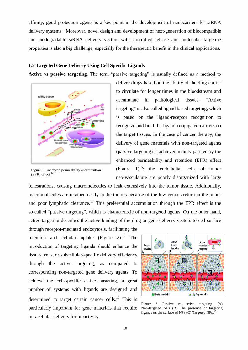
enhanced permeability and retention (EPR) effect
(Figure 1)15
: the endothelial cells of tumor
neo-vasculature are poorly disorganized with large
fenestrations, causing macromolecules to leak extensively into the tumor tissue. Additionally,
macromolecules are retained easily in the tumors because of the low venous return in the tumor
and poor lymphatic clearance.16
This preferential accumulation through the EPR effect is the
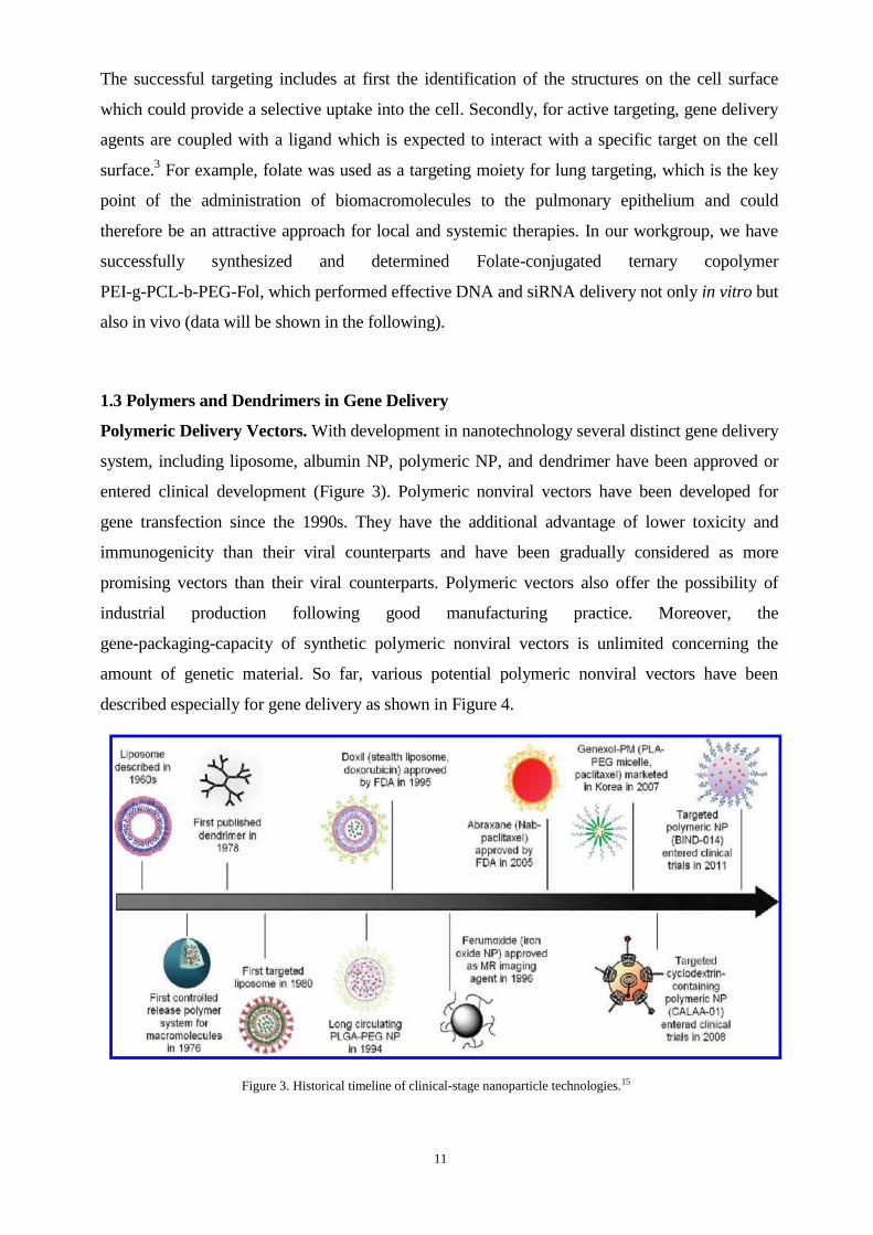
so-called “passive targeting”, which is characteristic of non-targeted agents. On the other hand,
active targeting describes the active binding of the drug or gene delivery vectors to cell surface
through receptor-mediated endocytosis, facilitating the
retention and cellular uptake (Figure 2).10
The
introduction of targeting ligands should enhance the
tissue-, cell-, or subcellular-specific delivery efficiency
through the active targeting, as compared to
corresponding non-targeted gene delivery agents. To
achieve the cell-specific active targeting, a great
number of systems with ligands are designed and
determined to target certain cancer cells.17
This is
particularly important for gene materials that require
intracellular delivery for bioactivity.
Figure 1. Enhanced permeability and retention
(EPR) effect.14
Figure 2. Passive vs active targeting. (A)
Non-targeted NPs (B) The presence of targeting
ligands on the surface of NPs (C) Targeted NPs.15
11
The successful targeting includes at first the identification of the structures on the cell surface
which could provide a selective uptake into the cell. Secondly, for active targeting, gene delivery
agents are coupled with a ligand which is expected to interact with a specific target on the cell
surface.3 For example, folate was used as a targeting moiety for lung targeting, which is the key
point of the administration of biomacromolecules to the pulmonary epithelium and could
therefore be an attractive approach for local and systemic therapies. In our workgroup, we have
successfully synthesized and determined Folate-conjugated ternary copolymer
PEI-g-PCL-b-PEG-Fol, which performed effective DNA and siRNA delivery not only in vitro but
also in vivo (data will be shown in the following).
1.3 Polymers and Dendrimers in Gene Delivery
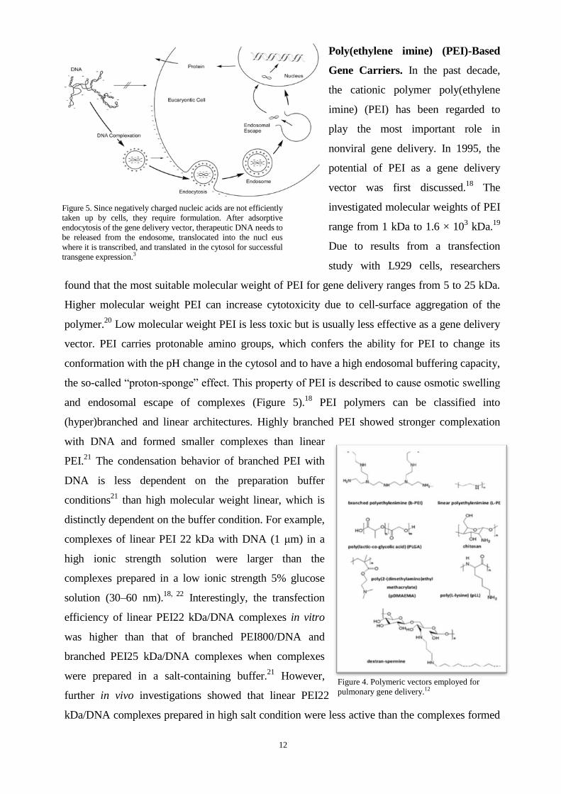
Polymeric Delivery Vectors. With development in nanotechnology several distinct gene delivery
system, including liposome, albumin NP, polymeric NP, and dendrimer have been approved or
entered clinical development (Figure 3). Polymeric nonviral vectors have been developed for
gene transfection since the 1990s. They have the additional advantage of lower toxicity and
immunogenicity than their viral counterparts and have been gradually considered as more
promising vectors than their viral counterparts. Polymeric vectors also offer the possibility of
industrial production following good manufacturing practice. Moreover, the
gene-packaging-capacity of synthetic polymeric nonviral vectors is unlimited concerning the
amount of genetic material. So far, various potential polymeric nonviral vectors have been
described especially for gene delivery as shown in Figure 4.
Figure 3. Historical timeline of clinical-stage nanoparticle technologies.15
12
Poly(ethylene imine) (PEI)-Based
Gene Carriers. In the past decade,
the cationic polymer poly(ethylene
imine) (PEI) has been regarded to
play the most important role in
nonviral gene delivery. In 1995, the
potential of PEI as a gene delivery
vector was first discussed.18
The
investigated molecular weights of PEI
range from 1 kDa to 1.6 × 10³ kDa.
19
Due to results from a transfection
study with L929 cells, researchers
found that the most suitable molecular weight of PEI for gene delivery ranges from 5 to 25 kDa.
Higher molecular weight PEI can increase cytotoxicity due to cell-surface aggregation of the
polymer.20
Low molecular weight PEI is less toxic but is usually less effective as a gene delivery
vector. PEI carries protonable amino groups, which confers the ability for PEI to change its
conformation with the pH change in the cytosol and to have a high endosomal buffering capacity,
the so-called “proton-sponge” effect. This property of PEI is described to cause osmotic swelling
and endosomal escape of complexes (Figure 5).18
PEI polymers can be classified into
(hyper)branched and linear architectures. Highly branched PEI showed stronger complexation
with DNA and formed smaller complexes than linear
PEI.21
The condensation behavior of branched PEI with
DNA is less dependent on the preparation buffer
conditions21
than high molecular weight linear, which is
distinctly dependent on the buffer condition. For example,
complexes of linear PEI 22 kDa with DNA (1 μm) in a
high ionic strength solution were larger than the
complexes prepared in a low ionic strength 5% glucose
solution (30–60 nm).18, 22
Interestingly, the transfection
efficiency of linear PEI22 kDa/DNA complexes in vitro
was higher than that of branched PEI800/DNA and
branched PEI25 kDa/DNA complexes when complexes
were prepared in a salt-containing buffer.21
However,
further in vivo investigations showed that linear PEI22
kDa/DNA complexes prepared in high salt condition were less active than the complexes formed
Figure 4. Polymeric vectors employed for
pulmonary gene delivery.12
Figure 5. Since negatively charged nucleic acids are not efficiently
taken up by cells, they require formulation. After adsorptive
endocytosis of the gene delivery vector, therapeutic DNA needs to
be released from the endosome, translocated into the nucl eus
where it is transcribed, and translated in the cytosol for successful
transgene expression.3
13
in low salt condition (100-fold less). This indicates that efficient transgene expression strongly
depends on the size of the complexes.
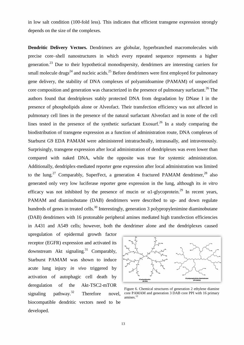
Dendritic Delivery Vectors. Dendrimers are globular, hyperbranched macromolecules with
precise core–shell nanostructures in which every repeated sequence represents a higher
generation.23
Due to their hypothetical monodispersity, dendrimers are interesting carriers for
small molecule drugs24
and nucleic acids.25
Before dendrimers were first employed for pulmonary
gene delivery, the stability of DNA complexes of polyamidoamine (PAMAM) of unspecified
core composition and generation was characterized in the presence of pulmonary surfactant.26
The
authors found that dendriplexes stably protected DNA from degradation by DNase I in the
presence of phospholipids alone or Alveofact. Their transfection efficiency was not affected in
pulmonary cell lines in the presence of the natural surfactant Alveofact and in none of the cell
lines tested in the presence of the synthetic surfactant Exosurf.26
In a study comparing the
biodistribution of transgene expression as a function of administration route, DNA complexes of
Starburst G9 EDA PAMAM were administered intratracheally, intranasally, and intravenously.
Surprisingly, transgene expression after local administration of dendriplexes was even lower than
compared with naked DNA, while the opposite was true for systemic administration.
Additionally, dendriplex-mediated reporter gene expression after local administration was limited
to the lung.27
Comparably, SuperFect, a generation 4 fractured PAMAM dendrimer,28
also
generated only very low luciferase reporter gene expression in the lung, although its in vitro
efficacy was not inhibited by the presence of mucin or α1-glycoprotein.29
In recent years,
PAMAM and diaminobutane (DAB) dendrimers were described to up- and down regulate
hundreds of genes in treated cells.30
Interestingly, generation 3 polypropylenimine diaminobutane
(DAB) dendrimers with 16 protonable peripheral amines mediated high transfection efficiencies
in A431 and A549 cells; however, both the dendrimer alone and the dendriplexes caused
upregulation of epidermal growth factor
receptor (EGFR) expression and activated its
downstream Akt signaling.31
Comparably,
Starburst PAMAM was shown to induce
acute lung injury in vivo triggered by
activation of autophagic cell death by
deregulation of the Akt-TSC2-mTOR
signaling pathway.32
Therefore novel,
biocompatible dendritic vectors need to be
developed.
Figure 6. Chemical structures of generation 2 ethylene diamine
core PAMAM and generation 3 DAB core PPI with 16 primary
amines.12

14
1.4 Pulmonary Gene Delivery
The potential of pulmonary gene delivery was reported in a large number of studies.33-36
Because
of the high affinity between the airway epithelium cells with the targeted delivery vectors, the lung
is a promising target organ for gene delivery. The administration of biomacromolecules like DNA
or siRNA to the pulmonary epithelium could be an attractive approach for local passive targeting
but systemic therapies. Compared with hydrophilic macromolecules like nucleic acids, small and
hydrophobic molecules lead to more rapid local and systemic effects because of the
air-blood-barrier.37
Therefore, for successful pulmonary gene delivery, formulation of the
therapeutic nucleic acids into nanosized carrier systems is necessary. Furthermore, successful
pulmonary gene delivery must overcome a number of biological barriers, which includes anatomic,
physical, immunologic, and metabolic barriers.3 In the two last decades, various potential
polymeric nonviral vectors have been developed for pulmonary gene transfection, such as
poly(ethylene imine) (PEI), poly(2-(dimethylamino)ethyl methacrylate) (pDMAEMA), the
polysaccharide chitosan, the biologically occurring polyamine spermine, and the biodegradable
noncationic polymer PLGA.38
For in vitro pulmonary gene delivery experiments, the application of
lung epithelial cells cannot be replaced by conventional cell culture, because the epithelium
differentiates in layers of cells with a distinct apical and basolateral side and connects each other
by tight junctions. The Calu-3 cell line, A549 epithelial cells and human primary small airway
epithelial cells (HSAEC) are classic models of airway epithelium. Although intratracheal
instillation was frequently applied in lab-scale with animals, clinical success was still not achieved,
which depends not only on the development of effective, biocompatible, and targeted gene delivery
vectors, but also requires deeper understanding of the mechanisms of pulmonary gene delivery.
Figure 7: After entering the alveoli, gene delivery systems can possibly interact with the alveolar linage fluid or
can be taken up by various cell types. Recognition by and uptake into macrophages should be avoided, for
example, by adjusting the size and surface of nanoparticles. Uptake into pneumocytes could lead to local
therapeutic effects, and transcytosis into the systemic circulation could lead to systemic wanted or unwanted
effects.3
15
1.5 Structure of the Thesis: Aims and Outline
This thesis focuses on a number of issues in non-viral polymeric delivery of nucleic acids
concerning biophysicochemical parameters in vitro and in vivo application.
The investigations in chapters 2-5 were gene delivery study with the use of PEI-based polymeric
gene delivery systems. We should answer the questions: why the principle of DNA transfection
cannot be directly applied for siRNA transfection and to search for the development of better
siRNA delivery systems, our work began with the study of the binding mechanism of nucleic
acids/polycations complexation and aggregation through different levels of hierarchy on the atomic
and molecular scale, with the novel synergistic use of molecular modeling, molecular dynamics
simulation, isothermal titration calorimetry and other characterization techniques (chapter 2).
These data were expected to explain the different nature and the different hierarchical mechanism
of formation of related polycation-siRNA and polycation-pDNA complexes, which is the
important base of the following research of the effective nucleic acids, especially siRNA delivery.
Chapter 3 concentrates on in vitro pDNA delivery with biodegradable amphiphilic copolymers
hy-PEI-g-(PCL-b-PEG)n, which was grafted with PCL-b-PEG chains onto hyper-branched
poly(ethylene imine). In this copolymer, poly(caprolactone) (PCL) acts as a linker between PEI
and PEG to increase the biodegradability of the copolymers and the permeability of the complexes
through the cell membranes. So far, the investigations about these copolymers were limited to the
discussion of the influence of PEI, PCL and PEG chain lengths. Therefore in this section, our study
focused on the influence of graft density by correlating physic-chemical and biological in vitro
properties of the complexes and expected that with the introduction of the grafted PCL-b-PEG
chains, the in vitro DNA delivery efficiency with the grafted PCL-b-PEG chains could be
improved.
Chapter 4 continues to describe the siRNA delivery efficiency of these biodegradable amphiphilic
grafted copolymers hy-PEI-g-PCL-b-PEG in vitro and in vivo. The purpose of this study was to
enhance the in vivo blood circulation time and siRNA delivery efficiency of the same copolymers
in chapter 3, by introducing high graft densities of PCL-PEG chains. We assumed that the effect of
PEG on prolonged circulating depends not only on its length or percentage, but also on the
structure or the shape of the amphiphilic copolymers, which have advantages especially for in vivo
siRNA delivery.
Following the successful design and characterization of biodegradable amphiphilic copolymers
hy-PEI-g-(PCL-b-PEG)n (chapter 3, 4), in chapters 5, we successfully synthesized and
characterized folate-conjugated ternary copolymers based on
polyethylenimine-graft-polycaprolactone-block-poly(ethylene glycol) (PEI-g-PCL-b-PEG-Fol) as
targeted DNA delivery system. We hypothesized that these conjugated copolymer would

16
efficiently transfect folate-overexpressing cells via folate receptor-mediated endocytosis, which is
especially meaningful for the pulmonary gene delivery.
The aim of Chapter 6 study was to identify suitable siRNA delivery systems based on
hyperflexible generation 2-4 triazine dendrimers by correlating physico-chemical and biological in
vitro and in vivo properties of the complexes with their thermodynamic interaction features
simulated by molecular modeling.
Chapter 7 is the research about novel water soluble, degradable polymers based on
poly(N,N-dimethylaminoethyl methacrylate) (PDMAEMA) p-DNA delivery system. We expected
lower cytotoxicity but efficiently transfect of pDNA with these degradable polymers.
All results are summarized in Chapter 8, where an outlook also provides information on further
possible applications and developments.
1.6 References
1.Riehemann, K.; Schneider, S. W.; Luger, T. A.; Godin, B.; Ferrari, M.; Fuchs, H.,
Nanomedicine--challenge and perspectives. Angew Chem Int Ed Engl 2009, 48 (5), 872-97.
2.Petros, R. A.; DeSimone, J. M., Strategies in the design of nanoparticles for therapeutic
applications. Nat Rev Drug Discov 2010, 9 (8), 615-27.
3.Merkel, O. M.; Zheng, M.; Debus, H.; Kissel, T., Pulmonary gene delivery using polymeric
nonviral vectors. Bioconjug Chem 2011, 23 (1), 3-20.
4.Nguyen, J.; Steele, T. W. J.; Merkel, O.; Reul, R.; Kissel, T., Fast degrading polyesters as
siRNA nano-carriers for pulmonary gene therapy. Journal of Controlled Release 2008, 132 (3),
243-251.
5.Yan, F.; Zhang, C.; Zheng, Y.; Mei, L.; Tang, L. N.; Song, C. X.; Sun, H. F.; Huang, L. Q., The
effect of poloxamer 188 on nanoparticle morphology, size, cancer cell uptake, and cytotoxicity.
Nanomedicine-Nanotechnology Biology and Medicine 2010, 6 (1), 170-178.
6.Ferrari, M., Cancer nanotechnology: opportunities and challenges. Nat Rev Cancer 2005, 5 (3),
161-71.
7.Zhang, L.; Gu, F. X.; Chan, J. M.; Wang, A. Z.; Langer, R. S.; Farokhzad, O. C., Nanoparticles
in medicine: therapeutic applications and developments. Clin Pharmacol Ther 2008, 83 (5),
761-9.
8.Mulligan, R. C., The basic science of gene therapy. Science 1993, 260 (5110), 926-32.
9.Pack, D. W.; Hoffman, A. S.; Pun, S.; Stayton, P. S., Design and development of polymers for
gene delivery. Nat Rev Drug Discov 2005, 4 (7), 581-93.
10.Farokhzad, O. C.; Langer, R., Impact of nanotechnology on drug delivery. ACS Nano 2009, 3
(1), 16-20.
11.Bernstein, E.; Caudy, A. A.; Hammond, S. M.; Hannon, G. J., Role for a bidentate
ribonuclease in the initiation step of RNA interference. Nature 2001, 409 (6818), 363-366.
12.Zamore, P. D.; Tuschl, T.; Sharp, P. A.; Bartel, D. P., RNAi: double-stranded RNA directs the
ATP-dependent cleavage of mRNA at 21 to 23 nucleotide intervals. Cell 2000, 101 (1), 25-33.
13.Merkel, O. M.; Mintzer, M. A.; Librizzi, D.; Samsonova, O.; Dicke, T.; Sproat, B.; Garn, H.;
Barth, P. J.; Simanek, E. E.; Kissel, T., Triazine dendrimers as nonviral vectors for in vitro and in
vivo RNAi: the effects of peripheral groups and core structure on biological activity. Mol Pharm
2010, 7 (4), 969-83.
14.Pavan, G. M.; Mintzer, M. A.; Simanek, E. E.; Merkel, O. M.; Kissel, T.; Danani, A.,
Computational insights into the interactions between DNA and siRNA with "rigid" and "flexible"
triazine dendrimers. Biomacromolecules 2010, 11 (3), 721-30.
17
15.Matsumura, Y.; Maeda, H., A new concept for macromolecular therapeutics in cancer
chemotherapy: mechanism of tumoritropic accumulation of proteins and the antitumor agent
smancs. Cancer Res 1986, 46 (12 Pt 1), 6387-92.
16.Cabral, H.; Nishiyama, N.; Kataoka, K., Supramolecular Nanodevices: From Design
Validation to Theranostic Nanomedicine. Accounts of Chemical Research 2011, 44 (10),
999-1008.
17.Shi, J. J.; Xiao, Z. Y.; Kamaly, N.; Farokhzad, O. C., Self-Assembled Targeted Nanoparticles:
Evolution of Technologies and Bench to Bedside Translation. Accounts of Chemical Research
2011, 44 (10), 1123-1134.
18.Boussif, O.; Lezoualc'h, F.; Zanta, M. A.; Mergny, M. D.; Scherman, D.; Demeneix, B.; Behr,
J. P., A versatile vector for gene and oligonucleotide transfer into cells in culture and in vivo:
polyethylenimine. Proc Natl Acad Sci U S A 1995, 92 (16), 7297-301.
19.Meunier-Durmort, C.; Grimal, H.; Sachs, L. M.; Demeneix, B. A.; Forest, C., Adenovirus
enhancement of polyethylenimine-mediated transfer of regulated genes in differentiated cells.
Gene Ther 1997, 4 (8), 808-14.
20.Fischer, D.; Bieber, T.; Li, Y.; Elsasser, H. P.; Kissel, T., A novel non-viral vector for DNA
delivery based on low molecular weight, branched polyethylenimine: effect of molecular weight
on transfection efficiency and cytotoxicity. Pharm Res 1999, 16 (8), 1273-9.
21.Wightman, L.; Kircheis, R.; Rossler, V.; Carotta, S.; Ruzicka, R.; Kursa, M.; Wagner, E.,
Different behavior of branched and linear polyethylenimine for gene delivery in vitro and in vivo.
J Gene Med 2001, 3 (4), 362-72.
22.Goula, D.; Remy, J. S.; Erbacher, P.; Wasowicz, M.; Levi, G.; Abdallah, B.; Demeneix, B. A.,
Size, diffusibility and transfection performance of linear PEI/DNA complexes in the mouse
central nervous system. Gene Ther 1998, 5 (5), 712-7.
23.Boas, U.; Heegaard, P. M., Dendrimers in drug research. Chem Soc Rev 2004, 33 (1), 43-63.
24.D'Emanuele, A.; Attwood, D., Dendrimer-drug interactions. Adv Drug Deliv Rev 2005, 57 (15),
2147-62.
25.Shcharbin, D. G.; Klajnert, B.; Bryszewska, M., Dendrimers in gene transfection. Biochemistry
(Mosc) 2009, 74 (10), 1070-9.
26.Ernst, N.; Ulrichskotter, S.; Schmalix, W. A.; Radler, J.; Galneder, R.; Mayer, E.; Gersting, S.;
Plank, C.; Reinhardt, D.; Rosenecker, J., Interaction of liposomal and polycationic transfection
complexes with pulmonary surfactant. J Gene Med 1999, 1 (5), 331-40.
27.Kukowska-Latallo, J. F.; Raczka, E.; Quintana, A.; Chen, C.; Rymaszewski, M.; Baker, J. R.,
Jr., Intravascular and endobronchial DNA delivery to murine lung tissue using a novel, nonviral
vector. Hum Gene Ther 2000, 11 (10), 1385-95.
28.Tang, M. X.; Redemann, C. T.; Szoka, F. C., Jr., In vitro gene delivery by degraded
polyamidoamine dendrimers. Bioconjug Chem 1996, 7 (6), 703-14.
29.Rosenecker, J.; Naundorf, S.; Gersting, S. W.; Hauck, R. W.; Gessner, A.; Nicklaus, P.; Muller,
R. H.; Rudolph, C., Interaction of bronchoalveolar lavage fluid with polyplexes and lipoplexes:
analysing the role of proteins and glycoproteins. J Gene Med 2003, 5 (1), 49-60.
30.Akhtar, S.; Benter, I., Toxicogenomics of non-viral drug delivery systems for RNAi: potential
impact on siRNA-mediated gene silencing activity and specificity. Adv Drug Deliv Rev 2007, 59
(2-3), 164-82.
31.Omidi, Y.; Barar, J., Induction of human alveolar epithelial cell growth factor receptors by
dendrimeric nanostructures. Int J Toxicol 2009, 28 (2), 113-22.
32.Li, C.; Liu, H.; Sun, Y.; Wang, H.; Guo, F.; Rao, S.; Deng, J.; Zhang, Y.; Miao, Y.; Guo, C.;
Meng, J.; Chen, X.; Li, L.; Li, D.; Xu, H.; Li, B.; Jiang, C., PAMAM nanoparticles promote acute
lung injury by inducing autophagic cell death through the Akt-TSC2-mTOR signaling pathway. J
Mol Cell Biol 2009, 1 (1), 37-45.
33.Kinsey, B. M.; Densmore, C. L.; Orson, F. M., Non-viral gene delivery to the lungs. Current
Gene Therapy 2005, 5 (2), 181-194.
34.Aneja, M. K.; Geiger, J. P.; Himmel, A.; Rudolph, C., Targeted gene delivery to the lung.
Expert Opin Drug Deliv 2009, 6 (6), 567-83.
18
35.Griesenbach, U.; Alton, E. W. F. W.; Co, U. C. F. G. T., Gene transfer to the lung: Lessons
learned from more than 2 decades of CF gene therapy. Advanced Drug Delivery Reviews 2009, 61
(2), 128-139.
36.Sanders, N.; Rudolph, C.; Braeckmans, K.; De Smedt, S. C.; Demeester, J., Extracellular
barriers in respiratory gene therapy. Adv Drug Deliv Rev 2009, 61 (2), 115-27.
37.Cryan, S. A.; Sivadas, N.; Garcia-Contreras, L., In vivo animal models for drug delivery across
the lung mucosal barrier. Adv Drug Deliv Rev 2007, 59 (11), 1133-51.
38.Park, T. G.; Jeong, J. H.; Kim, S. W., Current status of polymeric gene delivery systems. Adv
Drug Deliv Rev 2006, 58 (4), 467-86.
19
Chapter 2
TARGETING THE BLIND SPOT OF POLYCATIONIC
NANOCARRIER-BASED SIRNA DELIVERY
Submitted to ACS Nano
Mengyao Zheng†, Giovanni M. Pavan
‡, Manuel Neeb
§, Andreas K. Schaper
‖, Andrea
Danani‡, Gerhard Klebe
§, Olivia M. Merkel
†,⊥ and Thomas Kissel
†, *
Author contributions
T. K. guided and directed the research. O. M. M. and M. Z. designed the measurements. M. Z.
prepared the polyplexes for isothermal titration calorimetry and TEM. M.Z. carried out the
SYBR® Gold assay, heparin assay, dye quenching assay, dynamic light scattering/zeta potential
analysis, in vitro cell uptake (CLSM) and knockdown experiments (RT-PCR). M. Z., O. M. M. and
G. M. P. analysed the experimental data.

20
2.1 Abstract
Polycationic nanocarriers attract increasing attention to the field of siRNA delivery. We
investigated the mechanism of nucleic acids/polycations complexation and aggregation through
different levels of hierarchy on the atomic and
molecular scale with the novel synergistic use of
molecular modeling, molecular dynamics
simulation, isothermal titration calorimetry and
other characterization techniques. These data
suggest the different nature and the different
hierarchical mechanism of formation of related
polycation-siRNA and polycation-pDNA
complexes. The results of fluorescence quenching
assays indicated a biphasic behavior of siRNA
binding with polycations where molecular reorganization of the siRNA within the polycations
occurred at lower N/P-ratios (nitrogen/phosphorus). Additionally, heparin assays showed that the
stability of siRNA/polymer complexes is especially good at a rather lower N/P-ratio of 2.
Interestingly, with the following study of the relationship between nucleic acids/polycations
aggregation mechanism and in vitro siRNA delivery efficiency, which is performed by RT-PCR
and confocal laser scanning microscopy, we found that not only PEI25kDa but also the
PCL-PEG-modified copolymer showed the best knockdown effect with siRNA at N/P=2, although
higher N/P ratios were believed to be necessary until now by most of the researchers in the area of
polycationic nanocarrier-based siRNA delivery. Our results emphasize the importance of low N/P
ratios, which allow for excellent siRNA delivery efficiency, but have been disregarded like a
“blind spot” in previous reports on siRNA delivery. Our investigation highlights the formulation of
siRNA complexes from a thermodynamic point of view and opens new perspectives to advance the
rational design of new siRNA delivery systems.
KEYWORDS: siRNA delivery · DNA delivery · Polyethylenimine · Molecular modeling ·
Isothermal titration calorimetry · RT-PCR · Supramolecular complexation
2.2 Introduction
Nanomedicine is the engineering, manufacturing and application of nanotechnology for medical
applications, amongst others for drug or nucleic acids delivery.1, 2
One of the most important
applications of nanomedicine is gene delivery, a powerful approach for the treatment of cancer and
genetic diseases. Compared with viral counterparts and liposomes, polymeric gene delivery
Abstract graphic: polycationic nanocarrier/siRNA
complexation and cell uptake at different N/P ratios.

21
systems have the advantages of lower toxicity and immunogenicity by design, and allow for
industrial production involving good manufacturing practice.3 A wide range of polymeric vectors
were designed and developed based on the complexation of nucleic acids via electrostatic
interaction between the negatively charged phosphates along the nucleic acid backbone with the
positive charges on the cationic polymers.4 The cationic polymer poly(ethylenimine) (PEI) is one
of the best studied vectors for non-viral gene delivery. Starting in the 1990s, the polymeric
non-viral vector PEI has been developed to achieve successful delivery of nucleic acids like
plasmid DNA, antisense oligonucleotides, ribozymes, and siRNA.2 Since the discovery of gene
silencing by introduction of double-stranded RNA,5 RNA interference is widely used in functional
genomics and drug development.6, 7
Although the delivery of siRNA faces many of the same
barriers and intracellular steps as delivery of plasmid DNA, the delivery of siRNA appears more
difficult than DNA delivery, and the design of high affinity, good protection agents is a key point
in the development of nanocarriers for siRNA delivery systems. In this study, we used isothermal
titration calorimetry (ITC) to investigate the complexation behavior of siRNA and DNA with
polycations. These thermodynamic parameters also allow for the study of the hierarchical
aggregation phenomena which result from the biomolecular interactions between nucleic acids and
cationic polymers.8 Because the knowledge on structure conformation of cationic polymers and
genetic materials is limited, molecular dynamics (MD) simulation was used to investigate the local
mechanism of binding between pDNA or siRNA molecules and cationic polymers, providing
detailed insight into the structural conformations and binding behavior.9-11
This synergetic use of
MD simulation and ITC provides a complete description not only of the local binding between
polymers and nucleic acids but also of the hierarchical aggregation steps which occur during
polyplex formation. Additionally, the complexation of DNA and siRNA was also studied using
heparin assays and dye quenching assays, and subsequently in vitro transfection experiments were
conducted with both siRNA and pDNA. Our investigations are focused on the study of binding
mechanisms, the different location of plasmid DNA and siRNA within complexes of cationic
polymers, their different structural conformations and biophysical parameters as well as the size
and surface charge of the final polyplexes. By investigating these parameters and correlating them
to functional studies including knockdown of glyceraldehyde 3-phosphate dehydrogenase
(GAPDH) gene expression measured by RT-PCR, we try to find distinguishing features of siRNA
complexation and to explain why the principle of DNA transfection cannot generally be directly
applied to siRNA transfection.12
Our study of the complexation mechanism between nucleic acids and polycationic nanocarriers
describes the very different nature of polycation-siRNA and polycation-DNA hierarchical
aggregation. We demonstrate that siRNA complexation can be schematized into two “rigid” steps,

22
namely (i) polycation-siRNA primary complexation, followed by the (ii) hierarchical association
of multiple nanocomplexes into larger polyplexes (Figure 1A). DNA condensation, however,
involves three steps: after the (i) primary electrostatic interactions between polycations and DNA,
the saturated polycation-DNA complex can undergo (ii) structural rearrangement (folding),
followed by the (iii) hierarchical association of multiple nanocomplexes into larger polyplexes
(Figure 1B). In this hierarchical framework, siRNA aggregation results in a more uniform and
stable complex formation, at low N/P ratios already, which lead to increased siRNA delivery
efficiency. Interestingly, with the following study of the relationship between nucleic
acids/polycations aggregation mechanism and in vitro siRNA delivery efficiency, which is
performed by RT-PCR and confocal laser scanning microscopy, the polycationic nanocarriers
based siRNA delivery system showed the best knockdown effect with siRNA at N/P=2, although
higher N/P ratios were believed to be necessary until now by most of the researchers in the area of
polycationic nanocarrier-based siRNA delivery.
Figure 1. Model for different hierarchical aggregation mechanism. (A) PEI/siRNA. (B) PEI/pDNA. The synergic use of MD
simulations, ITC and dye quenching assays provides us a complete description not only of the local binding between polymers and
nucleic acids but also of the hierarchical aggregation steps which occur during polyplex formation. TEM: during reduction of the
silver cations into silver nanoparticles on the negatively charged sugar-phosphate backbone of the nucleic acids, siRNA and DNA
were stained with Ag (black) and then condensed with polycations at low and high N/P ratios.
23
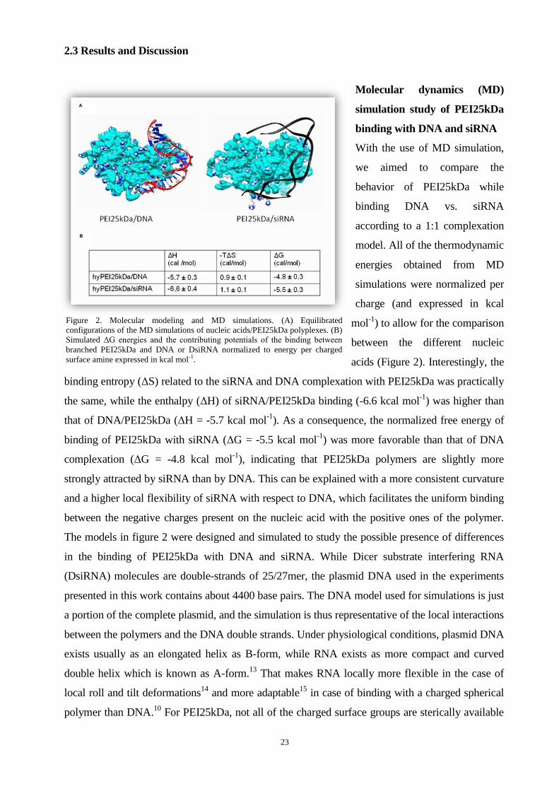
2.3 Results and Discussion
Molecular dynamics (MD)
simulation study of PEI25kDa
binding with DNA and siRNA
With the use of MD simulation,
we aimed to compare the
behavior of PEI25kDa while
binding DNA vs. siRNA
according to a 1:1 complexation
model. All of the thermodynamic
energies obtained from MD
simulations were normalized per
charge (and expressed in kcal
mol-1
) to allow for the comparison
between the different nucleic
acids (Figure 2). Interestingly, the
binding entropy (ΔS) related to the siRNA and DNA complexation with PEI25kDa was practically
the same, while the enthalpy (ΔH) of siRNA/PEI25kDa binding (-6.6 kcal mol-1
) was higher than
that of DNA/PEI25kDa (ΔH = -5.7 kcal mol-1
). As a consequence, the normalized free energy of
binding of PEI25kDa with siRNA (ΔG = -5.5 kcal mol-1
) was more favorable than that of DNA
complexation (ΔG = -4.8 kcal mol-1
), indicating that PEI25kDa polymers are slightly more
strongly attracted by siRNA than by DNA. This can be explained with a more consistent curvature
and a higher local flexibility of siRNA with respect to DNA, which facilitates the uniform binding
between the negative charges present on the nucleic acid with the positive ones of the polymer.
The models in figure 2 were designed and simulated to study the possible presence of differences
in the binding of PEI25kDa with DNA and siRNA. While Dicer substrate interfering RNA
(DsiRNA) molecules are double-strands of 25/27mer, the plasmid DNA used in the experiments
presented in this work contains about 4400 base pairs. The DNA model used for simulations is just
a portion of the complete plasmid, and the simulation is thus representative of the local interactions
between the polymers and the DNA double strands. Under physiological conditions, plasmid DNA
exists usually as an elongated helix as B-form, while RNA exists as more compact and curved
double helix which is known as A-form.13
That makes RNA locally more flexible in the case of
local roll and tilt deformations14
and more adaptable15
in case of binding with a charged spherical
polymer than DNA.10
For PEI25kDa, not all of the charged surface groups are sterically available
Figure 2. Molecular modeling and MD simulations. (A) Equilibrated
configurations of the MD simulations of nucleic acids/PEI25kDa polyplexes. (B)
Simulated ΔG energies and the contributing potentials of the binding between
branched PEI25kDa and DNA or DsiRNA normalized to energy per charged
surface amine expressed in kcal mol-1.
24
to bind a single strand of nucleic acids because a large part of charged amines is back folded.
During the binding between PEI25kDa and DNA/siRNA, parts of the positive surface charges of
the polymer establish strong electrostatic interactions with the nucleic acid. At the binding
interface, positive and negative charges neutralize each other. But moving away from the binding
site on the polymer surface, there are several other positively charged surface groups which do not
participate actively in the binding with the nucleic acid (“primary complexes” in figure 2).
These free charges can potentially lead to inter-particle electrostatic attractions with other
siRNA/DNA molecules giving rise to hierarchical aggregation phenomena. In fact, primary
complexes can aggregate further and re-organize into larger polyplexes.16
Therefore, there is a
balance that needs to be considered between the amount of charges and the ability to use these
charges. In this framework, it is evident that the pure binding between the polymer and the nucleic
acid which is depicted by MD simulation constitutes only the first, and most immediate step in a
complex hierarchical aggregation phenomenon which involves different scales and types of
interactions, from strong electrostatic to weaker hydrophobic intermolecular forces. This hierarchy
emerges when binding data from MD simulation are compared with the thermodynamic values
calculated based on ITC measurements. The consequent molecular complexes can potentially
undergo further structural reorganization and can interact with other polyplexes in solution. This
causes slower complexation as compared to siRNA, where a consistent structural rearrangement is
not expected due to the limited length of the nucleic acids. Moreover, DNA molecules need more
polycations to achieve complete condensation and to form stable polyplexes. This hypothesis was
challenged with the following ITC results.
Isothermal titration calorimetry (ITC)
The atomic binding results of local interactions from MD simulation are complemented by results
from isothermal titration calorimetry (ITC) experiments, which provide reliable thermodynamic
interpretation17
of the aggregation of multiple polycation/nucleic acid nanocomplexes into
higher-scale polyplexes. The ITC results are supported by the data from MD modeling and showed
the same tendency of the binding behavior between polycations and different nucleic acids: the
affinity between polycations and siRNA is higher than that between polycations and plasmid DNA,
and the formation of hierarchical polycation/siRNA polyplexes is much easier and more stable than
the complexation with plasmid DNA (Table 1). Even if the interaction between PEI25kDa and
DNA or siRNA is locally very similar, the flexible PEI25kDa/DNA nanocomplexes can undergo
structural rearrangement (folding), resulting in less uniform aggregation of multiple
nanocomplexes into larger polyplexes (Figure 1B). Moreover, ITC indicates also that DNA
molecules need a larger excess of polycations than siRNA to achieve complete condensation and to

25
form stable polyplexes (Table 1). If on an atomic level the pure polymer-nucleic acid molecular
recognition is controlled by electrostatic forces, on a higher-scale level, the inter-polyplex
interactions are also consistently characterized by hydrophobic forces, as is evidenced by data from
ITC (Table 1). Hydrophobic aggregation is assumed to be typically an entropy-driven assembly
phenomenon, accompanied by a lower favorable enthalpy (Table 1).18
This is particularly evident
in the case of DNA. In fact, if PEI is modified with hydrophobic poly(caprolactone) segments,
DNA/PEI25k-PCL1500-PEG2k nanocomplexes aggregate stronger due to increased
hydrophobicity19
and condense DNA more effectively. Concerning modified PEI25kDa, only
about 5 PEI25k-PCL1500-PEG2k molecules are required to condense one DNA molecule
(N-value or site), whereas 11 molecules unmodified PEI25kDa are needed for the same effect.
Figure 3. Thermodynamic interpretation was provided during Isothermal titration calorimetry. Standard binding isotherm curve of siRNA
and pDNA with polycations. The siRNA reorganization from the saturated complex into aggregates is an endothermic process, reflected in
an endothermic peak at N/P=1 in the ITC measurements.
Table 1. Thermodynamic parameters for the specific binding between polycations and DNA or siRNA.
All binding parameters are reliable experimental thermodynamic data calculated based on ITC.
The larger dissociation constant K of siRNA/polycation complexation reflects that the affinity
N
(sites)
K
(M-1
)
ΔH
(cal /mol)
ΔS
(cal/mol/deg)
ΔG
(cal/mol)
hyPEI25k/DNA 1.59±0.02 1.29E5±1.37E4 -2569±40.96 14.8 -6979.4
hyPEI25k/siRNA 2.26±0.03 2.23E6±9.28E5 -2172±48.09 21.8 -8668.4
hyPEI25k-PCL1500-PEG2k/DNA 0.683±0.01 2.58E5±3.20E4 -2795±57.88 15.4 -7384.2
hyPEI25k-PCL1500-PEG2k/siRNA 2.23±0.05 2.80E5±7.40E4 -2063±53.96 18.0 -7427.0

26
between polycations with siRNA is higher than that with pDNA. Concerning modified PEI, only
about 5 PEI25k-PCL1500-PEG2k molecules were required to condense one pDNA molecule
(N-value or site), whereas 11 PEI molecules were needed.
Fluorescence Quenching Assay
The dye quenching assay is another
method to investigate the binding behavior
of nucleic acids by polycations: the
fluorescence of labeled siRNA molecules
will be quenched by each other due to
close spatial proximity in complexes
where many siRNA molecules are
compacted. Although we have used
different polycations to condense siRNA,
each curve has a minimum of fluorescence
at N/P-ratio=1. After this minimum, the
fluorescence increases again with
increasing of N/P-ratio (Figure 4A).
Interestingly, an equilibrium in ITC is also
reached at remarkably lower N/P ratios for
siRNA than for DNA, highlighting the
noteworthiness of this low N/P ratio. The endothermic peaks of siRNA binding isotherm curves
close to N/P=1 (Figure 3), together with the dye quenching assay (Figure 4A) reveal a special
condensation phenomenon of siRNA: siRNA molecules “escape” from saturated “primary
multi-molecular nanocomplexes” at N/P=1 and reorganize into more stable nanocomplexes with a
lower energy level (N/P=2) (Figure 4B). This trend was already observed with siRNA20
and
oligonucleotides21
. Moreover, the particle size distribution (polydispersity index, PDI)
measurements indicate that siRNA can be condensed into more ordered and uniform polyplexes
with the lowest PDI at N/P=2 (Figure S2). Additionally, heparin assays confirmed that siRNA
polyplexes at N/P=2 are particularly stable against competing polyanions (Figure S1). Therefore,
we assumed that lower N/P-ratios (N/P=2 in case of PEI) are especially effective for siRNA
delivery.
Figure 4. Dye quenching assay. (A) The fluorescence of Tye563-labeled
siRNA molecules is quenched by each other in a “multi-molecular
complex” due to close spatial proximity. Each curve had a minimum of
fluorescence at N/P-ratio=1, after which the fluorescence increased
again due to a decreased number of siRNA molecules per polyplex,
resulting in less proximity of the labeled siRNA and thus in lower
quenching. This special phenomenon of short nucleic acids
condensation can be understood as a reorganization of the polyplexes.
(B) Molecular reorganization of the siRNA within the polycations at
lower N/P-ratios.

27
In vitro uptake and gene Knockdown effect
Interestingly, with the following study of the relationship between nucleic acids/polycations
aggregation mechanism and in vitro siRNA delivery efficiency, which is performed by RT-PCR
(Figure 5A, 5B) and confocal laser scanning microscopy (Figure 5C), we found that not only
PEI25kDa but also the PCL-PEG-modified copolymer hyPEI25k-PCL1500-PEG2k showed the
best intracellular delivery and knockdown effect with siRNA at N/P=2, although higher N/P ratios
were believed to be necessary until now by most of the researchers in the area of polycationic
nanocarrier-based siRNA delivery22-25
. In case of PEI25kDa, by increasing the N/P ratio, the
hGAPDH gene expression decreased from N/P 1 (53.26% knockdown) to N/P 2 (72.29%
knockdown) and increased again with the increasing of N/P ratios. The knockdown effect in the
graph is better at N/P 30 than at N/P 2, but the negative control at N/P 30 is also very low, which
indicates that at higher N/P-ratio, the knockdown effect is not only caused by gene silencing, but
also the cytotoxicity of the polycations. The CLSM micrographs reflected the same tendency:
although the siRNA could be delivered effectively into the cytosol at N/P 20, a less vital cell
morphology with partially dilapidated cellular membranes was observed, which indicated a high
cytotoxicity of these polycationic delivery agents at high N/P ratios. On the other hand, the siRNA
delivery efficiency at N/P 2 was as good as at N/P 20, but with a vital cell morphology (Figure
5C), as a result of more uniform and stable complex formation and lower cytotoxicity.
Figure 5. In vitro cell uptake and knockdown at different N/P-ratios. (A) Knockdown effect of siRNA/PEI25kDa polyplexes using
RT-PCR. (B) Knockdown effect of siRNA/PEI25k-PCL1500-PEG2k polyplexes using RT-PCR: polycations showed the best
knockdown effect with siRNA at N/P-ratio 2. In case of PEI25kDa, by increasing the N/P ratio, the GAPDH gene expression
decreased from N/P 1 (53.26% knockdown) to N/P 2 (72.29% knockdown) and increased again. The knockdown effect at N/P 20 and
30 seems better than at N/P 2, but the lower negative control bar indicated the toxicity at higher N/P ratio. (C) Confocal laser
scanning microscopy (CLSM) showed the cell uptake at different N/P ratios: both the uptake efficiency at N/P 2 and N/P 20 were
good, but N/P 20 was too toxic, causing a less vital cell morphology (siRNA was labeled with AF647 dyes; nuclei were stained with
DAPI and cell membranes were labeled with FITC-wheat germ agglutinin).

28
2.4 Conclusion
In our research we investigated the different complexation and aggregation mechanism between
polycationic nanocarriers and DNA or siRNA on the atomic and molecular scale. The novel
synergic use of MD simulations, ITC and dye quenching assay provided an exceptionally clear
depiction of the different hierarchical aspects which control the formation of polyplexes. It is well
accepted that the positively charged surface of poly(ethylenimine) nanocomplexes induces not only
increased cellular uptake through charge-mediated interactions26
(Figure 5C) but also
disadvantageous higher cytotoxicity (especially true for high N/P ratios). While researchers seek to
balance toxicity and transfection efficiency, our investigation highlights the need to address the
actual assembly of polyelectrolyte complexes and to optimize the formulation of siRNA complexes
from a thermodynamic point of view. Our study based on poly(ethylenimine) as a model
polycationic nanocarrier directs the attention to lower N/P ratios, which emerge as an unnoticed
“blind spot” in polycationic siRNA delivery. All our results emphasized one point: lower
N/P-ratios are especially effective for polycationic nanocarrier-based siRNA delivery. This could
have broad implications for the delivery of siRNA as less toxic and yet efficient delivery systems
have been the bottle-neck for the translation of this promising approach into the clinical arena. We
recommend to the scientific community working in the area of polycationic siRNA delivery to
study the actual assembly of self-assembled nanocarriers and thus to consider low N/P ratios,
which could be particularly important for siRNA delivery but have been disregarded in previous
studies.
2.5 Materials and Methods
Materials. Hyperbranched polyethylenimine (hy-PEI) 25kDa was obtained from BASF.
Poly(ethylene glycol) mono-methyl ether (mPEG) (5kDa) and ε-caprolactone were purchased from
Fluka (Taufkirchen, Germany). Beetle Luciferin, heparin sodium salt and all other chemicals were
obtained from Sigma–Aldrich (Steinheim, Germany). Luciferase-encoding plasmid (pCMV-Luc)
(LotNo.: PF461-090623) was amplified by The Plasmid Factory (Bielefeld, Germany). Negative
control sequence, hGAPDH-DsiRNA, and TYE546-DsiRNA were obtained from Integrated DNA
Technologies (IDT, Leuven, Belgium).
Molecular modeling and MD simulations. The binding of nucleic acid and PEI25kDa was
modeled according to a reported validated strategy9, 27
. The MD simulations were conducted
according to previous studies9, 27-29
.
Isothermal titration calorimetry. ITC was carried out with an iTC200 Micro Titration
Calorimeter (Microcal, Inc., Northampton, MA, USA) according to our earlier report30, 31
. The
baseline (dilution energy) was recorded by titrating redundant amounts of polymer into water.

29
After integration and fitting of the binding isotherm of peaks with a single-site-binding assumption,
the thermodynamic parameters enthalpy (ΔH), entropy (ΔS) and the dissociation constant K of
binding were calculated.
Dye quenching assay. Dye quenching assays were conducted according to a previous study by
Merkel et al.20
In vitro cell uptake and knockdown experiments. SKOV3 cells were seeded with 106 cells per
well in 6-wells 24 h prior to transfection and transfected with 50 pmol of siRNA. The mRNA was
isolated 24 h after transfection (PureLinkTM
RNA Mini Kit, Invitrogen GmbH, Germany) and
reverse transcribed to cDNA (First Strand cDNA Synthesis kit, Fermentas, Germany). RT-PCR
was performed using QuantiFastTM
SYBR® Green PCR Kits (Qiagen, Germany) and the
Rotor-Gene 3000 RT-PCR thermal cycler (Corbett Research, Sydney, Australia). For confocal
laser scanning microscopy, cells were incubated with nanocomplexes containing AF647 labeled
siRNA for 4h and then fixed. Nuclei were stained with DAPI and cell membranes were labeled
with FITC-wheat germ agglutinin (Invitrogen, Karlsruhe, Germany).
Transmission electron microscopy. Polyplexes were metalized during incubation with 0.005 M
AgNO3 for 2 hours at 25°C. TEM measurements were performed using a JEM-3010 microscope
(Jeol Ltd., Tokyo, Japan), operated at 300 kV, equipped with a high-resolution CCD camera for
image recording.
Statistics. All analytical assays were conducted in replicates of three or four. Results are given as
mean values+/−standard deviation. Two way ANOVA and statistical evaluations were performed
using Graph Pad Prism 4.03 (Graph Pad Software, La Jolla, USA).
2.6 Acknowledgements
The authors wish to acknowledge Dr. Ayse Kilic and Dr. Holger Garn (Institute of Laboratory
Medicine and Pathobiochemistry, Philipps Universität Marburg) for use of the Rotor-Gene real
time cycler, Eva Mohr (IPTB) for expert technical support in the cell culture, Michael Hellwig
(Center of Material Science, Philipps Universität Marburg) for TEM imaging, Prof. Dr. Wolfgang
Parak and Yu Xiang (Department of Physics, Philipps-Universität Marburg) for CLSM imaging
and Dr. Dafeng Chu (Department of Pharmaceutics and Biopharmacy, Philipps Universität
Marburg) for excellent discussions.
2.7 Supporting Informations
S1. Binding and protection efficiency and stability against competing polyanions
Method

30
The stability of complexes against competing polyanions, represented by heparin, was evaluated
by means of the change in fluorescence intensity obtained with the fluorescent intercalating probe
Sybr® Gold. To study the effect of N/P-ratio on the stability of complexes, polyplexes were
prepared in solutions at different N/P-ratio. 20 μL heparin (150 000 IU/g, Serva, Pharm., USPXV2,
Merck, Darmstadt, Germany) solution with a concentration of 1.5 IU/μmol siRNA was added into
200μL polyplex solution in each well of a 96-well plate (Perkin Elmer, Rodgau-Jügesheim) where
each well contained 1 μmol siRNA. After 20 min of incubation with the heparin solution at 25°C,
20 μL diluted Sybr® Gold solution (Invitrogen, Karlsruhe, Germany) were added. After another 20
min of incubation at 25°C, fluorescence was directly detected with a fluorescence plate reader
(BMG Labtech GMBH, Offenburg, Germany) at 495 nm excitation and 537 nm emission32
.
Results and Discussions
Sybr® Gold (Invitrogen) can be used to quantify purified DNA and RNA quickly and accurately
and is widely used to investigate the molecular interaction properties between nucleic acids and
gene delivery agents. Compared with gel-electrophoresis with EtBr, the Sybr Gold assay describes
the affinity of polymer and nucleic acids in a more quantitative and sensitive way. Free nucleic
acids, which are not condensed with polymers, can be quantified in an indirect approach with the
Sybr® Gold assay. In these assays, we observed good condensation of siRNA even at low N/P
ratios (Figure S1). PEI25k and PEG-PCL modified branched PEI25kDa showed complete
condensation at N/P=2 and above, whereas the condensation of siRNA with PEG-PCL modified
branched PEI25k was more efficient than with PEI25k. This can be explained by the higher
affinity of hyPEI25k-PCL1500-PEG2k, which was also shown by isothermal titration calorimetry
(ITC). The long PEG-PCL chain in hyPEI25k-PCL1500-PEG2k seems to be advantageous for
complex formation with not only DNA, but also siRNA.
Heparin is a polyanion and can compete with nucleic acids for interaction with polycations.
Polyplexes, which are formed only by electrostatic interaction, can be easily dissociated by the
competing polyanion heparin. The results of the heparin assay showed a very interesting trend. The
stability of the siRNA-polyplex did not increase regularly with an increase of the N/P-ratio. Based
on the results of the Sybr® Gold assay, PEI25k was expected to have an increased protection of the
siRNA at N/P=5 compared to N/P=2. However, the results of heparin assay showed that only
19.5% free siRNA was detected at N/P=2, whereas 47.8% free siRNA was observed at N/P=5.
Hypothesizing that the siRNA reorganizes after N/P=1 and distributes into more distinct
polyplexes, a lower energy level and more stable polyplexes would be obtained. Therefore the
stability of the polyplexes and the protection of siRNA against the competing heparin polyanions
are especially good at N/P=2. However, we assumed that at N/P=5, the “multi-molecular complex”
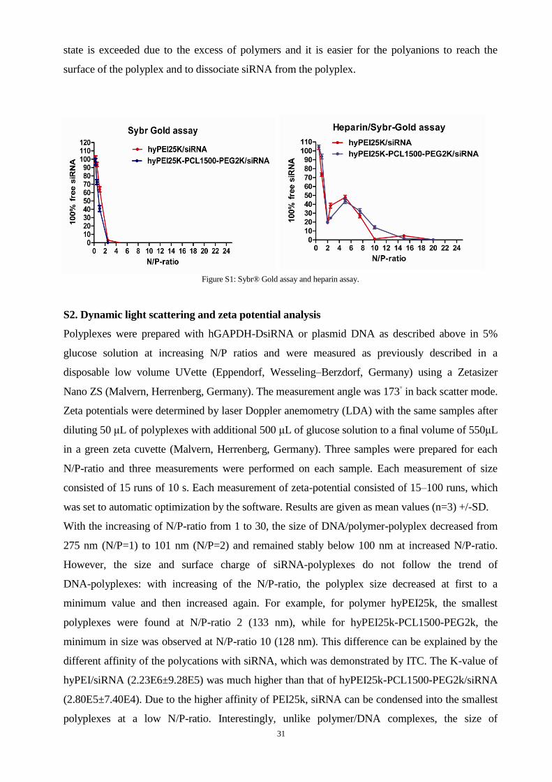
31
state is exceeded due to the excess of polymers and it is easier for the polyanions to reach the
surface of the polyplex and to dissociate siRNA from the polyplex.
Figure S1: Sybr® Gold assay and heparin assay.
S2. Dynamic light scattering and zeta potential analysis
Polyplexes were prepared with hGAPDH-DsiRNA or plasmid DNA as described above in 5%
glucose solution at increasing N/P ratios and were measured as previously described in a
disposable low volume UVette (Eppendorf, Wesseling–Berzdorf, Germany) using a Zetasizer
Nano ZS (Malvern, Herrenberg, Germany). The measurement angle was 173◦ in back scatter mode.
Zeta potentials were determined by laser Doppler anemometry (LDA) with the same samples after
diluting 50 μL of polyplexes with additional 500 μL of glucose solution to a final volume of 550μL
in a green zeta cuvette (Malvern, Herrenberg, Germany). Three samples were prepared for each
N/P-ratio and three measurements were performed on each sample. Each measurement of size
consisted of 15 runs of 10 s. Each measurement of zeta-potential consisted of 15–100 runs, which
was set to automatic optimization by the software. Results are given as mean values (n=3) +/-SD.
With the increasing of N/P-ratio from 1 to 30, the size of DNA/polymer-polyplex decreased from
275 nm (N/P=1) to 101 nm (N/P=2) and remained stably below 100 nm at increased N/P-ratio.
However, the size and surface charge of siRNA-polyplexes do not follow the trend of
DNA-polyplexes: with increasing of the N/P-ratio, the polyplex size decreased at first to a
minimum value and then increased again. For example, for polymer hyPEI25k, the smallest
polyplexes were found at N/P-ratio 2 (133 nm), while for hyPEI25k-PCL1500-PEG2k, the
minimum in size was observed at N/P-ratio 10 (128 nm). This difference can be explained by the
different affinity of the polycations with siRNA, which was demonstrated by ITC. The K-value of
hyPEI/siRNA (2.23E6±9.28E5) was much higher than that of hyPEI25k-PCL1500-PEG2k/siRNA
(2.80E5±7.40E4). Due to the higher affinity of PEI25k, siRNA can be condensed into the smallest
polyplexes at a low N/P-ratio. Interestingly, unlike polymer/DNA complexes, the size of

32
polymer/siRNA continues to increase if the N/P-ratio is further increased above the N/P ratio at
which the smallest polyplexes were formulated. At higher N/P-ratios, the PDI was also much
higher than the PDI at lower N/P-ratio.
Comparing the size distribution peaks based on volume at different N/P-ratios, we found that at
N/P-ratio 2, 94.9% of the total polyplex distribution, had a mean size of 91.91 nm. If the N/P-ratio
increased to 20, the characteristic peak of N/P-ratio 2 shifted only slightly, but contained only
35.5% of the polyplexes based on the volume distribution. Additionally, 15.4% of the polyplexes
had a mean hydrodynamic diameter of 224.8 nm, and 12.1% of the polyplexes were found in a
peak at 373.3 nm. Interestingly, in the dye quenching assay, almost no quenching could be
observed at N/P 20. It was therefore assumed that individual polyplexes carried comparatively little
siRNA at N/P 20, and that an excess of polymer was present as free polymer which can cause
aggregation of polyplexes. As a result, we observed by dynamic light scattering a highly disperse
distribution of peaks at N/P 20. Recent research of siRNA complexation by all-atom molecular
dynamics simulations also reported that at a low charge ratio or N/P-ratio, all cationic polymers
can bind to siRNA, but only a limited number of polymers can condense the siRNA at a high
charge ratio.
S3. In vitro transfection experiments with DNA
SKOV-cells were seeded with 0.2mL medium per well in 96-well plates (Nunc, Wiesbaden,
Germany) at the density of 30,000 cells/mL After 24h, 100μL medium (containing 10% serum)
plus 25μL polymer/pDNA-complex containing 0.5μg pDNA (pCMVLuc, Plasmid Factory,
Bielefeld, Germany) at different N/P-ratios were placed in each well. After 4h of incubation at
37.0◦C in humidified atmosphere with 5% CO2, the medium was replaced with 200μL fresh
medium containing 10% serum. After another 44h, cells were lysed in 100μL cell culture lysis

33
buffer (Promega, Mannheim, Germany) for 15min at 37 ◦C. A volume of 25μL of the cell lysate
was added in each well of an opaque 96-well plate (Perkin Elmer, Rodgau-Jügesheim). Luciferase
activity was quantified by injection of 50μL luciferase-assay-buffer, containing 10mM luciferin
(Sigma–Aldrich, Taufkirchen, Germany). The resulting photons were measured as relative light
units (RLU) with a plate luminometer (LumiSTAR Optima, BMG Labtech GmbH, Offenburg,
Germany). Protein concentration was determined using a Bradford BCA assay (BioRad, Munich,
Germany).
Figure S3: plasmid DNA transfection efficiency of hyPEI25kDa and copolymer hyPEI25k-PCL1.5k-PEG2k.
2.8 References
1.Riehemann, K.; Schneider, S. W.; Luger, T. A.; Godin, B.; Ferrari, M.; Fuchs, H., Nanomedicine--challenge
and perspectives. Angew Chem Int Ed Engl 2009, 48 (5), 872-97.
2.Petros, R. A.; DeSimone, J. M., Strategies in the design of nanoparticles for therapeutic applications. Nature
Reviews Drug Discovery 2010, 9 (8), 615-627.
3.Lollo, C. P.; Banaszczyk, M. G.; Chiou, H. C., Obstacles and advances in non-viral gene delivery. Curr Opin
Mol Ther 2000, 2 (2), 136-42.
4.Park, T. G.; Jeong, J. H.; Kim, S. W., Current status of polymeric gene delivery systems. Advanced Drug
Delivery Reviews 2006, 58 (4), 467-486.
5.Fire, A.; Xu, S.; Montgomery, M. K.; Kostas, S. A.; Driver, S. E.; Mello, C. C., Potent and specific genetic
interference by double-stranded RNA in Caenorhabditis elegans. Nature 1998, 391 (6669), 806-11.
6.Ferrari, M., Cancer nanotechnology: opportunities and challenges. Nat Rev Cancer 2005, 5 (3), 161-71.
7.Zhang, L.; Gu, F. X.; Chan, J. M.; Wang, A. Z.; Langer, R. S.; Farokhzad, O. C., Nanoparticles in medicine:
therapeutic applications and developments. Clin Pharmacol Ther 2008, 83 (5), 761-9.
8.Steuber, H.; Czodrowski, P.; Sotriffer, C. A.; Klebe, G., Tracing changes in protonation: a prerequisite to
factorize thermodynamic data of inhibitor binding to aldose reductase. J Mol Biol 2007, 373 (5), 1305-20.
9.Pavan, G. M.; Danani, A.; Pricl, S.; Smith, D. K., Modeling the Multivalent Recognition between Dendritic
Molecules and DNA: Understanding How Ligand "Sacrifice" and Screening Can Enhance Binding. Journal of
the American Chemical Society 2009, 131 (28), 9686-9694.
10.Pavan, G. M.; Kostiainen, M. A.; Danani, A., Computational Approach for Understanding the Interactions of
UV-Degradable Dendrons with DNA and siRNA. Journal of Physical Chemistry B 2010, 114 (17), 5686-5693.
34
11.Doni, G.; Kostiainen, M. A.; Danani, A.; Pavan, G. M., Generation-Dependent Molecular Recognition
Controls Self-Assembly in Supramolecular Dendron-Virus Complexes. Nano Letters 2011, 11 (2), 723-728.
12.Gary, D. J.; Puri, N.; Won, Y. Y., Polymer-based siRNA delivery: Perspectives on the fundamental and
phenomenological distinctions from polymer-based DNA delivery. Journal of Controlled Release 2007, 121
(1-2), 64-73.
13. Arnott, S., Principles of Nucleic-Acid Structure - Saenger,W. Nature 1984, 312 (5990), 174-174.
14.Noy, A.; Perez, A.; Lankas, F.; Javier Luque, F.; Orozco, M., Relative flexibility of DNA and RNA: a
molecular dynamics study. J Mol Biol 2004, 343 (3), 627-38.
15.Pavan, G. M.; Albertazzi, L.; Danani, A., Ability to adapt: different generations of PAMAM dendrimers show
different behaviors in binding siRNA. J Phys Chem B 2010, 114 (8), 2667-75.
16.Merkel, O. M.; Mintzer, M. A.; Librizzi, D.; Samsonova, O.; Dicke, T.; Sproat, B.; Garn, H.; Barth, P. J.;
Simanek, E. E.; Kissel, T., Triazine dendrimers as nonviral vectors for in vitro and in vivo RNAi: the effects of
peripheral groups and core structure on biological activity. Mol Pharm 2010, 7 (4), 969-83.
17.Koch, C.; Heine, A.; Klebe, G., Tracing the Detail: How Mutations Affect Binding Modes and
Thermodynamic Signatures of Closely Related Aldose Reductase Inhibitors. Journal of Molecular Biology 2011,
406 (5), 700-712.
18.Doni, G.; Kostiainen, M. A.; Danani, A.; Pavan, G. M., Generation-dependent molecular recognition controls
self-assembly in supramolecular dendron-virus complexes. Nano Lett 2011, 11 (2), 723-8.
19.Zheng, M.; Liu, Y.; Samsonova, O.; Endres, T.; Merkel, O.; Kissel, T., Amphiphilic and biodegradable
hy-PEI-g-PCL-b-PEG copolymers efficiently mediate transgene expression depending on their graft density. Int J
Pharm 2011.
20.Merkel, O. M.; Beyerle, A.; Librizzi, D.; Pfestroff, A.; Behr, T. M.; Sproat, B.; Barth, P. J.; Kissel, T.,
Nonviral siRNA delivery to the lung: investigation of PEG-PEI polyplexes and their in vivo performance. Mol
Pharm 2009, 6 (4), 1246-60.
21.Van Rompaey, E.; Engelborghs, Y.; Sanders, N.; De Smedt, S. C.; Demeester, J., Interactions between
oligonucleotides and cationic polymers investigated by fluorescence correlation spectroscopy. Pharmaceutical
Research 2001, 18 (7), 928-936.
22.Park, J. W.; Bae, K. H.; Kim, C.; Park, T. G., Clustered magnetite nanocrystals cross-linked with PEI for
efficient siRNA delivery. Biomacromolecules 2011, 12 (2), 457-65.
23.Wu, Y.; Wang, W. W.; Chen, Y. T.; Huang, K. H.; Shuai, X. T.; Chen, Q. K.; Li, X. X.; Lian, G. D., The
investigation of polymer-siRNA nanoparticle for gene therapy of gastric cancer in vitro. International Journal of
Nanomedicine 2010, 5, 129-136.
24.Dimitrova, M.; Affolter, C.; Meyer, F.; Nguyen, I.; Richard, D. G.; Schuster, C.; Bartenschlager, R.; Voegel,
J. C.; Ogier, J.; Baumert, T. F., Sustained delivery of siRNAs targeting viral infection by cell-degradable
multilayered polyelectrolyte films. Proc Natl Acad Sci U S A 2008, 105 (42), 16320-5.
25.Urban-Klein, B.; Werth, S.; Abuharbeid, S.; Czubayko, F.; Aigner, A., RNAi-mediated gene-targeting through
systemic application of polyethylenimine (PEI)-complexed siRNA in vivo. Gene Ther 2005, 12 (5), 461-6.
26.Godbey, W. T.; Wu, K. K.; Mikos, A. G., Size matters: molecular weight affects the efficiency of
poly(ethylenimine) as a gene delivery vehicle. J Biomed Mater Res 1999, 45 (3), 268-75.
35
27.Merkel, O. M.; Zheng, M.; Mintzer, M. A.; Pavan, G. M.; Librizzi, D.; Maly, M.; Hoffken, H.; Danani, A.;
Simanek, E. E.; Kissel, T., Molecular modeling and in vivo imaging can identify successful flexible triazine
dendrimer-based siRNA delivery systems. J Control Release 2011, 153 (1), 23-33.
28.Pavan, G. M.; Mintzer, M. A.; Simanek, E. E.; Merkel, O. M.; Kissel, T.; Danani, A., Computational Insights
into the Interactions between DNA and siRNA with "Rigid" and "Flexible" Triazine Dendrimers.
Biomacromolecules 2010, 11 (3), 721-730.
29.Jensen, L. B.; Mortensen, K.; Pavan, G. M.; Kasimova, M. R.; Jensen, D. K.; Gadzhyeva, V.; Nielsen, H. M.;
Foged, C., Molecular characterization of the interaction between siRNA and PAMAM G7 dendrimers by SAXS,
ITC, and molecular dynamics simulations. Biomacromolecules 2010, 11 (12), 3571-7.
30.Klebe, G.; Steuber, H.; Czodrowski, P.; Sotriffer, C. A., Tracing changes in protonation: A prerequisite to
factorize thermodynamic data of inhibitor binding to aldose reductase. Journal of Molecular Biology 2007, 373
(5), 1305-1320.
31.Klebe, G.; Koch, C.; Heine, A., Tracing the Detail: How Mutations Affect Binding Modes and
Thermodynamic Signatures of Closely Related Aldose Reductase Inhibitors. Journal of Molecular Biology 2011,
406 (5), 700-712.
36
Chapter 3
AMPHIPHILIC AND BIODEGRADABLE hy-PEI-g-PCL-b-PEG
COPOLYMERS EFFICIENTLY MEDIATE TRANSGENE EXPRESSION
DEPENDING ON THEIR GRAFT DENSITY
Published in International Journal of Pharmaceutics, Theme Issue “Non-viral gene medicine”.
(2011) 427, 80-7.
Mengyao Zheng1, Yu Liu
1, Olga Samsonova
1, Thomas Endres, Olivia Merkel, and Thomas
Kissel*
1Both authors contributed equally to this work.
Author contributions
T. K. guided and directed the research. Y. L. synthesized and characterized the polymers. M. Z.
designed the measurements and carried out all of the biological and biophysical experiments. M. Z.
and O. M. M. analysed the experimental data.

37
3.1 Abstract
Novel biodegradable amphiphilic copolymers
hy-PEI-g-PCL-b-PEG were prepared by grafting
PCL-b-PEG chains onto hyper-branched
poly(ethylene imine) as non-viral gene delivery
vectors. Our investigations focused on the
influence of graft densities of PCL-b-PEG
chains on physico-chemical properties, DNA
complexation and transfection efficiency. We
found that the transfection efficiencies of these
polymers increased at first towards an optimal
graft density (n=3) and then decreased. The
buffer-capacity-test showed almost exactly the
same tendency as transfection efficiency.
Cytotoxicity (MTT-assay) depended on the
cooperation of PEG molecular weight and graft
density of PCL-b-PEG chains. With increasing
the graft density, cytotoxicity, zeta-potential, affinity with DNA, stability of the polyplexes and
CMC-values were reduced strongly and regularly. Increasing the excess of polymer over DNA was
shown to result in a decrease of the observed particle size to 100–200 nm. The application of these
copolymers in siRNA transfection will be also under investigation and efficient transfection
efficiency is expected with polymer hyPEI25K-(PCL570-PEG5K)3 and polymer
hyPEI25K-(PCL570-PEG5K)5.
Keyword:Transfection;Non-viral gene delivery;Biodegradable copolymer;Hyper-branched
poly(ethylene imine) (hyPEI);Graft density;Buffer-capacity
3.2 Introduction
Thanks to advances in molecular biology and genomic research, numerous diseases have been
given a genetic identity for which gene therapy may provide a possible treatment (Pawliuk et al.,
2001; von Laer et al., 2006; Wong et al., 2006). For viral gene delivery techniques, viruses can be
transformed into gene-delivery vehicles by replacing parts of the genome of a virus with a
therapeutic gene (Pack et al., 2005). But viral vectors could induce cancer (Check, 2002;
Hacein-Bey-Abina et al., 2003), and initiate an immunogenic response which has led to a fatal
outcome (Marshall, 2000). Instead of viral gene delivery techniques, non-viral gene delivery
Graphical Abstract: Schematic conformation of polyplexes
from hyPEI25k-(PCL570-PEG5k)n: at graft densities of 1 to
5, PEG-PCL chains stay in separated blocks; but it was
assumed to form a continuous corona at n=20.

38
techniques have been investigated, which are considered as a safer, less pathogenic and
immunogenic gene delivery alternative (Wong et al., 2007). Studies demonstrated that polymeric
vectors might be advantageous over viruses and liposomes (Merdan et al., 2002) for gene delivery,
regarding safety, immunogenicity, mutagenicity and production costs (Han et al., 2000).
Hyper-branched polyethylenimine (hy-PEI) is one of the most successfully used cationic
polymers for gene delivery both in vitro and in vivo (Boussif et al., 1995; Godbey et al., 1999).
HyPEI can condense DNA via electrostatic interaction to particles with sizes compatible with
cellular uptake while providing steric protection from nuclease degradation (Bielinska et al.,
1997). HyPEI has a unique property of endosomal buffering capacity due to protonable amino
groups. This function causes osmotic swelling and subsequent endosomal disruption (Boussif et
al., 1995), thus permitting the escape of endocytosed materials. However, PEI has a relatively
high cytotoxicity due to the high density of cationic groups, especially at high molecular weights
(Fischer et al., 1999; Kunath et al., 2003). PEI-PEG copolymers are less toxic than PEI (Sung et
al., 2003). However they are not able to condense DNA into small particles less than 200 nm,
which is critical for cell uptake and transfection efficiency (Park et al., 2005; Pun et al., 2004).
Another drawback of PEI-PEG are the non-biodegradable bonds between PEI and PEG chains.
Polymers that are not eliminated from the circulation may accumulate in tissues and cells. To
resolve this problem, biodegradable hy-PEI-g-(PCL-b-PEG)n copolymers were synthesized
(Shuai et al., 2003). Poly(caprolactone) (PCL) acts as a linker between PEI and PEG to increase
the biodegradability of the copolymers. Hydrophobic PCL improves also the overall permeability
of the complexes through the cell membranes. Another advantage of PCL is the shielding of the
positive charges and the condensation of the DNA into small complexes of 100 nm (Han et al.,
2001; Kono et al., 2005; Tian et al., 2007; Wang et al., 2002).
The copolymers hy-PEI-g-(PCL-b-PEG)n have been studied so far as potential gene delivery
systems (Liu et al., 2009; Shuai et al., 2003). These investigations were limited to the discussion
of the influence of PEI, PCL and PEG chain lengths. Nevertheless, the influence of graft density
has not been studied systematically. In this report, a panel of hyPEI25K-(PCL570-PEG5k)n
block copolymers was synthesized to elucidate the effect of graft density on physicochemical
properties, DNA complexation and biological activities as non-viral gene delivery vectors. It is
39
commonly believed that the molecular weight of PEI most suitable for gene transfer ranges
between 5 and 25 kDa. Higher molecular weights lead to increased cytotoxicity (Fischer et al.,
2003), presumably due to aggregation to huge clusters of the cationic polymer on the outer cell
membrane, which thereby induce necrosis (Freshney, 2005). To study their physicochemical
properties and complexation with DNA, measurements of size and zeta-potential, heparin-assay,
critical micelle concentration (CMC) and buffer-capacity were performed. The physico-chemical
properties of these complexes were then compared with in vitro results of cytotoxicity tests
(MTT), confocal laser scanning microscopy (CLSM) and transfection experiments. These results
provide a basis for the rational design of block copolymers as gene delivery systems.
3.3. Materials and Methods
Materials
Hy-PEI with molecular weight of 25 kDa was obtained from BASF. Poly (ethylene glycol)
mono-methyl ether (mPEG) (5 kDa) and ε-caprolactone were purchased from Fluka (Taufkirchen,
Germany), and all other chemicals were obtained from Sigma-Aldrich (Steinheim, Germany).
Hy-PEI-PCL-mPEG was synthesized as reported previously (Liu et al., 2009). DNA from herring
testes (Type XIV, 0.3-6.6 MDa, 400-10 000 bp) was from bought from Sigma (Steinheim,
Germany), and Luciferase-Plasmid (pCMV-Luc) (Lot No.:PF461-090623) was amplified by The
Plasmid Factory (Bielefeld, Germany).
Polyplex formation
All complexes of DNA and polymer were prepared freshly before use. Luciferase-Plasmid and
DNA from herring testes were stored at -20°C. Before use, 5% glucose solution and other buffer
solutions were filtered freshly through 0.20 μm pore sized filters (Nalgene® syringe filter,
Sigma–Aldrich, Taufkirchen, Germany). The volume of a 1 mg/mL (based on hyPEI25k) polymer
stock solution required for a certain N/P ratio (=Nitrogen/Phosphorus-ratios) was calculated as
follows (Liu et al., 2009):
VDNA = (Ccopolymer * Vcopolymer * 330) / (CDNA * 43 * N/P)
Ccopolymer = the concentration of the stock copolymer

40
CDNA = the concentration of the stock DNA solution
A certain amount of polymer stock solution was diluted with 5% glucose solution or other buffer
solutions to a final volume of 50 μL, which was mixed with an equal volume of diluted DNA
aliquots in microcentrifuge tubes by pippetting (IKA, Stauffen), and incubated for 20 min before
use for complex and equilibrium formation.
Cell culture
Cells were seeded at a density of 3.5 * 103 cells/cm
2 in dishes (10 cm diameter, Nunclon Dishes,
Nunc, Wiesbaden, Germany) and incubated at 37°C in humidified 5% CO2 atmosphere
(CO2-Incubator, Integra Biosciences, Fernwald, Germany). Medium (Dulbecco’s modified Eagle
medium, supplemented with 10% serum) was exchanged every 2 days. Cells were split after 7
days, when confluence was reached.
Measurement of size and zeta-potential
Three buffer-solutions (5% glucose, pH 6.6; 10 mM TE-buffer, pH 9.0; 15 mM acetate-buffer, pH
5.5) were used for the measurement of size and zeta-potential, which were monitored with a
Malvern Zetasizer Nano ZS (Marvern Instrument, Worcestershire, UK) at 25 °C. The measurement
angle was 173 ° in backscatter mode. Following size measurements, zeta-potential measurements
were performed with the same samples after diluting 50 μL of polyplexes with additional 500 μL
of buffer solution to a final volume of 550 μL with a DNA concentration of 1.82 ng/μL. Three
samples were prepared for each N/P-ratio and three measurements were performed on each
sample. Each measurement of size consisted of 15 runs of 10 sec. Each measurement of
zeta-potential consisted of 15–100 runs, which was set to automatic optimization by the software.
Buffer-capacity
Titration studies were performed to determine the buffer-capacity of the studied polymers.
Therefore, 0.4 mL of aqueous polymer solution with the concentration of 2.5 mg/mL was titrated
with standard 0.1 N HCl, until the pH of the polymer solutions decreased nearby pH=2.5. The
pH-value was detected with a pH-meter (Hanna PH210 Microprocessor pH Meter) and an
electrode (Inlab, Mettler Toledo, Schwerzenbach, Switzerland) at 25 °C.
41
Critical micelle concentration (CMC)
In this experiment, the surface tension with increased polymer concentration in water was
measured with a tensionmeter (Krüss Tensionmeter Control Panel; K11-MK3) at 25 °C. Data
correlation with the polymer structure was performed using “Origin 7.0” (Origin-Labsoftware,
Northampton, USA).
Heparin-assay
The effect of heparin on the stability of complexes was evaluated by means of the change in
fluorescence intensity obtained with the fluorescent intercalating probe sybr-gold (Creusat et al.,
2010). To study the effect of pH on the stability of complexes, polyplexes were prepared in
solutions with different pH and ionic strength. Heparin solution (150000 IE/g, Serva, Pharm.,
USPXV2, Merck, Darmstadt, Germany) was diluted to a concentration of 0.521 mg/mL.
Increasing amounts (0 – 17.5 µL) of heparin were added to the 96-well plate (Perkin Elmer,
Rodgau-Jügesheim) where each well contained 200 μL of polymer/HT-DNA-complexes at N/P 10,
Subsequently, 20 μL diluted sybr-gold-solution (Invitrogen, Karlsruhe, Germany) were added.
After 20 min of incubation at 25 °C, fluorescence was directly detected with a fluorescence plate
reader (BMG Labtech GMBH, Offenburg, Germany) at 495 nm excitation and 537 nm emission.
MTT-assay
In vitro cytotoxicity tests of the copolymers were performed by MTT-assays. Pure polymers were
selected instead of DNA polyplexes to measure the cytotoxicity in a “worst case scenario” since it
has been reported that the cytotoxicity was reduced when polymers were complexed with DNA
(Godbey et al., 1999).The assays were performed as previously described (Liu et al., 2009).
Briefly, L929 cells were seeded in 96-well cell culture-coated microtiter plates at the density of
8,000 cells/well and incubated in DMEM low glucose (PAA, Cölbe, Germany) supplemented with
10% fetal calf serum (Cytogen, Sinn, Germany) in humidfied atmosphere with 5% CO2 at 37 °C
for 24 h prior to the treatment with polymer solutions of increasing concentration (from 9.77E-4
mg/mL to 0.5 mg/mL). After 24 h, the medium was replaced with 200 μL serum free medium and
20 μL 3-(4,5-dimethylthiazol-2-yl)-2,5-diphenyltetrazolium bromide (MTT, Sigma–Aldrich,

42
Germany) reaching a final concentration of 0.5 mg MTT/mL. Cells were incubated for another 4 h
before 200 μL of dimethylsulfoxide (DMSO) was added to dissolve the purple formazane product.
The absorption was quantified using a plate reader (Titertek plus MS 212, ICN, Germany) at
wavelengths of 570 nm and 690 nm. The IC50 was calculated as the polymer concentration which
inhibits growth of 50% of cells relative to non-treated control cells.
Confocal laser scanning microscopy (CLSM)
MeWo-cells were seeded in 8 well-chamberslides (Lab-Tek; Rochester, NY, USA) at 50,000
cells/well and incubated for 24 h in DMEM high glucose (PAA, Cölbe, Germany) supplemented
with 10% fetal calf serum (Cytogen, Sinn, Germany) in humidified atmosphere with 5% CO2 at 37
°C. The DNA labeling steps were performed at room temperature and in the dark to protect
fluorescent markers. YOYO-1 stock solution (Invitrogen, Karlsruhe, Germany) was diluted 50-fold
with TE-buffer and 30 μL of DAPI stock solution (6 µg/mL, Molecular Probes, Eugene, OR, USA)
was diluted with 1 mL PBS. Plasmid-DNA (pDNA) was incubated with YOYO-1 solution for 30
minutes at a weight ratio of 1:15. Complexes of YOYO-1-labeled pDNA were formed as usual by
incubation with polymer solution at N/P 15 in 5% glucose solution for 20 minutes followed by
addition of 25 μL of polyplex solution, containing 0.5 μg plasmid-DNA, to 375 μL fresh culture
medium with 10% FCS in each well. After incubation for 4 h, each chamber was washed twice
with 0.5 mL PBS, and the cells were then fixed by incubating each chamber for 20 minutes with
0.5 mL of 4% paraformaldehyde in PBS (4% PFA). Subsequently, 100 μL DAPI-solution was
added per chamber and incubated for another 20 minutes in the dark. The cells were washed three
times with 0.5 mL PBS before being fixed with Fluorsafe (Calbiochem, San Diego, USA) and
covered with a No. 1.5 thickness cover slip (Menzel Gläser, Braunschweig, Germany). YOYO-1
labeled DNA was excited with a 488 nm argon laser, while DAPI-stained chromosomal DNA was
excited with an enterprise laser with an excitation wavelength of 364 nm, and CLSM was
performed by using a 385 nm long pass filter and a band-pass filter of 505–530 nm in the
single-track mode (Axiovert 100 M and CLSM 510 Scanning Device; Zeiss, Oberkochem,
Germany).
In vitro transfection experiments with DNA
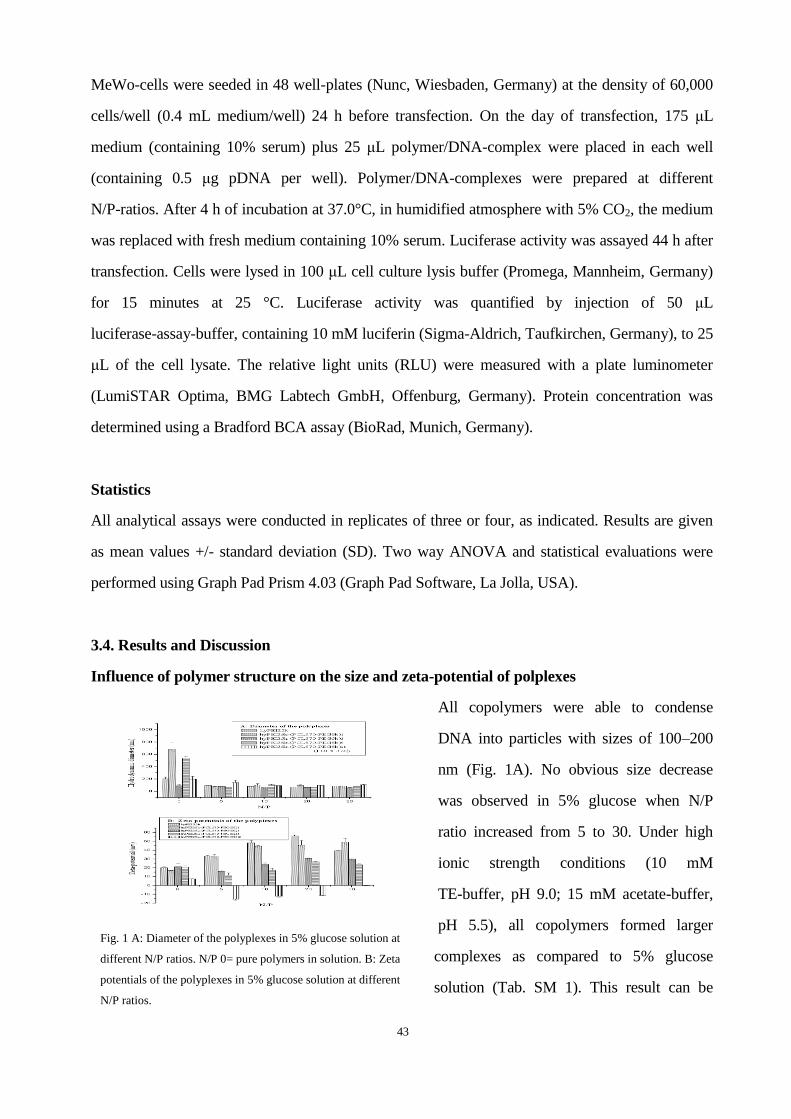
43
MeWo-cells were seeded in 48 well-plates (Nunc, Wiesbaden, Germany) at the density of 60,000
cells/well (0.4 mL medium/well) 24 h before transfection. On the day of transfection, 175 μL
medium (containing 10% serum) plus 25 μL polymer/DNA-complex were placed in each well
(containing 0.5 μg pDNA per well). Polymer/DNA-complexes were prepared at different
N/P-ratios. After 4 h of incubation at 37.0°C, in humidified atmosphere with 5% CO2, the medium
was replaced with fresh medium containing 10% serum. Luciferase activity was assayed 44 h after
transfection. Cells were lysed in 100 μL cell culture lysis buffer (Promega, Mannheim, Germany)
for 15 minutes at 25 °C. Luciferase activity was quantified by injection of 50 μL
luciferase-assay-buffer, containing 10 mM luciferin (Sigma-Aldrich, Taufkirchen, Germany), to 25
μL of the cell lysate. The relative light units (RLU) were measured with a plate luminometer
(LumiSTAR Optima, BMG Labtech GmbH, Offenburg, Germany). Protein concentration was
determined using a Bradford BCA assay (BioRad, Munich, Germany).
Statistics
All analytical assays were conducted in replicates of three or four, as indicated. Results are given
as mean values +/- standard deviation (SD). Two way ANOVA and statistical evaluations were
performed using Graph Pad Prism 4.03 (Graph Pad Software, La Jolla, USA).
3.4. Results and Discussion
Influence of polymer structure on the size and zeta-potential of polplexes
All copolymers were able to condense
DNA into particles with sizes of 100–200
nm (Fig. 1A). No obvious size decrease
was observed in 5% glucose when N/P
ratio increased from 5 to 30. Under high
ionic strength conditions (10 mM
TE-buffer, pH 9.0; 15 mM acetate-buffer,
pH 5.5), all copolymers formed larger
complexes as compared to 5% glucose
solution (Tab. SM 1). This result can be
Fig. 1 A: Diameter of the polyplexes in 5% glucose solution at
different N/P ratios. N/P 0= pure polymers in solution. B: Zeta
potentials of the polyplexes in 5% glucose solution at different
N/P ratios.

44
explained by the shielding-effect of glucose molecules (Petersen et al., 2002). On the other hand,
the polyplexes can aggregate in the buffer-solutions due to the reduced surface charge and lack of
repulsion of the polymers (Petersen et al., 2002). The results of size measurements yielded
comparable values as recently reported (Liu et al., 2009).
The zeta-potential was reduced with an increasing number n of PCL570-PEG5k segments, and was
always highest in non-buffered glucose solution (Fig. 1A and Tab. SM 1). All polyplexes except
hyPEI25k-(PCL570-PEG5k)20 showed a positively charged surface at all buffer conditions. Due to
the high graft density of hyPEI25k-(PCL570-PEG5k)20, the polymer loses its ability to condense
DNA into a stable polyplexes as most positive charges of hyPEI are shielded, resulting in
negatively charged polyplexes. It is assumed that the DNA remains only on the surface of this
polymer (Scheme 1).
Interestingly, no further increase of the zeta-potential was observed when increasing the N/P ratio
above 20, except for the polymer with graft density of 20. Therefor we hypothesize that for
polymers with a graft density ranged between 1 and 5 at N/P 20 a polymer concentration is reached
above which the additional polymer does not contribute to the condensation of DNA but is rather
present as free polymer.
Influence of polymer structure on the buffer-capacity
The most commonly used
pH-sensitive excipients for gene
delivery that exhibit the
so-called “proton-sponge effect”
are polymers such as PEI with
protonatable amino groups with
5< pKa <7 (Behr, 1997). The
polymers with high
buffer-capacity increase the ion
concentration in the endosome
and ultimately cause osmotic Fig. 2: Titration curve of aqueous polymer solution with standard 0.1 N HCl.

45
swelling and rupture of the endosome membrane, which releases the polyplexes into the cytosol.
Figure 2 shows the the buffer-capacity of the polymers which decreased in the following order:
hyPEI25k-(PCL570-PEG5k)3>hyPEI25k-(PCL570-PEG5k)5>hyPEI25K-(PCL570-PEG5k)1=hyP
EI25k>hyPEI25k-(PCL570-PEG5k)20. With these results, it was shown that a low grafting degree of
PEI with PCL-PEG segments can increase the accessibility of amines to be protonated, thereby
increasing the buffer capacity. Thus, stability of the complexes could additionally be increased.
Influence of polymer structure on the CMC
The critical micelle concentration (CMC) is defined as the concentration where the interfacial
tension reached a minimum. It is extremely valuable not only to predict the micelle-forming
capacity but also to determine the stability of the polymeric micelles, which was believed to play a
crucial role in DNA transfection. In general, all amphiphilic polymers were able to form micelles
at low concentrations (Tab. 1). For the copolymers with graft densities of 5 and 20, the CMC was
reached at lower concentrations (8.7*E-10 mol/L and 4.6*E-10 mol/L) in water. The results of
CMC measurements in water were expected to be a function of the PCL molecular weight.
Generally, increasing the amounts of hydrophobic segments decreases the CMC of copolymers.
This principle could clearly be observed in our study where CMC-values decreased exponentially
with the increase of PCL molecular weight, as shown in Figure 3. The tendency towards
aggregation may also be affected by the presence of shielding components, for example glucose
molecules or PEG chains in the copolymers, which may also decrease interactions between
individual complexes as well as interactions between complexes and blood components in the
systemic circulation (Petersen et al., 2002). Besides the shielding-effects of the PEG-PCL chains,
the faster degradation of the polymers with higher graft density can also change the CMC tendency
in base (Liu et al., 2010).

46
Tab. 1: CMC-values in water.
polymer CMC in water (pH 7.0)
( 10E-9 mol/L)
hyPEI25k none
hyPEI25k-(PCL570-PEG5k)1 16.76 ± 0.14
hyPEI25k-(PCL570-PEG5k)3 2.30 ± 0.11
hyPEI25k-(PCL570-PEG5k)5 0.87 ± 0.09
hyPEI25k-(PCL570-PEG5k)20 0.46 ± 0.02
Fig. 3: The CMC-values decreased with the increase of the percentage of PCL molecular weight in the copolymers.
Influence of polymer structure on the stability of polymer-DNA-complexes
Polymer/DNA-complexes, which are built
due to electrostatic interaction, can be
dissociated easily with the competing
polyanion heparin (Moret et al., 2001). The
release of DNA from complexes in the
presence of heparin is summarized in Figure
SM 1. The dissociation profiles all exhibit
significant dependency on the heparin
concentration. Another general trend is that all
polymers display more stable complexes in
acetate buffer as compared to other solutions.
Fig. 4: DNA dissociation from complexes by heparin competition
in different buffers.

47
Interestingly, hy-PEI25k-(PCL570-mPEG5k)1, released only 50% of the DNA load at 0.075 mg/ml
heparin, the highest concentration tested. However, unstable complexes were obtained in TE buffer
for all polymers. This can be explained by the fact that the amino groups in the polymers were
protonated under acidic conditions, while deprotonated in TE buffer. Comparing the diagrams in
Figure 4, no obvious difference was observed amongst the polymers in 5% glucose and TE buffer,
while the stability of complexes was increased PEG-PCL-grafting in acetate buffer at low heparin
concentrations. This may be caused by the shielding-effect of PCL-mPEG and a resulting
inaccessibility for heparin. Although our data emphasize the importance of protonation of the
polymer for stable interaction with DNA, a certain amount of lipophilic segments can even increase
the stability as shown by hyPEI25k-(PCL570-PEG5k)1. Indeed, reduced complexation ability of
copolymers may additionally facilitate the unpacking of the vector inside the cell, and a balance
between DNA-complexation and DNA-release is necessary. Many reports described similar
observations of increased transfection efficiency with reduction of positive charges (Banaszczyk et
al., 1999; Schaffer et al., 2000).
Influence of polymer structure on the cytotoxicity
To evaluate the cytotoxicity of hy-PEI-g-PCL-b-mPEG copolymers in L929 cells, MTT-assays
were performed. Considering the IC50 values, the cytotoxicity was clearly reduced with
increasing of the graft density of the PCL570-PEG5k segments (Fig.5). PEG is very hydrophilic
and considered to be safe by the FDA (Sung et al., 2003). By grafting PEG onto PEI, the toxicity
of hyPEI25k is known to be reduced (Petersen et al., 2002). At the same time, the PCL segment
is hydrophobic and shields the positive charges from hyPEI25K. On the other hand, the addition
of PCL-segments increases also the degradation of the copolymers (Liu et al., 2010). It was
interestingly found in Figure 6 that the IC50-values increased proportional as a function of the
parameter: (percent of PEG molecular weight in copolymer) × (graft density n). This result
provides a basis for the rational design of block copolymer with low cytotoxicity. But on the
other hand, with the decrease of positive charges on the polymers, the stability of the
polymer/DNA-complex and the interaction with negatively charged cell membranes can be
reduced. It was therefore hypothesized that a low graft density would be advantageous for
transfection.
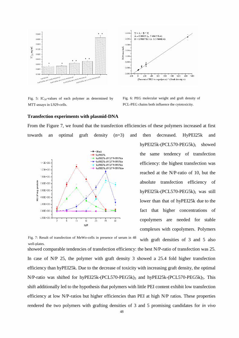
48
Transfection experiments with plasmid-DNA
From the Figure 7, we found that the transfection efficiencies of these polymers increased at first
towards an optimal graft density (n=3) and then decreased. HyPEI25k and
hyPEI25k-(PCL570-PEG5k)1 showed
the same tendency of transfection
efficiency: the highest transfection was
reached at the N/P-ratio of 10, but the
absolute transfection efficiency of
hyPEI25k-(PCL570-PEG5k)1 was still
lower than that of hyPEI25k due to the
fact that higher concentrations of
copolymers are needed for stable
complexes with copolymers. Polymers
with graft densities of 3 and 5 also
showed comparable tendencies of transfection efficiency: the best N/P-ratio of transfection was 25.
In case of N/P 25, the polymer with graft density 3 showed a 25.4 fold higher transfection
efficiency than hyPEI25k. Due to the decrease of toxicity with increasing graft density, the optimal
N/P-ratio was shifted for hyPEI25k-(PCL570-PEG5k)3 and hyPEI25k-(PCL570-PEG5k)5. This
shift additionally led to the hypothesis that polymers with little PEI content exhibit low transfection
efficiency at low N/P-ratios but higher efficiencies than PEI at high N/P ratios. These properties
rendered the two polymers with grafting densities of 3 and 5 promising candidates for in vivo
Fig. 5: IC50-values of each polymer as determined by
MTT-assays in L929-cells.
Fig. 6: PEG molecular weight and graft density of
PCL-PEG chains both influence the cytotoxicity.
Fig. 7: Result of transfection of MeWo-cells in presence of serum in 48
well-plates.
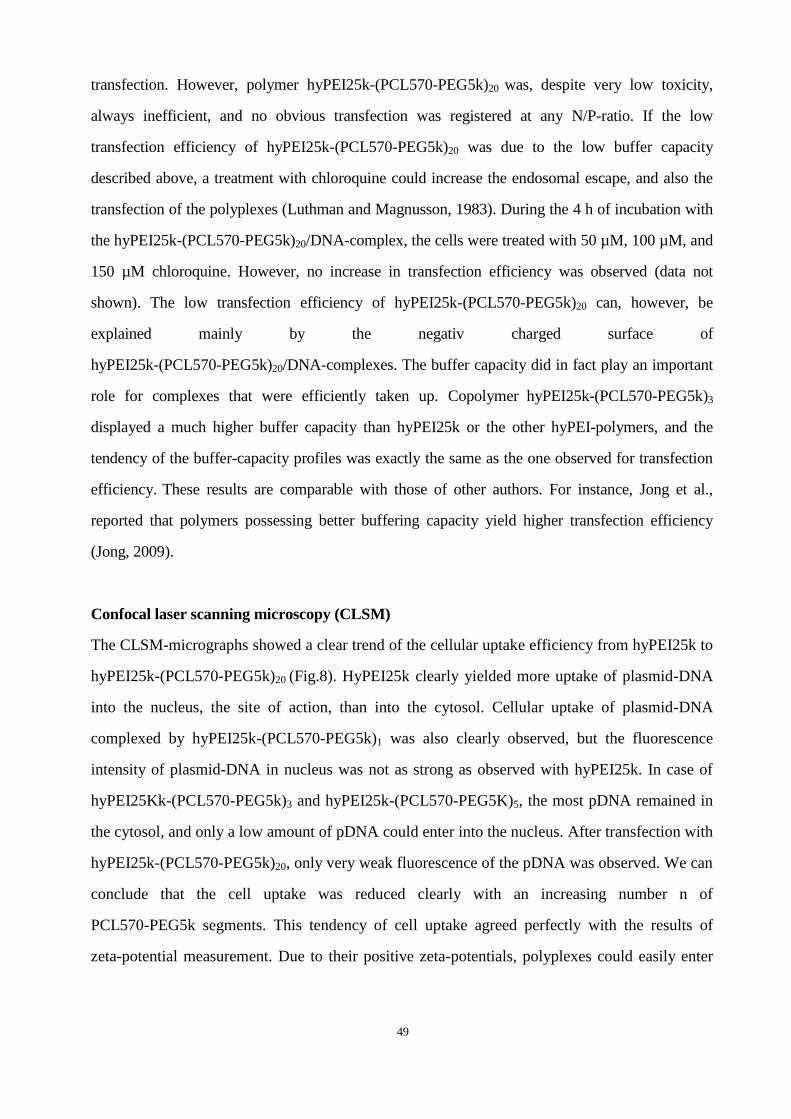
49
transfection. However, polymer hyPEI25k-(PCL570-PEG5k)20 was, despite very low toxicity,
always inefficient, and no obvious transfection was registered at any N/P-ratio. If the low
transfection efficiency of hyPEI25k-(PCL570-PEG5k)20 was due to the low buffer capacity
described above, a treatment with chloroquine could increase the endosomal escape, and also the
transfection of the polyplexes (Luthman and Magnusson, 1983). During the 4 h of incubation with
the hyPEI25k-(PCL570-PEG5k)20/DNA-complex, the cells were treated with 50 µM, 100 µM, and
150 µM chloroquine. However, no increase in transfection efficiency was observed (data not
shown). The low transfection efficiency of hyPEI25k-(PCL570-PEG5k)20 can, however, be
explained mainly by the negativ charged surface of
hyPEI25k-(PCL570-PEG5k)20/DNA-complexes. The buffer capacity did in fact play an important
role for complexes that were efficiently taken up. Copolymer hyPEI25k-(PCL570-PEG5k)3
displayed a much higher buffer capacity than hyPEI25k or the other hyPEI-polymers, and the
tendency of the buffer-capacity profiles was exactly the same as the one observed for transfection
efficiency. These results are comparable with those of other authors. For instance, Jong et al.,
reported that polymers possessing better buffering capacity yield higher transfection efficiency
(Jong, 2009).
Confocal laser scanning microscopy (CLSM)
The CLSM-micrographs showed a clear trend of the cellular uptake efficiency from hyPEI25k to
hyPEI25k-(PCL570-PEG5k)20 (Fig.8). HyPEI25k clearly yielded more uptake of plasmid-DNA
into the nucleus, the site of action, than into the cytosol. Cellular uptake of plasmid-DNA
complexed by hyPEI25k-(PCL570-PEG5k)1 was also clearly observed, but the fluorescence
intensity of plasmid-DNA in nucleus was not as strong as observed with hyPEI25k. In case of
hyPEI25Kk-(PCL570-PEG5k)3 and hyPEI25k-(PCL570-PEG5K)5, the most pDNA remained in
the cytosol, and only a low amount of pDNA could enter into the nucleus. After transfection with
hyPEI25k-(PCL570-PEG5k)20, only very weak fluorescence of the pDNA was observed. We can
conclude that the cell uptake was reduced clearly with an increasing number n of
PCL570-PEG5k segments. This tendency of cell uptake agreed perfectly with the results of
zeta-potential measurement. Due to their positive zeta-potentials, polyplexes could easily enter
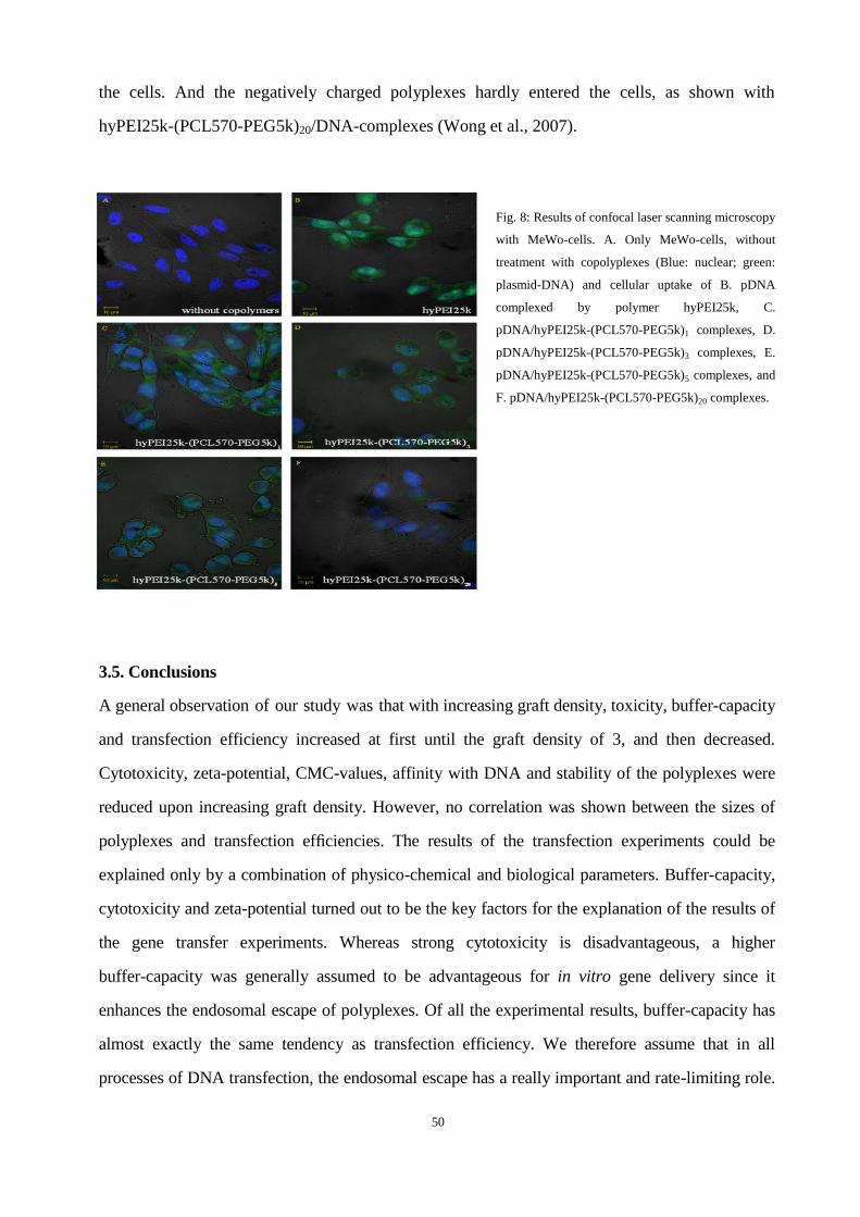
50
the cells. And the negatively charged polyplexes hardly entered the cells, as shown with
hyPEI25k-(PCL570-PEG5k)20/DNA-complexes (Wong et al., 2007).
Fig. 8: Results of confocal laser scanning microscopy
with MeWo-cells. A. Only MeWo-cells, without
treatment with copolyplexes (Blue: nuclear; green:
plasmid-DNA) and cellular uptake of B. pDNA
complexed by polymer hyPEI25k, C.
pDNA/hyPEI25k-(PCL570-PEG5k)1 complexes, D.
pDNA/hyPEI25k-(PCL570-PEG5k)3 complexes, E.
pDNA/hyPEI25k-(PCL570-PEG5k)5 complexes, and
F. pDNA/hyPEI25k-(PCL570-PEG5k)20 complexes.
3.5. Conclusions
A general observation of our study was that with increasing graft density, toxicity, buffer-capacity
and transfection efficiency increased at first until the graft density of 3, and then decreased.
Cytotoxicity, zeta-potential, CMC-values, affinity with DNA and stability of the polyplexes were
reduced upon increasing graft density. However, no correlation was shown between the sizes of
polyplexes and transfection efficiencies. The results of the transfection experiments could be
explained only by a combination of physico-chemical and biological parameters. Buffer-capacity,
cytotoxicity and zeta-potential turned out to be the key factors for the explanation of the results of
the gene transfer experiments. Whereas strong cytotoxicity is disadvantageous, a higher
buffer-capacity was generally assumed to be advantageous for in vitro gene delivery since it
enhances the endosomal escape of polyplexes. Of all the experimental results, buffer-capacity has
almost exactly the same tendency as transfection efficiency. We therefore assume that in all
processes of DNA transfection, the endosomal escape has a really important and rate-limiting role.

51
The zeta-potential of the complexes is also very important for the uptake of polyplexes. The high
surface charges enhance the adhesion of the cationic complexes to the negatively charged cell
membrane. Polymers with high buffer-capacity and zeta-potential but with low cytotoxicity showed
high transfection activities and seemed to be most efficient as in vitro gene transfer vectors (e. g.,
hyPEI25k-(PCL570-PEG5k)3). In vivo experiments and the evaluation of the described polymers for
siRNA delivery, such as elucidation of the structure of polyplexes are currently under way. Our
results provide a basis for the optimization of the molecular structure of gene delivery vectors with
higher buffer-capacity in the future.
3.6. Acknowledgments
We are grateful to Eva Mohr (Dept. of Pharmaceutics and Biopharmacy) for excellent technical
support. MEDITRANS, an Integrated Project funded by the European Commission under the Sixth
Framework (NMP4-CT-2006-026668), is gratefully acknowledged.
3.7. References
Banaszczyk, M.G., et al., 1999. POLY-L-LYSINE-GRAFT-PEG COMB-TYPE POLYCATION
COPOLYMERS FOR GENE DELIVERY. Journal of Macromolecular Science, Part A: Pure and Applied
Chemistry 36, 1061 - 1084.
Behr, J.-P., 1997. The Proton Sponge: a Trick to Enter Cells the Viruses Did Not Exploit. CHIMIA International
Journal for Chemistry 51, 34-36.
Bielinska, A.U., et al., 1997. The interaction of plasmid DNA with polyamidoamine dendrimers: mechanism of
complex formation and analysis of alterations induced in nuclease sensitivity and transcriptional activity of the
complexed DNA. Biochim Biophys Acta 1353, 180-190.
Boussif, O., et al., 1995. A versatile vector for gene and oligonucleotide transfer into cells in culture and in vivo:
polyethylenimine. Proc Natl Acad Sci U S A 92, 7297-7301.
Check, E., 2002. A tragic setback. Nature 420, 116-118.
Creusat, G., et al., 2010. Proton sponge trick for pH-sensitive disassembly of polyethylenimine-based siRNA
delivery systems. Bioconjug Chem 21, 994-1002.
Fischer, D., et al., 1999. A novel non-viral vector for DNA delivery based on low molecular weight, branched
polyethylenimine: effect of molecular weight on transfection efficiency and cytotoxicity. Pharm Res 16,
1273-1279.
Fischer, D., et al., 2003. In vitro cytotoxicity testing of polycations: influence of polymer structure on cell
viability and hemolysis. Biomaterials 24, 1121-1131.
Freshney, R.I., 2005. Culture of Animal Cells: A Manual of Basic Technique, 5th ed. Wiley-Liss, Wilmington,
DE.
Godbey, W.T., et al., 1999. Poly(ethylenimine) and its role in gene delivery. J Control Release 60, 149-160.
Hacein-Bey-Abina, S., et al., 2003. LMO2-associated clonal T cell proliferation in two patients after gene therapy
for SCID-X1. Science 302, 415-419.

52
Han, S., et al., 2001. Water-soluble lipopolymer for gene delivery. Bioconjug Chem 12, 337-345.
Han, S., et al., 2000. Development of biomaterials for gene therapy. Mol Ther 2, 302-317.
Jong, Y.C., 2009. Investigation of DNA Spectral Conformational Changes and Polymer Buffering Capacity in
Relation to Transfection Efficiency of DNA/Polymer Complexes. Journal of Pharmacy & Pharmaceutical
Sciences 12, 346-356.
Kono, K., et al., 2005. Transfection activity of polyamidoamine dendrimers having hydrophobic amino acid
residues in the periphery. Bioconjug Chem 16, 208-214.
Kunath, K., et al., 2003. Low-molecular-weight polyethylenimine as a non-viral vector for DNA delivery:
comparison of physicochemical properties, transfection efficiency and in vivo distribution with
high-molecular-weight polyethylenimine. J Control Release 89, 113-125.
Liu, Y., et al., 2009. A new synthesis method and degradation of hyper-branched polyethylenimine grafted
polycaprolactone block mono-methoxyl poly (ethylene glycol) copolymers (hy-PEI-g-PCL-b-mPEG) as potential
DNA delivery vectors. Polymer 50, 3895-3904.
Liu, Y., et al., 2010. Degradation of Hyper-Branched
Poly(ethylenimine)-graft-poly(caprolactone)-block-monomethoxyl-poly(ethylene glycol) as a Potential Gene
Delivery Vector. WILEY-VCH Verlag, pp. 1509-1515.
Luthman, H., Magnusson, G., 1983. High efficiency polyoma DNA transfection of chloroquine treated cells.
Nucleic Acids Res 11, 1295-1308.
Marshall, E., 2000. BIOMEDICINE:Gene Therapy on Trial. Science 288, 951-957.
Merdan, T., et al., 2002. Prospects for cationic polymers in gene and oligonucleotide therapy against cancer. Adv
Drug Deliv Rev 54, 715-758.
Moret, I., et al., 2001. Stability of PEI-DNA and DOTAP-DNA complexes: effect of alkaline pH, heparin and
serum. J Control Release 76, 169-181.
Pack, D.W., et al., 2005. Design and development of polymers for gene delivery. Nat Rev Drug Discov 4,
581-593.
Park, M.R., et al., 2005. Degradable polyethylenimine-alt-poly(ethylene glycol) copolymers as novel gene
carriers. J Control Release 105, 367-380.
Pawliuk, R., et al., 2001. Correction of sickle cell disease in transgenic mouse models by gene therapy. Science
294, 2368-2371.
Petersen, H., et al., 2002. Polyethylenimine-graft-poly(ethylene glycol) copolymers: influence of copolymer
block structure on DNA complexation and biological activities as gene delivery system. Bioconjug Chem 13,
845-854.
Pun, S.H., et al., 2004. Cyclodextrin-modified polyethylenimine polymers for gene delivery. Bioconjug Chem 15,
831-840.
Schaffer, D.V., et al., 2000. Vector unpacking as a potential barrier for receptor-mediated polyplex gene delivery.
John Wiley & Sons, Inc., pp. 598-606.
Shuai, X., et al., 2003. Novel Biodegradable Ternary Copolymers hy-PEI-g-PCL-b-PEG:  Synthesis,
Characterization, and Potential as Efficient Nonviral Gene Delivery Vectors. Macromolecules 36, 5751-5759.
Sung, S.-J., et al., 2003. Effect of Polyethylene Glycol on Gene Delivery of Polyethylenimine. Biological &
Pharmaceutical Bulletin 26, 492-500.
Tian, H., et al., 2007. Gene transfection of hyperbranched PEI grafted by hydrophobic amino acid segment
PBLG. Biomaterials 28, 2899-2907.
von Laer, D., et al., 2006. Gene therapy for HIV infection: what does it need to make it work? J Gene Med 8,
658-667.

53
Wang, D.-a., et al., 2002. Novel Branched Poly(Ethylenimine)-Cholesterol Water-Soluble Lipopolymers for
Gene Delivery. Biomacromolecules 3, 1197-1207.
Wong, L.F., et al., 2006. Lentivirus-mediated gene transfer to the central nervous system: therapeutic and
research applications. Hum Gene Ther 17, 1-9.
Wong, S.Y., et al., 2007. Polymer systems for gene delivery--Past, present, and future. Progress in Polymer
Science 32, 799-837.

54
Chapter 4
ENHANCING IN VIVO CIRCULATION AND SIRNA DELIVERY WITH
BIODEGRADABLE
POLYETHYLENIMINE-GRAFT-POLYCAPROLACTONE-BLOCK-POL
Y(ETHYLENE GLYCOL) COPOLYMERS
Accepted by Biomaterials
Mengyao Zhenga, Damiano Librizzi
b, Ayşe Kılıç
c, Yu Liu
a,d, Harald Renz
c, Olivia M.
Merkela,e,*
, Thomas Kissela
Author contributions
T. K. guided and directed the research. O. M. M. designed the measurements. M. Z. carried out
the dynamic light scattering/zeta potential analysis, SYBR
Gold assay, heparin assay, RT-PCR
and CLSM. Y. L. synthesized and characterized the polymers. M. Z. and O. M. M. analysed the
experimental data.
55
4.1 Abstract
The purpose of this study was to enhance the in vivo blood circulation time and siRNA delivery
efficiency of biodegradable copolymers
polyethylenimine-graft-polycaprolactone-block-poly(ethylene glycol) (hyPEI-g-PCL-b-PEG) by
introducing high graft densities of PCL-PEG chains. SYBR® Gold and heparin assays indicated
improved stability of siRNA/copolymer-complexes with a graft density of 5. At N/P 1, only 40%
siRNA condensation was achieved with non-grafted polymer, but 95% siRNA was condensed with
copolymer PEI25k-(PCL570-PEG5k)5. Intracellular uptake studies with confocal laser scanning
microscopy and flow cytometry showed that the cellular uptake was increased with graft density,
and copolymer PEI25k-(PCL570-PEG5k)5 was able to deliver siRNA much more efficiently into
the cytosol than into the nucleus. The in vitro knockdown effect of siRNA/hyPEI-g-PCL-b-PEG
was also significantly improved with increasing graft density, and the most potent copolymer
PEI25k-(PCL570-PEG5k)5 knocked down 84.43% of the GAPDH expression. Complexes of both
the copolymers with graft density 3 and 5 circulated much longer than unmodified PEI25kDa and
free siRNA, leading to a longer elimination half-life, a slower clearance and a three- or fourfold
increase of the AUC compared to free siRNA, respectively. We demonstrated that the graft density
of the amphiphilic chains can enhance the siRNA delivery efficiency and blood circulation, which
highlights the development of safe and efficient non-viral polymeric siRNA nanocarriers that are
especially stable and provide longer circulation in vivo.
Keywords
siRNA delivery
Biodegradable polycations
Non-viral polymeric nanocarrier
In vivo biodistribution and pharmacokinetics
SPECT imaging
4.2 Introduction
RNA interference (siRNA) promises great advantages for emerging therapeutic applications to
silence disease genes [1, 2]. In contrast to the efficient and reliable siRNA-mediated gene silencing
in vitro, only limited silencing of target gene expression in vivo has been achieved. One of the
56
reasons is that naked siRNA is very unstable in blood due to rapid enzymatic degradation, rapid
excretion, and nonspecific uptake by the reticuloendothelial system [3]. Therefore, it is a
formidable challenge to design and synthesize effective siRNA delivery systems, which are stable
but biodegradable and long circulating in vivo [4].
Copolymers hy-PEI-g-(PCL-b-PEG)n are amphiphilic biodegradable non-viral polymeric gene
delivery agents, which have shown a prominent gene delivery efficiency [5-7]. Positively charged
polyethylenimine (PEI) is the functional component which condenses the genetic material due to
electrostatic interactions and offers the buffer capacity with protonable amino groups to achieve
successful endosomal escape of polyplexes [8, 9]. Hydrophobic poly(caprolactone) (PCL)
increases the biodegradability of the copolymers and affects the hydrophilic–hydrophobic balance
of the polymer to enhance uptake of the complexes through cell membranes [5, 6]. PEG has been
widely used as a classical polymer to modify the surface of delivery systems like hydrophobic
colloids [10] or other polymeric delivery agents such as polycation hy-PEI, resulting in decreased
cytotoxicity, non-specific interaction of complexes with serum components and a prolonged blood
circulation [11-14]. We hypothesize that the effect of PEG on prolonged circulating and the
protection of siRNA with PCL-PEG chains depends not only on its content in a copolymer (length
or percentage), but also on the structure or the shape of the amphiphilic copolymer. Therefore, we
designed and synthesized a panel of biodegradable amphiphilic copolymers hy-PEI-g-PCL-b-PEG
with different graft densities of PCL-PEG chains as polymer-based siRNA delivery systems. We
expected that the PEG-PCL grafting degree would improve not only the gene silencing, but also
the circulating time in vivo. In our study, the physicochemical properties of these
siRNA/polymer-complexes were characterized with respect to particle size, zeta-potential, and
stability against competing heparin anions. Real-Time-PCR and confocal laser scanning
microscopy were used to measure the in vitro knock down efficiency and cell uptake. The siRNA
delivery efficiency and biodistribution of these triblock amphiphilic copolymers under in vivo
conditions was determined using single photon emission computed tomography (SPECT) imaging
with 111
In radiolabelled siRNA [15] and fluorescently labeled copolymer. Our study provides us
with an insight into advantageous structures and shapes of copolymers to promote the rapid
development of safe and efficient non-viral polymeric siRNA delivery nanocarriers that are
especially stable in vivo.

57
4.3 Methods and materials
Materials
hy-PEI with a molecular weight of 25 kDa was obtained from BASF. Poly(ethylene glycol)
mono-methyl ether (mPEG) (5kDa) and ε-caprolactone were purchased from Fluka (Taufkirchen,
Germany). Heparin sodium salt was from Sigma-Aldrich Laborchemikalien GmbH (Seelze,
Germany) and all other chemicals were obtained from Sigma–Aldrich (Steinheim, Germany).
hy-PEI-g-PCL-b-PEG was synthesized as reported previously [5, 6]. Lipofectamine™2000 (LF)
was bought from Invitrogen (Karlsruhe, Germany). Amine-modified firefly luciferase DsiRNA
with a C6-NH2 linker at the 3′of the sense strand, negative control sequence, hGAPDH-DsiRNA,
and TYE546-DsiRNA were obtained from Integrated DNA Technologies (IDT, Leuven, Belgium).
Balb/c mice were bought from Harlan Laboratories (Horst, The Netherlands). The chelator
2-(4-isothiocyanatobenzyl)-diethylenetriaminepentaacetic acid (p-SCN-Bn-DTPA) was purchased
from Macrocyclics (Dallas, TX, USA). The radioactive 111
InCl3 was purchased from Covidien
Deutschland GmbH (Neustadt a.d. Donau, Germany).
Polyplex preparation
Polyplexes were formed by diluting a calculated volume of 1 mg/mL polymer (based on PEI
25kDa) with 5% glucose solution in the first step. Equal volumes of diluted polymer and siRNA
solution were mixed followed by vigorous pipetting before letting the polyplexes form at room
temperature for 20 min. The N/P ratio (=nitrogen/phosphorus-ratios) was calculated as described
earlier [6].
Dynamic light scattering and zeta potential analysis
Polyplexes were prepared with hGAPDH-DsiRNA as described above at different N/P ratios and
measured in a disposable low volume UVette (Eppendorf, Wesseling–Berzdorf, Germany) using a
Zetasizer Nano ZS (Malvern, Herrenberg, Germany). The measurements in 5% glucose solution
were conducted in triplicates according to a previous work [16].
SYBR® gold assay and heparin assay: binding and protection efficiency and stability against
competing polyanions
58
The ability of hy-PEI-g-PCL-b-PEG copolymers to condense siRNA into small polyplexes was
studied in SYBR® Gold assays. Briefly, polyplexes were formed at different N/P ratios from 0 to
20. Polyplex solutions contained 1 µg hGAPDH-DsiRNA and the according amount of polymer in
each well of FluoroNunc 96 well plates (Nunc, Thermo Fisher Scientific, Langenselbold,
Germany). For heparin assays, polyplexes were prepared in solutions at different N/P-ratio like in
the SYBR® Gold assay described above. Additionally, 20 μL heparin solution with a concentration
of 1.5 IU/μmol siRNA was added to the polyplex solutions in each well of the 96-well plate
(Perkin Elmer, Rodgau-Jügesheim) and the polyplex solutions were incubated for 20 min at 25◦C,
before 20 μL diluted SYBR® Gold solution were added. The measurement of fluorescence was
conducted according to a previous study in quadruplets [17].
RT-PCR
SKOV3 cells were used for knock down experiments and the Hs_GAPDH scilencing was
measured with RT-PCR according to our previous work [18]. Briefly, 500,000 cells per well were
seeded in 6-well-plates 24 h prior to transfection and transfected with 100 pmol of siRNA in
triplicates. Real-Time PCR was performed using QuantiFast™ SYBR® Green PCR Kit and
Hs_GAPDH and Hs_ACTB_2_SG Primers (Qiagen, Germany) and the RotorGene3000 real-time
PCR thermal cycler (Qiagen, Hilden, Germany).
Flow Cytometry
SKOV3 cells were seeded in 24-well plates at a density of 60,000 cells per well 24h before
transfection with polyplexes consisting of 50 pmol of Alexa488-labeled siRNA at N/P 5. Cells
were incubated with the polyplexes in triplicates for 4h at 37 °C and were then washed with PBS
buffer (with Ca2+
and Mg2+
). The fluorescence bound on the cell-surface was quenched by 5 min
incubating with 0.4% trypan blue before the cells were trypsinized for 5 min and spun down in
serum containing medium and fixed with CellFIX (BD Biosciences, San Jose, CA). Treated and
untreated cells (as negative control) cells were measured using a FACSCantoII (BD Biosciences,
San Jose, CA) with excitation at 488 nm and the emission filter set to a 530/30 bandpass. In each
measurement, cells were gated to evaluate 10,000 viable cells, and the geometric mean
fluorescence intensity (MFI) was calculated as the mean value of 3 independent measurements.

59
Data acquisition and analysis was performed using FACSDiva software (BD Biosciences, San
Jose, CA) [17].
Confocal laser scanning microscopy (CLSM)
For CLSM, polymers were first labeled with FITC as described before complexation with
TYE546-DsiRNA [19] to detect polymer and siRNA in parallel. SKOV3 cells were seeded in 8
well-chamber slides (Lab-Tek; Rochester, NY, USA) 24h before transfection. For sample
preparation and confocal microscopy on a Zeiss Axiovert 100 Microscope and a Zeiss LSM 510
scanning device (Zeiss, Oberkochen, Germany), steps and settings were chosen as previously
described [20].
siRNA Radiolabeling and purification
Amine-modified siRNA (IDT, Leuven, Belgium) was coupled with p-SCN-Bn-DTPA,
radiolabeled with 111
InCl3 and purified using Absolutely RNA miRNA columns (Agilent,
Waldbronn, Germany) as described before [19].
In vivo imaging, pharmacokinetics and biodistribution
Balb/c mice at the age of 6 weeks (about 20 g) were used for in vivo experiments. All animal
experiments were carried out according to the German law of protection of animal life and were
approved by an external review committee for laboratory animal care. Balb/c mice were
anesthetized with xylazine and ketamine, and 5 animals per group were injected with polyplexes
containing 35 µg of radiolabelled siRNA and the corresponding amount of FITC labeled polymer
at N/P 5. Complexes were injected i.v. into the tail vein. For the pharmacokinetics study, 25 μL
blood samples were drawn at different time points within two hours and measured using a Gamma
Counter Packard 5005 (Packard Instruments, Meriden, CT). After 2h, biodistribution was recorded
using three-dimensional SPECT and planar gamma camera imaging (Siemens AG, Erlangen,
Germany). Finally, animals were sacrificed and the biodistribution in dissected organs was
quantified using a Gamma Counter Packard 5005.
Flow cytometric quantification of cellular tissue uptake
60
As described above, mice were injected with polyplexes containing 35 µg siRNA and FITC
labeled polymers at the N/P 5. Animals were sacrificed 2 h after injection, and the organs were
dissected. The intracellular uptake within the organs was determined by flow cytometry. To obtain
organ homogenates, lungs and livers were incubated in 2 mg/mL collagenase D solution (Roche,
Mannheim, Germany) at 37 °C for 20 min before sieving the tissue through 100 µm nylon cell
strainers (BD Falcon, Heidelberg, Germany). Cells were then suspended in 10 mL of PBS and
centrifuged at 350 g for 10 min. The cells were resuspended, once again centrifuged and
resuspended in 500 µL 4% paraformaldehyde for flow cytometry. The internalization of
fluorescently labeled polyplexes into lung and liver cells was measured on a FACSCantoTM Π
(BD Biosciences, San Jose, CA) with excitation at 488 nm and emission filter set to 530/30
bandpass. 10,000 viable cells were evaluated in each experiment and results are the mean values of
3 independent measurements.
Statistics
Results are given as mean values and standard deviation (SD). Statistical evaluation, calculation of
the AUC and two way ANOVA were performed using Graph Pad Prism 4.03 (Graph Pad
Software, La Jolla, CA).
4.4 Results and discussion
Polyplex size and zeta potential
The hydrodynamic diameters and surface charges of the siRNA/copolymer-complexes are listed in
Table 1. The polyplex sizes decreased with increasing N/P-ratios from 2 to 10. All copolymers
were able to condense siRNA into polyplexes with sizes of 78–142 nm (Tab. 1) at N/P 5 or N/P 10
in 5% glucose solution. However, the polyplex size at N/P-ratio 2 was larger than at the other
N/P-ratios, especially for the copolymer with graft density 1. This observation and the negative
zeta potentials at N/P 2 indicate that siRNA is not fully condensed into the core of the micelle-like
complexes. At N/P 5, all complexes revealed positive zeta potentials and rather narrow size
distributions (PDI), especially in case of PEI25k-(PCL570-PEG5k)3 . In a recent study, we
investigated the binding behavior of siRNA and hy-PEI-g-PCL-b-PEG by molecular dynamic
simulations, isothermal titration calorimetry and dye quenching assay (data not shown here). We
61
found that at low N/P ratios, siRNA complexation results in a more uniform and stable complex
formation, which leads to increased siRNA delivery efficiency. This explains the lower PDI values
as compared to N/P 10. Interestingly, amongst the three copolymers, PEI25k-(PCL570-PEG5k)5
and PEI25k-(PCL570-PEG5k)3 appeared to be most advantageous siRNA delivery agents, not only
for in vitro but also for in vivo applications, according to the hydrodynamic diameter and surface
properties. Both the hydrodynamic diameter and polydispersity of their siRNAcomplexes were
lower than those of the polyplexes formed with PEI25k-(PCL570-PEG5k)1. The decreased size of
those polyplexes despite higher molecular weight of the polymers can be explained by the micellar
behavior of the more amphiphilic copolymers. Moreover, the narrow size distribution
(0.23<PDI<0.31) of the complexes is very important for biological activity and corresponds with
what has been described as “coalesced” complexes before [20]. Additionally, the zeta potential of
siRNA/ PEI25k-(PCL570-PEG5k)5 was decreased as compared to the other copolymers, which
reflects less positive charges on the surface of the complexes and thus avoids high toxicity [16],
potentially less uptake into macrophages and therefore provides the prerequisites for prolonged
circulation in vivo.

62
Table 1. Hydrodynamic diameters and surface charges of the siRNA/copolymer-complexes.
PEI25k-(PCL570-PEG5k)1
Hydrodynamic diameter
Zeta-potential (mV) Size (nm) PDI
N/P2 1079 ± 169.98 0.30 ± 0.01 -4.14 ± 0.62
N/P5 123.2 ± 1.75 0.33 ± 0.01 19.97 ± 1.65
N/P10 107.23 ± 1.85 0.32 ± 0.01 25.1 ± 1.11
PEI25k-(PCL570-PEG5k)3
Hydrodynamic diameter
Zeta-potential (mV) Size (nm) PDI
N/P2 114.8 ± 1.74 0.33 ± 0.02 -11.07 ± 0.35
N/P5 93.56 ± 7.08 0.24 ± 0.02 13.87 ± 1.51
N/P10 92.58 ± 1.92 0.40 ± 0.02 15.11 ± 4.02
PEI25k-(PCL570-PEG5k)5
Hydrodynamic diameter
Zeta-potential (mV) Size (nm) PDI
N/P2 211.47 ± 6.87 0.23 ± 0.01 -14.97 ± 1.16
N/P5 142.0 ± 6.5 0.24 ± 0.01 7.04 ± 0.23
N/P10 78.35±4.62 0.31±0.02 10.87±0.49
Binding and protection efficiency and stability against competing polyanions
The condensation efficiency of siRNA within the copolymers can be investigated by SYBR® Gold
assays [21], which can quantify the amount of free or accessible siRNA that is able to bind SYBR®
Gold. The SYBR®
Gold assay revealed the different condensation abilities of the copolymers with
different graft densities: the affinity of siRNA with the copolymers was increased with increasing
N/P-ratio and siRNA could be condensed very well above N/P 5 with all of the copolymers.
Compared with PEI25k-(PCL570-PEG5k)1, the copolymers with graft density 3 and 5 showed not
only higher affinity to siRNA, but also the better protection against competing polyanions like
heparin molecules. For example, only 40% siRNA was condensed with PEI25k-(PCL570-PEG5k)1
at N/P 1, while about 95% siRNA was completely condensed with PEI25k-(PCL570-PEG5k)3 and
PEI25k-(PCL570-PEG5k)5.

63
The stability of polyplexes against competing
polyanions is very important for gene
delivery systems, especially for in vivo
application since stability of the polyplexes
can be strongly weakened in presence of
serum [22]. The process of complexation of
genetic material within polycations is entropy
driven [23] and can be significantly impaired
by the presence of other polyanions like
heparin. The differences of the stability
against polyanions were observed to be
related to the graft density (Fig. 1). The
higher grafted copolymers showed a better
protection of the siRNA against heparin, and
the stability of the polyplexes was increased with increasing graft density [7] from 1 to 5. At
N/P-ratio 10, after addition of heparin, 74% siRNA was condensed with
PEI25k-(PCL570-PEG5k)3 and 87% siRNA was condensed with PEI25k-(PCL570-PEG5k)5, while
only 41% siRNA was condensed with PEI25k-(PCL570-PEG5k)1. We assumed that the PCL-PEG
chains can increase the affinity of the amphiphilic copolymer with the 2’O-methylated siRNA [17],
and protect the siRNA very well from competing polyanions like heparin. In terms of stability, it is
advantageous if the interaction with siRNA is not only of electrostatic nature but if hydrophobic
forces that are not affected by competing polyanions stabilize the polyplexes. Interestingly, the
previous stability study using these copolymers with plasmid DNA showed different results
concerning stability: the stability of p-DNA/copolymer-complexes was decreased with an increase
of the graft density, and the copolymer with graft density 1 showed the best protection of the
p-DNA [16]. The copolymer with one PCL-PEG chain offered a better affinity to plasmid DNA
than the other copolymers. This observation demonstrates that the entropic gain evidenced by ITC
is indicative of the fact that inter-polyplex aggregations are controlled by hydrophobic forces.
Indeed, a certain amount of lipophilic PCL can increase the affinity between copolymer with pDNA
as shown by hyPEI25k-(PCL570-PEG5k)1, but with the increasing of graft density, reduced
Fig. 1. In vitro stability of polyplexes of the
siRNA/PEI25k-(PCL570-PEG5k)n-complexes as measured by (A)
SYBR® Gold and (B) heparin assays.

64
stability of copolymers with pDNA was observed, which could be caused by the shielding-effect of
PCL-mPEG segments. But different from pDNA, siRNA is locally more flexible in the case of
local roll and tilt deformations [24] and more adaptable [25] in case of binding with a charged
spherical polymer than DNA [26], which facilitates the uniform binding between the siRNA
molecules and the polymers. Therefore, the complexation ability of the copolymer with siRNA is
increased with the increasing of graft density.
In vitro cell uptake with CLSM and FACS
The in vitro intracellular uptake was studied with confocal laser scanning microscopy qualitatively
and flow cytometry quantitatively. The results of flow cytometry clearly revealed different cell
uptake in the amount of siRNA as a result of the differently grafted copolymers, as shown in Fig.
3. Compared with PEI25kDa (3968.3 MFI), all of the amphiphilic copolymers showed an
optimized cell uptake, especially in case of the copolymer with graft density 3 (8175.7 MFI).
PEI25k-(PCL570-PEG5k)5 seemed to showed reduced uptake as compared to
PEI25k-(PCL570-PEG5k)3, which could be the result of the lower zeta potential. However, the
differences between those two polymers were not significantly different. Our previous DNA
delivery research with the same copolymers indicated the same tendency with the copolymer with
graft density 3 displaying the best cell uptake and DNA transfection efficiency at N/P 25 [16].
However, flow cytometry quantifies the amount of fluorescently labeled genetic material not only
in the cytosol, but also in the nucleus. Therefore, confocal laser scanning microscopy was
performed additionally to detect differences in the subcellular distribution of the polyplexes made
of differently grafted copolymers. The CLSM images of each treatment group are shown in Fig. 2:
polyplexes formed with copolymer PEI25k-(PCL570-PEG5k)1 seemed to be taken up only to a low
extent, which corroborated the flow cytometry data. Interestingly, although the FACS had revealed
a trend in favor of PEI25k-(PCL570-PEG5k)3 over PEI25k-(PCL570-PEG5k)5 polyplexes,
nonetheless, more fluorescence of siRNA/ PEI25k-(PCL570-PEG5k)5-complexes was observed
distributed in the cytoplasm of the transfected cells by confocal microscopy. This property is
advantageous for siRNA delivery since the final target destination of siRNA is the cytoplasm,
whereas plasmid DNA must be transported into the nucleus. In other words, to achieve a successful
siRNA delivery, the siRNA must be delivered and released rapidly from its carrier upon
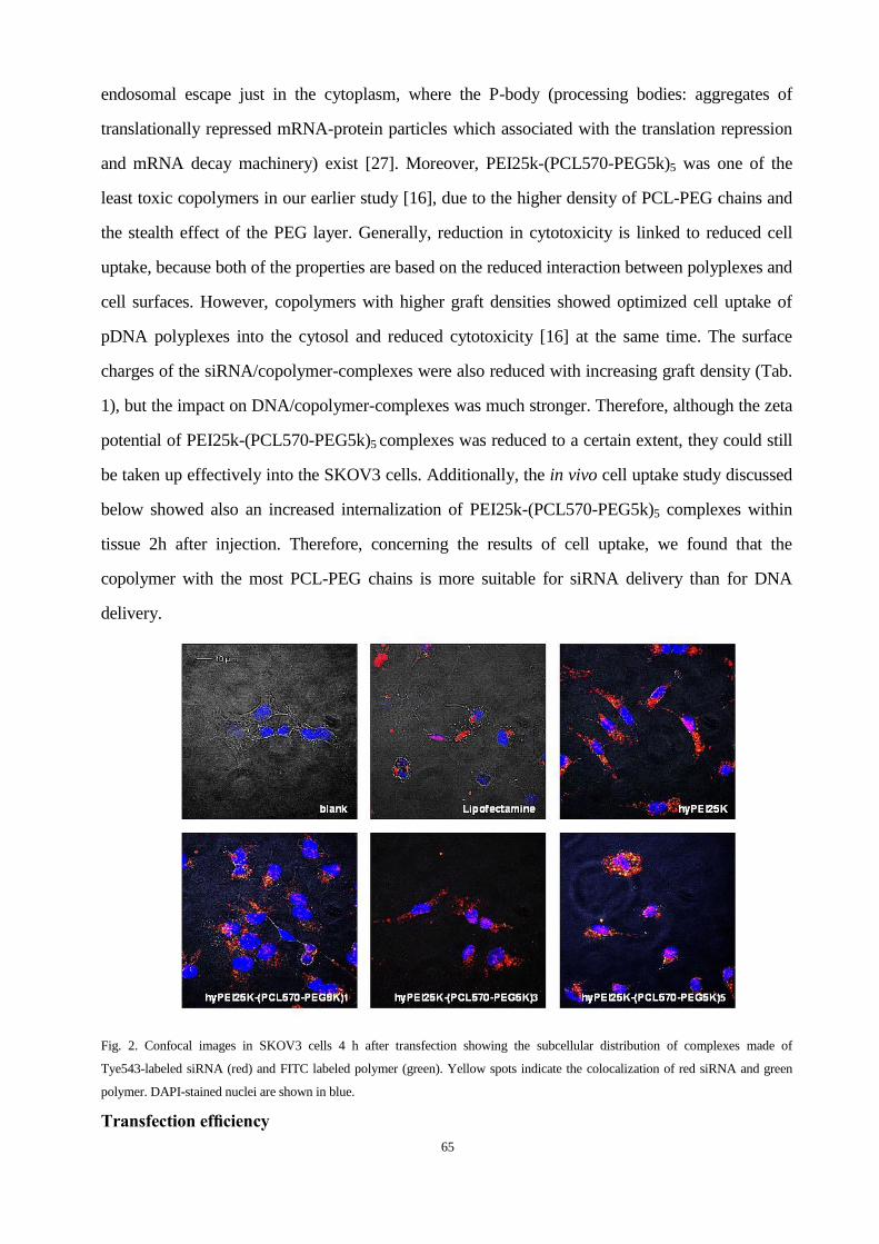
65
endosomal escape just in the cytoplasm, where the P-body (processing bodies: aggregates of
translationally repressed mRNA-protein particles which associated with the translation repression
and mRNA decay machinery) exist [27]. Moreover, PEI25k-(PCL570-PEG5k)5 was one of the
least toxic copolymers in our earlier study [16], due to the higher density of PCL-PEG chains and
the stealth effect of the PEG layer. Generally, reduction in cytotoxicity is linked to reduced cell
uptake, because both of the properties are based on the reduced interaction between polyplexes and
cell surfaces. However, copolymers with higher graft densities showed optimized cell uptake of
pDNA polyplexes into the cytosol and reduced cytotoxicity [16] at the same time. The surface
charges of the siRNA/copolymer-complexes were also reduced with increasing graft density (Tab.
1), but the impact on DNA/copolymer-complexes was much stronger. Therefore, although the zeta
potential of PEI25k-(PCL570-PEG5k)5 complexes was reduced to a certain extent, they could still
be taken up effectively into the SKOV3 cells. Additionally, the in vivo cell uptake study discussed
below showed also an increased internalization of PEI25k-(PCL570-PEG5k)5 complexes within
tissue 2h after injection. Therefore, concerning the results of cell uptake, we found that the
copolymer with the most PCL-PEG chains is more suitable for siRNA delivery than for DNA
delivery.
Fig. 2. Confocal images in SKOV3 cells 4 h after transfection showing the subcellular distribution of complexes made of
Tye543-labeled siRNA (red) and FITC labeled polymer (green). Yellow spots indicate the colocalization of red siRNA and green
polymer. DAPI-stained nuclei are shown in blue.
Transfection efficiency
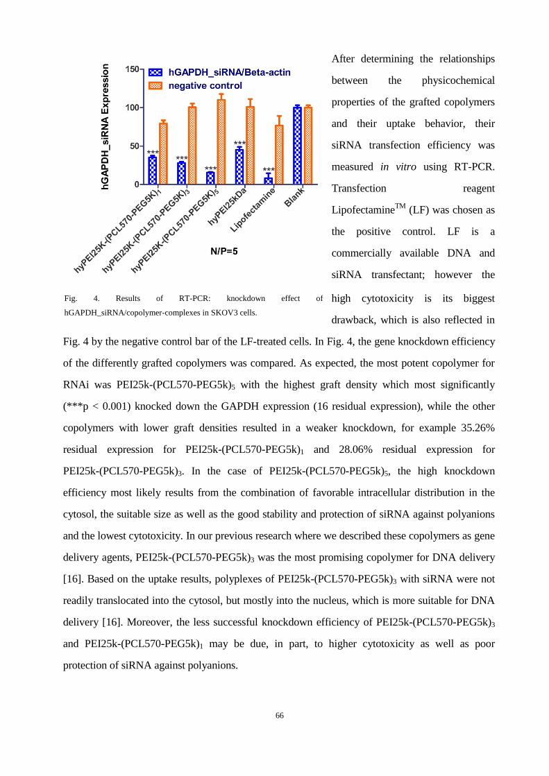
66
After determining the relationships
between the physicochemical
properties of the grafted copolymers
and their uptake behavior, their
siRNA transfection efficiency was
measured in vitro using RT-PCR.
Transfection reagent
LipofectamineTM
(LF) was chosen as
the positive control. LF is a
commercially available DNA and
siRNA transfectant; however the
high cytotoxicity is its biggest
drawback, which is also reflected in
Fig. 4 by the negative control bar of the LF-treated cells. In Fig. 4, the gene knockdown efficiency
of the differently grafted copolymers was compared. As expected, the most potent copolymer for
RNAi was PEI25k-(PCL570-PEG5k)5 with the highest graft density which most significantly
(***p < 0.001) knocked down the GAPDH expression (16 residual expression), while the other
copolymers with lower graft densities resulted in a weaker knockdown, for example 35.26%
residual expression for PEI25k-(PCL570-PEG5k)1 and 28.06% residual expression for
PEI25k-(PCL570-PEG5k)3. In the case of PEI25k-(PCL570-PEG5k)5, the high knockdown
efficiency most likely results from the combination of favorable intracellular distribution in the
cytosol, the suitable size as well as the good stability and protection of siRNA against polyanions
and the lowest cytotoxicity. In our previous research where we described these copolymers as gene
delivery agents, PEI25k-(PCL570-PEG5k)3 was the most promising copolymer for DNA delivery
[16]. Based on the uptake results, polyplexes of PEI25k-(PCL570-PEG5k)3 with siRNA were not
readily translocated into the cytosol, but mostly into the nucleus, which is more suitable for DNA
delivery [16]. Moreover, the less successful knockdown efficiency of PEI25k-(PCL570-PEG5k)3
and PEI25k-(PCL570-PEG5k)1 may be due, in part, to higher cytotoxicity as well as poor
protection of siRNA against polyanions.
Fig. 4. Results of RT-PCR: knockdown effect of
hGAPDH_siRNA/copolymer-complexes in SKOV3 cells.
67
Pharmacokinetics and biodistribution
The two most effective amphiphilic copolymers concerning the results of stability, in vitro cell
uptake and knockdown efficiency, PEI25k-(PCL570-PEG5k)3 and PEI25k-(PCL570-PEG5k)5,
were investigated in vivo to evaluate their pharmacokinetics and biodistribution behavior as
compared to polyplexes of PEI25kDa and naked siRNA. As measured by gamma scintillation
counting of blood samples (Fig. 5), polyplexes made of both the grafted copolymers
PEI25k-(PCL570-PEG5k)3 and PEI25k-(PCL570-PEG5k)5 were stable in vivo for at least 120 min
and circulated longer than siRNA and PEI25kDa/siRNA polyplexes. As described earlier,
complexes of unmodified PEI25kDa and siRNA dissociate easily upon intravenous administration
and the polymer shows a strong first-pass effect in the liver [11, 17]. The pharmacokinetic profile
of hy-PEI-complexed siRNA therefore strongly resembles that of free siRNA that is rapidly
excreted upon injection [28]. While PEGylation of hy-PEI further destabilized siRNA complexes
under in vivo conditions [22], we observed here that the amphiphilic triblock copolymers did not
only show increased stability against competing heparin polyanions in vitro but were also more
stable in vivo upon intravenous injection. This is reflected in longer circulation times, less steep
alpha and beta elimination phases, and increased area under the curve (AUC) values which were
calculated to analyze the differences in the circulation times of each load quantitatively (Fig. 5).
The AUC of PEI25k-(PCL570-PEG5k)5 and PEI25k-(PCL570-PEG5k)3 complexed siRNA is
almost four fold higher than that of PEI25kDa complexed or free siRNA (Supporting information).
It can be hypothesized that the polyplexes made of PEI25k-(PCL570-PEG5k)3 and
PEI25k-(PCL570-PEG5k)5 which display higher stability and decreased zeta potentials are less
prone to dissociation and rapid uptake into macrophages. Thus, these polyplexes remain within the
systemic circulation for a longer duration of time than instable complexes with high positive
surface charge. Due to extended circulation times, they are eventually taken up into the liver as
well (Fig. 6 and 7). However, since the cells types taking up those polyplexes in the liver were not
differentially identified, it is not clear if the uptake was mediated by Kupffer cells.
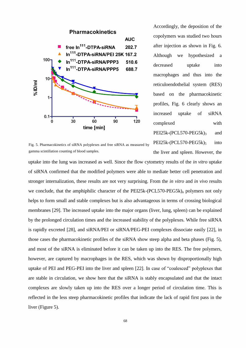
68
Accordingly, the deposition of the
copolymers was studied two hours
after injection as shown in Fig. 6.
Although we hypothesized a
decreased uptake into
macrophages and thus into the
reticuloendothelial system (RES)
based on the pharmacokinetic
profiles, Fig. 6 clearly shows an
increased uptake of siRNA
complexed with
PEI25k-(PCL570-PEG5k)3 and
PEI25k-(PCL570-PEG5k)5 into
the liver and spleen. However, the
uptake into the lung was increased as well. Since the flow cytometry results of the in vitro uptake
of siRNA confirmed that the modified polymers were able to mediate better cell penetration and
stronger internalization, these results are not very surprising. From the in vitro and in vivo results
we conclude, that the amphiphilic character of the PEI25k-(PCL570-PEG5k)n polymers not only
helps to form small and stable complexes but is also advantageous in terms of crossing biological
membranes [29]. The increased uptake into the major organs (liver, lung, spleen) can be explained
by the prolonged circulation times and the increased stability of the polyplexes. While free siRNA
is rapidly excreted [28], and siRNA/PEI or siRNA/PEG-PEI complexes dissociate easily [22], in
those cases the pharmacokinetic profiles of the siRNA show steep alpha and beta phases (Fig. 5),
and most of the siRNA is eliminated before it can be taken up into the RES. The free polymers,
however, are captured by macrophages in the RES, which was shown by disproportionally high
uptake of PEI and PEG-PEI into the liver and spleen [22]. In case of “coalesced” polyplexes that
are stable in circulation, we show here that the siRNA is stably encapsulated and that the intact
complexes are slowly taken up into the RES over a longer period of circulation time. This is
reflected in the less steep pharmacokinetic profiles that indicate the lack of rapid first pass in the
liver (Figure 5).
Fig. 5. Pharmacokinetics of siRNA polyplexes and free siRNA as measured by
gamma scintillation counting of blood samples.
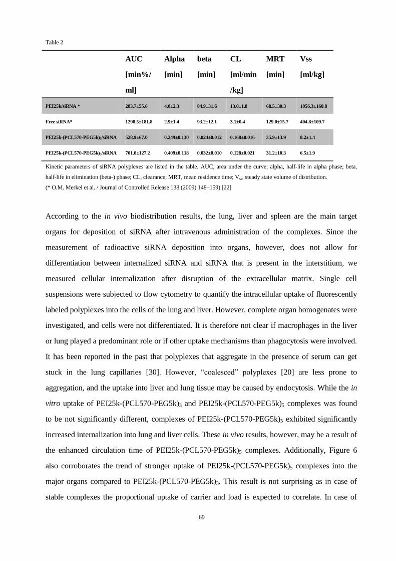
69
Table 2
AUC
[min%/
ml]
Alpha
[min]
beta
[min]
CL
[ml/min
/kg]
MRT
[min]
Vss
[ml/kg]
PEI25k/siRNA * 283.7±55.6 4.0±2.3 84.9±31.6 13.0±1.8 68.5±30.3 1056.3±160.8
Free siRNA* 1298.5±181.8 2.9±1.4 93.2±12.1 3.1±0.4 129.0±15.7 404.8±109.7
PEI25k-(PCL570-PEG5k)3/siRNA 528.9±67.0 0.249±0.130 0.024±0.012 0.168±0.016 35.9±13.9 8.2±1.4
PEI25k-(PCL570-PEG5k)5/siRNA 701.0±127.2 0.409±0.118 0.032±0.010 0.128±0.021 31.2±10.3 6.5±1.9
Kinetic parameters of siRNA polyplexes are listed in the table. AUC, area under the curve; alpha, half-life in alpha phase; beta,
half-life in elimination (beta-) phase; CL, clearance; MRT, mean residence time; Vss, steady state volume of distribution.
(* O.M. Merkel et al. / Journal of Controlled Release 138 (2009) 148–159) [22]
According to the in vivo biodistribution results, the lung, liver and spleen are the main target
organs for deposition of siRNA after intravenous administration of the complexes. Since the
measurement of radioactive siRNA deposition into organs, however, does not allow for
differentiation between internalized siRNA and siRNA that is present in the interstitium, we
measured cellular internalization after disruption of the extracellular matrix. Single cell
suspensions were subjected to flow cytometry to quantify the intracellular uptake of fluorescently
labeled polyplexes into the cells of the lung and liver. However, complete organ homogenates were
investigated, and cells were not differentiated. It is therefore not clear if macrophages in the liver
or lung played a predominant role or if other uptake mechanisms than phagocytosis were involved.
It has been reported in the past that polyplexes that aggregate in the presence of serum can get
stuck in the lung capillaries [30]. However, “coalesced” polyplexes [20] are less prone to
aggregation, and the uptake into liver and lung tissue may be caused by endocytosis. While the in
vitro uptake of PEI25k-(PCL570-PEG5k)3 and PEI25k-(PCL570-PEG5k)5 complexes was found
to be not significantly different, complexes of PEI25k-(PCL570-PEG5k)5 exhibited significantly
increased internalization into lung and liver cells. These in vivo results, however, may be a result of
the enhanced circulation time of PEI25k-(PCL570-PEG5k)5 complexes. Additionally, Figure 6
also corroborates the trend of stronger uptake of PEI25k-(PCL570-PEG5k)5 complexes into the
major organs compared to PEI25k-(PCL570-PEG5k)3. This result is not surprising as in case of
stable complexes the proportional uptake of carrier and load is expected to correlate. In case of

70
PEI-complexed and free siRNA, the overall recovery of siRNA in any of the organs is rather low
due to the instability of the complexes and the rapid excretion of the siRNA. The deposition of the
free polymer, in case of PEI 25kDa, however, was significantly higher [22].
Fig. 6. Biodistribution of siRNA polyplexes and free siRNA 2 h after i.v. administration as measured by gamma scintillation
counting of dissected organs.
4.5 Conclusion
In this study, we synthesized a panel of hy-PEI-g-PCL-b-PEG amphiphilic copolymers with
different graft densities of the PCL-PEG chains. Compared with copolymers of low graft density,
the copolymers with high graft densities (3 and 5) showed not only a higher affinity with siRNA, a
better protection against competing polyanions, but also an increased intracellular uptake into the
cytosol, which was detected with confocal laser scanning microscopy and flow cytometry.
Therefore, copolymers with higher graft density of PCL-PEG chains showed significant
advantages as siRNA delivery agents concerning in vitro transfection and in vivo pharmacokinetics
and biodistribution. In summary, we demonstrated that polymeric micelles, which are formed with
amphiphilic block copolymers have advantages especially for in vivo siRNA delivery, and that the

71
graft density of the amphiphilic chains can enhance the blood circulation, which is a key parameter
to promote the development of safe and efficient non-viral polymeric siRNA delivery in vivo.
4.6 Acknowledgements
We are grateful to PD Dr. Helmut Hoeffken (Nuclear Medicine Department, University Hospital
Marburg) for the generous use of equipment and facilities, and to Eva Mohr (Department of
Pharmaceutics and Biopharmacy) for excellent technical support. MEDITRANS, an Integrated
Project funded by the European Commission under the Sixth Framework
(NMP4-CT-2006-026668) is gratefully acknowledged.
4.7 References
1.Iorns E, Lord CJ, Turner N, Ashworth A. Utilizing RNA interference to enhance cancer drug discovery. Nat
Rev Drug Discov 2007;6(7):556-568.
2.Shim MS, Kwon YJ. Efficient and targeted delivery of siRNA in vivo. FEBS J 2010;277(23):4814-4827.
3.Whitehead KA, Langer R, Anderson DG. Knocking down barriers: advances in siRNA delivery. Nat Rev Drug
Discov 2009;8(2):129-138.
4.Grimm D, Kay MA. Therapeutic application of RNAi: is mRNA targeting finally ready for prime time? J Clin
Invest 2007;117(12):3633-3641.
5.Shuai XT, Merdan T, Unger F, Wittmar M, Kissel T. Novel biodegradable ternary copolymers
hy-PEI-g-PCL-b-PEG: synthesis, characterization, and potential as efficient nonviral gene delivery vectors.
Macromolecules 2003;36(15):5751-5759.
6.Liu Y, Nguyen J, Steele T, Merkel O, Kissel T. A new synthesis method and degradation of hyper-branched
polyethylenimine grafted polycaprolactone block mono-methoxyl poly (ethylene glycol) copolymers
(hy-PEI-g-PCL-b-mPEG) as potential DNA delivery vectors. Polymer 2009;50(16):3895-3904.
7.Liu Y, Samsonova O, Sproat B, Merkel O, Kissel T. Biophysical characterization of hyper-branched
polyethylenimine-graft-polycaprolactone-block-mono-methoxyl-poly(ethylene glycol) copolymers
(hy-PEI-PCL-mPEG) for siRNA delivery. J Control Release 2011;153(3):262-268.
8.Grzelinski M, Urban-Klein B, Martens T, Lamszus K, Bakowsky U, Hobel S, et al. RNA interference-mediated
gene silencing of pleiotrophin through polyethylenimine-complexed small interfering RNAs in vivo exerts
antitumoral effects in glioblastoma xenografts. Hum Gene Ther 2006;17(7):751-766.
9.Werth S, Urban-Klein B, Dai L, Hobel S, Grzelinski M, Bakowsky U, et al. A low molecular weight fraction of
polyethylenimine (PEI) displays increased transfection efficiency of DNA and siRNA in fresh or lyophilized
complexes. J Control Release 2006;112(2):257-270.
10.Mosqueira VCF, Legrand P, Gulik A, Bourdon O, Gref R, Labarre D, et al. Relationship between complement
activation, cellular uptake and surface physicochemical aspects of novel PEG-modified nanocapsules.
Biomaterials 2001;22(22):2967-2979.
72
11.Merdan T, Kunath K, Petersen H, Bakowsky U, Voigt KH, Kopecek J, et al. PEGylation of poly(ethylene
imine) affects stability of complexes with plasmid DNA under in vivo conditions in a dose-dependent manner
after intravenous injection into mice. Bioconjug Chem 2005;16(4):785-792.
12.Kunath K, von Harpe A, Petersen H, Fischer D, Voigt K, Kissel T, et al. The structure of PEG-modified
poly(ethylene imines) influences biodistribution and pharmacokinetics of their complexes with NF-kappa B
decoy in mice. Pharm Res 2002;19(6):810-817.
13.Sung SJ, Min SH, Cho KY, Lee S, Min YJ, Yeom YI, et al. Effect of polyethylene glycol on gene delivery of
polyethylenimine. Biol Pharm Bull 2003;26(4):492-500.
14.Sato A, Choi SW, Hirai M, Yamayoshi A, Moriyarna R, Yamano T, et al. Polymer brush-stabilized polyplex
for a siRNA carrier with long circulatory half-life. J Control Release 2007;122(3):209-216.
15.Merkel OM, Librizzi D, Pfestroff A, Schurrat T, Behe M, Kissel T. In vivo SPECT and real-time gamma
camera imaging of biodistribution and pharmacokinetics of siRNA delivery using an optimized radiolabeling and
purification procedure. Bioconjug Chem 2009;20(1):174-182.
16.Zheng M, Liu Y, Samsonova O, Endres T, Merkel O, Kissel T. Amphiphilic and biodegradable
hy-PEI-g-PCL-b-PEG copolymers efficiently mediate transgene expression depending on their graft density. Int J
Pharm 2011;427(1):80-87.
17.Merkel OM, Mintzer MA, Librizzi D, Samsonova O, Dicke T, Sproat B, et al. Triazine dendrimers as nonviral
vectors for in vitro and in vivo RNAi: the effects of peripheral groups and core structure on biological activity.
Mol Pharm 2010;7(4):969-983.
18.Endres T, Zheng M, Beck-Broichsitter M, Samsonova O, Debus H, Kissel T. Optimising the self-assembly of
siRNA loaded PEG-PCL-lPEI nano-carriers employing different preparation techniques. J Control Release 2012.
19.Merdan T, Kunath K, Fischer D, Kopecek J, Kissel T. Intracellular processing of poly(ethylene
imine)/ribozyme complexes can be observed in living cells by using confocal laser scanning microscopy and
inhibitor experiments. Pharm Res 2002;19(2):140-146.
20.Merkel OM, Zheng M, Mintzer MA, Pavan GM, Librizzi D, Maly M, et al. Molecular modeling and in vivo
imaging can identify successful flexible triazine dendrimer-based siRNA delivery systems. J Control Release
2011;153(1):23-33.
21.Merkel OM, Beyerle A, Librizzi D, Pfestroff A, Behr TM, Sproat B, et al. Nonviral siRNA delivery to the
lung: investigation of PEG-PEI polyplexes and their in vivo performance. Mol Pharm 2009;6(4):1246-1260.
22.Merkel OM, Librizzi D, Pfestroff A, Schurrat T, Buyens K, Sanders NN, et al. Stability of siRNA polyplexes
from poly(ethylenimine) and poly(ethylenimine)-g-poly(ethylene glycol) under in vivo conditions: effects on
pharmacokinetics and biodistribution measured by fluorescence fluctuation spectroscopy and single photon
emission computed tomography (SPECT) imaging. J Control Release 2009;138(2):148-159.
23.Bronich T, Kabanov AV, Marky LA. A thermodynamic characterization of the interaction of a cationic
copolymer with DNA. J Phys Chem B 2001;105(25):6042-6050.
24.Noy A, Perez A, Lankas F, Javier Luque F, Orozco M. Relative flexibility of DNA and RNA: a molecular
dynamics study. J Mol Biol 2004;343(3):627-638.
25.Pavan GM, Albertazzi L, Danani A. Ability to adapt: different generations of PAMAM dendrimers show
different behaviors in binding siRNA. J Phys Chem B 2010;114(8):2667-2675.

73
26.Pavan GM, Kostiainen MA, Danani A. Computational approach for understanding the interactions of
UV-degradable dendrons with DNA and siRNA. J Phys Chem B 2010;114(17):5686-5693.
27.Wood MJA, Jagannath A. Localization of double-stranded small interfering RNA to cytoplasmic processing
bodies is Ago2 dependent and results in up-regulation of GW182 and argonaute-2. Mol Biol Cell
2009;20(1):521-529.
28.Dykxhoorn DM, Palliser D, Lieberman J. The silent treatment: siRNAs as small molecule drugs. Gene Ther
2006;13(6):541-552.
29.Hsu CY, Hendzel M, Uludag H. Improved transfection efficiency of an aliphatic lipid substituted 2 kDa
polyethylenimine is attributed to enhanced nuclear association and uptake in rat bone marrow stromal cell. J Gene
Med 2010;13(1):46-59.
30.Malek A, Merkel O, Fink L, Czubayko F, Kissel T, Aigner A. In vivo pharmacokinetics, tissue distribution
and underlying mechanisms of various PEI(-PEG)/siRNA complexes. Toxicol Appl Pharmacol
2009;236(1):97-108.

74
Chapter 5
MODULAR SYNTHESIS OF FOLATE CONJUGATED TERNARY
COPOLYMERS:
POLYETHYLENIMINE-GRAFT-POLYCAPROLACTONE-BLOCK-POL
Y(ETHYLENE GLYCOL)-FOLATE FOR TARGETED GENE DELIVERY
Published in Bioconjugate Chemistry, 2012 May 11. DOI: 10.1021/bc300025d
Li Liu†,‡,§
, Mengyao Zheng†,‡
, Thomas Renette‡, Thomas Kissel
*,‡
† Both authors contributed equally to this work.
Author contributions
T. K. guided and directed the research. M. Z. and L. L. designed the measurements. M. Z. carried
out the SYBR
Gold assay, heparin assay, MTT and in vitro transfection experiment. L. L.
synthesized and characterized the polymers. M. Z. and L. L. analysed the experimental data.

75
5.1 Abstract
Folate receptor (FR) is overexpressed in a variety of human cancers. Gene delivery vectors
conjugated with folate as a ligand could possibly deliver gene materials into target tumor cells via
FR-mediated endocytosis. This study addresses novel folate-conjugated ternary copolymers based
on polyethylenimine-graft-polycaprolactone-block-poly(ethylene glycol) (PEI-g-PCL-b-PEG-Fol)
as targeted gene delivery system using a modular synthesis approach including “click” conjugation
of folate moieties with heterobifunctional PEG-b-PCL at PEG terminus and subsequently the
introduction of PEI by a Michael addition between folate-PEG-b-PCL and PEI via active PCL
terminus. This well controlled synthetic procedure avoids tedious separation of by-products. The
structure of PEI-g-PCL-b-PEG-Fol was confirmed by 1H NMR and UV spectra. DNA
condensation of PEI-g-PCL-b-PEG-Fol was tested using a SYBRTM
Gold quenching assay and
agarose gel electrophoresis upon heparin competition assay. Although PEI-g-PCL-b-PEG-Fol
could condense DNA completely at N/P ratio > 2, polyplexes of N/P ratio 10 with sizes at about
120 nm and positive zeta potentials were selected for further biological evaluations due to polyplex
stability. An enhancement of cellular uptake of PEI-g-PCL-b-PEG-Fol/pDNA polyplexes was
observed in FR over-expressing KB cells in comparison to unmodified PEI-g-PCL-b-PEG, through
flow cytometry analysis and confocal laser scanning imaging. Importantly, this enhanced cellular
uptake could be inhibited by free folic acid and did not occur in FR-negative A549 cells,
demonstrating specific cell uptake by FR-mediated endocytosis. Furthermore, the transfection
efficiency of PEI-g-PCL-b-PEG-Fol/pDNA polyplexes was increased approximately 14-fold in
comparison to folate-negative polyplexes. Therefore, the PEI-g-PCL-b-PEG-Fol merits further
investigation under in vivo conditions for targeting FR overexpressing tumors.
5.2 Introduction
Gene therapy has received increasing attention over the past two decades as a promising method
for treating various inherited and acquired human diseases.1-3
An efficient and safe delivery
system that delivers the therapeutic genes into target cells is regarded as prerequisite for
successful gene therapy. Recently, a large number of studies focused on the development of
polymer-based non-viral gene delivery vectors due to their non-immunogenicity, low cost,
physiochemical versatility and ease of manipulation. 4, 5
Among them, polyethylenimine
76
(PEI)-based delivery systems have emerged as one of the most successful and efficient
candidates for gene delivery both in vitro and in vivo. 6
PEI is a cationic polymer with a high
density of amines, which condense nucleic acids via electrostatic interaction into nano-scaled
polyplexes. Importantly, the protonable amine group endowed PEI-containing polyplexes with
efficient endosomal escape property through the proton-sponge mechanism, which was believed
to be the main reason for the relatively high gene-transfer activity of PEI. 6, 7
Nevertheless, the
PEI-based gene delivery systems have suffered from relatively high toxicity, especially at high
molecular weights, affecting their potential applications under in vivo conditions.
To decrease cytotoxicity and enhance gene transfection efficiency, many efforts relied on the
modification of PEI either with hydrophilic segments such as poly(ethylene glycol) (PEG) 8, 9
, or
hydrophobic segments such as poly(L-lactide-co-glycolide) 10
, or other biocompatible
compositions such as cyclodextrin 11
. Moreover, PEI derivates have also been decorated with
variety of targeting ligands 12
to generate gene delivery systems with targeting functionality for
specific tissues/cells. For example, the folate receptor (FR) is known to be over-expressed in
many types of carcinoma cells. 13-15
By conjugation with folate, the PEI-containing polyplexes
were expected to target the folate receptor on cell surface and transfect specific cells by
receptor-mediated endocytosis. 16-23
In previous studies of our group, a series of ternary copolymers based on PEI-g-PCL-b-PEG
were synthesized and investigated as potential gene delivery vectors. 24-27
The PEI-g-PCL-b-PEG
copolymers were shown to be biodegradable and amphiphilic in character; hypothetically
generating micelle-like polyplexes with excellent colloidal stability. Structure-function
relationships suggested that higher graft densities of PCL-PEG led to decreased cytotoxicity and
copolymers with short PCL segment displayed higher transfection efficiency in vitro compared
with PEI 25kDa. 25, 27
In this study, folate conjugated PEI-g-PCL-b-PEG was synthesized and examined for targeted
gene delivery. It should be noted that the folate moiety was designed to be conjugated at the
distal PEG end, instead of direct linkage to PEI. 22
This strategy provides more flexibility for the
folate ligands via PEG spacers possibly improving their target binding efficacy. Therefore, to
achieve the well-structured PEI-g-PCL-b-PEG-Folate copolymer, a modular synthesis procedure
was designed, which involved the conjugation of folate moiety with heterobifunctional

77
PEG-b-PCL by click chemistry followed by a Michael addition between folate-PEG-b-PCL and
PEI. The mild reaction conditions of click chemistry and Michael addition were beneficial to
maintain the biological activity of folate and to avoid tedious cleaning procedures. The resulting
copolymers were investigated regarding biophysical properties, DNA condensation and
cytotoxicity. Furthermore, cell-uptake and transfection efficiency of
PEI-g-PCL-b-PEG-Fol/pDNA complexes were evaluated in vitro in FR-positive KB cells to
determine their potential as targeted gene delivery vehicles.
5.3 Experimental Section
Materials. Heterobifunctional poly(ethylene glycol) (HO-PEG-COOH, 3 kDa) was purchased
from Rapp Polymere GmbH (Germany). ε-Caprolactone from Fluka was distilled before use
under vacuum over CaH2. Branched polyethylenimine (hy-PEI, 25kDa) was obtained from
BASF. Folic acid, N-hydroxysuccinimide, dicyclohexylcarbodiimide and solvents were
purchased from Acros. Propargylamine and acryloyl chloride were from Sigma-Aldrich.
1-azido-3-aminopropane was synthesized from 3-Bromopropylamine hydrobromide and sodium
azide (Details in Supplement information). All other reagents for synthesis were obtained from
Sigma-Aldrich and were used as received without further purification. Endotoxin-free
luciferase-encoding plasmid DNA (pCMV-luc) was provided by Plasmid Factory (Bielefeld,
Germany). SYBRTM
Gold and YOYO-1 were obtained from Invitrogen.
Synthesis of Azido-functionalized Folate. Azido-functionalized folate was prepared by a
method modified from the literature. 28
Folic acid (0.5 g, 1.135 mmol) was dissolved in DMSO
(20 mL) containing triethylamine (0.25 mL). After addition of N-hydroxysuccinimide (NHS)
(0.26 g, 2.2 equiv.), and dicyclohexylcarbodiimide (DCC) (0.25 g, 1.1 equiv.), the mixture was
stirred at room temperature in the dark for 24 h. Then, 1-azido-3-aminopropane (0.24 g, 2 equiv.)
was added into the mixture under stirring. The reaction was continued for another 24 h. After the
precipitated side-product dicyclohexylurea (DCU) was removed by filtration, the product was
precipitated in ethyl acetate and dried under vacuum. The crude product was purified by
dissolving in 1M NaOH and precipitation by addition of 1M HCl. The precipitates were

78
collected by centrifugation, washed with EtOH/H2O (1:1) and dried under vacuum to give an
orange-yellow product in 86% yield.
FT-IR (ν, cm-1
): 2800-3200, 2095 (-N3), 1682, 1602, 1506, 1297, 1236, 1174, 1126. 1H NMR
(d6-DMSO, 400M): δ (ppm)= 11.37 (s, 1H, -CONHCHCOOH), 8.62 (s, 1H, PtC7H), 7.93-7.95
(d, 1H, PtC6-CH2NH-Ph), 7.79-7.80 (d, 1H, -CONHCHCOOH), 7.63-7.60 (d, 2H, Ph-C2H and
Ph-C6H), 6.87 (br s, 2H, NH2), 6.62-6.59 (d, 2H, Ph-C3H and Ph-C5H), 4.46-4.44 (d, 2H,
PtC6-CH2NH-Ph), 4.30-4.26 (m, 1H, -CONHCHCOOH), 3.44-3.39 (m, 2H, -CH2N3), 3.29 (br
s, OH), 3.10-3.02 (m, 2H, -CONHCH2CH2CH2N3), 2.30-2.15 (m, 2H, -CH2CH2CONH),
2.05-1.85 (m, 2H, -CH2CH2CONH-), 1.60-1.56 (m, 2H, -CONHCH2CH2CH2N3). Pt = pteridine.
Synthesis of PCL-b-PEG with Heterobifunctional Terminal Group
(acrylate-PCL-b-PEG-alkyne). PCL-b-PEG was firstly synthesized by ring-opening
polymerization of ε-caprolactone initiated from the hydroxyl end group of HO-PEG-COOH.
Defined amounts of HO-PEG-COOH and caprolactone monomers were sealed in dry argon and
stirred at 120 ℃ for 24 h with Sn(Oct)2 (about 0.1% molar ratio of caprolactone) as catalyst. 29
The product was dissolved in chloroform and precipitated with cold methanol/ether (1/1, v/v).
The precipitate obtained as HO-PCL-b-PEG-COOH was dried under vacuum for 24 h for the
following modification. (Yield: 88%).
HO-PCL-b-PEG-COOH (0.3 mmol) was dissolved in dry dichloromethane (DCM) with NHS
(0.6 mmol) and DCC (1.2 mmol). The mixture was stirred at 0 °C for 1 h and then at room
temperature for 24 h, during which time the mixture became turbid due to DCU. Propargylamine
(0.6 mmol) and triethylamine were added into the above mixture, and stirred under room
temperature for another 24 h. The mixture was filtered to remove the precipitated DCU, and then
precipitated in cold ether. The precipitates were dried under vacuum for 24 h to obtain
HO-PCL-b-PEG-alkyne. (Yield: 79%).
Next, the terminal hydroxyl group of HO-PCL-b-PEG-alkyne (0.1 mmol) was coupled with
acryloyl chloride (0.2 mmol) in dry toluene containing triethylamine (0.2mmol). The reaction
mixture was stirred at 80 ℃ for 10 h, and then cooled to room temperature, filtered to remove
triethylamine hydrochloride and precipitated in cold n-hexane. The precipitates were collected
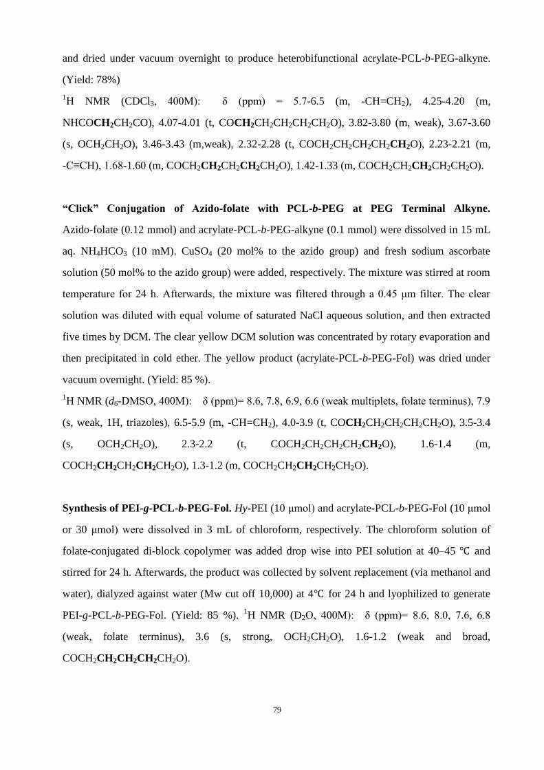
79
and dried under vacuum overnight to produce heterobifunctional acrylate-PCL-b-PEG-alkyne.
(Yield: 78%)
1H NMR (CDCl3, 400M): δ (ppm) = 5.7-6.5 (m, -CH=CH2), 4.25-4.20 (m,
NHCOCH2CH2CO), 4.07-4.01 (t, COCH2CH2CH2CH2CH2O), 3.82-3.80 (m, weak), 3.67-3.60
(s, OCH2CH2O), 3.46-3.43 (m,weak), 2.32-2.28 (t, COCH2CH2CH2CH2CH2O), 2.23-2.21 (m,
-C≡CH), 1.68-1.60 (m, COCH2CH2CH2CH2CH2O), 1.42-1.33 (m, COCH2CH2CH2CH2CH2O).
“Click” Conjugation of Azido-folate with PCL-b-PEG at PEG Terminal Alkyne.
Azido-folate (0.12 mmol) and acrylate-PCL-b-PEG-alkyne (0.1 mmol) were dissolved in 15 mL
aq. NH4HCO3 (10 mM). CuSO4 (20 mol% to the azido group) and fresh sodium ascorbate
solution (50 mol% to the azido group) were added, respectively. The mixture was stirred at room
temperature for 24 h. Afterwards, the mixture was filtered through a 0.45 μm filter. The clear
solution was diluted with equal volume of saturated NaCl aqueous solution, and then extracted
five times by DCM. The clear yellow DCM solution was concentrated by rotary evaporation and
then precipitated in cold ether. The yellow product (acrylate-PCL-b-PEG-Fol) was dried under
vacuum overnight. (Yield: 85 %).
1H NMR (d6-DMSO, 400M): δ (ppm)= 8.6, 7.8, 6.9, 6.6 (weak multiplets, folate terminus), 7.9
(s, weak, 1H, triazoles), 6.5-5.9 (m, -CH=CH2), 4.0-3.9 (t, COCH2CH2CH2CH2CH2O), 3.5-3.4
(s, OCH2CH2O), 2.3-2.2 (t, COCH2CH2CH2CH2CH2O), 1.6-1.4 (m,
COCH2CH2CH2CH2CH2O), 1.3-1.2 (m, COCH2CH2CH2CH2CH2O).
Synthesis of PEI-g-PCL-b-PEG-Fol. Hy-PEI (10 μmol) and acrylate-PCL-b-PEG-Fol (10 μmol
or 30 μmol) were dissolved in 3 mL of chloroform, respectively. The chloroform solution of
folate-conjugated di-block copolymer was added drop wise into PEI solution at 40–45 ℃ and
stirred for 24 h. Afterwards, the product was collected by solvent replacement (via methanol and
water), dialyzed against water (Mw cut off 10,000) at 4℃ for 24 h and lyophilized to generate
PEI-g-PCL-b-PEG-Fol. (Yield: 85 %). 1H NMR (D2O, 400M): δ (ppm)= 8.6, 8.0, 7.6, 6.8
(weak, folate terminus), 3.6 (s, strong, OCH2CH2O), 1.6-1.2 (weak and broad,
COCH2CH2CH2CH2CH2O).
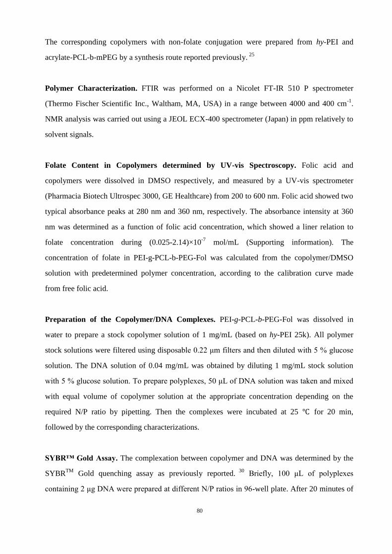
80
The corresponding copolymers with non-folate conjugation were prepared from hy-PEI and
acrylate-PCL-b-mPEG by a synthesis route reported previously. 25
Polymer Characterization. FTIR was performed on a Nicolet FT-IR 510 P spectrometer
(Thermo Fischer Scientific Inc., Waltham, MA, USA) in a range between 4000 and 400 cm-1
.
NMR analysis was carried out using a JEOL ECX-400 spectrometer (Japan) in ppm relatively to
solvent signals.
Folate Content in Copolymers determined by UV-vis Spectroscopy. Folic acid and
copolymers were dissolved in DMSO respectively, and measured by a UV-vis spectrometer
(Pharmacia Biotech Ultrospec 3000, GE Healthcare) from 200 to 600 nm. Folic acid showed two
typical absorbance peaks at 280 nm and 360 nm, respectively. The absorbance intensity at 360
nm was determined as a function of folic acid concentration, which showed a liner relation to
folate concentration during (0.025-2.14)×10-7
mol/mL (Supporting information). The
concentration of folate in PEI-g-PCL-b-PEG-Fol was calculated from the copolymer/DMSO
solution with predetermined polymer concentration, according to the calibration curve made
from free folic acid.
Preparation of the Copolymer/DNA Complexes. PEI-g-PCL-b-PEG-Fol was dissolved in
water to prepare a stock copolymer solution of 1 mg/mL (based on hy-PEI 25k). All polymer
stock solutions were filtered using disposable 0.22 μm filters and then diluted with 5 % glucose
solution. The DNA solution of 0.04 mg/mL was obtained by diluting 1 mg/mL stock solution
with 5 % glucose solution. To prepare polyplexes, 50 μL of DNA solution was taken and mixed
with equal volume of copolymer solution at the appropriate concentration depending on the
required N/P ratio by pipetting. Then the complexes were incubated at 25 ℃ for 20 min,
followed by the corresponding characterizations.
SYBR™ Gold Assay. The complexation between copolymer and DNA was determined by the
SYBRTM
Gold quenching assay as previously reported. 30
Briefly, 100 μL of polyplexes
containing 2 μg DNA were prepared at different N/P ratios in 96-well plate. After 20 minutes of

81
incubation at room temperature, 20 μL diluted 4×SYBRTM
Gold solution was added and
incubated for another 10 minutes in the dark. The fluorescence was directly detected with a
fluorescence plate reader (BMG Labtech, Offenburg) at 495 nm excitation and 537 nm emission.
Triplicate samples were investigated and the results were transformed into relative fluorescent
intensity values (Fsample/ Ffree DNA).
Heparin Competition Assay. The stability of polyplexes against heparin (a model molecule of
competing polyanion) was assessed by agarose gel electrophoresis in TAE buffer (0.04 M
Tris–acetate, 0.001 M EDTA, pH 7.4) containing 0.5 mg/mL ethidium bromide (EtBr). Heparin
(150 000 IU/g, Serva, Pharm., USPXV2, Merck, Darmstadt, Germany) solution was added to
reach a final heparin concentration of 0.5 mg/mL into the polyplex solution at different
N/P-ratios. After 15min of incubation with heparin, 25 μL of polyplex solution containing 1.5 μg
DNA was loaded into the agarose gel wells and the agarose gels were run in TAE buffer for 45
min at 80 V using an Electro-4 electrophoresis unit (Thermo Electron, Waltham, MA, USA). The
gels were recorded after irradiation with UV-light using a gel documentation system
(BioDocAnalyze, Biometra, Göttingen, Germany).
Size and zeta-potential analysis. The size and zeta potential of the polyplexes were monitored
by a dynamic light scattering (DLS) instrument (Zetasizer 3000HS, Malvern, Worcestershire,
UK). Polyplexes were measured in a low volume cuvette (100 μL) firstly, and then zeta-potential
measurements were performed on the samples prepared by diluting 100 μL of polyplexes
solution with additional 600 μL of 5 % glucose solution to a final volume of 700 μL in a
transparent zeta cuvette. The samples were carried out in the standard clear capillary
electrophoresis cell at 25 °C. Three measurements were performed on each sample.
Cell culture. Human epithelial nasopharyngeal carcinoma (KB) cells were gifts from Prof. P. S.
Low’s group (Purdue University) and continuously cultured in folate-free RPMI-1640 medium
supplemented with 10 % fetal calf serum (FCS) at 37 ℃ in a humidified atmosphere containing 5
% CO2. FR negative human lung epithelial carcinoma (A549) cells were obtained from DSMZ

82
(Braunschweig, Germany) and cultured in DMEM medium supplemented with 10 % FCS at 37
℃ in a humidified atmosphere containing 5 % CO2.
Cytotoxicity assay. KB cells were seeded into 96-well plates at a density of 8×10³
cells/well.
After 24 h, cell culture medium was aspirated and replaced by 200 μL of serial dilutions of
polymer stock solution in cell culture medium with FCS. The cells were then incubated for 24
hours at 37 °C. Afterwards, medium was replaced by medium without serum containing 0.5
mg/mL 3-(4,5-dimethylthiazol-2-yl)-2,5-diphenyl tetrazolium bromide (MTT). After 4 h
incubation at 37 °C in the dark, medium was removed and then 200 μL of DMSO was added to
dissolve the formazan crystals formed by proliferating cells. After 15 min incubation with
DMSO, measurement was performed using an ELISA reader (Titertek Plus MS 212, ICN,
Eschwege, Germany) at a wavelength of 570 nm and 690 nm. Relative viability was calculated
using wells with untreated cells as 100 % controls. Data are presented as mean values (±SD) of
four experiments. The IC50 values were calculated with Original 8 using Logistic fit.
Cellular Uptake by Flow Cytometry. KB cells and A549 cells were seeded at a density of
6×10⁴
cells/well in 24 well plates 24 h prior to the experiment. Polymers including PEI 25kDa,
PEI-g-PCL-b-mPEG (PCE3) and PEI-g-PCL-b-PEG-Fol (PCE3-F) were labeled with FITC in
parallel assay. Polyplexes were prepared at N/P=10 using pCMV-Luc as described above. Cells
were incubated with polyplexes containing 4 μg DNA per well for 4 h at 37 °C. In free folate
competition studies, the normal RPMI-1640 medium (containing 1 mg/L folic acid) was replaced
as incubation medium 1 h before polyplexes added. Afterwards, cells were washed with PBS
once and incubated with 0.4 % trypan blue solution for 5 min to quench extracellular
fluorescence. Cells were washed again, detached using 100 μL of trypsin and treated with 900
μL of PBS solution containing 10 % FCS. Cells were then collected by centrifugation and
resuspended in 300 μL of Cellfix solution (BD Biosciences, San Jose, CA) for cell fixation. Cell
suspensions were measured on a FACS CantoTM Π (BD Biosciences, San Jose, CA) with
excitation at 488 nm and emission filter set to 530/30 bandpass. 10,000 viable cells were
evaluated in each experiment and results are the mean of 3 independent measurements.
83
Confocal Laser Scanning Microscopy. KB cells were seeded at a density of 2×10⁴ cells per
well in 8 well chamber slides (Nunc, Wiesbaden, Germany) and allowed to grow for 24 h.
Polyplexes were prepared at N/P=10 as described above using YOYO-1 labeled pCMV-Luc.
Cells were incubated with polyplexes containing 1 μg DNA per well in medium with FCS for 4 h
at 37 °C. Subsequently, cells were washed with PBS, quenched with 0.4 % trypan blue solution,
washed again with PBS, fixed using 4 % paraformaldehyde in PBS, DAPI stained and washed
again with PBS. Finally, cells were embedded using FluorSave Reagent (Calbiochem, San
Diego, CA). A Zeiss Axiovert 100 M microscope coupled to a Zeiss LSM 510 scanning device
(Zeiss, Oberkochen, Germany) was used for confocal microscopy. For excitation of YOYO-1
fluorescence, an argon laser with an excitation wavelength of 488 nm was used. Fluorescence
emission was detected using a 505–530 nm bandpass filter. Transmission images were obtained
in the same scan.
In vitro Gene Transfection. KB cells were seeded in 48 well plates (1.5×10⁴ cells/well) 24 h
prior to the experiment. Polyplexes were prepared at N/P=10 as described above using plasmid
pCMV-luc. Medium was replaced by 200 μL of fresh cell culture medium with 10% FCS, then
50 μL of polyplexes (containing 1 μg pDNA) were added in each well. For folate competition
assay, normal RPMI1640 medium (containing 1mg/L folic acid) was replaced 1 h before
polyplexes addition. After incubation for 4 h, the medium was exchanged and cells were cultured
for another 44 h. Then cells were washed with PBS twice, lysed in 100 μL cell culture lysis
buffer for 15 min. Luciferase activity was quantified by injection of 50 μL luciferase assay
buffer, containing 10 mM luciferin, to 25 μL of the cell lysate. The relative light units (RLU)
were measured with a plate luminometer (LumiSTAR Optima, BMG Labtech GMBH,
Offenburg, Germany). Protein concentration was determined using a Pierce BCA protein assay
Kit (Thermo Scientific). All experiments were performed in triplicate and data were expressed in
RLU per mg protein (±SD).
Statistics. Significance between the means was tested by two way ANOVA and statistical
evaluations using GraphPad Prism 4.03 (Graph Pad Software, La Jolla, USA).

84
5.4 Results and Discussion
Synthesis and characterization of
PEI-g-PCL-b-PEG-Fol. In most of the
previously reported synthesis routes of
folate-receptor targeted polycations,
folate moieties were conjugated with
polycation either directly 22, 23, 31
or via
heterobifunctional PEG linker like
HOOC-PEG-NH2 through the
amine-carboxyl reaction using NHS
chemistry. 18, 19
NHS reactions are
widely used in bioconjugations due to
their mild reaction conditions, but
purification of the final product from excess NHS, by-product and solvent etc. may present
considerable challenges. On the other hand, it was postulated that the rigid folate moiety attached
at the distal end of flexible PEG chains would allow more efficient interaction and binding to
folate receptor on the cell surface. But in the conventional HOOC-PEG-NH2/NHS strategy,
random coupling between different PEG chains co-existed inevitably with PEG-Fol. The coupled
PEG chains were difficult to separate and may also generate byproducts.
In this study, the folate-conjugated ternary copolymer of PEI-g-PCL-b-PEG-Fol was synthesized
through a modular procedure as shown in Scheme1, which involved click conjugation of folate
moiety with alkyne-terminated PCL-b-PEG, followed by the Michael addition between
acrylate-terminated folate-PEG-b-PCL and PEI.
Firstly, the diblock copolymer of PCL-b-PEG was synthesized from heterobifunctional
HO-PEG-COOH initiated from the hydroxyl group. The molecular weight of PCL block could
be predetermined by the molar ratio of monomers to hydroxyl groups, which were confirmed by
1H NMR analysis through the calculation of the integral intensity at 1.3-1.4 ppm
(-COCH2CH2CH2CH2CH2O-, PCL) and 3.4-3.5 ppm (-CH2CH2O-, PEG) based on the known
molecular weight of PEG. The obtained diblock copolymer HO-PCL-b-PEG-COOH was then
Scheme 1. Synthesis strategy of PEI-g-PCL-b-PEG-Fol.

85
converted into alkyne end group at the PEG terminus and acrylate end group at the PCL
terminus, respectively. The structures of intermediates in different steps were characterized by
1H NMR analysis (Figure S-3), where the appearance of signals belonging to alkyne and double
bond was verified accordingly. Here, the heterobifunctional acrylate-PCL-b-PEG-alkyne was a
key element for obtaining regioselectivity during synthesis. On one hand, the PEG-terminal
alkyne was specific for conjugation with azido-folate through the copper(I)-catalyzed
azide-alkyne click reaction. This cycloaddition reaction is well known for its high efficiency and
selectivity under mild conditions, which has been applied in many bioconjugations of ligands or
drugs to polymers. 28, 32-34
On the other hand, the PCL-terminal acrylate group was reactive to
couple with the amino group of PEI according to a Michael addition under mild conditions
afterward. This strategy avoided the risk of intersectional reactions between di-block
copolymers. Importantly, both click conjugation and Michael addition were carried through
under the mild conditions, which were advantageous to maintain the activity of folate in the
objective material since folate is sensitive to light and heat.
Thereafter, the heterobifunctional acrylate-PCL-b-PEG-alkyne was transformed into folate
terminated PCL-b-PEG-Fol via “click” cyclo-addition with azido-modified folate in weak basic
aqueous solution (for water-soluble copolymer with short PCL block). The 1H NMR spectra in
Figure 1 (a) verified the structure of acrylate-PCL-b-PEG-Fol with signal assignment. The
successful conjugation of folate moiety onto PCL-b-PEG was further confirmed by UV spectra
as shown in Figure 2. Two absorbance peaks appeared at 280 nm and 360 nm respectively for
PCL-b-PEG-Fol, while its precursor PCL-b-PEG had no characteristic absorbance in this range.
The content of folate conjugated in PCL-b-PEG-Fol was about 2.1×10⁻⁷
mol/mg, obtained on the
basis of the standard curve using the UV absorbance at 360 nm as reference. This value is
equivalent to the conjugation percent of folate onto PCL-b-PEG as about 95% (mol.%).
86
Figure 1. 1H NMR spectra of (a) Fol-PEG-b-PCL with terminal
acrylate in d6-DMSO and (b) PEI-g-PCL-b-PEG-Fol in D2O.
Figure 2. UV absorbance of folic acid solution, folate-conjugated
copolymer solutions and control copolymer (without folate-conjugating)
solutions in DMSO.
Finally, the acrylate-PCL-b-PEG-Fol was linked to hy-PEI via Michael addition between the
active double bond and the amine group. Graft density of folate–conjugated branches could be
predetermined by the feed ratio of acrylate-PCL-b-PEG-Fol to PEI. Figure 1 (b) showed the 1H
NMR spectrum of the resulting copolymer PEI-g-PCL-b-PEG-Fol in D2O, where the proton
signals could be related to the PEI, PCL, PEG and folate moiety, respectively. Nevertheless, the
peaks belonging to PCL segments were rather weak due to its hydrophobicity. The ratio of

87
integral –CH2CH2O- to integral -CH2CH2NH- was calculated to give the graft density, which
coincided with the predetermined value. The compositions of synthesized PEI-g-PCL-b-PEG-Fol
copolymers were summarized in Table 1. The folate content was obtained by the UV absorbance.
Based on the previous investigations of PEI-g-PCL-b-PEG, those ternary copolymers with short
PCL segment and low graft density showed potential as efficient gene delivery carriers, like
hyPEI25k-(PCL570-mPEG5k)3. 27
Therefore, PEI-g-PCL-b-PEG-Fol copolymers with molecular
weight of PCL at 570 and graft density of three was designed and synthesized successfully here
for targeting purpose.
Table 1. Copolymers composition.
a Calculated from 1H NMR spectra.b Calculated from the UV absorbance at 360 nm.
Sample name Composition a Folate content (mol/mg)
b
PCE3-F PEI25k-(PCL570-PEG3k-Fol)3 1.53×10⁻⁸
PCE3 PEI25k-(PCL570-mPEG2k)3 /

88
Complexation of PEI-g-PCL-b-PEG-Fol with DNA.
The DNA condensing capabilities of folate-conjugated
copolymer (PCE3-F), non-folate-conjugated polymer
(PCE3) and unmodified PEI 25kDa were investigated
and compared by SYBRTM
Gold quenching assay, as
shown in Figure 3 (a). Generally, all the copolymers
showed significant fluorescence quenching above N/P
ratio of 2. This demonstrated that the copolymers
could condense DNA efficiently above N/P ratio of 2.
However, folate-conjugated copolymers exhibited less
efficient nucleic acid-binding efficiency than the
corresponding non-folate-conjugated
PEI-g-PCL-b-PEG regarding the fluorescence
quenching of polyplexes at N/P ratio of 1, indicating
the weakening effect of folate ligands on the
DNA-binding ability of copolymers to some extent.
The binding affinity of copolymers with DNA
originated from the electrostatic interaction between
negatively charged phosphates along nucleic acid and cationic PEI segments in copolymers. A sufficient
condensation of DNA into polyplexes is a perquisite to protect DNA from competing polyions, serum and
enzyme, etc. To further investigate the stability of polyplexes, the heparin competition assay was performed by
agarose gel electrophoresis. Figure 3 (b) showed that the polyplexes formed at N/P 2 could be dissociated to
release DNA if treated with heparin (0.5 mg/mL). Stable polyplexes against heparin could be obtained when
N/P ratio increased up to 5 for PCE3 and unmodified PEI 25kDa while till N/P 10 for the folate-conjugated
PCE3-F copolymer. These results confirm the similar trends as the SYBRTM
Gold quenching assay above, that
the binding affinity between folate-conjugated copolymers and DNA is lower. An explanation could be that
some folate ligands were buried inside the polyplexes during complexation due to the hydrophobic interaction
between folate and PCL 35
or/and the hydrogen bonding interaction between folate and PEI. The interactions of
buried folate ligands with positive PEI weakened electrostatic interactions between DNA and copolymers.
Figure 3. (a) Complexation of copolymers with pDNA
measured by SYBRTM Gold quenching assay. (b) Agarose gel
electrophoresis images of polyplexes at different N/P ratio
treated with heparin.
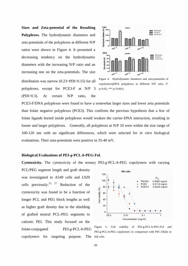
89
Sizes and Zeta-potential of the Resulting
Polyplexes. The hydrodynamic diameters and
zeta potentials of the polyplexes at different N/P
ratios were shown in Figure 4. It presented a
decreasing tendency on the hydrodynamic
diameters with the increasing N/P ratio and an
increasing one on the zeta-potentials. The size
distribution was narrow (0.23>PDI>0.15) for all
polyplexes, except for PCE3-F at N/P 3
(PDI>0.3). At certain N/P ratio, the
PCE3-F/DNA polyplexes were found to have a somewhat larger sizes and lower zeta potentials
than folate negative polyplexes (PCE3). This confirms the previous hypothesis that a few of
folate ligands buried inside polyplexes would weaken the carrier-DNA interaction, resulting in
looser and larger polyplexes. Generally, all polyplexes at N/P 10 were within the size range of
100-120 nm with no significant differences, which were selected for in vitro biological
evaluations. Their zeta-potentials were positive in 35-40 mV.
Biological Evaluations of PEI-g-PCL-b-PEG-Fol.
Cytotoxicity. The cytotoxicity of the ternary PEI-g-PCL-b-PEG copolymers with varying
PCL/PEG segment length and graft density
was investigated in A549 cells and L929
cells previously.25, 27
Reduction of the
cytotoxicity was found to be a function of
longer PCL and PEG block lengths as well
as higher graft density due to the shielding
of grafted neutral PCL-PEG segments to
cationic PEI. This study focused on the
folate-conjugated PEI-g-PCL-b-PEG
copolymers for targeting purpose. The
Figure 4. Hydrodynamic diameters and zeta-potentials of
copolymer/pDNA polyplexes at different N/P ratio. (*
p<0.05, *** p<0.001)
Figure 5. Cell viability of PEI-g-PCL-b-PEG-Fol and
PEI-g-PCL-b-PEG copolymers in comparison with PEI 25kDa in
KB cells.
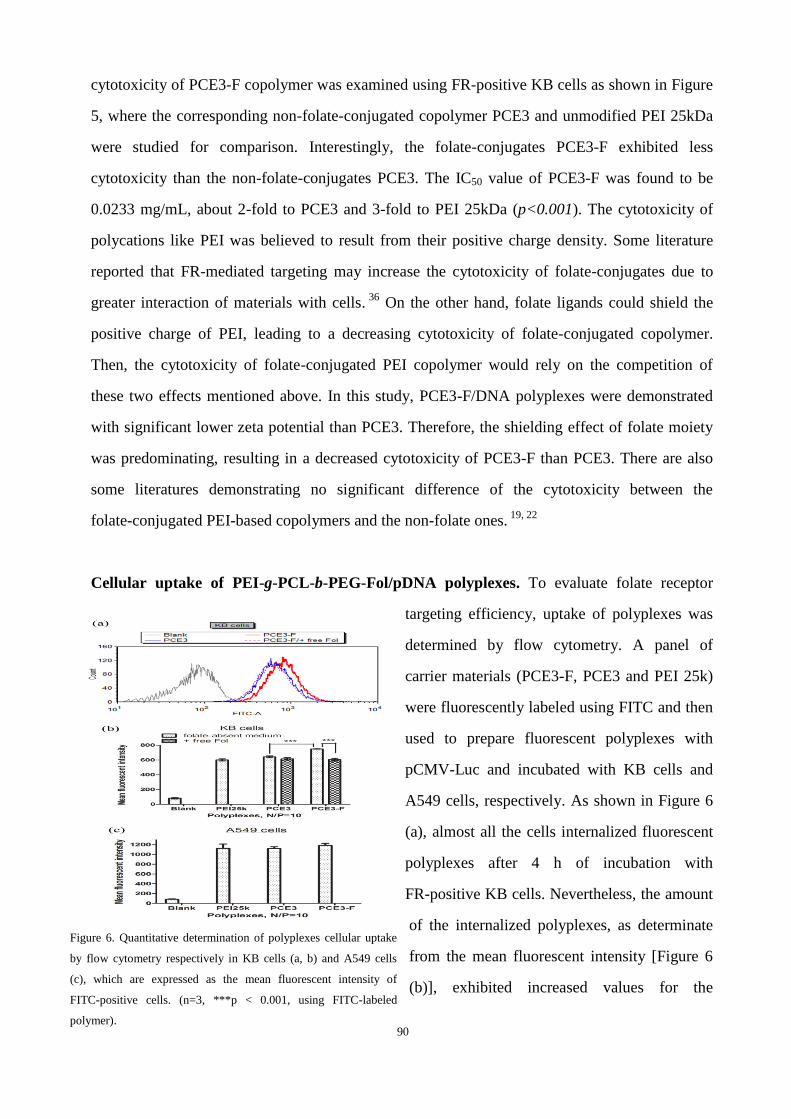
90
cytotoxicity of PCE3-F copolymer was examined using FR-positive KB cells as shown in Figure
5, where the corresponding non-folate-conjugated copolymer PCE3 and unmodified PEI 25kDa
were studied for comparison. Interestingly, the folate-conjugates PCE3-F exhibited less
cytotoxicity than the non-folate-conjugates PCE3. The IC50 value of PCE3-F was found to be
0.0233 mg/mL, about 2-fold to PCE3 and 3-fold to PEI 25kDa (p<0.001). The cytotoxicity of
polycations like PEI was believed to result from their positive charge density. Some literature
reported that FR-mediated targeting may increase the cytotoxicity of folate-conjugates due to
greater interaction of materials with cells. 36
On the other hand, folate ligands could shield the
positive charge of PEI, leading to a decreasing cytotoxicity of folate-conjugated copolymer.
Then, the cytotoxicity of folate-conjugated PEI copolymer would rely on the competition of
these two effects mentioned above. In this study, PCE3-F/DNA polyplexes were demonstrated
with significant lower zeta potential than PCE3. Therefore, the shielding effect of folate moiety
was predominating, resulting in a decreased cytotoxicity of PCE3-F than PCE3. There are also
some literatures demonstrating no significant difference of the cytotoxicity between the
folate-conjugated PEI-based copolymers and the non-folate ones. 19, 22
Cellular uptake of PEI-g-PCL-b-PEG-Fol/pDNA polyplexes. To evaluate folate receptor
targeting efficiency, uptake of polyplexes was
determined by flow cytometry. A panel of
carrier materials (PCE3-F, PCE3 and PEI 25k)
were fluorescently labeled using FITC and then
used to prepare fluorescent polyplexes with
pCMV-Luc and incubated with KB cells and
A549 cells, respectively. As shown in Figure 6
(a), almost all the cells internalized fluorescent
polyplexes after 4 h of incubation with
FR-positive KB cells. Nevertheless, the amount
of the internalized polyplexes, as determinate
from the mean fluorescent intensity [Figure 6
(b)], exhibited increased values for the
Figure 6. Quantitative determination of polyplexes cellular uptake
by flow cytometry respectively in KB cells (a, b) and A549 cells
(c), which are expressed as the mean fluorescent intensity of
FITC-positive cells. (n=3, ***p < 0.001, using FITC-labeled
polymer).
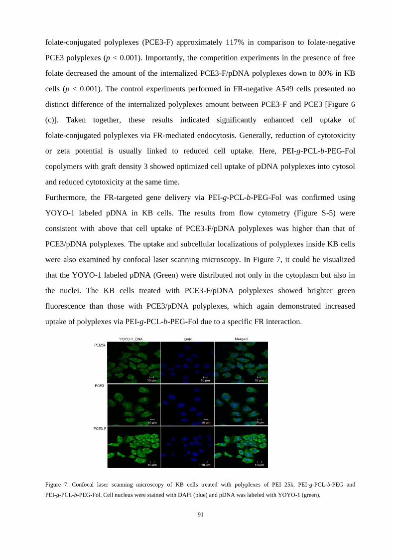
91
folate-conjugated polyplexes (PCE3-F) approximately 117% in comparison to folate-negative
PCE3 polyplexes (p < 0.001). Importantly, the competition experiments in the presence of free
folate decreased the amount of the internalized PCE3-F/pDNA polyplexes down to 80% in KB
cells (p < 0.001). The control experiments performed in FR-negative A549 cells presented no
distinct difference of the internalized polyplexes amount between PCE3-F and PCE3 [Figure 6
(c)]. Taken together, these results indicated significantly enhanced cell uptake of
folate-conjugated polyplexes via FR-mediated endocytosis. Generally, reduction of cytotoxicity
or zeta potential is usually linked to reduced cell uptake. Here, PEI-g-PCL-b-PEG-Fol
copolymers with graft density 3 showed optimized cell uptake of pDNA polyplexes into cytosol
and reduced cytotoxicity at the same time.
Furthermore, the FR-targeted gene delivery via PEI-g-PCL-b-PEG-Fol was confirmed using
YOYO-1 labeled pDNA in KB cells. The results from flow cytometry (Figure S-5) were
consistent with above that cell uptake of PCE3-F/pDNA polyplexes was higher than that of
PCE3/pDNA polyplexes. The uptake and subcellular localizations of polyplexes inside KB cells
were also examined by confocal laser scanning microscopy. In Figure 7, it could be visualized
that the YOYO-1 labeled pDNA (Green) were distributed not only in the cytoplasm but also in
the nuclei. The KB cells treated with PCE3-F/pDNA polyplexes showed brighter green
fluorescence than those with PCE3/pDNA polyplexes, which again demonstrated increased
uptake of polyplexes via PEI-g-PCL-b-PEG-Fol due to a specific FR interaction.
Figure 7. Confocal laser scanning microscopy of KB cells treated with polyplexes of PEI 25k, PEI-g-PCL-b-PEG and
PEI-g-PCL-b-PEG-Fol. Cell nucleus were stained with DAPI (blue) and pDNA was labeled with YOYO-1 (green).
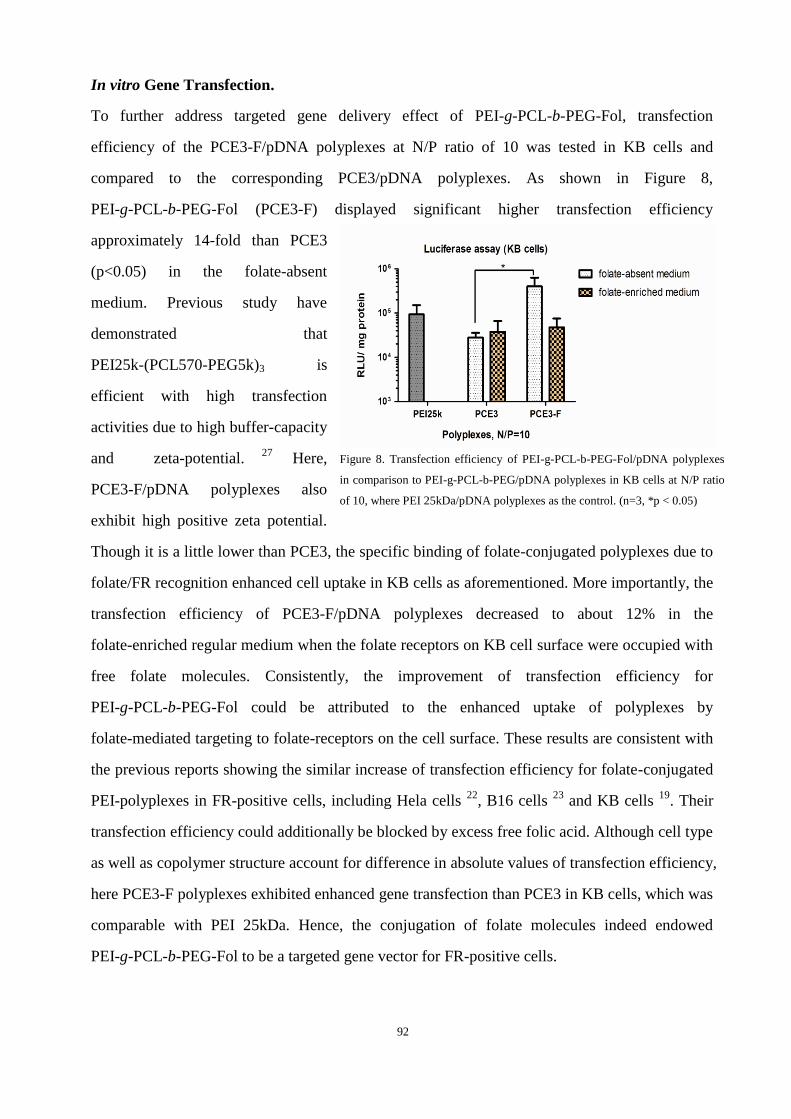
92
In vitro Gene Transfection.
To further address targeted gene delivery effect of PEI-g-PCL-b-PEG-Fol, transfection
efficiency of the PCE3-F/pDNA polyplexes at N/P ratio of 10 was tested in KB cells and
compared to the corresponding PCE3/pDNA polyplexes. As shown in Figure 8,
PEI-g-PCL-b-PEG-Fol (PCE3-F) displayed significant higher transfection efficiency
approximately 14-fold than PCE3
(p<0.05) in the folate-absent
medium. Previous study have
demonstrated that
PEI25k-(PCL570-PEG5k)3 is
efficient with high transfection
activities due to high buffer-capacity
and zeta-potential. 27
Here,
PCE3-F/pDNA polyplexes also
exhibit high positive zeta potential.
Though it is a little lower than PCE3, the specific binding of folate-conjugated polyplexes due to
folate/FR recognition enhanced cell uptake in KB cells as aforementioned. More importantly, the
transfection efficiency of PCE3-F/pDNA polyplexes decreased to about 12% in the
folate-enriched regular medium when the folate receptors on KB cell surface were occupied with
free folate molecules. Consistently, the improvement of transfection efficiency for
PEI-g-PCL-b-PEG-Fol could be attributed to the enhanced uptake of polyplexes by
folate-mediated targeting to folate-receptors on the cell surface. These results are consistent with
the previous reports showing the similar increase of transfection efficiency for folate-conjugated
PEI-polyplexes in FR-positive cells, including Hela cells 22
, B16 cells 23
and KB cells 19
. Their
transfection efficiency could additionally be blocked by excess free folic acid. Although cell type
as well as copolymer structure account for difference in absolute values of transfection efficiency,
here PCE3-F polyplexes exhibited enhanced gene transfection than PCE3 in KB cells, which was
comparable with PEI 25kDa. Hence, the conjugation of folate molecules indeed endowed
PEI-g-PCL-b-PEG-Fol to be a targeted gene vector for FR-positive cells.
Figure 8. Transfection efficiency of PEI-g-PCL-b-PEG-Fol/pDNA polyplexes
in comparison to PEI-g-PCL-b-PEG/pDNA polyplexes in KB cells at N/P ratio
of 10, where PEI 25kDa/pDNA polyplexes as the control. (n=3, *p < 0.05)
93
5.5 Conclusions
Folate-conjugated ternary copolymer PEI-g-PCL-b-PEG-Fol was successfully synthesized by
click cyclo-addition of azido-folate with heterobifunctional acrylate-PCL-b-PEG-alkyne,
followed by Michael addition with PEI, for targeted gene delivery. Lower cytotoxicity was
observed for PEI-g-PCL-b-PEG-Fol than PEI-g-PCL-b-PEG, which was much lower than
unmodified PEI 25kDa. The cellular uptake of polyplexes was enhanced by
PEI-g-PCL-b-PEG-Fol in FR over-expressing KB cells compared with those by
PEI-g-PCL-b-PEG. Importantly, this enhancement was inhibited by free folic acid, while did not
appear in FR-negative A549 cells. All these suggested the specific cell uptake of
PEI-g-PCL-b-PEG-Fol/pDNA polyplexes via folate receptor-mediated endocytosis.
Consequently, PEI-g-PCL-b-PEG-Fol/pDNA polyplexes revealed higher transfection than
PEI-g-PCL-b-PEG/pDNA. Here,PEI-g-PCL-b-PEG-Fol is a novel kind of ternary copolymers
with amphiphilicity, cationic property and folate-ligands, which endows them multifunctional
prospect to co-delivery both gene and hydrophobic chemotherapeutic drug to target tumor tissue.
Nevertheless, continued efforts still need to be considered to reduce nonspecific uptake and
increase transfection efficiency further. Additional studies on gene transfection in vivo and
utilizing these described folate-conjugated copolymers for targeted siRNA delivery are in
proceeding.
5.6 Acknowledgment. Financial support of Dr. Liu, L. by the Alexander von Humboldt
Foundation is gratefully acknowledged.
5.7 Supporting Information Available. Details of characterization of intermediate products,
standard line of UV absorbance of folate solution and FACS data of polyplexes with YOYO-1
labeled pDNA in KB cells. This material is available free of charge via the Internet at
http://pubs.acs.org.
5.8 REFERENCES.
(1) Olefsky, J. M. (2000) Diabetes: Gene therapy for rats and mice. Nature 408, 420-421.
94
(2) Merdan, T., Kopecek, J., and Kissel, T. (2002) Prospects for cationic polymers in gene and
oligonucleotide therapy against cancer. Adv. Drug Delivery Rev. 54, 715-758.
(3) Wagner, A. M., Schoeberlein, A., and Surbek, D. (2009) Fetal gene therapy: Opportunities and risks.
Adv. Drug Delivery Rev. 61, 813-821.
(4) Mintzer, M. A., and Simanek, E. E. (2008) Nonviral Vectors for Gene Delivery. Chem. Rev. 109,
259-302.
(5) Pack, D. W., Hoffman, A. S., Pun, S., and Stayton, P. S. (2005) Design and development of polymers
for gene delivery. Nat. Rev. Drug Discov. 4, 581-593.
(6) Boussif, O., Lezoualc'h, F., Zanta, M. A., Mergny, M. D., Scherman, D., Demeneix, B., and Behr, J.
P. (1995) A versatile vector for gene and oligonucleotide transfer into cells in culture and in vivo:
polyethylenimine. Proc. Natl. Acad. Sci. 92, 7297-7301.
(7) Behr, J.-P. (1997) The Proton Sponge: a Trick to Enter Cells the Viruses Did Not Exploit. CHIMIA
Int. J. Chem. 51, 34-36.
(8) Petersen, H., Fechner, P. M., Fischer, D., and Kissel, T. (2002) Synthesis, Characterization, and
Biocompatibility of Polyethylenimine-graft-poly(ethylene glycol) Block Copolymers.
Macromolecules 35, 6867-6874.
(9) Petersen, H., Fechner, P. M., Martin, A. L., Kunath, K., Stolnik, S., Roberts, C. J., Fischer, D., Davies,
M. C., and Kissel, T. (2002) Polyethylenimine-graft-Poly(ethylene glycol) Copolymers:Influence of
Copolymer Block Structure on DNA Complexation and Biological Activities as Gene Delivery
System. Bioconjugate Chem. 13, 845-854.
(10) Lee, M., Kim, M., Jang, Y., Lee, K., Kim, T., Kim, S., Park, T., Kim, H., and Jeong, J. (2011)
Polyethylenimine-g-poly(lactic-co-glycolic acid) as non-toxic micelle-type carrier for gene delivery.
Macromol. Res. 19, 688-693.
(11) Pun, S. H., Bellocq, N. C., Liu, A., Jensen, G., Machemer, T., Quijano, E., Schluep, T., Wen, S.,
Engler, H., Heidel, J., and Davis, M. E. (2004) Cyclodextrin-Modified Polyethylenimine Polymers for
Gene Delivery. Bioconjugate Chem. 15, 831-840.
(12) Dachs, G., Dougherty, G., Stratford, I., and Chaplin, D. (1997) Targeting gene therapy to cancer: a
review. Oncol. Res. 9, 313-25.
(13) Salazar, M., and Ratnam, M. (2007) The folate receptor: What does it promise in tissue-targeted
therapeutics? Cancer Metast. Rev. 26, 141-152.
(14) Low, P. S., and Kularatne, S. A. (2009) Folate-targeted therapeutic and imaging agents for cancer.
Curr. Opin. Chem. Biol. 13, 256-262.
(15) Xia, W., and Low, P. S. (2010) Folate-Targeted Therapies for Cancer. J. Med. Chem. 53, 6811-6824.
(16) Benns, J. M., Maheshwari, A., Furgeson, D. Y., Mahato, R. I., and Kim, S. W. (2001)
Folate-PEG-Folate-Graft-Polyethylenimine-Based Gene Delivery. J. Drug Target. 9, 123-139.
(17) Liang, B., He, M. L., Xiao, Z. P., Li, Y., Chan, C. Y., Kung, H. F., Shuai, X. T., and Peng, Y. (2008)
Synthesis and characterization of folate-PEG-grafted-hyperbranched-PEI for tumor-targeted gene
delivery. Biochem. Biophys. Res. Commun. 367, 874-880.
95
(18) Liang, B., He, M.-L., Chan, C.-y., Chen, Y.-c., Li, X.-P., Li, Y., Zheng, D., Lin, M. C., Kung, H.-F.,
Shuai, X.-T., and Peng, Y. (2009) The use of folate-PEG-grafted-hybranched-PEI nonviral vector for
the inhibition of glioma growth in the rat. Biomaterials 30, 4014-4020.
(19) Arote, R. B., Hwang, S. K., Lim, H. T., Kim, T. H., Jere, D., Jiang, H. L., Kim, Y. K., Cho, M. H., and
Cho, C. S. (2010) The therapeutic efficiency of FP-PEA/TAM67 gene complexes via folate
receptor-mediated endocytosis in a xenograft mice model. Biomaterials 31, 2435-2445.
(20) He, M. L. (2009) The use of folate-PEG-grafted-hybranched-PEI nonviral vector for the inhibition of
glioma growth in the rat. Int. J. Mol. Med. 24, 168.
(21) Hwa Kim, S., Hoon Jeong, J., Chul Cho, K., Wan Kim, S., and Gwan Park, T. (2005) Target-specific
gene silencing by siRNA plasmid DNA complexed with folate-modified poly(ethylenimine). J.
Controlled Release 104, 223-232.
(22) Cheng, H., Zhu, J. L., Zeng, X., Jing, Y., Zhang, X. Z., and Zhuo, R. X. (2009) Targeted Gene
Delivery Mediated by Folate-polyethylenimine-block-poly(ethylene glycol) with Receptor Selectivity.
Bioconjugate Chemistry 20, 481-487.
(23) Yao, H., Ng, S. S., Tucker, W. O., Tsang, Y. K. T., Man, K., Wang, X. M., Chow, B. K. C., Kung, H.
F., Tang, G. P., and Lin, M. C. (2009) The gene transfection efficiency of a
folate-PEI600-cyclodextrin nanopolymer. Biomaterials 30, 5793-5803.
(24) Shuai, X., Merdan, T., Unger, F., Wittmar, M., and Kissel, T. (2003) Novel Biodegradable Ternary
Copolymers hy-PEI-g-PCL-b-PEG: Synthesis, Characterization, and Potential as Efficient Nonviral
Gene Delivery Vectors. Macromolecules 36, 5751-5759.
(25) Liu, Y., Nguyen, J., Steele, T., Merkel, O., and Kissel, T. (2009) A new synthesis method and
degradation of hyper-branched polyethylenimine grafted polycaprolactone block mono-methoxyl poly
(ethylene glycol) copolymers (hy-PEI-g-PCL-b-mPEG) as potential DNA delivery vectors. Polymer
50, 3895-3904.
(26) Liu, Y., Samsonova, O., Sproat, B., Merkel, O., and Kissel, T. (2011) Biophysical characterization of
hyper-branched polyethylenimine-graft- polycaprolactone-block-mono-methoxyl-poly(ethylene
glycol) copolymers (hy-PEI-PCL-mPEG) for siRNA delivery. J. Controlled Release 153, 262-8.
(27) Zheng, M., Liu, Y., Samsonova, O., Endres, T., Merkel, O., and Kissel, T. (2011) Amphiphilic and
biodegradable hy-PEI-g-PCL-b-PEG copolymers efficiently mediate transgene expression depending
on their graft density. Int. J. Pharm. In Press, Corrected Proof.
(28) Lee, S. M., Chen, H. M., O'Halloran, T. V., and Nguyen, S. T. (2009) "Clickable" Polymer-Caged
Nanobins as a Modular Drug Delivery Platform. J. Am. Chem. Soc. 131, 9311-9320.
(29) Storey, R. F., and Sherman, J. W. (2002) Kinetics and Mechanism of the Stannous Octoate-Catalyzed
Bulk Polymerization of Caprolactone. Macromolecules 35, 1504-1512.
(30) Merkel, O. M., Mintzer, M. A., Librizzi, D., Samsonova, O., Dicke, T., Sproat, B., Garn, H., Barth, P.
J., Simanek, E. E., and Kissel, T. (2010) Triazine Dendrimers as Nonviral Vectors for in Vitro and in
Vivo RNAi: The Effects of Peripheral Groups and Core Structure on Biological Activity. Mol.
Pharmaceutics 7, 969-983.
(31) Jiang, H. L., Xu, C. X., Kim, Y. K., Arote, R., Jere, D., Lim, H. T., Cho, M. H., and Cho, C. S. (2009)
The suppression of lung tumorigenesis by aerosol-delivered

96
folate-chitosan-graft-polyethylenimine/Akt1 shRNA complexes through the Akt signaling pathway.
Biomaterials 30, 5844-5852.
(32) Parrish, B., and Emrick, T. (2006) Soluble Camptothecin Derivatives Prepared by Click Cycloaddition
Chemistry on Functional Aliphatic Polyesters. Bioconjugate Chem. 18, 263-267.
(33) Mindt, T. L., Muller, C., Stuker, F., Salazar, J. F., Hohn, A., Mueggler, T., Rudin, M., and Schibli, R.
(2009) A "Click Chemistry" Approach to the Efficient Synthesis of Multiple Imaging Probes Derived
from a Single Precursor. Bioconjugate Chemistry 20, 1940-1949.
(34) van Dijk, M., Rijkers, D. T. S., Liskamp, R. M. J., van Nostrum, C. F., and Hennink, W. E. (2009)
Synthesis and Applications of Biomedical and Pharmaceutical Polymers via Click Chemistry
Methodologies. Bioconjugate Chem. 20, 2001-2016.
(35) Valencia, P. M., Hanewich-Hollatz, M. H., Gao, W., Karim, F., Langer, R., Karnik, R., and
Farokhzad, O. C. (2011) Effects of ligands with different water solubilities on self-assembly and
properties of targeted nanoparticles. Biomaterials 32, 6226-6233.
(36) Zheng, Y., Cai, Z., Song, X., Yu, B., Bi, Y., Chen, Q., Zhao, D., Xu, J., and Hou, S. (2009) Receptor
mediated gene delivery by folate conjugated N-trimethyl chitosan in vitro. Int. J. Pharm. 382, 262-269.

97
Chapter 6
MOLECULAR MODELING AND IN VIVO IMAGING CAN IDENTIFY
SUCCESSFUL HYPERFLEXIBLE TRIAZINE DENDRIMER-BASED
SIRNA DELIVERY SYSTEMS
Published in The Journal of Control Release 2011, 153 (1), 23-33.
Olivia M. Merkela1*
, Mengyao Zhenga1
, Meredith A. Mintzerb, Giovanni M. Pavan
c,
Damiano Librizzid, Marek Maly
c,e, Helmut Höffken
d, Andrea Danani
c, Eric E. Simanek
f,
and Thomas Kissela
1Both authors contributed equally to this work
Author contributions
T. K. and E. E. S. guided and directed the research and directed the measurements. M. Z. carried
out the measurement of size and zetapotential, CLSM, in vitro knockdown experiment. M. A. M.
synthesized and characterized the polymers. M. Z. and O. M. M. analysed the experimental data.

98
6.1 Abstract
The aim of this study was to identify suitable siRNA delivery systems based on hyperflexible
generation 2-4 triazine dendrimers by correlating physico-chemical and biological in vitro and in
vivo properties of the complexes with their thermodynamic interaction features simulated by
molecular modeling. The siRNA binding properties of the dendrimers in comparison to PEI 25
kDa were simulated, binding and stability were measured in SYBR Gold assays, hydrodynamic
diameters and zeta potentials were investigated, and cytotoxicity was quantified. These parameters
were paralleled with cellular uptake of the complexes and their ability to mediate RNAi. The
radiolabeled complexes were administered intravenously, and their pharmacokinetic profiles and
biodistribution were assessed invasively and non-invasively. All flexible triazine dendrimers
formed thermodynamically more stable complexes than PEI. While PEI and generation 4
dendrimer interacted more superficially with siRNA, generation 2 and 3 virtually coalesced with
siRNA. These dendriplexes were therefore more efficiently charge-neutralized than PEI
complexes, reducing agglomeration. This was confirmed by results of hydrodynamic diameters
(72.0 nm – 153.5 nm) and zeta potentials (4.9 mV – 21.8 mV in 10 mM HEPES) of the
dendriplexes in comparison to PEI complexes (312.8 nm – 480.0 nm and 13.7 mV – 17.4 mV in 10
mM HEPES). All dendrimers, even generation 3 and 4, were less toxic than PEI, all dendriplexes
were efficiently endocytosed and showed significant and specific luciferase knockdown in
HeLa/Luc cells. Scintillation counting confirmed that the generation 2 triazine complexes showed
more than twofold prolonged circulation times as a result of their good thermodynamic stability,
whereas generation 3 complexes dissociated in vivo, and generation 4 complexes were captured by
the reticulo-endothelial system due to their increased surface charge. Since molecular modeling
helped to understand experimental parameters based on the dendrimers’ structural properties and
molecular imaging non-invasively predicted the in vivo fate of the complexes, both techniques can
efficiently support the rapid development of safe and efficient siRNA formulations that are stable
in vivo.
Keywords: Triazine dendrimers, RNA interference, molecular modeling, structure function
relationship, physico-chemical characterization, in vivo, biodistribution, pharmacokinetics, SPECT
imaging

99
6.2 Introduction
Dendrimers are increasingly employed for siRNA delivery. While PAMAM and its derivatives are
most widely used [1], polypropylenimine (PPI) [2], dendritic poly-L-lysine (PLL) [3] and newer
classes such as carbosilane dendrimers [4] and triazine dendrimers [5] are gaining more importance
in the field of non-viral siRNA delivery. About 50 reports of dendrimer-mediated siRNA delivery
can be found in the literature to date whereas only six studies describe in vivo results [2, 3, 5-8].
This can be understood as a consequence of the recent use of dendrimers for siRNA delivery
needing further optimization in terms of efficiency versus toxicity to allow in vivo administration.
Currently, the most relevant obstacle was seen in several studies where low generations of
dendrimers were not able to condense siRNA into uniformly small complexes [9-11]. The use of
higher generations such as 6 and 7 [1, 3, 9, 11-14], however, is often accompanied by an increase
in toxicity [15]. Therefore, many structural modifications, e.g. carboxylate-terminated [16],
acetylated [17], internally cationic and hydroxyl-terminated PAMAM dendrimers [18] were
synthesized for enhanced biocompatibility, that, however, exhibited decreased in vitro efficiency
[17, 18]. The lack of in vitro knockdown efficiency of PAMAM has been attributed to incomplete
endosomal release of the siRNA [19] and nuclear localization of oligonucleotides [20]. In an
earlier study, it was shown that endosomal release of siRNA can be modulated by introduction of
short lipophilic C6-groups in the periphery of triazine dendrimers [5]. The alkylated generation 2
“rigid core” dendrimer efficiently mediating in vitro gene silencing, however, accumulated
strongly in the lung after intravenous injection although hydrodynamic diameters of the complexes
with siRNA were 103 nm as measured in buffer [5]. Since complex formation with the generation
3 rigid dendrimer did not lead to smaller dendriplexes (178 nm) and since dendrimer G3-1 had
affected cell viability significantly stronger than G2-1 [21], this study focuses on a new panel of
“hyperflexible” triazine dendrimers. Flexible triethanolamine core PAMAM dendriplexes of
generation 7 were reported to exhibit almost no cytotoxicity in MTT and LDH assays [13],
indicating that a flexible core may reduce the toxicity of higher generation dendrimers. Similarly, a
flexible generation 2 triazine dendrimer F2-1 was previously shown to cause reduced hemolysis
than G1-1, G2-1, and G3-1 rigid core analogues [21]. However, F2-1 was found to present a
“collapsed” topology, leading to less interaction with siRNA than possible with the dendrimers that
were actually expected to be more rigid [22] and formation of loosely associated large

100
agglomerates of 286 nm in size [5]. In an approach to enhance the interaction with and
condensation of siRNA, new hyperflexible dendrimers of generation 2, 3, and 4 were synthesized
and characterized in this study concerning computed siRNA binding characteristics. The results
obtained from in silico simulations were compared with experimental physico-chemical parameters
such as siRNA complexation, complex stability, size, and zeta potentials. Since these properties are
expected to determine siRNA packaging, dendriplex endocytosis, unpackaging, stability in the
blood stream and thus the RNAi efficiency of siRNA formulations [23], it was investigated if in
silico results were able to predict the in vitro and in vivo performance of the dendriplexes and if
radioactive in vivo imaging could support to identify efficient siRNA delivery systems.
6.3 Experimental Section
Materials. Poly(ethylene imine) (Polymin™, 25 kDa) was a gift from BASF (Ludwigshafen,
Germany). Lipofectamine™2000 (LF) was bought from Invitrogen (Karlsruhe, Germany),
Beetle Luciferin, and heparin sodium salt from Sigma-Aldrich Laborchemikalien GmbH (Seelze,
Germany). 2’-O-Methylated 25/27mer DsiRNA targeting firefly luciferase (FLuc, sense:
5’-pGGUUCCUGGAACAAUUGCUUUUAdCdA, antisense:
3’-mGmAmCCmAAmGGmACmCUmUGmUUmAAmCGAAAAUGU), negative control
sequence (NegCon, sense: 5’-pCGUUAAUCGCGUAUAAUACGCGUdAdT, antisense:
3 -́mCmAmGCmAAmUUmAGmCGmCAmUAmUUmAUGCGCAUAp), TYE546- and 5’-sense
strand C6-amine modified DsiRNA were obtained from Integrated DNA Technologies (IDT,
Leuven, Belgium). Balb/c mice (6 weeks old) were bought from Harlan Laboratories (Horst, The
Netherlands). The chelator 2-(4-isothiocyanatobenzyl)-diethylenetriaminepentaacetic acid
(p-SCN-Bn-DTPA) was purchased from Macrocyclics (Dallas, TX, USA), SYBR® Gold from
Invitrogen (Karlsruhe, Germany), and all chemicals used for synthesis were obtained from
Sigma-Aldrich (St. Louis, MO).
Synthesis of new triazine dendrimers. The hyperflexible triazine dendrimers F2-2, F3, and F4-2
used in this study were synthesized following a previously described divergent approach [21, 24].
The final products and all intermediate structures were characterized by 1H and
13C NMR
spectroscopy, and mass spectrometry, as shown in the Supplementary data. For comparison,
101
generations 2, 3, and 4 were synthesized of which generation 2 and 4 bear the same periphery
(Figure 1), previously designated as periphery number 2 [24].
Molecular modeling. The model for Dicer Substrate siRNA was constructed with the NAB
module within the AMBER 11 suite of programs [25]. Since the study of multivalent molecular
recognition that characterizes the complexation was subject of this study, the binding between
siRNA and dendrimers in a 1:1 ratio was modeled according to a validated strategy previously
reported for the binding of dendrons and dendrimers with DNA [26, 27] and siRNA [28]. The
structures of F2-2, F3 and F4-2 were composed of different residues according to previous studies
on similar dendrimers [22]. The model of random branched 25kDa PEI was created and
pre-optimized using the Materials Studio (MS) software package (Accelrys). The total charges of
F2-2, F3, F4-2 and PEI are +12, +24, +48 and +98, respectively, at pH 7.4 [29]. Each of the
non-standard residues that compose the dendrimers was parameterized with the antechamber
module of AMBER 11 following a well validated procedure previously adopted [31].
Molecular dynamic simulations. All simulations and data analyses were performed with the
AMBER 11 suite of programs (sander.MPI and pmemd.cuda modules) [25]. The dendrimers were
solvated in a TIP3P water box [30], minimized and then equilibrated by running 10 nanoseconds
NPT molecular dynamics simulations, as described previously [22] to obtain a reliable
configuration for PEI, F2-2, F3 and F4-2 in solution (Figure S1). The water molecules and counter
ions were removed, and the polycations were placed in close proximity to the major groove of
DsiRNA. The four complexes were re-solvated, the resulting molecular systems were minimized
[22] and equilibrated for 20 ns in NPT periodic boundary condition at 300 K and 1 atm using a
time step of 2 fs, the Langevin thermostat, and a 10 Å cut-off. To treat long-range electrostatic
effects, the particle mesh Ewald (PME) approach was adopted [31], and the SHAKE algorithm was
used to constrain all bonds involving hydrogen atoms. Each of the molecular dynamics runs were
carried out using parm99 all-atom force field [32] with NVIDIA Tesla 2050 GPU cards. The free
Gbind Hbind Sbind) were calculated according to the MM-PBSA
approach [33] and the normal-mode analysis [34] on 100 MD frames taken from the equilibrated
102
MD trajectories according to previous studies on the interactions between dendrimers and nucleic
acids [22, 28].
Dendriplex formation. Dendriplexes were formed as previously described by adding 25 µl of a
calculated concentration of dendrimer to an equal volume of 2 µM siRNA followed by vigorous
pipetting [5]. Both siRNA and dendrimers were diluted with an isotonic solution of 5% glucose
unless otherwise described. The appropriate amount and “protonable unit” of each dendrimer to
afford a certain N/P ratio, which is the excess of polymer amines (N) over siRNA phosphates (P),
was calculated as previously described [21]. Polyplexes with PEI 25 kDa were formed as
described above, and lipoplexes with LF with 1 µl per 20 pmol were prepared as recommended by
the manufacturer.
SYBR Gold® Assay. The ability of the three dendrimers to bind and protect siRNA was studied as
previously reported and compared with PEI 25kDa [5]. Briefly, complexes of 1 µg siRNA were
prepared at different N/P ratios, incubated for 20 minutes before 50 µl of a 1x SYBR® Gold
solution was added and incubated for another 10 minutes in the dark. Free or accessible siRNA
was quantified using a SAFIRE II fluorescence plate reader (Tecan Group Ltd, Männedorf,
Switzerland) at 495 nm excitation and 537 nm emission wavelengths. The results are given as
mean relative fluorescence intensity values (n=3) +/- the standard deviation (SD), where
intercalation of free siRNA represents 100 % fluorescence, and non-intercalating SYBR® Gold in
buffer represents 0 % remaining fluorescence.
Heparin Competition Assay. The stability of the dendriplexes against competing polyanions,
such as the model molecule heparin, was studied as previously reported [5]. Briefly, dendriplexes
were formed at N/P=5, incubated with 50 µl of a 1x SYBR® Gold solution, and treated with
increasing amounts of heparin for 20 minutes. Fluorescence was quantified as described above.
Results are given as mean relative fluorescence intensity values (n=3) +/- SD, where free siRNA
represents 100 % fluorescence, and SYBR® Gold in buffer represents 0 % fluorescence.

103
Dynamic Light Scattering and Zeta Potential Analysis. Dendriplexes of the various generations
of hyperflexible triazine dendrimers and siRNA were characterized concerning their hydrodynamic
diameters and zeta potentials, whereas PEI 25kDa served as control. Dendriplexes and PEI
polyplexes were formed as described above in 5 % glucose, HEPES-buffered glucose (HBG: 5%
glucose, 10 mM HEPES, pH 7.4) or 10 mM HEPES buffer at increasing N/P ratios and measured
as previously described in a disposable low volume UVette (Eppendorf, Wesseling-Berzdorf,
Germany) using a Zetasizer Nano ZS (Malvern, Herrenberg, Germany) [5]. Zeta potentials were
determined by laser Doppler anemometry (LDA) after diluting the samples with 750 µl of the
equivalent solvent to a final volume of 800 µl and transferring the suspensions into a green zeta
cuvette (Malvern, Herrenberg, Germany). Results are given as mean values (n=3) +/- SD.
Cell Culture. HeLa cells stably expressing luciferase (HeLa/Luc) [35] were maintained at 100
µg/ml hygromycin B in DMEM high glucose (PAA Laboratories, Cölbe, Germany) supplemented
with 10 % fetal bovine serum (Cytogen, Sinn, Germany) in a humidified atmosphere with 5 % CO2
at 37°C, and seeded for experiments in antibiotics-free medium. L929 cells were cultured in
antibiotics-free DMEM high glucose (PAA Laboratories, Cölbe, Germany) supplemented with 10
% fetal bovine serum (Cytogen, Sinn, Germany).
Confocal laser scanning microscopy (CLSM). As previously described, uptake and subcellular
distribution of dendriplexes was investigated by confocal microscopy [5]. Briefly, HeLa/Luc cells
were plated on 16-chamber slides and transfected 24 h later at N/P ratios of 10 and 20 with 25
pmol TYE-546-labeled siRNA per chamber in a total volume of 250 µl. Cells transfected with free
siRNA or Lipofectamine™2000 were used as negative and positive controls, respectively. After 4
h of incubation, cells were washed, fixed with 4% paraformaldehyde, counterstained with
4',6-diamidino-2-phenylindole (DAPI) (Molecular Probes, Invitrogen, Karlsruhe, Germany), and
embedded with FluorSave (Calbiochem, Merck Biosciences, Darmstadt, Germany). For confocal
microscopy on a Zeiss Axiovert 100 M microscope and a Zeiss LSM 510 scanning device (Zeiss,
Oberkochen, Germany), lasers and filter settings were chosen as previously described [5].

104
Transfection efficiency. The ability of the different generation dendrimers to mediate RNA
interference in vitro was quantified by luciferase knockdown in HeLa/Luc cells as previously
reported [5]. Briefly, cells were seeded at a density of 15,000 cells per well in 48-well-plates and
treated with dendriplexes of 50 pmol siRNA (siFLuc or siNegCon) and different N/P ratios 24 h
after seeding. As positive controls, transfections were performed with Lipofectamine™2000. The
medium was changed 4 hours post transfection, cells were incubated for another 44 h before they
were washed with PBS buffer, lysed with CCLR (Promega) and assayed for luciferase expression
on a BMG luminometer plate reader (BMG Labtech, Offenburg, Germany) [5]. Results are given
as mean values (n=4) +/- SD.
Radiolabeling and Purification. Pharmacokinetics and biodistribution after i.v. injection were
investigated with radiolabeled siRNA administered freely or as dendriplexes. Polyplexes formed
with PEI 25kDa were administered as control. siRNA was labeled and purified as previously
described [35, 36]. Briefly, amine-modified siRNA was reacted with p-Bn-SCN-DTPA at pH 8.5
for 3 h before it was precipitated in 10% sodium acetate and 70% ethanol overnight. After
centrifugation for 5 min at 12,000 g, DTPA-coupled siRNA was dissolved in RNase free water,
annealed in presence of 111
InCl3 (Covidien Deutschland GmbH, Neustadt a.d. Donau, Germany)
for 2 min at 94°C, incubated for 30 min at room temperature and purified from free 111
InCl3 by size
exclusion chromatography (SEC) on PD-10 Sephadex G25 (GE Healthcare, Freiburg, Germany)
and RNeasy spin column purification as described earlier [35].
In vivo Imaging, Pharmacokinetics and Biodistribution
Circulation times and distribution within the body were determined as previously described [5].
Groups of 5 BALB/c mice were anesthetized intraperitoneally and injected with dendriplexes
containing 35 µg of siRNA and the corresponding amount of dendrimer at N/P 5. Control animals
received either free siRNA or PEI25kDa/siRNA polyplexes which were prepared and labeled as
previously described [35]. Pharmacokinetics were assessed by retro-orbitally withdrawing blood
samples, and biodistribution was recorded in three-dimensional SPECT and planar gamma camera
images 2 h after injection using a Siemens e.cam gamma camera (Siemens AG, Erlangen,
Germany) equipped with a custom built multiplexing multipinhole collimator and compared to

105
results of scintillation counting of dissected organs measured using a Gamma Counter Packard
5005 (Packard Instruments, Meriden, CT) [35].
Statistics. All analytical assays were conducted in replicates of three or four, as indicated and in
vivo experiments included 5 animals per group. Results are given as mean values +/- standard
deviation (SD). Two way ANOVA and statistical evaluations were performed using Graph Pad
Prism 4.03 (Graph Pad Software, La Jolla, USA).
6.4 Results and Discussion
Figure 1. Structures of hyperflexible core triazine dendrimers F of generation number 2, 3 and 4. The monochlorotriazine from
which the peripheral group 2 for generation 2 and 4 was obtained carries one hydroxyl groups and two amines.
Nomenclature, Synthesis and Design Criteria of the Synthesized Dendrimers. The
nomenclature of the new dendrimers described in this manuscript was in line with previously
described structures [21, 24] with respect to their generation, flexible core structure and surface
group functionalities as shown in Figure 1. Previous investigations of gene delivery with triazine
dendrimers had shown that flexibility is a key factor for promoting pDNA transfection [21]
whereas different triazine dendrimers had been most efficient for siRNA delivery [5]. This
difference was explained by the rigidity of siRNA in comparison to the rather flexible behavior of
pDNA [22]. Since the “flexible” dendrimer F2-1 earlier reported as siRNA vector [5] was shown
106
to have a collapsed structure [22], it was shown that the peripheral groups had a stronger influence
on the capacity of triazine dendrimers to deliver siRNA than the difference in flexibility between
G2-1 and F2-1. Therefore, in this study a different periphery with a diethylene glycol instead of
two ethylene glycol chains was investigated and the flexibility of the dendrimers was strongly
increased by variation of the generation to achieve hyperflexible structures. The synthetic yields
NMR and MS spectroscopic data for these new structures are provided in the Supporting
Information. The composition of the panel is summarized in Figure 1. For the physicochemical and
biological assays, branched poly(ethylene imine) of 25 kDa (PEI 25kDa) and/or LF were used as
controls.
Molecular dynamic simulations and energetic and structural analyses. First, the solution phase
structures were simulated as described above and shown in the Supplementary data (Figure S1).
Subsequently, the binding affinity of the dendrimers and PEI towards partially 2’O-methylated
siRNA was calculated as shown in Table 1. The binding energies were normalized per charge and
expressed in kcal mol-1
in order to allow direct comparison between the different polycations with
respect to the averaged interaction of each surface group (Table 2). High enthalpic gain at a lower
and unfavorable entropic loss is typical of electrostatic interactions with an overall gain in absolute
free energy . Although larger generations are expected to interact stronger with the nucleic
acids, the normalized binding energies showed a different trend. This can be understood as a result
of enhanced back folding of the peripheral groups with increasing generation. While the
normalized F2-1 with unmodified GL3 siRNA was only -4.5 kcal
mol-1
[22], the binding of F2-2 to DsiRNA was comparably stronger with -9.1 kcal mol-1
per amine
which indicates additional hydrophobic interactions between the dendrimers and partially
2’O-methylated siRNA. Interestingly, F3 was the dendrimer with the lowest affinity to DsiRNA
(-5.8 kcal mol-1
) reaching only reduced enthalpic attraction at the same entropic cost as F2-2. This
can be explained by the lack of primary amines in F3. Interestingly, all dendrimers showed similar
enthalpic attraction towards DsiRNA slightly decreasing with generation ( -13.4, -10.2, and
-9.7 kcal/mol, respectively), while the entropic cost per charge in F4-2 was approximately half of
that in F3 and F2-2. The binding with DsiRNA was therefore less entropically expensive for F4-2
than for F2-2 and F3, evidencing that F4-2 maintained higher residual flexibility during the binding

107
event, while F3 and F2-2 lost more degrees of freedom. The lower entropic cost of the
complexation indicates that F4-2 interacts more superficially with DsiRNA, which is typically
known from PAMAM dendrimers [28] and was also shown here for the interaction of DsiRNA
with PEI. The normalized value for PEI was the lowest among the tested panel. This indicates
that PEI complexes are thermodynamically less stable than triazine dendrimer complexes, and that
possibly not all charged amine groups are involved in a 1:1 complex with siRNA as a consequence
of the sphere-like shape of solvated PEI. The entropic loss in all reactions was very well
compensated by the enthalpic gain leading to thermodynamically stable complexes, even in case of
PEI. From the normalized free energies, it was hypothesized that the stability of the complexes
decreases in the following order: F2-2>F4-2>F3>PEI.
F2-2 F3 F4-2 PEI
-161.3 -243.8 -466.5 -647.3
- 52.1 103.7 114.7 105.7
-109.2 -140.1 -351.7 -541.5
Table 1. G energies and the contributing potentials of the binding between dendrimers or bPEI 25 kDa and DsiRNA expressed in
kcal mol-1.
F2-2 F3 F4-2 PEI
-13.4 -10.2 -9.7 -6.6
- 4.3 4.3 2.4 1.1
-9.1 -5.8 -7.3 -5.5
o
energy per charged surface amine expressed in kcal mol-1.
Additionally, the models in Figure 2 help to understand the differences between the siRNA binding
modalities of a globular molecule like PEI (Figure 2D) and flexible molecules like F2-2 and F3
(Figures 2A-B), while F4-2 holds an intermediate position. It is obvious that in PEI only on a
limited part of the charged surface groups interact actively with siRNA while a larger part of
charged amines is back folded. This assembly leads to a complex with a distinct PEI domain next
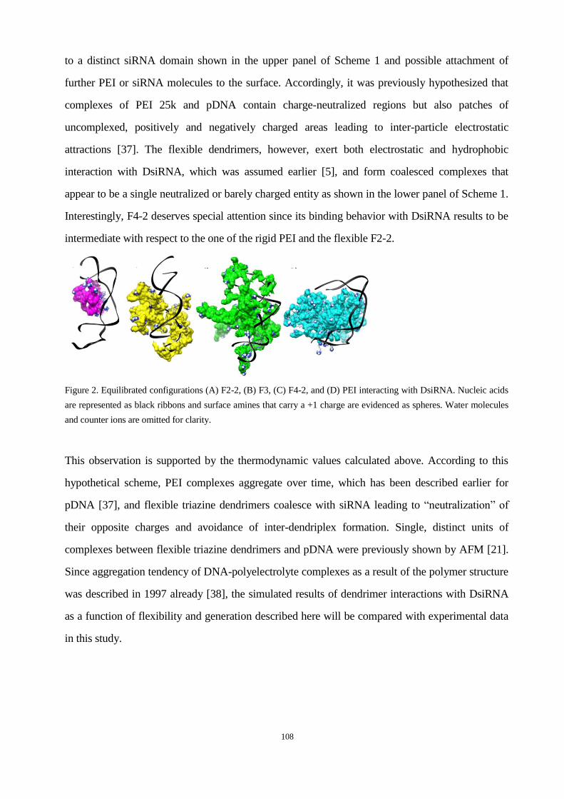
108
to a distinct siRNA domain shown in the upper panel of Scheme 1 and possible attachment of
further PEI or siRNA molecules to the surface. Accordingly, it was previously hypothesized that
complexes of PEI 25k and pDNA contain charge-neutralized regions but also patches of
uncomplexed, positively and negatively charged areas leading to inter-particle electrostatic
attractions [37]. The flexible dendrimers, however, exert both electrostatic and hydrophobic
interaction with DsiRNA, which was assumed earlier [5], and form coalesced complexes that
appear to be a single neutralized or barely charged entity as shown in the lower panel of Scheme 1.
Interestingly, F4-2 deserves special attention since its binding behavior with DsiRNA results to be
intermediate with respect to the one of the rigid PEI and the flexible F2-2.
Figure 2. Equilibrated configurations (A) F2-2, (B) F3, (C) F4-2, and (D) PEI interacting with DsiRNA. Nucleic acids
are represented as black ribbons and surface amines that carry a +1 charge are evidenced as spheres. Water molecules
and counter ions are omitted for clarity.
This observation is supported by the thermodynamic values calculated above. According to this
hypothetical scheme, PEI complexes aggregate over time, which has been described earlier for
pDNA [37], and flexible triazine dendrimers coalesce with siRNA leading to “neutralization” of
their opposite charges and avoidance of inter-dendriplex formation. Single, distinct units of
complexes between flexible triazine dendrimers and pDNA were previously shown by AFM [21].
Since aggregation tendency of DNA-polyelectrolyte complexes as a result of the polymer structure
was described in 1997 already [38], the simulated results of dendrimer interactions with DsiRNA
as a function of flexibility and generation described here will be compared with experimental data
in this study.
A) B) C) D)

109
Scheme 1. Hypothesized interaction of rigid polycations with siRNA leading to
complexes of charged patches and flexible polycations yielding coalesced
complexes of essentially neutralized charge.
Binding and protection efficiency and stability against competing polyanions. To compare the
hypothesized order of complex stability with experimental data, the binding affinity, siRNA
protection and dendriplex stability were investigated in SYBR Gold assays [5]. The latter are
developed from ethidium bromide displacement assays, which were found to yield
DNA/polycation interaction profiles in agreement with thermodynamic microcalorimetry data [39]
and allow to quantify siRNA available for intercalation of SYBR Gold. As shown in Figure 3A,
the binding process was titrated by increasing the polycation concentration, specified as N/P ratio.
The condensation of siRNA by PEI was very efficient and completely achieved at N/P 3, which is
in line with previous reports [5, 40]. From the normalized binding enthalpy of a 1:1 PEI/siRNA
complex, the complexation was expected to be less tight than the complexation of siRNA with
flexible triazine dendrimers. However, the overall binding forces of PEI complexes were higher
due to formation of multimolecular agglomerates. The condensation profile of F4-2 was
comparable to that of PEI, while in F2-2 dendriplexes a fraction of 5% free siRNA remained
accessible even at N/P 20, and F3 exhibited only low affinity towards siRNA as expected from the
simulations. It was surprising that F2-2 dendriplexes, which were hypothesized to be most stable,
seemed to condense siRNA to a lesser extent than F4-2 and F2-1 [5]. Apparently, the energetic
values obtained in the simulations described above can predict the affinity of macromolecules
whereas these numbers are not capable of predicting the spatial accessibility or the shielding and
protection of siRNA. From the models in Figure 2, however, it can clearly be understood that a
bulky molecule like F4-2 can mask siRNA more efficiently than a small dendrimer like F2-2. In
case of the flexible dendrimers, the increase in generation to F4-2 helped the shielding of siRNA,
as previously seen for pDNA [21], even though the increase in generation of the rigid dendrimer
G3-1 improved its siRNA binding efficiency only marginally as compared to G2-1 [5]. The low
affinity of F3 towards siRNA can be understood as a function of the lack of primary amines and is
in line with previous observations that the condensation properties of triazine dendrimers are most
importantly controlled by the end group modification rather than by the core structure [5]. The
110
poor condensation is also reflected in a low enthalpic attraction and high entropic loss of the F3
complex.
Since the stability of polyelectrolyte complexes is affected by the concentration of competing
polyions [41], the presence of serum [36] and the interaction with negatively charged
proteoglycans on the cell surface [42], stability of these complexes is one of the main factors
determining the efficacy of non-viral vectors. Therefore, the thermodynamic stability of the
complexes was compared with the experimental stability against the competing polyanionic model
molecule heparin. As shown in Figure 3B, PEI complexes started to release siRNA at heparin
concentrations of 0.25 IU per µg RNA. However, dendriplexes of F2-2 and F4-2 did not release
siRNA up to 0.5 IU heparin per µg siRNA, which is comparable to dendriplexes of G2-1 [5], and
were therefore more stable than PEI complexes at intermediate heparin concentrations. This can be
explained by the additional hydrophobic interactions of triazine dendrimers with amphiphilic
2’-O-methylated DsiRNA previously assumed [5] and confirmed by simulation. These
hydrophobic forces are not affected by competition with polyanions but are weaker than
electrostatic forces and can not compensate for a lack of the latter. Therefore, both dendriplexes
released about 90% of the load at 1 IU heparin per µg RNA where PEI complexes seemed to be
more stable due to a very high amount of positive charges in the periphery of PEI and the
possibility of a multimolecular assembly. Even though F2-2 complexes were hypothesized to be
thermodynamically more stable then F4-2 complexes, their profiles were comparable in terms of
stability against competing polyanions. Dendrimer F3, however, was hypothesized to have only
low affinity towards siRNA, according to the in silico data, and formed loose complexes with over
90% accessible siRNA at N/P 5 as shown in Figure 3A. Therefore, bound siRNA was easily
released by low concentrations of heparin, as shown in Figure 3B, leading to a gain in entropy.
This data reinforced the previous assumption that a low number of protonated amines in the
periphery would result in a lack of stability as shown for dendrimer G2-3 [5]. Taken together, the
hypothesized inferior stability of F2-2 and F4-2 complexes over F3 based on the simulated
energetic values was proven valid. Due to the fact that even the uncharged part of a dendrimer can
shield encapsulated siRNA, the calculated differences in complex stability between F2-2 and F4-2
could not be confirmed by these assays.

111
SYBR GOLD quenching assay
0 2 4 6 8 10 12 14 16 18 200
20
40
60
80
100PEI
F2-2
F3
F4-2
N/P ratio
co
nd
en
sati
on
[%
]Heparin Competition
0.0 0.1 0.2 0.3 0.4 0.5 0.6 0.7 0.8 0.9 1.00
25
50
75
100
PEI
F2-2
F3
F4-2
IU heparin/ µg siRNA
rele
ase [
%]
A B
Figure 3A: Complexation behavior of dendrimers as measured by SYBR Gold intercalation of residual free siRNA at increasing N/P
ratios. 3B: Release profiles of siRNA from polyelectrolyte complexes at N/P 5 as function of the concentration of heparin.
Dendriplex Size and zeta potential. Previously, siRNA complexes with triazine dendrimers were
formed in isotonic glucose solution only [5]. To optimize the dendriplex formulation as a function
of ionic strength and buffer capacity of the solvent [12], hydrodynamic diameters and zeta
potentials were measured in glucose, HBG, and HEPES buffer as described above. PEI was highly
efficient in condensing siRNA (Figure 3A), presumably due to the formation of multimolecular
complexes. Figure 2D, shows that PEI is more rigid than the flexible dendrimers and only binds
siRNA with a small share of its primary amines. As hypothesized earlier, this leads to distinct
regions on the surface that are not charge-neutralized (Scheme 1) and thus to inter-polyplex
attraction and aggregation, especially if incubated at 25°C [37]. This hypothesis from Scheme 1
based on the simulations was proven right for PEI/siRNA complexes by the hydrodynamic
diameters measured here. The aggregation tendency of the latter could be decreased if incubated at
0°C (data not shown), which is in line with PEI/DNA complexes [37], and could also slightly be
decreased with increasing N/P ratio if incubated at 25°C, as shown in Figure 4A-C. The decreased
aggregation tendency at higher N/P ratios can be understood as a result of electrostatic repulsion of
complexes with increased zeta potential (Figure 4D-F). Interestingly, the size of the dendriplexes
formed at room temperature at a certain N/P ratio was comparable for all dendrimers despite their
different charge densities, different peripheries and strongly different condensation profiles. Only
at N/P 20 in 5% glucose, F4-2 formed significantly smaller complexes than F2-2 and F3 (Figure
4A). The polydispersity was low for F2-2 and F3 formulations (0.12<PDI<0.35) in contrast to F4-2
(0.19<PDI<0.44) and PEI complexes (0.49<PDI<0.67), indicating that the hypothesis of

112
differences in the interaction of rigid and flexible polycations with siRNA leading to
multi-molecular PEI/siRNA agglomerates in contrast to coalesced dendriplexes shown in Figure 2
and Scheme 1 was right.
Glucose 5%
N/P
5
N/P
10
N/P
20
0
100
200
300
400
500
PEI
F3
F2-2
F4-2
***
hyd
rod
yn
am
ic d
iam
ete
r [n
m]
Glucose 5%
N/P
5
N/P
10
N/P
20
-10
0
10
20
30
40
PEI
F3
F2-2
F4-2
Zeta
Po
ten
tial
[mV
]HBG
N/P
5
N/P
10
N/P
20
0
100
200
300
400
500
PEI
F3
F2-2
F4-2
hyd
rod
yn
am
ic d
iam
ete
r [n
m]
HBG
N/P
5
N/P
10
N/P
20
0
10
20
30
40
PEI
F3
F2-2
F4-2
Zeta
Po
ten
tial
[mV
]
HEPES 10 mM
N/P
5
N/P
10
N/P
20
0
100
200
300
400
500
PEI
F3
F2-2
F4-2
hyd
rod
yn
am
ic d
iam
ete
r [n
m]
HEPES 10 mM
N/P
5
N/P
10
N/P
20
0
10
20
30
40
PEI
F3
F2-2
F4-2
Zeta
Po
ten
tial
[mV
]
A
B
C
D
E
F
Figure 4. Hydrodynamic diameters and zeta potentials of dendrimer/siRNA complexes in comparison to PEI complexes as a
function of solvent and N/P ratio.
The smallest particles (ca. 100 nm) were obtained in 10 mM HEPES, which was therefore used for
dendriplex formulation for in vitro and in vivo assays. The advantage of low ionic strength media
was previously described for the formulation of PAMAM dendriplexes of low generation [12]. The
sizes obtained in 5% glucose were comparable to the size of F2-1 dendriplexes (286 nm)
previously reported [5]. The zeta potentials measured in 5% glucose were in agreement with the
condensation behavior shown in Figure 3A and the differences in charge neutralization
hypothesized in Scheme 1 based on the simulations. While the siRNA was fully condensed into
positively charged complexes by PEI and F4-2 at N/P 5 already, F2-2 complexes were almost
neutral, and F3 complexes were negatively charged. While F2-2 and F3 coalesced with siRNA,
F4-2 oriented some of its peripheral amines towards the surface of the dendriplex, as shown in
Figure 2C, leading to higher zeta potential. As the charge density on the surface of F4-2
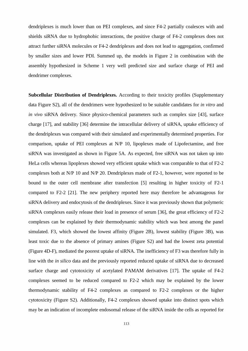
113
dendriplexes is much lower than on PEI complexes, and since F4-2 partially coalesces with and
shields siRNA due to hydrophobic interactions, the positive charge of F4-2 complexes does not
attract further siRNA molecules or F4-2 dendriplexes and does not lead to aggregation, confirmed
by smaller sizes and lower PDI. Summed up, the models in Figure 2 in combination with the
assembly hypothesized in Scheme 1 very well predicted size and surface charge of PEI and
dendrimer complexes.
Subcellular Distribution of Dendriplexes. According to their toxicity profiles (Supplementary
data Figure S2), all of the dendrimers were hypothesized to be suitable candidates for in vitro and
in vivo siRNA delivery. Since physico-chemical parameters such as complex size [43], surface
charge [17], and stability [36] determine the intracellular delivery of siRNA, uptake efficiency of
the dendriplexes was compared with their simulated and experimentally determined properties. For
comparison, uptake of PEI complexes at N/P 10, lipoplexes made of Lipofectamine, and free
siRNA was investigated as shown in Figure 5A. As expected, free siRNA was not taken up into
HeLa cells whereas lipoplexes showed very efficient uptake which was comparable to that of F2-2
complexes both at N/P 10 and N/P 20. Dendriplexes made of F2-1, however, were reported to be
bound to the outer cell membrane after transfection [5] resulting in higher toxicity of F2-1
compared to F2-2 [21]. The new periphery reported here may therefore be advantageous for
siRNA delivery and endocytosis of the dendriplexes. Since it was previously shown that polymeric
siRNA complexes easily release their load in presence of serum [36], the great efficiency of F2-2
complexes can be explained by their thermodynamic stability which was best among the panel
simulated. F3, which showed the lowest affinity (Figure 2B), lowest stability (Figure 3B), was
least toxic due to the absence of primary amines (Figure S2) and had the lowest zeta potential
(Figure 4D-F), mediated the poorest uptake of siRNA. The inefficiency of F3 was therefore fully in
line with the in silico data and the previously reported reduced uptake of siRNA due to decreased
surface charge and cytotoxicity of acetylated PAMAM derivatives [17]. The uptake of F4-2
complexes seemed to be reduced compared to F2-2 which may be explained by the lower
thermodynamic stability of F4-2 complexes as compared to F2-2 complexes or the higher
cytotoxicity (Figure S2). Additionally, F4-2 complexes showed uptake into distinct spots which
may be an indication of incomplete endosomal release of the siRNA inside the cells as reported for

114
siRNA complexes of PAMAM and Tat-conjugated PAMAM [19], SuperFect and guanylated
F2-1g complexes [5].
Transfection Efficiency. Since all dendriplexes were efficiently internalized into HeLa/Luc cells,
it was checked if the simulated data also corroborated their capacity to knockdown luciferase
expression in the same cell line. All dendriplexes mediated RNAi depending on the N/P ratio with
some formulations achieving effects comparable to Lipofectamine (LF). While the latter showed
considerable off-target effects in cells treated with lipoplexes of the negative control sequence, as
shown in Figures 5B-D, dendrimers F2-2 and F3 maintained strong transfection efficiency with
low or very low toxicity and off-target effects. The correlation between efficiency and toxicity has
always been a major drawback of non-viral vectors [44] and was only recently reported to be
overcome by disulfide cross-linked low molecular weight PEI for gene delivery [45] and
-CD onto PAMAM for siRNA delivery [46]. Other modifications of dendrimers,
however, such as internal quaternization in addition to a hydroxyl periphery of PAMAMs led to
poor biological activity [18]. With hyperflexible triazine dendrimers, efficient luciferase
knockdown was achieved even with the least toxic dendrimer F3, which supports the hypothesis of
efficiency of flexible dendrimers at reduced toxicity reported for G7 triethanolamine core
PAMAM dendriplexes [13]. The efficiency of F3 complexes was lower than that of F2-2 but
surprising taking into account that F3 did not efficiently protect siRNA from intercalation of
SYBR Gold. However, F3 complexes were small and monodisperse (Figure 4A-C) and were
therefore endocytosed to a certain degree (Figure 5A). However, increasing signs of off-target
effects were observed for flexible triazine dendrimers as a function of increasing amounts of
primary amines.
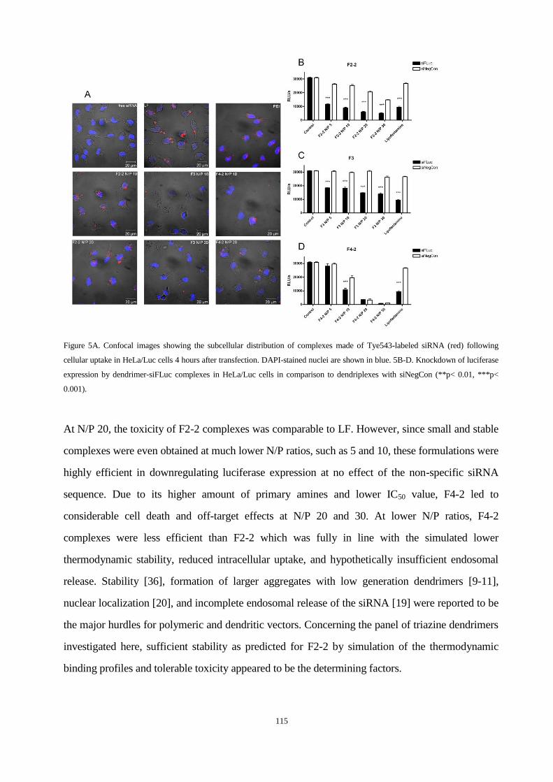
115
Figure 5A. Confocal images showing the subcellular distribution of complexes made of Tye543-labeled siRNA (red) following
cellular uptake in HeLa/Luc cells 4 hours after transfection. DAPI-stained nuclei are shown in blue. 5B-D. Knockdown of luciferase
expression by dendrimer-siFLuc complexes in HeLa/Luc cells in comparison to dendriplexes with siNegCon (**p< 0.01, ***p<
0.001).
At N/P 20, the toxicity of F2-2 complexes was comparable to LF. However, since small and stable
complexes were even obtained at much lower N/P ratios, such as 5 and 10, these formulations were
highly efficient in downregulating luciferase expression at no effect of the non-specific siRNA
sequence. Due to its higher amount of primary amines and lower IC50 value, F4-2 led to
considerable cell death and off-target effects at N/P 20 and 30. At lower N/P ratios, F4-2
complexes were less efficient than F2-2 which was fully in line with the simulated lower
thermodynamic stability, reduced intracellular uptake, and hypothetically insufficient endosomal
release. Stability [36], formation of larger aggregates with low generation dendrimers [9-11],
nuclear localization [20], and incomplete endosomal release of the siRNA [19] were reported to be
the major hurdles for polymeric and dendritic vectors. Concerning the panel of triazine dendrimers
investigated here, sufficient stability as predicted for F2-2 by simulation of the thermodynamic
binding profiles and tolerable toxicity appeared to be the determining factors.
116
Biodistribution and Pharmacokinetics. Since all dendrimer formulations successfully mediated
RNAi in vitro with negligible toxicity at N/P 5, all of them qualified for in vivo experiments. As
SPECT imaging of in vivo administration of radiolabeled siRNA was previously reported to
predict pharmacokinetics and biodistribution with good correlation to results obtained by
scintillation counting [35], the same technique was employed to gain insight into the in vivo
performance of hyperflexible dendriplexes. To date, information on biodistribution of dendritic
siRNA carriers is limited to a study of surface-engineered PPI where ex vivo fluorescence imaging
and confocal laser scanning microscopy (CLSM) was employed to investigate the organ
distribution of fluorescently labeled siRNA [2] and a second study that performed CLSM to trace
the fluorescently labeled G4 cystamine-core PAMAM-based carrier [7]. The only report on
pharmacokinetics of dendrimer-complexed siRNA investigated radioactively labeled siRNA and
rigid triazine dendrimers and described strong lung accumulation of radioactively and fluorescently
labeled siRNA [5]. As can be seen in Figure 6A and Movie 1 (Supporting Information), F2-2
dendriplexes did not accumulate in the lung, but in the liver, the kidneys and to some extent in the
bowel due to partial hepatobiliary excretion of amphiphilic DsiRNA [5]. Although F3 complexes
were stable enough in 10% serum containing medium to mediate RNAi in vitro, these complexes
dissociated in vivo as previously described for PEI complexes [36] leading to quantitative excretion
of free siRNA via the bowel and the bladder (Figure 6B and Movie 2). Interestingly, F4-2
complexes seemed to be most stable under in vivo conditions with very strong uptake of
radiolabeled siRNA into the liver, some excretion into the bladder and no noticeable excretion of
free siRNA via the bowel (Figure 6C and Movie 3).
Figure 6. Three-dimensional biodistribution of A. F2-2-siRNA-dendriplexes, B. F3-siRNA-dendriplexes, and C.
F4-2-siRNA-dendriplexes 2 hours after i.v. administration as registered by SPECT imaging.
117
These results were confirmed by scintillation counting of dissected organs as shown in Figure 7A.
Dendriplexes made of dendrimer F2-2 showed a higher signal in the heart 2 h after injection which
is in line with significantly prolonged circulation times (Figure 7B). Their advantageous
pharmacokinetics reflected in a more than twofold increased AUC versus free siRNA (Figure 7B)
can be explained by reduced uptake into the reticulo-endothelial system (RES) which results from
lower surface charge of F2-2 complexes compared to PEI or F4-2 complexes. Although F2-2
complexes were expected to be most stable, the SYBR Gold assay showed that some siRNA was
still accessible for intercalation. This finding corroborates the considerable amount of 9.8% of the
injected dose (ID) which was excreted as free siRNA via the bowel and 5.3% ID cleared through
the kidneys. All other formulations led to rapid clearance from the blood pool as shown by low
AUC values (167.2-276.7 %ID*min/ml), as previously reported for native siRNA [47]. Both free
siRNA and siRNA formulated with F3 were mostly cleared into the bowel and the bladder. The
similarity of the pharmacokinetic profiles and the deposition of free siRNA and siRNA/F3
complexes into the bowel is a strong indication of instability which was hypothesized due to
simulated thermodynamic data and the results from the SYBR Gold assay. F4-2 complexes,
however, were not rapidly cleared from the blood stream because of instability but because of
extensive capture by the RES. In fact, the uptake of 52.1% of the injected siRNA is a sign of
enhanced stability compared to PEI complexes, PEG-PEI complexes [36] and rigid triazine
dendriplexes [5]. This strong uptake of siRNA into the liver was previously reported for
surface-engineered PPI-based siRNA complexes [2] and was exploited for knockdown of ApoB in
healthy C57BL/6 mice with poly-L-lysine-based vectors [3] or G3 tetra-oleoyl lysine dendrimers
bound to the hydrophobic surface of single-walled carbon nanotubes (SWNT) [8]. While the
spleen took up additional 7.0% ID of F4-2 formulated siRNA, only 0.7% ID accumulated in the
kidneys and 2.3% ID were found in the bowel. As reported earlier, PEI complexes dissociated in
the liver which was the only organ in which PEI-formulated siRNA accumulated. This is strongly
in line with previous reports [36], and the differences of RES capture are in line with the different
interaction of rigid and flexible polycations with siRNA as hypothesized based on the simulations.
Radiolabeled siRNA complexed with PEI 25k showed the same pharmacokinetic profile as free
siRNA and siRNA complexed with F3, indicating the instability of both complexes as
hypothesized from the thermodynamic values.
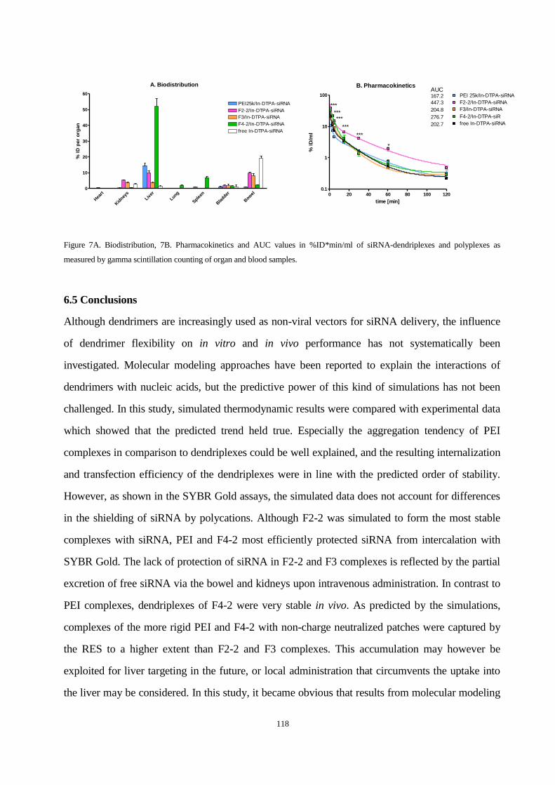
118
A. Biodistribution
Hea
rt
Kid
neys
Liver
Lung
Sple
en
Bla
dder
Bow
el0
10
20
30
40
50
60
free In-DTPA-siRNA
F4-2/In-DTPA-siRNA
F3/In-DTPA-siRNA
PEI25k/In-DTPA-siRNA
F2-2/In-DTPA-siRNA
% I
D p
er
org
an
B. Pharmacokinetics
0 20 40 60 80 100 1200.1
1
10
100
free In-DTPA-siRNA
PEI 25k/In-DTPA-siRNA
F4-2/In-DTPA-siR
F3/In-DTPA-siRNA
F2-2/In-DTPA-siRNA***
***
***
*
******
AUC167.2
447.3
202.7
276.7
204.8
time [min]
% I
D/m
l
Figure 7A. Biodistribution, 7B. Pharmacokinetics and AUC values in %ID*min/ml of siRNA-dendriplexes and polyplexes as
measured by gamma scintillation counting of organ and blood samples.
6.5 Conclusions
Although dendrimers are increasingly used as non-viral vectors for siRNA delivery, the influence
of dendrimer flexibility on in vitro and in vivo performance has not systematically been
investigated. Molecular modeling approaches have been reported to explain the interactions of
dendrimers with nucleic acids, but the predictive power of this kind of simulations has not been
challenged. In this study, simulated thermodynamic results were compared with experimental data
which showed that the predicted trend held true. Especially the aggregation tendency of PEI
complexes in comparison to dendriplexes could be well explained, and the resulting internalization
and transfection efficiency of the dendriplexes were in line with the predicted order of stability.
However, as shown in the SYBR Gold assays, the simulated data does not account for differences
in the shielding of siRNA by polycations. Although F2-2 was simulated to form the most stable
complexes with siRNA, PEI and F4-2 most efficiently protected siRNA from intercalation with
SYBR Gold. The lack of protection of siRNA in F2-2 and F3 complexes is reflected by the partial
excretion of free siRNA via the bowel and kidneys upon intravenous administration. In contrast to
PEI complexes, dendriplexes of F4-2 were very stable in vivo. As predicted by the simulations,
complexes of the more rigid PEI and F4-2 with non-charge neutralized patches were captured by
the RES to a higher extent than F2-2 and F3 complexes. This accumulation may however be
exploited for liver targeting in the future, or local administration that circumvents the uptake into
the liver may be considered. In this study, it became obvious that results from molecular modeling

119
approaches very well explain physico-chemical parameters and the in vitro behavior of
dendriplexes. These attributes, in turn, determine in vivo behavior such as stability in presence of
serum or uptake into the RES. Since in vivo conditions, however, involve the complex interplay of
dendriplexes with serum, cells, organs and metabolism, the prediction of the in vivo performance
of dendriplexes can not solely be simulated but should be supported by methods such as in vivo
imaging. If combined, however, molecular modeling and in vivo imaging very well represent the in
vitro and in vivo performance of non-viral vectors for siRNA delivery as shown in this study.
Further validation of in silico data by isothermal titration calorimetry is currently under way.
6.6 Acknowledgments
We are grateful to Brian Sproat (IDT/Chemconsilium) for supplying the siRNA within
MEDITRANS and to Eva Mohr (Dept. of Pharmaceutics and Biopharmacy) and Ulla Cramer
(Dept. of Nuclear Medicine) for excellent technical support. MEDITRANS, an Integrated Project
funded by the European Commission under the Sixth Framework (NMP4-CT-2006-026668), is
gratefully acknowledged. EES and MAM thank the N.I.H. (R01 GM 65460). MM acknowledges
the Czech national project OC10053.
6.7 References
[1] J. Zhou, J. Wu, N. Hafdi, J.P. Behr, P. Erbacher, L. Peng, PAMAM dendrimers for efficient siRNA delivery and
potent gene silencing, Chem Commun (Camb), (2006) 2362-2364.
[2] O. Taratula, O.B. Garbuzenko, P. Kirkpatrick, I. Pandya, R. Savla, V.P. Pozharov, H. He, T. Minko,
Surface-engineered targeted PPI dendrimer for efficient intracellular and intratumoral siRNA delivery, J Control
Release, 140 (2009) 284-293.
[3] K. Watanabe, M. Harada-Shiba, A. Suzuki, R. Gokuden, R. Kurihara, Y. Sugao, T. Mori, Y. Katayama, T.
Niidome, In vivo siRNA delivery with dendritic poly(L-lysine) for the treatment of hypercholesterolemia, Mol Biosyst,
5 (2009) 1306-1310.
[4] N. Weber, P. Ortega, M.I. Clemente, D. Shcharbin, M. Bryszewska, F.J. de la Mata, R. Gómez, M.A.
Muñoz-Fernández, Characterization of carbosilane dendrimers as effective carriers of siRNA to HIV-infected
lymphocytes, Journal of Controlled Release, 132 (2008) 55-64.
[5] O.M. Merkel, M.A. Mintzer, D. Librizzi, O. Samsonova, T. Dicke, B. Sproat, H. Garn, P.J. Barth, E.E. Simanek, T.
Kissel, Triazine Dendrimers as Nonviral Vectors for in Vitro and in Vivo RNAi: The Effects of Peripheral Groups and
Core Structure on Biological Activity, Mol Pharm, 7 (2010) 969-983.

120
[6] I.D. Kim, C.M. Lim, J.B. Kim, H.Y. Nam, K. Nam, S.W. Kim, J.S. Park, J.K. Lee, Neuroprotection by
biodegradable PAMAM ester (e-PAM-R)-mediated HMGB1 siRNA delivery in primary cortical cultures and in the
postischemic brain, J Control Release, 142 (2010) 422-430.
[7] A. Agrawal, D.H. Min, N. Singh, H. Zhu, A. Birjiniuk, G. von Maltzahn, T.J. Harris, D. Xing, S.D. Woolfenden,
P.A. Sharp, A. Charest, S. Bhatia, Functional delivery of siRNA in mice using dendriworms, ACS Nano, 3 (2009)
2495-2504.
[8] J. McCarroll, H. Baigude, C.S. Yang, T.M. Rana, Nanotubes functionalized with lipids and natural amino acid
dendrimers: a new strategy to create nanomaterials for delivering systemic RNAi, Bioconjug Chem, 21 (2010) 56-63.
[9] X.C. Shen, J. Zhou, X. Liu, J. Wu, F. Qu, Z.L. Zhang, D.W. Pang, G. Quelever, C.C. Zhang, L. Peng, Importance
of size-to-charge ratio in construction of stable and uniform nanoscale RNA/dendrimer complexes, Org Biomol Chem,
5 (2007) 3674-3681.
[10] R.L. Juliano, Intracellular delivery of oligonucleotide conjugates and dendrimer complexes, Ann N Y Acad Sci,
1082 (2006) 18-26.
[11] Y. Inoue, R. Kurihara, A. Tsuchida, M. Hasegawa, T. Nagashima, T. Mori, T. Niidome, Y. Katayama, O. Okitsu,
Efficient delivery of siRNA using dendritic poly(L-lysine) for loss-of-function analysis, J Control Release, 126 (2008)
59-66.
[12] A.P. Perez, E.L. Romero, M.J. Morilla, Ethylendiamine core PAMAM dendrimers/siRNA complexes as in vitro
silencing agents, Int J Pharm, 380 (2009) 189-200.
[13] X.X. Liu, P. Rocchi, F.Q. Qu, S.Q. Zheng, Z.C. Liang, M. Gleave, J. Iovanna, L. Peng, PAMAM dendrimers
mediate siRNA delivery to target Hsp27 and produce potent antiproliferative effects on prostate cancer cells,
ChemMedChem, 4 (2009) 1302-1310.
[14] Q. Yuan, E. Lee, W.A. Yeudall, H. Yang, Dendrimer-triglycine-EGF nanoparticles for tumor imaging and targeted
nucleic acid and drug delivery, Oral Oncol, 46 (2010) 698-704.
[15] S. Svenson, Dendrimers as versatile platform in drug delivery applications, European Journal of Pharmaceutics
and Biopharmaceutics, 71 (2009) 445-462.
[16] R. Jevprasesphant, J. Penny, R. Jalal, D. Attwood, N.B. McKeown, A. D'Emanuele, The influence of surface
modification on the cytotoxicity of PAMAM dendrimers, Int J Pharm, 252 (2003) 263-266.
[17] C.L. Waite, S.M. Sparks, K.E. Uhrich, C.M. Roth, Acetylation of PAMAM dendrimers for cellular delivery of
siRNA, BMC Biotechnology, 9 (2009).
[18] M.L. Patil, M. Zhang, O. Taratula, O.B. Garbuzenko, H. He, T. Minko, Internally cationic polyamidoamine
PAMAM-OH dendrimers for siRNA delivery: effect of the degree of quaternization and cancer targeting,
Biomacromolecules, 10 (2009) 258-266.
[19] H. Kang, R. DeLong, M.H. Fisher, R.L. Juliano, Tat-conjugated PAMAM dendrimers as delivery agents for
antisense and siRNA oligonucleotides, Pharmaceutical Research, 22 (2005) 2099-2106.
[20] M.L. Patil, M. Zhang, S. Betigeri, O. Taratula, H. He, T. Minko, Surface-modified and internally cationic
polyamidoamine dendrimers for efficient siRNA delivery, Bioconjug Chem, 19 (2008) 1396-1403.
[21] O.M. Merkel, M.A. Mintzer, J. Sitterberg, U. Bakowsky, E.E. Simanek, T. Kissel, Triazine dendrimers as nonviral
gene delivery systems: effects of molecular structure on biological activity, Bioconjug Chem, 20 (2009) 1799-1806.

121
[22] G.M. Pavan, M.A. Mintzer, E.E. Simanek, O.M. Merkel, T. Kissel, A. Danani, Computational insights into the
interactions between DNA and siRNA with "rigid" and "flexible" triazine dendrimers, Biomacromolecules, 11 (2010)
721-730.
[23] W.R. Sanhai, J.H. Sakamoto, R. Canady, M. Ferrari, Seven challenges for nanomedicine, Nat Nanotechnol, 3
(2008) 242-244.
[24] M.A. Mintzer, O.M. Merkel, T. Kissel, E.E. Simanek, Polycationic triazine-based dendrimers: effect of peripheral
groups on transfection efficiency, New J Chem, 33 (2009) 1918-1925.
[25] D.A. Case, T.A. Darden, T.E. Cheatham III, C.L. Simmerling, J. Wang, R.E. Duke, R. Luo, R.C. Walker, W.
Zhang, K.M. Merz, B. Robertson, B. Wang, S. Hayik, A. Roitberg, G. Seabra, I. Kolossvary, K.F. Wong, F. Paesani, J.
Vanicek, J. Liu, X. Wu, S. Brozell, T. Steinbrecher, H. Gohlke, Q. Cai, X. Ye, J. Wang, M.-J. Hsieh, G. Cui, D.R. Roe,
D.H. Mathews, M.G. Seetin, C. Sangui, V. Babin, T. Luchko, S. Gusarov, A. Kovalenko, P.A. Kollman, AMBER 11,
in, University of California, San Francisco, 2010.
[26] S.P. Jones, G.M. Pavan, A. Danani, S. Pricl, D.K. Smith, Quantifying the Effect of Surface Ligands on
Dendron-DNA Interactions: Insights into Multivalency through a Combined Experimental and Theoretical Approach,
Chemistry, 16 (2010) 4519-4532.
[27] G.M. Pavan, A. Danani, S. Pricl, D.K. Smith, Modeling the multivalent recognition between dendritic molecules
and DNA: understanding how ligand "sacrifice" and screening can enhance binding, J Am Chem Soc, 131 (2009)
9686-9694.
[28] G.M. Pavan, L. Albertazzi, A. Danani, Ability to adapt: different generations of PAMAM dendrimers show
different behaviors in binding siRNA, J Phys Chem B, 114 (2010) 2667-2675.
[29] J. Suh, H.J. Paik, B.K. Hwang, Ionization of Poly(ethylenimine) and Poly(allylamine) at Various pH's, Bioorganic
Chemistry, 22 (1994) 318-327.
[30] W.K. Jorgensen, M.J. Rice, Morphology of a very extensible insect muscle, Tissue Cell, 15 (1983) 639-644.
[31] T. Darden, D. York, L. Pedersen, Particle mesh Ewald: An N [center-dot] log(N) method for Ewald sums in large
systems, The Journal of Chemical Physics, 98 (1993) 10089-10092.
[32] W.D. Cornell, P. Cieplak, C.I. Bayly, I.R. Gould, K.M. Merz, D.M. Ferguson, D.C. Spellmeyer, T. Fox, J.W.
Caldwell, P.A. Kollman, A Second Generation Force Field for the Simulation of Proteins, Nucleic Acids, and Organic
Molecules, Journal of the American Chemical Society, 117 (1995) 5179-5197.
[33] J. Srinivasan, T.E. Cheatham, P. Cieplak, P.A. Kollman, D.A. Case, Continuum Solvent Studies of the Stability of
DNA, RNA, and Phosphoramidate−DNA Helices, Journal of the American Chemical Society, 120 (1998) 9401-9409.
[34] I. Andricioaei, M. Karplus, On the calculation of entropy from covariance matrices of the atomic fluctuations, The
Journal of Chemical Physics, 115 (2001) 6289-6292.
[35] O.M. Merkel, D. Librizzi, A. Pfestroff, T. Schurrat, M. Behe, T. Kissel, In vivo SPECT and real-time gamma
camera imaging of biodistribution and pharmacokinetics of siRNA delivery using an optimized radiolabeling and
purification procedure, Bioconjug Chem, 20 (2009) 174-182.
[36] O.M. Merkel, D. Librizzi, A. Pfestroff, T. Schurrat, K. Buyens, N.N. Sanders, S.C. De Smedt, M. Behe, T. Kissel,
Stability of siRNA polyplexes from poly(ethylenimine) and poly(ethylenimine)-g-poly(ethylene glycol) under in vivo
conditions: effects on pharmacokinetics and biodistribution measured by Fluorescence Fluctuation Spectroscopy and
Single Photon Emission Computed Tomography (SPECT) imaging, J Control Release, 138 (2009) 148-159.
122
[37] V.K. Sharma, M. Thomas, A.M. Klibanov, Mechanistic studies on aggregation of polyethylenimine-DNA
complexes and its prevention, Biotechnol Bioeng, 90 (2005) 614-620.
[38] M.X. Tang, F.C. Szoka, The influence of polymer structure on the interactions of cationic polymers with DNA and
morphology of the resulting complexes, Gene Ther, 4 (1997) 823-832.
[39] T. Ehtezazi, U. Rungsardthong, S. Stolnik, Thermodynamic analysis of polycation-DNA interaction applying
titration microcalorimetry, Langmuir, 19 (2003) 9387-9394.
[40] O.M. Merkel, A. Beyerle, D. Librizzi, A. Pfestroff, T.M. Behr, B. Sproat, P.J. Barth, T. Kissel, Nonviral siRNA
delivery to the lung: investigation of PEG-PEI polyplexes and their in vivo performance, Mol Pharm, 6 (2009)
1246-1260.
[41] V.A. Izumrudov, T.K. Bronich, M.B. Novikova, A.B. Zezin, V.A. Kabanov, Substitution reactions in ternary
systems of macromolecules, Polymer Science U.S.S.R., 24 (1982) 367-378.
[42] A.L. Bolcato-Bellemin, M.E. Bonnet, G. Creusat, P. Erbacher, J.P. Behr, Sticky overhangs enhance
siRNA-mediated gene silencing, Proc Natl Acad Sci U S A, 104 (2007) 16050-16055.
[43] I. Mellman, Endocytosis and molecular sorting, Annu Rev Cell Dev Biol, 12 (1996) 575-625.
[44] S. Hong, P.R. Leroueil, E.K. Janus, J.L. Peters, M.M. Kober, M.T. Islam, B.G. Orr, J.R. Baker, Jr., M.M.
Banaszak Holl, Interaction of polycationic polymers with supported lipid bilayers and cells: nanoscale hole formation
and enhanced membrane permeability, Bioconjug Chem, 17 (2006) 728-734.
[45] M. Breunig, U. Lungwitz, R. Liebl, A. Goepferich, Breaking up the correlation between efficacy and toxicity for
nonviral gene delivery, Proc Natl Acad Sci U S A, 104 (2007) 14454-14459.
[46] T. Tsutsumi, H. Arima, F. Hirayama, K. Uekama, Potential Use of Dendrimer/α-Cyclodextrin Conjugate as a
Novel Carrier for Small Interfering RNA (siRNA), Journal of Inclusion Phenomena and Macrocyclic Chemistry, 56
(2006) 81-84.
[47] D.M. Dykxhoorn, D. Palliser, J. Lieberman, The silent treatment: siRNAs as small molecule drugs, Gene Ther, 13
(2006) 541-552.

123
Chapter 7
DESIGN AND BIOPHYSICAL CHARACTERIZATION OF
BIORESPONSIVE DEGRADABLE POLY(DIMETHYLAMINOETHYL
METHACRYLATE) BASED POLYMERS FOR IN VITRO DNA
TRANSFECTION
Published in Biomacromolecules. 2012 Feb 13;13(2):313-22. Epub 2012 Jan 17.
Yi Zhang,†§
Mengyao Zheng,‡§
Thomas Kissel,*‡
Seema Agarwal*†
§Both authors contributed equally to this work
Author contributions
T. K. and S. A. guided the research. M. Z. and Y. Z. directed the measurements. Y. Z. synthesized
and characterized the polymers and M. Z. carried out the SYBR® Gold assay, heparin competition
assay, MTT assay, CLSM and in vitro transfection. M. Z. and Y. Z. analysed the experimental data.
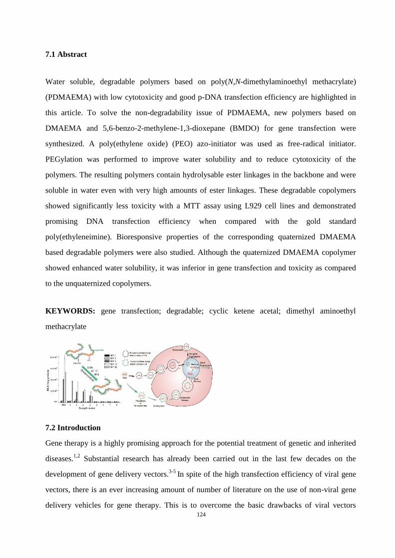
124
7.1 Abstract
Water soluble, degradable polymers based on poly(N,N-dimethylaminoethyl methacrylate)
(PDMAEMA) with low cytotoxicity and good p-DNA transfection efficiency are highlighted in
this article. To solve the non-degradability issue of PDMAEMA, new polymers based on
DMAEMA and 5,6-benzo-2-methylene-1,3-dioxepane (BMDO) for gene transfection were
synthesized. A poly(ethylene oxide) (PEO) azo-initiator was used as free-radical initiator.
PEGylation was performed to improve water solubility and to reduce cytotoxicity of the
polymers. The resulting polymers contain hydrolysable ester linkages in the backbone and were
soluble in water even with very high amounts of ester linkages. These degradable copolymers
showed significantly less toxicity with a MTT assay using L929 cell lines and demonstrated
promising DNA transfection efficiency when compared with the gold standard
poly(ethyleneimine). Bioresponsive properties of the corresponding quaternized DMAEMA
based degradable polymers were also studied. Although the quaternized DMAEMA copolymer
showed enhanced water solubility, it was inferior in gene transfection and toxicity as compared
to the unquaternized copolymers.
KEYWORDS: gene transfection; degradable; cyclic ketene acetal; dimethyl aminoethyl
methacrylate
7.2 Introduction
Gene therapy is a highly promising approach for the potential treatment of genetic and inherited
diseases.1,2
Substantial research has already been carried out in the last few decades on the
development of gene delivery vectors.3-5
In spite of the high transfection efficiency of viral gene
vectors, there is an ever increasing amount of number of literature on the use of non-viral gene
delivery vehicles for gene therapy. This is to overcome the basic drawbacks of viral vectors

125
which are immune response, limitations in the size of inserted DNA, difficulty in large scale
pharmaceutical grade production etc.6
Some non-viral DNA delivery systems include pure
plasmid DNA, lipoplexes (DNA complexed with cationic lipids), polyplexes (nucleic acid
complexes with polycations and encapsulated DNA in degradable polymer matrices). For
example, cationic polymers show the ability to form polyplex with DNA by electrostatic
interactions due to its polyanionic character.7-11
An example of a frequently studied polycation
for this purpose is polyethyleneimine (PEI), a gold standard with buffering properties (at
physiological pH only 25% of the amine groups are protonated) but with the major drawback of
cytotoxicity (half maximal inhibitory concentration (IC50) = ~ 8 µg•ml-1
).12
Recently, attention
has been given to poly(N,N-dimethylaminoethyl methacrylate) (PDMAEMA) as a non-viral gene
delivery system with buffering capacity and less cytotoxicity (IC50 = ~ 40µg•ml-1
) (pKa = 7.5).
This polymer is prepared by radical polymerization of the corresponding vinyl monomer. It was
shown for the first time in 1996 by Hennink et al. that PDMAEMA is an interesting vector for
designing of a gene transfection system.13
PDMAEMA contains tertiary amines for the complexation of DNA and reaches 90% of the
transfection efficiency of PEI (branched PEI 25 kDa). Since then, several aspects of this
transfection reagent have been modified i.e. the role of molecular weight, polyplex size and
transfection parameters, pH, ionic strength, temperature, viscosity, polymer/plasmid-DNA
(p-DNA) ratio and the presence of stabilizers on transfection efficiency of PDMAEMA.14-18
Despite so much research, the key problem of polycations like PEI and PDMAEMA is their
non-biodegradable nature, notable toxicity and the need for further improvement of transfection
efficiency.
Vinyl polymers like PDMAEMA, which can be easily synthesized by radical polymerization,
could be further designed to meet these requirements. Recently, Oupicky et al. reported
PDMAEMA copolymers with reducible –S-S- disulfide linkages using reversible addition
fragmentation transfer (RAFT) polymerization with comparable cytotoxicity and gene
transfection efficiency like homo PDMAEMA for the first time.19
Unfortunately, no data (in vivo
or in vitro) regarding biodegradation behavior was provided.
To solve the non-degradability issue of PDMAEMA, we recently showed the possibility of
forming a degradable and less toxic PDMAEMA by introducing ester linkages into the
126
PDMAEMA backbone. We consider radical-ring-opening polymerization of cyclic ketene
acetals a promising method for introducing degradable ester linkages into the polymer backbone,
which can be used to develop new gene transfection systems. Cyclic ketene acetals are the
isomers of the corresponding cyclic lactones and can undergo radical addition at the vinyl double
bond with subsequent ring-opening leading to the formation of polyesters. Free-radical
copolymerization of cyclic ketene acetal 5,6-benzo-2-methylene-1,3-dioxepane (BMDO), with
N, N-dimethylaminoethyl methacrylate (DMAEMA) can lead to the formation of degradable
PDMAEMA with ester linkages in the backbone.20
The polymers were not soluble in water,
therefore quaternization with alkyl bromide was carried out. Regardless of copolymer
composition, all of the polymers were less cytotoxic than PEI and showed very high cell
viability. Unfortunately, the system showed poor transfection efficiency which could be due to
the strong interactions between the positively charged units and DNA. Therefore, further
improvement was implemented in this system by designing a polymer avoiding quaternization of
PDMAEMA. Small poly(ethylene glycol) (PEG) hydrophilic blocks were introduced onto
degradable PDMAEMA units to enhance water solubility and reduce the cytotoxicity.21,22
Again,
simple free radical chemistry was used for this purpose and a poly(ethylene oxide) (PEO)
macro-azo-initiator was used. The success of this concept is highlighted in this work by giving
details about synthesis, cytotoxicity, polyplex formation and gene transfection.
7.3 Experimental Part
Materials. PEO macro-azo-initiator (WAKO Company Mn = 24 kDa, PEG block =
6000 g•mol-1
) and bromoethane (Acros, 99%) were used as received. DMAEMA (Acros) was
passed through a basic alumina column to remove the inhibitor. Dimethylformamide (DMF),
chloroform, pentane and methanol were distilled before use. BMDO was synthesized according
to our previous report.23
Luciferase-Plasmid (pCMV-Luc) (LotNo.: PF461-090623) was
amplified by The Plasmid Factory (Bielefeld, Germany). All other chemicals were obtained from
Sigma–Aldrich (Steinheim, Germany) and used as received.
Instrumentation. 1H (400,13 MHz) and
13C (100,21 MHz) spectra were recorded on a Bruker
DRX-400 spectrometer. Tetramethylsilane was used as internal standard. The molecular weight
of the polymers were measured with size exclusion chromatography at 25 °C with 1 liner PSS
127
suprema Max 1000 Å column and a differential refractive index detector (SEC curity RI, PSS).
0.3 mol•L-1
formic acid in water was used as an eluent at a flow rate of 0.5 mL•min-1
. An SEC
curity 1100 (PSS) pump was used for the experiment. Linear poly 2-vinylpyridine was used for
calibration. The injected volume was 100 µL and the polymer concentration was 1 mg•mL-1
.
Copolymerization of DMAEMA and BMDO with PEO Azo-initiator (general procedure).
As an example for polymerization reactions, the procedure for the synthesis of sample 4 is
described below. All of the sample names and monomer feed ratios are shown in Table 1 and
Table 2.
The monomer BMDO (0.99 g, 6.1 mmol) was dissolved in DMAEMA (0.1 mL, 0.59 mmol) in a
predried Schlenk tube under an argon atmosphere. The reaction mixture was degassed by three
freeze-pump-thaw cycles. The PEO azo-initiator with PEG 6000 block (0.41 g, 6.8•10-2
mmol)
was added to the still frozen solution. The Schlenk tube was closed, evacuated and refilled with
argon three times. This reaction mixture was placed immediately in a preheated oil bath at 70 °C
for 24 h. Then the Schlenk tube was taken out of the oil bath and shock cooled in an ice bath.
The reaction mixture was diluted with chloroform and precipitated in 200 mL of pentane which
yielded a white precipitate. This white polymer was washed with a small amount of water then
dissolved in chloroform and precipitated in pentane again. This procedure was repeated twice
and then purified by dialysis against water. The final copolymer was dried under a vacuum at 40
°C for 48 h.
Quaternization Reaction of Poly(PEO-co-(BMDO-co-DMAEMA)) Copolymers. 200 mg
copolymer (samples 1-4) were dissolved in 20 mL chloroform at room temperature in a flask.
0.5 mL methanol and 2 mL ethylbromide were added to the copolymer solution. The flask was
placed in a preheated oil bath at 45 °C for 40 h. Afterwards, the solvent was evaporated using a
rotary evaporator. The residue was dissolved again in methanol and precipitated in pentane. This
product was then purified by repeatedly dissolving in methanol and precipitating in pentane. The
final product was dried at 40 °C under vacuum for 48 h.
Hydrolytic Degradability. In general, 100 mg copolymer was dissolved in a flask containing
10 mL of 5 wt-% KOH in distilled water. This mixture was kept at room temperature for 48 h.
Then, 10 mL 10 wt-% HCl was added. This mixture was extracted with chloroform. The aqueous
128
phase was dried with a freeze dryer for 3 days. The remaining solid was than characterized with
NMR spectroscopy.
Enzymatic Degradability. 200 mg copolymer was solved in PBS buffer (0.1 M, pH = 7.4) and
Lipase from Pseudomonas Cepacia (10 mg•mL-1
) with a 0.2 mg•mL-1
NaN3 solution. This
mixture was then placed at 37 °C with shaking for different time. Then the mixture was dried
with a freezer dryer for 5 days. The remaining solid was also characterized with NMR
spectroscopy and GPC.
Cell Culture. L929 mouse fibroblasts cells (human adenocarcinoma) for MTT assay and
luciferase assay were seeded at a density of 5.0•103 cells•cm
-2 in dishes (10 cm diameter,
Nunclon Dishes, Nunc, Wiesbaden, Germany). The incubation condition was at 37 °C in a
humidified 8.5% CO2 atmosphere (CO2-Incubator, Integra Biosciences, Fernwald, Germany).24
The medium was exchanged every 3 days. Cells were split after 5 days when confluence was
reached.
Cytotoxicity Test using MTT Assay. The cell viability test (MTT assay) was performed
according to the method of Mosmann.25
Polymer solutions were prepared in a serum
supplemented tissue culture medium (Dulbecco’s modified Eagle’s medium, supplemented with
10% serum, without antibiotic) containing 2•10-3
M glutamine and was sterile filtered (0.2 µm,
Schleicher&Schüll, Dassel, Germany).
24 h before the MTT assay, L929 cells (8000 cells•well-1
) were seeded into 96-well plates (Nunc,
Wiesbaden, Germany). On the day of MTT assay, the culture medium was replaced by 200 μL of a
serially diluted polymer medium solution with a different concentration. After a further 24 h of
incubation at 37 °C, the cell culture medium was replaced with 200 μL medium containing 20 μL
sterile filtered MTT (3-(4,5-dimethyl-thiazol-2-yl)-2,5-diphenyl tetrazolium bromide) (Sigma,
Deisenhofen, Germany) stock solution in phosphate buffered saline (PBS) (5 mg•mL-1
) in each
well. The final concentration of MTT in each well was 0.5 mg•mL-1
. After a 4 h incubation at 37
°C in the dark, the medium was removed and 200 µL of DMSO was added in each well to dissolve
the purple formazane product. The measurement was performed spectrophotometrically with an
ELISA reader (Titertek Plus MD 212, ICN, Eschwege, Germany) at wavelengths of 570 nm and
690 nm. The calibration of the spectrometer to zero absorbance was performed using a culture
medium without cells and to 100% absorbance was performed using control wells containing
129
standard cell culture medium but without polymer. The relative viability (%) related to the control
wells containing the cell culture medium without polymer was calculated by the following
equation:
Relative cell growth = ((A 570) test- (A 690) test) / ((A 570) control – (A 690) control) (1)
Polyethyleneimine (PEI 25 kDa, BASF, Germany) was used as a positive control. The IC50 was
calculated using the Boltzman sigmoidal function from Microcal Origin1 v 7.0 (OriginLab,
Northampton, USA). It shows the polymer concentration, which inhibits growth of half of the cells
relative to non-treated control cells. The statistical analysis was conducted in a quadruplicate per
group. Statistical evaluation was done using the program Sigma Stat 3.5. The one way ANOVA
with Bonferroni t-test was performed for all of the MTT data.
Preparation of Nanoparticles for Samples 3 and 4. Nanoparticles of the samples 3 and 4 were
prepared by a solvent displacement technique.26
10 mg polymer was dissolved in 1 mL of acetone
or acetonnitrile. Under magnetic stirring, 0.5 mL of the obtained solution was injected with an
injection needle (0.6•30 mm) into 5 mL of distilled water at a constant flow rate (8.0 mL•min-1
).
After the injection, the suspension was stirred for about 2 h under reduced pressure to remove the
organic solvent. The resulting suspension contained 1 mg•mL-1
polymer concentration.
Preparation of Polyplex with Copolymer. A 5% glucose solution and p-DNA (plasmid-DNA)
for physicochemical-experiments was used for the polyplex formation. 5% glucose is an isotonic
solution. In the buffer-solutions, the surface charges of the polymers are reduced due to the
higher ionic strength, and the polyplexes aggregates to larger agglomerates due to the lack of
repulsion.21
In terms of dimension, complex formation in a glucose solution is most suitable for
transfection.27
All solutions were filtered with 0.20 μm pore sized filters (Nalgener syringe filter,
Sigma–Aldrich, Taufkirchen, Germany). 50 μL of p-DNA solution (40 ng•μL-1
) were placed in a
micro centrifuge tube. The volume of a 1 mg mL-1
(based on hy-PEI 25 kDa) polymer stock
solution (samples 1, 2 and 5-8) or suspension (samples 3, 4) required for a certain N/P ratio was
calculated by following equation:22
VDNA = (Ccopolymer × 10 µL × 330) / (CDNA × 157 × N/P)
Ccopolymer = concentration of the stock copolymer
CDNA = concentration of the stock DNA solution

130
A certain amount of polymer stock solution was diluted with buffer-solution to a final volume of
50 µL in a micro centrifuge tube. The 50 μL polymer aliquots were mixed with 50 µL diluted
p-DNA aliquots and then incubated for 30 min at room temperature for complexation and
equilibrium formation.
Zeta Potential and Size Measurements. The zeta potential and size measurements of the
polyplexes were monitored with Malvern Zetasizer Nano ZS (Marvern Instrument,
Worcestershire, UK). The viscosity (0.88 mPa•s) and the refractive index (1.33) of distilled
water at room temperature (RT) was used for data analysis. The measurement angle was 173° in
backscatter mode. This polyplex solution was prepared and incubated at RT for 30 min before
measurement. Subsequently, zeta-potential measurements were performed with the same
samples after diluting 50 µL of polyplexes with an additional 500 µL of 5% glucose solution to a
final DNA concentration of 1.82 ng•µL-1
and a final volume of 550 µL. A low volume cuvette
(100 µL) was used for the size measurements, and the measurements of zeta potential were
carried out in the standard clear capillary electrophoresis cell at room temperature. Three
samples were prepared for each N/P ratio, and three measurements were performed on each
sample. Each measurement of size consisted of 15 runs for 10 sec. Each measurement of zeta
potential consisted of 60 runs, which was set to automatic optimization by the software.
Confocal Laser Scanning Microscopy (CLSM). 24 h before of the cell uptake experiment,
L929-cells were seeded into 8 well-chamberslides (Lab-Tek, Rochester, NY, USA) at a seeding
density of 50,000 cells•well-1
. in a DMEM low glucose (PAA, Cölbe, Germany) medium, which
contained 10% fetal calf serum (Cytogen, Sinn, Germany) . Before complexation with the
copolymer, p-DNA was at first labeled with YOYO-1 (Invitrogen, Karlsruhe, Germany) at a
weight ratio of 1:15 at room temperature for 30 min in the dark to protect fluorescent markers.
The YOYO-1 labeled p-DNA was condensed with polymer at N/P 15 in a 5% glucose solution,
and the polyplexes were incubated for another 20 min at room temperature. 25 µL polyplex
solution containing 0.5 µg p-DNA and 375 μL medium with 10% FCS were added in each well.
The well-chamberslides were incubated for 4 h at 37 °C in a humidified 8.5% CO2 atmosphere.
After incubation, the cells were washed with a 0.5 mL PBS buffer and then fixated by 20 min of
incubation with 0.1 mL of 4% paraformaldehyde in PBS. 30 µL of a 6 µg•mL-1
DAPI solution
(Invitrogen, Karlsruhe, Germany) was diluted with 1 mL a PBS buffer. Then 100 µL DAPI
131
solutions were filled into each chamber for 20 min of incubation in the dark. Afterwards, the
cells were washed again three times with a 0.5 mL PBS buffer before being fixated with
Fluorsafe (Calbiochem, San Diego, USA) and covered with a No.1.5 thickness cover slip
(Menzel Gläser, Braunschweig, Germany). The CLSM measurements were performed with a
385 μL long pass filter and a band pass filter of 505-530 nm in the single-track mode (Axiovert
100M and CLDM 510 Scanning Device; Zeiss, Oberkochen, Germany). The excitation of
YOYO-1 labeled DNA was performed with a 488 nm argon laser while the excitation of
DAPI-stained chromosomal DNA was performed with an enterprise laser with an excitation
wavelength of 364 nm.
In Vitro Transfection. L929 cells were seeded with a density of 30000 cells•mL-1
in
96-well-plates (Nunc, Wiesbaden, Germany) 24 h before transfection. Each well contained 6000
cells in 0.2 mL medium. The preparation of the polyplex solution was described above. 25 μL of
polyplex solution and 175 μL of the medium (10% serum content) were placed in each well (0.5 μg
p-DNA content). The well plates were incubated for 4 h at 37 °C under an 8.5% CO2 atmosphere.
After 44 h, the cell medium was exchanged, and the cells were lysed in a 100 μL cell culture lysis
buffer (Promega, Mannheim, Germany) for 15 min at 37 °C. The quantification of lucifaerase
activity was determined by injecting a 50 μL luciferase assay buffer, containing 10 mM luciferin
(Sigma-Aldrich, Taufkirchen, Germany), into 25 μL of cell lysate. The relative light units (RLU)
were measured with a plate luminometer (LumiSTAR Optima, BMG Labtech GmbH, Offenburg,
Germany). The protein concentration was determined using a Bradford BCA assay (BioRad,
Munich, Germany). The measurement of the transfection activity was performed according to the
protocol provided by Promega (Madison, WI, USA). The statistical analysis was conducted in
quadruplicate per group. Statistical evaluation was done using the program Sigma Stat 3.5. The
One way ANOVA with Bonferroni t-test was performed for all the transfection data.
SYBR Gold® Assay. The polymer/p-DNA complexes were prepared at N/P = 0.25, 0.5, 1, 2, 4, 6,
8, 10 in 96 well-plates as described. 200 μL dilutions of polymers containing 0.5 μg DNA for the
SYBR Gold® assay were performed in a water solution. After 20 min of incubation at room
temperature, 20 μL of diluted SYBR Gold® solution (5 μL stock solution was diluted in 12.5 mL
water) was added to each well and incubated for another 20 min. SYBR Gold® is light sensitive,
and this experiment should be protected from direct light as much as possible. The fluorescence

132
was directly detected using a fluorescence plate reader (BMG Labtech, Offenburg) at 495 nm
excitation and 537 nm emission. Data was analyzed with “Origin 7.0”.
Heparin Competition Assay. Briefly, polyplexes were prepared in solutions at different
N/P-ratios like the SYBR Gold® assay. Additionally, a 20 μL heparin (150 000 IU/g, Serva,
Pharm., USPXV2, Merck, Darmstadt, Germany) solution with a concentration of 0.5 mg/mL was
added into a 200μL polyplex solution in each well of the 96-well plate (Perkin Elmer,
Rodgau-Jügesheim), where each well contained 0.5 μg p-DNA. After a 20 min incubation of the
heparin at 25°C, 20 μL of the diluted SYBR Gold® solution (Invitrogen, Karlsruhe, Germany)
were added. The measurement was performed in the same manner as for the SYBR Gold® assay.
7.4 Results and Discussion
Free radical polymerization of cyclic ketene acetal BMDO and vinyl monomer DMAEMA was
performed with different monomer ratios in the feed at 70 °C for 24 h. PEO macro-azo-initiator
with PEO 6 kDa block was used to start the reaction. The molecular weight of the PEO
azo-initiator was 24 kDa. A schematic illustration of the reaction is given in Scheme 1.
Scheme 1: Synthesis route for the formation of the poly(PEG-co-(BMDO-co-DMAEMA)) and
poly(PEG-co-(BMDO-co-DMAEMA))•EtBr.
The copolymer composition was determined by NMR. In the 1H NMR spectrum, the
characteristic peaks from both comonomers (BMDO and DMAEMA) and the PEG block from
initiator were seen. The peak assignments are given in Figure 1. The signal at 3.6 ppm resulted
from the PEG block (-OCH2- peak numbers 21, 22 in Fig. 1). The 2.2 ppm signal could be
assigned to the two methyl groups of DMAEMA (peak 8 in Fig. 1). Aromatic signals and
–OCH2- of BMDO were seen around 7 and 5 ppm, respectively (peaks 5 and 1 in Fig. 1). In the
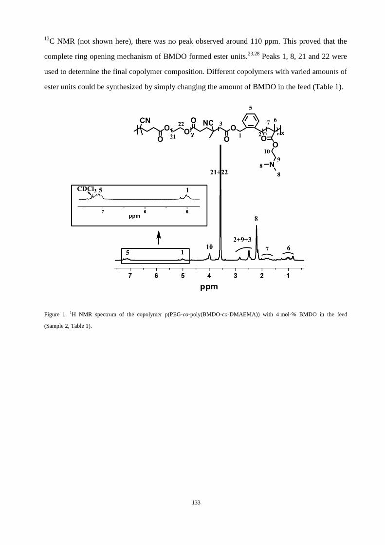
133
13C NMR (not shown here), there was no peak observed around 110 ppm. This proved that the
complete ring opening mechanism of BMDO formed ester units.23,28
Peaks 1, 8, 21 and 22 were
used to determine the final copolymer composition. Different copolymers with varied amounts of
ester units could be synthesized by simply changing the amount of BMDO in the feed (Table 1).
Figure 1. 1H NMR spectrum of the copolymer p(PEG-co-poly(BMDO-co-DMAEMA)) with 4 mol-% BMDO in the feed
(Sample 2, Table 1).

134
Table 1. Synthesis of the p(PEG-co-poly(BMDO-co-DMAEMA)) copolymers with PEO macro-azo-initiator at 70 °C for
24 h.
Sample Name
Feed ratio
molar ratio
BMDO:DMAEMA
Poylmer composition
molar ratio
BMDO:DMAEMA
Yield [%]
Solubility
1a
0 : 100 0 : 100 43 Water
2 10 : 90 4 : 96 70 Water
3 50 : 50 16: 84 45 Waterb
4 90 : 10 45: 55 32 Acetonnitrile
a This reaction was carried out for 50 min; b Maximum solubility in water 0.5 mg•mL-1.
The presence of PEG blocks from the initiator in the polymer chains increased the hydrophilicity
of these new copolymers and showed an improvement in the solubility behavior in water. In our
previous work, the random copolymer poly(BMDO-co-DMAEMA) showed limitations for use
as a gene transfection system due to insolubility in water and water miscible solvents like
acetonitrile.20
The use of a PEO macro-azo-initiator led to improved solubility of all of the
copolymers both in water and acetonitrile, even with high amounts of BMDO (Table 1).
The copolymers (Samples 1-4; Table 1) were further quaternized with ethylbromide via SN2
substitution. The properties of quaternized polymers are tabulated in the Table 2. After
quaternization, the solubility of the copolymer was further improved significantly. All
copolymers (even the polymer with BMDO: DMAEMA 45 : 55 molar ratio) could be solved in
water immediately.

135
Table 2. Quaternization reaction of the p(PEG-co-(BMDO-co-DMAEMA)) with ethyl bromide at 45 °C for 40 h.
Sampl
e
Copoylmer
composition
molar ratio
BMDO:DMAEMA
Quaternized
Sample
Quaternization
Yield
[%]
Mn Mwa
Solubility
H2Ob
[kDa]
1 0 : 100 5 100 54 322 +
2 4 : 96 6 100 46 127 +
3 16 : 84 7 100 26 67 +
4 45 : 55 8 92 13 36 +
a Mn, Mw were determined with water GPC; b + means soluble.
The 1H NMR spectrum after the quaternization reaction showed the shifting of peaks 8 and 9 to a
lower magnetic field (Figure 2). The addition of the ethyl groups (-CH2-) and –CH3 protons 23,
24 in Fig 2) was also observed at a high magnetic field. The degree of quaternization was
calculated using the integrals of the two methyl groups on the nitrogen atom of DMAEMA. The
quaternization reaction for most of the polymers was quantitative (Table 2). The molecular
weight and yield of the copolymer decreased with the increase of BMDO content. The
copolymers showed molecular weights between 13 kDa and 60 kDa. The polydispersity of the
polymers was high. This could be due to the formation of different multiblock copolymers with
PEG block and block of a copolymer of BMDO-co-DMAEMA or amphiphilic nature of the
block copolymers. Poly(PEG-co-(BMDO-co-DMAEMA•EtBr)) copolymer contained a
hydrophilic part, PEO, a hydrophobic part, BMDO, and the positivly charged PDMAEMA-EtBr.

136
This combination is a challenge for the column system and could lead to broad signal.
Figure 2. Comparison of NMRs of sample 3 and sample 7 (molar ratio of DMAEMA:BMDO is 15:85) before and after
quaternization reaction.
Figure 3. 1H NMR spectrum in CDCl3 before and after hydrolysis of poly(PEG-co-(BMDO-co-DMAEMA)) (sample 4): a)
before hydrolysis of the copolymer with molar ratio of BMDO:DMAEMA = 45:55; b) after 24 h hydrolysis in 5 wt-% KOH
solution; c) after 48 h hydrolysis in 5 wt-% KOH solution.
The hydrolytic degradation behavior of the new copolymers was studied under basic (pH = 9)
and enzymatic conditions. The degradation rate was determined by comparing peak integrals
before and after hydrolysis as shown for sample 4 (Figure 3). Proton 1 at 5 ppm showed the
characteristic proton peak in proximity to the ester bond of BMDO units. In Figure 3, the
reduced intensity of the proton 1 signal after 24 h degradation could be observed. After 24 h,
around 65% and after 48 h, nearly 93% of the ester bond was hydrolyzed.
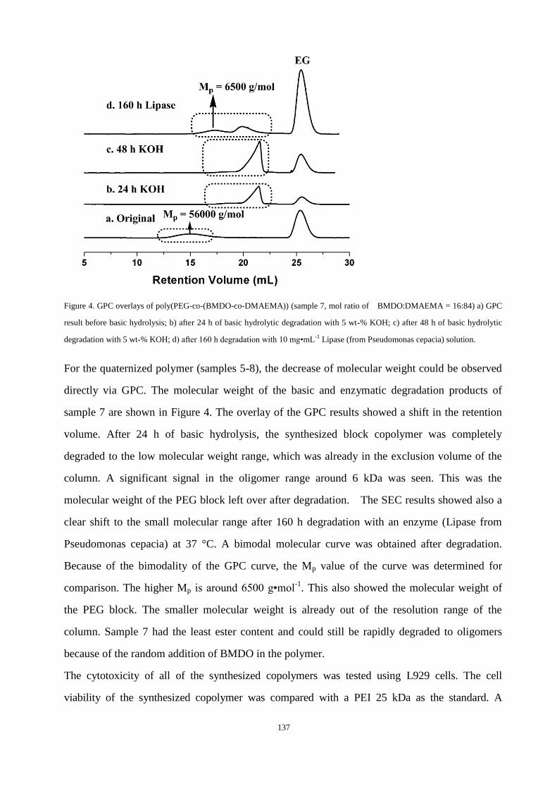
137
Figure 4. GPC overlays of poly(PEG-co-(BMDO-co-DMAEMA)) (sample 7, mol ratio of BMDO:DMAEMA = 16:84) a) GPC
result before basic hydrolysis; b) after 24 h of basic hydrolytic degradation with 5 wt-% KOH; c) after 48 h of basic hydrolytic
degradation with 5 wt-% KOH; d) after 160 h degradation with 10 mg•mL-1 Lipase (from Pseudomonas cepacia) solution.
For the quaternized polymer (samples 5-8), the decrease of molecular weight could be observed
directly via GPC. The molecular weight of the basic and enzymatic degradation products of
sample 7 are shown in Figure 4. The overlay of the GPC results showed a shift in the retention
volume. After 24 h of basic hydrolysis, the synthesized block copolymer was completely
degraded to the low molecular weight range, which was already in the exclusion volume of the
column. A significant signal in the oligomer range around 6 kDa was seen. This was the
molecular weight of the PEG block left over after degradation. The SEC results showed also a
clear shift to the small molecular range after 160 h degradation with an enzyme (Lipase from
Pseudomonas cepacia) at 37 °C. A bimodal molecular curve was obtained after degradation.
Because of the bimodality of the GPC curve, the Mp value of the curve was determined for
comparison. The higher Mp is around 6500 g•mol-1
. This also showed the molecular weight of
the PEG block. The smaller molecular weight is already out of the resolution range of the
column. Sample 7 had the least ester content and could still be rapidly degraded to oligomers
because of the random addition of BMDO in the polymer.
The cytotoxicity of all of the synthesized copolymers was tested using L929 cells. The cell
viability of the synthesized copolymer was compared with a PEI 25 kDa as the standard. A
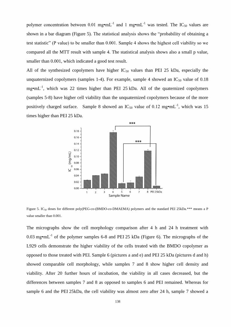
138
polymer concentration between 0.01 mg•mL-1
and 1 mg•mL-1
was tested. The IC50 values are
shown in a bar diagram (Figure 5). The statistical analysis shows the “probability of obtaining a
test statistic” (P value) to be smaller than 0.001. Sample 4 shows the highest cell viability so we
compared all the MTT result with sample 4. The statistical analysis shows also a small p value,
smaller than 0.001, which indicated a good test result.
All of the synthesized copolymers have higher IC50 values than PEI 25 kDa, especially the
unquaternized copolymers (samples 1-4). For example, sample 4 showed an IC50 value of 0.18
mg•mL-1
, which was 22 times higher than PEI 25 kDa. All of the quaternized copolymers
(samples 5-8) have higher cell viability than the unquaternized copolymers because of the more
positively charged surface. Sample 8 showed an IC50 value of 0.12 mg•mL-1
, which was 15
times higher than PEI 25 kDa.
Figure 5. IC50 doses for different poly(PEG-co-(BMDO-co-DMAEMA) polymers and the standard PEI 25kDa.*** means a P
value smaller than 0.001.
The micrographs show the cell morphology comparison after 4 h and 24 h treatment with
0.03 mg•mL-1
of the polymer samples 6-8 and PEI 25 kDa (Figure 6). The micrographs of the
L929 cells demonstrate the higher viability of the cells treated with the BMDO copolymer as
opposed to those treated with PEI. Sample 6 (pictures a and e) and PEI 25 kDa (pictures d and h)
showed comparable cell morphology, while samples 7 and 8 show higher cell density and
viability. After 20 further hours of incubation, the viability in all cases decreased, but the
differences between samples 7 and 8 as opposed to samples 6 and PEI remained. Whereas for
sample 6 and the PEI 25kDa, the cell viability was almost zero after 24 h, sample 7 showed a

139
reduced viability and sample 8 showed a minimal decrease in viability. All of these results
clearly show significantly reduced toxicity of the polymers compared to the accepted gold
standard PEI 25kDa.
Figure 6. 40×Micrographs of the L929 cells,
which were incubated with polymers for 4 h
and 24h, respectively. The concentration of
the polymers was 0.03 mg•mL-1. a) with
sample 6 for 4 h; b) with sample 7 for 4 h;
c) with sample 8 for 4 h; d) with PEI 25 kDa
for 4 h; e) with sample 6 for 24 h; f) with
sample 7 for 24 h; c) with sample 8 for 24 h;
d) with PEI 25 kDa for 24 h.
The hydrodynamic diameters of the polymer with a p-DNA complex at different N/P ratio were
measured at room temperature (Figure 7). This size measurement was performed for all of the
stable polyplexes at N/P ratios between 0 and 20. It has been reported that the acceptable size of
polyplex for endocytosis are less than 250 nm.29,30
The polydispersities of the polyplexes were all smaller than 0.3. All of the polyplex sizes were
less than 250 nm, and had already reached this size at an N/P ratio of 5. The size of the polyplex
depends on the N/P ratios and the polymer composition. With the increase of the N/P ratio, the
polyplex size decreased. With the increase of the PEG and BMDO part, the polyplex size

140
decreased as expected. That can be explained by the shielding effect of PEG.29
According to the
hydrodynamic size of the polyplexes, these copolymers are suitable candidates for gene
transfections.
Figure 7. Size of polyplexes formed with plasmid DNA (samples 1-8) at different N/P ratios by DLS (dynamic light scattering)
measurement.
Figure 8. The zeta potential of polyplexes (samples 1-8 with plasmid DNA) at different N/P ratios. Values are the means of 6
runs.
The zeta potential of the polyplex was determined at the N/P ratios of 5, 10, and 20 (Figure 8).
The zeta potential increased with the increasing N/P ratio. The polyplex with quaternized
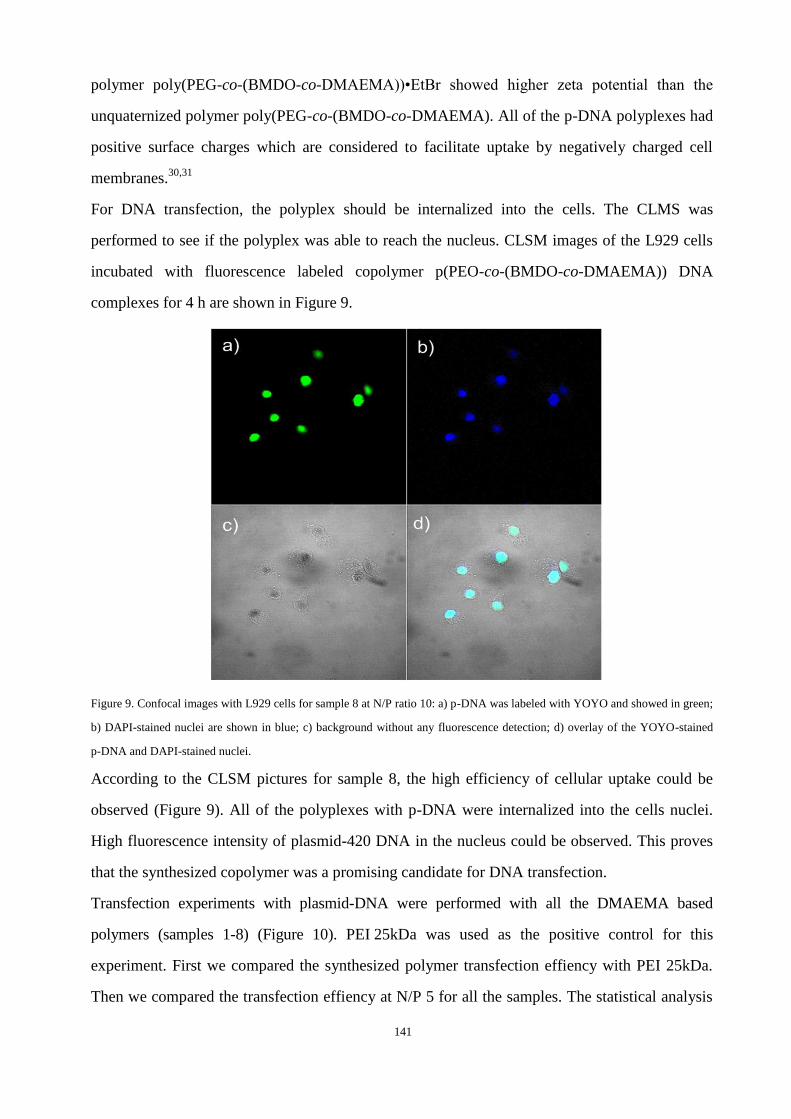
141
polymer poly(PEG-co-(BMDO-co-DMAEMA))•EtBr showed higher zeta potential than the
unquaternized polymer poly(PEG-co-(BMDO-co-DMAEMA). All of the p-DNA polyplexes had
positive surface charges which are considered to facilitate uptake by negatively charged cell
membranes.30,31
For DNA transfection, the polyplex should be internalized into the cells. The CLMS was
performed to see if the polyplex was able to reach the nucleus. CLSM images of the L929 cells
incubated with fluorescence labeled copolymer p(PEO-co-(BMDO-co-DMAEMA)) DNA
complexes for 4 h are shown in Figure 9.
Figure 9. Confocal images with L929 cells for sample 8 at N/P ratio 10: a) p-DNA was labeled with YOYO and showed in green;
b) DAPI-stained nuclei are shown in blue; c) background without any fluorescence detection; d) overlay of the YOYO-stained
p-DNA and DAPI-stained nuclei.
According to the CLSM pictures for sample 8, the high efficiency of cellular uptake could be
observed (Figure 9). All of the polyplexes with p-DNA were internalized into the cells nuclei.
High fluorescence intensity of plasmid-420 DNA in the nucleus could be observed. This proves
that the synthesized copolymer was a promising candidate for DNA transfection.
Transfection experiments with plasmid-DNA were performed with all the DMAEMA based
polymers (samples 1-8) (Figure 10). PEI 25kDa was used as the positive control for this
experiment. First we compared the synthesized polymer transfection effiency with PEI 25kDa.
Then we compared the transfection effiency at N/P 5 for all the samples. The statistical analysis
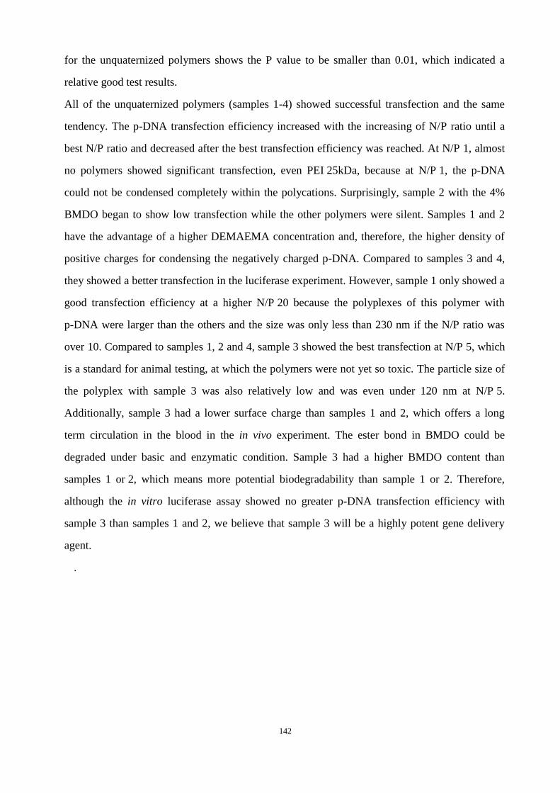
142
for the unquaternized polymers shows the P value to be smaller than 0.01, which indicated a
relative good test results.
All of the unquaternized polymers (samples 1-4) showed successful transfection and the same
tendency. The p-DNA transfection efficiency increased with the increasing of N/P ratio until a
best N/P ratio and decreased after the best transfection efficiency was reached. At N/P 1, almost
no polymers showed significant transfection, even PEI 25kDa, because at N/P 1, the p-DNA
could not be condensed completely within the polycations. Surprisingly, sample 2 with the 4%
BMDO began to show low transfection while the other polymers were silent. Samples 1 and 2
have the advantage of a higher DEMAEMA concentration and, therefore, the higher density of
positive charges for condensing the negatively charged p-DNA. Compared to samples 3 and 4,
they showed a better transfection in the luciferase experiment. However, sample 1 only showed a
good transfection efficiency at a higher N/P 20 because the polyplexes of this polymer with
p-DNA were larger than the others and the size was only less than 230 nm if the N/P ratio was
over 10. Compared to samples 1, 2 and 4, sample 3 showed the best transfection at N/P 5, which
is a standard for animal testing, at which the polymers were not yet so toxic. The particle size of
the polyplex with sample 3 was also relatively low and was even under 120 nm at N/P 5.
Additionally, sample 3 had a lower surface charge than samples 1 and 2, which offers a long
term circulation in the blood in the in vivo experiment. The ester bond in BMDO could be
degraded under basic and enzymatic condition. Sample 3 had a higher BMDO content than
samples 1 or 2, which means more potential biodegradability than sample 1 or 2. Therefore,
although the in vitro luciferase assay showed no greater p-DNA transfection efficiency with
sample 3 than samples 1 and 2, we believe that sample 3 will be a highly potent gene delivery
agent.
.
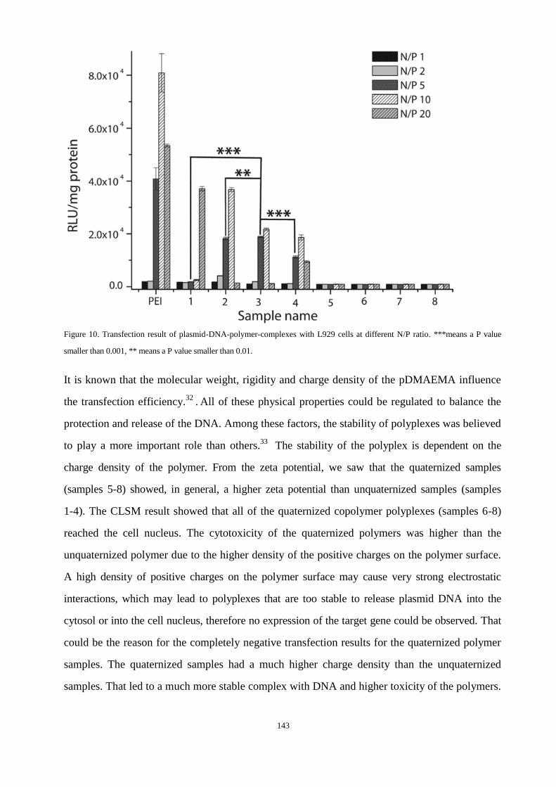
143
Figure 10. Transfection result of plasmid-DNA-polymer-complexes with L929 cells at different N/P ratio. ***means a P value
smaller than 0.001, ** means a P value smaller than 0.01.
It is known that the molecular weight, rigidity and charge density of the pDMAEMA influence
the transfection efficiency.32
. All of these physical properties could be regulated to balance the
protection and release of the DNA. Among these factors, the stability of polyplexes was believed
to play a more important role than others.33
The stability of the polyplex is dependent on the
charge density of the polymer. From the zeta potential, we saw that the quaternized samples
(samples 5-8) showed, in general, a higher zeta potential than unquaternized samples (samples
1-4). The CLSM result showed that all of the quaternized copolymer polyplexes (samples 6-8)
reached the cell nucleus. The cytotoxicity of the quaternized polymers was higher than the
unquaternized polymer due to the higher density of the positive charges on the polymer surface.
A high density of positive charges on the polymer surface may cause very strong electrostatic
interactions, which may lead to polyplexes that are too stable to release plasmid DNA into the
cytosol or into the cell nucleus, therefore no expression of the target gene could be observed. That
could be the reason for the completely negative transfection results for the quaternized polymer
samples. The quaternized samples had a much higher charge density than the unquaternized
samples. That led to a much more stable complex with DNA and higher toxicity of the polymers.
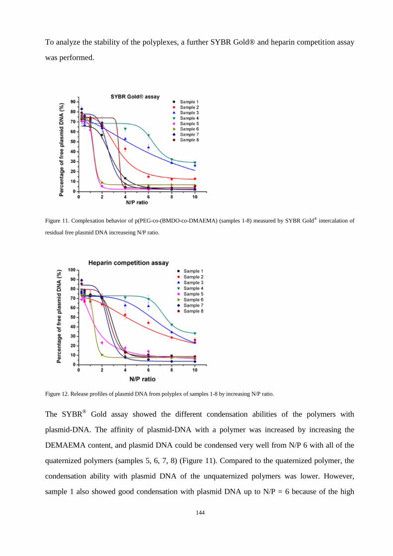
144
To analyze the stability of the polyplexes, a further SYBR Gold® and heparin competition assay
was performed.
Figure 11. Complexation behavior of p(PEG-co-(BMDO-co-DMAEMA) (samples 1-8) measured by SYBR Gold® intercalation of
residual free plasmid DNA increaseing N/P ratio.
Figure 12. Release profiles of plasmid DNA from polyplex of samples 1-8 by increasing N/P ratio.
The SYBR® Gold assay showed the different condensation abilities of the polymers with
plasmid-DNA. The affinity of plasmid-DNA with a polymer was increased by increasing the
DEMAEMA content, and plasmid DNA could be condensed very well from N/P 6 with all of the
quaternized polymers (samples 5, 6, 7, 8) (Figure 11). Compared to the quaternized polymer, the
condensation ability with plasmid DNA of the unquaternized polymers was lower. However,
sample 1 also showed good condensation with plasmid DNA up to N/P = 6 because of the high

145
DEMAEMA content, although it was unquaternized and had a less positive surface charge. The
other unquaternized polymers (samples 2, 3, 4) could not completely reach a complete p-DNA
condensation with an increasing N/P ratio, especially sample 4. The stability of polyplexes against
competing polyanions is also an important parameter for a gene delivery system, especially for in
vivo experiment, because the stability of the polyplexes can be strongly weakened by the presence
of serum in blood.34
The process of gene material complexation within polycations is entropy
driven and can be significantly impaired by the presence of other polyions like heparin.35
Differences in the stability against polyions were found to follow the same trend as the Sybr gold
assay, but the polyplexes formed with quaternized copolymers were less impaired by heparin
(Figure 12). That means the condensation of the plasmid DNA with quaternized copolymers was
complete. The plasmid DNA was very difficult to be released if delivered into the nuclei.
Therefore, no successful transfection was observed in the in vitro transfection experiment for the
quaternized polymers, in contrast to the successful transfection with unquaternized polymers
(Figure 13).
Figure 13. In vitro pDNA transfection mechanism with the synthesized polymer p(PEG-co-(BMDO-co-DMAEMA) (samples 1-4)
and p(PEG-co-(BMDO-co-DMAEMA)•EtBr (samples 5-8).
7.5 Conclusions
Novel degradable and biocompatible poly(PEG-co-(BMDO-co-DMAEMA) for gene transfection
were successfully synthesized via free radical polymerization. The solubility and the IC50 values
of the copolymers were significantly improved by bringing hydrophilic PEG blocks into the
polymer backbone. The toxicity of all the polymers was much lower than the positive control

146
PEI. The unquaternized copolymers showed a higher cell viability than the quaternized
copolymers as well as positive results in p-DNA transfection.
7.6 References
(1) Dalgleish, A. Gene Ther. 1997, 4, 629-630.
(2) Anderson, W. F. Science 1992, 256, 808-813.
(3) Fakhrai, H.; Dorigo, O.; Shawler, D. L.; Lin, H.; Mercola, D.; Black, K. L.; Royston, I.; Sobol, R. E. Proc.
Natl. Acad. Sci. U.S.A. 1996, 93, 2909-2914.
(4) Spear, M. A; Herrlinger, U.; Rainov, N.; Pechan, P.; Weissleder, R.; Breakefield, X. O. J. NeuroVirol. 1998,
4, 133-147.
(5) Takamiya, Y.; Short, M. P.; Ezzeddine, Z. D.; Moolten, F. L.; Breakefield, X. O.; Martuza, R. L. J. Neurosci.
Res. 1992, 33, 493-503.
(6) Azzam, T.; Domb, A. J. Curr. Drug Delivery 2004, 1, 165-193.
(7) Kabanov, A. V.; Felgner, P. L.; Seymour, L. J. In Self-assembling complexes for gene delivery - From
Laboratory to Clinical Trial; Wiley: NY, 1998; pp. 115-167.
(8) Felgner, P. L.; Heller, M. J.; Lehn, P.; Behr, J. P.; Szoka, J. F. C. In Am. Chem. Soc; Oxford University Press:
Washington, USA, 1996; pp. 177-190.
(9) Garnett, M. C. Crit. Rev. Ther. Drug Carrier Syst. 1999, 16, 147-207.
(10) Rolland, A. In Advanced Gene Delivery; Harwood Academic Publishers: The Netherlands, 1999; pp.
103-190.
(11) Chesnoy, S.; Huang, L. STP Pharma Sci. 9, 5-12.
(12) Neu, M.; Fischer, D.; Kissel, T. J. Gene Med. 2005, 7, 992-1009.
(13) Cherng, J. Y.; Wetering, P. van de; Talsma, H.; Daan, J. A. C.; Hennink, W. E. Pharm. Res. 1996, 13,
1038-1042.
(14) Arigita, C.; Zuidam, N. J.; Crommelin, D. J. A.; Hennink, W. E. Pharm. Res. 1999, 16, 1534-1541.
(15) Van de Wetering, P.; Cherng, J. Y.; Talsma, H.; Hennink, W. E. J. Controlled Release 1997, 49, 59-69.
(16) Jones, R. A; Poniris, M. H.; Wilson, M. R. J. Controlled Release 2004, 96, 379-391.
(17) Verbaan, F. J.; Klein Klouwenberg, P.; Van Steenis, J. H.; Snel, C. J.; Boerman, O.; Hennink, W. E.; Storm,
G. Int. J. Pharm. 2005, 304, 185-192.
(18) Cherng, J. Y.; Talsma, H.; Verrijk, R.; Crommelin, D. J.; Hennink, W. E. Eur. J. Pharm. Biopharm. 1999,
47, 215-224.
(19) You, Y.-Z. Manickam, D. S. Zhou, Q.-H.; Oupický, D. J. Controlled Release 2007, 122, 217-225.
(20) Agarwal, S.; Ren, L.; Kissel, T.; Bege, N. Macromol. Chem. Phys. 2010, 211, 905-915.
(21) Petersen, H.; Fechner, P. M.; Martin, A. L.; Kunath, K.; Stolnik, S.; Roberts, C. J.; Fischer, D.; Davies, M.
C.; Kissel, T. Bioconjugate Chem. 2002, 13, 845-854.
(22) Zheng, M.; Liu, Y.; Samsonova, O.; Endres, T.; Merkel, O.; Kissel, T. Int. J.Pharm. 2011.
(23) Wickel, H.; Agarwal, S. Macromolecules 2003, 36, 6152-6159.
(24) Samsonova, O.; Pfeiffer, C.; Hellmund, M.; Merkel, O.; M.; Kissel, T. Polymers 2011, 3, 693-718.
(25) Mosmann, T. J. Immunol. Methods 1983, 65, 55-63.

147
(26) Beck-Broichsitter, M.; Thieme, M.; Nguyen, J.; Schmehl, T.; Gessler, T.; Seeger, W.; Agarwal, S.; Greiner,
A.; Kissel, T. Macromol. Biosci. 2010, 10, 1527-35.
(27) Merkel, O. M.; Zheng, M.; Mintzer, M. A.; Pavan, G. M.; Librizzi, D.; Maly, M.; Höffken, H.; Danani, A.;
Simanek, E. E.; Kissel, T. J. Controlled Release 2011, 153, 23-33.
(28) Wickel, H.; Agarwal, S.; Greiner, A. Macromolecules 2003, 36, 2397-2403.
(29) Liu, Y.; Steele, T.; Kissel, T. Rapid Commun. 2010, 31, 1509-1515.
(30) Kunath, K.; Merdan, T.; Hegener, O.; Häberlein, H.; Kissel, T. J. Gene Med. 2003, 5, 588-599.
(31) Grayson, A. C. R.; Doody, A. M.; Putnam, D. Pharm. Res. 2006, 23, 1868-1876.
(32) Grigsby, C. L.; Leong, K. W. J. R. Soc., Interface 2010, 7, 67-82.
(33) Mao, S.; Neu, M.; Germershaus, O.; Merkel, O.; Sitterberg, J.; Bakowsky, U.; Kissel, T. Bioconjugate Chem.
2006, 17, 1209-1218.
(34) Merkel, O. M.; Librizzi, D.; Pfestroff, A.; Schurrat, T.; Buyens, K.; Sanders, N. N.; De Smedt, S. C.; Béhé,
M.; Kissel, T. J. Controlled Release 2009, 138, 148-159.
(35) Bronich, T.; Kabanov, A. V.; Marky, L. a J. Phys. Chem. B 2001, 105, 6042-6050.

148
8 SUMMARY
8.1 Summary
In this thesis, biodegradable non-viral polymeric nucleic acids delivery vectors were characterized
concerning biophysicochemical parameters.
In the first part of this research (chapter 2), to answer the questions: why the principle of DNA
transfection cannot be directly applied for siRNA transfection, we investigated the complexation
and aggregation mechanism of nucleic acids/polycations on the atomic and molecular scale. The
MD and ITC data showed us the different nature and the different hierarchical mechanism related
polycation-siRNA and polycation-pDNA complexes. The results of dye quenching assays indicated
a biphasic behavior of siRNA binding with polycations where molecular reorganization of the
siRNA within the polycations occurred at lower N/P-ratios (nitrogen/phosphorus). Additionally,
heparin assays showed that the stability of siRNA/polymer complexes is especially good at a rather
lower N/P-ratio of 2. Interestingly, with the following study of the relationship between nucleic
acids/polycations aggregation mechanism and in vitro siRNA delivery efficiency, which is
performed by RT-PCR and CLSM, we found that the copolymer showed the best knockdown
effect with siRNA at N/P=2. All our results emphasized one point: lower N/P-ratios are especially
effective for polycationic nanocarrier-based siRNA delivery, because siRNA aggregation results in
a more uniform and stable complex formation at low N/P ratios already, which lead to increased
siRNA delivery efficiency. This could have broad implications for the delivery of siRNA as less
toxic and yet efficient delivery systems have been the bottle-neck for the translation of this
promising approach into the clinical arena.
In chapter 3, novel biodegradable amphiphilic copolymers hy-PEI-g-PCL-b-PEG were prepared
by grafting PCL-b-PEG chains onto hyper-branched poly(ethylene imine) as non-viral gene
delivery vectors. With the question: how can the graft densities of PCL-b-PEG chains influence the
in vitro DNA delivery efficiency, our study began with the characterization of physico-chemical
properties and expected that with the introducing of the grafted PCL-b-PEG chains, the in vitro
DNA delivery efficiency with the grafted PCL-b-PEG chains could be improved. In the following
study, no correlation was shown between the sizes of polyplexes and transfection efficiencies, while
149
buffer-capacity, cytotoxicity and zeta-potential turned out to be the key factors for the explanation of
the results of the gene transfer experiments. Of all the experimental results, buffer-capacity has
almost exactly the same tendency as transfection efficiency. We therefore assume that in all
processes of DNA transfection, the endosomal escape has a really important and rate-limiting role.
This opens new perspectives to advance the rational design of new gene delivery systems.
The further investigation of these biodegradable grafted amphiphilic copolymers
hy-PEI-g-(PCL-b-PEG)n as potential siRNA delivery vectors was showed in chapter 4, which is the
direct continuation of Chapter 3. The purpose in this section was to enhance the in vivo blood
circulation time and siRNA delivery efficiency of biodegradable copolymers
polyethylenimine-graft-polycaprolactone-block-poly(ethylene glycol) (hyPEI-g-PCL-b-PEG) by
introducing high graft densities of PCL-PEG chains. The questions which this work was based on,
such as zeta size, siRNA comlexation efficiency, siRNA protection against competing polyanions,
in vitro RNAi, pharmacokinetic issues of in vivo administered siRNA, in vivo biodistribution and in
vivo stability of polyelectrolyte complexes are elucidated. Our study indicated that the effect of
PEG on prolonged circulating depends not only on its content in a copolymer (length or
percentage), but also on the structure or the shape of the amphiphilic copolymer. We demonstrated
that polymeric micelles, which are formed with amphiphilic block polymers have advantages
especially for in vivo siRNA delivery, and that the graft density of the amphiphilic chains can
enhance the blood circulation, which is a key parameter to promote the development of safe and
efficient non-viral polymeric siRNA delivery in vivo.
Although the copolymers hy-PEI-g-(PCL-b-PEG)n showed positive results as pDNA and siRNA
delivery vectors in chapter 3 and 4, the delivery of gene materials with these non-targeted
copolymers is achieved mainly passively by the passive targeting. Therefore, to optimize these
polymeric gene delivery vectors with targeting function, in chapter 5, folate conjugated
PEI-g-PCL-b-PEG was examined for targeted gene delivery. Lower cytotoxicity was observed for
PEI-g-PCL-b-PEG-Fol than PEI-g-PCL-b-PEG and the cellular uptake of polyplexes was enhanced
by PEI-g-PCL-b-PEG-Fol in FR over-expressing KB cells compared with those by
PEI-g-PCL-b-PEG. Importantly, this enhancement was inhibited by free folic acid, while did not
appear in FR-negative A549 cells. All these suggested the specific cell uptake of
PEI-g-PCL-b-PEG-Fol/pDNA polyplexes via folate receptor-mediated endocytosis. Consequently,
150
PEI-g-PCL-b-PEG-Fol/pDNA polyplexes revealed higher transfection than
PEI-g-PCL-b-PEG/pDNA. Additional studies on gene transfection in vivo and utilizing these
described folate-conjugated copolymers for targeted siRNA delivery are in proceeding.
In Chapter 6, the novel siRNA delivery systems based on hyperflexible generation 2-4 triazine
dendrimers was identified by correlating physico-chemical and biological in vitro and in vivo
properties of the complexes with their thermodynamic interaction features simulated by molecular
modeling and the influence of dendrimer flexibility has systematically been investigated and
discussed. In this study, molecular modeling helped to understand experimental parameters based
on the dendrimers’ structural properties and molecular imaging non-invasively predicted the in vivo
fate of the complexes, both techniques can efficiently support the rapid development of safe and
efficient siRNA formulations that are stable in vivo.
In Chapter 7, novel degradable and biocompatible poly(PEG-co-(BMDO-co-DMAEMA) for gene
transfection were successfully characterized. The physicochemical properties and in vitro pDNA
delivery efficiency of these polymers were characterized. The unquaternized copolymers showed
higher cell viability than the quaternized copolymers as well as positive results in p-DNA
transfection.
8.2 Perspectives
In continuation of the projects described, a number of further developments are possible. Chapter 2
is the about study of the complexation and aggregation mechanism of nucleic acids/polycations on
the atomic and molecular scale. Although we have indroduced the novel synergistic use of
molecular modeling, molecular dynamics simulation, isothermal titration calorimetry and other
characterization techniques, the novel research methods for the binding and aggregation
mechanism of nucleic acids/polycations are still needed. For example, more detection about the
nucleic acids localization with in polyplexes will also be very necessary, which depends exactly on
the new development of microscopical methods.
In chapter 3, the copolymers were designed to decrease the toxicity of hyPEI. But after the study of
buffer-capacity, we find that the endosomal escape has really a very important role of all of the

151
gene material delivery process. And a design or optimization of the molecule structure for a higher
buffer-capacity might be very meaningful in the future.
Chapter 3-5 described the copolymer hy-PEI-g-(PCL-b-PEG)n as novel gene delivery vectors. In
these studies, only copolymers with short PCL segments (weight of 570) were studied. It is still very
interesting to discuss the influence of long PCL segments in this structure.
In chapter 5, folate conjugated PEI-g-PCL-b-PEG showed improved targeted pDNA delivery.
These targeted vectors described above are certainly investigating for siRNA delivery in vitro and
in vivo, which is especially meaningful for the development of pulmonary siRNA delivery.
Moreover, cationic quantum dots (QDs) is a promising method to study the intracellular trafficking,
unpacking, and gene silencing. In the following study about the the intracellular trafficking and
unpacking mechanism, gene delivery vectors can be labeled with QDs. Fluorescence resonance
energy transfer (FRET) can be achieved between fluorescence-labeled siRNA and QDs-conjugated
polymers in the complex. If the siRNA is not condensed within the polymeric vectors, the FRET
effect should not be detected anymore, which describes indirectly the unpacking or release of the
siRNA from the polymers.
These multifunctional gene delivery systems with higher siRNA protection and loading efficiency,
better biocompatibility and transfection efficiency, targeting effect and long circulating time is still
challenging the area in cancer gene delivery. The “magic bullet” vision of Paul Ehrlich over 100
years ago is beginning to be realized, and with continued research and development.
8.3 Zusammenfassung
In der vorliegenden Arbeit wurden neue bioabbaubare Polymere für den Transport von
Nukleinsäuren bezüglich ihrer biophysikochemischen Eigenschaften charakterisiert. Im ersten Teil
dieser Arbeit (Kapitel 2) untersuchten wir die Komplexierung und den Aggregationsmechanismus
von Nukleinsäuren mit Polykationen auf atomarer und molekularer Ebene unter Verwendungen
der molekularen Modellierung, molekulardynamischen Simulation, isothermen
Titrationskalorimetrie und anderen Charakterisierungsmethoden, um die Frage zu beantworten,
warum ein guter pDNA-Vektor nicht unbedingt eben so gut für siRNA funktioniert. Diese Daten
zeigten uns sehr unterschiedliche natürliche Eigenschaften und den hierarchischen Mechanismus
der Komplexbildung von Polykationen/siRNA und Polykationen/pDNA. Mit der folgenden
152
Untersuchung der Beziehung zwischen dem
Nukleinsäuren/Polykationen-Aggregationsmechanismus und der
in-vitro-siRNA-Delivery-Effizienz, konnten wir zeigen, dass niedrige N/P-Verhältnisse
(Stickstoff/Phosphat) exzellente siRNA-Delivery-Effizienz ermöglichen, dies wurde aber in den
bisherigen Untersuchungen für siRNA-Transfektion mit Polykationen vernachlässigt.
In Kapitel 3 wurden neue bioabbaubare, amphipathische Copolymere hy-PEI-g-PCL-b-PEG durch
die Kupplung von PCL-b-PEG-Ketten auf dem hyper-verzweigten Poly(ethylenimin) als
nicht-virale Gentransfer-Vektoren charakterisiert. Der Einfluss der Kupplungsdichten von
PCL-b-PEG-Ketten auf physikalisch-chemische Eigenschaften, DNA-Komplexierung und
Transfektionseffizienz wurde untersucht und diskutiert. In dieser Studie wurde keine Korrelation
zwischen den Größen der Polyplexe und Transfektionseffizienz gezeigt, während sich die
Puffer-Kapazität, Zytotoxizität und Zeta-Potential als die entscheidenden Faktoren für Erklärung
der Ergebnisse der Gentransfektion erwiesen. Von allen experimentellen Ergebnissen, zeigte die
Puffer-Kapazität fast genau die gleiche Tendenz wie die Transfektionseffizienz. Wir gehen daher
davon aus, dass in allen Prozessen der DNA-Transfektion, der „endosomal escape“ eine wichtige
und geschwindigkeitsbestimmende Rolle spielt.
Weitere Untersuchungen dieser biologisch abbaubaren amphipathischen Copolymere
hy-PEI-g-(PCL-b-PEG)n als potentiale siRNA-delivery Vektoren wurden in Kapitel 4 berichtet.
Der Zweck der Untersuchungne dieses Kapitels war, die in vivo Blutzirkulationszeit und die
siRNA Transfektionseffizienz der bioabbaubaren Copolymere hy-PEI-g-(PCL-b-PEG)n zu
erhöhen. Unsere Studie zeigte, dass die Wirkung von PEG auf längere Blutzirkulationszeit nicht
nur vom prozentualen Inhalt in einem Copolymer abhängt, sondern auch von der Struktur oder
Form des Copolymers. Die Polymere-Mizellen, die mit amphipathischen Blockpolymeren
entstehen, haben Vorteile für siRNA Transfektion z.B. effektive in vivo siRNA Transfektion und
verlängerte Blutzirkulation. Im Kapitel 5, wurde das mit der Folsäure konjugierte Polymer
PEI-g-PCL-b-PEG-Fol bezüglich seiner biophysikochemischen Eigenschaften charakterisiert.
Geringere Zytotoxizität wurde bei PEI-g-PCL-b-PEG-Fol beobachtet. Die zelluläre Aufnahme
wurde durch die Kupplung von Folsäure verbessert. Wichtig ist, dass diese Verbesserung durch
freie Folsäure gehemmt wurde, und dass diese Verbesserung auch nicht in
Folat-Rezeptor-negativen A549-Zellen beobachtet wurde. Alle Daten bestätigen die Aufnahme der

153
PEI-g-PCL-b-PEG-Fol/pDNA Polyplexe über Folat-Rezeptor-vermittelte Endozytose. Die in vitro
DNA Transfektionseffizienz wurde deutlich erhöht durch die Kupplung von Folsäure. Die
zusätzlichen Untersuchungen in vivo und Nutzung dieser beschriebenen Folat-konjugierten
Copolymeren für gezielte siRNA Transfektion sind in Bearbeitung.
In Kapitel 6 wurden hyperflexible Triazin-Dendrimere der Generationen 2-4 als neuartige
siRNA-Delivery-Systeme bezüglich ihrer physikalisch-chemischer und biologischer in vitro und in
vivo Eigenschaften untersucht und diskutiert. In diesem Kapitel wurden die thermodynamische
Eigenschaften durch molekulare Modellierung simuliert, und der Einfluss der Flexibilität wurde
systematisch untersucht und diskutiert.
In Kapitel 7 wurden neuartige bioabbaubare und biokompatible Polymere der Zusamensetzung
Poly (PEG-co-(BMDO-co-DMAEMA) für die Gen-Transfektion erfolgreich bezüglich ihrer
physikalisch-chemischen und in vitro biologischen Eigenschaften charakterisiert. Die
quarternierten Copolymere zeigten niedrige Zytotoxizität als die quarternisierten Copolymere
sowie positive Ergebnisse in der DNA-Transfektion in vitro.
Weiterentwicklungen der beschriebenen nicht-virale Gen-Transfektion-Systeme sind immer
denkbar. In Kapitel 2 haben wir die Komplexierung und den Aggregationsmechanismus von
Nukleinsäuren mit Polykationen auf atomarer und molekularer Ebene untersucht. In diesem
Projekt wurden molekularen Modellierung, molekulardynamische Simulation, isotherme
Titrationskalorimetrie und andere Charakterisierungsmethoden angewendet. Trotzdem besteht
immer Bedarf für neue Untersuchungsmethode zur Charakterisierung der pDNA- oder
siRNA-Lokalisierung innerhalb der Komplexe. Es ist daher sinnvoll, neue mikroskopische
Methoden weiterzuentwickeln.
In Kapitel 3 haben wir anhand der Untersuchung des Einflusses der Kupplungsdichten von
PCL-b-PEG-Ketten auf physikalisch-chemische Eigenschaften, DNA-Komplexierung und
Transfektionseffizienz gefunden, dass der „endosomal escape“ eine sehr wichtige Rolle für die
Gen-Transfektion spielt. Und ein Design oder eine Optimierung des Polymers mit einer höherer
Puffer-Kapazität könnte sich in Zukunft als sehr sinnvoller weisen.
In Kapitel 3-5 werden die Copolymer hy-PEI-g-(PCL-b-PEG)n als neuartige Transfektionpolymere
beschrieben. In dieser Untersuchung wurden nur Copolymere mit kurzen PCL-Segmenten

154
(Gewicht von 570 Da) untersucht. Es wäre daher noch sehr interessant, den Einfluss der langen
PCL-Segmente in dieser Struktur zu diskutieren.
In Kapitel 5 wurden Folsäure-gekuppelte PEI-g-PCL-b-PEG-Konjugate charakterisiert, und
verbesserte pDNA Transfektion wurde beobachtet im Vergleich mit unmodifiziertem Polymer. Die
beschriebenen Vektoren mit Targeting-Liganden sind sicherlich wert, Weiter in vivo für siRNA
untersucht zu werden, welches besonders sinnvoll wäre für die Entwicklung der Gentransfektion in
der Lunge.
Kationische Quantenpunkte (QDs) können für die weitere Untersuchung der siRNA-Freisetzung
benutzt werden. Der Fluoreszenz-Resonanz-Energie-Transfer (FRET) Effekt zwischen
fluoreszenzmarkierter siRNA und mit QDs geladenen Polymeren könnte nachgewiesen werden,
wenn die siRNA innerhalb der Polymere kondensiert wird.
Diese multifunktionalen Gen-Transport-Systeme mit höherer siRNA-Verkapselung und höherem
Schutz-Effizienz, besserer Biokompatibilität und Transfektionseffizienz sowie zusätzlichem
Targeting-Effekt und langer Blutzirkulation sind jedoch eine Herausforderung für das Gebiet der
Genetherapie innrehalb der Krebsforschung. Die “magic bullet” Vision von Paul Ehrlich vor über
100 Jahren beginnt sich bei fortgesetzter Forschung und Entwicklung zu verwirklichen.

155
9 APPENDICES
9.1 Abbreviations
2’OMe 2’O-Methoxy-
AFM Atomic Force Microscopy
ANOVA Analysis of Variance
AUC Area under the Curve
CLSM Confocal Laser Scanning Microscopy
DLS Dynamic Light Scattering
DTPA Diethylenetriaminepentaacetic Acid
dsRNA Double-Stranded RNA
EGFP Enhanced Green Fluorescent Protein
EPR Enhanced Permeability and Retention
FACS Fluorescence Assisted Cell Sorting
FITC Fluorescein-Isothiocyanate
FOL folate acids
HEPES 2-[4-(2-Hydroxyethyl)-1-piperazinyl]ethanesulfonic Acid
IC50 Half-Inhibitory Concentration
LMW Low Molecular Weight
MTT 3-(4,5-Dimethylthiazol-2-yl)-2,5-diphenyltetrazolium Bromide
N/P Nitrogen to Phosphate Ratio
PBS Phosphate Buffered Saline
PCR Polymerase Chain Reaction
pDNA Plasmid DNA
PAMAM Polyamidoamine
PEG Polyethylenglycol
PEI Polyethylenimine
PDI Polydispersity Index
PLGA poly(lactic-co-glycolic acid
RISC RNA Induced Silencing Complex

156
RNAi RNA Interferance
siRNA Short Interfering RNA
SPECT Single Photon Emission Computed Tomography
9.2 List of Publications
9.2.1 Articles
1. Zheng, M. Y.; Liu, Y.; Samsonova, O.; Endres, T.; Merkel, O.; Kissel, T., Amphiphilic and
biodegradable hy-PEI-g-PCL-b-PEG copolymers efficiently mediate transgene expression
depending on their graft density. International Journal of Pharmaceutics 2012, 427 (1), 80-87.
(contributed equally first authors)
2. Zheng, M.; Pavan, G. M.; Neeb, M.; Schaper, K., A.; Danani, A.; Klebe, G.; Merkel, M. O.;
Kissel, T.: Targeting the blind spot of polycationic nanocarrier-based siRNA delivery (Submitted
to ACS Nano, 2012)
3. Zheng, M.; Merkel, M. O.; Librizzi, D.; Kilic, A.; Liu, Y.; Renz, H.; Kissel, T.: Enhancing in
vivo long-circulating and siRNA delivery efficiency of biodegradable high grafted
hy-PEI-g-PCL-b-PEG copolymers (Accepted by Biomaterials, 2012)
4. Zhang, Y.; Zheng, M.; Kissel, T.; Agarwal, S., Design and biophysical characterization of
bioresponsive degradable poly(dimethylaminoethyl methacrylate) based polymers for in vitro DNA
transfection. Biomacromolecules 2012, 13 (2), 313-22.
(contributed equally first authors)
5. Liu, L.; Zheng, M.; Renette, T.; Kissel, T., Modular Synthesis of Folate Conjugated Ternary
Copolymers: Polyethylenimine-graft-Polycaprolactone-block-Poly(ethylene glycol)-Folate for
Targeted Gene Delivery. Bioconjug Chem 2012.
(contributed equally first authors)
6. Merkel, O. M.; Zheng, M. Y.; Mintzer, M. A.; Pavan, G. M.; Librizzi, D.; Maly, M.; Hoffken,
H.; Danani, A.; Simanek, E. E.; Kissel, T., Molecular modeling and in vivo imaging can identify
successful flexible triazine dendrimer-based siRNA delivery systems. Journal of Controlled
Release 2011, 153 (1), 23-33.
(contributed equally first authors)
7. Merkel, O. M.; Zheng, M.; Debus, H.; Kissel, T., Pulmonary gene delivery using polymeric
nonviral vectors. Bioconjug Chem 2011, 23 (1), 3-20.
8. Endres, T.; Zheng, M.; Beck-Broichsitter, M.; Kissel, T., Lyophilised ready-to-use formulations
of PEG-PCL-PEI nano-carriers for siRNA delivery. Int J Pharm 2012, 428 (1-2), 121-4.

157
9. Endres, T.; Zheng, M.; Beck-Broichsitter, M.; Samsonova, O.; Debus, H.; Kissel, T.,
Optimising the self-assembly of siRNA loaded PEG-PCL-lPEI nano-carriers employing different
preparation techniques. J Control Release 2012.
10. Merkel, O. M.; Beyerle, A.; Beckmann, B. M.; Zheng, M. Y.; Hartmann, R. K.; Stoger, T.;
Kissel, T. H., Polymer-related off-target effects in non-viral siRNA delivery. Biomaterials 2011, 32
(9), 2388-2398.
11. Mattheis, C.; Zheng, M. Y.; Agarwal, S., Closing One of the Last Gaps in Polyionene
Compositions: Alkyloxyethylammonium Ionenes as Fast-Acting Biocides. Macromolecular
Bioscience 2012, 12 (3), 341-349.
12. Zheng, M.; Kissel, T.; Agarwal, S.: Gentherapie ohne Nebenwirkung-Neuartige Biopolymere
helfen bei der Gentransfektion
Laborpraxis, Juni, 2011
9.2.2 Poster Presentations
Mengyao Zheng, Yu Liu, Olga Samsonova, Thomas Endres, Olivia Merkel, and Thomas Kissel: Amphiphilic
and biodegradable hy-PEI-g-PCL-b-PEG copolymers efficiently mediate transgene expression depending on their
graft density, Jahrestagung der Deutschen Pharmazeutischen Gesellschaft e.V., Braunschweig, Germany, 4-6
October 6, 2010
Olivia Merkel, Mengyao Zheng, Meredith Mintzer, Giovanni Pavan, Damiano Librizzi, Eric Simanek, Thomas
Kissel: Molecular modeling and in vivo imaging can identify successful flexible triazine dendrimer-based siRNA
delivery systems, Controlled Release Society Local Chapter Meeting Germany , Jena, Germany, 15-16 March,
2011
Li Liu, Mengyao Zheng, Markus Benfer, Thomas Kissel: Multifunctional nano carrier based on
PEI-g-PCL-b-PEG-folate for targeted gene delivery. the 3rd European Science Foundation Summer School"
Nanomedicine 2011 ", Lutherstadt Wittenberg, Germany, 19-24 June 2011
158
Mengyao Zheng, Olivia M. Merkel, Damiano Librizzi, Thomas Kissel: Enhancing in vivo long-circulating and
siRNA delivery efficiency of biodegradable high grafted hy-PEI-g-PCL-b-PEG copolymers, International
Conference on Nanoscience & Technology, China 2011, Beijing, China, 7-9 September, 2011
Mengyao Zheng, Li Liu, Damiano Librizzi, Thomas Renette, Olivia M. Merkel, Thomas Kissel: Folate
conjugated copolymer (Fol-PEG-PCL-hyPEI) for efficient and targeted in vitro and in vivo sirna delivery,
Controlled Release Society Local Chapter Meeting Germany, Wuerzburg, Germany, March 29-30, 2012
9.2.3 Lectures
Mengyao Zheng, Olivia Merkel, Manuel Neeb, Michael Hellwig, Thomas Kissel: Structural Conformations and
Nucleic Acid Location within Amphyphilic hy-PEI-PCL-mPEG Complexes as Non-Viral Vectors, Lecture,
Controlled Release Society Local Chapter Meeting Germany, Jena, Germany, March 15-16, 2011
9.2.4 Abstracts
Li Liu, Mengyao Zheng, Markus Benfer, Thomas Kissel: Multifunctional nano carrier based on
PEI-g-PCL-b-PEG-folate for targeted gene delivery. the 3rd European Science Foundation Summer School"
Nanomedicine 2011 ", Lutherstadt Wittenberg, Germany, 19-24 June 2011
Mengyao Zheng, Olivia M. Merkel, Damiano Librizzi, Thomas Kissel: Enhancing in vivo long-circulating and
siRNA delivery efficiency of biodegradable high grafted hy-PEI-g-PCL-b-PEG copolymers, International
Conference on Nanoscience & Technology, China 2011, Beijing, China, 7-9 September, 2011
Mengyao Zheng, Li Liu, Damiano Librizzi, Thomas Renette, Olivia M. Merkel, Thomas Kissel: Folate
conjugated copolymer (Fol-PEG-PCL-hyPEI) for efficient and targeted in vitro and in vivo sirna delivery,
Controlled Release Society Local Chapter Meeting Germany, Wuerzburg, Germany, March 29-30, 2012
Mengyao Zheng, Yu Liu, Olga Samsonova, Thomas Endres, Olivia Merkel, and Thomas Kissel: Amphiphilic
and biodegradable hy-PEI-g-PCL-b-PEG copolymers efficiently mediate transgene expression depending on their

159
graft density, 8th World Meeting on Pharmaceutics, Biopharmaceutics and Pharmaceutical Technology, Istanbul,
Turkey 19th to 22
nd March, 2012
9.3 Curriculum Vitae
Persönliche Daten
Name: Mengyao Zheng
Geburtsdatum: 17.02.1983
Geburtsort: Beijing, China
Staatsangehörigkeit: Chinesisch
Anschrift: Wehrdaer Weg 3, 35037 Marburg
E-Mail: [email protected]
Ausbildung und Berufspraxis
Jul. 2001 Abitur, Gymnasium, Beijing, China (Note: sehr gut)
Sep. 2001 – Apr. 2003 Peking Pädagogische Universität (China), Fachbereich Chemie
Apr. 2004 – Jul. 2004 Philipps-Universität Marburg, Fachbereich Chemie
Oct. 2004 – Oct. 2009 Philipps-Universität Marburg Fachbereich Pharmazie
Feb.2009 Wahlpflichtpraktikum, Institut für Pharmazeutische Chemie, Philipps-Universität,
Marburg: Isolierung und Charakterisierung rekombinanter RNA-und
DNA-Polymerasen
Oct. 2009 2. Abschnitt der Pharmazeutischen Prüfung
Sep. 2010 Diplomverteidigung (Note: sehr gut)
Jan. 2010-jetzt Promotion; Wissenschaftliche Mitarbeiterin im Institut für Pharmazeutische
Technologie und Biopharmazie, Philipps-Universität Marburg

160
9.4 Danksagung
Mein ganz besonderer Dank gilt meinem Doktorvater Herrn Prof. Dr. Thomas Kissel.
Vor allem möchte ich mich für das mir entgegengebrachte Vertrauen und Ermutigungen bedanken,
wodurch ich mich selbst weiterentwickeln konnte. Ihre immer wieder überraschende Ideen,
interessante Diskussionen sowie Ihr großer Erfahrungsschatz haben bewirkt, dass mir den Weg in
die Forschung gebahnt. Von Ihnen habe ich gelernt, wie wichtig die Kreativität und Leidenschaft
für die Forschung sind. Auf der anderer Seite, man muss auch dazwischen selbst denken und
Erfahrungen sammeln, um das nicht realistische Konzept und den unnötigen Fehler zu vermeiden.
Herr Prof. Dr. Thomas Kissel stellt uns immer wertvolle gute Fragen, welche, in meiner Meinung,
noch wichtiger als die Lösungen selbst sind. Ich möchte mich auch für alle die Freiheiten und die
großartige Unterstützung bedanken, die ich zwischen meine Promotion genießen durfte.
Mein besonderer Dank gilt außerdem Frau Prof. Olivia M. Merkel. Frau Merkel hat mich eine
Vielzahl an Methoden beigebracht. Für die überraschende Ideen, interessante Diskussionen sowie
hilfreiche Unterstützung vom ersten bis zum letzten Tag möchte ich mich ganz besonders herzlich
bei Frau Merkel bedanken. Ihre Kreativität, Aktivität, Forschungsleidenschaft und
Durchführungsfähigkeit beeindrucken mich sehr.
Herrn Prof. Dr. Carsten Culmsee möchte ich einerseits für die Erstellung des Zweitgutachters,
andererseits aber auch für die offene Unterhaltung und Vorschlägen bezüglich weiterer
Karriereentwicklung nach dem 2. Staatexam bedanken.
Außerdem möchte ich mich ganz besonders herzlich bei Herrn Prof. Andreas Greiner und Frau
Prof. Seema Agarwal (Makromolekulare Chemie, Marburg) für das tolle Zusammenarbeit und
Forschungsstipendium bedanken. Bei Frau Prof. Seema Agarwal möchte ich mich herzlich für die
Synthese und viele interessante Ideen und Diskussionen bedanken. Zusätzlich bedanke ich mich
auch bei der gesamten Gruppe für die äußerst gastfreundliche Aufnahme am Institut.
Mein besonderer Dank gilt außerdem Herrn Prof. Andreas K. Schaper und Herrn Michael Hellwig
(Center of Material Science, Philipps Universität Marburg) for TEM imaging.
Bei Herrn Prof. Dr. Gerhard Klebe, Herrn Manuel Neeb und Herrn Dr. Adam Biela, möchte ich
mich für die Nutzung von ITC-Geräten und die Unterstützung in Fragen und Diskussionen
bedanken.

161
Bei Herrn Prof. Dr. Wolfgang Parak, Yu Xiang und Raimo Hartmann (Department of Physics,
Philipps-Universität Marburg) möchte ich mich für die Unterstützung CLSM imaging bedanken.
Herzlich danken möchte ich auch Herrn Prof. Dr. Harald Renz, Herrn Dr. Holger Garn (Institute of
Laboratory Medicine and Pathobiochemistry, Philipps Universität Marburg) und Frau Dr.
Agnieszka Turowska sowie der gesamten Gruppe am BMFZ für die freundliche Aufnahme im
Institut und für die lehrreiche und interessante Zusammenarbeit. Ohne die vielen etablierten
Untersuchungsmethoden, die Aufarbeitung einer Vielzahl von Organproben sowie das Anfertigen
mikroskopischer Präparate, wäre die Durchführung der in vivo Experimente nicht möglich
gewesen.
Ganz besonders möchte ich mich bei Herrn Damiano Librizzi (Institut für Nuklearmedizin,
Uni-Marburg) bedanken, die Tage und Nächte mit uns durchgearbeitet hat, um die in vivo
Versuche möglich zu machen.
Bei Herrn Prof. Dr. Roland Hartmann, Herrn Dr. Arnold Grünweller, möchte ich mich für die
Nutzung von Geräten und die Unterstützung in molekularbiologischen Fragen bedanken.
Mein Dank gilt besonders Frau Ayse Kilic für die Zusammenarbeit bezüglich Messung von FACS
sowie der Transfektion in den T-Zellen. Ohne die vielen etablierten Untersuchungsmethoden, wäre
die Durchführung der Experimente nicht möglich gewesen. Ebenso möchte ich mich für die vielen
Diskussionen mit Frau Ayse Kilic bedanken, die mir oftmals neue Perspektiven eröffnet haben.
Ganz besonders möchte ich mich bei Herrn Dr. Giovanni M. Pavan für die ausgezeichnete
Molecular dynamics simulation und weitere interessante Diskussion bedanken.
Allen Mitgliedern des Instituts für pharmazeutische Technologie und Biopharmazie bin ich zu
größtem Dank verpflichtet. Ich danke Frau Eva Mohr für ihre Arbeit in der Zellkultur sowie die
Organisation der Bestellungen für die Unterstützung in allen Fragen nach den Zellen.
Weiterhin möchte ich meinen Kollegen Julia Michaelis, Nadja Bege, Moritz Beck-Broichsitter,
Markus Benfer, Heiko Debus, Thomas Endres, Tobias Lebhardt, Dr. Dafeng Chu, Dr. Yu Liu, Dr.
Li Liu, Nan Zhao, Thomas Renette, Susanne Rösler, Christoph Schweiger, Eyas Dayyoub und für
die vielen gemeinsamen Stunden in der Uni und für die gemeinsamen Freizeitaktivitäten und
Freundschaft ganz herzlich danken. Besonders hervorheben möchte ich die ehemaligen Kollegen
Frau Dr. Olga Samsonova für die Vorstellung einigen Forschungsmethoden am Anfang meiner
Promotion.

162
Von tiefstem Herzen möchte ich mich bei meiner Familie für die immerwährende Unterstützung,
ihren Glauben an mich und ihre Liebe bedanken.
Related Documents