Disseminated Intravascular Disseminated Intravascular Coagulation Coagulation (DIC) (DIC) Department of Pathophysiology Department of Pathophysiology Shanghai Jiao-Tong University School Shanghai Jiao-Tong University School of Medicine of Medicine

Welcome message from author
This document is posted to help you gain knowledge. Please leave a comment to let me know what you think about it! Share it to your friends and learn new things together.
Transcript

Disseminated Intravascular CoagulationDisseminated Intravascular Coagulation(DIC)(DIC)
Department of PathophysiologyDepartment of Pathophysiology
Shanghai Jiao-Tong University School of MedicineShanghai Jiao-Tong University School of Medicine


Blood has a complex mechanism to keep the Blood has a complex mechanism to keep the balance between coagulation and anticoagulation by balance between coagulation and anticoagulation by which blood maintains its fluidity.which blood maintains its fluidity.
The process of haemostasis involves spasm of The process of haemostasis involves spasm of injured vessels, restriction of blood flow, formation of injured vessels, restriction of blood flow, formation of short-term platelet plug to seal minor vessels and short-term platelet plug to seal minor vessels and formation of strong fibrin clot, at last thrombus formation formation of strong fibrin clot, at last thrombus formation which closes the wound.which closes the wound.
The thrombus may be dissolved as soon as the The thrombus may be dissolved as soon as the injured vessel has healed in order to restore tissue injured vessel has healed in order to restore tissue perfusion.perfusion.
Introducton Introducton

Vessel wallVessel wall
PlateletsPlatelets
Coagulation factorsCoagulation factors
Cellular system: Monocyte/ MacrophageCellular system: Monocyte/ Macrophage
Anticoagulants in plasmaAnticoagulants in plasma (( TFPI,AT ,heparin co-factor IIⅢTFPI,AT ,heparin co-factor IIⅢ
))
Protein CProtein C systemsystem
Fibrinolytic systemFibrinolytic system
Coagulation & hemostasisCoagulation & hemostasis
AnticoagulationAnticoagulation

HemostasisHemostasis
VII/VIIa-TF-CaVII/VIIa-TF-Ca2+2+
XXaXaX
IXaIX
XII XIIa
PKKK
collagen HK
XI XIa
VIIIa Ca2+
Va Ca2+
ThrombinThrombin IIII
FribrinFribrin FibrinogenFibrinogenFM
VIII
VIIIa
Ca2+
TFPI
Intrinsic pathway Intrinsic pathway
Extrinsic pathway Extrinsic pathway
Coagulation CascadeCoagulation Cascade

Protein C systemProtein C system
IIIIFⅩFⅩ
ThrombinThrombin
PCIPCI PAIPAI
Protein CProtein C Fibrinolysis
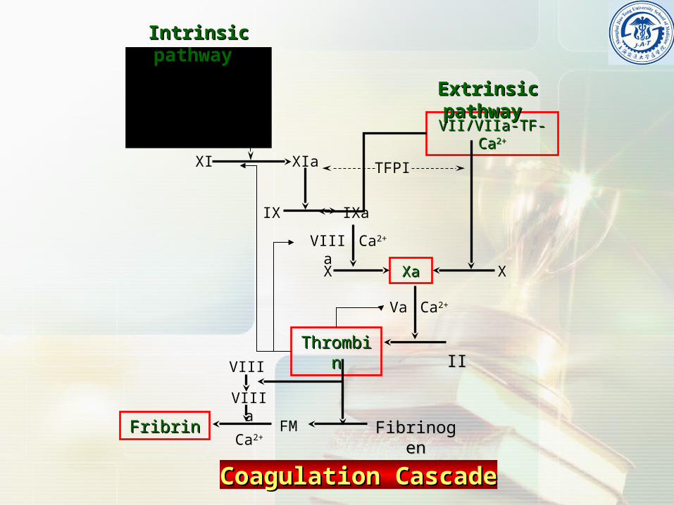
Plasminogen Plasminogen (PLg)(PLg)
t-PAt-PA
Plasmin (PLn)Plasmin (PLn)
ⅩⅩaa-- aⅤaⅤ -Ca-Ca2+2+-PL-PL ⅨⅨaa-- aⅧaⅧ -Ca-Ca2+2+-PL-PL
Activated PC(Activated PC(APCAPC)-PS)-PS
Coagulation
PC system
u-PAu-PA
TM
End
othe
lial
End
othe
lial
cel
lsce
lls
PlasminogenExtrinic activatorsExtrinic activators
ECECPro-PA
enzymes
t-PA, u-PA
SecretionSecretion
Urine ( UK , u-PA)Bile ( bilokinase)Latex 、 saliva 、 tearTissueTissue
lung 、 prostate 、 uterusBlood cell
RBC, Platelet
Thrombolytic drugsThrombolytic drugs
Streptokinase (SK)
Urokinase (UK)rt-PA
Proteolyses fibrinogen, fibronectin , laminin ,
thrombospondin ; Activates collagenases.
Plasmin
PAI-1
PAI-2
PAI-3
Histidine-rich glycoprotein
( HRG)Intrinic activatorsIntrinic activators
Coagulation
Ⅺa Ⅱa
Ⅻa Ⅻ
KK
PK
VEC-HK
α2-plasmin inhibitor (α2-PI)
α2- macroglobin (α2 –MG)
Fibrinolytic systemFibrinolytic system
Exogenous activatorsExogenous activators

Haemorrhage or thrombosis will appear when Haemorrhage or thrombosis will appear when the balance between coagulation and the balance between coagulation and coagulation is disturbed.coagulation is disturbed.
Inappropriate clotting of blood can obstruct vital Inappropriate clotting of blood can obstruct vital organ circulation. organ circulation.
Systemic activation of coagulation in its most Systemic activation of coagulation in its most extreme form is known as disseminated extreme form is known as disseminated intravascular coagulation (intravascular coagulation (DICDIC).).
Coagulation & anticoagulation Coagulation & anticoagulation imbalanceimbalance

Definition of DICDefinition of DIC
Causes of DICCauses of DIC
Pathogenesis of DICPathogenesis of DIC
Main Features of DICMain Features of DIC
TopicsTopics

Definition of DICDefinition of DIC

Disseminated intravascular coagulation (DIC) is a complex Disseminated intravascular coagulation (DIC) is a complex
systemic thrombohemorrhagic disorder involving the systemic thrombohemorrhagic disorder involving the
generation of intravascular fibrin and the consumption of generation of intravascular fibrin and the consumption of
procoagulants and platelets. The resultant clinical condition is procoagulants and platelets. The resultant clinical condition is
characterized by intravascular coagulation and hemorrhage.characterized by intravascular coagulation and hemorrhage.
DefinitionDefinition
Coagulation Hypercoagulable state ThrombusCoagulation Hypercoagulable state Thrombus
Fibrinolysis Hypocoagulable state HemorrhageFibrinolysis Hypocoagulable state Hemorrhage

Causes of DICCauses of DIC

The Accepted Disease Entities Generally The Accepted Disease Entities Generally Associated with DICAssociated with DIC
Sepsis/severe infectionSepsis/severe infection MalignancyMalignancy
solid and myeloproliferative malignanciessolid and myeloproliferative malignancies Obstetric complicationsObstetric complications
Amniotic fluid embolism, Abruptio placentaeAmniotic fluid embolism, Abruptio placentae
Retained dead fetus syndrome Retained dead fetus syndrome Trauma (neurotrauma) Trauma (neurotrauma) ,Organ destruction, Burns,Organ destruction, Burns Severe hepatic failureSevere hepatic failureRheumatologic illnessRheumatologic illness
Adult Stills disease, LupusAdult Stills disease, Lupus Vascular abnormalitiesVascular abnormalities
Kasabach-Merritt syndrome, Large vascular aneurysmsKasabach-Merritt syndrome, Large vascular aneurysmsHemolysisHemolysis
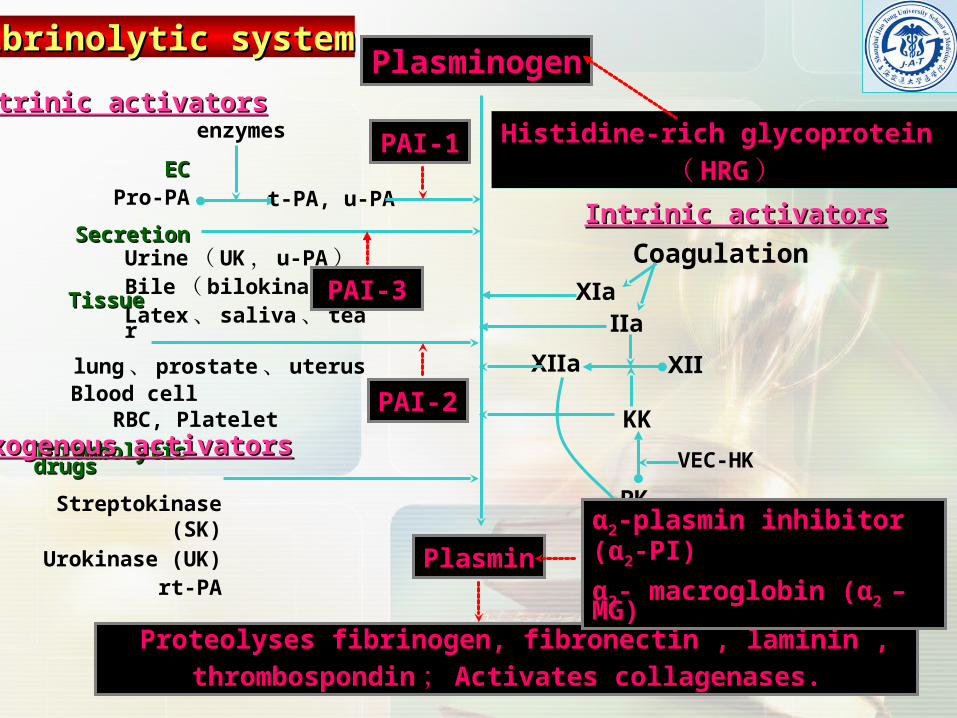
The diseases which associated with DIC The diseases which associated with DIC always have one or more triggering factors always have one or more triggering factors that can activate clotting factors and that can activate clotting factors and intravascular coagulation.intravascular coagulation.
① ① Release TFRelease TF
② ② VECVEC injuryinjury
③ ③ LPSLPS
④ ④ Ag-Ab complexAg-Ab complex
Triggering factorsTriggering factors
⑤ ⑤ ProteaseProtease
⑥ ⑥ MicroparticlesMicroparticles
⑦ ⑦ Pathogenic Microbes (viruses)Pathogenic Microbes (viruses)

Pathogenesis of DICPathogenesis of DIC

Activation of coagulation Activation of coagulation
Disabled anticoagulant mechanismDisabled anticoagulant mechanism
Impaired fibrinolysis Impaired fibrinolysis
PathophysiologyPathophysiology

Activation of coagulationActivation of coagulation
Tissue damage Tissue damage
Endothelial disruption Endothelial disruption
HemolysisHemolysis
LeukemiaLeukemia
Other procoagulant molecules enter Other procoagulant molecules enter the vascular systemthe vascular system

Suppression of Suppression of anticoagulant pathways anticoagulant pathways
Antithrombin activity is reduced Antithrombin activity is reduced
Protein C pathway is incapacitatedProtein C pathway is incapacitated
TFPI is another anticoagulant mechanism TFPI is another anticoagulant mechanism that is disabled that is disabled
Impaired fibrinolysis Impaired fibrinolysis
Experimental and clinical studies indicate that during
DIC, the fibrinolytic system is largely suppressed at
the time of maximal activation of coagulation.
This inhibition of fibrinolysis is caused by a sustained
rise in the plasma level of plasminogen activator
inhibitor-1 (PAI-1), the principal inhibitor of the
fibrinolytic system.

PlasminogenExtrinic activatorsExtrinic activators
ECECPro-PA
enzymes
t-PA, u-PA
SecretionSecretion
Urine ( UK , u-PA)Bile ( bilokinase)Latex 、 saliva 、 tearTissueTissue
lung 、 prostate 、 uterusBlood cell
RBC, Platelet
Thrombolytic drugsThrombolytic drugs
Streptokinase (SK)
Urokinase (UK)rt-PA
Proteolyses fibrinogen, fibronectin , laminin ,
thrombospondin ; Activates collagenases.
Plasmin
PAI-1
PAI-2
PAI-3
Histidine-rich glycoprotein
( HRG)Intrinic activatorsIntrinic activators
Coagulation
Ⅺa Ⅱa
Ⅻa Ⅻ
KK
PK
VEC-HK
α2-plasmin inhibitor (α2-PI)
α2- macroglobin (α2 –MG)
Secondary FibrinolysisSecondary Fibrinolysis
Exogenous activatorsExogenous activators

Proinflammatory Proinflammatory cytokinescytokines
Vascular endothelial cellsVascular endothelial cells
Mononuclear Mononuclear cellscells
TFTF expressionexpression
Impairment of Impairment of anticoagulantanticoagulantmechanismsmechanisms
PAI-1 mediated PAI-1 mediated inhibition of inhibition of fibrinolysisfibrinolysis
IntravascularIntravascularFn formationFn formation
InsufficientInsufficientFn removalFn removal
Fibrin depositionFibrin deposition
Schematic representation of pathogenetic pathways in DICSchematic representation of pathogenetic pathways in DIC

Predisposing factors to DICPredisposing factors to DIC

Impairment of reticuloendothelial systemImpairment of reticuloendothelial system
There is good evidence that most of the products of intravascular There is good evidence that most of the products of intravascular
coagulation (free fibrin, prothrombinase, PF3), as well as various coagulation (free fibrin, prothrombinase, PF3), as well as various
initiators of the process (endotoxin, tissue fragments, antigen-antibody initiators of the process (endotoxin, tissue fragments, antigen-antibody
complexes, thromboplastins, red cell stroma) are removed from the complexes, thromboplastins, red cell stroma) are removed from the
circulation by the reticuloendothelial system. The hepatic cells are of circulation by the reticuloendothelial system. The hepatic cells are of
primary importance in the clearance of activated coagulation factors primary importance in the clearance of activated coagulation factors
(IXa, Xa and XIIa). It has been suggested that, in DIC, various (IXa, Xa and XIIa). It has been suggested that, in DIC, various
substances saturate or block the clearance function of substances saturate or block the clearance function of
reticuloendothelial system in a manner comparable to that produced reticuloendothelial system in a manner comparable to that produced
experimentally in the general Shwartzman reaction (GSR) in animals.experimentally in the general Shwartzman reaction (GSR) in animals.
Reticuloendothelial system is suppressd by glucocorticoid or in the Reticuloendothelial system is suppressd by glucocorticoid or in the
patients with liver diseases. patients with liver diseases.

Hemostasis is intimately related to liver function, because most Hemostasis is intimately related to liver function, because most coagulation factors are synthesized by liver parenchymal cells and the coagulation factors are synthesized by liver parenchymal cells and the liver's reticuloendothelial system serves an important role in the clearance liver's reticuloendothelial system serves an important role in the clearance of activation products. of activation products. The extent of coagulation abnormalities depends upon the degree of The extent of coagulation abnormalities depends upon the degree of disturbed liver function. disturbed liver function. Acute or chronic hepatocellular diseases may display decreases in the Acute or chronic hepatocellular diseases may display decreases in the vitamin K-dependent factors (prothrombin; factors VII, IX, and X; proteins vitamin K-dependent factors (prothrombin; factors VII, IX, and X; proteins C and S), whereas other parameters remain normal. C and S), whereas other parameters remain normal. Patients with hepatic failure may present with the entire spectrum of Patients with hepatic failure may present with the entire spectrum of factor deficiencies and may even develop DIC. factor deficiencies and may even develop DIC. Patients with liver cirrhosis have a wide spectrum of abnormalities. Patients with liver cirrhosis have a wide spectrum of abnormalities. Except for factor VIII:C and von Willebrand factor, all procoagulant and Except for factor VIII:C and von Willebrand factor, all procoagulant and inhibitory factors are decreased, which is a reflection of impaired protein inhibitory factors are decreased, which is a reflection of impaired protein synthesis. Abnormal fibrinogen and prothrombin molecules can be synthesis. Abnormal fibrinogen and prothrombin molecules can be identified. Platelets are quantitatively and qualitatively altered, and most identified. Platelets are quantitatively and qualitatively altered, and most patients develop DIC.patients develop DIC.
Hepatic dysfunctionHepatic dysfunction

Hypercoagulable stateHypercoagulable state
It has been found that the platelet and several kinds of It has been found that the platelet and several kinds of
clotting factors (factor I, II, VII, VIII, IX and X, etc.) in blood clotting factors (factor I, II, VII, VIII, IX and X, etc.) in blood
are increased, while the substances with the action of are increased, while the substances with the action of
anticoagulation and with the activity of fibrinolysis are anticoagulation and with the activity of fibrinolysis are
deceased. For instance, the blood in pregnancy after 4 deceased. For instance, the blood in pregnancy after 4
months bigins to increase coagulability, which is most months bigins to increase coagulability, which is most
marked in the terminal stage of pregnancy. Therefore the marked in the terminal stage of pregnancy. Therefore the
incidence of DIC is elevated in obstetrical accidents. In incidence of DIC is elevated in obstetrical accidents. In
addition, acidosis, common in some patients, promotes addition, acidosis, common in some patients, promotes
the activation of clotting cascade by reducing the pH of the activation of clotting cascade by reducing the pH of
the blood.the blood.
Shock usually accompanies disorder of Shock usually accompanies disorder of microcirculation which is manifested by stasis of microcirculation which is manifested by stasis of blood flow, aggregation of blood cells and blood flow, aggregation of blood cells and appearance of sludging, stasis of the appearance of sludging, stasis of the microcirculation permits activated clotting factors microcirculation permits activated clotting factors to accumulated in one region making it easier to to accumulated in one region making it easier to develop into a state of DIC. The stasis of blood in develop into a state of DIC. The stasis of blood in giant hemangioma may somehow contribute to the giant hemangioma may somehow contribute to the development of DIC.development of DIC.
Disorder of microcirculationDisorder of microcirculation

Inhibition of fibrinolysisInhibition of fibrinolysis
Aging, smoking, pregnandiacy, diabetes.Aging, smoking, pregnandiacy, diabetes.
Using antifibrinolytic agents like EACAUsing antifibrinolytic agents like EACA and PAMBAand PAMBA

Main Features of DIC Main Features of DIC

Include petechiae and purpura (found in most patients), hemorrhagic bullae, wound bleeding; especially oozing from a surgical or traumatic wound is common in patients who have undergone surgery or suffered trauma. Oozing from venipuncture sites or intraarterial lines is another common finding. Large subcutaneous hematomas and deep tissue bleeding are also often seen.
The average patient with DIC usually bleeds from at least three unrelated sites and any combination may be seen.
Bleeding causes: ▲Clotting factors consumption ▲ FDP generation ▲ Activation of fibrinolytic system ▲ Vessel damage
BleedingBleeding

Excess bleedingExcess bleeding
ThrombusThrombus formation results in a diminished formation results in a diminished return of venous blood to the heartreturn of venous blood to the heart
Activation of the kinin system leads to Activation of the kinin system leads to increased vascular permeability, hypotension, increased vascular permeability, hypotension, andshockandshock
Creation of FDPCreation of FDP result in enhanced result in enhanced vasodilationvasodilation
Myocardial infarctionMyocardial infarction
ShockShock
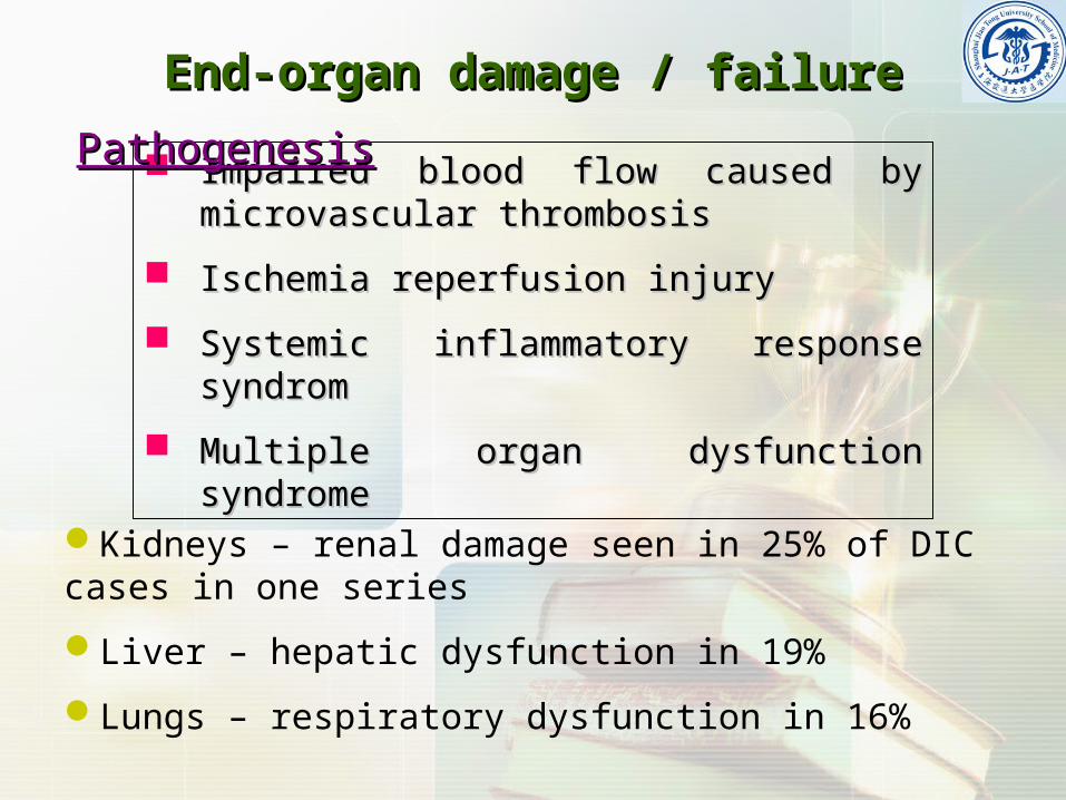
End-organ damage / failureEnd-organ damage / failure
Impaired blood flow caused by microvascular Impaired blood flow caused by microvascular thrombosisthrombosis
Ischemia reperfusion injuryIschemia reperfusion injury
Systemic inflammatory response syndrom Systemic inflammatory response syndrom
Multiple organ dysfunction syndromeMultiple organ dysfunction syndrome
PathogenesisPathogenesis
Kidneys – renal damage seen in 25% of DIC cases in one series
Liver – hepatic dysfunction in 19%
Lungs – respiratory dysfunction in 16%

A disorder in which narrowing or obstruction A disorder in which narrowing or obstruction of small blood vessels results in distortion of small blood vessels results in distortion and fragmentation of erythrocytes, hemolysis, and fragmentation of erythrocytes, hemolysis, and anemia.and anemia.
It is identified by the finding of anaemia and It is identified by the finding of anaemia and schistocytes ("bite cells") on microscopy of schistocytes ("bite cells") on microscopy of the blood film. the blood film.
Microangiopathic hemolytic anemiaMicroangiopathic hemolytic anemia


Primary diseasesPrimary diseases
tissue tissue injuryinjury
VECVEC damagedamage
Blood cells Blood cells injuryinjury
othersothers
TFTF or other procoagulant components releasingor other procoagulant components releasing
Activate coagulation cascadeActivate coagulation cascade
Fibrin depositFibrin depositthrombosisthrombosisSecondary fibrinolysisSecondary fibrinolysis
Bleeding Shock Hemolytic Anemia MODS
FDPFDP formationformationClotting factors Clotting factors consumptionconsumption
HypocoagulableHypocoagulable
HypercoagulableHypercoagulable

Types and stages of DICTypes and stages of DIC

StagesStages
Hypercoagulable stageHypercoagulable stage
Hypocoagulable stageHypocoagulable stage
Secondary fibrinolytic stageSecondary fibrinolytic stage

Activation of Activation of CoagulationCoagulation
Fibrin monomerFibrin monomer (FM)(FM)
Secondary Secondary fibrinolysisfibrinolysis
FDP (X fragment)FDP (X fragment)
soluble fibrin monomer soluble fibrin monomer complexcomplex (( SFMCSFMC ))
Protamine Protamine SulfateSulfate
FMFM
polymerization polymerization
XX fragmentfragment
positive 3P Testpositive 3P Test
Plasma Protamine Paracoagulation Plasma Protamine Paracoagulation

D-dimer formationD-dimer formation

Coagulant
Fibrinolysis
Coagulable
Stage HypercoagulableHypercoagulable HypocoagulableHypocoagulable Secondary FibrinolysisSecondary Fibrinolysis
Lab KPTT, PTPL activity PL count Fibrinogen Thrombus(+)
Bleeding time PT PL count Clotting factors Fibrinolysis /N
Fibrinolysis (euglobulin clot lysis time)
Plasminogen
3P test(+)

TypesTypes
The form of DIC depends on the rapidity and force of the initiating event, leading to the two primary forms of DIC:
Acute decompensated DIC
Chronic compensated DIC
Compensated DIC: When the stimulus for coagulation is mild, the liver can increase production of clotting factors to up to 5 times the normal rate, in an effort to maintain plasma levels. Similarly, platelet production can increase up to 10 times. Thus, although coagulation and fibrinolysis are in progress, platelet counts and fibrinogen levels may be normal or only marpinally reduced. These patients rarely bleed spontaneously or from minor trauma, but have severe haemorrhage if subjected to surgery.

Treatment of DICTreatment of DIC

Treatment of DIC
Cornerstone of management is the treatment of the underlying illness
Supportive management with▲ Disruption of coagulation cascade using
“lower dose” heparin-treatment, administration of ATIII and/or activated protein C (protein C
infusion has shown to be the first intervention proven to be effective in reducing the mortality in septic patients
▲ If bleeding is the predominant symptom Platelet infusion Coagulation factor substitution with fresh frozen plasma

Related Documents