MOLECULAR AND CELLULAR BIOLOGY, Feb. 1995, p. 1123–1135 Vol. 15, No. 2 0270-7306/95/$04.0010 Copyright q 1995, American Society for Microbiology Dissecting a Locus Control Region: Facilitation of Enhancer Function by Extended Enhancer-Flanking Sequences BRUCE J. ARONOW,* CATHERINE A. EBERT, M. TODD VALERIUS, S. STEVEN POTTER, DAN A. WIGINTON, DAVID P. WITTE, AND JOHN J. HUTTON Division of Basic Science Research, Department of Pediatrics, Children’s Hospital Medical Center, University of Cincinnati College of Medicine, Cincinnati, Ohio 45229 Received 14 July 1994/Returned for modification 17 August 1994/Accepted 3 November 1994 Using transgenic mice, we have defined novel gene regulatory elements, termed ‘‘facilitators.’’ These ele- ments bilaterally flank, by up to 1 kb, a 200-bp T-cell-specific enhancer domain in the human adenosine deaminase (ADA) gene. Facilitators were essential for gene copy-proportional and integration site-independent reporter expression in transgenic thymocytes, but they had no effect on the enhancer in transfected T cells. Both segments were required. Individual segments had no activity. A lack of facilitator function caused positional susceptibility and prevented DNase I-hypersensitive site formation at the enhancer. The segments were required to be at opposed ends of the enhancer, and they could not be grouped together. Reversing the orientation of a facilitator segment caused a partial loss of function, suggesting involvement of a stereospecific chromatin structure. trans-acting factor access to enhancer elements was modeled by exposing nuclei to a restriction endonuclease. The enhancer domain was accessible to the 4-cutter DpnII in a tissue- and cell- type-specific fashion. However, unlike DNase I hypersensitivity and gene expression, accessibility to the endonuclease could occur without the facilitator segments, suggesting that an accessible chromatin domain is an intermediate state in the activational pathway. These results suggest that facilitators (i) are distinct from yet positionally constrained to the enhancer, (ii) participate in a chromatin structure transition that is necessary for the DNase I hypersensitivity and the transcriptional activating function of the enhancer, and (iii) act after cell-type-specific accessibility to the enhancer sequences is established by factors that do not require the facilitators to be present. In contrast to the consistent behavior of most endogenous genes, cis-regulatory information carried in mammalian trans- genes is subject to notorious variability generally referred to as the genomic position effect (26, 28, 49). The basis for the genomic position effect is poorly understood. One cause of it may be the inability of transgenes to control chromatin struc- ture at genomic insertion sites (17, 63). A variety of studies with diverse eukaryotic systems indicate that the ability of trans-acting factors to function at cis-regulatory regions is strongly influenced by alterations of chromatin structure and organization (1, 4, 24, 30, 40, 42, 50). For example, nucleo- somes and nucleosome positioning (29, 35, 36, 53, 55, 56, 61, 62), and specific histones (11) or histone motifs (16, 59), can dramatically impact the ability of transcriptional machinery to address underlying cis elements. The regulation of chromatin domain structure and accessibility to trans-acting factors may be a fundamental mechanism by which the genome can spe- cifically and selectively express genes during developmental cell differentiation. Some transgene constructions exhibit a high degree of con- sistency among independent mouse lines with regard to cell type specificity, level of expression, and in some cases, propor- tionality to gene copy number. Reporter expression that is proportional to gene copy number is a remarkable property that indicates the ability of each transgene to form and regu- late an autonomously active locus. Sequences associated with the generation of these effects have been termed locus control regions (LCRs) (20, 27, 58). In light of their ability to domi- nantly control gene expression from any chromosomal region into which they are inserted, LCRs are appealing entities for mechanistic analyses of the elements and factors responsible for activating a chromatin domain. We have previously re- ported elements within the human adenosine deaminase (ADA) gene that bear analogy to LCRs. Human ADA defi- ciency causes severe combined immunodeficiency disease with a failure to develop T cells. ADA deficiency has been used as a model for corrective gene therapy based on gene introduc- tion into autologous hematopoietic stem cells (5). The gene is normally expressed at a high level in thymocytes, and we have sought to identify and understand the regulatory elements re- sponsible for controlling ADA cell type specificity and levels of expression in developing T cells. Expression of the ADA gene in the thymus is controlled by cis-regulatory elements in its first intron (2). The intron con- tains six DNase I-hypersensitive sites. In transiently transfected T cells, positive-acting cis elements are localized in a 200-bp domain that corresponds to the limits of hypersensitive site III (HS III) hypersensitivity (3). In transgenic mice, a much larger 2.25-kb regulatory region was necessary for consistent posi- tion-independent transgene expression in the thymus, thus in- dicating the functional characteristics of an LCR. Transgenic mice made from constructions that lacked terminal sequences of the 2.25-kb regulatory region exhibited inconsistent gene expression. Whereas DNase I hypersensitivity was formed at the HS III enhancer in thymocytes of transgenic mice that contained up to 100 copies of a large ADA-CAT reporter gene (2), hypersensitivity was formed poorly or not all at the HS III enhancer by most or all copies of mouse lines that contained poorly expressed transgenes lacking sequences 59 or 39 of the enhancer domain (3). This led us to term these enhancer flank- * Corresponding author. Mailing address: Division of Basic Science Research, NRB 2047, Department of Pediatrics, University of Cincin- nati College of Medicine, Children’s Hospital Medical Center, Cincin- nati, OH 45229. Phone: (513) 559-8163. Fax: (513) 559-4317. Elec- tronic mail address: [email protected]. 1123 on May 27, 2016 by guest http://mcb.asm.org/ Downloaded from

Welcome message from author
This document is posted to help you gain knowledge. Please leave a comment to let me know what you think about it! Share it to your friends and learn new things together.
Transcript
MOLECULAR AND CELLULAR BIOLOGY, Feb. 1995, p. 1123–1135 Vol. 15, No. 20270-7306/95/$04.0010Copyright q 1995, American Society for Microbiology
Dissecting a Locus Control Region: Facilitation of EnhancerFunction by Extended Enhancer-Flanking Sequences
BRUCE J. ARONOW,* CATHERINE A. EBERT, M. TODD VALERIUS, S. STEVEN POTTER,DAN A. WIGINTON, DAVID P. WITTE, AND JOHN J. HUTTON
Division of Basic Science Research, Department of Pediatrics, Children’s Hospital Medical Center,University of Cincinnati College of Medicine, Cincinnati, Ohio 45229
Received 14 July 1994/Returned for modification 17 August 1994/Accepted 3 November 1994
Using transgenic mice, we have defined novel gene regulatory elements, termed ‘‘facilitators.’’ These ele-ments bilaterally flank, by up to 1 kb, a 200-bp T-cell-specific enhancer domain in the human adenosinedeaminase (ADA) gene. Facilitators were essential for gene copy-proportional and integration site-independentreporter expression in transgenic thymocytes, but they had no effect on the enhancer in transfected T cells.Both segments were required. Individual segments had no activity. A lack of facilitator function causedpositional susceptibility and prevented DNase I-hypersensitive site formation at the enhancer. The segmentswere required to be at opposed ends of the enhancer, and they could not be grouped together. Reversing theorientation of a facilitator segment caused a partial loss of function, suggesting involvement of a stereospecificchromatin structure. trans-acting factor access to enhancer elements was modeled by exposing nuclei to arestriction endonuclease. The enhancer domain was accessible to the 4-cutter DpnII in a tissue- and cell-type-specific fashion. However, unlike DNase I hypersensitivity and gene expression, accessibility to theendonuclease could occur without the facilitator segments, suggesting that an accessible chromatin domain isan intermediate state in the activational pathway. These results suggest that facilitators (i) are distinct fromyet positionally constrained to the enhancer, (ii) participate in a chromatin structure transition that isnecessary for the DNase I hypersensitivity and the transcriptional activating function of the enhancer, and (iii)act after cell-type-specific accessibility to the enhancer sequences is established by factors that do not requirethe facilitators to be present.
In contrast to the consistent behavior of most endogenousgenes, cis-regulatory information carried in mammalian trans-genes is subject to notorious variability generally referred to asthe genomic position effect (26, 28, 49). The basis for thegenomic position effect is poorly understood. One cause of itmay be the inability of transgenes to control chromatin struc-ture at genomic insertion sites (17, 63). A variety of studieswith diverse eukaryotic systems indicate that the ability oftrans-acting factors to function at cis-regulatory regions isstrongly influenced by alterations of chromatin structure andorganization (1, 4, 24, 30, 40, 42, 50). For example, nucleo-somes and nucleosome positioning (29, 35, 36, 53, 55, 56, 61,62), and specific histones (11) or histone motifs (16, 59), candramatically impact the ability of transcriptional machinery toaddress underlying cis elements. The regulation of chromatindomain structure and accessibility to trans-acting factors maybe a fundamental mechanism by which the genome can spe-cifically and selectively express genes during developmentalcell differentiation.Some transgene constructions exhibit a high degree of con-
sistency among independent mouse lines with regard to celltype specificity, level of expression, and in some cases, propor-tionality to gene copy number. Reporter expression that isproportional to gene copy number is a remarkable propertythat indicates the ability of each transgene to form and regu-late an autonomously active locus. Sequences associated withthe generation of these effects have been termed locus control
regions (LCRs) (20, 27, 58). In light of their ability to domi-nantly control gene expression from any chromosomal regioninto which they are inserted, LCRs are appealing entities formechanistic analyses of the elements and factors responsiblefor activating a chromatin domain. We have previously re-ported elements within the human adenosine deaminase(ADA) gene that bear analogy to LCRs. Human ADA defi-ciency causes severe combined immunodeficiency disease witha failure to develop T cells. ADA deficiency has been used asa model for corrective gene therapy based on gene introduc-tion into autologous hematopoietic stem cells (5). The gene isnormally expressed at a high level in thymocytes, and we havesought to identify and understand the regulatory elements re-sponsible for controlling ADA cell type specificity and levels ofexpression in developing T cells.Expression of the ADA gene in the thymus is controlled by
cis-regulatory elements in its first intron (2). The intron con-tains six DNase I-hypersensitive sites. In transiently transfectedT cells, positive-acting cis elements are localized in a 200-bpdomain that corresponds to the limits of hypersensitive site III(HS III) hypersensitivity (3). In transgenic mice, a much larger2.25-kb regulatory region was necessary for consistent posi-tion-independent transgene expression in the thymus, thus in-dicating the functional characteristics of an LCR. Transgenicmice made from constructions that lacked terminal sequencesof the 2.25-kb regulatory region exhibited inconsistent geneexpression. Whereas DNase I hypersensitivity was formed atthe HS III enhancer in thymocytes of transgenic mice thatcontained up to 100 copies of a large ADA-CAT reporter gene(2), hypersensitivity was formed poorly or not all at the HS IIIenhancer by most or all copies of mouse lines that containedpoorly expressed transgenes lacking sequences 59 or 39 of theenhancer domain (3). This led us to term these enhancer flank-
* Corresponding author. Mailing address: Division of Basic ScienceResearch, NRB 2047, Department of Pediatrics, University of Cincin-nati College of Medicine, Children’s Hospital Medical Center, Cincin-nati, OH 45229. Phone: (513) 559-8163. Fax: (513) 559-4317. Elec-tronic mail address: [email protected].
1123
on May 27, 2016 by guest
http://mcb.asm
.org/D
ownloaded from
ing sequences ‘‘facilitators’’ and to postulate (i) that their pres-ence was required for the formation of hypersensitivity in mostchromosomal integration sites and (ii) that there was an abso-lute requirement for the formation of the HS for enhancerfunction. In the present study we have analyzed the inconsis-tent behavior of the transgenes that lacked the flanking se-quences, further dissected the flanking segments, and identi-fied constraints that govern facilitator function. The resultsindicate that the facilitators can cooperate with the enhanceronly if both segments are present and specifically positionedand oriented. It also appears likely that the segments functionsubsequent to the recognition of core elehancer elements bycell-type-specific factors.
MATERIALS AND METHODS
Transgenic analyses. Transgenic mice were made with the fertilized eggs of(C3H 3 C57BL/6)F1 hybrid parents by procedures described by Hogan et al.(31). F1 transgenic mice between 4 and 9 weeks of age were analyzed (2).Transgene copy numbers were determined by comparison of blot hybridizationsof restriction endonuclease-digested DNA isolated from tails with known stan-dards of human DNA and cloned plasmid DNA. For quantitative Southernanalyses, autoradiograms were generally analyzed with a PhosphorImager device(Molecular Dynamics Inc., Mountain View, Calif.) or as previously described.Transgenic tissue chloramphenicol acetyltransferase (CAT) assays, protein con-centration measurements, transgene copy number determinations, mouse dis-sections, in situ hybridization, and chromatin DNase I hypersensitivity analyseswere performed as previously described (2, 3, 60).Transgenes. Restriction fragments from various plasmids were isolated from
agarose gels and prepared for microinjection (3). Transgenes were designated asfollows: 59 flanking DNA/hypersensitive site(s) or intron segment placed down-stream of CAT. Restriction sites used for regulatory region subcloning areindicated in Fig. 1 and 3. The restriction sites used to generate short and longpromoters were EcoRI for 0.2-kb, BssHII for 0.3-kb, and NdeI for 3.7-kb pro-moter segments.DpnII endonuclease accessibility assay. Nuclei from cell lines or transgenic
mouse tissues were prepared (2). Approximately 2.5 3 106 hemocytometer-quantitated nuclei per assay were suspended at 48C in 0.2 ml of a buffer thatcontained 50 mM NaCl, 10 mM MgCl2, 1 mM dithiothreitol, and 10 mM TrisHCl (pH 7.9 at 258C). Various amounts of DpnII per ml and 50% glycerol wereadded to a final concentration of 2% glycerol. Reactions were allowed to proceedon ice for 60 min. Reduced-temperature reaction conditions minimized nonspe-cific cleavage of the enhancer region, which was observed at room temperature.DNA was purified from the nuclei with sodium dodecyl sulfate and proteinase K,phenol, and RNase A. A 5-mg portion of DNA was cut with KpnI and electro-phoresed through a mixed agarose gel of 1.5% SeaKem LE and 1% NuSieve(FMC) in 13 TAE buffer. Southern blot transfer was to Magnagraph membranes(MSI, Westboro, Mass.). Hybridization was to a random-primer-labeled probethat encompassed the 603-bp KpnI fragment from the regulatory region. Hybrid-izations and washes were done as recommended by the manufacturer.Quantitative analysis of the relative intensities of the bands released by partial
DpnII digestion of the KpnI fragment on Southern blots was accomplished by useof the PhosphorImager and ImageQuant software (Molecular Dynamics). Rect-angles were drawn down the length of each lane and subjected to ‘‘linegraph’’quantitation. Data were transferred to a spreadsheet file, graphed, and objectpasted into a draw file with height-scale normalization of the parental band toassess relative cleavage with respect to gene copy number.
RESULTS
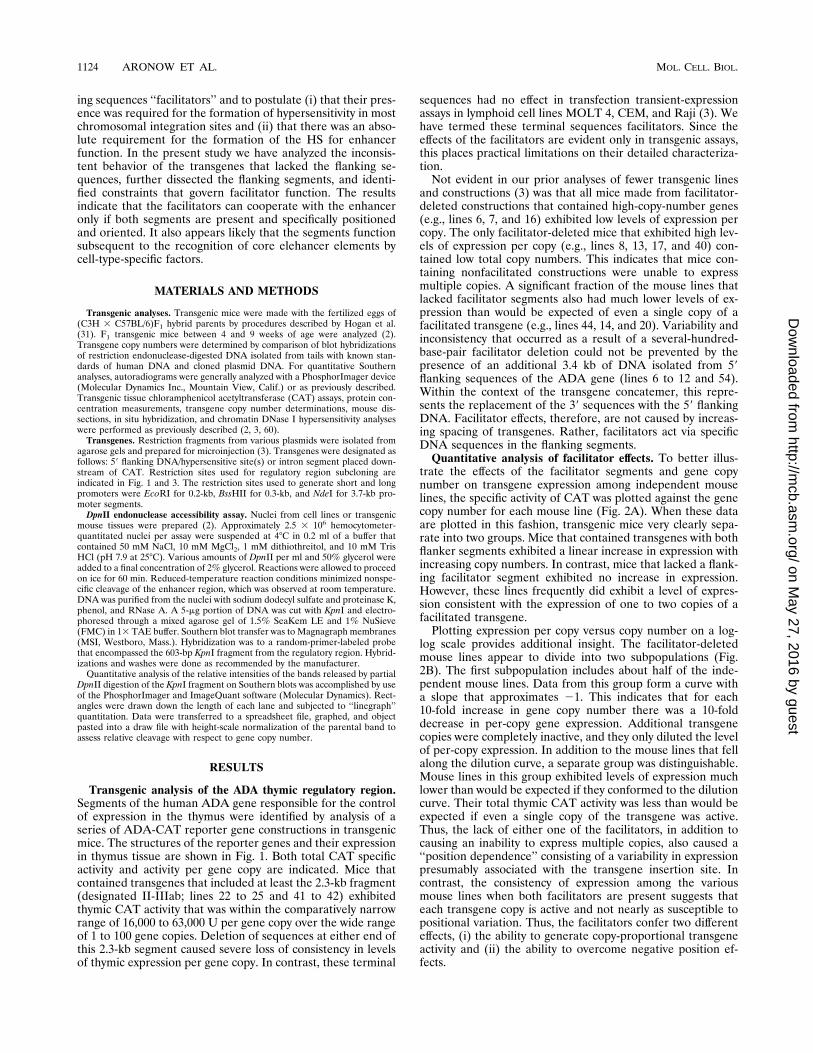
Transgenic analysis of the ADA thymic regulatory region.Segments of the human ADA gene responsible for the controlof expression in the thymus were identified by analysis of aseries of ADA-CAT reporter gene constructions in transgenicmice. The structures of the reporter genes and their expressionin thymus tissue are shown in Fig. 1. Both total CAT specificactivity and activity per gene copy are indicated. Mice thatcontained transgenes that included at least the 2.3-kb fragment(designated II-IIIab; lines 22 to 25 and 41 to 42) exhibitedthymic CAT activity that was within the comparatively narrowrange of 16,000 to 63,000 U per gene copy over the wide rangeof 1 to 100 gene copies. Deletion of sequences at either end ofthis 2.3-kb segment caused severe loss of consistency in levelsof thymic expression per gene copy. In contrast, these terminal
sequences had no effect in transfection transient-expressionassays in lymphoid cell lines MOLT 4, CEM, and Raji (3). Wehave termed these terminal sequences facilitators. Since theeffects of the facilitators are evident only in transgenic assays,this places practical limitations on their detailed characteriza-tion.Not evident in our prior analyses of fewer transgenic lines
and constructions (3) was that all mice made from facilitator-deleted constructions that contained high-copy-number genes(e.g., lines 6, 7, and 16) exhibited low levels of expression percopy. The only facilitator-deleted mice that exhibited high lev-els of expression per copy (e.g., lines 8, 13, 17, and 40) con-tained low total copy numbers. This indicates that mice con-taining nonfacilitated constructions were unable to expressmultiple copies. A significant fraction of the mouse lines thatlacked facilitator segments also had much lower levels of ex-pression than would be expected of even a single copy of afacilitated transgene (e.g., lines 44, 14, and 20). Variability andinconsistency that occurred as a result of a several-hundred-base-pair facilitator deletion could not be prevented by thepresence of an additional 3.4 kb of DNA isolated from 59flanking sequences of the ADA gene (lines 6 to 12 and 54).Within the context of the transgene concatemer, this repre-sents the replacement of the 39 sequences with the 59 flankingDNA. Facilitator effects, therefore, are not caused by increas-ing spacing of transgenes. Rather, facilitators act via specificDNA sequences in the flanking segments.Quantitative analysis of facilitator effects. To better illus-
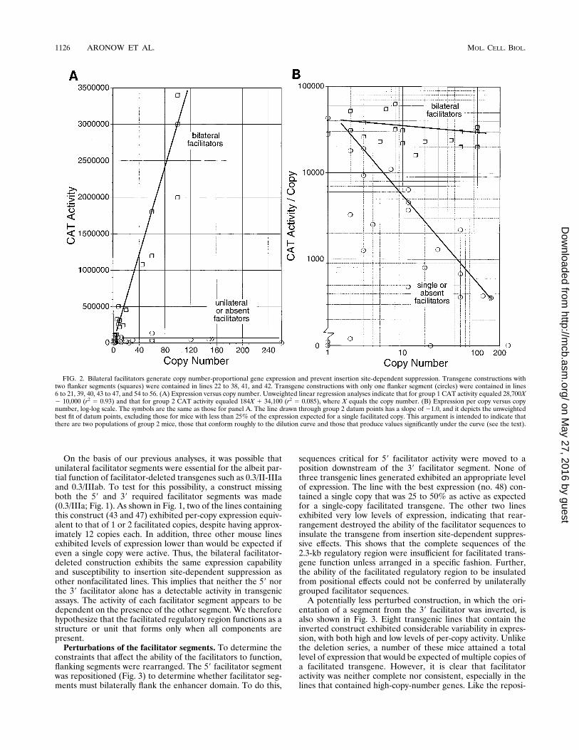
trate the effects of the facilitator segments and gene copynumber on transgene expression among independent mouselines, the specific activity of CAT was plotted against the genecopy number for each mouse line (Fig. 2A). When these dataare plotted in this fashion, transgenic mice very clearly sepa-rate into two groups. Mice that contained transgenes with bothflanker segments exhibited a linear increase in expression withincreasing copy numbers. In contrast, mice that lacked a flank-ing facilitator segment exhibited no increase in expression.However, these lines frequently did exhibit a level of expres-sion consistent with the expression of one to two copies of afacilitated transgene.Plotting expression per copy versus copy number on a log-
log scale provides additional insight. The facilitator-deletedmouse lines appear to divide into two subpopulations (Fig.2B). The first subpopulation includes about half of the inde-pendent mouse lines. Data from this group form a curve witha slope that approximates 21. This indicates that for each10-fold increase in gene copy number there was a 10-folddecrease in per-copy gene expression. Additional transgenecopies were completely inactive, and they only diluted the levelof per-copy expression. In addition to the mouse lines that fellalong the dilution curve, a separate group was distinguishable.Mouse lines in this group exhibited levels of expression muchlower than would be expected if they conformed to the dilutioncurve. Their total thymic CAT activity was less than would beexpected if even a single copy of the transgene was active.Thus, the lack of either one of the facilitators, in addition tocausing an inability to express multiple copies, also caused a‘‘position dependence’’ consisting of a variability in expressionpresumably associated with the transgene insertion site. Incontrast, the consistency of expression among the variousmouse lines when both facilitators are present suggests thateach transgene copy is active and not nearly as susceptible topositional variation. Thus, the facilitators confer two differenteffects, (i) the ability to generate copy-proportional transgeneactivity and (ii) the ability to overcome negative position ef-fects.
1124 ARONOW ET AL. MOL. CELL. BIOL.
on May 27, 2016 by guest
http://mcb.asm
.org/D
ownloaded from
FIG. 1. CAT expression in thymus tissue from ADA-CAT transgenic mice. Total expression is indicated as specific activity and per-copy expression. Indicatedsegments of the human ADA gene first intron encompassing various portions of the 2.3-kb thymic regulatory region are placed downstream of the CAT reporterpromoted by ADA gene 59 flanking DNA. Constructs are designated by 59 flanking DNA, HSs included, and additional flanking DNA. Roman numerals markDNase-hypersensitive sites. Copy number equals the number of transgene copies per diploid genome as determined by Southern blot comparison of transgenic mouseliver DNA with similarly cut human DNA. Band intensities were analyzed by surface emission scanning (Molecular Dynamics PhosphorImager). Specific activity of CATis given as picomoles of acetylated chloramphenicol per hour per 100 mg of protein. No mice with unrearranged transgenes were excluded from this chart.
VOL. 15, 1995 FACILITATED ENHANCER FUNCTION IN NUCLEAR CHROMATIN 1125
on May 27, 2016 by guest
http://mcb.asm
.org/D
ownloaded from
On the basis of our previous analyses, it was possible thatunilateral facilitator segments were essential for the albeit par-tial function of facilitator-deleted transgenes such as 0.3/II-IIIaand 0.3/IIIab. To test for this possibility, a construct missingboth the 59 and 39 required facilitator segments was made(0.3/IIIa; Fig. 1). As shown in Fig. 1, two of the lines containingthis construct (43 and 47) exhibited per-copy expression equiv-alent to that of 1 or 2 facilitated copies, despite having approx-imately 12 copies each. In addition, three other mouse linesexhibited levels of expression lower than would be expected ifeven a single copy were active. Thus, the bilateral facilitator-deleted construction exhibits the same expression capabilityand susceptibility to insertion site-dependent suppression asother nonfacilitated lines. This implies that neither the 59 northe 39 facilitator alone has a detectable activity in transgenicassays. The activity of each facilitator segment appears to bedependent on the presence of the other segment. We thereforehypothesize that the facilitated regulatory region functions as astructure or unit that forms only when all components arepresent.Perturbations of the facilitator segments. To determine the
constraints that affect the ability of the facilitators to function,flanking segments were rearranged. The 59 facilitator segmentwas repositioned (Fig. 3) to determine whether facilitator seg-ments must bilaterally flank the enhancer domain. To do this,
sequences critical for 59 facilitator activity were moved to aposition downstream of the 39 facilitator segment. None ofthree transgenic lines generated exhibited an appropriate levelof expression. The line with the best expression (no. 48) con-tained a single copy that was 25 to 50% as active as expectedfor a single-copy facilitated transgene. The other two linesexhibited very low levels of expression, indicating that rear-rangement destroyed the ability of the facilitator sequences toinsulate the transgene from insertion site-dependent suppres-sive effects. This shows that the complete sequences of the2.3-kb regulatory region were insufficient for facilitated trans-gene function unless arranged in a specific fashion. Further,the ability of the facilitated regulatory region to be insulatedfrom positional effects could not be conferred by unilaterallygrouped facilitator sequences.A potentially less perturbed construction, in which the ori-
entation of a segment from the 39 facilitator was inverted, isalso shown in Fig. 3. Eight transgenic lines that contain theinverted construct exhibited considerable variability in expres-sion, with both high and low levels of per-copy activity. Unlikethe deletion series, a number of these mice attained a totallevel of expression that would be expected of multiple copies ofa facilitated transgene. However, it is clear that facilitatoractivity was neither complete nor consistent, especially in thelines that contained high-copy-number genes. Like the reposi-
FIG. 2. Bilateral facilitators generate copy number-proportional gene expression and prevent insertion site-dependent suppression. Transgene constructions withtwo flanker segments (squares) were contained in lines 22 to 38, 41, and 42. Transgene constructions with only one flanker segment (circles) were contained in lines6 to 21, 39, 40, 43 to 47, and 54 to 56. (A) Expression versus copy number. Unweighted linear regression analyses indicate that for group 1 CAT activity equaled 28,700X2 10,000 (r2 5 0.93) and that for group 2 CAT activity equaled 184X 1 34,100 (r2 5 0.085), where X equals the copy number. (B) Expression per copy versus copynumber, log-log scale. The symbols are the same as those for panel A. The line drawn through group 2 datum points has a slope of 21.0, and it depicts the unweightedbest fit of datum points, excluding those for mice with less than 25% of the expression expected for a single facilitated copy. This argument is intended to indicate thatthere are two populations of group 2 mice, those that conform roughly to the dilution curve and those that produce values significantly under the curve (see the text).
1126 ARONOW ET AL. MOL. CELL. BIOL.
on May 27, 2016 by guest
http://mcb.asm
.org/D
ownloaded from

tioned construct, the inverted construct contained all of thesequences of the entire regulatory region. Thus, despite thepresence of the 39 facilitator segment sequences, misorienta-tion prevented the formation of a consistently active facilitatedregulatory region.Effects of facilitators on cell-type-specific expression. We
have previously reported that a large facilitated transgene(ADA CAT 4/0-VI) is expressed at high levels in transgenicmouse cortical thymocytes, as is the normal human ADA gene(2). To better understand defective and variable expression bynonfacilitated transgenes, we performed in situ hybridizationanalysis of CAT mRNA distribution within the thymus. A lackof facilitation could destroy the normal program of transgeneexpression during differentiation of thymocytes or cause a var-iegated expression among thymocyte types, perhaps reflectinga stochastic activation process under facilitator control. Asshown in Fig. 4a through d, CATmRNA was detectable at highlevels in the cortical thymocytes of four mouse lines: no. 35(3.7/0-VI; facilitated), 47 (0.3/IIIa; unfacilitated; 59 and 39 seg-ments deleted), 40 (0.3/IIIab; unfacilitated; 59 segment delet-ed), and 48 (0.3/I1.6 1 II; misplaced 59 facilitator). In all ofthese lines expression was strong in cortical thymocytes andstrongly diminished or absent in most medullary thymocytes.This pattern of expression is identical to that of ADA mRNAin the human thymus as detected by in situ hybridization (datanot shown) and that of ADA enzyme activity as previouslyshown (2).Not all unfacilitated lines, however, exhibited appropriate
cortical-medullary expression (Fig. 4e through g). Line 54 (3.7/
II-IIIa) exhibited an abnormally high level of medullary thy-mocyte expression that was in addition to a strong signal incortical thymocytes. Expression in medullary thymocytes waseven more exaggerated in line 15 (0.3/II-IIIa). This line alsoexhibited the expected signal in cortical thymocytes. Onemouse line, 21 (0.3/IIIab), exhibited variegated expression inboth cortical and medullary thymocytes. This line contains 120copies of the 59 facilitator-deleted construct pADA CAT 0.3/IIIab and expresses CAT mRNA at a level comparable to thatof one copy of a line made from pADA CAT 0.3/II-IIIab (Fig.1). Silver grains were evident over only 10 to 30% of its corticalthymocytes, with no detectable expression in the remainder ofthe cortical thymocytes. The cells expressing high levels ofCAT mRNA were not homogeneously interspersed with non-expressing cells. Rather, expressing and nonexpressing cellsoccurred in clusters that contained 5 to 25 cells per cluster percross section. Expressing clusters were distributed randomlythroughout all of the thymus lobules. Variegated expressionwas not likely due to the absence of specific elements in the 59facilitator because line 40, made from the same construct,expressed CAT mRNA as expected. The variegated pattern ofline 21 was most likely caused by the local chromatin environ-ment unique to its site of integration (position effect).DNase I hypersensitivity. We have previously shown that
DNA in thymic nuclei from lines 37 and 38 was virtually com-pletely cleaved by DNase I at HS III (2). This implies that eachcopy of HS III present in the 60 and 100 copies of the facili-tated transgenes in these lines, respectively, was present in ahypersensitive configuration. A similarly complete cleavage by
FIG. 3. Facilitator sequences interact with the enhancer in a position- and orientation-specific manner. Perturbed facilitator segments are incapable of conferringposition independence or gene copy-proportional expression in the thymus. Spleen activities are considerably more variable than thymus activity, and the lack offacilitation does not appear to account for variability in spleen expression. CAT activity units are the same as those in Fig. 1.
VOL. 15, 1995 FACILITATED ENHANCER FUNCTION IN NUCLEAR CHROMATIN 1127
on May 27, 2016 by guest
http://mcb.asm
.org/D
ownloaded from
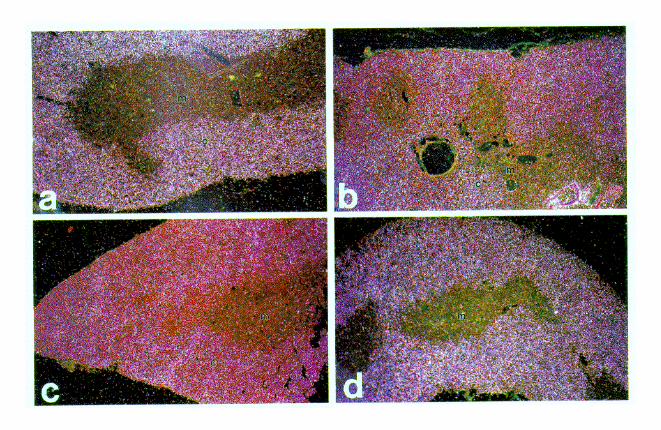
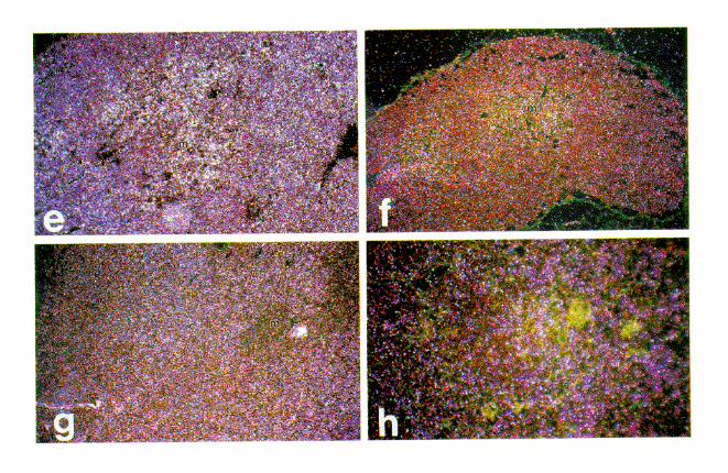
FIG. 4. Facilitation does not alter cell type specificity of expression, but there can be inappropriate regulation in a fraction of the unfacilitated mouse lines. Resultsof in situ hybridization analysis of CAT mRNA distribution in thymus tissue from several transgenic mouse lines are shown. Dark-field illumination demonstrates asignal as pinkish white grains over sites of mRNA accumulation. (a through d) Mouse lines 35 (3.7/0-VI), 43 (0.3/IIIa), 40 (0.3/IIIab), and 48 (0.3/IIIab1 II). Expressionis uniform among the cortical thymocytes (indicated by c) and much reduced in medullary thymocytes (indicated by m). (e through g) Mouse lines 15 (0.3/II-IIIa), 21(0.3/IIIab), and 54 (3.7/II-IIIa). These panels demonstrate atypical expression with a significant signal in medullary thymocytes. In addition to medullary expression(indicated by m), there is a uniform signal in cortical thymocytes shown in panels e and g (indicated by c) and a much reduced signal in medullary thymocytes. (h)Higher-power magnification of panel f. Expression is clearly nonuniform among the cortical thymocytes (indicated by c), with a signal present in 10 to 30% of the corticalthymocytes. A somewhat higher percentage of the medullary thymoyctes shown in panel f are positive, though the percentage is not as uniformly high as that for panele. Slides were exposed for various lengths of time (5 to 28 days) to optimize signal discrimination, counterstained with hematoxylin and eosin, and photographed ata final magnification of 332 (a through g) or 364 (h).
1128 ARONOW ET AL. MOL. CELL. BIOL.
on May 27, 2016 by guest
http://mcb.asm
.org/D
ownloaded from
Acrobat Notes
Color plate(s) available. Click on figure caption to view.
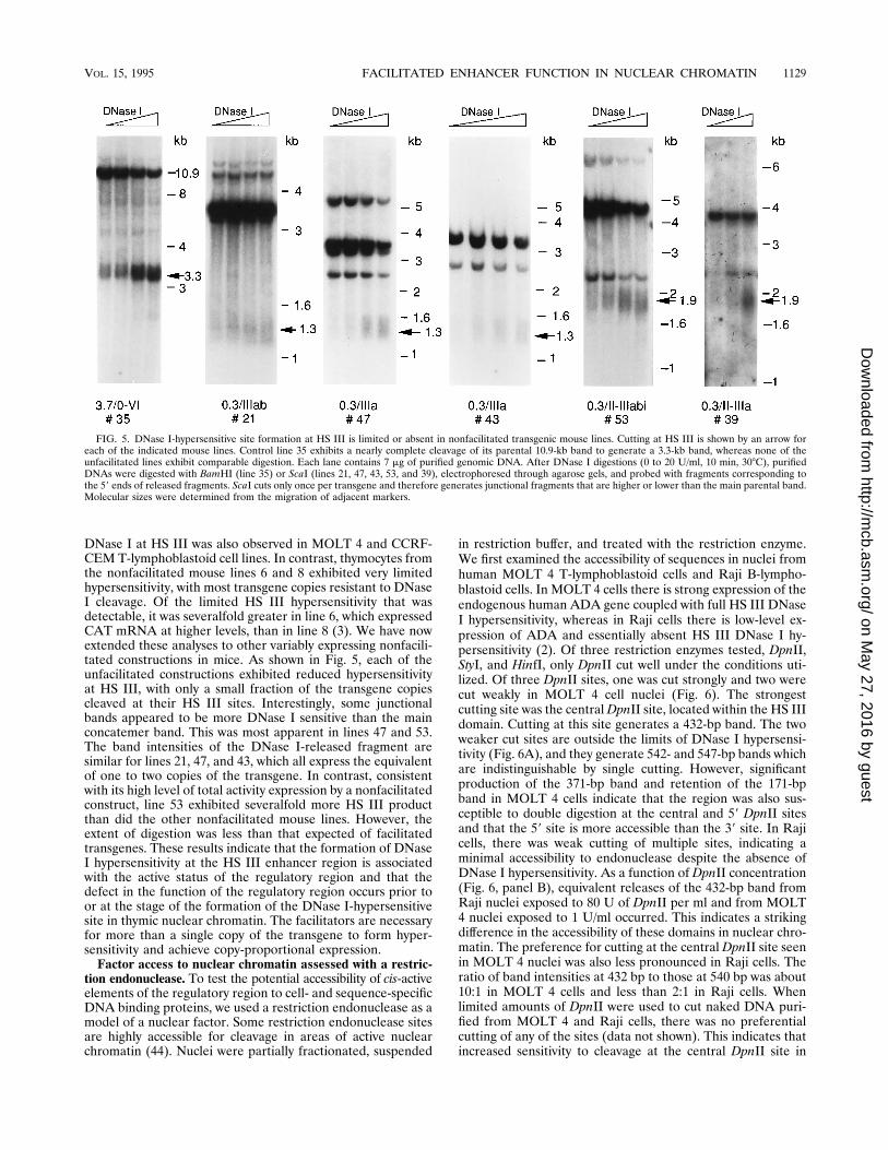
DNase I at HS III was also observed in MOLT 4 and CCRF-CEM T-lymphoblastoid cell lines. In contrast, thymocytes fromthe nonfacilitated mouse lines 6 and 8 exhibited very limitedhypersensitivity, with most transgene copies resistant to DNaseI cleavage. Of the limited HS III hypersensitivity that wasdetectable, it was severalfold greater in line 6, which expressedCAT mRNA at higher levels, than in line 8 (3). We have nowextended these analyses to other variably expressing nonfacili-tated constructions in mice. As shown in Fig. 5, each of theunfacilitated constructions exhibited reduced hypersensitivityat HS III, with only a small fraction of the transgene copiescleaved at their HS III sites. Interestingly, some junctionalbands appeared to be more DNase I sensitive than the mainconcatemer band. This was most apparent in lines 47 and 53.The band intensities of the DNase I-released fragment aresimilar for lines 21, 47, and 43, which all express the equivalentof one to two copies of the transgene. In contrast, consistentwith its high level of total activity expression by a nonfacilitatedconstruct, line 53 exhibited severalfold more HS III productthan did the other nonfacilitated mouse lines. However, theextent of digestion was less than that expected of facilitatedtransgenes. These results indicate that the formation of DNaseI hypersensitivity at the HS III enhancer region is associatedwith the active status of the regulatory region and that thedefect in the function of the regulatory region occurs prior toor at the stage of the formation of the DNase I-hypersensitivesite in thymic nuclear chromatin. The facilitators are necessaryfor more than a single copy of the transgene to form hyper-sensitivity and achieve copy-proportional expression.Factor access to nuclear chromatin assessed with a restric-
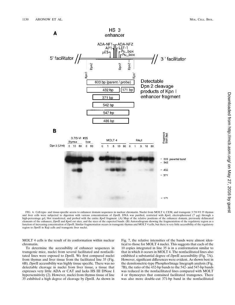
tion endonuclease. To test the potential accessibility of cis-activeelements of the regulatory region to cell- and sequence-specificDNA binding proteins, we used a restriction endonuclease as amodel of a nuclear factor. Some restriction endonuclease sitesare highly accessible for cleavage in areas of active nuclearchromatin (44). Nuclei were partially fractionated, suspended
in restriction buffer, and treated with the restriction enzyme.We first examined the accessibility of sequences in nuclei fromhuman MOLT 4 T-lymphoblastoid cells and Raji B-lympho-blastoid cells. In MOLT 4 cells there is strong expression of theendogenous human ADA gene coupled with full HS III DNaseI hypersensitivity, whereas in Raji cells there is low-level ex-pression of ADA and essentially absent HS III DNase I hy-persensitivity (2). Of three restriction enzymes tested, DpnII,StyI, and HinfI, only DpnII cut well under the conditions uti-lized. Of three DpnII sites, one was cut strongly and two werecut weakly in MOLT 4 cell nuclei (Fig. 6). The strongestcutting site was the centralDpnII site, located within the HS IIIdomain. Cutting at this site generates a 432-bp band. The twoweaker cut sites are outside the limits of DNase I hypersensi-tivity (Fig. 6A), and they generate 542- and 547-bp bands whichare indistinguishable by single cutting. However, significantproduction of the 371-bp band and retention of the 171-bpband in MOLT 4 cells indicate that the region was also sus-ceptible to double digestion at the central and 59 DpnII sitesand that the 59 site is more accessible than the 39 site. In Rajicells, there was weak cutting of multiple sites, indicating aminimal accessibility to endonuclease despite the absence ofDNase I hypersensitivity. As a function of DpnII concentration(Fig. 6, panel B), equivalent releases of the 432-bp band fromRaji nuclei exposed to 80 U of DpnII per ml and from MOLT4 nuclei exposed to 1 U/ml occurred. This indicates a strikingdifference in the accessibility of these domains in nuclear chro-matin. The preference for cutting at the central DpnII site seenin MOLT 4 nuclei was also less pronounced in Raji cells. Theratio of band intensities at 432 bp to those at 540 bp was about10:1 in MOLT 4 cells and less than 2:1 in Raji cells. Whenlimited amounts of DpnII were used to cut naked DNA puri-fied from MOLT 4 and Raji cells, there was no preferentialcutting of any of the sites (data not shown). This indicates thatincreased sensitivity to cleavage at the central DpnII site in
FIG. 5. DNase I-hypersensitive site formation at HS III is limited or absent in nonfacilitated transgenic mouse lines. Cutting at HS III is shown by an arrow foreach of the indicated mouse lines. Control line 35 exhibits a nearly complete cleavage of its parental 10.9-kb band to generate a 3.3-kb band, whereas none of theunfacilitated lines exhibit comparable digestion. Each lane contains 7 mg of purified genomic DNA. After DNase I digestions (0 to 20 U/ml, 10 min, 308C), purifiedDNAs were digested with BamHI (line 35) or ScaI (lines 21, 47, 43, 53, and 39), electrophoresed through agarose gels, and probed with fragments corresponding tothe 59 ends of released fragments. ScaI cuts only once per transgene and therefore generates junctional fragments that are higher or lower than the main parental band.Molecular sizes were determined from the migration of adjacent markers.
VOL. 15, 1995 FACILITATED ENHANCER FUNCTION IN NUCLEAR CHROMATIN 1129
on May 27, 2016 by guest
http://mcb.asm
.org/D
ownloaded from
MOLT 4 cells is the result of its conformation within nuclearchromatin.To determine the accessibility of enhancer sequences in
transgenic mice, nuclei from several facilitated and nonfacili-tated lines were exposed to DpnII. We first compared nucleifrom thymus and liver tissue from the facilitated line 35 (Fig.6B). DpnII accessibility was highly tissue specific. There was nodetectable cleavage in nuclei from liver tissue, a tissue thatexpresses very little ADA or CAT and lacks HS III DNase Ihypersensitivity (2). However, nuclei from thymus tissue of line35 exhibited a high degree of cleavage by DpnII. As shown in
Fig. 7, the relative intensities of the bands were almost iden-tical to those for MOLT 4 nuclei. This suggests that each of the10 copies integrated in line 35 is in a conformation similar tothat in which it occurs in MOLT 4. The nonfacilitated lines alsoexhibited a substantial degree of DpnII accessibility (Fig. 7A).However, significant differences were evident. As shown best inthe densitometric-type PhosphorImage linegraph analysis (Fig.7B), the ratio of the 432-bp bands to the 542- and 547-bp bandswas reduced in the nonfacilitated lines compared with MOLT4 or thymocytes that contained facilitated transgenes. Therewas also more double-cut 371-bp band in the nonfacilitated
FIG. 6. Cell-type- and tissue-specific access to enhancer domain sequences in nuclear chromatin. Nuclei from MOLT 4, CEM, and transgenic 3.7/0-VI 35 thymusand liver cells were subjected to digestion with various concentrations of DpnII. DNA was purified, restricted with KpnI, electrophoresed (7 mg) through ahigh-percentage gel, blot transferred, and probed with the entire KpnI fragment. (A) Map of the relative positions of the enhancer domain, previously delineatedelements of the enhancer, DpnII and KpnI cut sites, and the sizes of the expected bands. (B) Autoradiogram showing the fragmentation of the regulatory region as afunction of increasing concentration of DpnII. Similar fragmentation occurs in transgenic thymus and MOLT 4 cells, but there is very little accessibility of the regulatoryregion to DpnII in Raji cells and transgenic liver nuclei.
1130 ARONOW ET AL. MOL. CELL. BIOL.
on May 27, 2016 by guest
http://mcb.asm
.org/D
ownloaded from
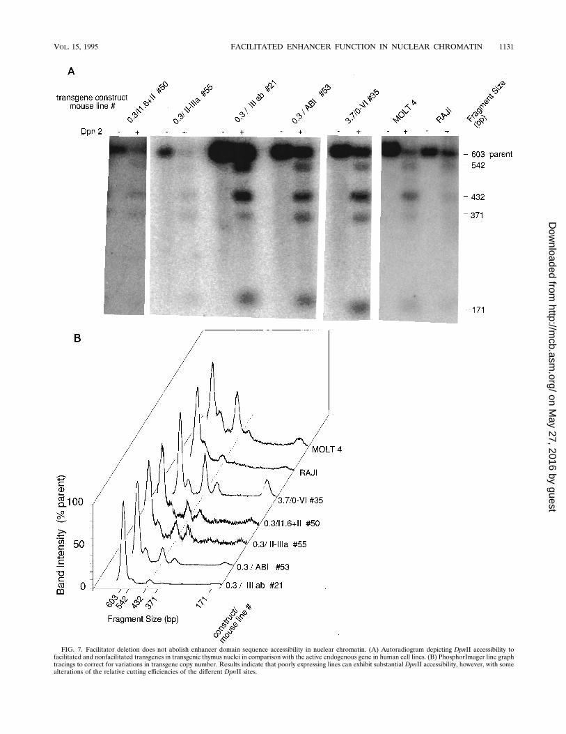
FIG. 7. Facilitator deletion does not abolish enhancer domain sequence accessibility in nuclear chromatin. (A) Autoradiogram depicting DpnII accessibility tofacilitated and nonfacilitated transgenes in transgenic thymus nuclei in comparison with the active endogenous gene in human cell lines. (B) PhosphorImager line graphtracings to correct for variations in transgene copy number. Results indicate that poorly expressing lines can exhibit substantial DpnII accessibility, however, with somealterations of the relative cutting efficiencies of the different DpnII sites.
VOL. 15, 1995 FACILITATED ENHANCER FUNCTION IN NUCLEAR CHROMATIN 1131
on May 27, 2016 by guest
http://mcb.asm
.org/D
ownloaded from
and very poorly expressing lines 50 and 55. This indicatessubstantial accessibility of nonfacilitated enhancer sequencesin thymocytes. Also, in contrast to the facilitated lines, thenonfacilitated lines exhibited increased accessibility of theflanking DpnII sites compared with the central site. Theseobservations indicate that factors present in developing thymo-cytes cause the regulatory domain to become accessible tofactors capable of recognizing specific sequences without thefunctional facilitators. However, the conformation of the non-facilitated enhancer domain is altered or disorganized com-pared with those of the facilitated versions present in human Tcells or mouse thymocytes expressing CAT mRNA at highlevels.The accessibility of the enhancer domain in mouse line 21
thymocyte nuclei was different from that in the three other non-facilitated mouse lines. While the total intensity of the cleavagebands was similar to that of the cleavage bands from the otherlines, given the high gene copy number there was much lesscleavage than expected (Fig. 7B). This line is also different fromother nonfacilitated lines in that it exhibited a speckled pattern ofexpression in the thymus. This indicates that in this line occurunique suppressive positional effects that appear to be differentfrom the generalized failure of multicopy expression observed inother lines that lack bilateral facilitators. Unlike those in othernonfacilitated lines that exhibited poor DNase I hypersensitivityand gene expression, most copies of the transgene in line 21exhibited little accessibility of the core enhancer region. However,despite the fact that most copies of the transgene appeared to beinaccessible to the restriction endonuclease, the ratio of singly cutproduct (540 bp plus 432 bp) to doubly cut product (371 bp) wassimilar to those for the lines in which most copies were accessible.This result indicates that there is normal accessibility of some ofthe gene copies and absent accessibility of most other copies. Ifeach of the copies were only partially accessible, there would belittle occurrence of doubly cut product.
DISCUSSION
Our results point to a critical role of nonenhancer sequencesin the ability of an enhancer domain to undergo a structuraltransition that enables it to function in transgenic nuclear chro-matin. Despite the tendency of most mammalian transgenes toexhibit line-to-line variable expression, it is a reasonable gen-eralization that large reporter constructs encompassing criticalregulatory elements tend to be expressed reliably and in pro-portion to gene copy number but that smaller constructs withdelineated enhancer domains tend to exhibit much less consis-tency. Our results provide a suggestion as to why this can bethe case. We have shown that in order for a segment of thehuman ADA gene first intron to function as an LCR, its 200-bpenhancer domain must be bilaterally flanked by 1-kb segmentsthat we have termed facilitators. LCRs have been generallydefined by their ability to confer integration-site-independentand gene-copy-proportional tissue-specific gene expressionupon a transgene, and they frequently appear to be related toDNase I-hypersensitive chromatin domains (27, 41, 54). SomeDNase I-hypersensitive sites correspond to the locations ofenhancers that are responsible for tissue- and cell-type-specificgene regulation (2, 23, 38, 52), but the molecular significanceand mechanism of HS formation in chromatin is unclear.DNase I-hypersensitive sites can represent a structural indica-tor of active or potentially active cis elements (6, 15, 18, 19, 33).Lu et al. (43) have characterized nucleosome-free sequencesbound to regulatory factors as HSs whereas McPherson et al.(46) have presented evidence that a hepatocyte-specific en-hancer can form a strong DNase I HS with bound transcription
factors in a positioned nucleosomal array without the ejectionof the underlying nucleosome. This suggests that a variety ofmechanisms may underlie regulatory HS formation.Our results indicate that information necessary to form hy-
persensitivity and allow activation at the HS III enhancer re-sides, in part, in sequences considerably removed from the HS,at least 3 to 5 nucleosomal lengths away from the enhancer.This appears to be quite different from the case with betaglobin genes, in which delineated DNase I-hypersensitive sitesappear to comprise functional LCRs (10, 21, 54, 57). However,other examples of LCRs seem to indicate that globin LCRs areatypical. Like the HS III T-cell enhancer of the ADA gene, themouse CD2 enhancer functions poorly as an LCR when se-quences flanking its major HS are deleted (25, 38, 39). Thisalso appears to be the case for the immunoglobulin heavychain enhancer, which is flanked by nuclear matrix attachmentregions (MARs) (7, 12, 14, 32).Defining the effects and constraints of the facilitators. The
surprising aspect of our present characterization is that theflanking facilitator sequences fail to act on the enhancer unlessboth are present and placed in specific positions relative to theenhancer. The failure to generate a facilitated regulatory do-main by moving the 59 facilitator downstream of the 39 facili-tator indicates that the physical linkage to the required se-quences is insufficient, perhaps indicating that the facilitatorand enhancer sequences are involved in a large stereospecificstructure. If binding to matrix components were the sole ex-planation of the requirement for 59 and 39 segments, facilitatorfunction would be accomplished by the misarranged versions.Alternatively, the requirement for bilateral binding to a rigidmatrix may allow the formation of torsional stress within theenhancer domain. However, the failure of a construction thatreverses the orientation of the 39 facilitator construct indicatesan additional constraint on the interaction between the facili-tators and the enhancer. Several of the lines containing thisconstruction, however, exhibited the highest levels of expres-sion by a ‘‘nonfacilitated’’ construction that we have seen.Interestingly, one of these lines also exhibited the highest levelof fractional cleavage of its thymic nuclear chromatin byDNase I, suggesting that its partially facilitated function was aresult of better DNase I-hypersensitive site generation. How-ever, most lines with inversion of the 39 facilitator show typicalunfacilitated function. This indicates that most integration sitesdo not permit the crippled facilitator to function as if it werestructurally normal.Unfacilitated transgenic constructions exhibit two forms of
failed expression. The first represents a failure of multiple genecopies to be expressed but a tendency for single, likely endcopies to be expressed. This indicates that the absence of thefacilitators could not be compensated by random transgeneinsertion sites and that the ‘‘compensation effect’’ of insertionsites is only able to act on immediately adjacent transgenes.The other component of attenuated nonfacilitated expressionwas an apparent susceptibility to positional effects. Overall,about one-quarter of the nonfacilitated mouse lines exhibitedconsiderably less expression than that expected of even a singlecopy, and we surmise that this represents positional repressiondue to the insertion site. Interestingly, in situ hybridizationanalysis indicated that in some of both the 59- and the 39-facilitator-deleted constructs, there was significant medullarythymocyte expression. This indicates that there are additionalpositional influences to which nonfacilitated transgenes aresusceptible that are not observable by total CAT activity assays.An additional line exhibited a speckled pattern of heteroge-neous expression in the thymus, despite exhibiting single-copytotal expression. Thus, in addition to crippling the quantitative
1132 ARONOW ET AL. MOL. CELL. BIOL.
on May 27, 2016 by guest
http://mcb.asm
.org/D
ownloaded from
function of the enhancer, the absence of facilitation also allowsfor at least some degree of misregulated expression.At what stage do the facilitators act on the enhancer? Do the
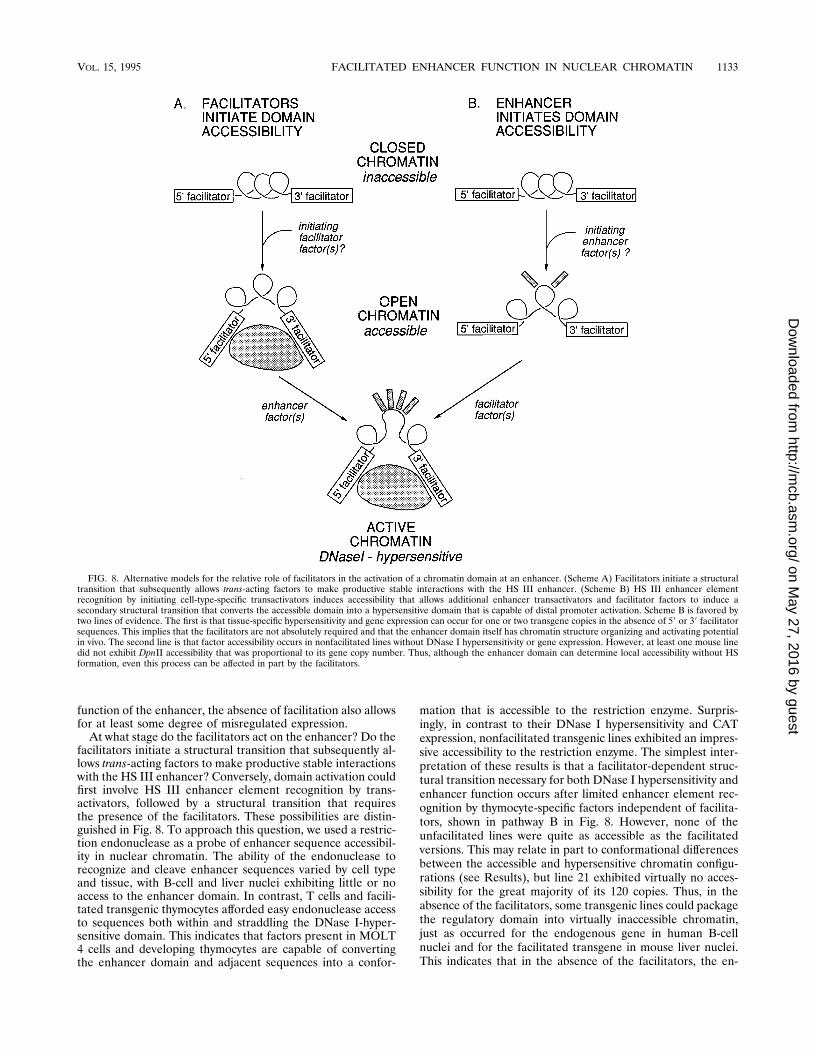
facilitators initiate a structural transition that subsequently al-lows trans-acting factors to make productive stable interactionswith the HS III enhancer? Conversely, domain activation couldfirst involve HS III enhancer element recognition by trans-activators, followed by a structural transition that requiresthe presence of the facilitators. These possibilities are distin-guished in Fig. 8. To approach this question, we used a restric-tion endonuclease as a probe of enhancer sequence accessibil-ity in nuclear chromatin. The ability of the endonuclease torecognize and cleave enhancer sequences varied by cell typeand tissue, with B-cell and liver nuclei exhibiting little or noaccess to the enhancer domain. In contrast, T cells and facili-tated transgenic thymocytes afforded easy endonuclease accessto sequences both within and straddling the DNase I-hyper-sensitive domain. This indicates that factors present in MOLT4 cells and developing thymocytes are capable of convertingthe enhancer domain and adjacent sequences into a confor-
mation that is accessible to the restriction enzyme. Surpris-ingly, in contrast to their DNase I hypersensitivity and CATexpression, nonfacilitated transgenic lines exhibited an impres-sive accessibility to the restriction enzyme. The simplest inter-pretation of these results is that a facilitator-dependent struc-tural transition necessary for both DNase I hypersensitivity andenhancer function occurs after limited enhancer element rec-ognition by thymocyte-specific factors independent of facilita-tors, shown in pathway B in Fig. 8. However, none of theunfacilitated lines were quite as accessible as the facilitatedversions. This may relate in part to conformational differencesbetween the accessible and hypersensitive chromatin configu-rations (see Results), but line 21 exhibited virtually no acces-sibility for the great majority of its 120 copies. Thus, in theabsence of the facilitators, some transgenic lines could packagethe regulatory domain into virtually inaccessible chromatin,just as occurred for the endogenous gene in human B-cellnuclei and for the facilitated transgene in mouse liver nuclei.This indicates that in the absence of the facilitators, the en-
FIG. 8. Alternative models for the relative role of facilitators in the activation of a chromatin domain at an enhancer. (Scheme A) Facilitators initiate a structuraltransition that subsequently allows trans-acting factors to make productive stable interactions with the HS III enhancer. (Scheme B) HS III enhancer elementrecognition by initiating cell-type-specific transactivators induces accessibility that allows additional enhancer transactivators and facilitator factors to induce asecondary structural transition that converts the accessible domain into a hypersensitive domain that is capable of distal promoter activation. Scheme B is favored bytwo lines of evidence. The first is that tissue-specific hypersensitivity and gene expression can occur for one or two transgene copies in the absence of 59 or 39 facilitatorsequences. This implies that the facilitators are not absolutely required and that the enhancer domain itself has chromatin structure organizing and activating potentialin vivo. The second line is that factor accessibility occurs in nonfacilitated lines without DNase I hypersensitivity or gene expression. However, at least one mouse linedid not exhibit DpnII accessibility that was proportional to its gene copy number. Thus, although the enhancer domain can determine local accessibility without HSformation, even this process can be affected in part by the facilitators.
VOL. 15, 1995 FACILITATED ENHANCER FUNCTION IN NUCLEAR CHROMATIN 1133
on May 27, 2016 by guest
http://mcb.asm
.org/D
ownloaded from
hancer binding factors are not the sole determinants that ini-tiate enhancer domain accessibility.Recent work by Jenuwein et al. (32) strongly supports the
idea that developmental activation of an enhancer domain is amultistep process. In their analyses of virus-transformed trans-genic pre-B cells, core elements of the immunoglobulin menhancer were capable of mediating a chromatin structuretransition that allowed adjacent sequences of the region tobecome accessible to T7 RNA polymerase in nuclear chroma-tin. This accessibility occurred without DNase I-hypersensitivesite formation or the productive stimulation of high-level RNApolymerase II activity. Their results indicate that in vivo chro-matin structure transitions leading to initial factor accessibilitycan be mediated by enhancer core elements but that thesestructure transitions are distinct from (or likely precede) theremodeling of an enhancer domain into a DNase I-hypersen-sitive structure. It is unclear whether accessibility without hy-persensitivity leads to the occupancy of critical enhancer ele-ments with stage-specific binding proteins and to what extentthe accessibility that was observed was cell type specific orreflective of domain activation in nonvirally transformed Bcells. Regardless, their studies indicate that additional cis in-formation is necessary for the transition to a DNase I-hyper-sensitive structure. This information has been ascribed to 59and 39 matrix attachment elements of the immunoglobulinenhancer region and other genes (7, 8, 12–14, 47), but theeffects of these elements when added to other promoters orenhancers in transgenes has been less than would be expectedfor independently active elements (45). Our results may offeran explanation for this discrepancy if the ability of the heter-ologous MAR-containing sequences to function is constrainedto specific positions and orientations with respect to the en-hancer or cis-regulatory domain.An appealing model for MAR function suggests that loop
anchorage provides insulating effects that allow the true po-tential of cis elements within the construct to function withoutinterference from the surrounding genes or regulatory ele-ments. However, attachment regions that immediately flankcis-regulatory domains (7, 8, 13, 14, 37) may be functionallydistinct from MAR sequences occurring at the boundaries ofgenes or loci (22, 47, 48, 51), and they may influence chromatinstructure at a different level. It is possible that certain classes ofchromatin-organizing sequences function as insulators (34)and that others act as agents that enable enhancer function.Our suggestion is that the ADA HS III facilitators participatein a structure transition that occurs after accessibility is estab-lished at the enhancer domain by initiating transactivators.However, as yet we do not know whether the facilitators sur-rounding the ADA gene HS III contain MAR activity.Our results are inconsistent with a simple model in which
attachment to a nuclear scaffold is sufficient for locus-activat-ing ability of a regulatory region, and they suggest that theactivation of the enhancer domain requires oriented DNAinteractions with constrained nuclear structures that extendwell beyond the enhancer domain. Identification of individualelements within the facilitator segments may point us towardsnovel and crucial nuclear factors responsible for organizingchromatin structure during developmental differentiation. Giventhe constraints for facilitator function, it is interesting to spec-ulate that the activation of the ADA thymic regulatory regionoccurs in the context of a large nuclear structure, such as thatwhich has been implicated in active transcription and process-ing centers in nuclei by confocal immunofluorescence (9, 64).Since the absence of facilitation can lead to a virtually com-plete extinction of enhancer function within cells that containall of the trans-acting factors responsible for generating en-
hancer activity, it is tempting to speculate that mechanisms forprocesses such as allelic exclusion or selective expression orinactivation of arrayed genes could work at a similar level as dofacilitators. Beyond the fundamental questions of how complexgenomes selectively and efficiently utilize cis-regulatory elements,an understanding of how chromatin structure is regulated may beof practical significance. In the emerging field of human genetherapy, stem cell-derived gene expression in particular may begreatly augmented by assemblies of gene regulatory elements thatinclude those responsible for governing chromatin structure.
ACKNOWLEDGMENTS
This work was supported by grants from the NIH (HD 19919 toJ.J.H. and DK47022 to B.J.A. and a Basic Research Grant from theMarch of Dimes Birth Defects Foundation (to B.J.A.).
REFERENCES
1. Affara, N., J. Fleming, P. S. Goldfarb, E. Black, B. Thiele, and P. R. Harri-son. 1985. Analysis of chromatin changes associated with the expression ofglobin and non-globin genes in cell hybrids between erythroid and othercells. Nucleic Acids Res. 13:5629–5644.
2. Aronow, B., D. Lattier, R. Silbiger, M. Dusing, J. Hutton, G. Jones, J. Stock,J. McNeish, S. Potter, D. Witte, et al. 1989. Evidence for a complex regula-tory array in the first intron of the human adenosine deaminase gene. GenesDev. 3:1384–1400.
3. Aronow, B. J., R. N. Silbiger, M. R. Dusing, J. L. Stock, K. L. Yager, S. S.Potter, J. J. Hutton, and D. A. Wiginton. 1992. Functional analysis of thehuman adenosine deaminase gene thymic regulatory region and its ability togenerate position-independent transgene expression. Mol. Cell. Biol. 12:4170–4185.
4. Barton, M. C., N. Madani, and B. M. Emerson. 1993. The erythroid proteincGATA-1 functions with a stage-specific factor to activate transcription ofchromatin-assembled beta-globin genes. Genes Dev. 7:1796–1809.
5. Blaese, R. M., and K. W. Culver. 1992. Gene therapy for primary immuno-deficiency disease. Immunodefic. Rev. 3:329–349.
6. Blom van Assendelft, G., O. Hanscombe, F. Grosveld, and D. R. Greaves.1989. The beta-globin dominant control region activates homologous andheterologous promoters in a tissue-specific manner. Cell 56:969–977.
7. Bode, J., Y. Kohwi, L. Dickinson, T. Joh, D. Klehr, C. Mielke, and S. T.Kohwi. 1992. Biological significance of unwinding capability of nuclear ma-trix-associating DNAs. Science 255:195–197.
8. Bonifer, C., A. Hecht, H. Saueressig, D. M. Winter, and A. E. Sippel. 1991.Dynamic chromatin: the regulatory domain organization of eukaryotic geneloci. J. Cell. Biochem. 47:99–108.
9. Carter, K. C., D. Bowman, W. Carrington, K. Fogarty, J. A. McNeil, F. S.Fay, and J. B. Lawrence. 1993. A three-dimensional view of precursor mes-senger RNA metabolism within the mammalian nucleus. Science 259:1330–1335.
10. Caterina, J. J., T. M. Ryan, K. M. Pawlik, R. D. Palmiter, R. L. Brinster,R. R. Behringer, and T. M. Townes. 1991. Human beta-globin locus controlregion: analysis of the 59DNase I hypersensitive site HS 2 in transgenic mice.Proc. Natl. Acad. Sci. USA 88:1626–1630.
11. Clark, A. C., D. Norris, M. A. Osley, J. S. Fassler, and F. Winston. 1988.Changes in histone gene dosage alter transcription in yeast. Genes Dev.2:150–159.
12. Cockerill, P. N., and W. T. Garrard. 1986. Chromosomal loop anchorage ofthe kappa immunoglobulin gene occurs next to the enhancer in a regioncontaining topoisomerase II sites. Cell 44:273–282.
13. Cockerill, P. N., and W. T. Garrard. 1986. Chromosomal loop anchoragesites appear to be evolutionarily conserved. FEBS Lett. 204:5–7.
14. Cockerill, P. N., M. H. Yuen, and W. T. Garrard. 1987. The enhancer of theimmunoglobulin heavy chain locus is flanked by presumptive chromosomalloop anchorage elements. J. Biol. Chem. 262:5394–5397.
15. Crowder, C. M., and J. P. Merlie. 1988. Stepwise activation of the mouseacetylcholine receptor delta- and gamma-subunit genes in clonal cell lines.Mol. Cell. Biol. 8:5257–5267.
16. Durrin, L. K., R. K. Mann, P. S. Kayne, and M. Grunstein. 1991. Yeasthistone H4 N-terminal sequence is required for promoter activation in vivo.Cell 65:1023–1031.
17. Eissenberg, J. C., and S. C. Elgin. 1991. Boundary functions in the control ofgene expression. Trends Genet. 7:335–340.
18. Ford, A. M., C. A. Bennett, L. E. Healy, E. Navarro, E. Spooncer, and M. F.Greaves. 1992. Immunoglobulin heavy-chain and CD3 delta-chain gene en-hancers are DNase I-hypersensitive in hemopoietic progenitor cells. Proc.Natl. Acad. Sci. USA 89:3424–3428.
19. Forrester, W. C., U. Novak, R. Gelinas, and M. Groudine. 1989. Molecularanalysis of the human beta-globin locus activation region. Proc. Natl. Acad.Sci. USA 86:5439–5443.
20. Forrester, W. C., S. Takegawa, T. Papayannopoulou, G. Stamatoyannoupou-
1134 ARONOW ET AL. MOL. CELL. BIOL.
on May 27, 2016 by guest
http://mcb.asm
.org/D
ownloaded from

los, and M. Groudine. 1987. Evidence for a locus activation region: theformation of developmentally stable hypersensitive sites in globin-expressinghybrids. Nucleic Acids Res. 15:10159–10177.
21. Fraser, P., S. Pruzina, M. Antoniou, and F. Grosveld. 1993. Each hypersen-sitive site of the human beta-globin locus control region confers a differentdevelopmental pattern of expression on the globin genes. Genes Dev. 7:106–113.
22. Gasser, S. M., and U. K. Laemmli. 1986. Cohabitation of scaffold bindingregions with upstream/enhancer elements of three developmentally regu-lated genes of D. melanogaster. Cell 46:521–530.
23. Georgopoulos, K., P. van den Elsen, E. Bier, A. Maxam, and C. Terhorst.1988. A T cell-specific enhancer is located in a DNase I-hypersensitive areaat the 39 end of the CD3-delta gene. EMBO J. 7:2401–2407.
24. Giese, K., J. Cox, and R. Grosschedl. 1992. The HMG domain of lymphoidenhancer factor 1 bends DNA and facilitates assembly of functional nucle-oprotein structures. Cell 69:185–195.
25. Greaves, D. R., F. D. Wilson, G. Lang, and D. Kioussis. 1989. Human CD239-flanking sequences confer high-level, T cell-specific, position-independentgene expression in transgenic mice. Cell 56:979–986.
26. Grosveld, F., M. Antoniou, G. van Assendelft, E. de Boer, J. Hurst, G.Kollias, F. MacFarlane, and N. Wrighton. 1987. The regulation of expres-sion of human beta-globin genes. Prog. Clin. Biol. Res. 251:133–144.
27. Grosveld, F., G. van Assendelft, D. R. Greaves, and G. Kollias. 1987. Posi-tion-independent, high-level expression of the human beta-globin gene intransgenic mice. Cell 51:975–985.
28. Groudine, M., and M. Linial. 1986. Chromatin structure and gene expressionin germ line and somatic cells. Adv. Exp. Med. Biol. 205:205–243.
29. Han, M., U. J. Kim, P. Kayne, and M. Grunstein. 1988. Depletion of histoneH4 and nucleosomes activates the PHO5 gene in Saccharomyces cerevisiae.EMBO J. 7:2221–2228.
30. Hirschhorn, J. N., S. A. Brown, C. D. Clark, and F. Winston. 1992. Evidencethat SNF2/SWI2 and SNF5 activate transcription in yeast by altering chro-matin structure. Genes Dev. 6:2288–2298.
31. Hogan, B., F. Constantini, and E. Lacy. 1986. Manipulating the mouseembryo. Cold Spring Harbor Laboratory, Cold Spring Harbor, N.Y.
32. Jenuwein, T., W. C. Forrester, R. G. Qiu, and R. Grosschedl. 1993. Theimmunoglobulin mu enhancer core establishes local factor access in nuclearchromatin independent of transcriptional stimulation. Genes Dev. 7:2016–2032.
33. Jimenez, G., S. D. Griffiths, A. M. Ford, M. F. Greaves, and T. Enver. 1992.Activation of the beta-globin locus control region precedes commitment tothe erythroid lineage. Proc. Natl. Acad. Sci. USA 89:10618–10622.
34. Kellum, R., and P. Schedl. 1992. A group of scs elements function as domainboundaries in an enhancer-blocking assay. Mol. Cell. Biol. 12:2424–2431.
35. Knezetic, J. A., G. A. Jacob, and D. S. Luse. 1988. Assembly of RNApolymerase II preinitiation complexes before assembly of nucleosomes al-lows efficient initiation of transcription on nucleosomal templates. Mol. Cell.Biol. 8:3114–3121.
36. Knezetic, J. A., and D. S. Luse. 1986. The presence of nucleosomes on aDNA template prevents initiation by RNA polymerase II in vitro. Cell45:95–104.
37. Laemmli, U. K., E. Kas, L. Poljak, and Y. Adachi. 1992. Scaffold-associatedregions: cis-acting determinants of chromatin structural loops and functionaldomains. Curr. Opin. Genet. Dev. 2:275–285.
38. Lake, R. A., D. Wotton, and M. J. Owen. 1990. A 39 transcriptional enhancerregulates tissue-specific expression of the human CD2 gene. EMBO J.9:3129–3136.
39. Lang, G., C. Mamalaki, D. Greenberg, N. Yannoutsos, and D. Kioussis.1991. Deletion analysis of the human CD2 gene locus control region intransgenic mice. Nucleic Acids Res. 19:5851–5856.
40. Lee, M. S., and W. T. Garrard. 1992. Uncoupling gene activity from chro-matin structure: promoter mutations can inactivate transcription of the yeastHSP82 gene without eliminating nucleosome-free regions. Proc. Natl. Acad.Sci. USA 89:9166–9170.
41. Li, Q. L., B. Zhou, P. Powers, T. Enver, and G. Stamatoyannopoulos. 1990.Beta-globin locus activation regions: conservation of organization, structure,and function. Proc. Natl. Acad. Sci. USA 87:8207–8211.
42. Lohr, D. 1993. Chromatin structure and regulation of the eukaryotic regu-latory gene GAL80. Proc. Natl. Acad. Sci. USA 90:10628–10632.
43. Lu, Q., L. L. Wallrath, H. Granok, and S. C. R. Elgin. 1993. (CT)n z (GA)nrepeats and heat shock elements have distinct roles in chromatin structureand transcriptional activation of the Drosophila hsp26 gene. Mol. Cell. Biol.13:2802–2814.
44. Ma, X. J., and A. G. Goodridge. 1992. Nutritional regulation of nucleosomalstructure at the chicken malic enzyme promoter in liver. Nucleic Acids Res.20:4997–5002.
45. McKnight, R. A., A. Shamay, L. Sankaran, R. J. Wall, and L. Hennighausen.1992. Matrix-attachment regions can impart position-independent regulationof a tissue-specific gene in transgenic mice. Proc. Natl. Acad. Sci. USA89:6943–6947.
46. McPherson, C. E., E. Y. Shim, D. S. Friedman, and K. S. Zaret. 1993. Anactive tissue-specific enhancer and bound transcription factors existing in aprecisely positioned nucleosomal array. Cell 75:387–398.
47. Mirkovitch, J., S. M. Gasser, and U. K. Laemmli. 1987. Relation of chro-mosome structure and gene expression. Philos. Trans. R. Soc. Lond. Ser. BBiol. Sci. 317:563–574.
48. Mirkovitch, J., S. M. Gasser, and U. K. Laemmli. 1988. Scaffold attachmentof DNA loops in metaphase chromosomes. J. Mol. Biol. 200:101–109.
49. Palmiter, R. D., and R. L. Brinster. 1986. Germ-line transformation of mice.Annu. Rev. Genet. 20:465–499.
50. Peterson, D. O. 1985. Alterations in chromatin structure associated withglucocorticoid-induced expression of endogenous mouse mammary tumorvirus genes. Mol. Cell. Biol. 5:1104–1110.
51. Phi-Van, L., J. P. von Kries, W. Ostertag, and W. H. Stratling. 1990. Thechicken lysozyme 59 matrix attachment region increases transcription from aheterologous promoter in heterologous cells and dampens position effects onthe expression of transfected genes. Mol. Cell. Biol. 10:2302–2307.
52. Pinkert, C. A., D. M. Ornitz, R. L. Brinster, and R. D. Palmiter. 1987. Analbumin enhancer located 10 kb upstream functions along with its promoterto direct efficient, liver-specific expression in transgenic mice. Genes Dev.1:268–276.
53. Roth, S. Y., M. Shimizu, L. Johnson, M. Grunstein, and R. T. Simpson. 1992.Stable nucleosome positioning and complete repression by the yeast alpha 2repressor are disrupted by amino-terminal mutations in histone H4. GenesDev. 6:411–425.
54. Ryan, T. M., R. R. Behringer, N. C. Martin, T. M. Townes, R. D. Palmiter,and R. L. Brinster. 1989. A single erythroid-specific DNase I super-hyper-sensitive site activates high levels of human beta-globin gene expression intransgenic mice. Genes Dev. 3:314–323.
55. Simpson, R. T. 1990. Nucleosome positioning can affect the function of acis-acting DNA element in vivo. Nature (London) 343:387–389.
56. Simpson, R. T. 1991. Nucleosome positioning: occurrence, mechanisms, andfunctional consequences. Prog. Nucleic Acid Res. Mol. Biol. 40:143–184.
57. Talbot, D., S. Philipsen, P. Fraser, and F. Grosveld. 1990. Detailed analysisof the site 3 region of the human beta-globin dominant control region.EMBO J. 9:2169–2177.
58. Tuan, D., W. Solomon, Q. Li, and I. M. London. 1985. The ‘‘beta-like-globin’’gene domain in human erythroid cells. Proc. Natl. Acad. Sci. USA 82:6384–6388.
59. Vettese, D. M., P. Walter, H. Chen, L. J. Juan, and J. L. Workman. 1994.Role of the histone amino termini in facilitated binding of a transcriptionfactor, GAL4-AH, to nucleosome cores. Mol. Cell. Biol. 14:970–981.
60. Witte, D. P., D. A. Wiginton, J. J. Hutton, and B. J. Aronow. 1991. Coordi-nate developmental regulation of purine catabolic enzyme expression ingastrointestinal and postimplantation reproductive tracts. J. Cell Biol. 115:179–190.
61. Workman, J. L., and A. R. Buchman. 1993. Multiple functions of nucleo-somes and regulatory factors in transcription. Trends Biochem. Sci. 18:90–95.
62. Workman, J. L., and R. E. Kingston. 1992. Nucleosome core displacement invitro via a metastable transcription factor-nucleosome complex. Science 258:1780–1784.
63. Wustmann, G., J. Szidonya, H. Taubert, and G. Reuter. 1989. The geneticsof position-effect variegation modifying loci in Drosophila melanogaster.Mol. Gen. Genet. 217:520–527.
64. Xing, Y., C. V. Johnson, P. R. Dobner, and J. B. Lawrence. 1993. Higher levelorganization of individual gene transcription and RNA splicing. Science259:1326–1330.
VOL. 15, 1995 FACILITATED ENHANCER FUNCTION IN NUCLEAR CHROMATIN 1135
on May 27, 2016 by guest
http://mcb.asm
.org/D
ownloaded from
Related Documents