Surface Plasmon Resonance Applications in Drug Discovery with an Emphasis on Small Molecule and Low Affinity Systems Inauguraldissertation zur Erlangung der Würde eines Doktors der Philosophie vorgelegt der Philosophisch-Naturwissenschaftliche n Fakultät der Universität Basel von Daniel Ricklin aus Zürich, Schweiz Referent: Prof. Dr. Beat Ernst Korreferent: Prof. Dr. Ueli Aebi Basel, Juni 2005

Welcome message from author
This document is posted to help you gain knowledge. Please leave a comment to let me know what you think about it! Share it to your friends and learn new things together.
Transcript

8/9/2019 DissB 7321 Mirroring
http://slidepdf.com/reader/full/dissb-7321-mirroring 1/278
Surface Plasmon ResonanceApplications in Drug Discovery
with an Emphasis on Small Molecule
and Low Affinity Systems
Inauguraldissertation
zur Erlangung der Würde eines Doktors der Philosophie
vorgelegt der Philosophisch-Naturwissenschaftlichen Fakultät
der Universität Basel
von
Daniel Ricklin
aus Zürich, Schweiz
Referent: Prof. Dr. Beat Ernst
Korreferent: Prof. Dr. Ueli Aebi
Basel, Juni 2005
8/9/2019 DissB 7321 Mirroring
http://slidepdf.com/reader/full/dissb-7321-mirroring 2/278
Genehmigt von der Philosophisch-Naturwissenschaftlichen Fakultät
auf Antrag von
Prof. Dr. Beat Ernst, Institut für Molekulare Pharmazie, Universität Basel
Prof. Dr. Ueli Aebi, M. E. Müller Institut, Biozentrum, Universität Basel
Basel, den 5. Juli 2005
Prof. Dr. Hans-Jakob Wirz
Dekan

8/9/2019 DissB 7321 Mirroring
http://slidepdf.com/reader/full/dissb-7321-mirroring 3/278
A discovery is said to be an accident
meeting a prepared mind…
(Albert Szent-Györgyi, 1893-1986)
Meinen Eltern
Johanna und Albert

8/9/2019 DissB 7321 Mirroring
http://slidepdf.com/reader/full/dissb-7321-mirroring 4/278
8/9/2019 DissB 7321 Mirroring
http://slidepdf.com/reader/full/dissb-7321-mirroring 5/278
- I -
Acknowledgement
This thesis was performed at the Institute of Molecular Pharmacy of the University of
Basel under the supervision of Prof. Dr. Beat Ernst, and was generously supported by
the Swiss National Science Foundation.
First and foremost, I thank Prof. Dr. Beat Ernst for his great scientific support, the
generous and modern infrastructure, and his constructive and fruitful discussions. With
its multidisciplinary and international atmosphere, the institute created a motivating,
challenging and encouraging environment. The integration of scientific seminars,
project meetings, teaching opportunities and supervision of diploma theses was very
stimulating for the development of skills beyond pure science.
I sincerely thank Prof. Dr. Ueli Aebi for being the co-referee of my thesis.
My deep and special thanks are also going to all the former and present colleagues atthe institute, who created a very comfortable working atmosphere and provided me
with proteins, analytes and good ideas. Daniel Strasser and Steven Knecht helped me
forming a ‘Biacore team’ and gave me many new inputs. Caroline Bellac with her
diploma thesis and Svenja Landweer in a ‘summer project’ were a tremendous support
for the experimental part of this thesis. Rita Born, Karin Johansson, Daniela Stokmaier,
Andrea Frey, Claudia Riva, Oleg Khorev, and Daniel Kreyenbühl were not only
responsible for many of the biological and chemical work in the asialoglycoprotein-
receptor project, but also supported me with critical and fruitful discussions during
project meetings. I also like to thank Dr.
Said Rabbani, Dr. Oliver Schwardt, Dr. BrianCutting, Gabriela Pernter, and Bea Wagner, for their administrative and technical help
as well as Matthias Studer and Andreas Stöckli for their computer support.
Dr. Angelo Vedani and Dr. Markus Lill from Biographics Laboratories in Basel helped
me in many aspects of molecular modeling, and Prof. Dr. Paul Jenö and Thierry Mini
from the Biocenter of the University of Basel performed the mass spectrometric
analysis of asialoglycoprotein. Prof. Thomas Peters, Dr. Hanne Peters, Dr. Thomas
Weimar, Thies Köhli, and Dr. Lars Herfurth from the Medical University of Lübeck,
Germany greatly facilitated my entrance in the field of Biacore analysis. I would like to
thank them for the collaboration in the GSLA-2 project, as well as Dr. John Magnanifrom GlycoTech Inc. in Rockville, USA, for his donations of the diagnostic antibody. I
also want to thank Prof. Dr. Alex Eberle from the Department of Research of the
University Hospital Basel for his collaboration in the hexahistidine project.
Hence performing a PhD thesis is not solely about science; it needs help and support
from many other sides. Therefore, I primarily want to thank my parents, who not only
supported me financially and morally throughout the whole course of my educational
career, but also let me feel their love and care. My special thanks are going to Salome
Lichtsteiner, who closely accompanied me during this thesis, shared my ups and down,
and always understood in motivating me to carry on. Finally, I would like to thank themany friends and relatives, who created the social ground and network for this work.

8/9/2019 DissB 7321 Mirroring
http://slidepdf.com/reader/full/dissb-7321-mirroring 6/278
- II -

8/9/2019 DissB 7321 Mirroring
http://slidepdf.com/reader/full/dissb-7321-mirroring 7/278
- III -
Summary
Surface plasmon resonance (SPR) technology evolved into a key technology for the
characterization of biomolecular interactions, and is integrated in many stages of the
drug discovery process. Despite recent developments in the area of instrumentsensitivity and data processing, working with small molecules and low affinity
interactions still remains a major challenge. The aim of this thesis was therefore to
evaluate and develop different methods for the accurate and reliable determination of
thermodynamic and kinetic information of such interaction systems.
Through participation in the international ABRF-MIRG’02 study, the instrument used
for this thesis was validated for small molecular analyses. The results obtained for a
small sulfonamide analyte binding to bovine carbonic anhydrase II were very close to
the study average and corresponded well with solution-based methods. Screening
experiments with human serum albumin and a set of known plasma protein bindersfurther confirmed the effectiveness of SPR for small molecule assays. However, the
albumin assay also revealed some limitations; while neutral and cationic drugs
generated very reproducible KD values, the deviations were usually larger for free
acids. Some compounds like diazepam or L-tryptophan showed a more complex
binding behavior. Most of these atypical signal effects could be attributed to ligand- or
pH -induced structural changes of albumin, which are well described in literature.
Finally, a new immobilization method for human serum albumin was developed by
targeting its single free cysteine residue for a reversible coupling to the sensor chip.
The interaction of monovalent carbohydrates with their protein targets is one of themost prominent examples of small molecule/low affinity systems. They play an
important role in many biological processes from cellular recognition to infection
diseases. In order to characterize such carbohydrate-protein interactions, a diagnostic
monoclonal antibody (GSLA-2) directed against a carbohydrate epitope was
investigated using a combination of SPR and nuclear magnetic resonance. By screening
the tetrasaccharide antigen sialyl Lewisa and a set of structurally related compounds,
the thermodynamic and kinetic binding properties as well as the recognition pattern
could be successfully described. With a KD in the low micromolar range and fast
kinetic on- (~10
4
M
-1
s
-1
) and off-rates (>0.1 s
-1
), the interaction correlated very wellwith earlier reports about carbohydrate-protein interactions. Truncation of the antibody
to its antigen-binding parts led to a significant increase in binding activity and reduced
non-specific binding.
The human hepatic asialoglycoprotein receptor served as a more complex example of
carbohydrate-binding proteins. This C-type lectin is involved in the clearance of
desialylated glycoproteins from blood and is regarded as an important target for
selective delivery of genes and drugs to the liver. After expression of the carbohydrate
recognition domain in E.coli, the lectin could be successfully purified using a
combination of different chromatographic steps and was immobilized on a SPR sensorchip. Binding of the physiological glycoprotein ligands asialofetuin and
8/9/2019 DissB 7321 Mirroring
http://slidepdf.com/reader/full/dissb-7321-mirroring 8/278
- IV -
asialoorosomucoid was characterized by a polyvalent interaction pattern with slow
dissociation rates and sub-nanomolar KD values. In contrast, monovalent sugars like
galactose or N -acetyl galactosamine showed fast kinetics and affinities in the micro- to
millimolar range. In addition, the processed SPR signals of all small sugar analytes had
a negative sign and had to be mirrored before analysis. The negative binding signalswere clearly concentration-dependent and could be fitted to a single binding site model.
The resulting affinity values were validated by a competitive ELISA method and with
literature values. Furthermore, the interaction was found to be strongly calcium- and
pH -dependent, as it is reported for the receptor. Ligand-induced conformational
changes or interactions of the immobilized lectin with the dextran matrix of the sensor
chip were evaluated as the most likely explanation of the negative SPR signals.
Whether this is an isolated behavior of the asialoglycoprotein receptor or whether these
observations could be applied to other lectins with shallow, surface-accessible binding
sites has to be investigated in more detail.
A combined analysis of all protein studies performed in this thesis clearly reveals the
benefits and limitations of SPR technology for the analysis of small molecules and low
affinity interactions. The label-free detection and the simultaneous evaluation of both
thermodynamic and kinetic parameters allow a rapid and deep insight into molecular
binding mechanisms, even at the limit of detectability. Careful assay design and proper
data processing are a prerequisite for the analysis of small molecules, since even small
signal deviations might significantly influence the binding constants. The studies of
human serum albumin and the asialoglycoprotein also revealed, that SPR detection
cannot be solely regarded as a mass detector. Structural changes of the immobilized
proteins or matrix-effect could also influence the detected SPR signal and should
always be considered in the planning and evaluation of experiments.
In a small pilot project, the molecular mechanism of the interaction between the
hexahistidine tag, which is widely used for the purification of recombinant proteins,
and immobilized nickel surfaces was investigated using SPR. By injecting a series of
oligohistidine peptides (His2-His10), the influence of the number of histidine residues
to the binding behavior could be evaluated. As expected, the His6 peptide revealed the
best compromise between rebinding and entropic effects, resulting in the lowest K D of
the series (34
nM). The distance between the two simultaneously binding imidazolerings was also found to play an important role for the binding strength, as is could be
shown by screening a series of His2Ala4 peptides.
8/9/2019 DissB 7321 Mirroring
http://slidepdf.com/reader/full/dissb-7321-mirroring 9/278
- V -
Abbreviations
ADME(T) Absorption, distribution, metabolism, excretion (toxicology)
ABRF Association of biomolecular research faculties
ACN Acetonitrile
AFM Atomic force microscopyAGP 1-Acid glycoprotein
Ala L-Alanine
ASF Asialofetuin
ASGP-R Asialoglycoprotein receptor
Asn L-Asparagine
ASOR Asialoorosomucoid
Asp L-aspartic acid
AU Absorbance units
AUC Analytical ultracentrifugationBSA Bovine serum albuminBXM (+)-Biotinyl-3-maleimidopropionamidyl-3,6-dioxaoctanediamine
(Biotin-PEO3-maleimide)
CA II Carbonic anhydrase II
CBS 4-Carboxybenzenesulfonamide
CD Circular dichroism
CM5 Carboxy-methylated chip surface (100 nm)
CMD Carboxymethyl dextranCRD Carbohydrate recognition domain
Cys L-Cysteine
Da DaltonDEAE Diethylaminoethyl
DIPEA N , N ’-Diisopropylethylamine
DMF Dimethylformamide
DMSO Dimethylsulfoxide
DNA Desoxyribonucleic acid
DPI Dual polarization interferometry
DTNB 5,5’-Dithio-bis(2-nitrobenzoic acid); Ellmans Reagent
DTT Dithiothreitol
E. coli Escherichia coli
EDC 1-Ethyl-3-(3-dimethylaminopropyl)-carbodiimide hydrochloride
EDTA Ethylenediaminetetraacetic acidELISA Enzyme-linked immunosorbent assay
ESI Electrospray ionizationF(ab’)2 Antigen-binding fragment of an antibody, dimeric
Fab’ Antigen-binding fragment of an antibody, monomeric
Fmoc 9-Fluorenylmethoxycarbonyl group
FPLC Fast protein liquid chromatography
Gal D-Galactose
GalNAc N -Acetyl-D-galactosamine
Glc D-Glucose
GlcNAc N -Acetyl-D-glucosamine
Gln L-Glutamine

8/9/2019 DissB 7321 Mirroring
http://slidepdf.com/reader/full/dissb-7321-mirroring 10/278
- VI -
Glu L-Glutamic acid
GMP Good manufacturing practice
GPCR G-Protein coupled receptor
GSH Glutathione, reduced form
GSLA-2 Monoclonal antibody (mouse IgG1) recognizing sialyl Lewis a
GSSG Glutathione, oxidized formH1 / HL-1 Hepatic lectin 1 (ASGP-R)
H2 / HL-2 Hepatic lectin 2 (ASGP-R)
hAGT Human O6-alkylguanine-DNA-alkyltransferase
HBS HEPES buffered salineHCl Hydrochloric acid
HEPES 4-(2-hydroxyethyl)-1-piperazineethanesulfonic acid
His L-Histidine
HMA Human mercaptalbumin
HNA Human non-mercaptalbumin
HOBt 1-Hydroxybenzotriazole
HPLC High performance liquid chromatographyHSA Human serum albumin
HTS High-throughput screening
HUGO Human genome organization
IC50 50% inhibition constant
IDA Iminodiacetic acid
IFC Integrated fluidic cartridge
IgG Immunoglobulin G
IMAC Immobilized-metal affinity chromatography
ITC Isothermal titration calorimetry
KA
Equilibrium association constant
KD Equilibrium dissociation constant
Ki Inhibition constant
koff Dissociation rate constant
kon Association rate constant
Lem Lemieux spacer
mAb Monoclonal antibodyMALDI Matrix-assisted laser desorption ionization
MBP Maltose-binding protein
MEA 2-Mercaptoethylamine
MES 2-Morpholinoethanesulfonic acid
Met L-MethionineMIRG Molecular interactions research group
MS Mass spectrometry
MW Molecular weight
NA Neutravidin
NaOH Sodium hydroxide
NEM N -Ethylmaleimide
NHS N -Hydroxysuccinimide
NME New molecular entities
NMR Nuclear magnetic resonance
NTA Nitrilotriacetic acid
OSM OrosomucoidP20 Polysorbate-20 (Tween-20)
8/9/2019 DissB 7321 Mirroring
http://slidepdf.com/reader/full/dissb-7321-mirroring 11/278
- VII -
PAGE Polyacrylamide gel electrophoresis
PBS Phosphate buffered saline
PDEA 2-(2-Pyridinyl-dithio)- ethaneamine
PPB Plasma protein binding
QCM Quartz crystal microbalancing
QUASAR Quasi-atomistic receptor (modeling)R Response
Req Equilibrium response
Rmax Maximum response
RP Reversed phase
RU Resonance unit
SA Streptavidin
SAR Structure-activity relationship
SDS Sodium dodecylsulfate
SEC Size exclusion chromatography
SFS Stopped-flow spectrometry
SKR Structure-kinetics relationshipsLea Sialyl Lewisa
sLex Sialyl Lewisx
SpA Staphylococcal protein A
SpG Streptococcal protein G
SPR Surface plasmon resonanceSTD Saturation transfer difference
TBTU 2-(1H-Benzotriazole-1-yl)-1,1,3,3-tetramethyluronium tetrafluoroborate
TDC Target definition compound
TFA Trifluoroacetic acid
TIR Total internal reflection
Tris Tris(hydroxymethyl)aminomethane
trNOE Transfer nuclear Overhauser effect
Trp L-Tryptophan

8/9/2019 DissB 7321 Mirroring
http://slidepdf.com/reader/full/dissb-7321-mirroring 12/278
- VIII -
8/9/2019 DissB 7321 Mirroring
http://slidepdf.com/reader/full/dissb-7321-mirroring 13/278
- IX -
Contents of the Thesis
Chapter 1: General Introduction Drug Discovery on the Move 2
The Need for Biosensors 6Structure and Aim of the Thesis 8
Chapter 2: Biacore Technology Introduction 12
Materials and Methods 39
Results and Discussion 44
Conclusions 48
Chapter 3: Human Serum Albumin Introduction 54
Materials and Methods 63
Results and Discussion 74
Conclusions 100
Chapter 4: Diagnostic Antibody GSLA-2 Introduction 106
Materials and Methods 112
Results and Discussion 118
Conclusions 134
Chapter 5: Asialoglycoprotein Receptor Introduction 140
Materials and Methods 149
Results and Discussion 160
Conclusions 199
Chapter 6: Hexahistidine Tag Introduction 206
Materials and Methods 210
Results and Discussion 214
Conclusions 224
Chapter 7: General Considerations Working with Small Molecules 228
Carbohydrate-Protein Interactions 236
Negative Binding Responses 239
Appendices A (CA II) 248
B (HSA) 249
C (GSLA-2) 255
D (ASGP-R) 256
E (HisTag) 261

8/9/2019 DissB 7321 Mirroring
http://slidepdf.com/reader/full/dissb-7321-mirroring 14/278
- X -

8/9/2019 DissB 7321 Mirroring
http://slidepdf.com/reader/full/dissb-7321-mirroring 15/278
8/9/2019 DissB 7321 Mirroring
http://slidepdf.com/reader/full/dissb-7321-mirroring 16/278
General Introduction Chapter 1
2
1.1 Drug Discovery on the Move
Drug discovery has gone through many changes in the last few decades. While it was
first dominated by traditional organic (medicinal) chemistry, screening of natural
products, and standard pharmacological assays, it changed dramatically with the
development of new technology in both chemistry and biology, as well as in
computational sciences and engineering. Molecular biology and biotechnology offered
a deeper insight into drug targets (enzymes, receptors, ion channels) and greatly
facilitated their production and mutation [1]. Molecular modeling technologies allowed
calculation and simulation of drug/protein interactions in silico, in some cases without
even knowing the structure of the target (e.g. QSAR studies [2]). Rational drug design
using protein models or surrogates was believed to revolutionize the process of
developing new drugs. Combinatorial chemistry opened the possibility to get access to
much larger compound libraries in shorter times than it was possible with rational
medicinal chemistry. The accessibility of large numbers of compounds triggered the
need for faster testing and screening, which led to the development of high-throughput
screening (HTS) or even ultra-HTS methodologies, where far more than ten-thousand
molecules could be screened in a single day. This field especially profited from
improvements in automation and miniaturization. The human genome projects
competitively performed by the international human genome organization (HUGO) [3]and the company Celera Genomics [4] as well as gene chips by the company Affymetrix
[5] induced a shift of interest towards finding new targets on the gene level. With
genomics still in progress, proteomics emerged as a new field looking no longer at
genes but on differences in the expression pattern of proteins in cells or tissues.
Proteomics combined traditional electrophoretic techniques (2D-PAGE) with new
developments in protein mass spectrometry (ESI, MALDI) to characterize and identify
protein targets. While each of these technological developments was first expected to
change the way of designing new drugs completely, enthusiasm was set back after a
while. Even worse, the number of new molecular entities (NME) on the market
remained constant or even decreased while development cost increased dramatically in
the last years [6]. Nowadays, the trend is turning to the combination of methods from
the fields mentioned above, from medicinal to combinatorial chemistry, from
biophysical methods to HTS, or from natural product screening to rational drug design.
8/9/2019 DissB 7321 Mirroring
http://slidepdf.com/reader/full/dissb-7321-mirroring 17/278
Chapter 1 General Introduction
3
2D-PAGE
protein
database
unknown
protein
protein
digestion
mass
spectrometry
database
search
identified
protein
A B
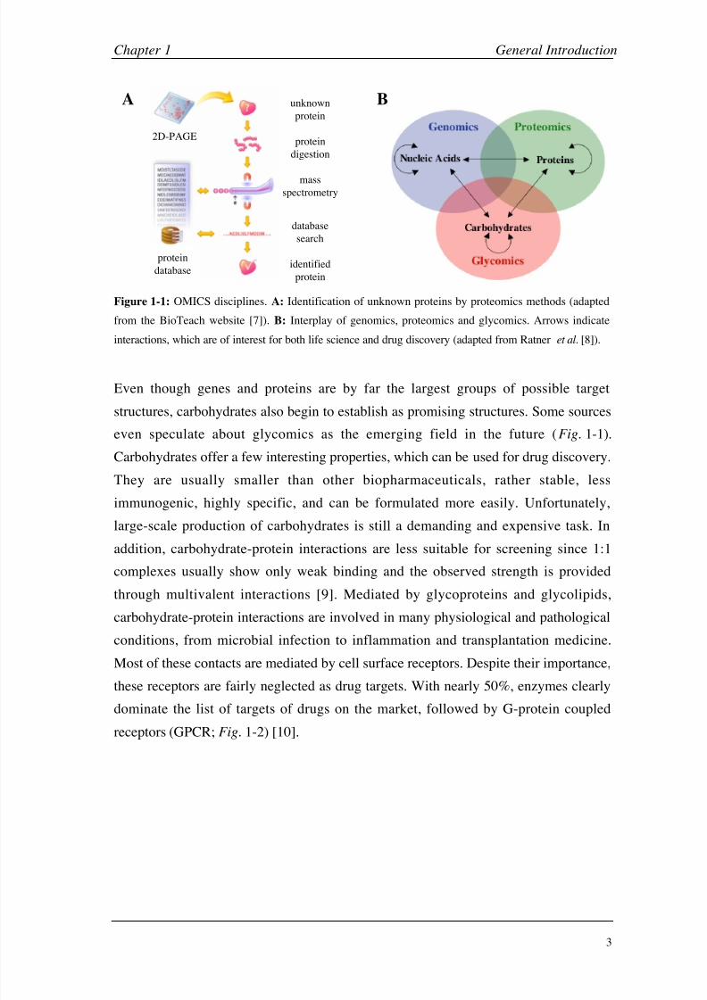
Figure 1-1: OMICS disciplines. A: Identification of unknown proteins by proteomics methods (adapted
from the BioTeach website [7]). B: Interplay of genomics, proteomics and glycomics. Arrows indicate
interactions, which are of interest for both life science and drug discovery (adapted from Ratner et al. [8]).
Even though genes and proteins are by far the largest groups of possible target
structures, carbohydrates also begin to establish as promising structures. Some sources
even speculate about glycomics as the emerging field in the future (Fig. 1-1).
Carbohydrates offer a few interesting properties, which can be used for drug discovery.
They are usually smaller than other biopharmaceuticals, rather stable, less
immunogenic, highly specific, and can be formulated more easily. Unfortunately,
large-scale production of carbohydrates is still a demanding and expensive task. In
addition, carbohydrate-protein interactions are less suitable for screening since 1:1
complexes usually show only weak binding and the observed strength is provided
through multivalent interactions [9]. Mediated by glycoproteins and glycolipids,
carbohydrate-protein interactions are involved in many physiological and pathological
conditions, from microbial infection to inflammation and transplantation medicine.
Most of these contacts are mediated by cell surface receptors. Despite their importance,
these receptors are fairly neglected as drug targets. With nearly 50%, enzymes clearly
dominate the list of targets of drugs on the market, followed by G-protein coupled
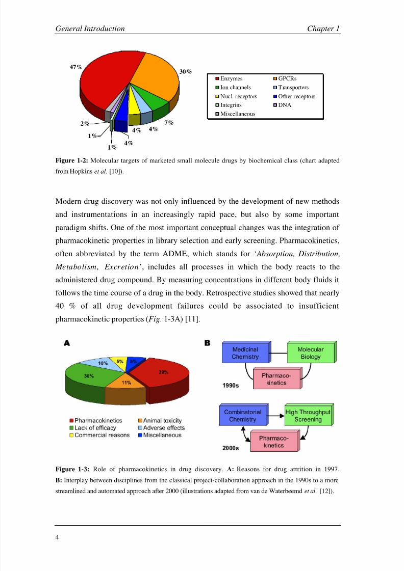
receptors (GPCR; Fig. 1-2) [10].
8/9/2019 DissB 7321 Mirroring
http://slidepdf.com/reader/full/dissb-7321-mirroring 18/278
General Introduction Chapter 1
4
4%
47%
7%
4%
30%
2%
1%
1% 4%
Enzymes GPCRs
Ion channels Transporters
Nucl. receptors Other receptors
Integrins DNA
Miscellaneous
Figure 1-2: Molecular targets of marketed small molecule drugs by biochemical class (chart adapted
from Hopkins et al. [10]).
Modern drug discovery was not only influenced by the development of new methods
and instrumentations in an increasingly rapid pace, but also by some important
paradigm shifts. One of the most important conceptual changes was the integration of
pharmacokinetic properties in library selection and early screening. Pharmacokinetics,
often abbreviated by the term ADME, which stands for ‘Absorption, Distribution,
Metabolism, Excretion’, includes all processes in which the body reacts to the
administered drug compound. By measuring concentrations in different body fluids it
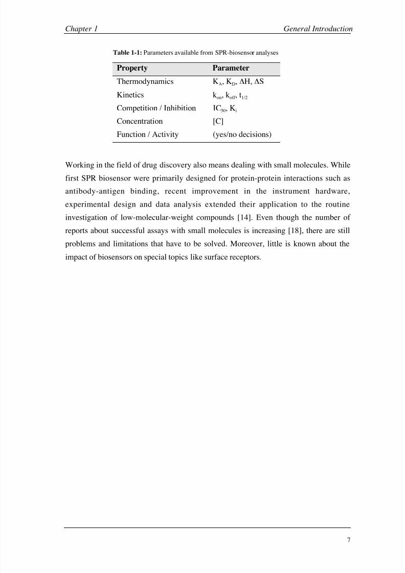
follows the time course of a drug in the body. Retrospective studies showed that nearly
40 % of all drug development failures could be associated to insufficient
pharmacokinetic properties (Fig. 1-3A) [11].
Figure 1-3: Role of pharmacokinetics in drug discovery. A: Reasons for drug attrition in 1997.
B: Interplay between disciplines from the classical project-collaboration approach in the 1990s to a more
streamlined and automated approach after 2000 (illustrations adapted from van de Waterbeemd et al. [12]).

8/9/2019 DissB 7321 Mirroring
http://slidepdf.com/reader/full/dissb-7321-mirroring 19/278
Chapter 1 General Introduction
5
In the past, the main effort in drug discovery was to optimize the efficacy and
specificity of a drug candidate. Problems concerning the pharmacokinetic properties of
the candidate drug were often detected at a later stage during pharmaceutical
development, in preclinical animal experiments or even during clinical studies. Failures
at this state, however, are very costly and have to be avoided. Therefore,
pharmacokinetic studies were integrated already in the discovery process, e.g. by
in
silico prediction of ADME properties during library design or early in
vitro
screening for undesired physico-chemical properties (Fig. 1-3B). These developments
inspired Van de Waterbeemd to redefine ADME as ‘Automated Decision-Making
Engine’ [12].
Traditionally, drug leads are optimized by just looking at their overall affinities or
activities for a target, i.e. their thermodynamic properties. These values just summarize
different entropic and enthalpic effects (e.g. hydrophobic or electrostatic interactions,
desolvatation, or induced fit). Similar affinities, however, can be the result of
completely different kinetic profiles (Fig. 1-4). Therefore, a much deeper insight into
drug-target interaction is available when the kinetic behavior of a compound is taken
into account, resulting in meaningful structure-kinetic relationships [13]. This opens
new possibilities for the development of drug candidates with tailor-made properties.
For example, an effective enzyme inhibitor should have a fast association rate constant
(rapid binding), but a very slow dissociation rate constant (sticking in the binding site).
101·10-31·102C
101·10-21·103B
105·10-15·104A
KD
[µM]
koff
[s-1]
kon
[M-1s-1]Curve
-50
0
100
200
300
400
0 100 200 300 400 500 600 700
Time [s]
S P R R
e s p o n s e [ R U ]
Association (kon
) Dissociation (koff
)
C
B
A
Equilibrium Constant (K D) =k off
k on
Figure 1-4: Simulated binding curves for a surface plasmon resonance (SPR) biosensor. Even though all
three curves (A, B, C) result in the same binding affinity (KD = 10 µM), the corresponding kinetic rate
constants (kon, koff ) might deviate significantly.

8/9/2019 DissB 7321 Mirroring
http://slidepdf.com/reader/full/dissb-7321-mirroring 20/278
General Introduction Chapter 1
6
1.2 The Need for Biosensors
The application of novel and efficient technologies is of high importance to the drug
discovery process, since they will lower development costs and decrease the time to
market [14]. Even though developments in the field of high-throughput screening and
computational chemistry greatly accelerated and facilitated the drug finding process,
there are significant limitations to overcome. An example are fluorescence-based HTS
assays, which may generate false positive (e.g. binding to the reporter enzyme [14] or
direct hydrophobic interaction of the label with the target [15]) or false negative results
(e.g. occluding of the binding site [15]). New technologies to confirm or refute
screening hits are therefore highly needed. Biosensors have attracted a great deal of
attention in this field in recent years.
The definition of the term biosensor is not very sharp and can be explained in different
ways. In principle, it is a device consisting of a biological part (e.g. DNA, protein, cell)
and a physical transducer (semiconductor, electrode, optical component). Biosensor
platforms are often miniaturized and work on small chips. First biosensor systems were
developed for clinical diagnostics and tailor-made for one specific target or assay. For
drug discovery, however, biosensors had to become much more flexible, allowing the
screening of a broad variety of compounds from different sources with a reasonable
throughput. Research and development in biosensors lead to many experimental or
commercial systems on different biological levels (cell, membranes, proteins) and
detection principles (electrochemical, optical) [16].
Surface plasmon resonance-based instruments are nowadays the most popular class of
biosensors. Their label-free detection, the real-time data acquisition possibilities, their
high degree of automation and throughput, as well as the ease of use made them to a
valuable tool in drug discovery. An extremely wide range of molecules can be
analyzed, from small drugs, DNA, peptides, or proteins up to virus particles or even
whole cells [17]. Compared to classical endpoint assays, which are mainly based on
competition or inhibition experiments, SPR sensors provide much more information
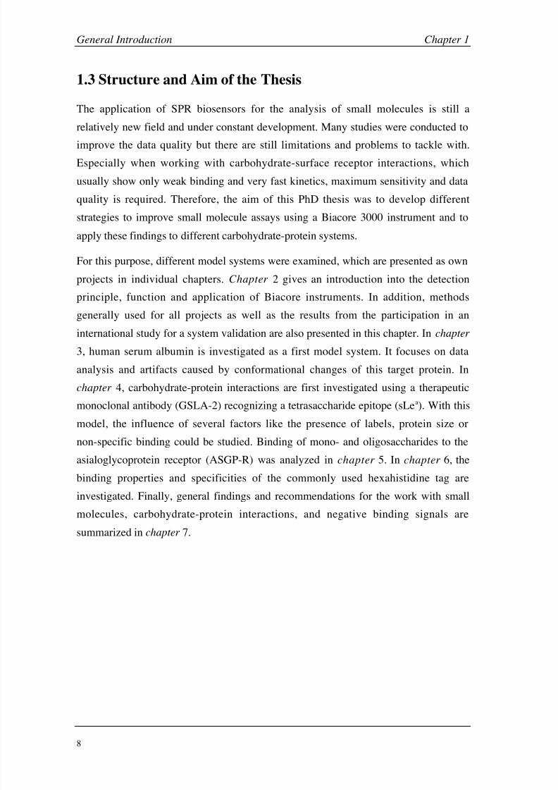
and properties simultaneously (Table 1-1).
8/9/2019 DissB 7321 Mirroring
http://slidepdf.com/reader/full/dissb-7321-mirroring 21/278
Chapter 1 General Introduction
7
Table 1-1: Parameters available from SPR-biosensor analyses
Property Parameter
Thermodynamics KA, KD, H, S
Kinetics kon, koff , t1/2
Competition / Inhibition IC50, Ki
Concentration [C]
Function / Activity (yes/no decisions)
Working in the field of drug discovery also means dealing with small molecules. While
first SPR biosensor were primarily designed for protein-protein interactions such as
antibody-antigen binding, recent improvement in the instrument hardware,
experimental design and data analysis extended their application to the routine
investigation of low-molecular-weight compounds [14]. Even though the number of
reports about successful assays with small molecules is increasing [18], there are still
problems and limitations that have to be solved. Moreover, little is known about the
impact of biosensors on special topics like surface receptors.
8/9/2019 DissB 7321 Mirroring
http://slidepdf.com/reader/full/dissb-7321-mirroring 22/278
General Introduction Chapter 1
8
1.3 Structure and Aim of the Thesis
The application of SPR biosensors for the analysis of small molecules is still a
relatively new field and under constant development. Many studies were conducted to
improve the data quality but there are still limitations and problems to tackle with.
Especially when working with carbohydrate-surface receptor interactions, which
usually show only weak binding and very fast kinetics, maximum sensitivity and data
quality is required. Therefore, the aim of this PhD thesis was to develop different
strategies to improve small molecule assays using a Biacore 3000 instrument and to
apply these findings to different carbohydrate-protein systems.
For this purpose, different model systems were examined, which are presented as own
projects in individual chapters. Chapter 2 gives an introduction into the detection
principle, function and application of Biacore instruments. In addition, methods
generally used for all projects as well as the results from the participation in an
international study for a system validation are also presented in this chapter. In chapter
3, human serum albumin is investigated as a first model system. It focuses on data
analysis and artifacts caused by conformational changes of this target protein. In
chapter 4, carbohydrate-protein interactions are first investigated using a therapeutic
monoclonal antibody (GSLA-2) recognizing a tetrasaccharide epitope (sLea). With this
model, the influence of several factors like the presence of labels, protein size or
non-specific binding could be studied. Binding of mono- and oligosaccharides to the
asialoglycoprotein receptor (ASGP-R) was analyzed in chapter 5. In chapter 6, the
binding properties and specificities of the commonly used hexahistidine tag are
investigated. Finally, general findings and recommendations for the work with small
molecules, carbohydrate-protein interactions, and negative binding signals are
summarized in chapter 7.
8/9/2019 DissB 7321 Mirroring
http://slidepdf.com/reader/full/dissb-7321-mirroring 23/278
Chapter 1 General Introduction
9
1.4 References
[1] J. Drews, Science 2000, 287 , 1960.
[2] A. Vedani, M. Dobler, J Med Chem 2002, 45, 2139.
[3] E. S. Lander, L. M. Linton, B. Birren, C. Nusbaum, M. C. Zody, J. Baldwin, K. Devon,
K. Dewar, M. Doyle, W. FitzHugh, R. Funke, D. Gage, K. Harris, A. Heaford, J.
Howland, L. Kann, J. Lehoczky, R. LeVine, P. McEwan, K. McKernan, J. Meldrim, J. P.
Mesirov, C. Miranda, W. Morris, J. Naylor, C. Raymond, M. Rosetti, R. Santos, A.
Sheridan, C. Sougnez, N. Stange-Thomann, N. Stojanovic, A. Subramanian, D. Wyman,
J. Rogers, J. Sulston, R. Ainscough, S. Beck, D. Bentley, J. Burton, C. Clee, N. Carter, A.
Coulson, R. Deadman, P. Deloukas, A. Dunham, I. Dunham, R. Durbin, L. French, D.
Grafham, S. Gregory, T. Hubbard, S. Humphray, A. Hunt, M. Jones, C. Lloyd, A.
McMurray, L. Matthews, S. Mercer, S. Milne, J. C. Mullikin, A. Mungall, R. Plumb, M.
Ross, R. Shownkeen, S. Sims, R. H. Waterston, et al., Nature 2001, 409, 860.
[4] J. C. Venter, M. D. Adams, E. W. Myers, P. W. Li, R. J. Mural, G. G. Sutton, H. O.
Smith, M. Yandell, C. A. Evans, R. A. Holt, J. D. Gocayne, P. Amanatides, R. M. Ballew,
D. H. Huson, J. R. Wortman, Q. Zhang, C. D. Kodira, X. H. Zheng, L. Chen, M. Skupski,
G. Subramanian, P. D. Thomas, J. Zhang, G. L. Gabor Miklos, C. Nelson, S. Broder, A.
G. Clark, J. Nadeau, V. A. McKusick, N. Zinder, A. J. Levine, R. J. Roberts, M. Simon,
C. Slayman, M. Hunkapiller, R. Bolanos, A. Delcher, I. Dew, D. Fasulo, M. Flanigan, L.
Florea, A. Halpern, S. Hannenhalli, S. Kravitz, S. Levy, C. Mobarry, K. Reinert, K.
Remington, J. Abu-Threideh, E. Beasley, K. Biddick, V. Bonazzi, R. Brandon, M.
Cargill, I. Chandramouliswaran, R. Charlab, et al., Science 2001, 291, 1304.
[5] Affymetrix Inc., Santa Clara, U.S.A. (www.affymetrix.com).
[6] E. F. Schmid, D. A. Smith, Drug Discov Today 2004, 9, 18.
[7] C. Antler, Investigating the cellular machinery: Protein identification, BioTech website
(http://www.bioteach.ubc.ca/Bioinformatics/InvestigatingTheCellularMachinery), 2005.
[8] D. M. Ratner, E. W. Adams, M. D. Disney, P. H. Seeberger, Chembiochem 2004, 5,
1375.
[9] Z. Shriver, S. Raguram, R. Sasisekharan, Nat Rev Drug Discov 2004, 3, 863.
[10] A. L. Hopkins, C. R. Groom, Nat Rev Drug Discov 2002, 1, 727.
[11] T. Kennedy, Drug Discovery Today 1997, 2, 436.
[12] H. van de Waterbeemd, E. Gifford, Nat Rev Drug Discov 2003, 2, 192.
[13] P. O. Markgren, W. Schaal, M. Hamalainen, A. Karlen, A. Hallberg, B. Samuelsson, U.
H. Danielson, J Med Chem 2002, 45, 5430.
[14] D. G. Myszka, R. L. Rich, Pharm. Sci. Technol. Today 2000, 3, 310.
[15] M. A. Cooper, Nat Rev Drug Discov 2002, 1, 515.
[16] M. Keusgen, Naturwissenschaften 2002, 89, 433.
[17] R. L. Rich, D. G. Myszka, Drug Discovery Today: Technologies 2004, 1, 301.
[18] R. L. Rich, D. G. Myszka, J Mol Recognit 2005, 18, 1.

8/9/2019 DissB 7321 Mirroring
http://slidepdf.com/reader/full/dissb-7321-mirroring 24/278
General Introduction Chapter 1
10

8/9/2019 DissB 7321 Mirroring
http://slidepdf.com/reader/full/dissb-7321-mirroring 25/278
Chapter 2
Biacore Technology

8/9/2019 DissB 7321 Mirroring
http://slidepdf.com/reader/full/dissb-7321-mirroring 26/278
Biacore Technology Chapter 2
12
2.1 Introduction
This chapter first gives an overview of the detection principle (surface plasmon
resonance, SPR) and the function of Biacore instruments. After an introduction in assay
design, immobilization techniques and data analysis, it covers the applications of
Biacore in the drug discovery and development process. Finally, alternative and
complementary biophysical methods are discussed and compared with the Biacore
technology.
2.1.1 Surface plasmon resonance and Biacore technology
Surface plasmon resonance is an electron charge density wave phenomenon first
observed as early as in the late 1950s [1]. But it took another ten years until its
mechanism and versatility was recognized. The first commercially available SPR
detection systems only appeared in the 1980s.
The underlying principles of this phenomenon are total internal reflection (TIR),
evanescence electric field (E), and surface plasmon waves. Total internal reflection
occurs when a light beam propagates through two non-absorbing media of different
refractive index (e.g. glass-air or glass-buffer). Above a critical incidence angle (),
the light beam is no longer refracted when it hits the interface of the two media, but is
fully reflected and propagates back into the source medium (Fig. 2-1A). Even though
the light beam keeps its net energy upon reflection, an electric field intensity called
evanescence wave (E) leaks into the other medium. This wave is exponentially
decreasing with distance from the interface (Fig. 2-1B). When the interface is coated
with a thin metal film the p-polarized component of the evanescence field penetrates
this layer and induces electromagnetic surface plasmon waves in the conducting metal.
Plasmons represent electron density fluctuations in a conducting metal and can be
regarded as the equivalent of photons in the case of light. A non-magnetic metal like
gold is normally used for these metal layers and the thickness has to be lower than the
wavelength of the incident light beam [2-4].
Since both photons and surface plasmons are a form of electromagnetic energy, they
can be fully described only by quantum physics. However, their properties can be
explained in a simplified manner as vector quantities. The light photon momentum at
the interface can be resolved into two vector components (parallel and perpendicular to
the interface). The magnitude of these incident light vectors (ilv) directly depends on
the light angle. The surface plasmon wave can be similarly described as a vector, which
8/9/2019 DissB 7321 Mirroring
http://slidepdf.com/reader/full/dissb-7321-mirroring 27/278
Chapter 2 Biacore Technology
13
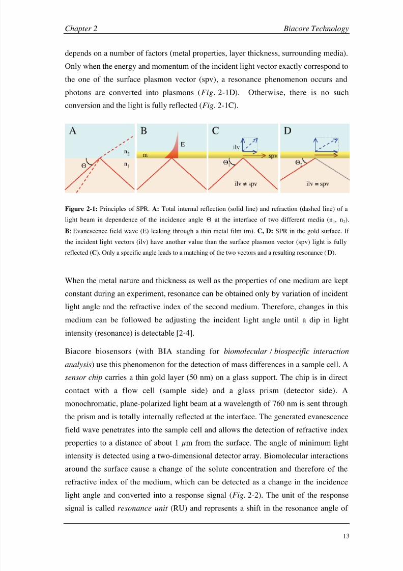
depends on a number of factors (metal properties, layer thickness, surrounding media).
Only when the energy and momentum of the incident light vector exactly correspond to
the one of the surface plasmon vector (spv), a resonance phenomenon occurs and
photons are converted into plasmons (Fig. 2-1D). Otherwise, there is no such
conversion and the light is fully reflected (Fig.
2-1C).
Figure 2-1: Principles of SPR. A: Total internal reflection (solid line) and refraction (dashed line) of a
light beam in dependence of the incidence angle at the interface of two different media (n1, n2).
B: Evanescence field wave (E) leaking through a thin metal film (m). C, D: SPR in the gold surface. If
the incident light vectors (ilv) have another value than the surface plasmon vector (spv) light is fully
reflected (C). Only a specific angle leads to a matching of the two vectors and a resulting resonance (D).
When the metal nature and thickness as well as the properties of one medium are kept
constant during an experiment, resonance can be obtained only by variation of incident
light angle and the refractive index of the second medium. Therefore, changes in this
medium can be followed be adjusting the incident light angle until a dip in light
intensity (resonance) is detectable [2-4].
Biacore biosensors (with BIA standing for biomolecular / biospecific interaction
analysis) use this phenomenon for the detection of mass differences in a sample cell. A
sensor chip carries a thin gold layer (50 nm) on a glass support. The chip is in direct
contact with a flow cell (sample side) and a glass prism (detector side). A
monochromatic, plane-polarized light beam at a wavelength of 760 nm is sent through
the prism and is totally internally reflected at the interface. The generated evanescence
field wave penetrates into the sample cell and allows the detection of refractive index
properties to a distance of about 1 µm from the surface. The angle of minimum light
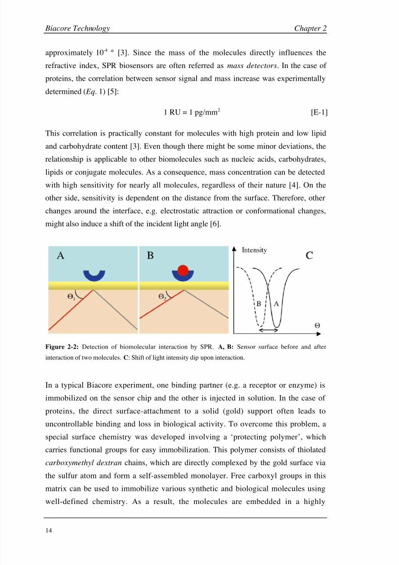
intensity is detected using a two-dimensional detector array. Biomolecular interactions
around the surface cause a change of the solute concentration and therefore of the
refractive index of the medium, which can be detected as a change in the incidence
light angle and converted into a response signal (Fig. 2-2). The unit of the response
signal is called resonance unit (RU) and represents a shift in the resonance angle of
8/9/2019 DissB 7321 Mirroring
http://slidepdf.com/reader/full/dissb-7321-mirroring 28/278
Biacore Technology Chapter 2
14
approximately 10-4 ° [3]. Since the mass of the molecules directly influences the
refractive index, SPR biosensors are often referred as mass detectors. In the case of
proteins, the correlation between sensor signal and mass increase was experimentally
determined ( Eq. 1) [5]:
1 RU = 1 pg/mm2 [E-1]
This correlation is practically constant for molecules with high protein and low lipid
and carbohydrate content [3]. Even though there might be some minor deviations, the
relationship is applicable to other biomolecules such as nucleic acids, carbohydrates,
lipids or conjugate molecules. As a consequence, mass concentration can be detected
with high sensitivity for nearly all molecules, regardless of their nature [4]. On the
other side, sensitivity is dependent on the distance from the surface. Therefore, other
changes around the interface, e.g. electrostatic attraction or conformational changes,
might also induce a shift of the incident light angle [6].
Figure 2-2: Detection of biomolecular interaction by SPR. A, B: Sensor surface before and after
interaction of two molecules. C: Shift of light intensity dip upon interaction.
In a typical Biacore experiment, one binding partner (e.g. a receptor or enzyme) is
immobilized on the sensor chip and the other is injected in solution. In the case of
proteins, the direct surface-attachment to a solid (gold) support often leads to
uncontrollable binding and loss in biological activity. To overcome this problem, a
special surface chemistry was developed involving a ‘protecting polymer’, which
carries functional groups for easy immobilization. This polymer consists of thiolated
carboxymethyl dextran chains, which are directly complexed by the gold surface via
the sulfur atom and form a self-assembled monolayer. Free carboxyl groups in this
matrix can be used to immobilize various synthetic and biological molecules using
well-defined chemistry. As a result, the molecules are embedded in a highly

8/9/2019 DissB 7321 Mirroring
http://slidepdf.com/reader/full/dissb-7321-mirroring 29/278
Chapter 2 Biacore Technology
15
hydrophilic hydrogel and are kept in a quasi-solvent environment [7]. Electrostatic
effects caused by remaining carboxyl groups can be suppressed in most cases by
adding salts (e.g. 150 mM NaCl) to the running buffer [3]. A schematic overview of the
experimental setup is visualized in figure 2-3.
buffer flow
light
source
detector
array
glass
prism
chip
matrix
target
analyte
gold layer
Figure 2-3: Experimental setup of Biacore instruments. A target molecule is
immobilized on a gold-coated sensor chip via a hydrogel matrix. Binding of analyte
molecules in solution is detected by SPR phenomenon.
Another problem that had to be addressed was the sample delivery system. In stationary
systems, mass transport of molecules to the surface is governed by diffusion and
convection processes. To ensure reliable results, incubation times of several hours were
necessary in this case, which is not suitable for real-time systems. A flow system in a
micro-flow cell offers a continuous transport of sample to and from the surface,
therefore minimizing diffusion and convection effects. Developments in
miniaturization led to an integrated fluidic cartridge (IFC; Fig. 2-5C), which further
reduced sample consumption and sample plug dispersion after injection [3].
The shift in resonance angle can be monitored in real-time and plotted in dependence of
time. From such a signal vs. time plot, called sensorgram, the different stages of a
binding event can be visualized and evaluated (Fig. 2-4). With only buffer running
through the flow system, the signal forms a stable baseline. Upon injection of the
analyte solution, the sensorgram is dominated by the association phase, where analyte

8/9/2019 DissB 7321 Mirroring
http://slidepdf.com/reader/full/dissb-7321-mirroring 30/278
Biacore Technology Chapter 2
16
molecules bind to the target on the chip. However, bound molecules already start
dissociating again during injection. After a certain injection time, a steady state is
reached, where binding and dissociating molecules are in equilibrium. As soon as the
injection is stopped, running buffer replaces the analyte cloud and only the pure
dissociation phase is visible. Some assays require an additional regeneration step to
reach the baseline again (Fig. 2-4).
b br
d
ss
a
injectionbuffer buffer bufferr
Figure 2-4: Schematic representation of a sensorgram as the time course of a
binding event. Baseline signal (b), association (a), steady state (ss), dissociation
(d), and regeneration (r) of tightly bound molecules.
From the shape of the binding curve kinetic parameters like the association and
dissociation rate constants (kon, koff ) can be fitted and calculated. The equilibrium
dissociation constant (KD) can be directly calculated from the kinetic rate constants
using equation 2 or independently determined from the steady state signals at different
concentrations. This steady state affinity can be calculated using equation 3, where Req
is the equilibrium response signal, KA is the equilibrium association constant ( Eq. 2), C
the concentration, Rmax the maximum possible response, and n a steric interference
factor.
K D =
k off
k on
[M] and K A =
k on
k off [M-1| [Eq. 2]
Req =
K A C Rmax
1+K A C n
[RU] [Eq. 3]
2.1.2 Biacore 3000
Biacore AB [8] introduced the first SPR biosensor in 1990 [9, 10]. It was primarily
designed for the analysis of protein-protein interactions (e.g. antibody-antigen) and had
8/9/2019 DissB 7321 Mirroring
http://slidepdf.com/reader/full/dissb-7321-mirroring 31/278
Chapter 2 Biacore Technology
17
limitations both in regard of sensitivity and automation. With the introduction of
Biacore 2000 in 1994 these problems were addressed and it was even possible to
investigate small molecules (< 500 Da). Another improvement of sensitivity was
realized with Biacore 3000 in 1998. Thanks to its flexibility and the ease of automation it
soon gained interest both in pharmaceutical industry and academic laboratories [11, 12].
Auto-
sampler
Pumps
Buffer CompartementChip Lock
Optical
unit
Integrated
Fluidic
Cartridge
(IFC)
A B C
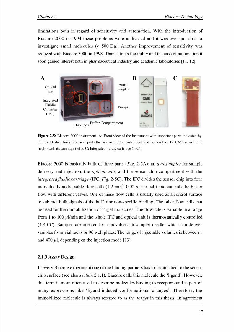
Figure 2-5: Biacore 3000 instrument. A: Front view of the instrument with important parts indicated by
circles. Dashed lines represent parts that are inside the instrument and not visible. B: CM5 sensor chip
(right) with its cartridge (left). C: Integrated fluidic cartridge (IFC).
Biacore 3000 is basically built of three parts (Fig. 2-5A); an autosampler for sample
delivery and injection, the optical unit , and the sensor chip compartment with the
integrated fluidic cartridge (IFC; Fig. 2-5C). The IFC divides the sensor chip into four
individually addressable flow cells (1.2 mm2, 0.02 µl per cell) and controls the buffer
flow with different valves. One of these flow cells is usually used as a control surface
to subtract bulk signals of the buffer or non-specific binding. The other flow cells can
be used for the immobilization of target molecules. The flow rate is variable in a range
from 1 to 100 µl/min and the whole IFC and optical unit is thermostatically controlled
(4-40°C). Samples are injected by a movable autosampler needle, which can deliver
samples from vial racks or 96 well plates. The range of injectable volumes is between 1
and 400 µl, depending on the injection mode [13].
2.1.3 Assay Design
In every Biacore experiment one of the binding partners has to be attached to the sensor
chip surface (see also section 2.1.1). Biacore calls this molecule the ‘ligand’. However,
this term is more often used to describe molecules binding to receptors and is part of
many expressions like ‘ligand-induced conformational changes’. Therefore, theimmobilized molecule is always referred to as the target in this thesis. In agreement

8/9/2019 DissB 7321 Mirroring
http://slidepdf.com/reader/full/dissb-7321-mirroring 32/278
Biacore Technology Chapter 2
18
with the Biacore nomenclature, the interacting molecule in solution is called analyte
(Fig. 2-6A). The expression ‘surface’ and derived terms like ‘surface density’ are
normally referred to the sensor chip with the immobilized target.
matrix
sensor chip
target
analyteA B C D
TDC
Figure 2-6: Comparison of different assay types. Direct binding assays fitting to a single-site model (A)
or a two independent-sites model (B). Surface competition assay (C) and inhibition in solution assay (D),
in which the analyte competes with a ‘target definition compound’ (TDC) for the same binding site.
The first strategic decision to make is which of the binding partners is immobilized. For
most of the systems, however, there is no real choice since multiple analytes will be
screened against a single target molecule. In order to get maximum signal responses,
systems with an immobilized small molecule and a bigger analyte (e.g. protein) in
solution are preferred. The maximum response for a SPR signal can be estimated using
equation 4.
Rmax
=
MW analyte
MW t arg et density t arg et valency [Eq. 4]
Unfortunately, for the majority of drug discovery applications the large molecule
(receptor, enzyme, etc.) will be the target and small molecules (MW < 500 Da) are used
for screening. This often leads to very small signals around the detection limit,
especially when the coupling results in a low density or a reduced activity of the target.
In addition, immobilization of the small molecule might change the binding eventdramatically, since multivalency or rebinding effects are often observed [14].
Second, several assay formats can be performed, of which the direct binding assay is
by far the most popular (Fig. 2-6A&B). However, competition assay formats might be
preferable for different reasons (e.g. small analyte size). In the surface competition
assay (Fig. 2-6C) the analyte is mixed with a constant concentration of a target
definition compound (TDC), which is normally a tight inhibitor. The TDC should form
a complex with a half-life of more than 20 s and should be at least 5 to 10 times larger
than the compounds to be screened. Changes of the overall binding response are then

8/9/2019 DissB 7321 Mirroring
http://slidepdf.com/reader/full/dissb-7321-mirroring 33/278
Chapter 2 Biacore Technology
19
evaluated and compared with the signal of the TDC alone. Both the direct and the
competition assay are sensitive to non-specific binding. A third format is the inhibition
in solution assay (Fig. 2-6D), where the TDC is immobilized and the analyte solutions
are mixed with a constant concentration of the target. The signal reflects the
concentration of free target and is therefore site-related - only analytes that interact
directly with the binding site inhibit the interaction. On the other hand, much higher
amounts of the target molecule are needed for this assay, which is often a problem in
the case of proteins. While all three formats are suitable for ranking experiments only
the direct assay can provide high-quality equilibrium and kinetic data [15].
2.1.4 Immobilization
Target immobilization is one of the most important and crucial steps in a Biacore
binding assay. Loss of target activity and many artifacts are directly related to
unfavorable coupling procedures. The unique properties of the hydrogel matrix used for
Biacore experiments (CM5 chip; Fig. 2-5B) offers many alternative strategies for
covalent immobilization of proteins, oligosaccharides, nucleotides, or small molecules
(Fig. 2-7).
While covalent coupling approaches usually generate stable surfaces with high density,
capturing techniques have the advantage of being fully regenerable and allow
immobilization from (crude) protein mixtures. These two general approaches also show
different results in respect of target orientation. They also lead to oriented and therefore
highly active surfaces but often show a lower density and stability (surface bleeding).

8/9/2019 DissB 7321 Mirroring
http://slidepdf.com/reader/full/dissb-7321-mirroring 34/278
Biacore Technology Chapter 2
20
sensor chip
matrix
OH
O
NHS/EDC Target
O
O
N
O
O
NH
O
NH
O
NH2
Aldehyde Coupling
(Targets with aldehydes/sugars)
Maleimid Coupling
(targets with free thiol; non-reducable)
Surface Thiol Coupling(Targets with acitvated thiol)
Ligand Thiol Coupling(Targets with free thiol)
NH
O
HN
O
N
O
O
NH
O
SHNH
O
SS N
Amine Coupling
(Targets with
primary amine)
Cystamine
DTTPDEA
EMCHHydrazine
Figure 2-7: Coupling methods for Biacore sensor chip CM5. After activation of the matrix-based
carboxyl groups by NHS and EDC, targets can be directly immobilized via primary amine groups or the
surface can be functionalized for alternative strategies (NHS = N -hydroxysuccinimide,, EDC = 1-ethyl-
3-(3-dimethylaminopropyl)-carbodiimide, PDEA = 2-(2-pyridinyldithio)ethaneamine), EMCH = N -(-
maleimidocaproic acid)-hydrazide,
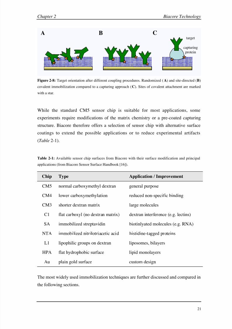
Covalent immobilization often attacks multiple attachment sites and therefore leads to
randomized coupling, which often results in a loss of activity (due to direct
modification of residues in the binding site, steric hindrance or conformational
changes) and surface heterogeneity (Fig. 2-8A). However, if a specific functional group
is available at a defined location of the target, site-specific coupling might allow the
generation of an oriented, homogeneous surface (Fig. 2-8B). Finally, capturing
approaches (Fig. 2-8C) make use of specific biomolecular interactions between the
target and an immobilized capturing protein.
8/9/2019 DissB 7321 Mirroring
http://slidepdf.com/reader/full/dissb-7321-mirroring 35/278
Chapter 2 Biacore Technology
21
A B C
capturing
protein
target
Figure 2-8: Target orientation after different coupling procedures. Randomized (A) and site-directed (B)
covalent immobilization compared to a capturing approach (C). Sites of covalent attachment are marked
with a star.
While the standard CM5 sensor chip is suitable for most applications, some
experiments require modifications of the matrix chemistry or a pre-coated capturing
structure. Biacore therefore offers a selection of sensor chip with alternative surface
coatings to extend the possible applications or to reduce experimental artifacts
(Table 2-1).
Table 2-1: Available sensor chip surfaces from Biacore with their surface modification and principal
applications (from Biacore Sensor Surface Handbook [16]).
Chip Type Application / Improvement
CM5 normal carboxymethyl dextran general purpose
CM4 lower carboxymethylation reduced non-specific binding
CM3 shorter dextran matrix large molecules
C1 flat carboxyl (no dextran matrix) dextran interference (e.g. lectins)
SA immobilized streptavidin biotinlyated molecules (e.g. RNA)
NTA immobilized nitrilotriacetic acid histidine-tagged proteins
L1 lipophilic groups on dextran liposomes, bilayers
HPA flat hydrophobic surface lipid monolayers
Au plain gold surface custom design
The most widely used immobilization techniques are further discussed and compared in
the following sections.

8/9/2019 DissB 7321 Mirroring
http://slidepdf.com/reader/full/dissb-7321-mirroring 36/278
Biacore Technology Chapter 2
22
Amine Coupling
The amine coupling procedure makes use of the primary amine groups on the protein
surface (lysine residues and the N -terminus), which directly react with active esters
generated by NHS/EDC activation (Fig.
2-9). In order to reach the highest efficiency of the reaction, proteins have to be pre-concentrated on the sensor-chip surface. This
surface attraction is reached by lowering the pH of the immobilization buffer just
below the pI value of the protein, where amine groups are positively charged and get
attracted by the negatively charged carboxyl group of the matrix. However, since the
reaction only takes place with uncharged amines, the immobilization pH should not be
too low [17].
A B
H2N
HN
NH
Protein
R
O
O
NH2
-amine
(N-terminus)
-amine
(lysine side chain)
O
NH
Figure 2-9: Amine coupling: Surface chemistry (A) and targeted amine groups at the N -terminus and the
lysine side chain (B).
The major advantage of amine coupling lies in its universality, stability and speed.
Nearly all proteins and peptides possess multiple primary amine groups ( N-terminus
and lysine residues), which are often surface-exposed due to their hydrophilicity. On
the other hand, since targetable lysine amines often are randomly distributed over the
protein surface, amine coupling leads to a random and non-predictable immobilization
of the molecule (Fig.
2-8A). This is especially problematic in the case of surface
receptors since their binding sites are directly accessible to the solvent and charged
residues like lysine are often involved in ligand binding. Therefore, amine coupling
sometimes leads to a massive decrease of surface activity, e.g. more than 80% loss is
reported for some antibodies, and might also influence binding affinity and kinetics
[18]. Different strategies have therefore been developed to overcome this problem. For
example, differences in the reactivity of the -amino group of the N-terminus and
-amino groups of the lysine side chain (Fig. 2-9B) were used for a site-specific
PEGylation of a somatostatin-analogue peptide at different pH values [19]. Other

8/9/2019 DissB 7321 Mirroring
http://slidepdf.com/reader/full/dissb-7321-mirroring 37/278
Chapter 2 Biacore Technology
23
groups tried to reversibly protect reactive lysines with 2,3-dimethylmaleic anhydride
[20, 21] or masking binding site amines by immobilizing the protein in the presence of
a known binder [22]. However, none of these approaches seem to be generally
applicable and neither method has been used for Biacore assays. Another drawback of
amine coupling is the requirement of acidic conditions for surface attraction. Some
proteins are not stable in the immobilization buffers required for amine coupling and
only inactive protein is therefore immobilized. Acidic proteins with pI values below 3.5
can hardly be immobilized via amine coupling. Finally, popular buffers and reagents
bearing primary amines like Tris cannot be used due to competition with the amino
groups of the protein. Sodium azide, which is frequently used as a preservative in
protein preparations, also might interfere with amine coupling and should therefore be
removed [23].
Thiol coupling
Immobilization of thiol-bearing targets can be performed either by formation of
disulfide bridges or by covalent reactions with maleimides. Since free thiol groups are
very rare compared to primary amines, these approaches often lead to a site-directed
and therefore oriented immobilization of the target. Disulfide bridge formation offers
the additional advantage that such bonds can be reduced leading to a fully regenerable
chip surface. However, since the spontaneous formation of disulfides is
thermodynamically not favored and takes very long, activation of the thiol group either
on the chip surface or in the target is needed (Fig. 2-10A).
A
O
HN
SH
O
HN
SS N
S
N
SH
O
HN
S
S
B
O
HNNH
O
NO O
S
or
Figure 2-10: Immobilization methods for thiol-bearing targets: surface and ligand thiol coupling (A) and
maleimide coupling (B).
8/9/2019 DissB 7321 Mirroring
http://slidepdf.com/reader/full/dissb-7321-mirroring 38/278
Biacore Technology Chapter 2
24
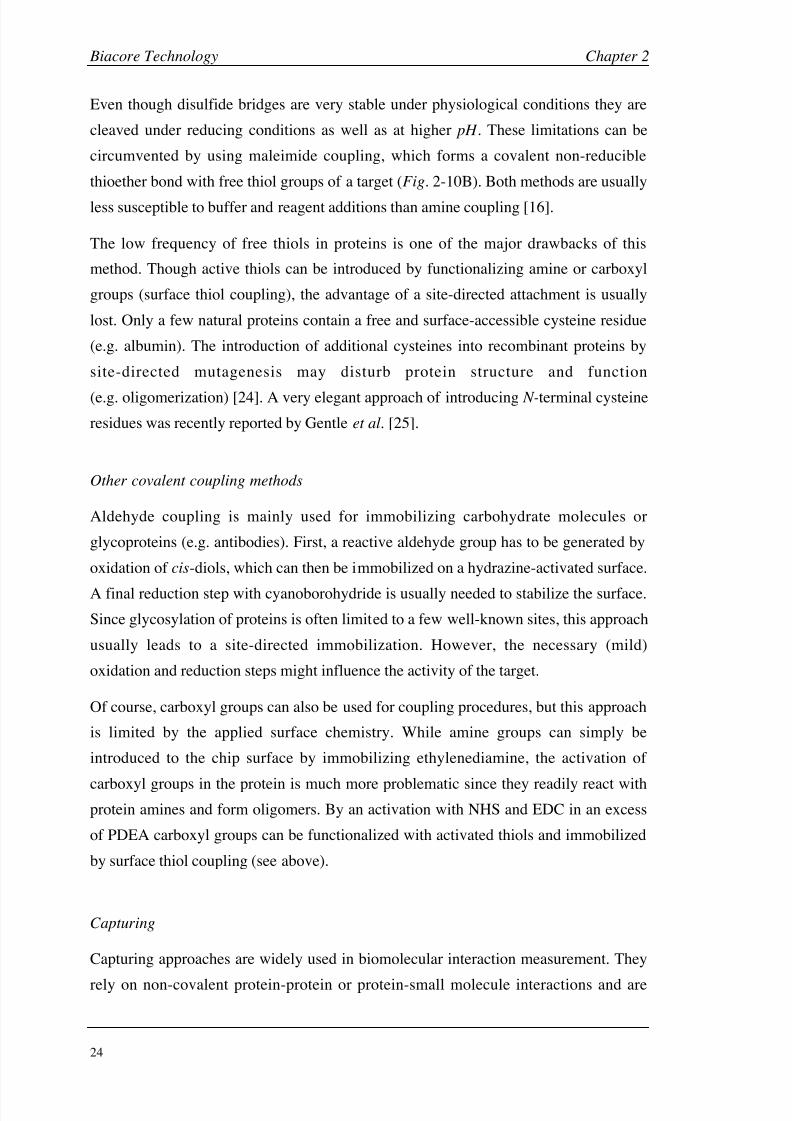
Even though disulfide bridges are very stable under physiological conditions they are
cleaved under reducing conditions as well as at higher pH . These limitations can be
circumvented by using maleimide coupling, which forms a covalent non-reducible
thioether bond with free thiol groups of a target (Fig. 2-10B). Both methods are usually
less susceptible to buffer and reagent additions than amine coupling [16].
The low frequency of free thiols in proteins is one of the major drawbacks of this
method. Though active thiols can be introduced by functionalizing amine or carboxyl
groups (surface thiol coupling), the advantage of a site-directed attachment is usually
lost. Only a few natural proteins contain a free and surface-accessible cysteine residue
(e.g. albumin). The introduction of additional cysteines into recombinant proteins by
site-directed mutagenesis may disturb protein structure and function
(e.g.
oligomerization) [24]. A very elegant approach of introducing N-terminal cysteine
residues was recently reported by Gentle et al. [25].
Other covalent coupling methods
Aldehyde coupling is mainly used for immobilizing carbohydrate molecules or
glycoproteins (e.g. antibodies). First, a reactive aldehyde group has to be generated by
oxidation of cis-diols, which can then be immobilized on a hydrazine-activated surface.
A final reduction step with cyanoborohydride is usually needed to stabilize the surface.
Since glycosylation of proteins is often limited to a few well-known sites, this approach
usually leads to a site-directed immobilization. However, the necessary (mild)
oxidation and reduction steps might influence the activity of the target.
Of course, carboxyl groups can also be used for coupling procedures, but this approach
is limited by the applied surface chemistry. While amine groups can simply be
introduced to the chip surface by immobilizing ethylenediamine, the activation of
carboxyl groups in the protein is much more problematic since they readily react withprotein amines and form oligomers. By an activation with NHS and EDC in an excess
of PDEA carboxyl groups can be functionalized with activated thiols and immobilized
by surface thiol coupling (see above).
Capturing
Capturing approaches are widely used in biomolecular interaction measurement. They
rely on non-covalent protein-protein or protein-small molecule interactions and are

8/9/2019 DissB 7321 Mirroring
http://slidepdf.com/reader/full/dissb-7321-mirroring 39/278
Chapter 2 Biacore Technology
25
especially suitable for experiments where both target and analyte have to be screened
simultaneously. In addition, capturing often serves as an easy way for oriented
coupling, since binding occurs at a well defined site of the target. Three major coupling
classes can be defined: antibody-antigen systems, interactions between proteins and
naturally occurring sites (e.g. Protein A/IgG) and capturing of artificially introduced
affinity tags (e.g. biotin or hexahistidine).
Antibody-antigen systems offer many advantages over other capturing approaches.
Interactions show normally high affinities (nanomolar range) and specificity. However,
production of antibodies against a new target can be very time and cost consuming and
care has to be taken to avoid overlaps between antibody and analyte binding sites.
Therefore, antibody systems used for Biacore analysis are often directed against
well-known antigens like tags or conserved domains of protein families.
Affinity tags are short peptide sequences or whole protein domains, which show high
affinity to a specific target structure. This could be another protein, a small molecule or
a metal ion. Tags are an established method in protein expression and purification, and
plasmids for the production of fusion proteins are readily available. Expressed tags can
be used for purification (affinity chromatography) as well as for immobilization on a
sensor chip. However, not every expression system tolerates a newly introduced
domain and special elution conditions might have to be applied during purification.This might lead to reduced yield or decreased activity of the proteins. An overview of
several important tag systems can be found in table 2-2 and in Terpe [26].
Table 2-2: Popular tag systems used for target capturing in Biacore experiments and other assays.
Tag Name Residues Captured by KD [nM] Ref a
His-tag 6(-10) Ni2+-NTA, anti-His5/6 nM-µMb [27]
Strep-tag II 8 Streptactin n.d. [28]
FLAG 8 Anti-FLAG mAb 412 [29]
SBP (streptavidin binding protein) 38 Streptavidin 2.5 [30]
Z/ZZ-domain (Protein A) 53/123 Human IgG1 (Fc) 17/1.5 [31]
Glutathione S-Transferase 211 Glutathione, anti-GST n.d. [32]
a All references and KD values refer to SPR experiments, except SBP (spin-filter binding inhibition assay).
b The isolated hexahistidine peptide was shown to bind differently to Ni-NTA than tagged proteins. At
least two His-tags were found to be necessary for a stable binding on a Biacore NTA-chip.
8/9/2019 DissB 7321 Mirroring
http://slidepdf.com/reader/full/dissb-7321-mirroring 40/278
Biacore Technology Chapter 2
26
A common disadvantage of all capturing approaches is their non-covalent character.
While mid-range affinities and non-physiological buffer conditions might be tolerable
for purification purposes, this might be a problem for the creation of stable sensor
surfaces. Captured surfaces often show a certain degree of bleeding (surface decay) and
finding selective regeneration conditions can be very difficult.
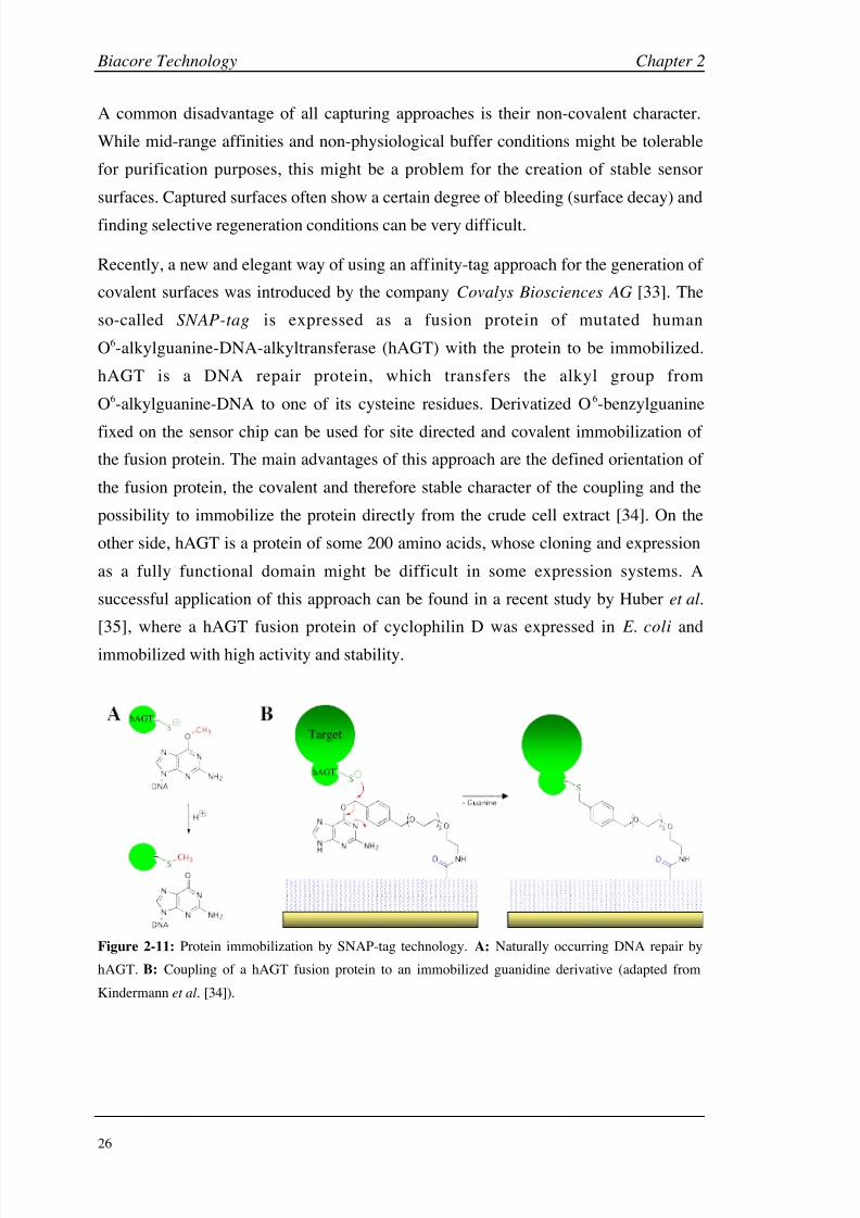
Recently, a new and elegant way of using an affinity-tag approach for the generation of
covalent surfaces was introduced by the company Covalys Biosciences AG [33]. The
so-called SNAP-tag is expressed as a fusion protein of mutated human
O6-alkylguanine-DNA-alkyltransferase (hAGT) with the protein to be immobilized.
hAGT is a DNA repair protein, which transfers the alkyl group from
O6-alkylguanine-DNA to one of its cysteine residues. Derivatized O6-benzylguanine
fixed on the sensor chip can be used for site directed and covalent immobilization of
the fusion protein. The main advantages of this approach are the defined orientation of
the fusion protein, the covalent and therefore stable character of the coupling and the
possibility to immobilize the protein directly from the crude cell extract [34]. On the
other side, hAGT is a protein of some 200 amino acids, whose cloning and expression
as a fully functional domain might be difficult in some expression systems. A
successful application of this approach can be found in a recent study by Huber et al.
[35], where a hAGT fusion protein of cyclophilin D was expressed in E. coli and
immobilized with high activity and stability.
Figure 2-11: Protein immobilization by SNAP-tag technology. A: Naturally occurring DNA repair by
hAGT. B: Coupling of a hAGT fusion protein to an immobilized guanidine derivative (adapted from
Kindermann et al. [34]).

8/9/2019 DissB 7321 Mirroring
http://slidepdf.com/reader/full/dissb-7321-mirroring 41/278
Chapter 2 Biacore Technology
27
2.1.5 Assay conditions
Biacore 3000 accepts a wide range of conditions and variation of parameters. On the
other hand, most of the experiment are conducted under near-physiological conditions
using water-based buffer systems and temperatures between 20 and 37°C. Buffers usedfor Biacore experiments are normally amine-free (to avoid conflicts in amine-coupling)
and contain a certain amount of salt for suppressing electrostatic effects on the
carboxylated matrix [3] (e.g. 10 mM HEPES or phosphate buffer at pH 7.4 with
150 mM NaCl). Reagents such as EDTA or polysorbate are often added to reduce
non-specific binding, but only after possible interferences with the binding experiment
have been excluded.
One of the unique features of the Biacore technology is its flow system. This ensures a
fast delivery of the sample to and from the surface. Variations of flow rate are suitable
for the detection of any mass transport effects. This phenomenon might occur when the
interaction between analyte and target is comparable or faster than the diffusion of
analyte from bulk solution to the surface. Mass transport is dependent on the flow rate,
cell dimensions and diffusion coefficient of the analyte [36, 37]. High flow rates
(50-100 µl/min) and a low surface densities are therefore recommended for the
reduction of these effect and highest data quality. However, the flow rate is often
limited by the sample consumption or the required injection time. Experimental serieswith variation of surface density and flow rate could therefore be helpful for the
detection of such effects and for finding a suitable compromise between sample
consumption, signal intensity and mass transport [38].
In order to clean all parts of the injection system and to equilibrate the surface, a series
of buffer blanks should be injected before each experiment [39]. Injection modes
especially designed for highest volume accuracy and high-resolution dissociation
phases (kinject command) should always be used for sample injections during analytescreening. Injections of buffer blanks before and within binding experiment, inclusion
of positive and negative controls, washing steps, as well as a proper maintenance
further increase the accuracy and quality of the binding data [13]. Sample injection
should be done randomized and in replicates to eliminate the total experimental noise.
Regeneration is one of the most critical parts of a binding assay, especially when
dealing with proteins. Too soft conditions lead to remaining analyte and a possible
carry-over effect, while too harsh conditions might denature the protein. Specific
methods like the removal of calcium ion in the case of C-type lectins are always
8/9/2019 DissB 7321 Mirroring
http://slidepdf.com/reader/full/dissb-7321-mirroring 42/278
Biacore Technology Chapter 2
28
preferred to unspecific approaches (acidic, basic or chaotropic conditions, detergents
and high salt concentrations). Sometimes a cocktail of different regeneration
compounds is needed and approaches to find a suitable combination are described in
literature [40]. To avoid any carry-over of the regeneration solution a buffer blank
injection should be performed at the end of each cycle [39].
2.1.6 Data analysis
Although generating Biacore data is fairly easy, the accurate interpretation of the
equilibrium, kinetic and thermodynamic data has proven to be more difficult.
Deviations from an expected binding model do not always represent a more complex
interaction but are often caused by experimental design.
Data processing should be done in an accurate and reproducible way in order to remove
matrix and bulk effects of the binding signals. This is especially necessary when
working with small molecules, since even small changes of the signal might lead to
variations in the binding constants. Therefore, advanced processing steps like blank
subtraction (double referencing) should be performed to remove even minor
experimental errors [39]. If no literature data are available about an interaction, data
should be fitted to a simple 1:1 binding model first ( Eq. 5). Since some targets posses
more than one binding site, the equation has to be extended to a two independentbinding site model. If mass transport effects (see section 2.1.5) are suspected or
reported, a mass transport coefficient (km) might be introduced ( Eq. 6).
A + B ABkon
koff [Eq. 5]
A0 A + B AB
km
km
kon
koff [Eq. 6]
Unfortunately, using the sum of two or more equations or increasing the number of
parameters will almost always lead to a better fit, regardless of the underlying binding
mechanism [38]. Careful validation with additional experimental or literature data is
therefore recommended before relying on a new binding model. Additional models for
surface heterogeneity or a drifting baseline are available in the evaluation software.
Even though a better fitting might represent a real effect on the surface, more time

8/9/2019 DissB 7321 Mirroring
http://slidepdf.com/reader/full/dissb-7321-mirroring 43/278
Chapter 2 Biacore Technology
29
should be invested to avoid such drifts or heterogeneities by changing the experimental
setup.
A proper data processing is especially important for fitting kinetic data. Initially,
different algorithms using curve transformation [41] or nonlinear last squares analysis[42] were used for the evaluation of the binding kinetics. However, these methods only
fitted single binding curves (or even portions thereof) and were found to be often
insufficient to discriminate between different binding mechanisms [38]. In the global
analysis approach, the association and dissociation phases of the entire data set are
fitted to a model simultaneously, resulting in very accurate and robust data [43].
Therefore, this method is implemented in the current evaluation software tools and
should always be used for kinetic fits.
2.1.7 Applications of Biacore in drug discovery
The analysis of molecular interactions is a key part of the drug discovery process.
Though the scientific community and pharmaceutical industry first hesitated to accept
SPR-based interaction studies [12], Biacore instruments and similar biosensors were
validated as an important biophysical method and are now well integrated. Currently,
these instruments are used in nearly every aspect of the drug discovery process, from
target identification, compound screening and lead optimization to supporting clinical
trials, regulatory approval and biopharmaceutical manufacturing (Fig. 2-12).
Lipid Absorption
Plasma Protein Binding
(Cytochrome Interaction)
Lipid Absorption
Plasma Protein Binding
(Cytochrome Interaction)
Concentration analysis
Quality control
Biopharm. production
Regulatory approval
Concentration analysis
Quality control
Biopharm. production
Regulatory approval
High-resolution screening
Structure-activity relationship
Kinetic analysis (SKR)
early ADMET
High-resolution screening
Structure-activity relationship
Kinetic analysis (SKR)
early ADMET
Affinity Ranking
Screening (HTS)
Secondary Screening
Affinity Ranking
Screening (HTS)
Secondary Screening
ID / Characterization
Target validation
Assay development
Ligand fishing
ID / Characterization
Target validation
Assay development
Ligand fishing
Target Hit Lead Candidate Drug
Figure 2-12: Application examples for Biacore 3000 and other SPR biosensor platforms in drug
discovery. (ID = identification, ADMET = pharmacokinetics and toxicology, HTS = high throughput
screening, SKR = structure-kinetic relationship).

8/9/2019 DissB 7321 Mirroring
http://slidepdf.com/reader/full/dissb-7321-mirroring 44/278
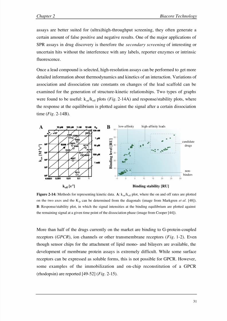
8/9/2019 DissB 7321 Mirroring
http://slidepdf.com/reader/full/dissb-7321-mirroring 45/278
Chapter 2 Biacore Technology
31
assays are better suited for (ultra)high-throughput screening, they often generate a
certain amount of false positive and negative results. One of the major applications of
SPR assays in drug discovery is therefore the secondary screening of interesting or
uncertain hits without the interference with any labels, reporter enzymes or intrinsic
fluorescence.
Once a lead compound is selected, high-resolution assays can be performed to get more
detailed information about thermodynamics and kinetics of an interaction. Variations of
association and dissociation rate constants on changes of the lead scaffold can be
examined for the generation of structure-kinetic relationships. Two types of graphs
were found to be useful: kon/koff plots (Fig. 2-14A) and response/stability plots, where
the response at the equilibrium is plotted against the signal after a certain dissociation
time (Fig.
2-14B).
k o n
[ M - 1 s - 1 ]
koff [s-1] Binding stability [RU]
B
i n d i n g l e v e l [ R U ]
A low-affinity high-affinity leads
candidate
drugs
non-
binders
B
Figure 2-14: Methods for representing kinetic data. A: kon/koff plot, where the on and off rates are plotted
on the two axes and the KD can be determined from the diagonals (image from Markgren
et
al. [48]).
B: Response/stability plot, in which the signal intensities at the binding equilibrium are plotted against
the remaining signal at a given time point of the dissociation phase (image from Cooper [44]).
More than half of the drugs currently on the market are binding to G-protein-coupled
receptors (GPCR), ion channels or other transmembrane receptors (Fig. 1-2). Even
though sensor chips for the attachment of lipid mono- and bilayers are available, the
development of membrane protein assays is extremely difficult. While some surface
receptors can be expressed as soluble forms, this is not possible for GPCR. However,
some examples of the immobilization and on-chip reconstitution of a GPCR
(rhodopsin) are reported [49-52] (Fig. 2-15).

8/9/2019 DissB 7321 Mirroring
http://slidepdf.com/reader/full/dissb-7321-mirroring 46/278
Biacore Technology Chapter 2
32
A B
biotinylated
rhodopsin
biotin-thiol streptavidin
streptavidin
immobilized
rhodopsin
alkyl linker
Figure 2-15: Immobilization and reconstitution of a GPCR (rhodopsin) on a sensor chip. A: Method of
Karlsson et al . [49] with covalent coupling on a L1 chip. B: Method of Bieri et al. [51] using
immobilized biotin molecules on a self-assembled monolayer, where streptavidin was captured. For
site-directed labeling, biotinylated rhodopsin was used.
Pharmacokinetic characterization of lead and candidate compounds gets increasingly
important (see chapter 1) and is performed at early stages of the discovery process
(early ADMET). The level of plasma protein binding is an important factor in the
delicate balance between intended physiological activity, long-term efficacy and
potential side effects of a drug [44]. Several assays are reported of drugs binding to
human serum albumin (HSA; see chapter 3) or 1
-acid glycoprotein (AGP). Recent
publications tend to combine such investigations and plot HSA against AGP binding to
get a quick overview of pharmacokinetic properties [11]. Absorption and membrane
permeabili ty can also be estimated by screening drugs against membranes and
liposomes, which were captured on a sensor chip [53, 54]. Cytochrome P450 (CyP450)
is a key player in drug metabolism and toxicity. Even though there is no direct binding
assay available so far, Biacore technology has successfully been used to determine
drug-induced induction of CyP450 mRNA by direct hybridization on immobilized
DNA oligomers [55].
Finally, SPR biosensors can be used for diagnostic assays during clinical studies (e.g.
determination of antibody serum titers), for monitoring expression levels or for batch
control in the manufacturing of biopharmaceutical compounds. For this purpose,
hardware (Biacore C) and software had to be adapted to fulfill the requirements for
good manufacturing practice (GMP) [12, 44]. The advantages of biosensors in this field
are their high accuracy and precision, the minimal time consumption for sample
preparation, short assay times and their ability to detect even low affinity antibodies.
8/9/2019 DissB 7321 Mirroring
http://slidepdf.com/reader/full/dissb-7321-mirroring 47/278
Chapter 2 Biacore Technology
33
Moreover, not only the total concentrations but also the active analyte fraction can be
detected [12].
Not only the possible application expanded over the years but also the available
instruments. In 2001, Biacore S51 was introduced. This instrument was especiallydesigned for the detection of small molecules at higher throughput. For this purpose,
the number of flow cells was increased from four to six while reducing the individual
flow cell area from 1 mm2 to 0.1 mm2. A hydrodynamic flow system lacking any valves
replaced the conventional fluidic cartridge leading to much cleaner sensorgrams,
especially during dissociation phase. With the possibility to use 384-well plates and the
completely automated (wizard-driven) software, throughput was increased [11, 56].
New developments in the field of SPR biosensors go into direction of microarray
formats, where the detection of multiple spots at the same time is possible. Applied
Biosystems Inc. introduced the first of these SPR arrays (8500 affinity chip analyzer) in
2003, which is capable of analyzing up to 400 spots on a single large flow cell but has a
detection limit of only 5000 Da [12]. Biacore recently announced to release a similar
system in early 2005 [12, 57].
2.1.8 Biophysical methods used in drug discovery
Apart from traditional high-throughput screening mostly based on fluorescence
read-outs, biophysical methods still are an invaluable area in drug discovery. They
allow much closer insight into drug-target interactions and are an alternative where
HTS is not possible. While several methods are also generating information about
binding affinity or kinetics and are therefore competing with SPR technology, others
can deliver a closer insight in more specific areas of molecular interactions. In
principle, two groups of methods can be differentiated: methods with a main focus on
interaction analysis and methods from structural biology that also provide information
about binding events (Table 2-3).
Among the group of interaction analysis methods, the merit of SPR biosensors are their
lack of any labeling, the real-time detection, independence on any spectroscopic
properties, as well as the high degree automation. However, analyzing small molecules
can be very challenging since the signal intensity is directly dependent on the size. In
addition, differences between specific and non-specific binding can hardly be
distinguished.
8/9/2019 DissB 7321 Mirroring
http://slidepdf.com/reader/full/dissb-7321-mirroring 48/278
Biacore Technology Chapter 2
34
Table 2-3: Comparison of biophysical methods in drug discovery with their analytical focus and some
important properties.
Method Main application focus Label-freeBuffer-
based
Non
destructive
Membrane
systems
SPR Affinity, kinetics + + + +/-
ITC Affinity, thermodynamics + + + -
AUC Size, stoichiometry, affinity + + + -
SFS Kinetics +/- + + -
DPI Density, layer thickness, size + + + +
QCM Mass changes, viscosity + + + +/-
MS Structure, identity + +/- - +/-
NMR Structure, dynamics +/- + + -
X-ray Structure + (+) - -
AFM Structure, imaging + + + +
CD Structure, conformation + + + +
Isothermal titration calorimetry (ITC) clearly overcomes the size limitations, since it
detects small changes in temperature caused by the absorption or release of heat upon
molecular interaction. It is also label-free, does not require an immobilization
(solution-based) and is the only method to differentiate between entropic (S) and
enthalpic (H) binding components [58]. Applications in drug discovery and life
science range from protein folding and protein-protein interactions to the analysis of
protein binding of small molecules [59]. The ability to get equilibrium binding data
makes it a valuable tool for the validation of Biacore results, especially when an
influence of immobilization effects is suspected. On the other hand, no kinetic
information is provided and the sample consumption is much higher (often in the
milligram range for both target and analyte). Analytical ultracentrifugation (AUC) is
not primarily a method for validating SPR results but generates useful supportive
information about the molecular mass, size, and shape of molecules in solution as well
as about binding stoichiometry and association energy. Improved data analysis and the
integration of fluorescence detection greatly increased the sensitivity and AUC sees
therefore a revival in drug discovery applications [60]. With dual-polarization

8/9/2019 DissB 7321 Mirroring
http://slidepdf.com/reader/full/dissb-7321-mirroring 49/278
Chapter 2 Biacore Technology
35
interferometry (DPI) a new detection technique for optical biosensors is under
development. With DPI changes in surface density and thickness can be detected,
which makes it especially interesting for the validation of immobilization processes
[61]. Since many protein-ligand interactions induce a conformational change, such
binding events might be quantified using DPI. However, only proof-of-concepts studies
are available in this area at the moment [62]. Another way for a label-free detection of
interactions is the use of quartz crystal microbalances (QCM), where frequency shifts
of a piezoelectric crystal upon changes in mass, viscosity, stiffness, conductivity, and
dielectric constant around a surface are measured. Even though the detection is not very
selective and analysis in liquid environments might be challenging, some application in
drug/small molecule detection are described [63, 64].
There are far less alternative methods available for getting kinetic information
compared to equilibrium binding. Stopped-flow spectroscopy (SFS), which can be
coupled to UV/VIS and fluorescence spectroscopy but also to NMR [65] and MS [66],
is the most important technique in this area. Two (or more) solutions are prepared in
separate syringes and simultaneously injected into a mixing chamber under high
pressure. The flow is then suddenly stopped and the reaction is measured at different
time points in the observation chamber (e.g. by fluorescence quenching) [67]. SFS is
often used for the investigation of protein folding and other macromolecular changes
but can also be applied to small molecules.
Some of the methods from structural biology can also be used to get binding
parameters. For example, mass spectrometry (MS) was used predominantly for the
analysis of small molecules for many years and only the development of soft ionization
methods (ESI, MALDI) allowed its application for the characterization of
biomacromolecules. The combination of these methods with two-dimensional
SDS-PAGE initiated one of the most important fields in drug discovery, called
proteomics, where the identity and abundance of disease-related proteins can be
determined from tissue extracts, body fluids, etc. Besides protein identification,
information about protein structure, posttranslational modifications or binding epitopes
can also be obtained [68]. Newer developments made it possible to characterize
enzyme/substrate interactions [69] or to get relative binding affinities of drug mixtures
binding to a protein (in combination with micro-size exclusion chromatography) [70].
Mass spectrometry can also be combined with Biacore instruments (BIA-SPR) for
ligand-fishing experiments (see section 2.1.7). One of the most powerful and emerging
biophysical methods in drug discovery, however, is nuclear magnetic resonance

8/9/2019 DissB 7321 Mirroring
http://slidepdf.com/reader/full/dissb-7321-mirroring 50/278
Biacore Technology Chapter 2
36
spectroscopy (NMR). The unique feature of NMR is its ability to get information about
both protein structure and dynamics. Therefore, NMR is not only used as an analytical
tool for molecule characterization but also for the description of binding events and the
validation of HTS hits. Technological developments like T1-relaxation, trNOESY or
STD-NMR opened the methods for the closer evaluation of ligand binding to proteins
[71]. For example, STD-NMR was used to determine the binding epitope of the
tetrasaccharide sLex on E-selectin [72]. NMR is especially sensitive for weak affinities
with KD up to 1-10 mM but has limitation for high-affinity interactions. For some
applications there are also limits for proteins with a size of more than 40-50 kDa or a
special isotopic labeling is required [71]. In many areas NMR is used complementary
with X-ray crystallography. Structural data from one (or both) of these methods are a
prerequisite for rational drug design, modeling and ligand docking studies. Thebottleneck of this technology has been the preparation of suitable crystals for X-ray
analysis. However, a great deal of effort has recently been invested to develop methods
that allow high-throughput crystallography [73]. Structural data from X-ray and NMR
are very helpful for the development of Biacore assays, e.g. for the selection of suitable
immobilization methods or pre-selection of interesting analytes.
Atomic force microscopy (AFM) is primarily an imaging tool, but has an interesting
potential for single molecule manipulation as well as in the analysis of intra- and
intermolecular binding forces. The method uses a thin sharp tip attached to a cantilever
for scanning a molecular probe (proteins, complexes, cells) positioned on a
piezoelectrical crystal, which moves the sample. Bending of the cantilever during
scanning is detected by a reflected LASER beam resulting in a topological map of the
probe [74]. Since the analysis can be performed under near-physiological conditions,
there are many applications in drug discovery [75] and the method was e.g. used for
monitoring the growth of amyloid fibrils in Alzheimer’s disease [76]. Force curve
experiments can be performed using AFM in order to determine the binding strength of molecules, which was applied to the characterization of the biotin-streptavidin
interaction [77]. In addition, cantilever technology can be used in a different way for
the determination of binding events. Cantilever arrays are now under development,
where one binding partner is fixed on an oscillating cantilever tip and changes in the
amplitude are detected upon binding of analytes in solution [78].
Circular dichroism (CD) is another biophysical method from structural biology with
possible applications in the analysis of molecular interactions. It detects the unequal
absorption of left- and right-handed circularly polarized light by optically active

8/9/2019 DissB 7321 Mirroring
http://slidepdf.com/reader/full/dissb-7321-mirroring 51/278
Chapter 2 Biacore Technology
37
molecules. Even small changes in the secondary structure (e.g. helicity) and
conformation of proteins can be monitored [79]. Even though this technique is usually
used to monitor protein folding, denaturation or conformational changes induced by
temperature, pH and other environmental factors, it can also be used for the evaluation
of binding constants, or to determine the number (and location) of amino acids
involved in the binding event [80]. However, since the absorption is monitored in the
near and far UV range, buffer components, ligands or other additives might interfere
with the signal and even optically inactive analyte show a signal when fixed in the
binding site.
2.1.9 Comparison of SPR technology (Biacore) with other methods
While the unique features of SPR-based biosensors were acknowledged rapidly in life
science research as well as in pharmaceutical industry, there was much more
skepticism whether biosensor data would match the results from solution-phase
methods [12]. Biosensor-based reaction constants are obtained from surface-based
experiments and their reliability was initially questioned due to a variety of potential
artifacts [38]. Target immobilization could lead to restrictions in its rotational freedom
and accessibility, which could affect binding parameters (affinity, kinetics). In addition,
the analyte has to be transported to and from the target surface in a rapid and uniformmanner to avoid concentration gradients at the surface [81].
Meanwhile, different studies have been performed, which compare Biacore data with
other biophysical methods, such as ITC, AUC and SFS. Carbonic anhydrase II was
chosen as a target in two of these studies [82, 83], because of its good characterization,
commercial availability and the formation of simple 1:1 complexes with
arylsulfonamide compounds. In both studies, equilibrium and thermodynamic
parameters (SPR and ITC) as well as kinetic constants (SPR and SFS) were in very
good agreement, showing that immobilization and sample delivery did not have a
significant influence on thermodynamic properties and data quality. The MIRG’02
study (see also section 2.3) [83] also compared ITC and SPR, but included AUC for the
analysis of molecular mass, homogeneity and assembly state. They not only tested
comparability but also reproducibility, since the same pair of target and analyte was
provided to several independent groups. Again, an excellent agreement for the binding
parameters was seen between the methods. In addition, having a universal and detailed
protocol as well as applying careful experimental handling were found to be essentialfor a comparable and reproducible assay with all technologies [83].

8/9/2019 DissB 7321 Mirroring
http://slidepdf.com/reader/full/dissb-7321-mirroring 52/278
Biacore Technology Chapter 2
38
In general, interaction constants obtained from Biacore experiment are not different
from those obtained by solution-based methods. However, especially the
immobilization step might lead to inactivation or structural changes of the binding site.
Therefore, Biacore results should always be validated, if a suitable method is available.
This could be done by comparing the results with data from other direct binding assays
or by measuring reference compounds known from literature. Result from competition
or inhibition assays (ELISA, fluorescence assays) might also give an idea about the
reliability of Biacore data, but should be used with care.
Biophysical methods are not only important and useful for data validation and the
development of Biacore assays. All of them have special foci, strengths and drawbacks.
Combining Biacore results with data from other methods may contribute to a very
detailed and accurate picture of a binding event. Therefore, those methods should be
regarded as complementary rather than alternative, and their integration in the drug
discovery process is increasingly important for many stages from target and hit
validation to lead optimization.

8/9/2019 DissB 7321 Mirroring
http://slidepdf.com/reader/full/dissb-7321-mirroring 53/278
Chapter 2 Biacore Technology
39
2.2 Materials and Methods
2.2.1 Materials
Reagents and proteins
NHS, EDC and ethanolamine solutions (prepared from the amine coupling kit), PDEA,
immobilization buffers (10 mM sodium acetate pH 4.0, pH 4.5, pH 5.0, and pH 5.5),
10 mM glycine regeneration solutions ( p H 1.5 - 3.0) as well as BIAdesorb ,
BIAdisinfectant , Normalizing, and Test solutions (maintenance kit) were directly
purchased from Biacore AB (Freiburg i. Br., Germany). All other reagents were from
Sigma (Fluka Holding AG, Buchs, Switzerland).
Equipment
All SPR analysis were performed on a Biacore 3000 system using research grade CM5
sensor chips (Biacore AB, Freiburg i.Br., Germany). The system was additionally
equipped with a Thermo Haake C10/K10 water bath system (Digitana AG, Horgen,
Switzerland) for temperature control of the Biacore 3000 autosampler. All vials, caps,
and other consumables were directly ordered at Biacore AB. A Branson 2510
ultrasonic water bath (Merck Schweiz AG, Dietikon, Switzerland) and a vacuubrandMZ-2C vacuum pump cooled by a Huber polystat cc1 system (E. Renggli AG,
Rotkreuz, Switzerland) were used for buffer degassing. Buffer pH values were
controlled using a Metrohm 691 pH -meter equipped with a combined pH glass
electrode with built-in temperature probe (No. 8.109.1236; Metrohm AG, Herisau,
Switzerland).
Software
All Biacore results were acquired using the instrument-bundled software Biacore
control 3000 (version 3.1). Data processing and steady state analysis were performed in
BIAevaluation software (version 4.0; Biacore AB, Freiburg i.Br., Germany) or in
Scrubber (BioLogic Software Pty Ltd., Campbell, Australia). BIAevaluation or CLAMP
XP (Center for Biomolecular Interaction Analysis, University of Utah, USA) [84] was
used for kinetic analyses. Prism (GraphPad Software Inc., San Diego, USA) was used
for the generation and fitting of some data plots. Certain curve processing steps,

8/9/2019 DissB 7321 Mirroring
http://slidepdf.com/reader/full/dissb-7321-mirroring 54/278
Biacore Technology Chapter 2
40
method generation and calculations were done in Microsoft Excel 2000 (Microsoft
Schweiz GmbH, Wallisellen, Switzerland).
Visualizations of crystal structure data were prepared in an open-source version of
PyMol for MacOS X (version 0.97; DeLano Scientific, San Carlos, USA) [85].
2.2.2 Preparation of running buffers
All buffers used for Biacore experiments were filtered to reduce particle load and avoid
clogging of the IFC (using nitrocellulose membranes with a pore size of 0.44 µm). In
addition, buffers were degassed every day by keeping them in an ultrasonic bath for at
least 10 min under reduced pressure (< 50 mbar). Biacore experiments were conducted
at 25°C unless otherwise noted. Samples were filled in 7
mm polypropylene vials,capped, and centrifuged before each run. Autosampler racks were kept at a constant
temperature of 20°C for reducing evaporation effects.
2.2.3 Instrument maintenance procedures
To ensure maximum instrument lifetime but also high data quality, maintenance
procedures were applied at a higher frequency than recommended by Biacore.
A
correlation between maintenance and data quality was for example demonstrated inCannon et al. [86]. Desorb routines (cleaning of the instrument with 0.5% SDS
followed by 50 mM glycine pH 9.5) were performed at least once per week but also
between experiment series and at first indications of decreased data quality. Microbial
growth was inhibited by applying the Sanitize procedure at least monthly using diluted
BIAdisinfectant solution (sodium hypochlorite). In addition, the system was rarely
turned off but kept under constant flow conditions (run or standby). Manual cleaning of
the needle, syringes, and the injection port were done on a regular basis. Instrument
performance was tested using the internal system check routine.
2.2.4 Initial preparation of new CM5 chips
In order to remove minor impurities from the chip surface, each CM5 chip was treated
with a selection of regeneration solutions prior to use (preconditioning). For this
purpose, a new sensor chip CM5 was inserted and primed three times with water. At a
flow rate of 50 µl/min solutions containing 50 mM NaOH, 10 mM HCl, 0.1 % SDS,
and 100
mM phosphoric acid, respectively, were injected twice for 20 s each. The

8/9/2019 DissB 7321 Mirroring
http://slidepdf.com/reader/full/dissb-7321-mirroring 55/278
Chapter 2 Biacore Technology
41
system was then primed at least three times with running buffer. To ensure highest
sensitivity, the SPR detector response was normalized before running assays, by
calibrating the detector at various light intensities under conditions of total internal
reflection (automated normalize procedure included in Biacore control software;
injection of 70% (v/v) glycerol in water).
2.2.5 Amine coupling
The standard amine coupling procedure was based on the descriptions in
Johnsson et al. [17]. Solutions of 0.1 M NHS, 0.4 M EDC and 1 M ethanolamine were
prepared in water as recommended by the manufacturer and stored at -20°C. Aliquots
of these solutions were just thawed shortly before use. The pH of the immobilization
buffer was chosen to reach a balance between the need for the biomolecule to be
sufficiently positively charged to be concentrated at the surface yet retain uncharged
amino groups for reaction with the activated matrix. This was done by injecting
constant concentrations of the target protein at pH values at or below its pI to a
non-activated flow cell and selecting the highest pH , which showed sufficiently high
attraction signals ( pH scouting).
Sensor chip surface was activated with a freshly prepared 1:1 mixture of NHS and EDC
solution for a specified activation time (3-15
min). Proteins to be immobilized were
diluted in an appropriate immobilization buffer to a concentration typically in the range
of 10-100 µg/ml and injected over the activated surface (immobilization time).
Deactivation of remaining active esters on the surface was done by injecting an
ethanolamine solution for at least the same time as used for activation (deactivation
time). Typically, the surface was washed with short pulses of a regeneration solution in
order to remove protein, which was bound only by electrostatic interactions. Finally,
the surface was allowed to stabilize and hydrate by exposition to pure running buffer
for several minutes or even hours, until a stable baseline could be detected.
2.2.6 Data processing and double referencing
Biosensor result files were directly loaded into Scrubber for further processing. After
the baseline of all flow cells were overlaid just before injection start, sensorgrams were
cropped and injection start points were aligned. The sensorgram of an empty flow cell
was subtracted from all the other sensorgrams (referencing). To reduce systematic
errors in curve shape, the average of an ensemble of blank injections was subtracted
8/9/2019 DissB 7321 Mirroring
http://slidepdf.com/reader/full/dissb-7321-mirroring 56/278
Biacore Technology Chapter 2
42
from the binding curves (double referencing). Finally, steady state signals of all
sensorgrams were plotted against their concentration and fitted to an appropriate
binding model.
2.2.7 Preparation of thiol surfaces
CM5 chips were preconditioned as described above. Standard amine coupling (see
section 2.2.5) was used for the immobilization of a thiol-bearing molecule. After
activation (5 min) 40 mM cystamine dihydrochloride in 0.1 M borate buffer pH 8.5 was
injected. The surface was then reduced by injecting a 3 min pulse of 0.1 M DTT in 0.1
M borate buffer pH 8.5 and deactivated for 5 min. This procedure typically yielded in a
baseline increase of 100-250 RU. Substitution of cystamine by GSSG was also tried but
showed much lower yields ( 80 RU) despite the higher molecular weight (data not
shown).
2.2.8 Instrument validation (ABRF-MIRG’02 study)
Through the participation in an international study [83] initiated by the Molecular
Interactions Research Group (MIRG) of the Association of Biomolecular Research
Facilities (ABRF), the instrument could be validated and the results could be compared
with 28 other Biacore laboratories. Each participant was provided with vials containing
75 µg freeze-dried bovine carbonic anhydrase II (CA II; Fig. 2-16A) and 0.6 ml of a
2 mM 4-carboxybenzenesulfonamide (CBS; Fig. 2-16B) solution in PBS. A detailed
assay protocol describing all steps from instrument cleaning and buffer preparation to
data analysis was also provided.
S
O
HO
O
O
NH2
A B
Figure 2-16: Interaction system used in the ABRF-MIRG’02 study. A: Crystal structure of carbonic
anhydrase II in complex with the sulfonamide inhibitor EG1 (PDB entry: 1CNW). B: Structure of the
analyte 4-carboxybenzenesulfonamide (CBS).
8/9/2019 DissB 7321 Mirroring
http://slidepdf.com/reader/full/dissb-7321-mirroring 57/278
Chapter 2 Biacore Technology
43
A tenfold phosphate buffer stock was prepared by mixing 0.2 M solutions of each
NaH2PO4 and Na2HPO4 in a ratio of 1:4 (v/v) and adjusting the pH to 7.4. Final 20 mM
PBS running buffer was obtained by a tenfold dilution of the stock solution, addition of
NaCl to a concentration of 150 mM and degassing (see section 2.2.2) just before use. A
new CM5 sensor chip was primed several times with water. After an initial desorb step
(see section 2.2.3) and chip preconditioning (see section 2.2.4), the system was primed
three times with freshly degassed PBS and the normalize procedure was performed. To
test the instrument cleanness and performance as well as to further equilibrate the
system, 30 buffer blanks were injected at a flow rate of 100 µl/min using the
instruments kinject command (1 min injection, 3 min dissociation).
CA II was reconstituted in 10 mM sodium acetate p H 5.0 to a concentration of
0.125
mg/ml. Standard amine coupling (see section 2.2.5) was used to generate CA-II
surfaces (10 µl/min, 7 min activation/deactivation, 10 min immobilization). One
high-density (11 kRU) and one medium-density (7.5 kRU) surface was created in this
way. Unmodified flow cells served as reference and control surfaces.
Six CBS solutions between 0.08 and 20.0 µM were prepared by threefold serial dilution
of the CBS stock solution in running buffer. Each sample was dispensed into triplicate
single-use, snap-capped plastic vials and randomized (order according to the protocol)
in the autosampler block. Samples were injected at 100
µl/min for 1
min (3
mindissociation) in three series separated by a blank. Nine additional blanks were injected
before the run but excluded from data processing. Since bound CBS dissociated
completely from the surface within the dissociation time, no regeneration step was
required. Data collection rate was set to ‘high’ for all experiments and data processing
was performed in BIAevaluat ion software. The complete dataset was
baseline-averaged, cropped and aligned manually. Sensorgrams of the reference surface
was subtracted from the sample cell for each injection separately. An average
sensorgram of all intermediate blanks was used for double referencing. Equilibrium and
kinetic rate constants were calculated by fitting processed sensorgrams to a simple
bimolecular reaction model in both CLAMP and BIAevaluation. Results obtained in this
way and raw data were sent to the study coordinator and compared with the whole
panel of Biacore users [83].

8/9/2019 DissB 7321 Mirroring
http://slidepdf.com/reader/full/dissb-7321-mirroring 58/278
Biacore Technology Chapter 2
44
2.3. Results and Discussion
2.3.1 Instrument validation (ABRF-MIRG’02 study)
Working with small molecules requires special care and procedures through all the
steps from experimental setup to data evaluation. Even small signal errors can have a
dramatic influence on the results of equilibrium or kinetic binding experiments.
Furthermore, there was much concern, whether surface-based methods like Biacore can
be really compared to experiments in solution. The ABRF-MIRG study was therefore
initiated in order to investigate the reproducibility of a small-molecule assay among
different laboratories and instruments from all over the world and to compare the
results with solution-based methods. A participation in this study not only allowed tocontribute to this important question but also to have a system for internal system
validation.
One of the key features of this study was that every participant was provided with
exactly the same batch of protein and analyte (CA II and CBS), as well as with a
detailed assay protocol. This eliminates many of the systematic errors in data
acquisition and evaluation, but also reduces artifacts due to sample impurities.
Since instrument purity is one of the prerequisites to obtain reliable data, thereproducibility and scattering of repeated buffer injections was first investigated
(Fig. 2-17). Even though the chip surfaces were unmodified, raw data of the 30 blank
injection was not flat but showed a typical curve shape with a rapid signal increase to
10 RU and a constant decrease to around -15 RU suggesting pressure or matrix effects
to be involved (Fig. 2-17A). Subtracting the signals of a reference flow cell
(referencing), which is the standard procedure recommended by Biacore, obviously
increased the signal quality by eliminating many on the bulk and matrix effects but
blank curves still showed deviations from the ideal flat shape (Fig.
2-17B). Only the
introduction of the double referencing procedure, where the average of all blanks is
subtracted from the individual curves, resulted in flat sensorgrams for all flow cells
(Fig. 2-17C).

8/9/2019 DissB 7321 Mirroring
http://slidepdf.com/reader/full/dissb-7321-mirroring 59/278
Chapter 2 Biacore Technology
45
0
-2
-4
-6
-8
2
4
6
8
0
0
10
-10
-20
-30
40 80 120 160 200 0 40 80 120 160 200 0 40 80 120 160 200
Time [s]
S P R R
e s p o n s e [ R U ] A B C
Figure 2-17: Test of instrument cleanness by repeated buffer injections (30 blanks). A: Raw data after
baseline normalization, alignment and cropping. B: Blanks after referencing (subtraction of reference
flow cell). C: Blanks after double referencing (average of all blanks subtracted from each individual
blank). Flow cells 1 (red), 3 (blue) and 4 (magenta) were used as control cells, flow cell 2 (green) as
reference cell.
Double referenced signals showed only small deviations from the zero baseline of
approximately ±1 RU, which is mainly caused by the instruments noise (±0.3 RU [13]).
Only the first injection showed a slightly higher deviation, which could be explained by
impurities in the autosampler system, lack of equilibration or other artifacts.
Comparison of all study data also confirmed that responses collected early in the run
often showed a certain drift that was eliminated over time [83]. Therefore, these results
clearly showed that reproducible flat blank signals could be obtained by applying the
additional double referencing step and that the instrument was clean and in good shape.
The same data processing procedure was then applied to the sensorgrams of the CBS
injection series. Here again, only double referencing generated high quality data and
flat blank signals (Fig. 2-18A-C). Simple referencing resulted in proper triplicates but
the curve shape implied that the binding equilibrium was not yet reached even at high
concentrations (Fig. 2-18B).
High-density and medium-density surfaces showed the same binding behavior
(Fig. 2-18D&E) and no binding could be detected on the unmodified control surface
(Fig. 2-18F). Sensorgrams processed in this way could easily be fitted to a simple
bimolecular binding model either by a global kinetic fit of the whole curve or by
equilibrium binding plots (Fig. 2-19).

8/9/2019 DissB 7321 Mirroring
http://slidepdf.com/reader/full/dissb-7321-mirroring 60/278
Biacore Technology Chapter 2
46
25
20
15
10
5
0
-5
0 40 80 120 0 40 80 120 0 40 80 120
25
20
15
10
5
0
-5
S P R R e s p o n s e [ R U ]
Time [s]
A B C
D E F
Figure 2-18: Data processing of CBS-CA II sensorgrams. A: Raw data after baseline normalization,
alignment and cropping. B: Data set after referencing (signal subtraction of reference flow cell). C: Data
set after double referencing, i.e. subtraction of blank average (insert). Sensorgrams of the high-density
(D), medium-density (E) and control surface (F).
10-7
20
15
10
5
0
S
P R R e s p o n s e [ R U ]
10-6 10-5 0 40 80 120 160
Concentration [M] Time [s]
A B
Figure 2-19: CBS-CA II Data Analysis (medium-density surface). A: Determination of the equilibrium
dissociation constant by fitting steady state response to a simple bimolecular binding model. B: Kinetic
analysis of sensorgrams by a global simultaneous fit of kon and koff in CLAMP. Simulated data from the
fitting algorithm (red curves) are superimposed to the processed binding data (black sensorgrams).
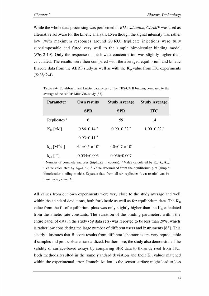
8/9/2019 DissB 7321 Mirroring
http://slidepdf.com/reader/full/dissb-7321-mirroring 61/278
Chapter 2 Biacore Technology
47
While the whole data processing was performed in BIAevaluation, CLAMP was used as
alternative software for the kinetic analysis. Even though the signal intensity was rather
low (with maximum responses around 20 RU) triplicate injections were fully
superimposable and fitted very well to the simple bimolecular binding model
(Fig.
2-19). Only the response of the lowest concentration was slightly higher than
calculated. The results were then compared with the averaged equilibrium and kinetic
Biacore data from the ABRF study as well as with the KD value from ITC experiments
(Table 2-4).
Table 2-4: Equilibrium and kinetic parameters of the CBS/CA II binding compared to the
average of the ABRF-MIRG’02 study [83].
Parameter Own results
SPR
Study Average
SPR
Study Average
ITC
Replicates a 6 59 14
KD [µM] 0.86±0.14 b
0.93±0.11 d
0.90±0.22 b 1.00±0.22 c
kon [M-1s-1] 4.1±0.5 104 4.0±0.7 104 -
koff [s-1] 0.034±0.003 0.036±0.007 -a Number of complete analyses (triplicate injections). b Value calculated by KD=koff /kon,
c Value calculated by KD=1/KA, d Value determined from the equilibrium plot (simple
bimolecular binding model). Separate data from all six replicates (own results) can be
found in appendix A.
All values from our own experiments were very close to the study average and well
within the standard deviations, both for kinetic as well as for equilibrium data. The KD
value from the fit of equilibrium plots was only slightly higher than the KD calculated
from the kinetic rate constants. The variation of the binding parameters within the
entire panel of data in the study (59 data sets) was reported to be less than 20%, which
is rather low considering the large number of different users and instruments [83]. This
clearly illustrates that Biacore results from different laboratories are very reproducible
if samples and protocols are standardized. Furthermore, the study also demonstrated the
validity of surface-based assays by comparing SPR data to those derived from ITC.
Both methods resulted in the same standard deviation and their KD values matched
within the experimental error. Immobilization to the sensor surface might lead to loss

8/9/2019 DissB 7321 Mirroring
http://slidepdf.com/reader/full/dissb-7321-mirroring 62/278
Biacore Technology Chapter 2
48
of activity but does not seem to alter the thermodynamic binding properties [83]. In a
similar study carried out by the same group [86], 36 Biacore users investigated the
interaction between azetazolamide and CA II in the same way (identical batches of
protein/analyte and protocols). Here again, the reproducibility and accuracy was very
high and the largest deviations were found to be the immobilization densities (due to
activation reagent quality) and signal artifacts (due to bad instrument maintenance) [86].
2.4 Conclusions
Biacore instruments are versatile tools with a huge potential both in life science and
drug discovery. Their ability to generate not only equilibrium binding but also kinetic
and thermodynamic data offer a much more detailed insight into a molecular
interaction compared to other screening and biophysical methods. Recent developments
in the field of data acquisition and processing expanded their applications for the
analysis of low molecular weight compounds.
In order to get most accurate data for small molecules on a Biacore 3000, every step
from sample preparation and assay design to data processing and maintenance must be
performed with care. Unfortunately, no internal test routines or reference systems are
available from Biacore to verify the performance and sensitivity of an instrument in
this area. An interaction pair consisting of the enzyme carbonic anhydrase II and the
sulfonamide inhibitor CBS as used in the ABRF MIRG’02 study showed to be a
suitable model systems for this purpose. Both molecules are commercially available in
high purity and the interaction follows a simple 1:1 binding model. Even though the
molecular weight of CBS (201 Da) is very close to the detection limit, reliable
equilibrium and kinetic binding data can be obtained when applying advanced data
processing steps.
The participation in the ABRF MIRG’02 study helped establishing a small molecule
test system and contributed to the investigation of the reproducibility of Biacore data
and their comparability with solution-based methods. The data sets obtained on
different SPR instruments by independent groups showed a very high reproducibility
and the resulting binding equilibrium data were comparable to ITC. The performance
of the instrument used in this thesis was found to be very good when the results were
compared with the study average.
8/9/2019 DissB 7321 Mirroring
http://slidepdf.com/reader/full/dissb-7321-mirroring 63/278
Chapter 2 Biacore Technology
49
2.5 References
[1] T. Turbadar, Proc Phys Soc (London) 1959, 73, 40.
[2] Surface Plasmon Resonance (Technology Note 1), Biacore AB, 2001.
[3] U. Jonsson, L. Fagerstam, B. Ivarsson, B. Johnsson, R. Karlsson, K. Lundh, S. Lofas, B.
Persson, H. Roos, I. Ronnberg, et al., Biotechniques 1991, 11, 620.
[4] K. Nagata, H. Handa, Real-Time Analysis of Biomolecular Interactions: Applications of
BIACORE , Springer, Tokio, 2000.
[5] E. Stenberg, B. Persson, H. Roos, C. Urbaniczky, J Colloid Interface Sci 1991, 143, 513.
[6] T. Mannen, S. Yamaguchi, J. Honda, S. Sugimoto, A. Kitayama, T. Nagamune, Anal
Biochem 2001, 293, 185.
[7] S. Lofas, B. Johnson, J Chem Soc Chem Commun 1990, 1526.
[8] Biacore AB, Uppsala, Sweden (www.biacore.com).[9] L. G. Fagerstam, A. Frostell, R. Karlsson, M. Kullman, A. Larsson, M. Malmqvist, H.
Butt, J Mol Recognit 1990, 3, 208.
[10] M. Malmqvist, Nature 1993, 361, 186.
[11] R. Karlsson, J Mol Recognit 2004, 17 , 151.
[12] R. L. Rich, D. G. Myszka, Drug Discovery Today: Technologies 2004, 1, 301.
[13] Biacore® 3000 Instrument Handbook , March 1999 ed., Biacore AB, Uppsala, 1999.
[14] Y. Shinohara, Y. Hasegawa, H. Kaku, N. Shibuya, Glycobiology 1997, 7 , 1201.
[15] R. Karlsson, M. Kullman-Magnusson, M. D. Hamalainen, A. Remaeus, K. Andersson, P.Borg, E. Gyzander, J. Deinum, Anal Biochem 2000, 278, 1.
[16] Biacore Sensor Surface Handbook , Biacore AB, Uppsala, 2003.
[17] B. Johnsson, S. Lofas, G. Lindquist, Anal Biochem 1991, 198, 268.
[18] A. A. Kortt, G. W. Oddie, P. Iliades, L. C. Gruen, P. J. Hudson, Anal Biochem 1997, 253,
103.
[19] M. Morpurgo, C. Monfardini, L. J. Hofland, M. Sergi, P. Orsolini, J. M. Dumont, F. M.
Veronese, Bioconjug Chem 2002, 13, 1238.
[20] T. Morcol, A. Subramanian, W. H. Velander, J Immunol Methods 1997, 203, 45.
[21] N. Endo, N. Umemoto, Y. Kato, Y. Takeda, T. Hara, J Immunol Methods 1987, 104, 253.
[22] A. Subramanian, W. H. Velander, J Mol Recognit 1996, 9, 528.
[23] G. A. Canziani, S. Klakamp, D. G. Myszka, Anal Biochem 2004, 325, 301.
[24] R. David, M. P. Richter, A. G. Beck-Sickinger, Eur J Biochem 2004, 271, 663.
[25] I. E. Gentle, D. P. De Souza, M. Baca, Bioconjug Chem 2004, 15, 658.
[26] K. Terpe, Appl Microbiol Biotechnol 2003, 60, 523.
[27] K. M. Muller, K. M. Arndt, K. Bauer, A. Pluckthun, Anal Biochem 1998, 259, 54.
[28] K. Busch, J. Piehler, H. Fromm, Biochemistry 2000, 39, 10110.[29] A. Einhauer, A. Jungbauer, J Chromatogr A 2001, 921, 25.
8/9/2019 DissB 7321 Mirroring
http://slidepdf.com/reader/full/dissb-7321-mirroring 64/278
Biacore Technology Chapter 2
50
[30] A. D. Keefe, D. S. Wilson, B. Seelig, J. W. Szostak, Protein Expr Purif 2001, 23, 440.
[31] L. Jendeberg, B. Persson, R. Andersson, R. Karlsson, M. Uhlen, B. Nilsson, J Mol
Recognit 1995, 8, 270.
[32] D. B. Jones, M. H. Hutchinson, A. P. Middelberg, Proteomics 2004, 4, 1007.
[33] Covalys Biosciences AG, Witterswil, Switzerland (www.covalys.com).
[34] M. Kindermann, N. George, N. Johnsson, K. Johnsson, J Am Chem Soc 2003, 125, 7810.
[35] W. Huber, S. Perspicace, J. Kohler, F. Muller, D. Schlatter, Anal Biochem 2004, 333, 280.
[36] P. Schuck, Biophys J 1996, 70, 1230.
[37] R. W. Glaser, Anal Biochem 1993, 213, 152.
[38] D. G. Myszka, Curr Opin Biotechnol 1997, 8, 50.
[39] D. G. Myszka, J Mol Recognit 1999, 12, 279.
[40] K. Andersson, M. Hamalainen, M. Malmqvist, Anal Chem 1999, 71, 2475.
[41] R. Karlsson, A. Michaelsson, L. Mattsson, J Immunol Methods 1991, 145, 229.
[42] D. J. O'Shannessy, M. Brigham-Burke, K. K. Soneson, P. Hensley, I. Brooks, Anal
Biochem 1993, 212, 457.
[43] L. D. Roden, D. G. Myszka, Biochem Biophys Res Commun 1996, 225, 1073.
[44] M. A. Cooper, Nat Rev Drug Discov 2002, 1, 515.
[45] D. Nedelkov, R. W. Nelson, Trends Biotechnol 2003, 21, 301.
[46] Graffinity Pharmaceuticals AG, Heidelberg, Germany (www.graffinity.com).
[47] C. L. Baird, D. G. Myszka, J Mol Recognit 2001, 14, 261.
[48] P. O. Markgren, W. Schaal, M. Hamalainen, A. Karlen, A. Hallberg, B. Samuelsson, U.
H. Danielson, J Med Chem 2002, 45, 5430.
[49] O. P. Karlsson, S. Lofas, Anal Biochem 2002, 300, 132.
[50] J. Northup, Methods Mol Biol 2004, 261, 93.
[51] C. Bieri, O. P. Ernst, S. Heyse, K. P. Hofmann, H. Vogel, Nat Biotechnol 1999, 17 , 1105.
[52] P. Stenlund, G. J. Babcock, J. Sodroski, D. G. Myszka, Anal Biochem 2003, 316 , 243.
[53] A. Frostell-Karlsson, H. Widegren, C. E. Green, M. D. Hamalainen, L. Westerlund, R.
Karlsson, K. Fenner, H. van de Waterbeemd, J Pharm Sci 2005, 94, 25.
[54] Y. N. Abdiche, D. G. Myszka, Anal Biochem 2004, 328, 233.
[55] M. Oyama, T. Ikeda, T. Lim, K. Ikebukuro, Y. Masuda, I. Karube, Biotechnol Bioeng
2000, 71, 217.
[56] D. G. Myszka, Anal Biochem 2004, 329, 316.
[57] E. Walladen, in Biacore Press Release, Vol. 2004, Biacore AB, 2004, Biacore SPR array.
[58] E. Freire, Drug Discovery Today: Technologies 2004, 1, 295.
[59] P. C. Weber, F. R. Salemme, Curr Opin Struct Biol 2003, 13, 115.
[60] T. Laue, Drug Discovery Today: Technologies 2004, 1, 309.

8/9/2019 DissB 7321 Mirroring
http://slidepdf.com/reader/full/dissb-7321-mirroring 65/278
Chapter 2 Biacore Technology
51
[61] G. H. Cross, A. A. Reeves, S. Brand, J. F. Popplewell, L. L. Peel, M. J. Swann, N. J.
Freeman, Biosens Bioelectron 2003, 19, 383.
[62] M. J. Swann, L. L. Peel, S. Carrington, N. J. Freeman, Anal Biochem 2004, 329, 190.
[63] C. K. O'Sullivan, G. G. Guilbault, Biosens Bioelectron 1999, 14, 663.
[64] K. S. Carmon, R. E. Baltus, L. A. Luck, Biochemistry 2004, 43, 14249.
[65] W. A. McGee, L. J. Parkhurst, Anal Biochem 1990, 189, 267.
[66] B. M. Kolakowski, L. Konermann, Anal Biochem 2001, 292, 107.
[67] P. W. Atkins, Physical chemistry, 6th ed., Oxford University Press, Oxford, 1998.
[68] G. L. Glish, R. W. Vachet, Nat Rev Drug Discov 2003, 2, 140.
[69] J. M. Wiseman, Z. Takats, B. Gologan, V. J. Davisson, R. G. Cooks, Angew Chem Int Ed
2005, 44, 913.
[70] P. A. Wabnitz, J. A. Loo, Rapid Commun Mass Spectrom 2002, 16 , 85.
[71] H. Widmer, W. Jahnke, Cell Mol Life Sci 2004, 61, 580.
[72] M. Rinnbauer, B. Ernst, B. Wagner, J. Magnani, A. J. Benie, T. Peters, Glycobiology
2003, 13, 435.
[73] T. L. Blundell, H. Jhoti, C. Abell, Nat Rev Drug Discov 2002, 1, 45.
[74] M. Stolz, D. Stoffler, U. Aebi, C. Goldsbury, J Struct Biol 2000, 131, 171.
[75] J. Legleiter, T. Kowalewski, Drug Discovery Today: Technologies 2004, 1, 163.
[76] C. Goldsbury, U. Aebi, P. Frey, Trends Mol Med 2001, 7 , 582.
[77] C. Yuan, A. Chen, P. Kolb, V. T. Moy, Biochemistry 2000, 39, 10219.
[78] J. Fritz, M. K. Baller, H. P. Lang, H. Rothuizen, P. Vettiger, E. Meyer, H. Guntherodt, C.
Gerber, J. K. Gimzewski, Science 2000, 288, 316.
[79] F. X. Schmid, in Protein structure : a practical approach (Ed.: T. E. Creighton), IRL
Press, Oxford, 1989, pp. XVIII.
[80] B. A. Wallace, R. W. Janes, Biochem Soc Trans 2003, 31, 631.
[81] D. G. Myszka, X. He, M. Dembo, T. A. Morton, B. Goldstein, Biophys J 1998, 75, 583.
[82] Y. S. Day, C. L. Baird, R. L. Rich, D. G. Myszka, Protein Sci 2002, 11, 1017.
[83] D. G. Myszka, Y. N. Abdiche, F. Arisaka, O. Byron, E. Eisenstein, P. Hensley, J. A. Thomson,
C. R. Lombardo, F. Schwarz, W. Stafford, M. L. Doyle, J Biomol Tech 2003, 14, 247.
[84] D. G. Myszka, T. A. Morton, Trends Biochem Sci 1998, 23, 149.
[85] W. L. Delano, Drug Discov Today 2005, 10, 213.
[86] M. J. Cannon, G. A. Papalia, I. Navratilova, R. J. Fisher, L. R. Roberts, K. M. Worthy, A. G.
Stephen, G. R. Marchesini, E. J. Collins, D. Casper, H. Qiu, D. Satpaev, S. F. Liparoto, D. A.
Rice, Gorshkova, II, R. J. Darling, D. B. Bennett, M. Sekar, E. Hommema, A. M. Liang, E. S.
Day, J. Inman, S. M. Karlicek, S. J. Ullrich, D. Hodges, T. Chu, E. Sullivan, J. Simpson, A.
Rafique, B. Luginbuhl, S. N. Westin, M. Bynum, P. Cachia, Y. J. Li, D. Kao, A. Neurauter,
M. Wong, M. Swanson, D. G. Myszka, Anal Biochem 2004, 330, 98.

8/9/2019 DissB 7321 Mirroring
http://slidepdf.com/reader/full/dissb-7321-mirroring 66/278
Biacore Technology Chapter 2
52
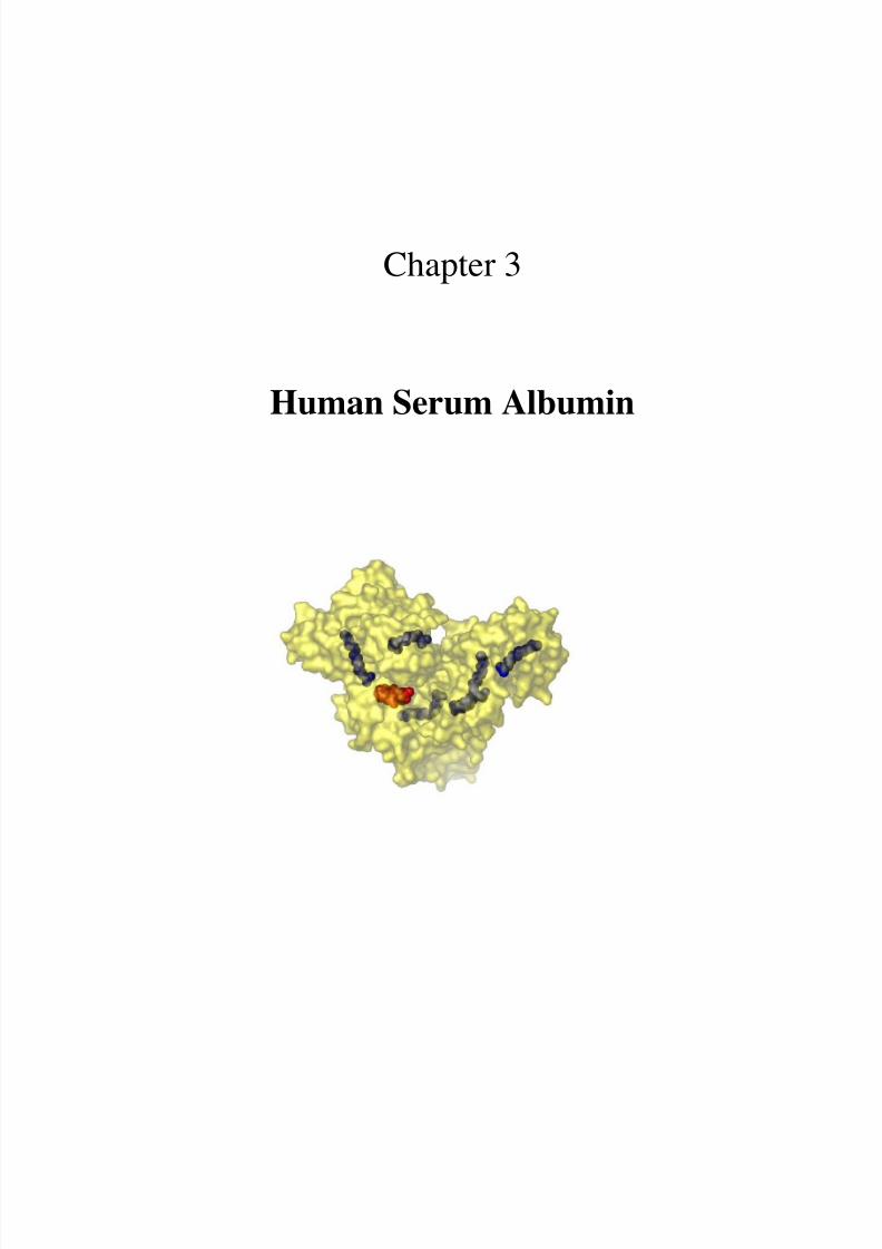
8/9/2019 DissB 7321 Mirroring
http://slidepdf.com/reader/full/dissb-7321-mirroring 67/278
Chapter 3
Human Serum Albumin
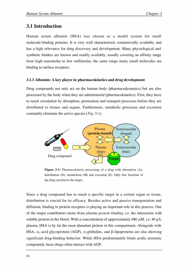
8/9/2019 DissB 7321 Mirroring
http://slidepdf.com/reader/full/dissb-7321-mirroring 68/278
Human Serum Albumin Chapter 3
54
3.1 Introduction
Human serum albumin (HSA) was chosen as a model system for small
molecule-binding proteins. It is very well characterized, commercially available, and
has a high relevance for drug discovery and development. Many physiological and
synthetic binders are known and readily availably, usually covering an affinity range
from high nanomolar to low millimolar, the same range many small molecules are
binding to surface receptors.
3.1.1 Albumin: A key player in pharmacokinetics and drug development
Drug compounds not only act on the human body (pharmacodynamics) but are also
processed by the body when they are administered (pharmacokinetics). First, they haveto reach circulation by absorption, permeation and transport processes before they are
distributed to tissues and organs. Furthermore, metabolic processes and excretion
constantly eliminate the active species (Fig. 3-1).
Plasma
(free)
Plasma
(protein-bound)
Extravascular
(bound)
Drug compound
Extravascular
(free)
Target
A
M
DD
E
D
Figure 3-1: Pharmacokinetic processing of a drug with absorption (A),
distribution (D), metabolism (M) and excretion (E). Only free fractions of
the drug can bind to the target.
Since a drug compound has to reach a specific target in a certain organ or tissue,
distribution is crucial for its efficacy. Besides active and passive transportation and
diffusion, binding to protein receptors is playing an important role in this process. One
of the major contributors stems from plasma protein binding, i.e. the interaction with
soluble protein in the blood. With a concentration of approximately 680 µM, i.e. 40 g/L
plasma, HSA is by far the most abundant protein in this compartment. Alongside with
HSA, 1-acid glycoprotein (AGP), -globulins, and -lipoproteins are also showing
significant drug-binding behavior. While HSA predominantly binds acidic aromatic
compounds, basic drugs often interact with AGP.
8/9/2019 DissB 7321 Mirroring
http://slidepdf.com/reader/full/dissb-7321-mirroring 69/278
Chapter 3 Human Serum Albumin
55
Plasma protein binding affects nearly all stages of pharmacodynamics and
pharmacokinetics. Since only unbound molecules are able to interact with their targets
[1], plasma binding directly affects metabolism, rapid clearance and toxicity.
Furthermore, many drugs competitively interact with the same binding site on HSA,
leading to displacement and sudden changes of a drugs’ plasma concentration.
Therefore, plasma protein binding was regarded as a somewhat dangerous property for
many years and was tried to keep on a moderate level. Newer developments in drug
discovery changed the view of the role of HSA and plasma binding completely.
Especially the contribution of HSA to an increase in elimination half time helps
developing drugs, which act longer (retard effect) and have to be administered less
often (leading to a better compliance). An attractive example of increased HSA binding
is the insulin derivative detemir (Novo Nordisk). By attaching a fatty acid moiety along-acting form of insulin could be developed [2].
Albumin was recognized as a principal blood component as early as 1839. The name
‘albumin’ was derived from the white color (lat: albus) of protein precipitates.
Although the most outstanding property is its ability to bind an broad variety of
endogenous and exogenous ligands, it performs many additional functions. For
example, HSA contributes 80% to colloid osmotic blood pressure, is mainly
responsible for the maintenance of blood pH , plays a major role in detoxification, and
sequesters oxygen free radicals [3].
3.1.2 Structure and properties of HSA
HSA is synthesized by the liver as a single peptide chain and is exported to circulation,
where it remains with a plasma half-life of 19 days. The non-glycosylated protein
consists of 585 amino acids resulting in a molecular weight of 66,500 Da. HSA
possesses an unusually high number of disulfide bridges; 34 of its 35 cysteine residuesare involved in 17 disulfide bridges while a single cysteine (Cys-34) remains free. Due
to its high amount of acidic amino acids, the net charge of albumin is clearly negative
[3, 4].
Even though HSA structure and binding features were explored for several decades, it
was not possible to obtain any crystal structure at a reasonable resolution for years. The
long period of frustration was ended in 1992, when a group from the NASA science
center presented a structure at 2.8 Å resolution [5], obtained under microgravity
environment in a U.S. space shuttle [4]. It shows HSA as a heart-shaped protein, which

8/9/2019 DissB 7321 Mirroring
http://slidepdf.com/reader/full/dissb-7321-mirroring 70/278
Human Serum Albumin Chapter 3
56
is organized in three homologous domains (I-III; Fig. 3-2A). Each of these domains
can be further divided into two subdomains (A, B) consisting of six and four -helices,
respectively. This leads to an unusually high content of helical structures of around
67% (Fig. 3-2B), which are stabilized by the large number of disulfide bridges forming
nine double loops. Such disulfide pairings between helical motifs are very unusual and
are believed to be responsible for some of the unique features of albumin. No -sheets
are present in the crystal structure [3, 5].
A B C
I
II
III
Trp-214
Cys-34
Figure 3-2: Crystal structure of HSA. A: Surface view with three domains (I in green, II in red, III in
blue), B: Secondary structure (helices in red, turns in green), C: Unique amino acids (single tryptophan
in blue, single free cysteine in orange)
Albumin shows an enormously high stability, and even exposure to very low or high
pH values ( pH
1.2-9), heat (up to 72°C; albumin preparations usually are pasteurizedfor 10 h at 60°C), or 8 M urea have no deleterious effects. The structure of HSA is
nevertheless rather flexible and very sensitive to environmental factors such as pH ,
ionic strength or temperature. For example, five pH -dependant conformations are
described in literature for human and bovine serum albumin (BSA) [4]. Besides the
N-form (normal) at physiological conditions, albumin undergoes two changes each at
lower and higher pH values. Around pH 4 the F-form (fast) is predominant, in which
the two halves of the molecule separate leading to a lengthening. Further decrease of
pH below 3.5 initiates the so-called E-form (expanded), where the domains are
unfolded as much as the disulfide pattern permits. The structural changes at higher pH
values are more subtle and gradual starting with the B-form (basic) between pH 7 and
9, which is discussed to have physiological importance. Finally, the A-form (aged) is
usually detected above pH 9-10, involves both ionic forces and hydrogen bonding, and
is not fully reversible (Table 3-1) [4].

8/9/2019 DissB 7321 Mirroring
http://slidepdf.com/reader/full/dissb-7321-mirroring 71/278

8/9/2019 DissB 7321 Mirroring
http://slidepdf.com/reader/full/dissb-7321-mirroring 72/278
Human Serum Albumin Chapter 3
58
Table 3-2: Extended Sudlow binding site classification of Carter [3] with the site names and locations as
well as some examples of known binders.
Site I II III + IV V VI
Name Warfarin-
azapropazone
Indole-
benzodiazepine
Long/medium
fatty acid
Metal
site 1
Metal
site 2
Location subdomain IIA subdomain IIIA uncertaina Cys-34 N-terminus
Binders warfarin
salicylate
bilirubinb
diazepam
naproxen
digitoxinb
oleate
myristate
palmitate
auranophan
cisplatin
Au(I),
Hg(II)
Cu(II)
Ni(II)
Co(II)
a The exact number and location of fatty acid binding sites is still not known. b Separate binding sites for
bilirubin and digitoxin are proposed but not confirmed.
N
CS SS S S SS S S SS S S SS S S SS S S SS S S SS S
S SS S S SS SS SS S S SS S
S SS S S SS SS SS S S SS S S SS S S SS S
Site I Site II
C34
W214
Cu
FA
AcSA
FA
FA
A B
Site ISite II
Figure 3-3: Location of Sudlow binding sites in HSA. A: Schematic overview adapted from Peters [4].
Several specific binding sites are symbolized with a red star (Cu: copper, FA: fatty acids, AcSA:
acetylsalicylic acid). Positions of disulfide bridges, Cys-34 and Trp-214 are also indicated. B: Crystal
structure (PDB code: 1BM0) of HSA with colored Sudlow binding sites and (hidden) location of Cys-34
(dotted orange circle).
Many investigations have been performed about the location and properties of the drug
binding sites. Pioneering work was done by Sudlow and coworkers [7, 8], who
identified two major binding sites located on subdomains IIA and IIIA, respectively
(Table 3-2, Fig. 3-3). The anticoagulant drug warfarin was described as a marker
compound for Sudlow site I (subdomain IIA), which is therefore often referred as
warfarin-azapropazone site. Ligands for this binding site typically are bulky
heterocyclic anions with the charge situated in fairly central position of the molecule
(Fig. 3-4A). They are mostly non-aromatic except for some phenyl groups. The large
specificity of site I is responsible for the cosmopolitan reputation of albumin amongtransport proteins [4]. The surface of the binding site is described as ‘an elongated

8/9/2019 DissB 7321 Mirroring
http://slidepdf.com/reader/full/dissb-7321-mirroring 73/278
Chapter 3 Human Serum Albumin
59
sock-shaped pocket wherein the foot region is primarily hydrophobic and the leg is
primarily hydrophilic’ [3]. Site I shows a high degree of adaptability and ligands can
induce conformational changes [4]. The other major binding site (Sudlow site II,
subdomain IIIA) is called indole-benzodiazepine site or diazepam site, according to its
marker drug. Site II ligands often are aromatic and neutral or anionic with a more
peripheral charge (Fig. 3-4B). Typical binders are indoles (e.g. L-tryptophan),
benzodiazepines (e.g. diazepam) and non-steroidal anti-inflammatory drugs
(e.g. naproxen). Binding to site II is more affected by HSA dimerization (non-thiol
dimers) and glycation than binding to site I [4]. Additional specific binding sites for
digitoxin, tamoxifen, and bilirubin are also proposed but their very existence is still
subject to some debate [4, 5, 9, 10]. Even though albumin binding of some cationic
drugs (e.g.
imipramine, quinidine) is also described in literature [11], these moleculesprimarily bind to 1-acid glycoprotein.
OO
OO
O
O
A B
Figure 3-4: Pharmacophore preference of the two major drug binding sites. A: Bulky heterocyclic ligand
for Sudlow site I with central negative charge (example: warfarin). B: Anionic aromatic ligand for
Sudlow site II with peripheral charge (example ibuprofen). Aromatic rings are highlighted in green,
heterocycles in blue and anionic groups in red.
Unfortunately, ligand-binding characterization is far more complicated in the case of
HSA compared to most other targets. Traditionally, localization of binding sites for a
specific drug has been done by competition experiments using known binders.
However, one drug can bind to more than one site simultaneously and two drugs can
bind at different location within the same binding site. Even more complicating is the
fact that binding of a first drug at a specific site can influence the activity of a second
drug at the same or even another site by allosteric effects [12]. Furthermore, it has been
shown that endogenous ligands such as fatty acids [13] as well as environmental factors
( pH , ions) can induce huge conformational changes thus influencing a drugs binding
behavior.

8/9/2019 DissB 7321 Mirroring
http://slidepdf.com/reader/full/dissb-7321-mirroring 74/278
Human Serum Albumin Chapter 3
60
Cys-34 also does also play a role in drug binding properties of HSA. Thiol-active
endogenous compounds such as L-cysteine or glutathione, but also drugs like
D-penicillamine, aurothiomalate, or cisplatin can form covalent bonds with the free
cysteine residue. More recent studies showed that Cys-34 also interacts with nitric
oxide forming S -nitrosoalbumin (~ 1% of the total albumin fraction) [4]. This supports
the theory of albumin being a key carrier or reservoir of nitric oxide [3]. Whether the
oxidation state of Cys-34 directly influences the affinities of reversibly binding
molecules is still discussed controversially.
Some metal ions like copper(II) and nickel(II) bind strongly at a defined site at the
N -terminus (Asp-Ala-His), other metals (Zn, Cd, Ca, Mn) also show a certain specific
but far weaker affinity to HSA [3, 4]. Chloride is the most important anionic element
binding specifically to HSA. Even though its KD value is only 1.4
mM [14], the high
abundance of chloride in the plasma (~ 100 mM) leads to significant binding and
competition effects (especially with site II ligands) [4]. In addition to the endogenous
ligands, HSA can also bind an impressive array of drug molecules from different
classes with a rather broad specificity.
3.1.4 HSA analysis on Biacore
Acquiring data about pharmacokinetic properties has evolved into one of the most
important and interesting applications for SPR instruments in drug discovery.
Traditionally, most of the available binding data were generated from equilibrium
dialysis experiments. However, these data give only information about the free or
bound fraction of a drug. If the drug is dialyzed against whole plasma (including AGP,
globulins and other proteins) these %bound PPB values might be significantly larger than
values derived from dialyses against purified albumin solutions (%bound HSA). This is
especially true for cationic compounds, which predominantly bind to AGP. Othermethods used for acquiring %bound or affinity data are fluorescence quenching [15],
ITC [16], affinity chromatography (HPAC) [17], capillary electrophoresis [18], or
differential CD [19]. While some of the published values are very consistent between
those methods, others differ from each other by orders of magnitude. SPR-based
analysis offers an interesting alternative, since only low amounts of HSA and unlabeled
compounds are needed. In addition, assays can be automated easily and may provide
information about kinetics as well as equilibrium affinity. Furthermore, differences of
high affinity binders (low or sub-µM range) can not been described accurately by
%bound values (> 97 %boundHSA) [20].

8/9/2019 DissB 7321 Mirroring
http://slidepdf.com/reader/full/dissb-7321-mirroring 75/278
Chapter 3 Human Serum Albumin
61
Three publications using Biacore for the characterization of HSA/drug interactions
have been released so far, with focus on proof-of-concept [20] and on improvement of
accuracy and robustness [21, 22], respectively. Ranking experiments to discriminate
between low, medium, and strong binders, high-resolution screening for exact affinity
values [20, 21], as well as competition experiments for characterizing primary binding
sites [22] were covered by these publications. Since HSA is described to react sensitive
on environmental changes (see section 3.1.2) assay parameters should be chosen with
care. Different parameters have been investigated so far: HSA stability on the sensor
chip [20, 21], effects of DMSO concentration and temperature [21], as well as
immobilization parameters, species differences of albumins, and the influence of
different batches and suppliers of HSA [22]. Some marker substances were investigated
in all three studies (e.g. warfarin), others only in one or two of them (e.g.
salicylic acid)and of some important markers there are no SPR data at all (e.g. diazepam,
L-tryptophan). There are not only some significant differences in the assay conditions
between the three reports, but the affinity values of some of the investigated drugs also
show a high variability (Table 3-3).
Table 3-3: Comparison of assay conditions and affinity data of the three available
HSA-drug binding studies using Biacore [20-22].
Year / Reference 2000 [20] 2001 [21] 2003 [22]
Temperature 25°C 25°C 25°C
Phosphate 67 mM 65.5 mM 10 mM
NaCl 70 mM 70 mM 150 mM
pH 7.4 7.4 7.4
DMSO conc. 5% 3% 3%
KD Warfarin 9 µM 2.5 µM 3.7 µM
KD Naproxen 26
µM 1.5
µM 10.6
µMKD Digitoxin 28 µM 38 µM 24.8 µM
KD Salicylate n.d. 48 µM 141 µM

8/9/2019 DissB 7321 Mirroring
http://slidepdf.com/reader/full/dissb-7321-mirroring 76/278
Human Serum Albumin Chapter 3
62
3.1.5 Aims in this project
Fast determination of HSA-binding data on drug-like substances and lead compounds
rely on a robust assay format. While the proof-of-concept for HSA assays on Biacore
has already been performed, there are still some open questions and deviations betweenvalues from different publications. The first aim of this project was therefore to
establish, validate, and optimize a HSA assay in terms of throughput and robustness.
Possible environmental factors such as pH shifts, buffer ionic strength, and oxidation
state of Cys-34 should be investigated more intensively. Finally, the possibility of using
this single free cysteine residue to perform a site-specific immobilization should be
explored.
8/9/2019 DissB 7321 Mirroring
http://slidepdf.com/reader/full/dissb-7321-mirroring 77/278
Chapter 3 Human Serum Albumin
63
3.2 Materials and Methods
This section only describes materials, equipment and procedures specifically used for
the albumin project. Materials and general methods used in all Biacore assays are
described in section 2.2.
3.2.1 Materials
Reagents and proteins
Human serum albumin used in all assays was from Sigma (Fluka AG, Buchs,
Switzerland) and was essentially globulin and fatty acid-free (No. A-3782). Different
batches of DMSO were used for the experiments. Dried DMSO from Riedel-de Haën
(No. 34943) was found to be especially suitable. All other reagents were purchased
from Sigma. Freshly bidistilled water delivered through quartz tubes was used for the
preparation of all Biacore buffers and solutions.
Drug Compounds
Diazepam, nitrazepam, and phenprocoumon were a gift from Roche AG (Basel,
Switzerland). Warfarin, digitoxin, indoprofen, cholic acid, L-tryptophan, L-kynurenine
and quinidine were from Sigma (Fluka AG, Buchs, Switzerland). Salicylic acid,
acetylsalicylic acid, quinine, and prednisone were from Siegfried AG (Zofingen,
Switzerland). Sodium salicylate was purchased from Hänseler AG (Herisau,
Switzerland). Naproxen (sodium salt) and salbutamol were a gift from Prof. Hans
Leuenberger (Institute of Pharmaceutical Technology, University of Basel,
Switzerland).
Equipment
HPLC separation were done on an Agilent 1100 purification system, equipped with a
quaternary pump, a cooled well-plate autosampler, a column thermostat, a DAD
detector, and a cooled analytical fraction collector (Agilent AG, Basel, Switzerland).
Size exclusion chromatography was performed on a TSK Gel G2000SW column (7.5 x
600 mm; Tosoh Biosciences GmbH, Stuttgart, Germany). A Molecular Devices
SpectraMax Plus UV absorbance plate reader was used for the Ellmans assays and a
SpectraMax Gemini XP fluorescence plate reader for measuring intrinsic tryptophanyl
emission (Bucher Biotec, Basel, Switzerland). A Bio-Rad SmartSpec 3000
8/9/2019 DissB 7321 Mirroring
http://slidepdf.com/reader/full/dissb-7321-mirroring 78/278
Human Serum Albumin Chapter 3
64
spectrophotometer equipped with a quartz cuvette was used for absorption
measurement in the UV range.
3.2.2 Preparation of running buffer
10 mM PBS with 3% DMSO was used as running buffer during the assays if not
differently described. A 1.03-fold PBS (10.3 mM sodium phosphate, 155 mM NaCl)
was prepared and adjusted to pH 7.30. After filtration and degassing (see section 2.2.2),
3% DMSO were added (using a glass pipette) and the final pH was verified to be 7.40
after DMSO addition. The prepared running buffer was used no longer than 24 hours to
avoid shifts in DMSO concentration.
3.2.3 Amine coupling of HSA
The protocol for the immobilization of albumin was the result of a combination and an
adaptation of different sources [20-22]. HSA was dissolved in water to a stock
concentration of 5 mg/ml and further diluted to 50 µg/ml in 10 mM sodium acetate
buffer pH 5.0 just before immobilization. Standard amine coupling (see section 2.2.5)
was used to reach surface densities of 8-18 kRU. Typical activation/deactivation as
well as immobilization times ranged from 5-7
min each. All surfaces were washed withthree consecutive pulse injections (20 s) of 50 mM NaOH to remove noncovalently
bound protein. Unmodified carboxymethyl dextran flow cells were used as reference
surfaces.
3.2.4 Stabilization of HSA surface
Even though HSA is immobilized covalently, surface decay of up to 80% within the
first few hours has been observed in previous studies [21, 22]. The reason for thisbehavior is still not known, and various attempts for stabilization have failed so far.
The most effective procedure was found to be repetitive buffer or sample injections
over several hours [22]. Therefore, 20 injections of each running buffer, 20 µM
warfarin and 6 µM naproxen were performed over 10 hours. DMSO calibration (see
section 3.2.5) was done before and after the stabilization process. Baseline signals as
well as difference signals for warfarin and naproxen were monitored and plotted
against time.

8/9/2019 DissB 7321 Mirroring
http://slidepdf.com/reader/full/dissb-7321-mirroring 79/278
Chapter 3 Human Serum Albumin
65
3.2.5 DMSO calibration
Since even small shifts in DMSO concentration cause huge changes in bulk signal
intensity, any difference between the volumes of the sample and reference surface are
clearly visible. Therefore, these DMSO signals have to be subtracted from the samplebinding response [20]. Calibration solutions ranging from approximately 2.9% to 3.1%
DMSO were prepared by adding 1 µl DMSO (A) or 50 µl PBS 1.03x (B) to 1 ml of
running buffer. Solutions A and B were mixed to get five calibration solutions (100%,
75%, 50%, 25%, 0% A). The calibration procedure was performed before each binding
experiment. Difference signals between sample and reference cell were plotted against
the signal on the reference cell and fitted using linear regression. Sample responses
were then corrected according to [20].
3.2.6 General optimization of the HSA assay
The aim of the general optimization experiments was to reach improvements in
throughput and handling while maintaining accuracy and reproducibility. Special care
was applied to the handling of DMSO-containing solutions. Therefore, different
batches of DMSO have been tested and a panel of sample containers (glass or plastic
vials, 96 well plates) was evaluated. Additional parameters such as vial capping,
autosampler temperature, the number of sample dilutions, and carry-over effects wereinvestigated. Available software for data evaluation ( BIAevaluation, Scrubber, Excel)
was compared concerning ease of use, automation, reproducibility and time
consumption. Finally, an application for the automated sample randomization and
method generation was developed in Excel based on visual basic scripts (see
appendix B1).
For all further assays, airtight rubber caps and polypropylene vials were used. The
autosampler area was maintained at a constant temperature of 20±2°C, while keepingthe IFC and detector at 25°C. Scrubber was used for data processing and evaluation.
All sensorgrams were double referenced and the whole curves were corrected for
DMSO effects. Equilibrium binding data were fitted either to a single-site or to a two
independent-sites model.
3.2.7 HSA affinity ranking
The binding strength of different known binders have been tested with a set chosen bycriteria such as commercial availability, structural diversity, covering of binding sites,

8/9/2019 DissB 7321 Mirroring
http://slidepdf.com/reader/full/dissb-7321-mirroring 80/278
Human Serum Albumin Chapter 3
66
functional groups, and available results. A list of the drugs used for different
experiments alongside with some important properties can be found in table 3-4, their
structures are shown in Fig. 3-5.
Table 3-4: Drug compounds used for HSA binding experiments with some important physico-chemical
properties and known plasma protein binding values.
Compound Abbr MW log P a pKa a %boundPPB b Site Charge
Acetylsalicylic acid AcSA 180.2 1.19 3.49 37 I anionic
Cholic acid ChoA 408.6 2.02 4.98 48 Ic anionic
Corticosterone Cort 346.5 1.94 - 68 n.d.c neutral
Diazepam Dzpm 284.7 2.82 3.40 93.2 II neutral
Digitoxin Dgtx 765.0 1.85 - 97 IIc neutral
Indoprofen Indo 281.3 2.77 4.38 98.5 II anionic
Naproxen Napr 230.3 3.18 4.15 99.5 II anionic
Nitrazepam Nitra 281.3 2.25 - 87 II neutral
Phenprocoumon Ppro 280.3 3.62 - 99.5 I neutral
Prednisone Pred 358.4 1.46 - 38 n.d.c neutral
Quinidine Qdne 324.4 3.44 8.56 88 II cationic
Quinine Quin 324.4 3.44 8.56 93 II cationic
Salbutamol Salb 239.3 0.64 - 8 n.d. neutralSalicylic acid ScyA 138.1 2.26 2.97 73 I anionic
L-Tryptophan LTrp 204.2 -1.05 7.38 80-90 II zwitter
Warfarin Warf 308.3 2.60 5.08 99.5 I neutral
a log P and pKa values were retrieved from the online PhysPro database [23] and are determined
experimentally (octanol/water distribution), except for the log P of salbutamol, which is only estimated
and pKa of indoprofen (calculated). b %bound values were collected from different sources. An extended
list with all references can be found in appendix B2. c Special binding sites for bile salts (bilirubin, cholic
acid) and steroids (especially digitoxin) are discussed, but not confirmed.
For affinity ranking, 30 mM stock solutions of each compound in DMSO were diluted
in PBS 1.03x to a concentration of 1 mM in 3% DMSO. These solutions were further
diluted in running buffer to a final concentration of 333 µM and injected as randomized
triplicates as described in section 3.2.6. Steady state binding signals was averaged
between 10 and 20 s after injection start, divided by the compounds molecular weight,
and evaluated in Excel. Average values from triplicate injections of all drugs were
ranked according to their normalized SPR signal and plotted against the corresponding
log P value or the bound fraction (%boundPPB). Finally, the shape of each binding curve
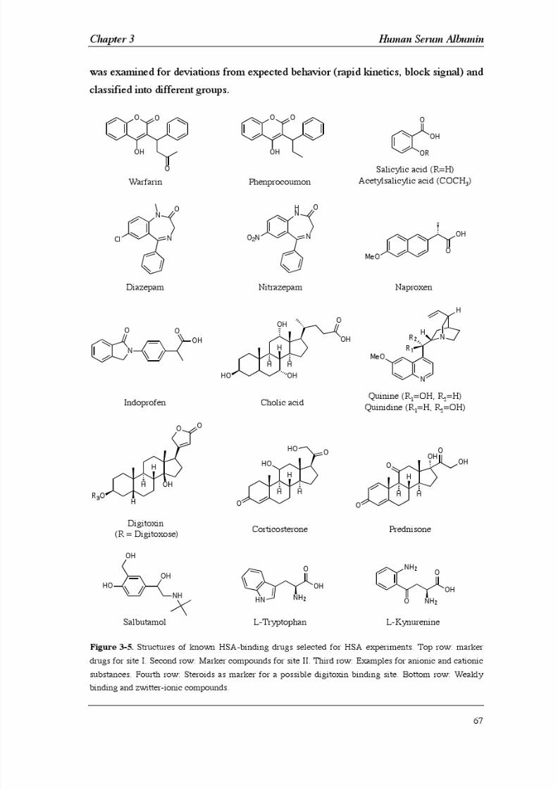
8/9/2019 DissB 7321 Mirroring
http://slidepdf.com/reader/full/dissb-7321-mirroring 81/278

8/9/2019 DissB 7321 Mirroring
http://slidepdf.com/reader/full/dissb-7321-mirroring 82/278
8/9/2019 DissB 7321 Mirroring
http://slidepdf.com/reader/full/dissb-7321-mirroring 83/278
Chapter 3 Human Serum Albumin
69
3.2.10 Influence of HSA redox state on drug binding
Oxidation state of the single free cysteine residue of HSA has shown to be highly
variable in commercial preparations [24]. Therefore, the influence of the HSA redox
state on binding affinities has been investigated. For preparing oxidized and reducedHSA samples, 4 mg/ml HSA (60 µM) in sample buffer (10.1 mM Na2HPO4, 1.76 mM
KH2PO4, 137 mM NaCl, 1 mM EDTA, pH 7.4) were mixed with 10fold molar ratios of
reduced (GSH) or oxidized glutathione (GSSG) or with an equimolar amount or a 5fold
molar excess of DTT. Incubation at 25°C was 1 hour for GSH and 3 hours for GSSG
and DTT [6, 25]. After the incubation time, excess of thiol-active reagents was
removed by size exclusion chromatography (TSK Gel G2000SW, isocratic in sample
buffer, 1 ml/min, ambient temperature, 30 min). Dimer and monomer peaks were
collected separately and latter was used for Ellmans assay.
The degree of HSA oxidation was determined by measuring the concentration of free
thiol groups using an enhanced Ellmans assay protocol [26]. HSA preparations were
diluted twofold in sample buffer to get an appropriate signal intensity.
Ellman/cystamine reagent was prepared by dissolving 77.1 mg DTNB and 56.3 mg
cystamine dihydrochloride (10 mM both) in 25 ml buffer 7.0 (100 mM NaH2PO4,
0.2 mM EDTA, pH 7.0) and readjusting the pH to 7.0. To 1 ml of HSA sample 200 µl
of strong buffer 8.2 (100 mM boric acid, 0.2 mM EDTA, pH
8.2) and 20 µl of Ellman/cystamine reagent were added and mixed immediately. After 5 min the
adsorption at 412 nm (A412s) was read against a water blank. Protein (A412p) and reagent
blanks (A412r) were measured in parallel. Concentration of free thiols was calculated
according to equation 7 with 412 = 14,150 M-1cm-1 and d = 1 cm.
[SH] = (A412s - A412p - A412r) / ( 412 x d) [Eq. 7]
Concentration of HSA in the same sample was determined by measuring absorption of
the protein blank at 279 nm (A279p) in a quartz cuvette and calculated according to
equation 8 with 412 = 0.531 gL-1cm-1 [27].
[HSA] = A279p / ( 279 x 1 cm) / 66,500 Da [Eq. 8]
Finally, the sulfhydryl concentration was divided by the HSA concentration to get the
molar ratio of free thiol groups.
HSA treated with DTT (1:1) and L-cysteine (1:10) as well as an untreated but separated
HSA sample were immobilized on different flow cells of a sensor chip using amine

8/9/2019 DissB 7321 Mirroring
http://slidepdf.com/reader/full/dissb-7321-mirroring 84/278
Human Serum Albumin Chapter 3
70
coupling (see sections 2.2.5 and 3.2.3). Warfarin, diazepam, digitoxin, naproxen,
indoprofen, and cholic acid were tested on the surface in a high-resolution assay (see
section 3.2.8).
3.2.11 Monitoring of pH -induced conformational changes
Conformational transitions induced by pH changes have been monitored using a variety
of methods [4]. We therefore investigated whether pH shifts could also influence
Biacore signals of immobilized HSA. A sensor surface containing 18 kRU of human
serum albumin was screened for conformational changes. For this purpose, 10 mM
buffer systems covering a pH range from 1.5 to 9.5 were prepared in steps of 0.5 units
(Glycine pH 1.5-3.0; Citrate pH 2.0-7.0; Phosphate pH 6.0-8.5; Borate pH 8.0-9.5). A
10 mM phosphate buffer at pH 7.5 was used as running buffer without any additives
(NaCl, EDTA, etc.). Each buffer was injected for 5 min and monitored for another
5 min after injection end. Sensorgrams were referenced against an untreated surface
and steady state signals were plotted against pH . In order to reduce buffer salt-induced
differences in signal intensity, values for citrate and borate injections were normalized
to the signals of phosphate buffer in the overlapping areas. Glycine injections were
normalized to the processed citrate curve in the same way.
To validate the observations made by Biacore analysis, the pH -induced transition of
HSA was monitored by fluorescence spectroscopy using the method of Ruker et al.
[28]. Samples containing 4.5 µM HSA were prepared by mixing 5 µl of a 450 µM
stock solution of HSA in water and 495 µl of 10 mM citrate or phosphate buffer at
different pH values. 350 µl of each sample were transferred to a black-walled 96 well
plates. All samples were measured at ambient temperature using an excitation
wavelength of 295 nm and scanning tryptophanyl emission from 320 - 350 nm in 1 nm
steps. The following instrument parameters were chosen for the analysis: no cut-off,medium sensitivity, and highest precision (30 reads/well). All signals were corrected
for buffer baseline fluorescence and all measurements were performed six times.
Averaged max values were plotted against pH .
3.2.11 Monitoring of ligand-induced conformational changes
Binding modes of diazepam and L-tryptophan were further analyzed using
high-resolution screening (see section 3.2.8). As a metabolite of L-tryptophan,L-kynurenine was selected to confirm the tryptophan results and was analyzed under
8/9/2019 DissB 7321 Mirroring
http://slidepdf.com/reader/full/dissb-7321-mirroring 85/278
Chapter 3 Human Serum Albumin
71
the same conditions (direct dissolvation in running buffer, fresh preparation). All
sensorgrams for tryptophan and kynurenine were processed normally but mirrored by
multiplication of each data point with -1. In addition, average binding was determined
not only during steady state (time range 20-25 s) but also just after injection end (time
range 31-35 s). Binding plots obtained by this adapted method could be fitted normally
to a single-site model.
3.2.12 Overlay of signal effects
1 mM of salicylic acid, sodium salicylate, and L-tryptophan were dissolved directly in
two running buffers of varying capacity (10/150, 67/93 mM phosphate/NaCl pH 7.4,
3% DMSO). The pH value of each solution as well as of the running buffers alone was
determined in order to identify any shifts in pH . Salicylic acid was screened under the
same conditions in both buffer systems (high-resolution screening).
An overlay of ligand- and pH -induced signal changes was prepared by dissolving
1 mM L-tryptophan in 10/150 mM phosphate/NaCl buffer 3% DMSO at pH 7.40 and
7.14 (equals a pH shift induced by 1 mM salicylic acid). Both solutions as well as a
buffer blank at pH 7.14 were diluted (threefold, serial) in running buffer pH 7.40 and
analyzed by high-resolution screening.
3.2.13 Thiol coupling
While thiol immobilization of proteins featuring natural or introduced thiol groups are
already described in literature [29], no application for albumin has been reported so far.
A novel in situ coupling method targeting the free Cys-34 in HSA was therefore
developed. A combination of a mild reducing agent (reduced glutathione) and a
thiol-reactive activating agent (DTNB) was used to generate free and reactive thiols in
the protein (Fig 3-6). HSA was dissolved in 10 mM phosphate buffer pH 7.40 at a
concentration of 2 mg/ml (30 µM). 60 µl of the HSA solution were mixed with 5 µl of
a 10 mM GSH and 5 µl of a 10 mM DTNB solution and incubated for 15 min at
ambient temperature. After incubation, 200 µl of a 10 mM formate buffer pH 3.5 were
added and the mixture was incubated for another 90 min. A thiol-functionalized surface
on a CM5 sensor chip was prepared as described in section 2.2.7. The thiol surface was
reduced again just before immobilization with a 2 min pulse of 100 mM DTT in
100
mM borate buffer pH
8.5. The protein was immobilized by injection of theHSA/GSH/DTNB mixture for 5 min at a flow rate of 10 µl/min and resulted in an

8/9/2019 DissB 7321 Mirroring
http://slidepdf.com/reader/full/dissb-7321-mirroring 86/278

8/9/2019 DissB 7321 Mirroring
http://slidepdf.com/reader/full/dissb-7321-mirroring 87/278
8/9/2019 DissB 7321 Mirroring
http://slidepdf.com/reader/full/dissb-7321-mirroring 88/278
Human Serum Albumin Chapter 3
74
3.3 Results & Discussion
Human serum albumin assays on Biacore systems are already described in literature
and are an important tool when developing drug-like substances. However, there are
some differences and limitations in the protocols so far available. Various aspects and
improvements were investigated in the present thesis:
• Assay conditions were further optimized in respect of throughput and data
quality (see section 3.3.1).
• The assay quality was evaluated by performing different high-resolution and
ranking studies (see sections 3.3.1 and 3.3.2).
• While some parameters (DMSO concentration, temperature, immobilization
conditions) have already been investigated, no attention has been paid on the
redox state of HSA and on the ion strength of the running buffer. These effects
were investigated in section 3.3.3 (buffer strength) and section 3.3.4 (redox state).
• Since conformational changes can have a direct influence on signal intensity
[32], experiments on pH - and ligand-induced conformational changes were
performed (see sections 3.3.5 to 3.3.7).
• Finally, a new coupling method was developed for the reversible
immobilization of HSA by its single free cysteine residue (see section 3.3.8).
3.3.1 General optimization of HSA assay conditions
To fulfill the requirements of a drug discovery environment, an assay has to be
accurate, reliable and fast. For this purpose, a HSA assay protocol was established on
the base of available protocols [20-22, 33-35] and optimized for fast and reliable
screening of drug-like substances. Many different parameters, from the number of
concentrations or the type of vials to buffer preparation data evaluation were tested. By
using polypropylene vials, which were tightly capped (rubber caps) and by cooling
autosampler racks to 15-20 °C reproducibility of triplicate injections was very high
even when injecting from the same vial three times. Vial capping was found to be
critical in earlier studies due to buffer dehydration after the hard-plastic caps were
penetrated the first time [36]. Nine concentrations prepared by threefold dilution were
found to be sufficient to characterize a drug in high resolution. This approach reduced
preparation time and the number of vials needed. In addition, up to eighthigh-resolution assays could be prepared on the two racks in parallel for screening over

8/9/2019 DissB 7321 Mirroring
http://slidepdf.com/reader/full/dissb-7321-mirroring 89/278
Chapter 3 Human Serum Albumin
75
night. Typical preparation times were around 30 minutes and high-resolutions
screening of one compound was slightly above three hours. Carry-over was only found
to be a problem with certain compounds. Injection of a short buffer pulse as described
in [22] did not always completely eliminate this behavior. Therefore, additional wash
steps for the needle and the IFC had to be included. For L-tryptophan, which showed
higher carry-over effect, an additional blank buffer injection after each injection was
mandatory.
DMSO quality – and particularly its water content – was found to be crucial for high
quality experiments. Some compounds with high log P values (i.e. digitoxin,
medazepam, progesterone) readily precipitated during dilution into running buffer
when using DMSO batches, which were not kept under dry conditions. Different types
of sample containers were tested for chemical resistance against DMSO containing
buffers. Standard polypropylene vials (7 mm) provided by Biacore AB had a good
resistance profile and were available at low price. Glass vials showed no benefit over
polypropylene vials. None of the tested 96 well plates were of practical use for DMSO
assays, since they all showed large signal intensities when buffer blank were injected
repeatedly. Not even well plates declared as ‘DMSO resistant’ passed the requirements.
Glass well plates are commercially available but very expensive. Therefore, drug stock
solutions at 100% DMSO were prepared in glass vials and all samples at final dilution
in sample buffer were measured in polypropylene vials. For preparation of running
buffer it had to be considered that DMSO will raise the pH . As a consequence,
DMSO-free buffers were prepared at slightly lower pH values ( pH 7.30) to reach
pH 7.4 after addition of DMSO. In any case, final pH should be checked after adding
DMSO.
For data evaluation, the instrument-bundled software BIAevaluation was compared to
the newly available software tool Scrubber. BIAevaluation is a flexible tool that allows
the combination of different result files into a single project file. It also offers several
curve manipulation tools and many fitting algorithms for kinetic and steady state
analysis. Unfortunately, there are no tools for automated DMSO correction or double
referencing of whole sensorgrams, curve alignment has to be done manually. In
addition, no model for two binding sites is included and fitting is restricted to
24 curves. While a two independent- sites model could easily be added to the software,
curves had to be exported to Excel for DMSO correction and double referencing and
re-imported for further analysis. The restriction to 24 binding curves is not convenient
for many high-quality analyses, since they normally consist of at least triplicate
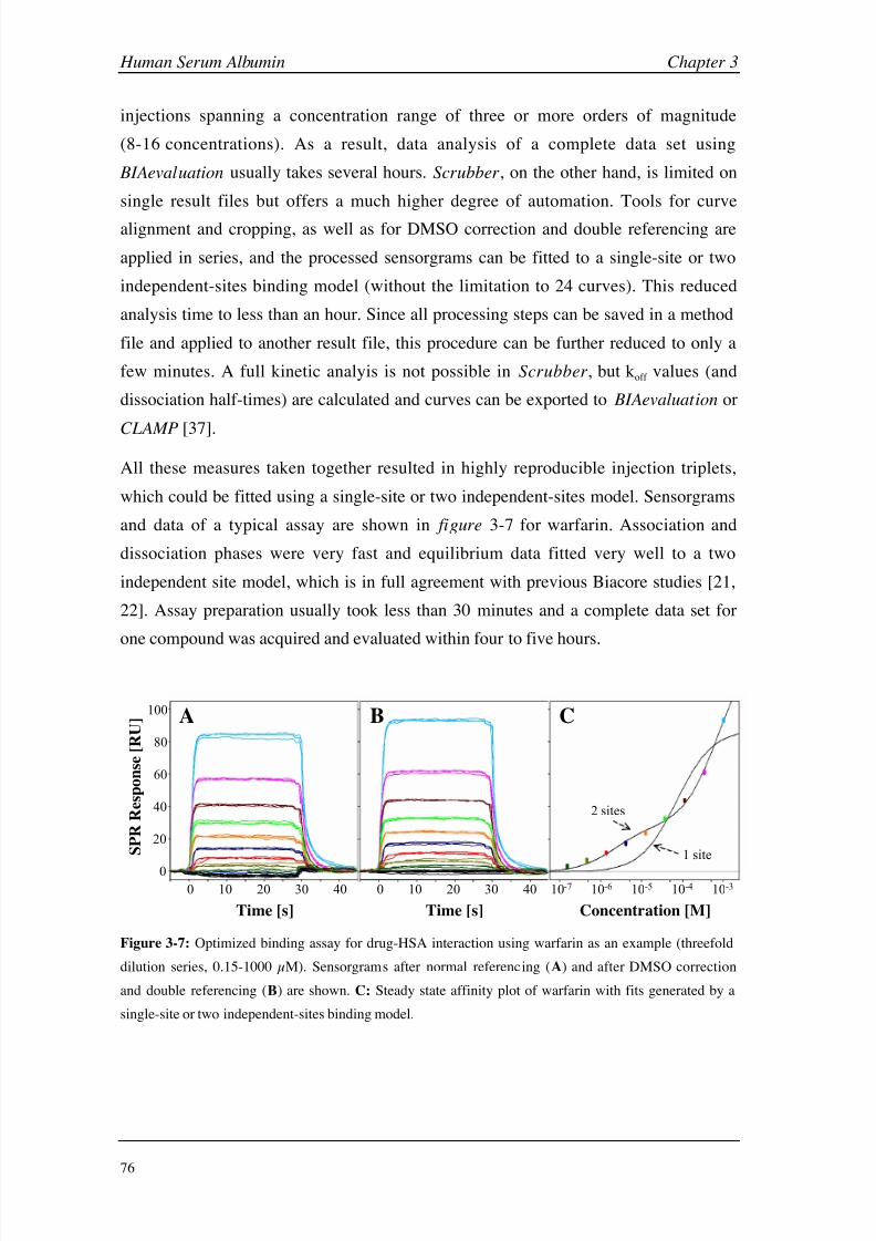
8/9/2019 DissB 7321 Mirroring
http://slidepdf.com/reader/full/dissb-7321-mirroring 90/278

8/9/2019 DissB 7321 Mirroring
http://slidepdf.com/reader/full/dissb-7321-mirroring 91/278
Chapter 3 Human Serum Albumin
77
3.3.2 HSA-drug binding studies
In principle, there are two major types of binding experiments, i.e. ranking and
high-resolution studies, that can be performed to get information about a drugs affinity
for HSA. In ranking studies, SPR signals at a single concentration are divided by thecompounds molecular weight. This normalized signal intensities give only qualitative
information about binding strength. To evaluate the optimized HSA assay, 16 different
compounds (Table 3-4, Fig. 3-5) were injected at a fixed concentration of 333 µM and
ranked by their normalized responses (Fig. 3-8).
-0.20
-0.10
0.00
0.10
0.20
0.30
0.40
0.50
0.60
0.70
S a l b
L T r p
A c S
A
Q d n e
P r e d
Q u
i n
S c y
A
C h o
A C o r t
D g
t x
D z p m
N i z p
I n d o
W a r f
N a p r
P p r o
N
o r m a l i z e d S P R R e s p o n s e [ R U / D
a ]
Figure 3-8: Ranking of 16 known HSA-binding drugs according to their normalized response at a
concentration of 333 µM (SPR signal divided by molecular weight). Abbreviations are listed in
table 3-4.
HSA is reported to predominantly bind hydrophobic (and anionic) drugs with high
affinity [4]. To correlate the ranked compounds with their physico-chemical properties
and biological relevance, the normalized responses were plotted against log P values
(Fig. 3-9A) and the bound fraction on plasma proteins (Fig. 3-9B).

8/9/2019 DissB 7321 Mirroring
http://slidepdf.com/reader/full/dissb-7321-mirroring 92/278
Human Serum Albumin Chapter 3
78
0
0.1
0.2
0.3
0.4
0.5
0.6
0.7
0 1 2 3 4
log P
N o r m a l i z e d S P R R e s p
o n s e [ R U / D a ]
9 90 99 99.9
% bound
PPro
Napr
Quin
Qdne
Salb
AcSAPred
ScyA
ChoA
Dgtx
Nitr
Indo
DzpmCort
Warf
Quin
Qdne
Nitr
PPro
Napr
Warf
Indo
Salb
AcSA
Pred ScyA
ChoA
Cort Dgtx
Dzpm
A B
Figure 3-9: Correlation of normalized SPR responses from ranking experiments and literature log
P
(A)and %bound values (B). To increase the resolution of high-affinity compounds %bound values were put
on a logarithmic scale. Abbreviations, references, log P and %bound values are listed in table 3-4.
Data from ranking experiments are in good agreement with literature [20]. Even small
differences between similar structures are clearly visible. For example, quinine is
ranked higher than quinidine, which is in agreement with plasma protein binding
studies of the two drugs [38]. Interestingly, these substances showed the most
prominent deviation from the correlation in both correlation plots. The differences in
the %bound-plot can be explained by the fact that the literature values refer to binding
to total plasma protein. As cationic drugs, quinine and quinidine bind strongly to
1-acid glycoprotein (AGP). HSA only contributes to around 35% of the total 70-90%
protein binding in the case of quinine [39]. Removing these drugs from the plot raises
the correlation coefficient significantly from 0.76 to 0.85, when additionally removing
nitrazepam it even increases to 0.91. The relatively high deviation in the log P-plot
might also be influenced by the cationic nature of these drugs, since binding of
positively charges compounds is unusual for HSA and therefore weaker than expected
for the whole plasma protein fraction. The higher standard deviation in the ranking
signal of acetylsalicylic acid might be explained by ionic effects or by transacetylation
upon binding, i.e. transfer of the acetyl group to Lys-199 of HSA [4].
All molecules (except L-tryptophan) showed very fast binding kinetics with a rapid
association phase and typical dissociation half-times at or below one second. Analyzing
the ranking sensorgrams in more detail led to a classification into four clearly
distinguishable classes:

8/9/2019 DissB 7321 Mirroring
http://slidepdf.com/reader/full/dissb-7321-mirroring 93/278
Chapter 3 Human Serum Albumin
79
A) Drugs showing a proper block signal (acetylsalicylic acid, cholic acid.
corticosterone, digitoxin, prednisone, quinidine, quinine)
B) Compounds with a multiphase association (diazepam, nitrazepam,
progesterone)
C) Compounds leading to a decreased baseline (naproxen) after the injection
D) Substances showing negative binding signals (L-tryptophan).
Examples of ranking curves are shown in figure 3-10. A complete overview can be
found in appendix B3.
0
0.2
0.4
0.6
N o r m a l i z e d S P R R e s p o n s e [ R U / D a ]
0
0.2
0.4
0.6
Time [s]
0 10 20 30 0 10 20 30
A B
C D
Figure 3-10: Examples of drug compound injections during ranking
experiments (333 µM, triplicate injections). Four classes of curve shapes can be
distinguished: proper block signals (A , digitoxin), multiphase association
(B, diazepam), negative post-injection baseline (C, naproxen), negative binding
signals (D, L-tryptophan).
Salbutamol could not be classified because of its low binding signal intensity. The two
coumarin derivatives warfarin and phenprocoumon only showed a small negative effect
on the post-injection baseline, while naproxen induced the most pronounced changes.
Multiphasic binding of nitrazepam was less obvious than those of diazepam andprogesterone, but resulted in an increased post-injection baseline. Interestingly, curve
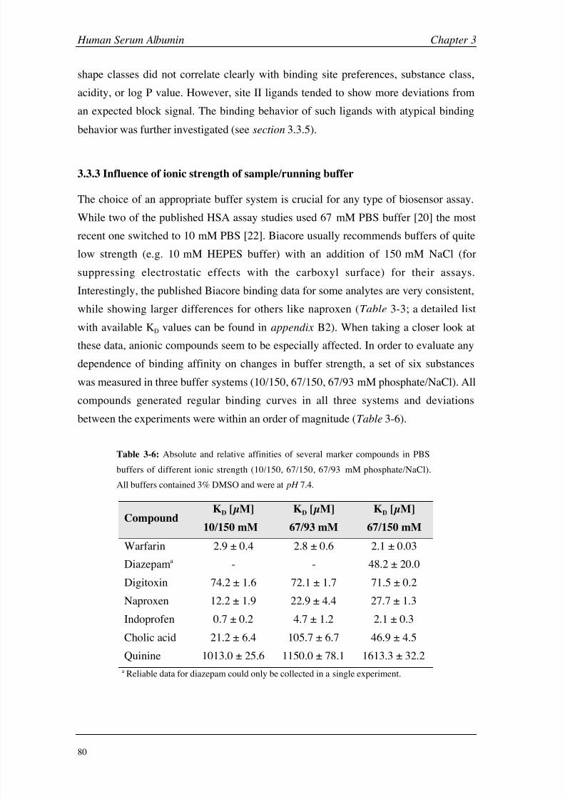
8/9/2019 DissB 7321 Mirroring
http://slidepdf.com/reader/full/dissb-7321-mirroring 94/278
Human Serum Albumin Chapter 3
80
shape classes did not correlate clearly with binding site preferences, substance class,
acidity, or log P value. However, site II ligands tended to show more deviations from
an expected block signal. The binding behavior of such ligands with atypical binding
behavior was further investigated (see section 3.3.5).
3.3.3 Influence of ionic strength of sample/running buffer
The choice of an appropriate buffer system is crucial for any type of biosensor assay.
While two of the published HSA assay studies used 67 mM PBS buffer [20] the most
recent one switched to 10 mM PBS [22]. Biacore usually recommends buffers of quite
low strength (e.g. 10 mM HEPES buffer) with an addition of 150 mM NaCl (for
suppressing electrostatic effects with the carboxyl surface) for their assays.
Interestingly, the published Biacore binding data for some analytes are very consistent,
while showing larger differences for others like naproxen (Table 3-3; a detailed list
with available KD values can be found in appendix B2). When taking a closer look at
these data, anionic compounds seem to be especially affected. In order to evaluate any
dependence of binding affinity on changes in buffer strength, a set of six substances
was measured in three buffer systems (10/150, 67/150, 67/93 mM phosphate/NaCl). All
compounds generated regular binding curves in all three systems and deviations
between the experiments were within an order of magnitude (Table 3-6).
Table 3-6: Absolute and relative affinities of several marker compounds in PBS
buffers of different ionic strength (10/150, 67/150, 67/93 mM phosphate/NaCl).
All buffers contained 3% DMSO and were at pH 7.4.
CompoundKD [µM]
10/150 mM
KD [µM]
67/93 mM
KD [µM]
67/150 mM
Warfarin 2.9 ± 0.4 2.8 ± 0.6 2.1 ± 0.03
Diazepama - - 48.2 ± 20.0
Digitoxin 74.2 ± 1.6 72.1 ± 1.7 71.5 ± 0.2
Naproxen 12.2 ± 1.9 22.9 ± 4.4 27.7 ± 1.3
Indoprofen 0.7 ± 0.2 4.7 ± 1.2 2.1 ± 0.3
Cholic acid 21.2 ± 6.4 105.7 ± 6.7 46.9 ± 4.5
Quinine 1013.0 ± 25.6 1150.0 ± 78.1 1613.3 ± 32.2
a Reliable data for diazepam could only be collected in a single experiment.

8/9/2019 DissB 7321 Mirroring
http://slidepdf.com/reader/full/dissb-7321-mirroring 95/278
Chapter 3 Human Serum Albumin
81
To visualize the susceptibility on buffer changes more clearly, all KD values were
divided (normalized) by the value in 10 mM PBS (Fig. 3-11).
0
1
2
3
4
5
6
7
8
Warf Napr Dgtx ChoA Indo Quin
R e l a t i v e A f f i n i t y [ r K D
]
Figure 3-11: Relative affinities (rKD) in PBS buffers pH 7.4 3% DMSO with different ionic strength (red
= 10 mM phosphate/150 mM NaCl, green = 67/93 mM, blue = 67/150 mM). All affinity values were
divided by the KD in 10/150 mM PBS.
Comparison of the relative binding affinities (Fig. 3-11) shows that uncharged ligands
like warfarin or digitoxin are nearly unaffected by changes in ion strength. Charged
ligands, however, are clearly dependant on both buffer capacity and ionic strength.
Two effects might be responsible for this behavior. First, the existence of a counter-ion
like sodium directly determines the charge and acidity of the anionic molecules,
therefore influencing ionic interactions or even conformational isoforms. Such a charge
dependant behavior was described for several indole derivatives [40]. Second, chloride
ions are known to interact with HSA (see section 3.1.3) and compete with several
ligands. Especially site II ligands often show a significant competition behavior with
chloride ions [4]. For example, the medium-chain fatty acid octanoate showed onlyone-third of the affinity in 130 mM chloride buffer compared to pure phosphate buffer
[41]. Similar effects have been demonstrated for L-tryptophan [40, 42] and diazepam
[4]. These findings demonstrate the importance of carefully matching both buffer
capacities and ionic strength between experiments.
3.3.4 Influence of HSA redox state on drug binding
Around 30% of the unpaired cysteine residue (Cys-34) of HSA forms mixed disulfides
with GSH and cysteine, dimers or higher oxidized states (see section 3.1.2).
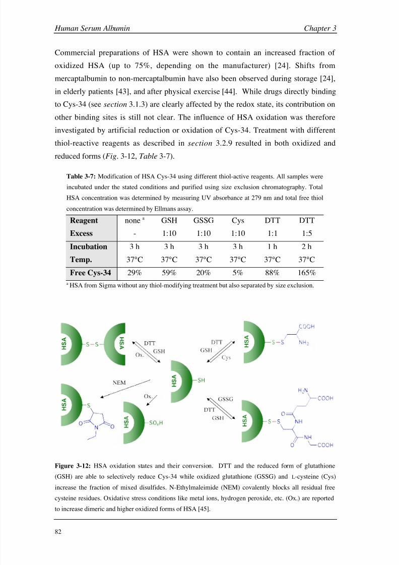
8/9/2019 DissB 7321 Mirroring
http://slidepdf.com/reader/full/dissb-7321-mirroring 96/278

8/9/2019 DissB 7321 Mirroring
http://slidepdf.com/reader/full/dissb-7321-mirroring 97/278
Chapter 3 Human Serum Albumin
83
With a fraction of around 30% free Cys-34, the untreated HSA from Sigma is exactly
within the usual ranges of commercial batches (25-59% [24]). As expected, the reduced
form of glutathione (GSH) significantly increased the free fraction while the oxidized
form (GSSG) slightly decreased it. The most effective oxidizing agent was found to be
L-cysteine, which formed mixed disulfides with Cys-34 very effectively after 3
hours at
37°C. N -Ethylmaleimide could also successfully be used to oxidize Cys-34 (less than
5% free Cys), but this would lead to a non-natural form of the HSA thiol and might
influence binding properties in a different way (size, charge state) than glutathione and
cysteine. Incubation of HSA with a fivefold excess of DTT for several hours [6] clearly
led to the reduction of more than one cysteine (i.e. 165% instead of 100%). However,
less than one disulfide bridge was cleaved in average under these conditions, since this
led to three free cysteine residues (i.e. 300%) per molecule HSA. To fully avoidcleavage of internal disulfide bridges, both the DTT excess and the incubation time
were reduced to yield a free fraction of nearly 90%. The relatively high tolerability of
reducing agents is in good agreement with the observation that the helical environment
of HSA protects the disulfide bridges from reduction [3].
After immobilization of untreated, reduced (DTT, 1:1) and oxidized (Cys 1:10) HSA to
different flow cells of a sensor chip, binding properties were evaluated by injection of
marker compounds targeting the two major drug binding sites (Sudlow sites I and II,
Table 3-2). To avoid any ionic masking effects, the experiments were repeated in three
buffer systems with varying buffer strength (10/150, 67/150, 67/93 mM
phosphate/NaCl). Binding affinities were determined in the same way as in
high-resolution experiments and compared between the HSA surfaces (Table 3-8).
Table 3-8: Absolute and relative affinities of several marker compounds on native, reduced, and
oxidized HSA surfaces. All values are averages over three experiments.
CompoundKD [µM]
native
KD [µM]
reduced
KD [µM]
oxidizedrK
D red
1 rKD ox
1
Warfarin 2.7 ± 0.5 2.9 ± 0.6 2.2 ± 0.2 1.1 0.8
Diazepam2 27.4 49.8 67.3 1.8 2.5
Digitoxin 72.6 ± 1.2 73.2 ± 2.3 72.0 ± 1.9 1.0 1.0
Naproxen 23.3 ± 7.8 20.5 ± 8.4 19.0 ± 8.2 0.9 0.9
Indoprofen 2.3 ± 1.3 2.5 ± 2.2 2.7 ± 2.7 1.2 0.9
Cholic Acid 58.8 ± 36.0 57.2 ± 48.6 57.8 ± 45.5 0.9 1.0
1 rKD red/ox = relative affinities of reduced/oxidized surfaces compared to the native HSA surface.2 reliable data for diazepam could only be collected in a single experiment; therefore, no
standard deviation is specified.

8/9/2019 DissB 7321 Mirroring
http://slidepdf.com/reader/full/dissb-7321-mirroring 98/278
Human Serum Albumin Chapter 3
84
Only Diazepam showed a significant difference in affinity between the surfaces.
However, this is caused mainly by the unusual binding mode of this substance
(see section 3.3.2) and is more than ten times higher than in literature [3]. No one of the
other compounds showed a significant change in its binding affinity in dependence of
the redox state. On the other side, the redox stability of the surfaces can hardly be
tested. Even though the reduced and oxidized HSA in solution was found to be stable at
4°C for several days (only a minor increase of less than 3% within one week was
detected for untreated HSA), redox processes - predominantly oxidation - could occur
on the chip during the course of experiments. The higher standard deviations for
anionic compounds are mainly caused by the differences in buffer strength and are in
agreement with the experiments in section 3.3.3.
3.3.5 Ligand-induced conformational changes
When comparing the sensorgrams of the investigated drug compounds from ranking
assays (see section 3.3.2), diazepam and L-tryptophan showed atypical curve shapes
and were therefore analyzed more extensively. Interestingly, no sensorgrams for these
two compounds have been published so far, despite their importance as known binders
or even marker drugs. One focus of our studies was the possibility of conformational
changes induced by these ligands. Several HSA-binding drugs are reported to showallosteric effects [12] and ligand-induced conformational changes were recently
expected to cause changes in SPR signal intensity [32] (see chapter 7).
L-tryptophan was the only compound causing negative binding signals during ranking.
This unusual behavior could be confirmed in a high-resolution assay (Fig. 3-13A).
However, mirroring the sensorgrams (Fig. 3-13B) led to concentration-dependant
binding curves, which could be plotted and fitted using steady state affinity analysis
(Fig.
3-13C). Triplicates showed a much higher accuracy and fitted better to a 1:1binding model when the signal was evaluated just after injection end (i.e. 32-35 s from
injection start) rather than on the steady state phase (25-27 s). A possible explanation of
this behavior might be an overlay of a negative signal effect caused by a
conformational change and a smaller positive signal contribution of increasing surface
concentrations of L-tryptophan. After injection end, the dissociation signal is generated
only by one component (most likely the conformational change). The KD value before
and after injection end were 165±5.6 µM and 173±7.4 µM, respectively. These values
are in good agreement with results from capillary electrophoresis studies (150
µM) [18].
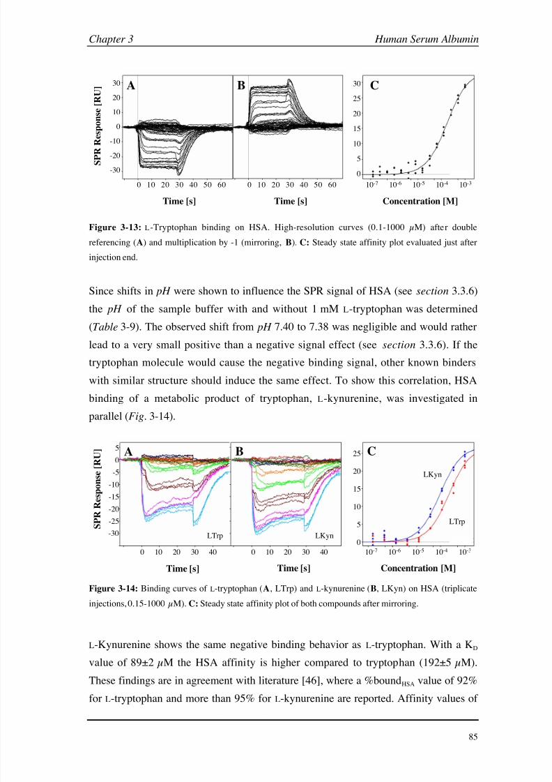
8/9/2019 DissB 7321 Mirroring
http://slidepdf.com/reader/full/dissb-7321-mirroring 99/278
Chapter 3 Human Serum Albumin
85
30
20
10
0
-10
-20
-30
0 10 20 30 40 50 60 0 10 20 30 40 50 60
S P R R e
s p o n s e [ R U ]
30
25
20
15
10
5
0
10-7 10-6 10-5 10-4 10-3
Time [s] Time [s] Concentration [M]
A B C
Figure 3-13: L-Tryptophan binding on HSA. High-resolution curves (0.1-1000 µM) after double
referencing (A) and multiplication by -1 (mirroring, B). C: Steady state affinity plot evaluated just after
injection end.
Since shifts in pH were shown to influence the SPR signal of HSA (see section 3.3.6)
the pH of the sample buffer with and without 1 mM L-tryptophan was determined
(Table 3-9). The observed shift from pH 7.40 to 7.38 was negligible and would rather
lead to a very small positive than a negative signal effect (see section 3.3.6). If the
tryptophan molecule would cause the negative binding signal, other known binders
with similar structure should induce the same effect. To show this correlation, HSA
binding of a metabolic product of tryptophan, L-kynurenine, was investigated in
parallel (Fig.
3-14).
5
0
-5
-10
-15
-20
-25 S P R R
e s p o n s e [ R U ]
-30
0 10 20 30 40
Time [s]
0 10 20 30 40
Time [s]
25
20
15
10
5
0
10-7 10-6 10-5 10-4 10-3
Concentration [M]
CBA
LKyn
LTrp
LKynLTrp
Figure 3-14: Binding curves of L-tryptophan (A, LTrp) and L-kynurenine (B, LKyn) on HSA (triplicate
injections, 0.15-1000 µM). C: Steady state affinity plot of both compounds after mirroring.
L-Kynurenine shows the same negative binding behavior as L-tryptophan. With a KD
value of 89±2 µM the HSA affinity is higher compared to tryptophan (192±5 µM).
These findings are in agreement with literature [46], where a %boundHSA value of 92%for L-tryptophan and more than 95% for L-kynurenine are reported. Affinity values of
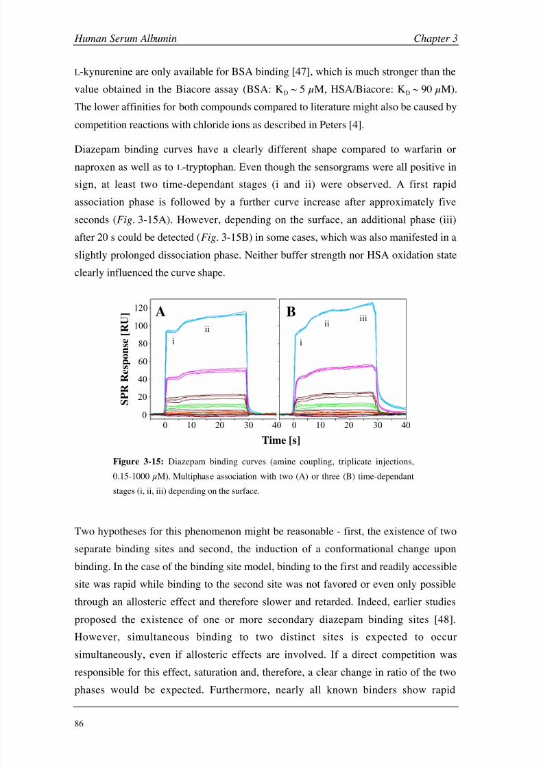
8/9/2019 DissB 7321 Mirroring
http://slidepdf.com/reader/full/dissb-7321-mirroring 100/278
Human Serum Albumin Chapter 3
86
L-kynurenine are only available for BSA binding [47], which is much stronger than the
value obtained in the Biacore assay (BSA: KD ~ 5 µM, HSA/Biacore: KD ~ 90 µM).
The lower affinities for both compounds compared to literature might also be caused by
competition reactions with chloride ions as described in Peters [4].
Diazepam binding curves have a clearly different shape compared to warfarin or
naproxen as well as to L-tryptophan. Even though the sensorgrams were all positive in
sign, at least two time-dependant stages (i and ii) were observed. A first rapid
association phase is followed by a further curve increase after approximately five
seconds (Fig. 3-15A). However, depending on the surface, an additional phase (iii)
after 20 s could be detected (Fig. 3-15B) in some cases, which was also manifested in a
slightly prolonged dissociation phase. Neither buffer strength nor HSA oxidation state
clearly influenced the curve shape.
0 10 20 40
Time [s]
0
20
40
60
100
S
P R R
e s p o n s e [ R U ]
80
30
A
0 10 20 4030
120 B
i
ii
i
ii iii
Figure 3-15: Diazepam binding curves (amine coupling, triplicate injections,
0.15-1000 µM). Multiphase association with two (A) or three (B) time-dependant
stages (i, ii, iii) depending on the surface.
Two hypotheses for this phenomenon might be reasonable - first, the existence of two
separate binding sites and second, the induction of a conformational change upon
binding. In the case of the binding site model, binding to the first and readily accessible
site was rapid while binding to the second site was not favored or even only possible
through an allosteric effect and therefore slower and retarded. Indeed, earlier studies
proposed the existence of one or more secondary diazepam binding sites [48].
However, simultaneous binding to two distinct sites is expected to occur
simultaneously, even if allosteric effects are involved. If a direct competition was
responsible for this effect, saturation and, therefore, a clear change in ratio of the twophases would be expected. Furthermore, nearly all known binders show rapid
8/9/2019 DissB 7321 Mirroring
http://slidepdf.com/reader/full/dissb-7321-mirroring 101/278
Chapter 3 Human Serum Albumin
87
association and dissociation phases, whereas the second phase of diazepam binding is
clearly slower. Conformational changes, on the other side, may vary from fast
(e.g. 100 ms for the N-F transition) to rather slow (e.g. in the case of N-B transitions).
The time lapse between the two phases observed in the binding curve might be
explained more easily with the conformational model. Since not every change in
conformation is expected to be readily visible as an SPR shift (see chapter 7), only later
stages might lead to a detectable change in shape or charge distribution.
Interestingly, both L-tryptophan and diazepam show one slow binding phase, indicating
a possibly common mechanism in the signal effect. Whereas the slow step occurs
during association in the case of diazepam, L-tryptophan possesses a decelerated
dissociation rate. The interference by diazepam with tryptophan binding has been
demonstrated [4, 49]. This leads to the conclusion that this specific location within
Sudlow site II might be especially susceptible to conformational changes upon ligand
binding.
However, such a hypothesis should be verified by another method. One very reliable
way of proofing conformational changes upon ligand binding is the use of X-ray
crystallography. Unfortunately, HSA crystal structure determination is rather difficult
[3] and there are only a few co-crystallization structures available. The only single drug
molecules bound to HSA are the anesthetics halothane and propofol, which are notreadily available for SPR analysis due to size and volatility issues and seem to rather
stabilize than change HSA conformation [50]. In addition, there is a HSA structure in
complex with warfarin, but it also contains fatty acid molecules (myristic acid), which
themselves induce a large conformational change and enhance warfarin binding.
Therefore, only small conformational changes could be detected when compared to the
HSA-myristate complex without warfarin [13]. No high-resolution crystals are
available for site II binders like diazepam or L-tryptophan.
Circular dichroism and fluorescence measurement are also two well-established
methods for the detection of conformational changes. However, since fluorescence
assays monitor changes of the single intrinsic tryptophan residue (Trp-214), binding of
extrinsic tryptophan molecules will interfere with such an analysis. Circular dichroism,
on the other side, detects the optical activity of asymmetric molecules in the far and
near UV range. Even though it is possible to see conformational changes using this
method, differentiation between changes of the protein and the drug is very difficult.
Even molecules that are achiral show optical activity when immobilized at a certain
conformation upon protein binding [19].
8/9/2019 DissB 7321 Mirroring
http://slidepdf.com/reader/full/dissb-7321-mirroring 102/278
Human Serum Albumin Chapter 3
88
A biointeraction chromatography study published recently [51] supports the theory of
ligand-induced conformational changes by L-tryptophan. While an excess of phenytoin
in the mobile phase only showed a competitive interaction behavior on L-Trp binding,
the reversed experiment (excess L-Trp) altered phenytoin binding in a negative
allosteric way, as it is typical for conformational effects. It was also proposed that the
binding site of L-tryptophan might be located deeper in the pocket of Sudlow site II
than the one of phenytoin [51].
While no reference literature could be found about negative binding signals induced by
ligand binding, there is one study [32] that also reported negative post-injection
baselines similar to those observed in the case of naproxen and other anti-inflammatory
and coumarin drugs. This effect was demonstrated for the interaction of carbohydrates
with mannose binding protein and was also attributed to conformational changes. More
evidence for this hypothesis came from the example of tissue transglutaminase binding,
where binding of calcium ions (35 Da) induced much higher binding signals
(> 1,000 RU) than expected from a simple mass increase [32]. Since transglutaminase
is known to undergo significant conformational changes upon binding of calcium ions
[52], this was explained to be the main reason for this behavior.
The observed anomalies in the binding curves were highly reproducible over different
experiments with changes of flow cells, chips, or analyte preparation. However,depending on the HSA surface, effects like multiphasic association and especially
negative post-injection signals were more or less pronounced. No obvious correlation
between changed parameters (flow cell, immobilization density, HSA oxidation state)
has been found. A possible explanation is that small fluctuations in the immobilization
procedure (activation time and efficacy, immobilization buffer pH , HSA purity) might
influence the way HSA is fixed on the carboxymethyl dextran surface and, as a
consequence, its susceptibility to conformational changes. Interestingly, both
Biacore-HSA studies performed in 67
mM PBS [20, 21] only showed ranking and KD
values but no sensorgrams (except for warfarin in [21]). The binding curves published
in the most recent Biacore-HSA study [22] show nearly no negative post-injection
effect for naproxen and warfarin but clearly for salicylic acid. All those studies used a
slightly higher immobilization pH (5.2 instead of 5.0), which might have an influence
on this effect.

8/9/2019 DissB 7321 Mirroring
http://slidepdf.com/reader/full/dissb-7321-mirroring 103/278
Chapter 3 Human Serum Albumin
89
3.3.6 pH -induced conformational changes
A possible contribution of conformational changes of albumin on SPR signals as
suspected in the case of ligand binding was further evaluated using a different
approach. HSA is known to undergo several conformational changes upon alteration of pH [3]. Different studies are published, where this change was followed by circular
dichroism and intrinsic fluorescence [28], changes in Cys-34 reactivity [53], or
modified ligand binding behavior [4]. Since such changes of the protein isoform might
also change the electron density around the gold surface and therefore the SPR signal,
this effect was studied by injecting buffer blanks at various pH values on a HSA
surface (Fig. 3-16).
-2000
0
2000
4000
6000
8000
0 2 4 6 8 10 12
pH value
S
P R r
e s p o n s e [ R U ]
F-E N-F N-B
Figure 3-16: pH -induced conformational changes monitored using Biacore. Blank injections were
performed with buffer systems covering a pH range from 1.5 to 9.5 ( pH 1.5 - 3.0: 10 mM glycine; blue,
pH 2.0 - 7.0: 10 mM citrate; red, pH
6.0 - 8.5: 10 mM phosphate; green, pH 8.0 - 9.5; 10 mM borate;
brown). The three known transitions are indicated at the bottom (F-E, N-F, N-B).
The observed pH -induced changes in SPR signal-intensity correlate very well with the
described mechanism of conformational changes. The negative signals below pH 3.5
can be attributed to an acid induced expansion (or F-E transition), where the expanded
structure leads to a decrease in electron density at the surface. The N-B transition
between pH 7 and 9 was also visible as a (smaller) decrease in signal below baseline.
With 7,000
RU the most remarkable change was a positive peak between pH
3.0 and6.0. Here again, a conformational change (N-F transition) is described in literature [28].

8/9/2019 DissB 7321 Mirroring
http://slidepdf.com/reader/full/dissb-7321-mirroring 104/278
Human Serum Albumin Chapter 3
90
The huge structural changes in this pH range were confirmed by measuring alterations
of the intrinsic fluorescence of Trp-214 (Fig. 3-17).
1000
1200
1400
1600
1800
2000
320 330 340 350
emission wavelength [nm]
F l u o r e s c e n c e ( a r b i t r a r y u n i t )
pH 2.0
pH 4.0
pH 7.0
A
324
326
328
330
332
334
336
338
340
0.0 1.0 2.0 3.0 4.0 5.0 6.0 7.0 8.0
pH value
E m i s s i o n
m a x
[ n m ]
B
Figure 3-17: pH -induced conformational changes of HSA in 10
mM citrate buffer, monitored by
intrinsic fluorescence measurement of Trp-214. A: Scan of Trp emission after excitation at 295 nm.
Emission spectra in citrate buffer pH 2, 4, and 7 are overlaid. B: Shift in emission maxima between pH 2
and 7 (solid line). SPR curve in the same range is shown as dashed line (simplified from Fig. 3-16).
The emission maximum shifts based on the change in the microenvironment of Trp-214
correlate very well with the signals observed with Biacore (Fig. 3-17B). The relatively
high standard deviations in the emission scan are caused by the shallow maxima at
higher pH values compared to those at pH
2 (Fig.
3-17A). However, since the peak
maximum exactly lies below the pI value of HSA, this effect could also be caused by
higher protonation of amine groups and, as a result, increased surface attraction
between HSA and the carboxymethyl dextran matrix. This second hypothesis is
supported by the study of Paynter et al. [54], where electrostatic interactions around the
pI values of three different proteins could be correlated with changes in SPR signal
intensity. A possible electrostatic surface attraction effect was investigated by injecting
HSA at low concentration in the same series of citrate buffer over an unmodified
sensor-chip surface (Fig. 3-18).
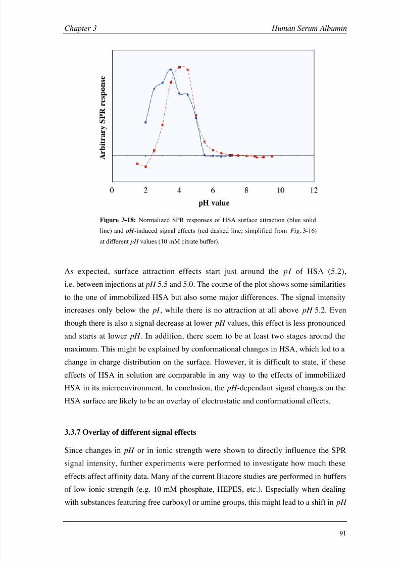
8/9/2019 DissB 7321 Mirroring
http://slidepdf.com/reader/full/dissb-7321-mirroring 105/278
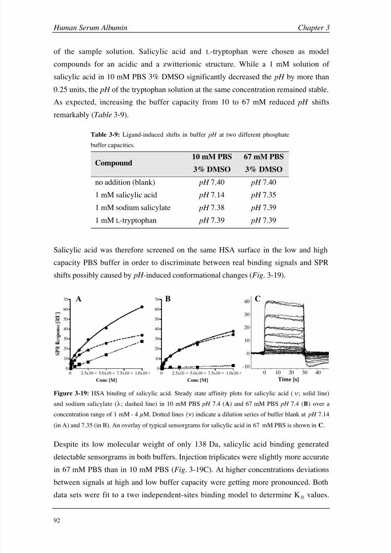
8/9/2019 DissB 7321 Mirroring
http://slidepdf.com/reader/full/dissb-7321-mirroring 106/278
Human Serum Albumin Chapter 3
92
of the sample solution. Salicylic acid and L-tryptophan were chosen as model
compounds for an acidic and a zwitterionic structure. While a 1 mM solution of
salicylic acid in 10 mM PBS 3% DMSO significantly decreased the pH by more than
0.25 units, the pH of the tryptophan solution at the same concentration remained stable.
As expected, increasing the buffer capacity from 10 to 67
mM reduced pH shifts
remarkably (Table 3-9).
Table 3-9: Ligand-induced shifts in buffer pH at two different phosphate
buffer capacities.
Compound10 mM PBS
3% DMSO
67 mM PBS
3% DMSO
no addition (blank) pH 7.40 pH 7.40
1
mM salicylic acid pH
7.14 pH
7.35
1 mM sodium salicylate pH 7.38 pH 7.39
1 mM L-tryptophan pH 7.39 pH 7.39
Salicylic acid was therefore screened on the same HSA surface in the low and high
capacity PBS buffer in order to discriminate between real binding signals and SPR
shifts possibly caused by pH -induced conformational changes (Fig. 3-19).
10 mM PBS
0 2.5x10 -4 5.0x10 -4 7.5x10 -4 1.0x10⁻³
0
10
20
30
40
50
60
70
Conc [M]
67 mM PBS
0 2.5x10 -4 5.0x10 -4 7.5x10 -4 1.0x10 -3
0
10
20
30
40
50
60
70
Conc [M]
40
30
20
10
0
-10
0 10 20 30 40
Time [s]
A B C
Figure 3-19: HSA binding of salicylic acid. Steady state affinity plots for salicylic acid ( ; solid line)
and sodium salicylate (; dashed line) in 10 mM PBS pH 7.4 (A) and 67 mM PBS pH 7.4 (B) over a
concentration range of 1 mM - 4 µM. Dotted lines () indicate a dilution series of buffer blank at pH 7.14
(in A) and 7.35 (in B). An overlay of typical sensorgrams for salicylic acid in 67 mM PBS is shown in C.
Despite its low molecular weight of only 138 Da, salicylic acid binding generated
detectable sensorgrams in both buffers. Injection triplicates were slightly more accurate
in 67 mM PBS than in 10 mM PBS (Fig. 3-19C). At higher concentrations deviations
between signals at high and low buffer capacity were getting more pronounced. Bothdata sets were fit to a two independent-sites binding model to determine KD values.

8/9/2019 DissB 7321 Mirroring
http://slidepdf.com/reader/full/dissb-7321-mirroring 107/278
Chapter 3 Human Serum Albumin
93
While salicylic acid in 67 mM PBS had a KD of 124±87 µM, the affinity for the same
interaction in 10 mM PBS was significantly lower (1460±771 µM). At the highest
concentration (1 mM salicylic acid) the signal difference between the two systems is
approximately 30-40 RU. A linear signal increase was observed on the same HSA
surface, when a dilution series of a 10
mM PBS buffer at pH
7.14 in running buffer
(Fig. 3-19A&B). This clearly demonstrated that the binding data for free salicylic acid
in 10 mM PBS are an overlay of a mass increase and a pH -induced signal change as
observed in section 3.3.6. As expected, there was no pH effect of sodium salicylate in
either buffer system (Table 3-9).
Figure 3-20: L-Tryptophan/HSA assay in 10 mM PBS with 3% DMSO (PBS3D) pH 7.40 as running
buffer. Sensorgrams at various concentrations (8-1000 µM) in sample buffer PBS3D at pH 7.40 (A) and
pH 7.14 (B). Highest concentrations (1 mM) are indicated by an arrow. C: Average responses of samples
at pH 7.40 (solid line) and pH
7.14 (dashed line) are plotted against concentration. A twofold dilution
series of PBS3D pH
7.14 in running buffer is shown as dotted line.

8/9/2019 DissB 7321 Mirroring
http://slidepdf.com/reader/full/dissb-7321-mirroring 108/278
Human Serum Albumin Chapter 3
94
In order to demonstrate this effect more clearly, the experiment was extended to
L-tryptophan, which typically generated negative binding signals (see section 3.3.5).
For this purpose 1 mM tryptophan was directly dissolved in 10 mM PBS pH 7.14
(equivalent to the sample buffer pH of 1 mM salicylic acid). This solution was further
diluted and screened in 10
mM PBS pH
7.4 as running buffer. Dilution series of
tryptophan at pH 7.4 and a buffer blank at pH 7.14 in running buffer were also screened
as controls (Fig. 3-20).
While L-tryptophan at pH 7.4 showed a normal but negative binding curve, a serial
dilution of PBS pH 7.14 generated an almost linear signal increase up to 40 RU. An
overlay of normal binding curve (up to 111 µM) and a pH -induced signal increase
(111 - 1000 µM) could be detected in the case of tryptophan at pH 7.14. While the
overlay of analyte-induced signals and pH -induced conformational changes are obvious
in the case of L-tryptophan because of different directionalities, it is impossible to
separate the two events in the case of salicylic acid. This implies that free acids should
be avoided if possible and that the pH value of starting concentration should be
carefully controlled.
3.3.8 Thiol Immobilization of HSA
Since oxidation of the unpaired cysteine residue 34 did not change the binding behaviorof HSA (see section 3.3.4) this site was chosen for an oriented immobilization
approach on a thiol-functionalized sensor chip. Thiol-coupled surfaces are expected to
be fully regenerable, therefore saving preparation time and cost. In addition, a change
of immobilization site from primary amine to a single thiol might have additional
benefits. Cys-34 is located in domain I of HSA while the two primary drug binding
sites are positioned in domains II and III respectively. Binding site II (domain III) is
reported to consist of a hydrophobic cavity next to a cationic patch. If this patch
consists of lysine residues, they could be blocked by the immobilization, which would
directly influence the binding signals (either by steric effects, conformational changes
or blocked functional groups). The crystal structure around the binding sites was
therefore analyzed in more detail (Fig. 3-21).
Potential target groups for amine coupling reactions can be identified around both
major drug binding sites. However, since there are much more surface-accessible lysine
residues distributed over the whole protein surface, a direct effect on the binding
signals may be possible, but difficult to quantify. Binding of atypical ligands like
diazepam or L-tryptophan is therefore of special interest.

8/9/2019 DissB 7321 Mirroring
http://slidepdf.com/reader/full/dissb-7321-mirroring 109/278
Chapter 3 Human Serum Albumin
95
BA
Figure 3-21: Distribution of surface-accessible lysine residues over the HSA crystal structures with
focus on Sudlow binding site I (A, green) and site II (B, yellow). Primary amine groups of lysine
residues are highlighted in red. Arrows indicate potential amine coupling targets around the binding sites
and a dotted orange circle (in A) represents the approximate position of Cys-34 (hidden on the opposite
side of the protein).
Optimization of coupling conditions
Standard ligand thiol immobilization using immobilized PDEA [29] did only lead to
low-density surfaces (appendix B4). Therefore, a novel in-situ immobilization method
was developed. In this approach, the reduced form of glutathione (GSH) was used as a
mild reducing agent (see section 3.3.4) and DTNB as an activator group. Incubation of
HSA with a 28-fold excess of both GSH and DTNB at low pH generated a reactive
HSA fraction, which could be directly immobilized on the thiol chip (Fig. 3-22).
Removal of one or both agents dramatically decreased the HSA surface-density (Fig. 3-22B).
0
5
10
15
0 200 400 600 800 1000
S P R R e s p o n s e [ k R U ]
Time
b i w s r w
O
NH
S
S (Cys-34)
HSA
cysteamine
sensor chip
matrix
A B
HDG
HD
HG
Figure 3-22: Thiol immobilization of HSA on a thiol-functionalized sensor chip. A: Schematic overview
of the assay surface. B: Immobilization sensorgrams of HSA mixtures with GSH (blue, HG), DTNB
(red, HD) and a combination of both agents (purple, HDG) showing different phases of the
immobilization process: b
=
baseline, i
=
injection of the mixture, w
=
NaOH wash, s
=
stable HSA
surface, r = DTT reduction.

8/9/2019 DissB 7321 Mirroring
http://slidepdf.com/reader/full/dissb-7321-mirroring 110/278
Human Serum Albumin Chapter 3
96
Even though glutathione is involved in the formation of disulfide bonds both in vivo
and in vitro its main function seems to be the one of a reducing agent in this case. This
was supported by the finding that another mild reducing agent, 2-mercaptoethylamine
(MEA), led to similar result. DTT could also be used, but surfaces generated this way
were less stable (appendix B5). Lower pH values during immobilization increased
surface density and stability much more than expected from a simple surface attraction.
The reason for this tendency might be an expansion of the HSA molecule during the
pH -induced N-F or F-E transition (see section 3.1.2), which makes Cys-34 more
accessible for a reaction with the surface [4]. In addition, Cys-34 has an unusual low
pKSH value of 7 (compared to pKSH 8.5 for free cysteine) making it to the most reactive
thiol in human serum [53]. This atypical property of Cys-34 was demonstrated by its
reactivity to 2,2-dithiopyridine, which is structurally related to DTNB. The authorsreported a minimum in rate constant at pH 5 and two maxima around pH 2.5 (caused
by the N-F-E transitions) and above pH 8, where the thiols are fully ionized [53].
Binding of long-chain fatty acids to HSA is known to induce massive conformational
changes [55]. These transitions also affect the crevice where Cys-34 is situated and
protected from oxidation. When adding three or more molar equivalents of oleic acid to
HSA, the solvent accessibility of Cys-34 is remarkably increased, as demonstrated by
fluorescent methods [56]. This effect is also visible when superimposing the crystal
structures of HSA with and without fatty acids bound (Fig. 3-23A). Cys-34 is only
accessible on the Connolly surface in the co-crystal with fatty acid molecules
(Fig. 3-23B). Addition of oleic acid to the HSA immobilization mixture was therefore
believed to increase immobilization density. Surprisingly, oleic acid did not improve
but decreased immobilization efficacy, resulting in lower surface densities
(Fig. 3-23C).
Finally, different ratios of HSA and the two additives (GSH and DTNB) as well as
variations in immobilization pH and incubation time were evaluated. A ratio of 1:28
was found to be best suited, and two incubation steps (10 min without and 90 min in
immobilization buffer) were necessary to yield optimum densities.

8/9/2019 DissB 7321 Mirroring
http://slidepdf.com/reader/full/dissb-7321-mirroring 111/278
Chapter 3 Human Serum Albumin
97
domain
II
A
top view
Cys-34Cys-34
Cys-34Cys-34
B
B
0
2
4
6
8
10
12
0 200 400 600 800 1000
S P R R
e s p o n s e
[ k R U ]
Time [s]
C
HDG
HDGO
H
Figure 3-23: Effect of oleic acid on HSA structure and thiol coupling efficacy. A: Overlay of crystal
structures of HSA alone (white) and in complex with oleic acid (blue). Oleic acid molecules are shown
in red in a top view albumin (bottom). B: View on domain I of the protein surface of HSA with the
position (yellow circles) and accessibility of Cys-34 (orange) in albumin with (bottom) and without oleic
acid (top). C: Overlay plot of thiol coupling sensorgrams of untreated HSA (grey, H), and in the mixture
with GSH/DTNB (purple, HDG) or GSH/DTNB/oleic acid (green, HDGO).
Evaluation of thiol-coupled surfaces
The stability of thiol-coupled surfaces was very high in running buffer. Compared to
amine-coupled surfaces, where the surface density decreases during several hours and a
special stabilization procedure is needed (see section 3.2.4), nearly no surface bleeding
was detected in the case of thiol immobilization (Fig. 3-24A). The same trend was
visible for warfarin activity: while the affinity after 10 hours was 87% of the initial
value in the case of amine-coupled surfaces, thiol-coupled surfaces retained 98%
activity after the same time period. The surprisingly high stability might be caused by
the protective microenvironment of Cys-34 (see section 3.1.2). Nevertheless, surfaces
generated in this way could easily be reduced with DTT and re-immobilized without
further treatment (Fig. 3-24B).
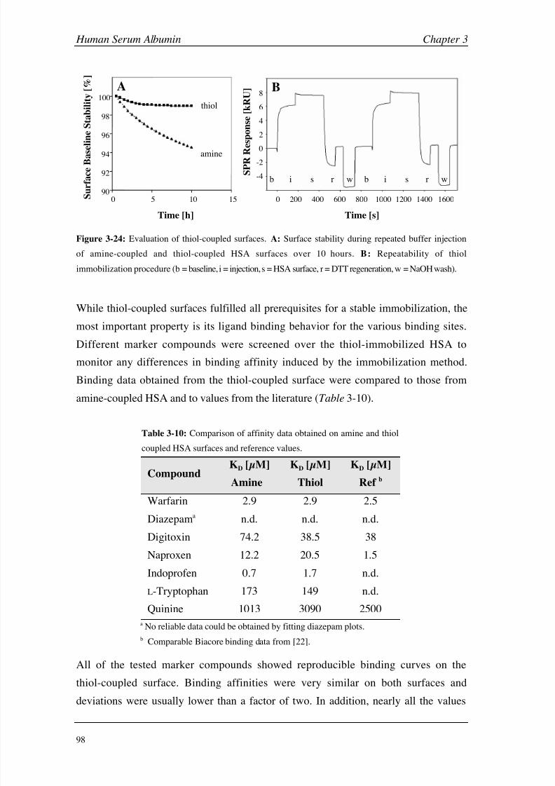
8/9/2019 DissB 7321 Mirroring
http://slidepdf.com/reader/full/dissb-7321-mirroring 112/278
Human Serum Albumin Chapter 3
98
90
92
94
96
98
100
0 5 10 15
Time [h]
S u r f a c e B a s e l i n
e S t a b i l i t y [ % ]
thiol
amine
S P R R e s p
o n s e [ k R U ]
-2
0
2
4
6
8
-4b i s r w b i s r w
0 400 600200 800 1000 1200 1400 1600
Time [s]
A B
Figure 3-24: Evaluation of thiol-coupled surfaces. A: Surface stability during repeated buffer injection
of amine-coupled and thiol-coupled HSA surfaces over 10 hours. B: Repeatability of thiol
immobilization procedure (b = baseline, i = injection, s = HSA surface, r = DTT regeneration, w = NaOH wash).
While thiol-coupled surfaces fulfilled all prerequisites for a stable immobilization, the
most important property is its ligand binding behavior for the various binding sites.
Different marker compounds were screened over the thiol-immobilized HSA to
monitor any differences in binding affinity induced by the immobilization method.
Binding data obtained from the thiol-coupled surface were compared to those from
amine-coupled HSA and to values from the literature (Table 3-10).
Table 3-10: Comparison of affinity data obtained on amine and thiol
coupled HSA surfaces and reference values.
CompoundKD [µM]
Amine
KD [µM]
Thiol
KD [µM]
Ref b
Warfarin 2.9 2.9 2.5
Diazepama n.d. n.d. n.d.
Digitoxin 74.2 38.5 38
Naproxen 12.2 20.5 1.5
Indoprofen 0.7 1.7 n.d.
L-Tryptophan 173 149 n.d.
Quinine 1013 3090 2500
a No reliable data could be obtained by fitting diazepam plots.
b Comparable Biacore binding data from [22].
All of the tested marker compounds showed reproducible binding curves on the
thiol-coupled surface. Binding affinities were very similar on both surfaces and
deviations were usually lower than a factor of two. In addition, nearly all the values

8/9/2019 DissB 7321 Mirroring
http://slidepdf.com/reader/full/dissb-7321-mirroring 113/278
Chapter 3 Human Serum Albumin
99
agreed very well with the ones found in literature. Compared to amine coupling,
quinine affinity was threefold lower on the thiol surface but less than twofold from the
reference value. The most prominent deviation to the literature value was detected in
the case of naproxen, which was around or more then tenfold lower, respectively. Since
naproxen generally showed higher KD values over all experiments, this phenomenon
might be caused by the substance quality. On the other hand, naproxen data also
deviated throughout the available Biacore studies and a KD of 26 µM can be found in
Frostell-Karlsson et al. [20], which is even higher than the value obtained on the thiol
surface.
S P R
R e s p o n s e
[ R U ] A
Time [s]
0 10 20 30 0 10 20 30 0 10 20 30 40 50
0
20
40
60
80
100
120
0
50
100
150
200
250
0
10
20
30
B C
Figure 3-25: Triplicate injections of warfarin (A), diazepam (B) and L-tryptophan (C) on a thiol-coupled
HSA surface (concentration range =0.15-1000 µM; L-Tryptophan were multiplied by -1).
Neither diazepam nor L-tryptophan showed a significantly different behavior on the
thiol-coupled surface compared to the amine-coupled one (Fig. 3-25). L-Tryptophan
signals were also negative after double referencing and had to be mirrored first
(Fig. 3-25C). The resulting KD value corresponds very well with the affinity obtained
on the standard amine surface. Diazepam curves showed a typical multiphase shape
(Fig. 3-25B) and could not be interpreted by standard fitting algorithms. These findings
clearly demonstrate that the signal effects are not primarily caused by theimmobilization method.

8/9/2019 DissB 7321 Mirroring
http://slidepdf.com/reader/full/dissb-7321-mirroring 114/278
Human Serum Albumin Chapter 3
100
3.4 Conclusion
Human serum albumin is one of the most important proteins in the human body. Its
huge concentration in the plasma and its unique possibility to bind a tremendous panel
of structurally diverse compound with rather high affinity not only make it to an
interesting but also to an indispensable target in drug discovery. Available assays
showed that Biacore is a versatile tool for the acquisition of HSA-drug binding
interactions, which offers many applications from ranking over high-resolution assays
to competition studies. However, not all available data are consistent between those
studies and with values obtained by alternative methods. Drugs binding to Sudlow site
II and compounds with a carboxyl group showed higher deviations. For some important
site II marker drugs like diazepam or L-tryptophan there are no Biacore data available
at all.
Assay optimization and evaluation
Different assay parameters that might be involved in affinity or signal artifacts were
therefore investigated in an optimized HSA assay system. Since DMSO is essential for
the solubilization of hydrophobic drugs, the quality of DMSO and the resistance of
vials had turned out to be highly important. When using tightly closing rubber caps and
a cooled autosampler, triplicate injections were accurate when injected three times from
a single vial instead of using separate vials. Together with a threefold dilution series
ranging from 0.15 to 1000 µM and by using Scrubber instead of BIAevaluation the
time for sample preparation, data acquisition and data evaluation could be remarkably
decreased. Ranking and high-resolution data obtained with a panel of known binders on
such an optimized assay correlated very well with literature and compound properties
(log P and %boundPPB). Even small differences between structurally similar compounds
could be evaluated this way.
Effect of changes in HSA redox state and running buffer ionic strength
The oxidation state of the single free cysteine residue (Cys-34) did not influence the
affinity of the investigated drug compounds. However, drugs interacting directly with
thiol groups (captopril, cisplatin, auranofan) are surely affected and a possible
contribution for another set of compounds has been suspected in single experiments
described in literature. Furthermore, HSA dimers (or higher oligomers) may have an
influence on binding site accessibilities. Such an effect is reported e.g. for

8/9/2019 DissB 7321 Mirroring
http://slidepdf.com/reader/full/dissb-7321-mirroring 115/278
Chapter 3 Human Serum Albumin
101
L-tryptophan [57]. An experiment where the monomer and the oligomer fraction of a
commercial HSA batch after size exclusion chromatography is immobilized on two
separate flow cells might give new insights in this phenomenon.
The influence of buffer ionic strength was much more pronounced and resulted inrather large deviations in affinity values when changing buffer composition. This might
be explained by shifts in charge state of both the ligands and the protein as well as with
a direct competition of chloride ions with site II binders. These compounds, and
especially structures carrying a carboxyl group, were especially affected by buffer
changes. Variation in ionic strength should therefore be investigated more
systematically and experiment in a chloride-free environment should be performed. In
any case, running buffer composition should be carefully matched between different
experiments. Furthermore, substances with free acid groups should be avoided, since
they may change the pH of the sample buffer and lead to pH -induced conformational
changes. This effect could be shown on the example of salicylic acid.
Signal effects induced by pH and ligands
Variations of pH around immobilized HSA also induced large positive and negative
SPR signals. Response curves obtained during a pH screening correlated very well with
known pH -induced structural transitions as described in literature or measured using
intrinsic fluorescence and circular dichroism. Though, especially the large signal
increase between pH 4 and 5 might also be influenced by charge effects (surface
attraction). Another observation concerning a possible contribution of conformational
changes to binding signals came from experiments with L-tryptophan and diazepam,
which both showed unusual binding curves. While diazepam binding was characterized
by a multiphasic binding, L-tryptophan generated negative binding signals, which
nevertheless could be mirrored and fitted to a single-site binding model. These effectscan hardly be explained by simple ligand binding and there are strong hints for a
contribution of conformational effects. At least the phenomenon of negative
post-injection baseline as observed in the case of naproxen and other HSA binders have
been reported in one study [32]. Both pH and ligand-induced conformational effects
may even overlay and mask a regular binding signal. Therefore, care has to be taken at
sample preparation (sample solution pH ) as well as during data evaluation and
interpretation.
8/9/2019 DissB 7321 Mirroring
http://slidepdf.com/reader/full/dissb-7321-mirroring 116/278
Human Serum Albumin Chapter 3
102
Negative binding signals as observed in the case of L-tryptophan are often regarded as a
general problem with the binding assay and therefore rejected. However, as clearly
shown in the current study, such data can be mirrored and fitted to
concentration-dependant binding models. The albumin assay used in this study was
successfully evaluated using different model compounds and validated with literature
values, both from other Biacore experiments and from other methods.
Thiol-coupling of HSA
The single free cysteine residue of HSA (Cys-34) could successfully be used for a
site-directed immobilization approach. When incubating HSA with a combination of
reduced glutathione and DTNB, surfaces of high density and stability were generated
on a thiol-functionalized sensor surface. The stability was even higher than for
comparable amine-coupled surfaces. Since neither activation and deactivation steps nor
a special stabilization procedure is needed, surface preparation times can be
considerably decreased. Ligand binding properties for the major binding sites did not
change significantly between the immobilization methods. One of the major advantages
of surfaces generated by thiol-coupling is their ability to completely remove the protein
under reducing conditions and re-immobilize a new batch without additional
procedures. This reusability of sensor-chip surfaces reduces experiment costs andincreases the flexibility in assay design. Possible applications for HSA are studies of
albumin mutants, effects of glycation or lysine acetylation, or simply the replacement
of ‘aged’ HSA between experimental series.
Albumin - a model protein for biochemistry?
Due to its high availability, low costs, its enormous stability and unusual ligand binding
properties, HSA is often utilized as a substitute for a typical protein in many assays.
However, as a protein, albumin is far away from typical. Its high sensitivity to changes
in pH , charge or temperature as well as it complex binding patterns involving allosteric
effects, direct competition and binding to multiple binding sites can lead to an overlay
of effects that can hardly be evaluated.

8/9/2019 DissB 7321 Mirroring
http://slidepdf.com/reader/full/dissb-7321-mirroring 117/278
Chapter 3 Human Serum Albumin
103
3.5 References
[1] H. van de Waterbeemd, E. Gifford, Nat Rev Drug Discov 2003, 2, 192.
[2] D. R. Owens, Nat Rev Drug Discov 2002, 1, 529.
[3] D. C. Carter, J. X. Ho, Adv Protein Chem 1994, 45, 153.
[4] T. Peters, All about albumin: biochemistry, genetics, and medical applications, Academic
Press, San Diego (Calif.), 1996.
[5] X. M. He, D. C. Carter, Nature 1992, 358, 209.
[6] M. Sogami, S. Nagoka, S. Era, M. Honda, K. Noguchi, Int J Pept Protein Res 1984, 24, 96.
[7] G. Sudlow, D. J. Birkett, D. N. Wade, Mol Pharmacol 1975, 11, 824.
[8] G. Sudlow, D. J. Birkett, D. N. Wade, Mol Pharmacol 1976, 12, 1052.
[9] I. Sjoholm, B. Ekman, A. Kober, I. Ljungstedt-Pahlman, B. Seiving, T. Sjodin, Mol
Pharmacol 1979, 16 , 767.
[10] J. Chen, C. Ohnmacht, D. S. Hage, J Chromatogr B Analyt Technol Biomed Life Sci
2004, 809, 137.
[11] U. Kragh-Hansen, Pharmacol Rev 1981, 33, 17.
[12] B. Honore, Pharmacol Toxicol 1990, 66 Suppl 2, 7.
[13] I. Petitpas, A. A. Bhattacharya, S. Twine, M. East, S. Curry, J Biol Chem 2001, 276 , 22804.
[14] G. Scatchard, W. T. Yap, J Am Chem Soc 1964, 86 , 3434.
[15] H. H. Parikh, K. McElwain, V. Balasubramanian, W. Leung, D. Wong, M. E. Morris, M.
Ramanathan, Pharm Res 2000, 17 , 632.[16] H. Aki, M. Yamamoto, J Pharm Sci 1994, 83, 1712.
[17] D. S. Hage, J Chromatogr B Analyt Technol Biomed Life Sci 2002, 768, 3.
[18] J. Ostergaard, C. Schou, C. Larsen, N. H. Heegaard, Electrophoresis 2002, 23, 2842.
[19] C. Bertucci, E. Domenici, Curr Med Chem 2002, 9, 1463.
[20] A. Frostell-Karlsson, A. Remaeus, H. Roos, K. Andersson, P. Borg, M. Hamalainen, R.
Karlsson, J Med Chem 2000, 43, 1986.
[21] R. L. Rich, Y. S. Day, T. A. Morton, D. G. Myszka, Anal Biochem 2001, 296 , 197.
[22] Y. S. Day, D. G. Myszka, J Pharm Sci 2003, 92, 333.
[23] in PhysPro Database, Vol. 2004, Syracuse Research Corporation, 1999-2004.
[24] T. Hayashi, H. Imai, K. Kuwata, M. Sogami, S. Era, Clin Chim Acta 2002, 316 , 175.
[25] T. Hayashi, K. Suda, H. Imai, S. Era, J Chromatogr B Analyt Technol Biomed Life Sci
2002, 772, 139.
[26] C. K. Riener, G. Kada, H. J. Gruber, Anal Bioanal Chem 2002, 373, 266.
[27] F. B. Edwards, R. B. Rombauer, B. J. Campbell, Biochim Biophys Acta 1969, 194, 234.
[28] M. Dockal, D. C. Carter, F. Ruker, J Biol Chem 2000, 275, 3042.
[29] B. Johnsson, S. Lofas, G. Lindquist, A. Edstrom, R. M. Muller Hillgren, A. Hansson, J Mol Recognit 1995, 8, 125.
8/9/2019 DissB 7321 Mirroring
http://slidepdf.com/reader/full/dissb-7321-mirroring 118/278
Human Serum Albumin Chapter 3
104
[30] Y. A. Gryzunov, A. Arroyo, J. L. Vigne, Q. Zhao, V. A. Tyurin, C. A. Hubel, R. E. Gandley,
Y. A. Vladimirov, R. N. Taylor, V. E. Kagan, Arch Biochem Biophys 2003, 413, 53.
[31] M. L. Connolly, Science 1983, 221, 709.
[32] J. E. Gestwicki, H. V. Hsieh, J. B. Pitner, Anal Chem 2001, 73, 5732.
[33] S. Nyberg, H. Nordin, R. Karlsson, M. Hamalainen, Characterization of drug-plasma
protein interactions using surface plasmon resonance, Biacore AB, Uppsala, 2002
[34] Evaluation of small molecule assays in Biacore® 2000 / 3000 (Biacore Knowledge and
Training Products Note), Biacore AB, Uppsala, 2003.
[35] Buffer and sample preparation for direct binding assay in DMSO using Biacore®
(Biacore Knowledge and Training Products Note), Biacore AB, Uppsala, 2003.
[36] R. J. Ober, E. S. Ward, Anal Biochem 1999, 271, 70.
[37] D. G. Myszka, T. A. Morton, Trends Biochem Sci 1998, 23, 149.
[38] G. W. Mihaly, M. S. Ching, M. B. Klejn, J. Paull, R. A. Smallwood, Br J Clin Pharmacol1987, 24, 769.
[39] S. Wanwimolruk, J. R. Denton, J Pharm Pharmacol 1992, 44, 806.
[40] R. H. McMenamy, J Biol Chem 1964, 239, 2835.
[41] B. Honore, R. Brodersen, Anal Biochem 1988, 171, 55.
[42] B. G. Jenkins, R. B. Lauffer, Mol Pharmacol 1990, 37 , 111.
[43] S. Era, T. Hamaguchi, M. Sogami, K. Kuwata, E. Suzuki, K. Miura, K. Kawai, Y.
Kitazawa, H. Okabe, A. Noma, et al., Int J Pept Protein Res 1988, 31, 435.
[44] H. Imai, T. Hayashi, T. Negawa, K. Nakamura, M. Tomida, K. Koda, T. Tajima, Y.
Koda, K. Suda, S. Era, Jpn J Physiol 2002, 52, 135.
[45] M. Anraku, K. Yamasaki, T. Maruyama, U. Kragh-Hansen, M. Otagiri,Pharm Res 2001, 18, 632.
[46] E. W. Holmes, P. M. Russell, G. J. Kinzler, E. W. Bermes, Jr., Clin Chim Acta 1994, 227 , 1.
[47] J. E. Churchich, Biochim Biophys Acta 1972, 285, 91.
[48] O. J. Bos, M. J. Fischer, J. Wilting, L. H. Janssen, Biochim Biophys Acta 1988, 953, 37.
[49] U. Kragh-Hansen, Biochem J 1983, 209, 135.
[50] A. A. Bhattacharya, S. Curry, N. P. Franks, J Biol Chem 2000, 275, 38731.
[51] J. Chen, D. S. Hage, Nat Biotechnol2004
, 22, 1445.[52] R. Casadio, E. Polverini, P. Mariani, F. Spinozzi, F. Carsughi, A. Fontana, P. Polverino
de Laureto, G. Matteucci, C. M. Bergamini, Eur J Biochem 1999, 262, 672.
[53] A. O. Pedersen, J. Jacobsen, Eur J Biochem 1980, 106 , 291.
[54] S. Paynter, D. A. Russell, Anal Biochem 2002, 309, 85.
[55] S. Curry, H. Mandelkow, P. Brick, N. Franks, Nat Struct Biol 1998, 5, 827.
[56] R. Narazaki, T. Maruyama, M. Otagiri, Biochim Biophys Acta 1997, 1338, 275.
[57] N. P. Sollenne, H. L. Wu, G. E. Means, Arch Biochem Biophys 1981, 207 , 264.

8/9/2019 DissB 7321 Mirroring
http://slidepdf.com/reader/full/dissb-7321-mirroring 119/278
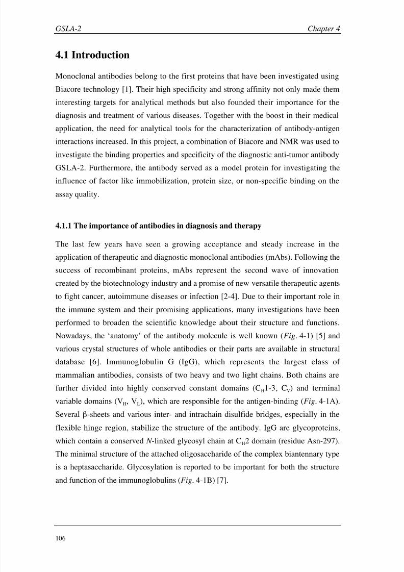
8/9/2019 DissB 7321 Mirroring
http://slidepdf.com/reader/full/dissb-7321-mirroring 120/278
GSLA-2 Chapter 4
106
4.1 Introduction
Monoclonal antibodies belong to the first proteins that have been investigated using
Biacore technology [1]. Their high specificity and strong affinity not only made them
interesting targets for analytical methods but also founded their importance for the
diagnosis and treatment of various diseases. Together with the boost in their medical
application, the need for analytical tools for the characterization of antibody-antigen
interactions increased. In this project, a combination of Biacore and NMR was used to
investigate the binding properties and specificity of the diagnostic anti-tumor antibody
GSLA-2. Furthermore, the antibody served as a model protein for investigating the
influence of factor like immobilization, protein size, or non-specific binding on the
assay quality.
4.1.1 The importance of antibodies in diagnosis and therapy
The last few years have seen a growing acceptance and steady increase in the
application of therapeutic and diagnostic monoclonal antibodies (mAbs). Following the
success of recombinant proteins, mAbs represent the second wave of innovation
created by the biotechnology industry and a promise of new versatile therapeutic agents
to fight cancer, autoimmune diseases or infection [2-4]. Due to their important role in
the immune system and their promising applications, many investigations have been
performed to broaden the scientific knowledge about their structure and functions.
Nowadays, the ‘anatomy’ of the antibody molecule is well known (Fig. 4-1) [5] and
various crystal structures of whole antibodies or their parts are available in structural
database [6]. Immunoglobulin G (IgG), which represents the largest class of
mammalian antibodies, consists of two heavy and two light chains. Both chains are
further divided into highly conserved constant domains (CH1-3, CV) and terminal
variable domains (VH, VL), which are responsible for the antigen-binding (Fig.
4-1A).
Several -sheets and various inter- and intrachain disulfide bridges, especially in the
flexible hinge region, stabilize the structure of the antibody. IgG are glycoproteins,
which contain a conserved N -linked glycosyl chain at CH2 domain (residue Asn-297).
The minimal structure of the attached oligosaccharide of the complex biantennary type
is a heptasaccharide. Glycosylation is reported to be important for both the structure
and function of the immunoglobulins (Fig. 4-1B) [7].
8/9/2019 DissB 7321 Mirroring
http://slidepdf.com/reader/full/dissb-7321-mirroring 121/278

8/9/2019 DissB 7321 Mirroring
http://slidepdf.com/reader/full/dissb-7321-mirroring 122/278
GSLA-2 Chapter 4
108
murine chimeric humanized human
Figure 4-2: Engineering of monoclonal antibodies. The immunogenicity of murine mAbs can be
reduced by replacing the constant regions of the mouse sequence (red) by their human counterparts
(blue). While chimeric mAbs contain the whole variable domain, only the antigen-binding regions
remain murine in the humanized mAbs.
Particularly in the case of life-threatening diseases like cancer or autoimmune
disorders, a high specificity and accuracy of the antibody-based diagnosis is an
absolute prerequisite. False negative results may prevent or retard an essential
treatment of the patient, while false positive diagnoses may burden the organism with
unnecessary cures and induce an enormous psychological distress [10]. In addition,
therapeutic antibodies with low specificity may bind to healthy tissues and cause severe
side effects. Therefore, a profound knowledge about the antibody-antigen interaction
on the molecular level helps to develop and select highly specific antibodies. One of
the most detailed methods for this purpose is X-ray crystallography, and several
hundred antibody-antigen complexes are already available in the PBD database [6, 11].
However, not all molecules can be resolved by crystallography and some important
structures such as carbohydrate-antibody complexes were found to be especially
challenging [12]. Nuclear magnetic resonance (NMR) evolved into a valuable
alternative for the analysis of such structures and was successfully applied to
antibody-antigen complexes [13]. While transfer nuclear Overhauser enhancement
(trNOE) experiments were used to describe the bioactive conformation of carbohydrateepitopes bound to monoclonal antibodies [14], saturation transfer difference (STD
NMR) [15] was used for mapping the binding epitopes of sugar-based antigens [16].
4.1.2 GSLA-2: a diagnostic monoclonal antibody
In this study, the tumor-diagnostic monoclonal antibody GSLA-2 was investigated.
Since GSLA-2 is exclusively used for in-vitro analytical and diagnostic purposes, the
immunogenic potential of the murine IgG1 mAb (see section 4.1.1) is not relevant.

8/9/2019 DissB 7321 Mirroring
http://slidepdf.com/reader/full/dissb-7321-mirroring 123/278
Chapter 4 GSLA-2
109
GSLA-2 recognizes the tumor antigen CA19-9, which was identified as a
monosialoganglioside specifically present in patients with colon and pancreas cancer
but not in healthy individuals or in patients with other diseases [17, 18]. The molecular
binding epitope of this specific recognition was shown to be the tetrasaccharide sialyl
Lewisa (sLea), which is a major determinant of pancreatic and gastrointestinal cancer
[19, 20]. The carbohydrate epitope sLea, as well as the closely related tetrasaccharide
sialyl Lewisx (sLex) are both composed of a 5-acetylneuraminic acid (Neu5Ac), a
galactose, a N -acetylglucosamine (Glc N Ac), and a fucose moiety (Fig. 4-3A). The only
difference between the two molecules is the branching of Glc N Ac, where the
connections of the 3 and 4 position are reversed (Fig. 4-3B) [21].
Figure 4-3: Structure of the sialyl Lewisa (sLea) epitope. A: Representation of the tetrasaccharide sLea
with the individual sugar moieties highlighted in different colors. B: Three-dimensional structure of sLea
in comparison with the related sialyl Lewisx (sLex).
While most of the monoclonal antibodies, including GSLA-2, distinguish between the
two tetrasaccharide epitopes, there are rare exceptions such as the antibody HECA 452
[22]. E-selectin, an endothelial receptor that is involved in leukocyte trafficking and
tumor metastasis, has also been reported to interact with both Lewis antigens [23].
Adhesion of cancer cells to E-selectin can be mediated by sLea-carrying glycoproteins
(e.g. mucins) or glycolipids (i.e. gangliosides). While sLea was found to be mainly
responsible for adhesion of human colon, pancreas, and gastric cancer cells, the
interaction with lung, liver, and ovarian tumor cells was mediated by sLex [24].
The high specificity for tumors of the colon and pancreas made GSLA-2 an useful tool
for the diagnosis for these cancers. For example, the antibody was successfully used in
diagnostic ELISA against mucin and conjugated sLea [25, 26] and for western blot
detection of mucins [27, 28]. However, little is known about the exact binding mode
and the specificity towards sLea derivatives. We, therefore, investigated the interaction
between GSLA-2 and a set of carbohydrates on the molecular level.
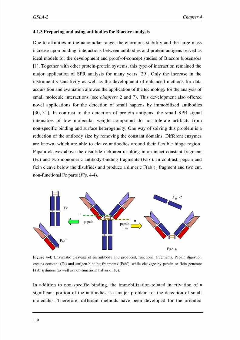
8/9/2019 DissB 7321 Mirroring
http://slidepdf.com/reader/full/dissb-7321-mirroring 124/278

8/9/2019 DissB 7321 Mirroring
http://slidepdf.com/reader/full/dissb-7321-mirroring 125/278
Chapter 4 GSLA-2
111
immobilization of monoclonal antibodies using either capturing or site-directed
coupling techniques [32-34]. Antibody molecules possess different distinct sites for a
possible attachment to the surface, such as amino groups of lysines, cysteines of the
hinge region (after mild reduction), the carbohydrate chain, or protein A binding sites
(Fig.
4-5).
amine groups thiol groups carbohydrate chain protein A binding site
Fc
Fab
Figure 4-5: Attachment sites for the immobilization of monoclonal antibodies (green). While the amine
groups of lysine residues (red spheres) are widely distributed, cysteine residues are usually less frequent
(the hinge region is highlighted by a red circle). Most mAbs only possess a single glycosylation site per
heavy chain (red lines), and a binding site for staphylococcal protein A and streptococcal protein G
(red/blue areas on the Fc part).
Alternatively to the enzymatic or biochemical approaches, the generation of smaller
antibody fragments like scFv (see section 4.1.1) or the introduction of specificattachment sites can also be performed by recombinant expression of engineered
antibodies [35, 36].
4.1.4 Aims in this project
The primary aim in this project is to get a deeper insight into the binding specificity of
the diagnostically relevant monoclonal antibody GSLA-2 (mouse IgG1), which
recognizes the sialyl Lewisa (sLea) epitope of some colon and pancreas cancer cells. For
this purpose, a Biacore assay based on covalently immobilized GSLA-2 is developed
and validated with sLea. A set of structurally related compounds is then screened and
the binding data are compared with results from STD NMR experiments. Since mouse
IgG1 molecules are the most important subclass of monoclonal antibodies, GSLA-2 is
further used as a model protein for the investigation of additional experimental
parameters, such as surface activity, protein size, immobilization density, or
non-specific binding.
8/9/2019 DissB 7321 Mirroring
http://slidepdf.com/reader/full/dissb-7321-mirroring 126/278
GSLA-2 Chapter 4
112
4.2 Materials and Methods
4.2.1 Materials
This section describes materials, equipment and procedures specifically used for the
GSLA-2 project. Materials and general methods used in all Biacore assays are
described in section 2.2.
Reagents and proteins
Murine monoclonal antibody GSLA-2 (IgG1, ~ 4 mg/ml in PBS 7.4, 0.005% sodium
azide) was a kind gift from Dr. John L. Magnani (GlycoTech, Gaithersburg, USA).
Staphylococcal protein A (SpA; soluble, essentially salt-free, Sigma P6031) and
streptococcal protein G (SpG; recombinant, expressed in E. coli, Sigma P4689) were
from Sigma (Fluka AG, Buchs, Switzerland). A murine monoclonal IgG1 directed
against sheep myoglobin (from the ‘Biacore 3000 getting started’ kit) as well as the
polyclonal rabbit anti-mouse IgG1 antibody was from Biacore (Biacore AB, Uppsala,
Sweden). Sialyl Lewisa attached to a Lemieux spacer (sLea-Lem) was a gift from
Prof. Dr. Ole Hindsgaul (Department of Chemistry, Carlsberg Laboratory,
Copenhagen, Danmark) and Dr. Oliver Schwardt (Institute of Molecular Pharmacy,
University of Basel, Switzerland). Label-free sialyl Lewisa (sLea) was purchased from
Sigma (Fluka AG, Buchs, Switzerland). The sources of the compounds tested in the
ranking experiments as well as reagent required for NMR experiments are specified in
Herfurth et al. [37]. Ficin from fig tree latex (lyophilized, Sigma F6008),
2-mercaptoethylamine hydrochloride, (+)-biotinamidohexanoic acid hydrazide (BHZ;
Sigma B3770) and N -ethylmaleimide were from Sigma (Fluka AG, Buchs,
Switzerland), and (+)-biotin-PEO3-maleimide was from Molecular BioSciences,
Boulder, USA.
Equipment
HPLC separations were done on an Agilent 1100 purification system, equipped with a
quaternary pump, a cooled well-plate autosampler, a column thermostat, a DAD
detector, and a cooled analytical fraction collector (Agilent AG, Basel, Switzerland).
Size exclusion chromatography was performed on a TSK Gel G2000SW column
(7.5 600 mm; Tosoh Biosciences GmbH, Stuttgart, Germany). Hi-Trap 5 ml desalting
columns (Amersham Biosciences, Otelfingen, Switzerland) were used for bufferexchange.
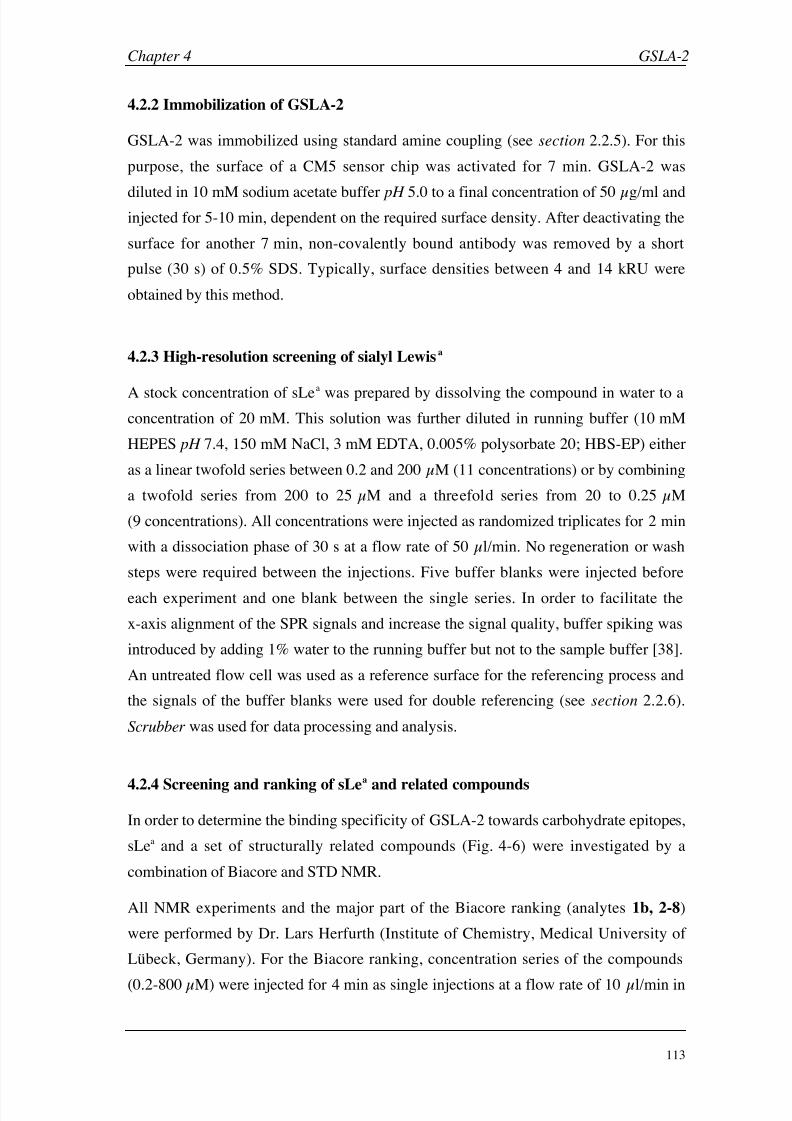
8/9/2019 DissB 7321 Mirroring
http://slidepdf.com/reader/full/dissb-7321-mirroring 127/278
Chapter 4 GSLA-2
113
4.2.2 Immobilization of GSLA-2
GSLA-2 was immobilized using standard amine coupling (see section 2.2.5). For this
purpose, the surface of a CM5 sensor chip was activated for 7 min. GSLA-2 was
diluted in 10
mM sodium acetate buffer pH
5.0 to a final concentration of 50
µg/ml andinjected for 5-10 min, dependent on the required surface density. After deactivating the
surface for another 7 min, non-covalently bound antibody was removed by a short
pulse (30 s) of 0.5% SDS. Typically, surface densities between 4 and 14 kRU were
obtained by this method.
4.2.3 High-resolution screening of sialyl Lewisa
A stock concentration of sLea was prepared by dissolving the compound in water to a
concentration of 20 mM. This solution was further diluted in running buffer (10 mM
HEPES pH 7.4, 150 mM NaCl, 3 mM EDTA, 0.005% polysorbate 20; HBS-EP) either
as a linear twofold series between 0.2 and 200 µM (11 concentrations) or by combining
a twofold series from 200 to 25 µM and a threefold series from 20 to 0.25 µM
(9 concentrations). All concentrations were injected as randomized triplicates for 2 min
with a dissociation phase of 30 s at a flow rate of 50 µl/min. No regeneration or wash
steps were required between the injections. Five buffer blanks were injected before
each experiment and one blank between the single series. In order to facilitate the
x-axis alignment of the SPR signals and increase the signal quality, buffer spiking was
introduced by adding 1% water to the running buffer but not to the sample buffer [38].
An untreated flow cell was used as a reference surface for the referencing process and
the signals of the buffer blanks were used for double referencing (see section 2.2.6).
Scrubber was used for data processing and analysis.
4.2.4 Screening and ranking of sLea
and related compounds
In order to determine the binding specificity of GSLA-2 towards carbohydrate epitopes,
sLea and a set of structurally related compounds (Fig. 4-6) were investigated by a
combination of Biacore and STD NMR.
All NMR experiments and the major part of the Biacore ranking (analytes 1b, 2-8)
were performed by Dr. Lars Herfurth (Institute of Chemistry, Medical University of
Lübeck, Germany). For the Biacore ranking, concentration series of the compounds
(0.2-800
µM) were injected for 4
min as single injections at a flow rate of 10
µl/min in

8/9/2019 DissB 7321 Mirroring
http://slidepdf.com/reader/full/dissb-7321-mirroring 128/278
8/9/2019 DissB 7321 Mirroring
http://slidepdf.com/reader/full/dissb-7321-mirroring 129/278
Chapter 4 GSLA-2
115
In order to complete the data set, analytes 1a, 9, and 10 (Fig. 4-6) were analyzed using
the high-resolution assay described in section 4.2.3. The influence of the Lemieux
spacer was further investigated by a comparative analysis of the thermodynamic and
kinetic binding properties of analytes 1a and 1b using Scrubber and CLAMP.
4.2.5 Production and evaluation of antibody fragments
60 µl of GSLA-2 was mixed with 30 µl Tris/EDTA buffer (50 mM Tris·HCl pH 7.0
2 mM EDTA). 8 µl of a ficin stock solution (1 mg/ml in Tris/EDTA buffer;
1.8 units/ml) and 2 µl cysteine activation solution (6.1 mg/ml in Tris/EDTA buffer)
were added and the mixture was incubated for 4 hours at 37°C. F(ab’)2 were separated
from Fc fragments and whole mAb using size exclusion chromatography (isocratic
conditions for 40 min, flow rate 1 ml/min, ambient temperature, running buffer: 50 mM
sodium acetate pH 5.0, 150 mM sodium chloride). 1 ml fractions were collected and
maximum concentrated F(ab’)2 fractions were around 0.1 mg/ml (Fig. 4-7).
IgG F(ab’)2
Fab’
Ficin
MEA SEC
SEC
Fab’-BXM
BXM
Figure 4-7: Schematic overview of the key steps in the antibody fragment
preparation. After the ficin digest, the F(ab’)2 fragments were isolated by
size-exclusion chromatography (SEC). A part of these fragments were further
cleaved by a mild reduction with 2-mercaptoethylamine (MEA). The free cysteine
residues were blocked either with biotin-maleimide (BXM) or with
N -ethylmaleimide (NEM).
Purified F(ab’)2 fractions (~ 0.1 mg/ml) were further cleaved by buffer exchange on a
HiTrap desalting column to phosphate/EDTA buffer (0.1 M sodium phosphate pH 6.0
5
mM EDTA) and addition of a 200
mM MEA solution in phosphate/EDTA buffer tofinal MEA concentration 5 mM. After incubation for 90 min at 37°C, any free thiol
8/9/2019 DissB 7321 Mirroring
http://slidepdf.com/reader/full/dissb-7321-mirroring 130/278
GSLA-2 Chapter 4
116
groups at the hinge region were either blocked by adding NEM solution or biotinylated
by adding biotin-maleimide solution to a concentration of 20 µM for 2 hours at room
temperature. Finally, buffer was exchanged twice to 10 mM sodium phosphate pH 5.0
to completely remove any free MEA and thiol active reagents (Fig. 4-7).
Purified preparations of whole GSLA-2 as well as of its F(ab’)2 and Fab’ fragments in
10 mM sodium acetate pH 5.0 were immobilized on separate flow cells of a CM5
sensor chip as described in section 4.2.2. One flow cell was left untreated as a reference
surface. All surfaces were simultaneously evaluated by high-resolution screening with
sLea-Lem (see section 2.1.3). Surface activity was calculated by dividing the measured
Rmax value by the theoretical Rmax ( Eq. 4, see section 2.1.3). For comparative plots, all
fragment densities were normalized to the weight and valency of the whole antibody
using equation 9:
Normalized Density =ValencyFragment MW GSLA 2
MW Fragment ValencyGSLA 2
DensitySurface [Eq. 9]
In-process controls of the enzymatic digest, the mild reduction and the separation steps
were performed by SDS-PAGE and dot blot analysis. A modified protocol from
Lämmli [40] was used for the preparation of SDS-PAGE (8-12% gels) and silver
staining or an improved protocol for Coomassie staining [41] was applied for detection.
4.2.6 Evaluation of non-specific binding
In order to investigate the contribution of different production batches or sources of
sLea-Lem (1b) for non-specific binding, two different batches of the compound were
screened according to the high-resolution protocol described in section 4.2.3. In
addition, the effect of buffer additions (3 mM EDTA, 0.005% polysorbate) was
evaluated by screening sLea
-Lem (1b) in various combinations of running and samplebuffers (HBS-N, HBS-P, HBS-EP). One sLea-Lem (1b ) batch showing high
non-specific binding was diluted to a concentration of 200 µM in each buffer and
injected for 2 min at a flow rate of 50 µl/min over GSLA-2 and its fragments
(see section 4.2.5). The injection series was repeated for all three running buffers.
8/9/2019 DissB 7321 Mirroring
http://slidepdf.com/reader/full/dissb-7321-mirroring 131/278
Chapter 4 GSLA-2
117
4.2.7 Comparison of capturing approaches
Immobilization protocols for staphylococcal protein A (SpA) and streptococcal protein
G (SpG) were developed based on methods from literature [33, 42, 43]. Stock solutions
of SpA and SpG were prepared by dissolving 1
mg lyophilized protein in 1
ml water.While SpA could directly be diluted in 10 mM sodium acetate pH 5.0, SpG solution
had to be dialyzed extensively against immobilization buffer (10 mM sodium acetate
pH 4.0) to remove the remaining Tris salt. The final concentration for both SpA and
SpG immobilization solutions was 50 µg/ml. Immobilization to a CM5 sensor chip was
done using standard amine coupling (see section 2.2.5) at a flow rate of 10 µl/min.
Activation and deactivation contact times were 10 min, the proteins were injected for
15 min. Both surfaces were evaluated by injecting a fivefold GSLA-2 concentration
series between 0.2 and 666
µM for 10
min at flow rate of 10
µl/min with a dissociation
phase of 10 min. The SpA surface was then regenerated by a 60 s pulse of 10 mM
glycine pH 1.5. The binding data were analyzed using Scrubber (affinity) and CLAMP
(kinetics). Finally, GSLA-2 was captured on immobilized SpA for 20 min at a
concentration of 0.1 mg/ml and a flow rate of 5 µl/min (mAb density ~ 3,000 RU), and
a 200 µM sLea-Lem (1b) solution was injected for 2 min. HBS-EP buffer was used
throughout immobilization, evaluation and capturing experiments.
In an alternative capturing approach, a rabbit anti-mouse IgG1 antibody wasimmobilized to a CM5 sensor chip using amine coupling. A 30 µg/ml solution of the
antibody in 10 mM sodium phosphate buffer pH 5.0 was injected for 7 min over the
activated surface at a flow rate of 5 µl/min. After deactivation for 7 min and a 2 min
wash step with 10 mM glycine pH 2.0, the surface showed a density of 12,000 RU. The
surface was evaluated by injecting GSLA-2 at concentrations of 3, 0.3, and 0.03 µM
for 5 min. Following a dissociation phase of 5 min, GSLA-2 was removed by injecting
10 mM glycine pH 2.0 three times for 120 min. For the sLea screening, GSLA-2 was
captured by injecting the antibody at a concentration of 400
µg/ml for 5
min (resulting
in a density of 2,200 RU). sLea-Lem (1b) was injected between 0.2 and 200 µM as
single injections for 1 min and the equilibrium responses were fitted to a single binding
site model.

8/9/2019 DissB 7321 Mirroring
http://slidepdf.com/reader/full/dissb-7321-mirroring 132/278
GSLA-2 Chapter 4
118
4.3 Results & Discussion
4.3.1 Development of a GSLA-2 Biacore assay
Covalent immobilization of monoclonal antibodies by standard amine coupling is awell-established procedure and often results in high-density surfaces. Even though the
coupling procedure may induce a significant loss in activity, the remaining binding
sites are usually sufficient for an interpretable signal. Therefore, GSLA-2 was directly
immobilized on a CM5 sensor chip using amine coupling, which yielded in high
surface densities ( 10,000 RU). Injection of a concentration series of sialyl Lewisa
with an attached Lemieux spacer (sLea-Lem, 1b) over the antibody surface generated
small (< 100 RU) but clearly detectable, and highly reproducible SPR signals. As
expected for carbohydrate-protein interactions, yet unusual for antibody-antigen
binding, very fast binding kinetics could be observed (Fig. 4-8A). Steady state
responses were concentration-dependent and could be fitted to a single binding site
model (Fig. 4-8B). Despite the fast association and dissociation rates, the complete data
set could be fitted kinetically to a simple Langmuir 1:1 binding model (Fig. 4-8C).
0
10
20
30
40
0 40 80 120 160 0 40 80 120 16010-7 10-6 10-5 10-4
Time [s] Concentration [M] Time [s]
S P R R e s p o n s e [
R U ] A B C
Figure 4-8: High-resolution GSLA-2 binding assay. A: Overlaid sensorgrams of a typical sLea-Lem (1b)
concentration series (0.2-200 µM), injected as randomized triplicates. B: Steady state responses of the
same data set, fitted to a single binding site model. C: Kinetic evaluation of the data set using CLAMP.
The sLea signals (blue) are overlaid with the simulated kinetic curves (red).
Due to the fast kinetics of the interaction and the rapid recurrence to the baseline, no
regeneration or wash steps were required. The steady state phase was reached within a
few seconds and was found to be very stable over several minutes. Fitting the signals
from the high-resolution assay to a single binding site model led to K D values in the
range of 8 to 15 µM. Again, such KD’s are rather high for typical antibody-binding
events, but expected for the interaction of monovalent sugars with their protein targets.
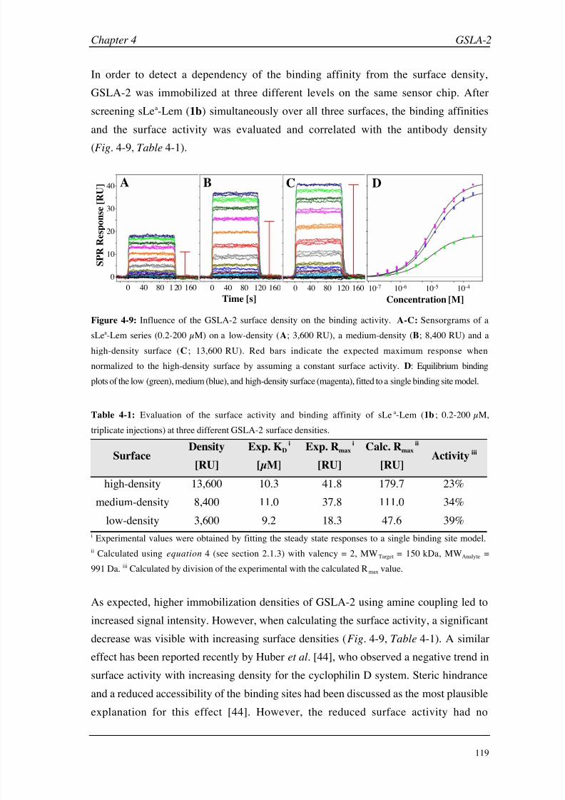
8/9/2019 DissB 7321 Mirroring
http://slidepdf.com/reader/full/dissb-7321-mirroring 133/278
Chapter 4 GSLA-2
119
In order to detect a dependency of the binding affinity from the surface density,
GSLA-2 was immobilized at three different levels on the same sensor chip. After
screening sLea-Lem (1b) simultaneously over all three surfaces, the binding affinities
and the surface activity was evaluated and correlated with the antibody density
(Fig.
4-9, Table 4-1).
10-7 10-6 10-5 10-4
Concentration [M]
0
10
20
30
40
S P R R
e s p o n s e [ R U ]
0 40 80 1 20 160
Time [s]
0 40 80 120 160 0 40 80 120 160
A B C D
Figure 4-9: Influence of the GSLA-2 surface density on the binding activity. A-C: Sensorgrams of a
sLea-Lem series (0.2-200 µM) on a low-density (A; 3,600 RU), a medium-density (B; 8,400 RU) and a
high-density surface (C ; 13,600 RU). Red bars indicate the expected maximum response when
normalized to the high-density surface by assuming a constant surface activity. D: Equilibrium binding
plots of the low (green), medium (blue), and high-density surface (magenta), fitted to a single binding site model.
Table 4-1: Evaluation of the surface activity and binding affinity of sLea-Lem (1b ; 0.2-200 µM,
triplicate injections) at three different GSLA-2 surface densities.
SurfaceDensity
[RU]
Exp. KD i
[µM]
Exp. Rmax i
[RU]
Calc. Rmax ii
[RU]Activity iii
high-density 13,600 10.3 41.8 179.7 23%
medium-density 8,400 11.0 37.8 111.0 34%
low-density 3,600 9.2 18.3 47.6 39%
i Experimental values were obtained by fitting the steady state responses to a single binding site model.
ii Calculated using equation 4 (see section 2.1.3) with valency = 2, MWTarget = 150 kDa, MWAnalyte =
991 Da. iii Calculated by division of the experimental with the calculated Rmax value.
As expected, higher immobilization densities of GSLA-2 using amine coupling led to
increased signal intensity. However, when calculating the surface activity, a significant
decrease was visible with increasing surface densities (Fig. 4-9, Table 4-1). A similar
effect has been reported recently by Huber et al. [44], who observed a negative trend in
surface activity with increasing density for the cyclophilin D system. Steric hindrance
and a reduced accessibility of the binding sites had been discussed as the most plausible
explanation for this effect [44]. However, the reduced surface activity had no
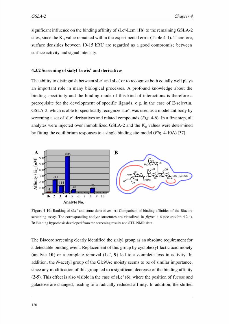
8/9/2019 DissB 7321 Mirroring
http://slidepdf.com/reader/full/dissb-7321-mirroring 134/278
GSLA-2 Chapter 4
120
significant influence on the binding affinity of sLea-Lem (1b) to the remaining GSLA-2
sites, since the KD value remained within the experimental error (Table 4-1). Therefore,
surface densities between 10-15 kRU are regarded as a good compromise between
surface activity and signal intensity.
4.3.2 Screening of sialyl Lewisa and derivatives
The ability to distinguish between sLea and sLex or to recognize both equally well plays
an important role in many biological processes. A profound knowledge about the
binding specificity and the binding mode of this kind of interactions is therefore a
prerequisite for the development of specific ligands, e.g. in the case of E-selectin.
GSLA-2, which is able to specifically recognize sLea, was used as a model antibody by
screening a set of sLea derivatives and related compounds (Fig. 4-6). In a first step, all
analytes were injected over immobilized GSLA-2 and the KD values were determined
by fitting the equilibrium responses to a single binding site model (Fig. 4-10A) [37].
O OO O
OO
O
OHOH
OH NHAcO(CH2)8COOCH3
OH
COONa
HOAcHN
HOOH
OH
OH
H3C
OHOH
600
500
400
300
200
100
01b 2 3 4 5 6 7 8 9 10
A B
Analyte No.
A f f i n i t y / K D
[ µ M ]
4
211
114
606
46 75
Figure 4-10: Ranking of sLea and some derivatives. A: Comparison of binding affinities of the Biacore
screening assay. The corresponding analyte structures are visualized in figure 4-6 (see section 4.2.4).
B: Binding hypothesis developed from the screening results and STD NMR data.
The Biacore screening clearly identified the sialyl group as an absolute requirement for
a detectable binding event. Replacement of this group by cyclohexyl-lactic acid moiety
(analyte 10) or a complete removal (Lea, 9) led to a complete loss in activity. In
addition, the N -acetyl group of the Glc N Ac moiety seems to be of similar importance,
since any modification of this group led to a significant decrease of the binding affinity
(2-5). This effect is also visible in the case of sLex (6), where the position of fucose and
galactose are changed, leading to a radically reduced affinity. In addition, the shifted

8/9/2019 DissB 7321 Mirroring
http://slidepdf.com/reader/full/dissb-7321-mirroring 135/278
Chapter 4 GSLA-2
121
position of the Lemieux-spacer might also contribute to this effect. Likewise, no
binding could be detected when replacing the whole Glc N Ac by a simple cyclohexane
moiety (7 and 10). Interestingly, the linear pentasaccharide LSTa (8) still showed a
small affinity to GSLA-2, despite the lack of fucose. The higher flexibility of LSTa
compared to sLex due to the absence of a branched fucose seems to allow a positioning
of the N -acetyl group that is similar to sLea. Finally, the removal of the methyl group of
the fucose moiety also had a negative influence on the binding affinity, as it is
illustrated by the comparison of analytes 3 and 4. These findings led to the binding
hypothesis, where the N -acetyl group of Glc N Ac and the Neu5Ac moiety (sialyl) are
essential for a high-affinity interaction with GSLA-2 [37] (Fig. 4-10B, red areas).
STD NMR experiments [37] offered an even deeper insight into the binding mode of
sLea and its derivatives. It could be shown that GSLA-2 recognizes the whole
tetrasaccharide epitope, since all protons of the four pyranose rings received saturation
(Fig. 4-10B, blue area). The importance of the two essential areas identified during
Biacore screening was confirmed by the NMR experiments. The N -acetyl groups from
both the Neu5Ac and the Glc N Ac moiety received a large fraction of the saturation
transfer. In contrast, only weak contacts were found for the Lemieux spacer. When
investigating the binding of sLex (6), it was observed that the antibody mainly
recognized the sialyl group, while no signal could be detected anymore for Glc N Ac. A
major difference to E- or P-selectin might be the fact that their binding site is rather
shallow [45] and interacts predominantly with the galactose and fucose moieties [46],
whereas the binding site of GSLA-2 seems to be deeper and therefore less tolerant
toward structural modifications of the tetrasaccharides [37].
Even though the Lemieux spacer [47] was found to interact only slightly with GSLA-2
by NMR experiments (see above), it might nevertheless influence the binding affinity
during Biacore experiments. Therefore, sialyl Lewisa in presence and absence of a
Lemieux-spacer (1a and 1b, Fig.
4-6) were injected at the same concentration range
(Fig. 4-11A&B). Both their equilibrium binding responses (Fig. 4-11B) and their
kinetic properties were compared and analyzed (Table 4-2).
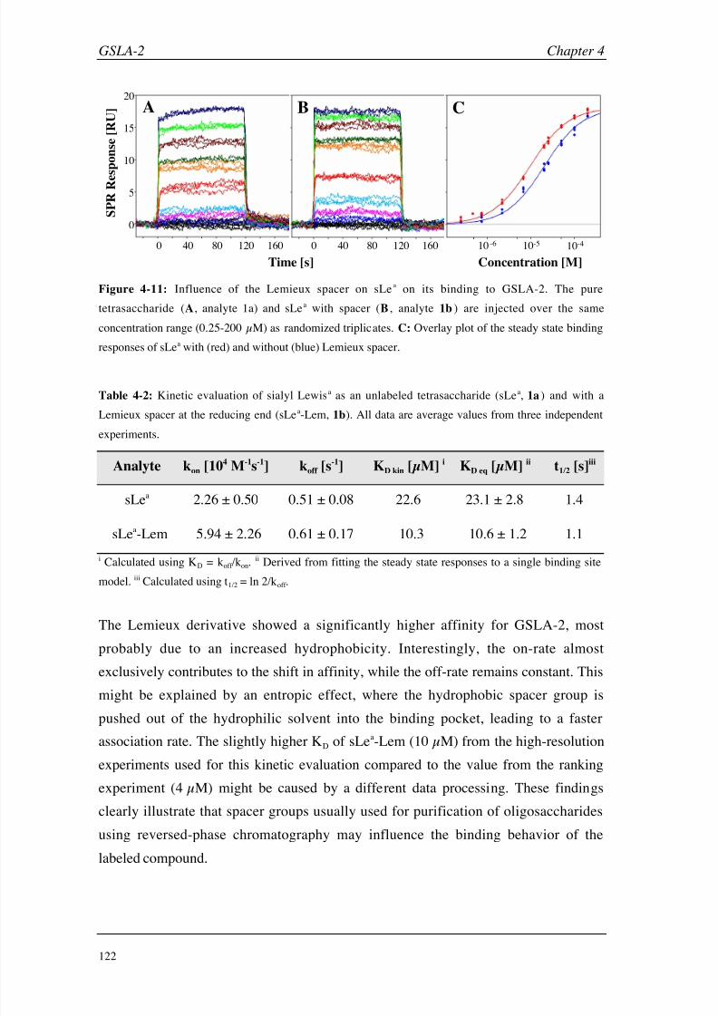
8/9/2019 DissB 7321 Mirroring
http://slidepdf.com/reader/full/dissb-7321-mirroring 136/278
GSLA-2 Chapter 4
122
0 40 80 120 160
0
5
10
15
20
0 40 80 120 160 10-6 10-5 10-4
Time [s] Concentration [M]
S P R R e
s p o n s e [ R U ] A B C
Figure 4-11: Influence of the Lemieux spacer on sLea on its binding to GSLA-2. The pure
tetrasaccharide (A, analyte 1a) and sLea with spacer (B , analyte 1b ) are injected over the same
concentration range (0.25-200 µM) as randomized triplicates. C: Overlay plot of the steady state binding
responses of sLea with (red) and without (blue) Lemieux spacer.
Table 4-2: Kinetic evaluation of sialyl Lewisa as an unlabeled tetrasaccharide (sLea, 1a ) and with a
Lemieux spacer at the reducing end (sLea-Lem, 1b). All data are average values from three independent
experiments.
Analyte kon [104 M-1s-1] koff [s-1] KD kin [µM] i KD eq [µM] ii t1/2 [s]iii
sLea 2.26 ± 0.50 0.51 ± 0.08 22.6 23.1 ± 2.8 1.4
sLea-Lem 5.94 ± 2.26 0.61 ± 0.17 10.3 10.6 ± 1.2 1.1
i Calculated using KD = koff /kon.ii Derived from fitting the steady state responses to a single binding site
model. iii Calculated using t1/2 = ln 2/koff .
The Lemieux derivative showed a significantly higher affinity for GSLA-2, most
probably due to an increased hydrophobicity. Interestingly, the on-rate almost
exclusively contributes to the shift in affinity, while the off-rate remains constant. This
might be explained by an entropic effect, where the hydrophobic spacer group is
pushed out of the hydrophilic solvent into the binding pocket, leading to a faster
association rate. The slightly higher KD of sLea-Lem (10 µM) from the high-resolution
experiments used for this kinetic evaluation compared to the value from the ranking
experiment (4 µM) might be caused by a different data processing. These findings
clearly illustrate that spacer groups usually used for purification of oligosaccharides
using reversed-phase chromatography may influence the binding behavior of the
labeled compound.

8/9/2019 DissB 7321 Mirroring
http://slidepdf.com/reader/full/dissb-7321-mirroring 137/278
Chapter 4 GSLA-2
123
4.3.3 Production and evaluation of antibody fragments
Antibodies offer an attractive opportunity to specifically cleave the molecule in order to
generate smaller fragments with preserved binding activity. Since the SPR signal is
dependent on the ratio between the molecular weights of the target and the analyte,binding to smaller fragments often generate higher signal intensities. In principle, two
enzymes are predominantly used for this purpose: papain, which generates monovalent
Fab’ fragments and pepsin for the preparation of dimeric F(ab’)2 fragments (Fig. 4-4,
see section 4.1.3). The pepsin-type digestion leaves the hinge region intact and the
F(ab’)2 can be further cleaved by mild reduction of the hinge disulfides. The resulting
free thiol groups can then be functionalized by thiol-active reagents and used for a
site-directed coupling approach [32, 48]. However, pepsinolysis of intact antibodies has
found to be difficult, and could only be improved by adding a deglycosylation step
prior to the digestion. The deglycosylation is rather time-consuming (24-48 h) and
involves different glycosidases. Therefore, a method was developed to cleave GSLA-2
without the need for deglycosylation and to biotinylate the reduced fragments [49]. In
this study, the cysteine protease ficin (EC 3.4.22.3) was identified as a valuable
alternative to pepsin, since it readily cleaves the intact mAb with high specificity and
could also be triggered to directly generate Fab’ fragments by increasing the activator
concentration (cysteine). Furthermore, an optimal concentration of mercaptoethylamine
as a mild reducing agent could be determined for the mild reduction of GSLA-2 F(ab’)2
[49]. These results were then combined for an efficient generation of GSLA-2
fragments. Size exclusion chromatography was found to be a suitable method for
monitoring the ficinolysis and to isolate the F(ab’)2 (Fig. 4-12).

8/9/2019 DissB 7321 Mirroring
http://slidepdf.com/reader/full/dissb-7321-mirroring 138/278
GSLA-2 Chapter 4
124
0
2
4
6
8
10
0 5 10 15 20 25
Time [min]
A b s o r b a n c e a t 2 8 0 n m [ m
A U ]
mAb
F(ab’)2
Fab’/Fc
Salts, Reagents, …
Figure 4-12: Monitoring of the ficin digestion by size exclusion chromatography from start (dark blue)
to 4 hours of incubation at 37°C (orange) in 30 min steps. While the first two peaks could be identified as
the intact antibody and the F(ab’)2 fragment, the third peak developed over the incubation time and are
most likely Fc or Fab’ fragments. The last peak group did not change significantly over time and consists
of buffer salts and reagents (ficin, cysteine, etc.).
By incubating the isolated F(ab’)2 fragments with mercaptoethylamine (MEA), the
hinge region was mildly reduced. The free thiols were either blocked or functionalized
with a thiol-active biotinylation reagent (Fig. 4-13A). Dot-blot analysis of the
biotinylated Fab’ with labeled extravidin (a derivative of avidin with reduced
non-specific binding) confirmed the successful addition of a biotin group to the protein
(Fig. 4-13B). Unfortunately, the biotinylated fragment could not be captured on a
custom-made neutravidin sensor chip. Neutravidin (a deglycosylated form of
streptavidin) was used instead of streptavidin because of its lower non-specific binding
[50]. It is not yet clear, whether the lack of capturing efficiency was caused by the
rather low concentration of the Fab’-biotin construct, by remaining impurities from the
biotinylation reagent, or by inactive neutravidin. Therefore, the experiment has to be
repeated on a commercially available streptavidin sensor chip.
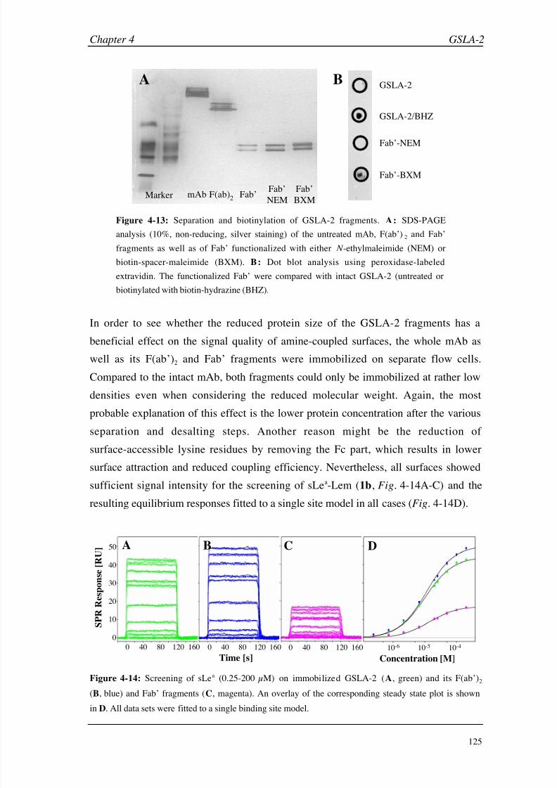
8/9/2019 DissB 7321 Mirroring
http://slidepdf.com/reader/full/dissb-7321-mirroring 139/278
Chapter 4 GSLA-2
125
mAb F(ab)2
Fab’ Fab’
NEM
Fab’
BXMMarker
GSLA-2
GSLA-2/BHZ
Fab’-NEM
Fab’-BXM
A B
Figure 4-13: Separation and biotinylation of GSLA-2 fragments. A: SDS-PAGE
analysis (10%, non-reducing, silver staining) of the untreated mAb, F(ab’)2 and Fab’
fragments as well as of Fab’ functionalized with either N -ethylmaleimide (NEM) or
biotin-spacer-maleimide (BXM). B: Dot blot analysis using peroxidase-labeled
extravidin. The functionalized Fab’ were compared with intact GSLA-2 (untreated or
biotinylated with biotin-hydrazine (BHZ).
In order to see whether the reduced protein size of the GSLA-2 fragments has a
beneficial effect on the signal quality of amine-coupled surfaces, the whole mAb as
well as its F(ab’)2 and Fab’ fragments were immobilized on separate flow cells.
Compared to the intact mAb, both fragments could only be immobilized at rather low
densities even when considering the reduced molecular weight. Again, the most
probable explanation of this effect is the lower protein concentration after the various
separation and desalting steps. Another reason might be the reduction of
surface-accessible lysine residues by removing the Fc part, which results in lower
surface attraction and reduced coupling efficiency. Nevertheless, all surfaces showed
sufficient signal intensity for the screening of sLea-Lem (1b, Fig. 4-14A-C) and the
resulting equilibrium responses fitted to a single site model in all cases (Fig. 4-14D).
10-6 10-5 10-4
Concentration [M]
0
10
20
30
40
S P R R e s p o n s e [ R U
]
0 40 80 120 160
Time [s]
0 40 80 120 160 0 40 80 120 160
50 A B C D
Figure 4-14: Screening of sLea (0.25-200 µM) on immobilized GSLA-2 (A, green) and its F(ab’)2
(B,
blue) and Fab’ fragments (C, magenta). An overlay of the corresponding steady state plot is shown
in D. All data sets were fitted to a single binding site model.
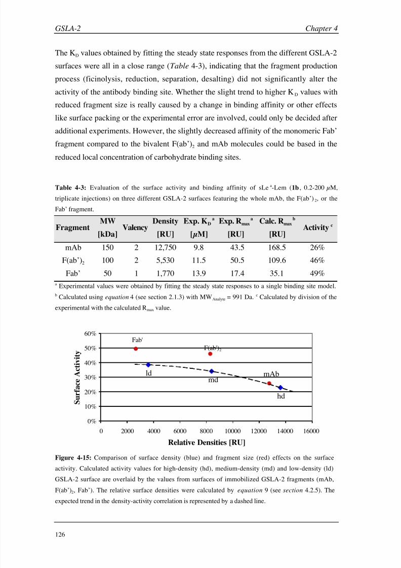
8/9/2019 DissB 7321 Mirroring
http://slidepdf.com/reader/full/dissb-7321-mirroring 140/278
GSLA-2 Chapter 4
126
The KD values obtained by fitting the steady state responses from the different GSLA-2
surfaces were all in a close range (Table 4-3), indicating that the fragment production
process (ficinolysis, reduction, separation, desalting) did not significantly alter the
activity of the antibody binding site. Whether the slight trend to higher KD values with
reduced fragment size is really caused by a change in binding affinity or other effects
like surface packing or the experimental error are involved, could only be decided after
additional experiments. However, the slightly decreased affinity of the monomeric Fab’
fragment compared to the bivalent F(ab’)2 and mAb molecules could be based in the
reduced local concentration of carbohydrate binding sites.
Table 4-3: Evaluation of the surface activity and binding affinity of sLea-Lem (1b , 0.2-200 µM,
triplicate injections) on three different GSLA-2 surfaces featuring the whole mAb, the F(ab’)2, or the
Fab’ fragment.
FragmentMW
[kDa]Valency
Density
[RU]
Exp. KDa
[µM]
Exp. Rmaxa
[RU]
Calc. Rmaxb
[RU]Activity c
mAb 150 2 12,750 9.8 43.5 168.5 26%
F(ab’)2 100 2 5,530 11.5 50.5 109.6 46%
Fab’ 50 1 1,770 13.9 17.4 35.1 49%
a Experimental values were obtained by fitting the steady state responses to a single binding site model.
b
Calculated using equation 4 (see section 2.1.3) with MWAnalyte = 991
Da.c
Calculated by division of theexperimental with the calculated Rmax value.
0%
10%
20%
30%
40%
50%
60%
0 2000 4000 6000 8000 10000 12000 14000 16000
Relative Densities [RU]
S u
r f a c e A c t i v i t y
Fab'
F(ab')2
mAb
hd
mdld
Figure 4-15: Comparison of surface density (blue) and fragment size (red) effects on the surface
activity. Calculated activity values for high-density (hd), medium-density (md) and low-density (ld)
GSLA-2 surface are overlaid by the values from surfaces of immobilized GSLA-2 fragments (mAb,
F(ab’)2, Fab’). The relative surface densities were calculated by equation 9 (see section 4.2.5). The
expected trend in the density-activity correlation is represented by a dashed line.

8/9/2019 DissB 7321 Mirroring
http://slidepdf.com/reader/full/dissb-7321-mirroring 141/278
Chapter 4 GSLA-2
127
By comparing the experimental and calculated Rmax values, a remarkable increase in the
surface activity could be observed for the two smaller fragments of GSLA-2. While this
effect could be partly induced by the lower surface density (see section 4.3.1,
Table 4-1), it does not explain the entire activity improvement. This becomes evident,
when the activities of the fragment experiment are directly compared with those from
the surface density experiment (Fig. 4-15). It clearly illustrates that the improved
surface activity caused by the cleavage of GSLA-2 to its F(ab’)2 and Fab’ fragments
was not only caused by a reduced density effects. While the surface activity for the
whole antibody lies exactly on the trend line, both fragment are shifted towards higher
surface activity. This effect might be caused by a better accessibility of the binding
sites or by an immobilization closer to the surface, which induces a higher SPR signal.
Hence, truncated mAb preparations seem to have an advantage over the intact moleculeand should be always considered for Biacore experiments. On the other side, the
fragment preparation procedure is still very time-consuming and usually results in
decreased target concentrations and lower surface densities. If the fragments are not
functionalized and are immobilized by standard amine coupling, the direct generation
of Fab’ fragments by papain might be advantageous since it requires less preparation
steps.
4.3.4 Evaluation of non-specific binding
During the high-resolution experiments, some sLea-Lem (1b) samples showed a clearly
two-phased binding signal (Fig. 4-16A). After a first rapid increase, a slower
association phase could be detected. As soon as the injection was stopped, the SPR
signal dropped rapidly with a subsequent slower dissociation period. The intensity of
the rapid phases corresponds with the block signals usually observed for the binding of
sLea. This suggested two independent binding events, one of which is believed to occur
non-specifically. One way of reducing non-specific binding is the immobilization of a
similar but inactive protein to the reference cell [30, 51]. Therefore, an anti-myoglobin
antibody of the same immunoglobulin class (mouse IgG1) was immobilized at a similar
density and used as a reference cell. Large non-specific signal effects as shown in
figure 4-16A could not be eliminated (see appendix C1), but the use of an antibody
reference often enhanced the signal quality in case of smaller signal impurities
(Fig. 4-16B&C). While the relative binding intensities and therefore the KD value
remained nearly constant, the reproducibility and shape of the signal were improved.
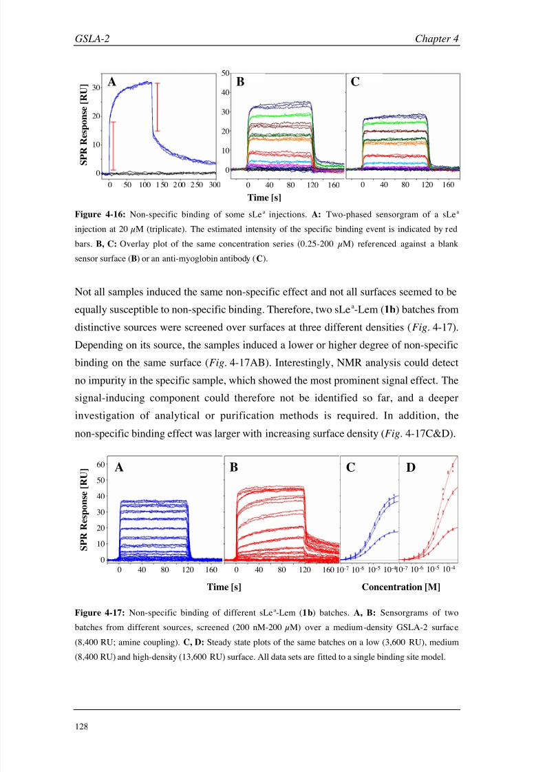
8/9/2019 DissB 7321 Mirroring
http://slidepdf.com/reader/full/dissb-7321-mirroring 142/278
GSLA-2 Chapter 4
128
0 50 100 1 50 2 00 2 50 300
0
10
20
30
0 40 80 120 160
0
10
20
40
30
50
0 40 80 120 160
A B C
Time [s]
S P R R e s p o n s e [ R U ]
Figure 4-16: Non-specific binding of some sLea injections. A: Two-phased sensorgram of a sLea
injection at 20 µM (triplicate). The estimated intensity of the specific binding event is indicated by red
bars. B, C: Overlay plot of the same concentration series (0.25-200 µM) referenced against a blank
sensor surface (B) or an anti-myoglobin antibody (C).
Not all samples induced the same non-specific effect and not all surfaces seemed to be
equally susceptible to non-specific binding. Therefore, two sLea-Lem (1b) batches from
distinctive sources were screened over surfaces at three different densities (Fig. 4-17).
Depending on its source, the samples induced a lower or higher degree of non-specific
binding on the same surface (Fig. 4-17AB). Interestingly, NMR analysis could detect
no impurity in the specific sample, which showed the most prominent signal effect. The
signal-inducing component could therefore not be identified so far, and a deeper
investigation of analytical or purification methods is required. In addition, the
non-specific binding effect was larger with increasing surface density (Fig. 4-17C&D).
10-7 10-6 10-5 10-410-7 10-6 10-5 10-40 40 80 120 160 0 40 80 120 160
0
10
20
30
40
50
60
Time [s]
S P R R e s p o n s e [ R U ]
Concentration [M]
A B C D
Figure 4-17: Non-specific binding of different sLea-Lem (1b) batches. A, B: Sensorgrams of two
batches from different sources, screened (200 nM-200 µM) over a medium-density GSLA-2 surface
(8,400 RU; amine coupling). C, D: Steady state plots of the same batches on a low (3,600 RU), medium
(8,400 RU) and high-density (13,600 RU) surface. All data sets are fitted to a single binding site model.

8/9/2019 DissB 7321 Mirroring
http://slidepdf.com/reader/full/dissb-7321-mirroring 143/278
Chapter 4 GSLA-2
129
Since F(ab’)2 and Fab’ fragments lack the Fc part, which could contribute to the
non-specific binding effect, a ‘contaminated’ batch of sLea-Lem (1b) was also tested on
these fragments. In the same experiment, the influence of the buffer compositions was
tested for both the sample and running buffer. For this purpose, a plain HBS buffer
(10
mM HEPES, 150
mM NaCl, pH
7.4; HBS-N) was compared to HBS containing
3 mM EDTA and 0.05% polysorbate (HBS-EP). The same concentration (20 µM) of
sLea-Lem (1b ) was screened in all combinations of running/sample buffers and
immobilized fragments (Fig. 4-18).
100
80
60
40
20
0
100
80
60
40
20
0
0 60 120 180 0 60 120 180 0 60 120 180
Time [s]
S P R R
e s p o n s e [ R U ]
whole mAb F(ab’)2 Fab’
H B S - N
H B S - E P
R u n n i n g B u f f e r
Antibody Fragment
Figure 4-18: Influence of buffer additions and antibody fragment size on the non-specific binding of
sLea-Lem (1b). The tetrasaccharide was diluted in either HBS-N (red) or HBS-EP (black) as sample
buffer and injected over whole antibody, F(ab’)2 and Fab’ surfaces. Either HBS-N (top row) or HBS-EP
(bottom row) was used as running buffer throughout the experiments.
A reduction of the antibody molecule to its antigen-binding parts was able to reduce but
not completely eliminate non-specific binding. This indicates that the Fc part is at least
partly involved in this effect. In addition, the choice of running and sample buffers
also influenced the degree of non-specific binding. When HBS-EP was used as sample
buffer, the undesired signal effect could be reduced to a minimum, especially in
combination with HBS-EP as running buffer.
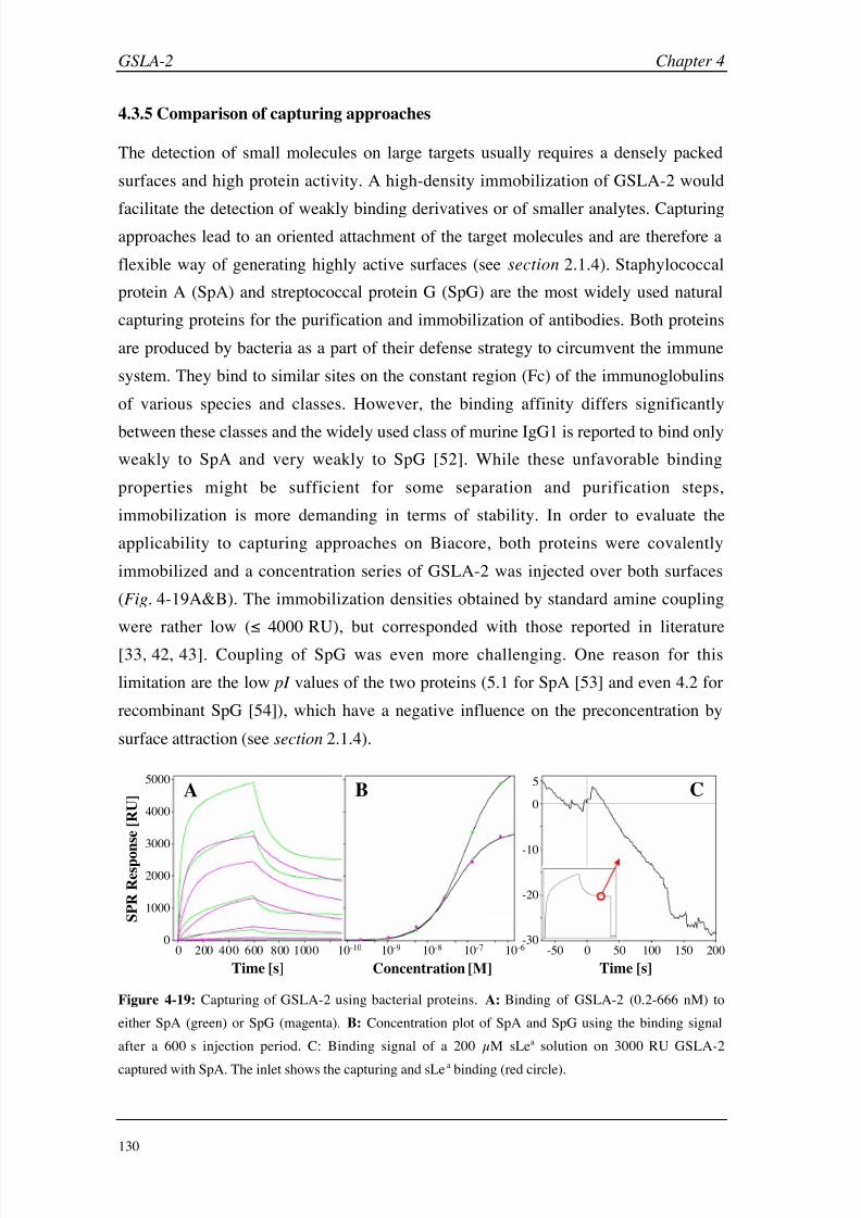
8/9/2019 DissB 7321 Mirroring
http://slidepdf.com/reader/full/dissb-7321-mirroring 144/278
GSLA-2 Chapter 4
130
4.3.5 Comparison of capturing approaches
The detection of small molecules on large targets usually requires a densely packed
surfaces and high protein activity. A high-density immobilization of GSLA-2 would
facilitate the detection of weakly binding derivatives or of smaller analytes. Capturingapproaches lead to an oriented attachment of the target molecules and are therefore a
flexible way of generating highly active surfaces (see section 2.1.4). Staphylococcal
protein A (SpA) and streptococcal protein G (SpG) are the most widely used natural
capturing proteins for the purification and immobilization of antibodies. Both proteins
are produced by bacteria as a part of their defense strategy to circumvent the immune
system. They bind to similar sites on the constant region (Fc) of the immunoglobulins
of various species and classes. However, the binding affinity differs significantly
between these classes and the widely used class of murine IgG1 is reported to bind only
weakly to SpA and very weakly to SpG [52]. While these unfavorable binding
properties might be sufficient for some separation and purification steps,
immobilization is more demanding in terms of stability. In order to evaluate the
applicability to capturing approaches on Biacore, both proteins were covalently
immobilized and a concentration series of GSLA-2 was injected over both surfaces
(Fig. 4-19A&B). The immobilization densities obtained by standard amine coupling
were rather low ( 4000 RU), but corresponded with those reported in literature
[33, 42, 43]. Coupling of SpG was even more challenging. One reason for this
limitation are the low pI values of the two proteins (5.1 for SpA [53] and even 4.2 for
recombinant SpG [54]), which have a negative influence on the preconcentration by
surface attraction (see section 2.1.4).
10-10 10-9 10-8 10-7 10-6
0
5
-10
-20
-300 50 100 150 200-50
0
1000
2000
3000
4000
5000
0 200 400 600 800 1000
Time [s] Concentration [M]
S P R R
e s p o n s e [ R U ] A B C
Time [s]
Figure 4-19: Capturing of GSLA-2 using bacterial proteins. A: Binding of GSLA-2 (0.2-666 nM) to
either SpA (green) or SpG (magenta). B: Concentration plot of SpA and SpG using the binding signal
after a 600 s injection period. C: Binding signal of a 200 µM sLea solution on 3000 RU GSLA-2
captured with SpA. The inlet shows the capturing and sLea binding (red circle).
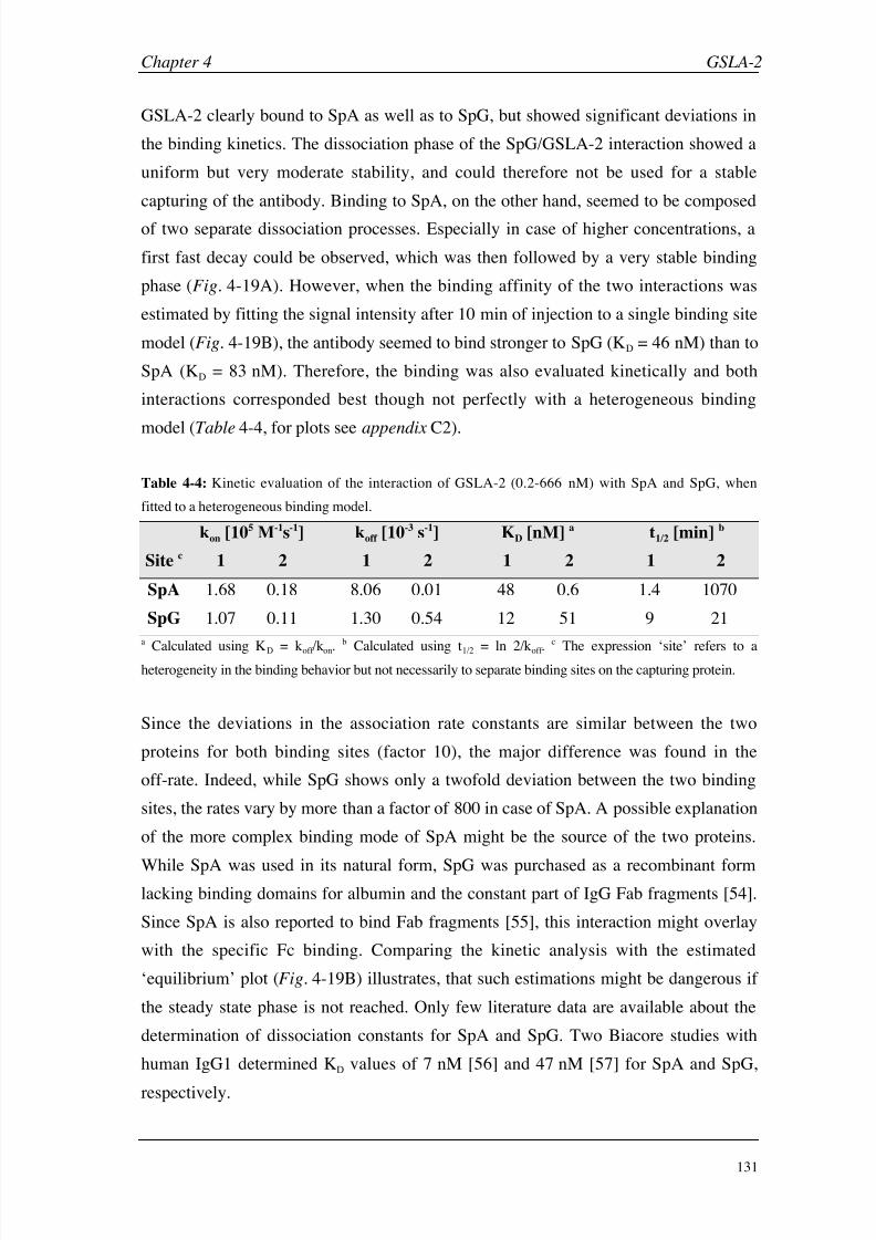
8/9/2019 DissB 7321 Mirroring
http://slidepdf.com/reader/full/dissb-7321-mirroring 145/278
Chapter 4 GSLA-2
131
GSLA-2 clearly bound to SpA as well as to SpG, but showed significant deviations in
the binding kinetics. The dissociation phase of the SpG/GSLA-2 interaction showed a
uniform but very moderate stability, and could therefore not be used for a stable
capturing of the antibody. Binding to SpA, on the other hand, seemed to be composed
of two separate dissociation processes. Especially in case of higher concentrations, a
first fast decay could be observed, which was then followed by a very stable binding
phase (Fig. 4-19A). However, when the binding affinity of the two interactions was
estimated by fitting the signal intensity after 10 min of injection to a single binding site
model (Fig. 4-19B), the antibody seemed to bind stronger to SpG (KD = 46 nM) than to
SpA (KD = 83 nM). Therefore, the binding was also evaluated kinetically and both
interactions corresponded best though not perfectly with a heterogeneous binding
model (Table 4-4, for plots see appendix C2).
Table 4-4: Kinetic evaluation of the interaction of GSLA-2 (0.2-666 nM) with SpA and SpG, when
fitted to a heterogeneous binding model.
kon [105 M-1s-1] koff [10-3 s-1] KD [nM] a t1/2 [min] b
Site c 1 2 1 2 1 2 1 2
SpA 1.68 0.18 8.06 0.01 48 0.6 1.4 1070
SpG 1.07 0.11 1.30 0.54 12 51 9 21
a
Calculated using KD = koff /kon.
b
Calculated using t1/2 = ln 2/koff .
c
The expression ‘site’ refers to aheterogeneity in the binding behavior but not necessarily to separate binding sites on the capturing protein.
Since the deviations in the association rate constants are similar between the two
proteins for both binding sites (factor 10), the major difference was found in the
off-rate. Indeed, while SpG shows only a twofold deviation between the two binding
sites, the rates vary by more than a factor of 800 in case of SpA. A possible explanation
of the more complex binding mode of SpA might be the source of the two proteins.
While SpA was used in its natural form, SpG was purchased as a recombinant formlacking binding domains for albumin and the constant part of IgG Fab fragments [54].
Since SpA is also reported to bind Fab fragments [55], this interaction might overlay
with the specific Fc binding. Comparing the kinetic analysis with the estimated
‘equilibrium’ plot (Fig. 4-19B) illustrates, that such estimations might be dangerous if
the steady state phase is not reached. Only few literature data are available about the
determination of dissociation constants for SpA and SpG. Two Biacore studies with
human IgG1 determined KD values of 7 nM [56] and 47 nM [57] for SpA and SpG,
respectively.
8/9/2019 DissB 7321 Mirroring
http://slidepdf.com/reader/full/dissb-7321-mirroring 146/278
GSLA-2 Chapter 4
132
While the dissociation rate of SpG is not sufficient for a stable capturing, the second
phase of the SpA dissociation seemed to fulfill these requirements. Therefore,
sLea-Lem (1b) was injected at a concentration that usually resulted in a saturation of
the binding sites (200 µM). However, the detected SPR signal ( 5 RU) was much
lower than expected and corresponded to an activity of only ~10%. In addition, the
surface showed a significant baseline drift, which makes an appropriate evaluation of
small signal intensities even more difficult (Fig. 4-19C). Non-specific binding of the
Fab’ fragments of GSLA-2 (see above) could be one reason of the low activity, since
steric hindrance might inhibit an interaction of sLea-Lem (1b) with the variable regions.
Another explanation might be found in a recent study by Sagawa et al. [55], where they
reported conformational changes of the antibody upon binding, which led to a
significant weakening of antigen interactions. A possible workaround for the rather lowsurface stability of SpA capturing was presented by Catimel et al. [33], who
cross-linked the antibody with SpA after the capturing by injecting dimethylpimelidate.
However, a decreased activity was also observed for this approach, because the
cross-linking reagent could also target lysine residues in the binding site [58].
In order to increase the specificity and stability of the IgG1 capturing, the experiment
was repeated with an immobilized polyclonal rabbit anti-mouse IgG1 antibody. Even
though the capturing antibody could be immobilized at a high surface density
(> 15,000 RU), the capturing density of GSLA-2 was rather low (< 5,000 RU). It is not
yet clear whether this limitation is caused by an immobilization-induced deactivation of
the primary antibody, steric hindrance, or by a SPR phenomenon (greater distance of
GSLA-2 from the surface). However, the captured GSLA-2 fraction was much more
stable than those on SpA or SpG, and was therefore suitable for the screening of small
molecules (Fig. 4-20A). The activity of the captured GSLA-2 surface was evaluated
by injecting a concentration series of sLea-Lem (1b, Fig. 4-20B&C).
Compared to SpA capturing, the baseline was more stable and the binding activity was
significantly increased (Fig. 4-20B). However, with slightly more than 33% the activity
was still far beyond the expectations for an oriented surface. The reason for this
phenomenon is not yet clear and has to be investigated further. Despite the low signal
intensities, the steady state responses could be fitted to a single site binding model
(Fig. 4-20C). The resulting KD of 15 µM was only slightly higher than the values
obtained from covalent coupling approaches (4-12 µM). Due to their low capturing
densities and binding activities, the investigated capturing approaches were not suitable
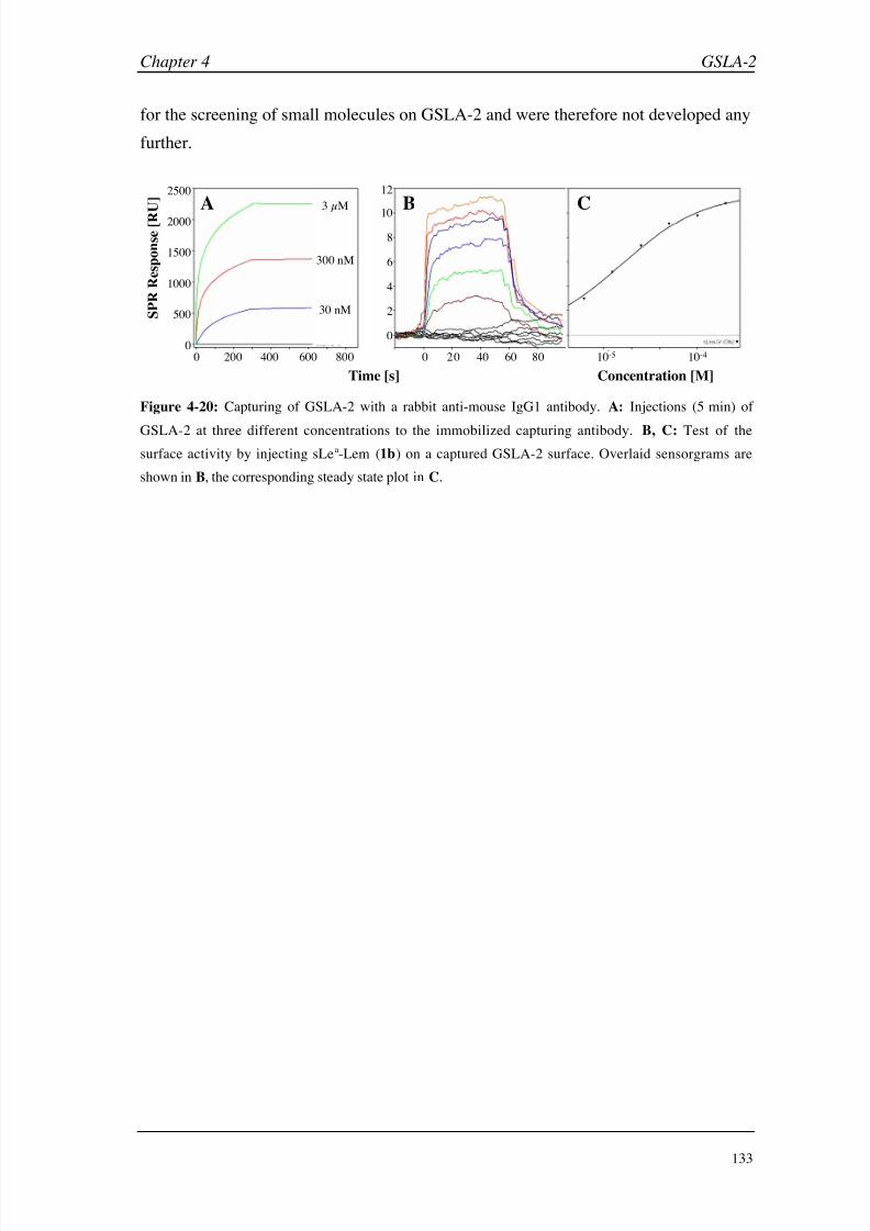
8/9/2019 DissB 7321 Mirroring
http://slidepdf.com/reader/full/dissb-7321-mirroring 147/278
Chapter 4 GSLA-2
133
for the screening of small molecules on GSLA-2 and were therefore not developed any
further.
0
2
4
6
8
10
12
0 20 40 60 80 10-5 10-40
500
1000
1500
2000
2500
0 200 400 600 800
Time [s] Concentration [M]
S P R R
e s p o n s e [ R
U ] A B C
30 nM
300 nM
3 µM
Figure 4-20: Capturing of GSLA-2 with a rabbit anti-mouse IgG1 antibody. A: Injections (5 min) of
GSLA-2 at three different concentrations to the immobilized capturing antibody. B, C: Test of the
surface activity by injecting sLea-Lem (1b) on a captured GSLA-2 surface. Overlaid sensorgrams are
shown in B, the corresponding steady state plot in C.

8/9/2019 DissB 7321 Mirroring
http://slidepdf.com/reader/full/dissb-7321-mirroring 148/278
GSLA-2 Chapter 4
134
4.4 Conclusions
The monoclonal antibody GSLA-2 shows a high selectivity of the sialyl Lewisa epitope
(sLea), and is therefore clinically used as a diagnostic tool for the detection of colon and
pancreas cancer, However, little is known about the molecular binding specificity and
its binding to related carbohydrates. Therefore, a stable and reproducible Biacore assay
was developed based on the covalent immobilization of GSLA-2 on the sensor chip
surface. Screening of sLea in solution resulted in kinetic properties, which are typical
for carbohydrate-protein interaction, such as rapid association and dissociation rate
constants and a rather low affinity (low micromolar range). This Biacore assay, in
combination with data from STD NMR experiments was used to screen a set of
structurally related compounds for binding to the antibody. Evaluation of Biacore data
identified the presence and orientation of the Neu5Ac and Glc N Ac moieties as essential
for a detectable binding event. Any replacement, removal, or reversal of Glc N Ac
(e.g. in the case of sLex) led to a loss of affinity. Furthermore, the methyl group of the
fucose moiety seems not to be essential but beneficial for binding. STD NMR
experiments showed that GSLA-2 recognized the whole tetrasaccharide epitope and
confirmed the high importance of the two N -acetyl groups. This allows a much deeper
insight into the binding mode of sLea to the antibody and helps explaining the
difference to the selectins, which recognize both sLea
and sLex
.
As a representative of the most popular class of monoclonal antibodies, GSLA-2 also
allowed to investigate different aspects of Biacore assay design and optimization. For
example, the influence of a labeling group to the binding affinity could be analyzed.
While a Lemieux spacer, attached to the reducing end of sLea for a facilitated
purification of the molecules, did only receive minor parts of the saturation transfer in
the STD NMR experiment, its contribution to the binding affinity was nevertheless
detectable in the Biacore assay. The slightly improved affinity is most likely caused by
hydrophobic interactions or by an improved kon (e.g. by ‘pushing’ the molecule out of
the solvent), and illustrates the great importance of the label-free SPR technology. As
expected, increasing the surface density of the immobilized antibody led to enlarged
signal intensities, while the KD value remained constant. The active protein fraction
also got smaller with increasing surface densities, indicating a limited accessibility of
binding sites.
In an additional experiment, the benefit of using antibody fragments was illustrated by
comparing the whole GSLA-2 antibody with its F(ab’)2 and Fab’ fragments. For this

8/9/2019 DissB 7321 Mirroring
http://slidepdf.com/reader/full/dissb-7321-mirroring 149/278
Chapter 4 GSLA-2
135
purpose, a method was developed for the fast and reliable digestion, cleavage,
purification and functionalization of such fragments. Both truncated antibody
molecules could be immobilized by amine coupling. While the relative immobilization
density was lower compared to untreated GSLA-2, the surface activity was remarkably
increased. In addition, F(ab’)2 and Fab’ fragments also showed a lower susceptibility
for non-specific binding, as it occurred with some sLea batches. No-specific binding
could also be reduced by adding EDTA (and polysorbate) to the running and sample
buffers. While the immobilization of fragmented antibodies was of an advantage in the
case of GSLA-2, the time-consuming procedure and the loss of protein through the
preparation process has to be considered.
Finally, three different methods for the capturing of GSLA-2 were compared in terms
of stability and activity. The affinity of the mouse IgG1 towards staphylococcal protein
A and streptococcal protein G was too weak for a stable capturing baseline and
generated rather low surface densities. In contrast, a specific rabbit anti-mouse IgG1
antibody was able to capture GSLA-2 with slightly higher density and a remarkably
increased stability. However, none of the tested capturing surfaces showed an improved
activity as it had been expected for an oriented immobilization. Due to the limitations
of the capturing approach, covalent coupling is clearly recommended, since it
generated highly dense and suitably active antibody surfaces. If higher signal intensities
are required for future experiments, an optimization of the Fab’ immobilization density
seems to be most promising, since these fragments showed an excellent combination of
specificity and activity in the screening assays.

8/9/2019 DissB 7321 Mirroring
http://slidepdf.com/reader/full/dissb-7321-mirroring 150/278
GSLA-2 Chapter 4
136
4.5 Literature
[1] M. Malmqvist, Nature 1993, 361, 186.
[2] J. M. Reichert, A. K. Pavlou, Nat Rev Drug Discov 2004, 3, 383.
[3] S. Y. Tetin, S. D. Stroupe, Curr Pharm Biotechnol 2004, 5, 9.
[4] O. H. Brekke, I. Sandlie, Nat Rev Drug Discov 2003, 2, 52.
[5] E. A. Padlan, Mol Immunol 1994, 31, 169.
[6] Protein Data Bank: The worldwide repository for macromolecular structures, Research
Collaboratory for Structural Bioinformatics (RCSB), http://www.pdb.org/, 2005.
[7] R. Jefferis, J. Lund, Chem Immunol 1997, 65, 111.
[8] M. R. Clark, Chem Immunol 1997, 65, 88.
[9] G. Kohler, C. Milstein, Nature 1975, 256 , 495.
[10] J. Wardle, R. Pope, J Psychosom Res 1992, 36 , 609.
[11] H. M. Berman, J. Westbrook, Z. Feng, G. Gilliland, T. N. Bhat, H. Weissig, I. N.
Shindyalov, P. E. Bourne, Nucleic Acids Res 2000, 28, 235.
[12] D. R. Bundle, in Glycosciences: Status and Perspectives (Eds.: H. J. Gabius, S. Gabius),
Chapman and Hall, Weinheim, 1997, pp. 311.
[13] H. Maaheimo, P. Kosma, L. Brade, H. Brade, T. Peters, Biochemistry 2000, 39, 12778.
[14] T. Haselhorst, J. F. Espinosa, J. Jimenez-Barbero, T. Sokolowski, P. Kosma, H. Brade, L.
Brade, T. Peters, Biochemistry 1999, 38, 6449.
[15] M. Mayer, B. Meyer, Angew Chem Int Ed 1999, 38, 1784.[16] M. A. Johnson, B. M. Pinto, J Am Chem Soc 2002, 124, 15368.
[17] J. L. Magnani, M. Brockhaus, D. F. Smith, V. Ginsburg, M. Blaszczyk, K. F. Mitchell, Z.
Steplewski, H. Koprowski, Science 1981, 212, 55.
[18] J. L. Magnani, B. Nilsson, M. Brockhaus, D. Zopf, Z. Steplewski, H. Koprowski, V.
Ginsburg, J Biol Chem 1982, 257 , 14365.
[19] T. Nakayama, M. Watanabe, T. Katsumata, T. Teramoto, M. Kitajima,Cancer 1995, 75, 2051.
[20] T. Nakagoe, T. Sawai, T. Tsuji, M. Jibiki, M. Ohbatake, A. Nanashima, H. Yamaguchi,
T. Yasutake, H. Ayabe, K. Arisawa, Tumour Biol 2001, 22, 115.
[21] U. Spohr, O. Hindsgaul, R. U. Lemieux, Can J Chem 1985, 63, 2644.
[22] A. M. Duijvestijn, E. Horst, S. T. Pals, B. N. Rouse, A. C. Steere, L. J. Picker, C. J.
Meijer, E. C. Butcher, Am J Pathol 1988, 130, 147.
[23] E. L. Berg, M. K. Robinson, O. Mansson, E. C. Butcher, J. L. Magnani, J Biol Chem
1991, 266 , 14869.
[24] M. Ugorski, A. Laskowska, Acta Biochim Pol 2002, 49, 303.
[25] G. Gorrin, S. Christensen, M. Moore, K. Trujillo, M. Farwell, Y. Teramoto, J. L.
Magnani, Tumour Biol 1997, 18, Supp. 2: 79.

8/9/2019 DissB 7321 Mirroring
http://slidepdf.com/reader/full/dissb-7321-mirroring 151/278
Chapter 4 GSLA-2
137
[26] P. D. Rye, N. V. Bovin, E. V. Vlasova, A. A. Molodyk, A. Baryshnikov, F. T. Kreutz, W.
I. Garinther, B. C. Schultes, A. A. Noujaim, R. Madiyalakan, J. Magnani, O. Nilsson, K.
Nilsson, K. Nustad, L. Norum, H. Bell, Y. Cao, M. R. Suresh, D. L. Very, J. V. Freeman,
K. K. Yeung, J. Hilgers, Tumour Biol 1998, 19, 390.
[27] H. S. Silverman, S. Parry, M. Sutton-Smith, M. D. Burdick, K. McDermott, C. J. Reid, S. K.Batra, H. R. Morris, M. A. Hollingsworth, A. Dell, A. Harris, Glycobiology 2001, 11, 459.
[28] H. S. Silverman, M. Sutton-Smith, K. McDermott, P. Heal, S. H. Leir, H. R. Morris, M.
A. Hollingsworth, A. Dell, A. Harris, Glycobiology 2003, 13, 265.
[29] R. L. Rich, D. G. Myszka, J Mol Recognit 2000, 13, 388.
[30] M. Strandh, B. Persson, H. Roos, S. Ohlson, J Mol Recognit 1998, 11, 188.
[31] M. Adamczyk, J. A. Moore, Z. Yu, Methods 2000, 20, 319.
[32] P. Peluso, D. S. Wilson, D. Do, H. Tran, M. Venkatasubbaiah, D. Quincy, B. Heidecker,
K. Poindexter, N. Tolani, M. Phelan, K. Witte, L. S. Jung, P. Wagner, S. Nock, Anal
Biochem 2003, 312, 113.
[33] B. Catimel, M. Nerrie, F. T. Lee, A. M. Scott, G. Ritter, S. Welt, L. J. Old, A. W.
Burgess, E. C. Nice, J Chromatogr A 1997, 776 , 15.
[34] B. Johnsson, S. Lofas, G. Lindquist, A. Edstrom, R. M. Muller Hillgren, A. Hansson, J
Mol Recognit 1995, 8, 125.
[35] A. Skerra, J Mol Recognit 2000, 13, 167.
[36] W. M. Mullett, E. P. Lai, J. M. Yeung, Methods 2000, 22, 77.
[37] L. Herfurth, B. Ernst, B. Wagner, D. Ricklin, D. S. Strasser, J. L. Magnani, A. J. Benie,
T. Peters, J Med Chem 2005, submitted .[38] M. J. Cannon, G. A. Papalia, I. Navratilova, R. J. Fisher, L. R. Roberts, K. M. Worthy, A.
G. Stephen, G. R. Marchesini, E. J. Collins, D. Casper, H. Qiu, D. Satpaev, S. F.
Liparoto, D. A. Rice, Gorshkova, II, R. J. Darling, D. B. Bennett, M. Sekar, E.
Hommema, A. M. Liang, E. S. Day, J. Inman, S. M. Karlicek, S. J. Ullrich, D. Hodges, T.
Chu, E. Sullivan, J. Simpson, A. Rafique, B. Luginbuhl, S. N. Westin, M. Bynum, P.
Cachia, Y. J. Li, D. Kao, A. Neurauter, M. Wong, M. Swanson, D. G. Myszka, Anal
Biochem 2004, 330, 98.
[39] L. Herfurth, PhD thesis, Molekulare Erkennung von Kohlenhydraten durch monoklonale
Antikörper und ein Lektin - NMR-Experimente, Medical University of Lübeck (Lübeck,
Germany), 2002.
[40] U. K. Laemmli, Nature 1970, 227 , 680.
[41] D. Kang, Y. S. Gho, M. Suh, C. Kang, Bull Korean Chem Soc 2002, 23, 1511.
[42] R. L. Rich, D. G. Myszka, J Mol Recognit 2001, 14, 223.
[43] R. J. Darling, U. Kuchibhotla, W. Glaesner, R. Micanovic, D. R. Witcher, J. M. Beals,
Biochemistry 2002, 41, 14524.
[44] W. Huber, S. Perspicace, J. Kohler, F. Muller, D. Schlatter, Anal Biochem2004, 333, 280.
[45] W. S. Somers, J. Tang, G. D. Shaw, R. T. Camphausen, Cell 2000, 103, 467.

8/9/2019 DissB 7321 Mirroring
http://slidepdf.com/reader/full/dissb-7321-mirroring 152/278
GSLA-2 Chapter 4
138
[46] M. Rinnbauer, B. Ernst, B. Wagner, J. Magnani, A. J. Benie, T. Peters, Glycobiology
2003, 13, 435.
[47] R. U. Lemieux, D. R. Bundle, D. A. Baker, J Am Chem Soc 1975, 97 , 4076.
[48] D. S. Wilson, J. Wu, P. Peluso, S. Nock, J Immunol Methods 2002, 260, 29.
[49] C. Bellac, Diploma thesis, Herstellung von Methoden zur selektiven Kopplung von
Targetproteinen an Biosensoren, University of Basel (Basel, Switzerland), 2002.
[50] Y. Hiller, J. M. Gershoni, E. A. Bayer, M. Wilchek, Biochem J 1987, 248, 167.
[51] R. J. Ober, E. S. Ward, Anal Biochem 1999, 271, 70.
[52] E. Harlow, D. P. Lane, Using antibodies: a laboratory manual, 2nd rev. ed., Cold Spring
Harbor Laboratory, Cold Spring Harbor, 1999.
[53] I. Bjork, B. A. Petersson, J. Sjoquist, Eur J Biochem 1972, 29, 579.
[54] C. R. Goward, J. P. Murphy, T. Atkinson, D. A. Barstow, Biochem J 1990, 267 , 171.
[55] T. Sagawa, M. Oda, H. Morii, H. Takizawa, H. Kozono, T. Azuma, Mol Immunol 2005, 42, 9.
[56] H. G. Svensson, H. R. Hoogenboom, U. Sjobring, Eur J Biochem 1998, 258, 890.
[57] S. Radaev, P. D. Sun, J Biol Chem 2001, 276 , 16478.
[58] E. C. Nice, B. Catimel, Bioessays 1999, 21, 339.

8/9/2019 DissB 7321 Mirroring
http://slidepdf.com/reader/full/dissb-7321-mirroring 153/278
Chapter 5
Asialoglycoprotein Receptor

8/9/2019 DissB 7321 Mirroring
http://slidepdf.com/reader/full/dissb-7321-mirroring 154/278
Asialoglycoprotein receptor Chapter 5
140
5.1 Introduction
Drug and gene targeting to specific organs is a promising approach for the development
of highly effective therapies while reducing side-effects. Carbohydrate-lectin
interactions between liver-cell receptors and their physiological (or synthetic) ligands
have been described as an efficient method showing high specificity. However, for the
development of new carbohydrate-based agents, a detailed knowledge about binding
modes and affinities is crucial. Therefore, the interaction of the asialoglycoprotein
receptor (ASGP-R) with some of its natural ligands has been investigated using
Biacore.
5.1.1 The asialoglycoprotein receptor as a promising drug target
The liver is the major metabolic organ in the human body and is responsible of clearing
the blood from undesired endo- and exogenous compounds. Therefore, a plethora of
enzymes and receptors is specialized in metabolizing and excreting different classes of
molecules. With some 500,000 receptors/hepatocyte, the asialoglycoprotein receptor is
one of the most abundant receptors and an important contributor to this hepatic
machinery [1, 2]. It was first described by Ashwell and Morell [3], when they
discovered that glycoproteins featuring a terminal galactose (or N -acetyl
galactosamine) were rapidly removed from the circulation by a receptor-mediated
mechanism. Normally, sialic acid residues mask the penultimate galactose of many
oligosaccharide chains. However, these masking groups might be removed by
sialidases, resulting in asialoglycoproteins. Many studies have been performed in recent
years in order to investigate the differential biological function and physiological role
between sialylated and desialylated glycoproteins. Suppression of antigenicity, masking
and metastasis of tumor cells, clearance of apoptotic liver cells or regulation of
glycoprotein hormones are some of the proposed functions. However,
receptor-deficient mice did not show significantly elevated plasma level of
asialoglycoproteins nor was their life span influenced in any way [1]. Therefore, the
exact physiological relevance of the receptor is still not clear and has to be investigated
further.
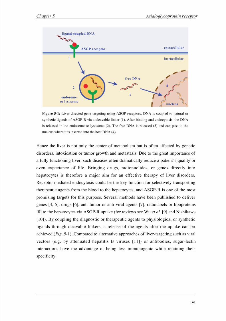
8/9/2019 DissB 7321 Mirroring
http://slidepdf.com/reader/full/dissb-7321-mirroring 155/278
Chapter 5 Asialoglycoprotein receptor
141
1
2
3
4
extracellular
intracellular
endosome
or lysosome
ASGP rece ptor
ligand-coupled DN A
free DN A
nucleus
Figure 5-1: Liver-directed gene targeting using ASGP receptors. DNA is coupled to natural or
synthetic ligands of ASGP-R via a cleavable linker (1). After binding and endocytosis, the DNA
is released in the endosome or lysosome (2). The free DNA is released (3) and can pass to the
nucleus where it is inserted into the host DNA (4).
Hence the liver is not only the center of metabolism but is often affected by genetic
disorders, intoxication or tumor growth and metastasis. Due to the great importance of
a fully functioning liver, such diseases often dramatically reduce a patient’s quality or
even expectance of life. Bringing drugs, radionuclides, or genes directly intohepatocytes is therefore a major aim for an effective therapy of liver disorders.
Receptor-mediated endocytosis could be the key function for selectively transporting
therapeutic agents from the blood to the hepatocytes, and ASGP-R is one of the most
promising targets for this purpose. Several methods have been published to deliver
genes [4, 5], drugs [6], anti-tumor or anti-viral agents [7], radiolabels or lipoproteins
[8] to the hepatocytes via ASGP-R uptake (for reviews see Wu et al. [9] and Nishikawa
[10]). By coupling the diagnostic or therapeutic agents to physiological or synthetic
ligands through cleavable linkers, a release of the agents after the uptake can be
achieved (Fig. 5-1). Compared to alternative approaches of liver-targeting such as viral
vectors (e.g. by attenuated hepatitis B viruses [11]) or antibodies, sugar-lectin
interactions have the advantage of being less immunogenic while retaining their
specificity.

8/9/2019 DissB 7321 Mirroring
http://slidepdf.com/reader/full/dissb-7321-mirroring 156/278
Asialoglycoprotein receptor Chapter 5
142
H1 H1
H2
cytosolic
extracellular
A B
transmembrane
domain
cytosolic end
stalk region
C-type lectin
domain
5.1.2 Structure and function of ASGP-R
Isolation of ASGP-R from liver preparations showed that the receptor consists of two
related proteins in a concentration ratio of 3:1. Each polypeptide chain is in the range of
40-60
kDa and is glycosylated with two or three N-linked oligosaccharide chains. Thetwo receptor subtypes are named HL-1 (i.e. hepatic lectin 1) and HL-2, the human
receptors are usually simply referred to as H1 and H2 [12]. The sequence identity of the
two subunits is 55 % for the human receptor [13]. Different studies suggested that both
subunits are required to build a functional receptor, forming a hetero-oligomeric
receptor complex. Hence the exact stoichiometry is still not fully solved, with
suggestion from tri- to hexameric complexes of various H1/H2 ratios [12]. Since
triantennary ligands were found to bind with a very high affinity (see section 5.1.4), a
trimeric receptor consisting of two H1 and one H2 subunits is expected to be the
minimum requirement (Fig. 5-2A) [14].
Figure 5-2: Structure of the asialoglycoprotein receptor. A: Hetero-oligomeric complex
of two H1 and one H2 subunits, which is believed to be the minimum requirement of a
functional receptor. B: Anatomy of a receptor subunit with different intra- and
extracellular domains. Disulfide bridges are indicated as orange lines, the calcium ion
involved in ligand binding is indicated by a pink circle.
Both ASGP-R subtypes are membrane-bound proteins, which consist of approximately
300 amino acids. Their general structure can be differentiated in a small N-terminal
cytoplasmic end, a single transmembrane domain (~20 hydrophobic residues), and a
large C-terminal exoplasmic part, consisting of a stalk region and the carbohydrate
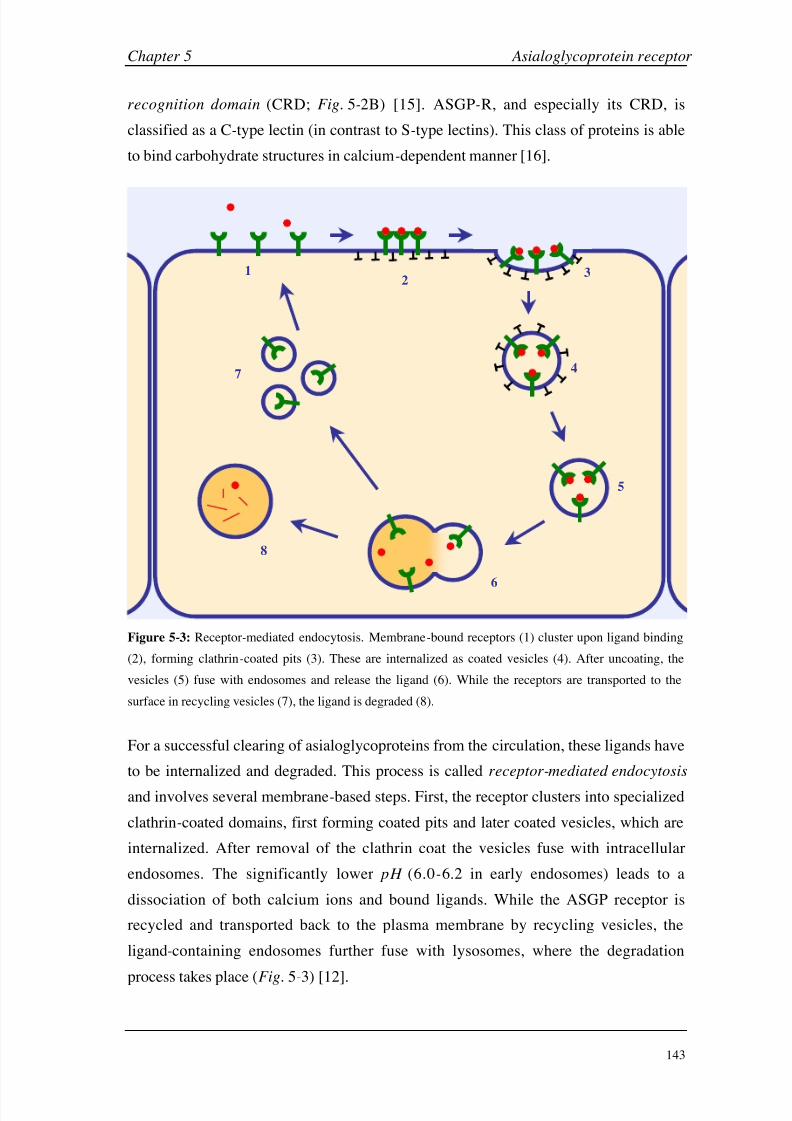
8/9/2019 DissB 7321 Mirroring
http://slidepdf.com/reader/full/dissb-7321-mirroring 157/278
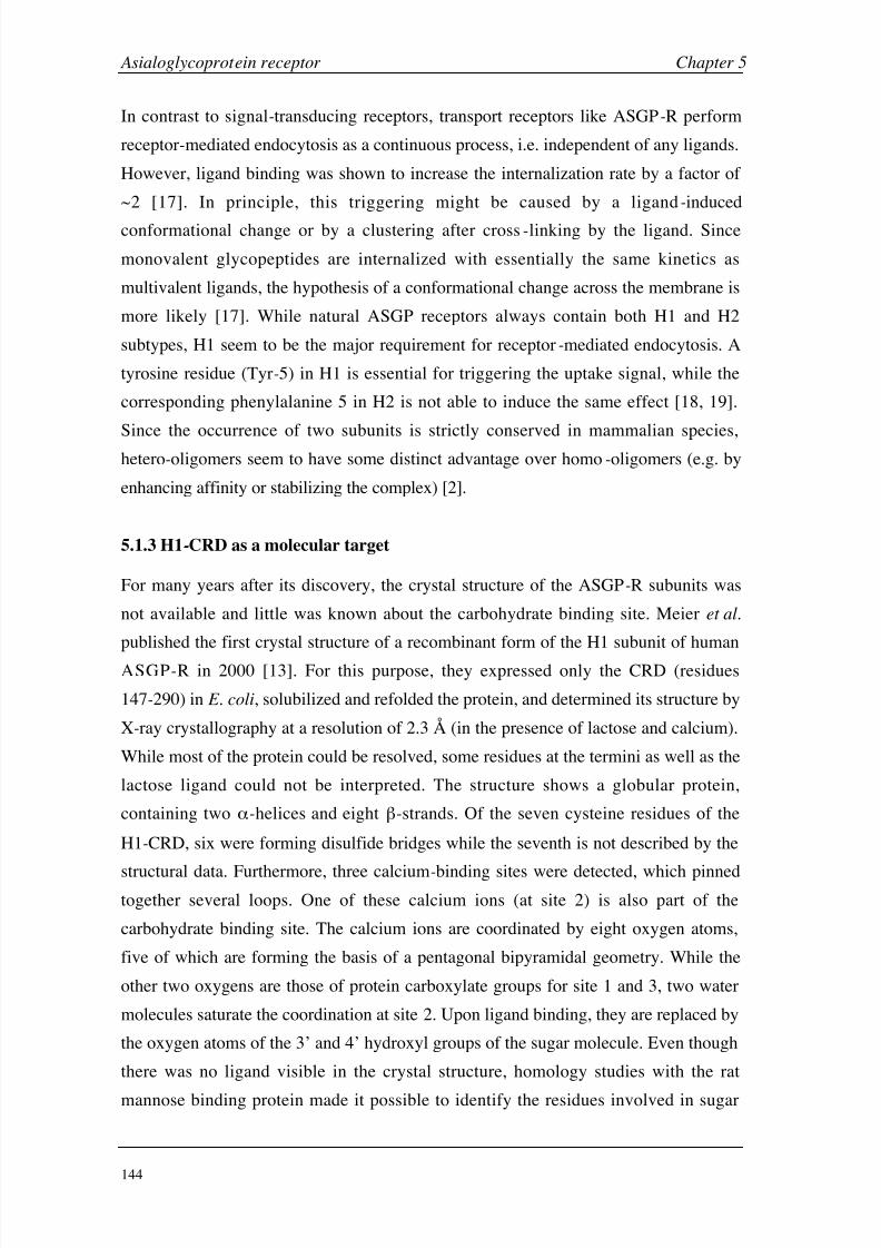
8/9/2019 DissB 7321 Mirroring
http://slidepdf.com/reader/full/dissb-7321-mirroring 158/278
Asialoglycoprotein receptor Chapter 5
144
In contrast to signal-transducing receptors, transport receptors like ASGP-R perform
receptor-mediated endocytosis as a continuous process, i.e. independent of any ligands.
However, ligand binding was shown to increase the internalization rate by a factor of
~2 [17]. In principle, this triggering might be caused by a ligand-induced
conformational change or by a clustering after cross-linking by the ligand. Since
monovalent glycopeptides are internalized with essentially the same kinetics as
multivalent ligands, the hypothesis of a conformational change across the membrane is
more likely [17]. While natural ASGP receptors always contain both H1 and H2
subtypes, H1 seem to be the major requirement for receptor-mediated endocytosis. A
tyrosine residue (Tyr-5) in H1 is essential for triggering the uptake signal, while the
corresponding phenylalanine 5 in H2 is not able to induce the same effect [18, 19].
Since the occurrence of two subunits is strictly conserved in mammalian species,hetero-oligomers seem to have some distinct advantage over homo-oligomers (e.g. by
enhancing affinity or stabilizing the complex) [2].
5.1.3 H1-CRD as a molecular target
For many years after its discovery, the crystal structure of the ASGP-R subunits was
not available and little was known about the carbohydrate binding site. Meier et al.
published the first crystal structure of a recombinant form of the H1 subunit of human
ASGP-R in 2000 [13]. For this purpose, they expressed only the CRD (residues
147-290) in E. coli, solubilized and refolded the protein, and determined its structure by
X-ray crystallography at a resolution of 2.3 Å (in the presence of lactose and calcium).
While most of the protein could be resolved, some residues at the termini as well as the
lactose ligand could not be interpreted. The structure shows a globular protein,
containing two -helices and eight -strands. Of the seven cysteine residues of the
H1-CRD, six were forming disulfide bridges while the seventh is not described by the
structural data. Furthermore, three calcium-binding sites were detected, which pinnedtogether several loops. One of these calcium ions (at site 2) is also part of the
carbohydrate binding site. The calcium ions are coordinated by eight oxygen atoms,
five of which are forming the basis of a pentagonal bipyramidal geometry. While the
other two oxygens are those of protein carboxylate groups for site 1 and 3, two water
molecules saturate the coordination at site 2. Upon ligand binding, they are replaced by
the oxygen atoms of the 3’ and 4’ hydroxyl groups of the sugar molecule. Even though
there was no ligand visible in the crystal structure, homology studies with the rat
mannose binding protein made it possible to identify the residues involved in sugar
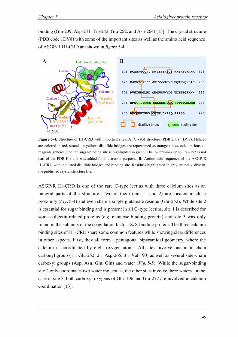
8/9/2019 DissB 7321 Mirroring
http://slidepdf.com/reader/full/dissb-7321-mirroring 159/278
Chapter 5 Asialoglycoprotein receptor
145
binding (Gln-239, Asp-241, Trp-243, Glu-252, and Asn-264) [13]. The crystal structure
(PDB code 1DV8) with some of the important sites as well as the amino acid sequence
of ASGP-R H1-CRD are shown in figure 5-4.
146 175
176 205
206 PVNTWMGLHD QNGPWKWVDG TDYETGFKNW 235
236 RPEQPDD W YG HGLGGGE 265
266 DKASQ EPPLL 290
QDWEN
B
disulfide bridge binding site
DVCQRPYRWV CETEL
CPV NWVEHERSCY WFSRSGKAWA
DADNYCRLED AHLVVVTSWE EQKFVQHHIG
DCA HFTDDGRW ND
MGSERTC
free Cys152
N (Met)
Galactose Binding Site
Calcium 1
Calcium 3
Calcium 2
DisulfideCys153>164
DisulfideCys181>276
Disulfide
Cys254>268
C
A
Figure 5-4: Structure of H1-CRD with important sites. A: Crystal structure (PDB entry 1DV8). Helices
are colored in red, strands in yellow, disulfide bridges are represented as orange sticks, calcium ions as
magenta spheres, and the sugar-binding site is highlighted in green. The N-terminus up to Cys-152 is not
part of the PDB file and was added for illustration purpose. B: Amino acid sequence of the ASGP-R
H1-CRD with indicated disulfide bridges and binding site. Residues highlighted in grey are not visible in
the published crystal structure file.
ASGP-R H1-CRD is one of the rare C-type lectins with three calcium sites as an
integral parts of the structure. Two of them (sites 1 and 2) are located in close
proximity (Fig. 5-4) and even share a single glutamate residue (Glu-252). While site 2
is essential for sugar binding and is present in all C-type lectins, site 1 is described for
some collectin-related proteins (e.g. mannose-binding protein) and site 3 was only
found in the subunits of the coagulation factor IX/X binding protein. The three calcium
binding sites of H1-CRD share some common features while showing clear differences
in other aspects. First, they all form a pentagonal bipyramidal geometry, where the
calcium is coordinated by eight oxygen atoms. All sites involve one main-chain
carbonyl group (1 = Glu-252, 2 = Asp-265, 3 = Val-190) as well as several side-chain
carboxyl groups (Asp, Asn, Glu, Gln) and water (Fig. 5-5). While the sugar-binding
site 2 only coordinates two water molecules, the other sites involve three waters. In the
case of site 3, both carboxyl oxygens of Glu-196 and Glu-277 are involved in calcium
coordination [13].
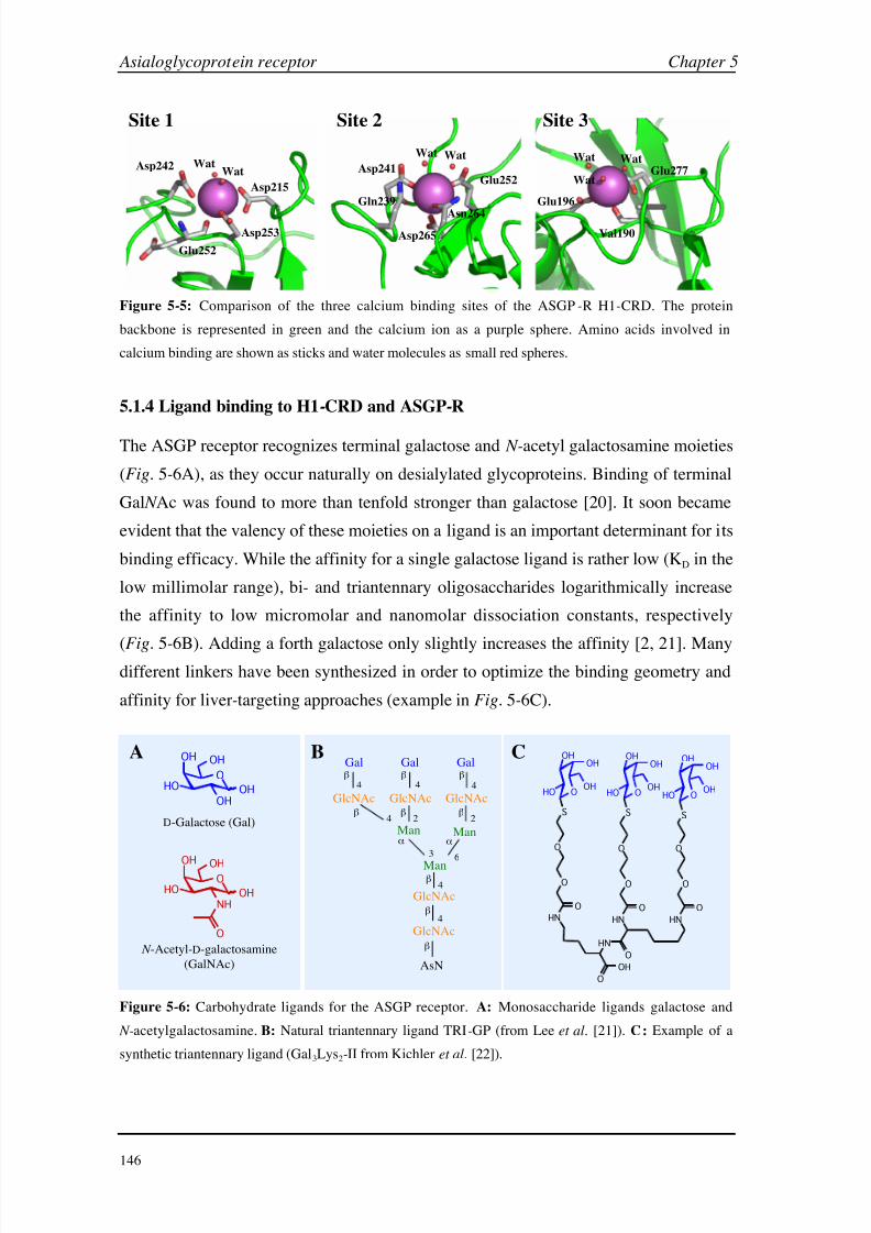
8/9/2019 DissB 7321 Mirroring
http://slidepdf.com/reader/full/dissb-7321-mirroring 160/278
Asialoglycoprotein receptor Chapter 5
146
Site 1 Site 2 Site 3
Glu252
Asp242
Asp253
Asp215
WatWat Asp241
Glu252
Asp265
Asn264
Gln239
Wat Wat
Val190
Glu277
Glu196
WatWat
Wat
Figure 5-5: Comparison of the three calcium binding sites of the ASGP-R H1-CRD. The protein
backbone is represented in green and the calcium ion as a purple sphere. Amino acids involved in
calcium binding are shown as sticks and water molecules as small red spheres.
5.1.4 Ligand binding to H1-CRD and ASGP-R
The ASGP receptor recognizes terminal galactose and N-acetyl galactosamine moieties
(Fig. 5-6A), as they occur naturally on desialylated glycoproteins. Binding of terminal
Gal N Ac was found to more than tenfold stronger than galactose [20]. It soon became
evident that the valency of these moieties on a ligand is an important determinant for its
binding efficacy. While the affinity for a single galactose ligand is rather low (KD in the
low millimolar range), bi- and triantennary oligosaccharides logarithmically increase
the affinity to low micromolar and nanomolar dissociation constants, respectively
(Fig.
5-6B). Adding a forth galactose only slightly increases the affinity [2, 21]. Manydifferent linkers have been synthesized in order to optimize the binding geometry and
affinity for liver-targeting approaches (example in Fig. 5-6C).
Gal Gal Gal
GlcNAcGlcNAcGlcNAc
GlcNAc
GlcNAc
AsN
Man
ManMan
O
OH
HO
OH
OH
OH
N -Acetyl-D-galactosamine
(GalNAc)
D-Galactose (Gal)
O
OH
HO
OH
NH
OH
O
HN
O
O
HN
OH
O
S
O
OHOH
OHHO
O
O
S
O
OHOH
OHHO O
OHOH
OHHO
S
O
O
HN
OOO
HN
O
A B C
4 4 4
4 2 2
63
4
4
Figure 5-6: Carbohydrate ligands for the ASGP receptor. A: Monosaccharide ligands galactose and
N-acetylgalactosamine. B: Natural triantennary ligand TRI-GP (from Lee et al. [21]). C: Example of a
synthetic triantennary ligand (Gal3Lys2-II from Kichler et al. [22]).

8/9/2019 DissB 7321 Mirroring
http://slidepdf.com/reader/full/dissb-7321-mirroring 161/278
Chapter 5 Asialoglycoprotein receptor
147
The observation that the change to triantennary oligosaccharides generated the most
remarkable increase in binding affinity, led to more hypothesis about a possible
receptor and binding geometry (Fig. 5-7). Antibody cross-linking studies and lateral
diffusion measurements suggested a heterohexa- (Fig. 5-7B) or pentameric geometry
(Fig.
5-7C) [12], while oxidative cross-linking of H1 and H2 peptides with terminal
cysteine residues resulted in homotrimers (H1), homotetramers (H2) and 2:2
heterotetramers (H1/H2 mixtures) [23]. Investigations with a triantennary glycopeptide
on rat ASGP-R suggested a triangular arrangement of the three galactose residues with
distances of 15, 22 and 25 Å [24, 25] (Fig. 5-7A). Further studies showed that two
galactose moieties specifically interact with HL-1 subunits and the third galactose with
HL-2 [26].
H2
H1
Gal
Gal
Gal
GN
Man
GN
Man
GN
GN
ManGN
Asn
2 2 Å
25 Å
1 5 Å
A B CH2
H1
Figure 5-7: Possible binding geometries of triantennary ASGP-R ligands. A: Dimensions of an optimal
triantennary oligosaccharide (GN = N-Acetyl glucosamine). B, C: Hypothetical models for the
arrangement of the hetero-oligomeric receptor complex (all images adapted from Geffen and Spiess [12]).
As it is typical for C-type lectins, binding to the H1-CRD of the receptor shows an
absolute requirement for Ca2+[16]. Two to three calcium ions were found to be bound
per CRD in the case of rabbit ASGP-R, with equilibrium dissociation constants of
0.3-1 mM for the individual calcium ions [27]. Since the bound ligand has to be
released after fusion with hepatic endosomes, the binding affinity is also strongly
pH-dependent. Conformational changes within the CRD lead to the release of both the
ligand and the calcium ions when lowering the pH below 6.5 [28].
Some of the best-characterized ligands are the desialylated glycoproteins
asialoorosomucoid (ASOR) and asialofetuin (ASF) with five and three N-linked
glycans, respectively [17]. ASOR shows equilibrium dissociation constants of ~ 40 nM
for cells expressing only H1 and ~ 10 nM for the functional receptor [29]. Even though
binding modes and affinities for many natural ligands and multivalent Gal or Gal N Ac

8/9/2019 DissB 7321 Mirroring
http://slidepdf.com/reader/full/dissb-7321-mirroring 162/278
Asialoglycoprotein receptor Chapter 5
148
conjugates with synthetic spacers have been investigated, little is known about the
potential of carbohydrate mimics and no SPR-based binding data are available.
5.1.5 Aims in this project
The primary aim in this project is the development of a Biacore assay for the
characterization of the molecular interaction between ASGP-R H1-CRD and their
natural (and synthetic) ligands. For this purpose, the expression and purification of the
protein is optimized and the product is characterized by different methods. H1-CRD is
then immobilized on the sensor chip and the assay conditions have to be optimized.
Different known physiological binding properties such as the C-type lectin specific
calcium-dependency or the pH-dependent release of ligands are validated using the
molecular protein assay. Finally, the assay is validated with a panel of different ligands
(glycoproteins, mono-, di- and oligosaccharides) and used to characterize a set of
monoclonal anti-H1-CRD antibodies.
8/9/2019 DissB 7321 Mirroring
http://slidepdf.com/reader/full/dissb-7321-mirroring 163/278
Chapter 5 Asialoglycoprotein receptor
149
5.2 Materials and Methods
This section describes materials, equipment and procedures specifically used for the
ASGP-R project. Materials and general methods used in all Biacore assays are
described in section 2.2.
5.2.1 Materials
Reagents and proteins
Reversed-phase HPLC was performed with gradient-grade acetonitrile and
trifluoroacetic acid (Riedel-de Haën). Gradient-grade HPLC water (Fluka) was used for
weak anion exchange chromatography and freshly bidistilled water delivered through
quartz tubes was used for the preparation of all other buffers and solutions.
Neuraminidase agarose from Clostridium perfringens (EC 3.2.1.18, Type VI-A, Sigma
N5254) was used for enzymatic desialylation of orosomucoid (1-acid glycoprotein
from human plasma, Fluka 50646). Asialofetuin from fetal calf serum was
commercially available as desialylated product (Type I, acidic desialylation, Sigma
A4781). D-Galactose was from Senn Chemicals AG (Dielsdorf, Switzerland),
N-acetyl-D-galactosamine from Acros (Chemie Brunschwig AG, Basel, Switzerland),
D-galactosamine from New Zealand Pharmaceuticals (Palmerston North, NewZealand), and methyl -D-galactopyranoside from Sigma. D-Lactose, D-glucose, and
methyl -D-galactopyranoside were gifts from various sources. Polyacrylamide-type
glycoconjugates containing 20 mol% of either -D-glucose or -D-Gal N Ac and 5%
biotin (No. 0022-BP and 0031-BP; Lectinity Holdings Inc., Moscow, Russia) was used
for both Biacore experiments and the solid-phase competition assay. Carboxymethyl
dextran (Fluka 86524, sodium salt, 12 kDa, 1.5 mmol carboxyl groups/g) as well as all
other reagents from Sigma, Riedel-de Haën and Fluka were from Fluka AG, Buchs,
Switzerland.
Equipment
HPLC separations were done on an Agilent 1100 purification system, equipped with a
quaternary pump, a cooled well-plate autosampler, a column thermostat, a DAD
detector, and a cooled analytical fraction collector (Agilent AG, Basel, Switzerland).
Size exclusion chromatography was performed on a TSK Gel G2000SW column
(7.5
600
mm; Tosoh Biosciences GmbH, Stuttgart, Germany). Two identical columns(polymer-based DEAE columns, 7.5 80 mm) from different brands, i.e. Phenomenex
8/9/2019 DissB 7321 Mirroring
http://slidepdf.com/reader/full/dissb-7321-mirroring 164/278
Asialoglycoprotein receptor Chapter 5
150
Biosep-DEAE-P and Shodex IEC DEAE-825 (both from Brechbühler AG, Schlieren,
Switzerland), were used for weak anion exchange separation. For affinity
chromatography, empty Bioscale MT-2 columns (2 ml volume; Bio-Rad Laboratories
AG, Reinach, Switzerland) were filled with differently derivatized sepharose.
Reversed-phase HPLC was performed on a SpectraSystem HPLC system consisting of
a vacuum degasser, an autosampler, a binary pump and a UV detector (Thermo
Separation) using Vydac 214TP54 columns (RP-C4. 4.6 150 mm).
5.2.2 Expression and purification of recombinant H1-CRD (PhD thesis of R. Born [30])
Human hepatic asialoglycoprotein receptor H1-CRD was produced recombinantly
based on a published method [13, 31]. The method was further optimized by Rita Born
[30], in order to increase both yield and purity. Briefly, a truncated form of the H1
subunit of the ASGP receptor including the whole CRD domain (amino acid residues
147-291) was expressed in E. coli. After lysis and solubilization of the inclusion
bodies, the protein was refolded by dialysis and affinity-purified on a galactose
sepharose column. A schematic overview of the major expression and purification steps
is visualized in figure 5-8A&B.
5.2.3 Separation of H1-CRD monomers and dimers
Since the H1-CRD fractions after expression and purification (see section 5.2.2) always
contained a variable amount of a dimeric species as well as some minor impurities,
different HPLC methods for a further separation and purification of H1-CRD were
developed. Four different chromatographic principles (size exclusion, reversed-phase,
weak anion exchange and affinity chromatography) were applied and compared,
Separation according on the molecular weight of the monomer and dimer fractions was
performed with size exclusion chromatography. Unprocessed H1-CRD samples were
injected on the column equilibrated with 10 mM HEPES pH 7.4 and separated under
isocratic conditions at a flow rate of 1 ml/min and ambient temperature. Specific lectin
interactions with the silica-based column material was tested by adding 2 mM CaCl2 to
the running buffer.
In a reversed-phase separation approach, a water-to-acetonitrile gradient was used to
isolate H1-CRD monomers from dimers. 0.1% trifluoroacetic acid (TFA) was added to
all solvents to improve separation and peak shapes. After equilibration of the
C4
RP-column in water/0.1% TFA, the sample was injected and a 10
min linear

8/9/2019 DissB 7321 Mirroring
http://slidepdf.com/reader/full/dissb-7321-mirroring 165/278
Chapter 5 Asialoglycoprotein receptor
151
gradient from 0 to 40% acetonitrile (ACN) in 0.1% TFA was performed, followed by a
shallow gradient from 40 to 52.5% ACN/TFA within 10 min. Finally, the gradient was
raised to 95% ACN/TFA and the column was washed for 5 min at these conditions
before returning to 100% water/TFA. Collected fractions were analyzed using
SDS -PAGE adapted from the method by Laemmli [32] (15% SDS-PAGE,
non-reducing conditions, silver staining).
E.coli inclusion bodies protein in solution
induction
expression lysis solubilization
protein in solution
dialysis
soluble/folded proteins
affinity chromatography
(Gal-sepharose, FPLC)
active proteins
active proteins
anion exchange
(DEAE, HPLC) dimers
monomers
affinity chromatography
(GalNAc-sepharose,
HPLC)conc. dimers
conc. dimers
A
B
C
conc. monomers
Figure 5-8: Simplified overview of expression (A), primary purification (B) and monomer/dimer
separation (C) in the production cycle of H1-CRD.
A polymer-based DEAE column was chosen for weak anion exchange (Fig. 5-8C) in
order to have a higher flexibility for elevated pH conditions. Different ionic strength,
running buffer pH and temperatures were evaluated. Finally, 25 mM Tris-HCl pH 8.0
(running buffer A) was identified as an ideal starting condition. Running buffer B was
prepared by adding 250 mM calcium chloride to running buffer A. In order to ensure a
8/9/2019 DissB 7321 Mirroring
http://slidepdf.com/reader/full/dissb-7321-mirroring 166/278
Asialoglycoprotein receptor Chapter 5
152
maximum resolution, the column was washed with a cycle of water, 0.1N NaOH,
water, and 100% B for at least one hour each at a reduced flow rate of 0.1 -0.2 ml/min.
After these washing and regeneration steps, the column was extensively equilibrated
with running buffer at 15% B for at least 8 hours (overnight). Since purified H1-CRD
samples already contained 120
mM NaCl (see section 5.2.2), the high salt load had to
be reduced prior to injection on the DEAE column. Therefore, all H1-CRD samples
underwent a buffer exchange on a HiTrap desalting column into running buffer A. The
separation of monomers was performed at 20°C and a flow rate of 0.5 ml/min. After
injection, the sample was washed for 2 min at 15% B and the linear gradient was raised
from 15% to 35% B within 25 min. All peaks were detected at 280 nm and collected
for further analysis. In case of automated separation cycles, both the autosampler and
the fraction collector were kept at 10-15 °C in order to prevent the samples fromevaporation and (proteolytic) degradation.
For HPLC-based affinity chromatography, galactose or Gal N Ac were coupled to
sepharose using the divinylsulfone method [33]. 20 ml of packed sepharose 6B were
washed three times with 20 ml water and centrifuged for 5 min at 3000 rpm. After
suspension in 20 ml 0.5 M carbonate buffer pH 11.0, the sepharose was activated by
addition of 2 ml divinylsulfone and incubation for 1 hour under constant shaking at
1000 rpm. The activated material was extensively washed in a glass frit with 2 l water
and split up into three parts. While two fractions were resuspended in 1 volume of
0.5 M carbonate buffer pH 10.0 containing either 20% (m/v) galactose or 20% (m/v)
Gal N Ac, the third fraction served as blank control and was suspended in pure carbonate
buffer. The coupling process was performed overnight under constant shaking at
300 rpm. Following one centrifugation (5000 rpm for 5 min) and three wash steps with
water, the fractions were suspended in 1 volume part 0.5 M carbonate buffer pH 8.5
and blocked with 0.02 volume parts of 2-mercaptoethanol (shaking for 2 hours at
300
rpm). The fraction were washed three times with a tenfold volume of water andpacked into empty columns at a flow rate of 2 ml/min. After connection to the
instrument, the affinity columns were properly equilibrated with running buffer
(10 mM HEPES pH 7.4, 10 mM CaCl2) for at least one hour. For purification and
affinity tests, samples were injected at a flow rate of 1 ml/min and kept at isocratic
condition for 5 min in order to wash away any inactive proteins and other impurities.
A linear gradient to an elution buffer (10 mM HEPES pH 7.4, 2 mM EDTA) was
performed for 10 min with a following plateau phase of 2 min at 100% elution
conditions. Blank runs were subtracted from the sample injections in order to increase
8/9/2019 DissB 7321 Mirroring
http://slidepdf.com/reader/full/dissb-7321-mirroring 167/278
Chapter 5 Asialoglycoprotein receptor
153
the sensitivity of the method. Samples before and after anion exchange (see above)
were tested for any loss in activity. For this purpose, constant volumes of the
unprocessed sample and the monomer fraction of the same batch after DEAE
separation were compared. Denatured H1-CRD was used as a negative control. The
specificity of the interaction was further evaluated by injecting the same sample on all
three columns (Gal-, Gal N Ac, and blank sepharose). When affinity chromatography
was used to concentrate samples after DEAE separation (Fig. 5-8C), multiple injections
were performed under isocratic running buffer conditions, before a single elution
gradient was started. Collected fractions were analyzed using SDS-PAGE as described
earlier.
5.2.4 Characterization of H1-
CRD by mass spectrometry
H1-CRD samples were either desalted by reversed-phase HPLC (see section 5.3.3) or
by elution from ZipTips. Protein mass analysis was performed using ESI or MALDI
technology before and after reduction (DTT) and alkylation (iodoacetamide) of the
proteins. For the sequence confirmation experiments, H1-CRD monomer was digested
using trypsin or LysC protease and analyzed by ESI-MS. All mass spectrometry
experiments were performed by Thierry Mini in the laboratory of Prof. Paul Jenö
(Biocenter, University of Basel, Switzerland).
3.2.5 Development and optimization of a H1-CRD Biacore assay
Immobilization of H1-CRD
The distribution and accessibility of potential sites for immobilization on the H1-CRD
molecule was investigated using crystal structure data (PDB code 1DV8) [13]. Six
residues of the N-terminus, which were not represented in the crystal structure
(147-152), were directly added in PyMol. The accessible areas of the protein werevisualized by a Connolly surface model [34], in which the lysine residues, the
N-terminus, Cys-152, as well as the residues involved in carbohydrate binding were
highlighted in different colors (Fig. 5-22, see section 5.3.4).
Separated H1-CRD monomer and dimer fractions were concentrated and
buffer-exchanged using affinity chromatography on HPLC (see section 5.2.3). Since
the used sample buffer (10 mM HEPES pH 7.4, 2 mM EDTA) did not contain any
primary amines, those samples could be directly used for immobilization. Monomers
and dimers were immobilized on separate flow cells of the same CM5 sensor chip
8/9/2019 DissB 7321 Mirroring
http://slidepdf.com/reader/full/dissb-7321-mirroring 168/278
Asialoglycoprotein receptor Chapter 5
154
using standard amine coupling procedure (see section 2.2.5). All samples were diluted
in 10 mM acetate buffer pH 4.5 to a final concentration of approximately 20 µg/ml.
After activating the surface for 5 to 10 min at at flow rate of 5 µl/min, the sample was
injected for 5 to 15 min and the surface was deactivated for a time corresponding to the
activation phase. Typical surface densities ranged from 1800 to 2500 RU for the
monomer and 1,900 to 3,000 RU for the dimer.
Alternatively, a thiol immobilization method was developed based on the procedure
used for human serum albumin (see section 3.2.13). First, a thiol-functionalized surface
was prepared (see section 2.2.7). Usually, 20 µl H1-CRD monomer (~ 200 µg/ml) were
mixed with 5 µl each of 10 mM GSH and 10 mM DTNB. After 2 hours of incubation at
ambient temperature, 200 µl 10 mM sodium acetate buffer pH 4.0 were added and the
sample was incubated for another 2
hours. Finally, the sample was injected for 60 to
120 minutes at a flow rate of 2 µl/min and washed with three consecutive 30 s pulses of
0.1% SDS at a flow rate of 50 µl/min. Surface densities up to 4,000 RU could be
obtained with this method. In contrast to the thiol immobilization procedure for
albumin, the modified method for ASGP-R was far less reproducible and
concentrations as well as incubation times had to be slightly adapted for each
immobilization.
Buffer composition
A buffer system based on the Biacore HBS-N buffer (10 mM HEPES pH 7.4, 150 mM
NaCl) was used as running buffer. The optimal calcium concentration was determined
by injecting a constant concentration of asialofetuin (2.5 µM ASF) in running buffer
containing 2, 20, 50, or 100 mM CaCl2 and the signal intensity was evaluated. The
affinity of H1-CRD for calcium ions was further investigated by injecting a twofold
serial dilution of CaCl2 between 30 µM and 50 mM in calcium-free running buffer.
Steady state data were fitted to a single site binding model as well as to a two
independent sites model. In order to identify the essential components of the running
buffer, Gal N Ac was screened as described in section 5.2.6 in four different buffer
compositions (Table 5-1).

8/9/2019 DissB 7321 Mirroring
http://slidepdf.com/reader/full/dissb-7321-mirroring 169/278
Chapter 5 Asialoglycoprotein receptor
155
Table 5-1: Buffer compositions for the identification of the components
essential for ligand binding.
System Buffer pH NaCl CaCl2
1 10 mM HEPES 7.4 150 mM 50 mM
2 10
mM HEPES 7.4 150
mM 5
mM
3 10 mM imidazole 7.4 150 mM 50 mM
4 10 mM HEPES 7.4 no 50 mM
A hypothetical binding of HEPES to the carbohydrate binding site was further
investigated with docking studies by Dr. Markus Lill in the group of Dr. Angelo
Vedani (Biographics Laboratories 3R, Basel, Switzerland).
Influence of pH
The influence of buffer acidity was studied by screening galactose and Gal N Ac (see
section 5.2.6) in buffer system 4 (Table 5-1) at pH 7.4, 7.0, 6.5, 6.0, and 5.5. Both the
signal intensity and the binding affinity were calculated and compared. Since some pH
values were below the buffer capacity range of HEPES, the buffer was carefully
controlled before and after the experiment. In addition, the pH of the buffer at pH 5.5
was measured after addition of 10 mM galactose in order to detect any analyte-induced
shifts in pH .
In order to investigate a potential release of calcium ions at lower pH values, a series of
HEPES buffer blanks was injected over immobilized H1-CRD monomer. The buffers
ranged from pH 4.0 to 7.5 in steps of 0.25 units and contained either 50 mM CaCl2 or
100 mM NaCl. All solution were injected in triplicates for 1 min at a flow rate of
50 µl/min. An empty flow cell served as a reference surface. Equilibrium data of the
induced SPR signals were averaged and plotted against the buffer pH .
DMSO tolerability
Galactose and Gal N Ac were screened (see section 5.2.6) in buffer system 4 (Table 5-1),
which was either DMSO-free or contained 5% DMSO. In case of DMSO addition, a
calibration procedure as described in section 3.2.5 was performed. Differences in the
binding affinity for the two ligands between the buffer systems were evaluated.

8/9/2019 DissB 7321 Mirroring
http://slidepdf.com/reader/full/dissb-7321-mirroring 170/278
Asialoglycoprotein receptor Chapter 5
156
5.2.6 H1-CRD ligand binding
The newly developed Biacore assay was validated by screening of some known natural
ligands. For this purpose, two different asialoglycoproteins as well as small mono- and
disaccharides were analyzed by high-resolution screening and the values werecompared with a solid-phase competition assay, which was developed by Daniela
Stokmaier in our laboratory [35]. Finally, the Biacore assay was also used to
characterize a panel of monoclonal anti-H1-CRD antibodies. Data processing of all
Biacore data as well as evaluation of equilibrium data was done in Scrubber while
kinetic analyzes were performed in CLAMP (see section 2.2.1).
Asialoglycoproteins
While asialofetuin (ASF) was commercially available, no desialylated derivative of
orosomucoid (OSM; i.e. 1-acid glycoprotein) is commercially available. Therefore,
orosomucoid had to be desialylated before screening. In principle, two methods are
available for this purpose: the acidic desialylation using sulfuric acid [36] and the
enzymatic cleavage of sialic acid moieties by a neuraminidase [37]. Both methods were
performed and compared with each other. For the acidic desialylation, the glycoprotein
was dissolved in water to a concentration of 1 mg/ml and acidified by adding an equal
volume of 0.1 N H2SO4 to a final concentration of 0.5
mg/ml OSM in 0.05 N sulfuric
acid. After incubation for 1 hour at 80 °C and constant shaking at 500 rpm, the solution
was neutralized by adding 1N NaOH. An OSM blank was prepared by substituting the
acid by pure water. In the enzymatic desialylation approach, the protein was dissolved
in reaction buffer (0.1 M sodium acetate pH 5.0, 2 mM CaCl2) to a concentration of
12.5 mg/ml. 1 ml of the OMS solution was mixed with 300 µl of the
neuraminidase-agarose suspension (0.125 U) and incubated for 4 hours at 37 °C under
shaking at 800 rpm. After centrifugation for 5 min at 13,000 rpm, the ASOR-containing
supernatant was collected and the enzyme was washed twice with reaction buffer.
Since the pI of orosomucoid is reported to increase from 2.7 to 5.0 after removal of its
sialic acids [37], a weak anion exchange method was developed to separate ASOR
from OSM. For an in-process monitoring, small samples of the desialylation mixture
were taken immediately after enzyme addition and after 1, 2, 3, and 4 hours of
incubation. All samples were injected to a DEAE column equilibrated at 25 mM
HEPES pH 7.4, 2 mM CaCl2, 12.5 mM NaCl at a flow rate of 0.5 ml/min at 20 °C.
After 2 min at starting conditions a linear NaCl gradient was applied to 175 mM NaCl
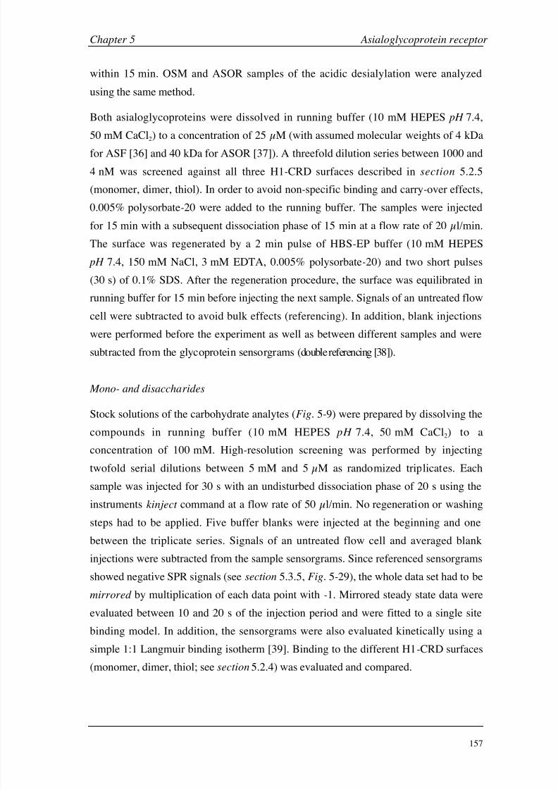
8/9/2019 DissB 7321 Mirroring
http://slidepdf.com/reader/full/dissb-7321-mirroring 171/278
Chapter 5 Asialoglycoprotein receptor
157
within 15 min. OSM and ASOR samples of the acidic desialylation were analyzed
using the same method.
Both asialoglycoproteins were dissolved in running buffer (10 mM HEPES pH 7.4,
50
mM CaCl2) to a concentration of 25
µM (with assumed molecular weights of 4
kDafor ASF [36] and 40 kDa for ASOR [37]). A threefold dilution series between 1000 and
4 nM was screened against all three H1-CRD surfaces described in section 5.2.5
(monomer, dimer, thiol). In order to avoid non-specific binding and carry-over effects,
0.005% polysorbate-20 were added to the running buffer. The samples were injected
for 15 min with a subsequent dissociation phase of 15 min at a flow rate of 20 µl/min.
The surface was regenerated by a 2 min pulse of HBS-EP buffer (10 mM HEPES
pH 7.4, 150 mM NaCl, 3 mM EDTA, 0.005% polysorbate-20) and two short pulses
(30
s) of 0.1% SDS. After the regeneration procedure, the surface was equilibrated in
running buffer for 15 min before injecting the next sample. Signals of an untreated flow
cell were subtracted to avoid bulk effects (referencing). In addition, blank injections
were performed before the experiment as well as between different samples and were
subtracted from the glycoprotein sensorgrams (double referencing [38]).
Mono- and disaccharides
Stock solutions of the carbohydrate analytes (Fig.
5-9) were prepared by dissolving thecompounds in running buffer (10 mM HEPES pH 7.4, 50 mM CaCl2) to a
concentration of 100 mM. High-resolution screening was performed by injecting
twofold serial dilutions between 5 mM and 5 µM as randomized triplicates. Each
sample was injected for 30 s with an undisturbed dissociation phase of 20 s using the
instruments kinject command at a flow rate of 50 µl/min. No regeneration or washing
steps had to be applied. Five buffer blanks were injected at the beginning and one
between the triplicate series. Signals of an untreated flow cell and averaged blank
injections were subtracted from the sample sensorgrams. Since referenced sensorgrams
showed negative SPR signals (see section 5.3.5, Fig. 5-29), the whole data set had to be
mirrored by multiplication of each data point with -1. Mirrored steady state data were
evaluated between 10 and 20 s of the injection period and were fitted to a single site
binding model. In addition, the sensorgrams were also evaluated kinetically using a
simple 1:1 Langmuir binding isotherm [39]. Binding to the different H1-CRD surfaces
(monomer, dimer, thiol; see section 5.2.4) was evaluated and compared.
8/9/2019 DissB 7321 Mirroring
http://slidepdf.com/reader/full/dissb-7321-mirroring 172/278
Asialoglycoprotein receptor Chapter 5
158
O
HO
HO
O
O
HO
OH
OH
OH
OH
OH
O
HO
HO
OH
OHOMe
O
HO
HO
OH
OH
OMe
OHO
HO
OH
OH OH
O
HO
HO
OH
NH2 OH
O
HO
HO
OH
NH
O
OH
O
HO
OH
OH
OH
HO
D-Galactose
(180.2 Da) N-Acetyl-D-galactosamine
(Gal N Ac; 221.2 Da)
D-Galactosamine
(179.2 Da)
D-Glucose
(180.2 Da)
Methyl -D-galactopyranoside
(194.2 Da)
Methyl -D-galactopyranoside
(194. 2 Da)Lactose
(342.3 Da)
Figure 5-9: Mono- and disaccharides used for the validation of the H1-CRD Biacore assay. The
galactose core structure is visualized in blue, modifications are highlighted in red.
Polyvalent, polymer-bound forms of Gal N Ac and glucose were investigated by
injecting the biotinylated glycoconjugates used for the solid-phase competition assay
over amine-coupled H1-CRD monomer and dimer surfaces under the same buffer
conditions. Both polymers (10 µg/ml) were injected for 3 min followed by another
3
min of undisturbed dissociation at a flow rate of 20
µl/min. In order to test for
competition effects, a 10 mM galactose solution was injected for 1 min and the
(negative) signal intensity as well as the post-injection drop of the signal was evaluated.
Finally, the surface was regenerated by a 1 min pulse of HBS-EP buffer (see above)
and equilibrated in running buffer for 5 min.
Solid-phase competition assay
Biacore-derived KD values were compared with a solid-phase competition assay, whichwas developed in a diploma thesis by Daniela Stokmaier [30, 35], based on a similar
polymer-assay for E-selectin [40].
Monoclonal anti-H1-CRD antibodies
A set of six monoclonal mouse anti-human H1-CRD antibodies, which were produced
with hybridoma technology and preselected using an ELISA [30], were ranked and
screened with the Biacore assay. All antibodies were purified by affinity
chromatography and were classified as mouse IgG1 [30]. In a first ranking assay, each

8/9/2019 DissB 7321 Mirroring
http://slidepdf.com/reader/full/dissb-7321-mirroring 173/278
Chapter 5 Asialoglycoprotein receptor
159
antibody was injected at a concentration of 2 µg/ml for 5 min with a dissociation phase
of 5 min. The assay was performed in 10 mM HEPES pH 7.4, 50 mM CaCl2 at a flow
rate of 10 µl/min. Potential blocking properties of the antibodies were tested by
injecting a 1 min pulse of 5 mM galactose before mAb injection and after the
dissociation phase. Calcium dependency was evaluated with an injection of HBS-EP
buffer (see above) for 1 min. Finally, the surface was regenerated by a 12 s pulse of
10 mM HCl at a flow rate of 50 µl/min and was equilibrated in running buffer for
10 min. The same assay was repeated using HBS-EP as the running buffer. The
maximum intensities at injection end were plotted against the remaining signal
intensity after 5 min of dissociation (stability plot). In addition, blocking properties
were evaluated by comparing the galactose signal before and after antibody injection.
The three antibodies with the most promising binding properties selected for kinetic
screening and were prepared as twofold dilution series between 140 nM and 140 pM in
HBS-EP as a running buffer. Samples were injected in a randomized order for 10 min
at a flow rate of 25 µl/min. After a 10 min dissociation phase, the surface was
regenerated by two 12 s injections of 10 mM HCl at a flow rate of 50 µl/min and was
equilibrated in running buffer for 5 min. The full data sets (140 nM - 140 pM) as well
as a reduced data set (9 nM - 140 pM) were kinetically evaluated in CLAMP by
applying different interaction models.
5.2.6 Evaluation of negative binding signals
In order to evaluate a weak specific interaction of the immobilized lectin with the
carboxymethyl dextran (CMD) hydrogel of the sensor chip, soluble CMD was added to
the running buffer. For this purpose, galactose and Gal N Ac were screened
(see section 5.2.5) in 10 mM HEPES pH 7.4, 50 mM CaCl2 containing 1 mg/ml CMD,
10 mg/ml CMD, or no CMD addition. Both the signal intensities and binding affinities
were compared.

8/9/2019 DissB 7321 Mirroring
http://slidepdf.com/reader/full/dissb-7321-mirroring 174/278
Asialoglycoprotein receptor Chapter 5
160
5.3 Results & Discussion
5.3.1 Expression and purification of ASGP-R H1-CRD
The ability to test physiological and synthetic ligands on an isolated form of a receptor
is a prerequisite for a deep understanding of molecular binding events. Since only the
extracellular carbohydrate recognition domain of the human hepatic asialoglycoprotein
receptor is involved in ligand binding and posttranslational glycosylation does not
influence its activity, the lectin domain can be expressed as a truncated protein in
E. coli. Even though a protocol for the expression and purification already existed [13],
the yields were rather low and the protein fraction contained several populations.
Therefore, the expression system as well as the purification protocols were further
optimized. By selecting a different strain of E.
coli (AD494 instead of JM109), theexpression yield could be remarkably increased (Fig.
5-10A). The combination of cell
lysis by sonication, solubilization of the inclusion bodies and refolding by dialysis
followed by affinity purification on a galactose-sepharose column (Fig. 5-8A&B)
generated a highly active protein fraction. However, non-reducing SDS-PAGE analysis
always showed two protein bands at approximately 17 and 34 kDa, indicating a
dimerization. In addition, the ratio between the two populations was found to highly
variable between different production batches (Fig. 5-10B). Some additional bands
around the monomer and dimer fractions were visible (‘satellite bands’,
see appendix D3, lanes 4 and 5).
A B
Optimization of H1-CRD production: yield
normalized to yield of Spiess M.t l)
D
M
Lit. Start Strain Expr. Purif.0
1
2
3
4
5
6
7
8
9
10
Optimization Step
I m p
r o v e m e n t ( F a c t o r )
Figure 5-10: Optimized expression of ASGP-R H1-CRD. A: Comparison of the expression levels
obtained by the existing protocol published in literature (Lit.; [31]) and after its application (Start), as
well as after the optimization of the E. coli strain (Strain), the expression method (Expr.), and the
purification protocol (Purif.). B: Western blot of different H1-CRD production batches, demonstrating
the high variability in the monomer (M) to dimer (D) ratio (blotted from a non -reducing
15%
SDS-PAGE, primary antibody
=
monoclonal anti-H1-CRD B01.4 (see section 5.3.5), secondary
antibody = anti-mouse Fc, HRP-conjugate). All images are adapted from Born [30].
8/9/2019 DissB 7321 Mirroring
http://slidepdf.com/reader/full/dissb-7321-mirroring 175/278
Chapter 5 Asialoglycoprotein receptor
161
5.3.2 Separation of H1-CRD monomers and dimers
In contrast to the purification of the active protein by separating it from other cellular
impurities, the separation of monomer and dimer populations was found to be more
complicated. While the major difference of the two proteins is their size, only littlevariations are expected in case of their net charge or affinity. In order to get maximum
resolution and short analysis/separation times, high-performance liquid
chromatography (HPLC) was used for the evaluation of different separation methods.
Size exclusion HPLC
Since the H1-CRD dimer should have twice the molecular mass of the monomer, a
separation by size seems to be the method of choice. Unfortunately, the absolute
masses and therefore also the mass difference are still relatively low for a successful
separation on size exclusion columns. The separation is mainly dependent on the
material and the length of the column, while parameters such as the column diameter,
flow rate, or solvent properties are less important [41]. Even though a rather large
column (60 cm) was used for the separation, both fractions appeared as a single peak.
In some cases, a small shoulder of the monomer peak was visible, which might contain
H1-CRD dimer. For a complete baseline separation, extremely large columns or
another column material (e.g.
pore size) was required.
In addition, the lectin was found to interact with the column material in a specific
manner. When calcium-containing buffers were used for the separation, the protein
remained on the column and only eluted after a switch to calcium-free buffers
(Fig. 5-11). This phenomenon could be explained by a calcium-dependent interaction
of the CRD with hydroxyl-groups of the silica-based column material. Addition of
EDTA to the running buffer as well as an increase of total ion concentration (e.g. by
adding NaCl) could reduce such interferences. However, highly ionic solutions are notfeasible for Biacore immobilization, since they suppress preconcentration of the protein
on the sensor chip surface (see section 2.1.4). Due to its low resolution and the
undesired interaction properties, the size exclusion method was regarded as not suitable
for the separation of H1-CRD monomers and dimers.
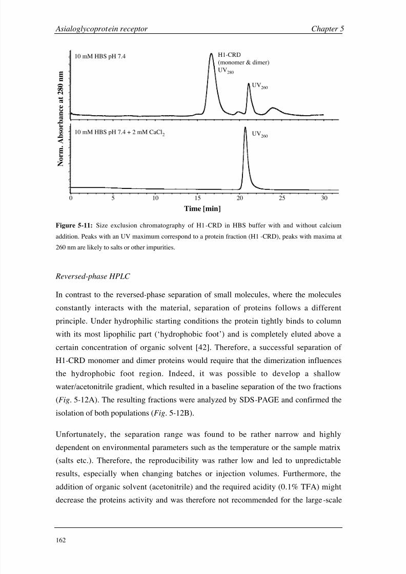
8/9/2019 DissB 7321 Mirroring
http://slidepdf.com/reader/full/dissb-7321-mirroring 176/278
Asialoglycoprotein receptor Chapter 5
162
0 5 10 15 20 25 30
10 mM HBS pH 7.4
10 mM HBS pH 7.4 + 2 mM CaCl2
H1-CRD
(monomer & dimer)
UV280
UV260
UV260
Time [min]
N o r m . A b s o r b a n c e a t 2 8 0 n m
Figure 5-11: Size exclusion chromatography of H1-CRD in HBS buffer with and without calcium
addition. Peaks with an UV maximum correspond to a protein fraction (H1-CRD), peaks with maxima at
260 nm are likely to salts or other impurities.
Reversed-phase HPLC
In contrast to the reversed-phase separation of small molecules, where the molecules
constantly interacts with the material, separation of proteins follows a different
principle. Under hydrophilic starting conditions the protein tightly binds to column
with its most lipophilic part (‘hydrophobic foot’) and is completely eluted above a
certain concentration of organic solvent [42]. Therefore, a successful separation of
H1-CRD monomer and dimer proteins would require that the dimerization influences
the hydrophobic foot region. Indeed, it was possible to develop a shallow
water/acetonitrile gradient, which resulted in a baseline separation of the two fractions
(Fig.
5-12A). The resulting fractions were analyzed by SDS-PAGE and confirmed theisolation of both populations (Fig. 5-12B).
Unfortunately, the separation range was found to be rather narrow and highly
dependent on environmental parameters such as the temperature or the sample matrix
(salts etc.). Therefore, the reproducibility was rather low and led to unpredictable
results, especially when changing batches or injection volumes. Furthermore, the
addition of organic solvent (acetonitrile) and the required acidity (0.1% TFA) might
decrease the proteins activity and was therefore not recommended for the large-scale

8/9/2019 DissB 7321 Mirroring
http://slidepdf.com/reader/full/dissb-7321-mirroring 177/278
Chapter 5 Asialoglycoprotein receptor
163
production of functional protein. This method was therefore only used for analytical
purposes, e.g. the preparation of fully desalted samples for mass spectrometry.
10 12 14 16 16 20
0
100
200
300
400
Time [min]
A b s o r b a n c e a t 2 8 0 n m [ m
A U ]
A B
a b c d
a b c d
17 kDa
34 kDa
Figure 5-12: Reversed-phase separation of H1-CRD monomer and dimer fractions. A: Chromatogram
showing two clearly separated peaks. B: SDS-PAGE analysis of the collected fractions (a-d) of
chromatogram A (15% gel, non-reducing, silver staining).
Weak anion exchange HPLC (DEAE column)
Anion exchange separates different species according to their surface charge. While
completely different proteins with similar charge distributions might elute in a very
similar manner, even single residue mutations in the same protein can be separated in
other cases [43]. A polymer-based DEAE column equilibrated with Tris buffer was
used for the separation of H1-CRD. The polymer material has the advantage of being
stable in a much wider pH range than silica-based columns are. The pH optimum for a
proper separation was found to be at pH 8.0. This is higher than expected, since values
just one unit above the protein’s pI (i.e. approximately pH 6 for H1-CRD) are usually
sufficient for weak anion exchange. However, since the aim was not to separate
H1-CRD from structurally different impurities but to separate closely related
oligomeric forms, a higher pH seems to be necessary to gain additional resolution.
Higher pH values had been avoided to exclude a possible disruption of intramolecular
disulfide bridges. Calcium chloride was chosen for the gradient elution due to its high
chloride activity and since calcium containing buffers were expected to retain the
proteins activity after elution. Reproducible separations of H1-CRD monomers and
dimers in less than 30 min could be established with a linear gradient from 37.5 to
100 mM CaCl2. Different batches of H1-CRD were separated and the collected peak
fractions were analyzed by SDS-PAGE (Fig. 5-13).
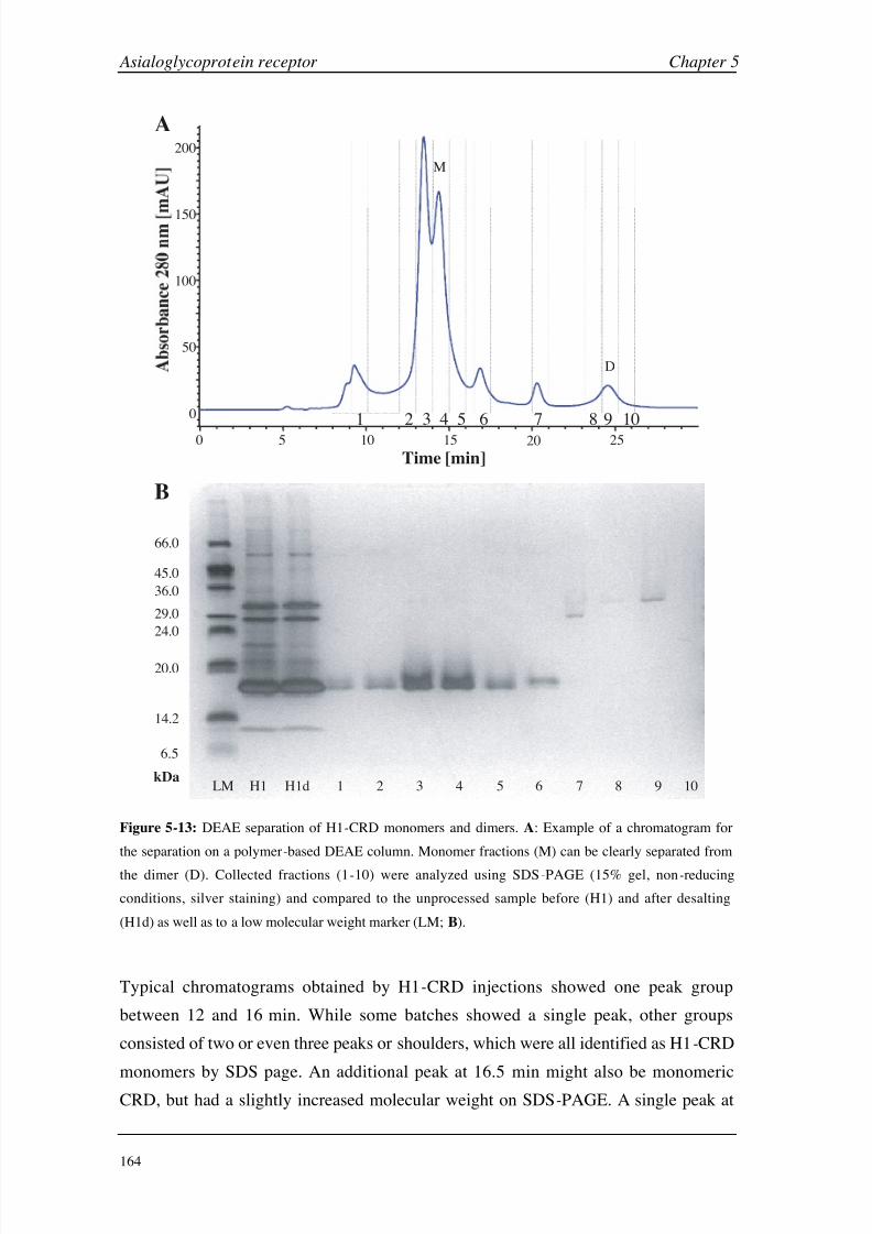
8/9/2019 DissB 7321 Mirroring
http://slidepdf.com/reader/full/dissb-7321-mirroring 178/278
Asialoglycoprotein receptor Chapter 5
164
LM H1 H1d 1 2 3 4 6 8 9 105 7
6.5
14.2
20.0
24.0
29.0
36.0
45.0
66.0
kDa
0 5 10 15 20 25
0
50
100
150
200
1 2 3 4 5 6 7 8 9 10
Time [min]
A
B
M
D
Figure 5-13: DEAE separation of H1-CRD monomers and dimers. A: Example of a chromatogram for
the separation on a polymer-based DEAE column. Monomer fractions (M) can be clearly separated from
the dimer (D). Collected fractions (1-10) were analyzed using SDS-PAGE (15% gel, non-reducing
conditions, silver staining) and compared to the unprocessed sample before (H1) and after desalting
(H1d) as well as to a low molecular weight marker (LM; B).
Typical chromatograms obtained by H1-CRD injections showed one peak group
between 12 and 16 min. While some batches showed a single peak, other groups
consisted of two or even three peaks or shoulders, which were all identified as H1-CRD
monomers by SDS page. An additional peak at 16.5 min might also be monomeric
CRD, but had a slightly increased molecular weight on SDS-PAGE. A single peak at
8/9/2019 DissB 7321 Mirroring
http://slidepdf.com/reader/full/dissb-7321-mirroring 179/278
Chapter 5 Asialoglycoprotein receptor
165
20.5 min corresponded to a satellite band below the dimer on SDS-PAGE but could not
be identified by mass spectrometry. Finally, H1-CRD dimer eluted at 24 min as a single
peak. The DEAE separation method therefore proofed to be suitable for isolating pure
H1-CRD monomer and dimer fraction as well as for removing minor impurities.
The reason for the heterogeneity in the monomeric region is not yet clear and possible
causes include folding or disulfide isomerization, proteolytic degradation during
production and storage, or mutations. Since the uneven number of cysteine residue
seems to promote the formation of mixed disulfides (see section 5.3.3), these small
heterogeneities in retention on HPLC and SDS could be explained by different thiol
adducts, which alter surface charge and molecular weight of the protein. The slightly
increased pH value of 7.8-8.0 during storage and separation could further promote
disulfide-shuffling reactions. Since DEAE is able to separate monomeric and dimeric
forms of the same protein, the region around the dimerization site (e.g. free cysteine
residues) seems to directly influence the interaction of the protein with the column
resin. Similar effects are also described in literature. For example, DEAE anion
exchange chromatography was used for the separation of different oxidation states of
human serum albumin, which also only differ in the state of one cysteine residue
(i.e. free Cys, mixed disulfides, and oxidized Cys) [44, 45]. The matrix of the
polymer-based DEAE column was also identified to be crucial for a successful
separation in the case of HSA [44].
While the relative peak intensities varied significantly between batches, they were very
consistent within one batch (Fig. 5-14A). In contrast to RP-HPLC, the retention times
changed only slightly when increasing the injection volume. Small shifts in retention
times (± 0.5 min) were sometimes observed upon buffer changes, most likely due to
small differences in buffer pH or calcium concentrations. However, properly
equilibrated systems showed a very high reproducibility (Fig. 5-14B). Desalting was
found to be a crucial step in the process. Since high NaCl concentrations (500
mM)
were used during refolding and FPLC separation, direct injection of those samples on
the DEAE column essentially inhibited binding of the protein to the column and a large
fraction eluted already after the void volume. The desalting step on a HiTrap desalting
column did not change the composition of H1-CRD samples (Fig. 5-13, lanes H1 and
H1d) nor did it influence retention times during DEAE separation. Water quality was
found to be crucial for this type of chromatography and only bidistilled or
gradient-grade HPLC water could be used. Deionized and filtered water always
generated a significant background signal, which often covered dimer peaks.

8/9/2019 DissB 7321 Mirroring
http://slidepdf.com/reader/full/dissb-7321-mirroring 180/278
Asialoglycoprotein receptor Chapter 5
166
300
200
100
0
0 10 20 30
Time [min]
A b s o r b a n c e a t
2 8 0 n m [ m A U ]
10 20 30
0
200
400
600
800
A B
Figure 5-14: Reproducibility of H1-CRD separation. A : Different batches during production
development showing varying concentrations and impurities but very reproducible retention times.
B: Repetitive injections of the same batch leads to absolute reproducibility of intensities and retention times.
Weak anion exchange separation was found to be best suited for the analysis and
production of pure H1-CRD monomers and dimers. By extending the gradient down to
12.5 mM CaCl2 it was even possible to fully separate H1-CRD monomers and dimers
from H2-CRD monomers and dimers [46] using a single column/buffer system
(Fig. 5-15). This expands the analytical applications of this method while avoiding
possible cross-contaminations of the two subunits.
H1-CRD
H2-CRD
H1/H2 mix
10 20 30 400
Time [min]
A b s o r
b a n c e a t 2 8 0 n m
Figure 5-15: Monomer/dimer separation of H1-CRD (blue, top) and H2-CRD (red,
middle) as well as of a H1/H2-CRD mixture (purple, bottom) using a single gradient
run on a DEAE column.
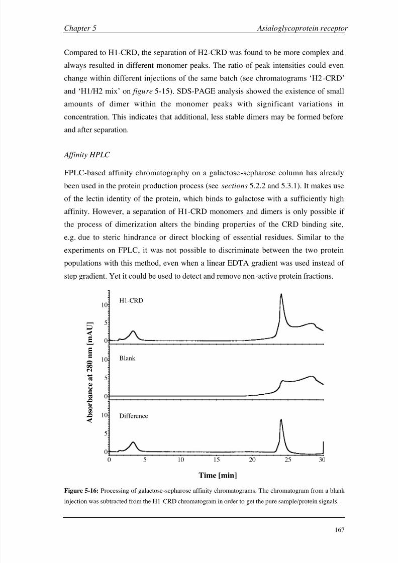
8/9/2019 DissB 7321 Mirroring
http://slidepdf.com/reader/full/dissb-7321-mirroring 181/278

8/9/2019 DissB 7321 Mirroring
http://slidepdf.com/reader/full/dissb-7321-mirroring 182/278
Asialoglycoprotein receptor Chapter 5
168
Every change to the elution buffer caused a significant peak-like increase in the
baseline. Therefore, a blank run had to be subtracted from the sample run for highest
sensitivity, especially at low protein concentration (Fig. 5-16). A small signal shortly
after injection start was usually detected and is believed to contain salts and inactive
protein.
A first application of this method was the verification of H1-CRD activity before and
after DEAE separation (Fig. 5-17A). No loss of activity could be detected when the
samples were separated on the DEAE column. The injection peak was even slightly
decreased indicating a higher purity of the sample. In contrast, a deactivated H1-CRD
sample (before refolding) did not bind to the affinity column (Fig. 5-17A). Since the
sepharose material itself was suspected to interact non-specifically with the lectin, a
blank sepharose column was compared to galactose and Gal N Ac-coupled columns
(Fig. 5-17B). While the protein clearly bound to both derivatized columns and did not
elute until EDTA was added, there was no active protein peak detectable on the blank
sepharose. However, the protein did not appear in the injection peak but eluted widely
distributed within the first ten minutes. An extended washing phase is therefore
recommended. No major difference in the separation properties and efficiency of
galactose and Gal N Ac columns could be detected.
Gal N Ac-sepharose
Gal-sepharose
Blank sepharose
200 5 10 15 20100 5 15
Time [min]
A b
s o r b a n c e a t 2 8 0 n m [ m
A U ]
H1-CRD before DEAE
H1-CRD after DEAE
Inactive H1-CRD
A B
Figure 5-17: Affinity chromatography experiments with H1-CRD. A: Verification of the protein activity
before and after DEAE separation. An inactive H1-CRD fraction was used as negative control.
B: Binding specificity test on Gal- and Gal N Ac-sepharose columns compared to a blank sepharose
column.
8/9/2019 DissB 7321 Mirroring
http://slidepdf.com/reader/full/dissb-7321-mirroring 183/278
Chapter 5 Asialoglycoprotein receptor
169
While it was not possible to separate monomers and dimers using affinity
chromatography on a Gal- or Gal N Ac sepharose column, the method had its benefit in
the post-production phase. In one run it was possible to check for the activity of
DEAE-separated fractions, increase their concentration by multiple injections prior to a
single elution, and simultaneously exchange the buffer to an alternative system
compatible with Biacore immobilization or other applications (i.e. HEPES buffer).
5.3.3 Characterization of H1-CRD using mass spectrometry
H1-CRD samples, which were separated and desalted with RP-HPLC
(see section 5.3.2), were further characterized using mass spectrometry. Since E. coli
was used as an expression system, no glycosylation or phosphorylation is performed
posttranslationally, and the measured masses were expected to correlate with the
theoretical ones (Table 5-2). However, isolated monomer showed a single main peak
on ESI-MS with a mass of 16,934 Da, which was significantly lower than expected
(Fig. 5-18A). The mass difference of -57 Da did not correspond to the deletion or
proteolysis of an amino acid. If the protein monomer has been expressed with a lower
mass, the doubled difference should also be visible in the dimer fraction. ESI and
MALDI analysis of the dimer fraction showed relatively wide peaks with maxima
between 33,690 and 33,700 Da (Fig. 5-18B). This peak widening might be caused by
disulfide shuffling. Here again, the difference to the expected mass (33,982 Da) was
much larger than expected (approximately -280 Da) and clearly different from the
114 Da difference expected when doubling the monomer mass. This led to the
hypothesis that the resulting mass differences might be the sum of an amino acid
deletion and some adducts. The large discrepancy between monomer and dimer
deviations suggested that a free cysteine residue (only 6 of the 7 cysteines of H1-CRD
are involved in disulfide bridges; see section 5.1.3) could be involved in both
dimerization and mass adducts. Therefore, an additional ESI analysis of the monomerfraction was performed under reducing conditions (Fig. 5-18C). The resulting mass
was significantly lower than the one measured under non-reducing conditions. The
difference of 131 Da exactly corresponded to the deletion of methionine residue
(Fig. 5-20). Indeed, it is reported in literature that E. coli is able to remove a N-terminal
methionine under certain conditions (the second amino acid has to be small) [47, 48].
Since these conditions are given in the case of H1-CRD (the second amino acid is a
glycine), a posttranslational cleavage of the terminal methionine is highly likely.
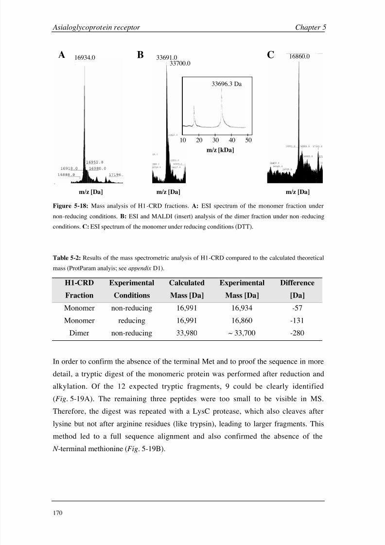
8/9/2019 DissB 7321 Mirroring
http://slidepdf.com/reader/full/dissb-7321-mirroring 184/278

8/9/2019 DissB 7321 Mirroring
http://slidepdf.com/reader/full/dissb-7321-mirroring 185/278
Chapter 5 Asialoglycoprotein receptor
171
147 GSERTCCPV NWVEHERSCY WFSRSGKAWA 175
176 DADNYCRLED AHLVVVTSWE EQKFVQHHIG 205
206 PVNTWMGLHD QNGPWKWVDG TDYETGFKNW 235
236 RPEQPDDWYG HGLGGGEDCA HFTDDGRWND 265
266 DVCQRPYRWV CETELDKASQ EPPLL 290
T T2 T3 T54
T5 T6 T7
T7 T8 T9
T9 T
T T T 2
A B
L L2
L2 L3
L3 L4 L5
L5
L5 L6
147 GSERTCCPV NWVEHERSCY WFSRSGKAWA 175
176 DADNYCRLED AHLVVVTSWE EQKFVQHHIG 205
206 PVNTWMGLHD QNGPWKWVDG TDYETGFKNW 235
236 RPEQPDDWYG HGLGGGEDCA HFTDDGRWND 265
266 DVCQRPYRWV CETELDKASQ EPPLL 290
Figure 5-19: Proteolytic digests of H1-CRD analyzed by ESI-MS. Reduced and alkylated monomer
fractions were digested with trypsin (A; T1-T12) or LysC protease (B; L1-L6). Identified peptidic
fragments are highlighted in green, unidentified in red. No fragments with an N-terminal methionine
residue could be detected.
With the knowledge about the absence of the terminal Met, the differences between
calculated and measured masses were recalculated (Table 5-3). While the monomer
under reducing condition very well correlates with the calculated mass, there is now a
positive difference under non-reducing conditions. This strongly supports the
hypothesis of an adduct on the free cysteine residue of H1 -CRD, which is readily
cleavable when adding reducing agents. Comparison of the mass difference (74 Da)
with the ABRF DeltaMass database [49] indicated an addition of 2-mercaptoethanol
[50] (Fig. 5-20). Since this reducing agent was used during the refolding process, an
addition seems to be reasonable. Following this hypothesis, the mass increase changed
to +305 Da when a combination of reducing and oxidizing glutathione was used in the
refolding step during production development (Fig. 5-20; see appendix D2). Again, this
increase corresponds to an addition of glutathione to the free sulfhydryl group. This
behavior of a glutathiolation during refolding had been also observed in another study
[51]. The small difference of ~ 18 Da in case of the monomer is rather caused by the
wide peak maximum or by disulfide shuffling than by a real change of mass. Anadditional MS analysis of the dimer fraction in the laboratory of Prof. Jasna
Peter-Katalinic (University of Münster, Germany) revealed a mass of 33,706 Da, which
further reduces the difference to the calculated mass.

8/9/2019 DissB 7321 Mirroring
http://slidepdf.com/reader/full/dissb-7321-mirroring 186/278
Asialoglycoprotein receptor Chapter 5
172
Table 5-3: Recalculated comparison of the results of the mass spectrometric analysis of H1-CRD
compared and the calculated theoretical mass without the N-terminal methionine residue.
H1-CRD
Fraction
Experimental
Conditions
Calculated
Mass [Da]
Experimental
Mass [Da]
Difference
[Da]
Monomer non-reducing 16,860 16,934 +74
Monomer reducing 16,860 16,860 ±0
Dimer non-reducing 33,718 ~ 33,700 -18
H2N
HN
S
O
NH
O
SH
H2NNH
O
SH
HN
NH
NH2
O
COOH
S
O
HOOC
S
H2NNH
O
OH
S
S
H2NNH
O
H1-CRD H1-CRD
H1-CRD
H1-CRD
2-ME
(78 Da)
GSH
(305 Da)
Met
(131 Da)
Gly Gly
a
c
b
Figure 5-20: Mass changes by derivatization of H1-CRD at the N-terminus or cysteine residues. After a
posttranslational removal of the N-terminal methionine (a), the free cysteine could be conjugated by
2-mercaptoethanol (2-ME; b) or glutathione (GSH; c) during refolding.
SDS-PAGE analysis showed further evidence for a disulfide-based dimerization, since
the protein showed two bands under non-reducing conditions (17 and 33 kDa), while a
single band (17 kDa) was visible in reducing buffer (see appendix D3). This leads to
the hypothesis that a single free cysteine residue is responsible for dimerization.
Formation of mixed disulfides during refolding could act as a protection group for the
cysteine and therefore inhibits the dimerization process (Fig.
5-21). This also helpsexplaining the rather large variations in the monomer-dimer ratio during production,
since a competitive reaction between monomers and thiol-active agents is expected to
be highly dependent on concentrations, pH , salts, or temperature.
In the crystal structure of H1-CRD, only one of the two vicinal cysteine residues
(Cys-152 and Cys-153) is resolved [13]. Since Cys-153 was reported to be involved in
disulfide bridge formation, the position of Cys-152 should be rather fixed and therefore
visible in the crystal. A derivatization of this residue at its thiol group might be a
possible reason why Cys-152 could not be positioned into electron density. However,

8/9/2019 DissB 7321 Mirroring
http://slidepdf.com/reader/full/dissb-7321-mirroring 187/278
Chapter 5 Asialoglycoprotein receptor
173
the N-terminal disulfide bridge (Cys-153/Cys-163) could not be confirmed by mass
spectrometric analysis [31]. Therefore, it is rather difficult to determine, which cysteine
residues are effectively involved in mixed disulfides or dimerization. Mutation
experiments seem to be the most promising way to learn more about the disulfide
structure of H1-CRD. Deletion or mutation of the single free cysteine could
successfully prevent dimer formation. On the other hand, this cysteine residue could
also be used for a site-specific labeling or immobilization of the protein by adding
thiol-active compounds, which allow labeling of the protein (e.g. by biotin or a
fluorescent dye) during refolding steps.
SHS S–
HS– x 2
protected monomer free monomer dimer
S S
Figure 5-21: Dimerization hypothesis for H1-CRD. During refolding, the single free cysteine residue of
H1-CRD (middle) could form dimers with another CRD (right) or might be protected from dimerization
by thiol-active reagents like 2-mercaptoethanol or glutathione (left).
5.3.4 Development and optimization of a Biacore assay
Immobilization of H1-CRD
Standard amine coupling usually leads to a randomized immobilization on the sensorchip, since it targets all primary amines (lysines, N-terminus) that are available on the
surface (see section 2.1.4). Therefore, an analysis of the Connolly surface was done to
estimate the risk of an immobilization-induced deactivation of the binding site
(Fig. 5-22). Neither the lysine residues nor the N-terminus are directly involved in
ligand binding, which makes a direct blocking of the binding site very unlikely.
Nevertheless, immobilization might lead to conformational changes or steric hindrance
and, as a consequence, influence the affinity indirectly.
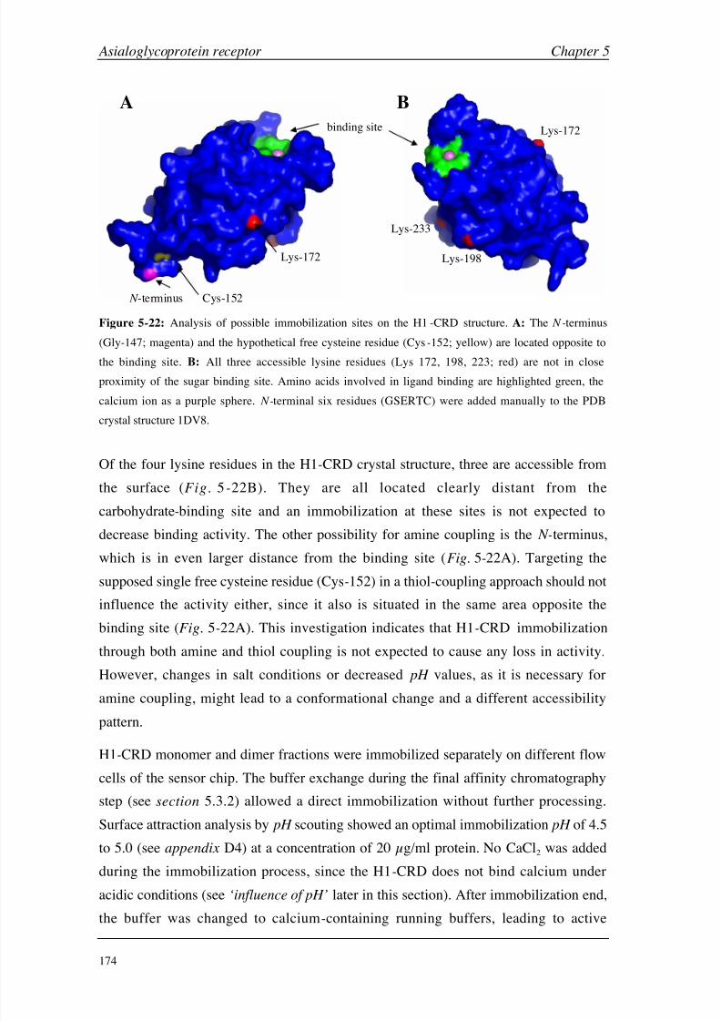
8/9/2019 DissB 7321 Mirroring
http://slidepdf.com/reader/full/dissb-7321-mirroring 188/278

8/9/2019 DissB 7321 Mirroring
http://slidepdf.com/reader/full/dissb-7321-mirroring 189/278
Chapter 5 Asialoglycoprotein receptor
175
H1-CRD. Using this method, H1-CRD fractions could be reproducibly immobilized on
activated sensor chips (Fig. 5-23A&B).
5
10
15
20
0
S P R R e s p o n s e [ k R U
]
Time [s]
0 1000 2000 1000 0 1000 20000
a
c
d
a
c
d
c
w
A B C
Figure 5-23: Immobilization of H1-CRD on CM5 sensor chips. A: Amine coupling of the monomer
fraction with activation (a), coupling (c) and deactivation step (d). B: Amine coupling of the dimer
fraction. C: Thiol coupling of H1-CRD after in-situ reduction/activation on a thiol-functionalized sensor
chip surface (w = washing step).
As expected by the larger molecular weight of the H1-CRD dimer, the immobilization
of dimer fractions usually led to higher surface densities. In addition, surface attraction
and immobilization efficiency was also significantly increased in case of the dimer.
This might be caused by the higher local concentration of primary amine groups on themolecule. The binding activities of immobilized monomer and dimer surfaces were
evaluated by injecting a Gal N Ac concentration series (see section 5.3.5) and comparing
the signal intensities and KD values (Table 5-4).
In contrast to the binding affinity (KD), which only showed slight variations between
the two fractions, the calculated activity deviated more clearly. While both surfaces
generated negative binding signals (see section 5.3.5), the signal intensity was
unexpectedly high in case of the H1-CRD dimer and even exceeded the calculated Rmax
value. This indicates that the signal generation of the Gal N Ac/H1-CRD interaction is
not solely caused by a mass increase upon binding but also includes another component
(see section 5.3.6). The calculated affinity of 150% also shows that equation 4
(see section 2.1.3) does not sufficiently describe Rmax for atypical binding signals as in
the case of immobilized H1-CRD dimer.
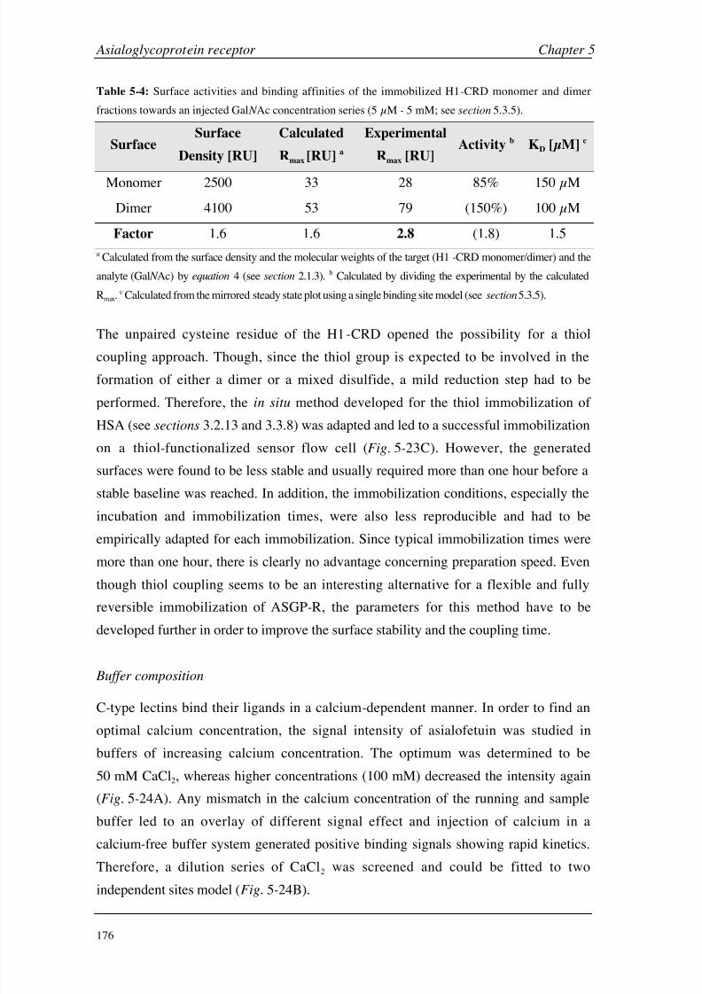
8/9/2019 DissB 7321 Mirroring
http://slidepdf.com/reader/full/dissb-7321-mirroring 190/278
Asialoglycoprotein receptor Chapter 5
176
Table 5-4: Surface activities and binding affinities of the immobilized H1-CRD monomer and dimer
fractions towards an injected Gal N Ac concentration series (5 µM - 5 mM; see section 5.3.5).
SurfaceSurface
Density [RU]
Calculated
Rmax [RU] a
Experimental
Rmax [RU]Activity b KD [µM] c
Monomer 2500 33 28 85% 150
µM
Dimer 4100 53 79 (150%) 100 µM
Factor 1.6 1.6 2.8 (1.8) 1.5
a Calculated from the surface density and the molecular weights of the target (H1 -CRD monomer/dimer) and the
analyte (Gal N Ac) by equation 4 (see section 2.1.3). b Calculated by dividing the experimental by the calculated
Rmax.c Calculated from the mirrored steady state plot using a single binding site model (see section 5.3.5).
The unpaired cysteine residue of the H1-CRD opened the possibility for a thiol
coupling approach. Though, since the thiol group is expected to be involved in the
formation of either a dimer or a mixed disulfide, a mild reduction step had to be
performed. Therefore, the in situ method developed for the thiol immobilization of
HSA (see sections 3.2.13 and 3.3.8) was adapted and led to a successful immobilization
on a thiol-functionalized sensor flow cell (Fig. 5-23C). However, the generated
surfaces were found to be less stable and usually required more than one hour before a
stable baseline was reached. In addition, the immobilization conditions, especially the
incubation and immobilization times, were also less reproducible and had to beempirically adapted for each immobilization. Since typical immobilization times were
more than one hour, there is clearly no advantage concerning preparation speed. Even
though thiol coupling seems to be an interesting alternative for a flexible and fully
reversible immobilization of ASGP-R, the parameters for this method have to be
developed further in order to improve the surface stability and the coupling time.
Buffer composition
C-type lectins bind their ligands in a calcium-dependent manner. In order to find an
optimal calcium concentration, the signal intensity of asialofetuin was studied in
buffers of increasing calcium concentration. The optimum was determined to be
50 mM CaCl2, whereas higher concentrations (100 mM) decreased the intensity again
(Fig. 5-24A). Any mismatch in the calcium concentration of the running and sample
buffer led to an overlay of different signal effect and injection of calcium in a
calcium-free buffer system generated positive binding signals showing rapid kinetics.
Therefore, a dilution series of CaCl2 was screened and could be fitted to twoindependent sites model (Fig. 5-24B).

8/9/2019 DissB 7321 Mirroring
http://slidepdf.com/reader/full/dissb-7321-mirroring 191/278
Chapter 5 Asialoglycoprotein receptor
177
0 20 40 60 80
0 100 200 300
0
200
400
600
800
1000
0 2·10-2 6·10-2 1·10-1
0
100
200
300
10-110-210-310-4
S P R R
e s p o n s e [ R U ]
Time [s] Concentration [M]
2 mM
20 mM
100 mM
50 mM
2
20
50
100
A
B
1 site
2 sites
Figure 5-24: Calcium dependency of H1-CRD. A: Optimization of the calcium
concentration for the binding assays. Asialofetuin (0.1
mg/ml) was injected at
different calcium concentrations. B: Direct binding of calcium chloride to H1-CRD.
CaCl2 was injected from 30 µM to 50 mM over a H1-CRD surface in calcium-free
buffer and fitted to single site (blue) and two independent binding sites models (red).
The steady state analysis indicated a high affinity (KD = 170 µM) and a low affinity
binding site (KD = 16 mM). This is in good agreement with a study by Andersen et al.
[27], in which they determined KD’s of 350 µM and 7 mM for the interaction of
calcium with isolated rabbit ASGP-R subunits. In agreement with the crystal structure,
they found three calcium ions per polypeptide chain and, after competition experiments
with MgCl2, stated two high affinity and one low affinity site.
The requirement of a rather high calcium concentration was confirmed by screening
Gal N Ac as a monovalent carbohydrate (see section 5.3.5) in buffer containing 5 mM or
50 mM CaCl2. While a clear and reproducible binding signal could be detected in the
high calcium buffer, reducing the calcium concentration led to nearly complete signal
loss (Fig. 5-25).

8/9/2019 DissB 7321 Mirroring
http://slidepdf.com/reader/full/dissb-7321-mirroring 192/278
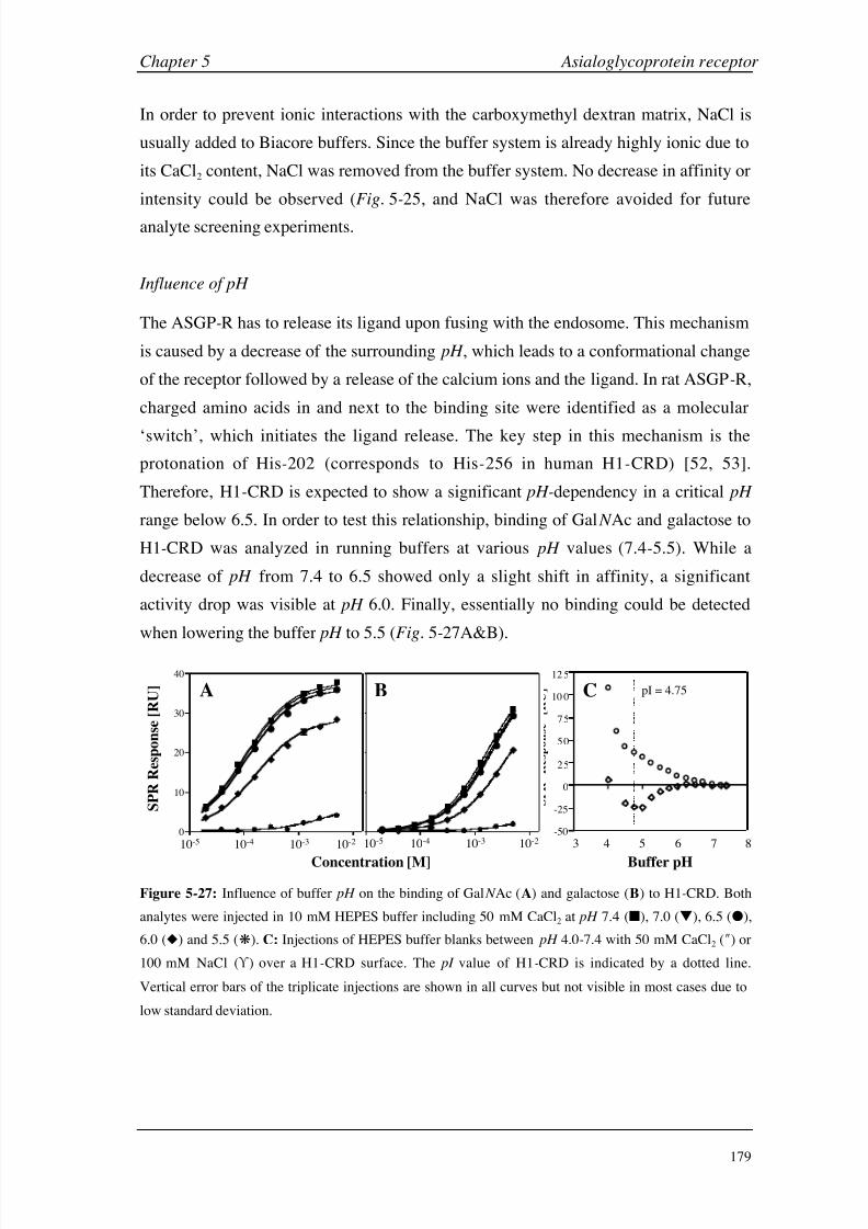
8/9/2019 DissB 7321 Mirroring
http://slidepdf.com/reader/full/dissb-7321-mirroring 193/278
Chapter 5 Asialoglycoprotein receptor
179
In order to prevent ionic interactions with the carboxymethyl dextran matrix, NaCl is
usually added to Biacore buffers. Since the buffer system is already highly ionic due to
its CaCl2 content, NaCl was removed from the buffer system. No decrease in affinity or
intensity could be observed (Fig. 5-25, and NaCl was therefore avoided for future
analyte screening experiments.
Influence of pH
The ASGP-R has to release its ligand upon fusing with the endosome. This mechanism
is caused by a decrease of the surrounding pH , which leads to a conformational change
of the receptor followed by a release of the calcium ions and the ligand. In rat ASGP-R,
charged amino acids in and next to the binding site were identified as a molecular
‘switch’, which initiates the ligand release. The key step in this mechanism is the
protonation of His-202 (corresponds to His-256 in human H1-CRD) [52, 53].
Therefore, H1-CRD is expected to show a significant pH-dependency in a critical pH
range below 6.5. In order to test this relationship, binding of Gal N Ac and galactose to
H1-CRD was analyzed in running buffers at various pH values (7.4-5.5). While a
decrease of pH from 7.4 to 6.5 showed only a slight shift in affinity, a significant
activity drop was visible at pH 6.0. Finally, essentially no binding could be detected
when lowering the buffer pH to 5.5 (Fig. 5-27A&B).
-50
-25
0
25
50
75
10 0
12 5
S P R
R e s p o n s e
[ R U ]
S P R R e s p o n s e [ R U ]
0
10
20
30
40
S P R R e s p o n s e [ R U ]
A B
10-5 10-4 10-3 10-2
Concentration [M]
10-5 10-4 10-3 10-2
S P R R
e s p o n s e [ R U ]
3 4 5 6 7 8
Buffer pH
pI = 4.75C
Figure 5-27: Influence of buffer pH on the binding of Gal N Ac (A) and galactose (B) to H1-CRD. Both
analytes were injected in 10 mM HEPES buffer including 50 mM CaCl2 at pH 7.4 (), 7.0 (), 6.5 (),
6.0 () and 5.5 (). C: Injections of HEPES buffer blanks between pH 4.0-7.4 with 50 mM CaCl2 () or
100 mM NaCl ( ) over a H1-CRD surface. The pI value of H1-CRD is indicated by a dotted line.
Vertical error bars of the triplicate injections are shown in all curves but not visible in most cases due to
low standard deviation.

8/9/2019 DissB 7321 Mirroring
http://slidepdf.com/reader/full/dissb-7321-mirroring 194/278
Asialoglycoprotein receptor Chapter 5
180
This observation is in good agreement with literature. In case of the rat ASGP-R, a
rapid decrease of ligand binding was reported when lowering the pH from 8.8 to 4.8.
The midpoint of ligand release was found to be pH 7.1 and almost all ligand was
released at the endosomal p H of 5.4. The release mechanism was clearly
calcium-dependent. After an increase from 1
mM (physiological) to 5
mM CaCl2 the
ligand binding remained stable until a pH of ~7.4 while the endpoint stayed at pH ~ 5
(midpoint < pH 6.5) [52]. The significantly higher calcium concentration in the present
Biacore assay is therefore expected to cause H1-CRD releasing its ligand even later.
From an experimental point of view, these data also imply only a minor sensitivity
towards small shifts in pH as they might occur during buffer preparation or as they
could be induced by acidic analytes (see section 3.3.7). When lowering the pH during
buffer blank injections at 50
mM CaCl2, the signal began to drop significantly below pH 6.5 indicating that calcium is released from the protein. After further decreasing the
pH below the CRD’s pI (4.75) the SPR signal increased again, because surface
attraction effects between the negatively charged matrix and the protonated protein
became dominant. This effect could be confirmed by replacing the CaCl2 with 100 mM
NaCl, leading to a slow but steady signal increase until the pI , below which the signal
raise was accelerated significantly (Fig. 5-27C). This indicates that the signal drop is
selectively caused by the release of calcium ion. Whether the steady increase between
pH
7 and 4.75 is solely generated by attraction effects or whether a reported
conformational change of ASGP-R at lower pH values [28] is involved, can hardly be
determined by Biacore experiments alone.
DMSO tolerability
The development and synthesis of novel carbohydrate mimics or conjugates is often
accompanied by a significant increase in hydrophobicity. Therefore, organic modifiers
have to be added to the solvent and DMSO has proofed to be ideally suited for mostapplications due to its miscibility with water and its biocompatibility. However,
addition of DMSO nevertheless might influence protein activity or binding properties.
In order to test H1-CRD for any DMSO sensitivity, carbohydrate screening (galactose
and Gal N Ac) was performed in DMSO-free buffer and buffer containing 5 % DMSO
(Table 5-5). Both the binding affinities and the signal intensities (Rmax) were not
significantly influenced by the addition of 5% DMSO. This makes the Biacore assay
suitable for the screening of hydrophobic, drug-like substances.
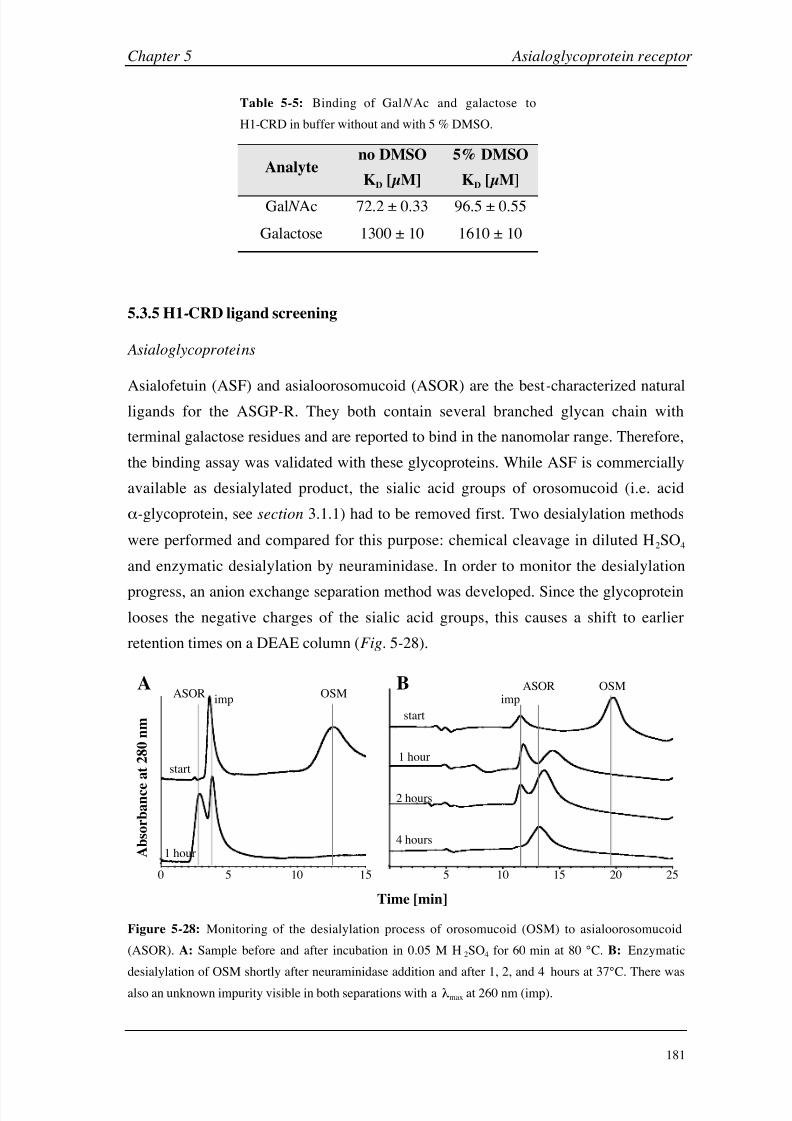
8/9/2019 DissB 7321 Mirroring
http://slidepdf.com/reader/full/dissb-7321-mirroring 195/278
Chapter 5 Asialoglycoprotein receptor
181
Table 5-5: Binding of Gal N Ac and galactose to
H1-CRD in buffer without and with 5 % DMSO.
Analyteno DMSO
KD [µM]
5% DMSO
KD [µM]
Gal N Ac 72.2 ± 0.33 96.5 ± 0.55
Galactose 1300 ± 10 1610 ± 10
5.3.5 H1-CRD ligand screening
Asialoglycoproteins
Asialofetuin (ASF) and asialoorosomucoid (ASOR) are the best-characterized naturalligands for the ASGP-R. They both contain several branched glycan chain with
terminal galactose residues and are reported to bind in the nanomolar range. Therefore,
the binding assay was validated with these glycoproteins. While ASF is commercially
available as desialylated product, the sialic acid groups of orosomucoid (i.e. acid
-glycoprotein, see section 3.1.1) had to be removed first. Two desialylation methods
were performed and compared for this purpose: chemical cleavage in diluted H2SO4
and enzymatic desialylation by neuraminidase. In order to monitor the desialylation
progress, an anion exchange separation method was developed. Since the glycoprotein
looses the negative charges of the sialic acid groups, this causes a shift to earlier
retention times on a DEAE column (Fig. 5-28).
Time [min]
0 min0 5 10 15 20
5 10 15 20 25
start
1 hour
2 hours
4 hours
OSMASORimp
B
min0 2 4 6 8 10 12 14
0 5 10
1 hour
start
ASORimp
OSMA
A b s o r b a n c e
a t 2 8 0 n m
15
Figure 5-28: Monitoring of the desialylation process of orosomucoid (OSM) to asialoorosomucoid
(ASOR). A: Sample before and after incubation in 0.05 M H2SO4 for 60 min at 80 °C. B: Enzymatic
desialylation of OSM shortly after neuraminidase addition and after 1, 2, and 4 hours at 37°C. There was
also an unknown impurity visible in both separations with a max at 260 nm (imp).

8/9/2019 DissB 7321 Mirroring
http://slidepdf.com/reader/full/dissb-7321-mirroring 196/278
Asialoglycoprotein receptor Chapter 5
182
While the process was complete after one hour in the case of the acidic desialylation
(Fig. 5-28A), the enzymatic method was significantly more time-consuming (4 hours;
Fig. 5-28B). In both cases, an unknown impurity with an absorption maximum at
260 nm was detected even at start conditions. Interestingly, the impurity seems to
vanish during enzymatic but not during acidic desialylation.
Both asialoglycoproteins were screened over different H1-CRD surfaces in order to
evaluate their kinetic properties and to detect differences between the immobilization
methods described in section 5.3.4. The binding specificity for desialylated
glycoproteins bearing terminal Gal/Gal N Ac moieties was tested by injecting similar
concentrations of ASF, ASOR as well as orosomucoid (OSM) before desialylation over
a H1-CRD surface. While latter showed no detectable binding, both asialoglycoproteins
bound clearly and specifically to the lectin with rather slow binding kinetics
(Fig. 5-29A). The two active glycoproteins were then screened over three H1-CRD
surfaces (amine-coupling of monomers and dimers, thiol-coupling of monomers) and
the resulting sensorgrams were kinetically evaluated (Fig. 5-29B&C).
1000
800
600
400
200
0
S P R R
e s p o n s e [ R U ]
Time [s]
0 200 400 600 0 400 800 1200 1600 0 400 800 1200 1600
ASF
ASOR
OSM
A B CASF ASOR
Figure 5-29: Binding of asialoglycoproteins to immobilized H1-CRD. A: Comparison of the binding
specificity of asialofetuin (ASF), asialoorosomucoid (ASOR) and orosomucoid (OSM) at 2.5 µM on a
low-density surface. B: Injection series of ASF (1000-4 nM) in 10 mM HBS. 50 mM CaCl2, 0.005%
polysorbate 20, pH
7.4 over a thiol-coupled H1-CRD surface. Simulated kinetic curves of a bivalent
binding model are overlaid in red. C: Injection series of ASOR under the same conditions as in B.
An unusually fast increase of the signal at injection start is indicated by a red circle.
Asialofetuin showed clearly concentration-dependent and reproducible binding signals
with slow kinetic off -rates. As expected, the sensorgrams could not be fitted to a simple
Langmuir 1:1 binding model, because polyvalent binding, rebinding effects,
microheterogeneities of the glycosylation pattern as well as possible surface
heterogeneity from protein immobilization are influencing the binding behavior.

8/9/2019 DissB 7321 Mirroring
http://slidepdf.com/reader/full/dissb-7321-mirroring 197/278
Chapter 5 Asialoglycoprotein receptor
183
Therefore, acceptable fits resulted when using a surface heterogeneity model while the
best matches were realized with a bivalent analyte model (Fig. 5-29B, Table 5-6).
Table 5-6: Kinetic evaluation of asialofetuin binding to three different H1-CRD surfaces using a
bivalent analyte binding model (4-1000
nM in 10
mM HBS, 50
mM CaCl2, 0.005% polysorbate 20, pH 7.4). Overlay sensorgrams of all binding curves and fits can be found in appendix D5.
Surface Density [RU] kon [M-1s-1] koff [s
-1] Rmax [RU] KD [nM] c t1/2 [min] d
Monomer a 1900 76 · 103 6 · 10-5 705 0.79 190
Dimer a 1850 154 · 103 3 · 10-5 1235 0.20 375
Thiol b 2400 123 · 103 4 · 10-5 1010 0.31 300
a Prepared by amine coupling of monomer or dimer fractions. b Prepared by thiol coupling of the
monomer fraction. c Calculated as KD = koff /kon.d Calculated as t1/2 = ln 2/koff .
Asialoorosomucoid showed an even more complex binding pattern. After a rapid signal
increase at injection start, the association phase significantly slowed down. The
dissociation part was monophasic and similar to the one of ASF. Therefore, an overlay
of two independent binding events as observed in the case of the GSLA-2 antibody
(see section 4.3) can be excluded. Depending on the surface, higher concentrations of
ASOR (0.3-1 µM) generated large negative drifts during injection (see appendix D6).
As a consequence, the sensorgrams could not appropriately be fitted to a common
kinetic model. The reason for this atypical binding behavior is not known. An
accumulation or competition of impurities as seen during desialylation cannot be
excluded. Therefore, ASOR experiments have to be repeated after a more intense
purification and characterization of the glycoprotein.
As mentioned above, a bivalent analyte model generated the most appropriate kinetic
description of the ASF binding behavior, even though no complete fit could be
achieved (Fig. 5-29B). Fetuin is reported to bear three sites for N-linked glycosylation,
each carrying either a bi- or triantennary glycan [54, 55]. In contrast to ASOR, which
only carries terminal galactose, ASF also shows heterogeneities in the carbohydrate
moieties with a Gal:Gal N Ac ratio of 4:1 [20]. Therefore, multivalent binding is
expected for both glycoproteins and does also explain, why a bivalent analyte model
describes the interaction rather well but not completely. Even though bivalent binding
implies two sets of rate constants for the primary and secondary binding event,
respectively, only one set can be obtained by the Biacore experiment. When an already
bound analyte molecule interacts with its second (or third) binding site, no increase of
mass concentration and therefore no additional SPR signal can be detected [56].
8/9/2019 DissB 7321 Mirroring
http://slidepdf.com/reader/full/dissb-7321-mirroring 198/278
Asialoglycoprotein receptor Chapter 5
184
Significant variations between the monomer and dimer surfaces were found for the
kinetic rate constants. While the on-rate of the dimer was increased by a factor of two,
its dissociation rate was slower by the same factor. This resulted in a four times higher
affinity and clearly prolonged dissociation half -life of more than six hours.
The thiol-coupled surface was expected to show properties more similar to the
monomer rather than the dimer fraction, due to its mode of immobilization. However,
all values were between the two amine-coupled surfaces and clearly closer to the dimer
fraction. This might be explained by the slightly higher surface density obtained by this
coupling method resulting in an increased rate of rebinding and multivalency. In
addition, an immobilization solely around the N-terminus and therefore opposite to the
binding region (Fig. 5-22) could improve the accessibility of the binding sites. The
obtained KD’s between 200 and 800 pM are lower than the IC50 values reported for the
interaction with isolated human ASGP-R lectins (17 nM) [20]. Dissociation constants
for the interaction with overexpressed H1 in the absence of H2 were only obtained for
ASOR (40 nM) but not for ASF [29].
Mono- and disaccharides
The asialoglycoprotein receptor shows a high selectivity for galactose moieties and its
derivatives (see section 5.1). Gal N Ac is the natural monosaccharide with the highestaffinity for the ASGP-R, at least ten times better than galactose [20, 57]. The expected
affinity in the micromolar range is well suited for SPR detection, whereas the small
molecular weight around 200 Da was identified as a potential problem. First test
injections with 1 mM solutions of different carbohydrate analytes provided a confusing
result, since all injections caused negative binding signals. This behavior usually
indicates problems in the assay design and are therefore mostly rejected. However,
comparison of the samples showed a tendency in the negative signal intensity, which
directly correlated to the expected affinity of the tested sugars. Therefore, a
concentration series of Gal N Ac was injected and analyzed more deeply. While the SPR
signals on the reference flow cell always had a positive sign, the H1-CRD flow cell
generated responses both in the positive and negative range (Fig. 5-30A). Referencing
of the signals (i.e. subtraction of the reference flow cell) resulted in negative
sensorgrams for each sample injection (Fig. 5-30B). Since Scrubber allows the
multiplication of the sensorgrams with a constant value (included for the normalization
of values from different assay types), the binding curves could be mirrored by
multiplication with -1 (Fig. 5-30C). Processed sensorgrams from randomized triplicate

8/9/2019 DissB 7321 Mirroring
http://slidepdf.com/reader/full/dissb-7321-mirroring 199/278
Chapter 5 Asialoglycoprotein receptor
185
injections not only were fully reproducible, but also showed the fast binding kinetics
expected for protein-carbohydrate interactions [58, 59] resulting in stable steady state
phases within a few seconds (Fig. 5-30D). When fitting these equilibrium values to a
single binding site model, the data points matched the model with high accuracy
(Fig.
5-30E) and led to dissociation constants (KD in the rage of 90-150
µM for
Gal N Ac), which were well within the expected range. In addition, the sensorgrams
could also be kinetically fitted to a simple Langmuir 1:1 interaction model
(Fig. 5-30F). These results indicated that the Biacore binding assay generated reliable
interaction data despite the unusual occurrence of negative SPR signals. A detailed
discussion of possible causes for the negative responses can be found in section 5.3.6.
0
100
200
-100
-200
140
120
100
80
60
40
20
0
0 10 20 30 40 50 0 10 20 30 40 50 0 10 20 30 40 50
0 10 20 30 40 500 10 20 30 40 50 10-5 10-4 10-3 10-2
S P R R e s p o n s e [ R U
]
S P R R e s p o n s e [ R U ]
Time [s]
Time [s] Concentration [M] Time [s]
A B C
DE F
Figure 5-30: Data processing (A-C) and data evaluation (D-F) of Gal N Ac binding to H1-CRD.
Referencing of the biosensor raw data (A) by subtracting the signals of an empty reference cell (red)
from the measurement cell (green) leads to negative binding signals (B) that can be mirrored to get
interpretable data (C). Resulting overlay plots (D) can be either evaluated by fitting the equilibria to a
single binding site model (E) or by kinetic fitting of the whole sensorgrams to a Langmuir 1:1 interaction
model (F).
In order to validate the binding assay and get more detailed information about the
binding motif of the ligands, a set of galactose derivatives as well as glucose (negative
control) was screened. All carbohydrate analytes showed negative binding signals with

8/9/2019 DissB 7321 Mirroring
http://slidepdf.com/reader/full/dissb-7321-mirroring 200/278
Asialoglycoprotein receptor Chapter 5
186
fast kinetic properties. After mirroring, all binding equilibria could be fitted to a single
binding site model and resulted in affinities between 0.1 and 5 mM (Table 5-7).
As expected, Gal N Ac was identified as the best binding monosaccharide,
approximately 10 to 15 times better than galactose and lactose. Galactose derivativesbearing a methyl residue at the reducing end bound with a lower affinity than galactose,
with the beta form showing a slightly better KD. While galactosamine bound with
detectable but clearly weaker affinities, glucose binding was beyond detection limit.
The KD values obtained in the Biacore assay were compared to IC50 values from a
solid-phase competition assay, were a polymeric Gal N Ac competes with the analytes
for immobilized H1-CRD (Fig. 5-32A) [35].
Table 5-7: Equilibrium binding constants of a panel of carbohydrate analytes binding to immobilized
H1-CRD monomer and dimer fractions (triplicate injections, 5 µM - 5 mM) compared with IC50 values
of a solid-phase competition assay [35] and of literature data [57, 60, 61] (SPR fits see appendix D7).
Carbohydrate AnalyteMonomer
KD [µM]
Dimer
KD [µM]
SPCA a [35]
IC50 [µM]
Literature
Values
Gal N Ac 150 ± 0.9 104 ± 0.3 97 90 b
Lactose 2090 ± 50 1390 ± 10 1100 300 b
Galactose 1460 ± 10 1590 ± 10 n.d. 1700 b
Methyl -D-galactopyranoside 2200 ± 40 1800 ± 10 1800 1000 b
Methyl -D-galactopyranoside 2760 ± 60 2350 ± 20 2600 1600 b
Galactosamine 4170 ± 110 4630 ± 50 2600 > 25000 c
Glucose > 10 mM > 10 mM n.d. 60000 b
a Solid-phase competition assay [35]. b IC50 values [µM] from isolated rabbit ASGP-R lectin [57, 60]
c IC50 value [µM] from rat hepatic membranes [61]. The assays were performed with radioactively
labeled ligands.
Not only the general ranking of the analytes was congruent, but also the absolute
affinity values were found to be very close. The rather high calcium concentration of
the Biacore assay does not seem to clearly influence binding, since a much lower
concentration of 5 mM was used in the solid-phase competition assay. When
comparing the binding affinities to published literature values [57, 60, 61], the general
ranking and the affinity ranges are similar again. While lactose is reported to bind
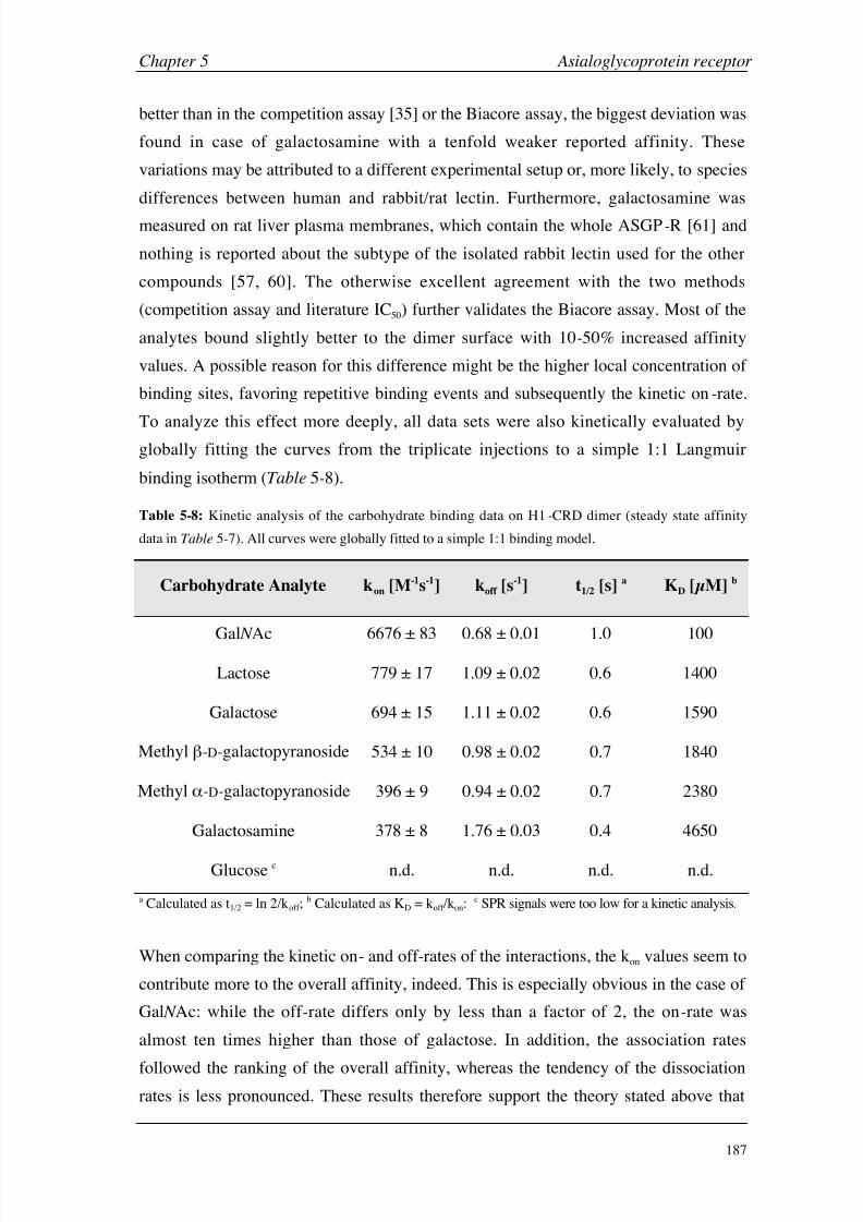
8/9/2019 DissB 7321 Mirroring
http://slidepdf.com/reader/full/dissb-7321-mirroring 201/278
Chapter 5 Asialoglycoprotein receptor
187
better than in the competition assay [35] or the Biacore assay, the biggest deviation was
found in case of galactosamine with a tenfold weaker reported affinity. These
variations may be attributed to a different experimental setup or, more likely, to species
differences between human and rabbit/rat lectin. Furthermore, galactosamine was
measured on rat liver plasma membranes, which contain the whole ASGP-R [61] and
nothing is reported about the subtype of the isolated rabbit lectin used for the other
compounds [57, 60]. The otherwise excellent agreement with the two methods
(competition assay and literature IC50) further validates the Biacore assay. Most of the
analytes bound slightly better to the dimer surface with 10-50% increased affinity
values. A possible reason for this difference might be the higher local concentration of
binding sites, favoring repetitive binding events and subsequently the kinetic on-rate.
To analyze this effect more deeply, all data sets were also kinetically evaluated byglobally fitting the curves from the triplicate injections to a simple 1:1 Langmuir
binding isotherm (Table 5-8).
Table 5-8: Kinetic analysis of the carbohydrate binding data on H1-CRD dimer (steady state affinity
data in Table 5-7). All curves were globally fitted to a simple 1:1 binding model.
Carbohydrate Analyte kon [M-1s-1] koff [s
-1] t1/2 [s] a KD [µM] b
Gal N Ac 6676 ± 83 0.68 ± 0.01 1.0 100
Lactose 779 ± 17 1.09 ± 0.02 0.6 1400
Galactose 694 ± 15 1.11 ± 0.02 0.6 1590
Methyl -D-galactopyranoside 534 ± 10 0.98 ± 0.02 0.7 1840
Methyl -D-galactopyranoside 396 ± 9 0.94 ± 0.02 0.7 2380
Galactosamine 378 ± 8 1.76 ± 0.03 0.4 4650
Glucose c n.d. n.d. n.d. n.d.
a Calculated as t1/2 = ln 2/koff ;b Calculated as KD = koff /kon:
c SPR signals were too low for a kinetic analysis.
When comparing the kinetic on- and off -rates of the interactions, the kon values seem to
contribute more to the overall affinity, indeed. This is especially obvious in the case of
Gal N Ac: while the off -rate differs only by less than a factor of 2, the on-rate was
almost ten times higher than those of galactose. In addition, the association rates
followed the ranking of the overall affinity, whereas the tendency of the dissociationrates is less pronounced. These results therefore support the theory stated above that

8/9/2019 DissB 7321 Mirroring
http://slidepdf.com/reader/full/dissb-7321-mirroring 202/278

8/9/2019 DissB 7321 Mirroring
http://slidepdf.com/reader/full/dissb-7321-mirroring 203/278
Chapter 5 Asialoglycoprotein receptor
189
weakly bind to H1-CRD, poly-Gal N Ac showed a strong activity for both monomer and
dimer surfaces. In contrast to the very fast kinetic profile of the free monosaccharide,
the polymer-bound sugar featured a very slow dissociation rate. Injection of
monovalent galactose clearly competed for the same binding site and caused a
detectable drop in the polymer binding. Finally, the surface could be completely
regenerated with EDTA, indicating a calcium-dependent and therefore specific binding.
The difference in the signal intensity between the monomer and dimer surface is
mainly caused by variations in the surface density of both proteins.
0 100 200 300 400 500 600 700 800
-200
-100
0
100
200
300
400
500
S P R R
e s p o n s e [ R U ]
Time [s]
i d g r
plate
H1-CRD
analyte
polymer
biotin
strept-
avidin
peroxi-
dase D
M
BA
Gal N Ac/Glc
Figure 5-32: H1-CRD polymer assay. A: Overview of the solid-phase polymer competition assay [35]
used for the screening of H1-CRD ligands (adapted from Bovin [62]). B: Injection of poly-Gal N Ac (red)
and poly-glucose (blue) over immobilized H1-CRD monomer (M) and dimer (D) fractions. After an
injection (i) and dissociation phase (d), 10 mM galactose (g) were injected to test for competition effects.
Finally, the surface was regenerated by injecting EDTA buffer (r).
anti-H1-CRD antibodies
Monoclonal antibodies have developed into some of the most powerful tools in both
molecular biology and medicine. Their high selectivity and usually strong affinities
make them also interesting for the purification and characterization of recombinant
proteins. For the development of Biacore assays, monoclonal antibodies are important
in two major areas, i.e. capturing of the proteins for oriented immobilization
(see section 2.1.4) as well as for blocking the active protein in order to evaluate binding
specificity.
A set of monoclonal antibodies directed against the human ASGP-R H1-CRD [30] was
screened for their activity towards monomer and dimer fractions. In a first step, six
antibodies were injected at a constant concentration and their signal intensities,

8/9/2019 DissB 7321 Mirroring
http://slidepdf.com/reader/full/dissb-7321-mirroring 204/278
Asialoglycoprotein receptor Chapter 5
190
dissociation stability, blocking capabilities and calcium/EDTA sensitivity were
evaluated (Fig. 5-33A).
g i d g e r
Int StaA B C
S P R R
e s p o n s e
I n t e n s i t y [ k R U ]
Time [s] Stability / Intensity [%]
00 200 400 600 800 1000
2.5
2.0
1.5
1.0
0.5
090 95 100 90 95 100 105
B01.4
B01.4
C11.1
C18.1C09.1
C23.9C48.8C23.9
C11.1
C18.1
C09.1C48.8
Figure 5-
33: Ranking of a set of H1-CRD-binding antibodies. A: Setup of the ranking assay with apreliminary injection of 5 mM galactose (g), antibody injection (i), dissociation phase (d), a second
galactose injection for testing blocking properties, an EDTA pulse (e) and the regeneration with HCl (r).
Data points used for the ranking (Int, Sta) are indicated by dashed circles. B, C: Ranking of the antibody
samples by plotting the signal intensity after the 5 min injection (Int) against the quotient of the stability
after 5 min dissociation (Sta) and the intensity in buffer containing 50 mM CaCl2 (B) or 3 mM EDTA
(C). Values for the monomer surface are indicated in red and for the dimer surface in blue.
Even though some of the antibodies seem to block sugar binding in the solid-phase
competition assay [30], none of the screened antibodies showed a clear blockage of the
binding site in the Biacore assay. The differences between the galactose signal before
and after the antibody injection were below 10% for all samples (see appendix D8).
This discrepancy to the results of the competition assay might e.g. be caused by a steric
hindrance of the Gal N Ac-polymer. While the general ranking of the antibodies did not
change when switching from calcium- to EDTA-containing buffer, there were
nevertheless some significant variations visible in the binding behavior of the samples.
Particularly, the stability of the dimer binding seemed to increase slightly, which also
led to higher signal intensities (shift to the upper right corner in figure 5-33B&C).
These differences might be explained by small conformational changes of the H1-CRD
when adding or removing calcium, resulting in a changed accessibility of the binding
epitopes. However, the relatively high calcium concentration might also influence the
binding in a non-specific manner. Therefore, the following screening experiments were
performed in calcium-free buffer. Since antibodies with a fast and tight binding profile
are preferred for diagnostic or analytical applications, the three antibodies that matched
best to this profile (B01.4, C11.1, C18.1) were selected for a further high-resolution
screening on both H1-CRD surfaces (Fig. 5-34).

8/9/2019 DissB 7321 Mirroring
http://slidepdf.com/reader/full/dissb-7321-mirroring 205/278
Chapter 5 Asialoglycoprotein receptor
191
6
5
4
3
2
1
0
0 200 400 600 800 1000
S P R R e s p o n s e [ k R U ]
0 200 400 600 800 1000 0 200 400 600 800 1000
6
5
4
3
2
1
0
mAb B01.4 mAb C11.1 mAb C18.1
H 1 - C R D
M o n o m e r
H 1 - C R D D i m e r
Time [s]
Figure 5-34: Screening of the three monoclonal antibodies B01.4, C11.1, and C18.1 on H1 -CRD
monomer and dimer surfaces over a concentration range between 140 nM and 140 pM in HBS buffer
containing 3 mM EDTA. Association and dissociation phases were acquired for ten minutes each.
In correlation with the data from the ranking experiments, the selected antibodies
selectively bound to both H1-CRD surfaces in a clearly concentration-dependent
manner. While all antibodies showed a very slow dissociation phase, the signal
intensity was significantly different between the samples and the surfaces, despite the
molecular weight and concentration was the same for all samples. Interestingly, none of
the tested antibodies showed a significantly faster off -rate. This could be influenced by
the fact that the antibodies were selected by an ELISA method [30], where only
antibodies with a tight binding to the protein withstood the washing conditions. In order
to estimate and compare the kinetic properties of the antibodies, a reduced data set
(140 pM - 9 nM; six data points) was fitted to a Langmuir 1:1 binding model
(Table 5-9, Fig. 5-35).

8/9/2019 DissB 7321 Mirroring
http://slidepdf.com/reader/full/dissb-7321-mirroring 206/278
Asialoglycoprotein receptor Chapter 5
192
Table 5-9: Binding properties of the three screened antibodies calculated from kinetic fits
using a simple 1:1 Langmuir binding model with data sets between 140 pM and 9 nM. Values
for interactions with the monomer and dimer surface are compared.
KD [pM] a t1/2 [min] b
Antibody Monomer Dimer Monomer Dimer
B01.4 308 334 71 129
C11.1 78 n.d. c 447 n.d. c
C18.1 233 124 133 346
a Calculated as KD = koff /kon.b Calculated as t1/2 = ln 2/koff .
c No binding properties could be
calculated for antibody C11.1 on the dimer surface because of its very slow dissociation phase.
0.E+00
1.E+05
2.E+05
3.E+05
4.E+05
5.E+05
6.E+05
B01.4 C11.1 C18.1
6
5
4
3
2
1
0 0.0E+00
3.0E-05
6.0E-05
9.0E-05
1.2E-04
1.5E-04
1.8E-04
B01.4 C11.1 C18.1
1.8
1.5
1.2
0.9
0.6
0.3
0
k o n
[ 1 0 5 M
- 1 s - 1 ]
k o f f
[ 1 0 - 4 s
- 1 ]
Figure 5-35: Comparison of kinetic on- and off -rates of the three selected antibodies on the H1-CRD
monomer (red bars) and dimer (blue bars) surfaces after fitting to a Langmuir 1:1 binding model. No
off -rate could be calculated for the antibody C11.1 on the dimer surface, due to the slow dissociation
phase.
While the difference in KD between the monomer and dimer fractions was rather low in
the case of B01.4, the dimer affinity of C18.1 was almost doubled. Since the antibody
C11.1 showed essentially no dissociation from the dimer surface in the measured time
period, no off -rate and therefore no binding properties could be calculated. However,
this exceptionally slow dissociation also indicates an increased affinity for the dimer.
A closer look at the on- and off -rates showed a general tendency of lower values in
case of the dimer interaction. Translated into a mechanic model, this means that the
antibodies bind slower but tighter to the dimers. Reduced accessibility (e.g. due to
steric hindrance) might be the cause for the reduced on-rate, while the prolonged
dissociation may be influenced by the enriched local concentration around the initial
binding site, which enforces rebinding or concerted binding. However, while both
monomer rate constants for B01.4 are approximately twice as high compared to the
dimer values (Fig. 5-35), therefore compensating in the calculated KD ( Eq . 2,
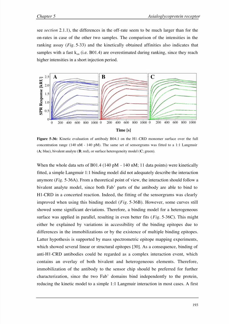
8/9/2019 DissB 7321 Mirroring
http://slidepdf.com/reader/full/dissb-7321-mirroring 207/278
Chapter 5 Asialoglycoprotein receptor
193
see section 2.1.1), the differences in the off -rate seem to be much larger than for the
on-rates in case of the other two samples. The comparison of the intensities in the
ranking assay (Fig. 5-33) and the kinetically obtained affinities also indicates that
samples with a fast kon (i.e. B01.4) are overestimated during ranking, since they reach
higher intensities in a short injection period.
0 200 400 600 800 1000
0
0.5
1.0
1.5
2.0
2.5
S
P R R e s p o n s e [ k R U ]
Time [s]
0 200 400 600 800 1000 0 200 400 600 800 1000
A B C
Figure 5-36: Kinetic evaluation of antibody B04.1 on the H1-CRD monomer surface over the full
concentration range (140 nM - 140 pM). The same set of sensorgrams was fitted to a 1:1 Langmuir
(A; blue), bivalent analyte (B; red), or surface heterogeneity model (C; green).
When the whole data sets of B01.4 (140 pM - 140 nM; 11 data points) were kinetically
fitted, a simple Langmuir 1:1 binding model did not adequately describe the interaction
anymore (Fig. 5-36A). From a theoretical point of view, the interaction should follow a
bivalent analyte model, since both Fab’ parts of the antibody are able to bind to
H1-CRD in a concerted reaction. Indeed, the fitting of the sensorgrams was clearly
improved when using this binding model (Fig. 5-36B). However, some curves still
showed some significant deviations. Therefore, a binding model for a heterogeneous
surface was applied in parallel, resulting in even better fits (Fig. 5-36C). This might
either be explained by variations in accessibility of the binding epitopes due to
differences in the immobilizations or by the existence of multiple binding epitopes.
Latter hypothesis is supported by mass spectrometric epitope mapping experiments,
which showed several linear or structural epitopes [30]. As a consequence, binding of
anti-H1-CRD antibodies could be regarded as a complex interaction event, which
contains an overlay of both bivalent and heterogeneous elements. Therefore,
immobilization of the antibody to the sensor chip should be preferred for further
characterization, since the two Fab’ domains bind independently to the protein,
reducing the kinetic model to a simple 1:1 Langmuir interaction in most cases. A first
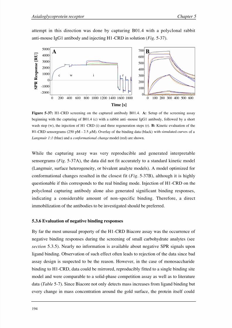
8/9/2019 DissB 7321 Mirroring
http://slidepdf.com/reader/full/dissb-7321-mirroring 208/278
Asialoglycoprotein receptor Chapter 5
194
attempt in this direction was done by capturing B01.4 with a polyclonal rabbit
anti-mouse IgG1 antibody and injecting H1-CRD in solution (Fig. 5-37).
0
1000
2000
3000
4000
5000
-1000
-2000
S P R R e s p o n s e [ R U ]
0
Time [s]
200 400 600 800 1000 1200 1400 1600 1800 0 100 200 300 400 500 600
0
100
200
300
400
500
600
700A B
c w i r
Figure 5-
37: H1-CRD screening on the captured antibody B01.4. A: Setup of the screening assaybeginning with the capturing of B01.4 (c) with a rabbit anti-mouse IgG1 antibody, followed by a short
wash step (w), the injection of H1-CRD (i) and three regeneration steps (r). B: Kinetic evaluation of the
H1-CRD sensorgrams (250 pM - 2.5 µM). Overlay of the binding data (black) with simulated curves of a
Langmuir 1:1 (blue) and a conformational change model (red) are shown.
While the capturing assay was very reproducible and generated interpretable
sensorgrams (Fig. 5-37A), the data did not fit accurately to a standard kinetic model
(Langmuir, surface heterogeneity, or bivalent analyte models). A model optimized for
conformational changes resulted in the closest fit (Fig. 5-37B), although it is highly
questionable if this corresponds to the real binding mode. Injection of H1-CRD on the
polyclonal capturing antibody alone also generated significant binding responses,
indicating a considerable amount of non-specific binding. Therefore, a direct
immobilization of the antibodies to be investigated should be preferred.
5.3.6 Evaluation of negative binding responses
By far the most unusual property of the H1-CRD Biacore assay was the occurrence of
negative binding responses during the screening of small carbohydrate analytes (see
section 5.3.5). Nearly no information is available about negative SPR signals upon
ligand binding. Observation of such effect often leads to rejection of the data since bad
assay design is suspected to be the reason. However, in the case of monosaccharide
binding to H1-CRD, data could be mirrored, reproducibly fitted to a single binding site
model and were comparable to a solid-phase competition assay as well as to literature
data (Table 5-7). Since Biacore not only detects mass increases from ligand binding but
every change in mass concentration around the gold surface, the protein itself could

8/9/2019 DissB 7321 Mirroring
http://slidepdf.com/reader/full/dissb-7321-mirroring 209/278
Chapter 5 Asialoglycoprotein receptor
195
also contribute to the observed overall signal. Several mechanistic hypotheses seem to
be reasonable, including (i) conformational changes, (ii) lectin-interaction with the
dextran matrix or (iii) electrostatic interactions with matrix-bound carboxyl groups.
Hypothesis I: conformational change
Essentially two observations support the hypothesis of a conformational change as a
possible cause for the negative binding signals in the case of ASGP-R. First, lowering
the pH to slightly acidic conditions (from pH 7.4 to 5.6) induces a conformational
change, during which calcium ions and ligands are released. This has been shown
e.g. by iodination [28] and mutation experiments [52, 53]. More importantly, ligand
binding is also reported to induce a conformational change across the membrane in
order to induce the receptor-mediated endocytosis (see section 5.1.2) [17, 19].
However, nothing is known about the exact mechanism nor are there any structural data
available, which describe these changes.
At the moment, there is no proof that conformational changes can be monitored on SPR
instruments, and only sporadic reports of this phenomenon are available [63-68]. The
most interesting study in this context has been performed with another lectin, i.e. the
maltose-binding protein (MBP) [66]. Injection of different sugars on the immobilized
MBP induced a drop of the post-injection baseline, which could be correlated with theknown analyte activity. However, no negative responses for the binding equilibria
during analyte injection were reported. Even though the signals were rather low
( 20 RU), they were fitted to a single site model and resulted in a maltose KD similar
to values obtained by other methods. Interestingly, MBP is known to undergo a
substantial conformational change (‘venus fly trap’-like hinge twist) upon mannose
binding, as it has been shown by crystal structure analysis [69]. Based on the SPR data
of MBP and another protein (transglutaminase; see also section 3.3.5), they concluded
that a decrease in the hydrodynamic radius or volume of a protein might lead to a
negative SPR signal and vice versa. However, the question remains open, if the
directionality of such changes plays a role for the intensity and sign of the resulting
SPR response. For example, a longitudinal stretch of an immobilized protein is
expected to induce a more significant change than internal changes or expansions in
parallel to the chip surface (Fig. 5-38A&B). An additional hint for structural changes
being involved in the biding of carbohydrates to H1-CRD comes from the differences
between amine- and thiol-coupled lectin surfaces. While Gal N Ac bound clearly worse

8/9/2019 DissB 7321 Mirroring
http://slidepdf.com/reader/full/dissb-7321-mirroring 210/278
Asialoglycoprotein receptor Chapter 5
196
to the thiol-coupled surface than to the amine-coupled surface, the interaction with ASF
seemed to be nearly not affected (Fig. 5-38C&D).
The overall binding signal is believed to consist of a negative component generated by
a conformational change and a positive mass contribution. In the case of ASF, the smallnegative part is negligible due to the large molecular weight of the glycoprotein
(> 40 kDa), while it seems to be dominant for the small monosaccharide (221 Da).
Since the overall activity of H1-CRD is not lost after thiol immobilization, as it is
demonstrated by ASF-binding, the immobilization method seems to selectively
influence the generation of the negative component. This might correlate well with the
dramatic loss of signal intensity for Gal N Ac (Fig. 5-38C) but also for the slightly
increased response of ASF on the thiol surface compared to the amine -coupled
monomer surface, which should be still capable to combine negative and positive
signals (Fig. 5-38D).
A B
Figure 5-38: Conformational change hypothesis for the generation of negative binding signals. While
some conformational changes could induce a negative SPR signal by decreasing the mass concentration
(A), others are not detectable (B). Binding of a monosaccharide (Gal N Ac; C) and an asialoglycoprotein
(ASF; D) on amine-coupled monomer (green) and dimer surfaces (blue) as well as to a thiol-coupled
H1-CRD surface (magenta).
As in the case of human serum albumin (see section 3.3), monitoring of conformational
changes with alternative methods like circular dichroism (CD) could confirm the SPR
results. However, since H1-CRD contains 11 tryptophan and 6 tyrosine residues
distributed over the whole protein chain, small changes at a specific site are much more
difficult to follow than in the case of HSA with only one tryptophan residue. Indeed,
tryptophan fluorescence experiments did not show any calcium or analyte-induced
changes of the CD spectrum in the case of H1-CRD. Again, co-crystallization of the
lectin with Gal N Ac or another analyte should provide a deeper insight in the bindingmechanism.

8/9/2019 DissB 7321 Mirroring
http://slidepdf.com/reader/full/dissb-7321-mirroring 211/278
Chapter 5 Asialoglycoprotein receptor
197
Hypothesis II: Competitive interactions with the dextran matrix
Another plausible explanation of the negative signal effect is the replacement of a
heavier or multiple components by the small analyte. Since the signal returns to the
initial value after injection end, this component has to be available in the runningbuffer. Different buffer ingredients were therefore considered causing the signal effect.
HEPES, which was suspected to interact with the calcium binding site, was excluded as
a possibility by substituting it with imidazole (see section 5.3.4). Calcium ions were the
second component to investigate, because they were found to be essential for high
signal intensities (Fig. 5-25). Since the values between the Biacore assay at 50 mM
CaCl2 and the solid-phase competition assay at 1 mM CaCl2 are very similar, the
calcium concentration seems to be mainly responsible for the signal generation rather
than the affinity. It is therefore possible that several calcium ions (MW = 40
Da) are
competitively released upon ligand binding, which results in a negative SPR signal. On
the other hand, binding signals of ASF were also dependent on the calcium
concentration, indicating that calcium contributes to the binding activity of the proteins.
Binding to dextran was therefore considered as another plausible hypothesis, since the
carboxymethyl dextran hydrogel of the sensor chip mainly consists of a linear glucose
polysaccharide [70]. Even though direct injection of H1-CRD in 10 mM HEPES
50 mM CaCl2 did not show significant binding to a plain CM5 sensor chip surface and
injections of free glucose and polymeric glucose resulted in barely detectable signals,
the enhanced local concentration around the lectin might lead to a weak but constant
binding. Upon ligand binding, the protein is released from the dextran and changes its
orientation. After injection end, dextran binding becomes predominant again and the
protein switches back (Fig. 5-39A). If this hypothesis was true, binding results had to
be regarded as competitive (IC50) rather than direct binding (KD). This hypothesis was
further investigated by screening Gal N Ac and galactose in running buffer at three
different concentrations (0, 1, 10
mg/ml) of soluble carboxymethyl dextran (CMD;Fig.
5-39B&C). CMD is also recommended to avoid non-specific binding in
Biacore-based ligand fishing assays, e.g. for recovering samples for mass spectrometry
[71].
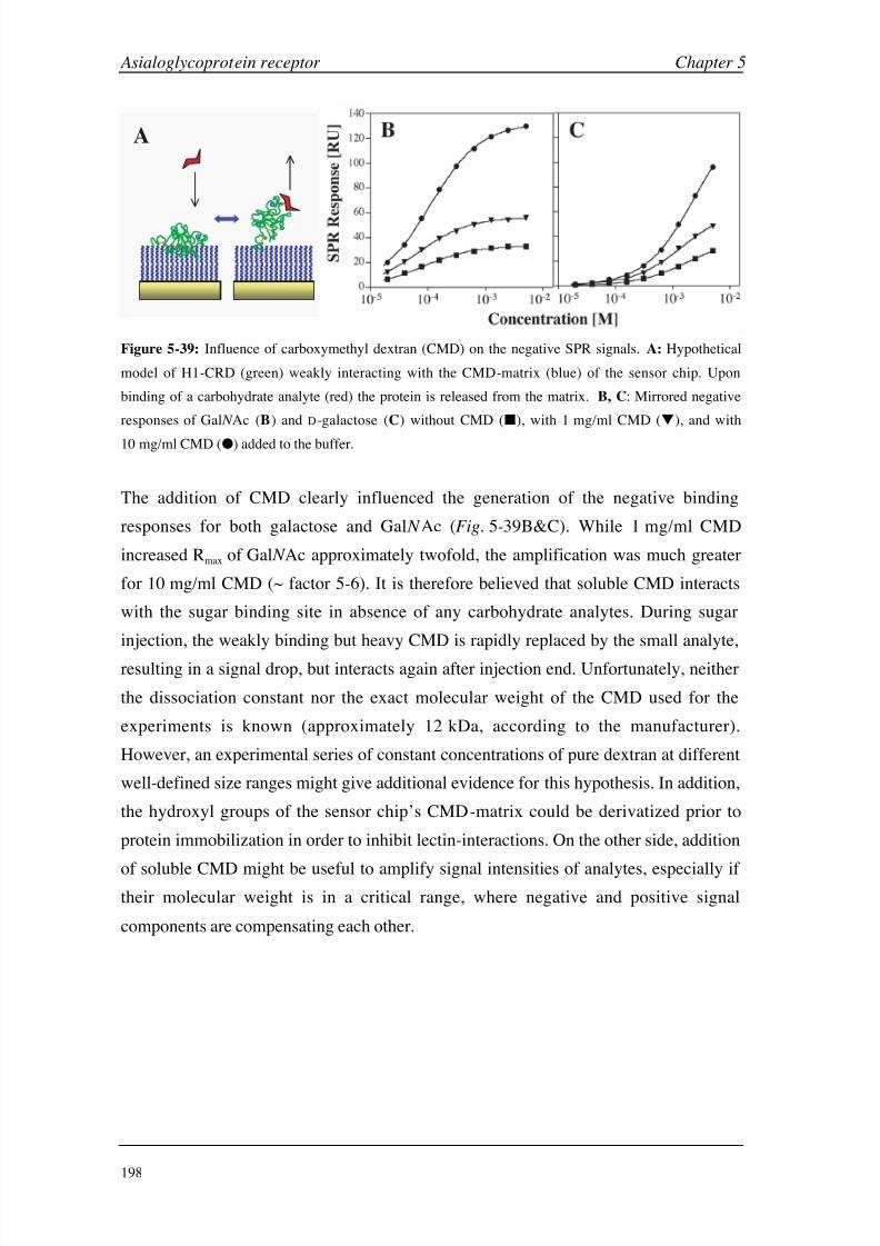
8/9/2019 DissB 7321 Mirroring
http://slidepdf.com/reader/full/dissb-7321-mirroring 212/278
Asialoglycoprotein receptor Chapter 5
198
A
Figure 5-39: Influence of carboxymethyl dextran (CMD) on the negative SPR signals. A: Hypothetical
model of H1-CRD (green) weakly interacting with the CMD-matrix (blue) of the sensor chip. Upon
binding of a carbohydrate analyte (red) the protein is released from the matrix. B, C: Mirrored negative
responses of Gal N Ac (B) and D-galactose (C) without CMD (), with 1 mg/ml CMD (), and with
10
mg/ml CMD () added to the buffer.
The addition of CMD clearly influenced the generation of the negative binding
responses for both galactose and Gal N Ac (Fig. 5-39B&C). While 1 mg/ml CMD
increased Rmax of Gal N Ac approximately twofold, the amplification was much greater
for 10 mg/ml CMD (~ factor 5-6). It is therefore believed that soluble CMD interacts
with the sugar binding site in absence of any carbohydrate analytes. During sugar
injection, the weakly binding but heavy CMD is rapidly replaced by the small analyte,
resulting in a signal drop, but interacts again after injection end. Unfortunately, neither
the dissociation constant nor the exact molecular weight of the CMD used for the
experiments is known (approximately 12 kDa, according to the manufacturer).
However, an experimental series of constant concentrations of pure dextran at different
well-defined size ranges might give additional evidence for this hypothesis. In addition,
the hydroxyl groups of the sensor chip’s CMD-matrix could be derivatized prior to
protein immobilization in order to inhibit lectin-interactions. On the other side, addition
of soluble CMD might be useful to amplify signal intensities of analytes, especially if their molecular weight is in a critical range, where negative and positive signal
components are compensating each other.

8/9/2019 DissB 7321 Mirroring
http://slidepdf.com/reader/full/dissb-7321-mirroring 213/278

8/9/2019 DissB 7321 Mirroring
http://slidepdf.com/reader/full/dissb-7321-mirroring 214/278

8/9/2019 DissB 7321 Mirroring
http://slidepdf.com/reader/full/dissb-7321-mirroring 215/278
Chapter 5 Asialoglycoprotein receptor
201
Therefore, no exact affinity and kinetic values could be obtained for this interaction. A
more extended purification or characterization of the glycoprotein seems to be required.
Calcium concentration and pH were shown to be important for a successful interaction
of lectin. Any reduction or removal of calcium dramatically decreased the bindingsignals of all tested ligands. This effect was more prominent in the case of the small,
monovalent carbohydrates. With 50 mM CaCl2, the optimal calcium concentration for
the Biacore assay was found to be clearly higher than the physiological concentration
(1 mM). By injecting calcium chloride to immobilized H1-CRD, two independent
binding sites with KD values of 170 and 16000 µM could be detected, which is in
agreement with literature. When the pH was lowered from 7.4 (plasma) to 5.5
(endosome), the protein basically lost its binding activity. Further experiments showed
that a release of calcium ions below pH
6 is responsible for this effect. These
observations clearly fit with the described mechanism of receptor -mediated
endocytosis, where calcium and ligands are released in the early endosome ( pH ~ 6).
Furthermore, the Biacore assay was successfully used to rank and characterize a set of
monoclonal antibodies binding to the human ASGP-R H1-CRD. Since these antibodies
were pre-selected by an ELISA method, all tested samples showed a very tight binding
to the lectin with dissociation half -times of 1-7.5 hours and KD values in the picomolar
range. Similar to the asialoglycoproteins, the kinetic profile did not follow a simple 1:1binding mode and the interaction was significantly stronger for the dimer surface. On
the other side, the calcium dependency was far smaller compared to the carbohydrates
and glycoproteins. None of the antibodies showed a significant blocking behavior in
the ranking assay. The H1-CRD Biacore assay was therefore shown to be a valuable
alternative to the ELISA for the evaluation and characterization of monoclonal
antibodies during production.
The evaluation of the assay with a broad spectrum of natural ligands, from small sugarsto asialoglycoproteins and monoclonal antibodies, clearly showed that immobilized
H1-CRD can be used for the screening and characterization of carbohydrate-lectin
interactions as well as antibody-antigen interactions. The kinetic profiles of the
substance classes showed significant differences, which all were within expectations.
Comparison of the affinity values with available literature data and alternative binding
assays resulted in a very good agreement. Together with the observed interaction
properties like calcium and pH dependency, these findings confirm the validity of the
H1-CRD Biacore assay, despite the unusual occurrence of negative SPR signals or the
high calcium requirements. Further development steps should therefore concentrate on
8/9/2019 DissB 7321 Mirroring
http://slidepdf.com/reader/full/dissb-7321-mirroring 216/278
Asialoglycoprotein receptor Chapter 5
202
the origin of the negative binding responses and on the reduction of the calcium
concentration to near physiological values.
5.5 References
[1] S. Ishibashi, R. E. Hammer, J. Herz, J Biol Chem 1994, 269, 27803.
[2] Y. C. Lee, R. T. Lee, in Carbohydrates in Chemistry and Biology, Vol. 4, 1st edition ed.
(Eds.: B. Ernst, G. W. Hart, P. Sinay), Wiley-VCH, Weinheim, 2000, pp. 549.
[3] G. Ashwell, A. G. Morell, Adv Enzymol Relat Areas Mol Biol 1974, 41, 99.
[4] J. M. Wilson, M. Grossman, C. H. Wu, N. R. Chowdhury, G. Y. Wu, J. R. Chowdhury, J
Biol Chem 1992, 267 , 963.
[5] U. Westerlind, J. Westman, E. Tornquist, C. I. Smith, S. Oscarson, M. Lahmann, T.Norberg, Glycoconj J 2004, 21, 227.
[6] T. Yao, S. Degli Esposti, L. Huang, R. Arnon, A. Spangenberger, M. A. Zern, Am J
Physiol 1994, 267 , G476.
[7] E. A. Biessen, A. R. Valentijn, R. L. De Vrueh, E. Van De Bilt, L. A. Sliedregt, P. Prince,
M. K. Bijsterbosch, J. H. Van Boom, G. A. Van Der Marel, P. J. Abrahams, T. J. Van
Berkel, Faseb J 2000, 14, 1784.
[8] P. C. Rensen, S. H. van Leeuwen, L. A. Sliedregt, T. J. van Berkel, E. A. Biessen, J Med
Chem 2004, 47 , 5798.
[9] J. Wu, M. H. Nantz, M. A. Zern, Front Biosci 2002, 7 , d717.
[10] M. Nishikawa, Biol Pharm Bull 2005, 28, 195.
[11] L. Wang, S. Kaneko, M. Honda, K. Kobayashi, Virus Res 2002, 85, 187.
[12] I. Geffen, M. Spiess, Int Rev Cytol 1992, 137B, 181.
[13] M. Meier, M. D. Bider, V. N. Malashkevich, M. Spiess, P. Burkhard, J Mol Biol 2000,
300, 857.
[14] H. F. Lodish, Trends Biochem Sci 1991, 16 , 374.
[15] K. B. Chiacchia, K. Drickamer, J Biol Chem 1984, 259, 15440.
[16] K. Drickamer, J Biol Chem 1988, 263, 9557.
[17] M. D. Bider, M. Spiess, FEBS Lett 1998, 434, 37.
[18] C. Fuhrer, I. Geffen, K. Huggel, M. Spiess, J Biol Chem 1994, 269, 3277.
[19] C. Fuhrer, I. Geffen, M. Spiess, J Cell Biol 1991, 114, 423.
[20] J. U. Baenziger, Y. Maynard, J Biol Chem 1980, 255, 4607.
[21] Y. C. Lee, R. R. Townsend, M. R. Hardy, J. Lonngren, J. Arnarp, M. Haraldsson, H.
Lonn, J Biol Chem 1983, 258, 199.
[22] A. Kichler, F. Schuber, Glycoconj J 1995, 12, 275.
[23] M. D. Bider, J. M. Wahlberg, R. A. Kammerer, M. Spiess, J Biol Chem 1996, 271,
31996.

8/9/2019 DissB 7321 Mirroring
http://slidepdf.com/reader/full/dissb-7321-mirroring 217/278
Chapter 5 Asialoglycoprotein receptor
203
[24] Y. C. Lee, R. R. Townsend, M. R. Hardy, J. Lonngren, K. Bock, in Biochemical and
Biophysical Studies of Proteins and Nucleic Acids (Eds.: T. B. Lo, T. Y. Liu, C. H. Li),
Elsevier, New York, 1984, pp. 349.
[25] R. T. Lee, P. Lin, Y. C. Lee, Biochemistry 1984, 23, 4255.
[26] K. G. Rice, O. A. Weisz, T. Barthel, R. T. Lee, Y. C. Lee, J Biol Chem 1990, 265, 18429.
[27] T. T. Andersen, J. W. Freytag, R. L. Hill, J Biol Chem 1982, 257 , 8036.
[28] M. DiPaola, F. R. Maxfield, J Biol Chem 1984, 259, 9163.
[29] M. D. Bider, R. Cescato, P. Jeno, M. Spiess, Eur J Biochem 1995, 230, 207.
[30] R. Born, Production and Characterization of Antibodies against the Human Hepatic
Asialoglycoprotein Receptor, PhD thesis, University of Basel (Basel, Switzerland), 2005.
[31] M. D. Bider, M. Spiess, Expression, characterization, and crystallization of the
carbohydrate recognition domain of subunit H1 of the asialoglycoprotein receptor,
unpublished manuscript, 1997.
[32] U. K. Laemmli, Nature 1970, 227 , 680.
[33] N. Fornstedt, J. Porath, FEBS Lett 1975, 57 , 187.
[34] M. L. Connolly, Science 1983, 221, 709.
[35] D. Stokmaier, Receptor-mediated endocytosis in human hepatocytes, diploma thesis,
University of Basel (Basel, Switzerland), 2004.
[36] R. G. Spiro, J Biol Chem 1960, 235, 2860.
[37] E. A. Popenoe, R. M. Drew, J Biol Chem 1957, 228, 673.
[38] D. G. Myszka, J Mol Recognit 1999, 12, 279.
[39] BIAevaluation Software Handbook: version 3.0, July 1997 ed., Biacore AB, Uppsala, 1997.
[40] G. Weitz-Schmidt, D. Stokmaier, G. Scheel, N. E. Nifant'ev, A. B. Tuzikov, N. V. Bovin,
Anal Biochem 1996, 238, 184.
[41] P. Stanton, Methods Mol Biol 2004, 251, 55.
[42] D. Carr, The Handbook of Analysis and Purification of Peptides and Proteins by
Reversed-Phase HPLC , Third Edition ed., Grace Vydac, Hesperia CA, 2002.
[43] P. Stanton, Methods Mol Biol 2004, 251, 23.
[44] S. Era, T. Hamaguchi, M. Sogami, K. Kuwata, E. Suzuki, K. Miura, K. Kawai, Y.
Kitazawa, H. Okabe, A. Noma,
et
al., Int J Pept Protein Res 1988, 31, 435.
[45] T. Hayashi, S. Era, K. Kawai, H. Imai, K. Nakamura, E. Onda, M. Yoh, Pathophysiology
2000, 6 , 237.
[46] S. Rabbani, I. Gasser, B. Ernst, unpublished results.
[47] T. Meinnel, Y. Mechulam, S. Blanquet, Biochimie 1993, 75, 1061.
[48] I. E. Gentle, D. P. De Souza, M. Baca, Bioconjug Chem 2004, 15, 658.
[49] Delta Mass: A Database of Protein Post-Translational Modifications, The association of
biomolecular research facilities, http://www.abrf.org/index.cfm/dm.home, 2005.
[50] V. V. Papov, S. A. Gravina, J. J. Mieyal, K. Biemann, Protein Sci 1994, 3, 428.

8/9/2019 DissB 7321 Mirroring
http://slidepdf.com/reader/full/dissb-7321-mirroring 218/278
Asialoglycoprotein receptor Chapter 5
204
[51] S. J. Stahl, P. T. Wingfield, J. D. Kaufman, L. K. Pannell, V. Cioce, H. Sakata, W. G.
Taylor, J. S. Rubin, D. P. Bottaro, Biochem J 1997, 326 ( Pt 3), 763.
[52] S. Wragg, K. Drickamer, J Biol Chem 1999, 274, 35400.
[53] H. Feinberg, D. Torgersen, K. Drickamer, W. I. Weis, J Biol Chem 2000, 275, 35176.
[54] M. G. Yet, C. C. Chin, F. Wold, J Biol Chem 1988, 263, 111.
[55] E. D. Green, G. Adelt, J. U. Baenziger, S. Wilson, H. Van Halbeek, J Biol Chem 1988,
263, 18253.
[56] K. Nagata, H. Handa, Real-Time Analysis of Biomolecular Interactions: Applications of
BIACORE , Springer, Tokio, 2000.
[57] D. T. Connolly, R. R. Townsend, K. Kawaguchi, W. R. Bell, Y. C. Lee, J Biol Chem
1982, 257 , 939.
[58] T. Weimar, B. Haase, T. Kohli, J Carbohydrate Chemistry 2000, 19, 1083.
[59] Y. Shinohara, Y. Hasegawa, H. Kaku, N. Shibuya, Glycobiology 1997, 7 , 1201.[60] R. T. Lee, R. W. Myers, Y. C. Lee, Biochemistry 1982, 21, 6292.
[61] D. Yi, R. T. Lee, P. Longo, E. T. Boger, Y. C. Lee, W. A. Petri, Jr., R. L. Schnaar,
Glycobiology 1998, 8, 1037.
[62] N. V. Bovin, Glycoconj J 1998, 15, 431.
[63] H. Sota, Y. Hasegawa, M. Iwakura, Anal Chem 1998, 70, 2019.
[64] Z. Salamon, S. Cowell, E. Varga, H. I. Yamamura, V. J. Hruby, G. Tollin, Biophys J
2000, 79, 2463.
[65] S. Boussaad, J. Pean, N. J. Tao, Anal Chem 2000, 72, 222.
[66] J. E. Gestwicki, H. V. Hsieh, J. B. Pitner, Anal Chem 2001, 73, 5732.
[67] T. Mannen, S. Yamaguchi, J. Honda, S. Sugimoto, A. Kitayama, T. Nagamune, Anal
Biochem 2001, 293, 185.
[68] S. Paynter, D. A. Russell, Anal Biochem 2002, 309, 85.
[69] S. L. Mowbray, M. O. Sandgren, J Struct Biol 1998, 124, 257.
[70] S. Lofas, B. Johnson, J Chem Soc Chem Commun 1990, 1526.
[71] Recovering samples from Biacore® 3000 for mass spectrometry (Technology Note 2),
Biacore AB, 2002.
8/9/2019 DissB 7321 Mirroring
http://slidepdf.com/reader/full/dissb-7321-mirroring 219/278
Chapter 6
Hexahistidine Tag
8/9/2019 DissB 7321 Mirroring
http://slidepdf.com/reader/full/dissb-7321-mirroring 220/278
Hexahistidine Tag Chapter 6
206
6.1 Introduction
The purification of recombinant proteins and peptides is an important challenge in life
science. A widely used approach in this context is the introduction of short peptide tags
for a selective capturing of the protein on a chromatographic support. For this purpose,
the hexahistidine tag shares the widest acceptance. Despite the successful usage of this
tag in many applications, little is known about the exact binding mechanism.
6.1.1 The hexahistidine tag – a successful story
Some thirty years ago, the idea arose of using surface-bound chelators like
iminodiacetic acid (IDA; Fig. 6-1A) to capture bivalent transition metal ions (Ni2+,
Zn2+, Co2+, and Cu2+) for the single-step isolation and purification of proteins [1]. This
method relies on the coordinative binding of certain amino acid residues of the protein
to be purified (most likely histidine and to a lower extent also cysteine residues) to the
free valencies of the transition metal ion [2-4]. Today known as immobilized-metal
affinity chromatography (IMAC), this technology is not limited to chromatographic
purposes but can also be applied to other areas such as surface plasmon resonance
(SPR) [5] or electrochemistry [6], where it allows to predict and control the orientation
of proteins immobilized at different interfaces. The use of IMAC exhibits a number of
advantages over other biospecific interactions such as enzymes and their cofactors,
receptors and their ligands, or antibodies and their antigens. The benefits are high
protein loading, mild elution conditions, simple regeneration and low costs. These
factors are decisive when large-scale purification procedures for industrial applications
have to be developed.
The pioneering work of Hochuli and his coworkers [7, 8] broadened the technique for
the efficient purification of recombinant proteins with metal-affinity. First, they
described a new chelator, nitrilotriacetic acid (NTA; Fig.
6-1B), which is able to
coordinate Ni2+ with high stability leaving two coordination sites for ligand binding [7].
Second, Hochuli investigated a number of fusion proteins bearing 2-6 consecutive
histidine residues at either the C - or N -terminus of dihydrofolate reductase [8]. A tag
consisting of six histidines was found to result in the highest purity and yield of the
protein, and was therefore recommended for the purification of recombinant proteins
[8]. Since it is rather uncommon that numerous histidines are expressed in close
proximity at the surface of naturally occurring proteins, such oligohistidine affinity tags
guarantee a high selectivity. Nowadays, the hexahistidine tag is the most commonly

8/9/2019 DissB 7321 Mirroring
http://slidepdf.com/reader/full/dissb-7321-mirroring 221/278
Chapter 6 Hexahistidine Tag
207
used system for the purification of recombinant proteins and is applied to proteins from
several expression systems, from bacteria and yeast to insect and mammalian cells [9].
As a consequence, several hundred His-tagged structures are deposited in the PDB [10]
and other protein databases.
O
Lig O
NM
CO
CO
Lig
O
CO
O
Lig O
NM
CO
CO
Lig
Lig
A B
Figure 6-1: Coordination of a hexavalent metal ion like Ni2+ to iminodiacetic acid (IDA; A)
and nitrilotriacetic acid (NTA; B). The two chelators (blue) are bound to the metal (magenta)
and are fixed on a solid support (grey), leaving two and three coordination sites for ligands
(green), respectively. Metal coordination bonds are represented in red. The illustrations are
adapted from Hochuli et al. [7].
Although the His tag technology has become a standard procedure for the purification
of recombinant proteins, the molecular basis of the metal ion chelating properties is still
not fully understood. Only rare cases are known, where dissociation constants or even
kinetic rate constants (kon / koff ) of oligohistidines have been determined [5]. More data
are available from experiments with His tag-fusion proteins captured on Ni2+-NTA
biosensor chips [5, 11]. Single-molecule experiments using scanning force microscopy
revealed that His tags form different types of complexes with significantly different
stabilities and energy landscapes along their force-driven dissociation pathways
[12, 13]. Only recently, elucidation of a possible binding mechanism of metal ions to
various His tag motifs (ranging from His2 to His6) was performed by molecular
simulations [14].
6.1.2. The need for further developments
Although IMAC technology is widely used in protein purification, the application still
has its limits. The purification and especially the immobilization of recombinant
proteins often suffers from the relatively low affinity of the His tag due to unfavorable
steric conditions. Furthermore, the introduction of extended tags might elicit undesiredchanges in protein properties such as decrease of solubility [15], misfolding [16] or

8/9/2019 DissB 7321 Mirroring
http://slidepdf.com/reader/full/dissb-7321-mirroring 222/278
Hexahistidine Tag Chapter 6
208
dimerization [17]. For pharmaceutical-grade proteins, where the native structure is
usually required, the His tag has to be removed chemically or enzymatically. The fact
that the methodology for the final removal of the tag by chemical means (i.e. a
chemically cleavable linker) is not yet developed, is a evident drawback. In addition,
the His tag strategy is hardly ever used for the production of synthetic peptides,
because additional synthetic efforts were necessary for the introduction of the tag.
A possible application of a peptide purification system, which makes use of improved
metal-affinity and linker technologies, is shown in figure 6-2.
bead
full peptide
capped peptides
peptide tag with
photo-cleavable
linker group
Ni2+-NTA
resin
irradiation
purified peptide
A B
Figure 6-2: Application of improved peptide tags for the purification of synthetic peptides from
solid-phase synthesis. A metal-affinity peptide tag (e.g. hexahistidine) with an attached photo-cleavable
linker group is coupled as a final step of the synthesis. Incomplete, end-capped peptides are not coupled
to the tag by this method (A). After cleavage of the peptides from the resin, the mixture is applied to a
metal-affinity column (e.g. Ni2+-NTA), where only the tagged peptides are binding. By irradiating the
column, the linker releases the peptide in a pure and active manner (B).
While the His tag technology was successfully applied to the immobilization of
recombinant proteins on sensor chips of SPR biosensors [5], the stability of the
captured surfaces is still a problem. With an estimated KD of 10-6 M, a single 6-His tag
was found to be insufficient for a stable binding and the use of two consecutive
hexahistidine tags was recommended [5]. Even with this modification, surface bleeding
might remain an obstacle, especially when measuring small molecules. As a
consequence, capturing on NTA sensor chips only contribute to 2% of all SPR

8/9/2019 DissB 7321 Mirroring
http://slidepdf.com/reader/full/dissb-7321-mirroring 223/278
Chapter 6 Hexahistidine Tag
209
experiments published in 2003 [18]. A possible workaround is the use of anti-hexahis
or anti-pentahis antibodies [19], which represented 10% of the capturing approaches in
2003 [18]. Interactions of histidine tags with monoclonal antibodies usually show KD
values in the range of 10-7 to 10-8 M [19]. Hence, an improvement of the chelating
properties would certainly increase the use of IMAC for SPR experiments.
An advanced tag technology would have a major impact on drug design and
development. Tags with improved chelating properties would not only lead to
improved purification procedures of recombinant proteins but also open up the
possibility for the site-directed, oriented, and stable immobilization of recombinant
proteins on analytical surfaces. This would allow rapid determinations of
thermodynamic and kinetic binding properties of small molecules, e.g. by target-based
binding assays, surface plasmon resonance experiments or atomic force microscopy.
6.1.3 Aims in this project
Increasing the knowledge about the binding process of oligohistidines to Ni2+-NTA
surfaces is the major aim in this project. Biacore technology can be used for the
determination of dissociation constants and the qualitative analysis of the binding
event. For this purpose, oligohistidine tags are not investigated as fusion proteins but as
isolated peptides. The effect of peptide length on binding properties is examined in
order to proof the existence of an ideal length concerning the binding affinity. In
addition, the optimal distance of two histidine residues is determined by the preparation
and analysis of several mixed histidine-alanine hexapeptides. This project was
performed in collaboration with Steven Knecht (Institute of Molecular Pharmacy,
University of Basel, Switzerland).

8/9/2019 DissB 7321 Mirroring
http://slidepdf.com/reader/full/dissb-7321-mirroring 224/278
Hexahistidine Tag Chapter 6
210
6.2 Materials and Methods
This section describes materials, equipment and procedures specifically used for the
HisTag project. Materials and general methods used in all Biacore assays are described
in section 2.2.
6.2.1 Materials
Reagents
9-Fluorenylmethoxycarbonyl- (Fmoc) protected His(Trt)-NovaSyn TGT resin and
1-hydroxybenzotriazole (HOBt) were purchased from NovaBiochem (VWR
International AG, Lucerne, Switzerland), dihistidine (His2), Fmoc-protected His(Trt)
and Ala as well as 2-(1H-benzotriazole-1-yl)-1,1,3,3-tetramethyluronium
tetrafluoroborate (TBTU) from Bachem (Bachem AG, Bubendorf, Switzerland). All
solvents used for the peptide synthesis were purchased from PerSeptive Biosystems or
Applied Biosystems. HPLC-grade water, acetonitrile, and trifluoroacetic acid (TFA)
were used during peptide purification (Fluka AG, Buchs, Switzerland).
Equipment
A Pioneer automated peptide synthesizer was used for the synthesis of both
oligohistidine and His2Ala4-peptides. Oligohistidine peptides were purified on a
combination of a Jasco PV-980 intelligent HPLC pump system and a Jasco UV-1570
intelligent UV/VIS detector (Jasco GmbH, Gross-Umstadt, Germany) equipped with a
preparative C18 column (SymmetryPrep, 19 150 mm, 7 µm; Waters AG, Rupperswil,
Switzerland). HPLC purification of His2Ala4 peptides was performed on an Agilent
1100 purification system, equipped with a quaternary pump, a cooled well-plate
autosampler, a column thermostat, a DAD detector, and a cooled analytical fraction
collector (Agilent AG, Basel, Switzerland) using a Phenomenex Jupiter C18 column
(4.6 150 mm, 5 µm; Brechbühler AG, Schlieren, Switzerland). Mass spectrometric
analysis of the peptides was performed either on a Waters micromass ZQ (negative
ionization mode) or a Finnigan LCQ Deca system (positive ionization mode). NTA
sensor chips were used for all Biacore assays (Biacore AB, Uppsala, Sweden).
8/9/2019 DissB 7321 Mirroring
http://slidepdf.com/reader/full/dissb-7321-mirroring 225/278
Chapter 6 Hexahistidine Tag
211
6.2.2 Synthesis and purification of oligohistidine peptides
Except for the dihistidine, the whole series of oligohistidines (His3-His10), each of
them with a free N -terminal amine and a C -terminal acid, were synthesized using
continuous-flow technology and Fmoc-strategy. The Fmoc-group was removed with20% piperidine (v/v) in dimethylformamide (DMF) and the resin was subsequently
washed with pure DMF. Coupling steps were performed using 0.5 M DIPEA and
TBTU/HOBt in 0.5 M DMF as activator solutions. Fully protected peptide products
were cleaved from the resin using a cleavage mixture containing 5% thioanisole, 4.5%
water and 0.5% ethane-1,2-dithiol in TFA (all v/v) and then concentrated in the rotary
evaporator before precipitation with ice-cold tert -butyl methyl ether. Crude peptides
were purified by reversed-phase HPLC. All peptides except trihistidine were purified
with a gradient of acetonitrile in water (0-35%, containing 0.1 % TFA). For
H-(His)3-OH the aqueous phase had to be changed to 10 mM ammonium acetate
pH 8.8 to get longer retention on the column with the same gradient. Major peaks were
collected and analyzed by mass spectrometry in the negative or positive ionization mode.
6.2.3 Oligohistidine binding assay
In order to avoid non-specific binding and increase the detection sensitivity, increasing
amounts of EDTA and polysorbate were added to the different running buffers. 10
mM
HEPES, 150 mM NaCl, 50 µM EDTA, pH 7.4 was used as eluent buffer and 10 mM
HEPES, 150 mM NaCl, 3 mM EDTA, 0.005% polysorbate 20 (HBS-EP) as dispensor
buffer. While the eluent buffer was connected to the left pump of Biacore 3000,
responsible for the constant flow and sample injection, the right pump (sample
preparation, wash steps) was attached to the dispensor buffer. Tenfold dilution series of
oligohistidines were done in eluent buffer according to table 6-1 and were freshly
prepared before each experiment.
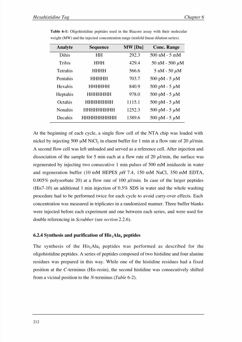
8/9/2019 DissB 7321 Mirroring
http://slidepdf.com/reader/full/dissb-7321-mirroring 226/278
Hexahistidine Tag Chapter 6
212
Table 6-1: Oligohistidine peptides used in the Biacore assay with their molecular
weight (MW) and the injected concentration range (tenfold linear dilution series).
Analyte Sequence MW [Da] Conc. Range
Dihis HH 292.3 500 nM - 5 mM
Trihis HHH 429.4 50
nM - 500
µM
Tetrahis HHHH 566.6 5 nM - 50 µM
Pentahis HHHHH 703.7 500 pM - 5 µM
Hexahis HHHHHH 840.9 500 pM - 5 µM
Heptahis HHHHHHH 978.0 500 pM - 5 µM
Octahis HHHHHHHH 1115.1 500 pM - 5 µM
Nonahis HHHHHHHHH 1252.3 500 pM - 5 µM
Decahis HHHHHHHHHH 1389.6 500 pM - 5
µM
At the beginning of each cycle, a single flow cell of the NTA chip was loaded with
nickel by injecting 500 µM NiCl2 in eluent buffer for 1 min at a flow rate of 20 µl/min.
A second flow cell was left unloaded and served as a reference cell. After injection and
dissociation of the sample for 5 min each at a flow rate of 20 µl/min, the surface was
regenerated by injecting two consecutive 1 min pulses of 500 mM imidazole in water
and regeneration buffer (10 mM HEPES pH 7.4, 150 mM NaCl, 350 mM EDTA,
0.005% polysorbate 20) at a flow rate of 100
µl/min. In case of the larger peptides
(His7-10) an additional 1 min injection of 0.5% SDS in water and the whole washing
procedure had to be performed twice for each cycle to avoid carry-over effects. Each
concentration was measured in triplicates in a randomized manner. Three buffer blanks
were injected before each experiment and one between each series, and were used for
double referencing in Scrubber (see section 2.2.6).
6.2.4 Synthesis and purification of His2Ala4 peptides
The synthesis of the His2Ala4 peptides was performed as described for the
oligohistidine peptides. A series of peptides composed of two histidine and four alanine
residues was prepared in this way. While one of the histidine residues had a fixed
position at the C -terminus (His-resin), the second histidine was consecutively shifted
from a vicinal position to the N -terminus (Table 6-2).

8/9/2019 DissB 7321 Mirroring
http://slidepdf.com/reader/full/dissb-7321-mirroring 227/278
Chapter 6 Hexahistidine Tag
213
Table 6-2: Mixed His2Ala4 peptide series with their
sequence and molecular weight.
Analyte Sequence MW [Da]
HisAla1 AAAAHH 576.6
HisAla2 AAAHAH 576.6
HisAla3 AAHAAH 576.6
HisAla4 AHAAAH 576.6
HisAla5 HAAAAH 576.6
Due to their small size and relatively high hydrophilicity, a separation of these peptides
with a traditional reversed-phase peptide purification approach (water / acetonitrile /
TFA) was not possible. Therefore, the peptides were separated under basic conditions,above their theoretical pI of 6.92 (calculated using the PeptideMass tool [20]). For this
purpose, a silica-based C18 column with extended pH tolerability was selected and
equilibrated with 10 mM ammonium acetate buffer pH 8.8. After sample injection and
an isocratic phase of 2 min, a linear gradient to 5% acetonitrile was applied within
20 min. The relatively wide peaks were completely collected and the solvents and
ammonium acetate were removed by lyophilization over night.
6.2.5 His2Ala4 peptide binding assay
For the screening of the His2Ala4 peptides, the same experimental setup as described in
section 6.2.3 was used. The purified peptides were diluted in eluent buffer to a stock
concentration of 25 mM, and fivefold linear dilution series between 0.32-5000 µM
were prepared. After loading a single flow cell with nickel, the peptide samples were
injected for 1 min with a dissociation phase of 20 s at a flow rate of 20 µl/min. The
surface was regenerated with a 1 min pulse of regeneration buffer. The signals of an
unloaded NTA flow cell were used for referencing and buffer blank injection before(3 blanks) and between (1 blank) the cycles for double referencing (see section 2.3.6).

8/9/2019 DissB 7321 Mirroring
http://slidepdf.com/reader/full/dissb-7321-mirroring 228/278
Hexahistidine Tag Chapter 6
214
6.3 Results & Discussion
6.3.1 Synthesis and purification of oligohistidine peptides
For any synthesized peptide, a yield of at least 40% was achieved after HPLC
purification. Much higher concentrations of the injected solution could be reached with
precipitated peptides than with directly lyophilized products, which led to a shorter
purification process. HPLC chromatograms of crude peptides normally consisted of
one major peak and a few byproducts. The addition of 0.1% TFA to both separation
buffers lowered the pH specifically below the pI of histidine (7.6) and therefore led to a
partial protonation of the oligohistidines. This gave the peptides a very hydrophilic
character and aggravated their retention on C18 columns. On different scales of
hydrophobicity, histidine is placed in the middle among the 20 naturally occurringamino acids [21]. As a consequence, it has also a small lipophilic character allowing a
proper retention on a C18 column. A direct correlation between peptide length and
retention time was observed for the HPLC purification (Table 6-3).
Table 6-3: Data from the HPLC purification (retention time) and the mass
spectrometric analysis of the oligohistidine peptide series (calculated and
measured monoisotopic masses).
PeptideRetention
Time [min] a
Calculated
Mass [Da]
Experimental
Mass [Da]
His3 7.9 b 428.2 428.2
His4 4.5 566.3 565.2
His5 4.7 703.3 703.2
His6 6.0 840.4 840.3
His7 9.0 977.4 977.4
His8 12.5 1114.4 1114.4
His9 13.4 1250.5 1250.7
His10 14.2 1388.6 1388.5
a His4-10 were separated using a water/acetonitrile/TFA gradient. b For
His3, water/TFA was substituted with ammonium acetate buffer pH 8.8.
While His4-10 could be easily separated with the water/acetonitrile/TFA system, the
trihistidine peptide eluted already with the injection peak under these conditions. By
using ammonium acetate at pH 8.8 instead of 0.1% TFA, the average charge of the
trihistidine could be decreased resulting in a stronger lipophilic character. The increase
of buffer pH prolonged the retention time on the C18 column from 2.5 min (injection

8/9/2019 DissB 7321 Mirroring
http://slidepdf.com/reader/full/dissb-7321-mirroring 229/278
Chapter 6 Hexahistidine Tag
215
peak) up to 7.9 min using the same gradient. ESI/MS analysis was successful using
cone voltages from 50 to 80 V, which led to single and double charged hydrogen
adducts and small amounts of single charged sodium adducts in positive mode
(Table 6-3). Only single charged species and small fragments, arisen from the applied
ionization energy, were visible in the negative mode.
6.3.2 Oligohistidine binding assay
The method of the binding assay was based on the study of Nieba et al. [5] and the
recommendations of Biacore [22]. However, the eluent buffer was prepared without
any addition of polysorbate 20, since a slow but steady decrease of binding activity of
the peptides was observed, when a polysorbate-containing sample buffer was used. The
reason for this behavior is not yet understood and could include complexation effects.
Furthermore, two additional washing steps (500 mM imidazole and 0.1% SDS) were
introduced besides the regeneration with 350 mM EDTA in order to fully avoid any
carry-over effects. After the complete regeneration, the surface could be reproducibly
loaded with nickel before each sample injection. While the amount of captured nickel
ions was highly constant within one experimental series, the nickel capacity could vary
between individuals flow cells and was found to decrease slightly over several weeks
of chip usage. Using the modified assay procedure, all peptide samples could be
measured in triplicates with high signal intensity and reproducibility (Fig. 6-3).
0 100 200 300 400 0 100 200 300 400 0 100 200 300 400
Time [s]
S P R R
e s p o n s e ( s
c a l e d i n d i v i d u a l l y )
His2 His3 His4
His5 His6 His7
His8 His9 His10
500
Figure 6-3: Sensorgrams of the oligohistidine peptides His2-His10. All samples were injected as
randomized triplicates. The concentration range of individual samples is listed in table 6-1.
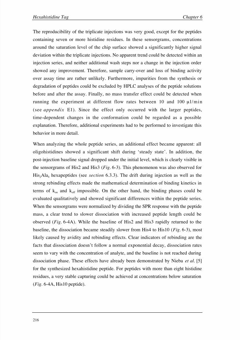
8/9/2019 DissB 7321 Mirroring
http://slidepdf.com/reader/full/dissb-7321-mirroring 230/278
Hexahistidine Tag Chapter 6
216
The reproducibility of the triplicate injections was very good, except for the peptides
containing seven or more histidine residues. In these sensorgrams, concentrations
around the saturation level of the chip surface showed a significantly higher signal
deviation within the triplicate injections. No apparent trend could be detected within an
injection series, and neither additional wash steps nor a change in the injection order
showed any improvement. Therefore, sample carry-over and loss of binding activity
over assay time are rather unlikely. Furthermore, impurities from the synthesis or
degradation of peptides could be excluded by HPLC analyses of the peptide solutions
before and after the assay. Finally, no mass transfer effect could be detected when
running the experiment at different flow rates between 10 and 100 µ l /min
(see appendix E1). Since the effect only occurred with the larger peptides,
time-dependent changes in the conformation could be regarded as a possibleexplanation. Therefore, additional experiments had to be performed to investigate this
behavior in more detail.
When analyzing the whole peptide series, an additional effect became apparent: all
oligohistidines showed a significant shift during ‘steady state’. In addition, the
post-injection baseline signal dropped under the initial level, which is clearly visible in
the sensorgrams of His2 and His3 (Fig. 6-3). This phenomenon was also observed for
His2Ala4 hexapeptides (see section 6.3.3). The drift during injection as well as the
strong rebinding effects made the mathematical determination of binding kinetics in
terms of kon and koff impossible. On the other hand, the binding phases could be
evaluated qualitatively and showed significant differences within the peptide series.
When the sensorgrams were normalized by dividing the SPR response with the peptide
mass, a clear trend to slower dissociation with increased peptide length could be
observed (Fig. 6-4A). While the baseline of His2 and His3 rapidly returned to the
baseline, the dissociation became steadily slower from His4 to His10 (Fig. 6-3), most
likely caused by avidity and rebinding effects. Clear indicators of rebinding are thefacts that dissociation doesn’t follow a normal exponential decay, dissociation rates
seem to vary with the concentration of analyte, and the baseline is not reached during
dissociation phase. These effects have already been demonstrated by Nieba et al. [5]
for the synthesized hexahistidine peptide. For peptides with more than eight histidine
residues, a very stable capturing could be achieved at concentrations below saturation
(Fig. 6-4A, His10 peptide).

8/9/2019 DissB 7321 Mirroring
http://slidepdf.com/reader/full/dissb-7321-mirroring 231/278

8/9/2019 DissB 7321 Mirroring
http://slidepdf.com/reader/full/dissb-7321-mirroring 232/278
Hexahistidine Tag Chapter 6
218
hexahistidine showed the highest affinity among the peptide series. Additional histidine
residues did not improve the affinity any further, but led to an increase in K D until it
reached a value of 165 nM for the decahistidine. The initial improvement in KD
(His2-His6) can be explained by thermodynamic means ( Eq. 10). An increasing
number of interacting groups directly improve the enthalpy term (H) due to a higher
probability of simultaneous electrostatic interactions (e.g. by rebinding). The higher
flexibility of longer peptides might further increase this probability up to a certain
point. This finally leads to a slower dissociation rate with increasing length. However,
the free binding enthalpy (G) is also dependent on entropy (S), and when a peptide
length of six residues is transgressed, the entropy seems to become the dominating term
of the equation. With increasing length, peptides have more possibilities to adopt
different conformations. The loss of entropy by forcing the molecule into a bindingconformation increases with each additional residue and the free enthalpy is increasing,
which leads to weaker binding. Therefore, a peptide length of six histidine residues
seem to be an optimal compromise between the enthalpic and entropic components.
G=H-TS [Eq. 10]
The estimated KD values for the free His6 peptide in this study are remarkably stronger
than those for hexahistidine-tagged proteins reported by Nieba et al. [5] (~1 µM). Even
though these authors also investigated the interaction of the free peptide with the nickel
surface, no KD value had been reported. Limited accessibility of the tag, steric
hindrance, or electrostatic interactions with the tag are possible explanations for the
discrepancy between the free peptide and the tagged proteins. The strength of this
technology lies in the stability, meaning a strong rebinding during dissociation.
Whereas the short peptides, especially di- and trihistidine, show a very rapid
dissociation without rebinding, the rebinding effect appears with tetrahistidine and
becomes more and more obvious with increasing peptide length.
6.3.3 Synthesis and purification of His2Ala4 peptides
The Ni2+-NTA surface allows the simultaneous binding of two histidine residues
(Fig. 6-1, see section 6.1.1). In order to determine the optimal distance between the two
interacting imidazole groups, a series of His2Ala4 hexapeptides was prepared, where the
position of one histidine was consecutively shifted while the second was fixed at the
C -terminus. While the synthesis of the peptide series was comparable to those of the
oligohistidine peptides (see section 6.3.1), the purification was much more challenging.

8/9/2019 DissB 7321 Mirroring
http://slidepdf.com/reader/full/dissb-7321-mirroring 233/278
Chapter 6 Hexahistidine Tag
219
Similar to His3, the peptide peaks overlaid with the injection peak of the HPLC
separation when standard conditions were used (water/acetonitrile/TFA). When these
peaks were collected and used for Biacore screening, the SPR signal always showed a
strong non-specific component (see appendix E2). Therefore, ammonium acetate was
used instead of water/TFA due to its higher pH range, low UV cut-off, and volatility.
With this method, it was possible to fully separate the peptides from the reagents used
for the synthesis.
6.3.4 His2Ala4peptide assay
Based on the findings of the oligohistidine system (see section 6.3.2), a similar assay
was developed for the screening of the His2Ala4 peptides. As expected and similar to
the His2 peptide (see Fig.
6-3), the kinetic rate constants of the whole peptide series
was very fast (Fig. 6-6).
400
300
200
100
0
400
300
200
100
0
0 20 40 60 0 20 40 60
0 20 40 60
AAAAHH AAAHAH AAHAAH
AHAAAH HAAAAH
Time [s]
S P R R
e s p o n s e [ R U ]
Figure 6-6: Sensorgrams of the different His2Ala4 peptides over a concentration range of 0.32-5000 µM
(fivefold linear dilution; randomized triplicate injections).
While the overall shape of the sensorgrams approximately remained constant for all
peptides, there were differences visible in terms of signal intensity and equilibrium drift
(Fig. 6-6). Due to the fast kinetics and the complete return to the baseline, less
regeneration/wash steps were required compared to the oligohistidine assay (only asingle EDTA injection). Since the amino acid composition and the peptide length was
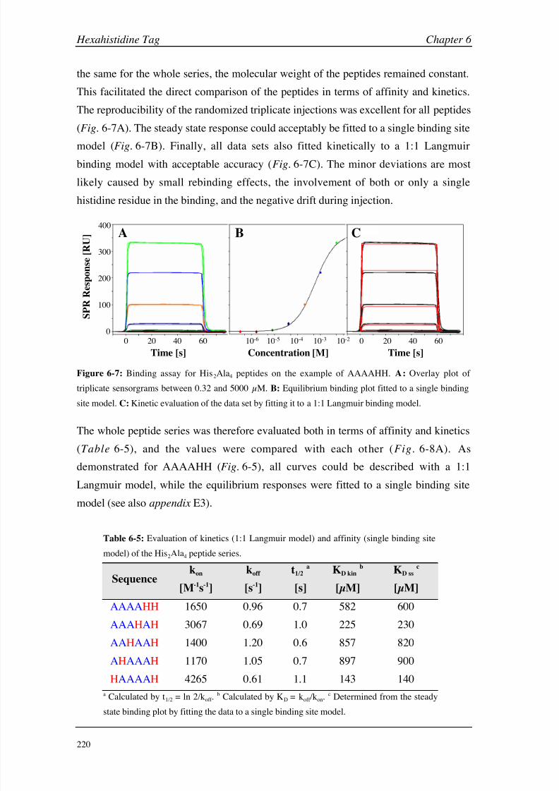
8/9/2019 DissB 7321 Mirroring
http://slidepdf.com/reader/full/dissb-7321-mirroring 234/278
Hexahistidine Tag Chapter 6
220
the same for the whole series, the molecular weight of the peptides remained constant.
This facilitated the direct comparison of the peptides in terms of affinity and kinetics.
The reproducibility of the randomized triplicate injections was excellent for all peptides
(Fig. 6-7A). The steady state response could acceptably be fitted to a single binding site
model (Fig.
6-7B). Finally, all data sets also fitted kinetically to a 1:1 Langmuir
binding model with acceptable accuracy (Fig. 6-7C). The minor deviations are most
likely caused by small rebinding effects, the involvement of both or only a single
histidine residue in the binding, and the negative drift during injection.
0 20 40 60 0 20 40 6010-6 10-5 10-3 10-210-4
0
100
200
300
400
Concentration [M]
S P R R e s p o n s e [ R U ]
Time [s] Time [s]
A B C
Figure 6-7: Binding assay for His2Ala4 peptides on the example of AAAAHH. A: Overlay plot of
triplicate sensorgrams between 0.32 and 5000 µM. B: Equilibrium binding plot fitted to a single binding
site model. C: Kinetic evaluation of the data set by fitting it to a 1:1 Langmuir binding model.
The whole peptide series was therefore evaluated both in terms of affinity and kinetics
(Table 6-5), and the values were compared with each other (Fig. 6-8A). As
demonstrated for AAAAHH (Fig. 6-5), all curves could be described with a 1:1
Langmuir model, while the equilibrium responses were fitted to a single binding site
model (see also appendix E3).
Table 6-5: Evaluation of kinetics (1:1 Langmuir model) and affinity (single binding site
model) of the His2Ala4 peptide series.
Sequencekon
[M-1s-1]
koff
[s-1]
t1/2 a
[s]
KD kin b
[µM]
KD ss c
[µM]
AAAAHH 1650 0.96 0.7 582 600
AAAHAH 3067 0.69 1.0 225 230
AAHAAH 1400 1.20 0.6 857 820
AHAAAH 1170 1.05 0.7 897 900
HAAAAH 4265 0.61 1.1 143 140
a Calculated by t1/2 = ln 2/koff .b Calculated by KD = koff /kon.
c Determined from the steady
state binding plot by fitting the data to a single binding site model.

8/9/2019 DissB 7321 Mirroring
http://slidepdf.com/reader/full/dissb-7321-mirroring 235/278
Chapter 6 Hexahistidine Tag
221
AAAAHH
1
AAAHAH
2
AAHAAH
3
AHAAAH
4
HAAAAH
5
k o n
[ 1 0 3 M - 1 s - 1 ] | k
o f f
[ s - 1 ]
1 2 3 4 5
0
2
4
6
8
10
K D
[ 1 0 - 4 M
]
A B
His2Ala4 peptide
Figure 6-8: Binding properties and geometries of the His2Ala4 peptide series. A: Kinetic properties as an
overlay of kon (blue lines), koff (red lines) and KD (green columns) for the individual peptides (1-5).
B: Hypothetical binding geometries of the five investigated peptides. Histidine residues are represented
in red and alanine in blue, nickel as a purple sphere and NTA as green lines. The black, dashed triangle
symbolizes the optimal binding geometry.
Comparison of the kinetic properties revealed some interesting similarities and
differences. While there were only minor deviations in the kinetic off-rate between the
five peptides, the calculated association phase showed significantly higher deviations.
The significant deviation between the KD values of the HH peptide (123 µM; Table
6-4) and the AAAAHH peptide (582 µM; Table 6-5) could be explained by steric or
entropic effects. While peptides AAAAHH, AAHAAH, and AHAAAH were rather
similar with a slight trend to weaker affinities, AAAHAH and HAAAAH bound muchstronger to the nickel surface (Fig. 6-8A). This effect was influenced by both on- and
off-rates, but with a much stronger contribution from the association rate. Interestingly,
these two peptides also showed the most prominent drift during steady state (Fig. 6-6;
see next paragraph). These findings indicate that a rather small gap between the two
binding residues is preferred. Both a vicinal position as well as a larger spacer of 2-3
alanines led to a significant loss of affinity. HAAAAH, with four linking alanine
residues is believed to possess enough flexibility for establishing an optimal binding
geometry again (Fig.
6-8B). This hypothesis also leads to suggestions for further
experiments. First, the gap between the two histidine residues could be fine-tuned by
substituting the intermediate alanine by other moieties, e.g. with different sizes or
limited conformational flexibility. Second, the effect of larger spacers (i.e. more than 4
alanines) should be investigated. Finally, the histidine positions of peptide 2 and 5
could be combined resulting in a peptide with the sequence HAAHAH.
Similar to the experiments with oligohistidine peptides (see section 6.3.2), a significant
drift during the steady state phase was observed (Fig.
6-6). This effect was mostobvious for AAAHAH and HAAAAH, which also showed the strongest binding
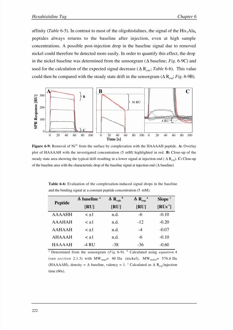
8/9/2019 DissB 7321 Mirroring
http://slidepdf.com/reader/full/dissb-7321-mirroring 236/278
Hexahistidine Tag Chapter 6
222
affinity (Table 6-5). In contrast to most of the oligohistidines, the signal of the His2Ala4
peptides always returns to the baseline after injection, even at high sample
concentrations. A possible post-injection drop in the baseline signal due to removed
nickel could therefore be detected more easily. In order to quantify this effect, the drop
in the nickel baseline was determined from the sensorgram ( baseline; Fig.
6-9C) and
used for the calculation of the expected signal decrease ( Rcalc; Table 6-6). This value
could then be compared with the steady state drift in the sensorgram ( Rexp; Fig. 6-9B).
A BB
C
36 RU
C
4 RU
0
100
200
300
0 20 40 60 80 100 0 20 40 60 80 100 0 20 40 60 80 100
Time [s]
S P R R
e s p o n s e [ R U ]
Figure 6-9: Removal of Ni2+ from the surface by complexation with the HAAAAH peptide. A: Overlay
plot of HAAAAH with the investigated concentration (5 mM) highlighted in red. B: Close-up of the
steady state area showing the typical drift resulting in a lower signal at injection end ( Rexp). C: Close-up
of the baseline area with the characteristic drop of the baseline signal at injection end ( baseline).
Table 6-6: Evaluation of the complexation-induced signal drops in the baseline
and the binding signal at a constant peptide concentration (5 mM).
Peptide baseline a
[RU]
Rcalcb
[RU]
Rexpa
[RU]
Slope c
[RUs-1]
AAAAHH < ±1 n.d. -6 -0.10
AAAHAH < ±1 n.d. -12 -0.20
AAHAAH < ±1 n.d. -4 -0.07AHAAAH < ±1 n.d. -6 -0.10
HAAAAH -4 RU -38 -36 -0.60
a Determined from the sensorgram (Fig. 6-9). b Calculated using equation 4
(see
sect ion 2.1.3) with MWtarget= 60 Da (nickel), MWanalyte= 576.6 Da
(HAAAAH), density = baseline, valency = 1. c Calculated as Rexp/injection
time (60s).

8/9/2019 DissB 7321 Mirroring
http://slidepdf.com/reader/full/dissb-7321-mirroring 237/278
Chapter 6 Hexahistidine Tag
223
Due to the rather low signals for the baseline drop ( 4 RU), the expected steady state
shift could only be calculated for HAAAAH. Longer injection times or higher peptide
concentrations might improve the signal intensity. For HAAAAH, the loss of Ni2+ from
the surface corresponded very well with the signal drop during the injection phase
(Table 6-6). A removal of nickel ions leads to a decreased binding capacity of the
surface. Therefore, a lower number of interactions with peptide molecules occurs per
time unit resulting in an equilibrium drift. While the baseline drop was too small to
detect for most of the peptides, the steady state shift could be determined for all
samples. Similar to the affinity measurements (Table 6-5), AAAHAH and HAAAAH
showed the strongest effect. The complexation effect might also be explained in terms
of solution-based stability constants [23]. While the affinity for the Ni2+-NTA in
solution (-log
KD
=
11.26) is approximately 350 times stronger than the Ni-Hisinteraction (-log KD = 8.69), additional histidine residues could decrease this difference
and lead to a strong competition between NTA and the peptide for the nickel ion.
Therefore, a complexation of nickel seems to be a reasonable explication of the steady
state drift.
8/9/2019 DissB 7321 Mirroring
http://slidepdf.com/reader/full/dissb-7321-mirroring 238/278
Hexahistidine Tag Chapter 6
224
6.4 Conclusions
The hexahistidine tag has found its way to the most important tagging technology for
the purification of recombinant proteins through recent years. Despite their numerous
applications, little is known about the exact mechanism of the metal binding. In
addition, the technology is only of limited use for some important applications such as
the immobilization of tagged proteins to Ni2+-NTA sensor chips, due to insufficient
stability of the capturing. The hexahistidine tag had been selected as an ideal structure
by empirical screening and has been used without any changes ever since. Therefore,
the rational alteration of the tag structure based on binding geometries and kinetic data
might improve the properties and broaden the application of metal-affinity tags.
In a first step, the ideal peptide length of a consecutive oligohistidine peptide seriesbetween two and ten His residues was tested on a Ni2+-NTA surface using Biacore.
With an increasing number of histidine moieties, the kinetic properties changed
significantly to slower dissociation rates. Due to the complex binding mode, which is
believed to include avidity, rebinding, and removal of nickel from the surface, a
quantitative evaluation of this series was not possible. However, the trend to increased
surface stability for larger peptides was clearly visible and could be demonstrated when
overlaying the individual dissociation phases. Despite the steady increase in
dissociation stability, the estimated overall affinity only improved until a peptide length
of six histidine residues and became weaker when the series was continued to ten
histidines. This indicates a strong contribution from the association phase, which might
be influenced by the increased conformational flexibility and a loss of entropy for
longer peptides. Therefore, the hexahistidine unit, identified by Hochuli et al. [8] from
the screening of tagged model proteins, seems to represent the best compromise
between flexibility and stability, indeed.
Even though the best binding properties were obtained with six consecutive histidine
residues, only two moieties can bind simultaneously to a captured nickel ion. Knowing
the ideal distance and binding geometry might therefore help identifying alternative
spacers for metal affinity tags. For this purpose, a series of His2Ala4 hexapeptides with
varying distance of the two His was tested using the same assay. While all peptides
featured fast kinetic properties, the steady state affinity showed significant variations.
The KD values did not follow a linear trend to stronger or weaker binding with
increasing distance of the imidazole rings, but seem to clearly prefer a spacer length of
either one or four alanine residues between the histidines. Similar to the oligohistidine

8/9/2019 DissB 7321 Mirroring
http://slidepdf.com/reader/full/dissb-7321-mirroring 239/278
Chapter 6 Hexahistidine Tag
225
peptides, the KD seemed to be strongly influenced by the association rate constant,
underlining the importance of a predefined binding geometry. Based on these results,
novel combinations of histidine with other natural or synthetic amino acids could be
developed using Biacore and molecular modeling in order to identify improved binding
motifs.
A common phenomenon in both assays was a strong negative drift in the equilibrium
responses during peptide injection. In case of the His2Ala4 peptides, this drift became
stronger with increasing affinity of the peptide. Since the nickel baseline was found to
be reproducibly decreased after peptide injection, a removal of nickel ions from the
surface through complexation by His-containing peptides in solution seems to be a
reasonable explanation. Indeed, the signal drop during injection correlated very well
with the decreased binding capacity of the nickel surface.
As a consequence, the strong interaction of oligohistidine tags with surface-bound
nickel seems to be a combination of rebinding effects, an optimal binding geometry,
and a limited flexibility (i.e. peptide length). Most of these points have a great potential
for improvement and could lead to the development of short tags suitable for both
peptide/protein purification and surface immobilization.

8/9/2019 DissB 7321 Mirroring
http://slidepdf.com/reader/full/dissb-7321-mirroring 240/278
Hexahistidine Tag Chapter 6
226
6.5 Literature
[1] J. Porath, J. Carlsson, I. Olsson, G. Belfrage, Nature 1975, 258, 598.
[2] E. K. Ueda, P. W. Gout, L. Morganti, J Chromatogr A2003
, 988, 1.
[3] G. S. Chaga, J Biochem Biophys Methods 2001, 49, 313.
[4] V. Gaberc-Porekar, V. Menart, J Biochem Biophys Methods 2001, 49, 335.
[5] L. Nieba, S. E. Nieba-Axmann, A. Persson, M. Hamalainen, F. Edebratt, A. Hansson, J.
Lidholm, K. Magnusson, A. F. Karlsson, A. Pluckthun, Anal Biochem 1997, 252, 217.
[6] J. Madoz-Gurpide, J. M. Abad, J. Fernandez-Recio, M. Velez, L. Vazquez, C.
Gomez-Moreno, V. M. Fernandez, J Am Chem Soc 2000, 122, 9808.
[7] E. Hochuli, H. Dobeli, A. Schacher, J Chromatogr 1987, 411, 177.
[8] E. Hochuli, H. Bannwarth, R. Döbeli, R. Gentz, D. Stüber, Bio/Technology 1988, 9, 1321.
[9] K. Terpe, Appl Microbiol Biotechnol 2003, 60, 523.
[10] Protein Data Bank: The worldwide repository for macromolecular structures, Research
Collaboratory for Structural Bioinformatics (RCSB), http://www.pdb.org/, 2005.
[11] P. D. Gershon, S. Khilko, J Immunol Methods 1995, 183, 65.
[12] M. Conti, G. Falini, B. Samori, Angew Chem Int Ed Engl 2000, 39, 215.
[13] L. Schmitt, M. Ludwig, H. E. Gaub, R. Tampe, Biophys J 2000, 78, 3275.
[14] H. L. Liu, Y. Ho, C. M. Hsu, J Biomol Struct Dyn 2003, 21, 31.
[15] E. A. Woestenenk, M. Hammarstrom, S. van den Berg, T. Hard, H. Berglund, J Struct
Funct Genomics 2004, 5, 217.
[16] A. Chant, C. M. Kraemer-Pecore, R. Watkin, G. G. Kneale, Protein Expr Purif 2005, 39, 152.
[17] J. Wu, M. Filutowicz, Acta Biochim Pol 1999, 46 , 591.
[18] R. L. Rich, D. G. Myszka, J Mol Recognit 2005, 18, 1.
[19] K. M. Muller, K. M. Arndt, K. Bauer, A. Pluckthun, Anal Biochem 1998, 259, 54.
[20] M. R. Wilkins, I. Lindskog, E. Gasteiger, A. Bairoch, J. C. Sanchez, D. F. Hochstrasser,
R. D. Appel, Electrophoresis 1997, 18, 403.
[21] J. L. Cornette, K. B. Cease, H. Margalit, J. L. Spouge, J. A. Berzofsky, C. DeLisi, J Mol
Biol 1987, 195, 659.
[22] Capture of histidine-tagged proteins using NTA or anti-histidine antibodies (Application
Note 12), Biacore AB, 2002.
[23] T. E. Furia, in CRC handbook for food additives, 2nd ed. (Ed.: T. E. Furia), The Chemical
Rubber Co., Cleveland, 1972.

8/9/2019 DissB 7321 Mirroring
http://slidepdf.com/reader/full/dissb-7321-mirroring 241/278
Chapter 7
General Considerations

8/9/2019 DissB 7321 Mirroring
http://slidepdf.com/reader/full/dissb-7321-mirroring 242/278
General Considerations Chapter 7
228
The previous chapters showed different applications of the surface plasmon resonance
(SPR) technology for characterizing molecular interactions with emphasis on small
molecules and carbohydrate-protein interactions. Both areas are rather challenging
compared to the classical Biacore applications such as antibody-antigen interactions,
and provide some pitfalls and limitations. The aim of this chapter is therefore to
summarize general considerations and recommendations both from the literature and
our own experience. After focusing on general assay design and applications to small
molecules, the importance and special features of carbohydrate-protein interactions will
be discussed. Negative SPR binding signals were one of the most unusual and
interesting finding of this thesis, and are therefore reconsidered from a general point of
view.
7.1 Working with small molecules
The major component of the SPR signal generation is an increase of the local electron
density around the gold surface upon ligand binding. Since larger molecules induce a
more intense shift of the resonance angle, Biacore instruments are often referred to as
‘mass detectors’. As a consequence, small molecules show only SPR signals of low
intensity and are more difficult to detect. While larger responses often tolerate small
heterogeneities and artifacts, these inaccuracies can lead to severe deviations of the
binding parameters in case of low molecular weight analytes. Alongside with the
improvement of instrument sensitivity, a number of protocols and recommendations for
such assays have been developed. A good overview about small molecule assays on
Biacore and other SPR instruments is given in reviews by Rich and Myszka [1-5].
Careful planning of a Biacore assay is a prerequisite for a successful analysis, and
includes the selection of targets and analytes, assay design, data processing, evaluation
of the results, and instrument maintenance (Fig. 7-1).

8/9/2019 DissB 7321 Mirroring
http://slidepdf.com/reader/full/dissb-7321-mirroring 243/278
Chapter 7 General Considerations
229
Target/Analyte
Selection
Assay Design
Data Acquisition & Processing
Data Evaluation
& Validiation
Instrument
Maintenance
Figure 7-1: Key steps in the planning of a Biacore assay. Instrument
maintenance is an important part of the whole cycle and should be
considered throughout the entire process.
7.1.1 Target and analyte selection
When an interaction between two different molecules is measured, the molecular
properties and the quality of the binding partners are vital for the outcome of the assay.
As much information as possible about target and analytes should be collected to avoid
inactivation effects, binding interferences and artifacts. Some of the most important
parameters to consider are listed in table 7-1.
Table 7-1: Molecular properties of analytes and targets that are of
importance for assay development.
Protein (Target) Small Molecule (Analyte)
• Size / Molecular weight • Molecular weight
• pI value • pK a values
• Purity (SDS-PAGE, SEC) • Purity (HPLC)
•
Stability (pH, SDS, ...) •
Stability
• Free amines, thiols, ... • Spacers, ...
• Tags, capturing domains • log P, solubility
Size / Molecular weight
The size of the binding partners is directly responsible for the intensity of the SPR
signal (see equation 4, section 2.1.3). Therefore, the target size should be kept as low as
possible, and protein domains that are not involved in the interaction should be

8/9/2019 DissB 7321 Mirroring
http://slidepdf.com/reader/full/dissb-7321-mirroring 244/278
General Considerations Chapter 7
230
removed. However, any removal of protein domains has to be validated carefully, since
even domains distant from the binding site may contribute to the protein activity
(stabilization etc.). Smaller protein fragments might also show a better binding activity
and reduced non-specific binding, as it could be demonstrated for the monoclonal
antibody GSLA-2 (see sections 4.3.3 and 4.3.4). In case of the analyte in solution, the
molecule should exceed at least 200 Da, but higher molecular weights are clearly
preferred. However, for most drug discovery applications the lead or candidate compounds
are kept below 500 Da to fulfill the requirements of the Lipinski ‘rule of 5’ [6].
pI / pK a
While the pI value of a protein target is important for its immobilization (see section
2.1.3), the pK a of a small analyte molecule can also influence the outcome of a Biacore
experiment. As demonstrated for the interaction of salicylic acid with human serum
albumin (see section 3.3.7), acidic compounds might exceed the capacity of the running
buffer and lead to a shift in pH . In case of pH -sensitive proteins like HSA, this might
generate an artificial SPR signal that overlays with the binding response. Therefore,
free carboxylic group should be avoided if possible, and the pH of the injected sample
with the highest concentration should be carefully controlled. An increase of the
running buffer capacity might be required, if ligand-induced pH shifts are detected.
Target and analyte purity
The purity of both the target and the analytes is one of the most important parameters in
a SPR experiment. Kapoor et al. suggest a purity of at least 90% [7], but these values
are highly dependent on the detection method and the type of assay. Since any increase
in mass or electron density around the gold surface is equally detected, a differentiation
between specific and non-specific signals is nearly impossible. In some cases,
non-specific binding can be detected by a clearly biphasic sensorgram as it had been
observed for some sLea batches in case of GSLA-2 (see section 4.3.4).
Chromatographic purification and sensitive analytics should therefore be performed for
each analyte molecule (e.g. LC-MS). Similarly, impurities in the protein fraction might
lead to heterogeneous binding signals and non-specific binding. Again, highly selective
purification steps (e.g. affinity chromatography) and analysis by non-reducing
SDS-PAGE are an important way for avoiding such complications. Oligomerization of
the target should also be considered as an impurity, since the increased local
concentration is likely to influence the kinetic and thermodynamic properties of the
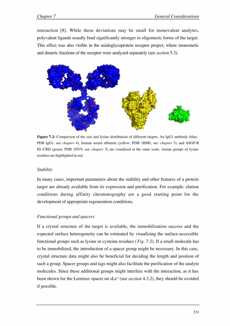
8/9/2019 DissB 7321 Mirroring
http://slidepdf.com/reader/full/dissb-7321-mirroring 245/278
Chapter 7 General Considerations
231
interaction [8]. While these deviations may be small for monovalent analytes,
polyvalent ligands usually bind significantly stronger to oligomeric forms of the target.
This effect was also visible in the asialoglycoprotein receptor project, where monomeric
and dimeric fractions of the receptor were analyzed separately (see section 5.3).
Figure 7-2: Comparison of the size and lysine distribution of different targets. An IgG1 antibody (blue;
PDB IgG1, see chapter 4), human serum albumin (yellow; PDB 1BM0, see chapter 3), and ASGP-R
H1-CRD (green; PDB 1DV8, see chapter 5) are visualized at the same scale. Amine groups of lysine
residues are highlighted in red.
Stability
In many cases, important parameters about the stability and other features of a protein
target are already available from its expression and purification. For example, elution
conditions during affinity chromatography are a good starting point for the
development of appropriate regeneration conditions.
Functional groups and spacers
If a crystal structure of the target is available, the immobilization success and theexpected surface heterogeneity can be estimated by visualizing the surface-accessible
functional groups such as lysine or cysteine residues (Fig. 7-2). If a small molecule has
to be immobilized, the introduction of a spacer group might be necessary. In this case,
crystal structure data might also be beneficial for deciding the length and position of
such a group. Spacer groups and tags might also facilitate the purification of the analyte
molecules. Since these additional groups might interfere with the interaction, as it has
been shown for the Lemieux spacer on sLea (see section 4.3.2), they should be avoided
if possible.
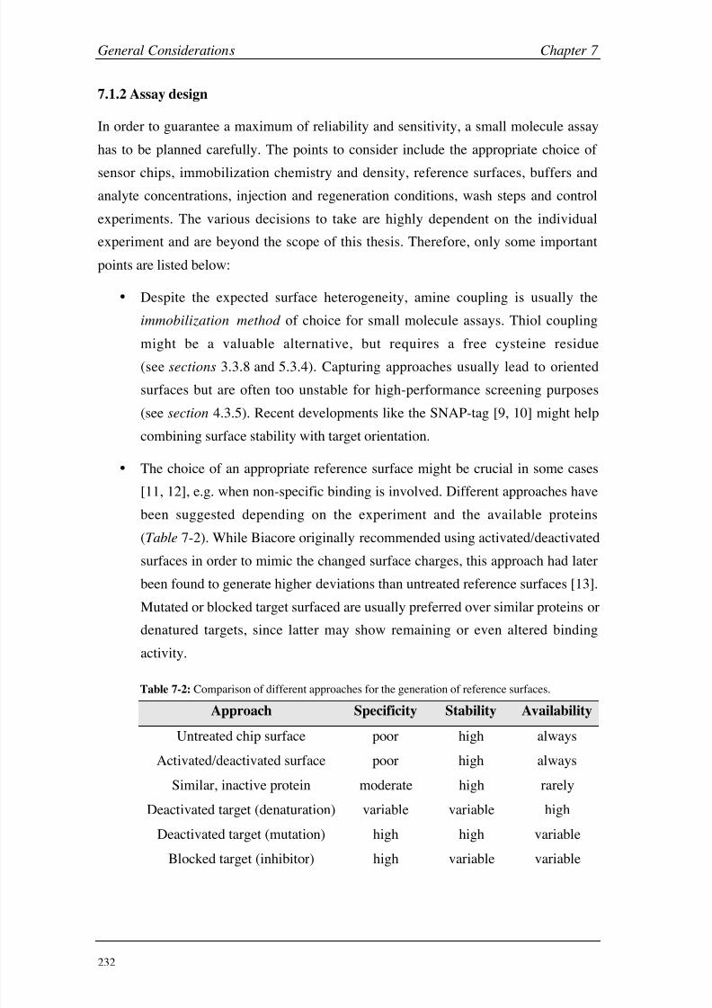
8/9/2019 DissB 7321 Mirroring
http://slidepdf.com/reader/full/dissb-7321-mirroring 246/278
General Considerations Chapter 7
232
7.1.2 Assay design
In order to guarantee a maximum of reliability and sensitivity, a small molecule assay
has to be planned carefully. The points to consider include the appropriate choice of
sensor chips, immobilization chemistry and density, reference surfaces, buffers andanalyte concentrations, injection and regeneration conditions, wash steps and control
experiments. The various decisions to take are highly dependent on the individual
experiment and are beyond the scope of this thesis. Therefore, only some important
points are listed below:
• Despite the expected surface heterogeneity, amine coupling is usually the
immobilization method of choice for small molecule assays. Thiol coupling
might be a valuable alternative, but requires a free cysteine residue
(see sections 3.3.8 and 5.3.4). Capturing approaches usually lead to oriented
surfaces but are often too unstable for high-performance screening purposes
(see section 4.3.5). Recent developments like the SNAP-tag [9, 10] might help
combining surface stability with target orientation.
• The choice of an appropriate reference surface might be crucial in some cases
[11, 12], e.g. when non-specific binding is involved. Different approaches have
been suggested depending on the experiment and the available proteins
(Table
7-2). While Biacore originally recommended using activated/deactivated
surfaces in order to mimic the changed surface charges, this approach had later
been found to generate higher deviations than untreated reference surfaces [13].
Mutated or blocked target surfaced are usually preferred over similar proteins or
denatured targets, since latter may show remaining or even altered binding
activity.
Table 7-2: Comparison of different approaches for the generation of reference surfaces.
Approach Specificity Stability Availability
Untreated chip surface poor high always
Activated/deactivated surface poor high always
Similar, inactive protein moderate high rarely
Deactivated target (denaturation) variable variable high
Deactivated target (mutation) high high variable
Blocked target (inhibitor) high variable variable

8/9/2019 DissB 7321 Mirroring
http://slidepdf.com/reader/full/dissb-7321-mirroring 247/278
Chapter 7 General Considerations
233
• A well-known or already validated analyte with a rather high molecular weight
should be chosen for assay development . This assures sufficient signal
intensities and facilitates the detection of irregularities.
•
If DMSO is required to dissolve the analytes (as in case of human serumalbumin, see section 3.3), its concentration should be kept as low as possible
(1-5% preferred, max. 8%) and a DMSO calibration is mandatory [14]. In
addition, the effect of different DMSO concentration on the binding affinity
should be controlled carefully [15].
• Injection of sample triplicates and randomization of the samples increases the
reliability and is an important tool for the recognition of carry-over effects.
Additional information to assay design can be found in the Biacore sensor surface
handbook [16] as well as in some reviews [7, 17-20].
7.1.3 Data acquisition and processing
The following procedures were found to be helpful or even essential for the processing
of low-intensity SPR signals, as they are often observed when dealing with small
molecules:
•
Extensive priming, normalizing, degassing, frequent exchange of buffers, and
short centrifugation of all samples are important steps for avoiding signal
disturbances by air bubbles and particles.
• Tight capping of the sample vials (rubber caps) and temperature control of the
autosampler compartment (~20°C). This usually allows the reproducible
measurement of sample triplicates even from the same vial (see chapters 3-5).
• Buffer spiking [21] facilitates the alignment of small signals before referencing
and leads to more accurate difference sensorgrams (see section 4.2.3).
• Data acquisition rates should be set to the maximum to assure most accurate
fits and avoid loss of information at injection start and end due to the de-spiking
procedure. This is especially important when kinetic investigations are
performed.
• Referencing to an appropriate surface (see also section 7.1.2) can be vital,
especially when non-specific binding is observed. During method development,
two or three reference surfaces on the same chip (e.g. untreated vs.
activated/deactivated surface, or inactive target surface) should be created and
evaluated.

8/9/2019 DissB 7321 Mirroring
http://slidepdf.com/reader/full/dissb-7321-mirroring 248/278
General Considerations Chapter 7
234
• Double referencing [18] eliminates most of the systemic noise during data
processing and was used in all projects. Five to ten ‘warm-up’ blanks at the
beginning of each experiment and one or two blank between each series are
normally sufficient.
•
De-spiking (included in Scrubber) automatically removes signal spikes caused
by small differences in the detection time as a result of the serially connected
flow cells or by air bubbles.
Reproducible data processing is essential for a high-quality analysis of low-molecular
weight data sets. Unfortunately, the current software tool provided by Biacore
( BIAevaluation) requires most of the processing steps to be done manually, which
could lead to small deviations. In addition, important steps like double referencing and
DMSO correction are not implemented. The kinetic models included in BIAevaluation
cover most of the application and can be extended easily. However, the limitation to 24
simultaneous binding curves is not suitable for high quality experiments (triplicate
injections etc.). Scrubber, a software tool released in 2003, overcomes most of the
limitations of BIAevaluation by offering standardized and highly automated data
processing including double referencing and DMSO correction. Since Scrubber does
not include a kinetic module, CLAMP [22] is a valuable alternative for the evaluation
of kinetic data. Future versions of Scrubber are planned to include the functionalcapabilities of CLAMP [23]. Therefore, Scrubber (and CLAMP) should be preferred for
the analysis of small molecule assays on Biacore 3000.
7.1.4 Data evaluation and assay validation
Results obtained by Biacore experiments were shown to be very reproducible and to
correspond with solution-based experiments in many cases [21, 24-26]. However, SPR
signals represent changes in the electron density around the gold surface, which are
usually caused by analyte binding but could also include conformational changes,
non-specific binding, or bulk effects. Therefore, a critical evaluation of the data is
evident and includes the selection of appropriate binding models, correlation with
literature data and validation with other analytical methods. In many cases, more
complex binding models lead to better fit results, but this effect can be simply a cause
of higher number of mathematical parameters to define a curve. As a consequence,
complex binding models should always be questioned critically and correlated with
known mechanistic properties of a binding event. For example, a bivalent binding
model is very unlikely for the interaction between two monovalent molecules. In any
8/9/2019 DissB 7321 Mirroring
http://slidepdf.com/reader/full/dissb-7321-mirroring 249/278
Chapter 7 General Considerations
235
case, kinetic analysis should be performed by globally fitting the entire data set
[27, 28]. In addition, mass transport to and from the surface [29-31] should always be
considered and tested by variation of the flow rates. Finally, the Biacore assay should
always be validated by different methods or experiments. This could include a
comparison with known binding data from literature, as it was done in the case of
albumin (see chapter 3) or with data from ELISA (e.g. ASGP-R, see chapter 5) or
other biophysical methods (e.g. GSLA-2, chapter 4). Alternatively, the confirmation of
a targets’ known binding properties such as the calcium- or pH-dependency of ASGP-R
(see chapter 5) can also be included as part of the validation process.
7.1.5 Instrument maintenance and validation
Unspecific adsorption of proteins and analytes as well as microbial growth can
dramatically influence the performance and reproducibility of SPR experiments. As a
consequence, a comparative study with several Biacore instruments investigating the
same enzyme/inhibitor pair was able to attribute a lack of reproducibility to bad
instrument maintenance [21]. Biacore recommends to clean the system at least once a
week and to perform a disinfection routine every month. However, working with small
molecules might require higher cleaning frequencies and the use of additional wash
solutions [18, 19]. Additional desorb routines between experiments are therefore
suggested if the target tolerates a removal of the sensor chip. In addition, the running
buffer should be switched to pure water after the experiment, in order to avoid salt
build-up in the instrument [18].
Even when an instrument is purified and maintained regularly, small deviations in the
detection unit cannot be excluded. Especially when dealing with small molecules, a
maximum in terms of performance and sensitivity is required. Unfortunately, neither
the Biacore system check procedure nor the myoglobin/anti-myoglobin mAb system
from the getting-started-kit are appropriate for testing the instruments’ sensitivity.
Therefore, an established low molecular weight system should be selected for this
purpose. Both the targets and a set of analytes should be commercially available in
good quality, and the interaction should be well characterized. Two of the investigated
systems in this thesis essentially fulfill these requirements, i.e. bovine carbonic
anhydrase II (see section 2.3) and human serum albumin (see chapter 3). Even though
the interaction of small drugs with albumin is one of the most intensely described
interaction systems, the complexity of its binding events, the rather high sensitivity
towards environmental factors and the requirement of DMSO for most analytes reduces
8/9/2019 DissB 7321 Mirroring
http://slidepdf.com/reader/full/dissb-7321-mirroring 250/278
General Considerations Chapter 7
236
its suitability for validation purposes. The carbonic anhydrase system, on the other
hand, was found to be very robust and its reproducibility has been validated using
Biacore instruments from various laboratories [21, 26]. In addition, this drug-enzyme
interaction not only provides affinity constants but also kinetic parameters, which can
be compared with literature.
7.2 Carbohydrate-protein interactions
Among the biomolecular interactions, those between carbohydrates and their protein
targets belong to the most unusual but also most interesting ones. They are involved in
many critical processes like inflammatory responses, cellular trafficking and signaling,
fertilization, tumor metastasis, or infection diseases. Proteins that specifically interact
with carbohydrates are referred to as lectins, which include many subclasses like
selectins, annexins, or siglecs. Some of them are soluble in the cytosol or in body
fluids, while many others are embedded in membranes. Carbohydrates are usually
attached to other structures like proteins or lipids, and the glycosylation of proteins is
one of the most important posttranslational modifications. Compared to proteins and
nucleic acids, the information content of carbohydrates is much larger due to their
complex stereochemistry and their branched structures [32]. Their involvement in
many pathogenic conditions makes lectins an interesting target in drug discovery.
However, the generation of inhibitors for lectin-interactions was found to be very
challenging. Carbohydrate binding sites are often rather shallow and solvent-exposed,
and large areas of the lectin might be involved in the binding. The forces involved in
lectin interactions with monovalent carbohydrates are generally weak and often feature
equilibrium dissociation constants in the micro- to millimolar range. The low affinity is
a consequence of very rapid binding kinetics with dissociation half-times of less than a
second. Even interactions with specific antibodies, usually characterized by very strong
affinities, might remain in the micromolar range when a carbohydrate epitope is
involved [12]. This effect was also observed in the case of sLea binding to GSLA-2
(see section 4.3.2), where the interaction resulted in a KD of ~ 10 µM. In contrast to the
affinity, the binding specificity is very high. For example, the carbohydrate recognition
domain (H1-CRD) of human ASGP-R only recognizes galactose and its derivatives
while it does not bind to glucose (see section 5.3.5). This selectivity is achieved by
multiple polar (hydroxyl groups) and non-polar interactions (carbon face of the sugar
ring) [33-35]. In contrast to the strong monovalent interactions of most peptides and

8/9/2019 DissB 7321 Mirroring
http://slidepdf.com/reader/full/dissb-7321-mirroring 251/278
Chapter 7 General Considerations
237
drugs, carbohydrates increase their affinity mainly by multivalent binding to
surface-bound targets. Glycoproteins usually contain multiple glycan chains and are
able to address more than one receptor. This receptor-clustering is an important
mechanism in cell-cell recognition and signaling pathways [8, 36, 37]. Avidity and
rebinding effects predominantly influence the dissociation rate and often lead to
binding affinities in the nanomolar range [8]. This effect has also been observed for the
ASGP receptor, where monovalent galactose bound with a KD of 10-3 M while
galactose-bearing glycoproteins were able to binding around 10-9 M (see section 5.3.5;
Fig. 7-3B&C). Carbohydrate-protein interactions are far more complex than many
other biological binding event, ranging from mono- to multivalent, from fast to slow,
and from weak to strong. As a consequence, the knowledge about kinetic and
thermodynamic properties of carbohydrate-lectin interactions becomes of a paramountimportance [7, 8, 38].
A B C
Figure 7-3: Comparison of typical sensorgrams for enzyme-drug (A; carbonic anhydrase II vs. CBS, see
section 2.3), lectin-monosaccharide (B; ASGP-R H1-CRD vs. GalNAc, see chapter 5.3.5), and
lectin-glycoprotein interactions (C; ASGP-R H1-CRD vs. asialofetuin, see section 5.3.5).
The use of SPR technology for the characterization of carbohydrate-protein interactions
provides many advantages. Sugars usually lack any chromophore or fluorophore,
making the detection with traditional analytical methods rather challenging. Since SPR
is independent on specific functional groups, Biacore technology is a valuable
alternative for the detection of carbohydrates [7]. In addition, data about affinity and
kinetics of an interaction can be addressed in a single experiment, therefore facilitating
the assessment of structure-activity relationships. Compared to peptides and synthetic
molecules, the synthesis of carbohydrates is often more complicated and
time-consuming, and automation processes are still under development [32]. As many
of the biologically relevant lectins and carbohydrates are embedded in membranes or
attached to larger structures, surface-based SPR assays are believed to be much closer
to reality than solution-based experiments [36]. This advantage might be used to

8/9/2019 DissB 7321 Mirroring
http://slidepdf.com/reader/full/dissb-7321-mirroring 252/278
General Considerations Chapter 7
238
investigate clustering effects and multivalent binding, e.g. by reverse the molecules on
the surface and in solution, or by varying the immobilization density [37, 39-41].
Variation of the immobilization density cannot only influence the binding
stoichiometry [37], but also lead to a change in the selectivity of the lectin [41]. For
example, the same interaction pair can lead to completely different sensorgrams,
dependent on which partner is immobilized on the sensor chip [39]. Finally, the flow
system might induce an additional critical component, since some of the interactions
between sugars and lectins occur in the blood circulation and were found to be
flow-dependent (e.g. selectin-induced tethering and rolling during inflammation
processes) [42, 43]. However, evaluation of flow-dependency might interfere with
mass transport effects (see section 7.1.4). The fast kinetics for monovalent sugars has
clear benefits for Biacore experiments, since binding equilibria are reached withinseconds (Fig. 7-3B), therefore reducing the injection and assay time. In addition, the
rapid return to the baseline eliminates the need for any regeneration conditions,
preventing the target from any damage and increasing the life-time of the protein
surface. Even if multivalent, tight binding event are analyzed, the carbohydrates can
usually be removed by specific conditions (e.g. removal of calcium in the case of
C-type lectins; see chapter 5) or by competition with monovalent sugars [7]. Kinetic
analysis of the binding data is often more challenging in case of carbohydrate-protein
interaction. While the rapid kinetic rate constants of monovalent sugars are usually
close to the detection limit of the Biacore instrument, the properties of multivalent
ligands are influenced by rebinding effects and heterogeneities. Immobilization density
is an important factor in this context and an optimal compromise between signal
intensity and rebinding/avidity has to be elaborated [8]. Finally, some lectins might
interact with the hydrogel that covers Biacore sensor chips, since is consists of a
glucose polymer (dextran). For example, the sensor surface had to be changed to a
glycolipid layer in a competition assay with soluble concanavalin A against
immobilized sugar, since the tetrameric lectin was found to interact with the dextran
matrix [40]. A similar effect might also be involved in the generation of negative
binding signals, as they have been observed in the case of the ASGP-R H1-CRD
(see sections 5.3.6 and 7.3). Furthermore, negative binding has also been detected in
our laboratory with other lectins such as the myelin-associated glycoprotein (MAG) or
E-selectin though not fully investigated [44, 45]. Such matrix interactions might
therefore be a general problem for the characterization of lectin interactions, both when
immobilized and in solution.
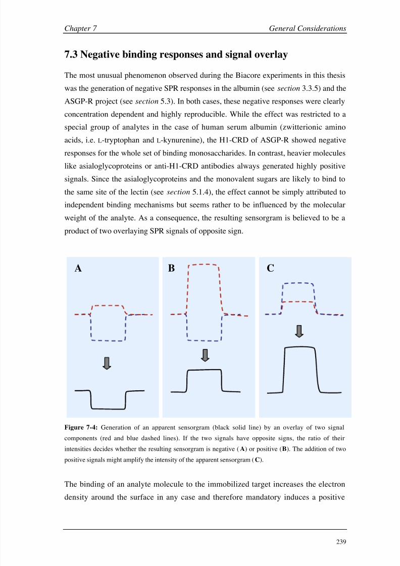
8/9/2019 DissB 7321 Mirroring
http://slidepdf.com/reader/full/dissb-7321-mirroring 253/278
Chapter 7 General Considerations
239
A CB
7.3 Negative binding responses and signal overlay
The most unusual phenomenon observed during the Biacore experiments in this thesis
was the generation of negative SPR responses in the albumin (see section 3.3.5) and the
ASGP-R project (see section 5.3). In both cases, these negative responses were clearly
concentration dependent and highly reproducible. While the effect was restricted to a
special group of analytes in the case of human serum albumin (zwitterionic amino
acids, i.e. L-tryptophan and L-kynurenine), the H1-CRD of ASGP-R showed negative
responses for the whole set of binding monosaccharides. In contrast, heavier molecules
like asialoglycoproteins or anti-H1-CRD antibodies always generated highly positive
signals. Since the asialoglycoproteins and the monovalent sugars are likely to bind to
the same site of the lectin (see section 5.1.4), the effect cannot be simply attributed to
independent binding mechanisms but seems rather to be influenced by the molecular
weight of the analyte. As a consequence, the resulting sensorgram is believed to be a
product of two overlaying SPR signals of opposite sign.
Figure 7-4: Generation of an apparent sensorgram (black solid line) by an overlay of two signal
components (red and blue dashed lines). If the two signals have opposite signs, the ratio of their
intensities decides whether the resulting sensorgram is negative (A) or positive (B). The addition of two
positive signals might amplify the intensity of the apparent sensorgram (C).
The binding of an analyte molecule to the immobilized target increases the electron
density around the surface in any case and therefore mandatory induces a positive
8/9/2019 DissB 7321 Mirroring
http://slidepdf.com/reader/full/dissb-7321-mirroring 254/278
General Considerations Chapter 7
240
signal effect, which is directly dependent on the molecular weight of the analyte. If the
second negative signal component is independent on the analyte size, the resulting
sensorgram could be either positive (large analyte) or negative (small analyte), as it is
shown in figure 7-4A&B. An experimental series with H1-CRD analytes of constant
affinity but varying molecular weight might help confirming this hypothesis. As a
consequence of this signal generation model, an overlay of two positive signal
component should amplify the intensity of the resulting sensorgram (Fig. 7-4C).
Indeed, a corresponding effect was observed in the case of sialic acid binding to HSA
(see section 3.3.7), where a pH-induced conformational change was suspected to
overlay with the binding signal.
The question remains, what might cause the additional signal component? In general,
Biacore is often referred to as a ‘mass detector’, since any binding of an analyte leads
to an increased mass and electron concentration, which then induces the SPR signal.
However, any other change in the electron density like fluctuations in the matrix length
or the protein layer could also influence the response. Different models might be
considered (Fig. 7-5), of which the most reasonable are discussed in more detail.
• Since interactions between two molecules do not occur statically but include
many dynamic steps (induced fit of the protein, conformational adaptation of
the analyte), the protein layer might change its shape, thickness, and densityupon ligand binding. Such conformational changes (Fig. 7-5B) might well
change the electron density and the SPR response.
• Even if only minor changes in the protein structure are induced, they still might
influence the exposition of charged residues on the protein surface. Since the
carboxymethyl dextran surface is negatively charged under physiological
conditions, this could lead to an electrostatic attraction or repulsion of the
protein (Fig.
7-5C) resulting in a changed distance to the gold surface.
• When a buffer component is able to interact with the target, the addition of an
analyte molecule might lead to a competitive removal of this component
(solution competition, Fig. 7-4D). If the buffer substance has a higher molecular
weight than the analyte, this removal induces a negative signal.
• Interaction of a lectin with the glucose-based dextran matrix could also
influence the packing density of the surface. Upon analyte binding, the protein
(or matrix chains) are competitively released resulting in a decreased densityand a negative signal (surface competition, Fig. 7-5E).
8/9/2019 DissB 7321 Mirroring
http://slidepdf.com/reader/full/dissb-7321-mirroring 255/278
Chapter 7 General Considerations
241
The latter two models could be the reason for the effects observed in the atypical
binding properties of ASGP-R H1-CRD (see section 5.3). When calcium-containing
running buffer is used for the screening, the lectin domain binds to the dextran matrix
and pulls the protein closer to the surface. As soon as an analyte molecule reaches the
lectin, a competition with the binding to the surface takes place and the protein flips
back, leading to the negative SPR signal (see section 5.3.5). When soluble dextran is
added to the running buffer, the model might change from a surface to a solution
competition situation. This is supported by the fact that increasing dextran
concentrations amplify the negative response while retaining the overall affinity (see
section 5.3.6). The good correlation with reported affinities and the dependency from
calcium and buffer pH indicate a specific reaction. Even though the interaction of
H1-CRD with monovalent glucose was found to be very weak (see section 5.3.5), thehigh local concentration might lead to a considerable binding affinity for the dextran
matrix. As a consequence of this possible interaction, it is hard to determine if the
experiment is closer to a direct (KD) or competitive (IC50) determination of the affinity.
Exchange or derivatization of the surface matrix could give a deeper insight in the
underlying principles and might confirm the stated hypothesis. In addition, other lectins
should be analyzed for the same effect.
In case of human serum albumin, conformational changes or an influence of surface
charges are more likely to induce the effects observed for L-tryptophan and
L-kynurenine (see section 3.3.5). Since no other class of analytes showed negative
responses, the effect has to be correlated to the analytes themselves or their binding
site. However, since both the signal intensities and the molecular weight of the two
substances are rather low, the negative binding effect could be masked by an overlay
with a more intense positive binding signal in case of other analytes. For salicylic acid,
the overlay of two positive signals induced by the binding event and a pH-induced
conformational change is highly likely. An increase of the running buffer capacity orthe use of sodium salicylate successfully eliminated the pH-dependent signal
component (see section 3.3.7).

8/9/2019 DissB 7321 Mirroring
http://slidepdf.com/reader/full/dissb-7321-mirroring 256/278
General Considerations Chapter 7
242
Figure 7-5: Comparison of different hypothetical model for the generation of positive and negative
sensorgrams (A) besides the simple mass addition. B: Binding of an analyte (red) to the immobilized
protein (green) induces a conformational change. C: Analyte binding leads to the exposition of positive
(pale red circle) or negative surface charges (pale blue circle). D: A large buffer component (orange)
binds to the protein and is competitively removed upon ligand binding. E: The protein interacts with the
dextran matrix (blue) of the sensor chip (yellow) and is released when the analyte competes for the
binding site.

8/9/2019 DissB 7321 Mirroring
http://slidepdf.com/reader/full/dissb-7321-mirroring 257/278
Chapter 7 General Considerations
243
Unfortunately, little is reported in literature about signal abnormalities or changes of
the protein/matrix layer during Biacore experiments [46-51]. The first description of
negative responses was published by Gestwicki et al. [49], who observed negative
post-injection responses when injecting different saccharides to maltose-binding
protein (MBP). The detected post-injection signals were concentration-dependent but
rather small, and nothing was stated about negative equilibrium responses. The author
attributed this effect to ligand-induced conformational changes, and emphasized this
theory with a second example, where small calcium ions induced a large SPR signal
when interacting with transglutaminase [49]. Since MBP is also a lectin, a competitive
mechanism as postulated for H1-CRD might also be possible. Conformational changes
were also stated by Mannen et al. [50] for the signal generation of a series of
immobilized proteins at varying pH. However, Paynter et al. [51] questioned theconclusions of this study and suggested an electrostatic interaction with the surface as a
more plausible mechanism, after they did some additional experiments with different
proteins and peptides. The unspecific detection of any changes in the electron density
around the gold surface by SPR makes a clear distinction between different
mechanisms very difficult and only carefully planned control experiments might give
rise to a specific model. Furthermore, the simultaneous occurrence of more than one
effect (e.g. conformational and electrostatic changes) is very likely. As a consequence,
more experiments have to be performed in order to bring more light in the complex
effect that might be involved in the SPR signal generation.

8/9/2019 DissB 7321 Mirroring
http://slidepdf.com/reader/full/dissb-7321-mirroring 258/278
General Considerations Chapter 7
244
7.4 References
[1] R. L. Rich, D. G. Myszka, J Mol Recognit 2000, 13, 388.
[2] R. L. Rich, D. G. Myszka, J Mol Recognit 2001, 14, 273.
[3] R. L. Rich, D. G. Myszka, J Mol Recognit 2002, 15, 352.
[4] R. L. Rich, D. G. Myszka, J Mol Recognit 2003, 16 , 351.
[5] R. L. Rich, D. G. Myszka, J Mol Recognit 2005, 18, 1.
[6] C. A. Lipinski, F. Lombardo, B. W. Dominy, P. J. Feeney, Adv Drug Deliv Rev 2001, 46 , 3.
[7] M. Kapoor, C. J. Thomas, K. Bachhawat-Sikder, S. Sharma, A. Surolia, Methods
Enzymol 2003, 362, 312.
[8] E. Duverger, N. Frison, A. C. Roche, M. Monsigny, Biochimie 2003, 85, 167.
[9] M. Kindermann, N. George, N. Johnsson, K. Johnsson, J Am Chem Soc 2003, 125, 7810.
[10] W. Huber, S. Perspicace, J. Kohler, F. Muller, D. Schlatter, Anal Biochem 2004, 333, 280.
[11] R. J. Ober, E. S. Ward, Anal Biochem 1999, 271, 70.
[12] M. Strandh, B. Persson, H. Roos, S. Ohlson, J Mol Recognit 1998, 11, 188.
[13] R. Karlsson, Working with small molecules in Biacore. in Biacore Symposium 2002,
Chicago, USA, 2002.
[14] A. Frostell-Karlsson, A. Remaeus, H. Roos, K. Andersson, P. Borg, M. Hamalainen, R.
Karlsson, J Med Chem 2000, 43, 1986.
[15] R. L. Rich, Y. S. Day, T. A. Morton, D. G. Myszka, Anal Biochem 2001, 296 , 197.
[16] Biacore Sensor Surface Handbook , Biacore AB, Uppsala, 2003.
[17] D. G. Myszka, Curr Opin Biotechnol 1997, 8, 50.
[18] D. G. Myszka, J Mol Recognit 1999, 12, 279.
[19] D. G. Myszka, Strategies for kinetic analysis of small molecules using Biacore, in
Biacore Workshop 2001, Fulda, Germany, 2001.
[20] R. Karlsson, M. Kullman-Magnusson, M. D. Hamalainen, A. Remaeus, K. Andersson, P.
Borg, E. Gyzander, J. Deinum, Anal Biochem 2000, 278, 1.
[21] M. J. Cannon, G. A. Papalia, I. Navratilova, R. J. Fisher, L. R. Roberts, K. M. Worthy, A.
G. Stephen, G. R. Marchesini, E. J. Collins, D. Casper, H. Qiu, D. Satpaev, S. F.Liparoto, D. A. Rice, Gorshkova, II, R. J. Darling, D. B. Bennett, M. Sekar, E.
Hommema, A. M. Liang, E. S. Day, J. Inman, S. M. Karlicek, S. J. Ullrich, D. Hodges, T.
Chu, E. Sullivan, J. Simpson, A. Rafique, B. Luginbuhl, S. N. Westin, M. Bynum, P.
Cachia, Y. J. Li, D. Kao, A. Neurauter, M. Wong, M. Swanson, D. G. Myszka, Anal
Biochem 2004, 330, 98.
[22] D. G. Myszka, T. A. Morton, Trends Biochem Sci 1998, 23, 149.
[23] T. A. Morton, BioLogic Pty Ltd, personal communication, 2005.
[24] J. Deinum, L. Gustavsson, E. Gyzander, M. Kullman-Magnusson, A. Edstrom, R.
Karlsson, Anal Biochem2002
, 300, 152.[25] Y. S. Day, C. L. Baird, R. L. Rich, D. G. Myszka, Protein Sci 2002, 11, 1017.
8/9/2019 DissB 7321 Mirroring
http://slidepdf.com/reader/full/dissb-7321-mirroring 259/278
Chapter 7 General Considerations
245
[26] D. G. Myszka, Y. N. Abdiche, F. Arisaka, O. Byron, E. Eisenstein, P. Hensley, J. A.
Thomson, C. R. Lombardo, F. Schwarz, W. Stafford, M. L. Doyle, J Biomol Tech 2003,
14, 247.
[27] T. A. Morton, D. G. Myszka, I. M. Chaiken, Anal Biochem 1995, 227 , 176.
[28] L. D. Roden, D. G. Myszka, Biochem Biophys Res Commun 1996, 225, 1073.
[29] R. W. Glaser, Anal Biochem 1993, 213, 152.
[30] P. Schuck, Biophys J 1996, 70, 1230.
[31] D. G. Myszka, X. He, M. Dembo, T. A. Morton, B. Goldstein, Biophys J 1998, 75, 583.
[32] D. B. Werz, P. H. Seeberger, Chemistry 2005.
[33] U. Spohr, O. Hindsgaul, R. U. Lemieux, Can J Chem 1985, 63, 2644.
[34] R. U. Lemieux, Chem Soc Rev 1989, 18, 347.
[35] W. I. Weis, K. Drickamer, Annu Rev Biochem 1996, 65, 441.
[36] H. Sota, R. T. Lee, Y. C. Lee, Y. Shinohara, Methods Enzymol 2003, 362, 330.
[37] C. W. Cairo, J. E. Gestwicki, M. Kanai, L. L. Kiessling, J Am Chem Soc 2002, 124, 1615.
[38] C. R. Bertozzi, L. L. Kiessling, Science 2001, 291, 2357.
[39] Y. Shinohara, Y. Hasegawa, H. Kaku, N. Shibuya, Glycobiology 1997, 7 , 1201.
[40] D. A. Mann, M. Kanai, D. J. Maly, L. L. Kiessling, J Am Chem Soc 1998, 120, 10575.
[41] N. Horan, L. Yan, H. Isobe, G. M. Whitesides, D. Kahne, Proc Natl Acad Sci U S A 1999,
96 , 11782.
[42] R. Alon, S. Chen, K. D. Puri, E. B. Finger, T. A. Springer, J Cell Biol 1997, 138, 1169.
[43] M. W. Nicholson, A. N. Barclay, M. S. Singer, S. D. Rosen, P. A. van der Merwe, J Biol
Chem 1998, 273, 763.
[44] D. S. Strasser, S. Shelke, B. Ernst, unpublished results.
[45] D. Ricklin, Z. Dragic, B. Ernst, unpublished results.
[46] H. Sota, Y. Hasegawa, M. Iwakura, Anal Chem 1998, 70, 2019.
[47] Z. Salamon, S. Cowell, E. Varga, H. I. Yamamura, V. J. Hruby, G. Tollin, Biophys J
2000, 79, 2463.
[48] S. Boussaad, J. Pean, N. J. Tao, Anal Chem 2000, 72, 222.
[49] J. E. Gestwicki, H. V. Hsieh, J. B. Pitner, Anal Chem 2001, 73, 5732.
[50] T. Mannen, S. Yamaguchi, J. Honda, S. Sugimoto, A. Kitayama, T. Nagamune, Anal
Biochem 2001, 293, 185.
[51] S. Paynter, D. A. Russell, Anal Biochem 2002, 309, 85.

8/9/2019 DissB 7321 Mirroring
http://slidepdf.com/reader/full/dissb-7321-mirroring 260/278
General Considerations Chapter 7
246
8/9/2019 DissB 7321 Mirroring
http://slidepdf.com/reader/full/dissb-7321-mirroring 261/278
Appendices
A: Carbonic Anhydrase II (CA II)
B: Human Serum Albumin (HSA)
C: Diagnostic Antibody GSLA-2
D: Asialoglycoprotein Receptor (ASGP-R)
E: Hexahistidine Tag (HisTag)
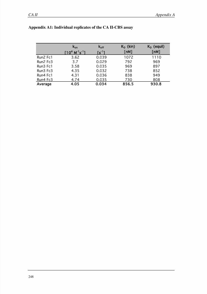
8/9/2019 DissB 7321 Mirroring
http://slidepdf.com/reader/full/dissb-7321-mirroring 262/278
CA II Appendix A
248
Appendix A1: Individual replicates of the CA II-CBS assay
k
on
k
off
K
D
kin) K
D
equil)
[10
4
M
-1
s
-1
] [s
-1
]
[nM] [nM]
Run2 Fc1 3.62 0.039 1072 1110
Run2 Fc3 3.7 0.029 792 969
Run3 Fc1 3.58 0.035 969 897
Run3 Fc3 4.35 0.032 738 852
Run4 Fc1 4.31 0.036 838 949
Run4 Fc3 4.74 0.035 730 808
Average 4.05 0.034 856.5 930.8
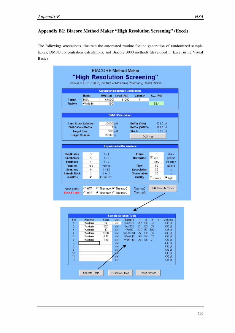
8/9/2019 DissB 7321 Mirroring
http://slidepdf.com/reader/full/dissb-7321-mirroring 263/278
Appendix B HSA
249
Appendix B1: Biacore Method Maker “High Resolution Screening” (Excel)
The following screenshots illustrate the automated routine for the generation of randomized sample
tables, DMSO concentration calculations, and Biacore 3000 methods (developed in Excel using Visual
Basic).
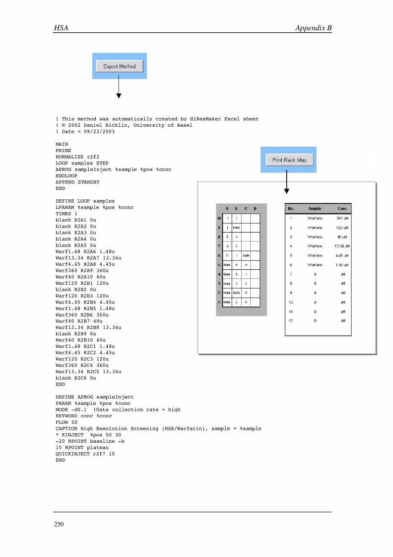
8/9/2019 DissB 7321 Mirroring
http://slidepdf.com/reader/full/dissb-7321-mirroring 264/278
HSA Appendix B
250
! This method was automatically created by HiResMaker Excel sheet
! © 2002 Daniel Ricklin, University of Basel
! Date = 09/23/2003
MAIN
PRIME
NORMALIZE r2f2
LOOP samples STEP
APROG sampleInject %sample %pos %conc
ENDLOOP
APPEND STANDBY
END
DEFINE LOOP samples
LPARAM %sample %pos %conc
TIMES 1
blank R2A1 0u
blank R2A2 0u
blank R2A3 0u
blank R2A4 0u
blank R2A5 0u
Warf1.48 R2A6 1.48u
Warf13.34 R2A7 13.34u
Warf4.45 R2A8 4.45u
Warf360 R2A9 360u
Warf40 R2A10 40u
Warf120 R2B1 120u
blank R2B2 0u
Warf120 R2B3 120u
Warf4.45 R2B4 4.45u
Warf1.48 R2B5 1.48u
Warf360 R2B6 360u
Warf40 R2B7 40u
Warf13.34 R2B8 13.34u
blank R2B9 0u
Warf40 R2B10 40u
Warf1.48 R2C1 1.48u
Warf4.45 R2C2 4.45u
Warf120 R2C3 120u
Warf360 R2C4 360u
Warf13.34 R2C5 13.34u
blank R2C6 0u
END
DEFINE APROG sampleInject
PARAM %sample %pos %conc
MODE -d0.1 !Data collection rate = high
KEYWORD conc %conc
FLOW 50
CAPTION High Resolution Screening (HSA/Warfarin), sample = %sample
* KINJECT %pos 50 30
-20 RPOINT baseline -b
15 RPOINT plateau
QUICKINJECT r2f7 10
END
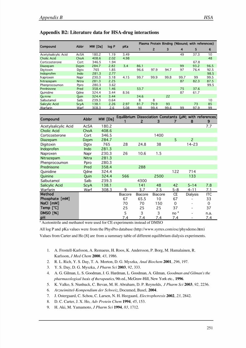
8/9/2019 DissB 7321 Mirroring
http://slidepdf.com/reader/full/dissb-7321-mirroring 265/278
Appendix B HSA
251
Appendix B2: Literature data for HSA-drug interactions
a Acetonitrile and methanol were used for CE experiments instead of DMSO
All log P and pKa values were from the PhysPro database (http://www.syrres.com/esc/physdemo.htm)
Values from Carter and Ho [8] are from a summary table of different equilibrium dialysis experiments.
1. A. Frostell-Karlsson, A. Remaeus, H. Roos, K. Andersson, P. Borg, M. Hamalainen, R.
Karlsson, J Med Chem 2000, 43, 1986.
2. R. L. Rich, Y. S. Day, T. A. Morton, D. G. Myszka, Anal Biochem 2001, 296 , 197.
3. Y. S. Day, D. G. Myszka, J Pharm Sci 2003, 92, 333.
4. A. G. Gilman, L. S. Goodman, J. G. Hardman, L. Goodman, A. Gilman, Goodman and Gilman's the
pharmacological basis of therapeutics, 9th ed., McGraw-Hill, New York etc., 1996.
5. K. Valko, S. Nunhuck, C. Bevan, M. H. Abraham, D. P. Reynolds, J Pharm Sci 2003, 92, 2236.
6. Arzneimittel-Kompendium der Schweiz, Documed, Basel, 2004.
7. J. Ostergaard, C. Schou, C. Larsen, N. H. Heegaard, Electrophoresis 2002, 23, 2842.
8. D. C. Carter, J. X. Ho, Adv Protein Chem 1994, 45, 153.
9.
H. Aki, M. Yamamoto, J Pharm Sci 1994, 83, 1712.
1 2 3 4 5 6
Acetylsalicylic Acid AcSA 180.2 1.19 3.49 49 37.3 10
Cholic Acid ChoA 408.6 2.02 4.98 48
Corticosterone Cort 346.5 1.94 67.8Diazepam Dzpm 284.7 2.82 3.4 86.1 99 93.2 96.5
Digitoxin Dgtx 765 1.85 96.6 97.9 94.7 97 76.4 92.5
Indoprofen Indo 281.3 2.77 98.5
Naproxen Napr 230.3 3.18 4.15 99.7 99.9 99.8 99.7 99 99.5
Nitrazepam Nitra 281.3 2.25 87 82.3 87.5
Phenprocoumon Ppro 280.3 3.62 99.5
Prednisone Pred 358.4 1.46 53.7 75 37.6
Quinidine Qdne 324.4 3.44 8.56 87 61.7
Quinine Quin 324.4 3.44 34.6 22
Salbutamol Salb 239.3 0.64 8 8 10
Salicylic Acid ScyA 138.1 2.26 2.97 81.7 79.9 93 73 85
Warfarin Warf 308.3 2.6 5.08 98 99.4 99.6 99 97.9 99
Plasma Protein Binding ( bound; with references)
Compound Abbr MW [Da] log P pKa
1 2 3 7 8 9
Acetylsalicylic Acid AcSA 180.2 7.7
Cholic Acid ChoA 408.6
Corticosterone Cort 346.5 1400Diazepam Dzpm 284.7 5 2
Digitoxin Dgtx 765 28 24.8 38 14–23
Indoprofen Indo 281.3
Naproxen Napr 230.3 26 10.6 1.5
Nitrazepam Nitra 281.3
Phenprocoumon Ppro 280.3Prednisone Pred 358.4 288
Quinidine Qdne 324.4 122 714
Quinine Quin 324.4 566 2500 133
Salbutamol Salb 239.3 4300
Salicylic Acid ScyA 138.1 141 48 42 5–14 7.8Warfarin Warf 308.3 9 3.7 2.5 5–8 4–11 7.1
Method Biacore Biacore Biacore CE Dialysis ITC
Phosphate [mM] 67 65.5 10 67 - 33
NaCl [mM] 70 70 150 0 - 0
Temp [°C] 25 25 25 37 - 37
DMSO [ ] 5 3 3 no a - n.a.
pH 7.4 7.4 7.4 7.4 - 7.4
Equil ibrium Dissociation Constants (µM; with references)
Compound Abbr MW [Da]

8/9/2019 DissB 7321 Mirroring
http://slidepdf.com/reader/full/dissb-7321-mirroring 266/278
HSA Appendix B
252
Appendix B3: Sensorgrams from the ranking assay
A panel of known HSA binders was injected at a constant concentration (333 µM in 10 mM PBS,
3% DMSO) as triplicates and classified according to their curve shapes (see legend at the end of this
section).
Acetylsalicylic acid Cholic acid Corticosterone
Diazepam Digitoxin Indoprofen
Naproxen NitrazepamPhenprocoumon
Prednisone Progesterone Quinidine

8/9/2019 DissB 7321 Mirroring
http://slidepdf.com/reader/full/dissb-7321-mirroring 267/278
Appendix B HSA
253
Classifications of the sensorgrams according to their shape:
Proper block signals
Multiphase association
Negative post-injection baselines
Negative binding signals
Not determined (low signal intensity)
Quinine Salicylic acid Salbutamol
L-Tryptophan Warfarin
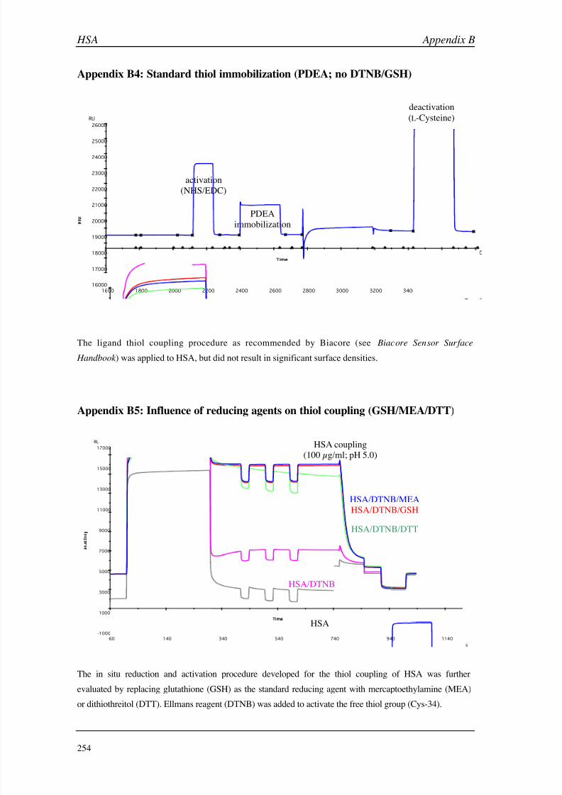
8/9/2019 DissB 7321 Mirroring
http://slidepdf.com/reader/full/dissb-7321-mirroring 268/278
HSA Appendix B
254
Appendix B4: Standard thiol immobilization (PDEA; no DTNB/GSH)
The ligand thiol coupling procedure as recommended by Biacore (see Biacore Sensor Surface
Handbook ) was applied to HSA, but did not result in significant surface densities.
Appendix B5: Influence of reducing agents on thiol coupling (GSH/MEA/DTT)
The in situ reduction and activation procedure developed for the thiol coupling of HSA was further
evaluated by replacing glutathione (GSH) as the standard reducing agent with mercaptoethylamine (MEA)
or dithiothreitol (DTT). Ellmans reagent (DTNB) was added to activate the free thiol group (Cys-34).
16000
17000
18000
19000
20000
21000
22000
23000
24000
25000
26000
1600 1800 2000 2200 2400 2600 2800 3000 3200 3400 3600 380
im s
RU
s
p
o
n
s
activation
(NHS/EDC)
PDEA
immobilization
HSA coupling
(100 µg/ml; pH 5.0)
deactivation
(L-Cysteine)
-1000
1000
3000
5000
7000
9000
11000
13000
15000
17000
-60 140 340 540 740 940 1140
sim
RU
s
p
o
n
s
HSA
HSA/DTNB
HSA/DTNB/DTT
HSA/DTNB/MEA
HSA/DTNB/GSH
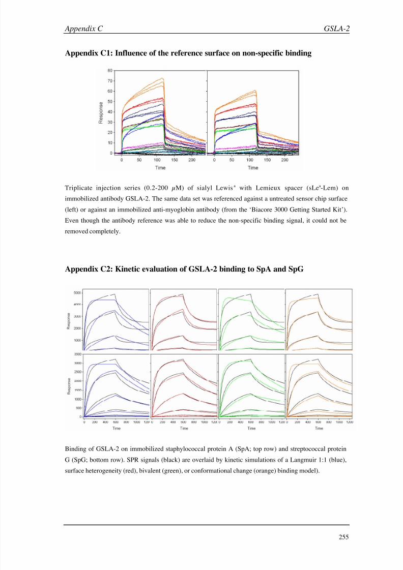
8/9/2019 DissB 7321 Mirroring
http://slidepdf.com/reader/full/dissb-7321-mirroring 269/278

8/9/2019 DissB 7321 Mirroring
http://slidepdf.com/reader/full/dissb-7321-mirroring 270/278
ASGP-R Appendix D
256
Appendix D1: H1-CRD data from ProtParam (www.expasy.org)
Parameters with the N -terminal methionine residue:
Parameters after removal of the terminal methionine residue:
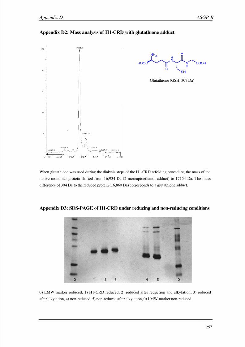
8/9/2019 DissB 7321 Mirroring
http://slidepdf.com/reader/full/dissb-7321-mirroring 271/278
Appendix D ASGP-R
257
Appendix D2: Mass analysis of H1-CRD with glutathione adduct
When glutathione was used during the dialysis steps of the H1-CRD refolding procedure, the mass of the
native monomer protein shifted from 16,934 Da (2-mercaptoethanol adduct) to 17154 Da. The mass
difference of 304 Da to the reduced protein (16,860 Da) corresponds to a glutathione adduct.
Appendix D3: SDS-PAGE of H1-CRD under reducing and non-reducing conditions
0) LMW marker reduced, 1) H1-CRD reduced, 2) reduced after reduction and alkylation, 3) reduced
after alkylation, 4) non-reduced, 5) non-reduced after alkylation, 0) LMW marker non-reduced
HOOC
H
N NH
COOH
NH2
OSH
O
Glutathione (GSH; 307 Da)
2 531 40 0
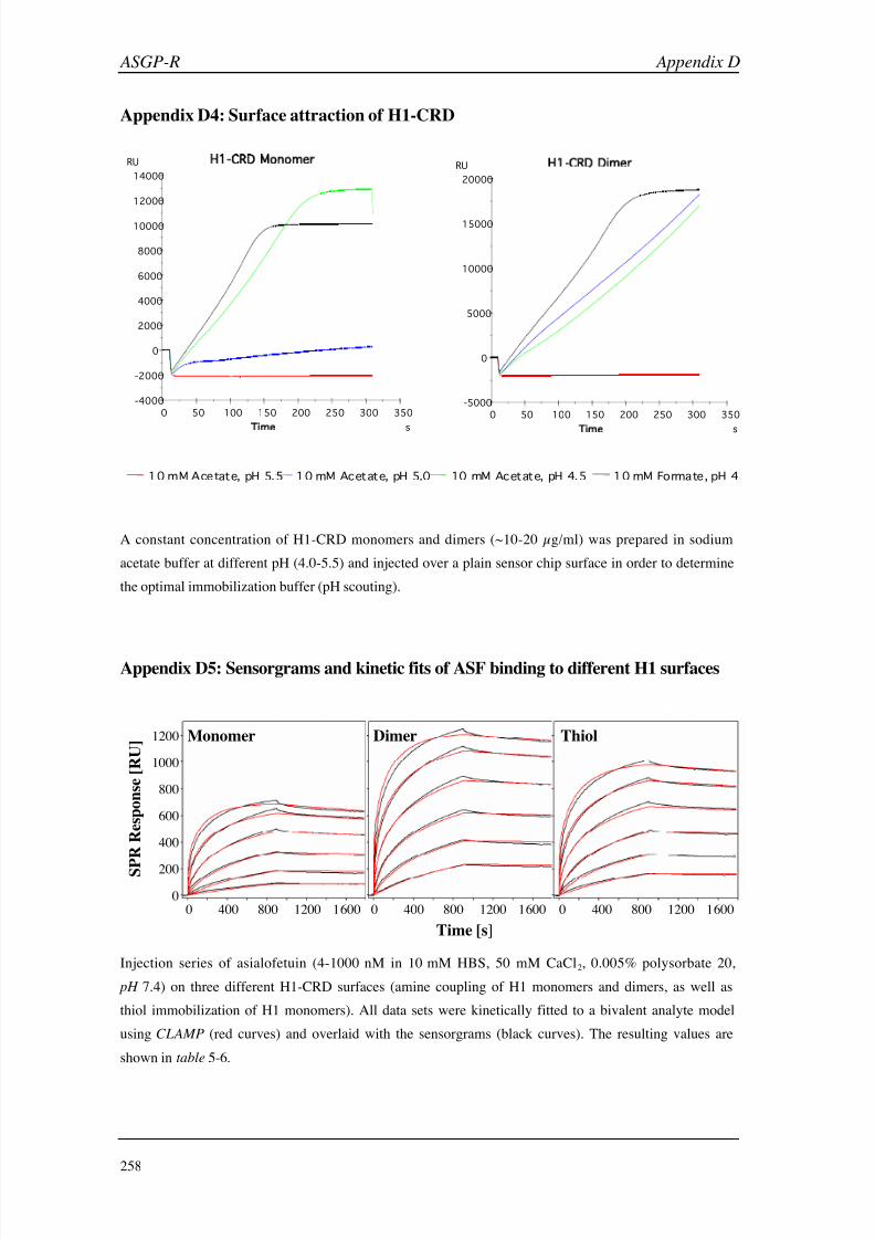
8/9/2019 DissB 7321 Mirroring
http://slidepdf.com/reader/full/dissb-7321-mirroring 272/278
ASGP-R Appendix D
258
Appendix D4: Surface attraction of H1-CRD
A constant concentration of H1-CRD monomers and dimers (~10-20 µg/ml) was prepared in sodium
acetate buffer at different pH (4.0-5.5) and injected over a plain sensor chip surface in order to determine
the optimal immobilization buffer (pH scouting).
Appendix D5: Sensorgrams and kinetic fits of ASF binding to different H1 surfaces
0 400 800 1200 1600 0 400 800 1200 1600 0 400 800 1200 1600
0
200
400
600
800
1000
1200
S P R R
e s p o n s e [ R U ]
Time [s]
Monomer Dimer Thiol
Injection series of asialofetuin (4-1000 nM in 10 mM HBS, 50 mM CaCl2, 0.005% polysorbate 20,
pH 7.4) on three different H1-CRD surfaces (amine coupling of H1 monomers and dimers, as well as
thiol immobilization of H1 monomers). All data sets were kinetically fitted to a bivalent analyte model
using CLAMP (red curves) and overlaid with the sensorgrams (black curves). The resulting values are
shown in table 5-6.
-5000
0
5000
10000
15000
20000
0 50 100 150 200 250 300 350
H1 CRD DimerRU
Time
s
-4000
-2000
0
2000
4000
6000
8000
10000
12000
14000
0 50 100 150 200 250 300 350
H1 CRD MonomerRU
Time
s
10 mM Acetate, pH 5.5 10 mM Acetate, pH 5.0 10 mM Acetate, pH 4.5 10 mM Formate, pH 4

8/9/2019 DissB 7321 Mirroring
http://slidepdf.com/reader/full/dissb-7321-mirroring 273/278
Appendix D ASGP-R
259
Appendix D6: Sensorgrams of ASOR binding to H1-CRD
Binding of asialoorosomucoid (ASOR; 4-1000 nM in 10 mM HEPES pH 7.4, 50 mM CaCl2) to
immobilized H1-CRD dimer. In contrast to asialofetuin, ASOR shows a complex binding mode with a
biphasic association phase, and a negative equilibrium drift at high concentrations.
Appendix D7: Equilibrium response plots of monovalent galactose derivatives
Galactose Gal N Ac Galactosamine
Methyl -galactopyranoside Methyl -galactopyranoside Lactose
Glucose
Affinity analysis of galactose and derivatives (monovalent carbohydrates) after screening over immobilized
H1-CRD dimer. Triplicate injections between 5 µM and 5 mM were fitted to a single binding site model.
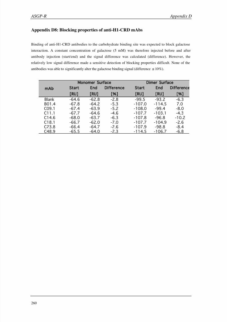
8/9/2019 DissB 7321 Mirroring
http://slidepdf.com/reader/full/dissb-7321-mirroring 274/278
ASGP-R Appendix D
260
Appendix D8: Blocking properties of anti-H1-CRD mAbs
Binding of anti-H1-CRD antibodies to the carbohydrate binding site was expected to block galactose
interaction. A constant concentration of galactose (5 mM) was therefore injected before and after
antibody injection (start/end) and the signal difference was calculated (difference). However, the
relatively low signal difference made a sensitive detection of blocking properties difficult. None of the
antibodies was able to significantly alter the galactose binding signal (difference 10%).
Start End Difference Start End Difference
[RU] [RU] [ ] [RU] [RU] [ ]
Blank -64.6 -62.8 -2.8 -99.5 -93.2 -6.3
B01.4 -67.8 -64.2 -5.3 -107.0 -114.5 7.0
C09.1 -67.4 -63.9 -5.2 -108.0 -99.4 -8.0
C11.1 -67.7 -64.6 -4.6 -107.7 -103.1 -4.3C14.6 -68.0 -63.7 -6.3 -107.8 -96.8 -10.2
C18.1 -66.7 -62.0 -7.0 -107.7 -104.9 -2.6
C23.8 -66.4 -64.7 -2.6 -107.9 -98.8 -8.4
C48.9 -65.5 -64.0 -2.3 -114.5 -106.7 -6.8
Dimer Surfaceonomer Surface
mAb

8/9/2019 DissB 7321 Mirroring
http://slidepdf.com/reader/full/dissb-7321-mirroring 275/278
Appendix E HisTag
261
Appendix E1: Mass transport analysis of the hexahistidine peptide
Mass transport analysis of the hexahistidine peptide (5 µM) at flow rates of 5 (black), 20 (blue), and
50 µl/min (red).
Appendix E2: Non-specific binding of HisAla peptides after standard purification
When the HisAla peptides were purified on HPLC using under standard conditions (water, acetonitrile,
0.1% TFA), the peptides eluted close to the injection peak. Fractions from such separation steps caused a
significant non-specific signal on the NTA sensor chip (red sensorgrams). The shift to basic elution
conditions (ammonium acetate pH 8.8) allowed the collection of pure peptide fractions, which showed a
clear SPR binding signal (blue sensorgrams).
Blank HisAla1 HisAla2
HisAla3 HisAla4 HisAla5

8/9/2019 DissB 7321 Mirroring
http://slidepdf.com/reader/full/dissb-7321-mirroring 276/278
8/9/2019 DissB 7321 Mirroring
http://slidepdf.com/reader/full/dissb-7321-mirroring 277/278
DA N I E L R I C K L I N
Metzerstrasse 29 • CH-4056 Basel • Phone +41 613821556 • email: [email protected]
CURRICULUM VITAE
Date and place of birth 26
September 1974, ZurichNationality Swiss
Education
2001 - 2005 University of Basel, Institute of Molecular Pharmacy
PhD thesis in the group of Prof. B. Ernst
‘Surface Plasmon Resonance Applications in Drug
Discovery (with an emphasis on small molecule and low
affinity systems)’
1993 - 1999 Swiss Federal Institute of Technology (ETH Zurich)
Diploma in Pharmaceutical Sciences
1988 - 1993 Kantonsschule Wiedikon (grammar school), Zurich
Practical Experience
2000 Proteome Center Rostock, Germany
Internship in the group of Prof. M. O. GlockerStudy about ‘Analysis of antibody profiles in
autoimmune diseases using mass spectrometry’
Development of a database for proteomic data
1998 (April – May) Tumor Biology Center, Freiburg, Germany
Practical course in pharmaceutical chemistry
in the group of Dr. F. Kratz
Development of macromolecular anticancer drugs
1996 - 1997 Albis Apotheke, ZurichPractical year in a public pharmacy
Additional Skills
Languages German: native speaker
English: CAE; certificate in scientific writing
French: working knowledge
PC skills Advanced knowledge in Windows, MacOS, Unix, MS
Office applications, database, internet, HTML

8/9/2019 DissB 7321 Mirroring
http://slidepdf.com/reader/full/dissb-7321-mirroring 278/278
DA N I E L R I C K L I N
Metzerstrasse 29 • CH-4056 Basel • Phone +41 613821556 • email: [email protected]
Scientific Qualifications
Papers The following results from my thesis have been submitted or are in the final stage of
Related Documents