Disruption of Mouse Cenpj, a Regulator of Centriole Biogenesis, Phenocopies Seckel Syndrome Rebecca E. McIntyre 1 , Pavithra Lakshminarasimhan Chavali 2 , Ozama Ismail 3. , Damian M. Carragher 3. , Gabriela Sanchez-Andrade 4. , Josep V. Forment 5 , Beiyuan Fu 6 , Martin Del Castillo Velasco-Herrera 1 , Andrew Edwards 7 , Louise van der Weyden 1 , Fengtang Yang 6 , Sanger Mouse Genetics Project 3 , Ramiro Ramirez-Solis 3 , Jeanne Estabel 3 , Ferdia A. Gallagher 8 , Darren W. Logan 4 , Mark J. Arends 9 , Stephen H. Tsang 10,11 , Vinit B. Mahajan 10 , Cheryl L. Scudamore 12 , Jacqueline K. White 3 , Stephen P. Jackson 5 , Fanni Gergely 2 , David J. Adams 1 * 1 Experimental Cancer Genetics, Wellcome Trust Sanger Institute, Hinxton, United Kingdom, 2 Cancer Research UK Cambridge Research Institute, Li Ka Shing Centre and Department of Oncology, University of Cambridge, Cambridge, United Kingdom, 3 Mouse Genetics Project, Wellcome Trust Sanger Institute, Hinxton, United Kingdom, 4 Genetics of Instinctive Behaviour, Wellcome Trust Sanger Institute, Hinxton, United Kingdom, 5 The Gurdon Institute and Department of Biochemistry, University of Cambridge, Cambridge, United Kingdom, 6 Molecular Cytogenetics, Wellcome Trust Sanger Institute, Hinxton, United Kingdom, 7 Wellcome Trust Center for Human Genetics, Oxford, United Kingdom, 8 Department of Radiology, Addenbrooke’s Hospital, University of Cambridge, Cambridge, United Kingdom, 9 Department of Pathology, Addenbrooke’s Hospital, University of Cambridge, Cambridge, United Kingdom, 10 Department of Ophthalmology and Visual Sciences, University of Iowa, Iowa City, Iowa, United States of America, 11 Bernard and Shirlee Brown Glaucoma Laboratory, Department of Ophthalmology, College of Physicians and Surgeons, Columbia University, New York, New York, United States of America, 12 Department of Pathology and Infectious Diseases, Royal Veterinary College, London, United Kingdom Abstract Disruption of the centromere protein J gene, CENPJ (CPAP, MCPH6, SCKL4), which is a highly conserved and ubiquitiously expressed centrosomal protein, has been associated with primary microcephaly and the microcephalic primordial dwarfism disorder Seckel syndrome. The mechanism by which disruption of CENPJ causes the proportionate, primordial growth failure that is characteristic of Seckel syndrome is unknown. By generating a hypomorphic allele of Cenpj, we have developed a mouse (Cenpj tm/tm ) that recapitulates many of the clinical features of Seckel syndrome, including intrauterine dwarfism, microcephaly with memory impairment, ossification defects, and ocular and skeletal abnormalities, thus providing clear confirmation that specific mutations of CENPJ can cause Seckel syndrome. Immunohistochemistry revealed increased levels of DNA damage and apoptosis throughout Cenpj tm/tm embryos and adult mice showed an elevated frequency of micronucleus induction, suggesting that Cenpj-deficiency results in genomic instability. Notably, however, genomic instability was not the result of defective ATR-dependent DNA damage signaling, as is the case for the majority of genes associated with Seckel syndrome. Instead, Cenpj tm/tm embryonic fibroblasts exhibited irregular centriole and centrosome numbers and mono- and multipolar spindles, and many were near-tetraploid with numerical and structural chromosomal abnormalities when compared to passage-matched wild-type cells. Increased cell death due to mitotic failure during embryonic development is likely to contribute to the proportionate dwarfism that is associated with CENPJ-Seckel syndrome. Citation: McIntyre RE, Lakshminarasimhan Chavali P, Ismail O, Carragher DM, Sanchez-Andrade G, et al. (2012) Disruption of Mouse Cenpj, a Regulator of Centriole Biogenesis, Phenocopies Seckel Syndrome. PLoS Genet 8(11): e1003022. doi:10.1371/journal.pgen.1003022 Editor: Veronica van Heyningen, Medical Research Council Human Genetics Unit, United Kingdom Received December 1, 2011; Accepted August 23, 2012; Published November 15, 2012 Copyright: ß 2012 McIntyre et al. This is an open-access article distributed under the terms of the Creative Commons Attribution License, which permits unrestricted use, distribution, and reproduction in any medium, provided the original author and source are credited. Funding: This work was supported by the Wellcome Trust (grant number 098051; DMC, OI, GS-A, SMGP, DWL, JKW, DJA), Cancer Research UK (REM, PLC, MDCV-H, LvdW, FG, DJA, MJA), a Royal Society University Research Fellowship (FG), le Fondation Je ´ro ˆme Lejeune (AE), Research to Prevent Blindness (NIH 1K08EY020530-01A1; SHT, VBM), and the MRC (CLS). The SPJ Laboratory is supported by grants from Cancer Research UK, the European Union, and the European Research Council, and is made possible by core infrastructure funding from Cancer Research UK and the Wellcome Trust. The funders had no role in study design, data collection and analysis, decision to publish, or preparation of the manuscript. Competing Interests: The authors have declared that no competing interests exist. * E-mail: [email protected] . These authors contributed equally to this work. Introduction Seckel syndrome is a clinically and genetically heterogeneous primordial dwarfism disorder that is characterised by intrauterine growth retardation, postnatal dwarfism, severe microcephaly, mental retardation, a prominent curved nose and receding chin, together with other clinical abnormalities [1,2,3]. Mutations in five loci have been linked with Seckel syndrome: SCKL1 and SCKL2 are due to mutation of the genes for the DNA damage response proteins ATR and CtIP (RBBP8), respectively; SCKL4 and SCKL5 are due to mutation of the genes for the centrosomal proteins CENPJ (Centromere protein J, or centrosomal P4.1-associated protein, CPAP; Figure 1A) and CEP152; while the gene responsible for SCKL3 is currently unknown [4,5,6,7]. Mutations in PCNT (pericentrin), another centrosomal protein, have been associated with both Seckel syndrome and the overlapping PLOS Genetics | www.plosgenetics.org 1 November 2012 | Volume 8 | Issue 11 | e1003022

Welcome message from author
This document is posted to help you gain knowledge. Please leave a comment to let me know what you think about it! Share it to your friends and learn new things together.
Transcript
Disruption of Mouse Cenpj, a Regulator of CentrioleBiogenesis, Phenocopies Seckel SyndromeRebecca E. McIntyre1, Pavithra Lakshminarasimhan Chavali2, Ozama Ismail3., Damian M. Carragher3.,
Gabriela Sanchez-Andrade4., Josep V. Forment5, Beiyuan Fu6, Martin Del Castillo Velasco-Herrera1,
Andrew Edwards7, Louise van der Weyden1, Fengtang Yang6, Sanger Mouse Genetics Project3,
Ramiro Ramirez-Solis3, Jeanne Estabel3, Ferdia A. Gallagher8, Darren W. Logan4, Mark J. Arends9,
Stephen H. Tsang10,11, Vinit B. Mahajan10, Cheryl L. Scudamore12, Jacqueline K. White3,
Stephen P. Jackson5, Fanni Gergely2, David J. Adams1*
1 Experimental Cancer Genetics, Wellcome Trust Sanger Institute, Hinxton, United Kingdom, 2 Cancer Research UK Cambridge Research Institute, Li Ka Shing Centre and
Department of Oncology, University of Cambridge, Cambridge, United Kingdom, 3 Mouse Genetics Project, Wellcome Trust Sanger Institute, Hinxton, United Kingdom,
4 Genetics of Instinctive Behaviour, Wellcome Trust Sanger Institute, Hinxton, United Kingdom, 5 The Gurdon Institute and Department of Biochemistry, University of
Cambridge, Cambridge, United Kingdom, 6 Molecular Cytogenetics, Wellcome Trust Sanger Institute, Hinxton, United Kingdom, 7 Wellcome Trust Center for Human
Genetics, Oxford, United Kingdom, 8 Department of Radiology, Addenbrooke’s Hospital, University of Cambridge, Cambridge, United Kingdom, 9 Department of
Pathology, Addenbrooke’s Hospital, University of Cambridge, Cambridge, United Kingdom, 10 Department of Ophthalmology and Visual Sciences, University of Iowa,
Iowa City, Iowa, United States of America, 11 Bernard and Shirlee Brown Glaucoma Laboratory, Department of Ophthalmology, College of Physicians and Surgeons,
Columbia University, New York, New York, United States of America, 12 Department of Pathology and Infectious Diseases, Royal Veterinary College, London, United
Kingdom
Abstract
Disruption of the centromere protein J gene, CENPJ (CPAP, MCPH6, SCKL4), which is a highly conserved and ubiquitiouslyexpressed centrosomal protein, has been associated with primary microcephaly and the microcephalic primordial dwarfismdisorder Seckel syndrome. The mechanism by which disruption of CENPJ causes the proportionate, primordial growthfailure that is characteristic of Seckel syndrome is unknown. By generating a hypomorphic allele of Cenpj, we havedeveloped a mouse (Cenpjtm/tm) that recapitulates many of the clinical features of Seckel syndrome, including intrauterinedwarfism, microcephaly with memory impairment, ossification defects, and ocular and skeletal abnormalities, thusproviding clear confirmation that specific mutations of CENPJ can cause Seckel syndrome. Immunohistochemistry revealedincreased levels of DNA damage and apoptosis throughout Cenpjtm/tm embryos and adult mice showed an elevatedfrequency of micronucleus induction, suggesting that Cenpj-deficiency results in genomic instability. Notably, however,genomic instability was not the result of defective ATR-dependent DNA damage signaling, as is the case for the majority ofgenes associated with Seckel syndrome. Instead, Cenpjtm/tm embryonic fibroblasts exhibited irregular centriole andcentrosome numbers and mono- and multipolar spindles, and many were near-tetraploid with numerical and structuralchromosomal abnormalities when compared to passage-matched wild-type cells. Increased cell death due to mitotic failureduring embryonic development is likely to contribute to the proportionate dwarfism that is associated with CENPJ-Seckelsyndrome.
Citation: McIntyre RE, Lakshminarasimhan Chavali P, Ismail O, Carragher DM, Sanchez-Andrade G, et al. (2012) Disruption of Mouse Cenpj, a Regulator ofCentriole Biogenesis, Phenocopies Seckel Syndrome. PLoS Genet 8(11): e1003022. doi:10.1371/journal.pgen.1003022
Editor: Veronica van Heyningen, Medical Research Council Human Genetics Unit, United Kingdom
Received December 1, 2011; Accepted August 23, 2012; Published November 15, 2012
Copyright: � 2012 McIntyre et al. This is an open-access article distributed under the terms of the Creative Commons Attribution License, which permitsunrestricted use, distribution, and reproduction in any medium, provided the original author and source are credited.
Funding: This work was supported by the Wellcome Trust (grant number 098051; DMC, OI, GS-A, SMGP, DWL, JKW, DJA), Cancer Research UK (REM, PLC, MDCV-H,LvdW, FG, DJA, MJA), a Royal Society University Research Fellowship (FG), le Fondation Jerome Lejeune (AE), Research to Prevent Blindness (NIH 1K08EY020530-01A1;SHT, VBM), and the MRC (CLS). The SPJ Laboratory is supported by grants from Cancer Research UK, the European Union, and the European Research Council, and ismade possible by core infrastructure funding from Cancer Research UK and the Wellcome Trust. The funders had no role in study design, data collection and analysis,decision to publish, or preparation of the manuscript.
Competing Interests: The authors have declared that no competing interests exist.
* E-mail: [email protected]
. These authors contributed equally to this work.
Introduction
Seckel syndrome is a clinically and genetically heterogeneous
primordial dwarfism disorder that is characterised by intrauterine
growth retardation, postnatal dwarfism, severe microcephaly,
mental retardation, a prominent curved nose and receding chin,
together with other clinical abnormalities [1,2,3]. Mutations in five
loci have been linked with Seckel syndrome: SCKL1 and SCKL2
are due to mutation of the genes for the DNA damage response
proteins ATR and CtIP (RBBP8), respectively; SCKL4 and SCKL5
are due to mutation of the genes for the centrosomal proteins
CENPJ (Centromere protein J, or centrosomal P4.1-associated
protein, CPAP; Figure 1A) and CEP152; while the gene
responsible for SCKL3 is currently unknown [4,5,6,7]. Mutations
in PCNT (pericentrin), another centrosomal protein, have been
associated with both Seckel syndrome and the overlapping
PLOS Genetics | www.plosgenetics.org 1 November 2012 | Volume 8 | Issue 11 | e1003022

dwarfism disorder, microcephalic osteodysplastic primordial
dwarfism type II (MOPDII) [8,9,10]. Interestingly, mutations in
the centrosomal proteins CEP152 (MCPH4) and CENPJ (MCPH6),
which are thought to interact with each other during centriole
biogenesis [11,12], have also been associated with primary
autosomal recessive microcephaly, a genetically heterogeneous
condition caused by mutation of one of eight loci (MCPH1–8;
[13,14,15]) result in clinically indistinguishable features that
include mental retardation and a severely reduced brain size of
greater than two standard deviations below the average. Primary
autosomal recessive microcephaly presents at birth or becomes
apparent within the first few years of life [16]. Primary
microcephaly is thought to be caused by a reduction in
neurogenesis while the proportionate dwarfism of Seckel syndrome
is thought to be the result of premature death of proliferating cells;
it is not clear why different mutations in centrosomal proteins
cause Seckel syndrome, MOPDII or primary microcephaly.
CENPJ is a conserved, ubiquitously expressed centrosomal
protein with a key role in centriole biogenesis [17,18,19,20,21].
The centrosome is a major microtubule organizing centre in
somatic cells that undergoes a duplication cycle that is tightly
coupled with DNA replication (reviewed by [22] and [23]). Briefly,
in G1 the centrosome is composed of a pair of loosely connected
centrioles that are embedded in a proteinaceous matrix. In concert
with DNA replication, a single procentriole forms next to each
parental centriole during S-phase. The procentrioles continue to
elongate and by the onset of mitosis two centrosomes are present,
each comprising an older and younger centriole. The two
centrosomes aid the formation of the poles of the bipolar spindle,
the molecular machinery responsible for correct segregation of
sister chromatids into daughter cells. Centrosome attachment to
the poles also ensures that each daughter cell inherits a single
centrosome, thus tightly regulating ploidy and centrosome
numbers. Impaired centrosome duplication cycles or a failure of
centrosome segregation result in abnormal centrosome numbers
that in turn perturb bipolar spindle assembly and chromosome
segregation [24].
CENPJ contains 17 exons and encodes a 1338 amino acid
residue protein with a chromosomal segregation ATPase domain
and a T-complex protein 10 (TCP10)-like C-terminal domain.
Seckel-syndrome of a consanguineous Saudi Arabian family has
been associated with a homozygous splice acceptor mutation in the
last nucleotide of CENPJ intron 11 (Figure 1A) that results in the
production of three transcripts lacking either exon 12, exons 11
and 12 or exons 11, 12 and 13 [4]. Three CENPJ-microcephaly
mutations in three consanguineous Pakistani families have been
reported to date and all are predicted to cause a truncating stop
codon (Figure 1A; [14,15]).
ATR, RBBP and CEP152 have been shown to play a role in
maintaining genomic stability through regulation of the DNA
damage response [5,6,25], however such a role has not yet been
defined for CENPJ. We set out to develop a mouse model of
CENPJ-Seckel syndrome in order to establish the mechanism by
which mutation of CENPJ results in this subtype of primordial
dwarfism. We show that the Cenpj hypomorphic mouse that we
created recapitulates many key features of Seckel syndrome,
including microcephaly with memory impairment, dwarfism from
birth, and skeletal abnormalities. We further establish that wide-
scale genomic instability is the likely cause of cell death within
Cenpjtm/tm embryos and suggest that this contributes to the
developmental phenotypes observed in CENPJ-Seckel patients.
Results
Generation and phenotyping of a Cenpj hypomorphicmouse
Knockout mice carrying the Cenpjtm1a(EUCOMM)Wtsi allele
(Figure 1A and Figure S1A) were generated on a C57BL/6NTac;
C57BL/6-Tyrc-Brd background by the Sanger Mouse Genetics
Project as part of the European Conditional Mouse Mutagenesis
Program (EUCOMM; [26]). Correct gene targeting in founder
mice was determined by a combination of standard and
quantitative PCR (Figure S1). LacZ staining was detected in the
brain and kidneys, while strong staining was present in the testes of
mice heterozygous for the Cenpjtm1a(EUCOMM)Wtsi allele (Figure S2A).
The tm1a(EUCOMM)Wtsi gene-trap cassette that was intro-
duced into the Cenpj locus is designed to truncate mRNA
expression and to generate out-of-frame products following the
deletion of a critical exon. Previous studies have indicated that
mRNAs of certain microcephaly-associated genes are very stable
[27] prompting us to perform a detailed analysis of expression and
splicing at the Cenpjtm1a(EUCOMM)Wtsi locus. We generated
Cenpjtm1a(EUCOMM)Wtsi/tm1a(EUCOMM)Wtsi (Cenpjtm/tm) mouse embryon-
ic fibroblasts (MEFs; 13.5 d.p.c.) and performed SYBR Green
qPCR on cDNA using primers spanning the boundaries between
different exons (Figure 1A). We observed a low but detectable
amount of splicing over the gene-trap cassette in Cenpjtm/tm MEFs
(2.160.5% of wildtype exon 4–5 levels) and immunoblotting
(Figure 1B) confirmed the production of low levels of apparently
full-length Cenpj protein [27]. Splicing from exons 3 to 6 and 4 to
6 was detected in both Cenpjtm/tm and wildtype MEFs (Figure S2B).
Between exons 3 and 6 the level of splicing detected in Cenpjtm/tm
MEFs was increased relative to the levels in control MEFs
(444695%), while decreased levels of splicing were observed
between exons 4 and 6 (2.160.5%). Using the web-based ExPASy
translation tool (http://web.expasy.org/translate/) we predict that
mRNAs that are spliced between exons 3–6 and exons 4–6 lead to
the production of proteins truncated in exon 6 (Figure S2C).
Upstream of the tm1a(EUCOMM)Wtsi cassette (exons 1–2) Cenpj
mRNA levels were 68619% of wildtype levels. Downstream (from
exon 6 to 17) of the tm1a(EUCOMM)Wtsi cassette Cenpj mRNA
levels were approximately 20% of the levels observed in MEFs
from wildtype littermates (mean6SEM, n = 3). In summary,
Cenpjtm/tm MEFs are able to produce small amounts of full-length
Cenpj protein due to splicing over the tm1a(EUCOMM)Wtsi
gene-trap cassette (exons 4–5) and we predict that small amounts
of truncated, N-terminal Cenpj protein (corresponding to exons 1
to 3 or 1 to 4) will also be produced.
Author Summary
Mutation of the gene CENPJ has been found to causeprimary microcephaly, an inherited disorder that is char-acterised by severely reduced brain size. More recently,mutation of CENPJ has been associated with Seckelsyndrome, a disorder that is characterised by a severereduction in both brain and body size that is apparent atbirth, mental retardation, and skeletal abnormalities, inaddition to a number of other clinical manifestations. Here,we have generated a mouse that expresses only low levelsof mouse Cenpj protein and find that it recapitulates manyof the key features of Seckel syndrome. Moreover, we findthat errors during the proliferation of Cenpjtm/tm cellsfrequently lead to abnormal numbers of chromosomes ordamaged chromosomes, which is likely to be the cause ofincreased cell death during embryonic development and tocontribute to the proportionate dwarfism that is character-istic of Seckel syndrome.
Disruption of Cenpj Phenocopies Seckel Syndrome
PLOS Genetics | www.plosgenetics.org 2 November 2012 | Volume 8 | Issue 11 | e1003022
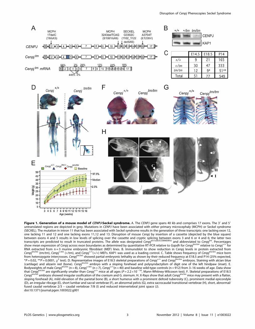
Figure 1. Generation of a mouse model of CENPJ-Seckel syndrome. A. The CENPJ gene spans 40 kb and comprises 17 exons. The 39 and 59
untranslated regions are depicted in grey. Mutations in CENPJ have been associated with either primary microcephaly (MCPH) or Seckel syndrome(SECKEL). The mutation in intron 11 that has been associated with Seckel syndrome results in the generation of three transcripts: one lacking exon 12,one lacking 11 and 12 and one lacking exons 11,12 and 13. Disruption of mouse Cenpj by insertion of a cassette (depicted by the blue square)between exons 4 and 5 results in low levels of splicing over the cassette and cryptic splicing between exons 3 and 6 or 4 and 6; the latter twotranscripts are predicted to result in truncated proteins. The allele was designated Cenpjtm1a(EUCOMM)Wtsi and abbreviated to Cenpjtm. Percentagesshow mean expression of Cenpj across exon boundaries as determined by quantitative RT-PCR relative to Gapdh for Cenpjtm/tm relative to Cenpj+/+ forRNA extracted from n = 3 murine embryonic fibroblast (MEF) lines. B. Immunoblot to show reduction in Cenpj levels in protein extracted fromCenpjtm/tm (tm/tm), Cenpj+/tm (+/tm), and Cenpj+/+(+/+) MEFs. KAP1 was used as a loading control. C. Table shows frequency of Cenpjtm/tm mice bornfrom heterozygote intercrosses. Cenpjtm/tm showed partial embryonic lethality as shown by their reduced frequency at E18.5 and P14 (25% expected,*P = 0.02, **P = 0.0001, x2 test). D. Representative images of E18.5 skeletal preparations of Cenpj+/+ and Cenpjtm/tm embryos. Staining with alcian blue(cartilage) and alizarin red (bone). Cenpjtm/tm embryo with a sloping forehead and polysyndactylism of digit one of the left hindpaw (inset). E.Bodyweights of male Cenpjtm/tm (n = 8), Cenpj+/tm (n = 7), Cenpj+/+(n = 40) and baseline wild-type controls (n = 912) from 3–16 weeks of age. Data showthat Cenpjtm/tm are significantly smaller than Cenpj+/+ mice at all ages (P = 2.2610216, Mann-Whitney-Wilcoxon test). F. Skeletal preparations of E18.5Cenpjtm/tm embryos showed irregular ossification of the cranium and G. sternum. H. X-Rays show that adult Cenpjtm/tm mice may present with a flatter,sloping forehead (A), mild elevation of the parietal bone (B), a short humerus with a prominent deltoid tuberosity (C), prominent medial epicondyle(D), an irregular ribcage (E), short lumbar and sacral vertebrae (F), an abnormal pelvis (G), extra sacrocaudal transitional vertebrae (H), short, abnormal/fused caudal vertebrae 2/3 – caudal vertebrae 7/8 (I) and reduced intervertebral joint space (J).doi:10.1371/journal.pgen.1003022.g001
Disruption of Cenpj Phenocopies Seckel Syndrome
PLOS Genetics | www.plosgenetics.org 3 November 2012 | Volume 8 | Issue 11 | e1003022

Phenotyping of mice was performed at the Wellcome Trust
Sanger Institute (http://www.sanger.ac.uk/mouseportal/
search?query = cenpj). Cenpjtm1a(EUCOMM)Wtsi/+ intercrosses gave
close to the expected Mendelian frequency (25%) of homozygote
embryos at E14.5 (23.5%); however, by E18.5 this had reduced to
11.6%, suggesting that disruption of Cenpj causes partial embryonic
lethality between E14.5 and E18.5 (x2 test, P = 0.02; Figure 1C).
The majority of runted pups identified between P0 and P21 were
Cenpjtm/tm (22% of Cenpjtm/tm offspring vs. 4.7% for Cenpj+/tm, and
2.5% for Cenpj+/+). Stunted growth was unlikely to be the result of
a major feeding problem, since milk spots were observed in the
stomachs of pups of all genotypes at P0. At P14, the frequency of
Cenpjtm/tm mice was not significantly different to that found at
E18.5 (P14: 9.2% vs. E18.5: 11.6%; Figure 1C), suggesting that
although dwarfed, Cenpjtm/tm mice are not postnatally sub-viable.
Cenpj-deficiency causes intrauterine and postnatalgrowth retardation
One of the defining characteristics of primordial dwarfism
disorders, such as Seckel syndrome, is a fetus that is small for its
gestational age with postnatal growth retardation [8,28,29];
specifically, the Saudi Arabian CENPJ-Seckel kindred all have
anthropometric values at least seven standard deviations below the
mean [4]. Cenpjtm/tm mice showed intrauterine growth retardation
(Figure 1D; mean 6 SEM bodyweight at E18.5, Cenpj+/+
1.1260.03 g, Cenpjtm/tm 0.860.04 g; P = 0.0001, t-test; crown-
rump length at E18.5, Cenpj+/+ 23.560.26 mm, Cenpjtm/tm
20.760.54 mm; P = 0.0001, t-test). From 3–16 weeks, Cenpjtm/tm
mice were significantly smaller than wild-type controls
(P = 2.2610216, Mann-Whitney-Wilcoxon test; Figure 1E). The
body weight of adult Cenpjtm/tm animals was 64% of wild-type
controls (mean 6 SEM bodyweight at 16 weeks, Cenpj+/+
39.161.35 g, Cenpjtm/tm 25.461.34 g; P = 9.3610211, t-test;
Figure 1E) and length was 76% of controls (mean 6 SEM nose-
to-tail base length at 14 weeks, Cenpj+/+10.460.05 cm, Cenpjtm/tm
7.960.07; P = 2.2610216, t-test; Figure S2D).
Skeletal abnormalities and abnormal ossification of bonefrom Cenpjtm/tm mice
The skeletal abnormalities of Seckel syndrome associated with
an intron 11 mutation in CENPJ include a receding chin, high
forehead and prominent nasal spine [4]. Although the facial
features of the CENPJ-Seckel kindred (two siblings and three
cousins) were strikingly similar, the skeletal survey of sibling one
was largely normal and that of sibling two revealed 11 ribs instead
of 12 and a steep acetabular roof [4]. These findings highlight the
fact that the same mutation results in clinical heterogeneity and
prompted us to perform a thorough skeletal analysis of Cenpjtm/tm
embryos and adult mice; Cenpjtm/tm embryos had significantly
smaller skulls (Figure 1D and 1H; mean 6 SEM at E18.5: skull
length, Cenpj+/+ 9.860.12 mm, Cenpjtm/tm 9.360.17 mm,
P = 0.0379; inner canthal distance Cenpj+/+ 3.2260.05 mm,
Cenpjtm/tm 2.9660.09 mm, P = 0.0198) and adult mice presented
with a flatter, sloping forehead and mild elevation of the parietal
bone compared to controls (Figure 1H; 16 weeks of age, n = 8
males and n = 7 females). Although the Saudi Arabian CENPJ-
Seckel kindred do not have clinodactyly (curvature of the fifth
finger), it is a frequently reported characteristic of Seckel patients
[1,4,29,30]. We did not observe clinodactyly in Cenpjtm/tm mice,
however we noted polysyndactylism of the first digit of the left hind
paw in 2/9 Cenpjtm/tm embryos (Figure 1D, inset), which has also
been reported in mutant Pcnt (pericentrin) mice [31]. Furthermore,
retarded ossification and decreased bone age is reported in the
majority of cases of Seckel syndrome, although this clinical
abnormality was not specifically addressed for the CENPJ-Seckel
kindred [1]. A higher proportion of Cenpjtm/tm embryos showed
incomplete or irregular ossification of the parietal and occipital
bones when compared to controls (Figure 1F; 3/5 Cenpjtm/tm, 1/38
Cenpj+/tm, 1/13 Cenpj+/+). In addition, a subset of Seckel patients,
including a CENPJ-Seckel patient, have 11 ribs instead of the usual
12 [4,30,32]. Although all ribs were present in Cenpjtm/tm embryos,
we noted that the attachment of the ribs to the sternum followed
an irregular pattern that closely corresponded to the asymmetrical
distribution of ossification centers along the sternum (Figure 1G;
3/5 Cenpjtm/tm). Adult Cenpjtm/tm mice displayed an irregular
ribcage, with crowding of the ribs (Figure 1H; 9/15). Moreover,
a subset of Seckel patients have been reported to have bilateral
dislocation of the hips and elbows, with a decreased range of
motion at the elbows [1]. While we did not find any evidence of
dislocation we noted that the humeri of adult Cenpjtm/tm mice were
anatomically disproportionate when compared with those of wild-
type mice; the deltoid tuberosities were closer to the greater
tubercle when normalized to humeri length (mean6SEM.: right
humeri, Cenpj+/+ 49.660.28%, Cenpjtm/tm 47.160.93%; P = 0.02, t-
test; Figure 1H), and humeri were sometimes bowed (6/15) with a
very prominent medial epicondyle (11/15; Figure 1H). Further-
more, all adult Cenpjtm/tm mice (15/15) displayed an abnormal
pelvis that was wider at the iliac crests (Figure 1H, iliac crest
normalised to ischiac, mean6S.E.M.: Cenpj+/+ 69.761.18%,
Cenpjtm/tm 60.861.72%; P = 0.0003) and sometimes asymmetrical.
Finally, we observed that all Cenpjtm/tm mice had a reduced
intervertebral joint space in the lumbar and caudal regions
(Figure 1H). In general, lumbar and sacral vertebrae were shorter
and Cenpjtm/tm mice had one to two extra sacrocaudal transitional
vertebrae as a result. In 13/15 Cenpjtm/tm mice, Caudal 2/3 –
Caudal 7/8 were abnormal in morphology and fused (Figure 1H).
Neuropathological abnormalities and memoryimpairment
Microcephaly is one of the defining characteristics of Seckel
syndrome [1]. Microcephaly has been clinically defined as a head
circumference of at least two standard deviations below the normal
range; and in the case of Seckel syndrome associated with
mutations in intron 11 of CENPJ, head circumference is seven
standard deviations below the mean [4,33]. The average Cenpjtm/tm
mouse brain weight was two standard deviations below that of
control mice (Figure 2A; P = 0.0002, t-test). Although the two and
four year-old siblings with CENPJ-Seckel syndrome described to
date had relatively normal magnetic resonance imaging (MRI),
cranial MRI of adult patients with Seckel syndrome has revealed
several neuroanatomical abnormalities aside from a reduction in
brain volume [4,32,34,35]. We therefore assessed the area of brain
regions and the thickness of the neuronal layers of the adult mouse
brain (16 weeks; Figure S3). Although the patterning of the
hippocampal layers appeared normal, the length of the dentate
gyrus was significantly reduced in Cenpjtm/tm when compared to
Cenpj+/+ control mice (mean6SEM.: Cenpj+/+ 4380664 mm,
Cenpjtm/tm 37976181 mm, P = 0.01, t-test; Figure 2B). The average
thickness of the cortex, which is often reduced with mutation of
microcephaly genes in mice [36,37], and of the molecular,
striatum radiatum and oriens layers of the hippocampus were
not significantly different to wild-type controls. Similarly, the total
areas of the hippocampus, corpus callosum and dorsal third
ventricle were unchanged as was the total internal length of the
pyramidal cell layer.
History was suggestive of normal cognitive and motor
development for four of the five cases within the CENPJ-Seckel
Disruption of Cenpj Phenocopies Seckel Syndrome
PLOS Genetics | www.plosgenetics.org 4 November 2012 | Volume 8 | Issue 11 | e1003022
kindred while one patient clearly had intellectual impairment (IQ
60; MRI not performed [4]). Since the hippocampus is involved in
learning and memory formation and since Seckel patients
generally display learning impairments [3,34], we performed a
social recognition test with Cenpjtm/tm and control animals
[38,39,40]; Figure 2C and 2D). Thus, on day one, mice were
tested for habituation-dishabituation: male mice were presented
with a novel, anaesthetized stimulus mouse and the time of
investigation was recorded. Mice were then given a 10 minute
resting period before this was repeated a further three times with
the same stimulus mouse. On the fifth trial, mice were presented
with an unfamiliar stimulus mouse (Figure 2C). Both Cenpjtm/tm
(n = 7) and Cenpj+/+ (n = 7) mice recognized and habituated to the
novel stimulus mouse, as there was a decline in investigation time
over the first four trials that was recovered on trial five (Figure 2C,
two-way ANOVA, repeated measures for trial F4,48, = P,0.001,
effect for genotype F1,48 = 0.5482, P = 0.433, interaction
F4,48 = 0.09258, P = 0.9844), when they were exposed to a novel
mouse (Figure 2C. Trial four vs. trial five, P = 0.0033 and
P = 0.0074, post-hoc t-test). These data suggest that olfaction in
Cenpjtm/tm mice is not markedly affected. Twenty-four hours after
the habituation-dishabituation test, a discrimination-based olfac-
tory memory test was performed. When given a choice between
the familiar (same stimulus animal used for trials one to four) and a
new unfamiliar mouse, Cenpj+/+ animals spent less time investi-
gating the familiar mouse than the unfamiliar one (Figure 2D.
P = 0.0326, t-test). However, Cenpjtm/tm mice were less able to
recognize the familiar from the unfamiliar animal as shown by the
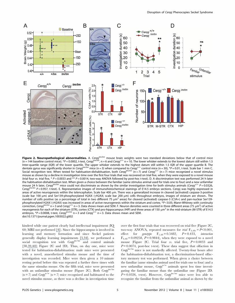
Figure 2. Neuropathological abnormalities. A. Cenpjtm/tm mouse brain weights were two standard deviations below that of control mice(n = 144 baseline control mice). *P = 0.0002, t-test, Cenpjtm/tm, n = 6 and Cenpj+/+ n = 10. The lower whisker extends to the lowest datum still within 1.5Inter-quartile range (IQR) of the lower quartile. The upper whisker extends to the highest datum still within 1.5 IQR of the upper quartile B. Thedentate gyrus was significantly shorter in Cenpjtm/tm mice (n = 3) when compared to Cenpj+/+ control mice (n = 30), *P = 0.01, t-test. Scale bar 1 mm C.Social recognition test. When tested for habituation-dishabituation, both Cenpjtm/tm (n = 7) and Cenpj+/+ (n = 7) mice recognized a novel stimulusmouse as shown by a decline in investigation time over the first four trials that was recovered on trial five, when they were exposed to a novel mouse(trial four vs. trial five, * P = 0.0033 and ** P = 0.0014, two-way ANOVA followed by post-hoc t-test). D. A discrimination test was performed 24 h laterthe habituation-dishabituation test. When given a choice between the familiar (same stimulus animal used for trials one to four) and a new unfamiliarmouse 24 h later, Cenpjtm/tm mice could not discriminate as shown by the similar investigation time for both stimulus animals (Cenpj+/+ P = 0.0326,Cenpjtm/tm P = 0.957, t-test). E. Representative images of immunohistochemical stainings of E14.5 embryo sections. Cenpj was highly expressed inareas of active neurogenesis within the telencephalon. Scale bar 400 mm. There was a generalized increase in cleaved (activated) caspase-3-positive(scale bar 100 mm) and Ser139-phosphorylated H2AX (cH2AX; scale bar 200 mm) cells throughout embryos, images of striatum are shown.. Thenumber of cells positive (as a percentage of total in two different 75 mm2 areas) for cleaved (activated) caspase-3 (C3A+) and pan-nuclear Ser139-phosphorylated H2AX (cH2AX) was increased in areas of active neurogenesis within the striatum and cortex. *P,0.05; Mann-Whitney with continuitycorrection, Cenpjtm/tm n = 3 and Cenpj+/+ n = 3. Data shows mean and SEM. F. Neuron densities were counted in three different areas (75 mm2) of activeneurogenesis for each of the striatum (STR), cortex (CTX) and pro-hippocampus (HIP) and three areas of 150 mm2 in the mid-striatum (M-STR) of E14.5embryos, *P = 0.0008, t-test, Cenpjtm/tm n = 3 and Cenpj+/+ n = 3. Data shows mean and SEM.doi:10.1371/journal.pgen.1003022.g002
Disruption of Cenpj Phenocopies Seckel Syndrome
PLOS Genetics | www.plosgenetics.org 5 November 2012 | Volume 8 | Issue 11 | e1003022
similar investigation time for both stimulus animals (Figure 2D.
P = 0.957, t-test; Normalized discrimination Cenpj+/+ vs Cenpjtm/tm,
P = 0.0417). In summary, short-term memory and olfaction
appear to be unaffected in Cenpjtm/tm mice, however long-term
memory was significantly impaired. All other tests of neurological
function were normal, including open field, grip strength, modified
SHIRPA (the SmithKline Beecham, Harwell, Imperial College,
Royal London Hospital, phenotype assessment is a set of
behavioural tests designed to test muscle, cerebellar, sensory and
neuropsychiatric function), auditory brainstem response and hot
plate assessment.
Increased DNA damage, apoptosis, and reduced neurondensity in Cenpjtm/tm mice
All of the genes associated with Seckel syndrome have so far
been shown to result in defective DNA damage responses and a
lowered apoptotic threshold [5,6,7,25,41]. To test whether Cenpj-
deficiency is associated with elevated levels of DNA strand breaks
and/or apoptosis, we performed immunohistochemical staining of
E14.5 embryos. Phosphorylation of histone H2AX on serine 139
(cH2AX) by the ATR, DNA-PK or ATM kinases occurs at sites
flanking DNA strand breaks and enhances the recruitment of
DNA repair proteins to sites of damage [42]; if the damage is
irreparable then the cell death cascade is normally activated [43].
Compared to controls, there was a general increase in the number
of cH2AX-positive cells throughout Cenpjtm/tm embryos (Figure S4)
and this was most pronounced in the developing telencephalon
(Figure 2E; mean6SEM. cH2AX-positive cells as a percentage of
total: striatum Cenpj+/+ 0.4760.2%, Cenpjtm/tm 2.360.3%,
P = 0.004; cortex Cenpj+/+ 0.0%, Cenpjtm/tm 1.360.3%, P = 0.01;
pro-hippocampus Cenpj+/+ 0.0%, Cenpjtm/tm 0.960.3%, P = 0.03.
Mann-Whitney with continuity correction). Similarly, there was an
increase in the number of cleaved caspase-3-positive cells
throughout Cenpjtm/tm embryos (Figure S4), although this was most
pronounced in areas of active neurogenesis (as determined by
Ki67 staining; Figure S4) within the telencephalon, where Cenpj
was most highly expressed (Figure 2E; mean6S.E.M. cleaved
caspase-3-positive cells as a percentage of total cells: striatum
Cenpj+/+ 0.0%, Cenpjtm/tm 1.960.2%, P = 0.001; cortex Cenpj+/+
0.0%, Cenpjtm/tm 0.660.2%, P = 0.05; pro-hippocampus Cenpj+/+
0.0%, Cenpjtm/tm 0.560.3%, P = 0.14, t-test). There was no
detectable difference in the patterns of cellular proliferation
between Cenpjtm/tm and Cenpj+/+ embryos when examined using
the marker Ki67 (Figure S4).
Consistent with increased levels of apoptosis in the developing
telencephalon of Cenpjtm/tm embryos, reports of fetal stage Seckel
syndrome (loci responsible unknown) have shown reduced neuron
density and disorganization of cortical layers at 30 weeks gestation
[28,29]. We therefore quantified neuron densities in areas of active
neurogenesis within the telencephalon (areas of Ki67-positive
staining; Figure S4) and in the mid-striatum of embryos during
mid-neurogenesis (E14.5) and found that, in general, the number
of neurons were decreased in Cenpjtm/tm embryos and the reduction
was significant for the striatum (Figure 2F; mean6SEM. for n = 3
(average of two different 75 mm2 areas) in striatum: Cenpj+/+
104.963.1, Cenpjtm/tm 94.763.8, P = 0.0008, t-test).
Delayed puberty of female Cenpjtm/tm miceSeveral clinical reports of patients with Seckel syndrome have
described precocious puberty or premature thelarche [44,45]. It is
not yet known whether the sexual development of patients with
Seckel syndrome associated with CENPJ mutations is normal since
the cases described so far report the phenotype of infants [4]. A
thorough histopathological analysis of adult male and female
Cenpjtm/tm and Cenpj+/tm mice (n = 3 of each gender and genotype at
16 wks) revealed several anomalies, including corticomedullary
pigmentation in the adrenals of female Cenpjtm/tm mice (Figure 3A).
Corticomedullary pigmentation is associated with ‘X-zone’
degeneration in female mice, a sex hormone-dependent change
that occurs during puberty in virgin females and is often complete
by 16 weeks in C57BL/6 mice, or earlier in pregnant females [46].
By using cleaved-caspase 3 as a marker of apoptosis, we found that
the adrenals of virgin Cenpjtm/tm female mice have pronounced and
ongoing X-zone degeneration at 16 weeks when compared to
virgin Cenpj+/+ females (Figure 3A). Wild-type C57BL/6 female
mice reach sexual maturity at around 6–7 weeks of age. These
findings suggest that, in contrast to Seckel syndrome patients,
puberty is delayed in Cenpjtm/tm female mice. In support of this,
breeding records of females set up with Cenpjtm/tm males at 6–7
weeks of age showed that Cenpjtm/tm females produce their first litter
around four weeks later than Cenpj+/+ females (P = 0.012, t-test;
Figure 3B). There were no morphological differences in the
reproductive tract of male or female Cenpjtm/tm animals when
examined at 16 weeks of age (data not shown).
Abnormal development and structural abnormalities ofthe eye in Cenpjtm/tm mice
Although not reported for the CENPJ-Seckel kindred [4],
several individuals that have been clinically diagnosed with Seckel
syndrome have ocular defects, such as spontaneous lens disloca-
tion, myopia, astigmatism, and retinal degeneration [47] [48]. A
higher proportion of Cenpjtm/tm embryos had secondary anoph-
thalmia (E18.5, 0/13 Cenpj+/+, 1/38 Cenpj+/tm and 1/5 Cenpjtm/tm)
and a higher proportion of Cenpjtm/tm pups still had their eyes
closed at P14 (5/28 Cenpjtm/tm vs. 1/46 wild-type). At 16 weeks of
age, histological analysis of the eyes from Cenpjtm/tm mice showed
various structural abnormalities. In the anterior segment, the
corneal endothelium and Descemet’s membrane was occasionally
broken (Figure 3C). The anterior chamber was of normal depth
but the angle was anteriorly displaced in some cases (Figure 3C).
Significant cataracts were not observed in Cenpjtm/tm mice but the
iris showed adhesions to the lens and its base was anteriorly shifted
in relation to the ciliary body (Figure 3C). Also, the ciliary body
processes were spaced far apart or blunted in Cenpjtm/tm animals,
and in some cases, ciliary process morphology was abnormal
(Figure 3C). In the retina, the photoreceptor nuclei were variably
reduced in number and columns were loosely packed or
disorganized in Cenpjtm/tm animals (Figure 3C). Other cell layers,
including the retinal ganglion, inner nuclear, and retinal pigment
epithelium appeared normal, and the optic nerve did not show
thinning. At E14.5, Cenpj was highly expressed in the retina
neuroblast layer, where cells are rapidly differentiating and
proliferating, but not in the inner retinal ganglion cell progenitor
layer (Figure 3D).
Delayed response to glucose challenge in Cenpjtm/tm
miceDuring routine phenotyping, mice were subject to an intra-
peritoneal glucose tolerance test, in which mice were fasted for
16 hours, a bolus of glucose was administered intraperitoneally
and blood glucose concentration was monitored for 2 hours.
Fifteen minutes after administration of glucose, 4/4 female Cenpjtm/
tm mice (Cenpj+/+ 20.560.7 mmol/l, Cenpjtm/tm 30.761.60 mmol/l,
P = 261025, t-test) and 2/5 male Cenpjtm/tm had blood glucose
levels greater than or equal to the 97.5th centile of baseline controls
(n = 670 females, n = 669 males), although this had returned to
normal by 30 minutes (Figure 3E).
Disruption of Cenpj Phenocopies Seckel Syndrome
PLOS Genetics | www.plosgenetics.org 6 November 2012 | Volume 8 | Issue 11 | e1003022

Cenpj-deficiency is associated with karyomegaly ofcardiomyocytes in young mice
Despite being one of the less frequently reported characteristics
of Seckel syndrome, there are numerous case-reports of severe
cardiac anomalies in Seckel syndrome patients [49,50,51,52,53].
Strikingly, the majority of 16-week old Cenpjtm/tm mice (5/6) and
only 1/6 Cenpj+/tm and 0/4 wildtype mice showed disorganization
of cardiomyocytes with an increased incidence of karyomegaly and
multinucleate cells, predominantly within the interventricular
septum, papillary muscle and inner myocardium (Figure S2E).
Cardiomyocyte karyomegaly has previously been observed in wild-
type mice [54] where it may be associated with reparative
processes [55] and may represent polyploidy [56]. Although the
incidence and extent of karyomegaly was noticeably increased in
hearts from Cenpjtm/tm mice compared to wildtype animals in this
study, there was no evidence of fibrosis (consistent with previous
cardiac damage) based on trichrome staining or alterations in
apoptosis or proliferation (cleaved caspase-3 and Ki67, respec-
tively; data not shown). Interestingly, the preponderance of
karyomegaly in cardiomyocytes, hepatocytes and cells of the
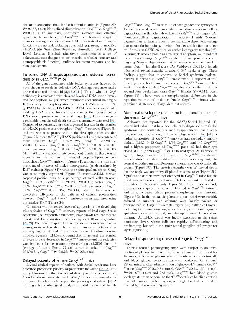
Figure 3. Delayed onset to puberty and ocular, endocrine, haematological, and plasma abnormalities. A. Periodic acid-Schiff (PAS)staining and cleaved (activated) caspase-3 immunostaining of adrenal sections from 16 week-old virgin female Cenpjtm/tm mice (n = 3) confirmedcorticomedullary pigmentation and ongoing apoptosis in the X-zone, respectively (representative images, scale bars 100 mm). B. Breeding records ofCenpjtm/tm females set up with Cenpjtm/tm males at 6–7 weeks of age showed that Cenpjtm/tm females produce their first litter around four weeks laterthan Cenpj+/+ females. *P = 0.012, t-test. C. Top panel shows normal cornea from a Cenpj+/+ mouse. Cenpjtm/tm mice had disruption of the Descemet’smembrane and corneal endothelium (arrow). Middle panel shows normal anterior segment from a Cenpj+/+ mouse. The angle was displacedanteriorly in eyes from Cenpjtm/tm mice and ciliary process morphology was abnormal. (a, angle; i, iris; cb, ciliary body; l, lens). Bottom panel showsnormal retina from a Cenpj+/+ mouse eye. The retina photoreceptor cells of Cenpjtm/tm mice were reduced in number and showed columnardisorganized (arrow). (ONL, outer nuclear layer). D. Immunohistochemical staining for Cenpj in Cenpj+/+ embryo eye (E14.5; RNL retinal neuroblastlayer). E. Intra-peritoneal glucose tolerance test to show that female Cenpjtm/tm mice have a 15 minute delay in response to glucose challenge (n = 4Cenpjtm/tm vs. n = 32 Cenpj+/+, *P = 261025, t-test). Graph also shows n = 9 Cenpjtm/+ and n = 670 baseline wildtype controls. F. Plasma albumin levelswere decreased in Cenpjtm/tm males (n = 8 Cenpjtm/tm vs. n = 35 Cenpj+/+, *P = 4.961025, t-test). Graph also shows n = 7 Cenpjtm/+ and n = 768 baselinewildtype controls. G. Flow cytometric analysis of peripheral blood leukocytes in Cenpjtm/tm mice revealed an increase in the number of CD8+CD3+ andH. total CD3+ cells. Data shows total counts per 30 000 propidium-iodide (PI) negative, CD45-positive cells from male mice. For n = 9 Cenpjtm/tm vs.n = 30 Cenpj+/+: CD3+CD8+ *P = 0.0002 and CD3 *P = 2.961025, Mann-Whitney-Wilcoxon test. Graphs also show n = 7 Cenpj+/tm and n = 356 baselinewildtype controls. For all ‘Box and Whisker’ plots, the lower whisker extends to the lowest datum still within 1.5 Inter-quartile range (IQR) of the lowerquartile. The upper whisker extends to the highest datum still within 1.5 IQR of the upper quartile.doi:10.1371/journal.pgen.1003022.g003
Disruption of Cenpj Phenocopies Seckel Syndrome
PLOS Genetics | www.plosgenetics.org 7 November 2012 | Volume 8 | Issue 11 | e1003022
Harderian glands was increased in aged Cenpjtm/tm mice (13-month
old) when compared to age-matched control (Figure S2E).
Hypoalbuminemia of Cenpjtm/tm miceClinical chemistry was performed on animals at 16 weeks of
age. Albumin levels were generally decreased in Cenpjtm/tm mice of
both genders compared to controls, and this was statistically
significant for males (mean6S.E.M, Cenpj+/+ 25.160.31, Cenpjtm/tm
21.960.52, P = 4.961025, t-test; Figure 3F).
Increased levels of CD3+CD8+ T cells in Cenpjtm/tm miceFlow cytometric analysis of peripheral blood leukocytes at 16
weeks of age revealed a marked increase in the frequency of the
CD8 T cell subset (CD3+ CD8+) in both genders of Cenpjtm/tm mice
compared to wild-type controls (mean6S.E.M Males: CD8+CD3+
gated on PI2 CD45+: Cenpj+/+ 4.3%60.18, Cenpjtm/tm 7.5%60.55,
P = 0.0002 Mann-Whitney-Wilcoxon test; Figure 3G). Further-
more, the increase in the proportion of the peripheral blood CD8
T cell population was reflected in a concomitant increase in the
frequency of total T cells in Cenpjtm/tm mice (mean6S.E.M, CD3+
gated on PI2 CD45+: Cenpj+/+ 9%60.39, Cenpjtm/tm 14%60.41,
P = 2.961025, Mann-Whitney-Wilcoxon test; Figure 3H). The
frequency of CD4 T cells in the peripheral blood was unaffected
(data not shown).
Increased genomic instability in embryonic fibroblastsfrom Cenpj-deficient mice
CENPJ depletion in cultured cells has been reported to impair
centriole assembly, disrupt centrosome integrity and lead to the
formation of monopolar and multipolar spindles instead of bipolar
spindles [19,20,21,24,57]. To assess the cellular phenotype of
Cenpjtm/tm mice, MEFs were derived from Cenpj+/+, Cenpj+/tm and
Cenpjtm/tm littermates. For all experiments, MEFs were of early
passage (P,5) and were passage-matched. Cenpj protein levels
were reduced in the centrosomes of Cenpjtm/tm MEFs, but residual
protein remained detectable in most cells (Figure 4A). Consistent
with previous findings, frequencies of both monopolar and
multipolar spindles were elevated in two independently derived
Cenpjtm/tm MEF lines (Figure 4B; Cenpj+/+: 2.1% monopolar and
8% multipolar; Cenpjtm/tm (1): 10.6% monopolar and 20.8%
multipolar; Cenpjtm/tm (2): 8.9% monopolar and 18.9% multipolar).
Distribution and intensities of the centrosomal proteins c-tubulin
(Figure 4A), CDK5RAP2 (Figure 4B), and pericentrin (data not
shown) were unaffected in the mutant. We next asked whether
spindle abnormalities are accompanied by aberrant centrosome
and centriole numbers in Cenpjtm/tm MEFs. A normal mitotic cell
contains two centrosomes, each containing a pair of centrioles.
Supernumerary centrioles were visible in mitotic Cenpjtm/tm cells
(Figure S5). To facilitate counting of centrioles, cells were arrested
in mitosis using monastrol, a microtubule motor poison that
prevents separation of spindle poles and thereby generates
monopoles [58]. Cells with three or four centrioles were
considered normal, since it is not always possible to resolve
centrioles within a pair. We observed an increase in cells
containing both too few (#2) and too many centrioles ($5) in
the mutant (Figure 4C). To survive, cells with supernumerary
centrosomes must either inactivate these or cluster active
centrosomes into two poles, a process that ensures bipolar division
[23,59]. Clustered centrosomes were indeed observed in Cenpjtm/tm
MEFs (Figure 4B). Centrosome clustering however does not
prevent unequal partitioning of centrosomes into daughter cells
(Figure S5), which ultimately causes a disassociation between
centrosome numbers and DNA ploidy.
Cell-cycle analysis of Cenpjtm/tm MEFs revealed a significant
increase in the number of 4C and elevated levels of .4C cells
(Figure 5A), indicative of polyploidy. We examined the ploidy of
fifty metaphase spreads of Cenpjtm/tm and Cenpj+/+ MEFs and found
that a remarkably high percentage of Cenpjtm/tm cells were near
tetraploid (Cenpj+/+ 11% vs. Cenpjtm/tm 41%). Twenty metaphase
spreads with good fluorescent in situ hybridization (FISH) signals
were selected for multiplex-FISH karyotyping which confirmed
that many cells were near tetraploid and revealed additional
defects such as aneuploidy, centromere loss, centric fusions
(Figure 5B) and translocations (Figure 5C; for a breakdown of
anomalies see Figure S2F). Consistently, we found evidence of
lagging chromosomes in anaphase Cenpjtm/tm MEFs (Figure S5).
Furthermore we show that adult Cenpjtm/tm mice have an increased
prevalence of micronucleated normochromatic erythrocytes
(P = 0.000004, t-test; Figure 5D), thus confirming that these
mutants have spontaneous genomic instability.
The genomic instability observed in Cenpjtm/tm mice doesnot reflect defective ATR- or ATM-dependent DNA–damage signaling
Seckel syndrome belongs to a group of genome instability
disorders collectively referred to as DNA-damage response and
repair-defective syndromes [60]. So far, all cells derived from
Seckel patients have been found to be impaired in signaling
mediated by the DNA-damage responsive protein kinase ATR,
and therefore display reduced phosphorylation of downstream
ATR substrates such as the checkpoint kinase Chk1, and have
impaired G2/M cell-cycle checkpoint arrest upon treatment with
DNA-damaging agents [60,61]. We therefore treated fibroblasts
from Cenpjtm/tm embryos (13.5 d.p.c) with the DNA damaging
agent camptothecin, a DNA topoisomerase I inhibitor that causes
DNA double-strand breaks specifically in S-phase [62]. Analyses
revealed that Cenpjtm/tm fibroblasts were proficient for ATR-
dependent and ATM-dependent phosphorylation of Chk1 (pS345)
and KAP1 (pS824), respectively, and showed normal activation of
cH2AX (Figure 5E). We found no evidence of an impaired G2/M
DNA damage checkpoint as determined by the percentage of
MPM2-positive cells following irradiation (Figure S2G). Further-
more, CtIP-Seckel cells show defective phosphorylation of
replication protein A (RPA) after camptothecin treatment, a
phenotype associated with impaired DNA-end resection and
homologous recombination [6]. However, we found no evidence
of this in Cenpjtm/tm MEFs (Figure 5E).
Discussion
The Cenpj hypomorphic mouse (Cenpjtm/tm) that we have created
displays many of the classical clinical features of Seckel syndrome,
including intrauterine and postnatal dwarfism, microcephaly, a
sloping forehead, neuropathogical abnormalities, memory impair-
ment and genomic instability [1,4,7,15,28,63]. In addition, we
have shown that Cenpjtm/tm mice display some of the less frequently
reported characteristics of the syndrome, including retarded bone
ossification [1,32,64], as well as vertebral abnormalities and
several other interesting histopathological and hematological
abnormalities that have not previously been reported in patients.
Microcephaly versus dwarfism of Cenpjtm/tm miceNeuroepithelial cells have apical-basal polarity, and the switch
from proliferative, symmetric to neurogenic, asymmetric division is
controlled by the orientation of the spindle pole during mitotic
division [65]. Primary microcephaly is caused by mutations of
centrosomal proteins and is thought to arise from an increase in
Disruption of Cenpj Phenocopies Seckel Syndrome
PLOS Genetics | www.plosgenetics.org 8 November 2012 | Volume 8 | Issue 11 | e1003022
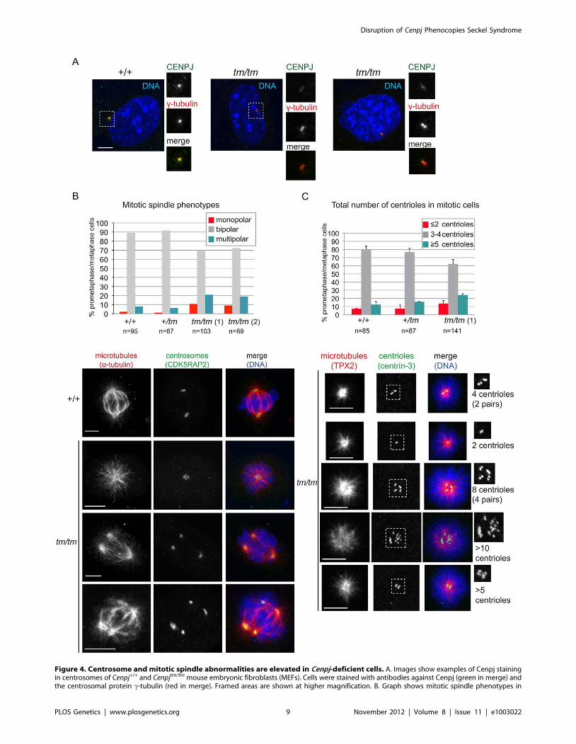
Figure 4. Centrosome and mitotic spindle abnormalities are elevated in Cenpj-deficient cells. A. Images show examples of Cenpj stainingin centrosomes of Cenpj+/+ and Cenpjtm/tm mouse embryonic fibroblasts (MEFs). Cells were stained with antibodies against Cenpj (green in merge) andthe centrosomal protein c-tubulin (red in merge). Framed areas are shown at higher magnification. B. Graph shows mitotic spindle phenotypes in
Disruption of Cenpj Phenocopies Seckel Syndrome
PLOS Genetics | www.plosgenetics.org 9 November 2012 | Volume 8 | Issue 11 | e1003022

asymmetric divisions that reduces the size of the neural progenitor
pool available for future brain growth, hence the growth deficit is
restricted to the brain [66]. Seckel syndrome is characterized by
microcephaly and a small body size (proportionate dwarfism).
Interestingly, different mutations in the centrosomal proteins
CENPJ or CEP152 can cause microcephaly or Seckel syndrome
[4,14,15]. The CENPJ-microcephaly mutations reported to date
affect exons 2 (17delC), 11 (3243–3246delTCAG) and 16
(A3704T) [14,15]; these mutations are predicted, but not proven,
to cause defects in spindle pole orientation and proliferation of
neural progenitors in a similar manner to other microcephaly
genes. CENPJ-Seckel syndrome has been associated with a
homozygous splice acceptor mutation in the last nucleotide of
CENPJ intron 11 that results in the skipping of either exon 12,
exons 12 and 13 or exons 11,12 and 13 during transcription [4].
This may represent a cellular attempt to salvage this important
protein since the latter two transcripts are predicted to result in in-
frame deletion and preservation of the C-terminus [4].
Notably, we found that insertion of a cassette between exons 4
and 5 of Cenpj resulted in splicing over the cassette and cryptic
splicing, such that three different Cenpj mRNAs were expressed at
very low levels: full length, one lacking exons 4 and 5 and one
lacking exon 5. Skipping of exons 4 and 5 or exon 5 is predicted to
result in a premature stop codon and protein products that are
truncated after translation of exon 3 or 4, respectively. Immuno-
blotting and immunofluorescence revealed higher than expected
levels of apparently full-length Cenpj protein were present in
Cenpjtm/tm MEFs, which may be the result of post-transcriptional or
translational regulation since the level of full-length Cenpj mRNA
in Cenpjtm/tm MEFs was only 2% of wild-type levels. Furthermore,
mRNA levels varied greatly between Cenpjtm/tm mouse embryonic
fibroblasts, which may be due to genetic modifiers.
Together with studies of CENPJ-Seckel cells, which have shown
that mutations in CENPJ may result in exon skipping and the
generation of multiple transcripts that may generate in-frame
protein products [4], these data suggest that expression of this
critical protein may be rescued to some extent by cryptic splicing
over deleterious mutations. These produce alternatively spliced
mRNAs and thus the same mutation might not result in the same
mRNA or protein levels in each individual. Moreover, cryptic
splicing may also differ between tissues. Without a complete
examination of the effects of different CENPJ-mutations on
mRNA levels and splicing, and CENPJ protein levels, it is dif-
ficult to say why CENPJ mutations can either result in pri-
mary microcephaly or Seckel syndrome, or why the
Cenpjtm1a(EUCOMM)Wtsi allele results in a mouse with a Seckel
syndrome-like phenotype. However, we propose that Cenpjtm/tm
mice display a Seckel syndrome-like phenotype, rather than
primary microcephaly, due to a major reduction in full length
Cenpj protein and therefore a lack of the protein domain(s)
encoded by exons 11, 12 and/or 13.
The microcephaly of Cenpjtm/tm mice (brain weight two standard
deviations below the mean) was not as severe as CENPJ-Seckel
syndrome patients, who display anthropometric values that are all
at least seven standard deviations below the mean [4]. The
evolutionary lineage leading to humans is marked by a dramatic
increase in brain size, suggesting that disruption of genes involved
in neurogenesis will have a less profound effect in mice than in
humans [67]. However, this is confounded by the fact that there
are several mouse models of microcephaly, such as the humanized
ATR-Seckel mouse and the Cdk5rap2 mutant mouse, which display
severe reductions in brain size [25,37]. The discrepancy between
microcephaly of Cenpjtm/tm mice and CENPJ-Seckel patients may
instead be due to the hypomorphic nature of the
Cenpjtm1a(EUCOMM)Wtsi allele or by the rapid evolution of the Cenpj
gene between mice (80% sequence identity) and humans; the
human CENPJ protein may be more efficient at regulating
neurogenesis than that of the mouse [67].
The dwarfism and microcephaly of Cenpjtm/tm mice appeared to
be the result of widespread DNA damage and apoptosis in
embryos, rather than a reduction in cell proliferation. The level of
cell death within the forebrain of the Cenpj-hypomorph embryonic
mouse brain was comparable with that of the humanized ATR-
Seckel mouse (approximately 1.5%; [25]). Similarly, ATR-, CtIP-,
CEP152- and PCNT-Seckel cells have increased levels of DNA
damage and a lowered apoptotic threshold with no change in the
rate of proliferation [5,6,25]. In contrast to cells from ATR-, CtIP-,
CEP152- and PCNT-Seckel syndrome patients, we have shown
that MEFs from Cenpj-deficient mice are not impaired in ATR-
dependent DNA damage signaling but instead show an elevated
frequency of extra centrioles, multipolar spindles, and near
tetraploid karyotypes. We suspect that the embryonic fibroblast
line showing 41% near tetraploid cells could come from an
embryo that would not have survived to term, indicating that
genomic instability may also explain the sub-Mendelian birth ratio
of Cenpjtm/tm mice. We also found evidence of chromosome
missegregation, chromosomal translocations and centric fusions in
Cenpjtm/tm MEFs. Increased levels of pan-nuclear cH2AX in
embryos may be the result of chromosome breakage, micronucleus
formation or missegregation [68], however it is possible that this
reflects phosphorylation of H2AX during apoptosis-driven frag-
mentation of DNA [69].
Cenpj is required for normal neuronal density and long-term memory
The neuropathological features of Cenpjtm/tm E14.5 embryos
were remarkably similar to fetal stage Seckel syndrome. At E14.5,
we found there was a reduction in neuron density within the
developing telencephalon of Cenpjtm/tm mice. There are only two
neuropathological reports of fetal stage Seckel syndrome (30 weeks
gestation), although both showed that the cortical layers of the
telencephalon were thin and that neuronal populations were less
MEFs derived from Cenpj+/+, Cenpj+/tm and two independent Cenpjtm/tm embryos (littermates, +/+ MEFs passage 4, +/tm and tm/tm MEFs passage 3):tm/tm (1) and tm/tm (2). Number of mitotic cells scored are shown for each genotype. Examples for monopolar and multipolar spindle are shown.Note cell on bottom panels forming a bipolar spindle by clustering supernumerary centrosomes. Cells were stained with antibodies against a-tubulin(green in merge) and the centrosomal protein, Cdk5RAP2 (red in merge). C. Graph shows centriole numbers in mitotic MEFs of indicated genotypes(littermates, +/+ MEFs passage 4, +/tm and tm/tm MEFs passage 3). Cells were arrested in mitosis with monastrol that caused monopolar spindleformation and facilitated visualization of centrioles. Note that mitotic cells should normally contain a total of 4 centrioles, but even in wild-type cellswe occasionally detect 3 centrioles probably due to insufficient spatial resolution, so 3 or 4 centrioles were considered a single class. Data werecollected from two independent experiments; bars show mean 6SD, number of mitotic cells scored are shown for each genotype. Images belowdepict examples for cells with different centriole numbers (top cell with 4 centrioles is normal, all other cells have too few or too many centrioles).Cells were stained with antibodies against the microtubule-binding protein Tpx2 (green in merge) and the centriolar protein, centrin-3 (red in merge).Framed areas are shown at higher magnification. Scale bars = 5 mm.doi:10.1371/journal.pgen.1003022.g004
Disruption of Cenpj Phenocopies Seckel Syndrome
PLOS Genetics | www.plosgenetics.org 10 November 2012 | Volume 8 | Issue 11 | e1003022
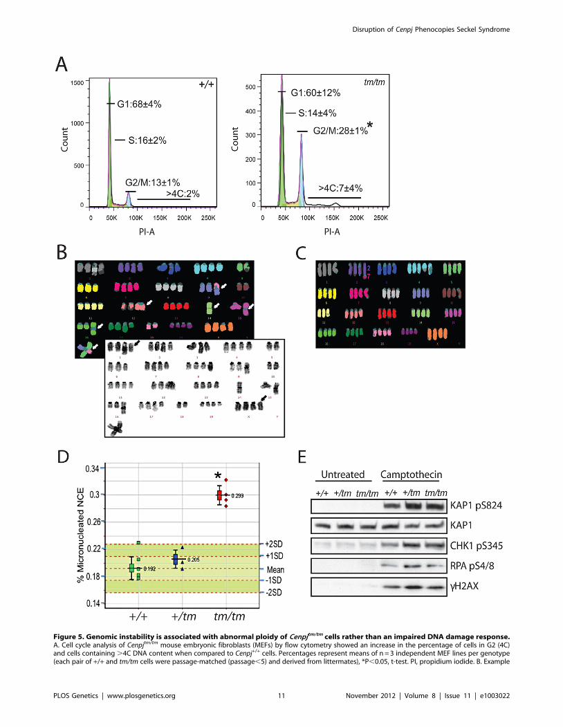
Figure 5. Genomic instability is associated with abnormal ploidy of Cenpjtm/tm cells rather than an impaired DNA damage response.A. Cell cycle analysis of Cenpjtm/tm mouse embryonic fibroblasts (MEFs) by flow cytometry showed an increase in the percentage of cells in G2 (4C)and cells containing .4C DNA content when compared to Cenpj+/+ cells. Percentages represent means of n = 3 independent MEF lines per genotype(each pair of +/+ and tm/tm cells were passage-matched (passage,5) and derived from littermates), *P,0.05, t-test. PI, propidium iodide. B. Example
Disruption of Cenpj Phenocopies Seckel Syndrome
PLOS Genetics | www.plosgenetics.org 11 November 2012 | Volume 8 | Issue 11 | e1003022
dense and less organized than age- or length-matched controls
[28,29]. As with Cenpjtm/tm mice, the hippocampal formation was
short in one fetus, but displayed normal cytoarchitectural
progression [28,29]. Both reports indicated that the major nuclear
groups of the basal ganglia, thalamus, cerebellum and brainstem
showed no abnormalities in fetal stage Seckel syndrome [28,29].
Interestingly, we saw a .50% reduction in the number of
Cenpjtm/tm embryos between mid neurogenesis (E14.5) and the
completion of neurogenesis (E18.5), when Cenpj is strongly
expressed in the ventricular layers of the diencephalon, telen-
cephalon, midbrain and cerebellum (www.emouseatlas.org, www.
eurexpress.org), suggesting that Cenpj-deficiency during this critical
period of neurogenesis causes partial lethality.
The majority of patients with Seckel syndrome are reported to
have an IQ of ,50 and are delayed in speech and reaching motor
milestones, as well as displaying pyramidal signs, hyperactivity and
an attention deficit [3,34]. Cranial MRI of adult patients with
Seckel syndrome has shown a reduction in brain volume,
especially the cerebral cortex, a simplified gyral pattern (number
of gyri reduced and shallow sulci), poorly developed frontal lobes,
agenesis of the corpus callosum, reduction of white matter,
brainstem and cerebellar hypoplasia, and dysmorphic or enlarged
lateral ventricles [32,34,35]. A relatively normal MRI was
reported for two siblings (aged two and four years-old) of the
CENPJ-Seckel kindred and together with two cousins (aged five
and six years-old), all had a history of normal cognitive and motor
development [4]. The third cousin (MRI not performed, aged 16
years-old) had an IQ,60. Similarly, the brain regions of adult
Cenpjtm/tm mice appeared anatomically proportionate, although
these mice had a significantly shorter dentate gyrus than controls
and this was accompanied by cognitive impairments reminiscent
of Seckel syndrome patients.
Centrioles, mitotic spindles, and ploidydSas-4 is the Drosophila homologue of CENPJ. Unlike dSas-4-
depleted cells or dSas-4 mutant flies that progressively lose
centrioles, Cenpjtm/tm MEFs contain centrioles even after several
passages [70,71]. While the increase in Cenpjtm/tm cells with two or
fewer centrioles is consistent with an impairment of centriole
assembly, this effect is relatively mild, and therefore suggests that
the mutant expresses residual, functional Cenpj protein. Ciliogen-
esis requires centriole biogenesis and therefore dSas-4 mutants lack
both primary and motile cilia [70]. The role of CENPJ in
ciliogenesis has not been extensively explored in mammals, but
depletion of CENPJ in cultured cells is reported to impair primary
cilium formation [72]. Cenpjtm/tm mice (16 weeks old) did not
display phenotypes normally associated with ciliopathies such as
situs inversus or renal cystic disease, suggesting that sufficient
amounts of Cenpj are available in the mutant for cilia formation in
the majority of cells. However, the abnormalities in ciliary
processes and photoreceptor nuclei within the eye may be
attributed to ciliary defects. Moreover, unlike dSas-4 mutant
males that display loss of flagella and sperm motility, Cenpjtm/tm
male mice are fertile [70], which could again be due to residual
expression of Cenpj.
While Cenpjtm/tm MEFs displayed irregular centriole numbers
and mono- and multipolar spindles, they also showed extensive
polyploidy and aneuploidy. Thus, we cannot conclude whether
abnormal centriole and centrosome numbers are the cause or
consequence of aberrant ploidy. Figure S6A shows the possible
sequence of events that may lead to the abnormal ploidy of
CENPJ-Seckel cells. Aberrant centrosome numbers are known to
cause mitotic spindle abnormalities, culminating in mitotic delay,
chromosome missegegration, cytokinetic failure and polyploidy.
Prolonged mitotic delay can cause DNA damage, cell cycle arrest
and apoptosis [73,74]. Chromosome missegregation can also
damage chromosomes, hence triggering activation of DNA
damage checkpoints [68,75]. Chromosome instability could
therefore explain the increase in cH2AX levels and potentially,
the increase in apoptosis in the mutant embryonic brain. Of all
chromosome aberrations detected in the mutant MEFs, tetraploi-
dy was the most prominent. A common cause of tetraploidy is an
abortive mitotic cell cycle whereby cells enter but fail to complete
mitosis [76]. Mitotic spindle abnormalities in Cenpjtm/tm cells could
trigger extended mitotic arrest followed by mitotic slippage
producing a tetraploid cell (Figure S6A). Tetraploid Cenpjtm/tm
MEFs seem to be able to proliferate, since they represented almost
40% of the metaphase cells obtained for karyotyping. Interestingly,
dSas-4 mutant flies show only a small increase in the proportion of
aneuploid cells (1% in wild-type vs. 3% in mutants) and no
polyploidy [70], whereas the proportion of near tetraploid
Cenpjtm/tm embryonic fibroblasts was surprisingly high (,10% in
wildtype vs ,40% in Cenpjtm/tm MEFs). We suspect that Cenpj-
deficiency exacerbates tetraploidy in MEFs, which are particularly
susceptible to tetraploidy with passaging [77]. Nonetheless, adult
Cenpjtm/tm mice show increased micronucleus induction, which is
likely the result of lagging chromosomes and chromosome breakage.
Polyploidy as a potential cause of karyomegaly inCenpjtm/tm tissues
Cenpjtm/tm mice of both genders showed an increased incidence of
hypertrophic, disorganized cardiomyoctes with karyomegaly in the
endocarium and interventricular septum when compared to wild-
type mice. The areas showed no evidence of degeneration or repair,
however since a high proportion of Cenpjtm/tm MEFs are polyploid,
this is likely to be the cause of the karyomegaly. Although one of the
less frequently reported characteristics of Seckel syndrome, there are
numerous case-reports of severe cardiac anomalies in Seckel
syndrome patients, including atrial and ventricular septal defects,
pulmonary atresia, patent ductus arteriosus and congenital heart
disease [49,50,51,52,53]. It will be interesting to see whether
CENPJ-Seckel patients develop cardiac defects as they age. At 16
weeks of age Cenpjtm/tm mice showed hypoalbuminemia, which is
associated with chronic liver and kidney diseases, although
histopathological analysis of their livers and kidneys did not reveal
any abnormalities. However, the preponderance of karyomegaly in
the liver and Harderian glands was increased in aged Cenpjtm/tm mice.
Cenpj-deficiency may exacerbate this phenomenon in the cells of
both of these tissues, which are prone to karyomegaly [78].
multiplex fluorescent in situ hybridization (M-FISH; top) and DAPI banded (bottom) karyotype of a Cenpjtm/tm MEF metaphase (passage 4). Thekaryotype is near tetraploid, with centric fusions (white arrows) and chromosomes that have apparently lost their centromeres (black arrows). C.Example M-FISH of a Cenpjtm/tm MEF metaphase (passage 4) showing near tetraploid karyotype with a translocation (t(2;7)). D. Adult Cenpjtm/tm (n = 4)mice showed increased genomic instability when compared to Cenpj+/+ mice (n = 6) as determined by the increased prevalence of micronucleatednormochromatic erythrocytes using a flow cytometric assay of micronucleus formation. *P = 0.000004, t-test. The lower whisker extends to the lowestdatum still within 1.5 Inter-quartile range (IQR) of the lower quartile. The upper whisker extends to the highest datum still within 1.5 IQR of the upperquartile. E. Immunoblots show normal activation of DNA damage response markers in Cenpj-deficient MEFs (passage 2) before and after treatmentwith the DNA damaging agent camptothecin (1 mM for 1 h). KAP1 was used as a loading control.doi:10.1371/journal.pgen.1003022.g005
Disruption of Cenpj Phenocopies Seckel Syndrome
PLOS Genetics | www.plosgenetics.org 12 November 2012 | Volume 8 | Issue 11 | e1003022
Susceptibility to malignancyFamilial syndromes associated with genomic instability often
predispose to cancer formation since DNA damage is the source of
mutations that drive malignant transformation. However only a
few cancers have been reported for Seckel syndrome patients,
possibly due to the shorter life-expectancy of patients with
primordial dwarfism. Furthermore, since mutation of each of the
five known Seckel genes, ATR, PCNT, CENPJ, CEP152 and
RBBP8 (CtIP), cause genomic instability that is associated with
apoptosis, it is possible that Seckel cells may not have the
opportunity to accumulate cancer-causing mutations. The chro-
mosomal instability that is associated with Cenpj-deficiency could
result in aneuploidy or translocations that cause loss of tumour
suppressors or the formation of oncogenic fusion proteins,
respectively [79]. We are currently ageing a cohort of Cenpjtm/tm
mice (currently 7–14 months old) to determine whether Cenpj-
deficiency alters the frequency of malignancy or shortens life-
expectancy.
Cenpj-deficient mouse phenotypes for which there arecurrently no clinical correlates
We noted a small number of abnormalities in Cenpjtm/tm mice
that have not been previously reported for Seckel syndrome
patients or mouse models. Seckel syndrome is associated with
ocular defects in humans, including spontaneous lens dislocation,
myopia, astigmatism, and retinal degeneration. Ocular examina-
tion of CENPJ-Seckel patients has not yet been reported [47,48],
however Cenpj was highly expressed in the rapidly proliferating
retinal neuroblast layer in the 14.5 d.p.c. mouse embryo and
Cenpj-deficient mice presented with a number of ocular abnor-
malities. Furthermore, a small number of reports suggest that
Seckel-like syndromes are associated with precocious puberty or
premature thelarche [44,45]. In contrast, female Cenpjtm/tm mice
showed signs of delayed puberty, although the reproductive tract
appeared normal at 16 weeks. We built a protein-protein
interaction network using all known Seckel Syndrome associated
genes as query (Figure S6B). By using gene ontology enrichment
analysis we showed that 265 biological processes (level 3
classification) are significantly over-represented in the Seckel
syndrome network (Table S1). As expected, many of the processes
were involved in the regulation of cell cycle, cell growth and cell
death. Interestingly, the network was also enriched for genes
involved in the ‘response to hormone stimulus’ ‘ovulation cycle
process’ and ‘sex differentiation’ (Table S1). Transient insulin
resistance during puberty is a well documented phenomenon [80].
Cenpjtm/tm mice of both genders had a delayed response to glucose
challenge although this was more marked in 16 week-old female
mice, which may be explained by delayed puberty in female
Cenpjtm/tm mice. While there are no reports of an association
between abnormal glucose homeostasis and Seckel syndrome,
interestingly, most individuals with MOPDII, including PCNT-
MOPDII, develop insulin resistance and diabetes during child-
hood [81,82]. Aside from centrosome-mediated regulation of the
cell-cycle, PCNT is thought to regulate insulin secretory vesicle
docking in mouse pancreatic b-cells [83]. Whether CENPJ plays a
role in glucose homeostasis remains to be determined. Finally, the
proportion of CD8+CD3+ T cells were elevated in Cenpjtm/tm mice,
although this was more pronounced in males. The Seckel
syndrome protein-protein interaction network that we generated
was significantly enriched for genes involved in ‘leukocyte
mediated immunity’, ‘leukocyte mediated cytotoxicity’, ‘leukocyte
activation’ and ‘interleukin-2 production’ (Table S1). While we are
uncertain of the biological basis for these relationships, it will be
interesting to see whether there are clinical correlates for these
abnormalities and whether other mouse models of Seckel
syndrome or primordial dwarfism share these anomalies.
SummaryMouse models of Seckel syndrome may go some way towards
the molecular genetic delineation of this heterogeneous condition.
The generalized activation of apoptosis as a result of genomic
instability in ATR-Seckel and Cenpjtm/tm mouse embryos provides
one explanation for the proportionate dwarfism of Seckel
syndrome patients. In agreement with the intron 11 CENPJ-
Seckel mutation, which results in the formation of three
transcripts, we showed that Cenpj expression is rescued to some
extent by cryptic splicing over the cassette to produce a variety of
truncated mRNAs, and that there is a moderate degree of
individual variation in the ability of an organism to perform this
rescue. These data highlight the need for detailed mRNA
expression, splicing studies, and protein analysis to establish how
individual mutations affect the normal and cryptic splicing of
CENPJ mRNAs for each patient directly, and not with prediction
analysis tools, so as to understand why some CENPJ mutations
cause microcephaly and others Seckel syndrome and how the
same CENPJ mutation can cause clinical heterogeneity [4].
Materials and Methods
Confirmation of correct targeting and animal husbandryMutant mice carrying the Cenpjtm1a(EUCOMM)Wtsi allele were
generated on a C57BL/6NTac; C57BL/6-Tyrc-Brd background
(clone EPD0028_7_G05) by the Sanger Mouse Genetics Project as
part of the European Conditional Mouse Mutagenesis Program
(EUCOMM; [26]). Correct gene targeting in founder mice was
determined by a combination of standard PCR and quantitative
PCR (qPCR; see Figure S1 for more details). Following
confirmation of correct targeting, mice were genotyped for the
Cenpjtm1a(EUCOMM)Wtsi allele by PCR using primers specific to the
wildtype and mutant Cenpj alleles and to LacZ (Figure S1D). In
individual experiments, all mice were matched for age and gender.
A cumulative baseline was generated from data arising from
controls from the same genetic background, age and gender. The
care and use of all mice in this study was carried out in accordance
with UK Home Office regulations, UK Animals (Scientific
Procedures) Act of 1986. Mice were maintained in a specific
pathogen free unit on a 12 hr light: 12 hr dark cycle with lights off
at 7:30pm and no twilight period. The ambient temperature was
2162uC and the humidity was 55610%. Mice were housed using
a stocking density of 3–5 mice per cage (overall dimensions of
caging: (L6W6H) 36562076140 mm, floor area 530 cm2) in
individually ventilated caging (Tecniplast Seal Safe1284L) receiv-
ing 60 air changes per hour. In addition to Aspen bedding
substrate, standard environmental enrichment of two nestlets, a
cardboard Fun Tunnel and three wooden chew blocks was
provided. Mice were given water and diet ad libitum. At 4 weeks of
age, mice were transferred from Mouse Breeders Diet (Lab Diets,
5021–3) to a high fat (21.4% fat by crude content) dietary
challenge (Special Diet Services, Western RD 829100).
Quantitative real-time PCRTo test for expression of Cenpj and cryptic splicing, total RNA
was isolated from mouse embryonic fibroblasts (13.5 days post
coitum (d.p.c.), n = 3) using the RNeasy minikit (Qiagen). RT-
PCR was performed using the BD Sprint kit containing random
hexamers (BD Clontech, CA, USA). Primers were designed to
exon boundaries and sequences are available on request. cDNA
was quantified using SYBR Green on an ABI7900HT (ABI, CA,
Disruption of Cenpj Phenocopies Seckel Syndrome
PLOS Genetics | www.plosgenetics.org 13 November 2012 | Volume 8 | Issue 11 | e1003022

USA). Gene expression was normalised to Gapdh and to wild-type
control.
ImmunoblottingProtein extracts were prepared from mouse embryonic fibro-
blasts (13.5 d.p.c.) by directly harvesting cells in Laemmli buffer.
Proteins were separated by SDS-PAGE, and membranes were
incubated with primary antibodies to CENPJ (Stratech, Newmar-
ket, UK. Rabbit 1:500), KAP1 (Abcam, Cambridge, UK. Rabbit
1:10 000), KAP1 phospho-Ser-824 (Bethyl. Texas, US. Rabbit
1:1000), Chk1 phospho-Ser-345 (Cell Signaling, Boston, US.
Rabbit 1:5000), RPA phospho-Ser-4/8 (Bethyl, Texas, US. Rabbit
1:10 000), H2AX phospho-Ser-139 (cH2AX; Millipore, Billerica,
US. Mouse 1:1000). Camptothecin was from Sigma (Poole, UK).
LacZ stainingLacZ staining was performed on tissues perfused, removed and
fixed (30 min) with 4% paraformaldehyde (pH8). Tissues were
then washed three times in PBS. Tissues were then incubated at
4uC for 48 h in staining buffer (5 mM K3Fe(CN)6, 5 mM
K4Fe(CN)6, 0.02% IGEPAL CA-630, 0.01% deoxycholate,
2 mM MgCl2, 1 mg/ml bromo-chloro-indolyl-galactopyranoside
in the dark. Tissues were fixed again with 4% paraformaldehyde
overnight at 4uC before being transferred through increasing
glycerol concentrations and archiving in 70% glycerol+0.01%
sodium azide.
E18.5 measurements and skeletal preparationsAt 18.5 d.p.c. litters were harvested and euthanized. The
crown-rump length was measured and measurements of skulls
were adapted from published methods used to assess adult mouse
skulls [84]. Embryos were scalded at 67uC for 30 seconds to
facilitate removal of skin, muscle and fat. Dissected embryos were
then fixed in 100% ethanol for 48 h and transferred to acetone for
48 h to further de-fat the skeletons. Skeletal embryos were then
stained with 0.015% Alcian Blue (Sigma-Aldrich, UK; in Ethanol/
Glacial Acetic Acid) for 24 h, washed five times in 100% ethanol
and transferred to 0.1% potassium hydroxide overnight. Skeletons
were then stained with 0.005% Alizarin Red (Merck, UK; in 1%
KOH) for 3 h, washed with 1% KOH three times and transferred
to 20% Glycerol/1% KOH for clearing for ,3 days. Skeletons
were then transferred through increasing glycerol concentrations
and archived in 70% glycerol +0.01% sodium azide.
X-ray imagingX-ray imaging was performed at 14 weeks of age using a
Faxitron MX-20 cabinet (Faxitron Bioptics, IL, USA). Mice were
anesthetized with a preparation of 100 mg/kg Ketamine/10 mg/
kg Xylazine. Weight and body length were measured prior to the
scan. Five images were acquired: dorsoventral and lateral images
of the whole body at 61 magnification, dorsoventral and lateral
images of the head at 64 magnification and a dorsoventral image
of the left forepaw at 65 magnification. Images were acquired
using an energy of 23 kV for 10 seconds per image. Images were
studied visually to assess abnormalities within 41 standard
parameters. To show that humeri were anatomically dispropor-
tionate the right greater tubercle - deltoid tuberosity length (mm)
was normalized to the right greater tubercle – trochlea length
(mm) and data were represented as a percentage.
HistologyAdult tissue samples were fixed in 10% neutral buffered
formalin and E14.5 embryos were fixed in 4% PFA. Samples
were dehydrated, paraffin embedded and 4 mm sections were cut
before hematoxylin and eosin staining. Neuron densities of E14.5
embryo brains were determined by counting the number of nuclei
in three different areas that were 75 mm2 for the densely populated
ventricular zones and 150 mm2 for the less densely populated mid-
striatum. Adult neuroanatomical measurements (Figure S3) were
carried out on 40 mm thick Nissl stained sections using ImageJ
freeware (NIH). Heart fibrosis was assessed by Masson’s
Trichrome and X-zone pigmentation of the adrenals was
confirmed by Periodic Acid-Schiff’s staining using standard
methods. Whole mouse eyes were enucleated, fixed, and sectioned
for histological studies as previously described [85].
ImmunohistochemistrySections of paraffin embedded tissues were dewaxed and
antigen retrieval was performed in boiling 10 mM citrate buffer
pH6. Endogenous peroxidases were quenched in 3% hydrogen
peroxide (Sigma, UK) before blocking in normal serum (Vector-
Labs, UK). Sections were incubated in primary antibodies to
Cenpj (Stratech), cleaved caspase-3 (Cell Signaling Technologies,
CA, USA), Ki67 (DAKO Ltd, UK) and phospho-Ser 139 H2AX
(cH2AX, Cell Signaling Technologies, CA, USA). Vectorstain
ABC kit and DAB (VectorLabs, UK) were used according to the
manufacturer’s instructions. Sections were counterstained with
haematoxylin before clearing and mounting. The number of cells
positive for cleaved-caspase 3 or cH2AX were counted in two
areas of 75 mm2 the striatum, cortex and pro-hippocampus and
data shown are the percentage of total cells in the area. Counts
were performed independently by two individuals to confirm
reproducibility.
Neurobehavioral and sensory assessmentAt nine weeks of age, open field, grip strength and modified
SHIRPA were performed as described previously [86,87]. Hot
plate assessment was performed at 10 weeks of age using standard
techniques. Tests for social recognition and olfaction (Cenpjtm/tm
n = 9 and Cenpj+/+ n = 8) were performed on male mice at 3–6
months of age. Mice were habituated to a test arena identical to
their home cage for 10 min. For social recognition, a stimulus
mouse was placed into the test arena for 1 min, repeated four
times at 10 min intervals. In the fifth trial, a second stimulus
mouse was presented. 24 h later the test animals were presented
with the familiar animal from trials 1–4 and a new unfamiliar
animal, for 2 min. Trials were performed under red light and
recorded with an overhead camera and the videos scored blind of
genotype. The amount of time the test animal spent investigating,
by oronasal contact or approaching within 1–2 cm, was recorded.
Stimulus animals, 2–4 months old, were weight-matched to
Cenpjtm/tm mice and sedated with ketamine/xylazine (i.p. 1 g/
0.1 g per kg of body weight). C57BL/6NTac mice were used for
trials 1–4 and for the 24 h discrimination test, 129P2/OlaHsd
mice were used for trial 5. Three animals (two Cenpjtm/tm and one
Cenpj+/+) were taken out of the analysis because of their low
investigation times (less than 10 s on trial one or during the
discrimination test).
Glucose toleranceWe assessed glucose tolerance in mice fed on a high-fat diet
(Western RD, 829100, Special Diets Services) from 4 weeks of age
until 13 weeks of age. At 13 weeks, mice were fasted overnight
before a blood sample was taken and glucose was measured using
an Accu-Chek Aviva (Roche). To perform an intra-peritoneal
glucose tolerance test (IP-GTT), mice were fasted for 16 h, a bolus
of glucose was administered intraperitoneally and blood glucose
Disruption of Cenpj Phenocopies Seckel Syndrome
PLOS Genetics | www.plosgenetics.org 14 November 2012 | Volume 8 | Issue 11 | e1003022
concentration from the tail vein was measured using Accu-Chek
Aviva (Roche) after 15, 30, 60 and 120 min.
Clinical chemistryWe performed clinical chemistry on 16-week-old mice. Mice
were terminally anaesthetized and blood was collected from the
retro-orbital sinus into lithium-heparin tubes. The plasma was
immediately analyzed on an Olympus AU400 Analyzer.
Flow cytometryAt 16 weeks of age blood samples were collected into heparin-
coated tubes. The main leukocyte populations in peripheral blood
were characterized by 8-colour flow cytometry with an LSRII (BD
Bioscience, UK Biosciences, UK) and associated software. Briefly,
samples were centrifuged at 5000 g for 10 min at 8uC to remove
the plasma layer. Red blood cells were lysed (Pharmalyse, BD
Bioscience, UK Biosciences, UK) and samples were centrifuged at
400 g for 3 min to pellet the white cells. White cells were
resuspended in buffer (PBS pH 7.45 containing 0.5% BSA),
transferred to 96-well plate and washed in buffer several times
before incubation in 50 ml 10 mg/ml Mouse FcBlock (BD
Bioscience, UK Biosciences, UK) for 15 min on ice. Cells were
washed in buffer and incubated with 50 ml antibody mix from two
staining panels (Tables S2 and S3) for 15 min on ice. Propidium
iodide (2.5 mg/ml; Sigma, UK) was added to each well and cells
were incubated for a further 5 min. Cells were washed in buffer
several times before 30 000 propidium iodide-negative, CD45-
positive events were collected. Data were interpreted using FlowJo
(v7.6, Tree Star, Inc., OR, USA). The frequency of micronu-
cleated normochromatic erythrocytes was determined by flow
cytometry (FC500, Beckman Coulter, USA) as described previ-
ously [88] and data were interpreted using FlowJo (v9.3.1, Tree
Star, Inc., OR, USA). Cell cycle analysis was performed on mouse
embryonic fibroblasts (13.5 d.p.c.). Briefly, cells were fixed in 70%
ethanol overnight and stained with PI solution before analyzing on
a flow cytometer (LSR Fortessa, BD, USA). A total of 10,000
events were acquired per sample. The cells were gated on PI
fluorescence area versus PI fluorescence width to discriminate any
doublets and clumps. The gated events were displayed on a
histogram plot of PI fluorescence area. Data were analyzed using
FlowJo (v9.3.1, Tree Star, Inc., OR, USA).
ImmunofluorescenceMouse embryonic fibroblasts (13.5 d.p.c.) were collected and
cultured using standard methods. Primary antibodies used in this
study were CDK5RAP2 (Bethyl), centrin-3 (Abnova), TPX2
(Abnova), gamma-tubulin (GTU88; Sigma-Aldrich), alpha-tubulin
(DM1A; Sigma-Aldrich). Secondary antibodies conjugated to
Alexa Fluor 488 and 555 (Invitrogen) were used. DNA was
stained with Hoescht (Sigma-Aldrich). To detect centrosomal
markers, cells were fixed in 220uC methanol for 5 min. After
fixation, cells were processed for immunofluorescence and
microscopy as described in [89]. For centriole counts in
Figure 4C cells were treated with 100 mM monastrol for 16 h
before fixation.
G2 checkpoint assayCells were irradiated with 3 Gy ionizing radiation using a
Faxitron cabinet X-ray system and left recover for 8 h in the
presence of 1 mg/ml of nocodazole (Sigma, Poole, UK) to trap
mitotic cells. Cells were trypsinized and fixed with 4% parafor-
maldehyde, permeabilized with 16 phosphate buffered saline
containing 0.2% Triton X-100 for 30 min on ice, and incubated in
the presence of MPM2 primary antibodies (Millipore, Billerica,
US. Mouse 1:100) and then Alexa-Fluor-488 secondary antibodies
(Life Technologies, Paisley, Scotland. Goat anti-mouse 1:200) to
detect mitotic cells.
Multiplex-fluorescence in situ hybridization (FISH)Metaphase spreads from MEFs (Cenpjtm/tm and Cenpj+/+ litter-
mates) were prepared as for metaphase FISH and multiplex-FISH
was carried out as previously described [90].
Network generation and analysisA network of Protein-Protein interactions for Seckel syndrome
genes was generated using the Cytoscape 2.8.1 [91] plug-in
BisoGenet 1.41.00 [92]. The Entrez Gene symbols of the Seckel
syndrome associated genes (CENPJ, PCNT, CEP152, ATR, RBBP8
(CtIP), SCKL3, Entrez Gene IDs = 55835, 5116, 22995, 545, 5932
and 386616 respectively) [4,5,6,41] were used as query to build a
network of experimentally validated Protein-Protein interactions,
by adding neighbours to the input nodes up to a distance of one.
All the interactions are downloaded by BisoGenet from its own
database SysBiomics and have been validated by one or different
experimental methodologies such as X-ray crystallography, surface
plasmon resonance, two hybrid systems, three hybrid systems and
Western blot. The network’s characteristic path length was
calculated using Cytoscape’s built-in plug-in NetworkAnalyzer
[93].
Gene Ontology (GO) over-representation analysisThe GOs over-representation analysis was performed using the
Over-representation analysis tool of the Consensus Path Database
website [94]. Statistical significance of the different GOs repre-
sented in our network was calculated by the website’s tool using a
Hypergeometric test. After calculating the p-value, the tool
corrects it for the false discovery rate generating the corrected q-
value. A q-value ,0.01 was used as threshold for all significant
results.
Statistical analysesA Shapiro-Wilk normality test was performed to assess whether
data were normally distributed followed by an F-Test to assess
whether equality of variances could be assumed. The significance
of the difference between the means of both data sets was tested by
applying a two-sided T-Test, assuming variance equality whenever
the F-Test was positive (P.0.05). For all cases where the Shapiro-
Wilk normality test was negative (P.0.05) a Mann-Whitney non-
parametric test was applied to assess the significance. Statistical
analyses were performed using R-2.13.0 [95].
Supporting Information
Figure S1 A. Design and validation of the Cenpj allele. The
L1L2_gt1 cassette was inserted at basepair 57174548 of
chromosome 14 upstream of a Cenpj critical exon (exon 5, Build
37). The cassette is composed of an FRT-flanked lacZ/neomycin
sequence followed by a loxP site. An additional loxP site is inserted
downstream of the targeted exon at basepair 57173663. The
critical exon is thus flanked by loxP sites. Further information on
targeting strategies used for this and other KOMP alleles can be
found at http://www.knockoutmouse.org/aboutkompstrategies.
B. Correct targeting in founder mice was confirmed by standard
PCR using the primers shown in Figure S1D (for more details on
cassette quality control see http://www.knockoutmouse.org/kb/
entry/90/). Gels show the presence of LacZ, 59FRT and LoxP
sites, generation of a mutant band (MUT) and absence of
Disruption of Cenpj Phenocopies Seckel Syndrome
PLOS Genetics | www.plosgenetics.org 15 November 2012 | Volume 8 | Issue 11 | e1003022

backbone (VF4). C. Correct targeting was also confirmed by loss of
wildtype allele qPCR. A TaqMan qPCR assay was designed to the
wildtype sequence removed during recombineering of the mutant
allele. Samples were amplified in a multiplex reaction with a Tfrc
endogenous VIC labeled control (Applied Biosystems) and then
compared to known wildtype controls using the DDCt method.
Loss of one copy in heterozygotes and no amplification at all in
homozygotes strongly suggests that the targeting is correct. No loss
in copy number would indicate either a wildtype mouse
(confirmed by neo count qPCR) or an incorrect targeting event.
Targeting was also confirmed by traditional end point PCR by a
failure in homozygotes (detected by neo count qPCR) to amplify a
product designed to the wild-type allele, using primers flanking the
cassette insertion point. D. Primers used for quality control and
genotyping.
(PDF)
Figure S2 Analyses of Cenpjtm/tm mice A. LacZ staining in the
brain was restricted to the lining of the cerebellar aqueduct and
the fourth ventricle, which connected to a focus of staining at the
precommissural nucleus. Weak staining was observed around the
ventromedial preoptic nucleus. Strong LacZ staining was
observed in the testes (the epididymus contains endogenous
staining) and moderate staining in the medulla of the kidneys (the
cortex contains background staining). B. Cryptic splicing of Cenpj.
Percentages show Mean6S.E.M. expression of Cenpj across exon
boundaries as determined by quantitative RT-PCR relative to
Gapdh for Cenpjtm/tm relative to Cenpj+/+ for RNA extracted from
n = 3 MEF lines. C. Cenpj coding begins in Exon 2. Blue font
indicates alternate exons. Red font indicates amino acids encoded
across a splice junction. Alternatively spliced transcripts (Ex3–6
and Ex4–6) result in truncated protein products. Yellow highlight
indicates the point at which the protein goes out-of-frame
resulting in a stop codon. D. Measurements of nose-to-tail base
length of male Cenpj+/+ (n = 37), Cenpj+/tm (n = 7), Cenpjtm/tm (n = 8)
and baseline wild-type control (n = 790) mice at 14 weeks of age.
Data show that male Cenpjtm/tm mice are significantly shorter than
Cenpj+/+ mice (*P = 2.2610216, t-test). The lower whisker extends
to the lowest datum still within 1.5 Inter-quartile range (IQR) of
the lower quartile. The upper whisker extends to the highest
datum still within 1.5 IQR of the upper quartile. E. Represen-
tative haematoxylin and eosin stained sections. Karyomegaly
(arrow heads) of cardiomyocytes was increased at 16 weeks of age
in 5/6 Cenpjtm/tm and 1/6 Cenpj+/tm mice when compared to wild-
type control (0/4; Cenpjtm/tm vs Cenpj+/+, P = 0.048, Fisher exact
test). Histopathology at 13 months of age revealed an increased
prevalence of karyomegaly in cardiomyocytes (17.6% (16/91) vs
3.3% (4/120)), hepatocytes (9.3% (10/108) vs 3.0% (3/101)) and
cells of the Harderian glands (14.5% (18/124) vs 0.6% (1/155)) of
Cenpjtm/tm vs. age-matched wild-type mice. Scale bars 25 mm. F.
Twenty metaphases from Cenpjtm/tm and Cenpj+/+ MEFs (passage
4, derived from littermates) were examined by multiplex
fluorescent in situ hybridization. Table shows breakdown of
numerical and structural chromosomal aberrations. G. Percent-
age of MPM2-positive MEFs (passage 2) following irradiation
shows that the G2/M checkpoint is not impaired by Cenpj-
deficiency.
(PDF)
Figure S3 Brain measurement analysis. Diagram to show the
measurements of adult brains taken at 16 weeks of age. Thalamus,
mammillothalamic tracts and caudate were used as markers.
(PDF)
Figure S4 Apoptotic cells are scattered throughout Cenpjtm/tm
embryos. Representative whole embryo (14.5 d.p.c.) images show
immunohistochemical staining for Cenpj, Ki67 as a marker of
proliferation, cleaved (activated) caspase-3 as a marker of apoptosis
and Ser139-phosphorylated H2AX (cH2AX) as a marker of DNA
damage. Apoptotic cells were scattered throughout embryos and
this was more apparent in the forebrain (fb), eyes and limbs. The
pattern of cH2AX-positive staining was similar to cleaved caspase-
3 however this was also more apparent in the trigeminal ganglion
(tg) and liver. Scale bar 500 mm.
(PDF)
Figure S5 Centrosomal abnormalities of Cenpjtm/tm cells. A.
Images show examples of centrin-3 staining in centrosomes of
Cenpj+/+ and Cenpjtm/tm mouse embryonic fibroblasts (MEFs). Cells
were stained with antibodies against centrin-3 (red in merge) and
the mitotic spindle protein TPX-2 (green in merge). DNA is in
blue. Framed areas are shown at higher magnification. In
Cenpjtm/tm MEFs several centrioles are clustered in a broad spindle
pole. In the centre of the spindle two centrioles are visible: these do
not associate with a pole and do not seem to nucleate a major
microtubule aster, suggesting that these might be part of an
inactive centrosome. B. An example for multiple lagging
chromosomes in a cell with supernumerary centrosomes. Cells
were stained with antibodies against the centrosomal protein
CDK5RAP2 (red in merge). DNA is in green. Arrows mark
lagging chromosomes. Scale bars are 5 mm.
(PDF)
Figure S6 Proposed mechanism of cell death of CENPJ-SECKEL
cells and SECKEL protein interaction network. A. Flow diagram
to illustrate the sequence of events that may lead to chromosomal
instability, polyploidy or cell death of CENPJ-SECKEL cells.
Aberrant or supernumerary centrosomes are likely to increase the
frequency of multipolar spindle cell intermediates. Clustering
centrosomes may enable a bipolar division, however the presence
of extra centrosomes increases the frequency of merotelic
microtubule-kinetochore attachment errors and leads to lagging
chromosomes or missegregation of sister chromatids (aneuploidy).
Alternatively, cells with supernumerary centrosomes may undergo
a multipolar division; completion of cytokinesis would likely result
in non-viable progeny, whereas failure of cytokinesis could result
in tetraploidy. It is thought that aneuploid cells may also arise
through tetraploid intermediates. B. Using all known Seckel
Syndrome associated genes as query, we built a network with 130
genes (nodes) and 665 edges, where the edges represent an
experimentally validated protein-protein interaction between the
gene products. The pink colored nodes are the known Seckel –
syndrome associated genes and the blue colored genes are the ones
added by the network expansion analysis.
(PDF)
Table S1 Protein interaction network. Using gene ontology
enrichment analysis of the Seckel syndrome protein-protein
interaction network. 265 biological processes (level 3 classification)
are over-represented and many are related to the regulation of the
cell cycle, cell growth and cell death (q-val,0.01).
(XLSX)
Table S2 Peripheral blood leukocyte analyses: Staining panel 1.
A list of antibodies use in the peripheral blood straining 1, the
dilution they were used at, and the suppliers of these antibodies.
(DOCX)
Table S3 Peripheral blood leukocyte analyses: Staining panel 2.
A list of antibodies use in the peripheral blood straining 2, the
dilution they were used at, and the suppliers of these antibodies.
(DOCX)
Disruption of Cenpj Phenocopies Seckel Syndrome
PLOS Genetics | www.plosgenetics.org 16 November 2012 | Volume 8 | Issue 11 | e1003022
Acknowledgments
We thank Laura Huckins and Marian Levitina for assistance with
behavioural analysis and MaryAnn Mahajan (Omics Lab, University of
Iowa) for assistance with ocular histology.
Author Contributions
Conceived and designed the experiments: DJA JKW RR-S REM GS-A OI
DMC AE SPJ. Performed the experiments: REM OI DMC GS-A MDCV-H
AE VBM PLC JVF BF FY JE LvdW JKW MJA. Analyzed the data: REM OI
GS-A MDCV-H AE FAG DWL MJA SHT SMGP CLS FG. Wrote the
paper: REM FG SPJ DJA.
References
1. Majewski F, Goecke T (1982) Studies of microcephalic primordial dwarfism I:
approach to a delineation of the Seckel syndrome. American journal of medical
genetics 12: 7–21.
2. Faivre L, Le Merrer M, Lyonnet S, Plauchu H, Dagoneau N, et al. (2002)
Clinical and genetic heterogeneity of Seckel syndrome. American journal ofmedical genetics 112: 379–383.
3. Harsha Vardhan BG, Muthu MS, Saraswathi K, Koteeswaran D (2007) Bird-
headed dwarf of Seckel. Journal of the Indian Society of Pedodontics andPreventive Dentistry 25 Suppl: S8–9.
4. Al-Dosari MS, Shaheen R, Colak D, Alkuraya FS (2010) Novel CENPJ
mutation causes Seckel syndrome. J Med Genet 47: 411–414.
5. Kalay E, Yigit G, Aslan Y, Brown KE, Pohl E, et al. (2011) CEP152 is a genome
maintenance protein disrupted in Seckel syndrome. Nature genetics 43: 23–26.
6. Qvist P, Huertas P, Jimeno S, Nyegaard M, Hassan MJ, et al. (2011) CtIPMutations Cause Seckel and Jawad Syndromes. PLoS genetics 7: e1002310.
7. O’Driscoll M, Ruiz-Perez VL, Woods CG, Jeggo PA, Goodship JA (2003) A
splicing mutation affecting expression of ataxia-telangiectasia and Rad3-relatedprotein (ATR) results in Seckel syndrome. Nature genetics 33: 497–501.
8. Majewski F, Ranke M, Schinzel A (1982) Studies of microcephalic primordial
dwarfism II: the osteodysplastic type II of primordial dwarfism. Americanjournal of medical genetics 12: 23–35.
9. Rauch A, Thiel CT, Schindler D, Wick U, Crow YJ, et al. (2008) Mutations inthe pericentrin (PCNT) gene cause primordial dwarfism. Science 319: 816–819.
10. Willems M, Genevieve D, Borck G, Baumann C, Baujat G, et al. (2010)
Molecular analysis of pericentrin gene (PCNT) in a series of 24 Seckel/microcephalic osteodysplastic primordial dwarfism type II (MOPD II) families.
Journal of medical genetics 47: 797–802.
11. Hatch EM, Kulukian A, Holland AJ, Cleveland DW, Stearns T (2010) Cep152interacts with Plk4 and is required for centriole duplication. The Journal of cell
biology 191: 721–729.
12. Cizmecioglu O, Arnold M, Bahtz R, Settele F, Ehret L, et al. (2010) Cep152 actsas a scaffold for recruitment of Plk4 and CPAP to the centrosome. The Journal
of cell biology 191: 731–739.
13. Leal GF, Roberts E, Silva EO, Costa SM, Hampshire DJ, et al. (2003) A novel
locus for autosomal recessive primary microcephaly (MCPH6) maps to 13q12.2.
J Med Genet 40: 540–542.
14. Gul A, Hassan MJ, Hussain S, Raza SI, Chishti MS, et al. (2006) A novel
deletion mutation in CENPJ gene in a Pakistani family with autosomal recessive
primary microcephaly. J Hum Genet 51: 760–764.
15. Bond J, Roberts E, Springell K, Lizarraga SB, Scott S, et al. (2005) A
centrosomal mechanism involving CDK5RAP2 and CENPJ controls brain size.Nat Genet 37: 353–355.
16. Kaindl AM, Passemard S, Kumar P, Kraemer N, Issa L, et al. (2010) Many
roads lead to primary autosomal recessive microcephaly. Progress inneurobiology 90: 363–383.
17. Delaval B, Doxsey SJ (2010) Pericentrin in cellular function and disease. The
Journal of cell biology 188: 181–190.
18. Hung LY, Tang CJ, Tang TK (2000) Protein 4.1 R-135 interacts with a novel
centrosomal protein (CPAP) which is associated with the gamma-tubulin
complex. Mol Cell Biol 20: 7813–7825.
19. Kohlmaier G, Loncarek J, Meng X, McEwen BF, Mogensen MM, et al. (2009)
Overly long centrioles and defective cell division upon excess of the SAS-4-related protein CPAP. Current biology: CB 19: 1012–1018.
20. Schmidt TI, Kleylein-Sohn J, Westendorf J, Le Clech M, Lavoie SB, et al. (2009)
Control of centriole length by CPAP and CP110. Current biology: CB 19: 1005–1011.
21. Tang CJ, Fu RH, Wu KS, Hsu WB, Tang TK (2009) CPAP is a cell-cycle
regulated protein that controls centriole length. Nat Cell Biol 11: 825–831.
22. Nigg EA, Raff JW (2009) Centrioles, centrosomes, and cilia in health and
disease. Cell 139: 663–678.
23. Zyss D, Gergely F (2009) Centrosome function in cancer: guilty or innocent?Trends in cell biology 19: 334–346.
24. Ganem NJ, Godinho SA, Pellman D (2009) A mechanism linking extra
centrosomes to chromosomal instability. Nature 460: 278–282.
25. Murga M, Bunting S, Montana MF, Soria R, Mulero F, et al. (2009) A mouse
model of ATR-Seckel shows embryonic replicative stress and accelerated aging.
Nature genetics 41: 891–898.
26. Skarnes WC, Rosen B, West AP, Koutsourakis M, Bushell W, et al. (2011) A
conditional knockout resource for the genome-wide study of mouse genefunction. Nature 474: 337–342.
27. Sir JH, Barr AR, Nicholas AK, Carvalho OP, Khurshid M, et al. (2011) A
primary microcephaly protein complex forms a ring around parental centrioles.Nature genetics 43: 1147–1153.
28. Fitzgerald B, O’Driscoll M, Chong K, Keating S, Shannon P (2012)
Neuropathology of fetal stage Seckel syndrome: a case report providing a
morphological correlate for the emerging molecular mechanisms. Brain &development 34: 238–243.
29. Hori A, Tamagawa K, Eber SW, Westmeier M, Hansmann I (1987)
Neuropathology of Seckel syndrome in fetal stage with evidence of intrauterinedevelopmental retardation. Acta neuropathologica 74: 397–401.
30. Arnold SR, Spicer D, Kouseff B, Lacson A, Gilbert-Barness E (1999) Seckel-likesyndrome in three siblings. Pediatric and developmental pathology: the official
journal of the Society for Pediatric Pathology and the Paediatric Pathology
Society 2: 180–187.
31. Endoh-Yamagami S, Karkar KM, May SR, Cobos I, Thwin MT, et al. (2010) A
mutation in the pericentrin gene causes abnormal interneuron migration to the
olfactory bulb in mice. Developmental biology 340: 41–53.
32. Carfagnini F, Tani G, Ambrosetto P (1999) MR findings in Seckel’s syndrome:
report of a case. Pediatric radiology 29: 849–850.
33. Abuelo D (2007) Microcephaly syndromes. Seminars in pediatric neurology 14:118–127.
34. Capovilla G, Lorenzetti ME, Montagnini A, Borgatti R, Piccinelli P, et al. (2001)
Seckel’s syndrome and malformations of cortical development: report of threenew cases and review of the literature. Journal of child neurology 16: 382–386.
35. Shanske A, Caride DG, Menasse-Palmer L, Bogdanow A, Marion RW (1997)
Central nervous system anomalies in Seckel syndrome: report of a new familyand review of the literature. American journal of medical genetics 70: 155–158.
36. Gruber R, Zhou Z, Sukchev M, Joerss T, Frappart PO, et al. (2011) MCPH1
regulates the neuroprogenitor division mode by coupling the centrosomal cyclewith mitotic entry through the Chk1-Cdc25 pathway. Nature cell biology 13:
1325–1334.
37. Lizarraga SB, Margossian SP, Harris MH, Campagna DR, Han AP, et al.(2010) Cdk5rap2 regulates centrosome function and chromosome segregation in
neuronal progenitors. Development 137: 1907–1917.
38. Sanchez-Andrade G, Kendrick KM (2011) Roles of alpha- and beta-estrogenreceptors in mouse social recognition memory: effects of gender and the estrous
cycle. Hormones and behavior 59: 114–122.
39. Kogan JH, Frankland PW, Silva AJ (2000) Long-term memory underlying
hippocampus-dependent social recognition in mice. Hippocampus 10: 47–56.
40. Engelmann M, Hadicke J, Noack J (2011) Testing declarative memory inlaboratory rats and mice using the nonconditioned social discrimination
procedure. Nature protocols 6: 1152–1162.
41. Griffith E, Walker S, Martin CA, Vagnarelli P, Stiff T, et al. (2008) Mutations inpericentrin cause Seckel syndrome with defective ATR-dependent DNA damage
signaling. Nature genetics 40: 232–236.
42. Polo SE, Jackson SP (2011) Dynamics of DNA damage response proteins atDNA breaks: a focus on protein modifications. Genes & development 25: 409–
433.
43. Zhivotovsky B, Kroemer G (2004) Apoptosis and genomic instability. Naturereviews Molecular cell biology 5: 752–762.
44. Adiyaman P, Berberoglu M, Aycan Z, Evliyaoglu O, Ocal G (2004) Seckel-like
syndrome: a patient with precocious puberty associated with nonclassicalcongenital adrenal hyperplasia. Journal of pediatric endocrinology & metabo-
lism: JPEM 17: 105–110.
45. Stoppoloni G, Stabile M, Rinaldi MM, Prisco F, Rabuano RG, et al. (1992)Seckel syndrome: report of three sibships with the type I primordial dwarfism.
Possible linkage with HLA locus. Annales de genetique 35: 213–216.
46. Daughaday W (1941) A comparison of the X-zone of the adrenal cortex in twoinbred strains of mice. Cancer Research 1: 883–885.
47. Reddy S, Starr C (2007) Seckel syndrome and spontaneously dislocated lenses.
Journal of cataract and refractive surgery 33: 910–912.
48. Guirgis MF, Lam BL, Howard CW (2001) Ocular manifestations of Seckel
syndrome. American journal of ophthalmology 132: 596–597.
49. Can E, Bulbul A, Uslu S, Demirin H, Comert S, et al. (2010) A case of Seckelsyndrome with Tetralogy of Fallot. Genetic counseling 21: 49–51.
50. Ucar B, Kilic Z, Dinleyici EC, Yakut A, Dogruel N (2004) Seckel syndrome
associated with atrioventricular canal defect: a case report. Clinical dysmor-phology 13: 53–55.
51. Rappen U, von Brenndorff AI (1993) [Cardiac symptoms in 2 patients with
Seckel syndrome]. Monatsschrift Kinderheilkunde: Organ der DeutschenGesellschaft fur Kinderheilkunde 141: 584–586.
52. Howanietz H, Frisch H, Jedlicka-Kohler I, Steger H (1989) [Seckel dwarfism
based on a personal case]. Klinische Padiatrie 201: 139–141.
53. Fukuda S, Morishita Y, Hashiguchi M, Taira A (1991) [Seckel’s syndrome
associated with atrial septal defect: a case report and review of the literature in
Japan]. Kyobu geka The Japanese journal of thoracic surgery 44: 411–413.
Disruption of Cenpj Phenocopies Seckel Syndrome
PLOS Genetics | www.plosgenetics.org 17 November 2012 | Volume 8 | Issue 11 | e1003022
54. Elwell M, Mahler J (1999) Pathology of the Mouse; Maronpot R, editor: Cache
River Press.55. Liu Z, Yue S, Chen X, Kubin T, Braun T (2010) Regulation of cardiomyocyte
polyploidy and multinucleation by CyclinG1. Circulation research 106: 1498–
1506.56. Keenan CM, Vidal JD (2006) Standard morphologic evaluation of the heart in
the laboratory dog and monkey. Toxicologic pathology 34: 67–74.57. Cho JH, Chang CJ, Chen CY, Tang TK (2006) Depletion of CPAP by RNAi
disrupts centrosome integrity and induces multipolar spindles. Biochem Biophys
Res Commun 339: 742–747.58. Mayer TU, Kapoor TM, Haggarty SJ, King RW, Schreiber SL, et al. (1999)
Small molecule inhibitor of mitotic spindle bipolarity identified in a phenotype-based screen. Science 286: 971–974.
59. Gergely F, Basto R (2008) Multiple centrosomes: together they stand, dividedthey fall. Genes & development 22: 2291–2296.
60. Alderton GK, Joenje H, Varon R, Borglum AD, Jeggo PA, et al. (2004) Seckel
syndrome exhibits cellular features demonstrating defects in the ATR-signallingpathway. Human molecular genetics 13: 3127–3138.
61. Cimprich KA, Cortez D (2008) ATR: an essential regulator of genome integrity.Nature reviews Molecular cell biology 9: 616–627.
62. Pommier Y (2006) Topoisomerase I inhibitors: camptothecins and beyond.
Nature reviews Cancer 6: 789–802.63. O’Driscoll M, Gennery AR, Seidel J, Concannon P, Jeggo PA (2004) An
overview of three new disorders associated with genetic instability: LIG4syndrome, RS-SCID and ATR-Seckel syndrome. DNA repair 3: 1227–1235.
64. Kjaer I, Hansen N, Becktor KB, Birkebaek N, Balslev T (2001) Craniofacialmorphology, dentition, and skeletal maturity in four siblings with Seckel
syndrome. The Cleft palate-craniofacial journal: official publication of the
American Cleft Palate-Craniofacial Association 38: 645–651.65. Gotz M, Huttner WB (2005) The cell biology of neurogenesis. Nature reviews
Molecular cell biology 6: 777–788.66. Lu B, Jan L, Jan YN (2000) Control of cell divisions in the nervous system:
symmetry and asymmetry. Annual review of neuroscience 23: 531–556.
67. Evans PD, Vallender EJ, Lahn BT (2006) Molecular evolution of the brain sizeregulator genes CDK5RAP2 and CENPJ. Gene 375: 75–79.
68. Crasta K, Ganem NJ, Dagher R, Lantermann AB, Ivanova EV, et al. (2012)DNA breaks and chromosome pulverization from errors in mitosis. Nature 482:
53–58.69. Rogakou EP, Nieves-Neira W, Boon C, Pommier Y, Bonner WM (2000)
Initiation of DNA fragmentation during apoptosis induces phosphorylation of
H2AX histone at serine 139. The Journal of biological chemistry 275: 9390–9395.
70. Basto R, Lau J, Vinogradova T, Gardiol A, Woods CG, et al. (2006) Flieswithout centrioles. Cell 125: 1375–1386.
71. Dobbelaere J, Josue F, Suijkerbuijk S, Baum B, Tapon N, et al. (2008) A
genome-wide RNAi screen to dissect centriole duplication and centrosomematuration in Drosophila. PLoS biology 6: e224.
72. Graser S, Stierhof YD, Lavoie SB, Gassner OS, Lamla S, et al. (2007) Cep164, anovel centriole appendage protein required for primary cilium formation. The
Journal of cell biology 179: 321–330.73. Uetake Y, Sluder G (2010) Prolonged prometaphase blocks daughter cell
proliferation despite normal completion of mitosis. Current biology: CB 20:
1666–1671.74. Orth JD, Loewer A, Lahav G, Mitchison TJ (2012) Prolonged mitotic arrest
triggers partial activation of apoptosis, resulting in DNA damage and p53induction. Molecular biology of the cell 23: 567–576.
75. Janssen A, van der Burg M, Szuhai K, Kops GJ, Medema RH (2011)
Chromosome segregation errors as a cause of DNA damage and structuralchromosome aberrations. Science 333: 1895–1898.
76. Storchova Z, Pellman D (2004) From polyploidy to aneuploidy, genomeinstability and cancer. Nature reviews Molecular cell biology 5: 45–54.
77. Borel F, Lohez OD, Lacroix FB, Margolis RL (2002) Multiple centrosomes arise
from tetraploidy checkpoint failure and mitotic centrosome clusters in p53 and
RB pocket protein-compromised cells. Proceedings of the National Academy of
Sciences of the United States of America 99: 9819–9824.
78. Thoolen B, Maronpot RR, Harada T, Nyska A, Rousseaux C, et al. (2010)
Proliferative and nonproliferative lesions of the rat and mouse hepatobiliary
system. Toxicologic pathology 38: 5S–81S.
79. Hoeijmakers JH (2009) DNA damage, aging, and cancer. The New England
journal of medicine 361: 1475–1485.
80. Jeffery AN, Metcalf BS, Hosking J, Streeter AJ, Voss LD, et al. (2012) Age before
stage: insulin resistance rises before the onset of puberty: a 9-year longitudinal
study (EarlyBird 26). Diabetes care 35: 536–541.
81. Huang-Doran I, Bicknell LS, Finucane FM, Rocha N, Porter KM, et al. (2011)
Genetic defects in human pericentrin are associated with severe insulin
resistance and diabetes. Diabetes 60: 925–935.
82. Klingseisen A, Jackson AP (2011) Mechanisms and pathways of growth failure in
primordial dwarfism. Genes & development 25: 2011–2024.
83. Jurczyk A, Pino SC, O’Sullivan-Murphy B, Addorio M, Lidstone EA, et al.
(2010) A novel role for the centrosomal protein, pericentrin, in regulation of
insulin secretory vesicle docking in mouse pancreatic beta-cells. PloS one 5:
e11812.
84. Richtsmeier JT, Baxter LL, Reeves RH (2000) Parallels of craniofacial
maldevelopment in Down syndrome and Ts65Dn mice. Developmental
dynamics: an official publication of the American Association of Anatomists
217: 137–145.
85. Mahajan VB, Skeie JM, Assefnia AH, Mahajan M, Tsang SH (2011) Mouse eye
enucleation for remote high-throughput phenotyping. Journal of visualized
experiments: JoVE 57: e3184.
86. Hancock JM, Adams NC, Aidinis V, Blake A, Bogue M, et al. (2007) Mouse
Phenotype Database Integration Consortium: integration [corrected] of mouse
phenome data resources. Mammalian genome: official journal of the
International Mammalian Genome Society 18: 157–163.
87. Masuya H, Inoue M, Wada Y, Shimizu A, Nagano J, et al. (2005)
Implementation of the modified-SHIRPA protocol for screening of dominant
phenotypes in a large-scale ENU mutagenesis program. Mammalian genome:
official journal of the International Mammalian Genome Society 16: 829–837.
88. Dertinger SD, Torous DK, Tometsko KR (1996) Simple and reliable
enumeration of micronucleated reticulocytes with a single-laser flow cytometer.
Mutation research 371: 283–292.
89. Pierce MJ, Morse RP (2012) The neurologic findings in Taybi-Linder syndrome
(MOPD I/III): case report and review of the literature. American journal of
medical genetics Part A 158A: 606–610.
90. Loizou JI, Sancho R, Kanu N, Bolland DJ, Yang F, et al. (2011) ATMIN is
required for maintenance of genomic stability and suppression of B cell
lymphoma. Cancer Cell 19: 587–600.
91. Shannon P, Markiel A, Ozier O, Baliga NS, Wang JT, et al. (2003) Cytoscape: a
software environment for integrated models of biomolecular interaction
networks. Genome research 13: 2498–2504.
92. Martin A, Ochagavia ME, Rabasa LC, Miranda J, Fernandez-de-Cossio J, et al.
(2010) BisoGenet: a new tool for gene network building, visualization and
analysis. BMC bioinformatics 11: 91.
93. Assenov Y, Ramirez F, Schelhorn SE, Lengauer T, Albrecht M (2008)
Computing topological parameters of biological networks. Bioinformatics 24:
282–284.
94. Kamburov A, Wierling C, Lehrach H, Herwig R (2009) ConsensusPathDB–a
database for integrating human functional interaction networks. Nucleic acids
research 37: D623–628.
95. Team RDC (2011) R: A language and environment for statistical computing.
Vienna, Austria.: R Foundation for Statistical Computing.
Disruption of Cenpj Phenocopies Seckel Syndrome
PLOS Genetics | www.plosgenetics.org 18 November 2012 | Volume 8 | Issue 11 | e1003022
Related Documents


