original article 1462 www.moleculartherapy.org vol. 18 no. 8, 1462–1470 aug. 2010 © The American Society of Gene & Cell Therapy Antidepressants are one of the first-line treatments for neuropathic pain. Despite the influence of serotonin (5-hydroxytryptamine, 5-HT) in pain modulation, selec- tive serotonin reuptake inhibitors (SSRIs) are less effective than tricyclic antidepressants. Here, we show, in diabetic neuropathic rats, an alteration of the antihyperalgesic effect induced by stimulation of 5-HT 2A receptors, which are known to mediate SSRI-induced analgesia. 5-HT 2A receptor density was not changed in the spinal cord of diabetic rats, whereas postsynaptic density protein-95 (PSD-95), one of the PSD-95/disc large suppressor/zonula occludens-1 (PDZ) domain containing proteins interact- ing with these receptors, was upregulated. Intrathecal injection of a cell-penetrating peptidyl mimetic of the 5-HT 2A receptor C-terminus, which disrupts 5-HT 2A recep- tor–PDZ protein interactions, induced an antihyperalge- sic effect in diabetic rats, which results from activation of 5-HT 2A receptors by endogenous 5-HT. The peptide also enhanced antihyperalgesia induced by the SSRI fluoxetine. Its effects likely resulted from an increase in receptor responsiveness, because it revealed functional 5-HT 2A receptor-operated Ca 2+ responses in neurons, an effect mimicked by knockdown of PSD-95. Hence, 5-HT 2A receptor/PDZ protein interactions might contribute to the resistance to SSRI-induced analgesia in painful diabetic neuropathy. Disruption of these interactions might be a valuable strategy to design novel treatments for neuro- pathic pain and to increase the effectiveness of SSRIs. Received 27 January 2010; accepted 4 May 2010; published online 8 June 2010. doi:10.1038/mt.2010.101 INTRODUCTION Serotonin (5-hydroxytryptamine, 5-HT) is a key modulator of spinal nociceptive transmission. 1 Its predominant inhibitory role on persistent pain has definitely been established using mice lacking central 5-HT neurons (Lmx1b f/f/p mice). ese mice exhibit enhanced persistent inflammatory pain, which is attenuated by intrathecal injection of 5-HT. 2 Moreover, the analgesic effects of the selective serotonin reuptake inhibitor (SSRI) fluoxetine are abolished, indicating that pain relief by SSRI antidepressants is completely dependent on the central 5-HT system. However, 5-HT is much less potent against persistent neuropathic pain than against acute nociception, 3 consistent with clinical studies indicat- ing that SSRIs are less efficient than mixed 5-HT-norepinephrine reuptake inhibitors for treating chronic pain. 4 e analgesic effects of SSRIs are strongly dependent of spi- nal 5-HT 2A receptor activation. 5,6 Moreover, selective activation of spinal 5-HT 2A receptors suppresses allodynia in a rat model of spinal nerve ligation 7 and the reaction to inflammatory stimuli. 8 5-HT 2A receptors expressed on spinal GABAergic neurons 9 would mediate antinociceptive actions, consistent with the induction of GABAergic/glycinergic inhibitory potentials in the spinal cord upon 5-HT 2A receptor stimulation. 10 We hypothesize that the weak effectiveness of SSRIs in persistent pain might result from an alteration of 5-HT 2A receptor-mediated analgesic effects. 5-HT 2A receptors interact with a variety of intracellular pro- teins, which are essential for the regulation of their functional status. 11,12 ese include postsynaptic density protein-95 (PSD-95)/ disc large suppressor/zonula occludens-1 (PDZ) domain contain- ing proteins (PDZ proteins) of the membrane-associated guany- late kinase (MAGUK) family. 13 Several studies have revealed a role of MAGUKs in chronic neuropathic pain. Knockdown of spi- nal PSD-95 attenuates N-methyl-d-aspartate (NMDA)-induced thermal hyperalgesia and nerve injury-induced mechanical and thermal hyperalgesia during the development and maintenance of chronic neuropathic pain in rats. 14–16 Mutant mice expressing truncated PSD-95 exhibit a complete lack of reflex sensitization to mechanical and thermal nociceptive stimuli following chronic constriction of the sciatic nerve. 17 Spinal PSD-93 knockdown in rats and knockout in mice prevent NMDA receptor-dependent persistent pain induced by spinal nerve injury. 18,19 We reasoned The last two authors contributed equally to this work. Correspondence: Christine Courteix, Laboratoire de Pharmacologie, UFR Pharmacie, UFR Médecine, BP 38, 28 Place Henri Dunant, F-63001 Clermont-Ferrand Cedex 1, France. E-mail: [email protected] Disrupting 5-HT 2A Receptor/PDZ Protein Interactions Reduces Hyperalgesia and Enhances SSRI Efficacy in Neuropathic Pain Xavier Pichon 1,2 , Anne S Wattiez 1,2 , Carine Becamel 3–5 , Ingrid Ehrlich 6 , Joel Bockaert 3–5 , Alain Eschalier 1,2,7 , Philippe Marin 3–5 and Christine Courteix 1,2 1 Clermont Université, Université d’Auvergne, Pharmacologie fondamentale et clinique de la Douleur, Clermont-Ferrand, France; 2 Inserm, U766, Clermont-Ferrand, France; 3 Institut de Génomique Fonctionnelle, Universités de Montpellier, CNRS UMR 5203, Montpellier, France; 4 INSERM U661, Montpellier, France; 5 CNRS UPR 9023, Montpellier Cedex 5, France; 6 Hertie Institute for Clinical Brain Research, Tübingen, Germany; 7 CHU Clermont-Ferrand, Service de pharmacologie, Clermont-Ferrand, France

Welcome message from author
This document is posted to help you gain knowledge. Please leave a comment to let me know what you think about it! Share it to your friends and learn new things together.
Transcript
original article
1462 www.moleculartherapy.org vol. 18 no. 8, 1462–1470 aug. 2010
© The American Society of Gene & Cell Therapy
Antidepressants are one of the first-line treatments for neuropathic pain. Despite the influence of serotonin (5-hydroxytryptamine, 5-HT) in pain modulation, selec-tive serotonin reuptake inhibitors (SSRIs) are less effective than tricyclic antidepressants. Here, we show, in diabetic neuropathic rats, an alteration of the antihyperalgesic effect induced by stimulation of 5-HT2A receptors, which are known to mediate SSRI-induced analgesia. 5-HT2A receptor density was not changed in the spinal cord of diabetic rats, whereas postsynaptic density protein-95 (PSD-95), one of the PSD-95/disc large suppressor/zonula occludens-1 (PDZ) domain containing proteins interact-ing with these receptors, was upregulated. Intrathecal injection of a cell-penetrating peptidyl mimetic of the 5-HT2A receptor C-terminus, which disrupts 5-HT2A recep-tor–PDZ protein interactions, induced an antihyperalge-sic effect in diabetic rats, which results from activation of 5-HT2A receptors by endogenous 5-HT. The peptide also enhanced antihyperalgesia induced by the SSRI fluoxetine. Its effects likely resulted from an increase in receptor responsiveness, because it revealed functional 5-HT2A receptor-operated Ca2+ responses in neurons, an effect mimicked by knockdown of PSD-95. Hence, 5-HT2A receptor/PDZ protein interactions might contribute to the resistance to SSRI-induced analgesia in painful diabetic neuropathy. Disruption of these interactions might be a valuable strategy to design novel treatments for neuro-pathic pain and to increase the effectiveness of SSRIs.
Received 27 January 2010; accepted 4 May 2010; published online 8 June 2010. doi:10.1038/mt.2010.101
IntroductIonSerotonin (5-hydroxytryptamine, 5-HT) is a key modulator of spinal nociceptive transmission.1 Its predominant inhibitory role on persistent pain has definitely been established using mice
lacking central 5-HT neurons (Lmx1bf/f/p mice). These mice exhibit enhanced persistent inflammatory pain, which is attenuated by intrathecal injection of 5-HT.2 Moreover, the analgesic effects of the selective serotonin reuptake inhibitor (SSRI) fluoxetine are abolished, indicating that pain relief by SSRI antidepressants is completely dependent on the central 5-HT system. However, 5-HT is much less potent against persistent neuropathic pain than against acute nociception,3 consistent with clinical studies indicat-ing that SSRIs are less efficient than mixed 5-HT-norepinephrine reuptake inhibitors for treating chronic pain.4
The analgesic effects of SSRIs are strongly dependent of spi-nal 5-HT2A receptor activation.5,6 Moreover, selective activation of spinal 5-HT2A receptors suppresses allodynia in a rat model of spinal nerve ligation7 and the reaction to inflammatory stimuli.8 5-HT2A receptors expressed on spinal GABAergic neurons9 would mediate antinociceptive actions, consistent with the induction of GABAergic/glycinergic inhibitory potentials in the spinal cord upon 5-HT2A receptor stimulation.10 We hypothesize that the weak effectiveness of SSRIs in persistent pain might result from an alteration of 5-HT2A receptor-mediated analgesic effects.
5-HT2A receptors interact with a variety of intracellular pro-teins, which are essential for the regulation of their functional status.11,12 These include postsynaptic density protein-95 (PSD-95)/disc large suppressor/zonula occludens-1 (PDZ) domain contain-ing proteins (PDZ proteins) of the membrane-associated guany-late kinase (MAGUK) family.13 Several studies have revealed a role of MAGUKs in chronic neuropathic pain. Knockdown of spi-nal PSD-95 attenuates N-methyl-d-aspartate (NMDA)-induced thermal hyperalgesia and nerve injury-induced mechanical and thermal hyperalgesia during the development and maintenance of chronic neuropathic pain in rats.14–16 Mutant mice expressing truncated PSD-95 exhibit a complete lack of reflex sensitization to mechanical and thermal nociceptive stimuli following chronic constriction of the sciatic nerve.17 Spinal PSD-93 knockdown in rats and knockout in mice prevent NMDA receptor-dependent persistent pain induced by spinal nerve injury.18,19 We reasoned
The last two authors contributed equally to this work.Correspondence: Christine Courteix, Laboratoire de Pharmacologie, UFR Pharmacie, UFR Médecine, BP 38, 28 Place Henri Dunant, F-63001 Clermont-Ferrand Cedex 1, France. E-mail: [email protected]
Disrupting 5-HT2A Receptor/PDZ Protein Interactions Reduces Hyperalgesia and Enhances SSRI Efficacy in Neuropathic PainXavier Pichon1,2, Anne S Wattiez1,2, Carine Becamel3–5, Ingrid Ehrlich6, Joel Bockaert3–5, Alain Eschalier1,2,7, Philippe Marin3–5 and Christine Courteix1,2
1Clermont Université, Université d’Auvergne, Pharmacologie fondamentale et clinique de la Douleur, Clermont-Ferrand, France; 2Inserm, U766, Clermont-Ferrand, France; 3Institut de Génomique Fonctionnelle, Universités de Montpellier, CNRS UMR 5203, Montpellier, France; 4INSERM U661, Montpellier, France; 5CNRS UPR 9023, Montpellier Cedex 5, France; 6Hertie Institute for Clinical Brain Research, Tübingen, Germany; 7CHU Clermont-Ferrand, Service de pharmacologie, Clermont-Ferrand, France
Molecular Therapy vol. 18 no. 8 aug. 2010 1463
© The American Society of Gene & Cell Therapy5-HT2AR/PDZ Protein Disruption in Neuropathic Pain
that association of MAGUKs with spinal 5-HT2A receptors might also influence regulation of nociceptive transmission and con-tribute to both neuropathic pain-related behaviors and the weak effectiveness of SSRIs in this context.
Here, we have examined the effect of spinal 5-HT2A receptor stimulation and the influence of its interaction with PDZ proteins toward mechanical and thermal noxious stimuli in hyperalgesic diabetic neuropathic rats. We demonstrate a lack of sensitivity of diabetic hyperalgesic rats to antinociceptive effects induced by 5-HT2A receptor stimulation. This phenomenon reflected associa-tion of 5-HT2A receptors with PDZ proteins rather than receptor downregulation. Indeed, disrupting 5-HT2A receptor/PDZ protein interactions induced an antihyperalgesic effect in diabetic rats and strongly enhanced antihyperalgesia produced by fluoxetine.
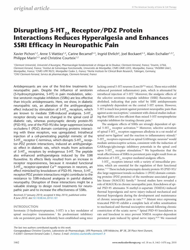
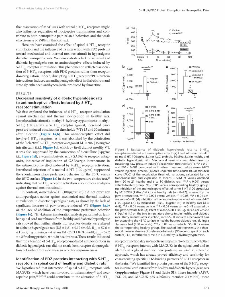
resultsdecreased sensitivity of diabetic hyperalgesic rats to antinociceptive effects induced by 5-Ht2A receptor stimulationWe first explored the influence of 5-HT2A receptor stimulation against mechanical and thermal nociception in healthy rats. Intrathecal injection of α-methyl-5-hydroxytryptamine (α-methyl-5-HT) (100 µg/rat), a 5-HT2A receptor agonist, increased paw-pressure-induced vocalization thresholds (VT) 15 and 30 minutes after injection (Figure 1a,b). This antinociceptive effect did involve 5-HT2A receptors, as it was abolished by the coinjection of the “selective” 5-HT2A receptor antagonist M100907 [150 ng/rat intrathecally (i.t.), Figure 1c], which by itself did not modify VT. It was also suppressed by the coinjection of bicuculline (3 µg/rat i.t., Figure 1d), a γ-aminobutyric acid (GABA)-A receptor antag-onist, indicative of implication of GABAergic interneurons in the antinociceptive effect induced by 5-HT2A receptor activation. Intrathecal injection of α-methyl-5-HT (100 µg/rat) suppressed the spontaneous place preference behavior for the 25 °C versus the 45 °C surface (Figure 1e) in the two-temperature choice test,20 indicating that 5-HT2A receptor activation also induces analgesia against thermal noxious stimuli.
In contrast, α-methyl-5-HT (100 µg/rat i.t.) did not exert any antihyperalgesic action against mechanical and thermal noxious stimulations in diabetic hyperalgesic rats, as shown by the lack of significant increase of paw pressure-induced VT (Figure 1a,b) or the lack of abolition of the temperature preference behavior (Figure 1e). [3H]-ketanserin saturation analysis performed on lum-bar spinal cord membranes from healthy and diabetic hyperalgesic rats showed that neither affinity nor receptor density were altered in diabetic hyperalgesic rats (Kd = 1.81 ± 0.17 nmol/l, Bmax = 57.6 ± 4.1 fmol/mg protein, n = 4 versus Kd = 2.03 ± 0.09 nmol/l, Bmax = 59.2 ± 4.8 fmol/mg protein, n = 4, in healthy rats). These results indicated that the alteration of 5-HT2A receptor-mediated antinociception in diabetic hyperalgesic rats did not result from receptor downregula-tion but rather from a decrease in receptor responsiveness.
Identification of PdZ proteins interacting with 5-Ht2A receptors in spinal cord of healthy and diabetic ratsWe hypothesized that interaction of spinal 5-HT2A receptors with MAGUKs, which have been involved in inflammatory21 and neu-ropathic pain,14,15,17–19 could contribute to the alteration of 5-HT2A
receptor functionality in diabetic neuropathy. To determine whether 5-HT2A receptors interact with MAGUKs in the spinal cord and to identify in a global manner these proteins, we used a proteomic approach, which has already proved efficiency and sensitivity for characterizing specific PDZ-binding partners of 5-HT receptors in the brain.13 We identified five protein partners of the 5-HT2A recep-tor in spinal cord extracts from healthy and diabetic hyperalgesic rats (Supplementary Figure S1 and Table S1). These include SAP97, PSD-95, and MAGUK p55 subfamily member 2 (MPP2), three
Vehicle
M100907
α-me-5-HT
α-me-5-HT+ M100907
***
#
##
4,000
3,000
2,000
AU
C (
g.m
inut
es)
1,000
−1,000
0
Healthy Diabetic
Vehicle
α-me-5-HT
**
#
#
120
100
80
60
40
20
45 °C0
Tim
e du
ratio
n (s
econ
ds)
Vehicle
Bicu
α-me-5-HT
α-me-5-HT+ Bicu
**
####
4,000
3,000
2,000A
UC
(g.
min
utes
)1,000
−1,000
0
Healthy
Diabetic
Vehicle
α-me-5-HT
***
#
4,000
3,000
2,000
AU
C (
g.m
inut
es)
1,000
−1,000
0
450Vehicle
Healthy
Diabetic
α-me-5HT400§§§
§§
350
300
VT
(g)
250
200
1500 30 60
Time (minutes)
BeforeSTZ
90 120
a
b c
d e
Figure 1 resistance of diabetic hyperalgesic rats to 5-Ht2A receptor- mediated antinociceptive effect. (a) Effect of α-methyl-5-HT (α-me-5-HT, 100 µg/rat i.t.) or NaCl (vehicle, 10 µl/rat i.t.) in healthy and diabetic hyperalgesic rats. Mechanical sensitivity was determined by measuring paw-pressure induced vocalization thresholds (VT). §§P < 0.01 and §§§P < 0.001 compared with values measured before α-me-5-HT/vehicle injection (time 0). (b) Area under the time-course (0–60 minutes) curve (AUC) of the vocalization threshold variations, calculated by the trapezoidal rule and expressed as means ± SEM of values obtained from 20 to 21 healthy and 6 to 10 diabetic rats. ***P < 0.001 versus vehicle-treated group. #P < 0.05 versus corresponding healthy group. (c) Inhibition of the antinociceptive effect of α-me-5-HT (100 µg/rat i.t.) by M100907 (150 ng/rat i.t.) in healthy rats (n = 8–12), assessed by the paw-pressure test. ***P < 0.001 versus vehicle. #P < 0.05, ##P < 0.01 ver-sus α-me-5-HT. (d) Inhibition of the antinociceptive effect of α-me-5-HT (100 µg/rat i.t.) by bicuculline (Bicu, 3 µg/rat i.t.) in healthy rats (n = 6–8). **P < 0.01 versus vehicle. ##P < 0.01 versus α-me-5-HT assessed by the paw-pressure test. (e) Effect of α-me-5-HT (100 µg/ rat i.t.) or vehicle (10 µl/rat i.t.) on the two-temperature choice test in healthy and diabetic rats. Thirty minutes after injection, α-me-5-HT induces a behavioral bias for occupying the 45 °C surface in healthy but not diabetic rats, during a 3-minute trial (180 seconds). **P < 0.01 versus vehicle. #P < 0.05 versus the corresponding healthy group. The dashed line represents the theo-retical mean in absence of preference behavior (90 seconds spent on each surface). i.t., intrathecal; α-me-5-HT, α-methyl-5-hydroxytryptamine.
1464 www.moleculartherapy.org vol. 18 no. 8 aug. 2010
© The American Society of Gene & Cell Therapy5-HT2AR/PDZ Protein Disruption in Neuropathic Pain
MAGUKs, and MAGI2, a MAGUK with inverted domains. These proteins, except for MPP2, were previously identified as PDZ part-ners of brain 5-HT2A receptors.13 The 5-HT2A receptor C-terminus also recruited Sec23A, one subunit of the coat complex II (COPII), involved in protein export from endoplasmic reticulum to either the endoplasmic reticulum–Golgi intermediate compartment or to the Golgi complex. Although Sec23A lacks any PDZ domains, the PDZ-binding motif located at the 5-HT2A receptor C-terminus contributed to its interaction with the receptor (Supplementary Figure S1a).
Proteomic analysis also revealed that the amount of PSD-95 (the first MAGUK that has been implicated in chronic pain14–16,18,19) recruited by the 5-HT2A receptor C-terminus in spinal cord was greater in diabetic hyperalgesic rats than in healthy rats (Supplementary Figure S1a). Correspondingly, western blotting experiments indicated an increase in PSD-95 expression in dorsal spinal cord of diabetic hyperalgesic rats, compared with healthy rats (+52 ± 4%, n = 4, Supplementary Figure S1b).
disruption of 5-Ht2A receptor/PdZ protein interactions suppresses hyperalgesia in diabetic ratsWe next evaluated the overall contribution of 5-HT2A receptor/PDZ protein interactions to pain sensitivity in diabetic neu-ropathic rats, using a peptide comprising the nine C-terminal residues of the 5-HT2A receptor that competes for interactions between the receptor’s PDZ-binding motif and target PDZ pro-teins. This peptide was fused N-terminally with the transduction domain of human immunodeficiency virus type 1 Tat protein (TAT-2ASCV) to allow its intracellular delivery.22 Pull-down
experiments showed that TAT-2ASCV, but not the correspond-ing peptide in which the C-terminal valine was substituted by an alanine (TAT-2ASCA), efficiently prevented association between the receptor and its MAGUK partners PSD-95 and SAP97 in vitro (Supplementary Figure S2a). The ability of these peptides to transduce into spinal neurons following intrathecal injection was verified by using methanethiosulfonate (MTS)-4-fluorescein-labeled peptides. Fluorescent labeling of spinal cord cells was detectable for 1 hour after intrathecal injection of MTS-4-TAT-2ASCV, but not following injection of the corresponding peptide lacking the Tat transduction domain (MTS-4-TAT-empty-2ASCV, Supplementary Figure S2b). Moreover, intrathecal injection of TAT-2ASCV decreased coimmunoprecipitation of PSD-95 and SAP97 with 5-HT2A receptors in spinal cord extracts (Supplementary Figure S2c), indicating that TAT-2ASCV inhib-ited interaction between 5-HT2A receptor and its MAGUK part-ners in vivo. In contrast, injection of TAT-2ASCA did not inhibit interaction between spinal 5-HT2A receptor and PDZ proteins (Supplementary Figure S2c). This peptide was further used as control in behavioral studies.
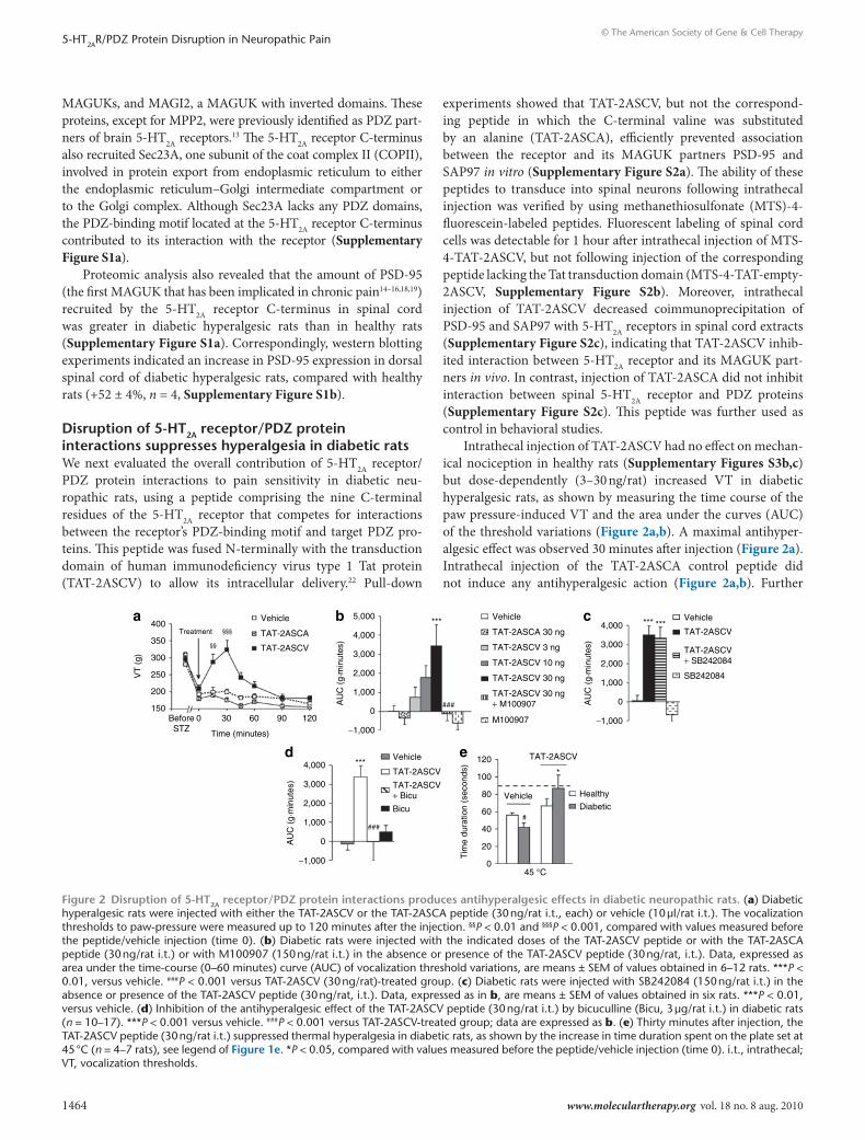
Intrathecal injection of TAT-2ASCV had no effect on mechan-ical nociception in healthy rats (Supplementary Figures S3b,c) but dose-dependently (3–30 ng/rat) increased VT in diabetic hyperalgesic rats, as shown by measuring the time course of the paw pressure-induced VT and the area under the curves (AUC) of the threshold variations (Figure 2a,b). A maximal antihyper-algesic effect was observed 30 minutes after injection (Figure 2a). Intrathecal injection of the TAT-2ASCA control peptide did not induce any antihyperalgesic action (Figure 2a,b). Further
Vehicle
Treatment TAT-2ASCA
TAT-2ASCV
400§§§
§§350
300
VT
(g)
250
200
1500 30 60
Time (minutes)
BeforeSTZ
90 120
a
M100907
***
4,000
5,000
3,000
2,000
AU
C (
g.m
inut
es)
−1,000
0###
Vehicle
TAT-2ASCA 30 ng
TAT-2ASCV 3 ng
TAT-2ASCV 10 ng
TAT-2ASCV 30 ng
TAT-2ASCV 30 ng+ M100907
b
1,000
*
Healthy
Diabetic
45 °C
120
100
80
60
40
20
0
Tim
e du
ratio
n (s
econ
ds)
#
Vehicle
TAT-2ASCVe***
###
Vehicle
TAT-2ASCV
TAT-2ASCV+ Bicu
Bicu
d4,000
3,000
2,000
AU
C (
g.m
inut
es)
−1,000
0
1,000
****** Vehicle
TAT-2ASCV
TAT-2ASCV+ SB242084
SB242084
c4,000
3,000
2,000
AU
C (
g.m
inut
es)
−1,000
0
1,000
Figure 2 disruption of 5-Ht2A receptor/PdZ protein interactions produces antihyperalgesic effects in diabetic neuropathic rats. (a) Diabetic hyperalgesic rats were injected with either the TAT-2ASCV or the TAT-2ASCA peptide (30 ng/rat i.t., each) or vehicle (10 µl/rat i.t.). The vocalization thresholds to paw-pressure were measured up to 120 minutes after the injection. §§P < 0.01 and §§§P < 0.001, compared with values measured before the peptide/vehicle injection (time 0). (b) Diabetic rats were injected with the indicated doses of the TAT-2ASCV peptide or with the TAT-2ASCA peptide (30 ng/rat i.t.) or with M100907 (150 ng/rat i.t.) in the absence or presence of the TAT-2ASCV peptide (30 ng/rat, i.t.). Data, expressed as area under the time-course (0–60 minutes) curve (AUC) of vocalization threshold variations, are means ± SEM of values obtained in 6–12 rats. ***P < 0.01, versus vehicle. ###P < 0.001 versus TAT-2ASCV (30 ng/rat)-treated group. (c) Diabetic rats were injected with SB242084 (150 ng/rat i.t.) in the absence or presence of the TAT-2ASCV peptide (30 ng/rat, i.t.). Data, expressed as in b, are means ± SEM of values obtained in six rats. ***P < 0.01, versus vehicle. (d) Inhibition of the antihyperalgesic effect of the TAT-2ASCV peptide (30 ng/rat i.t.) by bicuculline (Bicu, 3 µg/rat i.t.) in diabetic rats (n = 10–17). ***P < 0.001 versus vehicle. ###P < 0.001 versus TAT-2ASCV-treated group; data are expressed as b. (e) Thirty minutes after injection, the TAT-2ASCV peptide (30 ng/rat i.t.) suppressed thermal hyperalgesia in diabetic rats, as shown by the increase in time duration spent on the plate set at 45 °C (n = 4–7 rats), see legend of Figure 1e. *P < 0.05, compared with values measured before the peptide/vehicle injection (time 0). i.t., intrathecal; VT, vocalization thresholds.
Molecular Therapy vol. 18 no. 8 aug. 2010 1465
© The American Society of Gene & Cell Therapy5-HT2AR/PDZ Protein Disruption in Neuropathic Pain
supporting that the effect of TAT-2ASCV was due to the inhibi-tion of 5-HT2A receptor/PDZ protein interactions, its effect on mechanical hyperalgesia was entirely inhibited by the coadmin-istration of M100907 (150 ng/rat i.t., Figure 2b), whereas it was not affected by the 5-HT2C receptor antagonist SB242084 (150 ng/rat i.t., Figure 2c). Finally, inhibition of mechanical hyperalge-sia by TAT-2ASCV was abolished by the coadministration of the GABA-A receptor antagonist bicuculline (3 µg/rat i.t., Figure 2d). The maximally effective dose of TAT-2ASCV against mechanical hyperalgesia (30 ng/rat) also suppressed the spontaneous temper-ature preference behavior in diabetic hyperalgesic rats, whereas it did not induce analgesic effect against noxious thermal stimulus in healthy animals (Figure 2e).
Intrathecal injection of TAT-2ASCV (30 ng/rat) did not pre-vent co-immunoprecipitation of PSD-95 with NMDA recep-tors in spinal cord extracts (Supplementary Figure S3a), suggesting that this peptide specifically inhibited interaction of MAGUKs with 5-HT2A receptors but not their association with other receptors known to bind to these PDZ proteins and to be involved in pain processing. Further supporting its specific-ity toward 5-HT2A receptor-mediated antihyperalgesia, injec-tion of TAT-2ASCV did not affect NMDA-induced mechanical hypersensitivity in healthy rats (Supplementary Figures S3b,c). Furthermore, rats showed normal motor activity after admin-istration of TAT-2ASCV, as assessed by their spontaneous motor activity, grip strength, or righting score (Supplementary Figures S3d–f), indicating that the peptide did not affect motor functions. Collectively, our observations indicate that disruption of 5-HT2A receptor/PDZ protein interactions reveals an antihy-peralgesic action of 5-HT2A receptors activated by endogenous 5-HT in diabetic neuropathy.
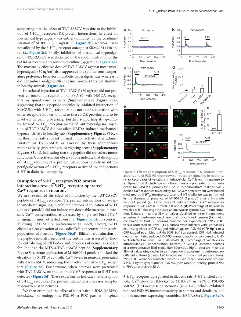
disruption of 5-Ht2A receptor/PdZ protein interactions reveals 5-Ht2A receptor-operated ca2+ responses in neuronsWe next examined the effect of inhibition by the TAT-2ASCV peptide of 5-HT2A receptor/PDZ protein interactions on recep-tor-mediated signaling in cultured neurons. Application of 5-HT (up to 10 µmol/l) did not induce any detectable increase in cyto-solic Ca2+ concentration, as assessed by single-cell Fura-2 Ca2+ imaging, in most of tested neurons (Figure 3a,d). In contrast, following TAT-2ASCV pretreatment (1 µmol/l, 1 hour), 5-HT elicited a clear elevation of cytosolic Ca2+ concentration in a sub-population of neurons (Figure 3b,d). Efficient transduction of the peptide into all neurons of the culture was assessed by fluo-rescent labeling of cell bodies and processes of neurons exposed for 1 hour to the MTS-4-TAT-2ASCV peptide (Supplementary Figure S4). Acute application of M100907 (1 µmol/l) blocked the elevation by 5-HT of cytosolic Ca2+ levels in neurons pretreated with TAT-2ASCV, indicating the involvement of 5-HT2A recep-tors (Figure 3c). Furthermore, when neurons were pretreated with TAT-2ASCA, no induction of Ca2+ responses to 5-HT was detected (Figure 3d). These experiments indicate that disruption of 5-HT2A receptor/PDZ protein interactions increases receptor responsiveness in neurons.
We then examined the effect of short hairpin RNA (shRNA) knockdown of endogenous PSD-95, a PDZ partner of spinal
5-HT2A receptors upregulated in diabetic rats. 5-HT elicited cyto-solic Ca2+ elevation (blocked by M100907) in ≈25% of PSD-95 shRNA (Hp1)-expressing neurons (n = 120), which exhibited reduced PSD-95 immunoreactivity in somata and dendrites, but not in neurons expressing scrambled shRNA (Scr1, Figure 3e,f).
e f **
30
40
% o
f GF
P-p
ositi
ve
resp
ondi
ng n
euro
ns
20
10
0 Scr1 Hp1
a 1.2
1
0.8
0.6
0.4
0.2
0 0 50
5-HT
F34
0/F
380
No peptide
100
Time (seconds)
200 150
GFP-Hp1 GFP-Hp1 GFP-Scr1 PSD-95
GFP-Hp1PSD-95
GFP-Hp1PSD-95
PSD-95
PSD-95
GFP-Scr1
GFP-Scr1
PSD-95
5-HT 5-HT 1.2
1
0.8
0.6
0.4
0.2
F34
0/F
380 M100907
0 100 200 Time (seconds)
400 300
b
5-HT 5-HT
1.2
1
0.8
0.6
0.4
0.2
0
F34
0/F
380
TAT-2ASCV
0 100 200
Time (seconds)
400 300
**
d 30
20
% o
f res
pond
ing
neur
ons
10
0
No pe
ptide
TAT
TAT-2ASCA
TAT-2ASCV
1.2
1
0.8
0.6
0.4
0.2
0
5-HT 5-HT
F34
0/F
380
TAT-2ASCV
M100907
0 100 200
Time (seconds)
400 300
c
Figure 3 effects of disruption of 5-Ht2A receptor/PdZ protein inter-actions and of Psd-95 knockdown on receptor signaling in neurons. (a–c) Recordings of variations in intracellular Ca2+ levels in response to a 10 µmol/l 5-HT challenge in cultured neurons pretreated or not with either TAT-2ASCV (1 µmol/l) for 1 hour. To demonstrate that the 5-HT-evoked Ca2+ responses revealed by TAT-2ASCV pretreatment were indeed mediated by 5-HT2A receptors, a second 5-HT challenge was performed in the absence or presence of M100907 (1 µmol/l) after a 3- minute washout period (c). Only traces of cells exhibiting Ca2+ increase in response to 5-HT are illustrated in b and c. (d) Percentage of neurons in which a 5-HT challenge induced an increase in cytosolic Ca2+ concentra-tion. Data are means ± SEM of values obtained in three independent experiments performed on different sets of cultured neurons (four fields containing at least 80 neurons counted per experiment). **P < 0.01 versus untreated neurons. (e) Neurons were infected with lentiviruses expressing either a GFP-tagged shRNA against PSD-95 (GFP-Hp1) or a GFP-tagged scrambled shRNA (GFP-Scr1) as control. GFP-Hp1-infected neurons exhibited reduced PSD-95 immunoreactivity, compared to GFP-Scr1-infected neurons. Bar = 20 µmol/l. (f) Recordings of variations in intracellular Ca2+ concentration (bottom) in GFP-Hp1-infected neurons of a representative field (top). Bar: 50 µmol/l. Right: data are means ± SEM of values obtained in three independent experiments performed on different cultures (at least 120 infected neurons counted per condition). **P < 0.01 versus Scr1-infected neurons. GFP, green fluorescent protein; 5-HT, 5-hydroxytryptamine; PSD-95, postsynaptic density protein-95; shRNA, short hairpin RNA.
1466 www.moleculartherapy.org vol. 18 no. 8 aug. 2010
© The American Society of Gene & Cell Therapy5-HT2AR/PDZ Protein Disruption in Neuropathic Pain
Thus, PSD-95 knockdown mostly reproduced the effect of TAT-2ASCV, underscoring the critical role of this MAGUK in the regulation of 5-HT2A receptor functional status.
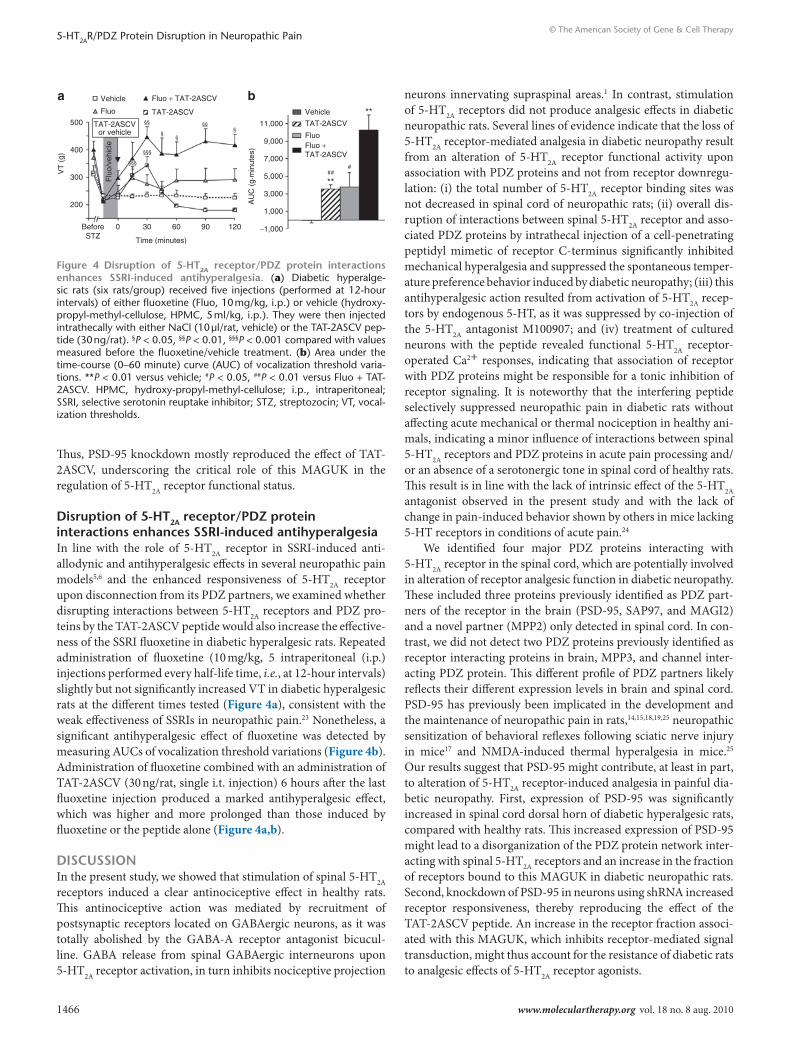
disruption of 5-Ht2A receptor/PdZ protein interactions enhances ssrI-induced antihyperalgesiaIn line with the role of 5-HT2A receptor in SSRI-induced anti-allodynic and antihyperalgesic effects in several neuropathic pain models5,6 and the enhanced responsiveness of 5-HT2A receptor upon disconnection from its PDZ partners, we examined whether disrupting interactions between 5-HT2A receptors and PDZ pro-teins by the TAT-2ASCV peptide would also increase the effective-ness of the SSRI fluoxetine in diabetic hyperalgesic rats. Repeated administration of fluoxetine (10 mg/kg, 5 intraperitoneal (i.p.) injections performed every half-life time, i.e., at 12-hour intervals) slightly but not significantly increased VT in diabetic hyperalgesic rats at the different times tested (Figure 4a), consistent with the weak effectiveness of SSRIs in neuropathic pain.23 Nonetheless, a significant antihyperalgesic effect of fluoxetine was detected by measuring AUCs of vocalization threshold variations (Figure 4b). Administration of fluoxetine combined with an administration of TAT-2ASCV (30 ng/rat, single i.t. injection) 6 hours after the last fluoxetine injection produced a marked antihyperalgesic effect, which was higher and more prolonged than those induced by fluoxetine or the peptide alone (Figure 4a,b).
dIscussIonIn the present study, we showed that stimulation of spinal 5-HT2A receptors induced a clear antinociceptive effect in healthy rats. This antinociceptive action was mediated by recruitment of postsynaptic receptors located on GABAergic neurons, as it was totally abolished by the GABA-A receptor antagonist bicucul-line. GABA release from spinal GABAergic interneurons upon 5-HT2A receptor activation, in turn inhibits nociceptive projection
neurons innervating supraspinal areas.1 In contrast, stimulation of 5-HT2A receptors did not produce analgesic effects in diabetic neuropathic rats. Several lines of evidence indicate that the loss of 5-HT2A receptor-mediated analgesia in diabetic neuropathy result from an alteration of 5-HT2A receptor functional activity upon association with PDZ proteins and not from receptor downregu-lation: (i) the total number of 5-HT2A receptor binding sites was not decreased in spinal cord of neuropathic rats; (ii) overall dis-ruption of interactions between spinal 5-HT2A receptor and asso-ciated PDZ proteins by intrathecal injection of a cell- penetrating peptidyl mimetic of receptor C-terminus significantly inhibited mechanical hyperalgesia and suppressed the spontaneous temper-ature preference behavior induced by diabetic neuropathy; (iii) this antihyperalgesic action resulted from activation of 5-HT2A recep-tors by endogenous 5-HT, as it was suppressed by co-injection of the 5-HT2A antagonist M100907; and (iv) treatment of cultured neurons with the peptide revealed functional 5-HT2A receptor-operated Ca2+ responses, indicating that association of receptor with PDZ proteins might be responsible for a tonic inhibition of receptor signaling. It is noteworthy that the interfering peptide selectively suppressed neuropathic pain in diabetic rats without affecting acute mechanical or thermal nociception in healthy ani-mals, indicating a minor influence of interactions between spinal 5-HT2A receptors and PDZ proteins in acute pain processing and/or an absence of a serotonergic tone in spinal cord of healthy rats. This result is in line with the lack of intrinsic effect of the 5-HT2A antagonist observed in the present study and with the lack of change in pain-induced behavior shown by others in mice lacking 5-HT receptors in conditions of acute pain.24
We identified four major PDZ proteins interacting with 5-HT2A receptor in the spinal cord, which are potentially involved in alteration of receptor analgesic function in diabetic neuropathy. These included three proteins previously identified as PDZ part-ners of the receptor in the brain (PSD-95, SAP97, and MAGI2) and a novel partner (MPP2) only detected in spinal cord. In con-trast, we did not detect two PDZ proteins previously identified as receptor interacting proteins in brain, MPP3, and channel inter-acting PDZ protein. This different profile of PDZ partners likely reflects their different expression levels in brain and spinal cord. PSD-95 has previously been implicated in the development and the maintenance of neuropathic pain in rats,14,15,18,19,25 neuropathic sensitization of behavioral reflexes following sciatic nerve injury in mice17 and NMDA-induced thermal hyperalgesia in mice.25 Our results suggest that PSD-95 might contribute, at least in part, to alteration of 5-HT2A receptor-induced analgesia in painful dia-betic neuropathy. First, expression of PSD-95 was significantly increased in spinal cord dorsal horn of diabetic hyperalgesic rats, compared with healthy rats. This increased expression of PSD-95 might lead to a disorganization of the PDZ protein network inter-acting with spinal 5-HT2A receptors and an increase in the fraction of receptors bound to this MAGUK in diabetic neuropathic rats. Second, knockdown of PSD-95 in neurons using shRNA increased receptor responsiveness, thereby reproducing the effect of the TAT-2ASCV peptide. An increase in the receptor fraction associ-ated with this MAGUK, which inhibits receptor-mediated signal transduction, might thus account for the resistance of diabetic rats to analgesic effects of 5-HT2A receptor agonists.
a b
11,000
9,000
7,000
5,000
500
Vehicle
Vehicle Fluo
Fluo Fluo + TAT-2ASCV
Fluo + TAT-2ASCV
TAT-2ASCV
TAT-2ASCV TAT-2ASCV or vehicle
400
300
VT
(g)
200
0 30 60
Time (minutes)
Flu
o/ve
hicl
e
AU
C (
g.m
inut
es)
90 120 Before STZ
3,000
1,000
−1,000
**
** ##
#
§
§ §
§§ §§
§§§
§§§
Figure 4 disruption of 5-Ht2A receptor/PdZ protein interactions enhances ssrI-induced antihyperalgesia. (a) Diabetic hyperalge-sic rats (six rats/group) received five injections (performed at 12-hour intervals) of either fluoxetine (Fluo, 10 mg/kg, i.p.) or vehicle (hydroxy-propyl-methyl-cellulose, HPMC, 5 ml/kg, i.p.). They were then injected intrathecally with either NaCl (10 µl/rat, vehicle) or the TAT-2ASCV pep-tide (30 ng/rat). §P < 0.05, §§P < 0.01, §§§P < 0.001 compared with values measured before the fluoxetine/vehicle treatment. (b) Area under the time-course (0–60 minute) curve (AUC) of vocalization threshold varia-tions. **P < 0.01 versus vehicle; #P < 0.05, ##P < 0.01 versus Fluo + TAT-2ASCV. HPMC, hydroxy-propyl-methyl-cellulose; i.p., intraperitoneal; SSRI, selective serotonin reuptake inhibitor; STZ, streptozocin; VT, vocal-ization thresholds.
Molecular Therapy vol. 18 no. 8 aug. 2010 1467
© The American Society of Gene & Cell Therapy5-HT2AR/PDZ Protein Disruption in Neuropathic Pain
A role of PSD-95 in hallucinogen and antipsychotic drug actions at 5-HT2A receptors has recently been described.26 The spe-cific influence of the interaction of 5-HT2A receptors with PSD-95 in the resistance of diabetic hyperalgesic rats to 5-HT2A receptor-mediated analgesic effects could not be definitely established by means of spinal PSD-95 knockdown using shRNA. Indeed, knockdown of PSD-95 was previously shown to strongly affect NMDA receptor-dependent hyperalgesia,16 precluding discrimi-nation between NMDA receptor- and 5-HT2A receptor- dependent effects on nociceptive transmission. This contrasts with the pep-tidyl mimetic approach used in the present report, which selec-tively impaired 5-HT2A receptor/PSD-95 interaction without affecting association of this MAGUK with NMDA receptor and thereby did not alter NMDA receptor-dependent modulation of pain hypersensitivity. Alternative strategies based on disruption of protein–protein interactions within the NMDA receptor com-plex have recently been proposed to suppress acute and chronic pain hypersensitivity depending on NMDA receptors.21,25,27 These studies together with the present findings point out the relevance of strategies blocking protein–protein interactions to manage chronic pain, with presumably less pronounced side effects than classical approaches based on receptor agonists or antagonists.
Due to the critical role of 5-HT2A receptors in SSRI-induced analgesic effects,5,6 the decreased analgesic function of these receptors in diabetic hyperalgesic rats might be responsible for the low efficacy of SSRIs in painful neuropathy, compared with other antidepressants inhibiting reuptake of norepinephrine as well, such as the tricyclic amitriptyline.23 Consistent with this hypothesis, disconnection of 5-HT2A receptors from their PDZ partners strongly enhanced the antihyperalgesic action of flu-oxetine in diabetic neuropathic rats. Moreover, a prolonged analgesia was observed in animals co-injected with fluoxetine and the TAT-2ASCV peptide. The low efficacy of SSRIs to relieve neu-ropathic pain lead clinicians to prescribe more efficacious but less tolerated compounds, such as amitriptyline, the gold standard of analgesic antidepressants.23 However, a substantial percentage of patients does not favorably respond to this compound,28 which might reflect an antagonistic activity at 5-HT2A receptor.29 Our data show that SSRIs become effective against neuropathic pain providing that spinal 5-HT2A receptors are disconnected from their associated PDZ proteins. Targeting interactions between 5-HT2A receptors and associated PDZ proteins, which is efficient against both mechanical and thermal hyperalgesia, two positive sensory signs frequently observed in neuropathic pain,30 could be an attractive strategy to increase the effectiveness of SSRIs in this context.
MAterIAls And MetHodsChemicals. Streptozocin (STZ, Zanosar) was purchased from Pfizer (Amboise, France), insulin (Lantus) from Sanofi-Aventis (Paris, France), α-methyl-5-HT maleate, SB242084, bicuculline and cocaine from Sigma (St Quentin Fallavier, France), fluoxetine from RPG Laboratory (Paris, France) and diazepam (Valium) from Roche (Neuilly-sur-Seine, France). M100907 was generously provided by Dr Clotilde Mannoury la Cour (Servier Pharmaceuticals, Croissy sur Seine, France). Drugs were dissolved in physiological saline (NaCl, 0.9%), except M100907, SB242084, and bicuculline, dissolved in 50, 50, and 30% dimethyl sulfoxide, respectively, fluoxetine, dissolved in hydroxy-propyl-methyl-cellulose (Colorcon,
Orpington, England), STZ, dissolved in distilled water and diazepam dis-solved in ethanol.
Peptides, shRNAs, and antibodies. Synthetic peptides (>95% purity) were purchased from Eurogentec (Seraing, Belgium). Peptide sequences were as follows: 5-HT2ASCVCt, TDNIETVNEKVSCV; 5-HT2ASCACt, TDNIETVNEKVSCA; TAT-2ASCV, YGRKKRRQRRRTVNEKVSCV; TAT-2ASCA, YGRKKRRQRRRTVNEKVSCA. The TAT-2ASCV peptide and the corresponding peptide lacking the TAT sequence (TAT-empty-2ASCV, TVNEKVSCV) were synthesized with an additional N-terminal cysteinyl residue and conjugated to 2-[(5-fluoresceinyl)aminocarbonyl]ethyl MTS (MTS-4-fluorescein; Toronto Research; North York, Ontario, Canada) to monitor peptide transduction into spinal cord neurons follow-ing intrathecal delivery by fluorescence microscopy.31
Constructs expressing short hairpin RNA (shRNA) against PSD-95 (Hp1) or scrambled shRNA (Scr1) under control of U6 promoter were modified from ref. 3 for lentiviral coexpression with green fluorescent protein under control of ubiquitin promoter.32 Lentiviral production has been described elsewhere.32,33 Cultured neurons were infected at 3 days in vitro and experiments were performed at 13 days in vitro.
The mouse monoclonal anti-5-HT2A receptor antibody was obtained from BD Biosciences (San Jose, CA), the rabbit polyclonal anti-5-HT2A receptor antibody from Abcam (Cambridge, UK), the mouse anti-PSD-95 IgG2A (clone K28/43) from Upstate (Lake Placid, NY), the mouse anti-SAP97 IgG1 from StressGen Bioreagents (Victoria, British Columbia, Canada), the mouse monoclonal anti-NR1 antibody from Zymed Laboratories (San Francisco, CA), the rabbit polyclonal anti-NR1 antibody from Upstate and the mouse anti-pan-actin monoclonal antibody from NeoMarkers (Fremont, CA).
Animals and behavioral testing. All experiments were performed on adult male Sprague–Dawley rats (200–250 g) (Charles River, Cléon, France) in accordance with the IAPS guidelines34 and were approved by the local eth-ics committee of Auvergne (agreement no. CE3-06). Diabetes was induced by an i.p. injection of STZ (72 mg/kg). Healthy rats only received dis-tilled water (5 ml/kg, i.p.). Diabetic rats (blood glucose level >14 mmol/l) received every other day a subcutaneous insulin injection (2 IU/rat), from 7 days after STZ injection. Diabetic rats were previously found to display abnormal pain-associated behaviors, measured as exaggerated responses to painful stimuli (hyperalgesia).35
Hypersensitivity to mechanical stimuli was determined by applying an increasing pressure to the left hind paw until a squeak was elicited (vocalization threshold expressed in grams, maximal pressure applied was 550 g). Data are expressed as mean ± SE of the mean (SEM) of the VT (g). To investigate global effects, areas under the time-course curves (AUC) of the vocalization threshold variations between 0 and 60 minutes were calculated using the trapezoidal rule as followed: ∑[(VTtime X – VTtime 0)· (time X – time X–1)]. Diabetic rats were considered hyperalgesic if the VT were reduced to at least 20%, compared with VT measured before STZ treatment. Hyperalgesia to thermal stimuli was assessed using the two-temperature choice test.20 Rats were freely moving on two adjoining plates set at different temperatures: 25 °C (thermoneutral plate) and the other one at 45 °C (nociceptive plate). They were free to choose between occupying the hot surface versus staying on the surface at neutral temperature. For this test, data are expressed as time spent on the hot surface (seconds). When both plates were set to 25 °C, rats spent about 50% of the time i.e., 90 seconds on each plate. When diabetic rats were presented to a choice of occupying the hot surface versus the neutral surface, a 20% decrease in the time spent on the hot surface was measured compared with healthy rats, indicating thermal hyperalgesia. Only hyperalgesic diabetic rats were included in the study. Different rats were used for the different behavioral testings.
Hyperalgesic diabetic and healthy rats were injected i.t. with drugs 3 weeks after the STZ/vehicle treatment. In experiments using α-methyl-5-HT or
1468 www.moleculartherapy.org vol. 18 no. 8 aug. 2010
© The American Society of Gene & Cell Therapy5-HT2AR/PDZ Protein Disruption in Neuropathic Pain
cell-penetrating peptides, the VT to paw-pressure were measured up to 120 minutes after the injection and the time spent on each surface was measured at 30 minutes. In experiments using fluoxetine, diabetic hyperalgesic rats received five fluoxetine injections (10 mg/kg i.p. each, one injection every 12 hours, 8:00 am and 8:00 pm). Six hours after the last fluoxetine injection, rats were injected with the TAT-2ASCV peptide (30 ng/rat, i.t.) or the vehicle and submitted to the paw-pressure test for 120 minutes.
Motor function, muscular tone, and coordination were estimated by measuring spontaneous locomotor activity, grip strength response, and righting reflex response, respectively. To determine spontaneous locomotor activity, rats were placed in a rectangular cage (45 × 30 × 20 cm) and allowed to investigate the cage for 3 minutes, during which the total number of movements were recorded (Actisystem, Penlab, Apelex). The grip strength was measured by placing the rat forelimbs on a dynamometer (Bioseb, Chaville, France) and pulling on its tail until the rat released the grill. At that time, the score (expressed in arbitrary units) is read on the strain gauge. For determination of the righting reflex response, rats were held on their back on a padded flat surface and then released. The righting reflex was considered positive (quoted 1) if the rats lying on their back were able to right themselves on all four limbs within 1 second. Three consecutive trials were performed (maximal score: 3). These parameters were determined after injection of either vehicle (10 µl/rat, i.t.), TAT-2ASCV (30 ng/rat, i.t.), cocaine (1 mg/kg, intravenously), or diazepam (2,5 mg/kg, i.p.).
All the in vivo experiments were performed blind by a single experimenter using a parallel group design. Treatments were randomized.
Preparation of spinal cord extracts. Hyperalgesic diabetic and healthy rats were sacrificed by decapitation. Lumbar enlargements of the spinal cord dorsal horn were rapidly removed and homogenized on ice with lysis buffer containing Tris-HCl, 50 mmol/l, pH 7.4; EDTA, 0.5 mmol/l; 3-[(3- cholamidopropyl) dimethylammonio]propanesulfonate (1.3%, wt/ vol), and a protease inhibitor cocktail (Roche Diagnostics, Mannheim, Germany). Then, samples were centrifuged for 1 hour at 10,000g and the supernatants containing solubilized proteins were collected.
Peptide affinity chromatography. Solubilized proteins (5 mg/condition) were incubated in the presence of either a peptide encompassing the 14 C-terminal residues of the mouse and rat 5-HT2A receptor (5-HT2ASCVCt) or a modified peptide in which the C-terminal valine residue was substituted into an alanine (5-HT2ASCACt), immobilized onto activated CH-sepharose 4B (GE Healthcare, Uppsala, Sweden), as previously described.13 Samples were washed five times with lysis buffer supplemented with 5 mol/l NaCl and once with lysis buffer without NaCl. Proteins retained by affinity were eluted off with either sodium dodecyl sulfate (SDS) sample buffer (Tris-HCl, 50 mmol/l, pH 6.8; SDS, 2%; glycerol 30%; dithiothreitol 100 mmol/l, and bromophenol blue) for SDS-polyacrylamide gel electrophoresis and immunoblotting or isoelectrofocusing medium containing urea (7 mol/l), thiourea (2 mol/l), 3-[(3-cholamidopropyl) dimethylammonio]pro-panesulfonate (4%), ampholines (preblended, pI 3.5–9.5, 8 mg/ml; GE Healthcare), dithiothreitol (100 mmol/l), tergitol NP7 (0.2%) and traces of bromophenol blue for 2-D electrophoresis.
2-D electrophoresis and 2-D gel protein pattern analysis. Proteins were first separated according to their isoelectric point along linear immobilized pH-gradient strips (pH 3–10, 18-cm long; GE Healthcare). Sample loading for the first dimension was performed by passive in-gel reswelling. After the first dimension, the immobilized pH-gradient strips were equilibrated for 10 minutes in a buffer containing urea (6 mol/l), Tris-HCl (50 mmol/l, pH 6.8), glycerol (30%), SDS (2%), dithiothreitol (10 mg/ ml), and bromophe-nol blue and then for 15 minutes in the same buffer containing 15 mg/ml iodoacetamide instead of dithiothreitol. For the second dimension, the strips were loaded onto vertical 12.5% SDS-polyacrylamide gel electropho-resis. The gels were stained with silver as previously described.36
Gels to be compared were always processed and stained in parallel. Gels were scanned using a computer-assisted densitometer. Spot detection, gel alignment, and spot quantification were performed using the Image Master Platinium/Melanie 5 software (GE Healthcare). Quantification of proteins was expressed as volumes of spots. To correct the variability resulting from silver staining, results were expressed as relative volumes of total spots in each gel.
Matrix assisted laser desorption/ionization time-of-flight mass spec-trometry and protein identification. Proteins of interest were excised and digested in gel using trypsin (Gold; Promega, charbonniéres-les-Bains, France), as previously described.13 Digest products were completely dehy-drated in a vacuum centrifuge and resuspended in 10 µl formic acid (2%), desalted using Zip Tips C18 (Millipore, Molsheim, France), eluted with 10 µl acetonitrile-trifluoroacetic acid (50–0.1%) and concentrated to 2 µl. Aliquots (0.5 µl) were mixed with the same volume of α-cyano-4-hydroxy-trans-cinnamic acid (Sigma; 10 mg/ml in acetonitrile-trifluoroacetic acid, 50–0.1%) and loaded on the target of an Ultraflex matrix assisted laser desorption/ionization time-of-flight mass spectrometer (Bruker Daltonik, Bremen, Germany). Analyses were performed in reflectron mode with an accelerating voltage of 25 kV and a delayed extraction of 50 ns. Spectra were analyzed using the FlexAnalysis software (version 2.4; Bruker Daltonik) and autoproteolysis products of trypsin (molecular weight: 842.51, 1,045.56, 2,211.10, 2,383.90) were used as internal calibrates. Identification of proteins was performed using the Mascot software (version 2.1; Matrix science, London, UK) against the Swiss-Prot or TrEMBL databases. The fol-lowing parameters were used for database interrogation: mass tolerance of 50 p.p.m. (even if the mass accuracy of our analyses was usually better than 20 p.p.m.); fixed chemical modification, carbamidomethylation of cysteins; variable chemical modification, oxidation of methionines; matching pep-tides with one missed cleavage accepted only when they included two con-secutive basic residues or when arginine or lysine residues were followed by one or several acidic residues inside the peptide aminoacid sequence. Mascot scores >63 and 68 were considered as significant (P < 0.01) for Swiss-Prot and TrEMBL database interrogation, respectively.
Co-immunoprecipitation. 3-[(3-Cholamidopropyl) dimethylammonio]propanesulfonate-soluble dorsal spinal cord protein extracts (5 mg/experi-ment) were incubated overnight at 4 °C with the rabbit polyclonal anti-5-HT2A receptor antibody or the monoclonal anti-NR1 antibody (5 µg). Samples were then incubated for 1 hour at 4 °C with 50 µl of immobilized protein A/G beads (Thermoscientific, Courtabeuf, France). After five washes with lysis buffer, immunoprecipitated proteins were eluted in SDS sample buffer, resolved by SDS-polyacrylamide gel electrophoresis and detected by western blotting.
Western blotting. Proteins resolved on polyacrylamide gels were trans-ferred electrophoretically to nitrocellulose membranes (Hybond-C; GE Healthcare) and stained with Ponceau red, to assess transfer of equal amounts of proteins in each lane. Membranes were blocked with 5% nonfat dry milk diluted in Tris-buffered saline Tween-20 and incubated succes-sively with primary antibodies (anti-5-HT2A receptor mouse IgG1, 1:500, anti-PSD-95 mouse IgG2A, 1:50,000, anti-SAP97 mouse IgG1, 1:500, rabbit anti-NR1, 1:500, mouse anti-pan-actin, 1:2,000) overnight at 4 °C and with a horse-radish peroxidase-conjugated anti-mouse antibody (1:3,000; GE Healthcare) for 1 hour at room temperature. Immunoreactivity was detected with an enhanced chemiluminescence method (ECL detection reagent; GE Healthcare).
[3H]-ketanserin saturation analysis. Lumbar dorsal spinal cords from healthy and diabetic hyperalgesic rats were homogenized in 50 mmol/l Tris-HCl buffer, pH 7.4, containing 0.5 mmol/l EDTA, 10 mmol/l MgSO4, and a cocktail of protease inhibitors (Roche Diagnostics, Basel, Switzerland) and homogenates were centrifuged for 5 minutes at 1,000 g. Supernatants were then centrifuged at 20,000 g for 20 minutes. The
Molecular Therapy vol. 18 no. 8 aug. 2010 1469
© The American Society of Gene & Cell Therapy5-HT2AR/PDZ Protein Disruption in Neuropathic Pain
pellets (membrane fraction) were resuspended in the binding buffer containing 50 mmol/l Tris-HCl, pH 7.4, 0.5 mmol/l EDTA, 10 mmol/l MgSO4, 10 µmol/l pargyline, and 0.1% (wt/vol) ascorbic acid. Membranes (100 µg/assay) were incubated with 0.5–15 nmol/l [3H]-ketanserin (67 Ci/mmol; Perkin Elmer, Courtaboeuf, France) for 2 hours at 22 °C in a total volume of 300 µl. Prazosin (30 nmol/l) was included in all assays to pre-clude binding to α1 receptors. Nonspecific binding was determined in the presence of 50 µmol/l mianserin. The reaction was terminated by rapid filtration over GF-B filters presoaked with 20 mg/ml bovine serum albu-min. Radioactivity retained on the filters was determined by scintillation counting. Bmax and Kd were determined by Scatchard analysis of satura-tion data.
Neuronal cultures and Ca2+ imaging. Primary cultures of cortical neurons were prepared as previously described.31 Briefly, cells from the cerebral cor-tex of 17-day-old Swiss mouse embryos were plated in serum-free medium on Lab-Tek II chamber slides (1 × 10⁶ cells/well; Nalge Nunc International, Naperville, IL), coated successively with poly-l-ornithine (moleculat weight = 40,000; 15 µg/ml) and 10% fetal calf serum + 1 µg/ml laminin (Sigma). The culture medium included a 1:1 mixture of Dulbecco’s modi-fied Eagle’s medium and F-12 nutrient supplemented with 33 mmol/l glu-cose, 2 mmol/l glutamine, 13 mmol/l NaHCO3, 5 mmol/l HEPES buffer, pH 7.4, 5 IU/ml-5 mg/ml) penicillin–streptomycin, and a mixture of salt and hormones containing 100 µg/ml transferrin, 25 µg/ml insulin, 20 nmol/l progesterone, 60 nmol/l putrescine, and 30 nmol/l Na2SeO3. Cultures were maintained for 7 days at 37 °C in a humidified atmosphere in 5% CO2, 95% air. At this stage, cultures were shown to contain at least 95% of neurons.31
Cells were loaded with Fura-2 acetoxymethyl ester (FluoProbes, Interchim, Montluçon, France) at a final concentration of 12.5 µmol/l for 30 minutes at 37 °C in Locke’s solution containing 140 mmol/l NaCl, 1.2 mmol/l KH2PO4, 5 mmol/l KCl, 1.2 mmol/l MgSO4, 10 mmol/l HEPES, 1.8 mmol/l CaCl2, and 10 mmol/l glucose. After loading, cells were rinsed twice and incubated for 30 minutes in dye-free Locke’s buffer. Lab-Teks were then placed on the stage of an IX70 Olympus microscope (Olympus, Tokyo, Japan) and continuously superfused with Locke’s solution. Imaging of intracellular calcium changes in individual cells treated with agonist was accomplished by ratiometric imaging of Fura-2 fluorescence at 340- and 380-nm excitation using the MetaFluor Imaging system (Molecular Devices, Sunnyvale, CA). Fluorescence was excited by illumination via a 20× water immersion objective with rapid light wavelength switching provided by a DG4 filter wheel (Sutter Instrument, Novato, CA) and detected by a charge-coupled device camera under the control of MetaFluor software. Before agonist stimulation, images were obtained for 30 seconds to establish a stable baseline Ca2+ measurement. Our standard protocol consisted of two sequential 5-HT applications, separated by 3-minute washouts to allow complete refilling of Ca2+ stores following the first 5-HT challenge. Individual Ca2+ traces in the figures are representative responses for a given field of cells.
Statistics. A two-ways variance analysis (analysis of variance, repeated measures) followed by Bonferroni’s test was performed to analyze the evo-lution of VT. An unpaired Student’s t-test was used to compare the effect of different treatments or to compare AUC between diabetic hyperalgesic rats and healthy rats.
Immunoblots were quantified by densitometric analysis, using the Phoretix Software and normalized against actin. Difference between diabetic hyperalgesic rats and healthy rats was analyzed by an unpaired Student’s t-test.
The statistics software used was InStat 3.00 (GraphPad, San Diego, CA). The significance level was set at P < 0.05.
suPPleMentArY MAterIAlFigure S1. Recruitment of spinal cord PDZ proteins by the 5-HT2A receptor C-terminus.
Figure S2. Inhibition of 5-HT2A receptor/PDZ protein interactions by a cell penetrating peptidyl mimetic of the 5-HT2A receptor C-terminus.Figure S3. Lack of effect of the TAT-2ASCV peptide on NMDA receptor/PSD-95 interaction, NMDA-induced mechanical hyperalgesia and motor function in healthy rats.Figure S4. Efficient penetration of the TAT-2ASCV peptide in cultured neurons.Table S1. Results of MALDI-TOF MS analyses.
AcKnoWledGMentsThis work was supported by the French National Agency for Research (no. ANR-08-MNP-026). A.E. and C.C. were supported by grants from the Institut UPSA de la Douleur (IUD, France), the French Ministère de l’Enseignement Supérieur et de la Recherche, l’Université d’Auvergne and INSERM. A.S.W. was supported by grant from the Région Auvergne – FEDER. C.B. and P.M. were supported by grants from the Fondation pour la Recherche Médicale (Contracts Equipe FRM 2005 and 2009), CNRS, INSERM, and la Région Languedoc-Roussillon. X.P. was a recipient of a doctoral fellowship from INSERM – Région Auvergne. Mass spectrometry experiments were carried out using facilities of the Functional Proteomic Platform of Montpellier Languedoc-Roussillon. The authors thank Anne-Marie Privat (INSERM U766, Clermont-Ferrand) for her technical assistance and Dr Luthi (Friedrich Miescher Institute, Basel, Switzerland) for supporting virus production and characterization in his laboratory.
reFerences1. Millan, MJ (2002). Descending control of pain. Prog Neurobiol 66: 355–474.2. Zhao, ZQ, Chiechio, S, Sun, YG, Zhang, KH, Zhao, CS, Scott, M et al. (2007).
Mice lacking central serotonergic neurons show enhanced inflammatory pain and an impaired analgesic response to antidepressant drugs. J Neurosci 27: 6045–6053.
3. Bardin, L, Schmidt, J, Alloui, A and Eschalier, A (2000). Effect of intrathecal administration of serotonin in chronic pain models in rats. Eur J Pharmacol 409: 37–43.
4. Saarto, T and Wiffen, PJ (2007). Antidepressants for neuropathic pain. Cochrane Database Syst Rev CD005454.
5. Honda, M, Uchida, K, Tanabe, M and Ono, H (2006). Fluvoxamine, a selective serotonin reuptake inhibitor, exerts its antiallodynic effects on neuropathic pain in mice via 5-HT2A/2C receptors. Neuropharmacology 51: 866–872.
6. Yokogawa, F, Kiuchi, Y, Ishikawa, Y, Otsuka, N, Masuda, Y, Oguchi, K et al. (2002). An investigation of monoamine receptors involved in antinociceptive effects of antidepressants. Anesth Analg 95: 163–8, table of contents.
7. Obata, H, Saito, S, Sasaki, M, Ishizaki, K and Goto, F (2001). Antiallodynic effect of intrathecally administered 5-HT(2) agonists in rats with nerve ligation. Pain 90: 173–179.
8. Sasaki, M, Obata, H, Saito, S and Goto, F (2003). Antinociception with intrathecal α-methyl-5-hydroxytryptamine, a 5-hydroxytryptamine 2A/2C receptor agonist, in two rat models of sustained pain. Anesth Analg 96: 1072–1078.
9. Wang, YY, Wei, YY, Huang, J, Wang, W, Tamamaki, N, Li, YQ et al. (2009). Expression patterns of 5-HT receptor subtypes 1A and 2A on GABAergic neurons within the spinal dorsal horn of GAD67-GFP knock-in mice. J Chem Neuroanat 38: 75–81.
10. Li, H, Lang, B, Kang, JF and Li, YQ (2000). Serotonin potentiates the response of neurons of the superficial laminae of the rat spinal dorsal horn to gamma-aminobutyric acid. Brain Res Bull 52: 559–565.
11. Jones, KA, Srivastava, DP, Allen, JA, Strachan, RT, Roth, BL and Penzes, P (2009). Rapid modulation of spine morphology by the 5-HT2A serotonin receptor through kalirin-7 signaling. Proc Natl Acad Sci USA 106: 19575–19580.
12. Allen, JA, Yadav, PN and Roth, BL (2008). Insights into the regulation of 5-HT2A serotonin receptors by scaffolding proteins and kinases. Neuropharmacology 55: 961–968.
13. Bécamel, C, Gavarini, S, Chanrion, B, Alonso, G, Galéotti, N, Dumuis, A et al. (2004). The serotonin 5-HT2A and 5-HT2C receptors interact with specific sets of PDZ proteins. J Biol Chem 279: 20257–20266.
14. Tao, F, Tao, YX, Gonzalez, JA, Fang, M, Mao, P and Johns, RA (2001). Knockdown of PSD-95/SAP90 delays the development of neuropathic pain in rats. Neuroreport 12: 3251–3255.
15. Tao, F, Tao, YX, Mao, P and Johns, RA (2003). Role of postsynaptic density protein-95 in the maintenance of peripheral nerve injury-induced neuropathic pain in rats. Neuroscience 117: 731–739.
16. Tao, YX, Huang, YZ, Mei, L and Johns, RA (2000). Expression of PSD-95/SAP90 is critical for N-methyl-d-aspartate receptor-mediated thermal hyperalgesia in the spinal cord. Neuroscience 98: 201–206.
17. Garry, EM, Moss, A, Delaney, A, O’Neill, F, Blakemore, J, Bowen, J et al. (2003). Neuropathic sensitization of behavioral reflexes and spinal NMDA receptor/CaM kinase II interactions are disrupted in PSD-95 mutant mice. Curr Biol 13: 321–328.
18. Tao, YX, Rumbaugh, G, Wang, GD, Petralia, RS, Zhao, C, Kauer, FW et al. (2003). Impaired NMDA receptor-mediated postsynaptic function and blunted NMDA receptor-dependent persistent pain in mice lacking postsynaptic density-93 protein. J Neurosci 23: 6703–6712.
1470 www.moleculartherapy.org vol. 18 no. 8 aug. 2010
© The American Society of Gene & Cell Therapy5-HT2AR/PDZ Protein Disruption in Neuropathic Pain
19. Zhang, B, Tao, F, Liaw, WJ, Bredt, DS, Johns, RA and Tao, YX (2003). Effect of knock down of spinal cord PSD-93/chapsin-110 on persistent pain induced by complete Freund’s adjuvant and peripheral nerve injury. Pain 106: 187–196.
20. Moqrich, A, Hwang, SW, Earley, TJ, Petrus, MJ, Murray, AN, Spencer, KS et al. (2005). Impaired thermosensation in mice lacking TRPV3, a heat and camphor sensor in the skin. Science 307: 1468–1472.
21. Tao, F, Su, Q and Johns, RA (2008). Cell-permeable peptide Tat-PSD-95 PDZ2 inhibits chronic inflammatory pain behaviors in mice. Mol Ther 16: 1776–1782.
22. Bertaso, F, Zhang, C, Scheschonka, A, de Bock, F, Fontanaud, P, Marin, P et al. (2008). PICK1 uncoupling from mGluR7a causes absence-like seizures. Nat Neurosci 11: 940–948.
23. Micó, JA, Ardid, D, Berrocoso, E and Eschalier, A (2006). Antidepressants and pain. Trends Pharmacol Sci 27: 348–354.
24. Kayser, V, Elfassi, IE, Aubel, B, Melfort, M, Julius, D, Gingrich, JA et al. (2007). Mechanical, thermal and formalin-induced nociception is differentially altered in 5-HT1A-/-, 5-HT1B-/-, 5-HT2A-/-, 5-HT3A-/- and 5-HTT-/- knock-out male mice. Pain 130: 235–248.
25. Florio, SK, Loh, C, Huang, SM, Iwamaye, AE, Kitto, KF, Fowler, KW et al. (2009). Disruption of nNOS-PSD95 protein-protein interaction inhibits acute thermal hyperalgesia and chronic mechanical allodynia in rodents. Br J Pharmacol 158: 494–506.
26. Abbas, AI, Yadav, PN, Yao, WD, Arbuckle, MI, Grant, SG, Caron, MG et al. (2009). PSD-95 is essential for hallucinogen and atypical antipsychotic drug actions at serotonin receptors. J Neurosci 29: 7124–7136.
27. Liu, XJ, Gingrich, JR, Vargas-Caballero, M, Dong, YN, Sengar, A, Beggs, S et al. (2008). Treatment of inflammatory and neuropathic pain by uncoupling Src from the NMDA receptor complex. Nat Med 14: 1325–1332.
28. Finnerup, NB, Otto, M, McQuay, HJ, Jensen, TS and Sindrup, SH (2005). Algorithm for neuropathic pain treatment: an evidence based proposal. Pain 118: 289–305.
29. Pälvimäki, EP, Roth, BL, Majasuo, H, Laakso, A, Kuoppamäki, M, Syvälahti, E et al. (1996). Interactions of selective serotonin reuptake inhibitors with the serotonin 5-HT2c receptor. Psychopharmacology (Berl) 126: 234–240.
30. Gold, MS, Chessell, I, Devor, M, Dray, A, Gereau, RWt, Kane, S et al. (2006). Peripheral nervous system targets: Rapporteur report. In: Campbell, JN, Basbaum, AI, Dray, A, Dubner, R, Dworkin, RH and Sang, CN (eds). Emerging Strategies for the Treatment of Neuropathic Pain. Seattle: IASP Press. pp. 3–36.
31. Gavarini, S, Bécamel, C, Altier, C, Lory, P, Poncet, J, Wijnholds, J et al. (2006). Opposite effects of PSD-95 and MPP3 PDZ proteins on serotonin 5-hydroxytryptamine2C receptor desensitization and membrane stability. Mol Biol Cell 17: 4619–4631.
32. Lois, C, Hong, EJ, Pease, S, Brown, EJ and Baltimore, D (2002). Germline transmission and tissue-specific expression of transgenes delivered by lentiviral vectors. Science 295: 868–872.
33. Ehrlich, I, Klein, M, Rumpel, S and Malinow, R (2007). PSD-95 is required for activity-driven synapse stabilization. Proc Natl Acad Sci USA 104: 4176–4181.
34. Zimmermann, M (1983). Ethical guidelines for investigations of experimental pain in conscious animals. Pain 16: 109–110.
35. Courteix, C, Eschalier, A and Lavarenne, J (1993). Streptozocin-induced diabetic rats: behavioural evidence for a model of chronic pain. Pain 53: 81–88.
36. Shevchenko, A, Wilm, M, Vorm, O and Mann, M (1996). Mass spectrometric sequencing of proteins silver-stained polyacrylamide gels. Anal Chem 68: 850–858.
Related Documents