Disrupted Functional Connectivity with Dopaminergic Midbrain in Cocaine Abusers Dardo Tomasi 1 *, Nora D. Volkow 1,2 , Ruiliang Wang 3 , Jean H. Carrillo 3,4 , Thomas Maloney 3 , Nelly Alia-Klein 3 , Patricia A. Woicik 3 , Frank Telang 1 , Rita Z. Goldstein 3 1 National Institute on Alcohol Abuse and Alcoholism, National Institutes of Health, Bethesda, Maryland, United States of America, 2 National Institute on Drug Abuse, National Institutes of Health, Bethesda, Maryland, United States of America, 3 Medical Department, Brookhaven National Laboratory, Upton, New York, United States of America, 4 Computer Science Department, SUNY at Stony Brook, Stony Brook, New York, United States of America Abstract Background: Chronic cocaine use is associated with disrupted dopaminergic neurotransmission but how this disruption affects overall brain function (other than reward/motivation) is yet to be fully investigated. Here we test the hypothesis that cocaine addicted subjects will have disrupted functional connectivity between the midbrain (where dopamine neurons are located) and cortical and subcortical brain regions during the performance of a sustained attention task. Methodology/Principal Findings: We measured brain activation and functional connectivity with fMRI in 20 cocaine abusers and 20 matched controls. When compared to controls, cocaine abusers had lower positive functional connectivity of midbrain with thalamus, cerebellum, and rostral cingulate, and this was associated with decreased activation in thalamus and cerebellum and enhanced deactivation in rostral cingulate. Conclusions/Significance: These findings suggest that decreased functional connectivity of the midbrain interferes with the activation and deactivation signals associated with sustained attention in cocaine addicts. Citation: Tomasi D, Volkow ND, Wang R, Carrillo JH, Maloney T, et al. (2010) Disrupted Functional Connectivity with Dopaminergic Midbrain in Cocaine Abusers. PLoS ONE 5(5): e10815. doi:10.1371/journal.pone.0010815 Editor: Pedro Antonio Valdes-Sosa, Cuban Neuroscience Center, Cuba Received January 19, 2010; Accepted May 3, 2010; Published May 25, 2010 This is an open-access article distributed under the terms of the Creative Commons Public Domain declaration which stipulates that, once placed in the public domain, this work may be freely reproduced, distributed, transmitted, modified, built upon, or otherwise used by anyone for any lawful purpose. Funding: U.S. Department of Energy (Office of Biological and Environmental Research), the National Institutes of Health (GCRC 5-MO1-RR-10710), the National Institute on Alcohol Abuse and Alcoholism (2RO1AA09481, R01AA09481 and Y1AA3009) and the National Institute on Drug Abuse (1R01DA023579 and R21DA02062). The funders had no role in study design, data collection and analysis, decision to publish, or preparation of the manuscript. Competing Interests: The authors have declared that no competing interests exist. * E-mail: [email protected] Introduction With repeated use, cocaine leads to neuroadaptations in dopaminergic function (as well as neuroadaptations in other catecholamines and glutamatergic and gabaergic systems) [1–4]. These neuroadaptations could interfere with the functional connectivity of brain regions modulated by dopamine and thus contribute to the decreased reward sensitivity, enhanced stress reactivity, and executive cognitive dysfunction reported in cocaine abusers [1,5–9]. The fluctuations of neural activity that mediate neuroadaptations [10] can alter dynamically the cerebral blood flow and volume [11] and produce synchronous magnetic resonance imaging (MRI) signals in different brain regions [12]. This synchronous MRI signal fluctuations have been used to assess the in-vivo functional connectivity of the human brain in resting-state conditions [13]. Here we studied the correlation between signals in midbrain, which is where dopamine (DA) neurons are located, and those in cortical and subcortical structures during sustained attention conditions as a way to assess the modulatory strength of the dopaminergic pathway on cognition in cocaine addiction. We used a sustained attention drug-word (DW) task that tests the processing of drug vs. matched neutral words [14]. Using this DW task we showed that compared to controls cocaine addicts had higher drug-cue related activation in midbrain [15], and hypo-activation in anterior cingulate cortex [16]. Based on our previous findings [2,15–18] and preclinical studies documenting disruption of dopaminergic pathways with repeated cocaine administration [19,20], we hypothesized that hypoactive brain regions in cocaine abusers would also show lower functional connectivity with midbrain. Methods Subjects Twenty healthy chronic cocaine abusers and 20 age-, gender-, and education-matched healthy control subjects participated in this study (Table 1). Participants were recruited from advertise- ments on public bulletin boards, in local newspapers, and by word- of-mouth. Eligible subjects were scheduled for an onsite evaluation that included a full physical and neurological examination by a neurologist. All subjects provided written informed consent as approved by the local Institutional Review Board (Stony Brook University’s Committee on Research Involving Human Subjects, CORIHS), and were screened for absence of medical, psychiatric or neurological diseases. A clinical psychologist conducted a semi- structured diagnostic interview which included the Structured Clinical Interview for DSM-IV Axis I Disorders [research version [21,22]] and the Addiction Severity Index [23]. PLoS ONE | www.plosone.org 1 May 2010 | Volume 5 | Issue 5 | e10815

Welcome message from author
This document is posted to help you gain knowledge. Please leave a comment to let me know what you think about it! Share it to your friends and learn new things together.
Transcript

Disrupted Functional Connectivity with DopaminergicMidbrain in Cocaine AbusersDardo Tomasi1*, Nora D. Volkow1,2, Ruiliang Wang3, Jean H. Carrillo3,4, Thomas Maloney3, Nelly
Alia-Klein3, Patricia A. Woicik3, Frank Telang1, Rita Z. Goldstein3
1 National Institute on Alcohol Abuse and Alcoholism, National Institutes of Health, Bethesda, Maryland, United States of America, 2 National Institute on Drug Abuse,
National Institutes of Health, Bethesda, Maryland, United States of America, 3 Medical Department, Brookhaven National Laboratory, Upton, New York, United States of
America, 4 Computer Science Department, SUNY at Stony Brook, Stony Brook, New York, United States of America
Abstract
Background: Chronic cocaine use is associated with disrupted dopaminergic neurotransmission but how this disruptionaffects overall brain function (other than reward/motivation) is yet to be fully investigated. Here we test the hypothesis thatcocaine addicted subjects will have disrupted functional connectivity between the midbrain (where dopamine neurons arelocated) and cortical and subcortical brain regions during the performance of a sustained attention task.
Methodology/Principal Findings: We measured brain activation and functional connectivity with fMRI in 20 cocaineabusers and 20 matched controls. When compared to controls, cocaine abusers had lower positive functional connectivityof midbrain with thalamus, cerebellum, and rostral cingulate, and this was associated with decreased activation in thalamusand cerebellum and enhanced deactivation in rostral cingulate.
Conclusions/Significance: These findings suggest that decreased functional connectivity of the midbrain interferes with theactivation and deactivation signals associated with sustained attention in cocaine addicts.
Citation: Tomasi D, Volkow ND, Wang R, Carrillo JH, Maloney T, et al. (2010) Disrupted Functional Connectivity with Dopaminergic Midbrain in CocaineAbusers. PLoS ONE 5(5): e10815. doi:10.1371/journal.pone.0010815
Editor: Pedro Antonio Valdes-Sosa, Cuban Neuroscience Center, Cuba
Received January 19, 2010; Accepted May 3, 2010; Published May 25, 2010
This is an open-access article distributed under the terms of the Creative Commons Public Domain declaration which stipulates that, once placed in the publicdomain, this work may be freely reproduced, distributed, transmitted, modified, built upon, or otherwise used by anyone for any lawful purpose.
Funding: U.S. Department of Energy (Office of Biological and Environmental Research), the National Institutes of Health (GCRC 5-MO1-RR-10710), the NationalInstitute on Alcohol Abuse and Alcoholism (2RO1AA09481, R01AA09481 and Y1AA3009) and the National Institute on Drug Abuse (1R01DA023579 andR21DA02062). The funders had no role in study design, data collection and analysis, decision to publish, or preparation of the manuscript.
Competing Interests: The authors have declared that no competing interests exist.
* E-mail: [email protected]
Introduction
With repeated use, cocaine leads to neuroadaptations in
dopaminergic function (as well as neuroadaptations in other
catecholamines and glutamatergic and gabaergic systems) [1–4].
These neuroadaptations could interfere with the functional
connectivity of brain regions modulated by dopamine and thus
contribute to the decreased reward sensitivity, enhanced stress
reactivity, and executive cognitive dysfunction reported in cocaine
abusers [1,5–9].
The fluctuations of neural activity that mediate neuroadaptations
[10] can alter dynamically the cerebral blood flow and volume [11]
and produce synchronous magnetic resonance imaging (MRI)
signals in different brain regions [12]. This synchronous MRI signal
fluctuations have been used to assess the in-vivo functional
connectivity of the human brain in resting-state conditions [13].
Here we studied the correlation between signals in midbrain, which
is where dopamine (DA) neurons are located, and those in cortical
and subcortical structures during sustained attention conditions as a
way to assess the modulatory strength of the dopaminergic pathway
on cognition in cocaine addiction. We used a sustained attention
drug-word (DW) task that tests the processing of drug vs. matched
neutral words [14]. Using this DW task we showed that compared
to controls cocaine addicts had higher drug-cue related activation in
midbrain [15], and hypo-activation in anterior cingulate cortex
[16]. Based on our previous findings [2,15–18] and preclinical
studies documenting disruption of dopaminergic pathways with
repeated cocaine administration [19,20], we hypothesized that
hypoactive brain regions in cocaine abusers would also show lower
functional connectivity with midbrain.
Methods
SubjectsTwenty healthy chronic cocaine abusers and 20 age-, gender-,
and education-matched healthy control subjects participated in
this study (Table 1). Participants were recruited from advertise-
ments on public bulletin boards, in local newspapers, and by word-
of-mouth. Eligible subjects were scheduled for an onsite evaluation
that included a full physical and neurological examination by a
neurologist. All subjects provided written informed consent as
approved by the local Institutional Review Board (Stony Brook
University’s Committee on Research Involving Human Subjects,
CORIHS), and were screened for absence of medical, psychiatric
or neurological diseases. A clinical psychologist conducted a semi-
structured diagnostic interview which included the Structured
Clinical Interview for DSM-IV Axis I Disorders [research version
[21,22]] and the Addiction Severity Index [23].
PLoS ONE | www.plosone.org 1 May 2010 | Volume 5 | Issue 5 | e10815
Subjects were included in the study if they were (1) able to
understand and give informed consent; and (2) had at least 12
years of education (or equivalent) and verbal intelligence $85
(estimated with the Wide Range Achievement Test reading subtest
standard score). Subjects were excluded if they had: (3) history of
head trauma or loss of consciousness (greater than 30 minutes) or
other neurological disease of central origin (including seizures); (4)
abnormal vital signs at time of screening or history of major
medical conditions that may alter cerebral function, such as
cardiovascular (including high blood pressure), endocrinological
(including metabolic), oncological or autoimmune diseases for
which the participant is required to take medications; (5) present or
past history of psychiatric disorders other than substance abuse in
cocaine abusers and nicotine dependence in all subjects; (6)
positive urine screens for psychoactive drugs (or metabolites) other
than cocaine (phencyclidine, benzodiazepines, cannabis, opiates,
barbiturates and inhalants); (7) pregnancy, as confirmed with a
urine test in all female subjects of childbearing age on study day;
and (8) metal implants or other contraindications for MRI.
Differences in age, education, and verbal IQ between the
groups were not statistically significant (p.0.07, two-sample t-test).
Similarly, the gender difference between groups was not
statistically significant (p = 0.27; Fisher’s exact test). There were
also no significant differences in symptoms of depression, as
assessed with the Beck Depression Inventory II edition [24],
between the cocaine and control groups (p = 0.093; two-sample t-
test). Fifteen cocaine subjects were cigarette smokers while only
five control subjects were cigarette smokers; the difference in
cigarette smoking between the groups was statistically significant
(p = 0.004; Fisher’s exact test). Subjects were allowed to smoke
regularly to minimize withdrawal symptoms. Specifically, seven
cocaine subjects smoked within 4 hours prior the study and 4
additional cocaine subjects smoked within 6 hours prior the study.
There was no difference in time since last cigarette (within
4 hours, .4 hrs, overnight or more) or in smoking frequency
between the groups (P.0.54 t-test). Nineteen cocaine subjects met
DSM-IV diagnosis for cocaine dependence or abuse, and had at
least a 12-month (4 days/week for all but one subject) history of
cocaine use (predominantly by the smoked route). Two cocaine
dependent subjects also met diagnosis for cannabis or alcohol
abuse. Two subjects had histories of past alcohol dependence.
Seventeen of the cocaine subjects had a positive urine toxicology
screen for cocaine on the day of the study, indicating that they
used cocaine during the prior 72 hours. The urine toxicology
screen was negative for the remaining three cocaine subjects and
for all control subjects.
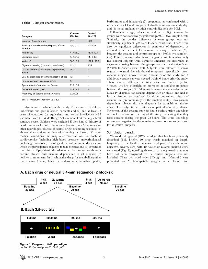
Stimulation paradigmWe used a drug-word (DW) paradigm that has been previously
described [14]. Briefly, 40 drug words matched on length,
frequency in the English language, and part of speech (noun,
adjective, adverb, verb) with 40 household-related (neutral) items
were used (Fig. 1); non-English words or slang words that may
have not been recognized by the control subjects were not
included. These two word types (‘‘Drug’’ and ‘‘Neutral’’) were
presented via MRI-compatible goggles in a blocked and
Table 1. Subject characteristics.
CategoryCocaine(N = 20)
Control(N = 20)
Number of men/women 17/3 13/7
Ethnicity Caucasian/Asian/Hispanic/African-American
1/0/2/17 5/1/3/11
Age (years) 41.465.0 38.3610.3
Education (years) 13.161.3 14.163.2
Verbal IQ 98.869.4 102.4621.3
Cigarette smoking (current or pass/never) 15/5 5/15
DSM-IV diagnoses of cocaine dependence/abuse
16/3 2
DSM-IV diagnoses of cannabis/alcohol abuse 1/1 2
Positive cocaine toxicology screen 17 2
Age at onset of cocaine use (years) 23.965.7 2
Cocaine duration (years) 15.364.9 2
Frequency of cocaine use (days/week) 3.962.2 2
doi:10.1371/journal.pone.0010815.t001
Figure 1. Drug-word fMRI paradigm.doi:10.1371/journal.pone.0010815.g001
Cocaine & Brain Connectivity
PLoS ONE | www.plosone.org 2 May 2010 | Volume 5 | Issue 5 | e10815
counterbalanced fashion across subjects. Each of the 2 DW
sequences (‘‘drug’’ or ‘‘neutral’’) had two 70-seconds long task
epochs that included 20 trials. Each trial comprised a 500 ms
fixation cross, a 2000 ms word presentation window, a 500 ms
response window, and a 500 ms feedback where ‘‘O’’ and ‘‘X’’
were displayed for correct and incorrect trials, respectively. The
words were presented using one of four possible colors (yellow,
blue, red, green) and the word color order was pseudo
randomized. Subjects were instructed to press one of four buttons
(yellow, blue, red, green) of the MRI-compatible Lumina LP-400
response pad (Cedrus Corp., San Pedro, CA) during the response
window matching the color of the word. A fixation cross against a
black background was presented during the three 20-seconds long
resting baseline epochs and subjects were instructed to not press
any button and rest motionless as possible during these epochs.
Thus, each sequence (‘‘drug’’ or ‘‘neutral’’) was 206 seconds long.
The task was developed in E-prime (Psychology Software Tools,
Inc., Pittsburgh, PA) and used a trigger pulse from the MRI
console for precise synchronization with fMRI acquisition.
Data acquisitionA 4-Tesla whole-body Varian/Siemens MRI scanner and a T2*-
weighted single-shot gradient-echo planar imaging (EPI) pulse
sequence (TE/TR = 20/1600 ms, 4-mm slice thickness, 33 coronal
slices, 64664 matrix size, 3.163.1 mm in-plane resolution, 90u-flipangle, 131 time points) were used to map the blood oxygenation
level dependent (BOLD) fMRI responses in the whole brain.
Padding was used to minimize motion. Subject motion was
monitored immediately after each fMRI run using a k-space motion
detection algorithm [25] written in IDL (ITT Visual Information
Solutions, Boulder, CO). Earplugs (acoustic noise attenuation:
28 dB; Aearo Ear TaperFit 2; Aearo Company) and headphones
(acoustic noise attenuation: 30 dB; Commander XG MRI Audio
System, Resonance Technology inc.) were used to minimize the
interference effect of scanner noise during fMRI [26].
Anatomical images were collected using a T1-weighted 3D-
MDEFT sequence [27] (TE/TR = 7/15 ms, 0.9460.9461.00 mm3 spatial resolution, axial orientation, 256 readout and
192696 phase-encoding steps, 16 minutes scan time) and a
modified T2-weigthed Hyperecho sequence [28] (TE/TR = 42/
10000 ms, echo train length = 16, 2566256 matrix size, 30
coronal slices, 0.8660.86 mm2 in-plane resolution, 5-mm thick-
ness, no gap, 2 min scan time), and reviewed to rule out gross
brain morphological abnormalities.
BOLD-fMRI analysesImage reconstruction was performed using an iterative phase
correction method in IDL that minimizes signal-loss artifacts in
EPI [29]. The first four volumes in the time series were discarded
to avoid non-equilibrium effects in the fMRI signal. Subsequent
analyses were performed with the statistical parametric mapping
package SPM2 (Welcome Department of Cognitive Neurology,
London UK). The images were motion corrected with a 12-
parameter affine transformation, spatially normalized to the
standard brain (using a 12-parameters affine transformation with
medium regularization and 16-nonlinear iterations and voxel size
of 36363 mm3), and smoothed (8-mm full-width-half-maximum
Gaussian kernel). Note that spatial normalization to the stereo-
tactic space was carried out using the standard SPM2 EPI
template.
The general linear model used to calculate the individual
BOLD contrast maps (drug epochs and neutral epochs) for each
fMRI time series consisted of a box-car design convolved with the
canonical hemodynamic response function (HRF) and a high-pass
filter (cut-off frequency: 1/520 Hz). The BOLD signal strength
was estimated without the removal of global effects (global
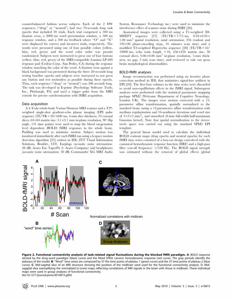
Figure 2. Functional connectivity analysis of task-related signal fluctuations during the blocked fMRI paradigm. A: BOLD responseelicited by the drug-word paradigm (black curve) and the fitted SPM2 canonic hemodynamic response (red curve). The gray periods identify theplateaus of the model. B: ‘‘Word’’ time series are composed by 37 the time points of plateau 1 (green curve) and the 37 time points of plateau 2 (bluecurve). C: Mid-sagittal slice of an MRI structure showing the position of the midbrain seed used for the functional connectivity analysis. D: Mid-saggital slice exemplifying the normalized (z-score) maps reflecting correlations of MRI signals in the brain with those in midbrain. These individualmaps were used in group analyses of functional connectivity.doi:10.1371/journal.pone.0010815.g002
Cocaine & Brain Connectivity
PLoS ONE | www.plosone.org 3 May 2010 | Volume 5 | Issue 5 | e10815
normalization) to minimize false deactivation signals [30,31]. The
estimated BOLD maps, contrasting word epochs (drug or neutral)
against baseline epochs, were included in a two-way (word type 6group) repeated measures analysis of variance (ANOVA) model in
SPM2. Four covariates (smoking status, urine results, gender, and
age) were included in this random-effects model in an effort to
control for potential confounds. Brain activation clusters were
corrected for multiple comparisons using the continuous random
field calculation implemented in SPM2. Clusters with at least 15
voxels (400 mm3) and pcorr ,0.05, corrected for multiple
comparisons at the cluster level, were considered significant in
group analyses of brain activation.
Functional ConnectivityA method recently proposed for studies based on blocked fMRI
datasets [32] was used to evaluate the functional connectivity of
the brain using the resting epochs. Here, we were interested in the
functional connectivity of the midbrain during the performance of
the sustained attention condition of the drug-word task, not during
resting-conditions. The task design was advantageous for this
purpose because the ‘‘word’’ epochs were 70 seconds long, almost
double the length of the resting blocks used by Fair and colleagues.
In order to minimize unwanted task-related effects on the strength
of correlations with midbrain (CM) we discarded 11 seconds from
each word epoch, using the canonic hemodynamic response
function to optimally select time points of the word epochs.
Specifically, for each time series, the plateaus of the SPM2 canonic
hemodynamic response function (Fig. 2A, red curve) were used to
identify the 37 consecutive time points of the plateaus (Fig. 2B
green and blue curves) corresponding to ‘‘word’’ epochs. This
procedure accounted for the initial delay of the hemodynamic
response and its return to baseline. The selected time points
corresponding to each word epoch were concatenated to form
‘‘word’’ (‘‘Drug’’ or ‘‘Neutral’’) time series with 74 time points.
‘‘Word’’ time series were carefully inspected to ensure minimal
baseline differences at the concatenation time points, and band-
pass filtered (0.01–0.1 Hz; Fig. 2B, red curve), a step that further
minimizes this baseline differences. A midbrain region (left
substantia nigra, SN; talairach position: xyz = [26, 215, 218]
mm; Fig. 2C, green circle) that showed higher drug-cue related
activation for cocaine addicts than for controls using a more
complex version of the DW task [15] was selected as the ‘‘seed’’
voxel for the functional connectivity analysis. Whole-brain maps
reflecting correlations between BOLD signals in the seed and
those in all other voxels in the brain were calculated separately for
each ‘‘word’’ time series. The Fisher transform was used to convert
the step distributed Pearson linear correlation factors into
normally distributed CM coefficients. These normalized CM
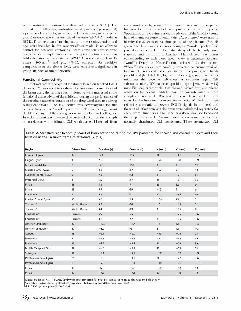
Table 2. Statistical significance (t-score) of brain activation during the DW paradigm for cocaine and control subjects and theirlocation in the Talairach frame of reference (x, y, z).
Region BA/nucleus Cocaine (t) Control (t) X [mm] Y [mm] Z [mm]
Fusiform Gyrus 19 17.1 16.4 24 281 212
Lingual Gyrus 18 23.9 25.0 224 278 29
Medial Frontal Gyrus 6 12.8 16.0 3 3 54
Middle Frontal Gyrus 6 4.2 2.7 227 0 48
Superior Frontal Gyrus 6 7.2 9.2 3 23 66
Precentral Gyrus 6 NS 2.7 45 23 36
Insula 13 5.1 7.1 36 12 9
Insula 13 5.7 6.9 245 9 6
Precuneus 7 5.9 6.1 30 245 48
Inferior Frontal Gyrus 10 3.6 2.5 236 45 3
Thalamus* Medial Dorsal 3.9 8.0 23 215 9
Thalamus* Medial Dorsal 4.4 8.9 3 215 9
Cerebellum* Culmen NS 5.5 23 245 26
Cerebellum* Culmen 3.6 7.7 3 245 26
Anterior Cingulate* 32 213.3 24.7 23 42 23
Anterior Cingulate* 32 28.9 NS 3 42 23
Cuneus 18 25.1 26.0 215 278 24
Precuneus 7 26.5 26.6 212 248 39
Precuneus 19 23.9 27.8 36 272 39
Middle Temporal Gyrus 39 24.6 28.8 42 272 24
Sub-Gyral 21 22.1 22.7 239 212 29
Parahippocampal Gyrus 36 23.5 24.7 24 242 26
Parahippocampal Gyrus 28 22.9 23.4 218 221 218
Insula 13 NS 22.7 239 212 18
Insula 13 24.6 24.1 36 218 18
Cluster statistics: Pcorr ,0.0005, familywise error corrected for multiple comparisons using the random field theory.*indicates clusters showing statistically significant between-group differences Pcorr ,0.05.doi:10.1371/journal.pone.0010815.t002
Cocaine & Brain Connectivity
PLoS ONE | www.plosone.org 4 May 2010 | Volume 5 | Issue 5 | e10815
maps (Fig. 2D) were computed and saved in Analyze format using
IDL, and loaded into SPM2 for group analyses of CM [33]. A
two-way (word type 6 group) repeated measures ANOVA
(random-effects) model with four covariates (smoking status, urine
results, gender, and age) was used for group analyses of CM.
Clusters with at least 15 voxels and pcorr ,0.05 (corrected for
multiple comparisons) were considered significant in group
analysis of CM signals.
Regions-of-interest (ROI) analysesThe relevant clusters were further evaluated with region-of-
interest (ROI) analyses to identify potential outliers that might
influence statistical analyses, and to report average values in a
volume comparable to the image smoothness (e.g. resolution
elements, or ‘‘resels’’ [34]) rather than single-voxel peak values.
The volume of the resels was estimated using the random field
calculation in SPM2 as a near cubic volume with Cartesian full-
width-half-maximum (FWHM) of [13.0, 12.3, 13.4] mm for group
analyses of brain activation and of [11.8, 11.0, 12.4] mm for group
analyses of CM. Note that (2n+1)3 voxels fit into ROI that are
symmetric around a peak voxel and that 3-mm isotropic voxels
were used. We did not use n = 0 ROIs because do not take full
advantage of the 13-mm isotropic resels (include only one voxel).
Similarly, n = 2 ROIs were not used because they include voxels
that do not belong to the average functional cluster defined by the
resels. Thus, n = 1 ROIs were used to maximize the number of
voxels within the smoothness of the data. Thus, the average
BOLD and CM signals in the left and right medial dorsal nuclei of
the thalamus (MDTHA; xyz = 63, 215, 9 mm), cerebellum
(CER; culmen; xyz = 63, 245, 26 mm), and the rostral anterior
cingulate cortex (rACC; xyz = 63, 42, 23 mm) were extracted
from the individual BOLD and CM maps in 96969 mm3 ROI
volumes containing 27 voxels to using IDL. We selected these
ROIs based on our previous fMRI studies [16–18,35]. These ROI
masks were created and centered at the precise coordinates listed
in Table 2; the coordinates of the ROI masks were kept fixed
across subjects and conditions. Note that compared to variable-
volume ROIs, fixed-volume (shape and size) ROIs are advanta-
geous in order to minimize statistical confounds. Brain regions
were labeled using the Talairach daemon (http://www.talairach.
org/) [36] and a query range of 5 mm.
Results
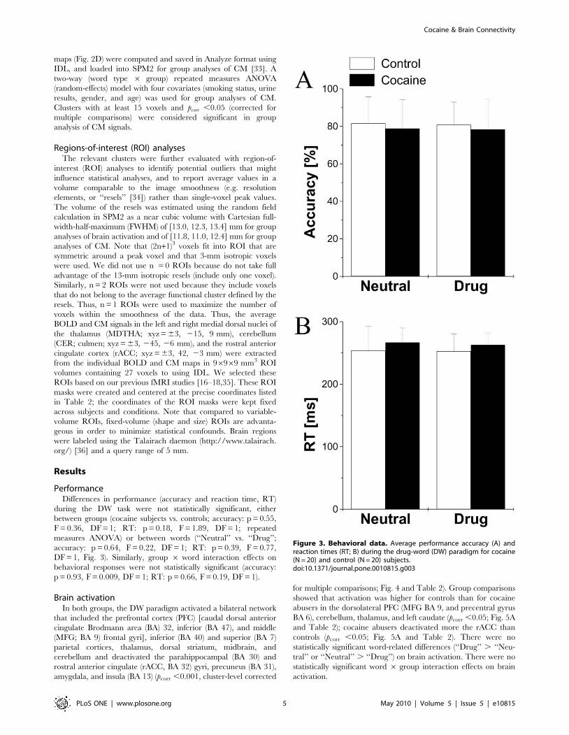
PerformanceDifferences in performance (accuracy and reaction time, RT)
during the DW task were not statistically significant, either
between groups (cocaine subjects vs. controls; accuracy: p = 0.55,
F = 0.36, DF = 1; RT: p = 0.18, F = 1.89, DF = 1; repeated
measures ANOVA) or between words (‘‘Neutral’’ vs. ‘‘Drug’’;
accuracy: p = 0.64, F = 0.22, DF = 1; RT: p = 0.39, F = 0.77,
DF = 1, Fig. 3). Similarly, group 6 word interaction effects on
behavioral responses were not statistically significant (accuracy:
p = 0.93, F = 0.009, DF = 1; RT: p = 0.66, F = 0.19, DF = 1).
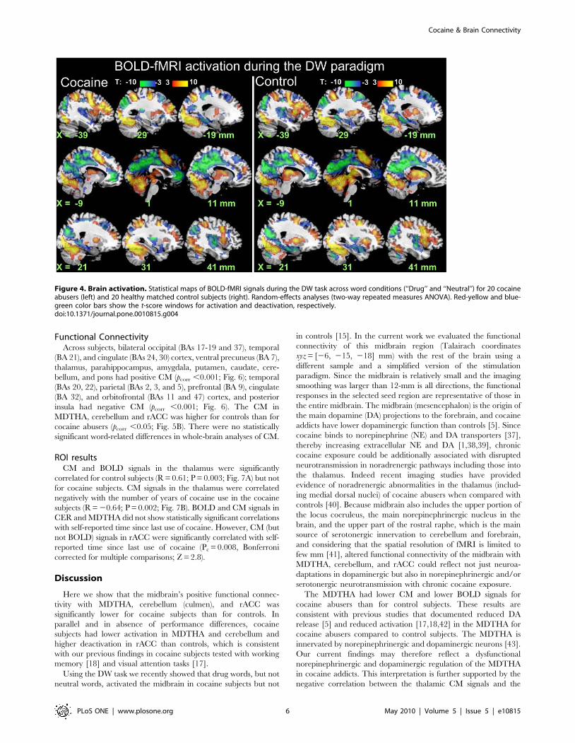
Brain activationIn both groups, the DW paradigm activated a bilateral network
that included the prefrontal cortex (PFC) [caudal dorsal anterior
cingulate Brodmann area (BA) 32, inferior (BA 47), and middle
(MFG; BA 9) frontal gyri], inferior (BA 40) and superior (BA 7)
parietal cortices, thalamus, dorsal striatum, midbrain, and
cerebellum and deactivated the parahippocampal (BA 30) and
rostral anterior cingulate (rACC, BA 32) gyri, precuneus (BA 31),
amygdala, and insula (BA 13) (pcorr ,0.001, cluster-level corrected
for multiple comparisons; Fig. 4 and Table 2). Group comparisons
showed that activation was higher for controls than for cocaine
abusers in the dorsolateral PFC (MFG BA 9, and precentral gyrus
BA 6), cerebellum, thalamus, and left caudate (pcorr ,0.05; Fig. 5A
and Table 2); cocaine abusers deactivated more the rACC than
controls (pcorr ,0.05; Fig. 5A and Table 2). There were no
statistically significant word-related differences (‘‘Drug’’ . ‘‘Neu-
tral’’ or ‘‘Neutral’’ . ‘‘Drug’’) on brain activation. There were no
statistically significant word 6 group interaction effects on brain
activation.
Figure 3. Behavioral data. Average performance accuracy (A) andreaction times (RT; B) during the drug-word (DW) paradigm for cocaine(N = 20) and control (N = 20) subjects.doi:10.1371/journal.pone.0010815.g003
Cocaine & Brain Connectivity
PLoS ONE | www.plosone.org 5 May 2010 | Volume 5 | Issue 5 | e10815
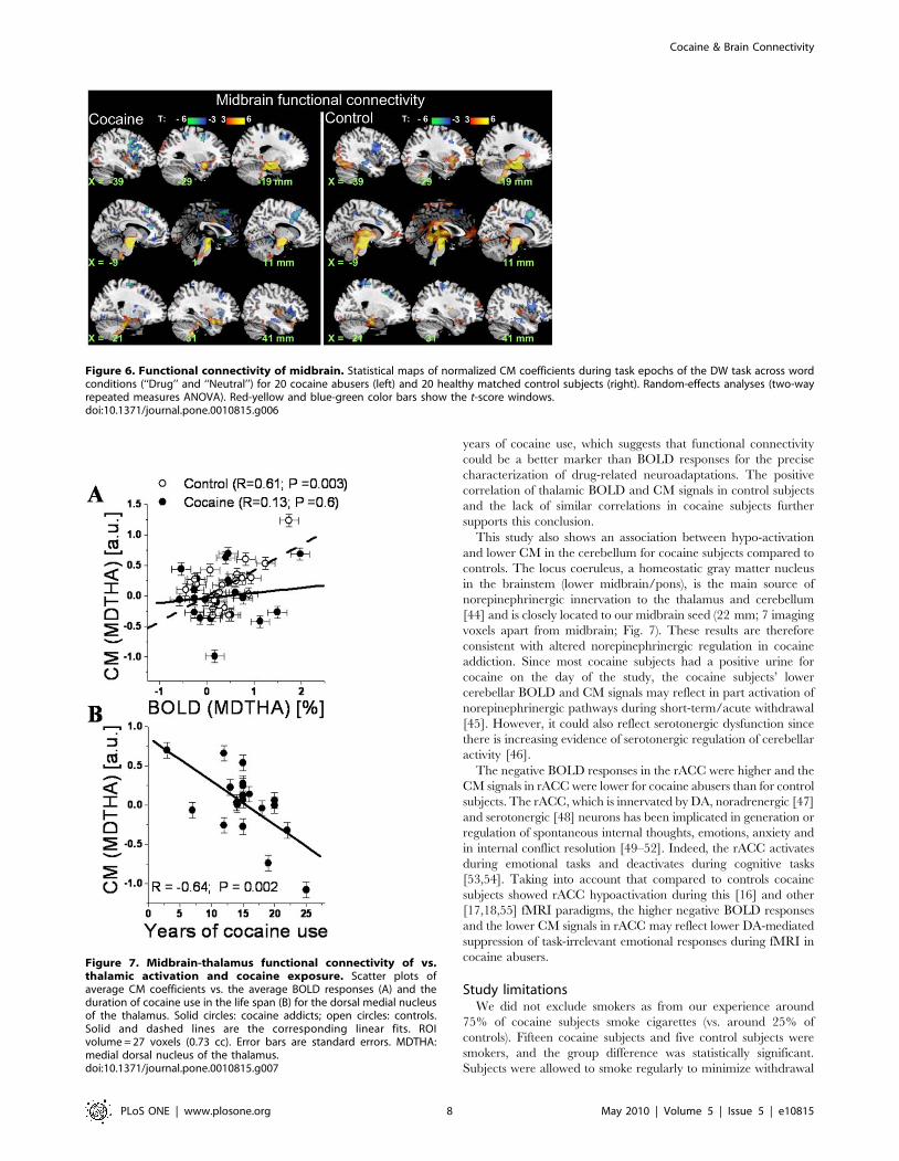
Functional ConnectivityAcross subjects, bilateral occipital (BAs 17-19 and 37), temporal
(BA 21), and cingulate (BAs 24, 30) cortex, ventral precuneus (BA 7),
thalamus, parahippocampus, amygdala, putamen, caudate, cere-
bellum, and pons had positive CM (pcorr ,0.001; Fig. 6); temporal
(BAs 20, 22), parietal (BAs 2, 3, and 5), prefrontal (BA 9), cingulate
(BA 32), and orbitofrontal (BAs 11 and 47) cortex, and posterior
insula had negative CM (pcorr ,0.001; Fig. 6). The CM in
MDTHA, cerebellum and rACC was higher for controls than for
cocaine abusers (pcorr ,0.05; Fig. 5B). There were no statistically
significant word-related differences in whole-brain analyses of CM.
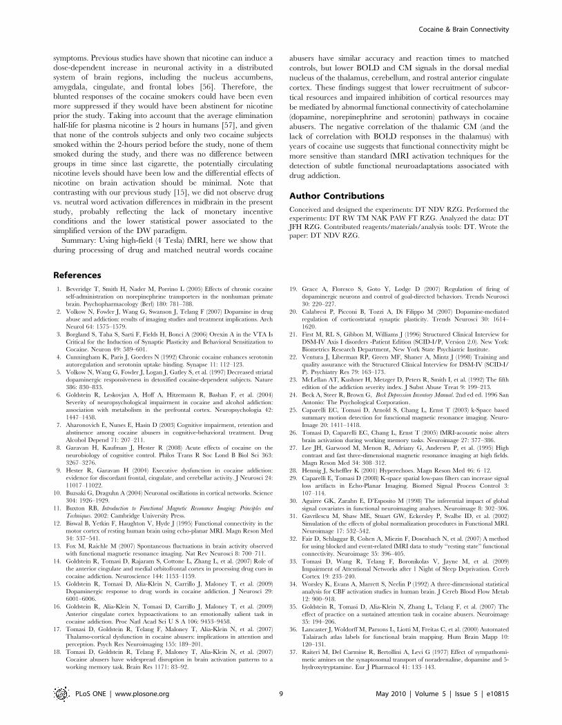
ROI resultsCM and BOLD signals in the thalamus were significantly
correlated for control subjects (R = 0.61; P = 0.003; Fig. 7A) but not
for cocaine subjects. CM signals in the thalamus were correlated
negatively with the number of years of cocaine use in the cocaine
subjects (R = 20.64; P = 0.002; Fig. 7B). BOLD and CM signals in
CER and MDTHA did not show statistically significant correlations
with self-reported time since last use of cocaine. However, CM (but
not BOLD) signals in rACC were significantly correlated with self-
reported time since last use of cocaine (Pc = 0.008, Bonferroni
corrected for multiple comparisons; Z = 2.8).
Discussion
Here we show that the midbrain’s positive functional connec-
tivity with MDTHA, cerebellum (culmen), and rACC was
significantly lower for cocaine subjects than for controls. In
parallel and in absence of performance differences, cocaine
subjects had lower activation in MDTHA and cerebellum and
higher deactivation in rACC than controls, which is consistent
with our previous findings in cocaine subjects tested with working
memory [18] and visual attention tasks [17].
Using the DW task we recently showed that drug words, but not
neutral words, activated the midbrain in cocaine subjects but not
in controls [15]. In the current work we evaluated the functional
connectivity of this midbrain region (Talairach coordinates
xyz = [26, 215, 218] mm) with the rest of the brain using a
different sample and a simplified version of the stimulation
paradigm. Since the midbrain is relatively small and the imaging
smoothing was larger than 12-mm is all directions, the functional
responses in the selected seed region are representative of those in
the entire midbrain. The midbrain (mesencephalon) is the origin of
the main dopamine (DA) projections to the forebrain, and cocaine
addicts have lower dopaminergic function than controls [5]. Since
cocaine binds to norepinephrine (NE) and DA transporters [37],
thereby increasing extracellular NE and DA [1,38,39], chronic
cocaine exposure could be additionally associated with disrupted
neurotransmission in noradrenergic pathways including those into
the thalamus. Indeed recent imaging studies have provided
evidence of noradrenergic abnormalities in the thalamus (includ-
ing medial dorsal nuclei) of cocaine abusers when compared with
controls [40]. Because midbrain also includes the upper portion of
the locus coeruleus, the main norepinephrinergic nucleus in the
brain, and the upper part of the rostral raphe, which is the main
source of serotonergic innervation to cerebellum and forebrain,
and considering that the spatial resolution of fMRI is limited to
few mm [41], altered functional connectivity of the midbrain with
MDTHA, cerebellum, and rACC could reflect not just neuroa-
daptations in dopaminergic but also in norepinephrinergic and/or
serotonergic neurotransmission with chronic cocaine exposure.
The MDTHA had lower CM and lower BOLD signals for
cocaine abusers than for control subjects. These results are
consistent with previous studies that documented reduced DA
release [5] and reduced activation [17,18,42] in the MDTHA for
cocaine abusers compared to control subjects. The MDTHA is
innervated by norepinephrinergic and dopaminergic neurons [43].
Our current findings may therefore reflect a dysfunctional
norepinephrinergic and dopaminergic regulation of the MDTHA
in cocaine addicts. This interpretation is further supported by the
negative correlation between the thalamic CM signals and the
Figure 4. Brain activation. Statistical maps of BOLD-fMRI signals during the DW task across word conditions (‘‘Drug’’ and ‘‘Neutral’’) for 20 cocaineabusers (left) and 20 healthy matched control subjects (right). Random-effects analyses (two-way repeated measures ANOVA). Red-yellow and blue-green color bars show the t-score windows for activation and deactivation, respectively.doi:10.1371/journal.pone.0010815.g004
Cocaine & Brain Connectivity
PLoS ONE | www.plosone.org 6 May 2010 | Volume 5 | Issue 5 | e10815
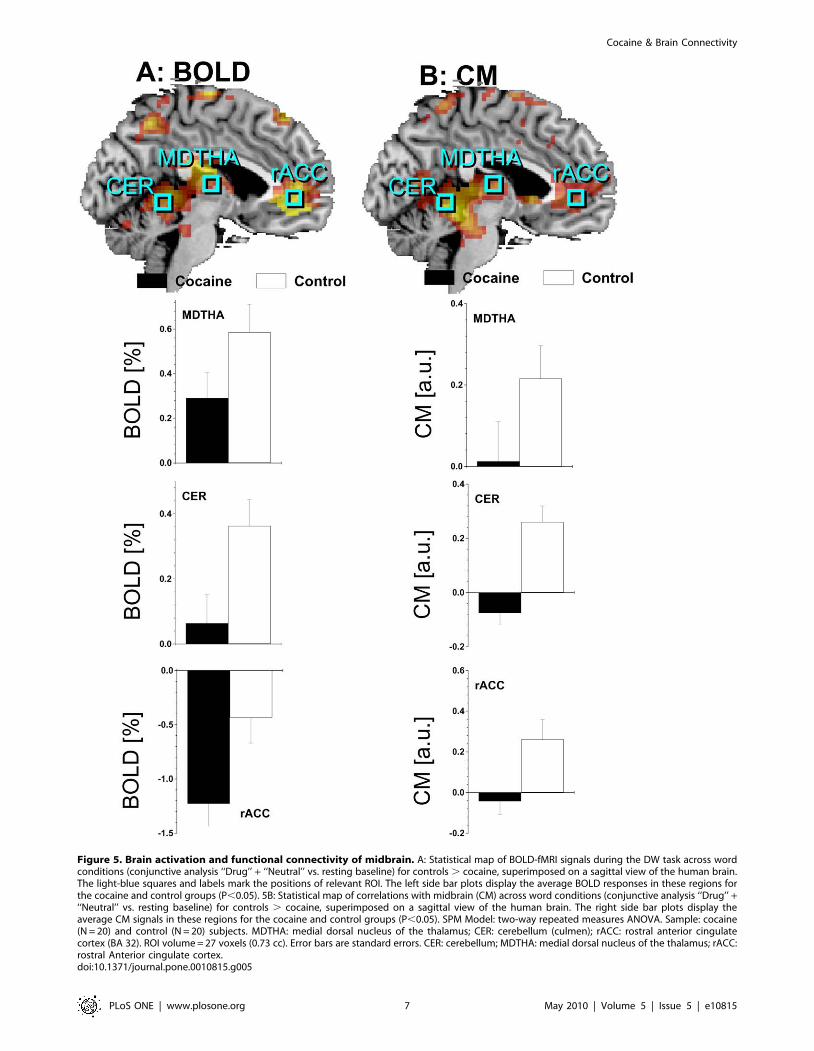
Figure 5. Brain activation and functional connectivity of midbrain. A: Statistical map of BOLD-fMRI signals during the DW task across wordconditions (conjunctive analysis ‘‘Drug’’ + ‘‘Neutral’’ vs. resting baseline) for controls . cocaine, superimposed on a sagittal view of the human brain.The light-blue squares and labels mark the positions of relevant ROI. The left side bar plots display the average BOLD responses in these regions forthe cocaine and control groups (P,0.05). 5B: Statistical map of correlations with midbrain (CM) across word conditions (conjunctive analysis ‘‘Drug’’ +‘‘Neutral’’ vs. resting baseline) for controls . cocaine, superimposed on a sagittal view of the human brain. The right side bar plots display theaverage CM signals in these regions for the cocaine and control groups (P,0.05). SPM Model: two-way repeated measures ANOVA. Sample: cocaine(N = 20) and control (N = 20) subjects. MDTHA: medial dorsal nucleus of the thalamus; CER: cerebellum (culmen); rACC: rostral anterior cingulatecortex (BA 32). ROI volume = 27 voxels (0.73 cc). Error bars are standard errors. CER: cerebellum; MDTHA: medial dorsal nucleus of the thalamus; rACC:rostral Anterior cingulate cortex.doi:10.1371/journal.pone.0010815.g005
Cocaine & Brain Connectivity
PLoS ONE | www.plosone.org 7 May 2010 | Volume 5 | Issue 5 | e10815
years of cocaine use, which suggests that functional connectivity
could be a better marker than BOLD responses for the precise
characterization of drug-related neuroadaptations. The positive
correlation of thalamic BOLD and CM signals in control subjects
and the lack of similar correlations in cocaine subjects further
supports this conclusion.
This study also shows an association between hypo-activation
and lower CM in the cerebellum for cocaine subjects compared to
controls. The locus coeruleus, a homeostatic gray matter nucleus
in the brainstem (lower midbrain/pons), is the main source of
norepinephrinergic innervation to the thalamus and cerebellum
[44] and is closely located to our midbrain seed (22 mm; 7 imaging
voxels apart from midbrain; Fig. 7). These results are therefore
consistent with altered norepinephrinergic regulation in cocaine
addiction. Since most cocaine subjects had a positive urine for
cocaine on the day of the study, the cocaine subjects’ lower
cerebellar BOLD and CM signals may reflect in part activation of
norepinephrinergic pathways during short-term/acute withdrawal
[45]. However, it could also reflect serotonergic dysfunction since
there is increasing evidence of serotonergic regulation of cerebellar
activity [46].
The negative BOLD responses in the rACC were higher and the
CM signals in rACC were lower for cocaine abusers than for control
subjects. The rACC, which is innervated by DA, noradrenergic [47]
and serotonergic [48] neurons has been implicated in generation or
regulation of spontaneous internal thoughts, emotions, anxiety and
in internal conflict resolution [49–52]. Indeed, the rACC activates
during emotional tasks and deactivates during cognitive tasks
[53,54]. Taking into account that compared to controls cocaine
subjects showed rACC hypoactivation during this [16] and other
[17,18,55] fMRI paradigms, the higher negative BOLD responses
and the lower CM signals in rACC may reflect lower DA-mediated
suppression of task-irrelevant emotional responses during fMRI in
cocaine abusers.
Study limitationsWe did not exclude smokers as from our experience around
75% of cocaine subjects smoke cigarettes (vs. around 25% of
controls). Fifteen cocaine subjects and five control subjects were
smokers, and the group difference was statistically significant.
Subjects were allowed to smoke regularly to minimize withdrawal
Figure 6. Functional connectivity of midbrain. Statistical maps of normalized CM coefficients during task epochs of the DW task across wordconditions (‘‘Drug’’ and ‘‘Neutral’’) for 20 cocaine abusers (left) and 20 healthy matched control subjects (right). Random-effects analyses (two-wayrepeated measures ANOVA). Red-yellow and blue-green color bars show the t-score windows.doi:10.1371/journal.pone.0010815.g006
Figure 7. Midbrain-thalamus functional connectivity of vs.thalamic activation and cocaine exposure. Scatter plots ofaverage CM coefficients vs. the average BOLD responses (A) and theduration of cocaine use in the life span (B) for the dorsal medial nucleusof the thalamus. Solid circles: cocaine addicts; open circles: controls.Solid and dashed lines are the corresponding linear fits. ROIvolume = 27 voxels (0.73 cc). Error bars are standard errors. MDTHA:medial dorsal nucleus of the thalamus.doi:10.1371/journal.pone.0010815.g007
Cocaine & Brain Connectivity
PLoS ONE | www.plosone.org 8 May 2010 | Volume 5 | Issue 5 | e10815
symptoms. Previous studies have shown that nicotine can induce a
dose-dependent increase in neuronal activity in a distributed
system of brain regions, including the nucleus accumbens,
amygdala, cingulate, and frontal lobes [56]. Therefore, the
blunted responses of the cocaine smokers could have been even
more suppressed if they would have been abstinent for nicotine
prior the study. Taking into account that the average elimination
half-life for plasma nicotine is 2 hours in humans [57], and given
that none of the controls subjects and only two cocaine subjects
smoked within the 2-hours period before the study, none of them
smoked during the study, and there was no difference between
groups in time since last cigarette, the potentially circulating
nicotine levels should have been low and the differential effects of
nicotine on brain activation should be minimal. Note that
contrasting with our previous study [15], we did not observe drug
vs. neutral word activation differences in midbrain in the present
study, probably reflecting the lack of monetary incentive
conditions and the lower statistical power associated to the
simplified version of the DW paradigm.
Summary: Using high-field (4 Tesla) fMRI, here we show that
during processing of drug and matched neutral words cocaine
abusers have similar accuracy and reaction times to matched
controls, but lower BOLD and CM signals in the dorsal medial
nucleus of the thalamus, cerebellum, and rostral anterior cingulate
cortex. These findings suggest that lower recruitment of subcor-
tical resources and impaired inhibition of cortical resources may
be mediated by abnormal functional connectivity of catecholamine
(dopamine, norepinephrine and serotonin) pathways in cocaine
abusers. The negative correlation of the thalamic CM (and the
lack of correlation with BOLD responses in the thalamus) with
years of cocaine use suggests that functional connectivity might be
more sensitive than standard fMRI activation techniques for the
detection of subtle functional neuroadaptations associated with
drug addiction.
Author Contributions
Conceived and designed the experiments: DT NDV RZG. Performed the
experiments: DT RW TM NAK PAW FT RZG. Analyzed the data: DT
JFH RZG. Contributed reagents/materials/analysis tools: DT. Wrote the
paper: DT NDV RZG.
References
1. Beveridge T, Smith H, Nader M, Porrino L (2005) Effects of chronic cocaine
self-administration on norepinephrine transporters in the nonhuman primate
brain. Psychopharmacology (Berl) 180: 781–788.
2. Volkow N, Fowler J, Wang G, Swanson J, Telang F (2007) Dopamine in drug
abuse and addiction: results of imaging studies and treatment implications. Arch
Neurol 64: 1575–1579.
3. Borgland S, Taha S, Sarti F, Fields H, Bonci A (2006) Orexin A in the VTA Is
Critical for the Induction of Synaptic Plasticity and Behavioral Sensitization to
Cocaine. Neuron 49: 589–601.
4. Cunningham K, Paris J, Goeders N (1992) Chronic cocaine enhances serotonin
autoregulation and serotonin uptake binding. Synapse 11: 112–123.
5. Volkow N, Wang G, Fowler J, Logan J, Gatley S, et al. (1997) Decreased striatal
dopaminergic responsiveness in detoxified cocaine-dependent subjects. Nature
386: 830–833.
6. Goldstein R, Leskovjan A, Hoff A, Hitzemann R, Bashan F, et al. (2004)
Severity of neuropsychological impairment in cocaine and alcohol addiction:
association with metabolism in the prefrontal cortex. Neuropsychologia 42:
1447–1458.
7. Aharonovich E, Nunes E, Hasin D (2003) Cognitive impairment, retention and
abstinence among cocaine abusers in cognitive-behavioral treatment. Drug
Alcohol Depend 71: 207–211.
8. Garavan H, Kaufman J, Hester R (2008) Acute effects of cocaine on the
neurobiology of cognitive control. Philos Trans R Soc Lond B Biol Sci 363:
3267–3276.
9. Hester R, Garavan H (2004) Executive dysfunction in cocaine addiction:
evidence for discordant frontal, cingulate, and cerebellar activity. J Neurosci 24:
11017–11022.
10. Buzsaki G, Draguhn A (2004) Neuronal oscillations in cortical networks. Science
304: 1926–1929.
11. Buxton RB, Introduction to Functional Magnetic Resonance Imaging: Principles and
Techniques. 2002: Cambridge University Press.
12. Biswal B, Yetkin F, Haughton V, Hyde J (1995) Functional connectivity in the
motor cortex of resting human brain using echo-planar MRI. Magn Reson Med
34: 537–541.
13. Fox M, Raichle M (2007) Spontaneous fluctuations in brain activity observed
with functional magnetic resonance imaging. Nat Rev Neurosci 8: 700–711.
14. Goldstein R, Tomasi D, Rajaram S, Cottone L, Zhang L, et al. (2007) Role of
the anterior cingulate and medial orbitofrontal cortex in processing drug cues in
cocaine addiction. Neuroscience 144: 1153–1159.
15. Goldstein R, Tomasi D, Alia-Klein N, Carrillo J, Maloney T, et al. (2009)
Dopaminergic response to drug words in cocaine addiction. J Neurosci 29:
6001–6006.
16. Goldstein R, Alia-Klein N, Tomasi D, Carrillo J, Maloney T, et al. (2009)
Anterior cingulate cortex hypoactivations to an emotionally salient task in
cocaine addiction. Proc Natl Acad Sci U S A 106: 9453–9458.
17. Tomasi D, Goldstein R, Telang F, Maloney T, Alia-Klein N, et al. (2007)
Thalamo-cortical dysfunction in cocaine abusers: implications in attention and
perception. Psych Res Neuroimaging 155: 189–201.
18. Tomasi D, Goldstein R, Telang F, Maloney T, Alia-Klein N, et al. (2007)
Cocaine abusers have widespread disruption in brain activation patterns to a
working memory task. Brain Res 1171: 83–92.
19. Grace A, Floresco S, Goto Y, Lodge D (2007) Regulation of firing of
dopaminergic neurons and control of goal-directed behaviors. Trends Neurosci
30: 220–227.
20. Calabresi P, Picconi B, Tozzi A, Di Filippo M (2007) Dopamine-mediated
regulation of corticostriatal synaptic plasticity. Trends Neurosci 30: 1614–
1620.
21. First M, RL S, Gibbon M, Williams J (1996) Structured Clinical Interview for
DSM-IV Axis I disorders -Patient Edition (SCID-I/P, Version 2.0). New York:
Biometrics Research Department, New York State Psychiatric Institute.
22. Ventura J, Liberman RP, Green MF, Shaner A, Mintz J (1998) Training and
quality assurance with the Structured Clinical Interview for DSM-IV (SCID-I/
P). Psychiatry Res 79: 163–173.
23. McLellan AT, Kushner H, Metzger D, Peters R, Smith I, et al. (1992) The fifth
edition of the addiction severity index. J Subst Abuse Treat 9: 199–213.
24. Beck A, Steer R, Brown G, Beck Depression Inventory Manual. 2nd ed ed. 1996 San
Antonio: The Psychological Corporation.
25. Caparelli EC, Tomasi D, Arnold S, Chang L, Ernst T (2003) k-Space based
summary motion detection for functional magnetic resonance imaging. Neuro-
Image 20: 1411–1418.
26. Tomasi D, Caparelli EC, Chang L, Ernst T (2005) fMRI-acoustic noise alters
brain activation during working memory tasks. Neuroimage 27: 377–386.
27. Lee JH, Garwood M, Menon R, Adriany G, Andersen P, et al. (1995) High
contrast and fast three-dimensional magnetic resonance imaging at high fields.
Magn Reson Med 34: 308–312.
28. Hennig J, Scheffler K (2001) Hyperechoes. Magn Reson Med 46: 6–12.
29. Caparelli E, Tomasi D (2008) K-space spatial low-pass filters can increase signal
loss artifacts in Echo-Planar Imaging. Biomed Signal Process Control 3:
107–114.
30. Aguirre GK, Zarahn E, D’Esposito M (1998) The inferential impact of global
signal covariates in functional neuroimaging analyses. Neuroimage 8: 302–306.
31. Gavrilescu M, Shaw ME, Stuart GW, Eckersley P, Svalbe ID, et al. (2002)
Simulation of the effects of global normalization procedures in Functional MRI.
Neuroimage 17: 532–542.
32. Fair D, Schlaggar B, Cohen A, Miezin F, Dosenbach N, et al. (2007) A method
for using blocked and event-related fMRI data to study ‘‘resting state’’ functional
connectivity. Neuroimage 35: 396–405.
33. Tomasi D, Wang R, Telang F, Boronikolas V, Jayne M, et al. (2009)
Impairment of Attentional Networks after 1 Night of Sleep Deprivation. Cereb
Cortex 19: 233–240.
34. Worsley K, Evans A, Marrett S, Neelin P (1992) A three-dimensional statistical
analysis for CBF activation studies in human brain. J Cereb Blood Flow Metab
12: 900–918.
35. Goldstein R, Tomasi D, Alia-Klein N, Zhang L, Telang F, et al. (2007) The
effect of practice on a sustained attention task in cocaine abusers. Neuroimage
35: 194–206.
36. Lancaster J, Woldorff M, Parsons L, Liotti M, Freitas C, et al. (2000) Automated
Talairach atlas labels for functional brain mapping. Hum Brain Mapp 10:
120–131.
37. Raiteri M, Del Carmine R, Bertollini A, Levi G (1977) Effect of sympathomi-
metic amines on the synaptosomal transport of noradrenaline, dopamine and 5-
hydroxytryptamine. Eur J Pharmacol 41: 133–143.
Cocaine & Brain Connectivity
PLoS ONE | www.plosone.org 9 May 2010 | Volume 5 | Issue 5 | e10815
38. Ritz M, Lamb R, Goldberg S, Kuhar M (1987) Cocaine receptors on dopamine
transporters are related to self-administration of cocaine. Science 237:1219–1223.
39. Tanda G, Pontieri F, Frau R, Di Chiara G (1997) Contribution of blockade of
the noradrenaline carrier to the increase of extracellular dopamine in the ratprefrontal cortex by amphetamine and cocaine. Eur J Neurosci 9: 2077–2085.
40. Ding Y, Singhal T, Planeta-Wilson B, Gallezot J, Nabulsi N, et al. (2010) PETimaging of the effects of age and cocaine on the norepinephrine transporter in
the human brain using (S,S)-[(11)C]O-methylreboxetine and HRRT. Synapse
64: 30–38.41. Tomasi D, Caparelli E (2007) Macrovascular contribution in activation patterns
of working memory. J Cereb Blood Flow Metab 27: 33–42.42. Kubler A, Murphy K, Garavan H (2005) Cocaine dependence and attention
switching within and between verbal and visuospatial working memory.Eur J Neurosci 21: 1984–1992.
43. Oke A, Solnick J, Adams R (1986) Catecholamine distribution patterns in rat
thalamus. Brain Res 269: 180–183.44. Rogawski M, Aghajanian G (1980) Modulation of lateral geniculate neurone
excitability by noradrenaline microiontophoresis or locus coeruleus stimulation.Nature 287: 731–734.
45. Kelley B, Yeager K, Pepper T, Beversdorf D (2005) Cognitive impairment in
acute cocaine withdrawal. Cogn Behav Neurol 18: 108–112.46. Dieudonne S (2001) Serotonergic neuromodulation in the cerebellar cortex:
cellular, synaptic, and molecular basis. Neuroscientist 7: 207–219.47. Morrison J, Molliver M, Grzanna R, Coyle J (1979) Noradrenergic innervation
patterns in three regions of medial cortex: an immunofluorescence character-ization. Brain Res Bull 4: 849–857.
48. Olpe H, Koella W (2001) The cortical projection of the dorsal raphe nucleus:
some electrophysiological and pharmacological properties. Brain Res 216:61–71.
49. Mason M, Norton M, Van Horn J, Wegner D, Grafton S, et al. (2007)
Wandering minds: the default network and stimulus-independent thought.
Science 315: 393–395.
50. Simpson JR, Drevets WC, Snyder AZ, Gusnard DA, Raichle ME (2001)
Emotion-induced changes in human medial prefrontal cortex: II. During
anticipatory anxiety. Proc Natl Acad Sci USA 98: 688–693.
51. Simpson JR, Snyder AZ, Gusnard DA, Raichle ME (2001) Emotion-induced
changes in human medial prefrontal cortex: I. During cognitive task
performance. Proc Natl Acad Sci USA 98: 683–687.
52. Horovitz S, Braun A, Carr W, Picchioni D, Balkin T, et al. (2009) Decoupling of
the brain’s default mode network during deep sleep. Proc Natl Acad Sci U S A
106: 11376–11381.
53. Bush G, Shin L, Holmes J, Rosen B, Vogt B (2003) The Multi-Source
Interference Task: validation study with fMRI in individual subjects. Mol
Psychiatry 8: 60–70.
54. Tomasi D, Ernst T, Caparelli EC, Chang L (2006) Common deactivation
patterns during working memory and visual attention tasks: An intra-subject
fMRI study at 4 Tesla. Hum Brain Mapp 27: 694–705.
55. Li C, Huang C, Yan P, Bhagwagar Z, Milivojevic V, et al. (2008) Neural
correlates of impulse control during stop signal inhibition in cocaine-dependent
men. Neuropsychopharmacology 33: 1798–1806.
56. Stein EA, Pankiewicz J, Harsch HH, Cho JK, Fuller SA, et al. (1998) Nicotine-
induced limbic cortical activation in the human brain: a functional MRI study.
155: 1009–1015.
57. Matta S, Balfour D, Benowitz N, Boyd R, Buccafusco J, et al. (2007) Guidelines
on nicotine dose selection for in vivo research. Psychopharmacology (Berl) 190:
269–319.
Cocaine & Brain Connectivity
PLoS ONE | www.plosone.org 10 May 2010 | Volume 5 | Issue 5 | e10815
Related Documents