RESEARCH Open Access Dispersion of single-walled carbon nanotubes by a natural lung surfactant for pulmonary in vitro and in vivo toxicity studies Liying Wang 1* , Vincent Castranova 1 , Anurag Mishra 1,2 , Bean Chen 1 , Robert R Mercer 1 , Diane Schwegler-Berry 1 , Yon Rojanasakul 2 Abstract Background: Accumulating evidence indicate that the degree of dispersion of nanoparticles has a strong influence on their biological activities. The aims of this study were to develop a simple and rapid method of nanoparticle dispersion using a natural lung surfactant and to evaluate the effect of dispersion status of SWCNT on cytotoxicity and fibrogenicity in vitro and in vivo. Results: The natural lung surfactant Survanta® was used to disperse single-walled carbon nanotubes (SWCNT) in a biological medium. At physiologically relevant concentrations, Survanta® produced well dispersed SWCNT without causing a cytotoxic or fibrogenic effect. In vitro studies show that Survanta®-dispersed SWCNT (SD-SWCNT) stimulated proliferation of lung epithelial cells at low doses (0.04-0.12 μg/ml or 0.02-0.06 μg/cm 2 exposed surface area) but had a suppressive effect at high doses. Non-dispersed SWCNT (ND-SWCNT) did not exhibit these effects, suggesting the importance of dispersion status of SWCNT on bioactivities. Studies using cultured human lung fibroblasts show that SD-SWCNT stimulated collagen production of the cells. This result is supported by a similar observation using Acetone/sonication dispersed SWCNT (AD-SWCNT), suggesting that Survanta® did not mask the bioactivity of SWCNT. Likewise, in vivo studies show that both SD-SWCNT and AD-SWCNT induced lung fibrosis in mice, whereas the dispersing agent Survanta® alone or Survanta®-dispersed control ultrafine carbon black had no effect. Conclusions: The results indicate that Survanta® was effective in dispersing SWCNT in biological media without causing cytotoxic effects at the test concentrations used in this study. SD-SWCNT stimulated collagen production of lung fibroblasts in vitro and induced lung fibrosis in vivo. Similar results were observed with AD-SWCNT, supporting the conclusion that Survanta® did not mask the bioactivities of SWCNT and thus can be used as an effective dispersing agent. Since excessive collagen production is a hallmark of lung fibrosis, the results of this study suggest that the in vitro model using lung fibroblasts may be an effective and rapid screening tool for prediction of the fibrogenic potential of SWCNT in vivo. Background Advances in nanotechnology have made possible the fabrication of materials at the nanoscale level. Carbon nanotubes (CNT) are a major class of nanomaterials possessing unique mechanical, electrical, and thermal properties. As the use of CNT has become more wide- spread, there has been a great concern about their potential adverse effects on human health and the envir- onment. Nanoparticles can come in contact with the human body through inhalation as well as ingestion and dermal deposition. Pulmonary exposure could occur due to aerosolization of nanomaterials including agglomer- ates of different size and shape. Individual CNT have a very high aspect ratio and can agglomerate into struc- tures which are micrometers in diameter in the dry state or upon suspension in polar and non-polar sol- vents [1-4]. Animal exposure studies have shown that a major pathologic effect of CNT exposure is pulmonary * Correspondence: [email protected] 1 National Institute for Occupational Safety and Health, HELD/PPRB, Morgantown, WV 26505, USA Full list of author information is available at the end of the article Wang et al. Particle and Fibre Toxicology 2010, 7:31 http://www.particleandfibretoxicology.com/content/7/1/31 © 2010 Wang et al; licensee BioMed Central Ltd. This is an Open Access article distributed under the terms of the Creative Commons Attribution License (http://creativecommons.org/licenses/by/2.0), which permits unrestricted use, distribution, and reproduction in any medium, provided the original work is properly cited.

Welcome message from author
This document is posted to help you gain knowledge. Please leave a comment to let me know what you think about it! Share it to your friends and learn new things together.
Transcript

RESEARCH Open Access
Dispersion of single-walled carbon nanotubes bya natural lung surfactant for pulmonary in vitroand in vivo toxicity studiesLiying Wang1*, Vincent Castranova1, Anurag Mishra1,2, Bean Chen1, Robert R Mercer1, Diane Schwegler-Berry1,Yon Rojanasakul2
Abstract
Background: Accumulating evidence indicate that the degree of dispersion of nanoparticles has a strong influenceon their biological activities. The aims of this study were to develop a simple and rapid method of nanoparticledispersion using a natural lung surfactant and to evaluate the effect of dispersion status of SWCNT on cytotoxicityand fibrogenicity in vitro and in vivo.
Results: The natural lung surfactant Survanta® was used to disperse single-walled carbon nanotubes (SWCNT) in abiological medium. At physiologically relevant concentrations, Survanta® produced well dispersed SWCNT withoutcausing a cytotoxic or fibrogenic effect. In vitro studies show that Survanta®-dispersed SWCNT (SD-SWCNT)stimulated proliferation of lung epithelial cells at low doses (0.04-0.12 μg/ml or 0.02-0.06 μg/cm2 exposed surfacearea) but had a suppressive effect at high doses. Non-dispersed SWCNT (ND-SWCNT) did not exhibit these effects,suggesting the importance of dispersion status of SWCNT on bioactivities. Studies using cultured human lungfibroblasts show that SD-SWCNT stimulated collagen production of the cells. This result is supported by a similarobservation using Acetone/sonication dispersed SWCNT (AD-SWCNT), suggesting that Survanta® did not mask thebioactivity of SWCNT. Likewise, in vivo studies show that both SD-SWCNT and AD-SWCNT induced lung fibrosis inmice, whereas the dispersing agent Survanta® alone or Survanta®-dispersed control ultrafine carbon black had noeffect.
Conclusions: The results indicate that Survanta® was effective in dispersing SWCNT in biological media withoutcausing cytotoxic effects at the test concentrations used in this study. SD-SWCNT stimulated collagen productionof lung fibroblasts in vitro and induced lung fibrosis in vivo. Similar results were observed with AD-SWCNT,supporting the conclusion that Survanta® did not mask the bioactivities of SWCNT and thus can be used as aneffective dispersing agent. Since excessive collagen production is a hallmark of lung fibrosis, the results of thisstudy suggest that the in vitro model using lung fibroblasts may be an effective and rapid screening tool forprediction of the fibrogenic potential of SWCNT in vivo.
BackgroundAdvances in nanotechnology have made possible thefabrication of materials at the nanoscale level. Carbonnanotubes (CNT) are a major class of nanomaterialspossessing unique mechanical, electrical, and thermalproperties. As the use of CNT has become more wide-spread, there has been a great concern about their
potential adverse effects on human health and the envir-onment. Nanoparticles can come in contact with thehuman body through inhalation as well as ingestion anddermal deposition. Pulmonary exposure could occur dueto aerosolization of nanomaterials including agglomer-ates of different size and shape. Individual CNT have avery high aspect ratio and can agglomerate into struc-tures which are micrometers in diameter in the drystate or upon suspension in polar and non-polar sol-vents [1-4]. Animal exposure studies have shown that amajor pathologic effect of CNT exposure is pulmonary
* Correspondence: [email protected] Institute for Occupational Safety and Health, HELD/PPRB,Morgantown, WV 26505, USAFull list of author information is available at the end of the article
Wang et al. Particle and Fibre Toxicology 2010, 7:31http://www.particleandfibretoxicology.com/content/7/1/31
© 2010 Wang et al; licensee BioMed Central Ltd. This is an Open Access article distributed under the terms of the Creative CommonsAttribution License (http://creativecommons.org/licenses/by/2.0), which permits unrestricted use, distribution, and reproduction inany medium, provided the original work is properly cited.

fibrosis, and that this effect is dependent on the physio-chemical properties of CNT [5-7]. The dispersion statusof single-walled carbon nanotubes (SWCNT) has beenshown to influence deposition pattern as well as biologi-cal effect [5,6]. Large agglomerates deposit in the proxi-mal alveoli and induce granulomas. In contrast, moredispersed structures can deposit in the distal alveoli,rapidly migrate into the alveolar walls, and induce inter-stitial fibrosis.To aid the investigations of pulmonary responses to
nanoparticle exposure, several in vitro and in vivo mod-els have been developed. These studies often rely on theuse of nanoparticle preparations suspended in physiolo-gical solutions. Since nanoparticles in solution tend toform coarse agglomerates in physiological media, devel-opment of methods to disperse nanoparticles is impor-tant in assessing their biological activities. Over theyears, a variety of methods have been described to dis-perse nanoparticles, including the use of cell culturereagents [8,9], dimethyl sulfoxide [10], acetone [5],pluronic surfactant [11], Tween 80 [12], and pulmonarylavage fluids [13]. Some of these methods are laborious,time consuming, potentially toxic, and may not mimicphysiological condition. Therefore, the aim of this studywas to develop a simple, rapid, and safe method ofnanoparticle dispersion using the natural lung surfactantSurvanta® for in vitro and in vivo studies. Survanta® is asurfactant replacement used by health care professionalsfor prevention and treatment of respiratory distress syn-drome in premature infants. It is a sterile product con-sisting of phospholipids and surfactant-associatedproteins SP-B and SP-C. Its commercial availability, bio-compatibility and safety make this preparation an attrac-tive dispersing agent for biological studies ofnanoparticles. In the present study, we evaluated theability of Survanta® to disperse SWCNT and investigatedthe bioactivity of dispersed SWCNT in vitro and in vivo.We also compared the effect of Survanta®-dispersedSWCNT to SWCNT dispersions prepared by previouslypublished acetone/sonication and aerosolization meth-ods [5,7,14].
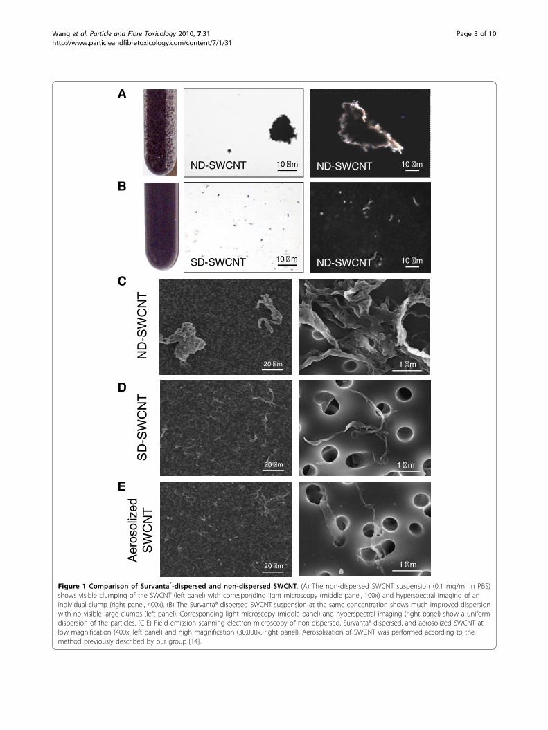
ResultsNanoparticle dispersion by Survanta®Visual inspection and corresponding micrographs of thenon-dispersed SWCNT (ND-SWCNT) and Survanta®-dispersed SWCNT (SD-SWCNT) are shown in Figure 1.The concentration of 150 μg/ml of Survanta® used inthis preparation is based on the content of lung surfac-tants found in rodent lung lavage fluids [13]. Micro-meter SWCNT agglomerates were noted inND-SWCNT suspension (Figure 1A). In contrast, Sur-vanta® dispersed SWCNT into smaller size structures asshown by visual inspection (Figure 1B, left panel) or by
regular light and hyperspectral microscopy (Figure 1B,middle and right panels). Field emission scanning elec-tron micrographs and count-mode particle size analysisof ND-SWCNT and SD-SWCNT suspended structuresare shown in Figure 1 C and 1D comparing to aeroso-lized SWCNT (Figure 1E) and Table 1, respectively. SD-SWCNT showed suspended structures of a smaller size,exhibiting count median width (CMW) and count med-ian length (CML) of 0.3 μm × 1 μm, compared to ND-SWCNT with 8 μm (CMW) × 22 μm (CML), respec-tively (Figure 2). These results indicate that Survanta®substantially improved the dispersion of SWCNT tosuch a degree that is comparable with the structuresizes reported for aerosolization of dry SWCNT [14].Table 1 gives the average diameter and length of
SD-SWCNT vs. ND-SWCNT. The mean width ofSD-SWCNT was 380 nm. About two-thirds of the SD-SWCNT were dispersed into structures with diametersless than the average diameter of 380 nm, and morethan 8% of the dispersed particles were less than 100nm in diameter (Table 2). In contrast, no particles withless than 380 nm in diameter were observed in ND-SWCNT preparations (Table 2). These results indicatethat Survanta® substantially improves the dispersion ofSWCNT at the concentration used.
Effect of Survanta® on cytotoxicityTo be useful as a dispersion agent for nanoparticles,Survanta® should exhibit no cytotoxic effect and shouldnot mask the bioactivity of nanoparticles. We first testedthe cytotoxic effect of Survanta® alone in the absence ofSWCNT on lung epithelial cells by lactate dehydrogen-ase (LDH) assay and by direct cell count. The resultsshow that at the concentrations tested (0.036-36 μg/ml),Survanta® had no significant effects on the LDH releaseand cell number as compared to non-treated control(Figure 3 A and 3B), suggesting its biocompatibility atthe test concentrations.
Effects of Survanta®-dispersed SWCNT on cell growth andtoxicityWe next evaluated the effects of SWCNT, dispersed ornon-dispersed by Survanta®, on cell toxicity and prolif-eration. Figure 4A shows that Survanta®-dispersedSWCNT exhibited a growth stimulating effect at lowdoses (0.02 and 0.06 μg/cm2 or 0.04 and 0.12 μg/ml)and suppressing effect at the highest dose tested (0.6μg/cm2 or 1.2 μg/ml), whereas non-dispersed SWCNThad no effect on cell proliferation as compared to non-treated or Survanta® only treated control. These resultssuggest the importance of dispersion status of SWCNTon their bioactivity, which is supported by the observa-tion that SWCNT dispersed by acetone/sonication (AD-SWCNT) also induced cell proliferation at the lowest
Wang et al. Particle and Fibre Toxicology 2010, 7:31http://www.particleandfibretoxicology.com/content/7/1/31
Page 2 of 10
20 �m
1 �m
1 �m
1 �m
20 �m
20 �m
A
B
Aer
osol
ized
SW
CN
TS
D-S
WC
NT
N
D-S
WC
NT
C
D
E
10 �m
10 �m
10 �m
10 �m
ND-SWCNT
SD-SWCNT
ND-SWCNT
ND-SWCNT
Figure 1 Comparison of Survanta®-dispersed and non-dispersed SWCNT. (A) The non-dispersed SWCNT suspension (0.1 mg/ml in PBS)shows visible clumping of the SWCNT (left panel) with corresponding light microscopy (middle panel, 100x) and hyperspectral imaging of anindividual clump (right panel, 400x). (B) The Survanta®-dispersed SWCNT suspension at the same concentration shows much improved dispersionwith no visible large clumps (left panel). Corresponding light microscopy (middle panel) and hyperspectral imaging (right panel) show a uniformdispersion of the particles. (C-E) Field emission scanning electron microscopy of non-dispersed, Survanta®-dispersed, and aerosolized SWCNT atlow magnification (400x, left panel) and high magnification (30,000x, right panel). Aerosolization of SWCNT was performed according to themethod previously described by our group [14].
Wang et al. Particle and Fibre Toxicology 2010, 7:31http://www.particleandfibretoxicology.com/content/7/1/31
Page 3 of 10

dose tested (Figure 4A). These data suggest that Sur-vanta® did not mask the bioproliferative activity ofSWCNT. LDH studies show that SD-SWCNT and othertest agents were non-toxic at all concentrations tested(Figure 4B). These results indicate that non-cytotoxicdoses of dispersed SWCNT can alter the growth patternof human lung epithelial cells. To our knowledge, this is
the first demonstration of the proliferative effect of low-dose SWCNT on lung epithelial cells.
Effect of dispersed SWCNT on collagen production bylung fibroblastsFibrosis is a fibroproliferative disorder characterized byoverproduction and accumulation of extracellular
Table 1 Size of non-dispersed and Survanta®-dispersedparticles
Width* (μm) Length* (μm)
SD-SWCNT 0.38 ± 0.02 1.42 ± 0.08
ND-SWCNT 12.35 ± 0.76 27.72 ± 1.86
SD-UFCB 0.70 ± 0.04 0.93 ± 0.05
ND-UFCB 5.07 ± 0.18 6.54 ± 0.2
* mean ± SD, n = 300.
Figure 2 Effect of Survanta® on SWCNT particle size anddistribution. SWCNT were dispersed in PBS in the presence orabsence of Survanta® as described in Figure 1. (A) Width distributionof non-dispersed and Survanta®-dispersed SWCNT. (B) Lengthdistribution of non-dispersed and Survanta®-dispersed SWCNT. CMW= count median width; CML = count median length; GSD =geometric standard deviation. The size and distribution values weredetermined from triplicate experiments with each experimentmeasuring a minimum of 300 particles.
Table 2 Percentage of well dispersed particles insuspension
% with width of <0.38 μm* % with width of <0.1 μm
SD-SWCNT 65.6% 8.6%
ND-SWCNT 0% 0%
% with width of <0.70 μm# % with width of <0.1 μm
SD-UFCB 73.0% 1.1%
ND-UFCB 0% 0%
* average width of SD-SWCNT; # average width of SD-UFCB.
Figure 3 Effect of Survanta® on cell toxicity and cell number.Subconfluent (80%) cultures of human lung epithelial BEAS-2B cellswere exposed to various concentrations of Survanta® and, at oneand two days after the treatment, cells were analyzed forcytotoxicity and cell number by LDH assay (A) and cell counting (B).Plots are mean ± S.D. (n = 4). No significant changes over non-treated controls were observed in all measurements at p < 0.05.
Wang et al. Particle and Fibre Toxicology 2010, 7:31http://www.particleandfibretoxicology.com/content/7/1/31
Page 4 of 10

matrix, notably collagens [5,14]. We tested whether SD-SWCNT can stimulate collagen production by lungfibroblasts in culture. AD-SWCNT which have beenshown to induce interstitial lung fibrosis [5] were usedas a positive control. Ultrafine carbon black (UFCB) dis-persed in Survanta® was used as a negative particle con-trol. The results show that as compared to non-treated control, Survanta® alone or SD-UFCB had nosignificant effect on cellular collagen content, as deter-mined by Sircol® assay which detects total collagen con-tent (Figure 5A). In contrast, SD-SWCNT and AD-SWCNT caused a substantial increase in cellular col-lagen content at 0.02 μg/cm2 (0.04 μg/ml). Western blotanalysis of collagen I, which is the most abundant col-lagen in the lung [15], shows a similar collagen-inducingeffect of SD-SWCNT and AD-SWCNT, whereas Sur-vanta® alone and SD-UFCB had a minimal effect (Figure5B). These results indicate that SD-SWCNT was able tostimulate collagen production in lung fibroblasts andthat Survanta® used for the SWCNT dispersion had nointerfering or masking effect on the collagen-inducingactivity of SWCNT, i.e., as compared to AD-SWCNT.
Effect of dispersed SWCNT on lung fibrosis in miceTo evaluate the in vivo fibrogenic effect of SWCNT, micewere treated with 10 μg/mouse, (~0.02 μg/cm2 of alveolarepithelial surface area in the mouse lung) SD-SWCNT orcontrol treatments, and analyzed for lung collagen content
Figure 4 Effect of SWCNT on cell proliferation and cell viability.Subconfluent (80%) cultures of lung epithelial BEAS-2B cells wereeither left untreated or treated with the indicated concentrations ofnon-dispersed (ND-), Survanta®-dispersed (SD-), or acetone/sonication-dispersed (AD-) SWCNT, or Survanta® alone, or notreatment (No Tx) for 24 h. (A) Cell proliferation was determined byhemocytometry. (B) Cell toxicity was determined by LDH assay. Plotsare mean ± S.D. (n = 4). *p < 0.05 versus non-treated control.
Figure 5 Effect of SWCNT on fibroblast collagen production.Subconfluent cultures of human lung fibroblast CRL-1490 cells wereeither left untreated (No Tx) or treated with Survanta® alone,Survanta®-dispersed SWCNT (SD-SWCNT), Survanta®-dispersedultrafine carbon black (SD-UFCB), or acetone/sonication-dispersedSWCNT (AD-SWCNT) at a particle concentration of 0.02 μg/cm2 or0.04 μg/ml. At 2 days after the treatment, cells were washed, lysed,and analyzed for total collagen content by the Sircol® assay (A) orcollagen I expression by Western blotting (B), as described in theMethods section. Equal amounts of total protein per sample wereused in all measurements. b-actin was used as a loading control forWestern blotting. The blot signals were quantified by densitometryand mean data from independent experiments (one of which isshown here) were normalized to the result obtained in cells withouttreatment. Plots are mean ± S.D. (n = 4). *p < 0.05 versus non-treated control.
Wang et al. Particle and Fibre Toxicology 2010, 7:31http://www.particleandfibretoxicology.com/content/7/1/31
Page 5 of 10

at two weeks post-treatment. Figure 6A shows that bothSD-SWCNT and AD-SWCNT were able to increase lungcollagen content as determined by Sircol® assay, whereasSD-UFCB or Survanta® only treatment had no significanteffect as compared to non-treated control (No Tx). Similarresults were observed with collagen I content as
determined by Western blot assay (Figure 6B), supportingthe fibrogenic effect of SWCNT. These results are consis-tent with the in vitro data and indicate the fibrogenic effectof SWCNT and lack of this effect by Survanta® alone. Theobserved similarity of the fibrogenic effect of SD-SWCNTand AD-SWCNT indicates that Survanta® did not maskthe bioactivity of SWCNT.
DiscussionThe major goal of this study was to evaluate the suit-ability of utilizing Survanta® as a dispersing agent forSWCNT to aid study of their bioactivity. The rationalebehind its use include its simplicity and rapidity ofnanoparticle dispersion (i.e., it is a single step process),biocompatibility (i.e., it has been approved for humanclinical use), and commercial availability particularly asa sterile preparation which greatly facilitates in vivo andin vitro studies that require sterile conditions. In addi-tion, one would argue that inhaled SWCNT would initi-ally interact with alveolar lining fluid, which is modeledby Survanta® suspension. However, the effectiveness ofthis preparation in dispersing nanoparticles and its pos-sible interfering effect on the bioactivities of SWCNTare not known, and, therefore, are investigated in thepresent study. The data presented demonstrate that Sur-vanta® when used at the indicated concentrations iseffective in dispersing SWCNT, yielding nanoparticleswith dimensions similar to those observed after aerosoli-zation of dry SWCNT or acetone/sonication dispersionof SWCNT [5,14]. Non-dispersed SWCNT form largeagglomerates in phosphate-buffered saline (PBS) with anaverage width of 12.35 μm and an average length of27.72 μm. In contrast, Survanta®-dispersed SWCNTform much smaller structures with an average width of0.38 μm and an average length of 1.42 μm. The majorityof the dispersed SWCNT is in the form of small bundleswith no or minimum detectable individual nanotubes.The reported average diameters of aerosolized drySWCNT and acetone/sonication-dispersed SWCNT are0.24 μm and 0.6 μm, respectively [5,14]. These resultsare in good agreement with previous reports showingcount median aerodynamic diameter of dry CNT gener-ated aerosols for inhalation studies [14,16], structuresize distribution found in the workplace [17], and CNTstructures produced by surfactant dispersion [18]. Thedata indicate that aerosolized dry SWCNT and surfac-tant-dispersed SWCNT form much smaller aggregatesthan SWCNT suspended in PBS. Previous animal stu-dies also showed that well-dispersed SWCNT after pul-monary administration exhibited similar depositedstructures and dimensions (e.g., submicron-sized aggre-gates) as those observed in this study [5].Ideally, in vivo pulmonary exposure studies should be
performed using an inhalation method, since it best
Figure 6 Effect of SWCNT on lung fibrosis in mice. Mice werepharyngeal aspirated with Survanta® alone, Survanta®-dispersedSWCNT (SD-SWCNT) or acetone/sonication-dispersed SWCNT (AD-SWCNT), or Survanta®-dispersed ultrafine carbon black SD-UFCB) atthe particle concentration of 10 μg/mouse. At 2 weeks after thetreatment, mice were sacrificed and the lungs were isolated, lysed,and analyzed for collagen content by the Sircol® assay (A) andWestern blotting (B). Equal amounts of total protein per samplewere used. b-actin was used as a loading control for Westernblotting. The blot signals were quantified by densitometry andmean data from three independent experiments were normalizedto the result obtained in cells without treatment. Plots are mean ±S.D. (n = 3). *p < 0.05 versus non-treated control (No Tx).
Wang et al. Particle and Fibre Toxicology 2010, 7:31http://www.particleandfibretoxicology.com/content/7/1/31
Page 6 of 10

mimics the human exposure condition. However, theneed for specialized facilities and equipment, trainedpersonnel, and large quantities of nanoparticles has lim-ited the use of this technology. Pulmonary aspirationrepresents an alternative method that has proven usefulin many pulmonary toxicity studies. This method is sim-ple, economical, uses small amounts of nanoparticles,and provides deep lung deposition as well as high corre-lation to the administered dose [19]. Recent studies byour group have shown that CNT administered by thismethod produced pulmonary fibrosis in lab animalssimilar to that observed after inhalation of CNT [5,6,14].These studies suggest that the aspiration method is areasonable alternative method to inhalation for thestudy of pulmonary fibrosis induced by nanoparticles.Present SWCNT-size analysis further supports theaspiration method using SD-SWCNT in which the aver-age diameter is comparable with aerosolized drySWCNT using for inhalation study.The use of Survanta® for nanoparticle dispersion pro-
vides an additional advantage over other methods of dis-persion for pulmonary studies as it better mimics thenatural lung condition. This is particularly important forin vitro studies which normally lack lung surfactants thatcould have an effect on cell interaction and bioactivity ofnanoparticles. Previous studies have shown that lung sur-factants aid in the displacement of particles from air to theaqueous phase and towards the lung epithelium [19]. Inaddition, when particles are present in peripheral airwaysand alveoli they exist in a completely immersed, wettedstate below the surfactant film [20]. These studies suggestthat experiments using lung surfactants may be more phy-siologically relevant than non-surfactant systems.A key concern about the use of Survanta® is its possi-
ble adverse effects on cells and tissues or masking effecton the exposed particles [21,22]. Our results show thatSurvanta®, when used at the indicated concentrations,had no significant cytotoxic effect on lung cells in vitroand did not induce collagen production or mask thefibrogenic effect of SWCNT either in vitro or in vivo.The results of this study also indicate that dispersionstatus of SWCNT is a key determinant of its biologicalactivities to induce cell proliferation and enhance col-lagen production.Another key finding of this study is the correlation
between in vitro and in vivo fibrogenic responses toSWCNT and control particles under different dispersionconditions. This finding suggests the potential utility ofin vitro lung fibroblasts as a predictive model for in vivofibrogenicity testing of CNT and other nanomaterials.Fibrogenicity testing of nanomaterials is usually per-formed using animals. However, this method of testing istime-consuming, laborious, and costly. This combinedwith the rapid growth in nanotechnology, which
produces an uncountable number and variety of nanoma-terials, makes it impractical to test all of these materialsusing animals. The in vitro model described here repre-sents an alternative method that could serve as a rapidscreening tool for fibrogenicity testing of a large numberof nanomaterials. This model can also be used to conductdetailed mechanistic studies of the fibrogenic effect ofnanoparticles, which may not be achievable in vivo.
ConclusionsThe present study describes a novel method of CNT dis-persion using the natural lung surfactant Survanta®. Sur-vanta® was shown to be effective in dispersing SWCNTand caused no cytotoxic or fibrogenic effect to the testlung cells under the experimental conditions. SD-SWCNTand AD-SWCNT similarly stimulated collagen productionof lung fibroblasts in vitro and both induced lung fibrosisin vivo, indicating the fibrogenicity of SWCNT and non-masking effect of Survanta®. The reported in vitro cellmodel system could potentially be used to aid the fibro-genicity testing of CNT of various size and functionaliza-tion as well as mechanistic studies of other nanoparticles.
MethodsParticlesSWCNT (CNI, Houston, TX) were produced by thehigh pressure CO disproportionation (HiPco) technique,employing CO in a continuous-flow gas phase as thecarbon feedstock and Fe(CO)5 as the iron-containingcatalyst precursor. These SWCNT were then purified byacid treatment to remove metal contaminates for use inthis study. Elemental analysis of the supplied SWCNTby nitric acid dissolution and inductively coupledplasma-atomic emission spectrometry (ICP-AES,NMAM #7300) showed that the SWCNT were 99% ele-mental carbon and 0.23% iron. The specific surface areawas measured at -196°C by the nitrogen absorption-desorption technique (Brunauer Emmet Teller method,BET) using a SA3100 Surface Area and Pore Size Analy-zer (Beckman Coulter, Fullerton, CA). The diameter andlength distribution of poorly and well-dispersed prepara-tions of SWCNT (without or with Survanta®) were mea-sured by field emission scanning electron microscopy.The surface area of dry SWCNT was 400-1,000 m2/g,and the length and width of individual (dry) SWCNTwere 0.1-1 μm and 0.8-1.2 nm, respectively. Characteri-zation studies were performed at NIOSH research facil-ities as previously described [23]. The same lot ofSWCNT was used for all experiments.
Particle dispersionSWCNT were dispersed by using Survanta® (AbbottLaboratories, Columbus, OH) or by the acetone-sonica-tion method as described previously [5]). Survanta®-
Wang et al. Particle and Fibre Toxicology 2010, 7:31http://www.particleandfibretoxicology.com/content/7/1/31
Page 7 of 10

dispersed SWCNT (SD-SWCNT) were prepared by dis-persing SWCNT (0.1 mg/ml) in PBS containing Sur-vanta® (150 μg/ml) with light sonication (Sonic VibraCell Sonicator, Sonic & Material Inc, Newtown, CT,USA) at a power of 130 W, frequency of 20 kHz, andamplitude settings of 60% for 5-10 seconds. Non-dispersed SWCNT (ND-SWCNT) were prepared simi-larly but in the absence of Survanta®. Acetone/sonicationdispersed SWCNT (AD-SWCNT) were prepared accord-ing to the method previously described [5]. Briefly,SWCNT were treated with acetone and placed in anultrasonic bath for 24 h. The dispersed CNT were thenfiltered from the solution using a 20-μm nylon meshscreen followed by a 0.2-μm polytetrafluoroethylene fil-ter. After filter collection, the dispersed CNT werewashed thoroughly with distilled water and suspended inPBS with 2-3 minute sonication (Sonic Vibra Cell Sonica-tor, Sonic & Material Inc, Newtown, CT, USA).
Particle imaging and size measurementsImages of SWCNT suspensions were obtained by fieldemission scanning electron microscopy and nanoscalehyperspectral microscopy. To assess the size distributionof SWCNT samples, a sample of each was taken and fil-tered through a polycarbonate filter (VCTP02500 iso-pore membrane; Millipore, Billerica, MA) to collect theparticles. After washing with water and drying, the filterwas cut into equal sections, mounted onto aluminumstubs with double-stick carbon tape, and sputter coatedwith gold/palladium. The deposited particles wereviewed under a field emission scanning electron micro-scope (model S-4800; Hitachi, Tokyo, Japan) at 400 and30,000 magnifications. The average length and width ofthe particles in each sample were determined by analysisof a minimum of 300 particles. The size and distributionvalues were determined from triplicate experiments.Representative micrographs of the SWCNT sampleswere taken using conventional and hyperspectral micro-scopy. The latter system (CytoViva, Auburn, AL) iscapable of identifying specific material at a sub100-nanometer resolution based on the material’sunique spectral signature. Hyperspectral images ofSWCNT were captured with the CytoViva spectrophot-ometer and an integrated CCD camera mounted on anOlympus BX-51 microscope at 400x.
Cell growth and cytotoxicityHuman lung epithelial BEAS-2B cells (American TypeCulture Collection, Manassas, VA) were incubated in a24-well plate at the density of 2 × 10⁴ cells/well in Dul-becco’s modified eagle medium containing 5% fetalbovine serum. The cells were treated with various con-centrations of Survanta®, ND-SWCNT, SD-SWCNT, or
AD-SWCNT at 37°C. At the indicated times after thetreatment, cell supernatants were collected and analyzedfor lactate dehydrogenase (LDH) as an indicator of celltoxicity. LDH activity was determined by LDH-catalyzedoxidation of pyruvate coupled with the reduction ofNAD at 340 nm using a commercial assay kit and aCobas Mira Plus transfer analyzer (Roche DiagnosticsSystem, Montclair, NJ). Cell growth was determined bydirect cell counting of the control and treated cells. Thecells were trypsinized, suspended in 100 μl culture med-ium, and 10 μl samples of the suspension were mixedwith trypan blue for cell number counting and determi-nation of cell viability using a hemocytometer.
Collagen assaysCollagen content was determined by Western blottingand the Sircol® assay (Biocolor Ltd., Belfast, UK).Human lung fibroblast CRL-1490 cells (ATCC, Mana-ssas, VA) or mice were treated with Survanta®, SWCNT,or control particles as described below. Treated cells ormouse lung tissues were lysed and cell/tissue lysateswere analyzed for protein content using a bicinchoninicacid protein assay kit (Pierce Biotechnology, Rockford,IL). For Western blot analysis, equal amounts of proteinper sample (25 μg) were resolved by 10% sodium dode-cyl sulfate-polyacrylamide gel electrophoresis (SDS-PAGE) and transferred onto a nitrocellulose membrane.The membrane was blocked with T-PBS (0.3% Tween-20 in PBS) containing 3% dry milk and incubated withprimary antibodies specific for collagen type I andb-actin (Fitzgerald, Concord, MA) at 4°C overnight.After three washes with T-PBS, the membrane wasincubated with peroxidase-conjugated secondary anti-body for 1 h and then washed with 0.05% Tween-20 inPBS. The immune complexes were detected by chemilu-minescence (Supersignal® West Pico, Pierce, Rockford,IL) and quantified using analyst/PC densitometry soft-ware (Bio-Rad Laboratories, Hercules, CA).For analysis of collagen content by the Sircol® assay,
cell/tissue lysates (50 μl) were incubated with Sirius redreagent (50 μl) for 30 min, after which the collagen-dyecomplex was precipitated by centrifugation at 16,000 gfor 5 min. The precipitates were washed with ethanoland dissolved in 0.5 M NaOH. The samples were thenintroduced into a microplate reader and absorbancedetermined at 540 nm.
Animal exposurePathogen-free male C57BL/6J mice (Jackson Labora-tories, Bar Harbor, ME) weighing 25-30 grams wereused. The animals were individually housed in anAssociation for Assessment and Accreditation ofLaboratory Animal Care-accredited facility and allowed
Wang et al. Particle and Fibre Toxicology 2010, 7:31http://www.particleandfibretoxicology.com/content/7/1/31
Page 8 of 10

to acclimate at least 1 week prior to use. All experi-mental procedures were conducted in accordance witha protocol approved by the NIOSH Institutional Ani-mal Care and Use Committee. The animals were trea-ted with the test materials by pharyngeal aspiration asdescribed previously [24]. Briefly, animals wereanesthetized by an intraperitoneal injection of keta-mine and xylazine (45 and 8 mg/kg) and placed on aboard in the supine position. The animal’s tongue wasextended with padded forceps. A suspension of thetest material (10 μg/100 ml per mouse) was placed onthe back of the tongue. A slight pull of the tongueresults in a reflex gasp and aspiration of the droplet.The tongue was held, and the animal was monitoredfor a few breaths after aspiration. All mice survived thepharyngeal aspiration procedure. At given post-expo-sure times, mice were sacrificed and lung tissues wereisolated, homogenized, lysed and analyzed for collagencontent by Western blot and Sircol® assays.
StatisticsData were analyzed by ANOVA (STATGRAF). Bartlett’stest was used to test for homogeneity of variancesbetween groups. Statistical differences were determinedby one-way ANOVA, with significance set at P < 0.05.When significant F-values were obtained, individualmeans were compared with control using a two-sidedDunnett’s test. P < 0.05 was considered to be significant.Data are given as means ± SD. The size distributions ofSWCNT particles in different media were determinedusing the procedures described in Hinds [25].
AcknowledgementsThis work was supported by NIOSH and the National Institutes of HealthGrant R01-HL095579.The findings and conclusions in this report are those of the authors and donot necessarily represent the views of the National Institute for OccupationalSafety and Health.
Author details1National Institute for Occupational Safety and Health, HELD/PPRB,Morgantown, WV 26505, USA. 2Department of Pharmaceutical Sciences, WestVirginia University, Morgantown, WV 26506, USA.
Authors’ contributionsLW conceived the study, carried out in vitro and in vivo experiments, anddrafted the manuscript. VC participated in the design of the study,evaluation of results, and helped to draft the manuscript. AM participated inin vitro studies and Western blot analysis. BC participated in particlemeasurements and data analysis. RM participated in animal exposure studies.DS-B carried out the electron microscopy. YR participated in study design,particle dispersion, collagen assays, and manuscript preparation. All authorsread and approved the final manuscript.
Competing interestsThe authors declare that they have no competing interests.
Received: 8 June 2010 Accepted: 19 October 2010Published: 19 October 2010
References1. Wang Y, Wu J, Wei F: A treatment method to give separated multi-
walled carbon nanotubes with high purity, high crystallization and alarge aspect ratio. Carbon 2003, 41:2939-2948.
2. Lam CW, James JT, McCluskey R, Hunter RL: Pulmonary toxicity of single-wall carbon nanotubes in mice 7 and 90 days after intratrachealinstillation. Toxicol Sci 2004, 77:126-134.
3. Borm PJ, Robbins D, Haubold S, Kuhlbusch T, Fissan H, Donaldson K,Schins R, Stone V, Kreyling W, Lademann J, Krutmann J, Warheit D,Oberdorster E: The potential risks of nanomaterials: a review carried outfor ECETOC. Part Fibre Toxicol 2006, 3:11.
4. Maynard AD, Baron PA, Foley M, Shvedova AA, Kisin ER, Castranova V:Exposure to carbon nanotube material: aerosol release during thehandling of unrefined single-walled carbon nanotube material. J ToxicolEnviron Health A 2004, 67:87-107.
5. Mercer RR, Scabilloni J, Wang L, Kisin E, Murray AR, Schwegler-Berry D,Shvedova AA, Castranova V: Alteration of deposition pattern andpulmonary response as a result of improved dispersion of aspiratedsingle-walled carbon nanotubes in a mouse model. Am J Physiol LungCell Mol Physiol 2008, 294:L87-97.
6. Shvedova AA, Kisin ER, Mercer R, Murray AR, Johnson VJ, Potapovich AI,Tyurina YY, Gorelik O, Arepalli S, Schwegler-Berry D, Hubbs AF, Antonini J,Evans DE, Ku BK, Ramsey D, Maynard A, Kagan VE, Castranova V, Baron P:Unusual inflammatory and fibrogenic pulmonary responses to single-walled carbon nanotubes in mice. Am J Physiol Lung Cell Mol Physiol 2005,289:L698-708.
7. Wang L, Mercer RR, Rojanasakul Y, Qiu A, Lu Y, Scabilloni JF, Wu N,Castranova V: Direct fibrogenic effects of dispersed single-walled carbonnanotubes on human lung fibroblasts. J Toxicol Environ Health A 2010,73:410-22.
8. Jia G, Wang H, Yan L, Wang X, Pei R, Yan T, Zhao Y, Guo X: Cytotoxicity ofcarbon nanomaterials: single-wall nanotube, multi-wall nanotube, andfullerene. Environ Sci Technol 2005, 39:1378-1383.
9. Monteiro-Riviere NA, Nemanich RJ, Inman AO, Wang YY, Riviere JE: Multi-walled carbon nanotube interactions with human epidermalkeratinocytes. Toxicol Lett 2005, 155:377-84.
10. Soto KF, Carrasco A, Powell TG, Garza KM, Murr LE: Comparative in vitrocytotoxicity assessment of some manufactured nanoparticulate materialscharacterized by transmission electron microscopy. J Nanopart Res 2005,7:145-169.
11. Cherukuri P, Bachilo SM, Litovsky SH, Weisman RB: Near-infraredfluorescence microscopy of single-walled carbon nanotubes inphagocytic cells. J Am Chem Soc 2004, 126:15638-15639.
12. Muller J, Huaux F, Moreau N, Misson P, Heilier JF, Delos M, Arras M,Fonseca A, Nagy JB, Lison D: Respiratory toxicity of multi-wall carbonnanotubes. Toxicol Appl Pharmacol 2005, 207:221-231.
13. Sager TM, Porter DW, Robinson VA, Lindsley WG, Schwegler-Berry DE,Castranova V: Improved method to disperse nanoparticles for in vitroand in vivo investigation of toxicity. Nanotoxicology 2007, 1:118-129.
14. Shvedova AA, Kisin E, Murray AR, Johnson VJ, Gorelik O, Arepalli S,Hubbs AF, Mercer RR, Keohavong P, Sussman N, Jin J, Yin J, Stone S,Chen BT, Deye G, Maynard A, Castranova V, Baron PA, Kagan VE: Inhalationvs. aspiration of single-walled carbon nanotubes in C57BL/6 mice:inflammation, fibrosis, oxidative stress, and mutagenesis. Am J PhysiolLung Cell Mol Physiol 2008, 295:L552-565.
15. Brinckerhoff CE, Matrisian LM: Matrix metalloproteinases: a tail of a frogthat became a prince. Nature Rev Mol Cell Biol 2002, 3:207-214.
16. McKinney W, Chen B, Frazer D: Computer controlled multi-walled carbonnanotube inhalation exposure system. Inhal Toxicol 2009, 21:1053-1061.
17. Han JH, Lee EJ, Lee JH, So KP, Lee YH, Bae GN, Lee S, Ji JH, Cho MH, Yu IJ:Monitoring multiwalled carbon nanotube exposure in carbon nanotuberesearch facility. Inhal Toxicol 2008, 20:741-749.
18. Porter DW, Sriram K, Wolfarth MG, Jefferson AM, Schwegler-Berry D,Andrew ME, Castranova V: A biocompatible medium for nanoparticledispersion. Nanotoxicol 2008, 2:144-154.
19. Schürch S, Bachofen H, Goerke J, Green F: Surface properties of ratpulmonary surfactant studied with the captive bubble method:adsorption, hysteresis, stability. Biochim Biophys Acta 1992, 1103:127-136.
20. Geiser M, Schurch S, Gehr P: Influence of surface chemistry andtopography of particles on their immersion into the lung’s surface-lininglayer. J Appl Physiol 2003, 94:1793-1801.
Wang et al. Particle and Fibre Toxicology 2010, 7:31http://www.particleandfibretoxicology.com/content/7/1/31
Page 9 of 10

21. Antonini JM, Reasor MJ: Effect of short-term exogenous pulmonarysurfactant treatment on acute lung damage associated with theintratracheal instillation of silica. J. Toxicol Environ Health 1994, 43:85-101.
22. Wang L, Scabilloni JF, Antonini JM, Castranova V, Rojanasakul Y, Roberts JR,Zhang Z, Mercer RR: Role of lung surfactant in phagocytic clearance ofapoptotic cells by macrophages in rats. Lab Invest 2006, 86:458-66.
23. Sargent LM, Shvedova AA, Hubbs AF, Salisbury JL, Benkovic SA, Kashon ML,Lowry DT, Murray AR, Kisin ER, Friend S, McKinstry KT, Battelli L,Reynolds SH: Induction of aneuploidy by single-walled carbonnanotubes. Environ Mol Mutag 2009, 50:708-17.
24. Rao GVS, Tinkle S, Weissman DN, Antonini JM, Kashon ML, Salmen R,Battelli LA, Willard PA, Hubbs AF: Efficacy of a technique for exposing themouse lung to particles aspirated from the pharynx. J Toxicol EnvironHealth A 2003, 66:1441-1452.
25. Hinds W: Aerosol Technology New York: John Wiley and Sons 1999, Chapter 4.
doi:10.1186/1743-8977-7-31Cite this article as: Wang et al.: Dispersion of single-walled carbonnanotubes by a natural lung surfactant for pulmonary in vitro and invivo toxicity studies. Particle and Fibre Toxicology 2010 7:31.
Submit your next manuscript to BioMed Centraland take full advantage of:
• Convenient online submission
• Thorough peer review
• No space constraints or color figure charges
• Immediate publication on acceptance
• Inclusion in PubMed, CAS, Scopus and Google Scholar
• Research which is freely available for redistribution
Submit your manuscript at www.biomedcentral.com/submit
Wang et al. Particle and Fibre Toxicology 2010, 7:31http://www.particleandfibretoxicology.com/content/7/1/31
Page 10 of 10
Related Documents

![Double-walled carbon nanotubes: synthesis, structural ...077-088]-01.pdf · Double-walled carbon nanotubes: synthesis, structural characterization, and ... are seamless cylindrical](https://static.cupdf.com/doc/110x72/5aa2b5537f8b9ac67a8d717c/double-walled-carbon-nanotubes-synthesis-structural-077-088-01pdfdouble-walled.jpg)