The Rockefeller University Press, 0021-9525/97/11/759/13 $2.00 The Journal of Cell Biology, Volume 139, Number 3, November 3, 1997 759–771 http://www.jcb.org 759 Dismantling Cell–Cell Contacts during Apoptosis Is Coupled to a Caspase-dependent Proteolytic Cleavage of b-Catenin Claudio Brancolini,* § Dean Lazarevic,* Joe Rodriguez, ‡ and Claudio Schneider* § *Laboratorio Nazionale Consorzio Interuniversitario Biotecnologie AREA Science Park, 34142 Trieste, Italy; ‡ Cold Spring Harbor Laboratory, Cold Spring Harbor, New York 11724; and § Dipartimento di Scienze e Tecnologie Biomediche, Sezione di Biologia, Universita’ di Udine, 33100 Udine, Italy Abstract. Cell death by apoptosis is a tightly regulated process that requires coordinated modification in cellu- lar architecture. The caspase protease family has been shown to play a key role in apoptosis. Here we report that specific and ordered changes in the actin cytoskele- ton take place during apoptosis. In this context, we have dissected one of the first hall- marks in cell death, represented by the severing of con- tacts among neighboring cells. More specifically, we provide demonstration for the mechanism that could contribute to the disassembly of cytoskeletal organiza- tion at cell–cell adhesion. In fact, b-catenin, a known regulator of cell–cell adhesion, is proteolytically pro- cessed in different cell types after induction of apopto- sis. Caspase-3 (cpp32/apopain/yama) cleaves in vitro translated b-catenin into a form which is similar in size to that observed in cells undergoing apoptosis. b-Cate- nin cleavage, during apoptosis in vivo and after caspase-3 treatment in vitro, removes the amino- and carboxy-terminal regions of the protein. The resulting b-catenin product is unable to bind a-catenin that is responsible for actin filament binding and organization. This evidence indicates that connection with actin fila- ments organized at cell–cell contacts could be disman- tled during apoptosis. Our observations suggest that caspases orchestrate the specific and sequential changes in the actin cytoskeleton occurring during cell death via cleavage of different regulators of the mi- crofilament system. Address all correspondence to Claudio Schneider, Laboratorio Nazionale, Consorzio Interuniversitario Biotechnologie, AREA Science Park, Padri- ciano 99, 34142 Trieste, Italy. Tel.: 39.40.398.985. Fax: 39.40.398.990. E-mail: [email protected] 1. Abbreviations used in this paper: aa, amino acid; Gas, growth arrest spe- cific protein; ICE, interleukin-1b–convertase enzyme; MMS, methyl- methanesulfonate. A poptosis is a fundamental cellular process in the de- velopment and homeostasis of all multicellular or- ganisms (Raff, 1992; Wyllie, 1995). Genetic analy- sis in Caenorhabditis elegans has identified both positive and negative regulators of apoptosis (Ellis et al., 1991). The ced-3 gene is essential for developmentally pro- grammed cell death in C. elegans: it encodes a protein which shares homology with mammalian interleukin- 1b–convertase enzyme (ICE; Yuan et al., 1993). ICE is a cystein protease that cleaves interleukin-1b pre- cursor at two aspartic residues and is activated by cleavage after specific aspartic residues of a 45-kD proenzyme, thus forming two subunits that together form the active site of the enzyme (Thornberry et al., 1992). The regulatory mech- anisms of the ICE remain to be clarified (Chinnaiyan et al., 1997). In general, it exhibits full activity only after pro- teolytic processing, which occurs either by self-cleavage or through cleavage by other proteases (Nicholson, 1996). Nine additional members of ICE-related proteases have been identified in mammals (Fernandes-Alnemri et al., 1994; Kumar et al., 1994; Wang et al., 1994; Faucheau et al., 1995; Fernandes-Alnemri et al., 1995a,b; Boldin et al., 1996; Duan et al., 1996a,b; Fernandes-Alnemri et al., 1996; Muzio et al., 1996). Based on amino acid (aa) 1 sequences and recognized substrates, they have been grouped in dif- ferent sub-families, and named according to the recently proposed caspase nomenclature (Alnemri et al., 1996). These proteases may overlap in their functions and/or reg- ulate one another’s activities. Indeed, recent observations suggest the existence of a hierarchically ordered proteolytic cascade during apopto- sis. In the case of CD95/FAS/APO-1-induced apoptosis, an initial activity related to ICE itself (caspase-1), and a later activity related to CPP32 (caspase-3), have been de- scribed using specific inhibitor substrates (Enari et al., 1996). Moreover, CD95/FAS/APO-1 receptors use the adaptor molecule MORT1/FADD to physically engage a cytosolic ICE-related cystein protease termed FLICE/ MACH (caspase-8) (Boldin et al., 1996; Muzio et al., 1996). on October 1, 2014 jcb.rupress.org Downloaded from Published November 3, 1997

Welcome message from author
This document is posted to help you gain knowledge. Please leave a comment to let me know what you think about it! Share it to your friends and learn new things together.
Transcript
The Rockefeller University Press, 0021-9525/97/11/759/13 $2.00The Journal of Cell Biology, Volume 139, Number 3, November 3, 1997 759–771http://www.jcb.org 759
Dismantling Cell–Cell Contacts during Apoptosis Is Coupled
to a Caspase-dependent Proteolytic Cleavage of
b
-Catenin
Claudio Brancolini,*
§
Dean Lazarevic,* Joe Rodriguez,
‡
and Claudio Schneider*
§
*Laboratorio Nazionale Consorzio Interuniversitario Biotecnologie AREA Science Park, 34142 Trieste, Italy;
‡
Cold Spring Harbor Laboratory, Cold Spring Harbor, New York 11724; and
§
Dipartimento di Scienze e Tecnologie Biomediche, Sezione di Biologia, Universita’ di Udine, 33100 Udine, Italy
Abstract.
Cell death by apoptosis is a tightly regulated process that requires coordinated modification in cellu-lar architecture. The caspase protease family has been shown to play a key role in apoptosis. Here we report that specific and ordered changes in the actin cytoskele-ton take place during apoptosis.
In this context, we have dissected one of the first hall-marks in cell death, represented by the severing of con-tacts among neighboring cells. More specifically, we provide demonstration for the mechanism that could contribute to the disassembly of cytoskeletal organiza-tion at cell–cell adhesion. In fact,
b
-catenin, a known regulator of cell–cell adhesion, is proteolytically pro-cessed in different cell types after induction of apopto-sis. Caspase-3 (cpp32/apopain/yama) cleaves in vitro
translated
b
-catenin into a form which is similar in size to that observed in cells undergoing apoptosis.
b
-Cate-nin cleavage, during apoptosis in vivo and after caspase-3 treatment in vitro, removes the amino- and carboxy-terminal regions of the protein. The resulting
b
-catenin product is unable to bind
a
-catenin that is responsible for actin filament binding and organization. This evidence indicates that connection with actin fila-ments organized at cell–cell contacts could be disman-tled during apoptosis. Our observations suggest that caspases orchestrate the specific and sequential changes in the actin cytoskeleton occurring during cell death via cleavage of different regulators of the mi-crofilament system.
Address all correspondence to Claudio Schneider, Laboratorio Nazionale,Consorzio Interuniversitario Biotechnologie, AREA Science Park, Padri-ciano 99, 34142 Trieste, Italy. Tel.: 39.40.398.985. Fax: 39.40.398.990. E-mail:[email protected]
1.
Abbreviations used in this paper
: aa, amino acid; Gas, growth arrest spe-cific protein; ICE, interleukin-1
b
–convertase enzyme; MMS, methyl-methanesulfonate.
A
poptosis
is a fundamental cellular process in the de-velopment and homeostasis of all multicellular or-ganisms (Raff, 1992; Wyllie, 1995). Genetic analy-
sis in
Caenorhabditis elegans
has identified both positiveand negative regulators of apoptosis (Ellis et al., 1991).
The
ced-3
gene is essential for developmentally pro-grammed cell death in
C. elegans
: it encodes a proteinwhich shares homology with mammalian interleukin-1
b
–convertase enzyme (ICE; Yuan et al., 1993).ICE is a cystein protease that cleaves interleukin-1
b
pre-cursor at two aspartic residues and is activated by cleavageafter specific aspartic residues of a 45-kD proenzyme, thusforming two subunits that together form the active site ofthe enzyme (Thornberry et al., 1992). The regulatory mech-anisms of the ICE remain to be clarified (Chinnaiyan etal., 1997). In general, it exhibits full activity only after pro-teolytic processing, which occurs either by self-cleavage orthrough cleavage by other proteases (Nicholson, 1996).
Nine additional members of ICE-related proteases have
been identified in mammals (Fernandes-Alnemri et al.,1994; Kumar et al., 1994; Wang et al., 1994; Faucheau etal., 1995; Fernandes-Alnemri et al., 1995
a
,
b
; Boldin et al.,1996; Duan et al., 1996
a
,
b
; Fernandes-Alnemri et al., 1996;Muzio et al., 1996). Based on amino acid (aa)
1
sequencesand recognized substrates, they have been grouped in dif-ferent sub-families, and named according to the recentlyproposed caspase nomenclature (Alnemri et al., 1996).These proteases may overlap in their functions and/or reg-ulate one another’s activities.
Indeed, recent observations suggest the existence of ahierarchically ordered proteolytic cascade during apopto-sis. In the case of CD95/FAS/APO-1-induced apoptosis,an initial activity related to ICE itself (caspase-1), and alater activity related to CPP32 (caspase-3), have been de-scribed using specific inhibitor substrates (Enari et al.,1996). Moreover, CD95/FAS/APO-1 receptors use theadaptor molecule MORT1/FADD to physically engage acytosolic ICE-related cystein protease termed FLICE/MACH (caspase-8) (Boldin et al., 1996; Muzio et al., 1996).
on October 1, 2014
jcb.rupress.orgD
ownloaded from
Published November 3, 1997
The Journal of Cell Biology, Volume 139, 1997 760
Caspase-8 might represent the most upstream protease in-volved in generating a death signal.
The central role of the caspase proteases in mammalianapoptosis is supported by the existence of specific inhibi-tors. The viral proteins p35 and CrmA (Bump et al., 1995;Xue and Horvitz, 1995), or aldehyde and fluormethylke-tone derivatives of the target cleavage sequences, suppressmammalian apoptosis as triggered by different stimuli(Miura et al., 1993; Gagliardini et al., 1994; Beidler et al.,1995; Nicholson et al., 1995).
If cystein proteases play a crucial role in apoptosis, spe-cific target proteins or death substrates become criticallyrelevant for the execution phase of the apoptotic program.
Specific cleavage by ICE-related cystein proteases ofthe DNA repair enzymes poly(ADP-ribose) polymerase(PARP) and the catalytic subunit of the DNA-dependentprotein kinase (DNA-PK), is possibly related to the inacti-vation of the DNA repair pathway as a choice of economy(Lazebnik et al., 1994; Song et al., 1996). Cleavage of thenuclear lamins is dependent on Mch2 (caspase-6) andseems to be essential for disassembling the nuclear struc-ture (Lazebnik et al., 1995; Takahashi et al., 1996). Othersubstrates, whose function is still unclear during apoptosis,are the 70-kD protein of the U1 small nuclear ribonucleo-protein (Casciola-Rosen et al., 1994), the sterol regulatoryelement-binding proteins SREBP-1 and SRBP-2 (Wang etal., 1996), the huntingtin protein (Goldberg et al., 1996),D4 GDP dissociation inhibitor (Na et al., 1996), and pRbtumor suppressor (Janicke et al., 1996).
Two interesting substrates for caspases are the proteinkinase C
d
and Gas2, the last being a component of the mi-crofilament system (Brancolini et al., 1992). In both cases,proteolytic processing results in a gain of function whichrelates to increased kinase activity in the case of PKC
d
(Emoto et al., 1995), or to an activity on the microfilamentsystem and on cell morphology in the case of Gas2 (Bran-colini et al., 1995). Recently, gain of function after caspase-3processing has also been demonstrated for the p21-acti-vated kinase 2 (Rudel and Bokoch, 1997), and the DNAfragmentation factor (Liu et al., 1997).
Apoptotic morphological features are generally similarin all systems where they have been studied: the nucleuscondenses and the cell shrinks and often fragments intoapoptotic bodies that are rapidly phagocytosed by neigh-boring cells (Kerr et al., 1972; Wyllie et al., 1980).
The present study addresses the roles and changes of themicrofilament system during apoptosis. By a detailed con-focal analysis on apoptotic NIH3T3 cells, we have identi-fied specific sequential changes in the microfilament sys-tem. Coordinately with actin filament reorganization inthe perinuclear area, the cell retracts from the adhesionsubstrate and severs contacts with neighboring cells.
Germane to the above mentioned changes, we also ob-served a specific modulation of the cell–cell contacts dur-ing apoptosis through proteolytic processing of
b
-cateninthereby losing the ability to bind
a
-catenin. Such proteolyticcleavage in vitro is mediated by the cystein-protease cpp32/apopain/yama (caspase-3).
In summary, the complex reorganization of cell mor-phology during apoptosis seems to be achieved by specificproteolytic processing of different regulators of actin cy-toskeleton.
Materials and Methods
Cell Lines and Culture Conditions
NIH3T3 and MDCK cells were grown in DME supplemented with 10%FCS, penicillin (100 U/ml), and streptomycin (100
m
g/ml). In each experi-ment 2.5
3
10
4
cells/ml were seeded in 35-mm Petri dishes.For serum starvation, medium was changed to 0.5% FCS when cells
were subconfluent and left in this medium for 48 h. For density-dependentinhibition, cells were plated at 10
4
/cm
2
in 10% FCS. 24 h after plating themedium was changed every 2 d. For induction of apoptosis the culture me-dium of arrested cells was replaced with serum-free DME. Nonadherentand adherent cells were harvested, washed in serum-free medium, and sol-ubilized in SDS-PAGE buffer. In the case of genotoxic-dependent apop-tosis, density-inhibited cells were treated with 20
m
g/ml of cisplatin for 4 h;nonadherent and adherent cells were harvested separately 24 h later. ForUV treatment, culture medium was removed, dishes were washed oncewith PBS, UVC irradiated (180 J/m
2
in PBS), and fresh medium, contain-ing 10% FCS, was added to the cells. Adherent and nonadherent cellswere harvested separately 24 h later. NIH3T3 cells were transfected aspreviously described (Brancolini et al., 1995).
Plasmid Construction
Mouse
b
-catenin cDNA (Hoschuetzky et al., 1994) was subcloned in-frame with a COOH-terminal VSV tag (
b
-catenin–VSV) in pMT2SM-tag(Bardelli et al., 1996) and pGDSV7S-tag (Brancolini et al., 1995). The
b
-cate-nin cDNA was amplified by PCR using a sense primer corresponding tothe T7 promoter (5
9
-AATACGACTCACTATAGGGC-3
9
) and a reverseprimer (VSV1) containing a SmaI site (5
9
-CCAAACTATGACTGGAG-GGCCCATT-3
9
). The carboxy-terminal deleted derivative of
b
-cateninwas produced by endonuclease digestion at the EcoRI site (nucleotides1361/aa 421) and a COOH-terminal VSV tag was introduced by PCR usinga sense primer corresponding to the T7 promoter, and a reverse primer(VSV2) containing a NotI site (5
9
-GATTGCGGCCGCAAGTGAGG-3
9
).A site-directed mutagenesis method by overlap extension through PCR
as previously described (Brancolini et al., 1995) was used to produce thepoint mutants D
→
A at different positions of the amino-terminal region of
b
-catenin. As external primers, the olT7 and olVSV2 were used. The pairsof complementary inverse oligos were the following:
b
-catenin 162–164D/A:ol162–164u:5
9
-CTAAACGCTGAGGCCCAGGTGGTA-3
9
ol162–164d:5
9
-TACCACCTGGGCCTCAGCGTTTAG-3
9
;
b
-catenin 144–145D/A:ol144–145u:5
9
-TATCAGGCTGCCGCGGAACTT-3
9
ol144–145d:5
9
-AAG-TTCCGCGGCAGCCTGAT A-3
9
.All constructs generated were sequenced using an automated (ALF)
system to check for the respective introduced mutations, deletions, andtranslating fidelity of the inserted PCR fragments.
Immunofluorescence Microscopy
NIH3T3 cells were grown under the described conditions and then fixedwith 3% paraformaldehyde in PBS for 20 min at room temperature. Fixedcells were washed with PBS/0.1 M glycine, pH 7.5, and then permeabilizedwith 0.1% Triton-X100 in PBS for 5 min. The coverslips were treated withthe anti–
b
-catenin monoclonal antibody directed against aa 571–781 (Sig-nal Transduction Laboratories, Lexington, KY) for 1 h in a moist chamberat 37
8
C. TRITC anti–mouse (Southern Biotechnologies, Birmingham,AL) was used as secondary antibody. Actin filaments were detected usingFITC-phalloidin (Sigma Chemical Co., St. Louis, MO) and nuclei were la-beled with propidium iodide 5 mM (Sigma Chemical Co.).
Cells were examined with a laser scan microscope (LSM 410; CarlZeiss, Inc., Thornwood, NY) equipped with a 488
l
argon laser and a 543 Ihelium neon laser. The following sets of filters were used: rhodamine(BP546, FT580, and LP 590); and fluoresceine (450–490, FT 510, andLP520).
Two-dimensional Gel Electrophoresis
NIH3T3 cells grown for 36 h in 0.5% FCS were labeled for 12 h in 1 ml ofDME methionine-free 0.5% FCS, containing 400 mCi/ml [
35
S]methionine(Amersham Int’l., Little Chalfont, UK). Serum-free medium containing acold methionine chase, (0.5 mM final concentration) was then added for 6 h.
Samples of total cellular proteins were prepared from adherent andnonadherent NIH3T3 cells after washing with cold PBS. Cells were lysedin hot (100
8
C) lysis solution (0.3% SDS, 200 mM dithiothreitol, 50 mM
on October 1, 2014
jcb.rupress.orgD
ownloaded from
Published November 3, 1997
Brancolini et al.
Dismantling Cell–Cell Contacts during Apoptosis
761
Tris-HCl, pH 8). Samples were then treated with 1:10 vol of nuclease solu-tion (50 mM MgCl
2
, 50 mM Tris-HCl, pH 7, 1 mg/ml DNaseI, 0.25 mg/mlRNaseA) for 10 min on ice, precipitated by adding ice-cold acetone to80% vol/vol, and centrifuged at 14,000
g
for 10 min. Air-dried pellets wereresuspended in a mixture of 1 vol lysis solution and 4 vol of sample buffer(9.9 M urea, 4% NP-40, 2.2% Millipore ampholytes, 100 mM dithiothrei-tol). Two-dimensional gels were run using the investigator two-dimen-sional electrophoresis system (Millipore Corp., Waters Chromatography,Milford, MA) with pH 3–10 and pH 4–8 Millipore ampholytes in the firstdimension and 10% acrylamide in the second dimension.
Immunoblotting
For Western blotting, proteins were transferred to 0.2-
m
m pore-size nitro-cellulose (Schleicher & Schuell Inc., Keene, NH) using a semidry blottingapparatus (Bio-Rad Laboratories, Hercules, CA) (transfer buffer: 20%methanol, 48 mM Tris, 39 mM glycine, and 0.0375% SDS). After stainingwith Ponceau S, the nitrocellulose sheets were saturated for 2 h in Blotto-Tween 20 (50 mM Tris-HCl, pH 7.5, 500 mM NaCl, 5% nonfat dry milk,and 0.1% Tween 20; Sigma Chemical Co.) and incubated overnight atroom temperature with a specific antibody: anti-Gas2, anti-actin, anti–
b
-cate-nin and anti–
a
-catenin (Transduction Laboratories, Lexington, KY), anti–
b
-catenin amino-terminal (McCrea et al., 1993), anti–PKC-
d
(Santa CruzBiotechnology, Inc., Santa Cruz, CA), and anti-VSV (Sigma ChemicalCo.). Blots were then rinsed three times with Blotto-Tween 20 and re-acted with peroxidase-conjugated goat anti–rabbit (Sigma Chemical Co.)or goat anti–mouse (Sigma Chemical Co.) for 1 h at room temperature.The blots were then washed four times in Blotto-Tween 20, rinsed in PBS,and developed with an enhanced chemiluminescent kit, as recommendedby the vendor (Amersham Int’l.).
Expression of Caspase-3 (CPP32) in Bacteria and In Vitro Protease Assay
Caspase-3 cDNA was subcloned in-frame into the BamH1 site of the bac-terial expression vector pQE-12 (QIAGEN Inc., Santa Clarita, CA). Cellswere grown to an A
600
of 0.6 and expression of caspase-3 was induced byadding isopropyl-
b
-
d
-thiogalactopyranoside to a final concentration of 1mM. After 2 h, cells were collected by centrifugation at 3,000
g
for 5 min,and then resuspended in 5 vol of caspase-3 buffer (Goldberg et al., 1996)with protease inhibitors (1 mM PMSF and 10
m
g/ml each aprotinin, leu-peptin, and pepstatin). Cells were lysed by sonication and debris was sedi-mented by centrifugation at 14,000
g
for 20 min. The resulting superna-tants were used for in vitro protease assays.
Caspase-3 and
b
-catenin were labeled with
35
S using the TNT-coupledreticulocyte lysate system (Promega Corp., Madison, WI). 1
m
l of trans-lated reticulocyte lysate was incubated with the appropiate dilution of thebacterial lysates in caspase-3 buffer (final volume of 10
m
l) for 1 h at 37
8
C.Reactions were terminated by adding 1 vol of SDS gel loading buffer andboiling for 3 min. The specific caspase-3/cpp32/apopain inhibitor Ac-Asp-Glu-Val-Asp-CHO was obtained from Bachem Bioscience (Bubendorf,Switzerland).
Immunoprecipitation
Analysis of
b
-catenin–
a
-catenin complexes in apoptotic and nonapoptoticcells was performed as previously described (Hulsken et al., 1994) withsome modifications. Cells were extracted with 140 mM NaCl, 4.7 mM KCl,0.7 mM MgSO
4
, 1.2 CaCl
2
, 20 mM Tris-HCl, pH 7.5, 5% glycerol, contain-ing 1% Triton X-100 and 1 mM PMSF, and 10
m
g/ml each of aprotinin, leu-peptin, antipain, and pepstatin. After centrifugation at 14,000
g
for 15 minmonoclonal antibodies, anti–
b
-catenin (Transduction Laboratories), oranti-pan cadherin (Sigma Chemical Co.) were added. After 3 h on ice,protein A–Sepharose (Pharmacia Biotech., Inc., Piscataway, NJ) was addedand incubation was prolonged for 1 h on ice. After a brief centrifugationin Eppendorf centrifuge, immunoprecipitates were washed with 100 mMNaCl, 5 mM EDTA, 20 mM Tris-HCl, pH 7.5, 1% Triton X-100 contain-ing 1 mM PMSF, and 10
m
g/ml each of aprotinin, leupeptin, antipain, andpepstatin.
Immunocomplexes were released by boiling 5 min in SDS samplebuffer, separated in a 10% SDS-PAGE, and Western blots were per-formed as described.
Results
Microfilament Reorganization during Apoptosis in NIH3T3 Cells
NIH3T3 fibroblasts efficiently organize stress fibers andadhesion plaques and form cell–cell junctional complexesand membrane ruffles. They represent an ideal system tostudy microfilament organization in response to differentenvironmental conditions. Furthermore, growth-arrestedNIH3T3 cells readily respond to complete absence of se-rum in the culture medium by undergoing apoptosis.
To study the changes in microfilament organization incells undergoing apoptosis, immunofluorescence analysiswas performed on growth-arrested NIH3T3 cells culturedfor a further 3 h in serum-free medium.
As shown in Fig. 1
a
, after 3 h of incubation in serum-free medium, changes in the microfilament organizationbecome clearly detectable in some cells as shown by phal-
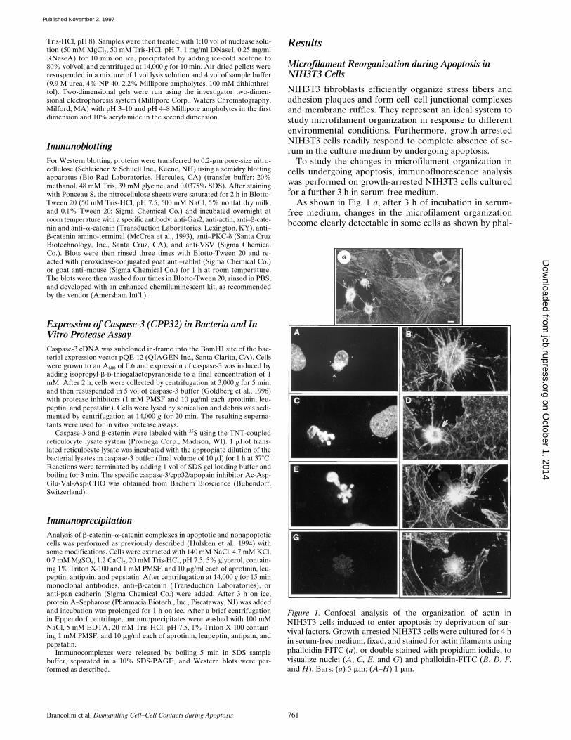
Figure 1. Confocal analysis of the organization of actin inNIH3T3 cells induced to enter apoptosis by deprivation of sur-vival factors. Growth-arrested NIH3T3 cells were cultured for 4 hin serum-free medium, fixed, and stained for actin filaments usingphalloidin-FITC (a), or double stained with propidium iodide, tovisualize nuclei (A, C, E, and G) and phalloidin-FITC (B, D, F,and H). Bars: (a) 5 mm; (A–H) 1 mm.
on October 1, 2014
jcb.rupress.orgD
ownloaded from
Published November 3, 1997
The Journal of Cell Biology, Volume 139, 1997 762
loidin staining (
arrows
). As a response to the absence ofsurvival factors, cells apparently sever contacts with neigh-boring cells, retract from the adhesion substrate, and dis-mantle stress fibers, extensively reorganizing their actincytoskeleton.
Apoptosis is always accompanied by alterations in thenuclear morphology, finally leading to nuclear fragmenta-tion and formation of the apoptotic bodies. We, therefore,decided to follow such nuclear alterations in relation tothe changes in actin organization during apoptosis.
When an initial alteration in nuclear architecture wasfirst detected (
arrow
, Fig. 1
A
) changes in the actin cytoskel-eton were also evident (Fig. 1
B
). As described above, thepeculiar features at this stage included the severing of con-tacts with neighboring cells, retraction of fibers, accumulationof actin in the perinuclear region and loss of stress fibers.
At further stages, nuclear alterations become more evi-dent leading to nuclear fragmentation (Fig. 1
C
). At thisstage, the actin cytoskeleton is clearly organized in a ring-like structure (Fig. 1
D
,
arrow
) with membrane blebbingalso clearly evident (data not shown). It is interesting tonote that nuclear fragments are external with respect tothe observed actin ring. This observation suggests thatcontraction of actin filaments present in the perinuclearregion might be important in the sprouting of the nuclearbodies. This hypothesis was supported by the most alteredphenotypes, showing the actin reorganization as summa-rized in Fig. 1,
E–H.
At these stages the actin ring ap-peared collapsed, with the nuclear fragments tending todrop away from the actin filaments. This detachmentprobably leads to the appearance of the phenotype repre-sented in Fig. 1,
G
and
H
, where only remnants of cyto-plasm containing a collapsed actin ring structure were de-tected. Such a phenotype could represent a final stage ofthe microfilament changes taking place during apoptosis.
From this preliminary analysis, it could be appreciatedthat apoptosis, as triggered by serum deprivation in NIH3T3fibroblasts, is coupled to well-defined and ordered changesof the microfilament system. Such changes are concomi-tant with membrane blebbing and alteration of the nucleararchitecture.
Analysis of Actin Cleavage during Apoptosis in NIH3T3
Since it has been reported that actin can be cleaved in anin vitro apoptotic system using purified caspase-1 (Mash-ima et al., 1995; Kayalar et al., 1996), we analyzed as if invivo, during apoptosis, actin was cleaved in our definedcellular system. In fact, this could possibly explain the dra-matic reorganization of the microfilament system occur-ring during cell death.
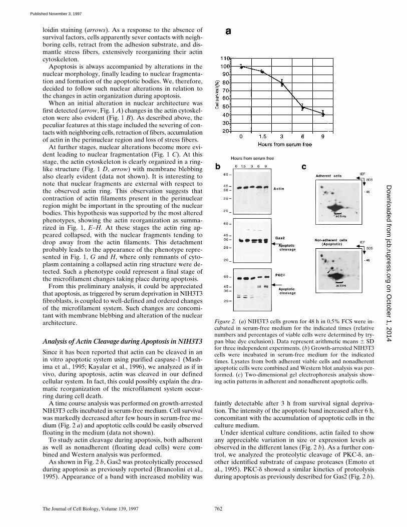
A time course analysis was performed on growth-arrestedNIH3T3 cells incubated in serum-free medium. Cell survivalwas markedly decreased after few hours in serum-free me-dium (Fig. 2 a) and apoptotic cells could be easily observedfloating in the medium (data not shown).
To study actin cleavage during apoptosis, both adherentas well as nonadherent (floating dead cells) were com-bined and Western analysis was performed.
As shown in Fig. 2 b, Gas2 was proteolytically processedduring apoptosis as previously reported (Brancolini et al.,1995). Appearance of a band with increased mobility was
faintly detectable after 3 h from survival signal depriva-tion. The intensity of the apoptotic band increased after 6 h,concomitant with the accumulation of apoptotic cells in theculture medium.
Under identical culture conditions, actin failed to showany appreciable variation in size or expression levels asobserved in the different lanes (Fig. 2 b). As a further con-trol, we analyzed the proteolytic cleavage of PKC-d, an-other identified substrate of caspase proteases (Emoto etal., 1995). PKC-d showed a similar kinetics of proteolysisduring apoptosis as previously described for Gas2 (Fig. 2 b).
Figure 2. (a) NIH3T3 cells grown for 48 h in 0.5% FCS were in-cubated in serum-free medium for the indicated times (relativenumbers and percentages of viable cells were determined by try-pan blue dye exclusion). Data represent arithmetic means 6 SDfor three independent experiments. (b) Growth-arrested NIH3T3cells were incubated in serum-free medium for the indicatedtimes. Lysates from both adherent viable cells and nonadherentapoptotic cells were combined and Western blot analysis was per-formed. (c) Two-dimensional gel electrophoresis analysis show-ing actin patterns in adherent and nonadherent apoptotic cells.
on October 1, 2014
jcb.rupress.orgD
ownloaded from
Published November 3, 1997
Brancolini et al. Dismantling Cell–Cell Contacts during Apoptosis 763
The behavior of actins during apoptosis in vivo was alsoanalyzed by two-dimensional electrophoresis. NIH3T3cells grown for 36 h in 0.5% FCS were labeled for 12 hwith [35S]methionine, and then simultaneously chased withcold methionine and cultured in serum-free medium. Af-ter 6 h, adherent and nonadherent cells were harvestedseparately and two-dimensional gel electrophoresis wasperformed as described in Materials and Methods. Thetwo-dimensional electrophoretic pattern of the differentactin isoforms (Garrels and Franza, 1989) did not show ap-preciable differences between normal and apoptotic cells.
Interestingly, a new band, which might represent a post-translational modification of an unknown product, becamedetectable in the lysates from apoptotic cells (Fig. 3 C, ar-rowhead). These results clearly demonstrate that duringapoptosis actin fails to be proteolytically processed, as trig-gered by survival signal deprivation in NIH3T3 cells. In-deed, resistance of actin to cleavage during apoptosis hasbeen recently reported (Song et al., 1997). Nevertheless,we can not exclude that a minor fraction of actin is pro-teolytically processed during apoptosis, or that this pro-cessing does not occur in NIH3T3 cells.
Modulation of Cell–Cell Contacts during Apoptosis in NIH3T3 Fibroblasts
Having demonstrated that actin is not cleaved in vivo dur-ing apoptosis in NIH3T3, we decided to search for otherregulators of the microfilament system that could be pro-cessed during apoptosis and possibly involved in the dra-matic reorganization occurring during cell death.
The previously reported analysis of microfilament changesduring apoptosis clearly suggested that a first hallmark of celldeath is the severing of contacts among neighboring cells.
Cell–cell contacts have been intensively studied in epi-thelial cells, where specialized domains of the plasmamembrane and the adherens junctions play a critical rolein cellular adhesiveness by providing a link between cellsurface adhesion molecules and the cytoskeleton (Geigerand Ayalon, 1992). Such an adhesive role is dependent onmembers of the cadherin superfamily of adhesive recep-tors and the cytoplasmic adaptor proteins a-, b-, andg-catenin/plakoglobin, which connect the cadherins to theactin filaments (for review see Gumbiner, 1996).
Fibroblasts in culture express functional cadherins (Itohet al., 1991), interact with one another, and assemble cad-herins–catenin complexes (Knudsen et al., 1995).
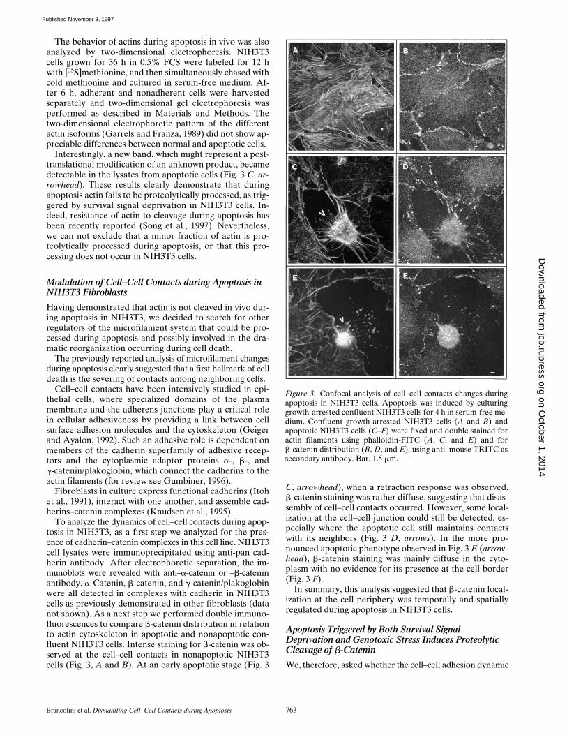
To analyze the dynamics of cell–cell contacts during apop-tosis in NIH3T3, as a first step we analyzed for the pres-ence of cadherin–catenin complexes in this cell line. NIH3T3cell lysates were immunoprecipitated using anti-pan cad-herin antibody. After electrophoretic separation, the im-munoblots were revealed with anti–a-catenin or –b-cateninantibody. a-Catenin, b-catenin, and g-catenin/plakoglobinwere all detected in complexes with cadherin in NIH3T3cells as previously demonstrated in other fibroblasts (datanot shown). As a next step we performed double immuno-fluorescences to compare b-catenin distribution in relationto actin cytoskeleton in apoptotic and nonapoptotic con-fluent NIH3T3 cells. Intense staining for b-catenin was ob-served at the cell–cell contacts in nonapoptotic NIH3T3cells (Fig. 3, A and B). At an early apoptotic stage (Fig. 3
C, arrowhead), when a retraction response was observed,b-catenin staining was rather diffuse, suggesting that disas-sembly of cell–cell contacts occurred. However, some local-ization at the cell–cell junction could still be detected, es-pecially where the apoptotic cell still maintains contactswith its neighbors (Fig. 3 D, arrows). In the more pro-nounced apoptotic phenotype observed in Fig. 3 E (arrow-head), b-catenin staining was mainly diffuse in the cyto-plasm with no evidence for its presence at the cell border(Fig. 3 F).
In summary, this analysis suggested that b-catenin local-ization at the cell periphery was temporally and spatiallyregulated during apoptosis in NIH3T3 cells.
Apoptosis Triggered by Both Survival Signal Deprivation and Genotoxic Stress Induces Proteolytic Cleavage of b-Catenin
We, therefore, asked whether the cell–cell adhesion dynamic
Figure 3. Confocal analysis of cell–cell contacts changes duringapoptosis in NIH3T3 cells. Apoptosis was induced by culturinggrowth-arrested confluent NIH3T3 cells for 4 h in serum-free me-dium. Confluent growth-arrested NIH3T3 cells (A and B) andapoptotic NIH3T3 cells (C–F) were fixed and double stained foractin filaments using phalloidin-FITC (A, C, and E) and forb-catenin distribution (B, D, and E), using anti–mouse TRITC assecondary antibody. Bar, 1.5 mm.
on October 1, 2014
jcb.rupress.orgD
ownloaded from
Published November 3, 1997
The Journal of Cell Biology, Volume 139, 1997 764
could be regulated by proteolytic processing of the cytoso-lic adaptor proteins during apoptosis. To test this hypothe-sis, Western analysis of b-catenin on both adherent as wellas nonadherent (floating dead cells) NIH3T3 cells inducedto enter apoptosis following serum-free culture conditionwas performed.
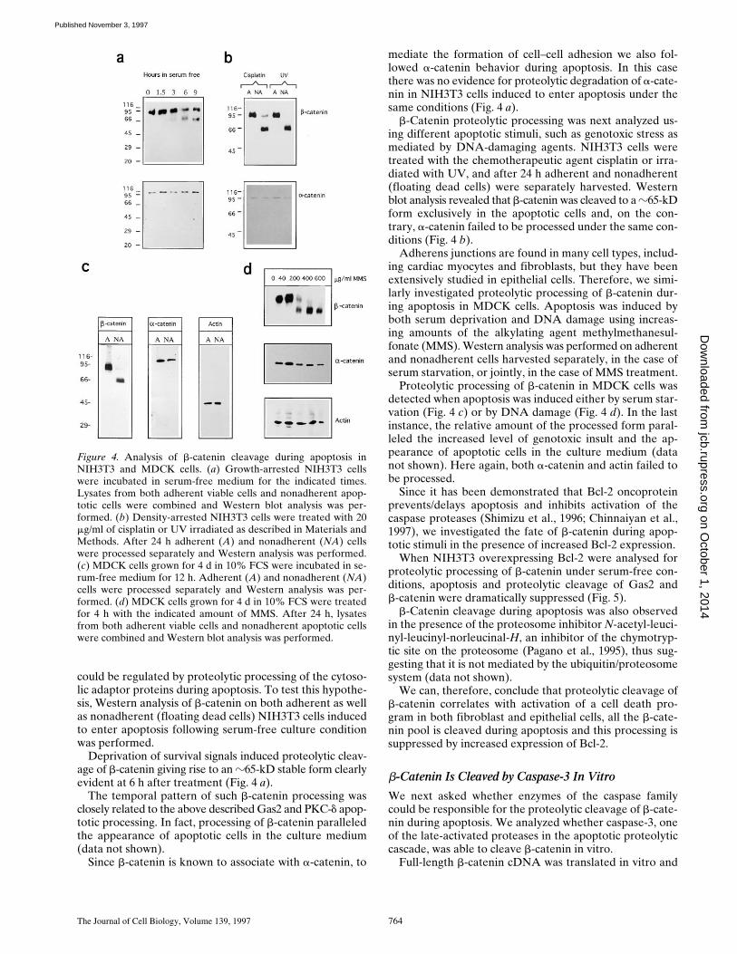
Deprivation of survival signals induced proteolytic cleav-age of b-catenin giving rise to an z65-kD stable form clearlyevident at 6 h after treatment (Fig. 4 a).
The temporal pattern of such b-catenin processing wasclosely related to the above described Gas2 and PKC-d apop-totic processing. In fact, processing of b-catenin paralleledthe appearance of apoptotic cells in the culture medium(data not shown).
Since b-catenin is known to associate with a-catenin, to
mediate the formation of cell–cell adhesion we also fol-lowed a-catenin behavior during apoptosis. In this casethere was no evidence for proteolytic degradation of a-cate-nin in NIH3T3 cells induced to enter apoptosis under thesame conditions (Fig. 4 a).
b-Catenin proteolytic processing was next analyzed us-ing different apoptotic stimuli, such as genotoxic stress asmediated by DNA-damaging agents. NIH3T3 cells weretreated with the chemotherapeutic agent cisplatin or irra-diated with UV, and after 24 h adherent and nonadherent(floating dead cells) were separately harvested. Westernblot analysis revealed that b-catenin was cleaved to a z65-kDform exclusively in the apoptotic cells and, on the con-trary, a-catenin failed to be processed under the same con-ditions (Fig. 4 b).
Adherens junctions are found in many cell types, includ-ing cardiac myocytes and fibroblasts, but they have beenextensively studied in epithelial cells. Therefore, we simi-larly investigated proteolytic processing of b-catenin dur-ing apoptosis in MDCK cells. Apoptosis was induced byboth serum deprivation and DNA damage using increas-ing amounts of the alkylating agent methylmethanesul-fonate (MMS). Western analysis was performed on adherentand nonadherent cells harvested separately, in the case ofserum starvation, or jointly, in the case of MMS treatment.
Proteolytic processing of b-catenin in MDCK cells wasdetected when apoptosis was induced either by serum star-vation (Fig. 4 c) or by DNA damage (Fig. 4 d). In the lastinstance, the relative amount of the processed form paral-leled the increased level of genotoxic insult and the ap-pearance of apoptotic cells in the culture medium (datanot shown). Here again, both a-catenin and actin failed tobe processed.
Since it has been demonstrated that Bcl-2 oncoproteinprevents/delays apoptosis and inhibits activation of thecaspase proteases (Shimizu et al., 1996; Chinnaiyan et al.,1997), we investigated the fate of b-catenin during apop-totic stimuli in the presence of increased Bcl-2 expression.
When NIH3T3 overexpressing Bcl-2 were analysed forproteolytic processing of b-catenin under serum-free con-ditions, apoptosis and proteolytic cleavage of Gas2 andb-catenin were dramatically suppressed (Fig. 5).
b-Catenin cleavage during apoptosis was also observedin the presence of the proteosome inhibitor N-acetyl-leuci-nyl-leucinyl-norleucinal-H, an inhibitor of the chymotryp-tic site on the proteosome (Pagano et al., 1995), thus sug-gesting that it is not mediated by the ubiquitin/proteosomesystem (data not shown).
We can, therefore, conclude that proteolytic cleavage ofb-catenin correlates with activation of a cell death pro-gram in both fibroblast and epithelial cells, all the b-cate-nin pool is cleaved during apoptosis and this processing issuppressed by increased expression of Bcl-2.
b-Catenin Is Cleaved by Caspase-3 In Vitro
We next asked whether enzymes of the caspase familycould be responsible for the proteolytic cleavage of b-cate-nin during apoptosis. We analyzed whether caspase-3, oneof the late-activated proteases in the apoptotic proteolyticcascade, was able to cleave b-catenin in vitro.
Full-length b-catenin cDNA was translated in vitro and
Figure 4. Analysis of b-catenin cleavage during apoptosis inNIH3T3 and MDCK cells. (a) Growth-arrested NIH3T3 cellswere incubated in serum-free medium for the indicated times.Lysates from both adherent viable cells and nonadherent apop-totic cells were combined and Western blot analysis was per-formed. (b) Density-arrested NIH3T3 cells were treated with 20mg/ml of cisplatin or UV irradiated as described in Materials andMethods. After 24 h adherent (A) and nonadherent (NA) cellswere processed separately and Western analysis was performed.(c) MDCK cells grown for 4 d in 10% FCS were incubated in se-rum-free medium for 12 h. Adherent (A) and nonadherent (NA)cells were processed separately and Western analysis was per-formed. (d) MDCK cells grown for 4 d in 10% FCS were treatedfor 4 h with the indicated amount of MMS. After 24 h, lysatesfrom both adherent viable cells and nonadherent apoptotic cellswere combined and Western blot analysis was performed.
on October 1, 2014
jcb.rupress.orgD
ownloaded from
Published November 3, 1997
Brancolini et al. Dismantling Cell–Cell Contacts during Apoptosis 765
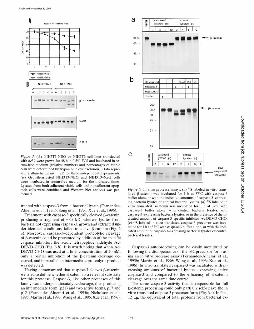
treated with caspase-3 from a bacterial lysate (Fernandes-Alnemri et al., 1995b; Song et al., 1996; Xue et al., 1996).
Treatment with caspase-3 specifically cleaved b-catenin,producing a fragment of z65 kD, whereas lysates frombacteria not expressing caspase-3, grown and extracted un-der identical conditions, failed to cleave b-catenin (Fig. 6a). Moreover, caspase-3–dependent proteolytic cleavageof b-catenin could be prevented by addition of the specificcaspase inhibitor, the acidic tetrapeptide aldehyde Ac-DEVD-CHO (Fig. 6 b). It is worth noting that when Ac-DEVD-CHO was used at a final concentration of 20 nM,only a partial inhibition of the b-catenin cleavage oc-curred, and in parallel an intermediate proteolytic productwas detected.
Having demonstrated that caspase-3 cleaves b-catenin,we tried to define whether b-catenin is a relevant substratefor this protease. Caspase-3, like other proteases of thisfamily, can undergo autocatalytic cleavage, thus producingan intermediate form (p21) and two active forms, p17 andp12 (Fernandes-Alnemri et al., 1995b; Nicholson et al.,1995; Martin et al., 1996; Wang et al., 1996; Xue et al., 1996).
Caspase-3 autoprocessing can be easily monitored byfollowing the disappearance of the p32 precursor form us-ing an in vitro protease assay (Fernandes-Alnemri et al.,1995b; Martin et al., 1996; Wang et al., 1996; Xue et al.,1996). In vitro translated caspase-3 was incubated with in-creasing amounts of bacterial lysates expressing activecaspase-3 and compared to the efficiency of b-catenincleavage over the same time course.
The same caspase-3 activity that is responsible for fullb-catenin processing could only partially self-cleave the invitro translated caspase-3 precursor form (Fig. 6 c). In fact,12 mg, the equivalent of total proteins from bacterial ex-
Figure 5. (A) NIH3T3-NEO or NIH3T3 cell lines transfectedwith bcl-2 were grown for 48 h in 0.5% FCS and incubated in se-rum-free medium (relative numbers and percentages of viablecells were determined by trypan blue dye exclusion). Data repre-sent arithmetic means 6 SD for three independent experiments.(B) Growth-arrested NIH3T3-NEO and NIH3T3–bcl-2 cellswere incubated in serum-free medium for the indicated times.Lysates from both adherent viable cells and nonadherent apop-totic cells were combined and Western blot analysis was per-formed.
Figure 6. In vitro protease assays. (a) 35S labeled in vitro trans-lated b-catenin was incubated for 1 h at 378C with caspase-3buffer alone or with the indicated amounts of caspase-3–express-ing bacteria lysates or control bacteria lysates. (b) 35S labeled invitro translated b-catenin was incubated for 1 h at 378C withcaspase-3 buffer alone, with control bacteria lysates, withcaspase-3–expressing bacteria lysates, or in the presence of the in-dicated amount of caspase-3–specific inhibitor Ac-DEVD-CHO.(c) 35S labeled in vitro translated caspase-3 precursor was incu-bated for 1 h at 378C with caspase-3 buffer alone, or with the indi-cated amount of caspase-3–expressing bacterial lysates or controlbacterial lysates.
on October 1, 2014
jcb.rupress.orgD
ownloaded from
Published November 3, 1997
The Journal of Cell Biology, Volume 139, 1997 766
tracts expressing caspase-3, were required for full process-ing of caspase-3 precursor with respect to the 2 mg suffi-cient for full b-catenin processing. Caspase-3–dependentprocessing specificity of b-catenin is also strengthened bythe observation that an amount of caspase-2 (ICH-1),enough for full autoprocessing, was unable to cleave b-cate-nin in vitro (data not shown).
In summary, these data indicate that b-catenin is a bet-ter substrate for caspase-3 than itself, thus strongly sug-gesting that b-catenin is a relevant caspase-3 substrate.
b-Catenin Is Cleaved at Different Sites by Caspase-3
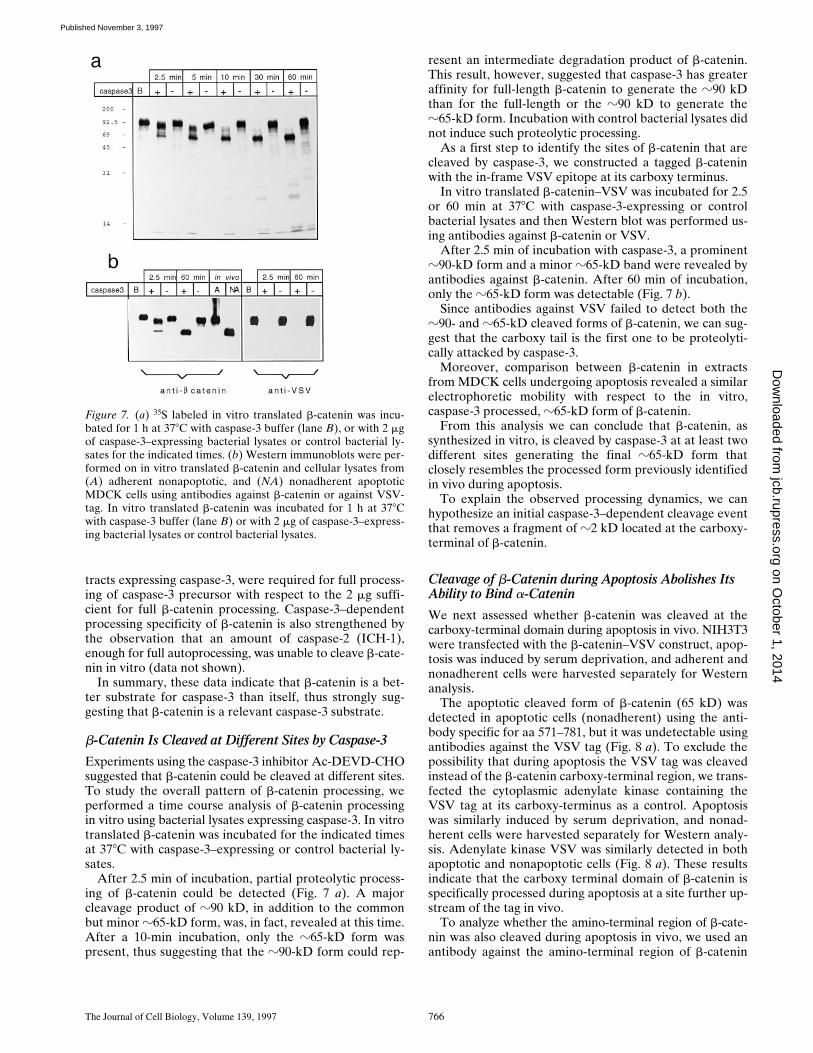
Experiments using the caspase-3 inhibitor Ac-DEVD-CHOsuggested that b-catenin could be cleaved at different sites.To study the overall pattern of b-catenin processing, weperformed a time course analysis of b-catenin processingin vitro using bacterial lysates expressing caspase-3. In vitrotranslated b-catenin was incubated for the indicated timesat 378C with caspase-3–expressing or control bacterial ly-sates.
After 2.5 min of incubation, partial proteolytic process-ing of b-catenin could be detected (Fig. 7 a). A majorcleavage product of z90 kD, in addition to the commonbut minor z65-kD form, was, in fact, revealed at this time.After a 10-min incubation, only the z65-kD form waspresent, thus suggesting that the z90-kD form could rep-
resent an intermediate degradation product of b-catenin.This result, however, suggested that caspase-3 has greateraffinity for full-length b-catenin to generate the z90 kDthan for the full-length or the z90 kD to generate thez65-kD form. Incubation with control bacterial lysates didnot induce such proteolytic processing.
As a first step to identify the sites of b-catenin that arecleaved by caspase-3, we constructed a tagged b-cateninwith the in-frame VSV epitope at its carboxy terminus.
In vitro translated b-catenin–VSV was incubated for 2.5or 60 min at 378C with caspase-3-expressing or controlbacterial lysates and then Western blot was performed us-ing antibodies against b-catenin or VSV.
After 2.5 min of incubation with caspase-3, a prominentz90-kD form and a minor z65-kD band were revealed byantibodies against b-catenin. After 60 min of incubation,only the z65-kD form was detectable (Fig. 7 b).
Since antibodies against VSV failed to detect both thez90- and z65-kD cleaved forms of b-catenin, we can sug-gest that the carboxy tail is the first one to be proteolyti-cally attacked by caspase-3.
Moreover, comparison between b-catenin in extractsfrom MDCK cells undergoing apoptosis revealed a similarelectrophoretic mobility with respect to the in vitro,caspase-3 processed, z65-kD form of b-catenin.
From this analysis we can conclude that b-catenin, assynthesized in vitro, is cleaved by caspase-3 at at least twodifferent sites generating the final z65-kD form thatclosely resembles the processed form previously identifiedin vivo during apoptosis.
To explain the observed processing dynamics, we canhypothesize an initial caspase-3–dependent cleavage eventthat removes a fragment of z2 kD located at the carboxy-terminal of b-catenin.
Cleavage of b-Catenin during Apoptosis Abolishes Its Ability to Bind a-Catenin
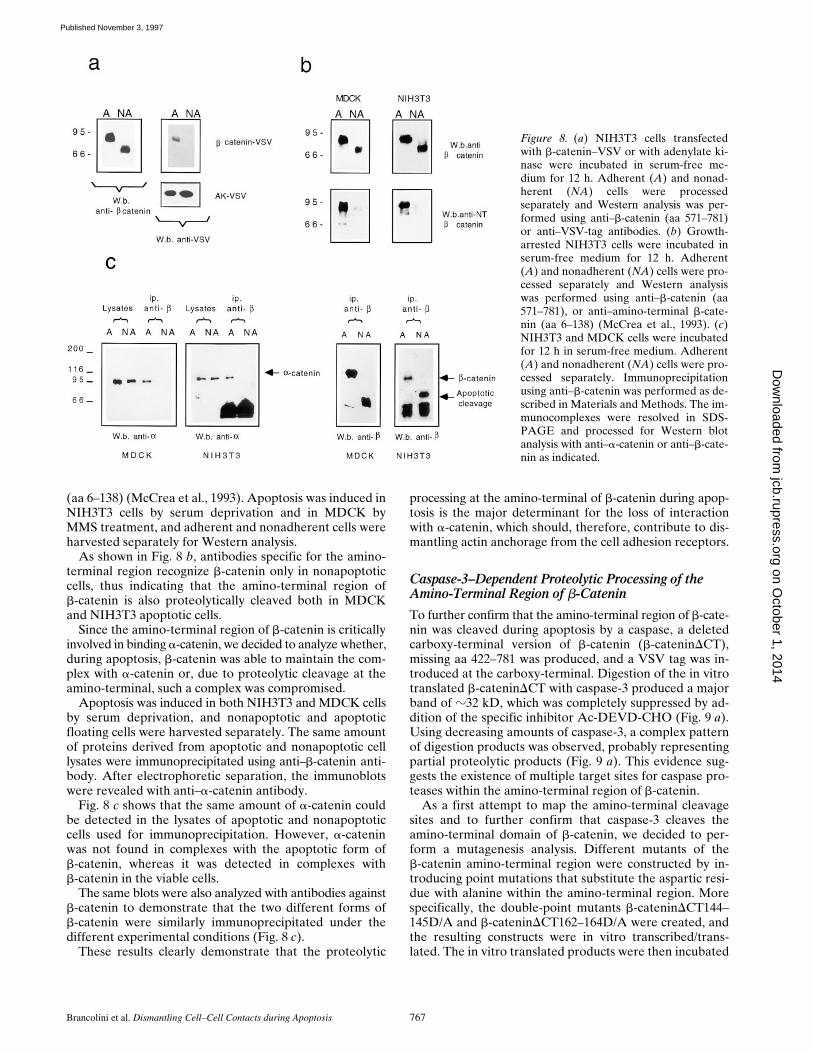
We next assessed whether b-catenin was cleaved at thecarboxy-terminal domain during apoptosis in vivo. NIH3T3were transfected with the b-catenin–VSV construct, apop-tosis was induced by serum deprivation, and adherent andnonadherent cells were harvested separately for Westernanalysis.
The apoptotic cleaved form of b-catenin (65 kD) wasdetected in apoptotic cells (nonadherent) using the anti-body specific for aa 571–781, but it was undetectable usingantibodies against the VSV tag (Fig. 8 a). To exclude thepossibility that during apoptosis the VSV tag was cleavedinstead of the b-catenin carboxy-terminal region, we trans-fected the cytoplasmic adenylate kinase containing theVSV tag at its carboxy-terminus as a control. Apoptosiswas similarly induced by serum deprivation, and nonad-herent cells were harvested separately for Western analy-sis. Adenylate kinase VSV was similarly detected in bothapoptotic and nonapoptotic cells (Fig. 8 a). These resultsindicate that the carboxy terminal domain of b-catenin isspecifically processed during apoptosis at a site further up-stream of the tag in vivo.
To analyze whether the amino-terminal region of b-cate-nin was also cleaved during apoptosis in vivo, we used anantibody against the amino-terminal region of b-catenin
Figure 7. (a) 35S labeled in vitro translated b-catenin was incu-bated for 1 h at 378C with caspase-3 buffer (lane B), or with 2 mgof caspase-3–expressing bacterial lysates or control bacterial ly-sates for the indicated times. (b) Western immunoblots were per-formed on in vitro translated b-catenin and cellular lysates from(A) adherent nonapoptotic, and (NA) nonadherent apoptoticMDCK cells using antibodies against b-catenin or against VSV-tag. In vitro translated b-catenin was incubated for 1 h at 378Cwith caspase-3 buffer (lane B) or with 2 mg of caspase-3–express-ing bacterial lysates or control bacterial lysates.
on October 1, 2014
jcb.rupress.orgD
ownloaded from
Published November 3, 1997
Brancolini et al. Dismantling Cell–Cell Contacts during Apoptosis 767
(aa 6–138) (McCrea et al., 1993). Apoptosis was induced inNIH3T3 cells by serum deprivation and in MDCK byMMS treatment, and adherent and nonadherent cells wereharvested separately for Western analysis.
As shown in Fig. 8 b, antibodies specific for the amino-terminal region recognize b-catenin only in nonapoptoticcells, thus indicating that the amino-terminal region ofb-catenin is also proteolytically cleaved both in MDCKand NIH3T3 apoptotic cells.
Since the amino-terminal region of b-catenin is criticallyinvolved in binding a-catenin, we decided to analyze whether,during apoptosis, b-catenin was able to maintain the com-plex with a-catenin or, due to proteolytic cleavage at theamino-terminal, such a complex was compromised.
Apoptosis was induced in both NIH3T3 and MDCK cellsby serum deprivation, and nonapoptotic and apoptoticfloating cells were harvested separately. The same amountof proteins derived from apoptotic and nonapoptotic celllysates were immunoprecipitated using anti–b-catenin anti-body. After electrophoretic separation, the immunoblotswere revealed with anti–a-catenin antibody.
Fig. 8 c shows that the same amount of a-catenin couldbe detected in the lysates of apoptotic and nonapoptoticcells used for immunoprecipitation. However, a-cateninwas not found in complexes with the apoptotic form ofb-catenin, whereas it was detected in complexes withb-catenin in the viable cells.
The same blots were also analyzed with antibodies againstb-catenin to demonstrate that the two different forms ofb-catenin were similarly immunoprecipitated under thedifferent experimental conditions (Fig. 8 c).
These results clearly demonstrate that the proteolytic
processing at the amino-terminal of b-catenin during apop-tosis is the major determinant for the loss of interactionwith a-catenin, which should, therefore, contribute to dis-mantling actin anchorage from the cell adhesion receptors.
Caspase-3–Dependent Proteolytic Processing of the Amino-Terminal Region of b-Catenin
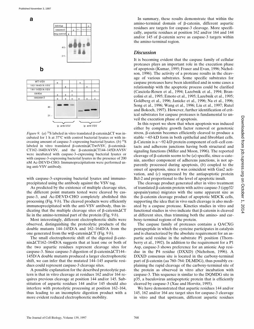
To further confirm that the amino-terminal region of b-cate-nin was cleaved during apoptosis by a caspase, a deletedcarboxy-terminal version of b-catenin (b-cateninDCT),missing aa 422–781 was produced, and a VSV tag was in-troduced at the carboxy-terminal. Digestion of the in vitrotranslated b-cateninDCT with caspase-3 produced a majorband of z32 kD, which was completely suppressed by ad-dition of the specific inhibitor Ac-DEVD-CHO (Fig. 9 a).Using decreasing amounts of caspase-3, a complex patternof digestion products was observed, probably representingpartial proteolytic products (Fig. 9 a). This evidence sug-gests the existence of multiple target sites for caspase pro-teases within the amino-terminal region of b-catenin.
As a first attempt to map the amino-terminal cleavagesites and to further confirm that caspase-3 cleaves theamino-terminal domain of b-catenin, we decided to per-form a mutagenesis analysis. Different mutants of theb-catenin amino-terminal region were constructed by in-troducing point mutations that substitute the aspartic resi-due with alanine within the amino-terminal region. Morespecifically, the double-point mutants b-cateninDCT144–145D/A and b-cateninDCT162–164D/A were created, andthe resulting constructs were in vitro transcribed/trans-lated. The in vitro translated products were then incubated
Figure 8. (a) NIH3T3 cells transfectedwith b-catenin–VSV or with adenylate ki-nase were incubated in serum-free me-dium for 12 h. Adherent (A) and nonad-herent (NA) cells were processedseparately and Western analysis was per-formed using anti–b-catenin (aa 571–781)or anti–VSV-tag antibodies. (b) Growth-arrested NIH3T3 cells were incubated inserum-free medium for 12 h. Adherent(A) and nonadherent (NA) cells were pro-cessed separately and Western analysiswas performed using anti–b-catenin (aa571–781), or anti–amino-terminal b-cate-nin (aa 6–138) (McCrea et al., 1993). (c)NIH3T3 and MDCK cells were incubatedfor 12 h in serum-free medium. Adherent(A) and nonadherent (NA) cells were pro-cessed separately. Immunoprecipitationusing anti–b-catenin was performed as de-scribed in Materials and Methods. The im-munocomplexes were resolved in SDS-PAGE and processed for Western blotanalysis with anti–a-catenin or anti–b-cate-nin as indicated.
on October 1, 2014
jcb.rupress.orgD
ownloaded from
Published November 3, 1997
The Journal of Cell Biology, Volume 139, 1997 768
with caspase-3–expressing bacterial lysates and immuno-precipitated using the antibody against the VSV tag.
As predicted by the existence of multiple cleavage sites,the different point mutants tested were cleaved by cas-pase-3, and Ac-DEVD-CHO completely abolished thisprocessing (Fig. 9 b). The cleaved products were efficientlyimmunoprecipitated with the anti-VSV antibody, thus in-dicating that the multiple cleavage sites of b-cateninDCTlie in the amino-terminal part of the protein (Fig. 9 b).
Most interestingly, different electrophoretic shifts wereobserved, distinguishing the proteolytic products of thedouble mutants 144–145D/A and 162–164D/A from theone generated from the wtb-cateninDCT (Fig. 9 b).
The small electrophoretic shift of the digested b-cate-ninDCT162–164D/A suggests that at least one or both ofthe two aspartic residues represent cleavage sites forcaspase-3. Since caspase-3 digestion of b-cateninDCT144–145D/A double mutants produced a larger electrophoreticshift, we can infer that the mutated 144–145 aspartic resi-dues could represent caspase-3 cleavage sites.
A possible explanation for the described proteolytic pat-tern is that in vitro cleavage at residues 162 and/or 164 re-quires previous cleavage at position 144 and/or 145. Sub-stitution of aspartic residues 144 and/or 145 should alsointerfere with proteolytic processing at position 162–164,thus leading to an incomplete digestion product with amore evident reduced electrophoretic mobility.
In summary, these results demonstrate that within theamino-terminal domain of b-catenin, different asparticresidues are targets for caspase-3 cleavage. More specifi-cally, aspartic residues at position 162 and/or 164 and 144and/or 145 of b-catenin serve as caspase-3 targets withinthe amino-terminal region.
DiscussionIt is becoming evident that the caspase family of cellularproteases plays an important role in the execution phaseof apoptosis (Kumar, 1995; Fraser and Evan, 1996; Nichol-son, 1996). The activity of a protease results in the cleav-age of various substrates. Some specific substrates forcaspase proteases have been identified and in some cases arelationship with the apoptotic process could be clarified(Casciola-Rosen et al., 1994; Lazebnik et al., 1994; Bran-colini et al., 1995; Emoto et al., 1995; Lazebnik et al., 1995;Goldberg et al., 1996; Janicke et al., 1996; Na et al., 1996;Song et al., 1996; Wang et al., 1996; Liu et al., 1997; Ruteland Bokoch, 1997). However, further identification of crit-ical substrates for caspase proteases is fundamental to un-veil the execution phase of apoptosis.
In this report we show that when apoptosis was inducedeither by complete growth factor removal or genotoxicstress, b-catenin becomes efficiently cleaved to produce astable z65-kD form in both epithelial and fibroblast cells.b-Catenin is a z92-kD protein component of cell–cell con-tacts and adherens junctions having both structural andsignaling functions (Miller and Moon, 1996). The reportedcleavage of b-catenin seems to be (a) specific, since a-cate-nin, another component of adherens junctions, is not ap-parently processed during apoptosis, (b) coupled to theonset of apoptosis, since it was coincident with Gas2 acti-vation, and (c) suppressed by the antiapoptotic proteinBcl-2 and proportional to the level of apoptotic insult.
The cleavage product generated after in vitro treatmentof translated b-catenin protein with active caspase-3 (cpp32/apopain/yama) migrates with the same apparent size asthe b-catenin cleavage product of apoptotic MDCK cells,supporting the idea that in vivo such cleavage is also medi-ated by a caspase protease. Kinetics studies in vitro andfunctional studies in vivo indicate that b-catenin is cleavedat different sites, thus trimming both the amino- and car-boxy-terminal regions of the protein.
The caspase family of proteases contains a QACXGpentapeptide in which the cysteine participates in catalysisand is characterized by the absolute requirement for an as-partic acid residue in the substrate P1 position (Thorn-berry et al., 1992). In addition to the requirement for a P1Asp, caspase-3 shows preference for an anionic Asp resi-due in the P4 residue (DXXD) (Nicholson, 1996). ADXXD consensus site is located in the carboxy-terminalpart of b-catenin (aa 760–764: DLMDG), thus possibly ex-plaining the rapid cleavage of the carboxy-terminal site ofthe protein as observed in vitro after incubation withcaspase-3. This sequence is similar to the DQMDG site inp35, a baculovirus antiapoptotic protein that is efficientlycleaved by caspase-3 (Xue and Horvitz, 1995).
We have demonstrated that aspartic residues 144 and/or145, 162 and/or 164 are target sites for caspase-3 cleavagein vitro and that upstream, different aspartic residues
Figure 9. (a) 35S labeled in vitro translated b-cateninDCT was in-cubated for 1 h at 378C with control bacterial lysates or with in-creasing amount of caspase-3–expressing bacterial lysates. (b) 35Slabeled in vitro translated b-cateninDCTwtVSV, b-cateninD-CT162–164D/AVSV, and the b-cateninDCT144–145D/AVSVwere incubated with caspase-3–expressing bacterial lysates orwith caspase-3–expressing bacterial lysates in the presence of 200nM Ac-DEVD-CHO. Immunoprecipitations were performed us-ing anti-VSV antibody.
on October 1, 2014
jcb.rupress.orgD
ownloaded from
Published November 3, 1997
Brancolini et al. Dismantling Cell–Cell Contacts during Apoptosis 769
within the amino-terminal region of b-catenin are alsocleaved by caspase-3, none of them showing a canonicalDXXD sequence. These observations raised the questionwhether caspase-3 is the protease involved in processing ofthe amino-terminal domain in vivo, or other caspases showhigher affinity for such amino-terminal sites. Indeed, therecent generation of caspase-3 null mice (Kuida et al.,1996), clearly demonstrates that caspase-3 play a criticalrole during morphogenetic cell death in the mammalianbrain but raises the possibility that other caspase proteasesmay have an important role during apoptosis in other tis-sues or in response to cell death-trigger.
Therefore, we can hypothesize that b-catenin could alsobe a target for other caspases; further studies are requiredto understand if other caspases are involved in b-cateninprocessing, and to identify the different aspartic residuescleaved during apoptosis.
Adherens junctions link cells together by organizing ac-tin filaments to the plasmamembrane. The transmem-brane receptors are the cadherins, a family of homophilicCa21-dependent cell–cell adhesion molecules (Takeichi,1995). Their crystal structures suggest that cadherin-medi-ated cell adhesion is not based solely on the stability of as-sociation among individual molecules, but rather by thegeneration of a cell adhesion zipper which provides amechanism to form strong intercellular bonds (Shapiro etal., 1995). In vivo strong intercellular bonds are dependentupon the association of the cadherin carboxy-terminal cy-toplasmic domain to the central region of b-catenin andg-catenin/plakoglobin (Nagafuchi and Takeichi, 1988; Ozawaet al., 1989; Hulsken et al., 1995; Sacco et al., 1995). Dele-tion of the b-catenin–binding site from cadherin cytoplasmicdomain renders the cadherin nonfunctional in a cell aggre-gation assay (Nagafuchi and Takeichi, 1988; Ozawa et al.,1989).
The central function of b-catenin as a regulator of adher-ens junctions has been further demonstrated in differentorganisms using different experimental approaches (Peiferet al., 1993; Oyama et al., 1994; Haegel et al., 1995). It hasbeen extensively demonstrated that b-catenin serves as anadapter to link cadherins to a-catenin and thereby to theactin cytoskeleton, since a-catenin seems to interact withboth actin and a-actinin (Knudsen et al., 1995; Rimm etal., 1995). In this context, b-catenin seems to be the criticalcomponent for cadherin–catenin complexes in the regula-tion of cellular adhesiveness (Hamaguchi et al., 1993;Cowin and Bruke, 1996).
The fundamental roles of a-catenin in cadherin cyto-skeleton interactions have also been clearly established invarious cell types. Nonadhesive epithelial tumor cells lack-ing a-catenin can be induced to form tightly adherent epi-thelia after reintroduction of a-catenin (Watabe et al.,1994). Moreover, expression of E-cadherin–a-catenin chi-meras has been reported to induce a strong and inflexibleadhesive phenotype (Nagafuchi et al., 1994).
We have demonstrated in vivo that proteolytic process-ing of the b-catenin at its amino-terminal domain impairsits ability to bind a-catenin. Amino acid residues 120–151of b-catenin, embedded in the amino-terminal domain andcontaining the first ARM repeat, are required for interac-tion with a-catenin (Aberle et al., 1994). This is confirmedin the human gastric cell line HSC-39 where a truncated
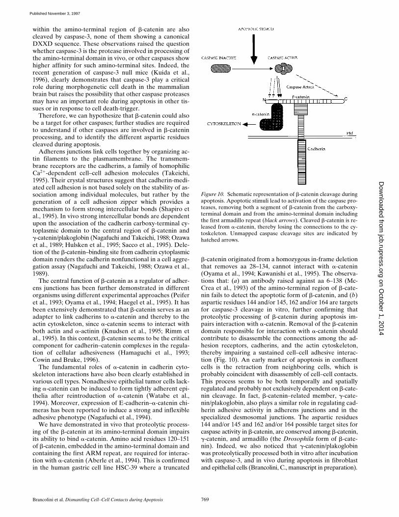
b-catenin originated from a homozygous in-frame deletionthat removes aa 28–134, cannot interact with a-catenin(Oyama et al., 1994; Kawanishi et al., 1995). The observa-tions that: (a) an antibody raised against aa 6–138 (Mc-Crea et al., 1993) of the amino-terminal region of b-cate-nin fails to detect the apoptotic form of b-catenin, and (b)aspartic residues 144 and/or 145, 162 and/or 164 are targetsfor caspase-3 cleavage in vitro, further confirming thatproteolytic processing of b-catenin during apoptosis im-pairs interaction with a-catenin. Removal of the b-catenindomain responsible for interaction with a-catenin shouldcontribute to disassemble the connections among the ad-hesion receptors, cadherins, and the actin cytoskeleton,thereby impairing a sustained cell–cell adhesive interac-tion (Fig. 10). An early marker of apoptosis in confluentcells is the retraction from neighboring cells, which isprobably coincident with disassembly of cell–cell contacts.This process seems to be both temporally and spatiallyregulated and probably not exclusively dependent on b-cate-nin cleavage. In fact, b-catenin–related member, g-cate-nin/plakoglobin, also plays a similar role in regulating cad-herin adhesive activity in adherens junctions and in thespecialized desmosomal junctions. The aspartic residues144 and/or 145 and 162 and/or 164 possible target sites forcaspase activity in b-catenin, are conserved among b-catenin,g-catenin, and armadillo (the Drosophila form of b-cate-nin). Indeed, we also noticed that g-catenin/plakoglobinwas proteolytically processed both in vitro after incubationwith caspase-3, and in vivo during apoptosis in fibroblastand epithelial cells (Brancolini, C., manuscript in preparation).
Figure 10. Schematic representation of b-catenin cleavage duringapoptosis. Apoptotic stimuli lead to activation of the caspase pro-teases, removing both a segment of b-catenin from the carboxy-terminal domain and from the amino-terminal domain includingthe first armadillo repeat (black arrows). Cleaved b-catenin is re-leased from a-catenin, thereby losing the connections to the cy-toskeleton. Unmapped caspase cleavage sites are indicated byhatched arrows.
on October 1, 2014
jcb.rupress.orgD
ownloaded from
Published November 3, 1997
The Journal of Cell Biology, Volume 139, 1997 770
It is interesting to note that E-cadherin complexes maypromote cell survival and suppress cell death in epithelialcells (Hermiston and Gordon, 1995). In this context, cleav-age of b-catenin might also suppress transduction of sur-vival signals, consequently contributing to the one-wayproceeding of the apoptotic process. The evidence thatsome components of microfilament system are substratesfor the caspase proteases (Brancolini et al., 1995; Martin etal., 1996; Na et al., 1996), suggests that these proteases co-ordinate actin architectural changes during apoptosis andthat such changes are important for a normal apoptoticphenotype (Cotter et al., 1992).
In conclusion, a complex reorganization of the microfil-ament system was observed during cell death: cells retractfrom adhesion substrate, sever the contacts with neigh-bors, and actin filaments are concentrated in the perinu-clear area in a ring-like organization. Collapsing of thering-like structure becomes evident with the proceeding ofthe morphological changes accompanied by nuclear frag-mentation, membrane blebbing, and formation of the apop-totic bodies.
In this context, b-catenin and Gas2 processing seem tohave different roles. Cleavage of Gas2 seems to be impor-tant to switch on the ability to rearrange the microfilamentsystem, which may be relevant for the organization of thering-like structure (Brancolini et al., 1995). Cleavage ofb-catenin seems to be responsible for switching off itsfunction as a regulator of adherens junctions, and is possiblyinstrumental for severing contacts with neighboring cells.
b-Catenin is also found in the nucleus and cytoplasm,where its function is not restricted to modulation of adher-ens junctions. Association of b-catenin with the tumor-suppressor gene product APC (adenomatous polyposis co-lon) (Rubinfeld et al., 1993) and the transcription factorsLEF-1/XTcf-3 (Behrens et al., 1996; Molenaar et al., 1996)have, in fact, been reported. Moreover, b-catenin, to-gether with the related protein g-catenin/plakoglobin andArmadillo, is implicated in the Wnt/wingless signalingpathway, which determines the dorsoventral axis in Xeno-pus and segment polarity in Drosophila (for review seeMiller and Moon, 1996).
To unveil the relationships among the specific roles ofb-catenin in the regulation of cell growth/differentiationand its proteolytic processing during apoptosis will be anexciting challenge for the future.
We would like to thank A. Sgorbissa for help in some experiments. Weare grateful to B.M. Gumbiner for providing anti–amino-terminal b-cateninantibody. D. Lazarevic is a European Association Cancer Research fellow.J. Rodriguez is a Howard Hughes Medical Institute Predoctoral Fellow.
This work was supported by Associazione Italiana Ricerca sul Cancro,Consorzio Nazionale delle Ricerche–Applicazioni Cliniche della RicercaOncologica, Ministero dell’ “UNIVERSITA” and della Ricerca Scienti-fica and Tecnologica. 40% to C. Schneider, and CNR Progetto StrategicoCiclo cellulare e Apoptosi to C. Brancolini.
Received for publication 3 April 1997 and in revised form 28 August 1997.
References
Aberle, H., S. Butz, J. Stappert, H. Weissig, R. Kemler, and H. Hoschuetzky.1994. Assembling of the cadherin catenin complex in vitro with recombinantproteins. J. Cell Sci. 107:3655–3663.
Alnemri, E.S., D.J. Livingston, D.W. Nicholson, G. Salvesen, N.A. Thornberry,W.W. Wong, and J. Yuan. 1996. Human ICE/CED3 protease nomenclature.Cell. 87:171.
Bardelli, A., P. Longati, D. Albero, S. Goruppi, C. Schneider, C. Ponzetto, and P.M.Comoglio. 1996. HGF receptor associates with the antiapoptotic protein BAG-1and prevents cell death. EMBO (Eur. Mol. Biol. Organ.) J. 15:6205–6212.
Behrens, J., J.P. von Kries, M. Kuhl, L. Bruhn, D. Wedlich, R. Grosschedl, andW. Birchmeier. 1996. Functional interaction of b-catenin with the transcrip-tion factor LEF-1 Nature (Lond.). 382:225–230.
Beidler, D., M. Tewari, P. Friesen, G.G. Poirier, and V. Dixit. 1995. The bacu-lovirus p35 protein inhibits Fas- and tumor necrosis factor-induced apopto-sis. J. Biol. Chem. 270:16526–16528.
Boldin, M.K., T.M. Goncharov, Y. Goltsev, and D. Wallach. 1996. Involvementof MACH a novel MORT1/FADD-interacting protease, in Fas/APO-1- andTNF receptor-induced cell death. Cell. 85:803–815.
Brancolini, C., S. Bottega, and C. Schneider. 1992. Gas2, a growth arrest-spe-cific protein, is a component of the microfilament network system. J. CellBiol. 117:1251–1261.
Brancolini, C., M. Benedetti, and C. Schneider. 1995. Microfilament reorgani-zation during apoptosis: the role of Gas2, a possible substrate for ICE-likeproteases. EMBO (Eur. Mol. Biol. Organ.) J. 14:5179–5190.
Bump, N.J, M. Hackett, M. Hugunin, S. Seshagiri, K. Brady, P. Chen, C. Ferenz,S. Franklin, T. Ghayur, P. Li, et al. 1995. Inhibition of ICE family protease bybaculovirus antiapoptotic protein p35. Science (Wash. DC). 269:1885–1888.
Casciola-Rosen, L.A., D.K. Miller, G.J. Anhalt, and A. Rosen. 1994. Specificcleavage of the 70-kD protein component of small nuclear ribonucleoproteinis a characteristic biochemical feature of apoptotic cell death. J. Biol. Chem.269:30757–30760.
Chinnaiyan, A.M., K. O’Rouke, B.R. Lane, and V.M. Dixit. 1997. Interactionof CED-4 with CED-3 and CED-9: a molecular framework for cell death.Science (Wash. DC). 275:1122–1126.
Cotter, T.G., S.V. Lennon, J.M. Glynn, and D.R. Green. 1992. Microfilamentdisrupting agents prevent the formation of apoptotic bodies in tumor cellsundergoing apoptosis. Cancer Res. 52:997–1005.
Cowin, P., and B. Bruke. 1996. Cytoskeleton-membrane interactions Curr.Opin. Cell Biol. 7:56–65.
Duan, H., A.M. Chinnaiyan, P.L. Hudson, J.P. Wing, W.-W. He, and V.M.Dixit. 1996a. ICE-LAP3, a novel mammalian homologue of the Caenorhab-ditis elegans cell death protein Ced-3 is activated during fas- and tumor ne-crosis factor-induced apoptosis. J. Biol. Chem. 271:1621–1625.
Duan, H., K. Orth, A.M. Chinnaiyan, G.G. Poirier, C.J. Froelich, W.-W. He,and V.M. Dixit. 1996b. ICE-LAP6, a novel member of the ICE/CED3 genefamily, is activated by the cytotoxic T cell protease granzyme B. J. Biol.Chem. 271:16720–16724.
Ellis, R.E., J. Yuan, and H.R. Horvitz. 1991. Mechanisms and functions of celldeath. Annu. Rev. Cell Biol. 7:663–698.
Emoto, Y., Y. Manome, G. Meinhardt, H. Kisaki, S. Kharbanda, M. Roberston,T. Ghayur, W.W. Wong, R. Kamen, R. Weichselbaum, and D. Kufe. 1995.Proteolytic activation of protein kinase Cd by an ICE-like protease in apop-totic cells. EMBO (Eur. Mol. Biol. Organ.) J. 14:6148–6156.
Enari, M., R. Talanian, W. Wong, and S. Nagata. 1996. Sequential activation ofICE-like and CPP32-like proteases during Fas-mediated apoptosis. Nature(Lond.). 380:723–726.
Faucheau, C., A. Diu, A.W.E. Chan, A.-M. Blanchet, C. Miossec, F. Herve, V.Collard-Dutilleul, Y. Gu, R.A. Aldape, J.A. Lippke, et al. 1995. A novel hu-man protease similar to the interleukin-1b converting enzyme induces apop-tosis in transfected cells. EMBO (Eur. Mol. Biol. Organ.) J. 14:1914–1922.
Fernandes-Alnemri, T., G. Litwack, and E.S., Alnemri. 1994. CPP32, a novelhuman apoptotic protein with homology to Caenorhabditis elegans cell deathprotein ced-3 and mammalian interleukin-1b–convertase enzyme. J. Biol.Chem. 269:30761–30764.
Fernandes-Alnemri, T., G. Litwack, and E.S. Alnemri. 1995a. Mch2, a newmember of the apoptotic CED3/ICE cysteine protease gene family. CancerRes. 55:2737–2742.
Fernandes-Alnemri, T., A. Takahashi, R. Armstrong, J. Krebs, L. Fritz, K.J To-maselli,. L. Wang, Z. Yu, C.M. Croce, G. Salveson, et al. 1995b. Mch3, anovel human apoptotic cystein protease highly related to CPP32. CancerRes. 55:6045–6052.
Fernandes-Alnemri, T., C. Armstrong, J. Krebs, S.M. Srinivasula, L. Wang, F.Bullrich, L.C. Fritz, J.A. Trapani, K.J. Tomaselli, G. Litwack, and E.S. Al-nemri. 1996. In vitro activation of CPP32 and Mch3 by Mch4, a novel humanapoptotic cystein protease containing two FADD-like domains. Proc. Natl.Acad. Sci. USA. 93:7464–7469.
Fraser, A., and G. Evan. 1996. A license to kill. Cell. 85:781–784.Gagliardini, V., P.A. Fernandez, R.K. Lee, H.C. Drexler, R.J. Rotello, M.C.
Fishman, and J. Yuan. 1994. Prevention of vertebrate neuronal cell death bythe crmA gene. Science (Wash. DC). 263:826–828.
Garrels, J.I., and R.B. Franza, Jr. 1989. The REF52 protein database. J. Biol.Chem. 264:5283–5298.
Geiger, B., and O. Ayalon. 1992. Cadherins. Annu. Rev. Cell Biol. 8:307–332.Goldberg, Y.P., D.W. Nicholson, D.M. Rasper, M.A. Kalchmen, H.B. Koide,
R.K. Graham, M. Bromm, P. Kazemi-Esfarjani, N.A. Thornberry, J.P. Vail-lancourt, and M.R. Hayden. 1996. Cleavage of huntingtin by apopain, aproapoptotic cysteine protease, is modulated by the polyglutamine tract.Nat. Genet. 13:442–449.
Gumbiner, B.M. 1996. Cell adhesion: the molecular basis of tissue architectureand morphogenesis. Cell. 84:345–357.
Haegel, H., M. Larue, M. Ohsugi, L. Fedorov, K. Herrenknecht, and R.
on October 1, 2014
jcb.rupress.orgD
ownloaded from
Published November 3, 1997
Brancolini et al. Dismantling Cell–Cell Contacts during Apoptosis 771
Kemler. 1995. Lack of b-catenin affects mouse development at gastrulation.Development (Camb.). 121:3529–3537.
Hamaguchi, M., N. Matsuyoshi, Y. Ohnishi, B. Gotoh, M. Takeichi, and Y. Na-gai. 1993. p60v-src causes tyrosine phosphorylation and inactivation of theN-cadherin-catenin cell adhesion system. EMBO (Eur. Mol. Biol. Organ.) J.12:307–314.
Hermiston, M.L., and J.I. Gordon. 1995. In vivo analysis of cadherin function in themouse intestinal epithelium: essential roles in adhesion, maintenance of dif-ferentiation, and regulation of programmed cell death. J. Cell Biol. 129:489–506.
Hoschuetzky, H., H. Aberle, and R. Kemler. 1994. b-Catenin mediates the in-teraction of the cadherin–catenin complex with epidermal growth factor re-ceptor. J. Cell Biol. 127:1375–1380.
Hulsken, J., W. Birchmeier, and J. Behrens. 1994. E-Cadherin and APC com-pete for the interaction with b-catenin and the cytoskeleton. J. Cell Biol. 127:2061–2069.
Itoh, M., S. Yonemura, A. Nagafuchi, S. Tsukita, and S. Tsukita. 1991. A 220-kDundercoat-constitutive protein: its specific localization at cadherin-basedcell–cell adhesion sites. J. Cell Biol. 115:1449–1462.
Janicke, R.U., P.A. Walker, X.Y. Lin, and A.G. Porter. 1996. Specific cleavageof the retinoblastoma protein by an ICE-like protease in apoptosis. EMBO(Eur. Mol. Biol. Organ.) J. 15:6969–6978.
Kawanishi, J., J. Kato, K. Sasaki, S. Fujii, N. Watanabe, and Y. Niitsu. 1995.Loss of E-cadherin-dependent cell adhesion due to mutation of the b-cate-nin gene in a human cancer cell line, HSC-39. Mol. Cell. Biol. 15:1175–1181.
Kayalar, C., T. Ord, M.P. Testa, L.-T. Zhong, and D.E. Bredesen. 1996. Cleav-age of actin by interleukin 1b–converting enzyme to reverse DNase I inhibi-tion Proc. Natl. Acad. Sci. USA. 93:2234–2238.
Kerr, J.F.R., A.H. Wyllie, and A.R. Currie. 1972. Apoptosis: a basic biologicalphenomenon with wide-ranging implications in tissue kinetics. Br. J. Cancer.26:239–257.
Kuida, K., T.S. Zheng, S. Na, C.-y. Kuan, D. Yang, H. Karasuyama, P. Rakic,and R.A. Flavell. 1996. Decreased apoptosis in the brain and premature le-thality in CPP32-deficient mice. Nature (Lond.). 384:368–375.
Kumar, S. 1995. ICE-like proteases in apoptosis. Trends Biochem. Sci. 20:198–202.Kumar, S., M. Kinoshita, M. Noda, N.G. Copeland, and N.A. Jenkins. 1994. In-
duction of apoptosis by the mouse Nedd2 gene, which encodes a protein sim-ilar to the product of the Caenorhabditis elegans cell death gene ced-3 andthe mammalian IL-1b-converting enzyme. Genes Dev. 8:1613–1626.
Kundsen, K.A., A.P. Soler, K.R. Johnson, and M.J. Wheelock. 1995. Interac-tion of a-actinin with the cadherin/catenin cell–cell adhesion complex viaa-catenin. J. Cell Biol. 130:67–77.
Lazebnik, Y.A., S.H. Kaufmann, S. Desnoyers, G.G. Poirier, and W.C. Earn-shaw. 1994. Cleavage of poly(ADP-ribose) polymerase by a proteinase withproperties like ICE. Nature (Lond.). 371:346–347.
Lazebnik, Y., A. Takahashi, R.D. Moir, R.D. Goldman, G.G. Poirier, S.H.Kaufmann, and W.C. Earnshaw. 1995. Studies of the lamin proteinase revealmultiple parallel biochemical pathways during apoptotic execution. Proc.Natl. Acad. Sci. USA. 92:9042–9046.
Liu, X., H. Zou, C. Slaughter, and X. Wang. 1997. DFF, a heterodimeric proteinthat functions downstream of caspase-3 to trigger DNA fragmentation dur-ing apoptosis. Cell. 89:175–184.
Martin, S.J., G.P. Amarante-Mendes, L. Shi, T.-H. Chuang, C.A. Casiano, G.A.O’Brien, P. Fitzgerald, E.M. Tan, G.M. Bokoch, A.H. Greenberg, and D.R.Green. 1996. The cytotoxic cell protease granzyme B initiates apoptosis in acell-free system by proteolytic processing and activation of the ICE/CED-3family protease, CPP32, via a novel two-step mechanism. EMBO (Eur. Mol.Biol. Organ.) J. 15:2407–2416.
Mashima, T., M. Naito, S. Kataoka, H. Kawai, and T. Tsuruo. 1995. Identifica-tion of Actin as substrate of ICE and an ICE-like protease and involvementof an ICE-LIKE protease but not ICE in VP-16-induced U937 apoptosis.Biochem. Biophys. Res. Commun. 217:1185–1192.
McCrea, P.D., W.M. Brieher, and B.M. Gumbiner. 1993. Induction of a second-ary body axis in Xenopus by antibodies to b-catenin. J. Cell Biol. 123:477–484.
Miller, J.R., and R.T. Moon. 1996. Signal transduction through b-catenin andspecification of cell fate during embryogenesis. Genes Dev. 10:2527–2539.
Miura, M., H. Zhu, R. Rotello, E.A. Hartweig, and J. Yuan. 1993. Induction ofapoptosis in fibroblasts by IL-1 b-converting enzymes, a mammalian homo-logue of the C. elegans cell death gene ced-3. Cell. 75:653–660.
Molenaar, M., M. van de Wetering, M. Oosterwegel, J. Peterson-Maduro, S.Godsave, V. Korinek, J. Roose, O. Destree, and H. Clevers. 1996. XTcf-3transcription factor mediates b-catenin–induced axis formation in Xenopusembryos. Cell. 86:391–399.
Muzio, M., A.M. Chinnaiyan, F.C. Kischkel, K. O’Rourke, A. Shevchenko, J.Ni, C. Scaffidi, J.D. Bretz, M. Zhang, R. Gentz, et al. 1996. FLICE, a novelFADD-homologous ICE/CED-3-like protease, is recruited to the CD95(Fas/APO-1) death-inducing signaling complex. Cell. 85:817–827.
Na, S., T.H. Chuang, A. Cunningham, T.G. Turi, J.H. Hanke, G.M. Bokoch,and D.E. Danley. 1996. D4-GDI, a substrate of CPP32, is proteolyzed duringFas-induced apoptosis. J. Biol. Chem. 271:11209–11213.
Nagafuchi, A., and A. Takeichi. 1988. Cell binding function of E-cadherin isregulated by the cytoplasmic domain. EMBO (Eur. Mol. Biol. Organ.) J. 7:3679–3684.
Nagafuchi, A., S. Ishihara, and S. Tsukita. 1994. The roles of catenins in the cad-herin-mediated cell adhesion: functional analysis of E-cadherin–a-cateninfusion molecules. J. Cell Biol. 127:235–245.
Nicholson, D.W. 1996. ICE/CED3-like protease as therapeutic targets for thecontrol of inappropriate apoptosis. Nat. Biotechnol. 14:297–301.
Nicholson, D.W., A. Ambereen, N. Thornberry, J.P. Vaillancourt, C.K. Ding,M. Gallant, Y. Gareau, P.R. Griffin, M. Labelle, Y. Lazebnik, et al. 1995.Identification and inhibition of the ICE/CED3 protease necessary for mam-malian apoptosis. Nature (Lond.). 376:37–43.
Oyama, T., Y. Kanai, A. Ochiai, S. Akimoto, T. Oda, K. Yanagihara, A. Na-gafuchi, S. Tuskita, S. Shibamoto, F. Ito, et al. 1994. A truncated b-catenindisrupts the interaction between E-cadherin and a-catenin: a cause of loss ofintercellular adhesiveness in human cancer cell lines. Cancer Res. 54:6282–6287.
Ozawa, M., H. Baribault, and R. Kemler. 1989. The cytoplasmic domain of thecell adhesion molecule uvomorulin associates with three independent pro-teins structurally related in different species. EMBO (Eur. Mol. Biol. Or-gan.) J. 8:1711–1717.
Pagano, M., S.W. Tam, A.M. Theodoras, P. Beer-Romero, G. Del Sal, V. Chau,P.R. Yew, G.F. Draetta, and M. Rolfe. 1995. Role of the ubiquitin-proteo-some pathway in regulating abundance of the cyclin-dependent kinase inhib-itor p27. Science (Wash. DC). 269:682–685.
Peifer, M., S. Orsulic, D. Sweeton, and E. Wieschaus. 1993. A role for theDrosophila segment polarity gene armadillo in cell adhesion and cytoskele-tal integrity during oogenesis. Development. 111:1029–1043.
Raff, M.C. 1992. Social controls on cell survival and death: an extreme view.Nature (Lond.). 356:397–400.
Rimm, D.L., E.R. Koslov, P. Kebriaei, C.D. Cianci, and J.S. Morrow. 1995. a1(E)-catenin is an actin binding and bundling protein mediating the attach-ment of F-actin to the membrane adhesion complex. Proc. Natl. Acad. Sci.USA. 92:8813–8817.
Rubinfeld, B., B. Souza, I. Albert, O. Muller, S.H. Chamberlain, F.R. Masiarz,S. Munemitsu, and P. Polakis. 1993. Association of the APC gene productwith b-catenin. Science (Wash. DC). 262:1731–1734.
Rudel, T., and G.M. Bokoch. 1997. Membrane and morphological changes inapoptotic cells regulated by caspase-mediated activation of PAK2. Science(Wash. DC). 276:1571–1574.
Sacco, P.A., M. McGranahan, M.J. Wheelock, and K.R. Johnson. 1995. Identifi-cation of plakoglobin domains required for association with N-cadherin anda-catenin. J. Biol. Chem. 270:20201–20206.
Shapiro, L., A.M. Fannon, P.D. Kwong, A. Thompson, M.S. Lehmann, G.Grubel, J.F. Legrand, A. Als-Neilson, J. Colman, and W.A. Hendrickson.1995. Structural basis of cell-cell adhesion by cadherins. Nature (Lond.). 374:327–336.
Shimizu, S., Y. Eguchi, W. Kamiike, H. Matsuda, and Y. Tsujimoto. 1996. Bcl-2expression prevents activation of the ICE protease cascade. Oncogene. 12:2251–2257.
Song, Q., S.P. Less-Miller, S. Kumar, N. Zhang, D.W. Chan, G.C.M. Smith, S.P.Jackson, E.S. Alnemri, G. Litwack, K.K. Khanna, and M.F. Lavin. 1996. DNA-dependent protein kinase catalytic subunit: a target for an ICE-like proteasein apoptosis. EMBO (Eur. Mol. Biol. Organ.) J. 15:3238–3246.
Song, Q., T. Wei, S. Lees-Miller, E. Alnemri, D. Watters, and M.F. Lavin. 1997.Resistance of actin to cleavage during apoptosis. Proc. Natl. Acad. Sci. USA.94:157–162.
Takahashi, A., E.S. Alnemri, Y. Lazebnik, T. Fernandes-Alnemri, G. Litwack,R.D. Moir, R.D. Goldman, G.G. Poirier, S. Kaufmann, and W.C. Earnshaw.1996. Cleavage of lamin A by Mch2a but not CPP32: multiple interleukin1b–converting enzyme-related proteases with distinct substrate recognitionproperties are active in apoptosis. Proc. Natl. Acad. Sci. USA. 93:8395–8400.
Takeichi, M. 1995. Morphogenetic role of classical cadherins. Curr. Opin. CellBiol. 7:619–627.
Thornberry, N.A., H.G. Bull, J.R. Calaycay, K.T. Chapman, A.D. Howard,M.J. Kostura, D.K. Miller, S.M. Molineaux, J.R. Weidner, J. Aunins, et al.1992. A novel heterodimeric cysteine protease is required for interleukin-1bprocessing in monocytes. Nature (Lond.). 356:768–774.
Wang, L., M. Miura, L. Bergeron, H. Zhu, and J. Yuan. 1994. Ich-1, an Ice/ced3-related gene, encodes both positive and negative regulators of pro-grammed cell death. Cell. 78:739–750.
Wang, X., N.G. Zelenski, J. Yang, J. Sakai, M.S. Brown, and J. Goldstein. 1996.Cleavage of sterol regulatory element binding proteins (SREBPs) by CPP32during apoptosis. EMBO (Eur. Mol. Biol. Organ.) J. 15:1012–1020.
Watabe, M., A. Nagafuchi, S. Tsukita, and M. Takeichi. 1994. Induction of po-larized cell–cell association and retardation of growth by activation of theE-cadherin-catenin adhesion system in a disperse carcinoma line. J. CellBiol. 127:247–256.
Wyllie, A.H. 1995. The genetic regulation of apoptosis. Curr. Opin. Genet. Dev.5:97–104.
Wyllie, A.H, J.F.R. Kerr, and A.R. Currie. 1980. Cell death: the significance ofapoptosis. Int. Rev. Cytol. 68:251–306.
Xue, D., and H.R. Horvitz. 1995. Inhibition of the Caenorhabditis elegans cell-death protease CED-3 by a CED-3 cleavage site in baculovirus p35 protein.Nature (Lond.). 377:248–251.
Xue, D., S. Shaham, and H.R. Horvitz. 1996. The Caenorhabditis elegans cell-death protein CED-3 is a cystein protease with substrate specificities similarto those of the human CPP32 protease. Genes Dev. 10:1073–1083.
Yuan, J., S. Shaham, S. Ledoux, H.M. Ellis, and H.R. Horvitz. 1993. The Cae-norhabditis elegans cell death gene ced-3 encodes a protein similar to mam-malian interleukin-1b–converting enzyme. Cell. 75:641–652.
on October 1, 2014
jcb.rupress.orgD
ownloaded from
Published November 3, 1997
Related Documents