Disentangling Brain Networks in Adult ADHD: Studies with fMRI and TMS Marc Schneider

Welcome message from author
This document is posted to help you gain knowledge. Please leave a comment to let me know what you think about it! Share it to your friends and learn new things together.
Transcript
Disentangling Brain Networks in Adult ADHD: Studies with fMRI and TMS
Marc Schneider


Disentangling Brain Networks in Adult ADHD: Studies with fMRI and TMS
Het ontrafelen van neuronale netwerken bij volwassenen met ADHD: Klinische onderzoeken met fMRI en TMS
Proefschrift
ter verkrijging van de graad van doctor aan deErasmus Universiteit Rotterdam
op gezag van derector magnificus
Prof.dr. H.G. Schmidt
en volgens besluit van het College voor Promoties.De openbare verdediging zal plaatsvinden opdonderdag 29 november 2012 om 13.30 uur
door
Marc Karl-Friedrich François Schneidergeboren te Fontainebleau (Frankrijk)

Promotiecommissie
Promotor: Prof.dr. W.M.A. Verhoeven
Overige leden: Prof.dr. S.A. Kushner Prof.dr. A. Aleman Prof.dr. J.W. van Strien
Copromotor: Dr. J.H.M. Tulen
The research described in this thesis was carried out at the Institute for Forensic Psychology and Psychiatry and the Clinics for Psychiatry and Psychotherapy of the University of Homburg/ Saar, Germany.
ISBN: 978-94-6191-479-8
Design/lay-out: In Zicht Grafisch Ontwerp, www.promotie-inzicht.nlPrint: Ipskamp Drukkers, www.ppi.nl
© 2012 Marc K.F. Schneider All rights reserved. No part of this publication may be reproduced or transmitted in any form or by any means, electronic or mechanical, including photocopying, recording or any information storage and retrieval without prior permission of the holder of the copyright.

Für meine Familie


Table of Contents
1| General Introduction 9
2| Brain imaging in adult Attention deficit hyperactivity disorder (ADHD) 27 Key Issues in Mental Health. Attention-Deficit Hyperactivity Disorder (ADHD) in adults.
Karger, Basel (2010). Vol. 176: 88-104.
3| Impairment of fronto-striatal and parietal cerebral networks correlates 49 with Attention Deficit Hyperactivity Disorder (ADHD) psychopathology in adults – a functional magnetic resonance imaging (fMRI) study
Psychiatry Res (2010) 183(1):75-84.
4| Impaired cortical inhibition in adult ADHD patients: a study with 77 transcranial magnetic stimulation
J Neural Transm Suppl (2007) 72:303-9.
5| The effects of long acting Methylphenidate in adults with Attention 95 Deficit Hyperactivity Disorder: A study with paired pulse Transcranial Magnetic Stimulation
Neuropsychobiology (2011) 64(4):195-201.
6| Reduced cortical inhibition in first-episode schizophrenia 111 Schizophr Res (2008) 105(1-3):252-61.
7| A similar but distinctive pattern of impaired cortical excitability in 131 first-episode schizophrenia and ADHD
Accepted in Neuropsychobiology.
8| General Discussion and conclusions 151
* | References 161 Summary 171 Samenvatting 173 Zusammenfassung 177 Acknowledgements 181 Curriculum vitae 183 List of publications 185


General Introduction1 |


11
1. General Introduction
1.1 ADHD in adultsAttention Deficit/ Hyperactivity Disorder (ADHD) is not only limited to young patients. It is increasingly diagnosed in adults. Although the estimated prevalence in Europe ranges between 2 and 3% (48), the knowledge about adult ADHD pathophysiology and its neurobiological basis developed only in the past two decades, primarily stimulated by the rapid developments in modern genetic and imaging techniques. ADHD in adulthood leads to an array of major psychosocial problems such as social maladaptation, academic underachieving, antisocial and aggressive behaviour, relation problems, high risk sexual behaviour and car accidents (11,12;55;67;66). These factors in total lead to a negative impact on social and economic well-being of the individual.As can be inferred from Table 1, the key problem in the diagnosis of ADHD in adulthood is that the clinical diagnostic items are still based on childhood criteria. This raises the question whether the diagnostic standards in adults are up to date.In 1995, Paul Wender and coworkers from the University of Utah stressed that the symptoms in adult ADHD are not just extrapolations from those in childhood and adolescence. This UTAH workgroup developed criteria for adult characteristics that differ significantly from those in childhood (117). The authors pointed out that, apart from the common features such as impulsivity, motor hyperactivity and attention deficit symptoms, affective lability, hot temper/ explosive short-lived outbursts, emotional overreactivity, and disorganization/ inability to complete tasks, are key symptoms of the adult ADHD syndrome. Wender et al. brought these aspects together in a structured interview, the Wender-Reimherr Adult Attention Deficit Disorder Scale (WRAADDS,117). The authorized German version is called the Wender Reimherr Interview (WRI,97).Despite the methodical shortcomings, it was decided to apply the international diagnostic standards and, therefore, the adult ADHD patients participating in the studies of this thesis were recruited fulfilling the combined type DSM IV criteria The WRI was used in the clinical studies to provide additional clinical data of the patients.ADHD phenotypes in adulthood show a varying psychopathological picture over time. During lifetime, the ADHD core features may change, because impulsivity and hyperactivity become less prominent over time than the attention deficit symptoms (15). Moreover, it has to be stressed that some patients diagnosed with ADHD in childhood don`t display the full clinical symptom scale in adulthood any more, leading to a so-called non-persistent or subclinical form of ADHD. Information about this phenotype is, however, very limited.
GENERAL INTRODUCTION 1

12
CHAPTER 1
Table 1 Diagnostic criteria for Attention-Deficit/Hyperactivity Disorder (DSM IV)
A. Either (1) or (2):
(1) inattention: six (or more) of the following symptoms of inattention have persisted for
at least 6 months to a degree that is maladaptive and inconsistent with developmental
level:
(a) often fails to give close attention to details or makes careless mistakes in
schoolwork, work, or other activities
(b) often has difficulty sustaining attention in tasks or play activities
(c) often does not seem to listen when spoken to directly
(d) often does not follow through on instructions and fails to finish school work,
chores, or duties in the workplace (not due to oppositional behavior or failure to
understand instructions)
(e) often has difficulty organizing tasks and activities
(f) often avoids, dislikes, or is reluctant to engage in tasks that require sustained
mental effort (such as schoolwork or homework)
(g) often loses things necessary for tasks or activities (e.g., toys, school assignments,
pencils, books, or tools)
(h) is often easily distracted by extraneous stimuli
(i) is often forgetful in daily activities
(2) hyperactivity-impulsivity: six (or more) of the following symptoms of hyperactivity-
impulsivity have persisted for at least 6 months to a degree that is maladaptive and
inconsistent with developmental level:
Hyperactivity
(a) often fidgets with hands or feet or squirms in seat
(b) often leaves seat in classroom or in other situations in which remaining seated is
expected
(c) often runs about or climbs excessively in situations in which it is inappropriate (in
adolescents or adults, may be limited to subjective feelings of restlessness)
(d) often has difficulty playing or engaging in leisure activities quietly
(e) is often “on the go” or often acts as if “driven by a motor”
(f ) often talks excessively
Impulsivity(g) often blurts out answers before questions have been completed
(h) often has difficulty awaiting turn
(i) often interrupts or intrudes on others (e.g., butts into conversations or games)
13
Finally, with increasing age, adults with ADHD have a serious risk of developing various comorbid psychiatric disorders, in particular affective disorders and substance use disorders (119;60). Personality disorders often coincide with ADHD (69). This severely hampers an adequate description of the core ADHD phenotype or endophenotype in clinical research. It is, therefore, crucial in pathophysiological research to investigate ADHD patients without these kinds of comorbidities. The studies of this thesis aim to contribute to the clarification of the neurobiological basis of impaired attentional and executive networks, as well as of motor disturbances, in adult ADHD patients in whom further comorbidity and history of drug abuse are absent.
1.2 Syndromal views of ADHD in historical perspectiveSince decades, ADHD is most frequently diagnosed in child and adolescence psychiatry. Therefore, the history of ADHD primarily means the history of ADHD in childhood. Although the discussion about the diversity of clinical concepts of ADHD is still going on, the historical perspective is important for a good understanding. The earliest mentioning of ADHD-like behaviour in adulthood may be deduced from the description
GENERAL INTRODUCTION 1
Table 1 Continued
B. Some hyperactive-impulsive or inattentive symptoms that caused impairment
were present before age 7 years.
C. Some impairment from the symptoms is present in two or more settings (e.g., at
school [or work] and at home).
D. There must be clear evidence of clinically significant impairment in social,
academic, or occupational functioning.
E. The symptoms do not occur exclusively during the course of a Pervasive
Developmental Disorder, Schizophrenia, or other Psychotic Disorder and are not
better accounted for by another mental disorder (e.g., Mood Disorder, Anxiety
Disorder, Dissociative Disorders, or a Personality Disorder).
Code based on type: 314.01 Attention-Deficit/Hyperactivity Disorder, Combined Type: if both Criteria A1 and A2
are met for the past 6 months
314.00 Attention-Deficit/Hyperactivity Disorder, Predominantly Inattentive Type: if
Criterion A1 is met but Criterion A2 is not met for the past 6 months
314.01 Attention-Deficit/Hyperactivity Disorder, Predominantly Hyperactive-Impulsive
Type: if Criterion A2 is met but Criterion A1 is not met for the past 6 months
Coding note: For individuals (especially adolescents and adults) who currently have
symptoms that no longer meet full criteria, “In Partial Remission” should be specified.
14
by Hippocrates more than 2500 years ago. In his “Aphorisms” (2), he depicted a patient who had a “quickened response to sensory experience, but also less tenaciousness because the soul moves on quickly to the next impression”. His advice was to treat this “overbalance of fire over water” with changes in nutrition: “barley rather than wheat bread, fish rather than meat, drinking water rather than wine”, and he suggested “many natural and diverse physical activities”. Such a concept of an association between diet and behaviour was recently “rediscovered” by a Dutch/Belgian research group demonstrating the burden of food substances on the severity of ADHD symptoms in children (87).In 1789, Sir Alexander Crichton described a “mental restlessness” in children which was associated with incapacity of attending (73). In his historical review, Eduard Seidler (99) described the development over time of the socio-medical interpretation of hyperkinetic behaviours in childhood. Such disinhibited motor signs were observed since the middle of the 19th century and interpreted as “naughtiness” (Hoffmann 1845), followed by “neurasthenia” (Beard 1869), “character weakness” (Strümpell 1890), “neuropathy” (Czerny 1908), “moral defect” (Still 1902), and in the 1950ies was labeled as “minimal brain damage” (see overview 73,99) in the context of ADHD-like clinical syndromatology of children who survived the Encephalitis lethargica and the influenca pandemy.In 1845, the German general practitioner and later psychiatrist Heinrich Hoffmann described the famous “Zappelphillip“ and “Hans Guck in die Luft“, presenting the phenotypes of the combined hyperkinetic and the inattentive subtype of childhood ADHD, however, without interpreting those as disorders. He classified this child behaviour as naughtiness only (99). In the same year, Wilhelm Griesinger described children with a “nervous constitution“, who “cannot be quiet for a moment … don´t have any attention“ and, therefore, suffer from a “disturbed reaction function of the central organ to stimuli that work on it”. Still (105,106) was the first who systematically mentioned the hyperkinetic disorder in the Anglo-saxonian scientific literature. In his opinion, challenging behaviours in children originated from deficits in moral functioning.The viewpoint that hyperkinetic disorders have to be seen as a syndromal entity started with Bradley in 1937 (18), who discovered that the stimulant drug Benzedrine ameliorated hyperactive behaviour in children and induced relaxation. Subsequently, methylphenidate was synthesized in the 1940ies for the treatment of the hyperkinetic syndrome as a form of “minimal brain damage syndrome”. Although in the 1950ies the concept of early birth brain damage was no longer considered to be valid, in the sixties the concept of minimal brain dysfunction in terms of hyperkinetic reaction of childhood was introduced with the DSM II (1968) in order to relativize the hypothesis of brain damage (73).In the 1980 version of the DSM III, the attention deficit component of the hyperkinetic disorder was first introduced and modified during subsequent editions until the present. In the DSM-IIIR (1987), Attention Deficit Hyperactivity Disorder was differentiated from
CHAPTER 1
15
Attention Deficit Disorder without hyperactivity and termed Undifferentiated Attention Deficit Disorder to accentuate a distinct concept of this entity. At present, hyperkinetic behaviours, attention deficits and their combination are classified apart from each other to underline possible different etiologies of a developmental disorder (DSM-IV-TR, 2000). Further nosological shifts are to be awaited in future classification systems, that may eventually include endophenotypes based on the executive functioning or on attention/motor phenomena in separate genetic subphenotypes.
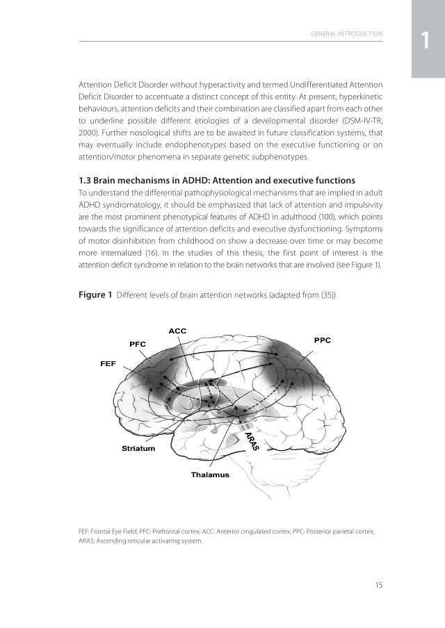
1.3 Brain mechanisms in ADHD: Attention and executive functionsTo understand the differential pathophysiological mechanisms that are implied in adult ADHD syndromatology, it should be emphasized that lack of attention and impulsivity are the most prominent phenotypical features of ADHD in adulthood (100), which points towards the significance of attention deficits and executive dysfunctioning. Symptoms of motor disinhibition from childhood on show a decrease over time or may become more internalized (16). In the studies of this thesis, the first point of interest is the attention deficit syndrome in relation to the brain networks that are involved (see Figure 1).
GENERAL INTRODUCTION 1
Figure 1 Different levels of brain attention networks (adapted from (35))
FEF: Frontal Eye Field; PFC: Prefrontal cortex; ACC: Anterior cingulated cortex; PPC: Posterior parietal cortex; ARAS: Ascending reticular activating system.
16
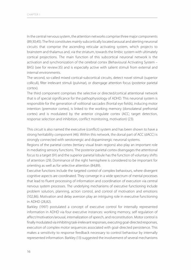
In the central nervous system, the attention networks comprise three major components (89;30;45). The first constitutes mainly subcortically located arousal and alerting neuronal circuits that comprise the ascending reticular activating system, which projects to brainstem and thalamus and, via the striatum, towards the limbic system with ultimately cortical projections. The main function of this subcortical neuronal network is the activation and synchronization of the cerebral cortex (Behavioural Activating System – BAS) (see for review:35) and is especially active with salient stimuli from external and internal environments. The second, so-called mixed cortical-subcortical circuits, detect novel stimuli (superior colliculi), filter irrelevant stimuli (pulvinar), or disengage attention focus (posterior parietal cortex). The third component comprises the selective or directed/cortical attentional network that is of special significance for the pathophysiology of ADHD. This neuronal system is responsible for the generation of volitional saccades (frontal eye fields), inducing motor intention (premotor cortex), is linked to the working memory (dorsolateral prefrontal cortex) and is modulated by the anterior cingulate cortex (ACC; target detection, response selection and inhibition, conflict monitoring, motivation) (23).
This circuit is also named the executive (conflict) system and has been shown to have a strong heritability component (46). Within this network, the dorsal part of ACC (dACC) is strongly connected with serotonergic and dopaminergic neuronal systems. Regions of the parietal cortex (tertiary visual brain regions) also play an important role in mediating sensory functions. The posterior parietal cortex disengages the attentional focus to a target (91) and the superior parietal lobule has the function of voluntary shifts of attention (29). Dominance of the right hemisphere is considered to be important for orienting as well as for selective attention (84;89). Executive functions include the targeted control of complex behaviours, where divergent cognitive aspects are coordinated. They converge in a wide spectrum of mental processes that lead to fluent processing of information and coordination of execution via central nervous system processes. The underlying mechanisms of executive functioning include problem solution, planning, action control, and control of motivation and emotions (102,86). Motivation and delay aversion play an intriguing role in executive functioning in ADHD (28,82).Barkley (1997) postulated a concept of executive control for internally represented information in ADHD via four executive instances: working memory, self regulation of affect/motivation/arousal, internalization of speech, and reconstitution. Motor control is finally modulated via inhibiting task-irrelevant responses, executing goal-directed responses, execution of complex motor sequences associated with goal-directed persistence. This makes a sensitivity to response feedback necessary to control behaviour by internally represented information. Barkley (13) suggested the involvement of several mechanisms
CHAPTER 1

17
in the behaviour inhibition in ADHD: inhibition of prepotent responses, stopping of ongoing responses, giving feedback on errors, and interference control. Motor coordination problems are key features of ADHD with an estimated prevalence of about 50 percent (90). Motor difficulties range from sensory-motor dys-coordination to a delay of motor milestones development. Dopaminergic and noradrenergic neuronal structures in fronto-subcortical systems play a crucial role in the pathophysiology of disturbed motor functioning in ADHD (47;88). Functional imaging techniques can disentangle how brain networks adapt to internal and external influencing factors in adult ADHD. As compared to radionuclid imaging techniques (Positron Emission Tomography (PET) and Single Photon Emission Computer Tomography (SPECT)), the functional Magnetic Resonance Technique (fMRI) is non-invasive and allows registration of attention and executive brain activity with a high spatial resolution. Since its time resolution is relatively low, research on the motor system of adult ADHD patients with its fast dynamic components can be investigated only difficultly by the fMRI technique. To analyze motor functions, the transcranial magnetic stimulation technique (TMS) was therefore chosen because of several advantages. This technique is also non-invasive, usually well tolerated and provides insights into the transmission processes involved in the motor system. Furthermore, specific pulse protocols of TMS can help to visualize pharmacological effects on the human motor system. In the following chapters the used neurophysiological techniques will be delineated in more detail.
1.4 Research techniques1.4.1 Functional Magnetic Resonance Imaging (fMRI) The magnetic resonance imaging (MRI) technique was first used in 1977 to visualize human tissues (32). The basic principle of MRI is the changing of the spin of protons which are present in all tissues (see for review:56). In principle, the spin aligns parallels with an extern magnetic field comparable to the way a compass needle is directed by the earth’s magnetic field, but it may be also antiparallel to it. A high frequency electro-magnetic coil within the scanner gives impulses with the resonance frequency of a tissue (Larmor frequency) to reach a synchronized spin deviation. This deviation takes place in a gyroscopic movement of the spin axis (precession) and gives a characteristic signal depending on the magnetic field strength and environment of the atomic nuclei, e.g. brain tissue. The time for the spins to reach the original alignment to the magnetic field or transversal to it, is called relaxation time, T
1 and T
2, respectively. Regional
differences in susceptibility for magnetization within a tissue lead to imaging contrasts and variations in signal phases, the so-called gradient echo sequences that are depicted by T
2* relaxation. The use of coils that can generate linear changes of magnetic fields for
very short moments, and the complex registration of the local frequency changes due to this, forms the basis for functional magnetic resonance imaging (fMRI).
GENERAL INTRODUCTION 1

18
The cerebral perfusion of neuronal or metabolic active brain structures increases much higher than its consumption of oxygen. The paramagnetic desoxygenated hemoglobin has a low signal, whereas the diamagnetic oxyhemoglobine in metabolic active brain regions results in a contrasting strong signal. This blood oxygenation level dependent (BOLD) contrast or the effects from the magnetic characteristics of desoxygenated and oxygenated hemoglobine, are the basic signal principles for fMRI. This non-invasive technique enables to perform repeated measurements of local brain activity. The Echo-Planar-Imaging (EPI) technique generates within a very short time (100ms) a two dimensional picture of brain slices. This is done by means of switching very fast between frequency gradients and different brain slices. As a consequence a whole brain activation picture within a short time frame can be achieved. The fMRI design of this thesis followed an event-related paradigm. This indicates that each event of interest was summarized and averaged in contrast to the block design which measures the same events repeatedly. The event related study design gives better contrasts of salient stimuli, here the NoGo events.
1.4.2 fMRI and ADHD A large number of neuroimaging studies is available of which the results support the neurobiological basis of ADHD from childhood to adolescence. Imaging data about adult ADHD are still sparse. Chapter 2 reviews the neurobiological aspects of ADHD in adulthood and the different attention networks that are involved in arousal, orienting and sustained focusing on external targets are discussed. However, only limited information is available about the underlying pathophysiological mechanisms. From imaging studies using stimulants, it has become obvious that dopaminergic dysbalances in forebrain and basal ganglia are involved in attention deficit and hyperactivity/impulsivity symptoms. Ventral and dorsolateral prefrontal cortex, anterior cingulate, insula, amygdala, hippocampus, and ventral striatum are suggested to be involved in ADHD pathology in children and adolescents (44;42,41;96; 70;25;98). Only a few studies are available about the pathophysiology of ADHD in adulthood and its correlation with the clinical phenotype. By means of PET, Volkow et al (111,112) demonstrated depressed dopaminergic activity in caudate and limbic brain regions in adult ADHD patients. The improvement of dopaminergic dysfunctions by application of methylphenidate appeared to be associated with the normalization of the clinical phenotype.In children with ADHD, Konrad et al. (71) used event related fMRI to investigate brain activations in the neuronal networks involved in attention processes in particular. In contrast to controls, the activation of the attentional networks followed a deviant pattern, in that there was less right-sided activation in the anterior cingulated gyrus during alerting, more fronto-striatal-insular activation during reorienting, and less fron-to-striatal activation for executive control. The BOLD signals appeared to be dysregulated
CHAPTER 1

19
in the putamen during reorienting and executive control, suggesting altered brain activation strategies in ADHD.In medication naïve children and adolescents with ADHD, it was demonstrated that functional abnormalities are task-specific and encompass not only fronto-striatal but also parietal and temporal cortices (103). In drug naïve adult patients with ADHD, it appeared that abnormal brain functioning was not limited to complex executive functions, because abnormal processing of numeric stimuli was noticed during both simple and complex cognitive tasks. The data from fMRI measurements during complex tasks performed by the patients showed greater activation of left hemispheric linguistic processing areas and failed to activate the bilateral parietal regions important for complex executive processes (57). There is still a need to understand how the clinical phenotype of adult ADHD correlates with the deviant brain activation patterns and which factors are involved in it. This is the central question of the study in chapter 3, which deals with drug naive adult ADHD patients without axis I comorbidity. In addition, the level of attention networks (ascending reticular activation system/ARAS vs. subcortical vs. cortical – arousal vs. orienting vs. top down system) which are impaired in ADHD, was the focus of interest in this study with adult patients with ADHD. Since patients with subclinical ADHD can also be diagnosed, the question was also whether in these patients pathophysiological parallels may be found or that their compensatory network activations can be differentiated, both qualitatively and quantitatively, from those displaying the full phenotype.
1.4.3 Paired pulse Transcranial magnetic stimulation (ppTMS)The transcranial magnetic stimulation technique has been shown to be useful for studying the complex central neuronal systems that are implicated in motor brain networks (123,72,85). Barker and colleagues were the first to use transcranial magnetic stimulation (TMS) techniques in men in 1985 (10). In contrast to the transcranial electric stimulation of the human cortex, the magnetic stimulation is non-invasive and painless. A further advantage of TMS is that the magnetic impulses penetrate skin, skull and brain tissues without Ohmic resistance or relevant energy loss. TMS results in an indirect transsynaptic activation of, in general, tangentially oriented interneurons, in contrast to the direct electrical stimulation of pyramid cell axons or the Ranvier nodes (92).A magnetic stimulator consists of two main components, the generator which is a large capacitor and the coil with a certain inductance and resistance that transfers the energy to the tissue. The capacitor is charged to about 2 kV and, after discharge, a strong biphasic peak current of 5-8 kA is generated for 300 µsec. The current pulse induces a magnetic field which again results in an electric field perpendicular to it. Typically, the figure of eight coil used in these experiments generates a maximum magnetic field strength of about 2,5 Tesla. The use of a figure of eight coils has, as compared to with the standard round coils, the advantage of generating very focused electric fields
GENERAL INTRODUCTION 1

20
perpendicular to the coil currents. The strength of the magnetic field declines with the inverse third power of the distance (43) and, consequently, the electric field intrudes the brain to about 2 cm. When the magnetically induced current sufficiently flows parallel to the brain surface, depolarization of neuronal membranes occurs, and hence an action potential is generated. Interneuronal structures that are oriented horizontally to the surface of the brain (34) are activated. TMS is thought to activate predominantly the pyramidal cells transsynaptically through excitatory interneuronal elements (3;34;36;81). Thus, cortical and some subcortical white matter brain structures can easily be stimulated with this technique. As first step, the resting motor threshold (RMT) and the active motor threshold (AMT) have to be defined via electrodes placed on the target muscle (in our studies: the first dorsal interosseus [FDI] muscle). RMT and AMT are supposed to be markers of membrane stability of cortical neurons (123). In our studies, RMT and AMT were determined according to the protocol of Kujirai et al. (72). Briefly, RMT was the minimal stimulus intensity that was required to produce motor potentials of more than 50 µV peak to peak amplitude in 50% of the test pulses. AMT was determined in analogy to RMT, while the subject tonically distended a dynamometer with the FDI at 10–20% of the maximum power.
CHAPTER 1
Figure 2 TMS setup with magnetic coil in a typical position at the primary motor cortex and EMG recording of the first dorsal interosseal muscle (FDI); adapted from (74)
Magnetic coil
SynchronizationMagnetic stimulator
EMG - FDI

21
While the magnetic coil is at the same place, the paired pulse TMS (ppTMS) technique induces two magnetic pulses: a conditioning pulse which is subthreshold in relation to the RMT and a test pulse which is suprathreshold in relation to the RMT. This technique was introduced by Kujirai et al. (1993) and enables to investigate primarily intracortical excitability (85). Between the subthreshold conditioning pulse (CP) and the suprathreshold test pulse (TP) there is a variable interstimulus interval (ISI) which modulates the magnetically evoked potential (MEP) amplitude of the target muscle. Inhibition is a cortical phenomenon which is considered to reflect the inhibitory activity of interneurons or connections between cortical output cells (115). Facilitation occurs at the level of the corticospinal neurons and upstream (123,125). ISI, and CP and TP amplitudes determine the brain circuits that are involved in the generation of the magnetically evoked motor potential (MEP). At an ISI of 1-5 msec, short intracortical motor inhibition (SICI) can be investigated, whereas at an ISI of 5-20 msec intracortical motor facilitation (ICF) can be studied.
GENERAL INTRODUCTION 1
Figure 3 A typical set of MEPs elicited by the ppTMS technique. 1: MEP with a suprathreshold test pulse alone. 2: a paired pulse stimulus with a subthreshold conditioning pulse and a suprathreshold test pulse. The short interstimulus interval (ISI) leads to a amplitude reduction of the originally MEP elicited by the test pulse alone: short intracortical inhibition (SICI). 3: with a longer ISI the MEP size gets higher than with the test pulse alone. This leads to intracortical facilitation (ICF).
1
2
3
Test pulse (tp) alone
Test pulse (tp)
ISI 3ms
ISI 11ms
(cp) (tp)
0,5mV10ms
MEP
Conditioning pulse (cp)

22
The cortical excitability as reflected by SICI and ICF is supposed to be modulated through complex neuronal transsynaptic systems (mainly GABA, dopamine, glutamate, serotonine). Several drugs influence the ppTMS motor inhibition as well as the facilitation (121). Dopaminergic and noradrenergic agonists have been shown to increase SICI, while dopamine antagonists have an inverse effect (121). The degree of SICI is also assumed to be increased by GABAergic action via cortical GABAergic interneurons, while ICF is decreased (65;118;122,74). Pharmacologic interactions with SICI and ICF may also vary dependently from e.g. health state, as it is known that paroxetine enhances ICF in healthy probands, but decreases ICF in schizophrenic patients (50). On the other hand, serotonine reuptake inhibitors can have opposite effects on SICI, dependent on the 5-HT transporter phenotype (40). In patients with Parkinson’s disease, it has been shown that dopamine deficiency is associated with reduced SICI (95;124). In addition, a shortening of silent period and reduction in SICI was observed in patients with Tourette disorder as compared to healthy controls. Cortical excitability was found to be asymmetric in treatment-refractory major depressive disorder compared to healthy controls (75). With the ppTMS technique, these investigators found that the left primary motor cortex showed significantly higher intracortical inhibition and facilitation whereas there was no significant hemispherical asymmetry in healthy controls.Thus, both the endophenotype of an individual and the clinical diagnosis influence the response of SICI and ICF to pharmacological agents.
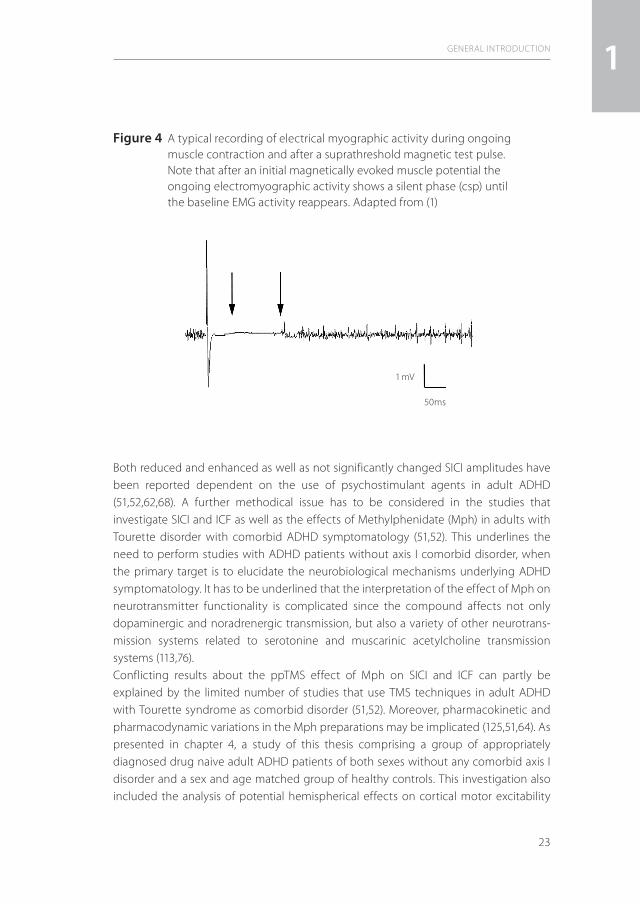
1.4.4 Principles of the Cortical Silent Period (CSP)The TMS impulse at the motor cortex during maintained voluntary muscle contraction results in the so-called cortical silent period (CSP), which reflects multifactor inhibitory mechanisms (ref). Dependent upon the strength of the TMS impulse, the ongoing elec-tromyographic activity is discontinued for a moment of several milliseconds. The CSP is defined by the time of salience until reappearance of the ongoing electromyographic activity. Although the neuronal mechanisms underlying the CSP are not fully understood as yet, GABAergic neurotransmission has been suggested to be involved (77). Lesion studies have led to the hypothesis that the CSP is generated in the primary motor cortex (114). Finally, it was shown that CSP and SICI have an inverse relationship and that SICI is suppressed while ICF is facilitated during CSP. The CSP can be considered as a measure of motor cortex excitability via influences from basal ganglia, ventrolateral thalamus and inhibitory interneurons (1)
1.4.5 ppTMS and ADHDThe ppTMS technique has been shown to provide a reliable test-retest stability of SICI and ICF in adults (93,74). Compared to the data from children with ADHD (78,79,19,20,21), the number of studies with ppTMS in adult patients is rather limited (61,62,94) and their results are equivocal.
CHAPTER 1
23
Both reduced and enhanced as well as not significantly changed SICI amplitudes have been reported dependent on the use of psychostimulant agents in adult ADHD (51,52,62,68). A further methodical issue has to be considered in the studies that investigate SICI and ICF as well as the effects of Methylphenidate (Mph) in adults with Tourette disorder with comorbid ADHD symptomatology (51,52). This underlines the need to perform studies with ADHD patients without axis I comorbid disorder, when the primary target is to elucidate the neurobiological mechanisms underlying ADHD symptomatology. It has to be underlined that the interpretation of the effect of Mph on neurotransmitter functionality is complicated since the compound affects not only dopaminergic and noradrenergic transmission, but also a variety of other neurotrans-mission systems related to serotonine and muscarinic acetylcholine transmission systems (113,76).Conflicting results about the ppTMS effect of Mph on SICI and ICF can partly be explained by the limited number of studies that use TMS techniques in adult ADHD with Tourette syndrome as comorbid disorder (51,52). Moreover, pharmacokinetic and pharmacodynamic variations in the Mph preparations may be implicated (125,51,64). As presented in chapter 4, a study of this thesis comprising a group of appropriately diagnosed drug naive adult ADHD patients of both sexes without any comorbid axis I disorder and a sex and age matched group of healthy controls. This investigation also included the analysis of potential hemispherical effects on cortical motor excitability
GENERAL INTRODUCTION 1
Figure 4 A typical recording of electrical myographic activity during ongoing muscle contraction and after a suprathreshold magnetic test pulse. Note that after an initial magnetically evoked muscle potential the ongoing electromyographic activity shows a silent phase (csp) until the baseline EMG activity reappears. Adapted from (1)
1 mV
50ms

24
and the correlation of the neurophysiological parameters with the clinical ADHD phenotype. In the long acting Methylphenidate (LA-Mph) study with ppTMS, the main focus of interest is the investigation of the impact of the chronically applied stimulant drug methylphenidate on motor overexcitability in adult ADHD and its correlation with the objective clinical phenotype.
2. Aims of this study
Since little is known about the general pathophysiological mechanisms underlying the phenotypical presentation of ADHD in adults, the aim of this thesis is to elucidate impairments in brain networks involved in attention processing and executive functioning by means of functional imaging techniques. Secondly, motor system functionality in adult ADHD is studied using ppTMS, a non-invasive technique that is considered to be the most appropriate for depicting the intracortical excitability and its modulation by compounds such as LA-Mph. As the psychopathology of ADHD is supposed to originate from a dysequilibrium of dopaminergic transmission, it is of special interest to investigate psychiatric symptoms which are assumed to be related to dopaminergic dysfunctionality, e.q. attention deficits, executive dysfunctioning and psychomotor disturbances. The results from the studies that include patients with (first episode) schizophrenia, may provide a better understanding of the specific psychopa-thology of adult ADHD. More specifically, the aims of these studies were1. To analyse central nervous systems involved in attention and execution by means of
functional magnetic resonance imaging, following a continuous performance test paradigm (Go/NoGo), in order to identify correlations between dysfunctions in these networks and the adult ADHD symptom profile.
2. To study cortical motor inhibition and facilitation with the paired pulse transcranial magnetic stimulation (ppTMS) technique in order to delineate relationships between overexcitability of the motor cortex and phenotypical presentation of adult ADHD.
3. To explore the effects of LA-Mph on cortical overexcitability in adults with ADHD by means of ppTMS.
4. To investigate the excitability of the motor neuronal system with ppTMS in another psychiatric disorder which is associated with dopamine driven cognitive and executive functioning deficits and disturbances of motor excitability, namely first-episode schizophrenia, and to compare the motor cortex excitability with ADHD patients in order to delineate putative overlapping pathophysiological mechanisms.
CHAPTER 1
25
3. Outline of the thesis
Chapter 2 provides an overview of imaging data in adult ADHD.Chapter 3 deals with a controlled fMRI study focused on attention networks and executive functions in adults with ADHD. Chapter 4 comprises a study with ppTMS in which this technique was used to investigate motor cortex excitability and executive functioning in adults with ADHD. In addition, the ppTMS parameters that reflect motor disinhibition were correlated with the symptom profile of adult patients with ADHD.Chapter 5 constitutes a clinical study in which the ppTMS technique is used to correlate motor overexcitability with the symptom profile of adult ADHD patients before and during treatment with LA-Mph. Chapter 6 describes a ppTMS study on abnormalities in motoneural systems of adult patients with first-episode schizophrenia and chapter 7 describes a study with adult patients with ADHD or first episode schizophrenia, diseases that are both supposed to have a dopaminergic brain transmission dysbalance. The motor excitability networks and hemispheric balance is evaluated in both patient categories. Chapter 8 concerns a general review of the fMRI and ppTMS data obtained from the above listed studies, and discusses the clinical relevance of the findings as well as the limitations and strengths of the subsequent clinical investigations. Some outlines for future investigations and the conclusions are also presented.
GENERAL INTRODUCTION 1


Brain imaging in adult Attention deficit hyperactivity disorder (ADHD)
Marc Schneider, Wolfgang Retz, and Michael Rösler
Published in W Retz, R Klein (ed.): Key Issues in Mental Health. Attention-Deficit Hyperactivity Disorder (ADHD) in adults. Karger, Basel (2010). Vol. 176: 88-104.
2 |

28
Abstract
This review provides an overview about recent findings in attention-deficit hyperactivity disorder (ADHD) brain imaging. ADHD is understood as a developmental disorder and several studies have addressed brain development in children and adolescents. The hallmarks of impairment of cerebral processing in ADHD are executive dysfunctions (motor execution, inhibitory control, working memory), as well as deficient attention processing. In adulthood, imaging studies have revealed disturbances in the prefrontal cortex, and anterior cingulated cortex (dACC) which are involved in the regulation of selective attention, executive control and decision-making. Dysfunction of basal ganglia is also a consistent finding in ADHD from childhood to adulthood. These findings suggest a persistent dysregulation of frontostriatal circuitries. The cerebellum, and its role in affect and cognition, is also persistently implicated in the pathology of ADHD. The cerebello-(thalamo-)striato-cortical network includes different attention networks and executive control instances. It appears from brain-imaging data in adults that the pathophysiological principles of ADHD do not profoundly change from childhood and adolescence to adulthood, regardless of some changes in psychopathology. The hypothesis of a neurodevelopmental disorder seems to be reinforced on the basis of imaging data of the adults.
CHAPTER 2

29
Introduction
Attention-deficit hyperactivity disorder (ADHD) affects adults with a prevalence of about 3–4% [1]. Whereas key symptoms of the psychopathological core symptoms in childhood are inattention, hyperactivity and impulsivity, adults with ADHD tend to be predominantly impaired by attention deficits and disorganization, whereas hyperactivity and impulsivity tend to ameliorate [2, 3]. Emotional dysregulation, mood and anxiety disorders as well as substance abuse are commonly described features and comorbidities in adult patients [4]. As clinical patterns differ between ADHD children and adults, and given that more than half of the children with ADHD do not continue being clinically affected in adulthood, one could hypothesize that adult patients with ADHD represent a distinct subpopulation with distinct neurobiological or environmental background. Therefore, there is a substantial need for neuroanatomical and functional neuroimaging investigations in adults suffering from ADHD. Indeed, like in many areas in neuropsy-chiatry, neuroimaging techniques have been intensively used also in ADHD research in the past few years.It is a principle question whether impairment of brain networks compares to those that have been extensively described in ADHD children. Thus, a key question in adult ADHD research is whether there is also impairment of frontostriatal/frontosubcortical networks. These pathways are involved in executive and motor control, as well as in inhibition of behavior and voluntary decision-making. Frontosubcortical networks contain a large amount of noradrenergic, dopaminergic as well as serotonergic projections. Striatal structures, such as putamen, globus pallidum and caudate nucleus, form a frontostriatal network and are typically impaired in ADHD [5]. Areas of particular interest are also the prefrontal and dorsolateral prefrontal cortex (PFC). The anterior cingulated cortex (ACC) seems to play a pivotal role in ADHD psychopathology. This region has widespread connections to forebrain and limbic structures. Besides its function as a conflict monitoring center, the ACC has the role of integrating polymodal inputs from different brain regions in the control of executive and inhibitory functions [6]. Finally, increasing attention is being paid to the cerebellum, which exerts strong influences on affective and cognitive function via thalamic connections [7, 8].
Brain Structure in ADHD
Global Neuroanatomical FindingsTotal cerebral volume reduction is well described in children and adolescents with ADHD [9–16], which is most prominent in the right hemisphere. The right hemisphere is hypothesized to play a dominate role in decision-making, inhibitory control and selective attention [17, 18]. It has been shown that damage of the right hemisphere can
BRAIN IMAGING IN ADULT ATTENTION DEFICIT HYPERACTIVITY DISORDER (ADHD)
2

30
lead to desynchronization of brain activity and neglect of sensory stimuli [19, 20]. Sowell et al. [21] showed that brain alterations in children with ADHD are focused on those brain regions which are relevance for attention, executive control and linguistic performance. More specifically, they could demonstrate that cortical abnormalities are mainly localized in inferior portions of dorsal PFC and bilateral anterior temporal cortex. Increased gray matter was seen in large portions of the posterior temporal and inferior parietal cortex bilaterally.Recently, Seidman et al. [22] investigated 24 adults with DSM-IV ADHD and 18 healthy controls comparable on age, socioeconomic status, sex, handedness, education, IQ, and achievement test performance. Compared to controls, adults with ADHD had significantly diminished overall cortical gray matter, and smaller prefrontal and ACC volumes in particular. The authors concluded that adults with ADHD have volume differences in brain regions associated with attention and executive control. These data are largely consistent with studies of children with ADHD, supporting the hypothesis that ADHD is a valid disorder with persistent biological features through all stages of life.
Region-Specific Neuroanatomical FindingsFrontal LobeSeveral studies have confirmed volume reductions of PFC in ADHD children, namely of the dorsolateral part (DLPFC) [9, 10, 13–16, 23]. The DLPFC plays an important role in attention, working memory, planning and organization of a task [18], whereas the ventrolateral prefrontal cortex (VLPFC) is involved in the regulation inhibitory control [24, 25]. The orbitofrontal cortex regulates social behavior and balance of inhibition and desinhibition as well as emotional attribution to decisions.Hesslinger et al. [26] found diminished left orbitofrontal brain volumes in adult ADHD patients. These regions are associated with social behavior and impulse control. Also in adults with ADHD, selective thinning of cerebral cortex in the networks that subserve attention and executive function was found by Makris et al. [27]. Significant cortical thinning in ADHD was seen especially in the right hemisphere involving the inferior parietal lobule, the dorsolateral prefrontal, and the anterior cingulated cortices. These neuroanatomical data give evidence to the frontal brain abnormalities also in ADHD adults, but the findings need further replication.
Anterior Cingulated CortexThe dorsal part of the anterior cingulated cortex (dACC) is crucial for executive functioning, inhibitory control monitoring, target detection, error processing as well as reward-based learning. Volume reductions in the right posterior cingulated gyrus in ADHD children have been reported [28]. In adults, lower volumes of the ACC could be shown by in the anatomical study of Makris et al. [27].
CHAPTER 2
31
Temporal LobeTemporal lobes have polymodal sensory integration functions in language comprehension as well as object identification (‘what system’), emotional regulation and memory function. The right temporal convexity plays an important role for visuospatial functions, whereas the left temporal convexity contains a large auditory association area which contributes to language comprehension [29]. Sowell et al. [21] demonstrated by anatomical brain surface analysis that children with ADHD had reduced anterior temporal lobe volumes bilaterally. Temporal lobe volume reduction as part of a general brain volume reduction in children and adolescents with ADHD was described by Castellanos et al. [12]. However, there is no substantial information about the interplay between cognition and affect in sensory processing, and its modulation by temporal lobes in ADHD.
Basal GangliaThe basal ganglia comprise of five nuclei: the caudate nucleus (‘cognitive associative’ striatum), putamen (sensorimotor striatum), nucleus accumbens (limbic striatum), globus pallidus and subthalamic nucleus. They are closely related to brainstem structures such as substantia nigra and the pedunculopontine nucleus. The striatum comprises the putamen and the pallidum, and displays a high density of dopaminergic neurons. Its main function is procedural learning and automatization of motor programs and behaviors and it serves to assemble complex response habits to strategically adapted environmental needs [30, 31]. ADHD-associated symptoms are associated with striatal damage [32].Basal ganglia volume reductions have been shown in several studies with ADHD children and adolescents. In most of them uni- or bilateral reduction of caudate volumes were found [9–12, 29, 33–36]. Schrimsher et al. [37] could predict the cumulative severity ratings of inattentive behaviors by measuring caudate volume asymmetry from serial sagittal magnetic resonance images from childhood to adolescence. Also unilateral volume reduction of the pallidum has been shown in several studies in children with ADHD [10–12, 28, 38].Until now, no evidence for basal ganglia volume reduction in adult ADHD has been reported. A possible explanation is that differences between controls and ADHD almost disappear with increasing age before adulthood [12].
Corpus CallosumThe corpus callosum connects homonymous regions of the cerebral hemispheres. Injury of callosal structures can lead to problems in holding sustained attention [39], with associated deficits in learning and memory [40]. The neuropsychological deficits after injury of corpus callosum are often subtle or lacking.Volume reduction of the corpus callosum is a common finding in studies with ADHD children and adolescents. Posterior regions of the corpus callosum are mostly affected [15, 41–45]. Data from adult ADHD are lacking so far.
BRAIN IMAGING IN ADULT ATTENTION DEFICIT HYPERACTIVITY DISORDER (ADHD)
2
32
Parietal LobePosner and Petersen [18] have described a posterior attention system located in the parietal lobe, which seems to be mainly modulated by noradrenergic transmission in contrast to the predominantly dopaminergic modulation of the frontal attention system. The posterior part of the parietal cortex is involved in orienting and selective attention networks [46]. It disengages the attentional focus from the contralateral target [47], and lesions of this region can lead to impaired attention [48, 49].Only few studies have addressed to parietal lobe structure and function in ADHD. Castellanos et al. [12] have shown that posterior parietal volume is reduced, whereas conversely Sowell et al. [21] have demonstrated an increase in cortical volume in children with ADHD. Given its importance in visuospatial orienting and as a region for polymodal sensory integration, it was Makris et al. [27] who pointed out volume reductions in the right inferior parietal lobule in adults with ADHD.
Occipital LobeIn line with the general findings of Castellanos’ work [12], pronounced reduction of left occipital brain volume in children with ADHD was also found by another study group [16]. No data are presently available regarding occipital lobe anatomy in adults with ADHD.
CerebellumThe role of the cerebellum in cognitive and affective function has been described in 20 patients with cerebellar lesions [50]. Aside from the well-known motor coordination problems, patients with cerebellar lesion display impairment of executive functions, visuospatial cognition deficits as well as blunting of affect and disinhibition of behavior. Cognitive cerebellar functions are located mainly in the posterior lobe (neocerebellum), whereas executive, visuospatial, and memory functions of neocerebellum are impaired when the lesions are located in the hemispheres and dentate nucleus [50, 51]. The vermis has been shown involved with affective disturbances [51]. The cerebellum projects via thalamus to areas in the PFC [52] and there are reciprocal projections from the PFC to cerebellum, thus forming a functional network that influences rather than generates motor control, inhibitory and executive functions. Moreover, several studies have shown that the cerebellum has modulatory effects on forebrain dopamine outflow [53–56].Several studies in ADHD in childhood and adolescence have shown structural cerebellar impairment [10–12, 15, 16, 57–59]. Indeed, the most marked neuroanatomical anomaly in ADHD has been described in the cerebellum, with volume changes more marked than in the PFC [12]. In children, reductions in right cerebellar hemisphere and vermis volume have been reported [11, 15, 16, 57–59]. These volume reductions correlated with attentional problems and global clinician ADHD ratings [12]. At present there are no data on cerebellar volume in adult ADHD.
CHAPTER 2

33
Other Brain RegionsIn children and adolescents with ADHD an increased volume of the hippocampus bilaterally, which is involved in attentional processes such as visuospatial working memory and executive functions, has been reported [60]. Moreover, some evidence for a reduced size of the basolateral amygdala was found in this study. Since affective symptoms and emotional instability are typical features of and affective disorders are highly prevalent in adult ADHD, altered amygdala and hippocampus volumes are of particular interest also for adults with ADHD. However, in a recent study with adults suffering from ADHD, no differences regarding hippocampus and amygdale volumes were found [61].
Brain Function in ADHD
Functional MRI (fMRI), MRI relaxometry and ligand-bound imaging techniques like SPECT or positron emission tomography (PET) have been used to study functional abnormalities of brain networks in ADHD. From a neurological viewpoint, attention networks can be basically distinguished into three components [18, 46, 62]:• The arousal and alerting networks are mainly subcortically located and constitute of
the ascending reticular activating system. They project to the whole brainstem and thalamus and, through the striatum, up to the limbic system to form cortical projections. The main function of this component is the activation and synchroniza-tion of the cerebral cortex during behavior and motivation, and has affinity to salient stimuli and memorization.
• The mixed cortical-subcortical orienting networks are involved in detection of novel stimuli (superior colliculi), filtration of relevant stimuli (pulvinar) and disengagement of attention focus (posterior parietal cortex).
• The selective (or directed/cortical) attentional network is of particular interest for ADHD pathophysiology. It involves frontal brain structures for generation of volitional saccades (frontal eye fields), induces motor intention (premotor cortex), is linked to the working memory (DLPFC) and is modulated by the ACC (target detection, response selection and inhibition, conflict monitoring, motivation) [63]. This network is also called executive (conflict) network and has been shown highly heritable [64]. Within this network, the dACC has strong connectivity to frontal brain structures with dense serotonergic and dopaminergic components. Regions of the parietal cortex also play an important role in mediating sensory functions. Th e posterior parietal cortex disengages the attentional focus to a target [47] and the superior parietal lobule has the function of voluntary shift s of attention [29]. Right hemisphere dominance could be found for the orienting as well as selective attention [17, 18].
BRAIN IMAGING IN ADULT ATTENTION DEFICIT HYPERACTIVITY DISORDER (ADHD)
2
34
Konrad et al. [65] have used event-related fMRI to investigate brain activations related to these three particular aspects of attention. It could be shown that children with ADHD recruited deviant brain regions for all three above-mentioned attentional networks. ADHD children had less right-sided activation in the anterior cingulated gyrus during alerting, more frontostriatal-insular activation during reorienting, and less frontostriatal activation for executive control. Dysregulation of blood oxygenation level-dependent signals was described in the putamen during reorienting and executive control, suggesting altered strategies in children with ADHD. In medication-naïve children and adolescents with ADHD, task-specific functional abnormalities in frontostriatal but also to parietal and temporal areas were found [66]. Hale et al. [67] concluded from their data that abnormal brain function among adult ADHD participants was not limited to complex executive functions. Abnormal processing of numeric stimuli was indicated during both simple and complex cognitive operations. For example, during the difficult tasks, they exhibited greater activation of left hemispheric linguistic-processing areas and failed to activate bilateral parietal regions important for the complex executive operations.
Anterior Cingulated CortexHypoactivation of the dACC has been consistently described in children and adolescents with ADHD using continuous performance paradigms, with results being similar using fMRI of PET-imaging techniques [25, 68–71]. These findings have led to the hypothesis that dACC plays a significant role in ADHD pathophysiology.Zametkin et al. [71] were the first to describe hypoactivity of dACC with PET in adult ADHD patients. According to the hypothesis of impairment of selective attention, several studies in adult ADHD have also shown hypofunctionality of the ACC [68, 70]. Following the executive attention hypothesis, the earliest fMRI study in adult ADHD was performed by Bush et al. [70], using a specially designed counting Stroop paradigm. This study demonstrated that the ‘cognitive division’ of the dACC was not activated in adult ADHD patients during interference conditions. As a compensatory mechanism, ADHD patients activated an alternative frontostriatal network by using different regions of lateral PFC, insular cortex, as well as unilateral activation of caudate, putamen, thalamus and pulvinar. These results may be interpreted as impairment of dACC function in ADHD subjects under conditions where interferences occur, while under conditions where subjects could focus on salient stimuli, there was no difference in dACC activation. This ‘normal attention but abnormal stimulus alerting and conflict effect’ has also been reported from a neuropsychological point by Oberlin et al. [72]. Only ADHD subjects with the combined type were impaired in their reactions to abrupt visual cues or those that contain conflicting spatial cues. These features were not found in adults with the ADHD-inattentive type.
CHAPTER 2
35
Besides the role of dACC in selective attentional processing, response selection and inhibition and performance monitoring [73], dACC is also thought to influence reward-based decision-making [74]. The larger the gain, the higher the activity in the pregenual ACC during the decision phase [75]. Ernst et al. [73] found differences in motivational behaviors in ADHD, especially when the patients had to weigh long-term versus short-term rewards. The patients used more parts of the right ACC than healthy controls.Memory performance was associated with activation of the ACC in healthy adolescents but with activation of the superior parietal lobe (SPL) and precuneus in adolescent ADHD patients [76]. The authors suggested that increased SPL activation in ADHD reflected attentional compensation for low ACC activation during the encoding and that the higher salience of emotional stimuli, in contrast, regulated the interplay between ACC and SPL in conjunction with improving memory to the level of healthy adolescents.Using a working memory paradigm, Wolf et al. [77] could recently demonstrate lower connectivity in ACC and higher connectivity in dorsal cingulate cortex in adults with ADHD and healthy controls. Another fMRI study found evidence for decreased functional connectivity between ACC and posterior cingulated regions including the precuneus [78].
Motor SystemThe execution of simple motor tasks reveals distinct cerebral activation pathways. Using a simple finger-tapping task, Mostofsky et al. [79] reported that children with ADHD had decreased contralateral motor cortex and right parietal cortex activation during right- and left-handed finger sequencing. These findings could be interpreted as anomalous development of cortical systems necessary for execution of patterned movements.In a study with PET, a correlation between motor hyperactivity with lower binding potential values for dopamine transporter (DAT) in the midbrain was shown in adolescents with ADHD [80]. Thus, altered dopamine signaling might have a causal relationship to hyperactivity. Studies with adult ADHD patients are not available so far.
Frontal CortexThe most consistent findings in the neuroimaging literature of ADHD are deficits in neural activity within frontostriatal and frontoparietal circuits. However, the results vary across subregions of the frontal cortex, suggesting that ADHD is not associated with dysfunction of any particular part of frontal cortex.The PFC is critical for the regulation of behavior, attention, and affect by use of represen-tational knowledge. The PFC is important for sustaining attention over a delay, inhibiting distraction, and dividing attention, while more posterior cortical areas are essential for perception and the allocation of attentional resources. The PFC in the right hemisphere
BRAIN IMAGING IN ADULT ATTENTION DEFICIT HYPERACTIVITY DISORDER (ADHD)
2
36
is particularly important for behavioral inhibition. Lesions to the PFC produce a profile of distractibility, forgetfulness, impulsivity, poor planning, and locomotor hyperactivity.Variable findings have been described for VLPFC and DLPFC. These brain regions also monitor attention, planning, working memory and executive control, especially with regard to inhibitory control [18, 81]. Rubia et al. [25] found hypoactivation in the right VLPFC and left caudatus of adolescents with ADHD, whilst Durston et al. [82] reported different activation of frontostriatal regions. Children with ADHD displayed more diffuse network activations including more posterior and dorsolateral prefrontal regions. Rubia et al. [83] reported that medication-naïve adolescent patients with ADHD showed significantly reduced brain activation in the right inferior PFC during successful motor response inhibition and in the precuneus and posterior cingulate gyrus during inhibition failure. These deficits correlated with behavioral scores of ADHD and persisted when corrected for medication history and performance discrepancies. Conversely, Ernst et al. [73] showed using PET that adult ADHD patients, as well as healthy controls, activated VLPFC and DLPFC including insula during a decision-making task. However, the activation of the dACC and hippocampus, subserving emotional and memory processes, was less extended in the ADHD group, who instead recruited the caudal part of the right ACC. These results were interpreted as a basis for problems of motivated behavior in ADHD.Evidence for significant frontal hypoactivity, including anterior cingulate, dorsolateral prefrontal and inferior prefrontal cortices comes from a meta-analysis of studies with fMRI in children and adolescents with ADHD [84]. Analyses of studies which used other than response inhibition paradigms revealed a more extensive pattern of hypofunction in patients with ADHD than those of response inhibition (thalamus, basal ganglia and parietal cortex). Studies of response inhibition displayed more limited group differences regarding activation of inferior PFC, medial wall regions, and the precentral gyrus.In adult ADHD patients, less activation and lower functional connectivity was observed during a working memory task in the left VLPC, while connectivity of the right PFC was increased when compared to control subjects together with functional changes in other brain regions [77]. Moreover, a correlation between activation of frontal cortical areas of adult ADHD subjects and inattention scores has been reported, suggesting a functional deficit within this network that depends on the degree of attention deficits [85]. On the other hand, increased activation of orbitofrontal cortex was found in response to gain outcomes during a monetary incentive delay task, suggesting that this part of frontal cortex is involved in abnormal reward processing in adult ADHD [86].
CerebellumDue to its involvement in cognitive, emotional processing and behavioral control, the cerebellum seems to be an important region of interest in ADHD research [50]. Anderson et al. [7] reported abnormalities of the vermis in children and adolescents in a MRI
CHAPTER 2
37
relaxometry study that could be influenced by methylphenidate, suggesting an influence of cerebellar function in ADHD. The effects of methylphenidate on cerebellum depended on pretreatment activity level. With fMRI, Schulz et al. [87] described a higher activity of the cerebellum in adolescents with ADHD. In contrast, Valera et al. [88] found significantly decreased activity in cerebellum and also occipital lobe of adult patient with ADHD, even though working memory performance did not differ significantly between ADHD and controls. Kim et al. [89] examined ADHD children with PET and found decreased bilateral cerebellar blood flow in ADHD compared to controls. Volkow et al. [90, 91] reported that methylphenidate could increase metabolic activity of the cerebellum in normal adults, dependent of dopamine receptor activity.Preliminary results from a study with ADHD children [92] hinted to a relation between cerebellum and forebrain dysfunction and ADHD symptomatology. The authors found with an ADHD diffusion tensor-imaging technique (DTI) prominent white matter abnormalities in the right premotor, right striatal, right cerebral peduncule, left cerebellar peduncule, left cerebellum and left parieto-occipital areas. In adults with ADHD, less activation during a working memory task and changes of functional connectivity of the cerebellum and cortical brain regions was described [77]. These results give additional evidence for corticopontocerebellar circuit deficits in ADHD.
Parietal CortexThe parietal cortex belongs to an attentional system that includes frontoparietal network structures [18, 93]. For example, orienting networks include the SPL, as well as the temporal parietal junction and frontal eye field [94]. Krauel et al. [76] suggested increased activation in some parietal regions as an attentional compensation for low ACC activation in healthy adolescents. Together with frontal brain areas, the alerting attentional network activates parietal and thalamic areas that are potentially susceptible to the actions of norepinephrine [95–97].Superior parietal and middle frontal areas are involved in visuospatial processing [98]. Silk et al. [99] have shown in an fMRI study with a mental rotational task, that ADHD children with combined subtype have lower activation of the action attentional system including superior parietal cortex as well as middle frontal areas. Patients had also increased activation of the posterior midline attentional system. This indicates that ADHD patients might also have parietal dysfunction as well as dysfunctions of the widespread frontal and striatal systems. As is the case with many of the networks discussed so far, these findings in children have yet to be extended to adults with ADHD. A first step towards this direction has been performed by Tamm et al. [100], who showed that adolescents with ADHD had significant impairments in their ability to direct and allocate attentional resources. This was associated with bilateral aberrations in the parietal attentional system.
BRAIN IMAGING IN ADULT ATTENTION DEFICIT HYPERACTIVITY DISORDER (ADHD)
2

38
Basal GangliaIn line with PET findings showing reduced basal ganglia perfusion in patients with ADHD [101], subsequent fMRI studies have reported abnormal activation of the striatum [25, 102–104].Although the main focus of the study of Bush et al. [70] was not the basal ganglia, they observed increased activation of the right putamen in adults with ADHD while performing a Stroop task. Recently, Plichta et al. [105] could show hyporesponsiveness of the ventral-striatal reward system in adults with ADHD, who were examined during a series of choices between two monetary reward options. In addition, they reported increased activation of the dorsal caudate nucleus and amygdala associated with delayed reward. Similarly, decreased activation in the ventral striatum during the anticipation of gain in a monetary incentive delay task was described in another recent study [86]. Moreover, the authors described in this study a negative correlation of ventral striatal activation with self-reported impulsivity and hyperactivity. Similar findings have been reported in a previous fMRI study of brain activation during a reward-anticipation task in adolescents with ADHD [104]. The negative correlation between impulsivity and striatal activation, which was found in both studies, has also been shown by Schneider et al. [85], who used an impulse-control paradigm. Taken together, these results suggest that striatal activation is involved in the processing of reward and the regulation of im-pulsive-hyperactive traits in adults with ADHD.
ADHD and Comorbid Disorders
Studies with patients who have comorbid disorders or brain lesions are of interest because they may help to validate the specificity of the hypothesized frontostriatal(-cerebellar) dysfunctions and compensatory mechanisms in ADHD.Bussing et al. [58] have suggested that no differences in cerebellar morphology could be found between ADHD children with and without comorbid conduct disorder. Also, in this study no differences were reported in volume measurements of frontostriatal structures. On the other hand, electrophysiological studies with event related potentials showed abnormalities in prefrontal lobe activation in teenagers with conduct disorder [106].Tourette’s syndrome (TS) is frequently comorbid with ADHD [107, 108]. In TS, basal ganglia volume reduction and loss of left > right side asymmetry of the globus pallidus is described in some but not all studies [108–110]. Some studies could not differentiate between TS and comorbid ADHD in terms of brain structure alterations, whereas some could find that patients with comorbid ADHD tended to have larger volumes across all cortical portions of those circuits to dorsal prefrontal and parietooccipital regions and smaller caudate nucleus volumes [111].
CHAPTER 2

39
Adler et al. [112] have used a simple attention task in adolescents with bipolar disorder and showed that comorbidity with ADHD was associated with less activation of the VLPFC, ACC and higher activation in posterior parietal cortex as well as middle temporal gyrus. Thus, comorbidity with ADHD might result in less activation of prefrontal regions while posterior parietal and temporal cortical areas are used as alternative pathways. Facial recognition is also impaired in ADHD in a similar way when compared with patients with schizophrenia [113]. Both groups display reduced activity in the medial prefrontal and amygdala brain regions required to process emotional faces.Autism may occur with ADHD and impairment of attention has been consistently reported in autism [114]. In anatomical studies, patients with autism displayed larger total brain and white matter volumes in caudate, globus pallidum, most cortical brain regions and in the cerebellum as compared to ADHD subjects [115]. Reduced fMRI activation was found primarily in amygdala of autistic patients during social tasks [116], but autistic spectrum disorders also display dysfunctional cerebellofrontal spatial attention system [117, 118] similar to ADHD.
Conclusions
In contrast to neuroimaging investigations in children and adolescents with ADHD, the number of studies in adult patients is still limited. Imaging data in general are often confounded by small sample sizes, non-replicated and sometimes even contradictory results. However, recent findings have shown similarities between abnormalities in adult ADHD patients and children with ADHD suggesting impairment of frontostriatal(-cerebellar) networks. Consistent findings have been reported regarding dysfunction of the striatum and the ACC. Prefrontal cortical structures also seem to play a pivotal role in ADHD psychopathology, although these findings are not specific to ADHD. As in children, the cerebellum is also dysfunctional in adults with ADHD. Moreover, there is increasing evidence that also parts of the posterior attention networks are less active in both childhood and adult ADHD. Functional abnormalities comprise frontostriatal, parietal and also temporal cortical areas in a task-specific manner. Attention orienting is less affected than salient stimulus or conflict alerting. Also, several ‘vertical’ levels of attention networks – beginning from the arousal to the orienting up to the selective attention network – are affected in ADHD.Data on ADHD patients with comorbid psychiatric disorders are not consistent so far and their contribution to our understanding of ADHD pathophysiology is limited. However, it seems that ADHD symptoms combined with other disorders are associated with frontostriatal dysfunction.Structural and functional brain-imaging investigations are an important source for our growing knowledge of ADHD pathophysiology. However, due to the lack of sensitivity
BRAIN IMAGING IN ADULT ATTENTION DEFICIT HYPERACTIVITY DISORDER (ADHD)
2

40
and specificity of the findings, neuroimaging techniques are not ready to be used as a diagnostic tool. It seems possible that with the progress in understanding the pathogenesis of ADHD together with the technical progress in brain-imaging techniques, we might overcome this shortcoming in future.
CHAPTER 2
41
References
1 Fayyad J, De Graaf R, Kessler R, Alonso J, Angermeyer M, Demyttenaere K, De Girolamo G, Haro JM, Karam EG, Lara C, Lépine JP, Ormel J, Posada-Villa J, Zaslavsky AM, Jin R: Cross-national prevalence and correlates of adult attention-deficit hyperactivity disorder. Br J Psychiatry 2007;190:402–409.
2 Biederman J, Mick E, Faraone SV: Age-dependent decline of symptoms of attention-deficit hyperactivity disorder: impact of remission definition and symptom type. Am J Psychiatry 2000;157:816–818.
3 Mannuzza S, Klein RG, Bessler A, Malloy P, LaPadula M: Adult outcome of hyperactive boys: educational achievement, occupational rank and psychiatric status. Arch Gen Psychiatry 1993;50:565–576.
4 Retz W, Retz-Junginger P, Hengesch G, Schneider M, Thome J, Pajonk FG, Salahi-Disfan A, Rees O, Wender PH, Rösler M: Psychometric and psychopathological characterization of young male prison inmates with and without attention-deficit/hyperactivity disorder. Eur Arch Psychiatry Clin Neurosci 2004;254:201–208.
5 Casey BJ, Castellanos FX, Giedd JN, Marsh WL, Hamburger SD, Schubert AB, Vauss YC, Vaituzis AC, Dickstein DP, Sarfatti SE, Rapoport JL: Implication of right frontostriatal circuitry in response inhibition and attention-deficit/hyperactivity disorder. J Am Acad Child Adolesc Psychiatry 1997;36:374–383.
6 Carter CS, Macdonald AM, Botvinick M, Ross LL, Stenger VA, Noll D, Cohen JD: Parsing executive processes: strategic vs. evaluative functions of the anterior cingulate cortex. Proc Natl Acad Sci USA 2000;97:1944–1948.
7 Anderson CM, Polcari A, Lowen SB, Renshaw PF, Teicher MH: Effects of methylphenidate on functional magnetic resonance relaxometry of the cerebellar vermis in boys with ADHD. Am J Psychiatry 2002;159:1322–1328.
8 Schmahmann JD: The cerebrocerebellar system: anatomic substrates of the cerebellar contribution to cognition and emotion. Int Rev Psychiatry 2001;13:247–260.
9 Filipek PA, Semrud-Clikeman M, Steingard R, Kennedy D, Biederman J: Volumetric MRI analysis: comparing subjects having attention-deficit hyperactivity disorder with normal controls. Neurology 1997;48:589–601.
10 Castellanos FX, Giedd JN, Marsh WL, Hamburger SD, Vaituzis AC, Dickstein DP, Sarfatti SE, Vauss YC, Snell JW, Lange N, Kaysen D, Krain AL, Ritchie GF, Rajapakse JC, Rapoport JL: Quantitative brain magnetic resonance imaging in attention-deficit hyperactivity disorder. Arch Gen Psychiatry 1996;53:607–616.
11 Castellanos FX, Giedd JN, Berquin PC, Walter JM, Sharp W, Tran T, Vaituzis AC, Blumenthal JD, Nelson J, Bastain TM, Zijdenbos A, Evans AC, Rapoport JL: Quantitative brain magnetic resonance imaging in girls with atten-tion-deficit/hyperactivity disorder. Arch Gen Psychiatry 2001;58:289–295.
12 Castellanos FX, Lee PP, Sharp W, Jeffries NO, Greenstein DK, Clasen LS, Blumenthal JD, James RS, Ebens CL, Walter JM, Zijdenbos A, Evans AC, Giedd JN, Rapoport JL: Developmental trajectories of brain volume abnormalities in children and adolescents with attention-deficit/hyperactivity disorder. JAMA 2002;288:1740–1748.
13 Kates WR, Frederikse M, Mostofsky SH, Folley BS, Cooper K, Mazur-Hopkins P, Kofman O, Singer HS, Denckla MB, Pearlson GD, Kaufmann WE: MRI parcellation of the frontal lobe in boys with attention-deficit hyperactivity disorder or Tourette syndrome. Psychiatry Res 2002;116:63–81.
14 Mostofsky S, Cooper K, Kates W, Denckla M, Kaufmann W: Smaller prefrontal and premotor volumes in boys with attention-deficit/hyperactivity disorder. Biol Psychiatry 2002;52:785–794.
15 Hill DE, Yeo RA, Campbell RA, Hart B, Vigil J, Brooks W: Magnetic resonance imaging correlates of attention-deficit/hyperactivity disorder in children. Neuropsychology 2003;17:496–506.
16 Durston S, Hulshoff Pol HE, Schnack HG, Buitelaar JK, Steenhuis MP, Minderaa RB, Kahn RS, van Engeland H: Magnetic resonance imaging of boys with attention-deficit/hyperactivity disorder and their unaffected siblings. J Am Acad Child Adolesc Psychiatry 2004;43:332–340.
17 Pardo JV, Pardo P, Janer K, Raichle ME: The anterior cingulated cortex mediates processing selection in the Stroop attention conflict paradigm. Proc Natl Acad Sci USA 1990;87:256–259.
18 Posner MI, Petersen SE: The attention system of the human brain. Annu Rev Neurosci 1990;13:25–42.19 Heilman KM, Van den Abell T: Right hemisphere dominance for attention: the mechanism underlying
hemispheric asymmetries of inattention (neglect). Neurology 1980;30:327–330.20 Weintraub S, Mesulam MM: Right cerebral dominance in spatial attention: further evidence based on
ipsilateral neglect. Arch Neurol 1987;44:621–625.
BRAIN IMAGING IN ADULT ATTENTION DEFICIT HYPERACTIVITY DISORDER (ADHD)
2
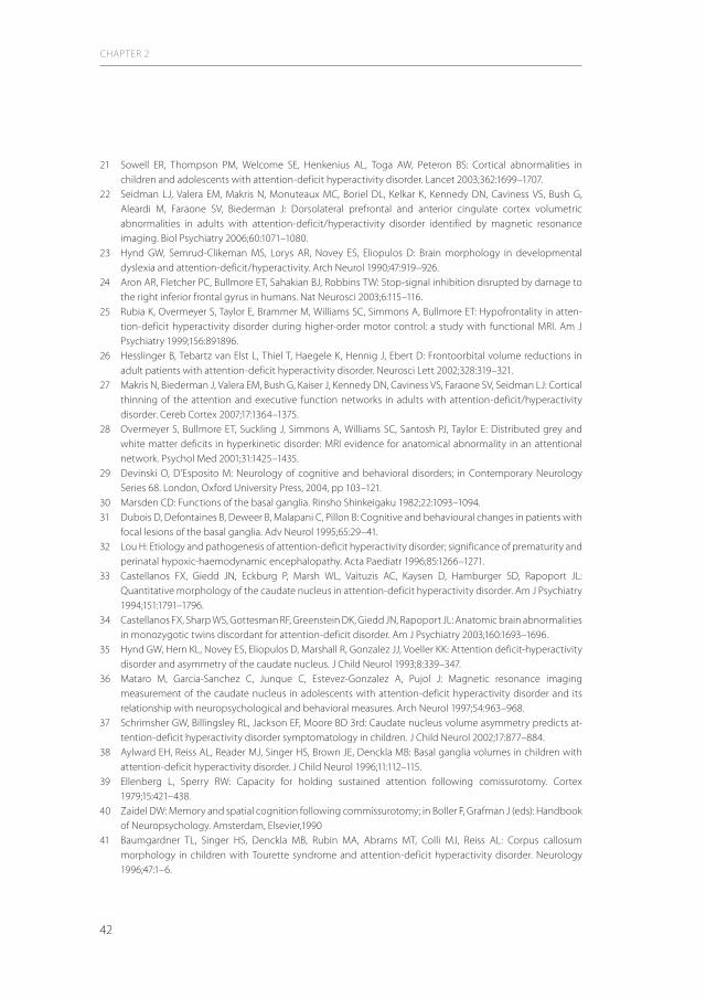
42
21 Sowell ER, Thompson PM, Welcome SE, Henkenius AL, Toga AW, Peteron BS: Cortical abnormalities in children and adolescents with attention-deficit hyperactivity disorder. Lancet 2003;362:1699–1707.
22 Seidman LJ, Valera EM, Makris N, Monuteaux MC, Boriel DL, Kelkar K, Kennedy DN, Caviness VS, Bush G, Aleardi M, Faraone SV, Biederman J: Dorsolateral prefrontal and anterior cingulate cortex volumetric abnormalities in adults with attention-deficit/hyperactivity disorder identified by magnetic resonance imaging. Biol Psychiatry 2006;60:1071–1080.
23 Hynd GW, Semrud-Clikeman MS, Lorys AR, Novey ES, Eliopulos D: Brain morphology in developmental dyslexia and attention-deficit/hyperactivity. Arch Neurol 1990;47:919–926.
24 Aron AR, Fletcher PC, Bullmore ET, Sahakian BJ, Robbins TW: Stop-signal inhibition disrupted by damage to the right inferior frontal gyrus in humans. Nat Neurosci 2003;6:115–116.
25 Rubia K, Overmeyer S, Taylor E, Brammer M, Williams SC, Simmons A, Bullmore ET: Hypofrontality in atten-tion-deficit hyperactivity disorder during higher-order motor control: a study with functional MRI. Am J Psychiatry 1999;156:891896.
26 Hesslinger B, Tebartz van Elst L, Thiel T, Haegele K, Hennig J, Ebert D: Frontoorbital volume reductions in adult patients with attention-deficit hyperactivity disorder. Neurosci Lett 2002;328:319–321.
27 Makris N, Biederman J, Valera EM, Bush G, Kaiser J, Kennedy DN, Caviness VS, Faraone SV, Seidman LJ: Cortical thinning of the attention and executive function networks in adults with attention-deficit/hyperactivity disorder. Cereb Cortex 2007;17:1364–1375.
28 Overmeyer S, Bullmore ET, Suckling J, Simmons A, Williams SC, Santosh PJ, Taylor E: Distributed grey and white matter deficits in hyperkinetic disorder: MRI evidence for anatomical abnormality in an attentional network. Psychol Med 2001;31:1425–1435.
29 Devinski O, D’Esposito M: Neurology of cognitive and behavioral disorders; in Contemporary Neurology Series 68. London, Oxford University Press, 2004, pp 103–121.
30 Marsden CD: Functions of the basal ganglia. Rinsho Shinkeigaku 1982;22:1093–1094.31 Dubois D, Defontaines B, Deweer B, Malapani C, Pillon B: Cognitive and behavioural changes in patients with
focal lesions of the basal ganglia. Adv Neurol 1995;65:29–41.32 Lou H: Etiology and pathogenesis of attention-deficit hyperactivity disorder; significance of prematurity and
perinatal hypoxic-haemodynamic encephalopathy. Acta Paediatr 1996;85:1266–1271.33 Castellanos FX, Giedd JN, Eckburg P, Marsh WL, Vaituzis AC, Kaysen D, Hamburger SD, Rapoport JL:
Quantitative morphology of the caudate nucleus in attention-deficit hyperactivity disorder. Am J Psychiatry 1994;151:1791–1796.
34 Castellanos FX, Sharp WS, Gottesman RF, Greenstein DK, Giedd JN, Rapoport JL: Anatomic brain abnormalities in monozygotic twins discordant for attention-deficit disorder. Am J Psychiatry 2003;160:1693–1696.
35 Hynd GW, Hern KL, Novey ES, Eliopulos D, Marshall R, Gonzalez JJ, Voeller KK: Attention deficit-hyperactivity disorder and asymmetry of the caudate nucleus. J Child Neurol 1993;8:339–347.
36 Mataro M, Garcia-Sanchez C, Junque C, Estevez-Gonzalez A, Pujol J: Magnetic resonance imaging measurement of the caudate nucleus in adolescents with attention-deficit hyperactivity disorder and its relationship with neuropsychological and behavioral measures. Arch Neurol 1997;54:963–968.
37 Schrimsher GW, Billingsley RL, Jackson EF, Moore BD 3rd: Caudate nucleus volume asymmetry predicts at-tention-deficit hyperactivity disorder symptomatology in children. J Child Neurol 2002;17:877–884.
38 Aylward EH, Reiss AL, Reader MJ, Singer HS, Brown JE, Denckla MB: Basal ganglia volumes in children with attention-deficit hyperactivity disorder. J Child Neurol 1996;11:112–115.
39 Ellenberg L, Sperry RW: Capacity for holding sustained attention following comissurotomy. Cortex 1979;15:421–438.
40 Zaidel DW: Memory and spatial cognition following commissurotomy; in Boller F, Grafman J (eds): Handbook of Neuropsychology. Amsterdam, Elsevier,1990
41 Baumgardner TL, Singer HS, Denckla MB, Rubin MA, Abrams MT, Colli MJ, Reiss AL: Corpus callosum morphology in children with Tourette syndrome and attention-deficit hyperactivity disorder. Neurology 1996;47:1–6.
CHAPTER 2

43
42 Giedd JN, Castellanos FX, Casey BJ, Kozuch P, King AC, Hamburger SD, Rapoport JL: Quantitative morphology of the corpus callosum in attention-deficit hyperactivity disorder. Am J Psychiatry 1994;151:665–669.
43 Hynd GW, Semrud-Clikeman M, Lorys AR, Novey ES, Eliopulos D, Lytinen H: Corpus callosum morphology in attention-deficit hyperactivity disorder: morphometric analysis of MRI. J Learn Disabil 1991;24:141–146.
44 Lyoo I, Noam G, Lee C, Lee H, Kennedy B, Renshaw P: The corpus callosum and lateral ventricles in children with attention-deficit hyperactivity disorder: a brain magnetic resonance imaging study. Biol Psychiatry 1996;40:1060–1063.
45 Semrud-Clikeman MS, Filipek PA, Biederman J, Steingard R, Kennedy D, Renshaw P, Bekken K: Attention-deficit hyperactivity disorder: Magnetic resonance imaging morphometric analysis of the corpus callosum. J Am Acad Child Adolesc Psychiatry 1994;33:875–881.
46 Fan J, Bruce T, McCandliss D, Fossella J, Flombaum J, Posner MI: The activation of attentional networks. Neuroimage 2005;26:471–479.
47 Rafal R, Robertson L: The neurology of visual attention; in Gazzaniga MS (ed): The Cognitive Neurosciences. Cambridge/MA, MIT Press, 1995,pp 625–648.
48 Posner MI: Attention in cognitive neuroscience: an overview; in Gazzaniga MS (ed): The Cognitive Neurosciences. Cambridge/MA, MIT Press, 1995,pp 615–624.
49 Ro T, Borden C, Driver J, Rafal R: Ipsilesional biases in saccades but not perception after lesions of the human inferior parietal lobule. J Cogn Neurosci 2001;13:920–929.
50 Schmahmann JD, Sherman JC: The cerebellar cognitive affective syndrome. Brain 1998;121:561–579.51 Schmahmann JD: Disorders of the cerebellum: ataxia, dysmetria of thought, and the cerebellar cognitive
affective syndrome. J Neuropsychiatry Clin Neurosci 2004;16:367–378.52 Middleton FA, Strick PL: Cerebellar projections to the prefrontal cortex of the primate. J Neurosci 2001;21:700–
712.53 Snider SR, Snider RS: Structural and functional relationships between cerebellum and catecholamine
systems: an overview. Exp Brain Res Suppl 1982;6:557–606.54 Tellerman K, Astrow A, Fahn S, Snider SR, Snider RS, Glassgold JM: Cerebellar control of catecholaminergic
activities: implications for drug therapy of movement disorders. Int J Neurol 1979;13:135–155.55 Glowinski J, Nieoullon A, Cheramy A: Regulations of the activity of the nigrostriatal dopaminergic pathways
by cortical, cerebellar, and sensory neuronal afferences. Adv Biochem Psychopharmacol 1978;19:75–87.56 Nieoullon A, Cheramy A, Glowinski J: Control of the activity of the two dopaminergic systems by cortical,
cerebellar and sensory afferents in the cat. Appl Neurophysiol 1979;42:51–53.57 Berquin PC, Giedd JN, Jacobsen LK, Hamburger SD, Krain AL, Rapoport JL, Castellanos FX: Cerebellum in at-
tention-deficit hyperactivity disorder: a morphometric MRI study. Neurology 1998;50:1087–1093.58 Bussing R, Grudnik J, Mason D, Wasiak M, Leonard C: ADHD and conduct disorder: an MRI study in a
community sample. World J Biol Psychiatry 2002; 3:216–220.59 Mostofsky SH, Reiss AL, Lockhart P, Denckla MB: Evaluation of cerebellar size in attention-deficit hyperactivity
disorder. J Child Neurol 1998;13:434–439.60 Plessen KJ, Bansal R, Zhu H, Whiteman R, Amat J, Quackenbush GA, Martin L, Durkin K, Blair C, Royal J,
Hugdahl K, Peterson BS: Hippocampus and amygdala morphology in attention-deficit/hyperactivity disorder. Arch Gen Psychiatry 2006;63:795–807.
61 Perlov E, Philipsen A, Tebartz van Elst L, Ebert D, Henning J, Maier S, Bubl E, Hesslinger B: Hippocampus and amygdala morphology in adults with attention-deficit hyperactivity disorder. J Psychiatry Neurosci 2008;33:509–515.
62 Coull JT, Frith CD, Frackowiak RS, Grasby PM: A fronto-parietal network for rapid visual information processing: a PET study of sustained attention and working memory. Neuropsychologia 1996;34:1085–1095.
63 Bush G, Luu P, Posner MI: Cognitive and emotional influences in anterior cingulate cortex. Trends Cogn Sci 2000:4:215–222.
64 Fan J, Wu Y, Fossella JA, Posner MI: Assessing the heritability of attentional networks. BMC Neurosci 2001;2:14.
BRAIN IMAGING IN ADULT ATTENTION DEFICIT HYPERACTIVITY DISORDER (ADHD)
2
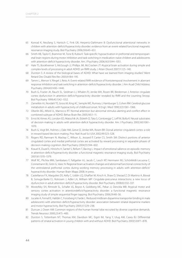
44
65 Konrad K, Neufang S, Hanisch C, Fink GR, Herpertz-Dahlmann B: Dysfunctional attentional networks in children with attention-deficit/hyperactivity disorder: evidence from an event-related functional magnetic resonance imaging study. Biol Psychiatry 2006;59:643–651.
66 Smith AB, Taylor E, Brammer M, Tone B, Rubia K: Task-specific hypoactivation in prefrontal and temporopari-etal brain regions during motor inhibition and task switching in medication-naïve children and adolescents with attention-deficit hyperactivity disorder. Am J Psychiatry 2006;163:1044–1051.
67 Hale TS, Bookheimer S, McGough JJ, Phillips JM, McCracken JT: Atypical brain activation during simple and complex levels of processing in adult ADHD: an fMRI study. J Atten Disord 2007;11:125–140.
68 Durston S: A review of the biological bases of ADHD: What have we learned from imaging studies? Ment Retard Dev Disabil Res Rev 2003;9:184–195.
69 Tamm L, Menon V, Ringel J, Reiss A: Event-related fMRI evidence of frontotemporal involvement in aberrant response inhibition and task switching in attention-deficit/hyperactivity disorder. J Am Acad Child Adolesc Psychiatry 2004;43:1430–1440.
70 Bush G, Frazier JA, Rauch SL, Seidman LJ, Whalen PJ, Jenike MA, Rosen BR, Biederman J: Anterior cingulate cortex dysfunction in attention-deficit/hyperactivity disorder revealed by fMRI and the counting Stroop. Biol Psychiatry 1999;45:1542–1552.
71 Zametkin AJ, Nordahl TE, Gross M, King AC, Semple WE, Rumsey J, Hamburger S, Cohen RM: Cerebral glucose metabolism in adults with hyperactivity of childhood onset. N Engl J Med 1990;323:1361–1366.
72 Oberlin BG, Alford JL, Marrocco RT: Normal attention but abnormal stimulus alerting and conflict effect in combined subtype of ADHD. Behav Brain Res 2005;165:1–11.
73 Ernst M, Kimes AS, London ED, Matochik JA, Eldreth D, Tata S, Contoreggi C, Leff M, Bolla K: Neural substrates of decision-making in adults with attention-deficit hyperactivity disorder. Am J Psychiatry 2003;160:1061–1070.
74 Bush G, Vogt BA, Holmes J, Dale AM, Greve D, Jenike MA, Rosen BR: Dorsal anterior cingulated cortex: a role in reward-based decision-making. Proc Natl Acad Sci USA 2002;99:523–528.
75 Rogers RD, Ramnani N, Mackay C, Wilson JL, Jezzard P, Carter CS, Smith SM: Distinct portions of anterior cingulated cortex and medial prefrontal cortex are activated by reward processing in separable phases of decision-making cognition. Biol Psychiatry 2004;55:594–602.
76 Krauel K, Duzel E, Hinrichs H, Santel S, Rellum T, Baving L: Impact of emotional salience on episodic memory in attention-deficit/hyperactivity disorder: a functional magnetic resonance imaging study. Biol Psychiatry 2007;61:1370–1379.
77 Wolf RC, Plichta MM, Sambataro F, Fallgatter AJ, Jacob C, Lesch KP, Herrmann MJ, Schönfeldt-Lecuona C, Connemann BJ, Grön G, Vasic N: Regional brain activation changes and abnormal functional connectivity of the ventrolateral prefrontal cortex during working memory processing in adults with attention-deficit/hyperactivity disorder. Human Brain Mapp 2008; in press.
78 Castellanos FX, Margulies DS, Kelly C, Uddin LQ, Ghaffari M, Kirsch A, Shaw D, Shezad Z, Di Martino A, Biswal B, Sonuga-Barke EJ, Rotrosen J, Adler LA, Milham MP: Cingulate-precuneus interactions: a new locus of dysfunction in adult attention-deficit/hyperactivity disorder. Biol Psychiatry 2008;63:332–337.
79 Mostofsky SH, Rimrodt SL, Schafer JG, Boyce A, Goldberg MC, Pekar JJ, Denckla MB: Atypical motor and sensory cortex activation in attentiondeficit/hyperactivity disorder: a functional magnetic resonance imaging study of simple sequential finger tapping. Biol Psychiatry 2006;59:48–56.
80 Jucaite A, Fernell E, Halldin C, Forssberg H, Farde L: Reduced midbrain dopamine transporter binding in male adolescents with attention-deficit/hyperactivity disorder: association between striatal dopamine markers and motor hyperactivity. Biol Psychiatry 2005;57:229–238.
81 Duncan J, Owen AM: Common regions of the human frontal lobe recruited by diverse cognitive demands. Trends Neurosci 2000;23:475–483.
82 Durston S, Tottenham NT, Thomas KM, Davidson MC, Eigsti IM, Yang Y, Ulug AM, Casey BJ: Differential patterns of striatal activation in young children with and without ADHD. Biol Psychiatry 2003;53:871–878.
CHAPTER 2
45
83 Rubia K, Smith AB, Brammer MJ, Toone B, Taylor E: Abnormal brain activation during inhibition and error detection in medication-naive adolescents with ADHD. Am J Psychiatry 2005;162:1067–1075.
84 Dickstein SG, Bannon K, Xavier Castellanos F, Milham MP: The neural correlates of attention-deficit hyperactivity disorder: an ALE meta-analysis. J Child Psychol Psychiatry 2006;47:1051–1062.
85 Schneider MF, Krick C, Retz W, Hengesch G, Retz-Junginger P, Reith W, Rösler M: Impairment of fronto-striatal and parietal cerebral networks correlates with attention-deficit hyperactivity disorder psychopathology in adults – a functional magnetic resonance imaging study. Psychiatry Res Neuroimaging 2009 (submitted).
86 Ströhle A, Stoy M, Wrase J, Schwarzer S, Schlangenhauf F, Huss M, Hein J, Nedderhut A, Neumann B, Gregor A, Juckel G, Knutson B, Lehmkuhl U, Bauer M, Heinz A: Reward anticipation and outcomes in adult males with attention-deficit/hyperactivity disorder. Neuroimage 2008;39:966–972.
87 Schulz KP, Fan J, Tang CY, Newcorn JH, Buchsbaum MS, Cheung AM, Halperin JM: Response inhibition in adolescents diagnosed with attention-deficit hyperactivity disorder during childhood: an event related FMRI study. Am J Psychiatry 2004;161:1650–1657.
88 Valera EM, Faraone SV, Biederman J, Poldrack RA, Seidman LJ: Functional neuroanatomy of working memory in adults with attention-deficit/hyperactivity disorder. Biol Psychiatry 2005;57:439–447.
89 Kim BN, Lee JS, Shin MS, Cho SC, Lee DS: Regional cerebral perfusion abnormalities in attention-deficit/hyperactivity disorder. Statistical parametric mapping analysis. Eur Arch Psychiatry Clin Neurosci 2002;252:219–225.
90 Volkow ND, Wang G-J, Fowler JS, Logan J, Angriest B, Hitzemann R, Lieberman J, Pappas N: Effects of meth-ylphenidate on regional brain glucose metabolism in humans: relationship to dopamine D2 receptors. Am J Psychiatry 1997;154:50–55.
91 Volkow ND, Wang GJ, Fowler JS, Hitzemann R, Gatley J, Ding YS, Wong C, Pappas N: Differences in regional brain metabolic responses between single and repeated doses of methylphenidate. Psychiatry Res 1998;83:29–36.
92 Ashtari M, Kumra S, Bhaskar SL, Clarke T, Thaden E, Cervellione KL, Rhinewine J, Kane JM, Adesman A, Milanaik R, Maytal J, Diamond A, Szeszko P, Ardekani BA: Attention-deficit/hyperactivity disorder: a preliminary diffusion tensor imaging study. Biol Psychiatry 2005;57:448–455.
93 Mesulam MM: Large-scale neurocognitive networks and distributed processing for attention, language and memory. Ann Neurol 1990;28:597–613.
94 Corbetta M, Shulman GL: Control of goal-directed and stimulus-driven attention in the brain. Nat Rev Neurosci 2002;3:201–215.
95 Coull JT, Buchel C, Friston KJ, Frith CD: Noradrenergically mediated plasticity in a human attentional neuronal network. Neuroimage 1999;10:705–715.
96 Coull JT, Frith CD, Buchel C, Nobre AC: Orienting attention in time: behavioural and neuroanatomical distinction between exogenous and endogenous shifts. Neuropsychologia 2000;38:808–819.
97 Coull JT, Nobre AC, Frith CD: The noradrenergic α2-agonist clonidine modulates behavioural and neuroana-tomical correlates of human attentional orienting and alerting. Cereb Cortex 2001;11:73–84.
98 Booth JR, Burman DD, Meyer JR, Lei Z, Trommer BL, Davenport ND, Li W, Parrish TB, Gitelman DR, Mesulam MM: Larger deficits in brain networks for response inhibition than for visual selective attention in attention-deficit hyperactivity disorder. J Child Psychol Psychiatry 2005;46:94–111.
99 Silk T, Vance A, Rinehart N, Egan G, O’Boyle M, Bradshaw JL, Cunnington R: Fronto-parietal activation in atten-tion-deficit hyperactivity disorder, combined type: functional magnetic resonance imaging study. Br J Psychiatry 2005;187:282–283.
100 Tamm L, Menon V, Reiss AL: Parietal attentional system aberrations during target detection in adolescents with attention-deficit hyperactivity disorder: event-related fMRI evidence Am J Psychiatry 2006;163:1033–1043.
101 Lou HC, Henriksen L, Bruhn P, Børner H, Nielsen JB: Striatal dysfunction in attention-deficit and hyperkinetic disorder. Arch Neurol 1989;46:48–52.
BRAIN IMAGING IN ADULT ATTENTION DEFICIT HYPERACTIVITY DISORDER (ADHD)
2
46
102 Teicher MH, Anderson CM, Polcari A, Glod CA, Maas LC, Renshaw PF: Functional deficits in basal ganglia of children with attention-deficit/hyperactivity disorder shown with functional magnetic resonance imaging relaxometry. Nat Med 2000;6:470–473.
103 Vaidya CJ, Austin G, Kirkorian G, Ridlehuber HW, Desmond JE, Glover GH, Gabrieli JD: Selective effects of methylphenidate in attention-deficit hyperactivity disorder: a functional magnetic resonance study. Proc Natl Acad Sci USA 1998;95:14494–14499.
104 Scheres A, Milham MP, Knutson B, Castellanos FX: Ventral striatal hyporesponsiveness during reward anticipation in attention-deficit/hyperactivity disorder. Biol Psychiatry 2007;61:720–724.
105 Plichta MM, Vasic N, Wolf RC, Lesch KP, Brummer D, Jacob C, Fallgatter AJ, Grön G: Neural hyporesponsive-ness and hyperresponsiveness during immediate and delayed reward processing in adult attention-deficit/hyperactivity disorder. Biol Psychiatry 2009;65:7–14.
106 Bauer LO, Hesselbrock VM: Brain maturation and subtypes of conduct disorder: interactive effects on p300 amplitude and topography in male adolescents. J Am Acad Child Adolesc Psychiatry 2003;42:106–115.
107 Comings DE, Comings BG: Tourette syndrome clinical and psychological aspects of 250 cases. Am J Hum Genet 1985;37:435–450.
108 Spencer T, Biedermann J, Harding M, Hallett JJ, Laurino JP, Benson M, Kiessling LS: The relationship between tic disorders and Tourette’s syndrome revisited. J Am Acad Child Adolesc Psychiatry 1995;34:1133–1139.
109 Peterson B, Riddle MA, Cohen DJ, Katz LD, Smith JC, Hardin MT, Leckman JF: Reduced basal ganglia volumes in Tourette’s syndrome using three-dimensional reconstruction techniques from magnetic resonance images. Neurology 1993;43:941–949.
110 Singer HS: Neurobiology of Tourette syndrome. Neurol Clin North Am 1997;15:357–378.111 Peterson BS: Neuroimaging studies of Tourette syndrome: a decade of progress. Adv Neurol 2001;85:179–
196.112 Adler CM, Delbello MP, Mills NP, Schmithorst V, Holland S, Strakowski SM: Comorbid ADHD is associated with
altered patterns of neuronal activation in adolescents with bipolar disorder performing a simple attention task. Bipolar Disord Dec 2005;7:577–588.
113 Marsh PJ, Williams LM: ADHD and schizophrenia phenomenology: visual scanpaths to emotional faces as a potential psychophysiological marker? Neurosci Biobehav Rev 2006;30:651–656.
114 Allen G, Courchesne E: Attention function and dysfunction in autism. Front Biosci 2001;6:D105–D119.115 Piven J, Arndt S, Bailey J, Andreasen N: Regional brain enlargement in autism: a magnetic resonance imaging
study. J Am Acad Child Adolesc Psychiatry 1996;35:530–536.116 Baron-Cohen S, Ring HA, Wheelwright S, Bullmore ET, Brammer MJ, Simmons A, Williams SC: Social
intelligence in the normal and autistic brain: an fMRI study. Eur J Neurosci 1999;11:1891–1898.117 Allen G, Muller RA, Courchesne E: Cerebellar function in autism: functional magnetic resonance image
activation during a simple motor task. Biol Psychiatry 2004;56:269–278.118 Haist F, Adamo M, Westerfield M, Courchesne E, Townsend J: The functional neuroanatomy of spatial
attention in autism spectrum disorder. Dev Neuropsychol 2005;27:425–458.
CHAPTER 2

47
BRAIN IMAGING IN ADULT ATTENTION DEFICIT HYPERACTIVITY DISORDER (ADHD)
2


Impairment of fronto-striatal and parietal cerebral networks correlates with Attention Deficit Hyperactivity Disorder (ADHD) psychopathology in adults – a functional magnetic resonance imaging (fMRI) study
Marc F. Schneider, Christoph M. Krick, Wolfgang Retz, Georges Hengesch, Petra Retz-Junginger, Wolfgang Reith, and Michael Rösler
Published in Psychiatry Res (2010) 183(1):75-84.
3 |

50
Abstract
Attention deficit hyperactivity disorder (ADHD) is a common, genetically transmitted childhood onset disorder with a high rate of persistence in adulthood. Although many studies have shown anatomical and functional abnormalities in children and adolescents, studies with adult patients are rare. 19 adults with ADHD (11 ADHD, combined type; 8 ADHD, partially remitted) and 17 controls were included in this functional magnetic imaging (fMRI) study. Brain activation was investigated with a continuous performance test (CPT). Impaired activation of a fronto-striatal and a parietal attentional network was observed during the NoGo condition in ADHD subjects. Correlations of reduced activity of caudate nuclei, anterior cingulate cortex as well as parietal cortical structures and increased activity of insular cortex with inattention and impulsivity symptom scores were found. The activation patterns were similar to those known from children and adolescents with ADHD. In conclusion we found not only a widespread dysfunction of brain regions that are involved in cognitive processing in adults with ADHD compared to controls, but also correlations between symptom severity and dysfunction of neuronal systems across adult subjects without ADHD, childhood but not persistent and persistent ADHD.
CHAPTER 3
51
1. Introduction
Attention deficit hyperactivity disorder (ADHD) is a common, genetically transmitted childhood onset disorder characterized by inattention, hyperactivity and impulsivity (Wender, 1995). According to the dimensional character of ADHD psychopathology, this condition has been suggested rather the extreme variant of a psychopathological continuum than a distinct disorder. Follow-up studies show that up to 60% of patients suffering from this disorder still show some or all of the ADHD characteristics as adults (Barkley, 2002). A cross-national prevalence of 3.4% for adult ADHD has been reported (Fayyad et al., 2007). Regarding the negative impact of ADHD concerning functional impairment and social outcome, and the need for optimizing diagnosis and treatment in adulthood, there is high interest in understanding the pathophysiological mechanisms in ADHD across the lifespan.Structural and functional imaging data are mainly based on studies with ADHD children (Sowell et al., 2003; Pliszka et al., 2006). Studies with functional neuroimaging techniques performed with ADHD children suggest dysfunction of the ventral and dorsolateral prefrontal cortex, the anterior cingulate, insula, amygdala, hippocampus, and ventral striatum (Ernst et al., 2002; Elliot et al., 1999; Rogers et al., 1999; Elliot et al., 2000; Knutson et al., 2001; Bussey et al., 1997; Schultz, 1999). In addition, it has been reported that meth-ylphenidate, which is highly effective in the treatment of ADHD and which exerts its effects via dopaminergic pathways, was able to normalize striatal circuitry function and could improve frontal activation in children and adolescents with ADHD (Vaidya et al., 1998; Shafritz et al., 2004). In detail, imaging data seem to be somewhat divergent and suggest that many brain regions are involved in the pathophysiology of ADHD, which might be at least partially due to small samples investigated and different neuropsychological paradigms applicated. However, there is converging evidence from these studies that behavioural and cognitive problems associated with ADHD are mainly associated with dysfunction of fronto-striatal regions or mainly in the forebrain located regions that influence inhibitory control mechanisms (Durston et al., 2002). In adults with ADHD marked reduction of global metabolism in both hemispheres (Zametkin et al., 1990) and also diminished dopaminergic uptake in the left and medial prefrontal cortex have been reported (Ernst et al., 1998). Similarly to childhood ADHD it has been shown that prefrontal cortex, anterior cingulate cortex and basal ganglia are also involved in adult ADHD (Bush et al., 1999; Ernst et al., 2003; Zametkin et al., 1990). In a functional magnetic resonance imaging (fMRI) study Bush and colleagues (1999) showed that specific regions of Anterior Cingulate Cortex (ACC), which have been shown to be involved in attention focusing and mediation of response selection, were disturbed in patients with ADHD, but a frontostriatal-insular network was activated instead.
CEREBRAL NETWORKS IN ADHD
3

52
Literature gives evidence for negative correlation of ventro-striatal activity with impulsivity and hyperactivity in adolescents and adults with ADHD (Scheres et al., 2007, Ströhle et al., 2008). ADHD patients can also overreact with defined cerebral regions in response to reward outcomes (Ströhle et al., 2008). Thus, recent imaging data underline the central role of fronto-striatal circuits in ADHD including the orbitofrontal, mesial and lateral prefrontal brain regions (Ernst et al., 2003; Durston et al., 2003; Rubia et al., 1999). Bush et al. (2008) pointed out the importance of the dorsal anterior midcingulate cortex that plays a key role in cognition, attention, target detection, motor control, error detection and feedback-based decision making and could show with fMRI technique in adult ADHD that methylphenidate could normalize hypofunction in this area. Due to the still limited number of studies with adults with ADHD and the lack of follow-up investigations of ADHD children, it remains unclear whether disturbed network activation differs between ADHD children and adults and whether clinical remission of ADHD psychopathology runs parallel to normalization of brain function. It should be considered that adults with ADHD might use at least partially compensatory mechanisms which are not yet present in ADHD children.In this study we aimed to investigate activation patterns of attentional networks in adults with ADHD. Due to the dimensional character of the disorder and the decline of symptoms in a considerable proportion of patients with age, we hypothezised that compared to controls there might be not only different activation patterns in ventro-striatal and parietal cortex, but also correlations between symptom severity and brain dysfunction across adult subjects without ADHD, childhood but not persistent and persistent ADHD.
2. Methods
2.1. Subjects Adult patients were recruited from a specialized ADHD outpatient unit of the Neuro- center located at the Saarland University Hospital. ADHD was diagnosed according to DSM-IV criteria after careful clinical investigation. None of the participants of the study had received any kind of stimulant medication before. They were diagnosed for the first time with ADHD. All participants of the study were right handed and without any medication. Subjects had no other psychiatric axis I disorder beside ADHD and no history of any CNS-affection in the 6 months before entering the study. SKID I interviews (Wittchen et al., 1997) were used to exclude relevant actual psychiatric and neurological disorders. IQ was assessed by use of the multiple-choice vocabulary test (MWT-B), which is a verbal test with German norms (Lehrl, 1995).Only ADHD patients with a total score of 30 or more on the German validated form of the Wender-Utah-Rating-Scale (WURS-k; Retz-Junginger et al., 2003) participated in this
CHAPTER 3

53
study, in order to ensure for childhood onset of ADHD symptoms. According to former validation studies, scores above this cut-off are highly indicative for childhood ADHD. Quantification of current ADHD symptoms according to DSM-IV was performed using a standardized and validated self-rating scale for adults (ADHD-SR; Rösler et al., 2004, 2008). The ADHD-SR consists of 18 items according to DSM-IV ADHD symptoms, which are rated on a 0 – 3 Likert scale. The maximum total score is 54 points, those of the inattention subscale 27 (9 items), of the impulsivity subscale 12 (4 items) and for the hyperactivity subscale 15 (5 items), respectively. ADHD-SR total and subscores were used for further correlation analyses of brain activations in the functional magnetic resonance imaging procedures.The term “not persistent ADHD” was used for patients with childhood ADHD who were partially remitted on the syndromatical level as discussed by Biederman and colleagues (2000). These patients have lost the full diagnostic status according to DSM-IV but still showed symptoms of ADHD and functional functional impairment.
2.2. Continuous Performance Test (CPT)According to Barkley (1997) the fundamental deficit in individuals with ADHD is one of self-regulation and the control over behavioral inhibition. It has been argued that this results primarily from frontal lobe dysfunction and not because of adverse environmental conditions. According to this concept, executive dysfunction including working memory, internalization of speech, sense of time and goal directed behavior are secondary psychological and functional problems of impulse control deficit and frontal brain plays a crucial role in its regulation. Therefore, the CPT NoGo condition was chosen as the primary target stimulus in statistical analyses.We used the continuous performance test (CPT), which is conceptualized for investigating sustained attention, vigilance and inhibitory control (Rosvold et al., 1956; Fallgatter and Strik, 1997). Briefly, a pseudorandom sequence of letters was presented 420ms each. Subjects were instructed to push a button of a MRI compatible optical fiber pad, if the letter ‘O’ (predatory condition) was immediately followed by the letter ‘X’ (Go condition), but not when another letter than ‘X’ followed the ‘O’ (NoGo condition). In neurophysi-ological studies a robust physiological frontalization of the brain electrical field during the NoGo condition and reduced frontal brain activity in ADHD children and adults has been shown (Fallgatter et al., 1997, 2004, 2005). CPTs with low rates of signal probability would likely demonstrate different relationships with ADHD symptoms and symptom domains (Conners 1994). But there is a controversial discussion over which CPT gives the best correlates with the ADHD symptoms (Corkum et al., 1993, 1995; Koelega, 1995). In a study examining performance on measures over time, increased mean hit reaction time and standard errors over time was highly associated with most ADHD symptoms (Epstein et al., 2003). In summary there is no “goldstandard” which CPT best fits with ADHD symptoms or subtypes.
CEREBRAL NETWORKS IN ADHD
3

54
The projection of the letters was performed by a computer-guided beamer on a screen while participants watched the screen through a mirror on the head coil positioned above their eyes. The speed of presentation for each letter in the CPT was set near the limit of overall cognitive attention and reaction time capacity. Thus, drifting away of the attention from the task presentation was minimized. Given the fact that eye fields were limited through a mirror system and the narrowed projection into a relative dark environment there were few possibilities to lose the optical focus. An automatic eye tracking system was not used.During fMRI recordings 30 Go events and 30 NoGo events were presented in a pseudor-andomized manner. A total of 285 sequences were recorded.
2.3. Magnetic Resonance Imaging2.3.1. Anatomical sequencesImaging was performed with a 1.5T Siemens Magnetom Vision whole body scanner system. A standard head coil was used. Head position was stabilized with vacuum cushion and fixed with adhesion bands to minimize head movements.For each subject T2-weighted anatomic sequences (data matrix 256 x 256 voxel, 0.93752 mm2 pixel size, 5 mm slice thickness, TR 4.6s, TE 99ms) in plane with the echo planar images were performed. Thus, functional images could be aligned with the three dimensional images. Anatomical abnormalities or symptomatic causes for attention deficit syndromes like liquor circulation disturbances, inflammatory syndromes or stroke could be excluded from the study.
2.3.2. Event related functional MRIGradient-echo EPI sequences with a TE of 60ms, flip angle of 90° and TR of 2.69 sec were recorded. The matrix of 64 x 64 had a field of view of 240mm, resulting in a planar resolution of 3.75 x 3.75 mm. The total number of slices was 24, the slice thickness was 5mm, the interslice gap was 0.5mm.Four volumes were discarded to allow stabilization of magnetization before starting of recording. A total of 285 volumes were recorded, containing 30 Go and 30 NoGo events, each. The runs were completed after 12 minutes.
2.4. Data Processing and AnalysisOffline reconstruction and realignment of functional images were performed using the procedure of Friston et al. (1995) using Statistical Parametric Mapping (SPM 99, The Wellcome Department of Cognitive Neurology, University College London, UK). Prior to data analysis motion artifacts were screened. All subjects with a translational artifact of more than 2.5mm and rotational head movement of more than 2.5° were excluded. In a second step, recordings were corrected for these movement artifacts and then filtered for low frequency signal fluctuations (high pass cut off frequency: 1/123 s-1; low pass cut
CHAPTER 3

55
off period: HRF waveform). Each mean image volume was then spatially normalized into the modified Talairach space employed in SPM 99 to a resulting resolution of 2x2x2mm using both affine and non-linear components. Finally, with a Gaussian kernel of two voxels/ 8x8x8mm spatial smoothing was performed in respect to the Gaussian random field theory.Group comparisons of mean functional images were done by independent two samples t-test for the conditions Go and NoGo, respectively. Correlation analysis of mean functional images with ADHD-self rating scores was done with linear regression analysis (SPM99). If not otherwise mentioned standard p threshold was set at 0.001 (uncorrected). Voxel extension was set at k=5.Correlation analyses were calculated by linear regression implemented in SPM with the activation - t values as the dependent and the explanatory variable of interest as the independent variable. In addition, t values at the respective correlated region of interest (ROI; see below) were extracted from SPM and Spearman correlations with the respective variable of interest were calculated.
3. Results
3.1. Patients characteristics19 subjects with lifetime ADHD (ADHD - 6 male/ 5 female; age 32.6 ± 9.4 years (mean ± SD), range 19-48 years; ADHD partially remitted - 7 male/ 1 female; age 32.4 ± 8.3 years (mean ± SD), range 18 – 45 years) and 17 controls (10 male/ 7 female; age 29.4 ± 8.6 years; range 18-45 years) entered the study. 11 subjects of the ADHD group fulfilled the diagnostic criteria for ADHD, combined type, 8 patients were diagnosed with ADHD, partially remitted according to DSM-IV. Mean IQ of ADHD subjects (106 ± 13 (mean ± SD), range 88-124; ADHD partially remitted 115 ± 18 (mean ± SD), range 95-145) was somewhat lower than mean IQ of the control group (126 ± 12 (mean ± SD), range 100-143), t-test, T=3.3, df(27) p=.003), but all IQs were within normal range. Ratings on the WURS-k and ADHD-SR of ADHD subjects and controls are given in table 1. There was no significant difference of the WURS-k scores between subjects with persisting ADHD and those with ADHD, partially remitted (Mann-Whitney test, Z=-.58, p=.60) whereas the current ADHD symptom scores (ADHD-SR) were significantly higher in the persistent ADHD group compared to subjects with ADHD, partially remitted and controls (Mann Whitney test, Z=-3.4 and -4.4, respectively, p=.000).
3.2. Continuous performance testConcerning results of the CPT, patients with ADHD, combined type made significantly more omission errors and had longer reaction times at the Go condition than controls. There was a non significant trend that patients with persisting ADHD made more
CEREBRAL NETWORKS IN ADHD
3
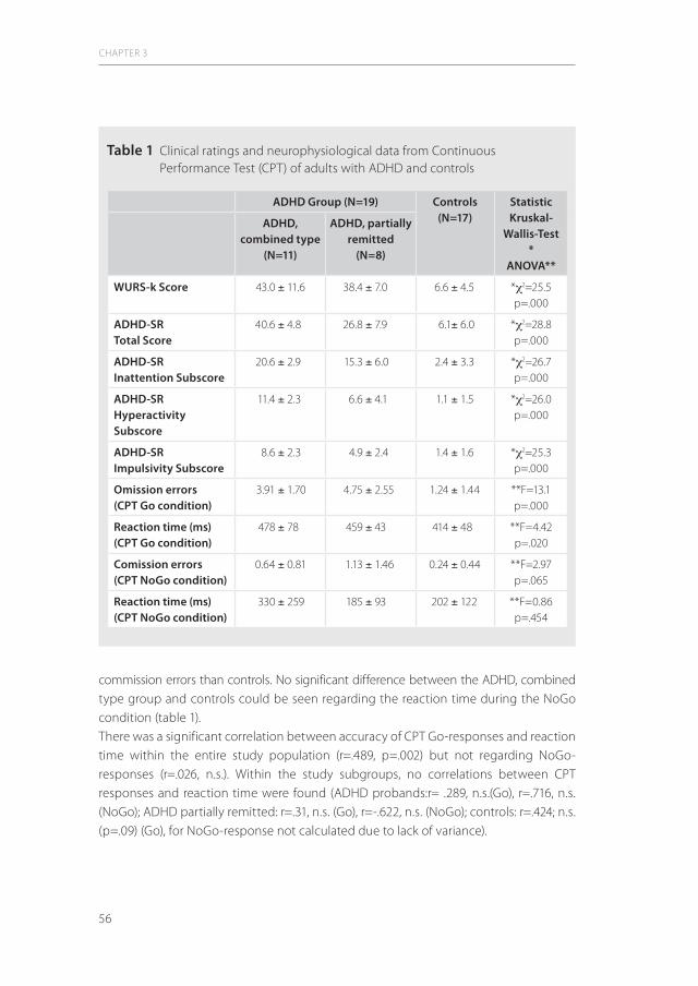
56
commission errors than controls. No significant difference between the ADHD, combined type group and controls could be seen regarding the reaction time during the NoGo condition (table 1).There was a significant correlation between accuracy of CPT Go-responses and reaction time within the entire study population (r=.489, p=.002) but not regarding NoGo- responses (r=.026, n.s.). Within the study subgroups, no correlations between CPT responses and reaction time were found (ADHD probands:r= .289, n.s.(Go), r=.716, n.s. (NoGo); ADHD partially remitted: r=.31, n.s. (Go), r=-.622, n.s. (NoGo); controls: r=.424; n.s. (p=.09) (Go), for NoGo-response not calculated due to lack of variance).
CHAPTER 3
Table 1 Clinical ratings and neurophysiological data from Continuous Performance Test (CPT) of adults with ADHD and controls
ADHD Group (N=19) Controls (N=17)
StatisticKruskal-
Wallis-Test *
ANOVA**
ADHD, combined type
(N=11)
ADHD, partially remitted
(N=8)
WURS-k Score 43.0 ± 11.6 38.4 ± 7.0 6.6 ± 4.5 *c2=25.5p=.000
ADHD-SR Total Score
40.6 ± 4.8 26.8 ± 7.9 6.1± 6.0 *c2=28.8p=.000
ADHD-SR Inattention Subscore
20.6 ± 2.9 15.3 ± 6.0 2.4 ± 3.3 *c2=26.7p=.000
ADHD-SRHyperactivity Subscore
11.4 ± 2.3 6.6 ± 4.1 1.1 ± 1.5 *c2=26.0p=.000
ADHD-SRImpulsivity Subscore
8.6 ± 2.3 4.9 ± 2.4 1.4 ± 1.6 *c2=25.3p=.000
Omission errors(CPT Go condition)
3.91 ± 1.70 4.75 ± 2.55 1.24 ± 1.44 **F=13.1p=.000
Reaction time (ms)(CPT Go condition)
478 ± 78 459 ± 43 414 ± 48 **F=4.42p=.020
Comission errors(CPT NoGo condition)
0.64 ± 0.81 1.13 ± 1.46 0.24 ± 0.44 **F=2.97p=.065
Reaction time (ms)(CPT NoGo condition)
330 ± 259 185 ± 93 202 ± 122 **F=0.86p=.454
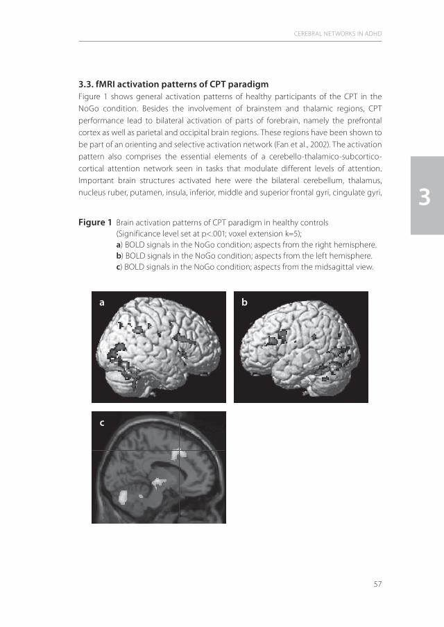
57
3.3. fMRI activation patterns of CPT paradigmFigure 1 shows general activation patterns of healthy participants of the CPT in the NoGo condition. Besides the involvement of brainstem and thalamic regions, CPT performance lead to bilateral activation of parts of forebrain, namely the prefrontal cortex as well as parietal and occipital brain regions. These regions have been shown to be part of an orienting and selective activation network (Fan et al., 2002). The activation pattern also comprises the essential elements of a cerebello-thalamico-subcortico- cortical attention network seen in tasks that modulate different levels of attention. Important brain structures activated here were the bilateral cerebellum, thalamus, nucleus ruber, putamen, insula, inferior, middle and superior frontal gyri, cingulate gyri,
CEREBRAL NETWORKS IN ADHD
3Figure 1 Brain activation patterns of CPT paradigm in healthy controls
(Significance level set at p<.001; voxel extension k=5); a) BOLD signals in the NoGo condition; aspects from the right hemisphere. b) BOLD signals in the NoGo condition; aspects from the left hemisphere. c) BOLD signals in the NoGo condition; aspects from the midsagittal view.
a
c
b

58
middle temporal gyri, respectively. In the parieto-occipital cortex, a posterior attention network activation was seen in the bilateral postcentral gyri, fusiform gyri, middle occipital gyri, and inferior parietal lobules, respectively.
3.4. Group comparison analysisAnalysis of group differences revealed lower NoGo activation in several brain regions of ADHD subjects (figure 2). While the right frontal and parietal lobes were generally less activated in ADHD patients compared to the healthy controls, bilateral parts of the occipital cortex displayed higher activation during CPT paradigm. In group comparison with healthy controls, ADHD patients revealed less activation also in parts of the right fronto-striatal network (Brodman area [BA] 8) and right caudate nucleus as well as right temporal lobe. In the parietal cortex there was less activation in left and right polymodal sensory integration areas (left parietal lobule and right supramarginal gyrus). Detailed information about significant results (p<.001) are given in table 2 a) and b). These activation differences were also evident but less significant with statistical correction with IQ. Lowered temporal brain activity from ADHD patients was then not any more significant.
Group comparison between patients with ADHD combined type and ADHD partially remitted revealed significantly higher activities in the ADHD pr group in the left premotor and prefrontal cortex but no differences in the striatal or parietal cortex activation. Patients with ADHD combined type tended to show higher activation of the right temporal lobe (middle and inferior temporal Gyrus), bilateral cerebellar hemispheres and bilateral paramedial thalamus and finally dorsal pontine brainstem (data not shown). Facing the small number and clinical/ symptomatical similarity from subjects of these groups one can mainly argue from these data that with regard to fronto-striatal and parietal activation patterns there are only few significant differences between the ADHD combined type and the ADHD partially remitted group. Further studies with bigger test populations are needed to analysis these aspects more in detail.
3.5. Correlations of activation patterns with ADHD-SR scoresCerebral activation provoked by the CPT NoGo condition was correlated and anticorrelated with ADHD-SR subscores in several brain regions. The data analysis was focused on neural regions that are involved in attention processes and inhibitory control, i.e. fronto-striatal as well as parietal brain. Brain regions with a significant increase or decrease of activation are listed in tables 3a) to f) in detail. The level of significance was set at p<.001, no correction for multiple tests was performed.
CHAPTER 3

59
CEREBRAL NETWORKS IN ADHD
3
Figure 2 Group contrast of fMRI activation (NoGo condition); a) Grey dots indicate brain regions where controls activate more than ADHD patients. Activity is reported for clusters with a contiguity threshold of k=5 voxels. Brightest gray reflects the threshold of p=.001, whereas black reflects the maximum p level that was found in the whole brain analysis (p=.00001). b) anatomical sections from this group contrast showing left superior parietal lobule and right caudate nucleus: regions were controls activate more than ADHD patients (Significance level set at p<.001; voxel extension k=5)
b
a

60
CHAPTER 3
Table 2 a) Group comparison showing brain regions where ADHD patients revealed lower activation compared to healthy controls (NoGo condition; p<0.001 (uncorrected)
Region Talairach coordinates x,y,z (mm)
Brodman area
Number of voxels
z-score z-score corrected for IQ
Right superior frontal gyrus
38,21,504,40,48
88
1729
3.613.76
n.s. 3.39
Left superior frontal gyrus
-16,-4,54 6 9 3.20 n.s.
Right caudate nucleus (head)
13,15,2 35 3.81 3.22
Right superior temporal gyrus
57,-30,9 42 17 3.49 n.s.
Right middle temporal gyrus
51,-66,18 39 23 3.34 n.s.
Right supramarginal gyrus
54,-51,35 40 38 3.60 3.51
Left superior parietal lobule
-26,-62,51 7 87 3.61 3.25
b) Group comparison showing brain regions where ADHD patients revealed higher activation compared to healthy controls (NoGo condition; p<0.001 (uncorrected)
Region Talairach coordinates x,y,z (mm)
Brodman area
Number of voxels
z-score z-score corrected for IQ
Left fusiform gyrus -20,-82,-9 19 42 4.65 3.56
Right lingual gyrus 2,-87,012,-90,-220,-92,-2
17,18 42 3.67 n.s.
Left lingual gyrus -24,-93,1 18 16 3.59 n.s.
Fusiform gyrus -48,-59,-9 37 5 3.27 n.s.
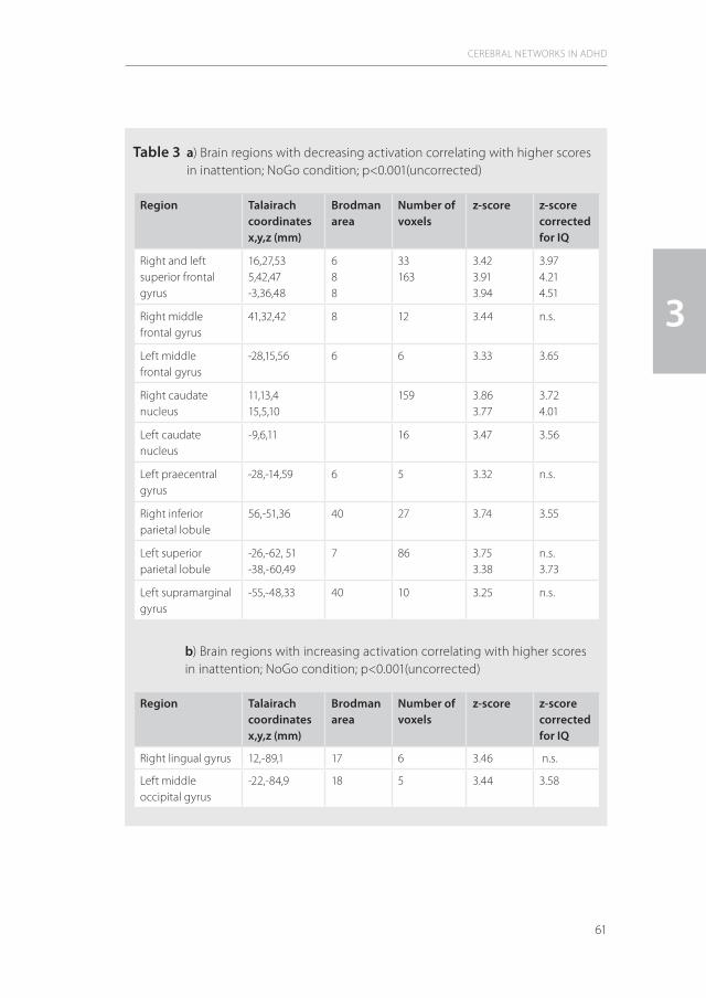
61
CEREBRAL NETWORKS IN ADHD
3
Table 3 a) Brain regions with decreasing activation correlating with higher scores in inattention; NoGo condition; p<0.001(uncorrected)
Region Talairach coordinates x,y,z (mm)
Brodman area
Number of voxels
z-score z-score corrected for IQ
Right and left superior frontal gyrus
16,27,535,42,47-3,36,48
688
33163
3.423.913.94
3.974.214.51
Right middle frontal gyrus
41,32,42 8 12 3.44 n.s.
Left middle frontal gyrus
-28,15,56 6 6 3.33 3.65
Right caudate nucleus
11,13,415,5,10
159 3.863.77
3.724.01
Left caudate nucleus
-9,6,11 16 3.47 3.56
Left praecentral gyrus
-28,-14,59 6 5 3.32 n.s.
Right inferior parietal lobule
56,-51,36 40 27 3.74 3.55
Left superior parietal lobule
-26,-62, 51-38,-60,49
7 86 3.753.38
n.s.3.73
Left supramarginal gyrus
-55,-48,33 40 10 3.25 n.s.
b) Brain regions with increasing activation correlating with higher scores in inattention; NoGo condition; p<0.001(uncorrected)
Region Talairach coordinates x,y,z (mm)
Brodman area
Number of voxels
z-score z-score corrected for IQ
Right lingual gyrus 12,-89,1 17 6 3.46 n.s.
Left middle occipital gyrus
-22,-84,9 18 5 3.44 3.58
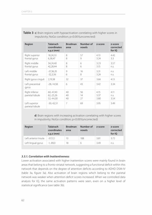
62
3.5.1. Correlation with InattentivenessLower activation associated with higher inattention scores were mainly found in brain areas that belong to a fronto-striatal network, suggesting a functional deficit within this network that depends on the degree of attention deficits according to ADHD DSM-IV (table 3a, figure 3a). Also activation of brain regions which belong to the parietal network was weaker when attention deficit scores increased. When we controlled data analysis for IQ, the same activation patterns were seen, even on a higher level of statistical significance (see table 3b).
CHAPTER 3
Table 3 c) Brain regions with hypoactivation correlating with higher scores in impulsivity; NoGo condition; p<0.001(uncorrected)
Region Talairach coordinates x,y,z (mm)
Brodman area
Number of voxels
z-score z-score corrected for IQ
Right superior frontal gyrus
18,24,536,38,47
88
579
4.133.24
4.423.3
Right middle frontal gyrus
34,24,4042,28,44
88
66
3.233.15
3.57n.s.
Left middle frontal gyrus
-37,36,35-32,3,56
96
168
3.313.24
n.s.n.s.
Right gyrus cinguli 2,19,38 32 37 3.66 4.13
Left precentral gyrus
-28,-14,58 6 43 3.42 3.34
Right inferior parietal lobule
60,-47,4062,-25,2652,-44,30
404040
561427
4.153.373.80
4.11n.s.3.54
Left superior parietal lobule
-30,-62,51 7 69 3.95 3.49
d) Brain regions with increasing activation correlating with higher scores in impulsivity; NoGo condition; p<0.001(uncorrected)
Region Talairach coordinates x,y,z (mm)
Brodman area
Number of voxels
z-score z-score corrected for IQ
Left anterior Insula -37,12,1 13 188 3.81 3.72
Left lingual gyrus -1,-89,0 18 6 3.49 n.s.

63
Increased brain activation was seen in secondary visual areas in lingual gyri (BA 18), arising the hypothesis that with increasing lack of attention there might be more effort in visual polymodal brain areas to try to compensate this lack of attention (table 3b). Non-parametric statistic revealed a significant negative correlation with medium correlation coefficient between left and right caudate nuclei activation and ADHD-SR inattention score (r
left=-.51, p=.002; r
right=-.60, p=.0001; figure). This result is shown in figure 3b.
3.5.2. Correlation with ImpulsivityWhen brain activity was correlated with ADHD-SR impulsivity subscores, also a fronto-parietal network appeared to be impaired with increasing psychopathology (table 3c). Frontal hypofunction comprised parts of the cingular cortex (BA 32) as an important region for integrating decisions and controlling motor execution (figure 4a). In this region
CEREBRAL NETWORKS IN ADHD
3
Table 3 e) Brain regions with decreasing activation correlating with higher scores in hyperactivity; NoGo condition; p<0.001(uncorrected)
Region Talairach coordinates x,y,z (mm)
Brodman area
Number of voxels
z-score z-score corrected for IQ
Right superior frontal gyrus
25,50,3 10 13 3.42 3.98
Left middle frontal gyrusLeft precentral gyrus
-12,1,58-20,-6,58-28,0,59-28,-14,59
6
6
658
3.163.183.213.67
n.s. 3.313.453.23
Left superior parietal lobule
-34,-60,51 7 41 3.76 3.27
f) Brain regions with increasing activation correlating with higher scores in hyperactivity; NoGo condition; p<0.001(uncorrected)
Region Talairach coordinates x,y,z (mm)
Brodman area
Number of voxels
z-score z-score corrected for IQ
Left anterior Insula -42,-8,5 13 73 3.75 n.s.
Right inferior temporal gyrus
45,-9,-20 20 8 3.30 3.70
Left lingual gyrus -1,-87,0 18 6 3.45 n.s.

64
CHAPTER 3
Figure 3 a) Caudate Nuclei. Picture displays anticorrelation of brain activation with increasing ADHD-SR inattention subscore Significance level set at p<.001; voxel extension k=5. b) Scatter plot depicting the anticorrelation between activation in the right caudate and ADHD-SR inattention subscore in adults with ADHD combined type (black triangles), ADHD, partially remitted (open triangles) and normal controls (circles);r=-.60; p=.000.
a
ADHD-SR Inattention Subscore
0,00 5,00 10,00 15,00 20,00 25,00
Righ
t Cau
date
Act
ivit
y
0,60
0,40
0,20
0,00
-0,20
-0,40
b

65
CEREBRAL NETWORKS IN ADHD
3
Figure 4 a) Anterior Cingulate Cortex (ACC/BA 32). Picture displays anticorrelation of brain activity with increasing ADHD-SR impulsivity subscore. b) Scatter plot depicting the anticorrelation between activation in the anterior cingulate cortex (BA 32) and ADHD-SR impulsivity subscore in adults with ADHD combined type (black triangles), ADHD, partially remitted (open triangles) and normal controls (circles); r=-.41; p=.014.
a
ADHD-SR Subscore Impulsivity
0,00 2,00 4,00 6,00 8,00 10,00 12,00
Righ
t ACC
Act
ivit
y
0,50
0,25
0,00
-0,25
-0,50
b

66
CHAPTER 3
Figure 5 a) Left Insular Cortex. Picture displays correlation of brain activity with increasing ADHD-SR impulsivity subscore;. b) Scatter plot depicting the correlation between activation in the left insula and ADHD-SR impulsivity subscore in adults with ADHD combined type (black triangles), ADHD, partially remitted (open triangles) and normal controls (circles);r=.52; p=.001.
a
b
ADHD-SR Impulsivity Subscore
0,00 2,00 4,00 6,00 8,00 10,00 12,00
Left
Insu
la A
ctiv
ity
0,40
0,30
0,20
0,10
0,00
-0,10
-0,20

67
of interest, non-parametric analysis revealed a medium correlation coefficient and a significant negative correlation between ADHD-SR impulsivity subscore and impaired activation (r=-.41, p=0.014), which is shown in figure 4b. Statistical control for IQ resulted in higher significance concerning the lowered activity from the right cingulated gyrus, but the lowered activity in the left middle frontal gyrus with increasing impulsivity was not any longer statistically significant. Increasing activation of left insula and secondary visual fields was detected with increasing impulsivity (figure 5a) also when corrected for IQ. For this region a medium correlation coefficient was found (r=.52, p=.001) (figure 5b).
3.5.3. Correlation with HyperactivityConcerning correlation of ADHD-SR hyperactivity subscore with activation of brain regions displayed a pattern similar to that found for inattention measures (tables 3e and 3f). Decreased activation of predominantly left frontal and parietal brain areas and an increased activation of insular cortex and gyrus lingualis were correlated with hyper -activity scores. When controlled for IQ, no correlation with left anterior insula and left lingual gyrus activation was found.
4. Discussion
According to our hypothesis of altered activation patterns in adults with ADHD, we found in this fMRI study correlates of decreased function within fronto-striatal and parietal brain networks associated with ADHD psychopathology. These findings corroborate functional imaging evidence of disturbed fronto-parietal networks, which has been described in adolescents with ADHD (Silk et al., 2005). In agreement with the literature concerning anatomical and functional changes within the brain of children, adolescents and adults with ADHD, the brain regions suggested to be involved in ADHD pathophysiology were found to be affected also in our study. These regions of interest are the anterior cingulate cortex (ACC), the prefrontal cortex, striatum, insula, the parietal cortex and cerebellum (Makris et al., 2007; Schneider et al., 2006). Makris et al. (2007) reported thinning of prefrontal and parietal cortical regions belonging to attention and executive networks in adult ADHD and of the ACC in particular. Also Seidman and colleagues (2006) could show in adult ADHD patients morphological alterations of the ACC and prefrontal cortex. The ACC is the largest component of the limbic system. It plays a crucial role in the coordination of complex cognitive functions and in motor control. Two functional subdivisions of the ACC are specialized in “cognitive/attentional” and “affective emotional” information processing (Bush et al., 1998, 1999). Bush et al. (1999) found that ACC activation in adult ADHD patients was specifically impaired during the interference condition of a counting stroop task. They
CEREBRAL NETWORKS IN ADHD
3

68
could also show improvement of ACC activity after 6 weeks of methylphenidate therapy (Bush et al., 2008). ACC dysfunction has been reported also in ADHD children (Rubia et al., 1999; Durston et al., 2003). In adult patients with ADHD dysfunction of ACC has been demonstrated also with positron emission tomography (Ernst et al., 2003) and neuro-physiological techniques (Fallgatter et al., 2005).Alike, also striatal structures have been suggested to play a crucial role in ADHD patho-physiology. An elevated dopamine transporter density in the basal ganglia compared to healthy controls has been reported in ADHD adults (Krause et al., 2000, 2005; Dougherty et al., 1999). Using positron emission tomography (PET) Zametkin and colleagues (1990) demonstrated decreased metabolism of caudate nuclei during cognitive tasks in adults with ADHD. In functional imaging studies decreased activation of the striatum in ADHD has been described (Lou et al., 1984, 1989; Vaidya et al., 1998; Durston et al., 2003). Ströhle and colleagues (2008) described decreased ventral striatal activation during a reward task in ADHD, underlining the pathophysiological importance of fronto-striatal structures in ADHD also in the frame of reward paradigms. The diminished bilateral activation of caudate nuclei found in this study is in line with these findings and support the notion of disturbed dopaminergic transmission in adult ADHD patients. A robust finding in our study was a dysfunction of the parietal cortex in adults with ADHD compared to controls. Diminished activation of the parietal lobule with increasing psychopathology was found throughout all ADHD-SR subscores. Only a handful of studies have addressed to parietal lobe structure and function in ADHD so far (Tamm et al., 2006; Smith et al., 2006), although this brain area plays an important role in orienting and selective attention networks (Fan et al., 2005). Posner et al. (1990) have described a posterior attention system located in the parietal lobe, which seems to be mainly modulated by noradrenergic transmission in contrast to the predominantly dopaminergic modulation of the frontal attention system. The posterior parietal cortex disengages the attentional focus from the contralateral target (Rafal et al., 1995), and lesions at this region can lead to impaired attention (Posner et al., 1995; Ro et al., 2001). Given its importance in visuo-spatial orienting and as a region for polymodal sensory integration, the parietal brain seems to be generally underestimated in context with attention deficits in ADHD.A lower metabolism and smaller volume in parietal regions has been shown in ADHD children (Castellanos et al., 2002, Filipek et al., 1997, Ernst et al., 1997). Konrad et al. (2006) have used fMRI to investigate brain activations related to these particular aspects of attention. In that study parietal brain regions were less recruited by ADHD compared to control children. Task-specific abnormalities of parietal and temporal cortices in children and adolescents with ADHD were also demonstrated by Smith and colleagues (2006). Booth and coworkers (2005) could show in children with ADHD that a small region of the superior parietal lobule displayed a weaker activation during a selective attentional task. The findings of our study compare to these reports and underline the role of
CHAPTER 3
69
parietal cortical dysfunction even in adult ADHD. Moreover, these findings lead to the hypothesis that in addition to the well described frontal brain regions also polymodal integrative cortical functions of the parietal cortex are impaired in ADHD pathology. The dysfunction in this brain area might lead to difficulties in integrating different inputs to an executive program and its inhibition. Interestingly, we did not only find decreased activation of fronto-striatal and parietal attention systems in ADHD subjects, but also an increase of activation of some brain regions. Activation of the insular cortex in adult ADHD has been found in a prior fMRI study in which a counting Stroop paradigma was used (Bush et al., 1999). It has been speculated that the “insular pathway” might present an alternative to the activation of ACC and the prefrontal network during attention tasks (Bush et al., 1999). Thus, adults with ADHD might rather activate a different neuronal network than just use a given brain network that works insufficiently. Another hypothesis might be the higher anticipatory fear during task performance because of cognitive and inhibitory impairments (Chua et al., 1999). Wager and Barrett (2008) could demonstrate that shifting attention and executive working memory activate anterior insulae. This could be enhanced with emotional tasks. The authors found that recall generated emotion activated especially the left insula. Thus, it should be also considered that ADHD patients might tend to subvocalize during their task, which might be an attempt to compensate inhibitory dyscontrol resulting from hyperactivity and impulsivity (Morris et al., 1999). Fear and subvocalization have been post-hoc reported from some individuals in our study, but this was not systematically documented.The results of this study indicate that ADHD associated dysfunction of attention networks might persist across the lifespan. Even in those individuals with a history of childhood but partially remitted ADHD in adulthood displayed abnormalities similar to those of subjects with persisting ADHD, combined type. Assuming that remission of ADHD psychopathology in these subjects is not a diagnostic artefact due to inappropriate diagnostic criteria for adult ADHD, it might be concluded that clinical remission depends on the individual’s capability to compensate persisting dysfunction of attentional networks via activation of alternative pathways.In this study we did not only compare brain activation patterns of adults suffering from ADHD with healthy controls during a cognitive task, but also looked for correlations between regional brain dysfunction and the degree of psychopathological abnormalities across the ADHD symptom domains. By doing this, we found that decrease of activation of frontal and parietal cortical areas was associated with an increase throughout the psychopathological domains of ADHD, inattention, hyperactivity and impulsivity. However, also differential effects were observed regarding the correlations between ACC dysfunction and impulsivity and between basal ganglia activation and inattention, supporting the concept of syndromatical ADHD subtypes (Biederman et al., 2000) and the hypothesis of independent changes of inattention and
CEREBRAL NETWORKS IN ADHD
3

70
hyperactivity-impulsivity symptom domains with age on a neuronal basis. However, it appears to be important to note that the correlations found in this study reflect a direct association between the severity of the clinical syndrome and the neuronal substrate of ADHD and underline the dimensional character of this disorder also in terms of their neuronal pathology. Several lines of evidence support the concept of ADHD as a dimensional disorder. For example, subthreshold ADHD cases showed fewer deviations regarding personality traits compared to full ADHD subjects, suggesting to present a milder form of ADHD that is consistent with dimensional views of this disorder (Faraone et al., 2009). Likewise, latent class analyses show that ADHD is best modeled as a continuum that is not caused by discrete dysfunction (Haslam et al., 2006). Further support comes from genetic studies, which have demonstrated the appropriateness of quantitative loci approaches in ADHD research, indicating no threshold effects on the risk for ADHD (Chen et al., 2008). DSM-IV refers to the dimensional character of the disorder by requiring at least 6 of 9 typical criteria of inattention and/or 6 of 9 of hyperactive-impulsive features. If less than at least 6 criteria of either inattention or hyperactive-impulsive behavior are observed, no diagnosis of ADHD can be made, even if some characteristics of the disorder are present. According to DSM-IV, adults who suffered from ADHD in childhood but do not longer fulfill at least 6 items of either syndrome domain are diagnosed with ADHD, partially remitted. Thus, the diagnoses of ADHD, partially remitted directly refers to the dimensional character of this disorder and reflects the well known decline of the disorder with age, at least on a diagnostic and to lesser extend to a syndromatic level (Biederman et al., 2000). Concerning the dimensional character of attention, motor activity and impulsive behavior in general, and the lack of specific psychopathological ADHD features, a syndromatic continuum from healthy persons to those with sub-syn-dromatic and full ADHD can be assumed. A limitation of this study is the recruitment of the patients from a specialized ambulance for ADHD and the high degree of preselection regarding psychiatric comorbidity and premedication. Therefore, the ADHD patients in this study might be not representative for typical adults with ADHD. However, the relatively low treatment prevalence of ADHD children in the past and the still lacking licence for any ADHD medication for ADHD adults in Germany allowed to include a quite homogeneous group of never medicated, but even moderately to severely affected ADHD patients in this study. Moreover, the limited number of ADHD patients in this study did not allow differentiat-ing gender effects on ADHD neurophysiology. Also, no individuals with ADHD, inattentive or hyperactive-impulsive type underwent imaging procedures. The effects of these ADHD subtypes on attention network activation could thus not be worked out. Further studies with specific design will be necessary to investigate these aspects. Hyperactivity of some ADHD patients led to the problem that movement artefacts prohibited from fMRI data analyzing procedures. Especially the severely hypermotoric
CHAPTER 3
71
ADHD patients had to be excluded from the study which could lead to a systematic bias. Finally, lacking structural data in this population, functional impairments might be confounded with structural differences between ADHD patients and controls, as they also concern the typical neural networks affected in attention processing (Makris et al., 2007).In conclusion the results of this study give evidence to the hypothesis of dysfunction of fronto-striatal as well as parietal attention networks in adult ADHD. Moreover, the results underline the notion that ADHD is a dimensional disorder and that the severity of symptoms correlates with the degree of dysfunction of these networks. The mechanisms underlying ADHD in adulthood seem to be very similar to those in childhood and adolescence, suggesting that there might not be a qualitative difference between childhood and adulthood regarding ADHD pathophysiology. In addition, adults diagnosed with ADHD, partially remitted according to DSM-IV displayed activation patterns that resembled those of the persistent ADHD subjects and therefore seem to close the pathophysiological gap between healthy adults and adults with persisting ADHD, combined type.
CEREBRAL NETWORKS IN ADHD
3
72
References
Barkley, R.A.,1997. ADHD and the nature of self-control. Guilford Press.Barkley, R.A.,2002. Major life activity and health outcomes associated with attention-deficit/hyperactivity
disorder. Journal of Clinical Psychiatry 63 (Supplement 12),10–15.Biederman, J., Mick, E., Faraone, S.V., 2000. Age-dependent decline of symptoms of Attention Deficit Hyperactivity
Disorder: Impact of remission definition and symptom type. American Journal of Psychiatry 157, 816-818.Booth, J.R., Burman, D.D., Meyer, J.R., Lei, Z., Trommer, B.L., Davenport, N.D., Li, W., Parrish, T.B., Gitelman, D.R.,
Mesulam, M.M., 2005. Larger deficits in brain networks for response inhibition than for visual selective attention in attention deficit hyperactivity disorder (ADHD). Journal of Child Psychology and Psychiatry. 46(1),94-111.
Bush, G., Frazier, J.A., Rauch, S.L., Seidman, L.J., Whalen, P.J., Jenike, M.A., et al., 1999. Anterior cingulate cortex dysfunction in attention deficit/hyperactivity disorder revealed by fMRI and the counting stroop. Biological Psychiatry 45,1542–1552.
Bush, G., Whalen, P.J., Rosen, B.R., Jenike, M.A., McInerney, S.C., Rauch, S.L., 1998. The counting Stroop: an interference task specialized for functional neuroimaging-validation study with functional MRI. Human Brain Mapping 6(4),270-82.
Bush, G., 2008. Neuroimaging of attention deficit hyperactivity disorder: can new imaging findings be integrated in clinical practice? Child and Adolescent Psychiatric Clinics of North America. 17(2), 385-404.
Bush, G., Spencer, T.J., Holmes, J., Shin, L.M., Valera, E.M., Seidman, L.J., Makris, N., Surman, C., Aleardi, M., Mick, E., Biederman, J., 2008. Functional magnetic resonance imaging of methylphenidate and placebo in attention-deficit/hyperactivity disorder during the multi-source interference task. Archives of General Psychiatry. 65(1),102-14.
Bussey, T.J., Everitt, B.J., Robbins, T.W., 1997. Dissociable effects of cingulate and medial frontal cortex lesions on stimulus-reward learning using a novel Pavlovian autoshaping procedure for the rat: implications for the neurobiology of emotion. Behavioural Neurosciences 111,908–919.
Castellanos, F.X., Lee, P.P., Sharp, W., Jeffries, N.O., Greenstein, D.K., Clasen, L.S., et al., 2002. Developmental trajectories of brain volume abnormalities in children and adolescents with attention-deficit/hyperactivity disorder. JAMA 288,1740 –1748.
Chen, W., Zhou, K., Sham, P., Franke, B., Kuntsi, J., Campbell, D., Fleischman, K., Knight, J., Andreou, P., Arnold, R., et al., 2008. DSM-IV combined type ADHD shows familial association with sibling trait scores: a sampling strategy for QTL linkage. American Journal of Medical Genetics (B Neuropsychiatric Genetics) 147B, 1450-1460.
Chua, P., Krams, M., Toni, I., Passingham, R., Dolan, R., 1999. A functional anatomy of anticipatory anxiety Neuroimage 9,563-71.
Conners,, C. K., 1994. The Conners Continuous Performance Test. Toronto, Canada: Multi-Health Systems.Corkum, P.V., Siegel, L.S., 1993. Is the Continuous Performance Task a valuable research tool for use with children
with Attention-Deficit-Hyperactivity Disorder? Journal of Child Psychology and Psychiatry 34(7),1217-39. Corkum, P.V., Siegel, L.S., 1995. Reply to Dr. Koelega: Is the Continuous Performance Task a valuable research tool
for use with children with Attention-Deficit-Hyperactivity Disorder? Journal of Child Psychology and Psychiatry 36(8),1487-1493.
Dougherty, D.D., Bonab, A.A., Spencer, T.J., Rauch, S.L., Madras, B.K., Fischman, A.J., 1999. Dopamine transporter density in patients with attention deficit hyperactivity disorder. Lancet 354(9196),2132-3.
Durston, S., Thomas, K.M., Worden, M.S., Yang, Y., Casey, B.J., 2002. The effect of preceding context on inhibition: an event-related fMRI study. Neuroimage 16(2),449-53.
Durston, S., Tottenham, N.T., Thomas, K.M., Davidson, M.C., Eigsti, I.M., Yang, Y., Ulug, A.M., Casey, B.J., 2003. Differential patterns of striatal activation in young children with and without ADHD. Biological Psychiatry 53(10),871-8.
Elliott, R., Friston, K.J., Dolan, R.J., 2000. Dissociable neural responses in human reward systems. Journal of Neuroscience 20,6159–6165.
Elliott, R., Rees, G., Dolan, R.J., 1999. Ventromedial prefrontal cortex mediates guessing. Neuropsychologia 37,403–411.
CHAPTER 3

73
Epstein, J.N., Erkanli, A., Conners, C.K., Klaric, J., Costello, J.E., Angold, A., 2003. Relations between Continuous Performance Test performance measures and ADHD behaviors. Journal of Abnormal Child Psychology 31(5),543-54.
Ernst, M., Bolla, K., Mouratidis, M., Contoreggi, C., Matochik, J.A., Kurian, V., Cadet, J.L., Kimes, A.S., London, E.D., 2002. Decision-making in a risk-taking task: a PET study. Neuropsychopharmacology 26,682–691.
Ernst, M., Cohen, R.M., Liebenauer, L.L., Jons, P.H., and Zametkin, A.J., 1997. Cerebral glucose metabolism in adolescent girls with attention-deficit/ hyperactivity disorder. Journal of the American Academy of Child and Adolescent Psychiatry 36,1399–1406.
Ernst, M., Kimes, A.S., London, E.D., Matochik, J.A., Eldreth, D., Tata, S., Contoreggi, C., Leff, M., Bolla, K., 2003. Neural substrates of decision making in adults with attention deficit hyperactivity disorder. American Journal of Psychiatry 160(6),1061-70.
Ernst, M., Zametkin, A.J., Matochik, J.A., Jons, P.H., Cohen, R.M., 1998. DOPA decarboxylase activity in attention deficit hyperactivity disorder adults. A [fluorine-18]fluorodopa positron emission tomographic study. Journal of Neuroscience 18(15),5901-7.
Fallgatter, A.J., Brandeis, D., Strik, W.K., 1997. A robust assessment of the NoGoanteriorization of P300 microstates in a cued continuous performance test. Brain Topography 9, 295–302.
Fallgatter, A.J., Ehlis, A.C., Seifert, J., Strik, W.K., Scheuerpflug, P., Zillessen, K.E., Herrmann, M.J., Warnke, A.., 2004. Altered response control and anterior cingulate function in attention-deficit/hyperactivity disorder boys. Clinical Neurophysiology 115, 973-81.
Fallgatter, A.J., Ehlis, A.C., Rösler, M., Strik, W.K., Blocher, D., Herrmann, M.J., 2005. Diminished prefrontal brain function in adults with psychopathology in childhood related to attention deficit hyperactivity disorder. Psychiatry Research 138, 157-69.
Fan, J., Bruce, T., McCandliss, D., Fossella, J., Flombaum, J., and Posner, M.I., 2005. The activation of attentional networks. Neuroimage 26,471–479.
Fan, J., McCandliss, B.D., Sommer, T., Raz, A., Posner, M.I., 2002. Testing the efficiency and independence of attentional networks. Journal of Cognitive Neuroscience. 14(3),340-7.
Faraone, S.V., Kunwar, A., Adamson, J., Biederman, J., 2009. Personality traits among ADHD adults: implications of late-onset and subthreshold diagnoses. Psychological Medicine 39, 685-693.
Fayyad, J., De Graaf, R., Kessler, R., Alonso, J., Angermeyer, M., Demyttenaere, K., De Girolamo, G., Haro, J.M., Karam, E.G., Lara, C., Lépine, J.P., Ormel, J., Posada-Villa, J., Zaslavsky, A.M., Jin, R.,, 2007. Cross-national prevalence and correlates of adult attention-deficit hyperactivity disorder. British Journal of Psychiatry 190,402-9.
Filipek, P.A., Semrud-Clikeman, M., Steingard, R., and Kennedy, D., Biederman, J., 1997. Volumetric MRI analysis: Comparing subjects having attention-deficit hyperactivity disorder with normal controls. Neurology 48,589 –601.
Friston, K.J., Holmes, A.P., Poline, J.B., Grasby, P.J., Williams, S.C., Frackowiak, R.S., Turner, R., 1995. Analysis of fMRI time-series revisited. Neuroimage 2(1),45-53.
Haslam, N., Williams, B., Prior, M., Haslam, R., Graetz, B., Sawyer, M., 2006. The latent structure of attention-deficit/hyperactivity disorder: a taxometric analysis. Australian and New Zealand Journal of Psychiatry 40, 639-647.
Knutson, B., Adams, C.M., Fong, G.W., Hommer, D., 2001. Anticipation of increasing monetary reward selectively recruits nucleus accumbens. Journal of Neuroscience, 21,RC159.
Koelega, H.S , 1995. Is the Continuous Performance Task useful in research with ADHD children? Comments on a review. Journal of Child Psychology and Psychiatry 36(8),1477-85; discussion 1487-93.
Konrad, K., Neufang, S., Hanisch, C., Fink, G.R., Herpertz-Dahlmann, B., 2006. Dysfunctional Attentional Networks in Children with Attention Deficit/Hyperactivity Disorder: Evidence from an Event-Related Functional Magnetic Resonance Imaging Study. Biological Psychiatry 59(7),643-651.
Krause, J., la Fougere, C., Krause, K.H., Ackenheil, M., Dresel, S.H., 2005. Influence of striatal dopamine transporter availability on the response to methylphenidate in adult patients with ADHD. European Archives of Psychiatry and Clinical Neurosciences 255(6),428-31.
CEREBRAL NETWORKS IN ADHD
3

74
Krause, K.H., Dresel, S.H., Krause, J., Kung, H.F., Tatsch, K., 2000. Increased striatal dopamine transporter in adult patients with attention deficit hyperactivity disorder: effects of methylphenidate as measured by single photon emission computed tomography. Neuroscience Letters. 285(2),107-10.
Lehrl, S., 1995. Mehrfachwahl-Wortschatz-Intelligenztest MWT-B. Manual zum MWTB. Medizinische Verlagsge-sellschaft mbH Balingen.
Levy, F., Hay, D.A., McStephen, M., Wood, C., Waldman, I., 1997. Attention-deficit hyperactivity disorder: a category or a continuum? Genetic analysis of a large-scale twin study. Journal of American Academy of Child and Adolescence Psychiatry. 36(6),737-44.
Lou, H. C., Henriksen, L., Bruhn, P., 1984. Focal cerebral hypoperfusion in children with dysphasia and/or attention deficit disorder. Archives of Neurology 41,825–829.
Lou, H. C., Henrikson, L., Bruhn, P., Berner, H., Nielsen, J.B, 1989. Striatal dysfunction in attention deficit and hyperkinetic disorder. Archives of Neurology 46(1),48-52.
Makris, N., Biederman, J., Valera, E.M., Bush, G., Kaiser, J., Kennedy, D.N., Caviness, V.S., Faraone, S.V., Seidman, L.J., 2007. Cortical thinning of the attention and executive function networks in adults with attention-deficit/hyperactivity disorder. Cerebral Cortex 17(6),1364-75.
Morris, J.S., Scott, S.K., Dolan, R.J., 1999. Saying it with feeling: neural responses to emotional vocalizations. Neuro-psychologia 37(10),1155-63.
Pliszka, S.R., Glahn, D.C., Semrud-Clikeman, M.S., Franklin, C., Perez, R., Xiong, J., Liotti, M., 2006. Neuromaging of inhibitory control areas in children with attention deficit hyperactivity disorder who were treatment naive or in long-term treatment. American Journal of Psychiatry 163(6), 1052-1060.
Posner, M.I., 1995. Attention in cognitive neuroscience: an overview. In Gazzaniga MS (ed.): The cognitive neurosciences. MIT Press, Cambridge MA, 615-624.
Posner, M.I., Petersen, S.E., 1990. The attention system of the human brain. Annual Revues of Neuroscience 13,25– 42.Rafal, R., Robertson, L., 1995. The neurology of visual attention. In: Gazzaniga MS (ed.) The cognitive neurosciences.
MIT Press , Cambridge, MA, 625-648.Retz-Junginger, P., Retz, W., Blocher, D., Stieglitz, R.D., Georg, T., Supprian, T., Wender, P.H., Rösler, M., 2003. Reliability
and validity of the Wender-Utah-Rating-Scale short form. Retrospective assessment of symptoms for attention deficit/hyperactivity disorder. Nervenarzt 74(11),987-93.
Ro, T., Borden, C., Driver, J., Rafal, R., 2001. Ipsilesional biases in saccades but not perception after lesions of the human inferior parietal lobule. Journal of Cognitive Neurosciences (13),920-929.
Rogers, R.D., Owen, A.M., Middleton, H.C., Williams, E.J., Pickard, J.D., Sahakian, B.J., Robbins, T.W., 1999. Choosing between small, likely rewards and large, unlikely rewards activates inferior and orbital prefrontal cortex. Journal of Neuroscience 19,9029–9038.
Rösler, M., Retz, W., Retz-Junginger, P., Thome, J., Supprian, T., Nissen, T., Stieglitz, R.D., Blocher, D., Hengesch, G., Trott, G.E., 2004. Tools for the diagnosis of attention-deficit/hyperactivity disorder in adults. Self-rating behaviour questionnaire and diagnostic checklist. Nervenarzt 75(9),888-95.
Rösler, M., Retz, W., Retz-Junginger, P., Stieglitz, R.D., Kessler, H., Reimherr, F., Wender, P.H., 2008. Attention deficit hyperactivity disorder in adults: Benchmarking diagnosis using the Wender-Reimherr adult rating scale. Nervenarzt. 79(3),320-7.
Rosvold, H.E., Mirsky, A.F., Sarason, I., Bransome, E.D., Beck, L.H., 1956. A continuous performance test of brain damage. Journal of Consulting and Psychology 20,343-350.
Rubia, K., Overmeyer, S., Taylor, E., Brammer, M., Williams, S.C., Simmons, A., Bullmore, E.T., 1999. Hypofrontality in attention deficit hyperactivity disorder during higher-order motor control: a study with functional MRI. American Journal of Psychiatry 156(6),891-6.
Scheres, A., Milham, M.P., Knutson, B., Castellanos, F.X., 2007. Ventral striatal hyporesponsiveness during reward anticipation in attention-deficit/hyperactivity disorder. Biological Psychiatry 61(5),720-4.
Schneider, M., Retz, W., Coogan, A., Thome, J., Rösler, M., 2006. Anatomical and functional brain imaging in adult attention-deficit/hyperactivity disorder (ADHD)-A neurological view. European Archives in Psychiatry and Clinical Neurosciences 256 Suppl 1, i32–i41.
CHAPTER 3

75
Schultz, W., 1999. The reward signal of midbrain dopamine neurons. News in Physiological Sciences 14,249–255.Seidman, L.J., Valera, E.M., Makris, N., Monuteaux, M.C., Boriel, D.L., Kelkar, K., Kennedy, D.N., Caviness, V.S., Bush, G.,
Aleardi, M., Faraone, S.V., Biederman, J., 2006. Dorsolateral prefrontal and anterior cingulate cortex volumetric abnormalities in adults with attention-deficit/hyperactivity disorder identified by magnetic resonance imaging. Biological Psychiatry 60(10),1071-80.
Shafritz, K.M., Marchione, K.E., Gore, J.C., Shaywitz, S.E., Shaywitz, B.A., 2004. The effects of methylphenidate on neural systems of attention in attention deficit hyperactivity disorder. American Journal of Psychiatry 161(11),1990-1997.
Silk, T., Vance, A., Rinehart, N., Egan, G., O’Boyle, M., Bradshaw, J.L., Cunnington, R., 2005. Fronto-parietal activation in attention-deficit hyperactivity disorder, combined type: functional magnetic resonance imaging study. British Journal of Psychiatry 187,282-3.
Smith, A.B., Taylor, E., Brammer, M., Tone, B., Rubia, K., 2006. Task-specific hypoactivation in prefrontal and tempo-roparietal brain regions during motor inhibition and task switching in medication-naïve children and adolescents with attention deficit hyperactivity disorder. American Journal of Psychiatry 163,1044-1051.
Sowell, E.R., Thompson, P.M., Welcome, S.E., Henkenius, A.L., Toga, A.W., Peteron, B.S., 2003. Cortical abnormalities in children and adolescents with attention-deficit hyperactivity disorder. Lancet 362,1699 –1707.
Ströhle, A., Stoy, M., Wrase, J., Schwarzer, S., Schlagenhauf, F., Huss, M., Hein, J., Nedderhut, A., Neumann, B., Gregor, A., Juckel, G., Knutson, B., Lehmkuhl, U., Bauer, M., Heinz, A., 2008. Reward anticipation and outcomes in adult males with attention-deficit/hyperactivity disorder. Neuroimage 39(3),966-72.
Tamm, L., Menon, V., Reiss, A., 2006. Parietal attetnional system aberrations during target detection in adolescents with attention deficit hyperactivity disorder: event-related fMRI evidence. American Journal of Psychiatry 163(6),1033-1043.
Vaidya, C.J., Austin, G., Kirkorian, G., Ridlehuber, H.W., Desmond, J.E., Glover, G.H., Gabrieli, J.D., 1998. Selective effects of methylphenidate in attention deficit hyperactivity disorder: a functional magnetic resonance study. Proceedings of the National Academy of Sciences USA. 95(24),14494-9.
Wager, T.D., Feldman Barrett, L., 2004. From affect to control: functional specialization of the insula in motivation and regulation. Published online at PsycExtra.
Wender, P.H., 1995. Attention-deficit hyperactivity disorder in adults. Oxford University Press, Oxford/ New York.Wittchen, H., Zaudig, M., Fydrich, T., 1997. SKID – strukturiertes klinisches Interview für DSM-IV. Hogrefe Verlag,
Göttingen.Zametkin, A.J., Nordahl, T.E., Gross, M., King, A.C., Semple, W.E., Rumsey, J., Hamburger, S., Cohen, R.M., 1990.
Cerebral glucose metabolism in adults with hyperactivity of childhood onset. New England Journal of Medicine 323(20),1361-6.
CEREBRAL NETWORKS IN ADHD
3

Impaired cortical inhibition in adult ADHD patients: a study with transcranial magnetic stimulation
Marc Schneider, Wolfgang Retz, Christine Freitag, Jennifer Irsch, Patrizia Graf, Petra Retz-Junginger, and Michael Rösler
Published in J Neural Transm Suppl (2007) 72:303-9.
4 |

78
Abstract
The aim of this study was to analyze motor inhibition and facilitation of adult ADHD patients using double pulse transcranial magnetic stimulation (TMS). Twenty-six right handed adult ADHD patients according to DSM-IV were investigated and compared to 26 age and sex-matched controls. In the left hemisphere, mean motor inhibition was 0.53 ± 0.33 (mean ± SD) in ADHD patients and 0.34 ± 0.16 (mean ± SD) in controls (p=0.012). There were no significant differences in motor excitability concerning facilitation or in the right hemisphere. Decreased motor inhibition correlated with a higher symptom score derived from the Wender Reimherr Interview (WRI) (rho=0.28; p=0.04) and also with self rated hyperactivity/impulsivity symptoms (rho=0.30; p = 0.03).In conclusion, decreased motor inhibition in adult ADHD corroborate similar findings in children with ADHD (Moll et al. 2000) and reflect disturbed impulsivity and hyperactivity on a neurophysiological level.
CHAPTER 4
79
Introduction
The Attention Deficit-/ Hyperactivity Disorder (ADHD) is one of the most frequent disorders not only in child and adolescent psychiatry but also in general psychiatry. The prevalence in adulthood has been determined as 4% (Kessler et al. 2006). According to DSM-IV the core psychopathology in all ages comprises hyperactivity, impulsivity and attention deficits. These symptoms are significantly correlated with deficits in social functioning (Barkley et al. 2006). In comparison with healthy controls individuals with ADHD have lower levels of school and vocational education, more job changes, higher rates of divorces (Biederman et al. 2006) and are at increased risk of severe accidental injuries (Grützmacher 2001). Persons with ADHD commit significantly more traffic violations compared with controls (Jerome et al. 2006). The prevalence of ADHD in forensic populations is remarkably high (Vermeiren 2003), particularly in young male offender populations (Rösler et al. 2004a). In this respect it is useful to question which abnormalities of the cerebral structures and functions may contribute to the emergence of psychopathology and social dysfunction. There is an overwhelming amount of evidence that ADHD is a disease with a profound genetic component (Faraone 2004). From twin studies, heritability estimates of 0.7 and higher were derived. These are among the highest heritability estimates in psychiatric disorders besides autism spectrum disorders. In molecular genetic studies associations were found predominately with genes controlling for the dopaminergic and serotonergic transmitter system (Heiser et al. 2004). Both transmitter systems are involved in the function of the prefrontal cortex. Dopamine plays a significant role in the anterior attention system (Pliszka 2005) and serotonin is a major component of the anterior cingulate cortex (Mantere et al. 2002), which seems to be affected in ADHD (Fallgatter et al. 2004, Seidman et al. 2006). In structural and functional imaging studies abnormalities of the prefrontal cortex are widely accepted findings, in addition to changes in the striatum and the cerebellum (Schneider et al. 2006). Transcranial magnetic stimulation (TMS) is a non-invasive method to examine cortical function and to study the activation of brain regions based on the ability of a magnetic field to penetrate skull and brain meninges, subsequently inducing neuronal depolarization and the generation of action potentials (Bailey et al. 2001). Single or paired pulse TMS and repetitive TMS (rTMS) is differentiated on the basis of stimulation type. The effects of single or paired pulse TMS do not outlast the period of stimulation, whereas rTMS can produce effects which outlast the stimulation period depending on the stimulation parameters (Ziemann 2004). TMS is presently under investigation as a treatment for different psychiatric disorders, mainly affective disorders (Simons and Dierick, 2005). In addition to therapeutic approaches, TMS can also be used to examine cortical function in different brain areas. One of the main areas of TMS application is the investigation of cortical excitability, which is mediated by neurochemical and synaptic processes in
IMPAIRED CORTICAL INHIBITION IN ADHD
4

80
neurons, which are affected by TMS. Strafella et al. (2003) demonstrated that rTMS of the prefrontal cortex induced focal dopamine release in the ipsilateral caudate nucleus and increased extracellular dopamine concentrations. TMS studies subsequently reported that excitability of the motor cortex was changed by stimulation of contralateral cortical areas (Baumer et al. 2006; Porro et al. 2007). Only a limited number of TMS investigations have been performed in individuals with ADHD. In ADHD children, Moll et al. (2000) reported reduced intracortical inhibition (ICI) associated with normal intracortical facilitation (ICF). After application of 10mg methyl-phenidate (MPH) ICI was enhanced in these children. Gilbert et al (2005) reported that ADHD scores in a sample of children and adults with Tourette syndrome were inversely correlated with ICI measured with TMS. In their study, hyperactivity scores, but not inattention, accounted for this finding. Moreover, disturbed transcallosally mediated contralateral motor inhibition in children with ADHD (Buchmann et al. 2003, Garvey et al. 2005) and modulation of cortical excitability by MPH and atomoxetine have been described (Buchmann et al. 2006, Gilbert et al. 2006) in TMS studies .In this study we hypothesized that impaired cortical inhibition found in children (Moll et al. 2000) is also present in adults with ADHD. We also hypothesized that reduced motor inhibition or increased facilitation is correlated with the severity of ADHD.
Methods
Subjects and InstrumentsThe study was performed on 26 right-handed subjects with ADHD recruited from a specialized ambulance for ADHD-associated disorders. Each group consisted of 13 female and male subjects, respectively. All patients were without any DSM-IV axis 1 diagnosis, and were completely drug naïve. Further exclusion criteria were any history of neurological events, such as brain injury or any kind of vascular, inflammatory or degenerative brain disturbance (e.g. meningitis, encephalitis in childhood, developmental or degenerative disorder). Patients with low intelligence (IQ<85) were not included in the study.The patients of the ADHD study group were initially diagnosed by experienced psychiatrists and fulfilled the diagnostic criteria of DSM-IV according to the ADHD-DC (ADHD Diagnostic Checklist, Rösler et al. 2004b). Twenty-four patients were classified as ADHD combined type and 2 as ADHD hyperactive/impulsive subtype. Control subjects were recruited from the staff of different university departments. They were matched with ADHD patients according to their sex and age. Table 1 gives descriptive statistics for ADHD cases and controls with regard to age, sex distribution and ADHD symptoms.
CHAPTER 4

81
All patients displayed a sum score of at least 30 points in the Wender-Utah Rating Scale (WURS-k, Retz-Junginger et al. 2002, 2003), which is highly indicative for childhood ADHD symptoms, obtained by self assessment. Diagnostic criteria of ADHD were first obtained from the ADHD self rating scale (ADHD-SR) for adults according to DSM IV (Rösler et al. 2004b). Total scores ranged from 0 to 54 points. Maximum score for the attention deficit subscale is 27 and 27 for the hyperactivity/impulsivity subscale. Second, individuals were interviewed by expert clinicians with the authorized German version of the Wender Reimherr Interview (WRI, Rösler et al., submitted) to obtain Utah criteria for adult ADHD. The WRI is also known as TAADDS (Wender 1995). The WRI is a semi-standardized diagnostic interview for the assessment of adult ADHD. It comprises 28 psychopathological items on 7 subscales which are inattention, hyperactivity, temper, affective lability, emotional hyperreagibility, disorganisation and impulsivity. Each item is quantified on a Lickert Scale ranging from 0 to 2 resulting in a maximum total WRI score of 56.
IMPAIRED CORTICAL INHIBITION IN ADHD
4
Table 1 Descriptive statistics for cases and controls
ADHD (N=26)Mean (Std)
Controls (N=26)Mean (Std)
Z-valuep-value
Age (SD) 32.4 (9.1) 32.2 (7.9) Z= -0.20.857
Gender 13 male13 female
13 male13 female
WURS-k* 47.3 (13.6) 7.0 (9.2) Z=6.1< 0.0001
ADHD-SR* total score 36.8 (6.3) 6.0 (5.17) Z=6.4< 0.0001
ADHD-SR attention deficit subscore*
18.2 (4.6) 3.0 (2.5) Z=6.3< 0.0001
ADHD-SR hyperactivity/ impulsivity subscore*
18.6 (4.1) 3.0 (3.3) Z=6.4< 0.0001
WRI score* 43.3 (5.4) 9.3 (8.2) Z=6.4< 0.0001
* information missing on one individual with ADHD.WURS-k: Wender-Utah-Rating Scale, German short version (Retz-Junginger et al. 2002, 2003)ADHD-SR: ADHD self-rating (Rösler et al. 2004b)WRI: Wender-Reimherr Interview (Rösler et al., 2008)

82
Patients with the clinical diagnosis of ADHD were included in the study only if (1) the WURS-k score was at least 30 points, (2) at least 6 of 9 items of inattention and/or hyperactivity/impulsivity were rated as present (score ≥ 1) on the ADHD-SR and (3) ADHD-SR attention deficit and/or hyperactivity/impulsivity subscores were at least 12 points.
Transcranial magnetic stimulation technique (TMS)TMS utilizes the principle of electromagnetic induction by the discharge of very large currents over a short period which flow through a copper-wire coil. A rapid time-varying magnetic field is induced by an impulse of approximately 270µsec. When the coil is held to the head of a subject, the magnetic field pulse induces a small current parallel to the plane of the coil in the adjacent second conductor, the brain. When the induced current flowing parallel to the brain surface is sufficient, depolarization of neuronal membranes occurs, and hence an action potential is generated. Thereby preferentially interneuronal elements are activated, which are oriented horizontally to the surface of the brain (Day et al. 1989). TMS is thought to predominantly activate the pyramidal cells transsynapti-cally through excitatory interneuronal elements (Amassian et al. 1990; Day et al. 1989; Di Lazzaro et al. 1998; Nakamura et al. 1996).With this technique, two magnetic stimuli are delivered in close sequence to the same cortical spot through a single stimulation coil (Kujirai et al. 1993). The first, conditioning, pulse (CP) is a subthreshold pulse and is considered to condition the response for the second, the test stimulus (TS) which is a suprathreshold stimulus and follows within a time period of several milliseconds. The magnetically evoked potential (MEP) obtained depends on the intensity of the CP, the interval between the stimuli (interstimulus interval, ISI), and the intensity of the TS (Pascual-Leone et al. 1998). Different circuits are recruited by varying intensities of CP and TS. At a ISI of 1 to 20 ms ICI and ICF may be studied. In our study, MEP’s after an ISI of 1, 3, and 5 ms were accepted to indicate inhibition, and MEP’s after an ISI of 7, 9, 11, 13, and 15 ms to indicate facilitation.TMS was applied with a Medtronic MagPro X 100 Stimulator with MagOption (Medtronic, Denmark). A figure of eight coil with a diameter of 65mm was placed at the skull above the supposed hand area of the motor cortex. Surface electromyography was recorded from the contralateral first dorsal interosseus muscle (FDI) with a standard electromyographic amplifier (Medtronic Keypoint 4; recording software Medtronic Keypoint V 5.01). The bandwidth of the filters was set to 1 Hz and 10 kHz, respectively. The optimal position of the coil was determined by moving the coil by 0.5 cm steps until an optimal MEP could be registered. Resting (RMT) and active motor thresholds (AMT) were determined according to the protocol of Kujirai et al. (1993). Briefly, RMT was the minimal stimulus intensity which was required to produce motor potentials of more than 50µV peak to peak amplitude in 50% of the testpulses. AMT was determined in analogy to RMT, while the subject tonically distended a dynamometer with 10-20% of maximum power.
CHAPTER 4
83
“In-Out”-curves were determined by increasing stimulus intensity in steps of 10% beginning from subthreshold intensities up to maximum stimulus output at 100%. Eight MEP´s were averaged at each stimulus level. Double-pulse stimulation was performed according to the technique of Kujirai et al. (1993). The subthreshold conditioning pulse (CP) was set at 80% of RMT and was followed by a suprathreshold test stimulus (TS). The TS was adjusted to produce a mean MEP of 0.5 to 1.2 mV peak to peak amplitude, when unconditioned (uMEP). The interstimulus intervals were set between 1 and 15 ms at distances of 2 ms. At each interstimulus interval 8 trials were averaged. The time between two measurements was at least 5 seconds to avoid secondary effects, such as potentiation. Peak-to-peak amplitudes of these conditioned MEP´s (cMEP) were set into relation to a MEP elicited by the mean of 8 testpulses without any conditioning prepulse (unconditioned MEP, uMEP). Data are presented as relative amplitudes of the uMEP (relative amplitude = cMEP/uMEP). Care was taken to relax the subjects and to avoid any movements of the extremities during recordings. Each MEP was checked optically to ensure that first dorsal interosseus muscle (FDI) was completely relaxed. Recordings were discarded if any evidence of electromyographic activity was detected and repeated.
Statistical analysisDescriptive statistics to compare ADHD scores and mean MEP after different ISI in cases and controls were performed by the non-parametric Wilcoxon rank sum test. Spearman correlations were calculated to assess correlation of ADHD scores with mean inhibitory MEP, i.e. the averaged response after an ISI of 1, 3, and 5ms.To compare inhibition and facilitation between groups, multivariate statistical analysis of co-variance (MANCOVA) adjusted for age, was performed on four blocks of variables: log transformed MEP after the ISI 1ms, 3ms, and 5ms on the (1) right and (2) left (inhibition) and log transformed MEP after the ISI 7ms, 9ms, 11ms, 13ms, and 15ms on the (3) right and (4) left (facilitation). Due to this multivariate analysis, no further adjustment for multiple testing was made. To assess the influence of attention and hyperactivity/impulsivity on inhibition, exploratory linear regression analyses were performed with the log transformed mean MEP after ISI 1ms, 3ms and 5ms (inhibition) of the left hemisphere as the dependent, and the ADHD self assessment scores or the Wender-Reimherr-Interview score as the independent variables, adjusted for age. Residuals were normally distributed.Statistical analyses were performed using SAS 8.2 (SAS Institute Inc., Cary, NC, USA).
IMPAIRED CORTICAL INHIBITION IN ADHD
4

84
Results
In table 1, descriptive data for ADHD patients and healthy controls are shown. There was no age difference in ADHD patients and controls. Groups were matched for sex. ADHD scores of the patients were significantly elevated compared to controls. Mean relative amplitude of resting motor threshold (RMT) was 50.0 (SD 6.5) (left hemisphere - LH) and 51.9 (SD 8.9) (right hemisphere - RH) in the ADHD group and 47.7 (SD 5.8) (LH) and 49.6 (SD 9.96) (RH) in the control group. For active motor threshold the mean relative amplitude was 43.6 (SD 6.2) (LH) and 42.9 (SD 7.5) (RH) in the ADHD group and 39.8 (SD 5.6) (LH) and 41.7 (SD 9.7) (RH) in the control group. Neither AMT nor RMT differed significantly between the groups on either hemisphere. There was also no correlation of thresholds with ADHD symptom rating scores. Means and standard deviations of input-output curves did not differ between patients and controls, indicating that excitatory mechanisms of motor systems did not differ between groups (figure 1).
Significant differences in motor excitability between ADHD patients and controls were found with the double pulse protocol. As shown in figure 2, the relative MEPs (cMEP/ uMEP) increased with longer ISIs and displayed a sigmoidal curve in both groups. Lowest relative amplitudes (<1) were found in short ISI (1–5ms) of paired-pulse curves, reflecting intracortical inhibition (ICI). Intracortical facilitation (ICF), corresponding to
CHAPTER 4
Figure 1 Input-output eyefit curve (stimulation left hemisphere); stimulus intensity [%] of maximum stimulator output; mean absolute amplitudes of the MEP [mV]; filled diamonds represent mean (SD) MEP in ADHD; open diamonds represent mean (SD) MEP in controls
Stimulus intensity [%]
20 40 60 80 100
Am
plit
ude[
mV
]
4
3,5
3
2,5
2
1,5
0,5
0
1

85
relative amplitudes >1 occurred at ISI of above 5ms and reached maximum values at 15ms ISI. ADHD patients showed generally higher mean relative amplitudes at each ISI compared to controls. There was also an increasing variability of the MEPs with increasing ISI in ADHD patients.
IMPAIRED CORTICAL INHIBITION IN ADHD
4
Figure 2 Relative amplitudes (conditioned MEP (cMEP)/unconditioned MEP (uMEP)) after double pulse stimulation in ADHD patients and controls: a) left hemisphere; b) right hemisphere.
Filled squares represent mean relative amplitudes in ADHD, filled diamonds represent standard deviation in
ADHD; open squares represent mean relative amplitude in controls, open diamonds represent standard
deviation in controls. ISI: interstimulus interval
a
bISI [msec]
ISI [msec]
Rel.
Am
pl. (
cMEP
/uM
EP)
Rel.
Am
pl. (
cMEP
/uM
EP)
3
3
3
2,5
2,5
2
2
1,5
1,5
1
1
0,5
0,5
0
0
1
1
3
3
5
5
7
7
9
9
11
11
13
13
15
15
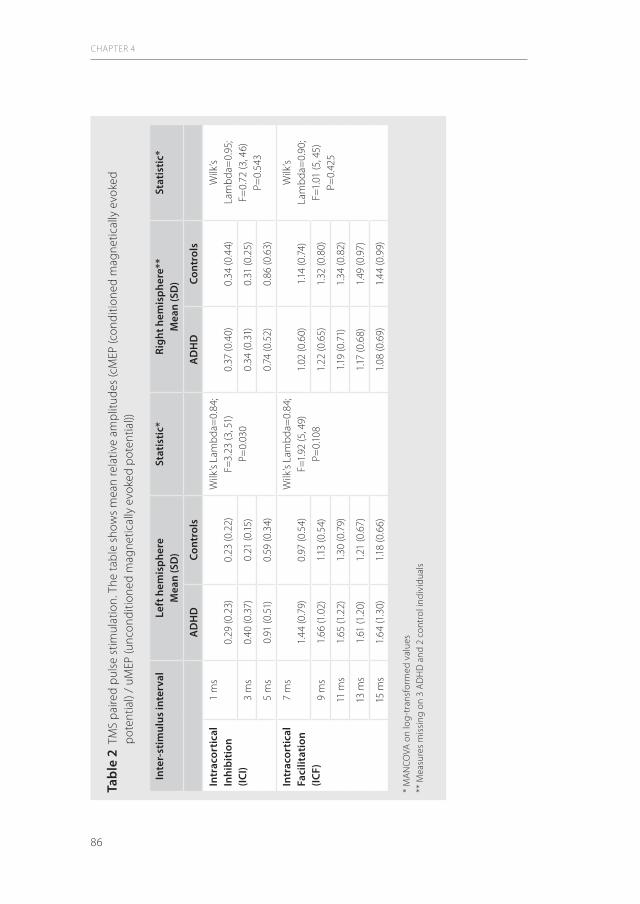
86
CHAPTER 4
Tabl
e 2
TM
S p
aire
d p
ulse
stim
ulat
ion.
The
tab
le s
how
s m
ean
rela
tive
amp
litud
es (c
MEP
(con
ditio
ned
mag
netic
ally
evo
ked
pot
entia
l) /
uMEP
(unc
ondi
tione
d m
agne
tical
ly e
voke
d p
oten
tial))
Inte
r-st
imul
us in
terv
alLe
ft h
emis
pher
eM
ean
(SD
)St
atis
tic*
Ri
ght h
emis
pher
e**
Mea
n (S
D)
Stat
isti
c*
AD
HD
Cont
rols
AD
HD
Cont
rols
Intr
acor
tica
l In
hibi
tion
(IC
I)
1 m
s0.
29 (0
.23)
0.23
(0.2
2)W
ilk’s
Lam
bda
=0.
84;
F=3.
23 (3
, 51)
P=0.
030
0.37
(0.4
0)0.
34 (0
.44)
Wilk
’s La
mb
da=
0.95
; F=
0.72
(3, 4
6)P=
0.54
33
ms
0.40
(0.3
7)0.
21 (0
.15)
0.34
(0.3
1)0.
31 (0
.25)
5 m
s0.
91 (0
.51)
0.59
(0.3
4)0.
74 (0
.52)
0.86
(0.6
3)
Intr
acor
tica
l Fa
cilit
atio
n (IC
F)
7 m
s1.
44 (0
.79)
0.97
(0.5
4)W
ilk’s
Lam
bda
=0.
84;
F=1.
92 (5
, 49)
P=0.
108
1.02
(0.6
0)1.1
4 (0
.74)
Wilk
’s La
mb
da=
0.90
; F=
1.01
(5, 4
5)P=
0.42
59
ms
1.66
(1.0
2)1.1
3 (0
.54)
1.22
(0.6
5)1.
32 (0
.80)
11 m
s1.
65 (1
.22)
1.30
(0.7
9)1.1
9 (0
.71)
1.34
(0.8
2)
13 m
s1.
61 (1
.20)
1.21
(0.6
7)1.1
7 (0
.68)
1.49
(0.9
7)
15 m
s1.
64 (1
.30)
1.18
(0.6
6)1.
08 (0
.69)
1.44
(0.9
9)
* M
AN
COVA
on
log
-tra
nsfo
rmed
val
ues
** M
easu
res
mis
sing
on
3 A
DH
D a
nd 2
con
trol
indi
vidu
als
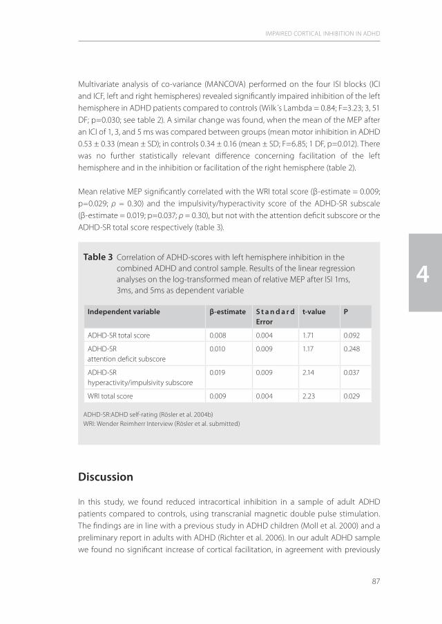
87
Multivariate analysis of co-variance (MANCOVA) performed on the four ISI blocks (ICI and ICF, left and right hemispheres) revealed significantly impaired inhibition of the left hemisphere in ADHD patients compared to controls (Wilk´s Lambda = 0.84; F=3.23; 3, 51 DF; p=0.030; see table 2). A similar change was found, when the mean of the MEP after an ICI of 1, 3, and 5 ms was compared between groups (mean motor inhibition in ADHD 0.53 ± 0.33 (mean ± SD); in controls 0.34 ± 0.16 (mean ± SD; F=6.85; 1 DF, p=0.012). There was no further statistically relevant difference concerning facilitation of the left hemisphere and in the inhibition or facilitation of the right hemisphere (table 2).
Mean relative MEP significantly correlated with the WRI total score (β-estimate = 0.009; p=0.029; ρ = 0.30) and the impulsivity/hyperactivity score of the ADHD-SR subscale (β-estimate = 0.019; p=0.037; ρ = 0.30), but not with the attention deficit subscore or the ADHD-SR total score respectively (table 3).
Discussion
In this study, we found reduced intracortical inhibition in a sample of adult ADHD patients compared to controls, using transcranial magnetic double pulse stimulation. The findings are in line with a previous study in ADHD children (Moll et al. 2000) and a preliminary report in adults with ADHD (Richter et al. 2006). In our adult ADHD sample we found no significant increase of cortical facilitation, in agreement with previously
IMPAIRED CORTICAL INHIBITION IN ADHD
4Table 3 Correlation of ADHD-scores with left hemisphere inhibition in the
combined ADHD and control sample. Results of the linear regression analyses on the log-transformed mean of relative MEP after ISI 1ms, 3ms, and 5ms as dependent variable
Independent variable β-estimate S t a n d a r d Error
t-value P
ADHD-SR total score 0.008 0.004 1.71 0.092
ADHD-SRattention deficit subscore
0.010 0.009 1.17 0.248
ADHD-SRhyperactivity/impulsivity subscore
0.019 0.009 2.14 0.037
WRI total score 0.009 0.004 2.23 0.029
ADHD-SR:ADHD self-rating (Rösler et al. 2004b)WRI: Wender Reimherr Interview (Rösler et al. submitted)
88
reported findings in ADHD children (Moll et al. 2000). Our findings therefore suggest that changes of motor excitability in adults with ADHD are similar to those in children with ADHD. When the dimensional characteristics of ADHD were assessed, inhibitory deficits in our ADHD subjects were positively correlated with two different symptom scores, i.e. the less inhibition the more symptoms. Interestingly, we found a significant correlation with hyperactivity and impulsivity, but not with inattention scores. Similarly, Gilbert et al. (2005) reported an inverse correlation (r=0.53) of ICI with ADHD symptom scores in Tourette patients. The strength of the association in the latter study was also greater with the hyperactivity/impulsivity subscore than with the inattention subscore, suggesting that hyperactive/impulsive behaviours are linked with cortical inhibitory dysfunction in Tourette patients. Compared to the study of Gilbert et al. (2005) however, the correlation between ICI and hyperactivity/impulsivity in our study was less pronounced. This difference could be due to a lack of motor tics or spontaneous motor activity in our study population without Tourette disorder or might be based on different pathophys-iological mechanisms underlying these disorders. Moreover, the WRI score that comprises not only ADHD core symptoms but also additional emotional components of adult ADHD according to the Utah criteria (Wender 2005) was significantly related to inhibitory deficits. This finding suggest, that emotional problems in ADHD like affective lability, emotional overreagibility and hot temper might be related to the same neuropathological processes as impulsivity and hyperactivity, i.e. deficits of cortical inhibition. Another result of our study was the identification of inter-hemispheric asymmetry of ICI deficits in ADHD patients, which was not present in healthy controls. In adults with ADHD, ICI deficits occurred only in the left hemisphere. Brain asymmetry in ADHD has been reported several times in the literature. Schrimsher et al. (2002) could predict the cumulative severity ratings of inattentive behaviours by measuring caudate volume asymmetry from serial sagittal magnetic resonance images from childhood to adolescence. Unilateral volume reductions of the pallidum have also been shown in several studies in children with ADHD (Aylward et al. 1996, Castellanos et al. 1996, 2002, Overmeyer et al. 2001). Most studies however, describe volume reductions within the right hemisphere. Therefore, the hypothetic mechanisms leading to reduced inhibition of the left side can only be deduced from unknown and complex mechanisms leading to contralateral effects.In our study we did not find differences between ADHD subjects and healthy controls regarding resting motor (RMT) and active motor thresholds (AMT). Also simple stimulus response curves did not differ between ADHD patients and controls. This agrees with previously reported observations in children with ADHD (Moll et al. 2000). Thus, there is no evidence for motor hyperexcitability within the motor system in ADHD at the membrane level of cortical neurons (Ziemann et al. 1996d).
CHAPTER 4
89
In general, our results suggest that TMS, and the paired-pulse technique in particular, is a useful tool for motor excitability research in ADHD. It allows the study of intracortical circuits (Ziemann 1999; Kujirai et al. 1993; Valls-Sol et al. 1992; Ziemann et al. 1996d) and displays little inter- and intraindividual variability (Maeda et al. 2002). ICI deficits have been shown to have reliable test-retest stability in adults with ADHD (Richter et al. 2006). ICI and ICF appear to be due to activation of separate circuits (Ziemann et al. 1996d), represented by inhibitory interneurons or inhibitory connections between cortical output cells (Wassermann et al. 1996) and facilitatory interactions partially in the motor cortex, at or upstream from corticospinal neurons (Ziemann et al. 1996d). Conditioning pulses in the double-pulse stimulation technique are thought to activate inhibitory interneurons within the motor system including projections onto corticospinal neurons (Kujirai 1993). Several neural transmission systems like GABAergic, dopaminergic and glutamatergic systems appear to play an important role in these mechanisms. Medications that enhance GABAergic activity have been shown to increase the degree of ICI and decrease ICF evoked by paired TMS stimuli (Inghilleri et al. 1996, Werhahn et al. 1998, Ziemann et al. 1996b, 1996c). Dopaminergic drugs have been shown to enhance ICI in normal subjects (Berardelli et al. 1996, Priori et al. 1994, Ridding et al. 1995, Ziemann et al. 1996a). Glutamatergic drugs have been shown to increase intracortical inhibition and decrease facilitation (Schwenkreis et al. 1999, Ziemann et al. 1998).In conclusion, this paired-pulse magnetic stimulation study is the first to demonstrate that intracortical inhibition in adult ADHD is reduced compared to matched controls and occurs only in the left hemisphere. These deficits correlate with ADHD sympto-matology, especially with hyperactivity and impulsivity behaviour. The findings therefore corroborate the hypothesis of inhibition deficits on a neuronal level. Compared to previous findings of double pulse TMS in ADHD children our results suggest that there is no fundamental difference between affected children and adults with respect to excitability of the frontal motor cortex. Further studies are needed to confirm and to further define these excitatory phenomena in ADHD to gain a greater insight into the etiology of ADHD and to generate useful endophenotypes of this disorder.
IMPAIRED CORTICAL INHIBITION IN ADHD
4
90
References
1. Amassian VE, Quirk GJ, Stewart M. (1990) A comparison of corticospinal activation by magnetic coil and electrical stimulation of monkey motor cortex. Electroencephalogr Clin Neurophysiol. 77: 390-401
2. Aylward EH, Reiss AL, Reader MJ, Singer HS, Brown JE, Denckla MB (1996) Basal ganglia volumes in children with attention-deficit hyperactivity disorder. J Child Neurol 11: 112–115
3. Bailey CJ, Karhu J, Ilmoniemi RJ (2001) Transcranial magnetic stimulation as a tool for cognitive studies. Scand J Psychol 42: 297-305
4. Barkley RA, Fischer M, Smallish L, Fletcher K (2006) Young adult outcome of hyperactive children: adaptive functioning in major life activities. J Am Acad Child Adolesc Psychiatry 45: 192-202
5. Baumer T, Bock F, Koch G, Lange R, Rothwell JC, Siebner HR, Münchau A (2006) Magnetic stimulation of human premotor or motor cortex produces interhemispheric facilitation through distinct pathways. J Physiol. 572: 857-868
6. Biederman J, Faraone SV, Spencer TJ, Mick E, Monuteaux MC, Aleardi M (2006) Functional impairments in adults with self-reports of diagnosed ADHD: A controlled study of 1001 adults in the community. J Clin Psychiatry 67: 524-540
7. Buchmann J, Gierow W, Weber S, Hoeppner J, Klauer T, Wittstock M, Benecke R, Haessler F, Wolters A (2006) Modulation of transcallosally mediated motor inhibition in children with attention deficit hyperactivity disorder (ADHD) by medication with methylphenidate (MPH). Neurosci Lett 405: 14-18
8. Buchmann J, Wolters A, Haessler F, Bohne S, Nordbeck R, Kunesch E (2003) Disturbed transcallosally mediated motor inhibition in children with attention deficit hyperactivity disorder (ADHD). Clin Neurophysiol 114: 2036-2042
9. Castellanos FX, Giedd JN, Marsh WL, Hamburger SD, Vaituzis AC, Dickstein DP, Sarfatti SE, Vauss YC, Snell JW, Lange N, Kaysen D, Krain AL, Ritchie GF, Rajapakse JC, Rapoport JL (1996) Quantitative brain magnetic resonance imaging in attention deficit hyperactivity disorder. Arch Gen Psychiatry 53: 607-616.
10. Castellanos FX, Lee PP, Sharp W, Jeffries NO, Greenstein DK, Clasen LS, Blumenthal JD, James RS, Ebens CL, Walter JM, Zijdenbos A, Evans AC, Giedd JN, Rapoport JL (2002) Developmental trajectories of brain volume abnormalities in children and adolescents with attention-deficit/hyperactivity disorder. JAMA 288: 1740-1748
11. Day BL, Dressler D, Maertens de Noordhout, Marsden CD, Nakashima K, Rothwell JC, Thompson PD (1989) Electric and magnetic stimulation of human motor cortex: surface EMG and single motor unit responses. J Physiol 412: 449-473.
12. Di Lazzaro V, Oliviero A, Profice P, Saturno E, Pilato F, Insola A, Mazzone P, Tonali P, Rothwell JC (1998) Comparison of descending volleys evoked by transcranial magnetic and electric stimulation in conscious humans. Electroencephalogr Clin Neurophysiol 109: 397-401
13. Fallgatter AJ, Ehlis AC, Seifert J, Strik WK, Scheuerpflug P, Zillessen KE, Herrmann MJ, Warnke A (2004) Altered response control and anterior cingulate function in attention-deficit/hyperactivity disorder boys. Clin Neurophysiol 115: 973-981
14. Faraone SV (2004) Genetics of adult attention-deficit/hyperactivity disorder. Psychiatr Clin North Am 27: 303-321
15. Garvey MA, Barker CA, Bartko JJ, Denckla MB, Wassermann EM, Castellanos FX, Dell ML, Ziemann U (2005) The ipsilateral silent period in boys with attention-deficit/hyperactivity disorder. Clin Neurophysiol 116: 1889-1896
16. Gilbert DL, Ridel KR, Sallee FR, Zhang J, Lipps TD, Wassermann EM (2006) Comparison of the inhibitory and excitatory effects of ADHD medications methylphenidate and atomoxetine on motor cortex. Neuropsy-chopharmacology 31: 442-449
17. Gilbert DL, Sallee FR, Zhang J, Lipps TD, Wassermann EM (2005) Transcranial magnetic stimulation-evoked cortical inhibition: a consistent marker of attention-deficit/hyperactivity disorder scores in Tourette syndrome. Biol Psychiatry 57: 1597-1600
18. Grützmacher, H (2001) Unfallgefährdung bei Aufmerksamkeits- und Hyperaktivitätsstörung. Deutsches Ärzteblatt 98: 1898-1900
CHAPTER 4

91
19. Heiser P, Friedel S, Dempfle A, Konrad K, Smidt J, Grabarkiewicz J, Herpertz-Dahlmann B, Remschmidt H, Hebebrand J (2004) Molecular genetic aspects of attention-deficit/hyperactivity disorder. Neurosci Biobehav Rev 28: 625-641
20. Inghilleri M, Berardelli A, Marchetti P, Manfredi M (1996) Effects of diazepam, baclofen and thiopental on the silent period evoked by transcranial magnetic stimulation in humans. Exp Brain Res 109: 467-472
21. Jerome L, Habinski L, Segal A (2006) Attention-deficit/hyperactivity disorder (ADHD) and driving risk: a review of the literature and a methodological critique. Curr Psychiatry Rep 8: 416-426
22. Kessler RC, Adler L, Barkley R, Biederman J, Conners CK, Demler O, Faraone SV, Greenhill LL, Howes MJ, Secnik K, Spencer T, Ustun TB, Walters EE, Zaslavsky AM (2006) The prevalence and correlates of adult ADHD in the United States: results from the National Comorbidity Survey Replication Am J Psychiatry 163: 716-723
23. Kujirai T, Caramia MD, Rothwell JC, Day BL, Thompson PD, Ferbert A, Wroe S, Asselman P, Marsden CD (1993) Corticocortical inhibition in human motor cortex. J Physiol. 471: 501-519
24. Maeda F, Gangitano M, Thall M, Pascual-Leone A (2002) Inter- and intra-individual variability of paired-pulse curves with transcranial magnetic stimulation (TMS). Clin Neurophysiol 113: 376-382
25. Mantere T, Tupala E, Hall H, Sarkioja T, Rasanen P, Bergstrom K, Callaway J, Tiihonen J (2002) Serotonin transporter distribution and density in the cerebral cortex of alcoholic and nonalcoholic comparison subjects: a whole-hemisphere autoradiography study. Am J Psychiatry 159: 599-606
26. Moll GH, Heinrich H, Trott G, Wirth S, Rothenberger A (2000) Deficient intracortical inhibition in drug-naive children with attention-deficit hyperactivity disorder is enhanced by methylphenidate. Neurosci Lett 284: 121-125
27. Nakamura H, Kitagawa H, Kawaguchi Y, Tsuji H (1996) Direct and indirect activation of human corticospinal neurons by transcranial magnetic and electrical stimulation. Neurosci Lett 210: 45-48
28. Overmeyer S, Bullmore ET, Suckling J, Simmons A, Williams SC, Santosh PJ, Taylor E (2001) Distributed grey and white matter deficits in hyperkinetic disorder: MRI evidence for anatomical abnormality in an attentional network. Psychol Med 31: 1425-1435
29. Pascual-Leone A, Tormos JM, Keenan J, Tarazona F, Canete C, Catala MD (1998) Study and modulation of human cortical excitability with transcranial magnetic stimulation. J Clin Neurophysiol 15: 333-343
30. Pliszka SR (2005) The neuropsychopharmacology of attention-deficit/hyperactivity disorder. Biol Psychiatry 57: 1385-1390
31. Porro CA, Martinig M, Facchin P, Maieron M, Jones AK, Fadiga L (2007) Parietal cortex involvement in the localization of tactile and noxious mechanical stimuli: A transcranial magnetic stimulation study. Behav Brain Res [Epub ahead of print] PMID: 17239452
32. Priori A, Berardelli A, Inghilleri M, Accornero N, Manfredi M (1994) Motor cortical inhibition and the dopaminergic system. Brain 117: 317-323
33. Retz-Junginger P, Retz W, Blocher D, Stieglitz RD, Georg T, Supprian T, Wender PH, Rösler M (2003) [Reliability and validity of the Wender-Utah-Rating-Scale short form. Retrospective assessment of symptoms for attention deficit/hyperactivity disorder.] Nervenarzt 74: 987-993
34. Retz-Junginger P, Retz W, Blocher D, Weijers HG, Trott GE, Wender PH, Rösler M (2002) [Wender Utah rating scale. The short-version for the assessment of the attention-deficit hyperactivity disorder in adults.] Nervenarzt 73: 830-838
35. Richter MM, Ehlis A-C, Bähne C-B, Scheuerpflug P, Jacob CP, Fallgatter AJ (2006) Inhibitory brain functions in adult ADHD patients. Nervenheilkunde 25: 657-661
36. Rösler M, Retz W, Retz-Junginger P, Hengesch G, Schneider M, Supprian T, Schwitzgebel P, Pinhard K, Dovi-Akue N, Wender P, Thome J (2004a) Prevalence of attention deficit-/hyperactivity disorder (ADHD) and comorbid disorders in young male prison inmates. Eur Arch Psychiatry Clin Neurosci. 254: 365-371
37. Rösler M, Retz W, Retz-Junginger P, Thome J, Supprian T, Nissen T, Stieglitz RD, Blocher D, Hengesch G, Trott GE (2004b) [Tools for the diagnosis of attention-deficit/hyperactivity disorder in adults. Self-rating behaviour questionnaire and diagnostic checklist.] Nervenarzt 75: 888-895
38. Rösler M, Retz W, Retz-Junginger P, Reimherr FW, Wender PH (2008) [Attention deficit hyperactivity disorder (ADHD) in adults: benchmarking diagnosis using the Wender-Reimherr interview.] Nervenarzt 79(3):320-7
IMPAIRED CORTICAL INHIBITION IN ADHD
4

92
39. Schneider M, Retz W, Coogan A, Thome J, Rösler M (2006) Anatomical and functional brain imaging in adult attention-deficit/hyperactivity disorder (ADHD)-A neurological view. Eur Arch Psychiatry Clin Neurosci 256 Suppl 1: i32-i41
40. Schwenkreis P, Witscher K, Janssen F, Addo A, Dertwinkel R, Zenz M, Malin JP, Tegenthoff M (1999) Influence of the N-methyl-D-aspartate antagonist memantine on human motor cortex excitability. Neurosci Lett. 270: 137-140
41. Schrimsher GW, Billingsley RL, Jackson EF, Moore BD 3rd (2002) Caudate nucleus volume asymmetry predicts attention-deficit hyperactivity disorder (ADHD) symptomatology in children. J Child Neurol 17: 877-884
42. Seidman LJ, Valera EM, Makris N, Monuteaux MC, Boriel DL, Kelkar K, Kennedy DN, Caviness VS, Bush G, Aleardi M, Faraone SV, Biederman J (2006) Dorsolateral prefrontal and anterior cingulate cortex volumetric abnormalities in adults with attention-deficit/hyperactivity disorder identified by magnetic resonance imaging. Biol Psychiatry 60: 1071-1080
43. Simons W, Dierick M (2005) Transcranial magnetic stimulation as a therapeutic tool in psychiatry. World J Biol Psychiatry 6: 6-25
44. Strafella AP, Paus T, Fraraccio M, Dagher A (2003) Striatal dopamine release induced by repetitive transcranial magnetic stimulation of the human motor cortex. Brain 126: 2609-2615
45. Valls-Solé J, Pascual-Leone A, Wassermann EM, Hallett M (1992) Human motor evoked responses to paired transcranial magnetic stimuli. Electroencephalogr Clin Neurophysiol 85: 355–364
46. Vermeiren R (2003) Psychopathology and delinquency in adolescents: a descriptive and developmental perspective. Clin Psychol Rev 23: 277-318
47. Wassermann EM, Samii A, Mercuri B, Ikoma K, Oddo D, Grill SE, Hallett M (1996) Responses to paired transcranial magnetic stimuli in resting, active, and recently activated muscles. Exp Brain Res 109: 158-163
48. Wender PH (1995) Attention-Deficit Hyperactivity Disorder in Adults.Oxford Univ Press,New York49. Ziemann U (1999) Intracortical inhibition and facilitation in the conventional paired TMS paradigm. Electro-
encephalogr Clin Neurophysiol Suppl 51: 127-13650. Ziemann U (2004) TMS induced plasticity in human cortex. Rev Neurosci 15: 253-26651. Ziemann U, Bruns D, Paulus W (1996a) Enhancement of human motor cortex inhibition by the dopamine
receptor agonist pergolide: evidence from transcranial magnetic stimulation. Neurosci Lett 208: 187-19052. Ziemann U, Lonnecker S, Steinhoff BJ, Paulus W (1996b) Effects of antiepileptic drugs on motor cortex
excitability in humans: a transcranial magnetic stimulation study. Ann Neurol 40: 367-37853. Ziemann U, Lonnecker S, Steinhoff BJ, Paulus W (1996c) The effect of lorazepam on the motor cortical
excitability in man. Exp Brain Res 109: 127-13554. Ziemann U, Rothwell JC, Ridding MC (1996d) Interaction between intracortical inhibition and facilitation in
human motor cortex. J Physiol 496: 873-881 55. Ziemann U, Tergau F, Wischer S, Hildebrandt J, Paulus W (1998) Pharmacological control of facilitatory I-wave
interaction in the human motor cortex. A paired transcranial magnetic stimulation study. Electroencepha-logr Clin Neurophysiol 109: 321-330
CHAPTER 4

93
IMPAIRED CORTICAL INHIBITION IN ADHD
4


The effects of long acting Methylphenidate in adults with Attention Deficit Hyperactivity Disorder: A study with paired pulse Transcranial Magnetic Stimulation
Marc K.F. Schneider, Wolfgang Retz, Gavril Gougleris, Willem M.A. Verhoeven, Joke H.M. Tulen, and Michael Rösler
Published in Neuropsychobiology (2011) 64(4):195-201.
5 |
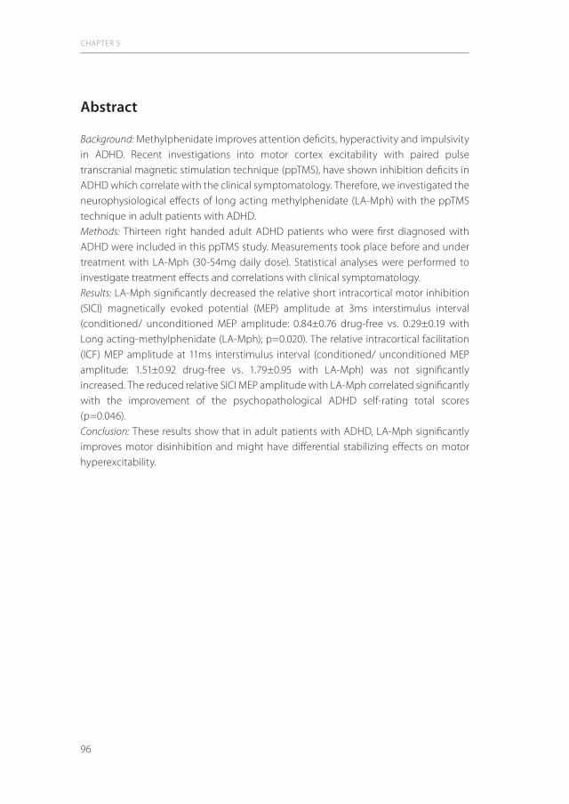
96
Abstract
Background: Methylphenidate improves attention deficits, hyperactivity and impulsivity in ADHD. Recent investigations into motor cortex excitability with paired pulse transcranial magnetic stimulation technique (ppTMS), have shown inhibition deficits in ADHD which correlate with the clinical symptomatology. Therefore, we investigated the neurophysiological effects of long acting methylphenidate (LA-Mph) with the ppTMS technique in adult patients with ADHD.Methods: Thirteen right handed adult ADHD patients who were first diagnosed with ADHD were included in this ppTMS study. Measurements took place before and under treatment with LA-Mph (30-54mg daily dose). Statistical analyses were performed to investigate treatment effects and correlations with clinical symptomatology.Results: LA-Mph significantly decreased the relative short intracortical motor inhibition (SICI) magnetically evoked potential (MEP) amplitude at 3ms interstimulus interval (conditioned/ unconditioned MEP amplitude: 0.84±0.76 drug-free vs. 0.29±0.19 with Long acting-methylphenidate (LA-Mph); p=0.020). The relative intracortical facilitation (ICF) MEP amplitude at 11ms interstimulus interval (conditioned/ unconditioned MEP amplitude: 1.51±0.92 drug-free vs. 1.79±0.95 with LA-Mph) was not significantly increased. The reduced relative SICI MEP amplitude with LA-Mph correlated significantly with the improvement of the psychopathological ADHD self-rating total scores (p=0.046).Conclusion: These results show that in adult patients with ADHD, LA-Mph significantly improves motor disinhibition and might have differential stabilizing effects on motor hyperexcitability.
CHAPTER 5

97
Introduction
Methylphenidate (Mph) is a psychostimulant and is used for more than 50 years in the treatment of attention deficit, hyperactivity and impulsivity symptoms in ADHD. It increases dopaminergic and noradrenergic synaptic efflux throughout the brain and has a rapid onset of action without attenuation of drug effects [1]. Low doses of Mph have limited influences on dopamine (DA) and norepinephrine (NE) in the prefrontal cortex only, without inducing effects on other areas of the brain at higher dosages [2]. The behavioural and cognitive effects of this stimulant drug are not specific and occur in ADHD as well as in normal subjects. The well known clinical experiences with Mph contrast with the lack of detailed knowledge about the neurobiological mechanisms of stimulant action on motor cortex in human ADHD. Recent imaging studies converge mainly towards improvement of disturbed fronto-striatal activity [3,4].With the paired pulse transcranial magnetic stimulation (ppTMS) technique, the short intracortical inhibition (SICI) and intracortical facilitation (ICF) can be analyzed [5]. The ppTMS technique is also an useful and non-invasive tool to investigate the influence of pharmacological modulation on intracortical motor excitability [6]. The recent literature is inconsistent regarding the effects of immediate release Mph on SICI and ICF in ADHD and their underlying mechanisms [7-12]. Indeed, immediate release Mph has disadvantages, in that it has to be given several times per day, whereas long acting Mph (LA-Mph) can be administered once or twice a day and induces less fluctuations in plasma concentration. Therefore, LA-Mph given once or twice per day has pharmacoki-netic advantages for the treatment of adults with ADHD [13], although the plasma concentration may show interindividual variations [14]. Over the past years, a limited number of studies with TMS in ADHD patients has been published in which immediate release Mph was used with, however, unequivocal results [7-12]. Until now, no study has been performed with the ppTMS technique that investigated the effects of LA-Mph. With LA-Mph, the clinical state of the ADHD patient is more stable and allows, therefore, for better clinical state correlations.In this study, it is hypothesized that LA-Mph improves disturbed ppTMS intracortical excitability in adult patients with ADHD and that these effects are positively correlated with clinical improvement of the ADHD symptoms.
Methods
Patient characteristics and medication proceduresThirteen right handed patients were recruited by experienced board certificated psychiatrists (MS, GG, WR) from specialized outpatient centres for ADHD-associated disorders (SHG Klinik für Psychiatrie und Psychotherapie, ADHS Ambulanz, Saarbrücken;
LONG ACTING METHYLPHENIDATE IN ADHD
5
98
Neurozentrum der Universität des Saarlandes, ADHS Ambulanz, Homburg) . They were diagnosed with ADHD combined type for the first time according to the diagnostic criteria of DSM IV (ADHD-DC: ADHD Diagnostic checklist [15]), and had used no stimulant medication before. The Wender Utah Self Rating Scale (WURS-k) [16,17]) was used to indicate childhood ADHD symptoms. Statistical test analysis indicated a sensitivity of 85% and specificity of 76% at a cutoff of 30 points. Its inner consistency is high (Cronbach α= 0.91) [17]. Patients with the clinical diagnosis of ADHD were included in the study if the WURS-k score was at least 30, and when at least 6 of 9 items of inattention and/or hyperactivity/impulsivity were present. As indicator of severity of symptoms, the ADHD self-rating scale for adults (ADHD-SR) according to DSM IV [15] was used (total score: 0 to 54; attention deficit and hyperactivity/impulsivity subscores: each maximal 27; test-retest reliability 0.80, inner consistency Cronbach α=0.90). A semistandardized expert clinical assessment for adult ADHD, the German validated version of the Wender Reimherr Interview (WRI) [18,19], was applied to quantify the Utah criteria for adult ADHD. The WRI is also known as WRAADDS [19] and comprises the subscales inattention, hyperactivity, temper, affective lability, emotional hyperreagibility, disorganization and impulsivity, yielding 28 psychopathological items, each being quantified on a Lickert Scale ranging from 0 to 2. This results in a maximum total WRI score of 56. The internal consistency of this interview is also high (Cronbach α=0.82) [18]. SCID-1 and 2 interviews were used to exclude further axis I and II DSM categorical diagnoses. The recruited patients had no psychoactive medication prior to the diagnosis of ADHD. None of them had additional DSM IV axis I or II diagnoses. As additional exclusion criteria served: drug abuse, relevant somatic problems, as well as any known conditions with metabolic or immunologic pathology. Other exclusion criteria were any kind of degenerative, traumatic, inflammatory or vascular brain disturbance (e.g. developmental disorder, meningitis, encephalitis in the anamneses, neurodegenerative brain disease), and low intelligence (IQ<85). Estimation of intelligence was done by the “Mehrfachwahl Wortschatztest (MWT-B)“, a verbal IQ test [20] .The patients gave written informed consent to participate in the study. The TMS study was approved by the local ethical committee. In this clinical study, oral dose of LA-Mph was increased gradually to obtain both an optimal individual dosage and to avoid side effects. Patients were examined by a board certified and experienced academic psychiatrist with intervals of two weeks. The first clinical and electrophysiological measurement took place just before the start of the medication. After the patients had reached the optimal individual dose of LA-Mph (Medikinet ret®, Concerta®) for at least two weeks, they were re-assessed with the above mentioned ADHD instruments. The second electrophysiological measurement was performed two weeks after the ADHD scores were decreased by at least 50% (ADHD-SR) according to the clinical interviews (WRI, ADHD-DC). The patients had to take the medication at an exactly defined time point (8.00h). The medication dosage should be
CHAPTER 5
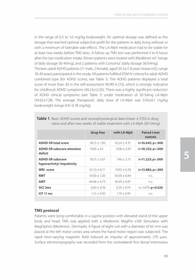
99
in the range of 0,3 to 1,0 mg/kg bodyweight. An optimal dosage was defined as the dosage that reached optimal subjective profit for the patients in daily living without or with a minimum of tolerable side effects. The LA-Mph medication had to be stable for at least two weeks before TMS tests. A follow up TMS test was performed 4 to 6 hours after the last medication intake. Eleven patients were treated with Medikinet ret® (range of daily dosage 30-40mg), and 2 patients with Concerta® (daily dosage 36/54mg). Thirteen adult ADHD patients (11 male, 2 female), aged 34.3±11.8 years (mean±SD, range 18-49 years) participated in the study. All patients fulfilled DSM IV criteria for adult ADHD combined type (for ADHD scores, see Table 1). The ADHD patients displayed a total score of more than 30 in the self-assessment WURS-k [15], which is strongly indicative for childhood ADHD symptoms (49.23±12.05). There was a highly significant reduction of ADHD clinical symptoms (see Table 1) under medication of 30-54mg LA-Mph (34.62±7.28). The average therapeutic daily dose of LA-Mph was 0.50±0.1 mg/kg bodyweight (range 0.41-0.78 mg/kg).
TMS protocolPatients were lying comfortably in a supine position with elevated stand of the upper body and head. TMS was applied with a Medtronic MagPro x100 Stimulator with MagOption (Medtronic, Denmark). A figure of eight coil with a diameter of 65 mm was placed at the left motor cortex area where the hand motor region was subjected. The rapid time-varying magnetic field induced an impulse of approximately 270 µsec. Surface electromyography was recorded from the contralateral first dorsal interosseus
LONG ACTING METHYLPHENIDATE IN ADHD
5
Table 1 Basic ADHD scores and neurophysiological data (mean ± STD) in drug naïve and after two weeks of stable treatment with LA-Mph (30-54mg)
drug-free with LA-Mph Paired t-test statistic
ADHD-SR total score 38.15 ± 7.00 16.54 ± 4.37 t=16.645; p<.000
ADHD-SR subscore attention deficit
19.85 ± 4.1 9.08 ± 3.59 t=18.252; p<.000
ADHD-SR subscore hyperactivity/ impulsivity
18.31 ± 5.07 7.46 ± 2.73 t=11.223; p<.000
WRI- score 42.23 ± 6.11 19.92 ± 6.28 t=13.883; p<.000
RMT 54.00 ± 7.20 56.00 ± 6.84 n.s.
AMT 44.46 ± 6.75 45.69 ± 6.87 n.s.
SICI 3ms 0.83 ± 0.76 0.29 ± 0.19 t= 2.679; p=0.020
ICF 11 ms 1.51 ± 0.92 1.79 ± 0.95 n.s.

100
muscle (FDI) with a standard electromyographic amplifier (Medtronic Keypoint 4; recording software Medtronic Keypoint V 5.01). The bandwidth of the filters was set to 1 Hz and 10 kHz, respectively. The optimal position of the coil was determined by moving the coil by 0.5 cm steps until an optimal magnetically evoked potential (MEP) could be registered. When the induced current flowing parallel to the brain surface was sufficient, depolarization of neuronal membranes occurred, and hence an action potential was generated and recorded at the FDI muscle of the right hand. Care was taken to relax the subjects and to avoid any movements of the extremities during recordings. Each MEP was checked optically to ensure that the FDI was completely relaxed. Recordings were discarded if any evidence of electromyographic background activity was detected and repeated.Resting (RMT) and active motor thresholds (AMT) were determined according to the protocol of Kujirai et al. [5]. Briefly, RMT was the minimal stimulus intensity which was required to produce motor potentials of more than 50 µV peak to peak amplitude in 50% of the test pulses. AMT was determined in analogy to RMT, while the subject tonically distended a dynamometer with 20% of maximum power. Two magnetic stimuli were delivered in close time sequence to the same cortical spot through a single stimulation coil [5] to perform the paired pulse protocols. The first, conditioning pulse (CP), was a subthreshold pulse that was considered to condition the response for the second, the test stimulus (TS), which was a suprathreshold stimulus. The TS followed within a time period of milliseconds. Different brain circuits are recruited by varying intensities of CP and TS. Eight MEP’s were averaged at each stimulus level. Paired pulse stimulation was performed according to the technique of Kujirai et al. [5]. The subthreshold conditioning pulse (CP) was set at 80% of RMT. The TS was adjusted to produce a mean MEP of 0.5–1.2mV peak to peak amplitude, when unconditioned (uMEP). The interstimulus intervals were set at 3ms and later at 11ms. At each interstimulus interval 8 trials were averaged. The time between two trial pulses was at least 5 sec to avoid secondary effects, such as potentiation. Peak-to-peak amplitudes of these conditioned MEP’s (cMEP) were set into relation to the uMEP. TMS data are presented as relative amplitudes (=cMEP/uMEP). At ISI 3ms, arelative MEP ratio of smaller than 1 is to be considered as short intracortical inhibition (SICI). At ISI 11ms, a ratio of more than 1 is to be considered as intracortical facilitation (ICF). In our study, relative MEP’s at an ISI of 3ms were accepted to indicate inhibition, and relative MEP’s at an ISI of 11ms to indicate facilitation [5,36].
Statistical analysisTo compare the differences of the dependent neurophysiological variables between the pre- and under medication with LA-Mph measurements, the Student’s t-test was performed. For the correlations between neurophysiological and clinical parameters, the Pearsons PM correlation coefficient (r) is used. An analysis of covariance (ANCOVA)
CHAPTER 5
101
was applied to study the influence of the covariates ADHD-SR total and subscores (attention deficit, hyperactivity/ impulsivity), WRI total and subscores (inattention, hyperactivity, temper, affective lability, emotional hyperreagibility, disorganization and impulsivity), intelligence, age, medication doses as well as body weight adapted medication doses, on the relative MEP amplitudes before and after treatment. The level of significance was set at p = .05. All analyses were performed witch SPSS 15.0
Results
In this study, thirteen adult ADHD patients with an age range of 18-49 years who met DSM IV criteria for adult ADHD combined type were treated with LA-Mph. Paired pulse transcranial magnetic stimulation was performed before and after 2 weeks of stable individualized oral dose. In the drug naïve condition, the left hemisphere resting motor threshold (RMT) in ADHD patients was 54.0±7.2% and 56.0±6.8% during treatment with LA-Mph. There was no significant difference between the RMT with or without LA-Mph, or between the AMT with or without LA-Mph (44.5±6.7% vs. 45.7±6.9% with MPH). There was also no significant correlation with motor thresholds and ADHD symptom rating scores.There was a significant difference in relative MEP amplitudes (cMEP/uMEP) between drug naïve conditions and medication with LA-Mph (Table 1 and Figure 1). The relative MEP amplitude at ISI of 3ms corresponds to SICI, whereas the ISI of 11ms corresponds to ICF (see also [21]). At ISI 3ms, the mean relative MEP was 0.83±0.76, and with LA-Mph it was lowered to 0.29±0.19. This relative amplitude reduction was significant (t=2.679; p=0.02). At ISI 11ms, drug naïve mean relative MEP amplitude was 1.51±0.92. With LA-Mph, it was 1.79±0.95. This increase in relative amplitude was not significant. The SICI and ICF amplitudes did not correlate with LA-MPH dosage.
The drug naïve relative MEP amplitudes at ISI 3ms correlated significantly with the ADHD-SR total score (r=0.590; p=0.034/ see Figure 2) and the ADHD-SR hyperactivity/ impulsivity subscore (r=0.604; p=0.029). The relative MEP amplitude at ISI 3ms under LA-Mph medication, correlated only weakly with the difference on the ADHD-SR score between drug naïve and medication condition (r=0.56; p=0.046). Correlations with ADHD-SR inattention subscores or WRI scores with relative MEP amplitudes at ISI 3 or 11 ms, as well as with the amplitude differences with and without LA-Mph, revealed no statistical significance.
Further analysis of covariance revealed that the neurophysiological data were not significantly influenced by medication doses, ADHD-SR and WRI subscores, age, and intelligence.
LONG ACTING METHYLPHENIDATE IN ADHD
5

102
Discussion
In this study, it was assessed whether LA-Mph has an influence on motor excitability in adult patients with ADHD. For that purpose, the ppTMS technique was used, which previously had already revealed a lowered SICI in adults with ADHD [21,22]. The ppTMS technique has been shown to provide a reliable test-retest stability of SICI and ICF in adults with ADHD [5,23]. RMT and AMT are supposed to be markers of membrane stability of cortical neurons [24]. In the present study, LA-Mph had no influence on RMT and AMT. These results are in line with those from other recent studies [7,9,10,11,25] and support the hypothesis that Mph does not affect membrane characteristics.Inhibition is a cortical phenomenon which is considered to reflect the inhibitory activity of interneurons in the motor cortex or of connections between cortical output cells [26]. In contrast, facilitation occurs at the level of the corticospinal neurons, and upstream [27,
CHAPTER 5
Figure 1 Effects of long acting methylphenidate (LA-Mph) on relative short intracortical inhibition (ISI 3ms) and intracortical facilitation amplitude (ISI 11ms) in a paired pulse TMS protocol (N= 13, MPH: 30–54mg).
Ordinate: relative MEP amplitude (cMEP/uMEP). Squares represent mean values, bars representing SD
Drugnaïve
Drugnaïve
WithLA-Mph
WithLA-Mph
Rela
tive
MEP
am
plit
ude
(cM
EP/u
MEP
)
3
2
1
0
p = -0.020
ISI3ms
ISI11ms
n.s.

103
24]. SICI and ICF are supposed to reflect cortical excitability independently and to be modulated through complex neural transsynaptic systems (mainly GABA, dopamine, glutamate, serotonine). Several drugs influence the ppTMS motor inhibition as well as the facilitation [6]. Pharmacologic interactions with SICI and ICF are suggested to depend from the clinical state, since it is known that paroxetine may enhance ICF in healthy probands, but decreases this parameter in patients with schizophrenia [28]. In healthy volunteers, it has been shown that, dependent on the 5-HT transporter genotype, serotonergic reuptake inhibitors may, however, have differential effects on SICI [29]. Thus, drug effects of Mph on motor excitability may vary dependent on the genetic phenotype.Mph increases the synaptic levels of dopamine via blockade of the dopamine transporter (DAT). In addition, Mph binds to and inhibits the neuronal norepinephrine transporter (NET) [30]. More neuronal transporters and receptors are affected, since Mph binds also
LONG ACTING METHYLPHENIDATE IN ADHD
5
Figure 2 Scatterplot of relative SICI MEP amplitudes (ISI 3ms) in drug naïve condition (open squares) and under medication with LA-Mph (black squares).
Abscissa: ADHD-SR scores. Linear regression fits are displayed: r= 0.589, p= 0.034 (drug naïve condition) and r= -0.096, n.s. (under medication with LA-Mph)
ADHD-SR score
Rela
tive
SIC
I MEP
am
plit
ude
3
2
1
0
0 10 20
r = -0,096
r = 0.589
30 40 50 60

104
to muscarinic and serotonergic receptors and to serotonin transporters in the brain [31]. Dopaminergic and noradrenergic agonists have been shown to increase SICI, while dopamine antagonists have a decreasing effect [6]. It is also suggested that the degree of SICI is increased by GABAergic action via cortical GABAergic interneurons, whereas ICF is decreased [32-35], [6],[36]. Whilst the clinical and pharmacological treatment effects of Mph are well described, the underlying basic pharmacological mechanisms in the motor cortex remain unclear as yet [37,38]. It is actually hypothesized that in ADHD a general deficit in the behavioural inhibition is present [39] and also in the inhibition of motor systems [40]. No direct evidence, however, is available which motor inhibition deficit is specifically involved in ADHD. This may be explained by the complex interactions between the motor cortex and the frontocortical regulatory systems (prefrontal cortex, caudate nucleus, and globus pallidus) and the parietal cortex which are all disturbed in ADHD [3,41]. Since Mph exerts effects on the total spectrum of ADHD symptoms, studying the effects of Mph in ADHD cannot be interpreted as an influence on motor systems only [38]. Although the data that support direct interactions of Mph on motor cortex are scarce, Lou and colleagues [42] demonstrated with PET-scan technique that Mph administration reduces blood flow in the primary motor and sensory cortex. These findings were interpreted as improvement of inhibition and consequently of motor overactivity also.Previous studies have shown higher basic SICI MEP amplitudes in ADHD patients, not only in childhood [11], but also in adults [8, 21]. The latter phenomenon appeared to correlate also with the clinical ADHD scores and ADHD hyperactivity/impulsivity subscores [8,21]. The here presented results show that LA-Mph in daily dosages between 30 and 54mg significantly reduces elevated SICI MEP amplitudes, but do not influence ICF MEP amplitudes. A significant correlation was found only between the difference in the two scores on the ADHD self rating instruments (baseline and 8 weeks treatment with Mph) and the second measurement of SICI MEP amplitude. This might be attributed to other factors than the ADHD phenotype influencing motor cortex exitability. Significant lowering of elevated SICI MEP amplitudes under Mph medication was reported by Moll and coworkers [11], as well as by Buchmann and coworkers in children with ADHD [7]. The latter also reported a correlation of reduced SICI MEP amplitudes with clinical ADHD symptomatology under treatment with Mph which is comparable to the here described results in adults with ADHD. The decline of hyperactivity/impulsivity symptoms in ADHD during lifetime [9,43] may explain why the power of the associations of the neurophysiological data with hyperactivity/impulsivity scores becomes less pronounced as observed in the present study. Our findings that SICI MEP amplitudes decrease during treatment with LA-Mph are partially in line with the observations in healthy adults by Ilic [10] and coworkers and also by Kirschner and coworkers [25] The latter investigators described, in addition to a lowering of SICI MEP amplitudes with Mph, an increase in ICF MEP amplitudes. This
CHAPTER 5

105
could lead to the hypothesis that Mph exerts effects on the cortical motor system but not specifically in ADHD. It is questionable whether short time treatment with 30mg Mph is sufficient to investigate putative effects of Mph on ADHD neurophysiology [8]. Therefore, the recruited patients were kept in a stable clinical condition for at least 2 weeks prior to the experimental phase. Furthermore, LA-Mph was used in order to achieve the most consistent biological availability and TMS was performed after the most optimal clinical response was achieved.The lack of correlation effects of LA-Mph and SICI with the inattention scale of ADHD-SR underlines our previously formulated hypothesis that the ppTMS technique reflects motor cortex excitability (hyperactivity/impulsivity) and not the attention dysfunction [21]. Furthermore, it should be mentioned that the WRI measures primarily disorganiza-tion, attention deficit and temper regulation problems, as well as affective aspects of impulsivity, but not motor phenomena. Therefore, effects on motor symptoms cannot be evaluated with this instrument. A limitation of this study is the open label design without including relevant control groups (healthy subjects treated with immediate release and/or long acting Mph, unmedicated ADHD patients). Moreover, the statistical power of the data with this small group of ADHD patients is low. Furthermore, plasma concentrations of LA-Mph, that could have elucidated abnormal metabolism of the compound, were not measured [14]. The latter might explain why clinical symptoms as assessed with the ADHD scoring instruments, had no significant influence on the variability of the neurophysiological data. Finally, genetic factors like subtypes of DAT or noradrenaline transporter may also account for different motor behaviours elicitated by Mph [29] cited in the literature.In conclusion, the results from this study demonstrate that LA-Mph decreases SICI MEP amplitudes in adults with ADHD, which correlated with the clinical improvement of ADHD symptoms. These results are in line with those from treatment studies with Mph in ADHD children. It can therefore be hypothesized that the pathophysiological mechanism, as reflected by paired pulse TMS in children with ADHD, is also present in adulthood. This may explain the identical response to pharmacological treatment in adults. Since the reports on treatment effects of Mph in adults with ADHD are so far equivocal, further studies are warranted especially with respect to pharmacokinetics and release of Mph.
AcknowledgementsThe authors are indebted to Ms. S Baumgart for her excellent technical support, and to Dr. No Sijben (ABC/OPES, Velp, The Netherlands) for his skillful analysis of the data.
LONG ACTING METHYLPHENIDATE IN ADHD
5

106
References
[1] Heal DJ, Cheetham SC, Smith SL: The neuropharmacology of ADHD drugs in vivo: insights on efficacy and safety. Neuropharmacology 2009;57(7-8):608-618.
[2] Berridge CW, Devilbiss DM, Andrzejewski ME, Arnsten AF, Kelley AE, Schmeichel B, Hamilton C, Spencer RC: Methylphenidate preferentially increases catecholamine neurotransmission within the prefrontal cortex at low doses that enhance cognitive function. Biol Psychiatry 2006;60(10):1111-1120.
[3] Vaidya CJ, Austin G, Kirkorian G, Ridlehuber HW, Desmond JE, Glover GH, Gabrieli JD: Selective effects of methylphenidate in attention deficit hyperactivity disorder: a functional magnetic resonance study. Proc Natl Acad Sci U S A 1998;95(24):14494-14499.
[4] Bush G, Spencer TJ, Holmes J, Shin LM, Valera EM, Seidman LJ, Makris N, Surman C, Aleardi M, Mick E, Biederman J: Functional magnetic resonance imaging of methylphenidate and placebo in attention-deficit/hyperactivity disorder during the multi-source interference task. Arch Gen Psychiatry 2008;65(1):102-114.
[5] Kujirai T, Caramia MD, Rothwell JC, Day BL, Thompson PD, Ferbert A, Wroe S, Asselman P, Marsden CD: Corti-cocortical inhibition in human motor cortex. J Physiol 1993;471:501-519.
[6] Ziemann U: TMS and drugs. Clinical Neurophysiol 2004;115:1717-1729.[7] Buchmann J, Gierow W, Weber S, Hoeppner J, Klauer T, Benecke R, Haessler F, Wolters A: Restoration of
disturbed intracortical motor inhibition and facilitation in attention deficit hyperactivity disorder children by methylphenidate. Biol Psychiatry 2007;62(9):963-969.
[8] Gilbert DL, Ridel KR, Sallee FR, Zhang J, Lipps TD, Wassermann EM: Comparison of the inhibitory and excitatory effects of ADHD medications methylphenidate and atomoxetine on motor cortex. Neuropsycho-pharmacology 2006;31(2):442-449.
[9] Hoeppner J, Neumeyer M, Wandschneider R, Herpertz SC, Gierow W, Haessler F, Buchmann J: Intracortical motor inhibition and facilitation in adults with attention deficit/hyperactivity disorder. J Neural Transm 2008;115(12):1701-1707.
[10] Ilić TV, Korchounov A, Ziemann U: Methylphenidate facilitates and disinhibits the motor cortex in intact humans. Neuroreport. 2003;14(5):773-776.
[11] Moll GH, Heinrich H, Trott G, Wirth S, Rothenberger A: Deficient intracortical inhibition in drug-naive children with attention-deficit hyperactivity disorder is enhanced by methylphenidate. Neurosci Lett 2000;284(1-2):121-125.
[12] Moll GH, Heinrich H, Rothenberger A: Methylphenidate and intracortical excitability: opposite effects in healthy subjects and attention-deficit hyperactivity disorder. Acta Psychiatr Scand 2003;107(1):69-72.
[13] Biederman J, Mick E, Surman C, Doyle R, Hammerness P, Harpold T, Dunkel S, Dougherty M, Aleardi M, Spencer T: A randomized, placebo-controlled trial of OROS methylphenidate in adults with attention-defi-cit/hyperactivity disorder. Biol Psychiatry 2006;59(9):829-835.
[14] Banaschewski T, Coghill D, Santosh P, Zuddas A, Asherson P, Buitelaar J, Danckaerts M, Döpfner M, Faraone SV, Rothenberger A, Sergeant J, Steinhausen HC, Sonuga-Barke EJ, Taylor E. Long-acting medications for the treatment of hyperkinetic disorders - a systematic review and European treatment guideline. Part 1: overview and recommendations. Z Kinder Jugendpsychiatr Psychother. 2008;36(2):81-94.
[15] Rösler M, Retz W, Retz-Junginger P, Thome J, Supprian T, Nissen T, Stieglitz RD, Blocher D, Hengesch G, Trott GE: Tools for the diagnosis of attention-deficit/hyperactivity disorder in adults. Self-rating behaviour questionnaire and diagnostic checklist. Nervenarzt 2004;75:888–895.
[16] Retz-Junginger P, Retz W, Blocher D, Weijers HG, Trott GE, Wender PH: Rösler M: Wender Utah rating scale. The short version for the assessment of the attentiondeficit hyperactivity disorder in adults. Nervenarzt 2002;73:830–838.
[17] Retz-Junginger P, Retz W, Blocher D, Stieglitz RD, Georg T, Supprian T, Wender PH, Rösler M: Reliability and validity of the Wender-Utah-Rating-Scale short form. Retrospective assessment of symptoms for attention deficit/hyperactivity disorder. Nervenarzt 2003;74:987–993.
[18] Rösler M, Retz W, Retz-Junginger P, Stieglitz RD, Kessler H, Reimherr F, Wender PH: Attention deficit hyperactivity disorder in adults. Benchmarking diagnosis using the Wender-Reimherr adult rating scale. Nervenarzt 2008;79(3):320-327.
CHAPTER 5

107
[19] Wender PH: Attention-Deficit Hyperactivity Disorder in Adults. New York, Oxford Univ Press, 1995.[20] Lehrl S: Mehrfachwahl Wortschatztest MWT-B. Erlangen: Perimed Verlag, 1975. [21] Schneider M, Retz W, Freitag C, Irsch J, Graf P, Retz-Junginger P, Rösler M: Impaired cortical inhibition in adult
ADHD patients: a study with transcranial magnetic stimulation. J Neural Transm Suppl 2007;72:303-309.[22] Richter MM, Ehlis A-C, Bähne C-B, Scheuerpflug P, Jacob CP, Fallgatter AJ: Inhibitory brain functions in adult
ADHD patients. Nervenheilkunde 2006;25:657–661.[23] Richter, MM, Ehlis, A-C, Jacob, CP, Fallgatter, AJ: Cortical excitability in adult patients with attention-deficit/
hyperactivity disorder (ADHD). Neuroscience Letters 2007;419:137-141.[24] Ziemann U, Rothwell JC, Ridding MC: Interaction between intracortical inhibition and facilitation in human
motor cortex. J Physiol 1996;496:873–881.[25] Kirschner J, Moll GH, Fietzek UM, Heinrich H, Mall V, Berweck S, Heinen F, Rothenberger A: Methylphenidate
enhances both intracortical inhibition and facilitation in healthy adults. Pharmacopsychiatry 2003;36(2):79-82.[26] Wassermann EM, Samii A, Mercuri B, Ikoma K, Oddo D, Grill SE, Hallett M: Responses to paired transcranial
magnetic stimuli in resting, active, and recently activated muscles. Exp Brain Res 1996;109:158–163.[27] Ziemann U, Tergau F, Wischer S, Hildebrandt J, Paulus W: Pharmacological control of facilitatory I-wave
interaction in the human motor cortex. A paired transcranial magnetic stimulation study. Electroen Clin Neuro 1998;109:321–330.
[28] Gerdelat-Mas A, Loubinoux I, Tombari D, Rascol O, Chollet F, Simonetta-Moreau M: Chronic administration of selective serotonin reuptake inhibitor (SSRI) paroxetine modulates human motor cortex excitability in healthy subjects. Neuroimage 2005;27(2):314-322.
[29] Eichhammer P, Langguth B, Wiegand R, Kharraz A, Frick U, Hajak G: Allelic variation in the serotonin transporter promoter affects neuromodulatory effects of a selective serotonintransporter reuptake inhibitor (SSRI). Psychopharmacology 2003;166(3):294-297.
[30] Volz TJ: Neuropharmacological mechanisms underlying the neuroprotective effects of methylphenidate. Curr Neuropharmacol 2008;6(4):379-385.
[31] Markowitz JS, DeVane CL, Pestreich LK, Patrick KS, Muniz R: A comprehensive in vitro screening of d-, l-, and dl-threo-methylphenidate: an exploratory study. J Child Adolesc Psychopharmacol 2006;16(6):687-698.
[32] Inghilleri M, Berardelli A, Marchetti P, Manfredi M: Effects of diazepam, baclofen and thiopental on the silent period evoked by transcranial magnetic stimulation in humans. Exp Brain Res 1996;109:467–472.
[33] Werhahn KJ, Kunesch E, Noachtar S, Benecke R, Classen J: Differential effects on motorcortical inhibition induced by blockade of GABA uptake in humans. J Physiol 1999;517:591-597.
[34] Ziemann U, Lonnecker S, Steinhoff BJ, Paulus W: Effects of antiepileptic drugs on motor cortex excitability in humans: a transcranial magnetic stimulation study. Ann Neuro 1996;40:367–378.
[35] Ziemann U, Lonnecker S, Steinhoff BJ, Paulus W: The effect of lorazepam on the motor cortical excitability in man. Exp Brain Res 1996;109:127–135.
[36] Maeda F, Gangitano M, Thall M, Pascual-Leone A: Inter- and intra-individual variability of paired-pulse curves with transcranial magnetic stimulation (TMS). Clin Neurophysiol 2002;113:376–382.
[37] Solanto MV: Neuropsychopharmacological mechanisms of stimulant drug action in attention-deficit hyperactivity disorder: a review and integration. Behav Brain Res. 1998;94(1):127-52.
[38] Moll GH, Heinrich H, Rothenberger A: Transcranial magnetic stimulation in child and adolescent psychiatry: excitability of the motor system in tic disorders and/or attention deficit hyperactivity disorders. Z Kinder Jugendpsychiatr Psychother. 2001;29(4):312-23.
[39] Barkley RA: Behavioral inhibition, sustained attention, and executive functions: constructing a unifying theory of ADHD. Psychol Bull. 1997;121(1):65-94.
[40] Sagvolden T, Sergeant JA: Attention deficit/hyperactivity disorder--from brain dysfunctions to behaviour. Behav Brain Res. 1998;94(1):1-10.
[41] Volkow ND, Wang GJ, Fowler JS, Gatley SJ, Logan J, Ding YS, Hitzemann R, Pappas N: Dopamine transporter occupancies in the human brain induced by therapeutic doses of oral methylphenidate.Am J Psychiatry. 1998;155(10):1325-31.
LONG ACTING METHYLPHENIDATE IN ADHD
5

108
[42] Lou HC, Henriksen L, Bruhn P: Focal cerebral hypoperfusion in children with dysphasia and/or attention deficit disorder.Arch Neurol. 1984;41(8):825-9.
[43] Biederman J, Mick E, Faraone SV:. Age-dependent decline of symptoms of Attention Deficit Hyperactivity Disorder: impact of remission definition and symptom type. American Journal of Psychiatry 2000;157:816–818.
CHAPTER 5

109
LONG ACTING METHYLPHENIDATE IN ADHD
5


Reduced cortical inhibition in first-episode schizophrenia
T. Wobrock, M. Schneider, D. Kadovic, T. Schneider-Axmann, W. Retz, M. Rösler, P. Falkai
Published in Schizophr Res (2008) 105(1-3):252-61.
6 |
112
Abstract
Disturbances in corticocortical and corticosubcortical circuits in schizophrenia have been described by previous neuroimaging and electrophysiological studies. Transcranial magnetic stimulation (TMS) provides a neurophysiological technique for the measurement of cortical excitability, especially of the motoneural system. Previous studies using paired-pulse TMS to investigate short-interval cortical inhibition (SICI) and intracortical facilitation (ICF), mainly involving chronic patients, have been inconsistent and only one study in first-episode patients has been conducted so far. We assessed SICI (interstimulus interval, ISI, 3 milliseconds, ms) and ICF (ISI 7 ms) in 29 first-episode schizophrenia patients (FE-SZ) with limited exposure to antipsychotic treatment compared to 28 healthy controls (HC). The amplitudes of the motor evoked potentials (MEPs) were measured in the left and right first dorsal interosseus muscle (FDI). The conditioning stimulus was set at 80 % intensity of resting motor threshold (RMT) and test stimulus (TS) was set at an intensity that produced a MEP amplitude of about 1 millivolt.Patients with first-episode schizophrenia demonstrated significant higher MEP amplitudes on left motor cortex (right FDI) and higher amplitudes at a trend level on right motor cortex (left FDI) for SICI compared to healthy control subjects (FE-SZ 41 % vs. HC 21 % of TS, p = 0.017 for left motor cortex, and FE-SZ 59 % vs. HC 31 % of TS, p = 0.059 for right motor cortex; Mann-Whitney-U-Test). No significant difference in MEPs could be detected for ICF on both hemispheres. In addition no difference was seen in left and right RMT comparing patients and control subjects. Our result of a reduced SICI in a large sample of well characterized first-episode schizophrenia patients suggests that a GABAergic deficit may be involved in schizophrenic pathophysiology, already early in the disease course, supporting the intracortical dys-connectivity hypothesis.
CHAPTER 6
113
1. Introduction
Transcranial magnetic stimulation (TMS) of the motor cortex has been investigated as a noninvasive approach for in vivo evaluation of inhibitory and excitatory cortical circuits (Maeda and Pascual-Leone 2003). In the primary motor cortex, TMS is thought to predominantly activate the pyramidal cells transsynaptically through excitatory inter -neuronal elements (Day et al. 1989).With paired-pulse techniques, interactions between the first and the second pulse occur. Typically, a subthreshold first (conditioning) stimulus induces a shortterm modulation of the motor evoked potential (MEP) from a suprathreshold second (test) stimulus (Kujirai et al. 1993). Short interstimulus intervals (ISIs) of 2–5 ms inhibit, and longer ISIs of 7–20 ms facilitate the MEP amplitude. This TMS-induced modulation must at least in part be generated cortically, presumably by intracortical interneurons modulating the activity of corticospinal neurons (Kujirai et al. 1993; Ziemann et al. 2000). It is believed that in short-interval intracortical inhibition (SICI), using ISIs of 2-5 ms, the sub-threshold first pulse produces an inhibitory post-synaptic potential (IPSP) at the corticospinal neurones through activation of a low-threshold cortical inhibitory circuit, which inhibits action potential generation by excitatory post-synaptic potentials (EPSPs) elicited by the supra-threshold second pulse (Kujirai et al. 1993, Ziemann 2004). The SICI is discussed to be mediated through GABAergic interneurons via GABA
A-receptors,
which modulate ligand-gated ion channels causing most of the fast synaptic inhibition in the mammalian brain (Nakamura et al. 1997). Consistent with this hypothesis GABA
A
agonists enhance SICI (Ziemann 2004). Intracortical facilitation (ICF), using ISIs longer than 7 ms, seems to be influenced by excitatory neuronal circuits in motor cortex, which are at least in part dissociable from the SICI network (Ziemann 2004). ICF may represent a net facilitation consisting of prevailing facilitation and weaker inhibition. The latency to onset of EPSP mediated by NMDA receptors is in the order of 10 ms, which would be consistent with the time course of ICF, and points towards the importance of glutamatergic transmission for ICF. According to this assumption, NMDA receptor antagonists, and in addition GABA
A agonists, mostly decrease ICF (Ziemann 2004).
Post-mortem investigation demonstrated reduced numbers of cortical gabaergic interneurons in the brain of patients who suffered from schizophrenia (Benes 1998), suggesting abnormalities in interneuro-pyramidal modulation. Reduced glutamic acid decarboxylase (GAD67) mRNA expression in the dorsolateral prefrontal cortex, an enzyme that synthesizes GABA, is one of the most consistent findings in schizophrenia in post mortem studies (Knable et al. 2002). Furthermore, a dysfunction of subpopulations of gabaergic interneurons in dorsolateral prefrontal cortex with the consequence of alteration in perisomatic inhibition of pyramidal neurons is made responsible for deficits in working memory, a core syndrome in schizophrenia (Lewis et al. 2005).
REDUCED CORTICAL INHIBITION IN FIRST-EPISODE SCHIZOPHRENIA
6

114
Motor deficit is a known feature in patients with schizophrenia with or without medication (Chen et al. 2000, Smith et al. 1999), and cortical inhibition deficit has been suggested as one component of the possible pathophysiological mechanisms in schizophrenia (Freedman et al. 1983). In consequence, these considerations build the background for investigating cortical inhibition in schizophrenia by transcranial magnetic stimulation. Previous studies using the paired-pulse paradigm in schizophrenic patients revealed controversial results. While in one study reduced SICI in a small sample of unmedicated schizophrenic patients, but not in patients treated with antipsychotics compared to healthy controls could be observed (Daskalakis et al. 2002), in another study reduced SICI and increased ICF in medicated, but not in drug-naïve schizophrenic patients was found (Pascual-Leone et al. 2002). One research group observed reduced SICI in medicated schizophrenic patients compared to controls (Fitzgerald et al. 2002a) and no difference in facilitation, but could not replicate this finding in a larger sample of patients (Fitzgerald et al. 2002b). An investigation of neuroleptic-naive first-episode patients, mainly consisting of patients with disorganised schizophrenia, showed no difference in SICI or ICF compared to healthy gender and age matched control subjects (Eichhammer et al. 2004). However this sample seems not to be representative of first-episode patients, as they mostly belong to the paranoid subtype of schizophrenia. In addition, there is some evidence that in schizophrenia patients the development of cerebral lateralization is disturbed (e.g. Crow et al. 1989). Healthy subjects may have a lower resting motor threshold (RMT) on the dominant (left) hemisphere in correlation to handedness and increased use of the dominant (right) hand (Triggs et al. 1994). Schizophrenia patients failed to show this normal laterality in physiologic differences in cortical motor representation compared to healthy controls in a previous study (Pascual-Leone et al. 2002). In the present study first-episode patients (FE-SZ), representative for this patient group and recruited from a hospital setting with minimal exposure to antipsychotics, were measured by paired-pulse TMS to examine their cortical excitability in comparison to healthy control subjects (HC). Additionally, motor threshold in both hemispheres were compared within the subgroups.
2. Methods
2.1. SubjectsThe study sample consists of 57 persons recruited at the University Hospital of the Saarland between 2003 and 2006, 29 patients with first-episode schizophrenia, all paranoid subtype, and 28 healthy control subjects. Subjects suffering from dementia, neurological illnesses, severe brain injuries or brain tumors as lifetime diagnoses were
CHAPTER 6
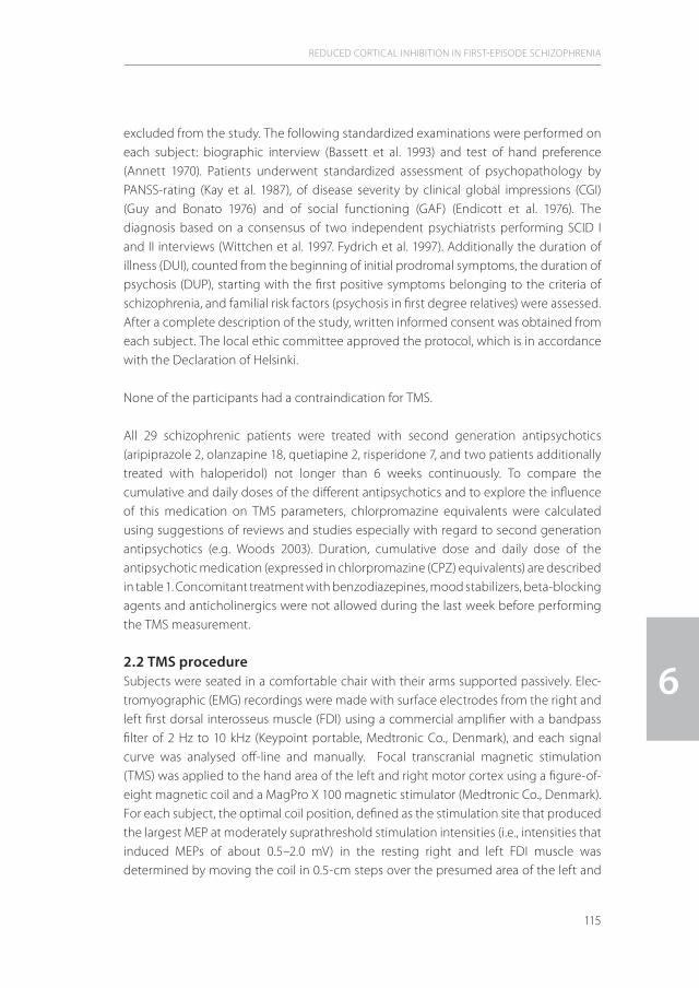
115
excluded from the study. The following standardized examinations were performed on each subject: biographic interview (Bassett et al. 1993) and test of hand preference (Annett 1970). Patients underwent standardized assessment of psychopathology by PANSS-rating (Kay et al. 1987), of disease severity by clinical global impressions (CGI) (Guy and Bonato 1976) and of social functioning (GAF) (Endicott et al. 1976). The diagnosis based on a consensus of two independent psychiatrists performing SCID I and II interviews (Wittchen et al. 1997. Fydrich et al. 1997). Additionally the duration of illness (DUI), counted from the beginning of initial prodromal symptoms, the duration of psychosis (DUP), starting with the first positive symptoms belonging to the criteria of schizophrenia, and familial risk factors (psychosis in first degree relatives) were assessed. After a complete description of the study, written informed consent was obtained from each subject. The local ethic committee approved the protocol, which is in accordance with the Declaration of Helsinki.
None of the participants had a contraindication for TMS.
All 29 schizophrenic patients were treated with second generation antipsychotics (aripiprazole 2, olanzapine 18, quetiapine 2, risperidone 7, and two patients additionally treated with haloperidol) not longer than 6 weeks continuously. To compare the cumulative and daily doses of the different antipsychotics and to explore the influence of this medication on TMS parameters, chlorpromazine equivalents were calculated using suggestions of reviews and studies especially with regard to second generation antipsychotics (e.g. Woods 2003). Duration, cumulative dose and daily dose of the antipsychotic medication (expressed in chlorpromazine (CPZ) equivalents) are described in table 1. Concomitant treatment with benzodiazepines, mood stabilizers, beta-blocking agents and anticholinergics were not allowed during the last week before performing the TMS measurement.
2.2 TMS procedureSubjects were seated in a comfortable chair with their arms supported passively. Elec-tromyographic (EMG) recordings were made with surface electrodes from the right and left first dorsal interosseus muscle (FDI) using a commercial amplifier with a bandpass filter of 2 Hz to 10 kHz (Keypoint portable, Medtronic Co., Denmark), and each signal curve was analysed off-line and manually. Focal transcranial magnetic stimulation (TMS) was applied to the hand area of the left and right motor cortex using a figure-of-eight magnetic coil and a MagPro X 100 magnetic stimulator (Medtronic Co., Denmark). For each subject, the optimal coil position, defined as the stimulation site that produced the largest MEP at moderately suprathreshold stimulation intensities (i.e., intensities that induced MEPs of about 0.5–2.0 mV) in the resting right and left FDI muscle was determined by moving the coil in 0.5-cm steps over the presumed area of the left and
REDUCED CORTICAL INHIBITION IN FIRST-EPISODE SCHIZOPHRENIA
6
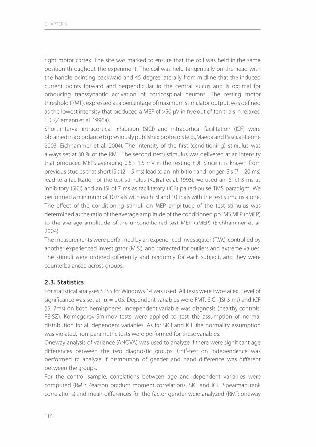
116
right motor cortex. The site was marked to ensure that the coil was held in the same position throughout the experiment. The coil was held tangentially on the head with the handle pointing backward and 45 degree laterally from midline that the induced current points forward and perpendicular to the central sulcus and is optimal for producing transsynaptic activation of corticospinal neurons. The resting motor threshold (RMT), expressed as a percentage of maximum stimulator output, was defined as the lowest intensity that produced a MEP of >50 µV in five out of ten trials in relaxed FDI (Ziemann et al. 1996a). Short-interval intracortical inhibition (SICI) and intracortical facilitation (ICF) were obtained in accordance to previously published protocols (e.g., Maeda and Pascual-Leone 2003, Eichhammer et al. 2004). The intensity of the first (conditioning) stimulus was always set at 80 % of the RMT. The second (test) stimulus was delivered at an intensity that produced MEPs averaging 0.5 - 1.5 mV in the resting FDI. Since it is known from previous studies that short ISIs (2 – 5 ms) lead to an inhibition and longer ISIs (7 – 20 ms) lead to a facilitation of the test stimulus (Kujirai et al. 1993), we used an ISI of 3 ms as inhibitory (SICI) and an ISI of 7 ms as facilitatory (ICF) paired-pulse TMS paradigm. We performed a minimum of 10 trials with each ISI and 10 trials with the test stimulus alone. The effect of the conditioning stimuli on MEP amplitude of the test stimulus was determined as the ratio of the average amplitude of the conditioned ppTMS MEP (cMEP) to the average amplitude of the unconditioned test MEP (uMEP) (Eichhammer et al. 2004). The measurements were performed by an experienced investigator (T.W.), controlled by another experienced investigator (M.S.), and corrected for outliers and extreme values. The stimuli were ordered differently and randomly for each subject, and they were counterbalanced across groups.
2.3. StatisticsFor statistical analyses SPSS for Windows 14 was used. All tests were two-tailed. Level of significance was set at a = 0.05. Dependent variables were RMT, SICI (ISI 3 ms) and ICF (ISI 7ms) on both hemispheres. Independent variable was diagnosis (healthy controls, FE-SZ). Kolmogorov-Smirnov tests were applied to test the assumption of normal distribution for all dependent variables. As for SICI and ICF the normality assumption was violated, non-parametric tests were performed for these variables. Oneway analysis of variance (ANOVA) was used to analyze if there were significant age differences between the two diagnostic groups. Chi²-test on independence was performed to analyze if distribution of gender and hand difference was different between the groups.For the control sample, correlations between age and dependent variables were computed (RMT: Pearson product moment correlations, SICI and ICF: Spearman rank correlations) and mean differences for the factor gender were analyzed (RMT: oneway
CHAPTER 6

117
ANOVA, SICI and ICF: Mann-Whitney U-test). Since all controls were right-handers, mean differences for factor hand preference was analyzed in the FE-SZ sample (RMT: oneway ANOVA, SICI and ICF: Mann-Whitney U-test).These preliminary calculations regulated the main analyses. For RMT analysis of covariance (ANCOVA, factor diagnosis, covariate age) was computed, since age correlated significantly with RMT. Furthermore, MANOVA with repeated measure design (within-subject factor side, between subject factor diagnosis) was performed to test the hypothesis, that in right-handed subjects RMT is lower on the left compared to the right motor cortex. For SICI and ICF variables Mann-Whitney U-test was used to analyze diagnosis differences. For the FE-SZ patients Spearman rank correlations between dependent variables and total PANSS scores, GAF, CGI, DUP and DUI were performed.
3. Results
3.1. Sociodemographic and clinical characteristicsThe first-episode patients did not differ significantly in age compared to the healthy control subjects (mean age 29.8 ± 8.5 years versus 32.2 ± 7.9 years, p = 0.28). The percentage of male patients was higher at a trend level in the schizophrenic patient group (p = 0.082). While all healthy control subjects were right-handers, in the FE-SZ group 4 patients were ambidexters (p=0.042). No subject fulfilled the criteria for borderline or antisocial personality disorder (due to SCID II interview, DSM-IV axis II, personality disorders), which may include abnormal high impulsivity potentially linked to reduced inhibition processes. In addition no healthy control subject fulfilled the criteria of any axis II disorder.The first-episode patients suffered from moderate to severe positive and negative symptoms according to the PANSS, accompanied by a severe degree of illness (CGI), and were severely impaired in their social functions (GAF) (for details see table 1).More severely ill patients received higher dosages (expressed in CPZ-equivalents), as the cumulative doses correlated positively with total PANSS score (rho = -0.65, p = 0.062) at a trend level an even significantly with CGI (rho = 0.65, p < 0.0005).
3.2. Influence of sociodemographic characteristics on TMS parametersA positive correlation was found between age and RMT on both hemispheres in the healthy control group (r = 0.36, p = 0.04, left motor cortex; r = 0.41, p = 0.04 , right motor cortex). In the healthy control group a significant influence of gender on cortical facilitation (ISI 7 ms, right side) was observed (male controls displayed enhanced facilitation; p = 0.004). In FE-SZ, for all dependent variables no significant differences between right-handers and ambidexters were observed.
REDUCED CORTICAL INHIBITION IN FIRST-EPISODE SCHIZOPHRENIA
6

118
3.3. Resting motor threshold (RMT)RMT was similar in first-episode patients and healthy controls on both sides (46.9 % in FE-SZ vs. 47.8 % in HC, p = 0.88 for left motor cortex and 48.3 % in FE-SZ vs. 49.6 % in HC;
CHAPTER 6
Table 1 Sociodemographic and clinical parameters
HC(N = 28)
FE-SZ(N = 29)
m sd m sd df F p
Age (years)a 32.18 7.86 29.79 8.47 1, 55 1.21 0.28
DUP (weeks) - - 50.41 64.0 - - -
DUI (weeks) - - 183.17 157.1 - - -
PANSS total score - - 91.48 17.2 - - -
PANSS positive score - - 22.14 6.1 - - -
PANSS negative score - - 21.45 6.6 - - -
PANSS gen. psychop. score - - 47.90 9.4 - - -
CGI - - 5.96 0.6 - - -
GAF - - 28.6 10.5 - - -
Daily dose of antipsychotics (CPZ-equivalents)
356.2 203.7
Duration of antipsychotic medication (days)
21.0 14.4
Cumulative dose of antipsychotics (CPZ-equivalents)
7565.2 7886.8
df Chi² p
Gender (male/female)b 14 / 14 21 / 8 1 3.02 0.082
Handedness (right/left/both) b 28 / 0 / 0 25 / 0 / 4 1 4.15 0.042
Psychosis in first degree relatives, no. of subjects (%)
0 (0) 12 (41.3) - - -
Legend:HC = healthy controls; FE-SZ = patients with first-episode schizophrenic; m = mean; sd = standard deviation; DUP = duration of psychosis; DUI = duration of illness (including initial prodrome); PANSS = Positive and Negative Syndrome Scale; gen, psychop. = general psychopathology; CGI = Clinical Global Impressions; GAF = Global Assessment of Functioning; no. = number; df = degrees of freedom; F = F-statistics; p = probability; CPZ = chlorpromazine.
a Analysis of variance (ANOVA); b Chi-Quadrat test

119
p = 0.73 for right motor cortex) (for details see table 2). When only the right-handed subjects were included in the analysis, RMT was significantly lower on the left compared to the right motor cortex in both subgroups (p = 0.039).
3.4. Short-interval intracortical inhibition (SICI) and intracortical facilitation (ICF)
In first-episode patients (FE-SZ) significantly higher MEP ratios (cMEP/uMEP) were measured at the inhibitory ISI of 3 ms compared to healthy controls (HC) on left motor cortex (MEP ratio 41 % in FE-SZ vs. 21 % in HC; p = 0.017) (figure 1), reflecting lower SICI in the patient group. When only the right-handed FE-SZ (MEP ratio 40 %) were analyzed, the difference compared to the control group remained significant (p = 0.033). On the right motor cortex this reduced SICI in FE-SZ (ISI 3 ms) was seen at a trend level (MEP ratio 59 % in FE-SZ vs. 31 % in HC; p = 0.059).
REDUCED CORTICAL INHIBITION IN FIRST-EPISODE SCHIZOPHRENIA
6
Figure 1 Error bars for paired-pulse inhibition in diagnostic groups
First-episode patients showed significantly lower decreased MEP amplitudes (higher ratio: conditioned MEP/unconditioned MEP; mean, 95 % CI) at the inhibitory ISI of 3 ms compared to healthy controls on left motor cortex (Z = -2.39; p = 0.017; Mann-Whitney-U-test).Abbreviations: CI = confidence interval; MEP = motor evoked potential; ISI = interstimulus interval; FE-SZ = patients with first-episode schizophrenia
FE-SZ Healthy Controls
95 %
CI;
ratio
: co
nditi
oned
MEP
/unc
ondi
tione
dMEP 0,50
0,40
0,30
0,20

120
For the MEP ratio of the facilitatory ISI of 7 ms (ICF) no significant differences between the two groups on both hemispheres could be observed (for details see table 2).
3.5. Correlation with clinical parametersThere was a positive correlation between PANSS total score and RMT of left motor cortex (rho = 0.395, p = 0.034; Spearman rank correlations) and a negative correlation between PANSS total score and cortical inhibition (MEP ratio at 3 ms) of left motor cortex in first-episode schizophrenia (rho = -0.403; p = 0.030; Spearman rank correlations). These correlations could not be found with the PANSS positive or negative subscore, but between RMT of left motor cortex and PANSS general psychopathology (rho = 0.385, p = 0.039; Spearman rank correlations). The detected correlations remained not significant after Bonferonni correction and have to be interpreted with caution. No
CHAPTER 6
Table 2 TMS parameters
HC(N = 28)
FE-SZ(N = 29)
m sd m sd df F p
RMT left motor cortex (%)a
47.82 6.5 46.93 6.9 1, 54 0.0 0.88
RMT right motor cortex (%)a
49.58 10,0 48.25 8.01, 51 0.1 0.73
df Z p
ppTMS ISI 3 ms left motor cortex (ratio cMEP/uMEP)b
0.20 0.1 0.41 0.3 1 -2.39 0.017
ppTMS ISI 7 ms left motor cortex (ratio cMEP/uMEP) b
0.96 0.5 1.12 1.0 1 -0.22 0.83
ppTMS ISI 3 ms right motor cortex (ratio cMEP/uMEP) b
0.31 0.2 0.59 0.9 1 -1.89 0.059
ppTMS ISI 7 ms right motor cortex (ratio cMEP/uMEP) b
1.14 0.7 1.48 1.4 1 -0.41 0.68
Legend:HC = healthy controls; FE-SZ = patients with first-episode schizophrenic; m = mean; sd = standard deviation; ppTMS = paired-pulse transcranial magnetic stimulation; ISI = interstimulus interval; mV = millivolt; N = number; df = degrees of freedom; F = F-statistics; Z = Z-value; p = probability; cMEP = conditioned motor evoked potential; uMEP = unconditioned motor evoked potential; RMT = resting motor thresholda Analysis of covariance (ANCOVA, factor diagnosis, covariate age ). percentage of maximum stimulator outputb Mann-Whitney-U-Test
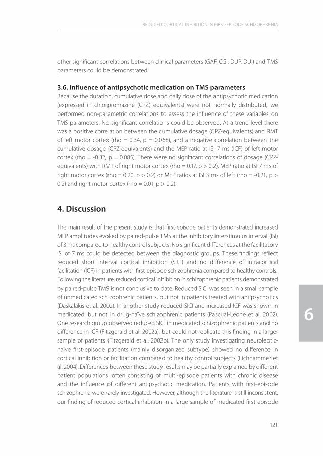
121
other significant correlations between clinical parameters (GAF, CGI, DUP, DUI) and TMS parameters could be demonstrated.
3.6. Influence of antipsychotic medication on TMS parametersBecause the duration, cumulative dose and daily dose of the antipsychotic medication (expressed in chlorpromazine (CPZ) equivalents) were not normally distributed, we performed non-parametric correlations to assess the influence of these variables on TMS parameters. No significant correlations could be observed. At a trend level there was a positive correlation between the cumulative dosage (CPZ-equivalents) and RMT of left motor cortex (rho = 0.34, p = 0.068), and a negative correlation between the cumulative dosage (CPZ-equivalents) and the MEP ratio at ISI 7 ms (ICF) of left motor cortex (rho = -0.32, p = 0.085). There were no significant correlations of dosage (CPZ-equivalents) with RMT of right motor cortex (rho = 0.17, p > 0.2), MEP ratio at ISI 7 ms of right motor cortex (rho = 0.20, p > 0.2) or MEP ratios at ISI 3 ms of left (rho = -0.21, p > 0.2) and right motor cortex (rho = 0.01, p > 0.2).
4. Discussion
The main result of the present study is that first-episode patients demonstrated increased MEP amplitudes evoked by paired-pulse TMS at the inhibitory interstimulus interval (ISI) of 3 ms compared to healthy control subjects. No significant differences at the facilitatory ISI of 7 ms could be detected between the diagnostic groups. These findings reflect reduced short interval cortical inhibition (SICI) and no difference of intracortical facilitation (ICF) in patients with first-episode schizophrenia compared to healthy controls. Following the literature, reduced cortical inhibition in schizophrenic patients demonstrated by paired-pulse TMS is not conclusive to date. Reduced SICI was seen in a small sample of unmedicated schizophrenic patients, but not in patients treated with antipsychotics (Daskalakis et al. 2002). In another study reduced SICI and increased ICF was shown in medicated, but not in drug-naïve schizophrenic patients (Pascual-Leone et al. 2002). One research group observed reduced SICI in medicated schizophrenic patients and no difference in ICF (Fitzgerald et al. 2002a), but could not replicate this finding in a larger sample of patients (Fitzgerald et al. 2002b). The only study investigating neuroleptic-naive first-episode patients (mainly disorganized subtype) showed no difference in cortical inhibition or facilitation compared to healthy control subjects (Eichhammer et al. 2004). Differences between these study results may be partially explained by different patient populations, often consisting of multi-episode patients with chronic disease and the influence of different antipsychotic medication. Patients with first-episode schizophrenia were rarely investigated. However, although the literature is still inconsistent, our finding of reduced cortical inhibition in a large sample of medicated first-episode
REDUCED CORTICAL INHIBITION IN FIRST-EPISODE SCHIZOPHRENIA
6

122
patients with limited exposure to antipsychotic medication strongly supports the hypothesis that inhibitory intracortical deficits may be part of the schizophrenic patho-physiology. Several other neurophysiological studies using other tools than TMS, have provided evidence for inhibitory deficits in schizophrenia. For instance schizophrenic patients showed reduced P50 wave suppression in an auditory conditioning test paradigm (Adler et al. 1982), potentially reflecting reduced inhibition in a cortico-sub-cortical loop. We could not detect significant differences in ICF between medicated first-episode patients and comparable healthy control subjects. Besides reduced SICI schizophrenia patients could have presented increased ICF, but in our study this was not the case.SICI is linked to the GABAergic system and the function of cortical GABAergic interneurons (Ziemann 2004). In healthy subjects GABA
A agonists and also glutamate
antagonists enhance SICI; the latter effect may be best explained through a decrease of facilitatory effects (Ziemann 2004). Recent investigations were able to segregate different GABAergic inhibitory circuits in human motor cortex and to show that SICI is probably mediated via a2- or a3-subunit rather than the a1-subunit of the GABA
A
receptor (Di Lazzaro et al. 2007). The a2-subunit is found only in about 15 % of cortical GABA receptors but at above 95 % of inhibitory synapses onto pyramidal neuron axon initial segments, especially in the superficial layers of the human cerebral cortex and seems to be related to higher affinity for GABA, which results in faster activation and slower deactivation times than the more common a1-subunit of the GABA
A receptor
(Lewis et al. 2005). On the other hand there are pharmacological studies suggesting that GABAergic activation (e.g. through benzodiazepines) mainly influence ICF rather than SICI in healthy controls (Ziemann et al. 1996b). Dopamine and noradrenaline agonists increase SICI, while dopamine antagonists like haloperidol decrease SICI (Ziemann 2004). Interestingly, the selective serotonine re-uptake inhibitor citalopram increased SICI in subjects, who were homozygotic for the long variant of the 5-HT transporter gene (Eichhammer et al. 2003). This finding suggests also an involvement of serotonine, dependent on genetic polymorphism, on cortical inhibition. On the other hand neither single nor chronic administration of paroxetine resulted in changes of SICI, but single dose of paroxetine decreased ICF, while chronic paroxetine administration enhances ICF in healthy volunteers (Gerdelat-Mas et al. 2005). This contrary effect on ICF emphasizes the different pharmacological action of a drug at cortical level depending on its acute or long-term administration. For ICF the majority of pharmacological studies on healthy subjects demonstrated a decrease of ICF by NMDA antagonists (Ziemann 2004). In contrast another study found an increase of ICF after ketamine application, a NMDA antagonist, potentially mediated by activating non-NMDA glutamatergic transmission via AMPA and kainate receptors (Di Lazzaro et al. 2003). In conclusion, our results of decreased SICI and unchanged ICF compared to healthy controls fits best to the hypothesis of GABAergic dysfunction in schizophrenia, although the dysbalance of
CHAPTER 6

123
other neurotransmitters like glutamate, dopamine and serotonine involved in SICI reduction could not be ruled out. Nevertheless, the cited findings in healthy subjects are not necessarily comparable to the results and conditions in schizophrenia patients, and neuronal networks in schizophrenia including adaptive processes during disease course and long-term medication may be responsible for differences.We found no significant differences in resting motor threshold (RMT) between schizophrenic patients and healthy controls. This is in line with the literature. Summing up the results of previous studies investigating RMT in schizophrenic patients, more than half of the studies (8 of 15) demonstrated no significant differences between patients and control subjects (Wobrock et al. 2007). In 3 studies a decreased RMT in schizophrenic patients was observed, as it has to be expected if reduced cortical inhibition and increased excitability of motor neurons is assumed (Abarbanel et al. 1996, Daskalakis et al 2002, Eichhammer 2004). In an other study a significant increased RMT in patients treated with antipsychotics compared to medication- free patients and normal controls was detected (Pascual-Leone et al. 2002), and in addition, a higher RMT on the left dominant hemisphere compared to the right side in medicated and unmedicated patients could be observed. This is remarkable concerning the observation that in some investigations healthy controls display a lower RMT on the left dominant side in inter-hemispheric comparison (Triggs et al. 1994). In our study we found no difference in laterality between controls and first-episode patients. In two studies investigating the effect of low frequency rTMS on RMT, in schizophrenic patients no increase of RMT was seen after rTMS compared to healthy controls (Oxley et al. 2004, Fitzgerald et al. 2004). This was discussed as reduced cortical neuroplasticity or missing cortical inhibition in schizophrenia. In addition, we found evidence for a correlation between total symptom severity, expressed by PANSS total score and increased RMT, as well as decreased cortical inhibition. This means that reduced cortical inhibition is inversely associated with symptom severity, which seems rather contraintuitive. An explanation may be that more severely ill patients receive higher dosages of antispsychotics (CPZ-equivalents), as they did in our study, and therefore the cortical inhibition deficits could be “overcorrected”. However the above mentioned negative correlation between SICI only on left motor cortex and PANSS total score could not be replicated with the PANSS positive and negative subscore. This may indicate that there is no stable correlation between symptom severity and cortical inhibition deficits at all. Furthermore, our findings of decreased cortical inhibition in schizophrenia may reflect a more general vulnerability factor (e.g. as a trait marker) than a characterization of psychopathology (e.g. as a state marker). Our study has several limitations. First of all, antipsychotic medication could be a considerable confounding factor using TMS paradigms (Davey et al. 1997). For instance, one study found a difference in RMT between patient treated with risperidone (increased
REDUCED CORTICAL INHIBITION IN FIRST-EPISODE SCHIZOPHRENIA
6

124
RMT) and olanzapine (decreased RMT) (Fitzgerald et al. 2002c). We did not find a differential influence of the used antipsychotics in our patient sample. This contributes to the result of another study with healthy control subjects, where an effect on resting motor threshold (RMT) after olanzapine or haloperidol administration could not be detected (Daskalakis et al. 2003). This was explained by the fact that RMT is conventionally regarded as a measure of the membrane excitability of corticospinal neurons and interneurons in the motor cortex, increased by drugs that block voltage-gated sodium channels and not affected by drugs that alter GABA, glutamate or dopamine transmission (Di Lazarro et al. 2003, Liepert et al. 1997, Werhahn et al. 1999, Ziemann et al.1996a, Ziemann et al. 1996b, Ziemann et al. 1997, Ziemann et al. 1998) like olanzapine or haloperidol.For paired-pulse measurements, medication influencing GABA, glutamate, serotonine or dopamine transmission should be potentially relevant, because the interneuronal-pyramidal circuits are mediated via these pathways. However, according to the literature, one would expect that antipsychotics should compensate the reduced inhibition and not produce it, because there are no studies available showing increased cortical inhibition in unmedicated or medicated schizophrenia and only one study demonstrating reduced cortical inhibition exclusively in medicated patients (Pascual-Leone et al. 2002). Studies of the medication effect on cortical inhibition performed on healthy people (mostly given as single dose) do not necessarily reflect the effect of the same psychopharmaco-logical drugs administered chronically to schizophrenic patients with a potentially different neuronal network. For future studies in schizophrenia patients it may be helpful to include other comparison groups e.g. patients with bipolar disorder treated with atypical antipsychotic drugs to control for effects of these drugs and to compare these effects with disease specific alterations.The strength of our study is that we were able to investigate a well characterized relatively large sample of first-episode patients which seems representative for this patient group in a clinical setting. Due to disease severity and the clinical necessity for medication it was not possible to measure these patients neuroleptic-naïve.The origin of reduced cortical inhibition measured by TMS in schizophrenia is still speculative. In one model reduced cortical inhibition in schizophrenia is explained by the assumption of a disorder of altered dopaminergic inputs from pyramidal to nonpyramidal neurons in the cortex (Benes 2000, Benes 1998). To support this hypothesis it was demonstrated that in schizophrenia dopaminergic projections to the cortex erroneously terminate on non-pyramidal cells (i.e. interneurons) to inhibit their function. Such connectivity would result in decreased activity of inhibitory interneurons and increased excitatory output in the cortex. Another explanation may be the finding of decreased numbers of (inhibitory, gabaergic) interneurons in the postmortem brains of schizophrenic patients, which could lead to reduced cortical inhibition (Benes 1991). There is an increasing literature pointing towards a GABAergic dysfunction in schizophrenia (Lewis et al. 2005). In individuals with schizophrenia an increased density of GABA
A
CHAPTER 6

125
receptors was found in ligand binding studies, probably indicating a local upregulation in response to a reduction in perisomatic inhibitory input from chandelier and wide arbor neurons (Lewis et al. 2005). In post mortem studies there is convincing evidence for decreased GABA synthesis in schizophrenia with the finding of reduced glutamic acid decarboxylase (GAD67) mRNA expression in dorsolateral prefrontal cortex (Knable et al. 2002). This reduced synthesis of GABA in a parvalbumin-containing subpopulation of inhibitory GABA neurons may be caused by deficiency in signalling through tyrosine kinase receptor B, the receptor for brain-derived neurotrophic factor (BDNF) (Lewis et al. 2005). One of the consequences of reduced GABAergic transmission may be a deficit in synchronisation of pyramidal cells resulting in e.g. working memory deficits, a core syndrome of schizophrenia (Lewis et al. 2005). Our finding of reduced SICI contributes to the hypothesis of GABAergic dysfunction in schizophrenia. However, it has to be kept in mind that histopathological studies demonstrating a reduced number of GABAergic interneurons in the primary motor cortex of schizophrenia patients are not available.A reduced SICI detected by TMS has been found in other neuropsychiatric disorders, i.e. unipolar depression (Bajbouj et al. 2006), attention-deficit/hyperactivity disorder (ADHD) (Richter et al. 2007) or obsessive-compulsive disorder (OCD) (Greenberg et al. 2000). With regard to these findings intracortical inhibition deficits seem not specifically restricted to schizophrenia. Future studies combining different modalities and investigational tools (e.g. TMS, event related potentials, magnetencephalography, structural and functional magnetic resonance imaging) may gain new insights in neuronal networks and either intracortical or cortico-subcortical connectivity.
REDUCED CORTICAL INHIBITION IN FIRST-EPISODE SCHIZOPHRENIA
6

126
References
1. Abarbanel, J.M., Lemberg, T., Yaroslavski, U., Grisaru, N., Belmaker, R.H., 1996. Electrophysiological responses to transcranial magnetic stimulation in depression and schizophrenia. Biol. Psychiatry 40 (2), 148-150.
2. Adler, L.E., Pachtman, E., Frank, R.D., Pecevich, M., Waldo, M.C., Freedman, R., 1982. Neuophysiological evidence for a defect in neuronal mechanisms involved in sensory gating in schizophrenia. Biol. Psychiatry 17, 639-654.
3. Annett, M., 1970. A classification of hand preference by association analysis. Br. J. Psychol. 61, 303–321.4. Bajbouj, M., Lisanby, S.H., Lang, U.E., Danker-Hopfe, H., Heuser, I., Neu, P., 2006. Evidence for impaired cortical
inhibition in patients with unipolar major depression. Biol. Psychiatry 59, 395-400. 5. Bassett, A.S., Collins, E.J., Nuttall, S.E., Honer, W.G., 1993. Positive and negative symptoms in families with
schizophrenia. Schizophr. Res. 11, 9–19. 6. Benes, F.M., McSparen, J., Bird, E.D., San Giovanni, J.P., Vincent, S.L., 1991. Deficits in small interneurons in
prefrontal and cingulated cortices of schizophrenic and schizoaffective patients. Arch. Gen. Psych. 48, 996 – 1001.
7. Benes, F.M., 1998. Model generation and testing to probe neural circuitry in the cingulate cortex of postmortem schizophrenic brain. Schizophr. Bull 24, 219-230.
8. Benes, F.M., 2000. Cortical Pathology: a new generation of quantitative microscopic studies. In: Harrison, P.J., Roberts, G.W. (Eds.), The Neuropathology of Schizophrenia: Progres and Interpretation. Oxford University Press, New York, NY, pp. 81-104.
9. Chen, Y.L., Chen, Y.H., Mak, F.L., 2000. Soft neurological signs in schizophrenic patients and their nonpsychotic siblings. J. Nerv. Ment. Dis. 188, 84-89.
10. Crow, T.J., Ball, J., Bloom, S.R., Bruton, C.J., Colter, N., Frith, C. D., Johnstone, E.C., Owens, D.G., Roberts, G.W., 1989. Schizophrenia as an anomaly of development of cerebral asymmetry: a post-mortem study and a proposal concerning the genetic basis of the disease. Arch. Gen. Psychiatry 46, 1145-1150.
11. Daskalakis, Z.J., Christensen, B.K., Chen, R., Fitzgerald, P.B., Zipursky, R.B., Kapur, S., 2002. Evidence for impaired cortical inhibition in schizophrenia using transcranial magnetic stimulation. Arch. Gen. Psychiatry 59 (4), 347-354.
12. Daskalakis, Z.J., Christensen, B.K., Chen, R., Fitzgerald, P.B., Zipursky, R.B., Kapur, S., 2003. Effect of antipsychotics on cortical inhibition using transcranial magnetic stimulation. Psychopharmacology (Berl) 170 (3), 255-262
13. Davey, N.J., Puri, B.K., Lewis, H.S., Lewis, S.W., Ellaway, P.H., 1997. Effects of antipsychotic medication on elec-tromyographic responses to transcranial magnetic stimulation of the motor cortex in schizophrenia. J. Neurol. Neurosurg. Psychiatry 63 (4), 468-473.
14. Day, B.L., Dressler, D., Maertens De Noordhout, A., Marsden, C.D., Nakashima, K., Rothwell, J.C., Thompson, P.D., 1989. Electrical and magnetic stimulation of human motor cortex: surface EMG and single motor-unit responses. J. Physiol. (Lond) 412, 449–473.
15. Di Lazarro, V., Oliviero, A., Profice, P., Pennisi, MA., Pilato, F., Zito, G., Dileone, M., Nicoletti, R., Pasqualetti, P., Tonalli, P.A., 2003. Ketamine increases human motor cortex excitability to transcranial magnetic stimulation. J. Physiol. 547, 485-496.
16. Di Lazzaro, V., Pilato, F., Dileone, M., Profice, P., Ranieri, F., Ricci, V., Bria, P., Tonali, P.A., Ziemann, U., 2007. Segregating two inhibitory circuits in human motor cortex at the level of GABAA receptor subtypes: a TMS study. Clin Neurophysiol. 118 (10), 2207-2214.
17. Eichhammer, P., Langguth, B., Wiegand, R., Kharraz, A., Frick, U., Hajak, G., 2003. Allelic variation in the serotonin transporter promoter affects neuromodulatory effects of a selective serotonin transporter reuptake inhibitor (SSRI). Psychopharmacology (Berl). 166 (3), 294-297.
18. Eichhammer, P., Wiegand, R., Kharraz, A., Langguth, B., Binder, H., Hajak, G., 2004. Cortical excitability in neu-roleptic-naive first-episode schizophrenic patients. Schizophr. Res. 67 (2-3), 253-259.
19. Endicott, J., Spitzer, R.L., Fleiss, J.L., Cohen, J., 1976. The Global Assessment Scale: A procedure for measuring overall severity of psychiatric disturbance. Arch. Gen. Psychiatry 33, 766-771.
20. Fitzgerald, P.B., Brown, T.L., Daskalakis, Z.J., deCastella, A., Kulkarni, J., 2002a. A study of transcallosal inhibition in schizophrenia using transcranial magnetic stimulation. Schizophr. Res. 56 (3), 199-209.
CHAPTER 6

127
21. Fitzgerald, P.B., Brown, T.L., Daskalakis, Z.J., Kulkarni, J., 2002b. A transcranial magnetic stimulation study of inhibitory deficits in the motor cortex in patients with schizophrenia. Psychiatry Res. 114 (1), 11-22.
22. Fitzgerald, P.B., Brown, T.L., Daskalakis, Z.J., Kulkarni, J., 2002c. A transcranial magnetic stimulation study of the effects of olanzapine and risperidone on motor cortical excitability in patients with schizophrenia. Psychop-harmacology (Berl) 162 (1), 74-81.
23. Fitzgerald, P.B., Brown, T.L., Marston, N.A., Oxley, T., De Castella, A., Daskalakis, Z.J., Kulkarni, J., 2004. Reduced plastic brain responses in schizophrenia: a transcranial magnetic stimulation study. Schizophr. Res. 71 (1), 17-26.
24. Freedman, R., Adler, L.E., Waldo, M.C., Pachtman, E., Franks, R.D., 1983. Neurophysiological evidence for a defect in inhibitory pathways in schizophrenia: comparison of medicated and drug-free patients. Biol. Psychiatry 18, 537–551.
25. Fydrich, T., Renneberg, B., Schmitz, B., Wittchen, H.U., 1997. Strukturiertes Klinisches Interview für DSM-IV. Achse II: Persönlichkeitsstörungen. A German, advanced adaptation of the original SCID version by RL. Spitzer, JB .Williams, M. Gibbon and MB. First. Hogrefe-Verlag, Göttingen.
26. Gerdelat-Mas, A., Loubinoux, I., Tombari, D., Rascol, O., Chollet, F., Simonetta-Moreau, M., 2005. Chronic administration of selective serotonin reuptake inhibitor (SSRI) paroxetine modulates human motor cortex excitability in healthy subjects. Neuroimage 27 (2), 314-322.
27. Greenberg, B.D., Ziemann, U., Cora-Locatelli, G., Harmon, A., Murphy, D.L., Keel, J.C., Wassermann, E.M., 2000. Altered cortical excitability in obsessive-compulsive disorder. Neurology 54 (1), 142-147.
28. Guy, W., Bonato, R.R., (Eds.), 1976. CGI: Clinical Global Impressions. In: Manual for the ECDEU Assessment Battery.2. Rev ed. Chevy Chase, Md: National Institute of Mental Health, pp. 12/1-12/6.
29. Kay, S.R., Fiszbein, A., Opler, L.A., 1987. The Positive and Negative Syndrome Scale (PANSS) for schizophrenia. Schizophr. Bull. 13, 261–276
30. Knable, M.B., Barci, B.M., Bartko, J.J., Webster, M.J., Torrey, E.F., 2002. Molecular abnormalities in the major psychiatric illnesses: classification and regression tree (CRT) analysis of post-mortem prefrontal markers. Mol. Psychiatry 7, 392-404.
31. Kujirai, T., Caramis, M.D., Rothwell, J.C., Day, B.L., Thompson, P.D., Ferbert, A., Wroe, S., Asselman, P., Marsden, C.D., 1993. Corticospinal inhibition in human motor cortex. J. Physiol. 471, 501-519.
32. Lewis, D.A., Hashimoto, T., Volk, D.W., 2005. Cortical inhibitory neurons and schizophrenia. Nat. Rev. Neurosci. 6, 312-324.
33. Liepert, J., Schwenkreis, P., Tegenthoff, M., Malin, J.P., 1997. The glutamate antagonist riluzole suppresses intracortical facilitation. J. Neural. Transm. 104, 1207–1214.
34. Maeda, F., Pascual-Leone, A., 2003. Transcranial magnetic stimulation: studying motor neurophysiology of psychiatric disorders. Psychopharmacology 168, 359-376.
35. Nakamura, H., Kitagawa, H., Kawaguchi, Y., Tsuji, H., 1997. Intracortical facilitation and inhibition after transcranial magnetic stimulation in conscious humans. J Physiol. 498 (Pt 3), 817-823.
36. Oxley, T., Fitzgerald, P.B., Brown, T.L., de Castella, A., Daskalakis, Z.J., Kulkarni, J., 2004. Repetitive transcranial magnetic stimulation reveals abnormal plastic response to premotor cortex stimulation in schizophrenia. Biol. Psychiatry 56 (9), 628-633.
37. Pascual-Leone, A., Manoach, D.S., Birnbaum, R., Goff, D.C., 2002. Motor cortical excitability in schizophrenia. Biol. Psychiatry 52 (1), 24-31.
38. Puri, B.K., Davey, N.J., Ellaway, P.H., Lewis, S.W., 1996. An investigation of motor function in schizophrenia using transcranial magnetic stimulation of the motor cortex. Br. J. Psychiatry 169 (6), 690-695.
39. Richter, M.M., Ehlis, A.C., Jacob, C.P., Fallgatter, A.J., 2007. Cortical excitability in adult patients with attention-deficit/hyperactivity disorder (ADHD). Neurosci. Lett. 419 (2), 137-141.
40. Smith, R.C., Hussain, M.I., Chowdhury, S.A., Stearns, A., 1999. Stability of neurological soft signs in chronically hospitalised schizophrenic patients. J. Neuropsychiatry Clin. Neurosci. 11, 91-96.
41. Triggs, W.J., Calvanio, R., Macdonell, R.A:, Cros, D., Chiappa, K.H., 1994. Physiological motor asymmetry in human handedness: evidence from transcranial magnetic stimulation. Brain Res. 636, 270-276.
REDUCED CORTICAL INHIBITION IN FIRST-EPISODE SCHIZOPHRENIA
6

128
42. Werhahn, K.J., Kunesch, E., Noachtar, S., Benecke, R., Classen, J., 1999. Differential effects on motorcortical inhibition induced by blockade of GABA uptake in humans. J. Physiol. (Lond) 517, 501-507
43. Wittchen, H.U., Wunderlich, U., Gruschwitz, S., Zaudig, M., 1997. Strukturiertes Klinisches Interview für DSM-IV. Achse I: Psychische Störungen. A German, advanced adaptation of the original SCID version by RL. Spitzer, JB .Williams, M. Gibbon and MB. First. Hogrefe-Verlag, Göttingen.
44. Wobrock, T., Kadovic, D., Falkai, P., 2007. Cortical excitability in schizophrenia. Studies using transcranial magnetic stimulation. Nervenarzt 78 (7), 753-754, 756-763.
45. Woods, S.W., 2003. Chlorpromazine equivalent doses for the newer atypical antipsychotics. J. Clin. Psychiatry 64, 663-667.
46. Ziemann, U., Lonnecker, S., Steinhoff, B.J., Paulus, W., 1996a. Effects of antiepileptic drugs on motor cortex excitability in humans: a transcranial magnetic stimulation study. Ann. Neurol. 40, 367–378.
47. Ziemann, U., Lonnecker, S., Steinhoff, B.J., Paulus, W., 1996b. The effect of lorazepam on the motor cortical excitability in man. Exp. Brain Res. 109,127–135
48. Ziemann, U., Tergau, F., Bruns, D., Baudewig, J., Paulus, W., 1997. Changes in human motor cortex excitability induced by dopaminergic and anti-dopaminergic drugs. Electroencephalogr. Clin. Neurophysiol. 105, 430–437.
49. Ziemann, U., Chen, R., Cohen, L.G., Hallett, M., 1998. Dextromethorphan decreases the excitability of the human motor cortex. Neurology 51, 1320–1324.
50. Ziemann, U., Hallett, M., 2000. Basic neurophysiological studies with TMS. In: George, M.S., Belmaker, R.H., (Eds.), Transcranial magnetic stimulation in neuropsychiatry. Vol. 1, American Psychiatric Press, Washington DC, pp. 45-98.
51. Ziemann, U., 2004. TMS and drugs. Clin. Neurophysiol. 115, 1717-1729.
CHAPTER 6

129
REDUCED CORTICAL INHIBITION IN FIRST-EPISODE SCHIZOPHRENIA
6


A similar but distinctive pattern of impaired cortical excitability in first-episode schizophrenia and ADHD
Alkomiet Hasan*, Marc Schneider*, Thomas Schneider-Axmann, Diane Ruge, Wolfgang Retz, Michael Rösler, Peter Falkai, Thomas Wobrock
* First and second author equally contributed to the manuscript
Accepted in Neuropsychobiology.
7 |

132
Abstract
Background: First-episode-schizophrenia (FE-SZ) and attention deficit hyperactivity disorder (ADHD) are both neuropsychiatric disorders associated with an impaired dopaminergic transmission. Though displaying different clinical phenotypes, a common pathophysiological pathway is discussed controversially. Several studies using transcranial magnetic stimulation (TMS) revealed abnormalities in human motor cortex excitability in both schizophrenia and ADHD patients. Studies on cortical excitability comparing these two diseases directly are lacking.
Method: In this study, a total of 94 subjects were analyzed. 25 FE-SZ patients were directly compared with 28 ADHD patients and 41 healthy controls (HC). We investigated cortical excitability (inhibitory and facilitatory networks) with single- and paired-pulse-TMS to the left and right motor cortex.
Results: Compared to healthy controls, FE-SZ/ADHD displayed an impaired cortical inhibition over the left hemisphere. Apart from an enhanced intracortical facilitation, FE-SZ did not differ compared to ADHD patients in the main outcome measures. Both patient groups presented a dysfunctional hemispheric pattern of cortical inhibition and facilitation in comparison with HC.
Conclusion: The results of this study indicate a pattern of cortical disinhibition and abnormal hemispheric balance of intracortical excitability networks in two different psychiatric diseases. These effects might be associated with an imbalance in GABAergic and dopaminergic transmission and might provide evidence for a common patho-physiological pathway of both diseases.
CHAPTER 7

133
Introduction
Attention deficit hyperactivity disorder (ADHD) and schizophrenia are both heterogeneous disorders sharing a psychopathological profile of attention deficits and difficulties in social interaction. However, very perspicacious distinguishing features between these diseases are delusions, hallucinations and negative symptoms as a clinical manifestation in schizophrenia patients [1]. There is comorbidity between these diseases and retrospective studies of individuals with schizophrenia have shown a history of premorbid or concomitant ADHD [2].With regard to the pathophysiology, both conditions are associated with a dysfunctional dopaminergic transmission of the limbic-frontal network and other midbrain structures [3,4]. An enhanced dopaminergic transmission in mesolimbic structures is associated with positive symptoms, whereas a dopaminergic underactivity in these brain structures has been linked to negative symptoms and cognitive dysfunction in schizophrenia patients. In ADHD, the pathophysiological role of the dopamine system remains elusive. Both, high midbrain dopamine activity or reduced dopamine activity are discussed to be involved in the appearance of ADHD symptoms and the discussion about hypo- or hyperdopaminergic transmission imbalance remains contradictory [4,5]. In some studies, a dopaminergic hyperfunction in the mesolimbic structures was correlated with hyperactivity and impulsivity and a dopaminergic hypofunction was found to underlie different cognitive symptoms [3,4,6-8]. Apart from dopamine, other neu-rotransmitter systems, especially GABAergic interneurons, are involved in the patho-physiology of these diseases [9,10]. Using paired-pulse transcranial magnetic stimulation (TMS) a reduced cortical inhibition as another common pathophysiological state was found in both diseases [11,12]. Paired-pulse TMS offers the possibility to investigate different mechanisms of cortical inhibition (short interval intracortical inhibition, SICI) and of cortical facilitation (intracortical facilitation, ICF) in the human brain [13]. SICI is discussed to be mediated by GABAergic interneurons via GABA
A-receptors and ICF might result from primarily
glutamatergic neurotransmission [14]. Furthermore, dopaminergic neurotransmission has an impact on SICI and ICF, because dopamine agonists lead to an enhancement of SICI and ICF and dopamine antagonists reduce SICI and ICF in healthy subjects [14]. The contralateral silent period (CSP), a single-pulse TMS measure, is at least in part the result of GABA
B mediated inhibitory mechanisms [15,16].
In schizophrenia patients, several TMS studies revealed abnormalities in motor-cortical excitability which have been linked to GABAergic and dopaminergic dysfunction [17-20]. Our research group has recently demonstrated a reduced SICI and a prolonged CSP in first-episode schizophrenia (FE-SZ). We hypothesized that reduced SICI points towards a GABA
A deficit and the prolonged CSP may reflect compensatory increased GABA
B
transmission induced by hyperactivity of the dopaminergic system [11,21].
TMS IN FIRST-EPISODE SCHIZOPHRENIA AND ADHD
7
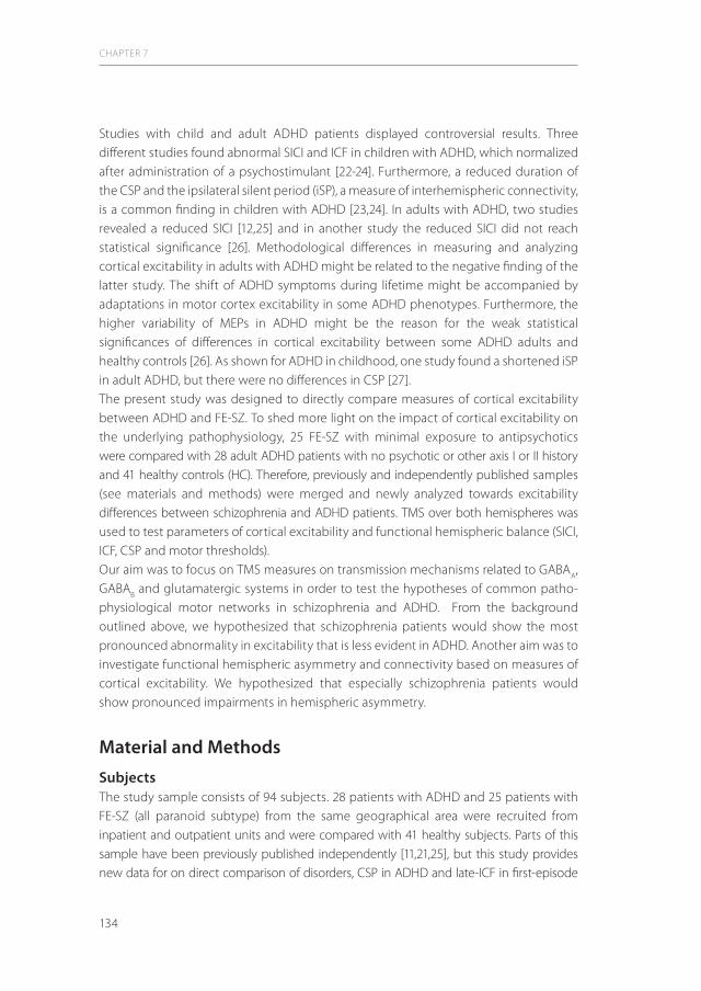
134
Studies with child and adult ADHD patients displayed controversial results. Three different studies found abnormal SICI and ICF in children with ADHD, which normalized after administration of a psychostimulant [22-24]. Furthermore, a reduced duration of the CSP and the ipsilateral silent period (iSP), a measure of interhemispheric connectivity, is a common finding in children with ADHD [23,24]. In adults with ADHD, two studies revealed a reduced SICI [12,25] and in another study the reduced SICI did not reach statistical significance [26]. Methodological differences in measuring and analyzing cortical excitability in adults with ADHD might be related to the negative finding of the latter study. The shift of ADHD symptoms during lifetime might be accompanied by adaptations in motor cortex excitability in some ADHD phenotypes. Furthermore, the higher variability of MEPs in ADHD might be the reason for the weak statistical significances of differences in cortical excitability between some ADHD adults and healthy controls [26]. As shown for ADHD in childhood, one study found a shortened iSP in adult ADHD, but there were no differences in CSP [27]. The present study was designed to directly compare measures of cortical excitability between ADHD and FE-SZ. To shed more light on the impact of cortical excitability on the underlying pathophysiology, 25 FE-SZ with minimal exposure to antipsychotics were compared with 28 adult ADHD patients with no psychotic or other axis I or II history and 41 healthy controls (HC). Therefore, previously and independently published samples (see materials and methods) were merged and newly analyzed towards excitability differences between schizophrenia and ADHD patients. TMS over both hemispheres was used to test parameters of cortical excitability and functional hemispheric balance (SICI, ICF, CSP and motor thresholds). Our aim was to focus on TMS measures on transmission mechanisms related to GABA
A,
GABAB and glutamatergic systems in order to test the hypotheses of common patho-
physiological motor networks in schizophrenia and ADHD. From the background outlined above, we hypothesized that schizophrenia patients would show the most pronounced abnormality in excitability that is less evident in ADHD. Another aim was to investigate functional hemispheric asymmetry and connectivity based on measures of cortical excitability. We hypothesized that especially schizophrenia patients would show pronounced impairments in hemispheric asymmetry.
Material and Methods
SubjectsThe study sample consists of 94 subjects. 28 patients with ADHD and 25 patients with FE-SZ (all paranoid subtype) from the same geographical area were recruited from inpatient and outpatient units and were compared with 41 healthy subjects. Parts of this sample have been previously published independently [11,21,25], but this study provides new data for on direct comparison of disorders, CSP in ADHD and late-ICF in first-episode
CHAPTER 7

135
schizophrenia. Subjects with a history of dementia, neurological illnesses and severe brain injuries were excluded from the study. All subjects were right handed according to a standardized test of hand preference [28].A clinical psychiatrist, blinded to the aims of the study, and a member of the study group (TW or MS) made a consensus diagnosis according to the German version of the Structural Clinical Interview for DSM IV [29]. Each subject underwent an assessment of disease severity (Clinical Global Impressions) [30] and an assessment of social functioning (Global Assessment of Functioning) [31]. In schizophrenia patients an assessment of psychopathology (Positive and Negative Syndrome Scale) [32], the duration of illness (DUI), counted from the beginning of initial prodromal symptoms and the duration of psychosis (DUP), counted from the onset of diagnostic/characteristic positive symptoms were evaluated according to the foregoing publication [11]. All schizophrenia patients were treated with second generation antipsychotics, but at the time of TMS measurements no patient had been treated longer than 6 weeks continuously. We calculated chlorpromazine equivalents [33] for the cumulative and daily doses of the different antipsychotics to explore the influence of this medication on the study results.The ADHD patients (combined type) were diagnosed according to the diagnostic criteria of DSM IV (ADHD-DC: ADHD Diagnostic checklist [34]), and had used no stimulant medication before. The Wender Utah Self Rating Scale – short version (WURS-k) [35]) was used to assess childhood ADHD symptoms. Patients with the clinical diagnosis of ADHD were included in the study if the WURS-k score was at least 30, and when at least 6 of 9 items of inattention and hyperactivity/impulsivity were present. As indicators of severity of symptoms, the ADHD self-rating scale for adults (ADHD-SR) according to DSM IV [34] was used (total score: 0 to 54; attention deficit and hyperactivity/impulsivity subscores: each maximal 27). SCID-1 and 2 interviews were used to exclude further axis I and II DSM categorical diagnoses. After a complete description of the study, written informed consent was obtained from each subject. The local ethics committee approved the protocol, which is in accordance with the Declaration of Helsinki.
TMS procedureAs described previously [11], subjects were seated in a comfortable reclining chair with their arms supported passively. Electromyographic (EMG) recordings from the right and left first dorsal interosseus muscle (FDI) were made with surface electrodes. Raw signals were amplified, bandpass filtered (2 Hz–10 kHz) and digitized using a commercial amplifier (Keypoint portable, Medtronic Co., Denmark). Each EMG recording was manually analyzed off-line. TMS was performed by using a MagPro X 100 magnetic stimulator (Medtronic Co., Denmark) and focal transcranial magnetic stimulation (TMS) was applied to the hand area of the left and right motor cortex with a standard figure-
TMS IN FIRST-EPISODE SCHIZOPHRENIA AND ADHD
7

136
of-eight magnetic coil. The optimal coil position was defined as the stimulation site that produced the largest motor evoked potential (MEP) at moderately suprathreshold stimulation intensities (i.e. intensities that induce MEPs of about 0.5–1.5 mV, SI1mV) in the resting right and left FDI muscle. The optimal position was marked to ensure that the coil was held in the correct position throughout the experiment. The coil was held tangentially to the head, with the handle pointing backwards and in an angle of 45 degrees lateral to the midline. This ensured that the induced current pointed forwards and perpendicular to the central sulcus, which is optimal for producing transsynaptic activation of corticospinal neurons. The resting motor threshold (RMT), expressed as a percentage of maximum stimulator output, was defined as the lowest intensity that produced an MEP of >50 µV in five out of ten trials in the relaxed FDI.In accordance with standard TMS publications, SICI and ICF were obtained [13,18], setting the intensity of the conditioning stimulus at 80 % of the RMT and the test stimulus at an intensity that produced MEPs averaging 0.5 - 1.5 mV in the resting FDI. SICI/ICF were measured with interstimulus intervals (ISIs) of 3, 5, 7 and 15 ms and we performed a minimum of 8 trials with each ISI and 10 trials with the test stimulus alone. The effect of the conditioning stimulus on MEP amplitude of the test stimulus was determined as the ratio of the average amplitude of the conditioned MEP (cMEP) to the average amplitude of the unconditioned test MEP (uMEP).CSP duration was obtained in moderately tonically active FDI (25 – 30 % of maximal contraction) by stimulating the contralateral motor cortex with intensities of 120 % and 140 % of RMT. For each intensity, 8 trials were performed and the mean CSP duration calculated. The CSP duration was defined as the time from the motor evoked potential (MEP) onset to the return of any voluntary EMG activity (absolute CSP) [36].All measurements and clinical characterizations were performed by an experienced investigator (FE-SZ: T.W.; ADHD: M.S.), controlled by another experienced investigator FE-SZ: M.S.; ADHD: T.W.), and corrected for outliers and extreme values. The data analysis was performed by A.H., T.W., M.S. and one statistician (T.SA.).
Statistical analysesFor statistical analyses, SPSS for Windows 17.0 was used. All tests were two-tailed. Level of significance was set at α = 0.05. Data are presented as mean ± standard deviation unless otherwise indicated. Kolmogorov-Smirnov tests were applied to test normal distribution and logarithmic transformation was used if normality assumption was violated (SICI and ICF). Independent factor was group (FE-SZ, ADHD, HC) and dependent variables were RMT, ISI (3, 5, 7, 15 ms) and CSP (intensities 120% and 140% of RMT) on both hemispheres. As initial analyses, Pearson correlations between age and dependent variables were calculated and One-way-ANOVA was performed to analyze if the factor gender influenced the dependent variables. If a significant influence of these intervening variables was found, the main analyses were adjusted for these variables.
CHAPTER 7
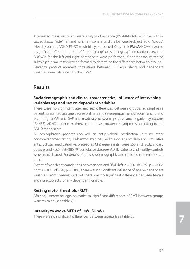
137
A repeated measures multivariate analysis of variance (RM-MANOVA) with the within- subject factor “side” (left and right hemisphere) and the between-subject factor “group” (Healthy control, ADHD, FE-SZ) was initially performed. Only if this RM-MANOVA revealed a significant effect or a trend of factor “group” or “side x group” interaction , separate ANOVA’s for the left and right hemisphere were performed. If appropriate, corrected Tukey´s post-hoc tests were performed to determine the differences between groups.Pearson’s product moment correlations between CPZ equivalents and dependent variables were calculated for the FE-SZ.
Results
Sociodemographic and clinical characteristics, influence of intervening variables age and sex on dependent variablesThere were no significant age and sex differences between groups. Schizophrenia patients presented a severe degree of illness and severe impairment of social functioning according to CGI and GAF and moderate to severe positive and negative symptoms (PANSS). ADHD patients suffered from at least moderate symptoms according to the ADHD rating score. All schizophrenia patients received an antipsychotic medication (but no other concomitant medication, like benzodiazepines) and the dosages of daily and cumulative antipsychotic medication (expressed as CPZ equivalents) were 356.21 ± 203.65 (daily dosage) and 7565.17 ±7886.79 (cumulative dosage). ADHD patients and healthy controls were unmedicated. For details of the sociodemographic and clinical characteristics see table 1. Except of significant correlations between age and RMT (left: r = 0.32, df = 92, p = 0.002; right: r = 0.31, df = 92, p = 0.003) there was no significant influence of age on dependent variables. From One-way-ANOVA there was no significant difference between female and male subjects for any dependent variable.
Resting motor threshold (RMT)After adjustment for age, no statistical significant differences of RMT between groups were revealed (see table 2).
Intensity to evoke MEPs of 1mV (SI1mV)There were no significant differences between groups (see table 2).
TMS IN FIRST-EPISODE SCHIZOPHRENIA AND ADHD
7

138
Short-interval intracortical inhibition (SICI) and intracortical facilitation (ICF)The RM-MANOVA showed a significant “side x group” interaction for ICF at 7ms (F (2, 86) = 6.08, p = 0.003) and ICF at 15ms (F (2, 85) = 5.20, p = 0.007) and a trend for the factor “group” for SICI at 3ms (F(2, 86) = 3.04, p = 0.053).
CHAPTER 7
Table 1 Demographic and Clinical Data of Study Subjects: Interval scaled data are presented as mean ± standard deviation. Values are expressed as c² statistics for categorical variables and F statistics for continuous variables
Variable FE-SZ ADHD HC Statistic P
N 25 28 41
Gender 18 M, 7 F 15 M, 13 F 20 M, 21 F c²(2)=3.522 0.17
Age (years) 29.96 ± 8.5 32.36 ± 9.1 33.37 ± 9.1 F(2, 91)=1.140 0.32
Schizophrenia patients
PANSS positivePANSS negativePANSS generalPANSS total
21.96 ± 5.721.76 ± 6.448.28 ± 8.392.00± 14.5
------------
------------
CGIGAFCPZ1CPZ2
5.88 ± 0.630.0 ± 10.5356 ± 204
7565 ± 7887
------------
ADHD PatientsADHD-SR total scoreAttention deficit subscoreSubscore ImpulsivitySubscore Hyperactivity
----------------
36.78 ± 6.318.22 ± 4.610.63 ± 2.17.93 ± 2.6
FE-SZ: first-episode schizophrenia, ADHD: attention deficit hyperactivity disorder; HC: healthy controls; P: error probability of first kind; N, number; F, female; M, male; CGI: Clinical Global impression; GAF: Global Assessment of Functioning; CPZ1: chlorpromazine equivalents, daily dosage; CPZ2: chlorpromazine equivalents, cumulative dosage; Total and subscores ADHD-SR.

139
TMS IN FIRST-EPISODE SCHIZOPHRENIA AND ADHD
7
Table 2 Main outcome parameters
Variable FE-SZ ADHD HC Statistics Factor
“Group” (P)
RMT left 47.68 ± 6.55 50.04 ± 6.50 47.49 ± 5.93 ~
RMT right 49.00 ± 7.43 51.92 ± 8.86 48.29 ± 8.92 ~
S1mV left 57.08 ± 9.31 64.79 ± 10.57 61.88 ± 10.70 ~
S1mV right 59.50 ± 10.33 64.56 ± 13.35 63.65 ± 14.48 ~
CSP120 left 160.82 ± 41.59 164.72 ± 33.78 133.64 ± 40.88 0.013
CSP120 right 165.04 ± 55.03 162.81 ± 43.89 166.48 ± 37.38 0.974
CSP140 left 203.89 ± 42.63 192.45 ± 46.53 172.36 ± 38.29 0.037
CSP140 right 210.38 ± 53.60 197.40 ± 57.13 186.34 ± 44.19 0.303
SICI 3ms left 39.61 ± 29.97 39.68 ± 36.95 23.90 ± 17.63 0.051
SICI 3ms right 51.11 ± 57.97 35.70 ± 32.54 32.00 ± 22.99 0.183
SICI 5ms left 82.88 ± 83.18 90.86 ± 51.28 63.86 ± 35.86 ~
SICI 5ms right 86.87 ± 74.57 74.69 ± 50.88 81.48 ± 55.73 ~
ICF 7ms left 110.68 ± 110.01 143.54 ± 79.73 94.35 ± 49.14 0.011
ICF 7ms right 148.23 ± 142.51 101.88 ± 58.60 113.56 ± 67.02 0.400
ICF 15ms left 162.95 ± 131.20 163.57 ± 130.08 119.18 ± 61.01 0.160
ICF 15ms right 190.86 ± 173.20 107.03 ± 67.72 146.68 ± 92.61 0.060
FE-SZ: first-episode schizophrenia, ADHD: attention deficit hyperactivity disorder; HC: healthy controls; P: error probability of first kind; RMT: resting motor threshold; S1mV: Intensity to evoke a MEP of 1 mV (peak to peak); CSP120: cortical contralateral silent period at 120% RMT; CSP140: cortical contralateral silent period at 140% RMT; SICI: Short-interval intracortical inhibition at one certain interstimulus interval (3ms, 5ms); ICF: Intracortical facilitation at a defined interstimulus interval (7ms, 15ms). Data are presented as mean ± standard deviation. Please notice that statistical analyses on SICI and ICF values were performed on log-transformed values (raw values are presented in this table). Statistics present results separately for the right and left hemisphere. ~ = RM – MANOVA did not show a side x group interaction or a group effect at least on trend level for this dependent variables, therefore no further ANOVAs were performed separately for the left and right hemisphere.

140
The subsequent ANOVA showed separately for the left hemisphere a trend for the factor group for SICI at 3ms (F (2, 90) = 3.08, p = 0.051, Tukey´s post-hoc test: trend for FE-SZ < HC (p = 0.080)) and a significant effect for ICF at 7ms (F(2, 90) = 4.76, p = 0.011, Tukey´s post-hoc test: ADHD > HC (p = 0.013), ADHD > FE-SZ (p = 0.044). ANOVA for the right hemisphere revealed a trend for ICF at 15ms (F(2, 85) = 2,92, p = 0.060, post-hoc Tukey´s test: trend for ADHD < FE-SZ (p = 0.051) (see figure 1 and 2 and table 2).
Cortical Silent Period (CSP)For CSP120, RM-MANOVA revealed a significant effect for the factor “group” (F(2, 47) = 3.84, p = 0.028) and a significant “side x group” interaction (F(2, 47) = 6.22, p = 0.004). For CSP140, RM-MANOVA showed only a significant effect for “group” (F(2, 46) = 3.36, p = 0.043), but no significant “side x group” interaction (F(2, 46) = 0.17, p = 0.84). ANOVA separately for the left and right hemisphere showed a significant effect for the
CHAPTER 7
Figure 1 Relative values for paired-pulse (SICI, ICF) measures on the left hemisphere expressed as percentage change compared to the testpulse in all groups
*p < 0.05 (RM-MANOVA), #0.05 ≤ p ≤ 0.060. At 3ms FE-SZ (p = 0.080) had a trend towards a reduced SICI compared to HC. ADHD patients displayed an enhanced ICF at 7ms compared to HC (p = 0.013) and to FE-SZ (p = 0.044). The numeric differences for ISI 5ms and ISI 15ms did not reach significance. Data are presented as mean ± standard error of the mean. P – values are corrected in accordance to Tukey´s test. Analyses were performed on logarithmic transformed data (see methods).
Interstimulus Interval
*
#
#
Perc
enta
ge o
f con
trol
siz
e
200 %
160 %
120 %
80 %
40 %
0 %3 ms 5 ms 7 ms 15 ms
Healthy Controls First-Episode Schizophrenia ADHD
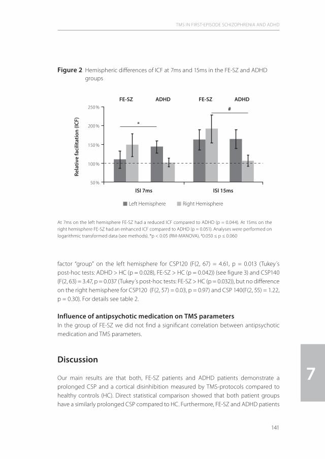
141
factor “group” on the left hemisphere for CSP120 (F(2, 67) = 4.61, p = 0.013 (Tukey´s post-hoc tests: ADHD > HC (p = 0.028), FE-SZ > HC (p = 0.042)) (see figure 3) and CSP140 (F(2, 63) = 3.47, p = 0.037 (Tukey´s post-hoc tests: FE-SZ > HC (p = 0.032)), but no difference on the right hemisphere for CSP120 (F(2, 57) = 0.03, p = 0.97) and CSP 140(F(2, 55) = 1.22, p = 0.30). For details see table 2.
Influence of antipsychotic medication on TMS parametersIn the group of FE-SZ we did not find a significant correlation between antipsychotic medication and TMS parameters.
Discussion
Our main results are that both, FE-SZ patients and ADHD patients demonstrate a prolonged CSP and a cortical disinhibition measured by TMS-protocols compared to healthy controls (HC). Direct statistical comparison showed that both patient groups have a similarly prolonged CSP compared to HC. Furthermore, FE-SZ and ADHD patients
TMS IN FIRST-EPISODE SCHIZOPHRENIA AND ADHD
7
Figure 2 Hemispheric differences of ICF at 7ms and 15ms in the FE-SZ and ADHD groups
At 7ms on the left hemisphere FE-SZ had a reduced ICF compared to ADHD (p = 0.044). At 15ms on the right hemisphere FE-SZ had an enhanced ICF compared to ADHD (p = 0.051). Analyses were performed on logarithmic transformed data (see methods). *p < 0.05 (RM-MANOVA), #0.050 ≤ p ≤ 0.060
ISI 7ms
*
ADHD ADHDFE-SZ FE-SZ
#
ISI 15ms
Rela
tive
faci
litat
ion
(ICF)
250 %
200 %
150 %
100 %
50 %
Left Hemisphere Right Hemisphere
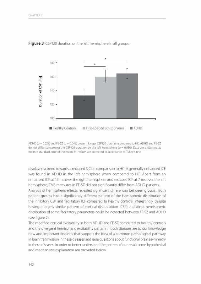
142
displayed a trend towards a reduced SICI in comparison to HC. A generally enhanced ICF was found in ADHD in the left hemisphere when compared to HC. Apart from an enhanced ICF at 15 ms over the right hemisphere and reduced ICF at 7 ms over the left hemisphere, TMS measures in FE-SZ did not significantly differ from ADHD patients. Analysis of hemispheric effects revealed significant differences between groups. Both patient groups had a significantly different pattern of the hemispheric distribution of the inhibitory CSP and facilitatory ICF compared to healthy controls. Interestingly, despite having a largely similar pattern of cortical disinhibition (CSP), a distinct hemispheric distribution of some facilitatory parameters could be detected between FE-SZ and ADHD (see figure 2). The modified cortical excitability in both ADHD and FE-SZ compared to healthy controls and the divergent hemispheric excitability pattern in both diseases are to our knowledge new and important findings that support the idea of a common pathological pathway in brain transmission in these diseases and raise questions about functional brain asymmetry in these diseases. In order to better understand the pattern of our result some hypothetical and mechanistic explanation are provided below.
CHAPTER 7
Figure 3 CSP120 duration on the left hemisphere in all groups
ADHD (p = 0.028) and FE-SZ (p = 0.042) present longer CSP120 duration compared to HC. ADHD and FE-SZ do not differ concerning the CSP120 duration on the left hemisphere (p = 0.950). Data are presented as mean ± standard error of the mean. P – values are corrected in accordance to Tukey´s test
*
*
Dur
atio
n of
CSP
[ms]
180
160
140
120
100
Healthy Controls First-Episode Schizophrenia ADHD

143
General remarksThe role of SICI and CSP in schizophrenia remains controversial. Different studies pointed towards a reduced SICI in medicated chronic schizophrenia patients [20,37,38] and FE-SZ [11], whereas other studies failed to show this difference [18,19]. CSP in medicated patients was found to be shortened [20] or prolonged [19,21,39] compared to healthy subjects. The situation for ADHD is clearer laid out. Two studies revealed a reduced SICI in adult ADHD patients [12,25], whereas in another study this could not reach statistical significance [26]. In contrast to schizophrenia patients, ADHD patients did not show a prolonged CSP compared to healthy controls in previous studies [27]. The reduced SICI and the enhanced ICF in FE-SZ and ADHD patients in our study can be considered as parameters of cortical disinhibition and might be linked to a dysfunction of the GABA
A neurotransmission. The prolonged CSP in both patient groups can be best
explained by an enhanced GABAB transmission [14]. To understand the relationship of
different inhibitory and facilitatory pathways, the two following aspects are important.
Possible mode of action with regard to intrahemispheric and interhemispheric networksFirst, it was shown that CSP and SICI have an inverse relationship and that SICI is suppressed and ICF is facilitated during CSP. SICI seems to be controlled by presynaptic GABA
B-receptor mediated autoinhibition on inhibitory GABA
A-interneurons, similar to a
presynaptic autoinhibition revealed by paired intracellular recordings in slices of rat and human motor cortex [40,41]. Therefore, one explanation of our results might be that the enhanced GABA
B-related inhibition (CSP) and the reduced GABA
A-related inhibition
(SICI)/enhanced glutamate-related facilitation (ICF) on the left hemisphere are interdependent and might reflect a compensatory circuit within this hemisphere [21]. However, we are not able to determine which network is dysfunctional and which network might compensate for this dysfunctionality. Second, ADHD patients and FE-SZ presented a modified hemispheric pattern for CSP and ICF compared to healthy controls. This is of particular importance, because abnormal brain laterality is a well known finding in these diseases. Especially an atypical lateralization to the right hemisphere explored in EEG and structural MRI studies is an important finding in ADHD and schizophrenia [42-44]. It could be hypothesized that this relationship is linked to an impaired interhemispheric connectivity/inhibition (IHI). We did not investigate this in our sample. However, one study found an impaired IHI in adult ADHD patients [27] and a reduced IHI was observed in schizophrenia patients [45]. But why do FE-SZ patients differ from ADHD concerning the hemispheric distribution of ICF? We know from other studies that a cortical disinhibition of the left hemisphere (hyperexciatbility) can result in a reduced facilitation (hypoexcitability) over the contralateral right hemisphere [46-48]. To understand our findings, the cortical disinhibition in both diseases on the left hemisphere (enhanced ICF in ADHD, reduced SICI in FE-SZ compared
TMS IN FIRST-EPISODE SCHIZOPHRENIA AND ADHD
7

144
to HC) should be primarily taken into consideration. Therefore, we would like to hypothesize that the relatively reduced ICF in ADHD patients on the right hemisphere might compensate for the enhanced ICF on the left hemisphere. In FE-SZ this compensatory mechanisms might be impaired and this might be reflected by the relatively facilitated ICF on the right hemisphere. In general, we would like to discuss an intrahemispheric compensatory mechanisms (reflected by CSP enhancement), which is unaffected in both patient groups and an inter- hemispheric compensatory circuit (reflected by a reduction of ICF), which seems to be exclusively active in the ADHD group.
Is there a hypothetical link to dopaminergic transmission? It is a remarkable finding that FE-SZ and ADHD did not differ in our outcome measures, except for the aforementioned difference in ICF in both hemispheres. As hypothesized in the introduction, these diseases might share in parts a common pathophysiological pathway and one possible pathway could be associated with dysfunctional dopaminergic transmission. Drug studies in healthy subjects and research on a hypodopaminergic disease (Parkinson's disease) revealed interesting findings about the effect of dopamine on TMS parameters. The CSP was prolonged after administration of dopamine agonists (L-Dopa, Pergolide) and shortened in patients with Parkinson's disease [14,49]. Hence, the prolonged duration of the CSP over the left hemisphere might be linked with the theory of dopaminergic hyperactivity in schizophrenia and ADHD. Here, the enhanced dopaminergic input would lead to a compensatory increase of GABAergic transmission and probably cause the CSP prolongation [50]. In ADHD there is a discussion over the possibility of dopaminergic dysbalance in frontal subcortical regions. Recently, we demonstrated an improvement of SICI in ADHD with long acting methylphenidate [51]. This was interpreted as a dopamine-related response to the stimulants, possibly reflecting a complex dopaminergic-GABAergic interaction. However, with the TMS technique we are not able to determine the localization of the affected brain regions involved in dopaminergic transmission nor directly probe dopaminergic functions. Therefore, currently this theory remains speculative.
LimitationsOur comparative study has several limitations. First of all, all schizophrenia patients received antipsychotic medication, which might have an impact on the results of TMS paradigms. We did not find a correlation between CPZ equivalents on the results of the TMS measures and an influence of antipsychotic drugs on TMS measurements is discussed controversially. One recent study showed that unmedicated schizophrenia patients and patients treated with second generation antipsychotics (risperidone, olanzapine, quetiapine) had a reduced CSP and that patients treated with clozapine had significant less SICI and a longer CSP compared to healthy controls [37]. All of our
CHAPTER 7
145
patients received second generation antipsychotic medication but, in contrast to Liu and colleagues [37] none of our patients received clozapine. Also, it is important to recognize that the schizophrenia patients in our study were at their first episode with a relatively short treatment history whereas Liu and co-authors investigated chronically ill schizophrenia patients and patients with a schizoaffective disorder. However, reviewing the literature, we would expect that antipsychotics should compensate for the reduced inhibition and not enhance it [11,14,21]. Furthermore it should be considered, that the drug naïve adult ADHD patients showed a motor disinhibition which was not present in healthy controls. Finally, there were no significant differences between ADHD and FE-SZ patients with regard to inhibitory TMS parameters. This might support the hypothesis that diminished cortical inhibition is genuine to these diseases. Nevertheless, our results in adult ADHD patients are partially in contrast with the results of two previous studies, where significant differences in SICI and CSP could not be found [26,27]. This could be due to different methodical procedures in TMS techniques as well as to divergent recruitment strategies. However, our results stay well in line with the diminished SICI seen in children and adolescents with ADHD [22,52]. Although FE-SZ and ADHD patients present a similar cortical disinhibition, we cannot rule out that different mechanisms may underlie these effects and that these are not specifically restricted to these diseases [53]. Cortical disinhibition, as revealed by TMS might reflect a general characteristic of psychiatric conditions, because different other psychiatric diseases, like depression or OCD, are known to show a cortical disinhibition [54,55]. ADHD is a dimensional diagnosis, but diagnosis of schizophrenia is based on the qualitative dimensions of symptoms. But, they still share some common executive dysfunctions (working memory, impulsivity etc.) and attention deficits, whereas the motor symptoms usually are opposite. Despite of the different subsumed pathophysi-ology and phenotypes, a common dopaminergic pathway has been discussed in both diseases [3,4]. However, it should be considered, that dopamine has a neuromodulatory function. The dopamine effect depends on spontaneous neuronal activity, dopamine concentration and dopaminergic sub-receptors [53]. Therefore, the transfer of our TMS-findings to the complex aspects of dopaminergic transmission mechanisms to both diseases can be done only in a limited manner. Therefore, the interpretation of our TMS results should be mainly focused on the discussed primary GABAergic transmission pathways. Finally, it should be noted that overlapping parts of this study have been published before (see method section). However, this is the first study which directly compares cortical excitability in first-episode schizophrenia and ADHD. Furthermore, we provide new CSP data for ADHD and new late-ICF data (15 ms) for first-episode schizophrenia and we show for the first time elaborate data for functional hemispheric asymmetry measured by TMS for both diagnostic groups. A detailed discussion specific for each
TMS IN FIRST-EPISODE SCHIZOPHRENIA AND ADHD
7

146
diagnostic groups and focusing on effects of one hemisphere can be found in our other publications [11,21,25].
Conclusions
In conclusion, the results of our study provide evidence for a cortical disinhibition in ADHD and FE-SZ and for a common pathophysiological pathway, which might involve GABAergic, glutamatergic and dopaminergic transmission. Furthermore, we were able to present a functional hemispheric asymmetry in both diseases. This comparative work supports the idea of common pathophysiological pathways in motor excitability in schizophrenia and ADHD.
CHAPTER 7

147
References
1 Ross RG, Olincy A, Harris JG, Sullivan B, Radant A: Smooth pursuit eye movements in schizophrenia and attentional dysfunction: Adults with schizophrenia, adhd, and a normal comparison group. Biol Psychiatry 2000;48:197-203.
2 Marsh PJ, Williams LM: Adhd and schizophrenia phenomenology: Visual scanpaths to emotional faces as a potential psychophysiological marker? Neurosci Biobehav Rev 2006;30:651-665.
3 Pani L: Clinical implications of dopamine research in schizophrenia. Curr Med Res Opin 2002;18 Suppl 3:s3-7.4 Solanto MV: Dopamine dysfunction in ad/hd: Integrating clinical and basic neuroscience research. Behav
Brain Res 2002;130:65-71.5 Ohno M: The dopaminergic system in attention deficit/hyperactivity disorder. Congenit Anom (Kyoto)
2003;43:114-122.6 Durston S: A review of the biological bases of adhd: What have we learned from imaging studies? Ment
Retard Dev Disabil Res Rev 2003;9:184-195.7 Carriere P, Bonhomme D, Lemperiere T: Amisulpride has a superior benefit/risk profile to haloperidol in
schizophrenia: Results of a multicentre, double-blind study (the amisulpride study group). Eur Psychiatry 2000;15:321-329.
8 Jucaite A, Fernell E, Halldin C, Forssberg H, Farde L: Reduced midbrain dopamine transporter binding in male adolescents with attention-deficit/hyperactivity disorder: Association between striatal dopamine markers and motor hyperactivity. Biol Psychiatry 2005;57:229-238.
9 Pouget P, Wattiez N, Rivaud-Pechoux S, Gaymard B: A fragile balance: Perturbation of gaba mediated circuit in prefrontal cortex generates high intraindividual performance variability. PLoS One 2009;4:e5208.
10 Hashimoto T, Bazmi HH, Mirnics K, Wu Q, Sampson AR, Lewis DA: Conserved regional patterns of gaba-related transcript expression in the neocortex of subjects with schizophrenia. Am J Psychiatry 2008;165:479-489.
11 Wobrock T, Schneider M, Kadovic D, Schneider-Axmann T, Ecker UK, Retz W, Rosler M, Falkai P: Reduced cortical inhibition in first-episode schizophrenia. Schizophr Res 2008;105:252-261.
12 Richter MM, Ehlis AC, Jacob CP, Fallgatter AJ: Cortical excitability in adult patients with attention-deficit/hyperactivity disorder (adhd). Neurosci Lett 2007;419:137-141.
13 Kujirai T, Caramia MD, Rothwell JC, Day BL, Thompson PD, Ferbert A, Wroe S, Asselman P, Marsden CD: Corti-cocortical inhibition in human motor cortex. J Physiol 1993;471:501-519.
14 Ziemann U: Tms and drugs. Clin Neurophysiol 2004;115:1717-1729.15 Werhahn KJ, Kunesch E, Noachtar S, Benecke R, Classen J: Differential effects on motorcortical inhibition
induced by blockade of gaba uptake in humans. J Physiol 1999;517 ( Pt 2):591-597.16 Siebner HR, Dressnandt J, Auer C, Conrad B: Continuous intrathecal baclofen infusions induced a marked
increase of the transcranially evoked silent period in a patient with generalized dystonia. Muscle Nerve 1998;21:1209-1212.
17 Pascual-Leone A, Manoach DS, Birnbaum R, Goff DC: Motor cortical excitability in schizophrenia. Biol Psychiatry 2002;52:24-31.
18 Eichhammer P, Wiegand R, Kharraz A, Langguth B, Binder H, Hajak G: Cortical excitability in neuroleptic-naive first-episode schizophrenic patients. Schizophr Res 2004;67:253-259.
19 Daskalakis ZJ, Christensen BK, Chen R, Fitzgerald PB, Zipursky RB, Kapur S: Evidence for impaired cortical inhibition in schizophrenia using transcranial magnetic stimulation. Arch Gen Psychiatry 2002;59:347-354.
20 Fitzgerald PB, Brown TL, Daskalakis ZJ, Kulkarni J: A transcranial magnetic stimulation study of inhibitory deficits in the motor cortex in patients with schizophrenia. Psychiatry Res 2002;114:11-22.
21 Wobrock T, Schneider-Axmann T, Retz W, Rosler M, Kadovic D, Falkai P, Schneider M: Motor circuit abnormalities in first-episode schizophrenia assessed with transcranial magnetic stimulation. Pharmacopsy-chiatry 2009;42:194-201.
22 Moll GH, Heinrich H, Trott G, Wirth S, Rothenberger A: Deficient intracortical inhibition in drug-naive children with attention-deficit hyperactivity disorder is enhanced by methylphenidate. Neurosci Lett 2000;284:121-125.
TMS IN FIRST-EPISODE SCHIZOPHRENIA AND ADHD
7

148
23 Buchmann J, Gierow W, Weber S, Hoeppner J, Klauer T, Benecke R, Haessler F, Wolters A: Restoration of disturbed intracortical motor inhibition and facilitation in attention deficit hyperactivity disorder children by methylphenidate. Biol Psychiatry 2007;62:963-969.
24 Gilbert DL, Bansal AS, Sethuraman G, Sallee FR, Zhang J, Lipps T, Wassermann EM: Association of cortical disinhibition with tic, adhd, and ocd severity in tourette syndrome. Mov Disord 2004;19:416-425.
25 Schneider M, Retz W, Freitag C, Irsch J, Graf P, Retz-Junginger P, Rosler M: Impaired cortical inhibition in adult adhd patients: A study with transcranial magnetic stimulation. J Neural Transm Suppl 2007:303-309.
26 Hoeppner J, Neumeyer M, Wandschneider R, Herpertz SC, Gierow W, Haessler F, Buchmann J: Intracortical motor inhibition and facilitation in adults with attention deficit/hyperactivity disorder. J Neural Transm 2008;115:1701-1707.
27 Hoeppner J, Wandschneider R, Neumeyer M, Gierow W, Haessler F, Herpertz SC, Buchmann J: Impaired trans-callosally mediated motor inhibition in adults with attention-deficit/hyperactivity disorder is modulated by methylphenidate. J Neural Transm 2008;115:777-785.
28 Annett M: A classification of hand preference by association analysis. Br J Psychol 1970;61:303-321.29 Wittchen HU, Wunderlich U, Gruschwitz S, Zaudig M: Strukturiertes klinisches interview für dsm-iv. Achse i:
Psychische störungen. A german, advanced adaptation of the original scid version by rl spitzer, jb williams, m gibbon and mb first. Hogrefe-Verlag, Göttingen, 1997.
30 Guy W BR: Cgi: Clinical global impressions., National Institute of Mental Health, 1976.31 Endicott J, Spitzer RL, Fleiss JL, Cohen J: The global assessment scale. A procedure for measuring overall
severity of psychiatric disturbance. Arch Gen Psychiatry 1976;33:766-771.32 Kay SR, Fiszbein A, Opler LA: The positive and negative syndrome scale (panss) for schizophrenia. Schizophr
Bull 1987;13:261-276.33 Woods SW: Chlorpromazine equivalent doses for the newer atypical antipsychotics. J Clin Psychiatry
2003;64:663-667.34 Rösler M, Retz W, Retz-Junginger P, Thome J, Supprian T, Nissen T, Stieglitz RD, Blocher D, Hengesch G, Trott
GE: [tools for the diagnosis of attention-deficit/hyperactivity disorder in adults. Self-rating behaviour questionnaire and diagnostic checklist]. Nervenarzt 2004;75:888-895.
35 Wender PH, Wolf LE, Wasserstein J: Adults with adhd. An overview. Ann N Y Acad Sci 2001;931:1-16.36 Daskalakis ZJ, Christensen BK, Chen R, Fitzgerald PB, Zipursky RB, Kapur S: Effect of antipsychotics on cortical
inhibition using transcranial magnetic stimulation. Psychopharmacology (Berl) 2003;170:255-262.37 Liu SK, Fitzgerald PB, Daigle M, Chen R, Daskalakis ZJ: The relationship between cortical inhibition,
antipsychotic treatment, and the symptoms of schizophrenia. Biol Psychiatry 2009;65:503-509.38 Hasan A, Nitsche MA, Rein B, Schneider-Axmann T, Guse B, Gruber O, Falkai P, Wobrock T: Dysfunctional
long-term potentiation-like plasticity in schizophrenia revealed by transcranial direct current stimulation. Behav Brain Res 2011;224:15-22.
39 Bajbouj M, Gallinat J, Niehaus L, Lang UE, Roricht S, Meyer BU: Abnormalities of inhibitory neuronal mechanisms in the motor cortex of patients with schizophrenia. Pharmacopsychiatry 2004;37:74-80.
40 McDonnell MN, Orekhov Y, Ziemann U: The role of gaba(b) receptors in intracortical inhibition in the human motor cortex. Exp Brain Res 2006;173:86-93.
41 Ni Z, Gunraj C, Chen R: Short interval intracortical inhibition and facilitation during the silent period in human. J Physiol 2007;583:971-982.
42 Hale TS, Smalley SL, Walshaw PD, Hanada G, Macion J, McCracken JT, McGough JJ, Loo SK: Atypical eeg beta asymmetry in adults with adhd. Neuropsychologia;48:3532-3539.
43 White T, Nelson M, Lim KO: Diffusion tensor imaging in psychiatric disorders. Top Magn Reson Imaging 2008;19:97-109.
44 Shapleske J, Rossell SL, Woodruff PW, David AS: The planum temporale: A systematic, quantitative review of its structural, functional and clinical significance. Brain Res Brain Res Rev 1999;29:26-49.
45 Fitzgerald PB, Brown TL, Daskalakis ZJ: The application of transcranial magnetic stimulation in psychiatry and neurosciences research. Acta Psychiatr Scand 2002;105:324-340.
CHAPTER 7

149
46 Suppa A, Ortu E, Zafar N, Deriu F, Paulus W, Berardelli A, Rothwell JC: Theta burst stimulation induces after-effects on contralateral primary motor cortex excitability in humans. J Physiol 2008;586:4489-4500.
47 Wassermann EM, Wedegaertner FR, Ziemann U, George MS, Chen R: Crossed reduction of human motor cortex excitability by 1-hz transcranial magnetic stimulation. Neurosci Lett 1998;250:141-144.
48 Heide G, Witte OW, Ziemann U: Physiology of modulation of motor cortex excitability by low-frequency suprathreshold repetitive transcranial magnetic stimulation. Exp Brain Res 2006;171:26-34.
49 Cantello R, Tarletti R, Civardi C: Transcranial magnetic stimulation and parkinson's disease. Brain Res Brain Res Rev 2002;38:309-327.
50 Beauregard M, Ferron A: Dopamine modulates the inhibition induced by gaba in rat cerebral cortex: An iontophoretic study. Eur J Pharmacol 1991;205:225-231.
51 Schneider M, Retz W, Rösler M: The effects of long acting methylphenidate in adults with attention deficit hyperactivity disorder: A study with paired pulse transcranial magnetic stimulation. Neuropsychobiology 2011;in press
52 Moll GH, Heinrich H, Trott GE, Wirth S, Bock N, Rothenberger A: Children with comorbid attention-deficit-hyperactivity disorder and tic disorder: Evidence for additive inhibitory deficits within the motor system. Ann Neurol 2001;49:393-396.
53 Nitsche MA, Monte-Silva K, Kuo MF, Paulus W: Dopaminergic impact on cortical excitability in humans. Rev Neurosci 2010;21:289-298.
54 Bajbouj M, Lisanby SH, Lang UE, Danker-Hopfe H, Heuser I, Neu P: Evidence for impaired cortical inhibition in patients with unipolar major depression. Biol Psychiatry 2006;59:395-400.
55 Greenberg BD, Ziemann U, Cora-Locatelli G, Harmon A, Murphy DL, Keel JC, Wassermann EM: Altered cortical excitability in obsessive-compulsive disorder. Neurology 2000;54:142-147.
TMS IN FIRST-EPISODE SCHIZOPHRENIA AND ADHD
7


General discussion and conclusions8 |

153
8. General discussion and conclusions
Attention Deficit/ Hyperactivity Disorder (ADHD) is characterized by shifting of sympto-matology from childhood to adulthood. Especially hyperactivity and impulsivity seem to become less prominent, and disorganization dominates the symptomatic expression of attention problems (15). To investigate the specific mechanisms leading to adult ADHD symptoms, this thesis provides evidence about the functional aspects of attention, executive and motor functions in this disorder. We assessed by functional magnetic resonance imaging (fMRI) the attention and executive brain networks in drug naive adult ADHD patients and implemented the data to the clinical phenotype (chapter 3). The motor cortex excitability and its clinical impact in adult ADHD patients was compared with healthy controls by means of the paired pulse transcranial magnetic stimulation (ppTMS) technique (chapter 4). The influence of long acting methylphenidate (LA-Mph), the standard medication for this disorder, on the motor system was assessed with this technique (chapter 5) to elucidate the underlying pathomechanisms. Motor excitability was studied in patients with schizophrenia in chapter 6 and subsequently ppTMS in ADHD and schizophrenia was compared to clarify common pathophysiological pathways and their specific components in diseases with dopaminergic dysbalances (chapter 7). As previously delineated in chapter 1, in terms of specific aims, the main results of the thesis will be discussed. The interpretation of the studies, the clinical implications, as well as the methodological strengths and limitations including the recommendations for future research are presented, and, finally, the conclusions of the thesis are formulated.
8.1 fMRI in adult patients with ADHDOur fMRI study with a continuous performance task paradigm showed altered activation patterns of a selective attention brain network in the fronto-striatal and parietal brain regions in adult patients with ADHD. These findings are in agreement with the existing hypotheses about the pathophysiology of ADHD. More explicitly, prefrontal cortex, anterior cingulated cortex (ACC) and striatum were implicated, that all can be considered as belonging to the frontal attentional system. In addition, the parietal lobule as part of the posterior attentional system appeared to be also hypofunctional (see overview from 89). From the dimensional perspective, these results demonstrated that lowered activation of these distinct attention networks correlates not only with the clinical phenotype of combined type ADHD, but also with ADHD symptomatology in partially remitted patients. This leads to the hypothesis that in patients with partially remitted ADHD similar brain networks are impaired as in those with adult ADHD, combined type. Furthermore, a functional hypoactivity of several brain networks was established that could be positively related to specific ADHD symptoms: ACC to impulsivity, caudate
GENERAL DISCUSSION AND CONCLUSIONS
8

154
nuclei to attention deficits, and parietal lobule to the complete symptom profile. In contrast, the left insula was associated with hyperactivation, as reflected in clinically observed increased hyperactivity and impulsivity. The latter can be considered as a compensatory overactivation of brain regions counterbalancing the impaired networks (22,24). Thus, the results of this study support the hypothesis that adult ADHD has to be considered as a dimensional disorder of which the severity of symptoms correlates with the degree of dysfunction in various brain networks, even in patients in whom ADHD symptoms were partially remitted after childhood. These observations in adults with ADHD are in line with those reported in younger patients and reflect pathophysiologi-cal trait characteristics of ADHD (caudate nucleus: 17, 37. ACC: 17, 38,39. Parietal lobe: 17).
8.2 ppTMS in drug naïve adult patients with ADHD versus healthy controlsNo differences were found between ADHD patients and healthy controls with respect to resting motor threshold (RMT) and active motor threshold (AMT). The same holds for the stimulus response curves. Thus, no evidence could be obtained for the presence of hyperexcitability within the motor systems at the membrane level of cortical neurons in adult patients with ADHD versus healthy controls (123). With the ppTMS technique, we found a reduced short intracortical inhibition (SICI) in adult ADHD patients as compared to controls. In the present study, no significant increase of cortical facilitation could be demonstrated. These findings are in line with a previous preliminary report on adults with ADHD (93). With respect to the dimensional characteristics of ADHD, a positive correlation was found between inhibitory deficits (SICI) and severity of symptoms scores (Wender-Re-imherr-Interview (WRI) total score and ADHD-self rating (ADHD-SR) hyperactivity and impulsivity subscores). Comparable findings were reported by Gilbert et al. (52) in Tourette patients with comorbid ADHD symptoms. In sum, the hyperactive/impulsive behaviours in adult ADHD patients are most probably linked to cortical inhibitory dysfunction.As presented in chapter 4, SICI deficits in ADHD patients appeared to have an inter-hemispheric asymmetry to the left side. Comparable findings have been reported with brain imaging techniques that described hemispheric asymmetries either globally or in distinct brain areas, specifically the caudate nucleus and pallidum (8,26,27,83,109). With respect to the SICI, several GABAergic and dopaminergic intracortical neural transmission systems have been reported to be involved (120,72,110,123). Deficits in SICI have been shown to have reliable test-retest stability in adults with ADHD (93) and little inter- and intraindividual variability (74). These observations lead to the hypothesis that attenuation of SICI results from GABAergic, glutamatergic and/or dopaminergic changes in transmission and reflects a trait marker of ADHD.
CHAPTER 8
155
Summarizing, this is the first study with ppTMS demonstrating reduced SICI, primarily in the left hemisphere, in adult patients with ADHD. Moreover, this lowered SICI correlated with the ADHD symptom profile, in particular with disinhibited behaviours. These findings are in line with the hypothesis of neuronal motor inhibition deficits in adult ADHD. TMS, more specifically ppTMS, appeared to be an appropriate technique to investigate motor excitability in ADHD.
8.3 ppTMS effects of LA-Mph in adults with ADHDThis study (chapter 5) was performed to investigate the effects of LA-Mph – the current pharmacological treatment of ADHD - on motor excitability in adults with ADHD. Previous findings of lowered SICI in ADHD, as indicator of elevated brain motor excitability, were the rationale for this study (93,94).Treatment with stimulants like LA-Mph should lead to normalization of elevated (lowered amplitude of) SICI. In contrast to a study with adult ADHD patients which showed no significant differences in ppTMS parameters under medication with Mph (62), Gilbert and colleagues (51,52) found an increase of amplitude of the SICI in adult patients with Tourette disorder comorbid with ADHD. Therefore, we studied the effects on ppTMS parameters in adult ADHD patients without comorbidity under stable medication with LA-Mph.The results demonstrated no effect of LA-Mph on RMT and AMT and support the hypothesis that the compound does not directly affect membrane characteristics. These observations are in line with those reported by other investigators (ADHD children: 78,79,19; adults with ADHD: 62).With LA-Mph in an individualized stable dose varying between 30 and 54 mg per day, a significantly reducing effect on SICI magnetically evoked potential (MEP) amplitudes was found, but not on ICF MEP amplitudes. SICI MEP amplitude appeared to be correlated with ADHD self rating scores at baseline, but not after 2 weeks of treatment. In children with ADHD, however, treatment with Mph induced significant decreases in SICI MEP amplitudes (78,19). It can, therefore, be hypothesized that the pathophysiolog-ical mechanisms, as reflected by ppTMS parameters, are present in children as well as in adults with ADHD.In summary, the results from this study demonstrate that, like in studies with children, LA-Mph decreases SICI MEP amplitudes in adults with ADHD. A direct clinical correlation of the neurophysiological parameters with the clinical improvement of ADHD symptoms could, however, not be established.
8.4 ppTMS in adult patients with first episode schizophrenia or ADHDADHD and other psychiatric disorders, like schizophrenia, share attention deficits, executive dysfunctions and motor disturbances. Since the pathophysiology of schizophrenia is supposed to be related to dysbalances in dopaminergic transmission, studies with ppTMS
GENERAL DISCUSSION AND CONCLUSIONS
8

156
were performed in patients with first episode schizophrenia and adult patients with ADHD (chapters 6 and 7). With respect to RMT in patients with first episode schizophrenia, a positive correlation was found with symptom severity as measured with the Positive and Negative Syndrome Scale (PANSS) total score, but not with the PANSS subscores. No differences in RMT could be established between patients with first episode schizophrenia and healthy controls. These results support the idea that membrane excitability is not primarily disturbed in schizophrenia.Concerning ppTMS, increased SICI MEP amplitudes were found in patients with first episode schizophrenia. With respect to ICF, no significant differences were observed between patients and healthy controls. These findings indicate a reduced SICI, without changes in ICF in patients with first-episode schizophrenia. It can, therefore, be hypothesized that inhibitory intracortical deficits may either be involved in the patho-physiology of this disorder or serve as a vulnerability factor. Since deficits in SICI have been demonstrated in a variety of neuropsychiatric disorders (e.g. unipolar depression: 9; ADHD: 94; obsessive compulsive disorder: 53), the ppTMS results in patients with adult ADHD were compared to those obtained in adult patients with first episode schizophrenia in order to delineate putative common psychopatho-logical pathways. From this comparative study using ppTMS, it appeared that patients with either first episode schizophrenia or adult ADHD patients displayed deficits in SICI and a prolonged cortical silent period (CSP) over the dominant left hemisphere. Enhancement of ICF, however, was only found in ADHD. This observation is not in line with those as reported in chapter 4, in which elevation of ICF did not reach the level of significance and may be explained from variations in the number of controls (26 vs 41). It has to be stressed, however, that elevated SICI and ICF MEP amplitudes both reflect a generally increased cortical excitability. Because SICI and ICF are thought to be primarily related to GABAergic neurotransmission (see also chapter 1.4.3), the observations in patients with first episode schizophrenia or ADHD of reduced and enhanced values for SICI and ICF, respectively, may indicate enhanced cortical disinhibition via GABAergic involvement. The prolonged CSP in both patient groups points in the same direction (121). It can, however, not be excluded that the increase of GABAergic transmission results from an enhanced compensatory dopaminergic activation in subcortical brain regions (101,104). In this respect, is could be postulated that the cortical-striatal-pallidal-thalamical-cortical loop is affected in both schizophrenia and ADHD (33,49,61,72,80). Although in patients with first episode schizophrenia and in patients with ADHD cortical disinhibition of the left hemisphere (hy-perexcitability) was found, it can not be excluded that this phenomenon results from reduced facilitation (hypoexcitability) over the contralateral right hemisphere (58,108,116). Most probably, in schizophrenia, intrahemispheric and interhemispheric compensatory mechanisms are involved as reflected by CSP enhancement and ICF modulation.
CHAPTER 8

157
In conclusion, the results of these studies support the ideas of cortical disinhibition in both ADHD and first episode schizophrenia and suggest common pathophysiological pathways related to GABAergic and dopaminergic neurotransmitter functionality.
8.5 Limitations and strengths of the studiesThe fMRI technique has several techniqual limitations which are mainly related to the slow time courses of the signals which hampers stable representation of rapidly changing executive and motor functions in human beings. In addition, repeated measurements in a group of patients are warranted to get a reliable outcome. Moreover, patients must lie in a resting state without significant motor activity for about 15 minutes. These factors partly explain the inconsistency of the results in patients with psychiatric disorders, particular in whom motor disinhibition is a key symptom such as ADHD. Thus, fMRI data have to be controlled for movement artifacts which have led to a significant number of drop-outs (ca. 15%) in the data as presented in this thesis.The main technical problem in applying TMS is the highly variable amplitude of the elicited motor signal. Therefore, averaging of the magnetically evoked potentials has to be performed in order to obtain reliable data. This specific technical limitation is particularly relevant in patients with highly increased motor activity (like in ADHD). Given this, the results of measurements in very hyperactive or restless patients had to be excluded from the data analyses.As mentioned in the discussion sections of the papers included in this thesis, the results have to be interpreted cautiously since several confounding factors were present. Apart from the technical difficulties as described above, the study groups comprised a limited number of patients only, which influenced the statistical power. Furthermore, in the study with ppTMS in patients with schizophrenia, the results may have been influenced by the use of antipsychotic medication. Finally, but relevant for all research in psychiatry, stability over time of the diagnosis and scoring instruments to assess intensity of symptoms cannot be clearly validated (e.g. the German validation of the adult ADHD scoring instruments is still going on). In this respect, it has to be stressed that the Diagnostic and Statistical Manual (DSM) is a categorical classification system and its symptom validity to establish psychiatric diagnoses is still a matter of debate. The latter has already resulted in several relevant adaptations in the subsequent issues of the DSM. Apart from the more general remarks on the limitations of these studies, their methodical strength has also to be underlined. All but the one study with LA-Mph followed a controlled design. The age of the included patients ranged from 18 to 49 years and neither age nor intelligence influenced the stability of the neurophysiological parameters. Moreover, no relevant gender effect of the ‘pure’ and drug-naïve ADHD patients could be demonstrated so that ‘real’ ADHD pathophysiology could be studied indeed. Finally, functionality of brain mechanisms in ADHD was compared with that in
GENERAL DISCUSSION AND CONCLUSIONS
8

158
another diagnostic group, first episode schizophrenia, a disease with parallels regarding cognitive psychopathology and brain transmission mechanisms.
8.6 Clinical relevance and future outlinesAs demonstrated in this thesis, the fMRI technique can be used to elegantly investigate brain networks involved in processing attention and executive brain functions. The results of the here presented studies demonstrate that the symptomatology of ADHD may persist over decades, but not always in a similar intensity and constellation. Interestingly, data from the literature indicate that alterations in the neurophysiological parameters as measured with fMRI are qualitatively rather stable over time. In other words, this functional disturbance in subcortical-cortical neuronal pathways originates from childhood and persists into adulthood irrespective the psychopathological presentation of the disorder. ADHD in adulthood can be described as an impairment of attention and executive functioning as well as motor inhibition, that may be considered as trait characteristics of the disorder. Therefore, this technique may be used in future studies to investigate putative genotype-phenotype relationships and associations between phenotypical presentations and sex and/or age. In addition, since the fMRI technique is non-invasive and can be performed repeatedly, it can be used to perform longitudinal studies aimed to detect compensatory or restorating mechanisms and effects of pharmacological interventions in individual patients with ADHD. This may ultimately lead to an endophenotypically orientated individualized diagnosis and treatment of adult patients with ADHD. PpTMS is a non-invasive technique too by which motor functionality, related to neu-rotransmitter systems, can be investigated in humans. Since the magnetically evoked potential (MEP) is the result of all excitatory and inhibitory postsynaptic potentials on the motor neuroaxons, ppTMS is one of the most appropriate techniques to study overall activity of pyramidal neurons including their dynamic compensatory capacities. Given its sensitivity to pharmacological influence and its correlation with clinical phenotypes, ppTMS can be applied in follow-up treatment studies with the emphasis on the long-term effects on motor systems in adults with ADHD. As compared to the data obtained by other investigators in children with ADHD, the results of the neurophysiological parameters in the here presented studies with adult ADHD patients suggest that treatment with LA-Mph induces similar effects. This method may eventually lead to the development of “therapeutic drug monitoring” and to a better understanding of motor cortex transmission in psychiatric disorders in general. An additional aspect to be considered is that age and gender are not of major significance in attention or motor brain network activation patterns in adults with ADHD, albeit that shifting of symptomatology occurs from childhood into adulthood (see chapter 1). Since the studies presented here were performed with drug naïve ADHD patients, the
CHAPTER 8

159
neurophysiological parameters can be considered to reflect trait factors in ADHD patients without drug-induced variations in neuroplasticity. Finally, in the study comparing patients with first episode schizophrenia and adults with ADHD, overlapping changes in MEP amplitudes were found which points towards a common neurobiological pathway including GABAergic and dopaminergic neuro-transmitter systems and may ultimately lead to more endophenotypically orientated treatment strategies. Future longitudinal studies are warranted to further elucidate the postulated endophenotypic trait stability of ADHD. These investigations may eventually disclose a clear genotype-phenotype relationship of ADHD and consequently a more targeted pharmacological treatment approach for this and other dopamine-related disorders or symptoms.
8.7 ConclusionsFour main conclusions can be formulated on the basis of this thesis.
The fMRI data from the controlled study demonstrated impairments of attentional and executive brain networks in adult ADHD in comparison to healthy controls. The hypo-activation in fronto-striatal and parietal brain networks correlated with the clinical ADHD symptom scores. Thus, ADHD patients used deviant brain networks via the left insula to compensate for the hypoactivation of the above mentioned attention networks.
PpTMS was used to investigate motor cortex excitability in adult patients with ADHD. The results of this controlled study showed that motor inhibition deficits localized in the left hemisphere were present in ADHD. The observed increases in inhibitory MEP amplitudes could be associated with the symptom profile.
The amplitude of the inhibitory MEP decreased during treatment with the dopamine agonistic compound LA-Mph, which suggested that dopaminergic dysfunctions are of significance in the pathophysiology of ADHD indeed.
In the controlled studies, results of ppTMS in patients with first episode schizophrenia were compared to those obtained in adult patients with ADHD. In patients from both diagnostic categories a diminished intracortical inhibition was demonstrated, especially in the left dominant hemisphere. In the right hemisphere, however, the intracortical facilitation appeared to be increased in patients with schizophrenia but not in those with ADHD. In addition, a prolonged CSP was found in both schizophrenic and ADHD patients suggesting that imbalances in GABAergic neurotransmission are also involved in the pathophysiology of motor inhibition. As a consequence, the equilibrium between
GENERAL DISCUSSION AND CONCLUSIONS
8

160
GABAergic and dopaminergic neurotransmission might be disturbed which indicates that schizophrenia and ADHD share common neurotransmitter deficits.
CHAPTER 8

161
References
1) Abbruzzese G, Trompetto C. (2002) Clinical and research methods for evaluating cortical excitability. J Clin Neurophysiol 19(4):307-21.
2) Adams F (1939) From Hippocrates: Aphorisms. In: The Genuine Works of Hippocrates (Translated from greek by Fancis Adams). The William and Wilkins Company, Baltimore.
3) Amassian VE, Quirk GJ, Stewart M (1990) A comparison of corticospinal activation by magnetic coil and electrical stimulation of monkey motor cortex. Electroen Clin Neuro 77:390–401.
4) American Psychiatric Association (1968) Diagnostic and statistical manual of mental disorders (2nd ed. DSM II). Washington, DC: Author.
5) American Psychiatric Association (1987) Diagnostic and statistical manual of mental disorders (3rd ed. DSM III-R). Washington, DC: Author
6) American Psychiatric Associations (1994) Diagnostic and statistical manual of mental disorders (4th edition DSM IV). Washington, DC: Author.
7) American Psychiatric Association (2000) Diagnostic and statistical manual of mental disorders (4th ed. DSM IV-TR). Washington, DC: Author.
8) Aylward EH, Reiss AL, Reader MJ, Singer HS, Brown JE, Denckla MB (1996) Basal ganglia volumes in children with attention-deficit hyperactivity disorder. J Child Neurol 11:112–115.
9) Bajbouj M, Lisanby SH, Lang UE, Danker-Hopfe H, Heuser I, Neu P (2006) Evidence for impaired cortical inhibition in patients with unipolar major depression. Biol. Psychiatry 59: 395-400.
10) Barker AT, Jalinous R, Freeston IL. (1985). „Non-invasive magnetic stimulation of human motor cortex“. The Lancet 1 (8437):1106–1107.
11) Barkley RA (2002) Major life activity and health outcomes associated with attention-deficit/hyperactivity disorder. J Clin Psychiatry 63 Suppl 12:10-5.
12) Barkley RA, Fischer M, Smallish L, Fletcher K (2006) Young adult outcome of hyperactive children: adaptive functioning in major life activities. J Am Acad Child Adolesc Psychiatry 45(2):192-202.
13) Barkley RA (1997) Behavioral inhibition, sustained attention, and executive functions: constructing a unifying theory of ADHD. Psychol Bull 121(1):65-94.
14) Beard GM (1869) Neurasthenia, or nervous exhaustion. Boston Med Surg J 80:217–221.15) Biederman J, Mick E, Faraone SV (2000) Age-dependent decline of symptoms of attention deficit
hyperactivity disorder: Impact of remission definition and symptom type. Am J Psychiatry 157:816 –818.16) Biederman J, Petty CR, Evans M, Small J, Faraone SV (2010) How persistent is ADHD? A controlled 10-year
follow-up study of boys with ADHD. Psychiatry Res 177(3):299-304.17) Booth JR, Burman DD, Meyer JR, Lei Z, Trommer BL, Davenport ND, Li W, Parrish TB, Gitelman DR, Mesulam
MM (2005) Larger deficits in brain networks for response inhibition than for visual selective attention in at-tention-deficit hyperactivity disorder. J Child Psychol Psychiatry 46:94–111.
18) Bradley Ch (1937) The Behaviour of Children receiving Benzedrine. Am J Psych 94:577–85.19) Buchmann J, Gierow W, Weber S, Hoeppner J, Klauer T, Benecke R, Haessler F, Wolters A (2007) Restoration of
disturbed intracortical motor inhibition and facilitation in attention deficit hyperactivity disorder children by methylphenidate. Biol Psychiatry 62(9):963-969.
20) Buchmann J, Gierow W, Weber S, Hoeppner J, Klauer T, Wittstock M, Benecke R, Haessler F,Wolters A (2006) Modulation of transcallosally mediated motor inhibition in children with attention deficit hyperactivity disorder (ADHD) by medication with methylphenidate (MPH). Neurosci Lett 405:14–18.
21) Buchmann J, Wolters A, Haessler F, Bohne S, Nordbeck R, Kunesch E (2003) Disturbed transcallosally mediated motor inhibition in children with attention deficit hyperactivity disorder (ADHD). Clin Neurophysiol 114:2036–2042.
22) Bush G, Frazier JA, Rauch SL, Seidman LJ, Whalen PJ, Jenike MA, Rosen BR, Biederman J (1999) Anterior cingulate cortex dysfunction in attention-deficit/hyperactivity disorder revealed by fMRI and the counting Stroop. Biol Psychiatry 45:1542–1552.
23) Bush G, Luu P, Posner MI (2000) Cognitive and emotional influences in anterior cingulate cortex. Trends Cogn Sci 4:215–222.
GENERAL DISCUSSION AND CONCLUSIONS
8
162
24) Bush G, Spencer TJ, Holmes J, Shin LM, Valera EM, Seidman LJ, Makris N, Surman C, Aleardi M, Mick E, Biederman J (2008) Functional magnetic resonance imaging of methylphenidate and placebo in attention-deficit/hyperactivity disorder during the multi-source interference task. Arch Gen Psychiatry 65(1):102-14.
25) Bussey TJ, Everitt BJ, Robbins TW (1997) Dissociable effects of cingulate and medial frontal cortex lesions on stimulus-reward learning using a novel Pavlovian autoshaping procedure for the rat: implications for the neurobiology of emotion. Behavioural Neurosciences 111: 908–919.
26) Castellanos FX, Giedd JN, Marsh WL, Hamburger SD, Vaituzis AC, Dickstein DP, Sarfatti SE, Vauss YC, Snell JW, Lange N, Kaysen D, Krain AL, Ritchie GF, Rajapakse JC, Rapoport JL (1996) Quantitative brain magnetic resonance imaging in attention-deficit hyperactivity disorder. Arch Gen Psychiatry 53:607–616.
27) Castellanos FX, Lee PP, Sharp W, Jeffries NO, Greenstein DK, Clasen LS, Blumenthal JD, James RS, Ebens CL, Walter JM, Zijdenbos A, Evans AC, Giedd JN, Rapoport JL (2002) Developmental trajectories of brain volume abnormalities in children and adolescents with attention-deficit/hyperactivity disorder. JAMA 288:1740–1748.
28) Castellanos FX, Sonuga-Barke EJ, Milham MP, Tannock R (2006) Characterizing cognition in ADHD: beyond executive dysfunction. Trends Cogn Sci 10(3):117-23.
29) Corbetta M, Miezin FM, Shulman GL, Petersen SE (1993) A PET study of visuospatial attention. J Neurosci 13(3):1202-26.
30) Coull JT, Frith CD, Frackowiak RS, Grasby PM (1996) A fronto-parietal network for rapid visual information processing: a PET study of sustained attention and working memory. Neuropsychologia 34(11):1085-95.
31) Czerny A (1942) Der Arzt als Erzieher des Kindes. Deuticke, 9.Aufl, Wien.32) Damadian R, Goldsmith M, Minkoff L (1977) NMR in cancer: XVI. FONAR image of the live human body.
Physiol Chem Phys 9(1):97-100, 108.33) Daskalakis ZJ, Christensen BK, Chen R, Fitzgerald PB, Zipursky RB, Kapur S (2002) Evidence for impaired
cortical inhibition in schizophrenia using transcranial magnetic stimulation. Arch Gen Psychiatry 59: 347-54.34) Day BL, Dressler D, Maertens de Noordhout, Marsden CD, Nakashima K, Rothwell JC, Thompson PD (1989)
Electric and magnetic stimulation of human motor cortex: surface EMG and single motor unit responses. J Physiol 412:449–473.
35) D‘Esposito M, Devinsky O (2004) Neurology of Cognitive and Behavioral Disorders. Oxford University Press, New York.
36) Di Lazzaro V, Oliviero A, Profice P, Saturno E, Pilato F, Insola A, Mazzone P, Tonali P, Rothwell JC (1998) Comparison of descending volleys evoked by transcranial magnetic and electric stimulation in conscious humans. Electroen Clin Neuro 109:397–401.
37) Durston S (2003) A review of the biological bases of ADHD: What have we learned from imaging studies? Ment Retard Dev Disabil Res Rev 9:184–195.
38) Durston S, Mulder M, Casey BJ, Ziermans T, van Engeland H (2006) Activation in ventral prefrontal cortex is sensitive to genetic vulnerability for attention-deficit hyperactivity disorder. Biol Psychiatry 60(10):1062-70.
39) Durston S, Davidson MC, Mulder MJ, Spicer JA, Galvan A, Tottenham N, Scheres A, Xavier Castellanos F, van Engeland H, Casey BJ (2007) Neural and behavioral correlates of expectancy violations in attention-deficit hyperactivity disorder. J Child Psychol Psychiatry. 48(9):881-9.
40) Eichhammer P, Langguth B, Wiegand R, Kharraz A, Frick U, Hajak G (2003) Allelic variation in the serotonin transporter promoter affects neuromodulatory effects of a selective serotonintransporter reuptake inhibitor (SSRI). Psychopharmacology 166(3):294-297.
41) Elliott R, Friston KJ, Dolan RJ, (2000) Dissociable neural responses in human reward systems. Journal of Neuroscience 20:6159–6165.
42) Elliott R, Rees G, Dolan RJ, (1999). Ventromedial prefrontal cortex mediates guessing. Neuropsychologia 37:403–411.
43) Epstein CM, Schwartzberg DG, Davey KR, Sudderth DB (1990) Localizing the site of magnetic brain stimulation in humans. Neurology 40(4):666-70.
44) Ernst M, Grant SJ, London ED, Contoreggi CS, Kimes AS, Spurgeon L (2003) Decision making in adolescents with behavior disorders and adults with substance abuse. Am J Psychiatry 160(1):33-40.
CHAPTER 8

163
45) Fan J, Bruce T, McCandliss D, Fossella J, Flombaum J, and Posner MI (2005) The activation of attentional networks Neuroimage 26:471–479.
46) Fan J, Wu Y, Fossella JA, Posner MI (2001) Assessing the heritability of attentional networks. BMC Neurosci. 2:14. Epub 2001 Sep 14.
47) Faraone SV, Doyle AE, Mick E, Biederman J (2001) Meta-analysis of the association between the 7-repeat allele of the dopamine D(4) receptor gene and attention deficit hyperactivity disorder. Am J Psychiatry 158(7):1052-7.
48) Fayyad J, De Graaf R, Kessler R, Alonso J, Angermeyer M, Demyttenaere K, De Girolamo G, Haro JM, Karam EG, Lara C, Lépine JP, Ormel J, Posada-Villa J, Zaslavsky AM, Jin R (2007) Cross-national prevalence and correlates of adult attention-deficit hyperactivity disorder. Br J Psychiatry 190:402-9.
49) Ferbert A, Priori A, Rothwell JC, Day BL, Colebatch JG, Marsden CD (1992) Interhemispheric inhibition of the human motor cortex. J Physiol 453:525-46.
50) Gerdelat-Mas A, Loubinoux I, Tombari D, Rascol O, Chollet F, Simonetta-Moreau M (2005) Chronic administration of selective serotonin reuptake inhibitor (SSRI) paroxetine modulates human motor cortex excitability in healthy subjects. Neuroimage 27:314–322.
51) Gilbert DL, Ridel KR, Sallee FR, Zhang J, Lipps TD, Wassermann EM (2006) Comparison of the inhibitory and excitatory effects of ADHD medications methylphenidate and atomoxetine on motor cortex. Neuropsycho-pharmacology 31(2):442-449.
52) Gilbert DL, Sallee FR, Zhang J, Lipps TD, Wassermann EM (2005) Transcranial magnetic stimulation-evoked cortical inhibition: a consistent marker of attention-deficit/hyperactivity disorder scores in Tourette syndrome. Biol Psychiatry 57:1597–1600.
53) Greenberg BD, Ziemann U, Cora-Locatelli G, Harmon A, Murphy DL, Keel JC, Wassermann EM (2000) Altered cortical excitability in obsessive-compulsive disorder. Neurology 54 (1):142-147.
54) Griesinger W (1859) Pathologie und Therapie der psychischen Krankheiten. Stuttgart.55) Grützmacher H (2001) Unfallgefährdung bei Aufmerksamkeits- und Hyperaktivititätsstörung. Dtsch
Ärzteblatt 98:1898-190.56) Habel U, Posse S, Schneider F (2002) Fundamentals of functional magnetic resonance imaging in clinical
psychology and psychiatry. Fortschr Neurol Psychiatr 70(2):61-70.57) Hale TS, Bookheimer S, McGough JJ, Phillips JM, McCracken JT (2007) Atypical brain activation during simple
& complex levels of processing in adult ADHD: an fMRI study. J Atten Disord. 11(2):125-40.58) Heide G, Witte OW, Ziemann U (2006) Physiology of modulation of motor cortex excitability by low-frequency
suprathreshold repetitive transcranial magnetic stimulation. Exp Brain Res 171:26-34.59) Herzog GH et al. (Hrsg): Heinrich Hoffmann. Leben und Werk in Texten und Bildern. Frankfurt/Main: Insel
Verlag 1990.60) Hesslinger B, Tebartz van Elst L, Mochan F, Ebert D (2003) A psychopathological study into the relationship
between attention deficit hyperactivity disorder in adult patients and recurrent brief depression. Acta Psychiatr Scand 107(5):385-9.
61) Hoeppner J, Neumeyer M, Wandschneider R, Herpertz SC, Gierow W, Haessler F, Buchmann J (2008a) Intracortical motor inhibition and facilitation in adults with attention deficit/hyperactivity disorder. J Neural Transm 115(12):1701-1707.
62) Hoeppner J, Wandschneider R, Neumeyer M, Gierow W, Haessler F, Herpertz SC et al.(2008b) Impaired trans-callosally mediated motor inhibition in adults with attention-deficit/hyperactivity disorder is modulated by methylphenidate. J Neural Transm 115:777-85.
63) Hoffmann H (1990) Schriften zur Psychiatrie. Siefert H, Herzog GH (Hrsg). Insel Verlag, Frankfurt/Main.64) Ilić TV, Korchounov A, Ziemann U (2003) Methylphenidate facilitates and disinhibits the motor cortex in
intact humans. Neuroreport 14(5):773-776.65) Inghilleri M, Berardelli A, Marchetti P, Manfredi M (1996) Effects of diazepam, baclofen and thiopental on the
silent period evoked by transcranial magnetic stimulation in humans. Exp Brain Res 109:467–472.
GENERAL DISCUSSION AND CONCLUSIONS
8

164
66) Jerome L, Segal A, Habinski L (2006) What we know about ADHD and driving risk: a literature review, meta-analysis and critique.J Can Acad Child Adolesc Psychiatry 15(3):105-25.
67) Kaya A, Taner Y, Guclu B, Taner E, Kaya Y, Bahcivan HG, Benli IT (2008) Trauma and adult attention deficit hyperactivity disorder.J Int Med Res 36(1):9-16.
68) Kirschner J, Moll GH, Fietzek UM, Heinrich H, Mall V, Berweck S, Heinen F, Rothenberger A (2003) Methylphe-nidate enhances both intracortical inhibition and facilitation in healthy adults. Pharmacopsychiatry 36(2):79-82.
69) Klein RG, Manuzza S (2010) Comorbidity in adult Attention-Deficit Hyperactivity Disorder in: Key Issues in Mental Health, Karger, Basel:126-143.
70) Knutson B, Adams CM, Fong GW, Hommer D (2001) Anticipation of increasing monetary reward selectively recruits nucleus accumbens. Journal of Neuroscience 21:RC159.
71) Konrad K, Gunther T, Hanisch C, Herpertz-Dahlmann B (2004) Differential effects of methylphenidate on attentional functions in children with attention-deficit/hyperactivity disorder. J Am Acad Child Adolesc Psychiatry 43(2):191-8.
72) Kujirai T, Caramia MD, Rothwell JC, Day BL, Thompson PD, Ferbert A, Wroe S, Asselman P, Marsden CD (1993) Corticocortical inhibition in human motor cortex. J Physiol 471:501–519.
73) Lange KW, Reichl S, Lange KM, Tucha L, Tucha O (2010) The history of attention deficit hyperactivity disorder. Atten Defic Hyperact Disord 2(4):241-55.
74) Maeda F, Gangitano M, Thall M, Pascual-Leone A: Inter- and intra-individual variability of paired-pulse curves with transcranial magnetic stimulation (TMS) (2002) Clin Neurophysiol 113:376–382.
75) Maeda F, Keenan J, Pascual-Leone A (2000) Interhemispheric asymmetry of motor cortical excitability as measured by transcranial magnetic stimulation in major depression. Br J Psychiatry 177:169–173
76) Markowitz JS, DeVane CL, Pestreich LK, Patrick KS, Muniz R (2006) A comprehensive in vitro screening of d-, l-, and dl-threo-methylphenidate: an exploratory study. J Child Adolesc Psychopharmacol 16(6):687-698.
77) McCormick DA (1989) GABA as an inhibitory neurotransmitter in human cerebral cortex. J Neurophysiol 62(5):1018-27.
78) Moll GH, Heinrich H, Rothenberger A (2003) Methylphenidate and intracortical excitability: opposite effects in healthy subjects and attention-deficit hyperactivity disorder. Acta Psychiatr Scand 107(1):69-72.
79) Moll GH, Heinrich H, Trott G, Wirth S, Rothenberger A (2000) Deficient intracortical inhibition in drug-naive children with attention-deficit hyperactivity disorder is enhanced by methylphenidate. Neurosci Lett 284(1-2):121-125.
80) Moll GH, Heinrich H, Trott GE, Wirth S, Bock N, Rothenberger A (2001) Children with comorbid attention-deficit-hyperactivity disorder and tic disorder: evidence for additive inhibitory deficits within the motor system. Ann Neurol 49:393-6.
81) Nakamura H, Kitagawa H, Kawaguchi Y, Tsuji H (1996) Direct and indirect activation of human corticospinal neurons by transcranial magnetic and electrical stimulation. Neurosci Lett 210:45–48.
82) Nigg JT, Casey BJ (2005) An integrative theory of attention-deficit/ hyperactivity disorder based on the cognitive and affective neurosciences. Dev Psychopathol 17(3):785-806.
83) Overmeyer S, Bullmore ET, Suckling J, Simmons A, Williams SC, Santosh PJ, Taylor E (2001) Distributed grey and white matter deficits in hyperkinetic disorder: MRI evidence for anatomical abnormality in an attentional network. Psychol Med 31:1425–1435.
84) Pardo JV, Pardo P, Janer K, and Raichle ME (1990) The anterior cingulated cortex mediates processing selection in the Stroop attention conflict paradigm. Proc Natl Acad Sci USA 87:256-259
85) Pascual-Leone A, Tormos JM, Keenan J, Tarazona F, Canete C, Catala MD (1998) Study and modulation of human cortical excitability with transcranial magnetic stimulation. J Clin Neurophysiol 15:333–343.
86) Pasini A, Paloscia C, Alessandrelli R, Porfirio MC, Curatolo P (2007) Attention and executive functions profile in drug naive ADHD subtypes. Brain Dev 29(7):400-8.
CHAPTER 8

165
87) Pelsser LM, Frankena K, Toorman J, Savelkoul HF, Dubois AE, Pereira RR, Haagen TA, Rommelse NN, Buitelaar JK (2011) Effects of a restricted elimination diet on the behaviour of children with attention-deficit hyperactivity disorder (INCA study): a randomised controlled trial. Lancet 377(9764):494-503.
88) Pliszka SR (2005) The neuropsychopharmacology of attention-deficit/hyperactivity disorder. Biol Psychiatry 57(11):1385-90.
89) Posner MI and Petersen SE (1990) The attention system of the human brain. Ann Rev Neurosci 13:25– 42.90) Racine MB, Majnemer A, Shevell M, Snider L (2008) Handwriting performance in children with attention
deficit hyperactivity disorder (ADHD). J Child Neurol 23(4):399-406.91) Rafal R and Robertson L (1995) The neurology of visual attention. In: Gazzaniga MS (ed.) The cognitive
neurosciences. MIT Press , Cambridge, MA:p.625-648.92) Ranck JB Jr. (1975) Which elements are excited in electrical stimulation of mammalian central nervous
system: a review. Brain Res 98(3):417-40.93) Richter MM, Ehlis A-C, Bähne C-B, Scheuerpflug P, Jacob CP, Fallgatter AJ (2006) Inhibitory brain functions in
adult ADHD patients. Nervenheilkunde 25:657–661.94) Richter MM, Ehlis A-C, Jacob CP, Fallgatter AJ (2007) Reduced cortical excitability in adult patients with atten-
tion-deficit/hyperactivity disorder (ADHD). Neuroscience Letters 419:137-141.95) Ridding MC, Inzelberg R, Rothwell JC (1995) Changes in excitability of motor cortical circuitry in patients with
Parkinson‘s disease. Ann Neurol. 37(2):181-8.96) Rogers RD, Owen AM, Middleton HC, Williams EJ, Pickard JD, Sahakian BJ, Robbins TW (1999) Choosing
between small, likely rewards and large, unlikely rewards activates inferior and orbital prefrontal cortex. Journal of Neuroscience 19:9029–9038.
97) Rösler M, Retz W, Thome J, Schneider M, Stieglitz RD, Falkai P (2006) Psychopathological rating scales for diagnostic use in adults with attention-deficit/hyperactivity disorder (ADHD). Eur Arch Psychiatry Clin Neurosci 256 Suppl 1:i3-11.
98) Schultz,W (1999) The reward signal ofmidbrain dopamine neurons. News in Physiological Sciences 14: 249–255.
99) Seidler E (2004) „Zappelphilipp“ und ADHS: Von der Unart zur Krankheit Dtsch Arztebl 101:A239–243.100) Seidman LJ, Doyle A, Fried R, Valera E, Crum K, Matthews L (2004) Neuropsychological function in adults with
attention-deficit/hyperactivity disorder. Psychiatr Clin North Am 27(2):261-82.101) Sergeant J (2000) The cognitive-energetic model: an empirical approach to attention-deficit hyperactivity
disorder. Neurosci Biobehav Rev 24(1):7-12. 102) Sergeant JA, Geurts H, Huijbregts S, Scheres A, Oosterlaan J (2003) The top and the bottom of ADHD: a neu-
ropsychological perspective. Neurosci Biobehav Rev 27(7):583-92.103) Smith AB, Taylor E, Brammer M, Toone B, Rubia K (2006) Task-specific hypoactivation in prefrontal and tem-
poroparietal brain regions during motor inhibition and task switching in medication-naive children and adolescents with attention deficit hyperactivity disorder. Am J Psychiatry 163(6):1044-51.
104) Sonuga-Barke EJ (2005) Causal models of attention-deficit/hyperactivity disorder: from common simple deficits to multiple developmental pathways.Biol Psychiatry 57(11):1231-8.
105) Still GF (1902) Some abnormal physical conditions in Children. Lancet 1:1008–12,1077–82,1163–68.106) Still GF (1902) Some abnormal psychical conditions in children: the Goulstonian lectures. Lancet 1:1008–1012.107) Strümpell L (1890) Die Pädagogische Pathologie oder die Lehre von den Fehlern der Kinder. Versuch einer
Grundlegung für gebildete Ältern, Studierende der Pädagogik, Lehrer, sowie für Schulbehörden und Kinderärzte. Leipzig.
108) Suppa A, Ortu E, Zafar N, Deriu F, Paulus W, Berardelli A, Rothwell JC (2008) Theta burst stimulation induces after-effects on contralateral primary motor cortex excitability in humans. J Physiol. 586(Pt 18):4489-500.
109) Uhlíkova P, Paclt I, Vaneckova M, Morcinek T, Seidel Z, Krasensky J, Danes J (2007) Asymmetry of basal ganglia in children with attention deficit hyperactivity disorder. Neuro Endocrinol Lett 28(5):604-9.
110) Valls-Solé J, Pascual-Leone A, Wassermann EM, Hallett M (1992) Human motor evoked responses to paired transcranial magnetic stimuli. Electroencephalogr Clin Neurophysiol 85: 355–364.
GENERAL DISCUSSION AND CONCLUSIONS
8

166
111) Volkow ND, Wang GJ, Fowler JS, Ding YS (2005) Imaging the effects of methylphenidate on brain dopamine: new model on its therapeutic actions for attention-deficit/hyperactivity disorder. Biol Psychiatry 57(11):1410-5.
112) Volkow ND, Wang GJ, Newcorn J, Fowler JS, Telang F, Solanto MV, Logan J, Wong C, Ma Y, Swanson JM, Schulz K, Pradhan K (2007) Brain dopamine transporter levels in treatment and drug naïve adults with ADHD. Neuroimage. 34(3):1182-90.
113) Volz TJ (2008) Neuropharmacological mechanisms underlying the neuroprotective effects of methylpheni-date. Curr Neuropharmacol 6(4):379-385.
114) VonGiesen HJ, Roick H, Benecke R (1994) Inhibitory actions of motor cortex following unilateral brain lesions as studied by magnetic brain stimulation. Exp Brain Res 99(1):84-96.
115) Wassermann EM, Samii A, Mercuri B, Ikoma K, Oddo D, Grill SE, Hallett M (1996) Responses to paired transcranial magnetic stimuli in resting, active, and recently activated muscles. Exp Brain Res 109:158–163.
116) Wassermann EM, Wedegaertner FR, Ziemann U, George MS, Chen R (1998) Crossed reduction of human motor cortex excitability by 1-Hz transcranial magnetic stimulation. Neurosci Lett. 250:141-4.
117) Wender PH (1995) Attention-Deficit Hyperactivity Disorder in Adults. Oxford Univ Press, New York.118) Werhahn KJ, Kunesch E, Noachtar S, Benecke R, Classen J (1999) Differential effects on motorcortical
inhibition induced by blockade of GABA uptake in humans. J Physiol 517:591-597.119) Wood D, Wender PH, Reimherr FW (1983) The prevalence of attention-deficit disorder, residual type, or
minimal brain dysfunction, in a population of male alcoholic patients. Am J Psychiatry 140:95-98.120) Ziemann U (1999) Intracortical inhibition and facilitation in the conventional paired TMS paradigm. Electro-
encephalogr Clin Neurophysiol Suppl 51: 127-136121) Ziemann U (2004) TMS and drugs. Clin Neurophysiol 115:1717–1729.122) Ziemann U, Lonnecker S, Steinhoff BJ, Paulus W (1996) Effects of antiepileptic drugs on motor cortex
excitability in humans: a transcranial magnetic stimulation study. Ann Neuro 40:367–378.123) Ziemann U, Rothwell JC, Ridding MC (1996) Interaction between intracortical inhibition and facilitation in
human motor cortex. J Physiol 496:873–881.124) Ziemann U, Tergau F, Bruns D, Baudewig J, Paulus W (1997) Changes in human motor cortex excitability
induced by dopaminergic and anti-dopaminergic drugs.125) Ziemann U, Tergau F, Wischer S, Hildebrandt J, Paulus W (1998) Pharmacological control of facilitatory I-wave
interaction in the human motor cortex: a paired transcranial magnetic stimulation study. Electroencephalo-gr Clin Neurophysiol 109:321–330.
CHAPTER 8

167
GENERAL DISCUSSION AND CONCLUSIONS
8


SummarySamenvattingZusammenfassungAcknowledgementsCurriculum vitaeList of publications
* |
EMBARGO
EMBARGO
EMBARGO


171
Summary
Attention Deficit/ Hyperactivity Disorder (ADHD) is characterized by a shifting of symptomatology from childhood to adulthood. Especially hyperactivity and impulsivity seem to become less prominent, and disorganization dominates the symptomatic expression of attention problems. Therefore, there is a need to investigate the patho-physiological brain mechanisms leading to ADHD symptomatology in adults and their links to the clinical phenotype. In this thesis, the functional magnetic resonance imaging (fMRI) technique was used to delineate the attention and executive impairments in ADHD. With the paired pulse transcranial magnetic stimulation (ppTMS) technique the brain motor excitability was analysed in ADHD and its modulation with the long acting stimulant methylphenidate. The comparison with patients with schizophrenia, a disorder which also includes attention deficits and motor phenomena, was included to delineate the common or distinguishing pathophysiological pathways.
First, in chapter 3 the attention network in adult ADHD patients was investigated as it still remains unclear whether ADHD psychopathology shows distinct deficits in adulthood. A total of 11 adult patients with ADHD, 8 probands with ADHD associated symptoms in childhood, and 17 controls were recruited for this event related fMRI study. With a continuous performance test (CPT) inhibitory functions were tested in the NoGo condition. It turned out that in ADHD patients not only the fronto-striatal network could be activated less, but also the posterior parietal attention system, reflecting hypofunc-tionality of a more broad attention network. These reduced activation patterns were similar to the patterns observed in studies with children and adolescents with ADHD. Linear regression analysis showed a correlation of reduced activities of caudate nuclei, anterior cingulate cortex as well parietal cortical structures with ADHD self-rating symptomatology. It is concluded that impairment of an array of brain regions involved in cognitive processing is present in adult patients with ADHD.
Second, motor inhibition and facilitation in adult ADHD patients was studied in chapter 4 using ppTMS. A group of 26 right handed adult ADHD patients (according to DSM-IV criteria) was investigated and compared to 26 age and sex-matched controls. In the left hemisphere, mean motor inhibition was decreased in ADHD patients as compared to controls. There were no significant differences in motor excitability concerning facilitation. Decreased motor inhibition correlated with a higher symptom score derived from the Wender Reimherr Interview and also with self rated hyperactivity/impulsivity symptoms. It was concluded that decreased motor inhibition in adult ADHD corroborates similar findings in children with ADHD and reflects disturbed impulsivity and hyperactivity on a neurophysiological level.
SUMMARY
172
Third, the neurophysiological effects of long acting Methylphenidate (LA-Mph) were investigated in chapter 5 in adult patients with ADHD using ppTMS technique. Thirteen right handed adult ADHD patients were included. Measurements took place before and under treatment with LA-Mph in a stable individualized daily dose. LA-Mph significantly decreased the relative short intracortical motor inhibition (SICI) magnetically evoked potential (MEP) amplitude. The relative intracortical facilitation (ICF) MEP amplitude was not increased. The reduced relative SICI MEP amplitude with LA-Mph correlated with the improvement of the psychopathological ADHD self-rating total scores. These results demonstrate that LA-Mph significantly improves motor disinhibition in adult patients with ADHD and might have differential stabilizing effects on motor hyperexcitability. Fourth, SICI and ICF were assessed in 29 first-episode schizophrenia patients with limited exposure to antipsychotic treatment and in 28 healthy controls in chapter 6. For SICI conditions, in patients with schizophrenia significantly higher MEP amplitudes were found from left motor cortex and, for MEPs from right motor cortex, a similar trend was observable. No difference in MEPs could be detected for ICF on either hemisphere. In addition, there was no difference in left and right resting motor threshold (RMT) comparing patients and controls. Finally, cortical excitability (inhibitory and facilitatory networks) was investigated in ADHD and schizophrenia with single- and paired-pulse-TMS to the left and right motor cortex in chapter 7. Compared to healthy controls, patients with either schizophrenia or ADHD displayed impaired cortical inhibition over the left hemisphere. Both patient groups presented a dysfunctional hemispheric pattern of cortical inhibition and facilitation in comparison with controls. These results indicate a pattern of cortical disinhibition and abnormal hemispheric balance of intracortical excitability networks in both dopamine-related diseases. These effects might be associated with an imbalance in GABAergic and dopaminergic transmission and might provide evidence for a common pathophysiological pathway of both diseases.
In chapter 8, the most important results in relation to the aims of this thesis were discussed and the conclusions were presented.
SUMMARY

173
Samenvatting
Attention Deficit/ Hyperactivity Disorder (ADHD) wordt gekenmerkt door een verschuiving in de klinische symptomatologie van de kindertijd naar volwassenheid. Vooral hyperactiviteit en impulsiviteit lijken minder prominent, terwijl desorganisatie de symptomatologie van de aandachtsproblemen domineert. De onderliggende pathofysiologie is echter niet bekend. Daarom is het noodzakelijk de pathofysiologische processen in het centrale zenuwstelsel die ten grondslag liggen aan ADHD symptomen bij volwassenen te onderzoeken, alsmede de verbanden met het klinische fenotype. In dit proefschrift werd de functionele magnetische resonantie imaging (fMRI) techniek gebruikt om de aandachts- en executieve functiestoornissen in ADHD te onderzoeken. Met de gepaarde puls transcraniële magnetische stimulatie (ppTMS) techniek werden de motorische excitabiliteit van de hersenen en de modulatie door de langwerkende afgiftevorm van methylfenidaat bij volwassen patiënten met ADHD geanalyseerd. E en vergelijkende analyse met ppTMS bij patiënten met schizofrenie, een aandoening die ook gepaard gaat met aandachtsstoornissen en psychomotorische verschijnselen, zou vervolgens de gemeenschappelijke of de te onderscheiden pathofysiologische mechanismen inzichtelijk kunnen maken.
Allereerst werden in hoofdstuk 3 de aandachtsnetwerken bij volwassen ADHD patiënten onderzocht om de mechanismen van ADHD psychopathologie in de volwassenheid aan te tonen. Een totaal van 11 volwassen patiënten met ADHD, 8 personen met aan ADHD geassocieerde symptomen in de kindertijd, en 17 controles werden gerekruteerd voor deze ‘event-related’ fMRI studie. Met een continue performance test (CPT) werden de inhibirende functies in de NoGo conditie getest. Bij ADHD patiënten bleek niet alleen het fronto-striatale-netwerk minder geactiveerd te worden, maar ook het posterieure pariëtale attentie systeem, ten gevolge van hypofunctionaliteit van een groter attentie netwerk. Deze verminderde activatie patronen waren vergelijkbaar met die zoals eerder gevonden in kinderen en adolescenten met ADHD. Lineaire regressie-analyses toonden een correlatie tussen verminderde activiteiten in de caudate nuclei, de anteriore cingulaire cortex en pariëtale corticale structuren en de ADHD symptomatologie op de zelfbeoordelingslijsten. Geconcludeerd wordt dat een reeks van hersengebieden die bij cognitieve verwerkingen betrokken zijn, bij volwassen patiënten met ADHD een veranderde functionele activiteit hebben.
Vervolgens werd in hoofdstuk 4 motorische inhibitie en facilitatie bij volwassen ADHD patiënten onderzocht met behulp van ppTMS. Een groep van 26 rechtshandige volwassen ADHD patiënten (volgens DSM-IV criteria) werd onderzocht en vergeleken met 26 leeftijd en geslacht gematchte controles. In de linker hersenhelft bleek de gemiddelde motorische inhibitie te zijn verminderd bij ADHD patiënten in vergelijking
SAMENVATTING
174
met controles. Er waren geen significante verschillen in motorische excitabiliteit met betrekking tot de facilitering. Verminderde motorische inhibitie correleerde met een hogere symptoom score, zoals afgeleid van het Wender Reimherr Interview en ook met de hyperactiviteit/impulsiviteitsscores van de ADHD zelfbeoordelingslijsten.
Geconcludeerd werd dat, evenals bij kinderen met ADHD, bij volwassenen met ADHD verminderde motorische inhibitie aanwezig is. De klinische symptomatologie van de impulsiviteit en hyperactiviteit sluit bij volwassenen met ADHD aan op de neurofysiolo-gisch meetbare disinhibite.
Daarna werden in hoofdstuk 5 de neurofysiologische effecten van langwerkend methylfenidaat (LA-Mph) bij volwassen patiënten met ADHD onderzocht met behulp van de ppTMS techniek. Dertien rechtshandige volwassen ADHD-patiënten werden gerecruteerd. Metingen vonden plaats vóór en tijdens de behandeling met LA-Mph in een stabiele individuele dagelijkse dosering. LA-Mph verminderde significant de relatieve korte intracorticale motorische inhibitorische (SICI) magnetisch evoked potential (MEP) amplitude. De relatieve intracorticale faciliterende (ICF) MEP amplitudes waren niet verhoogd. De verminderde relatieve SICI MEP amplitude met LA-Mph correleerde met een verbetering van de ADHD symptomen, zoals vastgelegd met de totaal scores op zelfbeoordelingslijsten. Deze resultaten tonen aan dat LA-Mph de motorische disinhibitie bij volwassen patiënten met ADHD significant verbetert en een gedifferentieerd stabiliserend effect heeft op de motorische hyperexcitabiliteit.
In hoofdstuk 6 werden SICI en ICF geanalyseerd in 29 eerste episode schizofrenie patiënten met een beperkte blootstelling aan antipsychotische behandeling en bij 28 gezonde controlepersonen. Voor de conditie SICI werden bij patiënten met schizofrenie significant hogere MEP amplitudes gevonden van de linker motorische cortex. Een soortgelijke trend werd ook voor de rechter motorische cortex gevonden. Geen verschil van MEP kon worden gedetecteerd in de ICF van beide hemisferen. Daarnaast was er geen verschil in de motorische drempel in rust (RMT) tussen patiënten en controles.
Tot slot werd in hoofdstuk 7 met behulp van single- en gepaarde puls-TMS de corticale excitabiliteit (inhiberende en faciliterende netwerken) over de linker en rechter motorische cortex onderzocht bij patienten met ADHD of schizofrenie. In vergelijking met gezonde controles vertoonden de patiënten met schizofrenie of ADHD een verminderde corticale inhibitie over de linker hemisfeer. In vergelijking met controles toonden beide groepen patiënten een disfunctioneel hemisferisch patroon van corticale inhibitie en facilitatie. Deze resultaten wijzen op een patroon van corticale desinhibitie en abnormale hemisferische balans van intracorticale excitabiliteit van neuronale netwerken in deze dopamine-gerelateerde ziekten. Deze effecten zijn
SAMENVATTING

175
waarschijnlijk gerelateerd aan een disbalans in GABA-erge en dopaminerge neurotrans-missie en wijzen op een gemeenschappelijke pathofysiologische factor.
In hoofdstuk 8 worden de belangrijkste resultaten met betrekking tot de doelstellingen van dit proefschrift besproken en de conclusies gepresenteerd.
SAMENVATTING


177
Zusammenfassung
Die Aufmerksamkeits-Defizit/ Hyperaktivitätsstörung (ADHS) ist durch eine klinische Symptomverlagerung von der Kindheit zum Erwachsenenalter charakterisiert.
Besonders Hyperaktivität und Impulsivität scheinen im Laufe der Zeit weniger prominent zu werden, währenddessen die Desorganisation symptomatisch für die Aufmerksamkeitsprobleme wird. Diese Aspekte unterstreichen die Notwendigkeit, die pathophysiologischen Mechanismen des Gehirns zu untersuchen, die zur ADHS Symptomatik bei Erwachsenen führen und die Zusammenhänge zum klinischen Phänotyp zu verdeutlichen. In dieser Arbeit wurde die funktionelle Magnetresonanzto-mographie (fMRI) angewandt, um die Aufmerksamkeits- und exekutiven Störungen bei ADHS darzustellen. Mit der transkraniellen Doppelpuls-Magnetstimulation (ppTMS) wurde die motorische Erregbarkeit des Gehirns bei ADHS inklusive deren Modulation mit der lang wirksamen Applikationsform des Stimulans Methylphenidat untersucht. Der Vergleich mittels ppTMS mit Patienten mit Schizophrenie, einer Krankheit, die auch Aufmerksamkeitsstörungen und psychomotorische Phänomene aufweist, könnte die Abgrenzung von gemeinsamen oder unterschiedlichen pathophysiologischen Vorgängen ermöglichen.
Zunächst wurden in Kapitel 3 die Aufmerksamkeitsnetzwerke bei erwachsenen ADHS- Patienten untersucht, zumal unklar war, ob die ADHS-Psychopathologie relevante funktionelle Defizite im Erwachsenenalter aufzeigt. Insgesamt 11 erwachsene Patienten mit ADHS, 8 Probanden mit ADHS-assoziierten Symptomen in der Kindheit, und 17 Kon-trollprobanden wurden für diese ereigniskorrelierte fMRI-Studie rekrutiert. Mit einem Continuous Performance-Test (CPT) wurden die inhibitorischen Funktionen in der NoGo Bedingung untersucht. Es stellte sich heraus, dass bei ADHS-Patienten nicht nur die fronto-striatalen Netzwerke weniger aktiviert wurden, sondern auch die posterioren Aufmerksamkeitssysteme, was auf Hypofunktionalität breiter Aufmerksamkeitsnetz-werke hindeutet. Diese reduzierten Aktivierungsmuster ähnelten den Mustern aus Studien mit Kindern und Jugendlichen mit ADHS. Die lineare Regressionsanalyse zeigte eine Korrelation von reduzierter Aktivität des Nucleus caudatus, anterioren cingulären Cortex als auch parietaler kortikaler Strukturen mit der Symptomatologie der ADHS-Selbstbeurteilungsskalen. Es wurde daraus gefolgert, dass Beeinträchtigungen einer Reihe von Hirnregionen bestehen, die mit der kognitiven Verarbeitung, bei erwachsenen Patienten mit ADHS vergesellschaftet sind.
Zweitens, motorische Inhibition und Fazilitation wurden bei erwachsenen ADHS- Patienten in Kapitel 4 mit Hilfe der ppTMS untersucht. Die physiologischen Parameter von 26 rechtshändigen erwachsenen ADHS-Patienten (nach DSM-IV) wurden mit denen
ZUSAMMENFASSUNG

178
von 26 Alters- und Geschlechts-gematchten Kontrollen verglichen. In der linken Hemisphäre, war die gemittelte motorische Inhibition bei den ADHS-Patienten im Vergleich zu den Kontrollen vermindert. Es gab keine signifikanten Unterschiede in der motorischen Erregbarkeit im Bezug auf die Fazilitation. Die verminderte motorische Inhibition korrelierte mit einem höheren Symptom-Score aus dem Wender Reimherr Interview und auch mit den Hyperaktivitäts-/ Impulsivitätsscores der ADHS-Selbstein-schätzungsskalen. Es konnte festgestellt werden, dass die verminderte motorische Inhibition bei erwachsenen ADHS-Patienten ähnliche Befunde erbrachte, die bei Kindern mit ADHS beschrieben wurden. Störungen der Impulsivität und Hyperaktivität spiegeln sich somit auf neurophysiologischer Ebene wider.
Drittens, wurden die neurophysiologischen Auswirkungen von lang wirksamem Methylphenidat (LA-Mph) in Kapitel 5 bei erwachsenen Patienten mit ADHS mit der ppTMS Technik untersucht. Dreizehn rechtshändige erwachsene ADHS-Patienten wurden in diese Studie eingeschlossen. Die Messungen fanden vor und während der Behandlung mit einer stabilen individuellen Tagesdosis LA-Mph statt. LA-Mph verringerte signifikant die relative, kurze intrakortikale motorische inhibitorische (SICI) magnetisch evozierte Potential-(MEP) Amplitude. Die relative intrakortikale Fazilitations (ICF)- MEP Amplitude wurde nicht erhöht. Die Verringerung der relativen SICI MEP Amplitude mit LA-MPH korrelierte mit der Verbesserung des psychopathologischen ADHD-Self-Rating-Gesamtscores. Diese Ergebnisse zeigen, dass LA-Mph deutlich die motorische Dysinhibition bei erwachsenen Patienten mit ADHS verbessert und eine differentielle stabilisierende Wirkung auf die motorische Übererregbarkeit haben könnte.
Viertens, SICI und ICF wurden in Kapitel 6 bei 29 ersterkrankten Schizophrenie-Patienten mit begrenzter Exposition mit antipsychotischer Behandlung und bei 28 gesunden Kontrollpersonen untersucht. Für die SICI-Bedingungen wurden bei den Patienten mit Schizophrenie deutlich höhere MEP-Amplituden im linken motorischen Kortex gefunden. Ein ähnlicher Trend war bei den Patienten im rechtshemispärischen motorischen Kortex zu beobachten. Kein Unterschied in den MEPs konnte für die ICF auf beiden Hemisphären nachgewiesen werden. Darüber hinaus ergaben sich im links- und rechtshemisphärischen Vergleich der motorischen Schwellen in Ruhe (RMT) von Patienten und Kontrollen keine Abweichungen. Schließlich wurde in Kapitel 7 die kortikale Erregbarkeit (inhibitorische und fazilitatorische Kreisläufe) bei ADHS und Schizophrenie mit single- und paired-pulse-TMS am linken und rechten motorischen Kortex untersucht. Im Vergleich zu gesunden Kontrollpersonen, wiesen Patienten mit Schizophrenie oder ADHS Verminderungen der kortikalen Inhibition über der linken Hemisphäre auf. Beide Patientengruppen präsentierten ein dysfunktionales Hemisphä-renmuster der kortikalen Inhibition und Fazilitation im Vergleich zu Kontrollen. Diese
ZUSAMMENFASSUNG

179
Ergebnisse zeigen ein Muster der kortikalen Enthemmung und abnormen hemisphäri-schen Balance der Erregbarkeit intrakortikaler Netzwerke in beiden dopaminergen Erkrankungen auf. Solche Effekte könnten mit einem Ungleichgewicht der GABAergen und dopaminergen Transmission in Verbindung gebracht werden und könnten auf einen gemeinsamen pathophysiologischen Weg der beiden Erkrankungen deuten.
In Kapitel 8 wurden die wichtigsten Ergebnisse in Bezug auf die Fragestellungen dieser Arbeit diskutiert und die Schlussfolgerungen präsentiert.
ZUSAMMENFASSUNG


181
Acknowledgements
Above all, I would like to thank all those people who made this thesis possible and an unforgettable experience for me. These studies could not have been accomplished without the dedicated effort of a large number of idealistic patients at the clinics for Psychiatry and Psychotherapy of the University of Homburg/Saar and other people who supported the scientific research with the intention to help patients.
I would like to express my sincere gratitude to Prof. Dr. Michael Rösler for the facilitation and continuous support of my research. I appreciated his professional competence, enthusiasm, motivation and guidance. He assisted me during the entire course of my research and offered guidance with respect to the forensic aspects of psychiatry.
Mijn grote dank gaat uit naar Prof. dr. Willem Verhoeven, mijn promotor, voor zijn nauwgezetheid bij het beoordelen van de wetenschappelijke manuscripten en vooral voor zijn persoonlijke betrokkenheid, waardoor het promotietraject vorm heeft gekregen. („Dies diem docet.“)
Mijn hartelijke dank gaat ook uit naar mijn copromotor, Dr. Joke Tulen, die mij bij het promotietraject heeft begeleid. Haar methodische ervaring en accuratesse zijn de kwaliteit van de manuscripten ten goede gekomen. Ik waardeer onze prettige manier van samenwerken.
Het bestuur van het Vincent van Gogh voor Geestelijke Gezondheidszorg in Venray wil ik mijn erkentelijkheid betuigen voor het faciliteren van mijn wetenschappelijke activiteiten.
I am sincerely grateful to the members of the manuscript committee, Prof. dr. Steven Kushner, Prof. dr. André Aleman and Prof. dr. Jan van Strien for their critical proof reading and also to Prof. dr. René Kahn and Prof. dr. Hans Hovens for their participation in the discussion of this thesis.
During the course of my academic endeavours, Prof. Dr. Wolfgang Retz has continuously mentored my work and gave me creative and ingenious suggestions. For his worthful discussions, productive ideas, guidance and support in many ways am I very grateful.
I would also like to thank Dr. Christoph Krick for his intrepid pragmatism and optimism with all the computer bugs and break downs we were confronted with.
ACKNOWLEDGEMENTS

182
I am indepted to Dr. Thomas Wobrock for these decisive moments when he gave me scientific impulses. I am still enjoying his inspiring and pleasant pragmatism.
I highly appreciate the diligent efforts of Prof. Dr. Christine Freitag, Dr. No Sijben and Dr. Thomas Schneider-Axmann, who by means of statistical analysis contributed to the quality of the scientific results.
I also would like to express my gratitude to Prof. Dr. Peter Falkai for his support in the scientific and clinical domain in Homburg and the open and collegial sphere he always created.
Special thanks are also addressed to Prof. Dr. Wolfgang Reith for the possibility to perform the studies with the MRI-scanner and for inviting me to participate in his scientific workgroup.
My further thanks go to the doctoral studends Gavril Gougleris, Patrizia Graf and Jennifer Irsch, who invested their time for the successful proceedings of this research in addition to their clinical duties and medical studies, respectively.
Furthermore, I also would like to express my gratitude to all my co-authors for their productive participation, and all those people, who cannot be mentioned individually but who supported me in the daily routine: the secretarial assistance, technical assistance, colleagues and medical students.
Meinen herzlichen Dank richte ich auch an meinen Bruder Dr. Eric Schneider und an Christian Schulte für ihren Beistand als “Paranimfen”.
Liebe Carola, Dir möchte ich sehr innig danken für Deine Liebe, die Du mir und unseren Kindern schenkst und für Deine Geduld während der Zeit, die ich mit der Wissenschaft und meinen Patienten verbringe. Du hast mir immer wieder die Kraft gegeben, auch in schwierigen Zeiten auf dem richtigen Weg zu bleiben, und Du bist immer für unsere Kinder da.
Lieber Jan, liebe Sarah und lieber Marvin, Euch alle drei habe ich über alles lieb und ich bin immer in Gedanken bei Euch. Für Eure Liebe und Euer Lachen danke ich Euch von ganzem Herzen. Ihr gebt mir viel Kraft, und ich bin sehr stolz auf Euch.
ACKNOWLEDGEMENTS


185
List of publications
Alkomiet Hasan*, Marc Schneider*, Thomas Schneider-Axmann, Diane Ruge, Wolfgang Retz, Michael Rösler, Peter Falkai, Thomas Wobrock (2012) A similar but distinctive pattern of impaired cortical excitability in first-episode schizophrenia and ADHD. Accepted in Neuropsychobiology.
Marc KF Schneider, Wolfgang Retz, Gavril Gougleris, Willem MA Verhoeven, Joke HM Tulen, Michael Rösler (2011) Effects of Long-Acting Methylphenidate in Adults with Attention Deficit Hyperactivity Disorder: A Study with Paired-Pulse Transcranial Magnetic Stimulation. Neuropsychobiology 64(4):195-201.
Philipp-Wiegmann F, Rösler M, Römer KD, Schneider M, Baumgart S, Retz W (2011) Reduced cortical inhibition in violent offenders: a study with transcranial magnetic stimulation. Neuropsychobiology 64(2):86-92.
Marc F Schneider, Christoph M Krick, Wolfgang Retz, Georges Hengesch, Petra Retz-Junginger, Wolfgang Reith, Michael Rösler (2010) Impairment of fronto-striatal and parietal cerebral networks correlates with Attention Deficit Hyperactivity Disorder (ADHD) psychopathology in adults - a functional magnetic resonance imaging (fMRI) study. Psychiatry Res 183(1):75-84.
Marc Schneider, Wolfgang Retz, Michael Rösler (2009) Brain imaging in adult attention deficit hyperactivity disorder (ADHD); in Wolfgang Retz, Rachel Klein (ed.): Atten-tion-Deficit Hyperactivity Disorder (ADHD) in Adults. Key Issues in Mental Health, Vol. 176:88-104; Karger, Basel.
Wobrock T, Schneider-Axmann T, Retz W, Rösler M, Kadovic D, Falkai P, Schneider M (2009) Motor circuit abnormalities in first-episode schizophrenia assessed with transcranial magnetic stimulation. Pharmacopsychiatry 42(5):194-201.
Wobrock T, Schneider M, Kadovic D, Schneider-Axmann T, Ecker UK, Retz W, Rösler M, Falkai P (2008) Reduced cortical inhibition in first-episode schizophrenia. Schizophr Res. 105(1-3):252-61.
Retz W, Freitag CM, Retz-Junginger P, Wenzler D, Schneider M, Kissling C, Thome J, Rösler M (2008) A functional serotonin transporter promoter gene polymorphism increases ADHD symptoms in delinquents: Interaction with adverse childhood environment. Psychiatry Research 158(2):123-31.
Schneider M, Retz W, Freitag C, Irsch J, Graf P, Retz-Junginger P, Rösler M. (2007) Impaired cortical inhibition in adult ADHD patients: a study with transcranial magnetic stimulation. J Neural Transm (72) Suppl.:303-9.
Reif A, Rösler M, Freitag CM, Schneider M, Eujen A, Kissling C, Wenzler D, Jacob CP, Retz-Junginger P, Thome J, Lesch KP, Retz W. (2007) Nature and nurture predispose to violent behavior: serotonergic genes and adverse childhood environment. Neu-ropsychopharmacology 32(11):2375-83.
LIST OF PUBLICATIONS
186
Retz-Junginger P, Retz W, Schneider M, Schwitzgebel P, Steinbach E, Hengesch G, Rösler M (2007) Gender differences in self-descriptions of child psychopathology in attention deficit hyperactivity disorder. Nervenarzt 78(9):1046-51.
Freitag CM, Kleser C, Schneider M, von Gontard A (2007) Quantitative Assessment of Neuromotor Function in Adolescents with High Functioning Autism and Asperger Syndrome. J Autism Dev Disorder 37(5):948-59.
Retz W, Retz-Junginger P, Schneider M, Scherk H, Hengesch G, Rösler M (2007) Drug addiction in young prison inmates with and without attention deficit hyperactivity disorder (ADHD). Fortschr Neurol Psychiatr 75(5):285-92.
Schneider M, Retz W, Coogan A, Thome J, Rosler M (2006) Anatomical and functional brain imaging in adult attention-deficit/hyperactivity disorder (ADHD) - A neurological view. Eur Arch Psychiatry Clin Neuroscience 256 Suppl 1:i32-i41.
Rösler M, Retz W, Thome J, Schneider M, Stieglitz RD, Falkai P (2006) Psychopathological rating scales for diagnostic use in adults with attention-deficit/hyperactivity disorder (ADHD). Eur Arch Psychiatry Clin Neurosci 256 Suppl 1:i3-i11.
Rösler M, Retz W, Retz-Junginger P, Hengesch G, Schneider M, Supprian T, Schwitzgebel P, Pinhard K, Dovi-Akue N, Wender P, Thome J (2004) Prevalence of attention deficit-/hyperactivity disorder (ADHD) and comorbid disorders in young male prison inmates. Eur Arch Psychiatry Clin Neurosci 254(6):365-71.
Retz W, Retz-Junginger P, Hengesch G, Schneider M, Thome J, Pajonk FG, Salahi-Disfan A, Rees O, Wender PH, Rosler M (2004) Psychometric and psychopathological char-acterization of young male prison inmates with and without attention deficit/hyperactivity disorder. Eur Arch Psychiatry Clin Neurosci 254(4):201-8.
A Reif, MF Schneider, S Kamolz, and B Pfuhlmann (2003) Homocysteinemia in psychiatric disorders: Association with dementia and depression, but not schizophrenia in female patients. J Neural Transm 110(12):1401-11.
Wessig C, Klein R, Schneider MF, Toyka KV, Naumann M, Sommer C (2003) Neuropathology and binding studies in anti-amphiphysin-associated stiff-person syndrome. Neurology 61(2):195-8.
Reif A, Schneider MF, Hoyer A, Schneider-Gold C, Fallgatter AJ, Roggendorf W, Pfuhlmann B (2003) Neuroleptic malignant syndrome in Kufs’ disease. J Neurol Neurosurg Psychiatry 74(3):385-7.
Schneider C, Pedrosa Gil F, Schneider M, Anetseder M, Kress W, Muller CR (2002) Intolerance to neuroleptics and susceptibility for malignant hyperthermia in a patient with proximal myotonic myopathy (PROMM) and schizophrenia. Neuromuscul Disord 12(1):31-5.
Reif A, Keller H, Schneider M, Kamolz S, Schmidtke A, Fallgatter AJ (2001) Carbohydrate-deficient transferrin is elevated in catabolic female patients. Alcohol Alcohol 36(6):603-7.
LIST OF PUBLICATIONS

187
Stucky CL, Koltzenburg M, Schneider M, Engle MG, Albers KM, Davis BM (1999) Overexpression of nerve growth factor in skin selectively affects the survival and functional properties of nociceptors. J Neurosci 19:8509-16.
Proebstle T, Mitrovics M, Schneider M, Hombach V, Rudel R (1995) Recombinant interleukin-2 acts like a class I antiarrhythmic drug on human cardiac sodium channels. Pflugers Arch 429(4):462-9.
Schneider M, Proebstle T, Hombach V, Hannekum A, Rudel R (1994) Characterization of the sodium currents in isolated human cardiocytes. Pflugers Arch 428(1):84-90.
Abstracts
Schneider M, Krick C, Retz W, Reith W, Rösler M (2005) fMRT-Untersuchungen von Auf-merksamkeitsnetzwerken bei erwachsenen ADHS-Patienten; Nervenarzt (76) Suppl. 1: S 176.
Schneider M, Krick C, Retz W, Reith W, Rösler M (2005) Störungen der Frontalhirnfunktio-nen bei ADHD im Erwachsenenalter. Eine fMRT-Studie; Nervenarzt (76) Suppl. 1: S 288.
Schneider M, Krick CM, Retz W, Hengesch G, Reith W, Rösler M (2004) FMRI activation patterns in adult patients with Attention-Deficit Hyperactivity disorder (ADHD); Klin Neurophysiol (35): l 249.
Schneider M, Krick CM, Retz W, Hengesch G, Reith W, Rösler M (2004) Genetische Modulation von Aufmerksamkeit und Impulsivität im FMRI bei ADHS; Nervenarzt (75) Suppl. 2: S 228.
Schneider M, Rösler M, Retz W (2004) Neurobiologische Aspekte bei ADHD; Nervenarzt (75); Suppl. 2: S 264.
M Schneider, C Schneider, MJ Herrmann, AJ Fallgatter (2002) Patienten mit myotoner Dystrophie weisen Störungen der visuellen Gedächtnisleistung und eine Anteriori-sierung der Go- und NoGo-Zentroide im Continuous Performance Task auf. Der Nervenarzt (73) Suppl. 1: S141-2.
Schneider M, Schneider C, Herrmann M, Fallgatter A (2002) Gestörte kognitive Verarbei-tungsprozesse bei Patienten mit proximaler myotoner Myopathie. Aktuelle Neurologie (29) Suppl. S2: S133.
LIST OF PUBLICATIONS

STELLINGEN
Behorende bij het proefschrift van Marc Schneider
1. Bij ADHD bij volwassenen vinden zich stoornissen bij hersenen attentie netwerken die betrokken zijn bij de verwerking van eenvoudige signalen en ook van signal conflicten. (Dit proefschrift)
2. Aandachtstekort en hyperactiviteit/impulsiviteit zijn geassocieerd met verminderde activatie van de neuronale circuits behorende bij attentie en motoriek. (Dit proefschrift)
3. Volwassenen met ADHD vertonen verminderde activatie van fronto-striatale en parietale hersengebieden in vergelijking met gezonde controles; deze verminderde activatie correleert met de klinische symptomatologie. (Dit proefschrift)
4. Verhoogde motorische excitabiliteit van de hersenen bij patiënten met ADHD is zichtbaar in de linker hemisfeer en correleert met het klinische fenotype. (Dit proefschrift)
5. Een behandeling met Methylfenidaat vermindert de symptomen van ADHD en normaliseert de verlaagde inhibitie van het motorische neurotransmissie systeem. (Dit proefschrift)
6. Bij patiënten met ADHD en schizofrenie wordt een hemisferische disbalans in de motorische excitabiliteit gevonden, hetgeen duidt op een mogelijke gemeenschappelijke pathofysiologische betrokkenheid van dopaminerge en GABAerge neurotransmissie systemen bij deze beide ziektebeelden. (Dit proefschrift)
7. There are no such things as applied sciences, only applications of science. (Louis Pasteur 1822-1895)
8. Human affairs are so obscure and various that nothing can be clearly known. (Desiderius Erasmus 1469-1536)
9. Auf dem Wege in die Irre ist der Rückschritt Fortschritt. (Josef Viktor Stummer 1910-1981)
10. Soms kan het zeer handig zijn, tijdens het eten van een kogelvis in een Japaans restaurant een pijlgif kikker in de buurt te hebben.
11. Essence of science. You first think of something that might be true - then you look to see if it is, and generally it isn’t. (Bertrand Russell 1872-1970)
Related Documents