Diseases of the Respiratory System Pathology Department of S iChuan University Su Xueying

Diseases of the Respiratory System Pathology Department of SiChuan University Su Xueying.
Dec 22, 2015
Welcome message from author
This document is posted to help you gain knowledge. Please leave a comment to let me know what you think about it! Share it to your friends and learn new things together.
Transcript

Diseases of the Respiratory System
Pathology Department of SiChuan University
Su Xueying

Normal structure of the respiratory tract
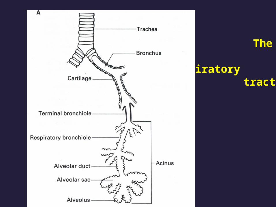
The lower respiratory
tract

Respiratory mucosa
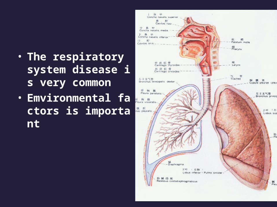
• The respiratory system disease is very common
• Emvironmental factors is important

Major aetiological factors in respiratory disease
• Emvironmental Smoking Lung cancer Chronic bronchitis and emphysema Susceptibility to infection Air pollution Chronic bronchitis Susceptibility to infection Infection Influenza Pneumonia Tuberculosis Occupation Lung cancer Mesothelioma
• Genetic Cystic fibrosis Some asthma

OUTLINE
1.Pulmonary Infections Bacteria Pneumonias Atypical Pneumonias Tuberculosis 2.Chronic Obstractive Lung Diseases (COPD) Emphysema Chronic Bronchitis 3.Bronchiectasis 4.Cor Pulmonale 5.Lung tumors

OUTLINE
1.Pulmonary Infections Bacteria Pneumonias Atypical Pneumonias Tuberculosis 2.Chronic Obstractive Lung Diseases (COPD) Emphysema Chronic Bronchitis 3.Bronchiectasis 4.Cor Pulmonale 5.Lung tumors

• Definition
Bacteria pneumonia is due to bacteria infection affecting distal airways, especially alveoli, with formation of an inflammatory exudate.
often follows a viral upper respiratory tract infection

• Streptococcus pneumoniae
(pneumococcus)• Staphylococcus• Haemophilus influenzae• Klebsiella pneumoniae• Moraxella catarrhalis

Lobar pneumonia
congestion stage
red hepatization
gray hepatization
resolution
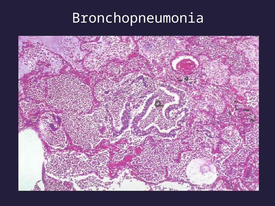
Bronchopneumonia


Lobar pneumonia
• Affects a large part, or the entirety of a lobe, frequently unilateral
• Affects otherwise healthy adults between 20 and 50 years of age, males more than females
• 90% due to Streptococcus pneumoniae
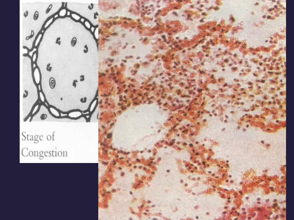

Stage of congestion
Red, edematous


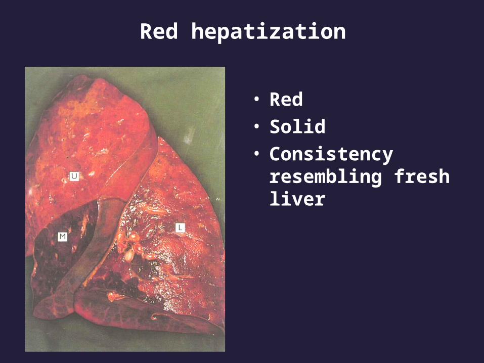
Red hepatization
• Red• Solid• Consistency
resembling fresh liver

Gray hepatization
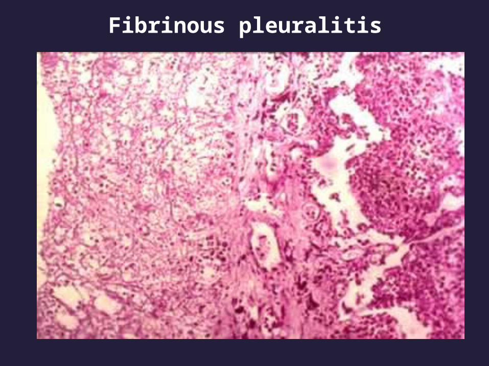
Fibrinous pleuralitis
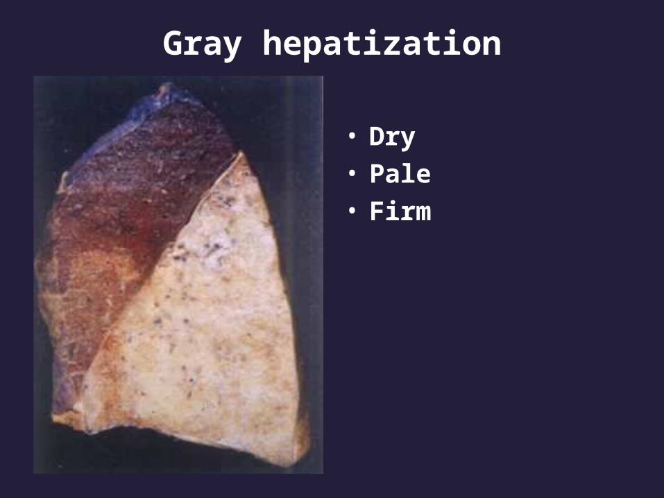
Gray hepatization
• Dry• Pale• Firm

Gray hepatization

Resolution stage
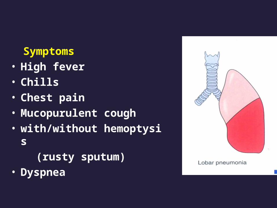
Symptoms• High fever• Chills• Chest pain• Mucopurulent cough• with/without hemoptysis
(rusty sputum)• Dyspnea
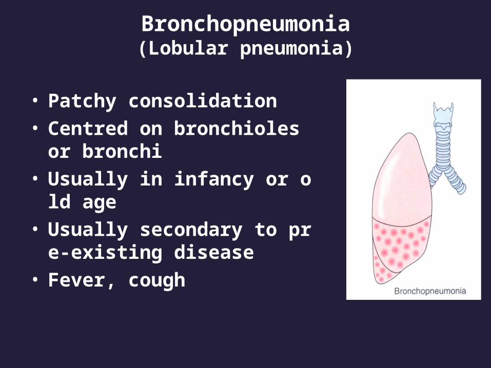
Bronchopneumonia(Lobular pneumonia)
• Patchy consolidation• Centred on bronchioles or br
onchi• Usually in infancy or old age• Usually secondary to pre-exis
ting disease• Fever, cough

Bronchopneumonia

Bronchopneumonia

Bronchopneumonia
Bronchopneumonia

Outcomesof Pneumonia
• Complete recovery
• Complications developed
Abscess formation
Empyema
Bacteremic dissemination
• Organization

Empyema
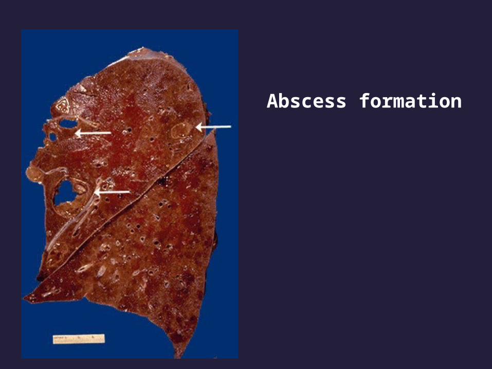
Abscess formation

abscess formation


Organization

• Diagnosis & Therapy
Physical examination
X-ray
Blood culture
Penicillin or other sensitive antibiotic
treatment

X-Ray

• Diagnosis & Therapy
Physical examination
X-ray
Blood culture
Penicillin or other sensitive antibiotic
treatment
OUTLINE
1.Pulmonary Infections Bacteria Pneumonias Atypical Pneumonias Tuberculosis 2.Chronic Obstractive Lung Diseases (COPD) Emphysema Chronic Bronchitis 3.Bronchiectasis 4.Cor Pulmonale 5.Lung tumors

Total Recovery Death
Mainland 1807 1165 79
Guangdong 1304 1110 46
Beijing 339 33 18
Shanxi 108 8 4
Neimeng 25 8 3
Guangxi 12 0 3
Hunan 6 5 1
SiChuan 5 3 1
Fujiang 3 0 0
Shanghai 2 0 0
Henan 2 0 0
Ningxia 1 0 0


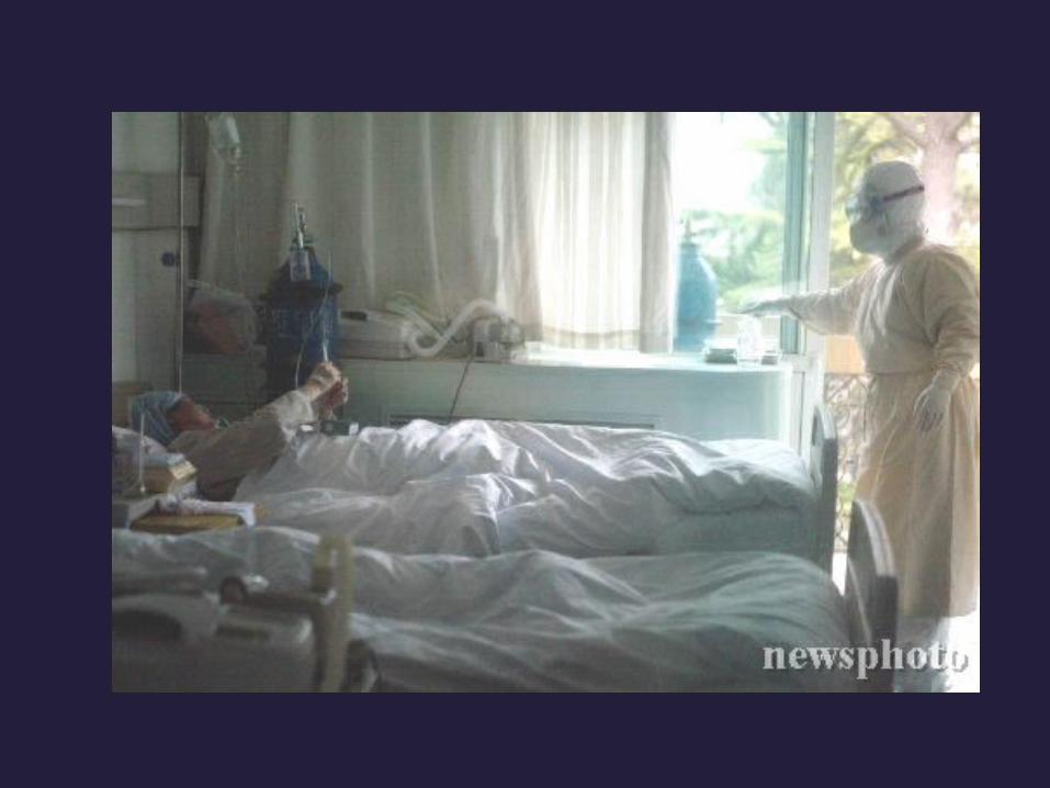

Atypical pneumonia
• The concept was set forth in 1938
• The clinical course is unlike the “typical” bacteria pneumonia
• Causes
mycoplasma
virus
chlamydia

• Gross morphology
Red, congested
Patchy or whole lobes

• Microscopic characteristic
the inflammatory reaction is largely
confined within the walls of the alveoli,
the septa are widened and edematous
with mononuclear cells infiltration---
interstitial pneumonia

interstitial pneumonia

Hyaline membrane

Virus inclusion body
• Clinical course
Cough, fever, headache, malaise
Sputum is modest
No bacteria be isolated
Leukocytosis is modest
Physical findings of consolidation is
varied

Prognosis
• Good in most uncomplicated cases
• Bad in complicated bacterial superinfection cases

OUTLINE
1.Pulmonary Infections Bacteria Pneumonias Atypical Pneumonias Tuberculosis 2.Chronic Obstractive Lung Diseases (COPD) Chronic Bronchitis Emphysema 3.Bronchiectasis 4.Cor Pulmonale 5.Lung tumors

Tuberculosis
• Tuberculosis (TB) is a communicable chronic granulomatous disease caused by Mycobacterium tuberculosis (tubercle bacillus).
• It usually involves the lungs but may affect any organ or tissue in the body.
• Typically, the centers of tubercular granulomas undergo caseous necrosis.
Epidemiology
• Tuberculosis remains a leading cause of death among medically and economically deprived persons throughout the world.
poverty
crowding
elderly person
chronic debilitating illness, including
AIDS
Epidemiology(western world)
• Deaths from tuberculosis peaked in 1800s, Steadily declined untill 1984, then increased beause of human immunodeficiency virus (HIV) infected
• 25,000 new cases in USA annually currently

Epidemiology(Asia)
• The incidence of TB in India is the highest in the world, china is the second
• It is estimated that there is near 400,000,000 persons have once been infected tubercle bacilli in china

Mycobacterium tuberculosis
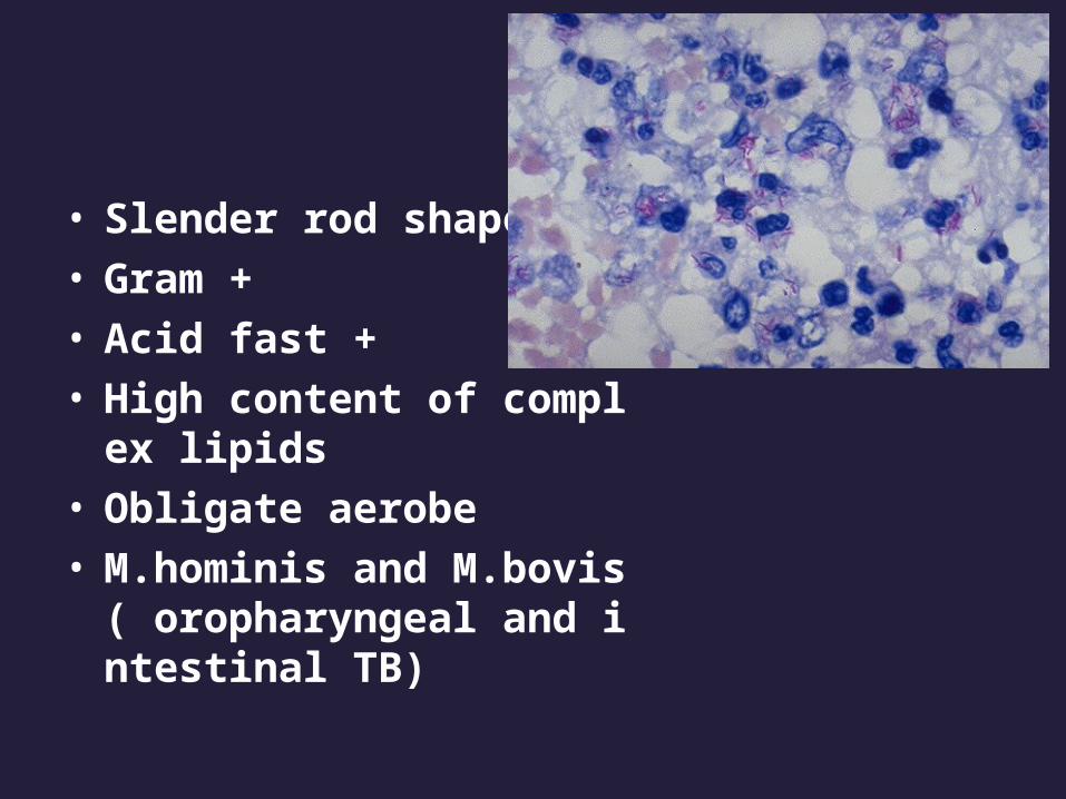
• Slender rod shape• Gram +• Acid fast +• High content of complex li
pids• Obligate aerobe • M.hominis and M.bovis( or
opharyngeal and intestinal TB)
Pathogenesis
• Development of a targeted T cell-mediated immunity (>3weeks) that confers resistance to the organism and results in development of tissue hypersensitivity leading to caseous necrosis and granuloma formation
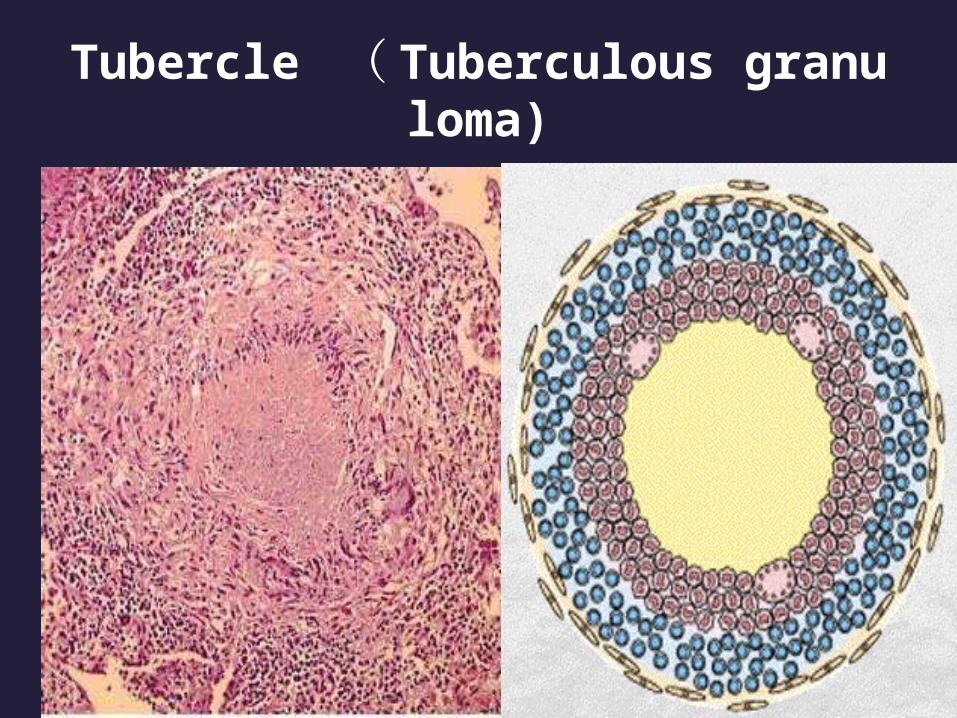
Tubercle ( Tuberculous granuloma)
•
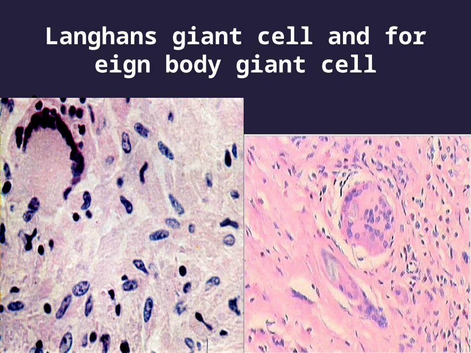
Langhans giant cell and foreign body giant cell
•

Primary tuberculosis
• Previously unexposed, unsensitized person, most frequently in children
• Almost in the lungs• Typically in the distal airspaces of the lower par
t of the upper lobe or the upper part of the lower lobe, usually closed to the pleura

Primary tuberculosis
• Ghon complex (primary complex)
parenchymal lesion
lymphatitis
lymph node involvement
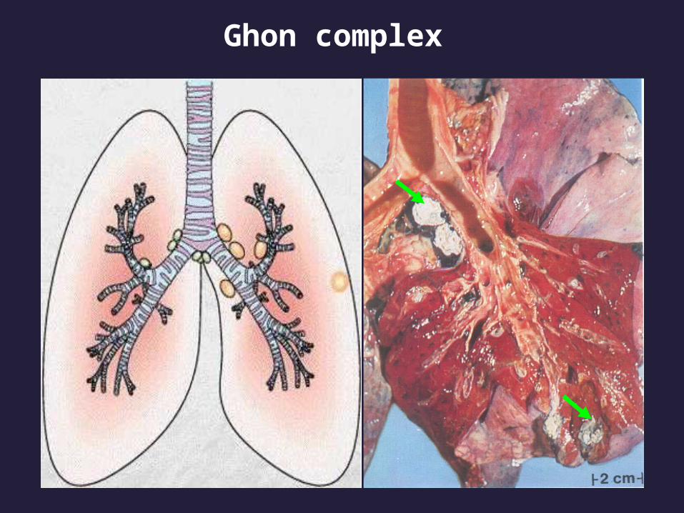
Ghon complex
•

Gohn complex

• Histology:
granulomatous inflammation with/without caseous necrosis

Clinical course
• Asymptom
• Fever, malaise, anorexia

Clinical course
• 95% cases development of cell-mediated immunity controls the infection and increases resistance
• The Gohn complex undergoes fibrosis and calcification
• The scaring foci may harbor viable bacilli for years
• A few immunocompromised patients develop progressive primary TB

Secondary Tuberculosis
• It arises in a previously sensitized host• reactivation of dormant primary lesions or exogenous reinfection • Less than 5% patients

Secondary Tuberculosis
• Pulmonary tuberculosis• Miliary Tuberculosis• Extrapulmonary tuberculosis

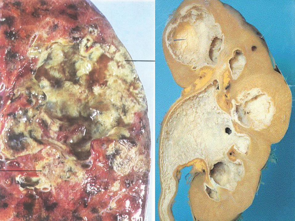
Secondary Pulmonary tuberculosis
• It is classically localized to the apex of upper lobes
• Cavitation occurs readly• The patient raises sputum c
ontaining bacilli

• Firm• Well circumscribed• Central caseation• Peripheral fibrosis
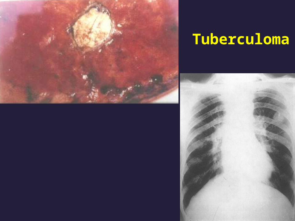
Tuberculoma•
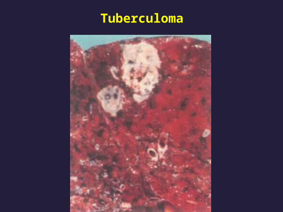
Tuberculoma

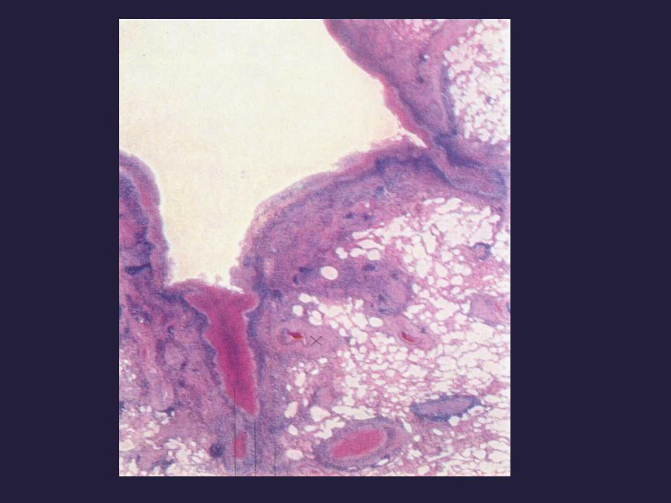
Progressive pulmonary tuberculosis
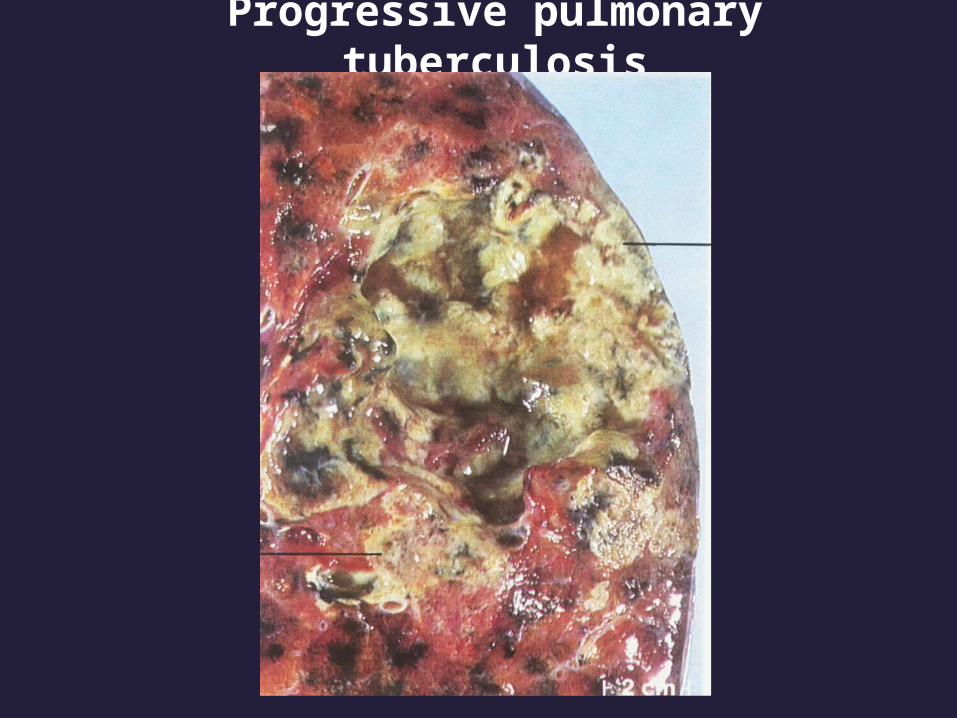
Progressive pulmonary tuberculosis
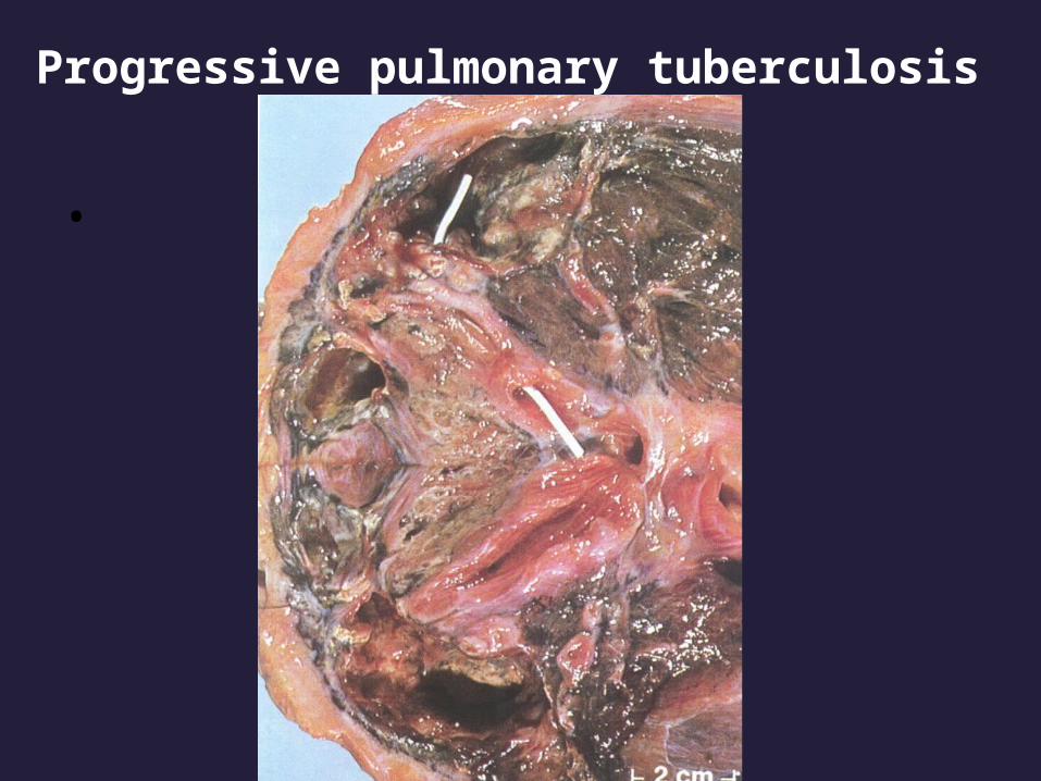
Progressive pulmonary tuberculosis
•

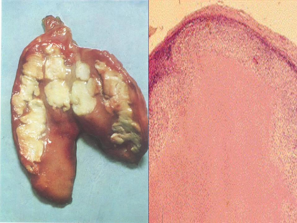
Miliary Tuberculosis
• Systemic miliary tuberculosis
liver, spleen, bone marrow, kidney,
fallopian tubes
• Pulmonary miliary tuberculosis

miliary tuberculosis of liver
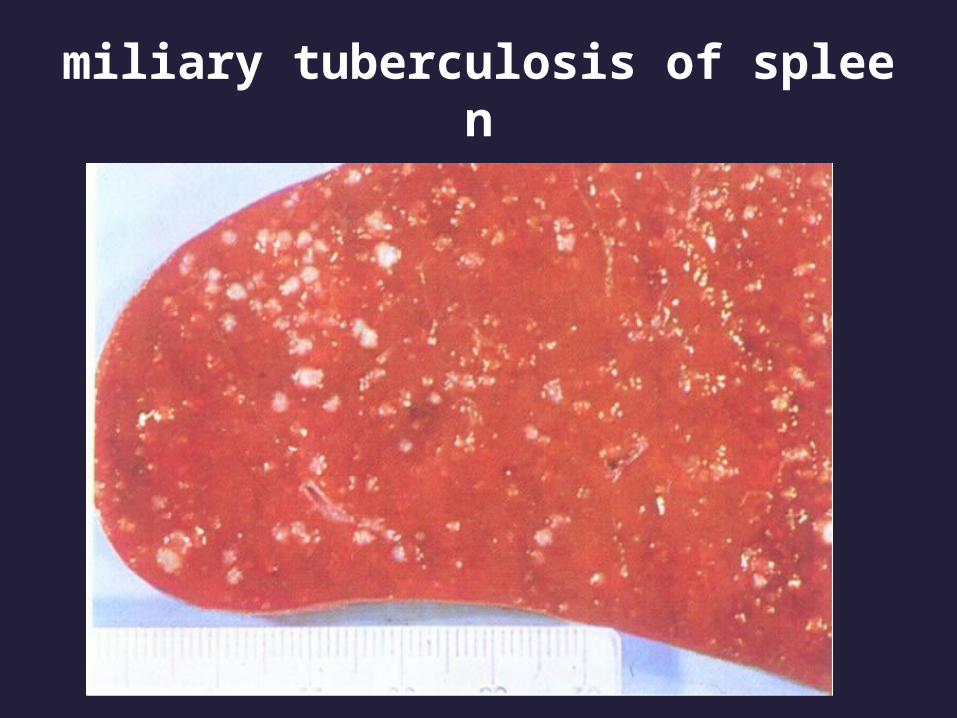
miliary tuberculosis of spleen

pulmonary miliary tuberculosis•
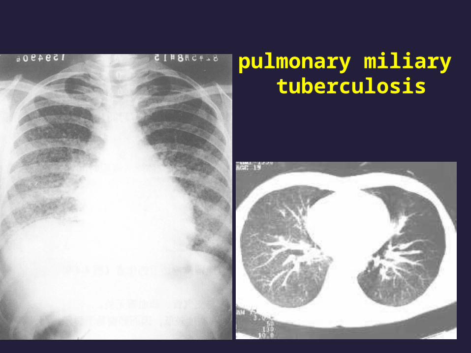
pulmonary miliary tuberculosis

Extrapulmonary tuberculosis
• Intestinal tuberculosis Secondary to the swallowing of coughed-up
infective material
Drinking of contaminated milk is the reason of primary lesion

Intestinal tuberculosis

lymphadenitis
• Most frequent form• Cervical region• Unifocal in HIV(-) pati
ents• Multifocal in HIV(+) p
atients

lymphadenitis
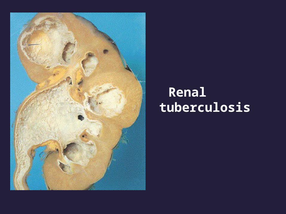
Renal tuberculosis

Vertebrae TB
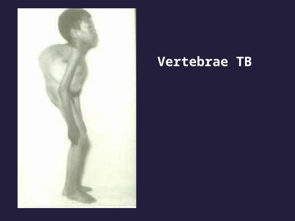
Vertebrae TB

Joint TB

Clinical course
• Asymptomatic
• Systemic symptoms
malaise
anorexia
weight loss
fever (low, remittent)
night sweats

Clinical course
• localizing pulmonary symptoms
Cough
Mucoid, purulent sputum
Hemoptysis
Pleural pain
Dyspnea• localizing extrapulmonary symptoms

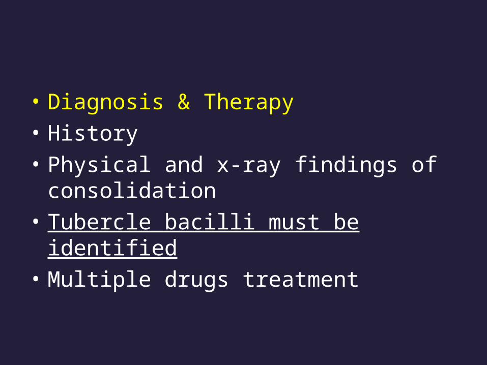
• Diagnosis & Therapy
• History
• Physical and x-ray findings of consolidation
• Tubercle bacilli must be identified
• Multiple drugs treatment

OUTLINE
1.Pulmonary Infections Bacteria Pneumonias Atypical Pneumonias Tuberculosis 2.Chronic Obstractive Lung Diseases (COPD) Chronic Bronchitis Emphysema 3.Bronchiectasis 4.Cor Pulmonale 5.Lung tumors
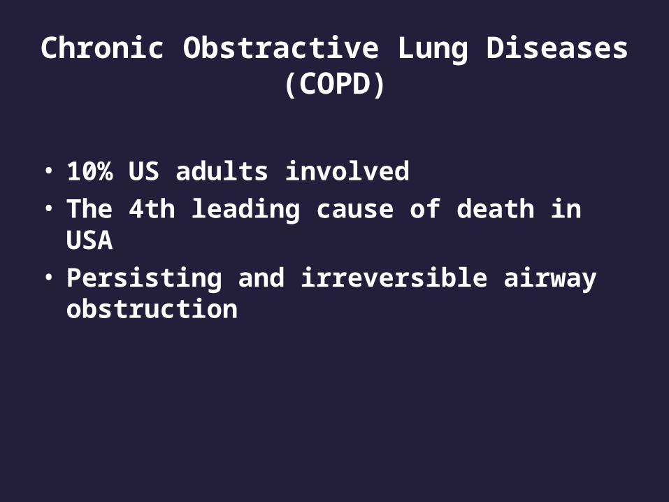
Chronic Obstractive Lung Diseases(COPD)
• 10% US adults involved• The 4th leading cause of death in USA• Persisting and irreversible airway
obstruction

OUTLINE
1.Pulmonary Infections Bacteria Pneumonias Atypical Pneumonias Tuberculosis 2.Chronic Obstractive Lung Diseases (COPD) Emphysema Chronic Bronchitis 3.Bronchiectasis 4.Cor Pulmonale 5.Lung tumors

• Definition
Emphysema is characterized by permnent
Enlargement of the air spaces distal to the terminal bronchioles accompanied by destruction of their walls

Emphysema vs overinflationWith destruction without destruction
Complicated reasons compensatory
obstructive

Emphysema vs chronic bronchitis
Morphologic feature clinical feature
Restricted to acinus large and small
airway
The two deaseas usually coexist

Types of Emphysema
• Centriacinar
• Panacinar
• Distal acinar
(according to the distribution of lesions in the lobule and acinus)

Normal structure of acinus


Centriacinar Emphysema
• Cigarette smoking• The upper lobe, apical segments
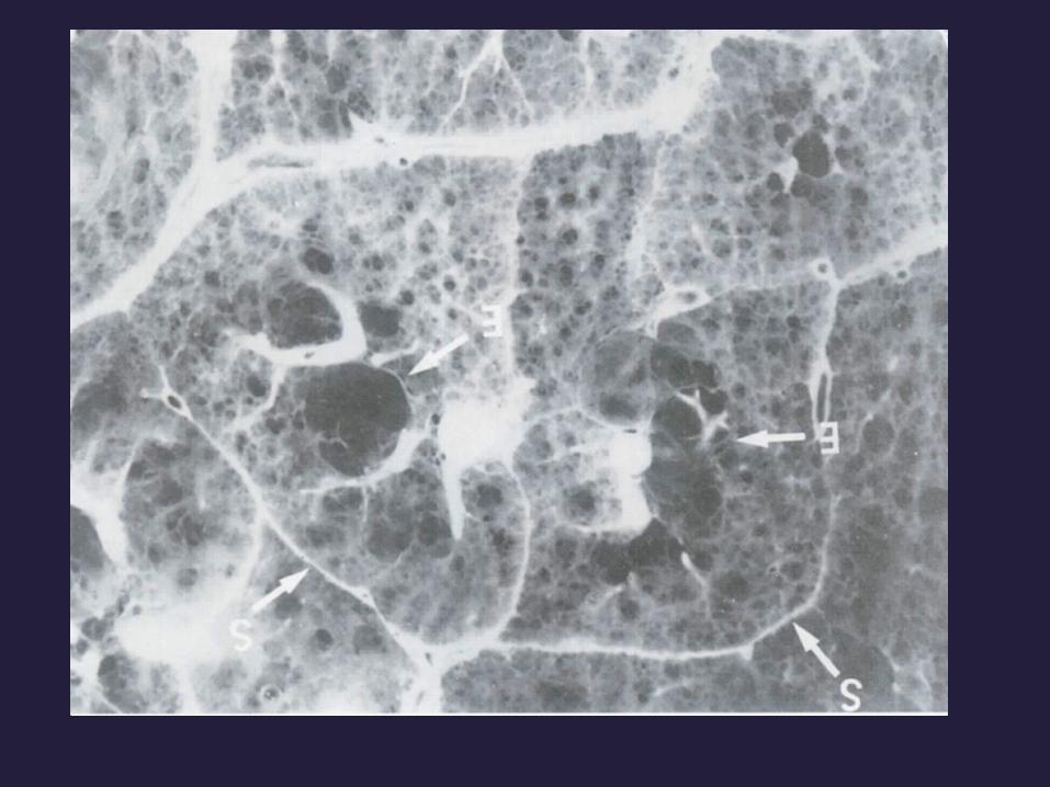
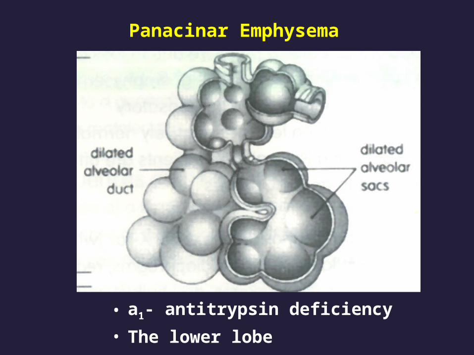
Panacinar Emphysema
• а1- antitrypsin deficiency
• The lower lobe

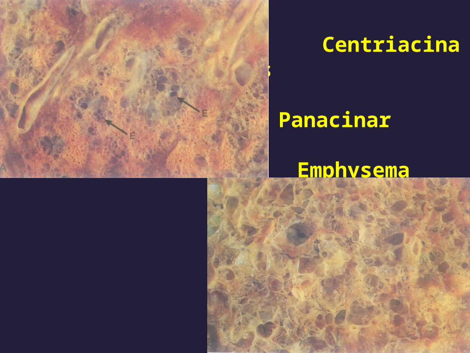
Centriacinar vs
Panacinar Emphysema
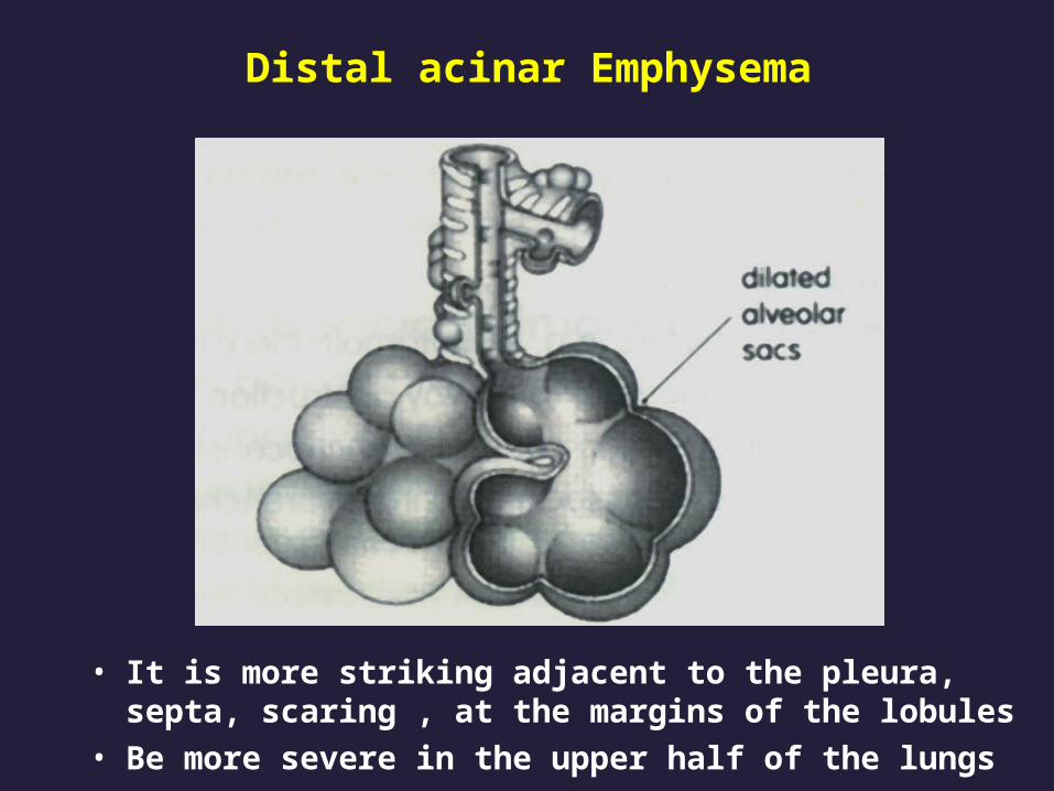
Distal acinar Emphysema
• It is more striking adjacent to the pleura, septa, scaring , at the margins of the lobules
• Be more severe in the upper half of the lungs
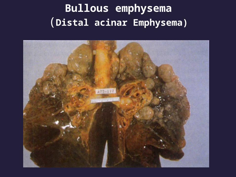
Bullous emphysema(Distal acinar Emphysema)

Bullous emphysema(Distal acinar Emphysema)


• With the destruction of alveolar walls and loss of elastic tissue , Small airways tend to collapse during expiration-----an important cause of chronic airflow obstruction

pathogenesis
• Protease-antiprotease imbalance
• Oxidant-antioxidant imbalance
These two imbalances are almost always coexist

• Proteolytic activity
• а1- antitrypsin

Pathogenesis of emphysema
Clinical course
• Dyspnea• cough, purulent sputum (with bronchiti
s) • barrel-chest• Secondary pulmonary hypertension d
evelops gradually

OUTLINE
1.Pulmonary Infections Bacteria Pneumonias Atypical Pneumonias Tuberculosis 2.Chronic Obstractive Lung Diseases (COPD) Emphysema Chronic Bronchitis 3.Bronchiectasis 4.Cor Pulmonale 5.Lung tumors
Chronic Bronchitis
• Definition (made on clinical ground)
persistent productive cough for at least
3 consecutive months in at least 2
consecutive years
It is often developed in middle age to
old men with cigarette smoking

• Causes
cigarette smoking
other air pollution
( sulfur dioxide, nitrogen dioxide)

• epidermal growth factor receptor
• Microbial infection

• Grossly, the mucosal of the trachea, bronchus, bronchiole is hyperemic, covered by a layer of mucinous or mucopurulent secretion
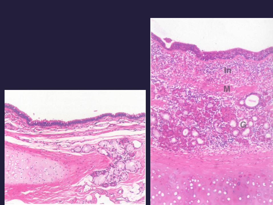


Increased numbers of chronic inflammatory cells in t
he submucosa.
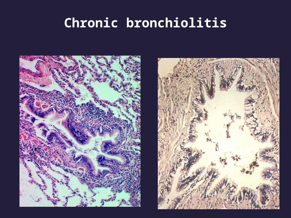
Chronic bronchiolitis

Chronic bronchiolitis

Clinical course• Cough with mucus or mucopurulent sputum• With/without ventilatiory dysfunction, hypoxemia, hypercapnia (COPD developed) • Secondary pulmonary hypertension develop
s gradually
OUTLINE
1.Pulmonary Infections Bacteria Pneumonias Atypical Pneumonias Tuberculosis 2.Chronic Obstractive Lung Diseases (COPD) Emphysema Chronic Bronchitis 3.Bronchiectasis 4.Cor Pulmonale 5.Lung tumors

• Definition Bronchiectasis is the permanent dilation of br
onchi and bronchioles caused by destruction of the muscle and elastic supporting tissue.
It is not a primary disease but rather is secondary to persisting infection or obstruction caused by variety of conditions
Conditions that predispose to bronchiecctasis
• Bronchial obstruction• Bacteria pneumonia• Congenital conditions

pathogenesis
• Obstruction Chronic infection
Tissue damage secretion accumulation
irreversible dilation

• Lower lobes bilaterally
• Distal bronchi and bronchioles are more severe

Cross-section of lung demonstrating dilated bronchi extending almost to the pleura

Bronchiectasis.

Dilated bronchus in which the mucosa and wall is not clearly seen because of the necrotizing inflammation

Clinical Course• severe, persistent cough with copious amou
nt of mucopurulent ,fetid sputum• Hemoptysis• symptoms are episodic and are precipitated
by upper respiratory tract infection• Secondary pulmonary hypertension develop
s gradually

OUTLINE
1.Pulmonary Infections Bacteria Pneumonias Atypical Pneumonias Tuberculosis 2.Chronic Obstractive Lung Diseases (COPD) Chronic Bronchitis Emphysema 3.Bronchiectasis 4.Cor Pulmonale 5.Lung tumors

Cor pulmonale
• Definition
It also called pulmonary heart disease, is used to describe disease of the right-side cardiac chambers caused by pulmonary hypertension resulting from pulmonary parenchymal or pulmonary vascular disease

Disorders that predispose to cor pulmonale
Diseases of the lungs Chronic obstructive lung disease Diffuse pulmonary interstitial fibrosis Extensive, persistent atelectasis Cystic fibrosis Diseases of pulmonary vessels Pulmonary embolism Primary pulmonary vascular sclerosis Extensive pulmonary arteritis Drug-, toxin-, or radiation-induced vascular sclerosis Disorders affecting chest movement Disorders inducing pulmonary arteriolar constriction
Heart changes
• right ventricular, and often right artrial hypertrophy.
• It may be dilated when ventricular failure develops.

• Conic of pulmonary artery bulges
• The point of the heart become blunt and round
• Thickness of the right ventricle exceeds the left ventricle
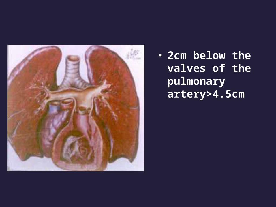
• 2cm below the valves of the pulmonary artery>4.5cm
Pulmonary changes
• Primary lung diseases (such as chronic bronchitis, emphysema)
• with blood vessel changes-----pulmonary hypertension.

Clinical course
• Right cardiac failure
• Respiratory failure

OUTLINE
1.Pulmonary Infections Bacteria Pneumonias Atypical Pneumonias Tuberculosis 2.Chronic Obstractive Lung Diseases (COPD) Chronic Bronchitis Emphysema 3.Bronchiectasis 4.Cor Pulmonale 5.Lung tumors

Lung tumors
• Bronchogenic carcinoma:95%• Miscellaneous group:5%
bronchial carcinoid tumor
fibrosarcoma
lymphoma
hamartoma

Bronchogenic carcinoma
• No.1 cause of cancer-related deaths in industrialized countries.
• Cigarette smoking is a important cause• The peak incidence occurs between ages 55
and 65 years.• The male to female ratio is 2:1• The prognosis of lung cancer is dismal

Histologic classification of Bronchogenic carcinoma
• Non-small cell lung carcinoma 1.squamous cell carcinoma(25%-30%) 2.adenocarcinoma,including bronchioloalveolar ca(30%-35%) 3.large cell carcinoma(10%-15%)
• Small cell carcinoma(20%-25%)• Combined pattens(5%-10%)
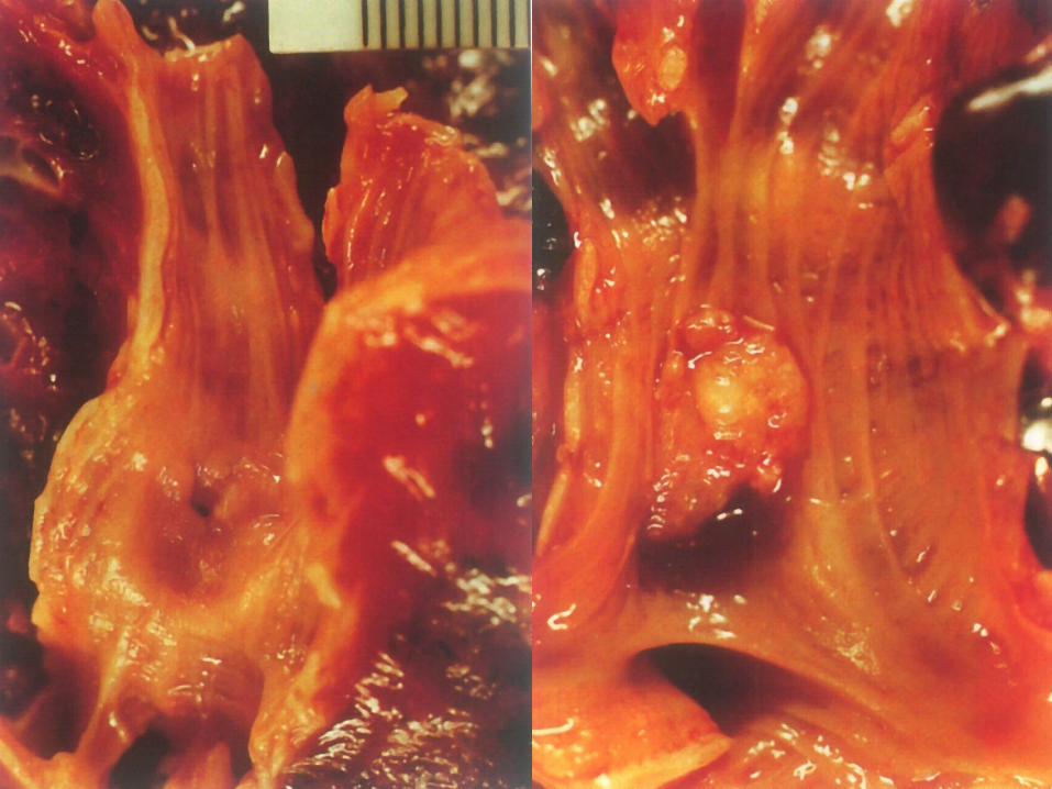
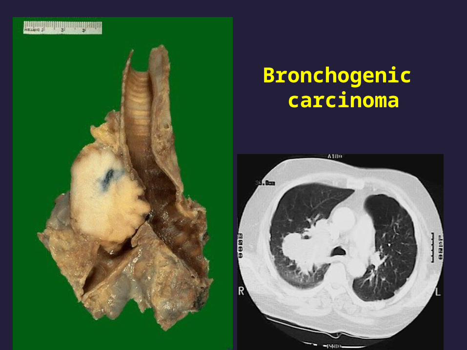
Bronchogenic carcinoma
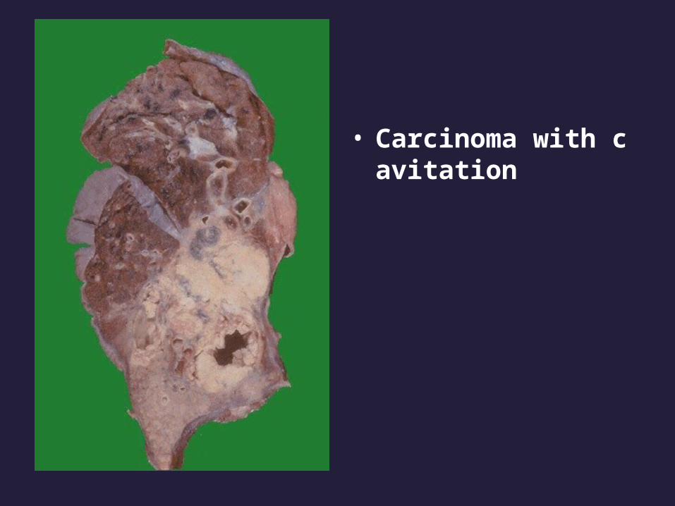
• Carcinoma with cavitation

Bronchogenic carcinoma
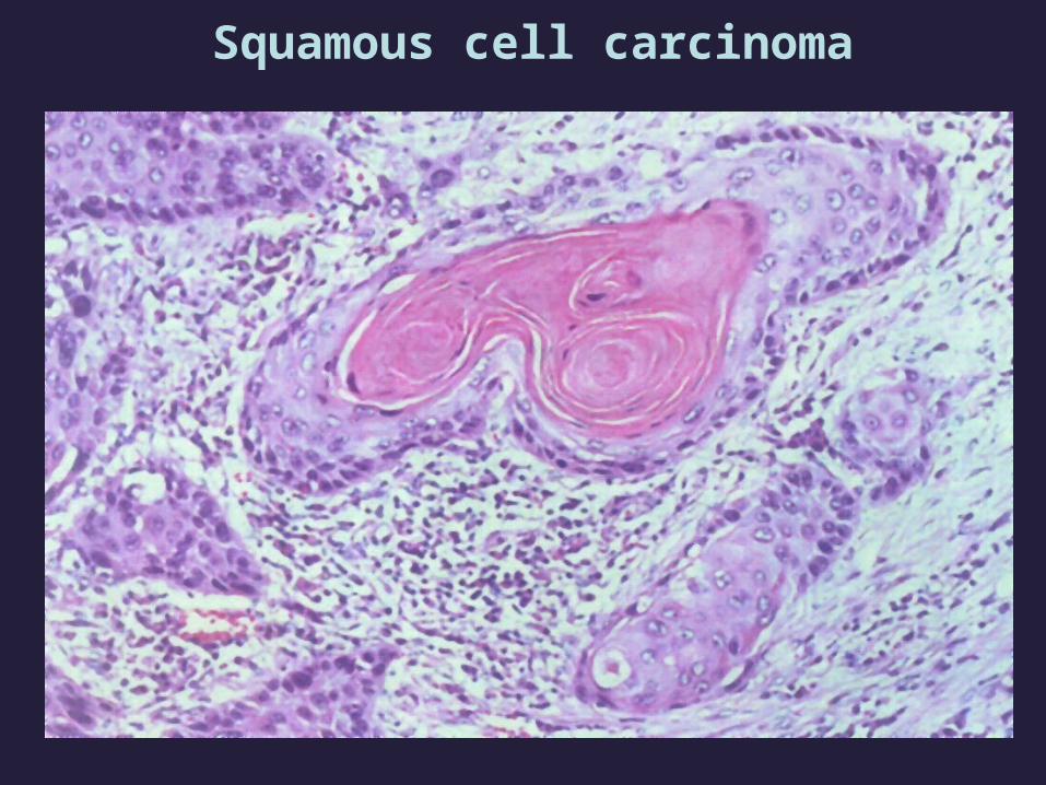
Squamous cell carcinoma

adenocarcinoma

Small cell carcinoma

Metastatic cacinoma

Clinical course
• Silent, insidious lesion• Chronic cough and expectoration• Hoarseness, chest pain, pleural or pericardial
effusion• Symptoms emanating from metastatic spread
to the brain, liver,or bone• NSCLCs have a better prognosis than SCLCs
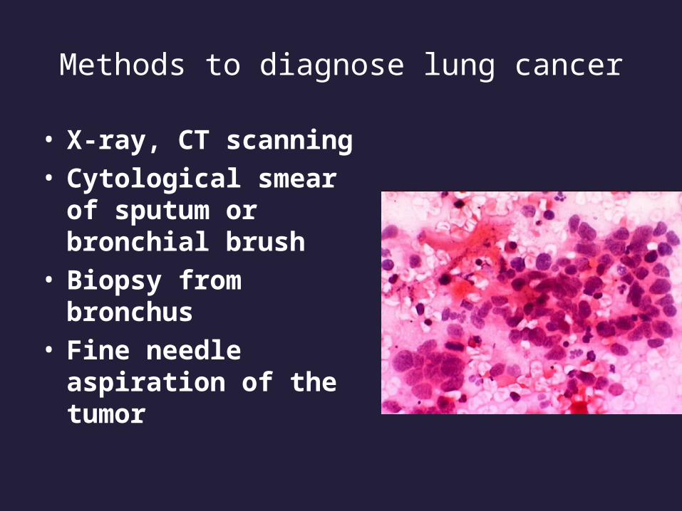
Methods to diagnose lung cancer
• X-ray, CT scanning• Cytological smear of
sputum or bronchial brush
• Biopsy from bronchus• Fine needle aspiration
of the tumor
Related Documents