Thorax (1947), 2, 91. DISEASES OF THE LUNG RESULTING FROM OCCUPATIONAL DUSTS OTHER THAN SILICA* BY KENNETH M. A. PERRY London It is only in recent years that considerable interest has been given to the dusts to which men are exposed at their work, even though silicosis is known to have occurred in prehistoric times. Many dusts are now recognized as dangerous, and in the extreme it may even be doubted whether any dust can be regarded as harmless. It is rational at least to suppose that the lung cannot become a physiological dust trap and yet retain its elasticity. It seems possible that any dust, no matter how innocuous in small concentrations, would in large enough quantity eventually overwhelm the defences of the lung and accumulate in such amounts as to impair function; such a form of lung disease would be the result of causes of a mechanical nature-the physical presence of large amounts of inert foreign material. The term "benign pneumoconiosis " has been given to this type of disease in order to contrast it with diseases resulting from the inhalation of siliceous matter. But besides this group of conditions occupational dust may give rise to inflammatory lesions, allergic responses, and neoplastic changes. INFLAMMATORY CHANGES Inflammatory changes may be caused by inorganic metals, such as manganese, beryllium, vanadium, and osmium, giving.rise to a chemical pneumonitis; and by organic matter, such as decaying hay and grain, bagasse, cotton fibre, and similar substances where the aetiology is somewhat obscure, though fungi are frequently blamed. Manganese Manganese occurs in nature as the oxide in the ores pyrolusite, braunite, hausmanite, and manganite, which are found in Russia, India, the Gold Coast of Africa, Brazil, the United States, and Cuba. Manganese appears to be an essential substance in plant and animal nutrition. Its most important alloys are spiegeltisen and ferro-manganese, but silico-manganese and silico-spiegel are used in certain grades of steel. Manganese bronze is used for marine construction and mining machinery where corrosion is serious. The *From the Department for Research in Industrial Medicine (Medical Research Council) at the London Hospital. A paper presented in July, 1947, at a meeting held in Newcastle of the Association for the Study of Diseases of the Chest. on March 13, 2022 by guest. Protected by copyright. http://thorax.bmj.com/ Thorax: first published as 10.1136/thx.2.2.91 on 1 June 1947. Downloaded from

Welcome message from author
This document is posted to help you gain knowledge. Please leave a comment to let me know what you think about it! Share it to your friends and learn new things together.
Transcript
Thorax (1947), 2, 91.
DISEASES OF THE LUNG RESULTING FROMOCCUPATIONAL DUSTS OTHER
THAN SILICA*BY
KENNETH M. A. PERRYLondon
It is only in recent years that considerable interest has been given to thedusts to which men are exposed at their work, even though silicosis is knownto have occurred in prehistoric times. Many dusts are now recognized asdangerous, and in the extreme it may even be doubted whether any dust canbe regarded as harmless. It is rational at least to suppose that the lung cannotbecome a physiological dust trap and yet retain its elasticity. It seems possiblethat any dust, no matter how innocuous in small concentrations, would in largeenough quantity eventually overwhelm the defences of the lung and accumulatein such amounts as to impair function; such a form of lung disease would bethe result of causes of a mechanical nature-the physical presence of largeamounts of inert foreign material. The term "benign pneumoconiosis " hasbeen given to this type of disease in order to contrast it with diseases resultingfrom the inhalation of siliceous matter. But besides this group of conditionsoccupational dust may give rise to inflammatory lesions, allergic responses, andneoplastic changes.
INFLAMMATORY CHANGESInflammatory changes may be caused by inorganic metals, such as manganese,
beryllium, vanadium, and osmium, giving.rise to a chemical pneumonitis; andby organic matter, such as decaying hay and grain, bagasse, cotton fibre, andsimilar substances where the aetiology is somewhat obscure, though fungi arefrequently blamed.
ManganeseManganese occurs in nature as the oxide in the ores pyrolusite, braunite, hausmanite,
and manganite, which are found in Russia, India, the Gold Coast of Africa, Brazil, theUnited States, and Cuba. Manganese appears to be an essential substance in plant andanimal nutrition. Its most important alloys are spiegeltisen and ferro-manganese, butsilico-manganese and silico-spiegel are used in certain grades of steel. Manganese bronzeis used for marine construction and mining machinery where corrosion is serious. The
*From the Department for Research in Industrial Medicine (Medical Research Council)at the London Hospital. A paper presented in July, 1947, at a meeting held in Newcastleof the Association for the Study of Diseases of the Chest.
on March 13, 2022 by guest. P
rotected by copyright.http://thorax.bm
j.com/
Thorax: first published as 10.1136/thx.2.2.91 on 1 June 1947. D
ownloaded from

KENNETH M. A. PERRY
metal is also alloyed with aluminium, tin, arsenic, antimony, bismuth, and boron. Man-ganese oxide is used for bleaching glass, manganese chloride is used in dyeing, the sulphatein calico printing, and the persulphate as an oxidizing agent in making organic products;while manganates and permanganates are used for preserving wood, for bleaching textilefibres, and for disinfecting and oxidizing purposes. Chronic manganese poisoning was firstdescribed in 1837 by Couper, and since that time some 353 cases have been reported.- Thecommonest pathological changes are those which occur in the central nervous system andresult in the Parkinsonian syndrome. Accounts of the condition have mostly come fromEurope, but cases have been reported in England by Charles (1927) and Owen and Cohen(1934), while recently a full review has been produced by Fairhall and Neal in the UnitedStates (1943).
In 1921 Brezina first drew attention to the relationship of manganese topneumonia; he reported that 5 out of 10 men working in a pyrolusite mill haddied of pneumonia in a period of two years. Baader (1933) ascribed the highincidence of pneumonia amongst workers making dry battery cells to manganese;while Elstad (1939) observed that the erection of an electrical plant for man-ganese smelting in Sauda in Norway was followed by a tenfold increase in themortality rate for pneumonia in that area. A pall of smoke containing man-ganese oxide with particles of less than 5 ,u overhurb; the town. Heine (1943)admitted the high incidence of pneumonia in Ge man factories handlingpyrolusite.
Riddervold and Halvorsen (1943) described Sauda as an isolated village inwestern Norway situated at the farthest end of a deep fjord and surroundedby high hills. Sudden changes in climate are experienced, with 2,000 mm.(78 in.) of rain a year. Before the erection of the factory in 1915 Sauda had1,500 inhabitants. The population slowly increased to 4,500. The factorystarted producing in 1923, and during that year an epidemic of lobar pneumoniaaffected the town. Every year since, the village has had a high mortality ratefrom pneumonia (Table I).
TABLE IMORTALITY (PER 100,000) FROM ALL KINDS OF PNEUMONIA IN VARIOUS LOCALITIES
(RIDDERVOLD AND HALVORSEN, 1943)
Sauda, Oslo Norway U.S.A. Pittsburgh Long Beach,Norway 1 1924-36 1924-36 1933-37 1929-31 U.S.A.
82 82
* Lobar pneumonia only.
A survey of the bacteriology of the " Sauda " preumonias showed that of46 cases 13 were due to pneumococcus Type 1, 4 to Type 2, and 5 to Type 3.A second attack of pneumonia was frequent. There were 2 cases of atypicalpneumonia due to pneumococcus Type 3. The age distribution of pneumoniawas: 0 to 15 years, 11 cases; 15 to 30 years, 11 cases; 30 to 40 years, 3 cases;
92
on March 13, 2022 by guest. P
rotected by copyright.http://thorax.bm
j.com/
Thorax: first published as 10.1136/thx.2.2.91 on 1 June 1947. D
ownloaded from

OCCUPATIONAL LUNG DISEASE
40 to 50 years, 5 cases (2 fatal); 50 to 60 years, 9 cases (2 fatal); over 60 years.6 cases (3 fatal), In only 6 out of the 14 cases investigated were contacts foundto have the same type of pneumococcus in the throat, but 46 per cent of healthypeople carried pneumococcus in the throat. Immediately before the war, Buttner(1939) reported that men working in a Rhenish pyrolusite mine suffered anaverage pneumonia rate of 17 per thousand with an average death rate of 6.3per thousand, compared with a pneumonia rate of 0.54 per thousand in acontrol group from Leipzig.
Lloyd-Davies (1946) recorded his observations on men employed in themanufacture of potassium permanganate who were exposed to the inhalation ofdust of manganese 'dioxide and higher oxides of manganese. The manganesecontent of the atmospheric dust collected by the electrostatic precipitator,expressed as manganese dioxide, varied from 41 to 66 per cent ; practically all theparticles were below 1 ,u in size, and 80 per cent were below 0.2 *. The groupconsisting of between 40 and 124 men had an incidence of pneumonia whichvaried from 1,500 to 6,300 per 100,000 in the period 1938 to 1945, compared withan average of 73 for the same period amongst male members of Boots' HealthInsurance Society. Naso-pharyngeal catarrh and attacks of bronchitis werefrequent, the latter sometimes being associated with a spasmodic element. Thesymptoms, however, always subsided after removal from exposure. The pneu-monia which occurred in the workers was not different clinically from that whichmay develop in any individual, and Lloyd-Davies illustrated his paper with radio-graphs showing consolidation in the upper lobe which resolved in a period of nineweeks. He suggested, however, that the fever and general condition of the patientresponded more slowly than usual to sulphonamides. No permanent pulmonarychanges were observed in the men either on clinical or on radiographicexamination.
Animal experiments in which mice were exposed to manganese dust werecomplicated by the obvious general toxic effect of the manganese, but the histo-logical effects on the lungs were uniform and striking. The changes were depen-dent on the length of exposure, and varied from slight mononuclear infiltration tointense mononuclear interstitial infiltration with many dust-laden cells, and finallyto consolidation, including haemorrhagic areas, leading to complete disorganiza-tion of the lung structure. The changes were most marked around the bronchi,and the cells lining the bronchi were swollen and had undergone a hydropicchange. Lloyd-Davie4 thinks the manganese has a direct influence on the cellsof the bronchial and alveolar epithelium. The presence of dust cells, even thoughfew, suggests that the dust is phagocytosed, but it is probable that after absorptionthe phagocytic cells are killed. Either before or after absorption manganeseappears to have a specific action, causing intense mononuclear proliferationand infiltration, and sufficient to cause consolidation of the lung with necrosisand haemorrhage.
G
93
on March 13, 2022 by guest. P
rotected by copyright.http://thorax.bm
j.com/
Thorax: first published as 10.1136/thx.2.2.91 on 1 June 1947. D
ownloaded from
KENNETH M. A. PERRY
BerylliumBeryllium was discovered in 1797, but in recent years it has become increasingly impor-
tant because of the properties of its alloys. Beryllium copper is corrosion resistant,non-rusting, non-sparking, and non-magnetic, and has good electrical conductivity, highstrength, and astonishing fatigue-resisting powers. It is used in precision instruments, alti-meters, airplane pipe lines, carburettors, and tejlephone switchboards, and also in aircraftproduction. Beryllium steel has such remarkably valuable properties that it will doubtlessreceive much commercial attention. Beryllium is important in the production of atomicenergy. The principal ore of beryllium is beryl, and from this the beryllium oxide isextracted by processing at high temperatures with concentrated acids and fusion methodswhich result in the production of fumes and dust. Weber and Engelhardt (1933) firstshowed that these dusts produced lung damage in guinea-pigs; while Fabrioni (1935) coinedthe term " berylliosis " for this pathological condition. Fairhall and his colleagues (1943),however. reported a full investigation of the toxicity of beryllium compounds in animals.T-hey succeeded in producing experimentally both pneumonitis and dermatitis with berylliumfluoride and oxyfluorido and certain hydrolysed beryllium salts such as the chloride andsulphate.
The earliest reports of poisoning in industrial workers came from Russia,Gelman (1936 and 1938) describing a condition in which shivering and fever,similar to metal fume fever, were followed in two days by extensive broncho-alveolitis. This lung condition he attributed to the action of fluorine, which wasseparated from beryllium oxyfluoride in the bronchioles and alveoli. Theseobservations were followed by a report on 46 cases of poisoning by Berkovitsand Izrael (1940). Inhalation of beryllium fluoride resulted in fever which sub-sided in a few days but was followed after a similar period by a second phase.characterized by cough, scanty sputum, dyspnoea, temperature of 1020 F. to1030 F. and cyanosis of face and extremities. Riles were heard in the lungs,particularly at the bases. The blood showed a fall of haemoglobin and leucocytes,lymphopenia, and a raised sedimentation rate, while radiographs revealed manysmall ill-defined discrete opacities, especially in the middle and lower areas.The patient usually recovered completely in from ten days to two months. Theberyllium fluoride affected mainly the smaller bronchi, which have no cartilagein their walls. These showed desquamation; their walls became infiltrated withleucocytes; and later they became blocked with inflammatory exudate, leadingto minute areas of collapse, with compensatory emphysema. Though resolutionof the acute stage was usually complete, it might be followed by fibrosis.
Full accounts of this form of poisoning appeared during the war in Germany(Meyer, 1942, Wurm and Ruger, 1942), and in the United States, wherevan Ordstrand and others (1943) gave an account of three cases of chemicalpneumonia among workers extracting beryllium oxide; Kress and Crispell (1944)reported. four cases in men working will fluorescent powders containingberyllium ; and Shilen and his colleagues (1944) reported disease of the respiratorytract in industries extracting beryllium from beryl ore. More recently,van Ordstrand and others (1945) have described 170 cases of poisoning which
94
on March 13, 2022 by guest. P
rotected by copyright.http://thorax.bm
j.com/
Thorax: first published as 10.1136/thx.2.2.91 on 1 June 1947. D
ownloaded from

OCCUPATIONAL LUNG DISEASE
have occurred among workers in three plants during the past four years. Themanifestations were dermatitis, chronic skin ulcer, and inflammatory changesin the respiratory tract producing in extreme cases diffuse pneumonitis. Ninetyof these patients had either upper or lower respiratory infections. Soreness ofnose and throat and epistaxis were the symptoms of upper respiratory infection,and most cases cleared in from three to six weeks. Lower respiratory tractinfections were characterized by non-productive cough, but with occasional blood-streaked mucoid sputum and dyspnoea. Rales were characteristically present inthe early phase of inspiration, fine at first, becoming coarse. Vital capacity wasreduced by as much as 30 per cent; there was slight fever. If the patient wasremoved from contact, no case of pneumonitis developed after three weeks fromthe onset of the bronchitis. The condition can be prevented if the workers use aface mask to stop dust entering the respiratory tract. Chemical pneumonitisdeveloped in 38 workers, 5 of whom died. Symptoms were cough with occasionalblood-streaked sputum, substernal burning pain, shortness of breath, cyanosis,abnormal taste, anorexia with some weight loss, and increasing fatigue. The onsetwas insidious and without symptoms. The vital capacity was reduced as low as2,000 ml., but the sedimentation rate was normal. Bronchoscopy showed ahyperaemic mucosa with some oedema. Radiographs showed diffuse hazinessof both lungs, followed by soft irregular areas of infiltration with prominenceof bronchial markings, and then by absorption of the soft infiltration and theappearance of discrete large or small conglomerate nodules scattered throughoutboth lung fields, and finally by the clearing of the lung fields after one to fourmonths. Necropsies showed atypical pneumonitis; striking features in lung-tissue sections were large numbers of plasma cells, relative absence of poly-morphonuclear infiltration, diffuse pulmonary oedema, and haemorrhagic extra-vasation; fibroblasts, with evidence of organization, were present. Berylliumpresent in the lung varied between 6.20 and 1.89 mg. per 10 g. of dry tissue.
Another and most interesting syndrome has been reported by Hardy andTabershaw (1946), Gardner (1946), and Hardy (1947). It appeared amongemployees of a firm manufacturing fluorescent lamps. Seventeen people (14men and 3 women) who worked in one building and handled a fluorescentpowder, zinc manganese beryllium silicate, during the same period of timedeveloped delayed pneumonitis, from which 6 of them died. The symptomsstarted either during the period of work or up to three years after leaving thefactory with an average of twenty months' duration, and consisted of loss ofappetite, loss of weight, dyspnoea on exertion, cough, and fatigue. At mosttimes rales were audible at the bases and in the axillae, and in a few instancesthere was a pleural friction rub. The radiographic appearances fell into threegroups. Stage 1 showed a fine granularity, uniform and diffuse and extending tothe periphery. Stage 2 was characterized- by a diffuse reticular pattern on thegranular background, the hilar vascular shadows becoming fuzzy, indistinct, and
95
on March 13, 2022 by guest. P
rotected by copyright.http://thorax.bm
j.com/
Thorax: first published as 10.1136/thx.2.2.91 on 1 June 1947. D
ownloaded from
KENNETH M. A. PERRY
slightly enlarged. In stage 3 distinct nodules appeared uniformly through thelungs, giving the appearance of a snow-storm, the hilar shadows appearing largeand indistinct. The appearances in this stage closely simulated sarcoidosis.Necropsy on one patient revealed focal and diffuse chronic granulomatous inflam-mation of all lobes of both lungs with organization and fibrosis, diffuse chronicgranulomatous inflammation of hilar lymph nodes with fibrosis, right ventricularand atrial cardiac hypertrophy and dilation, subacute progressive focal and diffusehepatic necrosis, and splenomegaly (400 g.) with fibrosis. There was apparentlyfocal intra-alveolar granulomatous change characterized by centrally andeccentrically located giant cells of a foreign-body type, surrounded by epithelioidcells; plasma cells, lymphocytes, and mononuclear cells infiltrated the lesionthroughout. There is manifestly a close clinical resemblance between this syn-drome and Boeck's sarcoidosis, even though the histology is somewhat different.
VanadiumWorld consumption of vanadium was more than trebled between 1936 and 1939. It ig
used for making special alloy steels and is an ingredient of the best grades of high-speedsteels, since it exerts a degasifying and deoxidizing action. It promotes fineness of grainand causes a stable solution of carbides to form in the iron matrix. The principal advan-tages of vanadium lie in the fatigue resistance which it imparts to the alloy, resistance toshock impact, and retention of strength and hardness when hot; and there is therefore nosatisfactory alternative metal for vanadium in the alloy steel field. It is also used as acatalyst for various chemical processes.
Dutton (1911) first described vanadium poisoning, the symptoms being dry,irritating, paroxysmal cough with severe haemoptysis, together with generaltoxaemic symptoms; he thought that tuberculosis was apt to supervene. Thebest review of vanadium poisoning, however, is that by Symanski (1939), whomade a careful study of 19 cases occurring in a metallurgical works in Germany.The effects appeared to be those of an acute irritant which resulted in the develop-ment of chronic bronchitis. Balestra and Molfino (1942) reported lung damageoccurring in labourers occupied in working over petroleum ash containingvanadium. Wyers (1946) described 10 cases of respiratory disease among 90workers manufacturing vanadium pentoxide. These patients had complained ofchronic bronchitis and shortness of breath, sometimes with,haemoptysis, andthree of them had developed pneumonia, as a result of which one died. Fourcases showed radiological reticulation (Plate VII). He concluded that coldsand pneumonia were more frequent than in the general population; but wasnot able to produce statistical evidence to support this.
OsmijmOsmium is one of the precious metals, and it is increasing in usefulness. Raymond
(1874) reported a fatal case of poisoning occurring in a man employed by Vulpian in Paris.Osmium tetroxide fume had given rise to a capillary bronchitis which killed the patient.Necropsy revealed a confluent bronchopneumonia. Brunot (1933) confimed that inanimals osmium tetroxide produces this condition, and the rarity of cases of the disease isdoubtless due to the extremely unpleasant nature of the fume; however, McLaughlin and
96
on March 13, 2022 by guest. P
rotected by copyright.http://thorax.bm
j.com/
Thorax: first published as 10.1136/thx.2.2.91 on 1 June 1947. D
ownloaded from

OCCUPATIONAL LUNG DISEASE
others (1946) recorded the occurrence of respiratory symptoms in workers handling osmiumin a precious metals refinery.
Organic vegetable materialsAgriculture, employing somewhere about 800,000 persons, is one of the largest
industries in the country. Its workers are liable to affection by dust of organicmaterial such as that arising from hay, straw, and grain; and they develop agroup of diseases labelled "asthma," "bronchitis," and "pneumonia," whichfrequently run a protracted course. The condition was first mentioned byCampbell (1932), has been clearly described by Fawcett (1938) in Westmorlandand Cumberland, and is seen by Fuller (1947) in Devonshire. It has been called" farmer's lung." Fawcett ascribes the disease to fungi; but though he clearlyestablishes the disease as an entity, the mycotic aetiology is not by any meansproved. There is no doubt that rotting organic vegetabie material containsinnumerable fungi, including aspergillus, and if these organisms are in thedust they also appear in the sputum; but this is no proof that the pneumoniais of mycotic origin. A similar condition has recently been reported fromScandinavia, where 9 cases were reported by Tornell (1946) and where the diseasewas called "thresher's lung." This author also reported marked improvementafter treatment with potassium iodide. Hoffmann .(1946) reported a similardisease occurring in the canton of Appenzell in Switzerland during the harvestof 1945. The crops of barley and oats had stood out in wet weather and weredried for only half a day before threshing. They were infected with a fungusChaetomium (ascomvcetes).
The patients suffer from gradually increasing dyspnoea which may get pro-gressively worse over a period of months, cough with scanty frothy sputum,slight fever, and increased sedimentation rate. Clinical signs include patches ofdullness and crepitations. In more severe cases shortness of breath is extreme,and muco-purulent sputum, haemoptysis, emaciation, and fever develop. Radio-graphic appearances in the early stages consist of a fine ieticulation which isfollowed by a rather soft snowflake mottling, widely distributed throughout bothlungs. The progress to the third stage is gradual, with an increase in the densityof the mottling and an increase in the hilar -and perihilar shadows; finally thelungs show patches of density due to coalescent areas of fibrosis, marked restric-tion of the diaphragm movements, and considerable areas of emphysema.
If the diagnosis is made early the condition responds readily fo treatment withpotassium iodide; but if it is undiagnosed it progresses and the patients becomechronic invalids with fibroid lungs. Necropsy on one case showed voluminouslungs with much emphysema and thickened pleura. There were many bands offibrous tissue traversing the lung substance.
BagasseBagasse is sugar cane after the sugar has been extracted. It is used for making boarding
for interior decoration and for thermal insulation. The sugar cane which is used for
97
on March 13, 2022 by guest. P
rotected by copyright.http://thorax.bm
j.com/
Thorax: first published as 10.1136/thx.2.2.91 on 1 June 1947. D
ownloaded from
8KENNETH M. A. PERRY
this purpose is grown in Louisiana and shipped to this country in bales. Amorphoussilica forms roughly 1 to 2 per cent of the bagasse, and quartz 0.1 per cent, many ofthe quartz particles being 20 to 30 A in size. It is most improbable, therefore, that therecould be any risk of silicosis. Duncan has examined samples of bagasse and foundmany fungi present. He estimated that 1 g. of air-borne dust contained 240 millionfungal spores.
W. E. Lloyd in 1940 first noticed an unusual kind of pneumonia in workerswith bagasse, and since then 14 cases with one death have been described in theUnited States (Jamison and Hopkins, 1941 ; Jamison and others, 1944; Sodemanand Pullen, 1943 and 1944), and one death from Mexico (Manas, 1945).Reference to the disease in England has been made by Castleden and Hamilton-Paterson (1943), while a full account of the incident has been recorded by Hunterand Perry (1946). The disease arose in England in workers on the machine whichbroke up the bales of bagasse. This machine was housed in a separate building,and there did not appear to be any risk in the main factory buildings. Before1940 the bales were broken in a soak pit by a wet process causing no dust, butthis became unsatisfactory when the bales were packed more tightly to saveshipping space. In 1940, 14 men were employed on this bagasse "shredder,"as the machine is known, for continuous periods of more than three days; in1941, 20 men; and in 1942, 10 men. Of the 14 men employed in 1940, 13 weretraced; of the 20 employed in 1941, 18 were traced; and of the 10 employed in1942, 8 were traced. Out of the 13 men traced who worked the shredder in 1940,7 suffered from a characteristic acute respiratory illness, and of the 8 menemployed during the first three months of 1941, 3 showed signs of this illness.Thus, out of 21 men employed on the shredder in a period of 15 months, 10(47.5 per cent) developed the illness. Symptoms usually began after the menhad been working on the machine for eight weeks. The disease manifested itselfas an acute febrile illness with extreme shortness of breath, cough with scanty,black, stringy sputum, and occasional haemoptysis. Signs were scattered through-out both lungs, and radiographs showed miliary shadows throughout the lungfields. The appearances were, therefore, those of an acute bronchiolitis. Thesymptoms gradually improved over a period of six weeks, at the end of whichtime radiographs showed the lung fields to be clear. One patient in 1941 diedafter 25 days' illness, but unfortunately no necropsy was performed. These casesgive strong evidence that a specific disease, manifesting itself by acute bronchio-litis, collapse, and pneumonia, has occurred in the lungs of men handlingbagasse.
In 1943 all the workers in the factory were interviewed and occupationalhistories taken. It was found that out of 163 men employed 22 had workedin the coal mines, and of these 19 showed radiographic evidence of either reticula-tion, nodulation, or massive shadows, a complicating factor not considered incertain reports on the disease. In 1944 further radiographs were made, and these
98
on March 13, 2022 by guest. P
rotected by copyright.http://thorax.bm
j.com/
Thorax: first published as 10.1136/thx.2.2.91 on 1 June 1947. D
ownloaded from

OCCUPATIONAL LUNG DISEASE
revealed a man who had worked on the shredder for a year and who showedthe radiographic changes in his chest already described. He had a cough andwas short of breath. It seems probable that the bronchioles become filled withvegetable dust which swells under the influence of the bronchial secretions,blocks the bronchioles, and gives rise to small areas of collapse. When theseareas become infected, typical acute bronchiolitis and pneumonia result. Intwo men the condition did not resolve, and they developed a fibrosis of lung withcough and sputum and great shortness of breath, together with radiographicchanges, which showed thick bands of fibrous tissue transversing the lung fieldso as to simulate cavities. One of these men died, and at necropsy he was foundto have chronic bronchiolitis and bronchiectasis of similar distribution to thatassociated with dust diseases. No large cavities were found. Fungi may playan important role in breaking down the fibre into a very fine vegetable dust,and may possibly even render this toxic. Aspergillus had been present in hissputum, but this is not evidence that it was the cause of the disease. It is presentin all specimens of bagasse dust, and therefore can be grown from the sputumof all men exposed to this dust. But the post-mortem material from the lungof the fatal case showed no evidence of aspergillus, and histological examinationshowed nothing to suggest a disease of fungal origin. The appearances werecertainly not those of silicosis. This disease resembled farmer's lung, " brokenwind" of horses, and in some ways byssinosis. These diseases may belong to asingle group, so that when the pathology is fully elucidated it may be foundcommon to all four. The radiographic changes in the lungs in bagassosis areillustrated in Plates VIII a, b, and c.
CottonIn America, Neal and others (1942) described an acute febrile illness with
cough, dyspnoea, tightness of the chest, and more general symptoms, occurringamong workers making mattresses from low-grade cotton, in cotton mills, andin cotton-seed processing plants where workers were exposed to high concentra-tions of stained cotton dust. They thought the condition was caused by a Gram-negative rod-shaped bacillus or its products in or on the dust from the stainedcotton. This may be so, but cotton is certainly a vegetable dust, and the aetiologyof this condition may possibly be similar to the above diseases. First Collis(1914) and then Middleton (1926) recorded that cotton weavers in Lancashiresuffered from an acute bronchitis from handling cotton fibre. This was an acuteillness with fever, cough and sputum, and residual symptoms which might lastup to two months. The most probable cause of the outbreaks was thought tobe the amount of moisture left in the warp, producing an abundant growth offungi, which developed during storage on the beams and produced, on weaving,a dust laden with conidia, spores, and fragments of mycelium which was breathedby the weavers.
99
on March 13, 2022 by guest. P
rotected by copyright.http://thorax.bm
j.com/
Thorax: first published as 10.1136/thx.2.2.91 on 1 June 1947. D
ownloaded from

KENNETH M. A. PERRY
AsTHMmnc CONDITIONSNearly every dust has beenreported at some time or another as giving rise
to asthma, but there seems nothing to be gained by referring to all the individualexamples of this condition that have been described in the literature. Thereare, however, certain well-defined allergic diseases resulting from exposure tooccupational dusts.
PlatinumPlatinum refiners recently complained that there was a high incidence of
asthma among their workers. Karasek and Karasek (1911) reported that theyfound among workers in forty photographic studios in Chicago eight cases ofpoisoning characterized by irritation of the throat and nasal passages, causingviolent sneezing and coughing. There was also bronchial irritation causingrespiratory difficulty so great that some were unable to use paper containingpotassium chloro-platinate. Workers in all the platinum refineries in Englandhave been examined (Hunter and others, 1945). Clinical and occupationalhistories, blood examinations, and radiographs of their chests have been 'taken.There are 114; 20 under 30 years of age, 30 between 30 and 40 years, 39 between40 and 50, 22 between 50 and 60, and 3 over 60. Eighteen had worked in therefineries for more than 25 years, 24 for more than 20 years, and 25 for morethan 10 years. Ninety-one of these workers came in contact with the complexsalts of platinum during the processes, and of these 52 suffered from a syndromesimilar to that described by the Karaseks. It started with repeated sneezing,and was followed by profuse running of the nose with watery discharge. Thenfollowed tightness of the chest, shortness of breath, and wheezing. These symp-toms persisted as long as the men were in the factory and for about an hourafter they left. The symptoms would then subside, but the men would wakein the early hours of the morning with a bout of coughing which might last halfan hour. A polycythaemia was observed in one man during an attack (redblood cells 6,350,000 per c.mm. of blood, haemoglobin 130 per cent).
An analysis of the occupation of the workers who complained of this asthma,as well as the circumstances in which it occurred, produced conclusive evidencethat the complex salts of platinum are the cause of this syndrome. The sievingof spongy platinum produced a much higher concentration of platinum in theatmosphere (400 to 900 pg. per cu.m.) than the process involving the complexsalts. Workers were observed sitting over this process without exhaust ventila-tion and without wearing masks, and yet in no instance was asthma found to
arise among them. In five instances workers who complained of asthma were
moved from the platinum refining department to the department refining rhodiumand ruthenium without recurrence of symptoms. The incidence was highest inthose in contact with the complex salts in dry form, but the disease did occur
if droplets were present in the atmosphere. The concentrations giving rise to
the symptoms varied from 5 to 70 ,ug. per cu.m.
100
on March 13, 2022 by guest. P
rotected by copyright.http://thorax.bm
j.com/
Thorax: first published as 10.1136/thx.2.2.91 on 1 June 1947. D
ownloaded from

OCCUPATIONAL LUNG DISEASE
ByssinosisJackson as early as 1818 drew attention to the fact that those who worked
in the cotton industry, particularly card-room workers, suffered from a charac-teristic respiratory disease. In 1862 Greenhow described the condition in areport to the Privy Council, and in 1932 the Home Office issued the Report ofthe Departmental Committee on Dust in Card Rooms in the Cotton Industry.This report collected the existing information about conditions in the mills, typesof machinery in use, and the nature of the cotton dust. It included an accountof the clinical examination and radiological findings in a group of workers atAshton-under-Lyne who suffered from the disease, and described the necropsyfindings in a few cases. The disease was named byssinosis from the Greek wordflvaaoC, meaning fine linen. Foreign literature on the condition is scanty, butZipperlen (1935) investigated patients suffering from asthma in the textile industryof Wiirtemberg, and recorded that there were many with severe attacks in whomstrong allergic reactions could be obtained with suitably prepared dust from thefactories; while Bolen (1943) reported two cases in the United States. In EnglandPrausnitz (1936) reported to the Medical Research Council on respiratory dustdisease in operatives in the cotton industry ; and Gill (1947) described the processof carding and the symptomatology of the disease.
Cotton is obtained chiefly from Egypt, America, and India, and arrives at the mill intightly compressed bales, which also contain much foreign material such as particles ofleaf and seed coat, cotton hairs, fragments of mould, and fine sand. The raw cotton istherefore first cleaned, and the tightly compressed cotton is pulled off in layers and hand-fed into a machine called the " hopper bale opener." It then passes into the blowing-roomwhere a series of machines, by. means of beating actions and currents of compressed air,frees it from most of the impurities. All these machines are enclosed in dust-proof covers;the machines, however, have to be cleaned, and this is a particularly dusty job for whichthe workers have to be suitably clothed and wear respirators. The clean cotton comingout of the blowing-room passes to the carding engines, and it is principally the strippersand grinders in charge of these engines who suffer from the disease. Usually each stripperand grinder is in charge of sixteen carding engines; each engine is cleaned four times dailyby a vacuum process, and twice weekly it is brush-stripped. The latter process is necessarysince the vacuum-cleaning fails to clean sufficiently deeply. The evidence is very strongthat the dust liberated by the carding process in the immediate vicinity of the carding enginecontains the injurious dust which causes byssinosis. The atnosphere is progressively saferthe greater the distance from the carding engines.
The usual history is that after working for severai years in the dusty atmo-sphere the man begins to sneeze and develops a dry and irritating cough with atight feeling in the chest and restricted intake of breath. At first the troubleis temporary, passing off in one or two days; but it returns after a short absencefrom work such .as a week-end. It has, in consequence, been called " Mondaymorning fever," " mill fever," and " factory fever." Disablement and incapacityfor work does not result at this stage, and the breathing will become normal onremoval from the dusty atmosphere. After exposure to the dust for ten or more
101
on March 13, 2022 by guest. P
rotected by copyright.http://thorax.bm
j.com/
Thorax: first published as 10.1136/thx.2.2.91 on 1 June 1947. D
ownloaded from

KENNETH M. A. PERRY
years, however, the symptoms become more se-vere and persistent, and the workersuffers from asthma and bronchitis associated with shallow breathing, cough,and a small amount of sticky mucoid phlegm. He loses weight and develops asallow complexion. Absence from work and incapacity increase, though removalfrom the dust causes improvement, with capacity to work in another atmosphere.In the last stage emphysema is severe. Cough is present, with mucoid or muco-purulent expectoration and great shortness of breath. In extreme cases the manmay stand with his hands pressed against his thighs to aid his breathing. Thedisease is incurable at this stage and many of these workers die before the ageof 50.
The physical signs are those of chronic bronchitis and emphysema, the respira-tion is laboured, with the accessory muscles called more and more into use.The thorax becomes typically barrel-shaped, and the patient tends to sit in acharacteristic attitude with the body bent slightly forward, the legs widely spaced,and the hands placed on the knees. The vital capacity is greatly reduced, andthe chest expansion may be less than one inch. Radiographic appearances areconsistent with chronic bronchitis and emphysema, though the shadows radiatingfrom the hilum are of greater intensity than those usuaily found in these condi-tions and suggest considerable fibrosis.
Necropsies have revealed chronic bronchitis and emphysema but do not showany specific feature which enables the bronchitis of card-room operatives to bedistinguished from that which occurs in the general population. Dunn andSheehan (1932) have reported that dilatation of the right heart was frequent.
Claims for compensation are made under " The Byssinosis (Benefit) Scheme,1941," which places assessment in the hands of a medical board appointed bythe Secretary of State. The Scheme decrees that the man must have been exposedto cotton dust for a minimum period of twenty years. Women do no strippingor grinding, and though they work in carding rooms and are liable to respiratorytroubles they cannot claim benefit under the Act.
No special medicinal treatment is of any value, and timely removal from thework is the only successful method of preventing the disease from progressing.Respirators have been tried but are not satisfactory since the men find themdifficult to work in and therefore do not wear them. Methods of suppressionof dust by exhaust ventilation and enclosing the machinery have been highlydeveloped in the industry and the disease is thus kept to a minimum. It is still,however, one of the most important industrial diseases at the present time.
Wheat dustLarge amounts of vegetable dust in a fine state of division may produce asthma and
bronchitis, and Duke (1935) reported asthma occurring in men exposed to the dusts arisingfrom the first cleaning of the wheat grain in the flour mills of Kansas City. The minutehairs of the grain were found responsible, and the injection of infinitesimal amounts ofwheat hair extract gave rise to severe local reactions and to asthma in sensitive millers.
102
on March 13, 2022 by guest. P
rotected by copyright.http://thorax.bm
j.com/
Thorax: first published as 10.1136/thx.2.2.91 on 1 June 1947. D
ownloaded from
OCCUPATIONAL LUNG DISEASE
NEOPLASTIC CONDrrIONSAgricola and Paracelsus in the sixteenth century described at length disease
of the lungs with a high mortality in the miners of the Erzgebirge in Saxony.The condition was known as " Bergkrankheit " (mountain disease). It was notuntil 1879 that it was identified by Haerting and Hesse as cancer of the lung.These authors attributed 75 per cent of all deaths in miners to the disease, whichusually developed after a period of 20 years' work in the mines. Arnstein (1913)distinguished the tumours as carcinoma, and differentiated them from lympho-sarcoma and endothelioma, as most neoplasms of lung at this time were thoughtto be. Since people in the district other than miners were only rarely affectedby this disease, all the early workers attributed the condition to the occupationof mining. The main ores in Schneeberg are sulphides and arsenides of nickeland cobalt-" Speiskobalt " (CoAs_) " Wassnickelkies " (Ni2As2) and " Rotnickel-kies" (NiAs)-while at Joachimstal on the southern side of Erzgebirge there isa high content of pitchblende and radium. Rostoski, Saupe, and Schmorl (1926)showed that the bore dust of these mines contained up to 0.45 per cent of arsenic,while the dust in the pits contained 0.1 per cent; but it has not been foundpossible to prove that arsenic is either the cause or a contributory agent. InSouthern Sweden, where Speiskobalt is also mined, pulmonary cancer is said tobe unknown, and in Schneeberg no other signs of arsenic poisoning are reported.Hueck (1939) has pointed out that multiplicity of tumours which is so frequentin arsenical cancer does not occur in Schneeberg miners, while estimations ofarsenic in the urine, hair, and nails (Rostoski and others, 1926), and in the lungs(Pirchan and Sikl, 1932; Zeil, 1935), give equivocal results. Work in theJoachimstal area by Loewy (1929) and Sikl (1930) has suggested that the radio-activity of the ores is the principal aetiological agent. Peller (1939) showed fromnecropsies that 50 per cent of the miners in this area died with lung cancers,while Lange (1935) gave similar figures for Schneeberg. Recently Lorenz (1944)expressed the opinion that the radio-activity could not be the sole cause of thecarcinomata, since similar doses of x- or y-rays do not produce lung tumoursin animals. He suggested that both arsenic and radio-activity might be contribu-tory factors.
Amor (1938) reported ten cases of carcinoma of the lung and numerous casesof carcinoma of the nose and nasopharynx among workers who had been exposedto arsenical dust for an average period of six years at a nickel works in SouthWales. The ores were from Sudbury in Canada and contained 2 per cent ofarsenic, and for a period of 25 years had had the copper extracted by the additionof sulphuric acid which had not been freed from arsenic; the subsequent grindingand calcination of the arsenical residue resulted in the escape of much dust fromthe apparatus then in use. Recently Thompson (1946) has been able successfully toperform pneumonectomy for carcinoma of the lung on two of these workers; bothwere squamous cell carcinomata (Plates IX and X). The arsenic contents of'the lungs
103
on March 13, 2022 by guest. P
rotected by copyright.http://thorax.bm
j.com/
Thorax: first published as 10.1136/thx.2.2.91 on 1 June 1947. D
ownloaded from
KENNETH M. A. PERRY
were 3.82 and 3.40 parts per million (p.p.m.), the content of the hair of the menwas 2.2 and 10.5 p.p.m., and they were excreting 41.5 and 39.8 MAg. per 24 hours.
Other evidence suggesting that arsenic produces carcinoma of lung is theexperimental work of Leitch (1922), who has succeeded in producing such growthsin mice with potassium arsenite. But the most conclusive evidence is thestatistical work of Bradford Hill (1947), which has shown that in one town inEngland where the principal industrial work is the manufacture of arsenicalsheep-dip the mortality rate for carcinoma in the chemical workers handlingarsenic is double that in the rest of the population of the town, a -statisticallysignificant difference. Bradford Hill showed that the difference is made up almostcompletely by carcinomata of the skin and bronchi.
PNEUMONOKONIOSISIt is only since 1930-that radiography of the chest has been used on a wide
scale. It is not surprising, therefore, that much new knowledge about the effectof dust on the lungs is beginning to accumulate. The characteristic radiographicchange attributed to pneumonokoniosis is diffuse shadowing, which if soft andirl-defined is called reticulation, and if hard is called nodulation. These shadowsmay become either fluffy and confluent or else conglomerate, when a massiveshadow results. The appearance varies widely with the degree of penetration ofthe x-ray; a soft film may show reticulation, a hard film a normal chest, whileon -an intermediate film opinion may vary substantially. There is no visibledifference between the nodulation caused by deposits of iron in the electric arcwelder's chest and that appearing in the classical silicotic or even in the personsuffering from chronic miliary tuberculosis, yet the pathological lesions are funda-mentally different.
It cannot be too strongly urged that these radiographic appearances are only shadows:more than that must not be read into them. The assessment of their significance is difficult,and since pneumonokoniosis is an industrial disease the problem -is always complicated bythe question of compensation under the Workmen's Compensation Act. This compensationis paid for the disability acquired by the workman through the inhalation of dust while atwork. It has always been recognized that the diagnosis of the disease and the assessmentof disability are difficult. For this reason the highly complicated Silicosis Acts were passedby Parliament and the Silicosis and Asbestosis Boards were created. The assessment ofdisability, however, remains difficult and at present is largely decided by radiological appear-ances. After their survey of chronic pulmonary disease in South Wales coalminers, Hartand Aslett (1942) suggested that the inclusion of reticulation as a compensatable diseasewould not lead to any large increase in the sertification rate because only a small propor-tion of these cases were significantly disabled; but the enactment of the Pneumonokoniosisin Coalminers Bill, 1943, has been followed by a very large 'increase in the number ofcertifications. This is because, in fact, if a man has been exposed to a dangerous dust andhas radiographic shadows it is impossible with existing methods to assess minor degrees ofdisability, and in English law, quite rightly, the workman must be given the benefit of anydoubt.
104
on March 13, 2022 by guest. P
rotected by copyright.http://thorax.bm
j.com/
Thorax: first published as 10.1136/thx.2.2.91 on 1 June 1947. D
ownloaded from

OCCUPATIONAL LUNG DISEASE
Assessment of disabilityAttempts have been made to arrive at some assessment of disability by methods other
than plain radiography. In the clinical examination of pneumonokoniotics the two mostvaluable observations are certainly the marked diminution of air entry on auscultation andthe poor chest measurement on inspiration and expiration, and it is hardly likely that moreinformation than this about functional disability can be obtained from radiographs. How-ever, McCann and Kaltreider (1937) have suggested that if the ratio of the areas of the' lungfields at maximal expiration and at maximal inspiration exceeds 71 per cent, measured with aplanimeter on doubly exposed radiographs, there will certainly be disability. The mostimportant laboratory methods are reaction to standard exercise, measurement of oxygencontent of blood, blood sedimentation rates, and estimation of lung volume, including vitalcapacity and residual air. The fact that no two workers employ exactly the same exercisetolerance test is in itself a testimony to the unsatisfactory nature of these tests, 5snd it isdoubtful if the more complicated exercises such as those involving the use of dumb-bellsor pedalling bicycles give much more information than simple stepping up and down on abox. In any case little information is likely to be gained unless the disease is advanced arlIthe disability obvious. Measurement of the oxygen content of the arterial blood is compli-cated by the fact that the technique of arterial puncture is difficult, and the range ofcomplete and incomplete saturation of the blood with oxygen is narrow; nevertheless, thisis a most promising field of research, and if it is possible to develop an oximeter which canestimate the oxygen saturation of the blood from the lobe of the ear, the solution of theproblem might be in sight. Little work has been done on blood sedimentation rates, andnothing is known of normal levels throughout the day in the industrial population as awhole. Most patients, however, who have real disability from pneumonokoniosis show anincreased sedimentation rate; this may in part be due to infection either tuberculous ornon-tuberculous, but it is possible that the onset of the disability is closely related to thecomplication of infection. It is, therefore, possible that the blood sedimentation rate willprove to be a valuable simple test.
Vital capacity is at the present time the best guide, and if tracings are taken in the waysuggested by Christie (1934) a permanent record is obtained. Vital capacity tracings are oflittle value for comparing one individual with another, but a series of tracings which showa decreasing vital capacity in a yiven individual over a period of time are of great signifi-cance. By taking the weight and height or stem height of the individual it is also possible tocompare his actualvital capacity with the expected one for an individual of his surface area.The greatest difficulty with these tracings is the variable co-operation of the patient; somedevelop a sense of suffocation, and with men seeking compensation the operator can never besure that they have emptied or filled their lungs to the limit. This can to a certain extent bechecked by measurement of the residual air, and McMichael's modification of the Christiemethod is comparatively simple, as is that described by Cournand and others (1941). McCannand Kaltreider (1937) have shown that a decrease in the total capacity and the vital capacity ofthe lungs with an increase in residual air is usual in advanced pneumonokoniosis, and theyconsider that when the ratio of residual air to total capacity exceeds 40 per cent some degreeof functional impairment is usually apparent. These authors also attempted to combinethis method of investigation with a response to exercise, and thought that a better method of'estimating disability is that of measuring the total ventilation during five minutes of exerciseat a rate of 300 kg.-m. per min. and 3 min. of subsequent rest. The resulting ratio oftotal ventilation to vital capacity is of great functional significance. They found that innormal healthy men the ratio varied from 20 to 48, and that when the value rose above 55shortness of breath was experienced. Estimations of the pulmonary reserve at definiterates of work also give an index of functional ability. Normal men have a pulmonaryreserve of 55 to 73 per cent when working at 300 kg.-m. per min. when they are using 27 to
105
on March 13, 2022 by guest. P
rotected by copyright.http://thorax.bm
j.com/
Thorax: first published as 10.1136/thx.2.2.91 on 1 June 1947. D
ownloaded from

KENNETH M. A. PERRY
45 per cent of their maximal ventilating capacity, and dyspnoea is experienced when 50 to60 per cent of the maximal ventilating capacity is used.
It would be better if compensation were not paid for partial disability but if the manwere removed from the dangerous dust and trained to fill a job which would give himequal or better prospects in life.
SiderosisMetals and their salts throw x-ray shadows the densities of which vary with
their atomic weight. Thus Arrigoni (1933), Pendergrass (1938), and Preti andTalini (1938) have shown that the inhalation of the dust of barytes and barium(atomic? weight 137.4) produces the densest of nodular shadows in the lung.Iron (atomic weight 57) also produces radiographic changes, a fact first describedby Zenker (1866). Much work has recently been done on this subject.
Doig and McLaughlin (1936) described the radiographic change which theyobserved in a group of six electric arc welders. These men showed well-developedreticulation and in some cases nodulation, and yet suffered very little in the wayof symptoms. Harding (1945) produced similar changes in rats by intratrachealinjection of iron oxide in the form of rouge. Autopsies on these animals, andby Enzer and Sander (1938) on an embalmed human being exhumed eighteenmonths after death, showed that the iron oxide was in the perivascular andp ribronchial lymphatics and that it did not produce fibrosis in the lung. It isremarkable that there is no more satisfactory necropsy reported in the literature,and it is to be hoped that at the first opportunity this deficiency will be remedied.Since this time the clinical picture has been observed by many more workers,including Britten and Walsh (1940), Harrold and others (1940), Sander (1944),and Groh (1944), while Enzer and others (1945) described a group of 15 caseswith full physiological tests, showing that they suffered no disability. Pendergrassand Leopold (1945) recorded 4 cases of siderosis occurring among 50 grindersof bearings made of chrome vanadium andschrome molybdenum tool steel con-taining 98 per cent iron, about 2 per cent alloy, and not more than 0.2 per centsilica. Artificial abrasion wheels composed of bakelite, silicon carbide (carborun-dum), and aluminium oxide had been used exclusively in the works for theprevious seventeen years. The four workers who showed radiological changeshad been employed for twelve, thirteen, fourteen, and seventeen years respectively.The dust to which they were exposed had a concentration of 4.9 million particlesper cubic foot of air, of which 96.5 per cent were less than 5 M in size and 99.5per cent less than 10 ,u. The average percentage of silica, as quartz, in the dustwas 0.43. These men had no symptoms apart from cough. Buckell and hercolleagues (1946) described the survey of 171 individuals exposed to iron dustin iron turneries. Radiographs of the chest were taken in all, and reticulationwas found in 15 persons; 5 of these had worked in the turnery for more thantwenty years. The changes are related to length of exposure and not to age,
lO6
on March 13, 2022 by guest. P
rotected by copyright.http://thorax.bm
j.com/
Thorax: first published as 10.1136/thx.2.2.91 on 1 June 1947. D
ownloaded from

OC(CUPATIONAL LUNG bISEASE
no reticulation being seen in 15 men over sixty years of age. Symptoms werefew; only 1 complained of shortness of breath, though 6 had cough. The dustobtained from rafter samples contained 37.1 per cent metallic iron and 23.3per cent iron oxide, while the total silica ;ontent was 5.4 per cent and the silicateresidue 1.3 per cent.
No reference will be made to the incidence of lung disease in foundry workers since,though iron figures prominently in the dust of the workshops, the problem- is complicatedby the presence of large quantities of silica. Likewise, though the radiographic changes inthe lungs of haemnatite iron ore miners are highly characteristic, these workers are exposedto a mixture of iron and quartz, and the result is a sidero-silicosis with all the symptoms anddisability caused by the more severe disease.
Plate XI shows the radiographic appearance of nodulation in the lungs ofan electric arc welder.
Argyro-siderosisMcLaughlin and others (1945) recorded their clinical and radiological findings
in four silver finishers who had been polishing silver-plated articles with rouge forperiods between twenty and forty years. The radiographs of the lungs of theseworkers showed the typical picture of reticulation (Plate XII a and b). All of themen had cough, and one brought up copious rouge-coloured sputum. One diedfrom bronchopneumonia after an operation for gastric ulcer, and a necropsywas performed. The lungs were emphysematous and of uniform grey-black cutsurface, but on microscopical examination there were no fibrotic changes, eithercollagenous or reticular, which -could be attributed to the dust (Barrie andHarding, 1947). Of great interest was the intra-vitam staining of the elastic tissuein the arterial and alveolar walls from the inhalation of the particles of silverover a period of forty years (Plate XIII a and b); and, though the great part of theradiographic reticulation was due to the iron oxide, a small part was doubtlessacccunted for by the silver. Chemical examination of the lung showed that ashconstituted 10.17 per cent of the dry weight, and of this ash 72 per cent was iron(reckoned as Fe2O,), and 6 per cent was silver, as metal.
AluminiumAluminium dust has been used extensively in making explosives, incendiary
mixtures, and paints, but there is conflicting evidence about its effect on the lung.There is evidence from Canada that the inhalation of aluminium dust willprevent silicosis, though the subsequent " patenting " of the method seems a littleunfortunate. Denny and others (1939) exposed eight rabbits to metallicaluminium dust of particle size below 5 a, and showed that no fibrosis wasproduced, but Belt and King (1943) repeated these experiments in rats and showedthat the rats' lungs treated the particles as foreign bodies, and formed smallconcretions with fibrous tissue around.
107
on March 13, 2022 by guest. P
rotected by copyright.http://thorax.bm
j.com/
Thorax: first published as 10.1136/thx.2.2.91 on 1 June 1947. D
ownloaded from
KENNETH M. A. PERRY
Repeated papers by Goralewski (1939, 1940, 1941, and 1943) from Germanygave details of various types of disease attributed to aluminium dust, and pressedfor them to be scheduled under the Workmen's Compensation Act in that country.Aluminium powder can be made by two processes; either it can be blown,when the particles will be covered with a thin film of alumina, or it can be stamped,when they are covered with a thin film of stearine. Seven hundred workers wereexamined; they were making aluminium powder by a stamping process, butparaffin-like substance.s were used owing to the shortage of stearine in Germany.The aluminium powder, which was 98 per cent metallic aluminium, containedabout four milliards of particles per gram of dust, and many of the factories wereblown up because an explosive concentration of aluminium dust was reached.The workers complained of dry cough with pain on breathing, shortness ofbreath, poor appetite, and gnawing abdominal pain. Spontaneous pneumothoraxwas described in four workers. Blood counts showed a relative lymphocytosiswith an eosinophilia up to 10 per cent. The sedimentation rate was within normallimits in 78 per cent of the cases, and the vital capacity was decreased in 27 outof 125 men examined. Radiographs showed focal shadows in the apical regionwith an increase of normal bronchial markings in the upper and middle thirdsof the lung, giving a reticular appearance which at a later stage tended to increaseand become confluent (Plates XIV, XV a, b, c, XVI, and XVII). The developmentof the illness was rapid, often within three months of starting work. It appearedto bear no relation to the length of exposure to aluminium. Histological examina-tion of the lung of a patient who died revealed coarse, branching, hyaline, colla-genous fibres which enclosed phagocytes containing fine and coarsely granularparticles distinguished by their jagged outline from carbon particles. A similarpicture was produced in animal experiments by Jotten and Eickhoff (1942). Jagerand Jager (1941) suggested that while aluminium powder is highly resistant toaerial oxidation it is freely soluble in sodium chloride solution, giving sodiumaluminate and aluminium chloride in equilibrium. A colloidal aluminiumhydroxide complex results if the sodium and chloride ions are allowed to diffuseaway, and if protein is also present it is co-precipitated round the partly dissolvedaluminium particles. Jager and Jager think that this complex caused the lungchanges. Koelsch (1942), however, believed that mechanical factors were respon-sible, and that the disease was a consequence of the unsatisfactory ventilation ofworkrooms resulting from the black-out. This year a similar condition has beenreported from Canada by Shaver and Riddell.
While there is no doubt about the entity described by these workers, evidenceof a different kind has come from America and England. Crombie and others(1944) investigated 125 workers employed in the Pittsburgh stamp mills of theAluminium Company of America. The workers here had been exposed toaluminium dust for periods ranging from 6 to 23 years, and- their health wasfound to be as good as that of the 3,000 other workers in the plant. Radiographs
108
on March 13, 2022 by guest. P
rotected by copyright.http://thorax.bm
j.com/
Thorax: first published as 10.1136/thx.2.2.91 on 1 June 1947. D
ownloaded from
OCCUPATIONAL LUNG DISEASE
of the chests of all the men taken each year for three years showed no abnor-malities that could be attributed to the inhalation of dust. They then treated 34silicotic miners by the daily in,halation of fine aluminium, powder freshly groundfrom small aluminium pellets in a specially constructed mill. Daily treatmentbegan with five-minute inhalations, which were gradually increased to 30 minutes.Some of the men received 300 treatments, but the majority only 200. Out of 34cases thus treated, clinical improvement in 19 was manifested by lessening ordisappearance of shortness of breath, cough, pain in the chest, and fatigue. In15 cases the condition became stationary, and remained so in spite of continuousemployment in silica dust throughout the treatment. The progress of the diseasewas assessed by means of tests of respiratory function, repeated at three-monthly intervals. Crombie and his colleagues therefore concluded that theinhalation of finely particulate aluminium powder is not harmful to humanlungs.
Hunter and others (1944) endeavoured to throw light on this problem bystudying the effect of dust on the lungs of grinders of duralumin aeroplanepropellers and of workers in a factory making aluminium powder by a blowingprocess, but no evidence has been found so far to show that the aluminium dusthas any harmful effect on these workers' -lungs. The grinders were exposed to amixture of aluminium dust arising from the propellers and alumina from thealundum grinding wheels. The average concentration of aluminium dust closeto the operator's mouth was found to be 3 mg. per, cubic metre of air, of whichthe particle size in 1 mg. was between 2 ,u and 7 ,, the remaining 2 mg. consistingof larger particles. The alumina particles were mostly of less than 1 ,u indiameter. The health of these workers, as shown by the number of sicknessesduring the preceding year, was better than that of those in a machine shopbelonging to the same company. None gave any history of spontaneous pneumo-thorax, and no abnormality was found in their blood counts. RadiogrEaphs weretaken of the chests of 92 workers: seven of these showed shadows in the peri-pheral parts of the lung which were different from those usually found in thissituation. These shadows are probably of little significance, but attention isdrawn to them since it is conceivable that they are shadows caused by concretionsof aluminium dust as described by Belt and King in their rats. Neverthelessthe men worked for long periods in the dust without any obvious effect on theirhealth. Sixty-two of .the workers were under, and 30 over, 40 years of age:28 had been on the job -for more than seven years, a further 9 for -more thanfive years, and 31 for more than two years.
The German work cannot, however, be disregarded, and it must be assumedthat metallic aluminium in sufficient concentration and of sufficiently smallparticle size will produce an acute and rapidly progressive disease in the lungs.This may possibly be related to the paraffin substances used to coat the Germanaluminium powder.
H
109
on March 13, 2022 by guest. P
rotected by copyright.http://thorax.bm
j.com/
Thorax: first published as 10.1136/thx.2.2.91 on 1 June 1947. D
ownloaded from
1KENNETH M. A. PERRY
GraphiteNo systematic survey of graphite workers has been carried out, but Dunner
(1945) argued from the finding of various degrees of fibrosis in the radiographs offive workers who had been unloading graphite from ships for five years that thismaterial could produce a pneumonokoniosis. In contrast to the marked radio-graphic abnormalities, there were but few physical signs on auscultation, andwhile 3 of the men were "chesty" (cough, sputum and shortness of breath)2 had no complaints, and all were fit for work. Later Dunner and Bagnall (1946)reported a case of " necrosis cavitation " in the lung of a graphite worker. Thesputum contained graphite though the patient had not worked for five years.It is not easy to understand what conclusions the authors wish to draw fromtheir discussion of these cases. It is known that the graphite to which theywere exposed was not pure but contained as much as 30 per cent silica. Graphite,which is found in Ceylon and Madagascar, is a crystalline modification of theelement carbon, and may be looked upon as intermediate between well-crystal-lized diamond on the one hand and amorphous carbon black or ash-free anthraciteon the other. Animal experiments with diamond, anthracite and other forms ofcoal, carried out by Gardner (1938), did not cause any fibrous reaction in thelung. It can therefore be accepted that pure graphite will not produce fibrosisof the lung.
SilicatesMost silicates will produce dust; and if workers are exposed to these dusts
they are likely to show radiographic changes in the lungs, and in certain instancesthey will develop serious diseases. The most important of these silicates arechina-clay (H,Al2Si4,09), talc (H2Mg3Si40O2), mica, sillimanite (Al2SiO5), andasbestos, the last-named producing by far the most serious illness.
China-clay, or kaolin.-This is hydrated silicate of aluminium, and at present is oneof the most important exports from the United Kingdom. It is an important ingredient inchina and earthenware pottery, and is used in the manufacture of a large number of othercommodities, including cotton cloth, paint, soap, and fine art paper. It is formed by thedecomposition of felspar by the removal from it of silicate of potassium through the actionof water containing carbon dioxide.
The largest production of china-clay in the world is in Cornwall, where itoccurs in deep pits from -which it is washed with water under pressure. Menare exposed to the dust in shovelling the clay after it has been dried in the kilns.No medical survey has been carried out in the Cornish china-clay industry, andtherefore little is known of the effect of this dust idi the-ungs. Middleton (1936),however, recorded the case of a worker aged 49 who showed the whole of bothlung fields covered with extremely fine mottling, most marked in the middlezones and at the right base. He considered the film suggested pneumonokoniosisnot of a silicotic type. Hale (1946) has some radiographs of workers in the
'110
on March 13, 2022 by guest. P
rotected by copyright.http://thorax.bm
j.com/
Thorax: first published as 10.1136/thx.2.2.91 on 1 June 1947. D
ownloaded from
OCCUPATIONAL LUNG DISEASE
industry showing well-marked reticulation. It is probable that these workershave few symptoms and that the dust does not produce great disability.
Talc, or French chalk.-This is hydrated magnesium silicate, and in mineral form isknown as steatite or soapstone. It is used as a filler for paper, soaps, and paints; for leatherdressing, in thF manufacture 'of rubber motor tyres, in making electric switchboards, andfor heat insulation of steam pipes.
The first account of changes in the lungs que to talc was given by Thorel(1896), followed by the report by Devoto and Cesabiachi (1911) of the necropsyof a woman who had worked in talc. The lung showed much talc dust butlittle fibrosis resulting. Zanelli (1931) recorded the case of a woman workerin a tyre factory, aged 27, who had early tuberculosis but also reticulation in thelung radiograph. Dreessen (1933) investigated the workers in a tremolite talcmine and mill, and found that those who had been employed for more thanfive years had radiographs which showed fine diffuse bilateral fibrosis, but thisappeared to cause little disability. With Dalla Valle (1935), he carried out afurther survey in Georgia and found that 16 out of 33 mill workers exposedto high concentrations of talc and 6 of 13 miners had pneumonokoniosis. In4 cases there was evidence of added pulmonary infection. Merewether (1933,1934) examined 24 workers who were exposed to talc in the manufacture ofrubber tyres for periods of between four and a half months and forty years.They showed no disablement though there were radiographic changes suggestingdiffuse interstitial fibrosis of the lungs. Siegal and ohers (1943) investigatedtremolite talc millers and miners in St. Lawrence County, New York. The talcore, which was of the fibrous variety known as asbestine, was mixed withtremolite and anthophylite, and showed a free silica content of 1 per cent or less.Microscopically, the dust was found to contain fine, straight, needle-like fibres,and dust counts ranged from 6 million to 5,000 million particles per cubic foot.This material was obtained by underground minig with inclined shafts todepths as low as 1,000 feet; each mine employed 15 to 17 men underground;221 miners and millers were examined and radiographic evidence of fibrosis wasfound in 32, giving an incidence of 14.5 per cent. -All the cases occurred inmen who had worked for ten years or longer, and the incidence in this groupwas 25.9 per cent. The appearances were of fibrosis of a fine, diffuse type,showing granulation or nodulation on a hazy background. The resultingdyspnoea, cough,, and fatigue were disabling, and many of the men showedclubbing of the fingers. There was some increased susceptibility to tuberculosis,three men showing evidence of the disease. Besides the fibrosis, deposits ofunidentified opaque material in the periphery of the lung called "talc plaques"were observed in 6.3 per cent of the workers. These plaques varied from singlelinear deposits in the region of the diaphragm to massive deposits, bizarre inshape, extending over a large part of the lung fields; they were irregular inshape, density, and the sharpness of their outlines. No deaths were recorded,
III
on March 13, 2022 by guest. P
rotected by copyright.http://thorax.bm
j.com/
Thorax: first published as 10.1136/thx.2.2.91 on 1 June 1947. D
ownloaded from
KENNETH M. A. PERRY
but the clinical and radiographic picture showed some resemblance to asbestosisrather than silicosis.
Plates XVIII a and b show the radiographic changes in the lungs of two talc-workers.
Sillimanite.-Sillimanite is an aluminium silicate, and is a very dense rock found inIndia. It is used as a refractory material in the manufacture of porcelain. When broken itproduces masses of prismatic crystals in the form of fibres which cannot be broken downinto fine fibres like asbestos but tend to break and become reduced to powder.
When handled in industry the rock is crushed, ground, and sieved, and thencalcined, during which a considerable quantity of dust is given off. Middleton(1936) reported the investigation of 15 men who were employed handling thismaterial. Their ages ranged from 24 to 50 years and the duration of theiremployment in the industry was from one and a, half to sixteen years. Chestradiographs showed abnormality in only 4 men, who had been working withthe material sixteen, eleven, seven, and five years, and in whom some reticula-tion was noted. These numbers are small, but suggest that the dust does notproduce any great damage in the lungs. Jotten and Eickhoff (1944) studiedthe effect of exposing 25 rabbits, for periods ranging from six months to twoyears, daily for a period of two hours, the dust particles being less than 5 , indiameter. Necropsies were carried out on 22 of these animals. Five diedfrom collapse or pneumonia after being dusted for periods up to a hundreddays, and showed minimal fibrosis; 14 survived for periods up to a year, andin this group there was more fibrosis with thickening of the interlobular septa.Three of the animals survived 673 days; these showed well-defined dust nodulesin the interlobular septa and in the perivascular and peribronchial regions,consisting of dust particles with fibroblast and early connective tissue formation.These animal experiments suggest, therefore, that the dust may not be quite asharmless as the clinical observations of Middleton suggest.
Mica.-Mica belongs to a group of widely distributed rock-forming minerals, of whichmuscovite and phlogopite are important commercially. Muscovite is a silicate of aluminiumwith potassium, and phlogopite is magnesium-aluminium silicate. The former is used for itstransparency and resistance to fire for windows of stoves and lanterns, while it is sometimesground up and used in the manufacture of paper, wall-paper, and paint, and as a lubricantand absorbent. The latter is used exclusively for electrical purposes. Dust is producedwhen the materials are being ground and finished.
Middleton (1936) reports that Ferguson examined 12 workers who were
exposed. to mica dust, but only 5 of them had been exposed for more than fiveyears and these complained of cough and shortness-'of breath. Radiographsshowed some fibrosis of the peribronchial type with some diffuse shadows inthe middle zone. Ferguson thought, therefore, that the dust of mica wasprobably capable of causing pulmonary fibrosis.
112
on March 13, 2022 by guest. P
rotected by copyright.http://thorax.bm
j.com/
Thorax: first published as 10.1136/thx.2.2.91 on 1 June 1947. D
ownloaded from
OCCUPATIONAL LUNG DISEASE
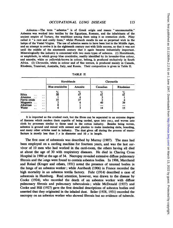
Asbestos.-The term " asbestos " is of Greek origin and means " unconsumable."Asbestos was worked into textiles by the Egyptians, Romans, and the inhabitants of theancient empire of Tartary, the wealthiest among them using it as cremation cloth. Plinycalled it "6a rare and costly linen," whilst Plutarch recalls its use as perpetual wick in thelamps of the Vestal Virgins. The use of asbestos seems to have been lost in the Middle Ages,and an attempt to revive it in the eighteenth century met with little success, so that it was notuntil the middle of the nineteenth century that it again became industrially important.Mineralogically the industry is concerned with two main types of asbestos. (1) Homblende,or amphibole, to which group blue crocidolite, readily identified by its lavender-blue colour,and amosite, white or yellowish-brown in colour, belong, is produced exclusively in SouthAfrica. (2) Chrysotile, white in colour and of fine texture, is produced mainly in Canada,Rhodesia, Transvaal, Australia, Italy, and Russia. Their composition is given in Table II.
TABLE It
Hornblende Chrysotile
Blue crocidolite Amosite Canadian Rhodesian
Silica .. .. 50 47 41 41Alumina .. 1 6 3 1Ferric oxide .. 35 37 2 4Magnesia .. 2 6 40 39Alkalines .. 8 1Water .. 4 3 14 15
It is imported as, the crushed rock, but the fibres can be separated to an extreme degreeof fineness which renders them capable of being carded, spun into yara, and woven intocloth by processes similar to those used in the cotton industry. Besides being woven,asbestos is ground and mixed with cement and plastics to make insulating slabs, boarding,and many other articles used in industry. The dust given off during the process of manu-facture is mostly less than 5 / in diameter and 10 in length.
The first case of asbestosis was described by Murray (1907). The man hadbeen employed on a carding machine for fourteen years, and was the last sur-vivor of 10 men who had worked in the card-room, the others having all diedat about the age of 30 with respiratory diseases. He died in Charing CrossHospital in 1900 at the age of 34. Necropsy revealed extensive diffuse pulmonaryfibrosis and the ;ungs were found to contain asbestos bodies. In 1906, Marchandand Reisal (Kruger and others, 1931) noted the presence of unusual bodies inthe lungs of an asbestos worker; while Auribault (1906) in France recorded thehigh mortality in an asbestos textile factory. Fahr (1914) described a case ofasbestosis in Hamburg. Real.attention, however, was drawn to the disease byCooke (1924), who recorded the death of an asbestos worker with diffusepulmonary fibrosis and pulmonary tuberculosis; while McDonald (1927) andCooke and Hill (1927) gave the fiist detailed descriptions of asbestos bodies andasserted that they originated in the inhaled dust. Seiler (1928, 1931) recorded thenecropsy on an asbestos worker who showed fibrosis but no evidence of tubercle.
113
on March 13, 2022 by guest. P
rotected by copyright.http://thorax.bm
j.com/
Thorax: first published as 10.1136/thx.2.2.91 on 1 June 1947. D
ownloaded from

KENNETH M. A. PERRY
The condition was fully described by Merewether and Price (1930) andMerewether (1933,. 1934). Clinically, the striking symptom is dyspnoea ofgradually increasing intensity, often associated with a dry cough; in the earlystages physical signs are slight or absent, but later riles throughout the lungdevelop and the fingers often become clubbed. Radiologically the mostcharacteristic feature is the " shaggy " outline of the heart shadow, while in therest of the lung field there is a fine fibrosis superimposed on lung fields whichpresent a "ground glass" appearance. The reticulation and nodulation whichare seen in silicosis do not occur in this disease.
At necropsy, pleural adhesions are extensive and dense, and there are oftenthick subpleural plaques of fibrous tissue. The consistency of the whole lungis increased, and there may be large areas of fibrous condensation which istough rather than stony hard as in silicosis. Other parts of the lung not affectedby the fibrotic change are congested or consolidated with inflammatory processes.Emp.ysema is extensive, but localized often to the lower and apical parts ofthe lung; pneumothorax sometimes occurs. The histology has been describedby Gloyne (1932), Gardner (1933), and Beger (1933). There is generalized fibrosiswith diffuse thickening of the alveolar walls, while throughout the lung tissueare seen fibres of asbestos, either singly or in clumps, as well as the characteristicasbestos bodies. The presence of these bodies in the sputum is described byStewart and Haddow (1929), Siinson and Strachan (1931), and Page (1935), andis a characteristic of the condition. Gloyne (1931) showed that they could alsobe found in the faeces.
Wood and Gloyne (1934) collected 100 cases of the disease and found that21 were complicated by pulmonary tuberculosis, but Middleton (1936) studied54 deaths occurring between 1930 and 1934 and found that tuberculosis waspresent in 24 (44 per cent). The periods of employment in the industry rangedfrom ten months in the disintegrating plant to twenty-three years' weaving, theaverage duration being just under ten years. He suggested that tuberculosis wasa slightly less frequent complication than in silicosis and that it is more frequentas a terminal complication than as a concomitant disease during the earlier stages.
Wyers (1947) showed that, as a result of measures enacted by the legislation of1931, the morbidity rate from asbestosis fell from 39.1 per 1,000 in 1932 to zeroin 1940, but that there was a small rise during the war years. Concomitant withthe fall in morbidity rate there were significant alterations in the character ofthe disease to a more chronic form. Clinically, this was manifested by anincrease in long survival periods, clubbing of the fingers (48.2 per centof cases) and sometimes of the toes, and perhaps by the high cancer rate inthose who have suffered from the disease for several years (15.3 per centpulmonary cancers and 20.4 per cent all cancers). Attention has also beendrawn to this aspect of the disease by Linzbach and Wedler -(1941). Radio-logically, the fine, diffuse, cobweb-like film formerly characteristic has now
114
on March 13, 2022 by guest. P
rotected by copyright.http://thorax.bm
j.com/
Thorax: first published as 10.1136/thx.2.2.91 on 1 June 1947. D
ownloaded from
OCCUPATIONAL LUNG DISEASE
largely given place to a coarser, more granular appearance. (Plate XIX a and b.)Pathologically, confluence of the blue polygonal areas is not so advanced as thatseen in the specimens of fifteen years ago. Wyers thought these changes were dueto longer exposures to lower concentrations of dust. The post-mortem appearancesof the lungs from 3 of Wyers' cases are illustrated in Plates XX, XXI, and XXII.
From his clinical studies of asbestosis Wyers concluded that it differed fromsilicosis in being a "clinical " rather than a "radiological " disease. Of theclinical signs he stressed the importance of clubbing of the fingers and symptomsof cardiovascular origin (raised diastolic blood pressure and accentuated secondpulmonary sound) as being always present when once established; whereas themarginal crepitations and fibrotic crackles tend to be evanescent. Of the com-plications, emphysema was always present, bronchopneumorra the commonesttermination, tuberculosis was of the acute, caseous type and occurred in one-thirdof the cases, right heart dilatation was common, and bronchiectasis rare.Pregnancy could not be shown to affect the progress of the disease adversely,and the obstetrician should be guided by the cardiovascular rather than thepulmonary signs.
The difference between the reaction of the tissues to silica and to asbestosis even more marked than the difference between the clinical pictures of silicosisand asbestosis and has been demonstrated by animal experiment. The44 solubility " theory is at present generally accepted as explaining the mechanismof silicosis, but Gloyne (1930) and Policard (1933) have shown that the injectionof asbestos dust does not cause an acute inflammatory reaction. Gardner andCummings (1931) found that asbestos accumulated in the bronchioles and thenin the alveoli, and that fibrosis first appeared after about 500 days' exposure.Gardner (1942) put forward the " mechanical " theory as an explanation of thedisease. It was the long fibre in the dust which caused asbestosis, and if theasbestos were ground sufficiently finely no disease resulted. He concluded thatthe disease results from the mechanical irritation produced by a combinationof the long fibre and the rhythmic movement of the lung. He also showed thatdifferent pathogenic responses resulted in different animals. King and others(1946), however, produced somewhat different results from the injection ofasbestos dust intratracheally into rabbits; they reported that the injection of.long fibres (15 ,A) produced a nodular reticulinosis comparable with the experi-mental silicotic nodule, whereas short fibres (2.5 F) produced a diffuse interstitialreticulinosis.
CONCLUSIONIn this discussion it has been shown that non-siliceous occupational dust
may give rise to distinct clinical diseases such as byssinosis and asbestosis, anddistinct radiological appearances such as those produced by siderosis, and alsothat it may predispose to ordinary lung diseases such as pneumonia and
115
on March 13, 2022 by guest. P
rotected by copyright.http://thorax.bm
j.com/
Thorax: first published as 10.1136/thx.2.2.91 on 1 June 1947. D
ownloaded from
KENNETH M. A. PERRY
carcinoma. This latter effect may perhaps be more important, but its possibleimportance has only recently been realized, and the means at present availablemake the proof difficult. In the case of manganese dust a works medical officerstudied the sick absences of a group of workers and noticed that there was ahigh incidence of pneumonia amongst them. He was able to compare thisincidence with. the large number of other workers under his care, and to carryout animal experiments, and by these means to prove the connexion betweenthe high incidence of pneumonia and exposure to manganese dust. In the caseof carcinoma resulting from arsenic, a statistician was able to survey the deathregister of a comparatively isolated small town with but' one industry. Sincecircumstances resulted in little change in work and residence, a statisticallysignificant result was found without undue difficulty. In the case of bagasse adisease was suspected and the small group, of people affected by it were foundto have been in ten different hospitals. The sarcoid-like syndrome resultingfrom exposure to beryllium was found to arise up to seventeen months afterleaving the work which caused it. These examples of the methods and diffi-culties involved in the study of diseases caused by occupational dust serve toindicate how little is known of these conditions and how much work remainsto be done on the subject. Certainly the conclusion that all dusts are potentiallydangerous cannot be resisted. It also seems probable that the provision ofadequate exhaust ventilation in factories in the next century will be a measureof public health comparable to the provision of adequate sanitation in the last.
SUMMARYThe literature of the diseases of the lung caused by occupational dust other
than silica is reviewed.Exposure to metallic dusts such as manganese, beryllium, vanadium, and
osmium causes chemical pneumonitis.A sarcoid-like syndrome may develop late after exposure to beryllium.Mouldy organic material, such as hay, corn, bagasse, and cotton, gives rise
to lcute bronchiolitis, but there is no conclusive evidence to support the theory thatthis results from aspergillus or other fungi.
Complex salts of platinum give rise to a form of asthma.Byssinosis affects a very large number of workers in the cotton-spinning
area of Lancashire.The incidence of carcinoma of lung is abnormally high in those exposed to
arsenic dust and to asbestos.Metals according to their atomic weight are opaque to x rays, and dusts such
as iron oxide and barium give rise to radiographic shadows; the changes causingthese shadows have been called siderosis and baritosis.
116
on March 13, 2022 by guest. P
rotected by copyright.http://thorax.bm
j.com/
Thorax: first published as 10.1136/thx.2.2.91 on 1 June 1947. D
ownloaded from
OCCUPATIONAL LUNG DISEASE
Silver is deposited in the elastic layer of the arteries of the lungs of wovkersexposed to its dust.
The silicates, particularly china-clay, talc, mica, sillimanite, and asbestos,give rise to various kinds of fibrosis in the lung, though not to a nodularfibrosis like quartz silica.
Graphite appears to be harmless.The methods of assessment of disability in the group of occupational dust
diseases which might be called "radiological" are briefly discussed; theseinclude siderosis, those due to mixed dusts of silicates, and the early stages ofsilicosis.
REFERENCESManganeseBaader, E. W. (1933). Arch. Gewerbepath. Hyg., 4, 101.Brezina, E. (1921). " Internationale tbersicht ilber Gewerbe Krankheiten nach den Berichten
den Gewerbeinspektionen der Ktilturlander tiber das Jahr 1913," p. 40. Berlin.Buttner, H. E. (1939). Report on Eighth International Congress for Industrial Accidents
and Occupational Diseases, Leipzig, 1939, 2, 1022.Charles, J. R. (1927). Brain, 50, 30.Couper, J. (1837). Brit. Ann. med. Pharm., 1, 41.Elstad, D. (1939). Report on Eighth International Congress for Industrial Accidents and
Occupational Diseases, Leipzig, 1939, 2, 1012.Fairhall, L. T., and Neal, P. A. (1943). Nat. Inst. Hlth. Bull., No. 182. Washington, D.C.Heine, W. (1943). Z. Hyg. Infektionskr., 125, 76.Lloyd-Davies, T. A. (1946). Brit. J. industr. Med., 3, 111.Owen, D., and Cohen, H. (1934). Lancet, 2, 989.Riddervold, J., and Halvorsen, K. (1943). Acta path. microbiol. Scand., 20, 272.
BerylliumBerkovits, M., and Izrael, B. (1940). Kin. Med., 18, 117.Fabrioni, S. M. (1935). Med. Lavoro, 26, 297.Gardner, L. U. (1946). Trans. indusir. Hyg. Foundation, Eleventh Annual Meeting, p. 1.Gelman, I. (1936). J. industr. Hyg., 18, 371.Gelman, I. (1938). " Occupation and Health," Geneva Suppl.Hardy, H. L. (1947). Bull. New Engl. med. Centre, 9, 16.Hardy, H. L., and Tabershaw, L. R. (1946). J. industr. Hyg., 28, 197.Hyslop, F., Palmes, E. D., Alford, W. C., Monaco, A. R., and Fairhall, L. T. (1943). Nat.
Inst. &Ith. Bull., No. 187. Washington, D.C.Kress, J. and Crispell, K. R. (1944). Guthrie clin. Bull., 13, 91.Meyer, H. E. (1942). Beitr. Klin. Tuberk., 98, 388.Shilen, J., Galloway, A. E., and Mellor, J. G. (1944). Industr. Med., 13, 464.Van Ordstrand, H. S., Hughes, R., and Carmody, M. G. (1943). Cleveland clin. Quart.,
10, 10.Van Ordstrand, H. S., Hughes, R., Carmody, M. G., and De Nardi, J. M. (1945). J. Amer.
med. Ass., 129, 1084.Weber, H. H., and Engelhardt, W. E. (1933). Zbl. Gewerbehyg., 10, 41.Wurm, H., and Ruger, H. (1942). Beitr. KlUn. Tuberk., 98, 396.
VanadiumBalestra, G., and Molfino, F. (1942). Rass. med. Ind., 13, 5.Dutton, W. F. (1911). J. Amer. med. Ass., 56, 1648.Svmanski, H. (1939). Arch. Gewerbepath. Hyg., 9, 295.Wyers, H. (1946). Brit. J. industr. Med., 3, 177.
OsmiumBrunot, F. R. (1933). J. industr. Hyg., 15, 136.-McLaughlin, A. I. G., Milton, R., and Perry, K. M. A. (1946). Brit. J. industr. Med., 3, 183.Raymond, F. (1874). Progras mid., 2, 373.
117
on March 13, 2022 by guest. P
rotected by copyright.http://thorax.bm
j.com/
Thorax: first published as 10.1136/thx.2.2.91 on 1 June 1947. D
ownloaded from

KENNETH M. A. PERRY
Farmer's LungCampbell, J. M. (1932). Brit. med. J., 2, 1143.Fawcett, R. (1935). Brit. J. Radiol., 9, 171, 354.Fawcett, R. (1938). Brat. J. Radiol., 11, 378.Fawcett, R. (1938). Amer. J. Roentgen., 39, 19.Fuller, C. J. (1947). Personal communication.Hoffmann, W. (1946). Schweiz. med. Wschr., 76, 988.Tdrnell, E. (1946). Accta med. scand., 125, 191.
BagasseCastleden, L. I. M., and Hamilton-Paterson, J. L. (1943). Brit. med. J., 2, 478.Hunter, D., and Perry, K. M. A. (1946). Brit. J. industr. Med., 3, 64.Jamison., S. C., Bryan, M. S., and Day, J. M. (1944). New Orleans med. surg. J., 96 291.Jamison, S. C., and Hopkins, J. (1941). New Orleans med. surg. J., 93, 580.Lloyd, W. E. (1940). Personal communication.Manas, M. A. (1945). Rev. Mexicana Tuberc., 7, 391.Sodeman, W. A., and Pullen, R. L. (1943). New Orleans med. surg. J., 95, 558.Sodeman, W. A. (1944). Arch. intern. Med., 73, 365.
CottonColfis, E. L. (1914). Proc. roy. Soc. Med., 8, 108.Middleton, E. L. (1926). J. industr. Hyg., 8, 428.Neal, P. A., Schneiter, R., and Caminita, B. H. (1942). J. Amer. med. Ass., 119, 1074.
PlatinumHunter, D., Milton, R., and Perry, K. M. A. (1945). Brit. J. industr. Med., 2, 92.Karasek, S. R., and Karasek, M. (1911). Rep. ill. State CommissionOcc. Dis., p. 97.
ByssinosisBolen, H. L. (1943). J. industr. Hyg., 25, 215.Dunn, S., and Sheehan, H. L. (1932). " Report on Ten Autopsies on Cotton-Mill Workers.
Appendix II. Report of the Departmental Committee on Dust in Card-rooms in theCotton Industry." London: H.M.S.O.
Gill, C. I. C. (1947). Brit. J. industr. Med., 4, 48.Greenhow, E. H. (1862). "Medical Officer of the Privy Council. Fourth Report."
H.M.S.O., p. 173.Jackson, J. (1818). London med. phys. J., 39, 464.Prausnitz, C. (1936). Spec. Rep. Ser. M.R.C., No. 212. London."'Report of the Departmental Committee on Dust in Card-rooms in the Cotton Industry"
(1932). London: H.M.S.O.Study of Sickness among operatives in Lancashire Cotton Spinning Mills " (1930). London:
Industr. Hlth. Research Bd., Rep. No. 590.Zipperlen, V. R. (1935). Arch. Hyg. Berlin, 113, 1,Wheat DustDuke, W. W. (1935). J. Amer. med. Ass., 105, 957.
ArsenicAmor, A. J. (1938). " Report on Eighth International Congress in Industrial Accidents and
Occupational Diseases." Leipzig, 1939, 2, 248.Arnstein, S. A. (1913). Verh. deutsch. path. Ges., 16, 332.Bradford Hill, A. (1947). To be published.Haerting, F. H., and Hesse, W. (1879). Viertel-jahrschr. Ger. Med. San-wesen NY., 30,
296'; 31, 102.Hueck, W. (1939). Z. Krebst., 49, 312.Lange, K. (1935). Z. Krebst., 42, 306.Leitch, A. (1922). Brit. med. J., 2, 11&7.Loewy, J. (1929). Med. Klin., 25, 141.Lorenz, E. (1944). J. Nat. Cancer Instt., 5, 1.
118
on March 13, 2022 by guest. P
rotected by copyright.http://thorax.bm
j.com/
Thorax: first published as 10.1136/thx.2.2.91 on 1 June 1947. D
ownloaded from

OCCUPATIONAL LUNG DISEASE
Peller, S. (1939). Human Biol., 11, 130.Pirchan, A., and Sikl, H. (1932). Amer. J. Can,.er, 16, 681.Rostoski, O., Saupe, E., and Schmorl, G. (1926). Z. Krebst., 23, 360.Rostoski, O., Saupe, E., and Schmorl, G. (1927). Z. Krebst., 25, 249.Sikl, H. (1930). Z. Krebst., 32, 609.Thompson, V. C. (1946). Personal communication.Zeil, R. (1935). J. industr. Hyg., 17, abstract text, p. 28.
PneumonokoniosisChristie, R. V. (1934). J. clin. Invest., 13, 295.Coumand, A., Baldwin, E. D., Darling, R. C., and Richards, D. W. (1941). J. clin. Invest.,
20, 681.Hart, P. D., and Aslett, E. A. (1942). Spec. Rep. Ser. M.R.C., No. 243. London.McCann, W. S., and Kaltreider, N. L. (1937). Pennsylvania med. J., 40, 901.McCann, W. S., and Kaltreider, N. L. (1937). J. clin. Invest., 16, 23.McMichael, J. (1939). Clin. Sci., 4, 167.
BariumArrigoni, A. (1933). Med. Lavoro, 24, 461.Pendergrass, E. P. (1938). " Lanzos Silicosis and Asbestosis," London, p. 137.Preti, L., and Talini, P. C. (1938). " Report on Eighth International Congress on Industrial
Accidents and Occupational Diseases." Leipzig, 2, 963.
SiderosisBritten, J. A., and Walsh, E. L. (1940). J. industr. Hyg., 22, 125.Buckell, M., Garrad, J., Jupe, M. H., McLaughlin, A. I. G., and Perry, K. M. A. (1946).
Brit. J. industr. Med., 3, 78.Doig, A. T., and McLaughlin, A. I. G. (1936). Lancet, 1, 771.Enzer, N., and Sander, 0. A. (1938). 1. industr. Hyg., 20, 333.Enzer, N., Simonsen, E., and Evans, A. M. (1945). J. industr. Hyg., 27, 147.Groh, J. A. (1944). lndustr. Med., 13, 398.Harding, H. E. (1945). Brit. J. industr. Med., 2, 32.Harrold, G. C., Meek, S. F., and McCord, C. P. (1940). J. industr. Hyg., 22, 347.Pendergrass, E. P., and Leopold, S. S. (1945). J. Amer. med. Ass., 127, 701.Sander, 0. A. (1944). J. industr. Hyg., 26, 79.Zenker, F. A. (1866). Dtsch. Arch. klin. Med., 2, 116.
Argyro-siderosisBarrie, J. M., and Harding, H. E. (1947). Brit. J. industr. Med. To be published.McLaughlin, A. I. G., Grant, J. L. A., Barrie, H. J., and Harding, H. E. (1945). Lancet,
1, 337.
AluminiumBelt, T. F., and King, E. J. (1943). J. Path. Bact., 55, 69.Crombie, D. W., Blaisdell, J. L., and MacPherson, G. (1944). Canad. med. Ass. J., 50, 318.Denny, J. J., Robson, W. D., Irwin, D. A. (1939). Canad. med. Ass. J., 40, 213.Denny, J. J., Robson, W. D., Irwin, D. A. (1939). Industr. Med., 8, 133.Goralewski, G. (1939). Arch. Gewerbepath. Hyg., 9, 676.Goralewski, G. (1940). Arch. Gewerbepath. Hyg., 10, 384.Goralewski, G. (1941). Arch. Gewerbepath. Hyg., 11, 106.Goralewski, G. (1943). Dtsch. Tuber-bl., 17, 3.Goralewski, G., and Jager, R. (1941). Arch. Gewerbepath. Hyg., 11, 102.Hunter, D., Milton, R., Perry, K. M. A., and Thompson, D. R. (1944). Brit. J. industr.
Med., 1, 159.J§ger, R., and Jager, F. (1941). Arch. Gewerbepath. Hyg., 11, 117.Jotten, K. W., and Eickhoff, W. (1942). Arch. Hyg. Bakt., 127, 344.Koelsch, F. (1942). Beitr. klin. Tuberk., 97, 688.Shaver, C. G., and Riddell, A. R. (1947). J. industr. Hyg., 29, 145.
119
on March 13, 2022 by guest. P
rotected by copyright.http://thorax.bm
j.com/
Thorax: first published as 10.1136/thx.2.2.91 on 1 June 1947. D
ownloaded from
KENNETH M. A. PERRY
GraphiteDunner, L. (1945).' Brit. J. Radiol., 18, 33.Dunner, L., and Bagnall, D. J. T. (1946). Brit.- . Radiol., 19, 165.Gardner, L. U. (1938). J. Amer.. med. Ass., 111, 1925.
China, ClayHale, L. W. (1946). Personal communication.Middleton, E.' L. (1936). Lancet, 2, 59.
TalcDevoto and Cesarbiachi (1911). "National Congress on Occupational Diseases." Turin.Dreessen, W. C. (1933). 1. induatr. Ilyg., 15, 66.Dreessen, W. C., and Dalla Valle, J..V. (1935). U.S. Publ. Hith. Wash., No. 5.Merewether, E. R. A.,(1933). Ann. Rep. Chief Insp. Factories. H.M.S.O., p. 63.Merewether, E. R. A. (1934). Ann. Rep. Chief Insp. Factories. H.MS.O., p. 65.Siegal, W., Smith, A. R.--and Greenburgh, L. (1943). Amer. J. Roentgen., 49, 11.Thorel, C. (1896). Beitr. path. Antat. allg. Path., 20, 85.Zanelli, A. (1931). Med. Lavoro, 1, 1.
SillimaniteJ6tten, K. W., and Eicklhoff, W. (1944). Arch. Gewerbepath. Hyg., 12, 223.Middleton, E. L. (1936). Lancet, 2, 59.
MicaFerguson, T. Quoted by Middleton, E; L. (1936).. Lancet, 2, 59.
Asbestos,Auribault (1906). Bull. de rlnspection du Travail, Paris, p. 126.Beger, P. J. (1933). Virchows Arch., 290, 280.Cooke, W. E. (1924). Brit. med. J.. 2, 147.Cooke, W. E. (1927). Brit. med. l.i 2, 1204.Cooke, W. E., and Hill, C. F. (1927). J. roy. Micros., Soc., 47, 222.Fahr, F. (1914). Dtsch. med. Wschr., 40, 1548.Gardner, L. U., and Cummings, D.. E. (1931). J. industr. Hyg., 13, 112.Gardner, L. U. (1933). J. Amer. med. Ass., 101, 595.Gardner, L. U. (1942). Amer. Rev. Tuberc., 45, 37-2.Gloyne, S. R. (1930). Tuberck, 9, 151.Gloyne, S. R. (1931). Tubercle, 12, 158.Gloyne, S. R. (1932). Lancet, 1, 1351.King, E. J., Clegg, J. W., and Rae, V. M. (1946). Thorax, 1, 188.Kruger, E., Rosloski, 0., and Saupe, E. (1931). Arch. Gewerbepath. Hyg., 2, 558.Linzbach, A. J., Wedler, H. W. (1941). Virchows Arch., 307, 387.McDonald, S. (1927). Brit. med. J., 2, 1025.Merewether, E. R. A., and Price, C. W. (1930). " Report on the Effects of Asbestos Dust
-on the Lungs and Dust Suppression in the Asbestos Industry." H.M.S.O.Merewether, E. R. A. (1933). Tubercle, 15, 69, 109, 152.Merewether, E. R. A. (1934). Ann. Rep. Chief Insp. Factories, H.M.S.O., P. 62.Middleton, E. L. (1936). Lancet, 2, 59.Murray, M. (1907). " Dept. Comm. on Compensation for Industr. Diseases." H.M.S.O.,
Cd. 3495, p. 14; Cd. 3496, p. 127.Page, R. C. (1935). Amer. J. med. Sci.. 89, 44.Policard, A. (1933). Bull. dHistologie appliquke, 7, 125.Seiler, H. E. (1928). Brit. med. 1., 2, 982.Seiler, H. E., and Gilman, M. D. (1931). Brit. med. J., 1, 1112.Simson, F. W., and Strachan, A. S. (1931). J. Path. Bact., 34, 1.Stewart, M. J., and Haddow. A. C. (1929). J. Path. Bact., 32, 172.Wood, W. B., and Gloyne, S. R. (1934). Lancet, 2, 1383.Wyers, H. (1947). Thesis, Glasgow. To be published.
120
on March 13, 2022 by guest. P
rotected by copyright.http://thorax.bm
j.com/
Thorax: first published as 10.1136/thx.2.2.91 on 1 June 1947. D
ownloaded from
Related Documents