Disease Progression in Plasmodium knowlesi Malaria Is Linked to Variation in Invasion Gene Family Members Atique M. Ahmed 1. , Miguel M. Pinheiro 2. , Paul C. Divis 1 , Angela Siner 1 , Ramlah Zainudin 1,3 , Ing Tien Wong 4 , Chan Woon Lu 5 , Sarina K. Singh-Khaira 6 , Scott B. Millar 2 , Sean Lynch 7 , Matthias Willmann 8 , Balbir Singh 1 , Sanjeev Krishna 1,6 , Janet Cox-Singh 1,2,6 * 1 Malaria Research Centre, University Malaysia Sarawak, Kuching, Sarawak, Malaysia, 2 School of Medicine, University of St Andrews, St Andrews, United Kingdom, 3 Faculty of Resource Science and Technology, University Malaysia Sarawak, Kuching, Sarawak, Malaysia, 4 Sibu Hospital, Sibu, Sarawak, Malaysia, 5 Sarikei Hospital, Sarikei, Sarawak, Malaysia, 6 Division of Clinical Sciences, St. George’s, University of London, London, United Kingdom, 7 Clinical Blood Sciences, St. George’s, University of London, London, United Kingdom, 8 Institute of Medical Microbiology and Hygiene, University of Tu ¨ bingen, Tu ¨ bingen, Germany Abstract Emerging pathogens undermine initiatives to control the global health impact of infectious diseases. Zoonotic malaria is no exception. Plasmodium knowlesi, a malaria parasite of Southeast Asian macaques, has entered the human population. P. knowlesi, like Plasmodium falciparum, can reach high parasitaemia in human infections, and the World Health Organization guidelines for severe malaria list hyperparasitaemia among the measures of severe malaria in both infections. Not all patients with P. knowlesi infections develop hyperparasitaemia, and it is important to determine why. Between isolate variability in erythrocyte invasion, efficiency seems key. Here we investigate the idea that particular alleles of two P. knowlesi erythrocyte invasion genes, P. knowlesi normocyte binding protein Pknbpxa and Pknbpxb, influence parasitaemia and human disease progression. Pknbpxa and Pknbpxb reference DNA sequences were generated from five geographically and temporally distinct P. knowlesi patient isolates. Polymorphic regions of each gene (approximately 800 bp) were identified by haplotyping 147 patient isolates at each locus. Parasitaemia in the study cohort was associated with markers of disease severity including liver and renal dysfunction, haemoglobin, platelets and lactate, (r = $0.34, p = ,0.0001 for all). Seventy- five and 51 Pknbpxa and Pknbpxb haplotypes were resolved in 138 (94%) and 134 (92%) patient isolates respectively. The haplotypes formed twelve Pknbpxa and two Pknbpxb allelic groups. Patients infected with parasites with particular Pknbpxa and Pknbpxb alleles within the groups had significantly higher parasitaemia and other markers of disease severity. Our study strongly suggests that P. knowlesi invasion gene variants contribute to parasite virulence. We focused on two invasion genes, and we anticipate that additional virulent loci will be identified in pathogen genome-wide studies. The multiple sustained entries of this diverse pathogen into the human population must give cause for concern to malaria elimination strategists in the Southeast Asian region. Citation: Ahmed AM, Pinheiro MM, Divis PC, Siner A, Zainudin R, et al. (2014) Disease Progression in Plasmodium knowlesi Malaria Is Linked to Variation in Invasion Gene Family Members. PLoS Negl Trop Dis 8(8): e3086. doi:10.1371/journal.pntd.0003086 Editor: Kenji Hirayama, Institute of Tropical Medicine (NEKKEN), Japan Received October 21, 2013; Accepted June 30, 2014; Published August 14, 2014 Copyright: ß 2014 Ahmed et al. This is an open-access article distributed under the terms of the Creative Commons Attribution License, which permits unrestricted use, distribution, and reproduction in any medium, provided the original author and source are credited. Funding: This study was funded by The Medical Research Council (MRC) UK; Grant number G0801971, http://www.mrc.ac.uk. MMP is supported by The Wellcome Trust (ISSF 097831/Z/11/Z), http://www.wellcome.ac.uk. The funders had no role in study design, data collection and analysis, decision to publish, or preparation of the manuscript. Competing Interests: The authors have declared that no competing interests exist. * Email: [email protected] . These authors contributed equally to this work. Introduction Plasmodium knowlesi malaria is widespread in Southeast Asia (SEA). Descriptions of the aetiology of knowlesi malaria support a zoonotic origin of infection [1] and highlight variability in disease severity between those at risk across the region [2]. For example, very young children living in a forested area of Southern Vietnam have asymptomatic mixed Plasmodium species infections that include P. knowlesi [3]. Adults and children in Malaysian Borneo experience symptomatic single species P. knowlesi infections that are severe in .10% of patients and can be fatal [4,5]. P. knowlesi transmission is restricted to the Leucosphyrus group of mosquito vectors found in forested areas of Southeast Asia [6,7]. The vector group is diverse and capable of simultaneous transmission of human and non-human primate adapted Plasmodium species [8]. The majority of reported cases of P. knowlesi malaria are associated with time spent in the jungle or jungle fringe areas where the ranges of the natural vertebrate hosts, the long and pig tailed macaques (Macaca fascicularis and Macaca nemestrina) and leucosphyrus vectors overlap [9,10]. However, a change in pattern has recently emerged in Malaysian Borneo, where children living in a deforested area are infected [11]. This new pattern may signal a change in vector or vector habitat preference and a move towards human-to- human transmission. Restricted spread of P. knowlesi within human populations is attributed to non-urban vector habitat. Also human-host adapted Plasmodium species, where prevalent, may present a biological barrier to the entry of P. knowlesi into human populations concurrently at risk from human adapted and zoonotic species infections. On a backdrop of vector, human and parasite diversity, PLOS Neglected Tropical Diseases | www.plosntds.org 1 August 2014 | Volume 8 | Issue 8 | e3086

Welcome message from author
This document is posted to help you gain knowledge. Please leave a comment to let me know what you think about it! Share it to your friends and learn new things together.
Transcript
Disease Progression in Plasmodium knowlesi Malaria IsLinked to Variation in Invasion Gene Family MembersAtique M. Ahmed1., Miguel M. Pinheiro2., Paul C. Divis1, Angela Siner1, Ramlah Zainudin1,3,
Ing Tien Wong4, Chan Woon Lu5, Sarina K. Singh-Khaira6, Scott B. Millar2, Sean Lynch7,
Matthias Willmann8, Balbir Singh1, Sanjeev Krishna1,6, Janet Cox-Singh1,2,6*
1 Malaria Research Centre, University Malaysia Sarawak, Kuching, Sarawak, Malaysia, 2 School of Medicine, University of St Andrews, St Andrews, United Kingdom,
3 Faculty of Resource Science and Technology, University Malaysia Sarawak, Kuching, Sarawak, Malaysia, 4 Sibu Hospital, Sibu, Sarawak, Malaysia, 5 Sarikei Hospital, Sarikei,
Sarawak, Malaysia, 6 Division of Clinical Sciences, St. George’s, University of London, London, United Kingdom, 7 Clinical Blood Sciences, St. George’s, University of London,
London, United Kingdom, 8 Institute of Medical Microbiology and Hygiene, University of Tubingen, Tubingen, Germany
Abstract
Emerging pathogens undermine initiatives to control the global health impact of infectious diseases. Zoonotic malaria is noexception. Plasmodium knowlesi, a malaria parasite of Southeast Asian macaques, has entered the human population. P.knowlesi, like Plasmodium falciparum, can reach high parasitaemia in human infections, and the World Health Organizationguidelines for severe malaria list hyperparasitaemia among the measures of severe malaria in both infections. Not allpatients with P. knowlesi infections develop hyperparasitaemia, and it is important to determine why. Between isolatevariability in erythrocyte invasion, efficiency seems key. Here we investigate the idea that particular alleles of two P. knowlesierythrocyte invasion genes, P. knowlesi normocyte binding protein Pknbpxa and Pknbpxb, influence parasitaemia andhuman disease progression. Pknbpxa and Pknbpxb reference DNA sequences were generated from five geographically andtemporally distinct P. knowlesi patient isolates. Polymorphic regions of each gene (approximately 800 bp) were identified byhaplotyping 147 patient isolates at each locus. Parasitaemia in the study cohort was associated with markers of diseaseseverity including liver and renal dysfunction, haemoglobin, platelets and lactate, (r = $0.34, p = ,0.0001 for all). Seventy-five and 51 Pknbpxa and Pknbpxb haplotypes were resolved in 138 (94%) and 134 (92%) patient isolates respectively. Thehaplotypes formed twelve Pknbpxa and two Pknbpxb allelic groups. Patients infected with parasites with particular Pknbpxaand Pknbpxb alleles within the groups had significantly higher parasitaemia and other markers of disease severity. Our studystrongly suggests that P. knowlesi invasion gene variants contribute to parasite virulence. We focused on two invasiongenes, and we anticipate that additional virulent loci will be identified in pathogen genome-wide studies. The multiplesustained entries of this diverse pathogen into the human population must give cause for concern to malaria eliminationstrategists in the Southeast Asian region.
Citation: Ahmed AM, Pinheiro MM, Divis PC, Siner A, Zainudin R, et al. (2014) Disease Progression in Plasmodium knowlesi Malaria Is Linked to Variation inInvasion Gene Family Members. PLoS Negl Trop Dis 8(8): e3086. doi:10.1371/journal.pntd.0003086
Editor: Kenji Hirayama, Institute of Tropical Medicine (NEKKEN), Japan
Received October 21, 2013; Accepted June 30, 2014; Published August 14, 2014
Copyright: � 2014 Ahmed et al. This is an open-access article distributed under the terms of the Creative Commons Attribution License, which permitsunrestricted use, distribution, and reproduction in any medium, provided the original author and source are credited.
Funding: This study was funded by The Medical Research Council (MRC) UK; Grant number G0801971, http://www.mrc.ac.uk. MMP is supported by TheWellcome Trust (ISSF 097831/Z/11/Z), http://www.wellcome.ac.uk. The funders had no role in study design, data collection and analysis, decision to publish, orpreparation of the manuscript.
Competing Interests: The authors have declared that no competing interests exist.
* Email: [email protected]
. These authors contributed equally to this work.
Introduction
Plasmodium knowlesi malaria is widespread in Southeast Asia
(SEA). Descriptions of the aetiology of knowlesi malaria support a
zoonotic origin of infection [1] and highlight variability in disease
severity between those at risk across the region [2]. For example,
very young children living in a forested area of Southern Vietnam
have asymptomatic mixed Plasmodium species infections that
include P. knowlesi [3]. Adults and children in Malaysian Borneo
experience symptomatic single species P. knowlesi infections that
are severe in .10% of patients and can be fatal [4,5].
P. knowlesi transmission is restricted to the Leucosphyrus group of
mosquito vectors found in forested areas of Southeast Asia [6,7]. The
vector group is diverse and capable of simultaneous transmission of
human and non-human primate adapted Plasmodium species [8].
The majority of reported cases of P. knowlesi malaria are associated
with time spent in the jungle or jungle fringe areas where the ranges
of the natural vertebrate hosts, the long and pig tailed macaques
(Macaca fascicularis and Macaca nemestrina) and leucosphyrus
vectors overlap [9,10]. However, a change in pattern has recently
emerged in Malaysian Borneo, where children living in a deforested
area are infected [11]. This new pattern may signal a change in
vector or vector habitat preference and a move towards human-to-
human transmission.
Restricted spread of P. knowlesi within human populations is
attributed to non-urban vector habitat. Also human-host adapted
Plasmodium species, where prevalent, may present a biological
barrier to the entry of P. knowlesi into human populations
concurrently at risk from human adapted and zoonotic species
infections. On a backdrop of vector, human and parasite diversity,
PLOS Neglected Tropical Diseases | www.plosntds.org 1 August 2014 | Volume 8 | Issue 8 | e3086
it would be folly for malaria elimination strategists to underesti-
mate the importance of the multiple geographically dispersed
entries of P. knowlesi into the human population [10]. The scene is
set for a host switch of P. knowlesi from macaques to humans if
pressed and as predicted by Garnham in 1966 [12].
Parasitaemia in malaria is a measure of the number of
parasitized erythrocytes in the infected host at the time of
sampling. The asexual replication cycle of P. knowlesi is 24 hours
and therefore parasitaemia can increase daily in uncontrolled
infections. Rising parasitaemia in P. knowlesi infections is
associated with disease severity frequently involving renal failure,
liver dysfunction and respiratory distress but not coma or severe
anaemia [5,13]. However, not all patients develop high parasit-
aemia, even following several days of untreated infection [5].
Parasite and/or host factors contributing to the rapid development
of hyperparasitaemia in some patients infected with P. knowlesihave not been investigated.
Successful erythrocyte invasion by the infective merozoite stage
of Plasmodium species is the result of a complex recognition,
reorientation and entry process orchestrated by merozoite protein
families conserved within the genus [14,15,16]. Of these, the
reticulocyte binding-like protein (RBP) family is present in all
Plasmodium species studied and members are involved in
erythrocyte selection and invasion (Table S1). P. falciparum has
five functional paralogous RBP members, PfRh1, PfRh2a,
PfRh2b, PfRh4 and PfRh5 [17]. This family of proteins was first
discovered in P. vivax [18,19]. P. vivax has two well described
functional members and more putative members have been
identified recently [20]. The P. falciparum Rh proteins are
thought to provide multiple invasion pathways allowing for
invasion of a wide range of erythrocyte phenotypes, lessening
restriction and explaining hyperparasitaemia [16,21,22]. There is
also evidence for differential expression of the PfRh genes in
human infections but with no clear association with parasitaemia
and invasion efficiency [22,23,24]. P. knowlesi has two members
of the RBP gene family, named P. knowlesi normocyte binding
proteins (Pknbp)xa and Pknbpxb [25]. Pknbpxa is located on
chromosome 14 and Pknbpxb on chromosome 7[26]. A recent
study on an experimental line of P. knowlesi demonstrated binding
of Pknbpxa but not Pknbpxb protein products to human
erythrocytes implicating Pknbpxa in human erythrocyte invasion
[27].
Here we report on a prospective study of patients with P.knowlesi malaria. We confirm and then exploit the association
between P. knowlesi parasitaemia with clinical and laboratory
measures of disease progression. We then address the question that
parasitaemia in naturally acquired human infections is associated
with particular alleles of the P. knowlesi merozoite invasion genes
Pknbpxa and Pknbpxb.
Materials and Methods
Ethics statementThis non-interventional study was approved by Medical
Research and Ethics Committee, Ministry of Health Malaysia
and the Ethics Committee Faculty of Medicine and Health
Sciences, University Malaysia Sarawak. The study was approved
to recruit patients 15 years and above with informed signed
consent. Children (,15 years) were not recruited into the study.
Study design and patient recruitmentTwo recruitment sites were selected for the study. The first,
Hospital Sarikei, serves four districts in the Sarikei Health
Division, population size 133,572 (2012) and Hospital Sibu, a
referral hospital serving the Rejang basin. Patients with micros-
copy positive all-cause malaria were recruited with consent by the
attending healthcare professionals. Each patient was given a
unique study identifier code. Retrospective exclusion was based on
PCR results. (For a detailed description of patient recruitment see
Text S1)
Blood sample processingSerum, plasma and whole blood samples were stored frozen on
site and transported at sub-zero temperatures to the Malaria
Research Centre, University Malaysia Sarawak, (UNIMAS) at
regular intervals during the study. DNA amplification, cloning and
sequencing were conducted in UNIMAS and serum and plasma
samples were shipped on dry ice to St George’s University of
London for glucose, lactate and IL-10 assays (See Text S1).
PCR confirmation of Plasmodium speciesDNA was extracted from dried bloodspot samples using the
InstaGene method [28] to confirm the infecting parasite species.
Nested PCR of the small subunit rRNA gene as described
previously was used as follows: The first nest used primer pairs
rPlu 5 and rPlu6 [29] and the second nests were specific for P.falciparum, P vivax [Paul C Divis, unpublished], P. malariae and
P. knowlesi [1,30].
Data collectionA study dataset containing patient demographic information,
history, clinical and laboratory information was prepared from the
study history sheet and patient case notes using FileMaker Pro
10v.1 (FileMaker Inc.). Alleles occurring at the two genetic loci
were added to the dataset during the course of the study. See
below.
De-selection of patients for the genotyping studyThere were 232 patients with PCR confirmed single species P.
knowlesi infections who fulfilled the study criteria (Figure S1a). Of
Author Summary
Plasmodium knowlesi, a parasite of Southeast Asianmacaques, has entered the human population. Approxi-mately 10% of P. knowlesi infections are severe, 1–2% arefatal, in Sarawak, Malaysian Borneo. Increase in parasitae-mia is associated with disease severity, but little is knownabout parasite virulence in this newly described humanpathogen. Here we present the results of a study on P.knowlesi parasites collected from 147 patients. We use theisolates to produce DNA sequences from a polymorphic(genetically variable) region of two P. knowlesi genes,Pknbpxa and Pknbpxb, that are involved in parasite entryinto host red blood cells. We addressed the question thatsome parasite genotypes may have an invasion advantageleading to severe disease in human infections. Weanalysed the DNA sequences with matched clinical andlaboratory data from the patient cohort (n = 147). Wefound that specific DNA sequences (Pknbpxa and Pknbpxballeles) clustered with high parasitaemia and markers ofdisease severity. Here, for the first time, we provideevidence that variant alleles of the Plasmodium Reticulo-cyte Binding-Like Protein invasion gene family caninfluence disease progression in patients with malaria.The biological characteristics of the variants will be studiedto aid our understanding of malaria pathophysiology andto inform intervention strategies.
Plasmodium knowlesi Malaria
PLOS Neglected Tropical Diseases | www.plosntds.org 2 August 2014 | Volume 8 | Issue 8 | e3086
these, 165 (71%) had mild disease with no abnormal clinical or
laboratory indicators [5,31]. In order to avoid generating
redundant information and incur unnecessary costs these patients
were sorted by parasitaemia and alternate patients were de-
selected without biasing the range of parasitaemia in this group.
Following de-selection, 147 patients were included in the genetic
association study. This group included patients with mild malaria
as above, all patients in the study with severe disease and all of
those with some abnormal findings but with otherwise uncompli-
cated malaria using the WHO criteria for complicated malaria in
the non-immune adult[31].
Analyses for association between parasitaemia, clinicaland laboratory markers of disease progression
Log transformed parasitaemia was normally distributed how-
ever some clinical and laboratory variables remained non-
normally distributed. Spearman’s (non-parametric) and Pearson’s
(parametric) correlation (r) were calculated Prism GraphPad v 4
and Stata 8 for Macintosh.
P. knowlesi Pknbpxa and Pknbpxb reference DNAsequence
Pure DNA template was extracted from frozen EDTA whole
blood samples using QIAamp DNA Mini kit (QIAGEN) following
the manufacturer’s instructions. Pknbpxa and Pknbpxb DNA
sequence was generated from five reference P. knowlesi patient
isolates using high stringency methodology as follows. Primer
sequences were designed from published Pknbpxa and Pknbpxbsequences EU867791and EU867792 respectively [25]. In the first
instance large segments of each gene (8501 bp Pknbpxa and
3506 bp Pknbpxb) were amplified and cloned as single fragments.
The 8501 bp Pknbpxa fragment was amplified using the
primer pair PknbpxaF5 5’AGGTGCAAGCTGGGAACAAG
and PknbpxaR2 5’CTACACGACACACAATGCACC with
LongRange PCR Enzyme Mix (QIAGEN). Each reaction was
in a final volume 25 mL containing 2 mL DNA template, 0.4 mM
each primer, 500 mM each dNTP and 1 U Long Range enzyme
in 1x Long Range buffer (2.5 mM MgCl2). The reaction
conditions were 93uC for 3 mins, 9 cycles at 93uC for 15 sec,
57uC for 30 sec, 68uC for 14 mins followed by 27 cycles at 93uCfor 15 sec, 57uC for 30 sec and 68uC for 14 mins with addition of
20 sec/cycle. The Pknbpxb 3506 bp fragment was amplified
using the primer pair Xb273F 5’GCATGGTCAAAA-
GAACCCC and Xb3430R 5’CTTCTATGGACGCTTCAGGT
with Elongase Enzyme Mix (Invitrogen, Life Technologies). Each
reaction was in a final volume of 20 mL containing 3 mL DNA
template, 0.25 mM each primer, 500 mM each dNTP, 1 U
Elongase in 1 x Elongase buffer (1.5 mM MgCl2) under the
following conditions: 93uC 30 sec, 34 cycles at 93uC for 30 sec,
55uC for 30 sec, 68uC for 3 min followed by a final extension at
68uC for 10 mins. The fragments were cleaned, gel purified and
cloned into the pCR TOPO XL vector, TOPO XL PCR
Cloning Kit (Invitrogen, Life Technologies) following the
manufacturer’s instructions. Resulting plasmids were recovered
using S.N.A.P. MiniPrep Kit (Invitrogen, Life Technologies).
Each clone was sequenced in the forward and reverse directions
beginning with M13F and M13R sequences flanking the insert
followed by walk-in sequencing. Each sequencing reaction was
performed in10 mL final volume with 2 mL BigDye Terminator
v3.1 Cycle Sequencing (Applied Biosystems, Life Technologies),
approximately 500 ng of plasmid with insert, 5 pmol primer with
35 cycles at 96uC for 20 sec, 50uC for 15 sec and 60uC for 4
minutes. Sequence reactions were ethanol/sodium acetate
precipitated as per manufacturers instructions and outsourced
to 1st BASE Pte Ltd (Malaysia) for sequencing. DNA sequences
were aligned by CLUSTALW using MegAlign (Lasergene v7.0,
DNASTAR). Two TOPO XL clones were generated from
independent PCR reactions for each gene per reference isolate.
Within clone DNA sequence conflicts were resolved before
aligning the sequence of each clone. Between clone conflicts per
isolate were resolved using a third clone.
Haplotyping patient isolates (Pknbpxa and Pknbpxb)The five reference sequences for Pknbpxa (8501 bp) and for
Pknbpxb (3506 bp) were aligned using CLUSTALW, MegAlign
Lasergene v 7.0 (DNASTAR) and exported to DnaSP v5.10 to
calculate Nucleotide diversity (p) [32,33]. Polymorphic fragments
approximately 1000 bp suitable for direct PCR sequencing were
identified in each gene. The primer pair PknbpxaF5 5’-AGG-
TGCAAGCTGGGAACAAG-3’ and 7428R1 5’- GCCAAGTC-
CAAACTTTTCCC-3’ was designed to amplify a polymorphic
Pknbpxa 1184 bp fragment under the following conditions: 3.0 mL
DNA template, 0.4 U Phusion High-fidelity DNA polymerase
(Thermo Scientific), 0.25 mM each primer, 500 uM each dNTP, 1
x Phusion buffer (1.5 mM MgCl2) in 20 mL final volume. The
cycling conditions were 98uC for 30 sec and then 38 cycles at
98uC for 7 sec, 64.8uC for 20 sec and 72uC for 36 sec, followed by
a final extension at 72uC for 10 minutes. Samples that failed to
amplify were repeated with a new forward primer Ex1F 5’-
GGTCCAAGAAATGTGCAAATG-3’ designed from the chro-
mosomal fragment Pk_strainH_chr14, correct at the time of the
study (PlasmoDB [26]). The fragments were amplified using 3 mL
DNA template, 1 U Elongase (Invitrogen life Technologies),
0.25 mM each primer, 500 mM each dNTP, 1 x Elongase buffer
(1.5 mM MgCl2) in 20 mL final volume. Cycling conditions were:
93uC for 30 sec, 35 cycles at 93uC for 30 sec, 56uC for 30 sec and
68uC for 120 sec, followed by a final extension at 68uC for 10
minutes. The primer pair Xb272F and 3430R (as above) was used
to amplify the Pknbpxb 3506 bp fragment in patient isolates as
follows: 0.25 mM each primer; 4.0 mL DNA template; 500 mM
each dNTP; 1 x Phusion buffer and 0.4 U Phusion DNA
polymerase in 20 uL final volume. Cycling conditions were
98uC for 30 sec, then 38 cycles at 98uC 7 sec, 64.3uC 20 sec,
72uC 105 sec with a final step 72uC for 10 mins. Samples that
were not amplified were repeated with the same primer pair and
Elongase (Invitrogen Life Technologies) as per Pknbpxb amplifi-
cation for cloning, table S2.
Direct PCR sequencingFifteen mL of PCR products were cleaned with the PCR DNA
fragments extraction kit, (Geneaid, Biotech Ltd.) as per manufac-
turer’s instructions. Direct PCR sequencing was performed using
5 pmol primer (PknbpxaF11 5’-TAAGCGAATCGAATAAG-
CAGCAG-3 for the Pknbpxa fragment and XB2318F 5’-
GGTGTTCATGAAGATGTGCG-3’ for the Pknbpxb fragment)
with 2 mL BigDye Terminator v3.1 Cycle Sequencing (Applied
Biosystems, Life Technologies). Between 34–36 ng of PCR
template was included in 10 uL final volume reactions under the
following conditions: 96uC 20 sec, 50uC 15 sec 60uC 4 min for 35
cycles. The reactions were Ethanol/Na acetate precipitated before
sending to 1st BASE Pte Ltd (Malaysia) for sequencing as above.
Only sequences with unambiguous base calls were included see
table S2 which summarises PCR reactions and gives the number
repeated, the number excluded and why. Sequences with two calls
at particular sites, indicative of mixed genotype infections, were
among those excluded.
Plasmodium knowlesi Malaria
PLOS Neglected Tropical Diseases | www.plosntds.org 3 August 2014 | Volume 8 | Issue 8 | e3086
Nucleic acid sequence analysesNucleotide sequence diversity (p) was determined using DnaSP
v5.10 software. A sliding window of 400 bases with 25 base steps
was used when analysing the 8501 bp Pknbpxa fragment and 200
bases with step size 25 for the 3506 bp Pknbpxb fragment using
DnaSP v5.10 software. Nucleotide diversity for the haplotyping
fragments (approximately 800 bp in length) for both genes was
determined on sliding window of 100 bases, with a step size of
10 bp. Parsimony informative sites, singleton sites, and the
number of synonymous and non-synonymous substitutions were
determined using DnaSP v5.10. The number of haplotypes (H)
and haplotype diversity (Hd) for each locus was determined using
DnaSP v5.10. A Minimum Spanning Haplotype Network was
derived using Arlequin v3.5 [34]
Genetic markers of disease progressionContinuous clinical and laboratory variables were tested for
normality and those that failed the Kolmogorov-Smirnov test were
log transformed. Outliers were identified using the interquartile
rule. Values that were not within 61.5 times the interquartile
range were removed (Table S3). It should be noted that outliers
were retained in the patient summaries (Table 1) and other
analyses.
To identify the haplotypes groups, in Pknbpxa and Pknbpxbgenes, a systematic method was applied to count the number of
variations in each position and aggregate those with same number
of occurrences. Only positions with mean allelic frequency .12 %
were considered. The assigned groups covered most of the possible
allelic combinations (Table S4).
Pknpbxa and then Pknbpxb haplotyping sequences from each
patient isolate were aligned and polymorphisms with minimal
allelic frequency (MAF) .12% were added to the patient dataset.
The method above was applied to identify haplotype combination
groups each with 2-3 alleles for Pknpbxa and Pknpbxb (Table S4).
All data, clinical, laboratory and allelic groups were combined and
imported into Prism GraphPad v 4 for graphical representation
and tests for significant differences. In order to validate our results
thirteen computer-generated randomized datasets were created.
For this the patient clinical and laboratory data (23 variables) were
assigned the haplotype data in a random fashion and the
randomization repeated 12 times. We then performed ’t’ tests on
the 299 possible combinations (23613) to detect statistically
significant clustering within the random groups. By applying
confidence limits of 95%, 14.95 combinations out of 299 would be
expected to be significant by chance alone. We detected only 8
random events. Therefore it is highly unlikely that our findings
could be the result of chance or statistical artifact.
All variables were tested independently even when likely to be
on the same causal pathway. Based on the Minimum Spanning
Haplotype Network method, Arlequin v3.5 [34], the haplotype
groups were mapped onto the minimum spanning network by
applying the analysis of molecular variance (AMOVA). The
networks were resolved first with the Gephi v0.8.2 Beta [35] and
manually edited to include missing mutations, Arlequin v3.5, to
connect each haplotype identified in the study.
In addition association between parasitaemia and relevant
variables were adjusted for potential confounders by multivariate
linear regression using a stepwise backward elimination process
using Stata version 12.0 (Stat Corp., College Station, TX, USA).
Variables associated with parasitaemia in a univariate analysis (p,
0.1) were included in the multivariate models. p = ,0.05 was
required to retain variables in the final model. Data were
transformed when heteroskedasticity was detected (Cook and
Weisberg’s test).
Linkage disequilibriumLinkage disequilibrium (LD) was inferred with Haploview [36]
and the data were transformed to be loaded with the X
chromosome format. Both Pknbpxa and Pknbpxb haplotypes per
patient isolate were analysed as a continuum to deduce LD within
and between them.
Results
Patient cohortOf 389 patients admitted into the study between January 2008
and February 2011, 304 had PCR-confirmed single species
Plasmodium infections: 232 (76%) P. knowlesi, 24 (8%) P.falciparum and 48 (16%) P. vivax (Text S1 and Figures S1a
and S1b). Demographic information with pre-treatment, clinical
and routine laboratory results for all three species infections are
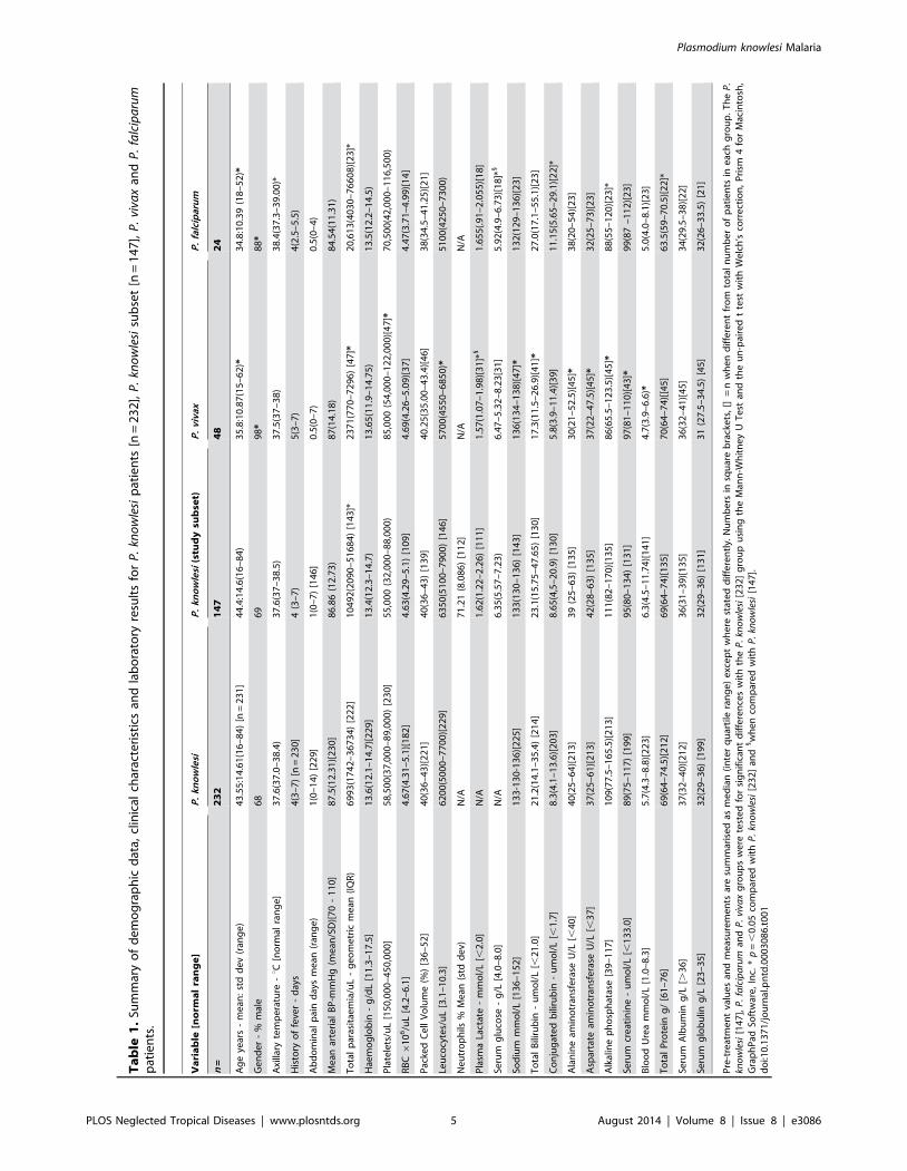
summarised in table 1. P. vivax and P. falciparum data are
included for comparison. Eighty-five P. knowlesi patients were
deselected (see below and materials and methods section) creating
a subset of 147 P. knowlesi patients for Pknbpxa and Pknbpxbgenotyping.
Clinical, demographic and laboratory data of the P. knowlesisubset [n-147] were representative of the total number of knowlesi
patients [n = 232] with the exception of parasitaemia (Table 1).
The subset of patients [n = 147] had a significantly higher
geometric mean parasitaemia 10492 (2090-51684) parasites/uL
(IQR) compared with 6993 (1742-36734) in the larger group,
p = 0.03 (Mann Whitney U test). Parasitaemia was not used as a
criterion for de-selection but a shift in parasitaemia was expected
when approximately 50% of the relatively large group of patients
with mild malaria (no abnormal clinical or laboratory results) were
randomly removed. Forty-six (46) women and 101 men remained
in the subset (n = 147). Women had a higher pulse rate, lower
PCV and haemoglobin than men but this is unlikely to be related
to their infection (Figure S2). The following variables; conjugated
bilirubin (p = 0.021, total protein (p = 0.001), serum albumin
(p = 0.031) and serum globulin (p = 0.001) all associated with
recruitment hospital site, probably due to different measuring
systems, and were removed from further analyses.
Association between parasitaemia, clinical and laboratorymarkers of disease progression
The clinical and laboratory variables (Table 1) were analysed
individually for association with parasitaemia (Table 2 for the
n = 147 subset and Figure S3 for the n = 232 complete P. knowlesicohort). Parasitaemia in both groups was associated with plasma
lactate, serum creatinine and blood urea (r = .0.50; p = ,0.0001)
and with haemoglobin, neutrophils, platelets, total bilirubin, AST
and sodium levels (r = .0.33 and ,0.50, p = ,0.0001).
P. knowlesi Pknbpxa and Pknbpxb reference sequencesLocal Pknbpxa and Pknbpxb diversity was determined by
sequencing, to high stringency, large fragments of both genes from
five P. knowlesi isolates collected in different locations and times
(Table S5). P. knowlesi Pknbpxa (8501 bp) comprising all but the
first 18 and last 689 bases of exon II was amplified, cloned and
sequenced (Figure 1a) and Pknbpxb (3506 bp) comprising exon I,
the intron and 3102 bp of exon II (Figure 2a).
Pknbpxa reference sequences were aligned with the published
sequence from the P. knowlesi experimental H line EU867791.
There were no gaps in the alignment and phylogenetic analysis
indicated that the Pknbpxa gene was dimorphic (Figure S4). The
8501 bp Pknbpxa gene fragment was polymorphic (nucleotide
diversity, p= 0.014260.0029, n = 5) and comprised 168 non-
Plasmodium knowlesi Malaria
PLOS Neglected Tropical Diseases | www.plosntds.org 4 August 2014 | Volume 8 | Issue 8 | e3086
Ta
ble
1.
Sum
mar
yo
fd
em
og
rap
hic
dat
a,cl
inic
alch
arac
teri
stic
san
dla
bo
rato
ryre
sult
sfo
rP
.kn
ow
lesi
pat
ien
ts[n
=2
32
],P
.kn
ow
lesi
sub
set
[n=
14
7],
P.
viva
xan
dP
.fa
lcip
aru
mp
atie
nts
.
Va
ria
ble
[no
rma
lra
ng
e]
P.
kno
wle
siP
.kn
ow
lesi
(stu
dy
sub
set)
P.
viva
xP
.fa
lcip
aru
m
n=
23
21
47
48
24
Ag
eye
ars
-m
ean
:st
dd
ev
(ran
ge
)4
3.5
5:1
4.6
1(1
6–
84
)[n
=2
31
]4
4.4
:14
.6(1
6–
84
)3
5.8
:10
.87
(15
–6
2)*
34
.8:1
0.3
9(1
8–
52
)*
Ge
nd
er
-%
mal
e6
86
99
8*
88
*
Axi
llary
tem
pe
ratu
re-uC
[no
rmal
ran
ge
]3
7.6
(37
.0–
38
.4)
37
.6(3
7–
38
.5)
37
.5(3
7–
38
)3
8.4
(37
.3–
39
.00
)*
His
tory
of
feve
r-
day
s4
(3–
7)
[n=
23
0]
4(3
–7
)5
(3–
7)
4(2
.5–
5.5
)
Ab
do
min
alp
ain
day
sm
ean
(ran
ge
)1
(0–
14
)[2
29
]1
(0–
7)
[14
6]
0.5
(0–
7)
0.5
(0–
4)
Me
anar
teri
alB
P-m
mH
g(m
ean
/SD
)[7
0-
11
0]
87
.5(1
2.3
1)[
23
0]
86
.86
(12
.73
)8
7(1
4.1
8)
84
.54
(11
.31
)
To
tal
par
asit
aem
ia/u
L-
ge
om
etr
icm
ean
(IQ
R)
69
93
(17
42
–3
67
34
)[2
22
]1
04
92
(20
90
–5
16
84
)[1
43
]*2
37
1(7
70
–7
29
6)
[47
]*2
0,6
13
(40
30
–7
66
08
)[2
3]*
Hae
mo
glo
bin
-g
/dL
[11
.3–
17
.5]
13
.6(1
2.1
–1
4.7
)[2
29
]1
3.4
(12
.3–
14
.7)
13
.65
(11
.9–
14
.75
)1
3.5
(12
.2–
14
.5)
Pla
tele
ts/u
L[1
50
,00
0–
45
0,0
00
]5
8,5
00
(37
,00
0–
89
,00
0)
[23
0]
55
,00
0(3
2,0
00
–8
8,0
00
)8
5,0
00
(54
,00
0–
12
2,0
00
)[4
7]*
70
,50
0(4
2,0
00
–1
16
,50
0)
RB
C6
10
6/u
L[4
.2–
6.1
]4
.67
(4.3
1–
5.1
)[1
82
]4
.63
(4.2
9–
5.1
)[1
09
]4
.69
(4.2
6–
5.0
9)[
37
]4
.47
(3.7
1–
4.9
9)[
14
]
Pac
ked
Ce
llV
olu
me
(%)
[36
–5
2]
40
(36
–4
3)[
22
1]
40
(36
–4
3)
[13
9]
40
.25
(35
.00
–4
3.4
)[4
6]
38
(34
.5–
41
.25
)[2
1]
Leu
cocy
tes/
uL
[3.1
–1
0.3
]6
20
0(5
00
0–
77
00
)[2
29
]6
35
0(5
10
0–
79
00
)[1
46
]5
70
0(4
55
0–
68
50
)*5
10
0(4
25
0–
73
00
)
Ne
utr
op
hils
%M
ean
(std
de
v)N
/A7
1.2
1(8
.08
6)
[11
2]
N/A
N/A
Pla
sma
Lact
ate
-m
mo
l/L
[,2
.0]
N/A
1.6
2(1
.22
–2
.26
)[1
11
]1
.57
(1.0
7–
1.9
8)[
31
]*1
1.6
55
(.91
–2
.05
5)[
18
]
Seru
mg
luco
se-
g/L
[4.0
–8
.0]
N/A
6.3
5(5
.57
–7
.23
)6
.47
–5
.32
–8
.23
[31
]5
.92
(4.9
–6
.73
)[1
8]*
1
Sod
ium
mm
ol/
L[1
36
–1
52
]1
33
-13
0-1
36
)[2
25
]1
33
(13
0–
13
6)
[14
3]
13
6(1
34
–1
38
)[4
7]*
13
2(1
29
–1
36
)[2
3]
To
tal
Bili
rub
in-
um
ol/
L[,
21
.0]
21
.2(1
4.1
–3
5.4
)[2
14
]2
3.1
(15
.75
–4
7.6
5)
[13
0]
17
.3(1
1.5
–2
6.9
)[4
1]*
27
.0(1
7.1
–5
5.1
)[2
3]
Co
nju
gat
ed
bili
rub
in-
um
ol/
L[,
1.7
]8
.3(4
.1–
13
.6)[
20
3]
8.6
5(4
,5–
20
.9)
[13
0]
5.8
(3.9
–1
1.4
)[3
9]
11
.15
(5.6
5–
29
.1)[
22
]*
Ala
nin
eam
ino
tran
sfe
rase
U/L
[,4
0]
40
(25
–6
4)[
21
3]
39
(25
–6
3)
[13
5]
30
(21
–5
2.5
)[4
5]*
38
(20
–5
4)[
23
]
Asp
arta
team
ino
tran
sfe
rase
U/L
[,3
7]
37
(25
–6
1)[
21
3]
42
(28
–6
3)
[13
5]
37
(22
–4
7.5
)[4
5]*
32
(25
–7
3)[
23
]
Alk
alin
ep
ho
sph
atas
e[3
9–
11
7]
10
9(7
7.5
–1
65
.5)[
21
3]
11
1(8
2–
17
0)[
13
5]
86
(65
.5–
12
3.5
)[4
5]*
88
(55
–1
20
)[2
3]*
Seru
mcr
eat
inin
e-
um
ol/
L[,
13
3.0
]8
9(7
5–
11
7)
[19
9]
95
(80
–1
34
)[1
31
]9
7(8
1–
11
0)[
43
]*9
9(8
7–
11
2)[
23
]
Blo
od
Ure
am
mo
l/L
[1.0
–8
.3]
5.7
(4.3
–8
.8)[
22
3]
6.3
(4.5
–1
1.7
4)[
14
1]
4.7
(3.9
–6
.6)*
5.0
(4.0
–8
.1)[
23
]
To
tal
Pro
tein
g/
[61
–7
6]
69
(64
–7
4.5
)[2
12
]6
9(6
4–
74
)[1
35
]7
0(6
4–
74
)[[4
5]
63
.5(5
9–
70
.5)[
22
]*
Seru
mA
lbu
min
g/L
[.3
6]
37
(32
–4
0)[
21
2]
36
(31
–3
9)[
13
5]
36
(32
–4
1)[
45
]3
4(2
9.5
–3
8)[
22
]
Seru
mg
lob
ulin
g/L
[23
–3
5]
32
(29
–3
6)
[19
9]
32
(29
–3
6)
[13
1]
31
(27
.5–
34
.5)
[45
]3
2(2
6–
33
.5)
[21
]
Pre
-tre
atm
en
tva
lue
san
dm
eas
ure
me
nts
are
sum
mar
ise
das
me
dia
n(i
nte
rq
uar
tile
ran
ge
)e
xce
pt
wh
ere
stat
ed
dif
fere
ntl
y.N
um
be
rsin
squ
are
bra
cke
ts,
[]=
nw
he
nd
iffe
ren
tfr
om
tota
ln
um
be
ro
fp
atie
nts
ine
ach
gro
up
.T
he
P.
kno
wle
si[1
47
],P
.fa
lcip
aru
man
dP
.vi
vax
gro
up
sw
ere
test
ed
for
sig
nif
ican
td
iffe
ren
ces
wit
hth
eP
.kn
ow
lesi
[23
2]
gro
up
usi
ng
the
Man
n-W
hit
ne
yU
Te
stan
dth
eu
n-p
aire
dt
test
wit
hW
elc
h’s
corr
ect
ion
,P
rism
4fo
rM
acin
tosh
,G
rap
hP
adSo
ftw
are
,In
c.*
p=
,0
.05
com
par
ed
wit
hP
.kn
ow
lesi
[23
2]
and
1w
he
nco
mp
are
dw
ith
P.
kno
wle
si[1
47
].d
oi:1
0.1
37
1/j
ou
rnal
.pn
td.0
00
30
86
.t0
01
Plasmodium knowlesi Malaria
PLOS Neglected Tropical Diseases | www.plosntds.org 5 August 2014 | Volume 8 | Issue 8 | e3086

Table 2. Clinical and laboratory measures of disease progression that associate with P. knowlesi parasitaemia.
Variable - association with parasitaemia n = (r) p value
Age in years 143 +0.274* 0.0009
Haemoglobin g/d: 142 20.366 ,0.0001
PCV (%) 135 20.282 0.0009
Log10 WBC/uL 143 +0.444 ,0.0001
% Neutrophils 108 +0.392 ,0.0001
Log10 Platelets/uL 143 20.327* ,0.0001
Log10 Lactate mmol/L 110 +0.501 ,0.0001
Log10 Serum creatinine umol/L 129 +0.528 ,0.0001
Log10 Blood urea mmol/L 137 +0.601 ,0.0001
Log10 Total bilirubin umol/L 128 +0.403* ,0.0001
Log10 Conjugated bilirubin umol/L 128 +0.330 ,0.0001
Log10 AST U/L 132 +0.355* ,0.0001
Log10 IL-10 pg/mL 142 +0.362 ,0.0001
Sodium mmol/L 139 20.328 ,0.0001
The (r) statistic was calculated using Prism 4 for Macintosh, GraphPad Software, Inc. Pearson’s correlation was used for parametric data and marked* otherwise for non-parametric data Spearman’s correlation test was used.doi:10.1371/journal.pntd.0003086.t002
Figure 1. P. knowlesi Pknbpxa organisation and diversity. Schematic representation of Pknbpxa 9578 bp. (A) Exon 1 and the intron (solid line)are followed by exon II begining at nucleotide 389 (EU867791). Pknbpxa cysteine residues at codon positions 181,239,283,311 and 315 that areimplicated in erythrocyte binding, Meyer, et al., [23], were within the haplotyping fragment and conserved in all patient isolates. (B)A fragment fromnucleotide 389–8889 (8501bp) was amplified and sequenced in five reference isoates. Synonymous (short vertical lines) and non-synonymous (longvertical lines)mutations are marked. (C) Graphical representation of a sliding window plot of nucleotide diversity per site. Diversity (p) was calculatedusing DnaSP v5.10 with window length 400 bp and step size 25 bp. Maximum diversity (p = 0.024) was observed between nucleotide positions 389and 1388 (hatched line).doi:10.1371/journal.pntd.0003086.g001
Plasmodium knowlesi Malaria
PLOS Neglected Tropical Diseases | www.plosntds.org 6 August 2014 | Volume 8 | Issue 8 | e3086
synonymous and 63 synonymous polymorphic sites (Figures 1a-c
and Table 3). There was a cluster of non-synonymous SNP’s from
base 389 to 1388 (Figure 1c) and this region was selected for direct
PCR sequencing and haplotyping 147 patient isolates at the
Pknbpxa locus. The Pknbpxa reference sequences were submitted
to GenBank, Accession Numbers KF186568-KF186572, (Table
S5).
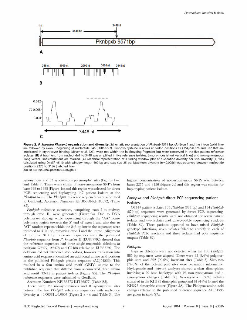
Pknbpxb reference sequences, comprising exon I to midway
through exon II, were generated (Figure 2a). Due to DNA
polymerase slippage while sequencing through the "AA" homo
polymeric region towards the 5’ end of exon I and also due to
"AT" tandem repeats within the 243 bp intron the sequences were
trimmed to 3100 bp, removing exon I and the intron. Alignment
of the five 3100 bp reference sequences with the published
Pknbpxb sequence from P. knowlesi H (EU867792) showed that
the reference sequences had three single nucleotide deletions at
positions G2477, A2478 and C2488 relative to EU867792. The
deletions did not introduce stop codons, however translation into
amino acid sequence identified an additional amino acid position
in the published Pknbpxb protein sequence (ACJ54536). This
resulted in a four amino acid motif (GKIY) unique to the
published sequence that differed from a conserved three amino
acid motif (ENL) in patient isolates (Figure S5). The Pknbpxbreference sequences were submitted to GenBank,
Accession Numbers KF186573-KF186577, (Table S5).
There were 20 non-synonymous and 8 synonymous sites
between the five Pknbpxb reference sequences with nucleotide
diversity p = 0.0038160.0007 (Figure 2 a - c and Table 3). The
highest concentration of non-synonymous SNPs was between
bases 2275 and 3156 (Figure 2c) and this region was chosen for
haplotyping patient isolates.
Pknbpxa and Pknbpxb direct PCR sequencing patientisolates
Of 147 patient isolates 138 Pknbpxa (885 bp) and 134 Pknbpxb(879 bp) sequences were generated by direct PCR sequencing.
Pknbpxa sequencing results were not obtained for seven patient
isolates and two isolates had unacceptable sequencing readouts
(Table S2). Three patients appeared to have mixed Pknbpxbgenotype infections, seven isolates failed to amplify in each of
Pknbpxb PCR reactions and three isolates had poor sequence
outputs (Table S2).
PknbpxaGaps or deletions were not detected when the 138 Pknbpxa
885 bp sequences were aligned. There were 83 (9.4%) polymor-
phic sites and 802 (90.6%) invariant sites (Table 3). Sixty-two
(74.6%) of the polymorphic sites were parsimony informative.
Phylogenetic and network analyses showed a clear dimorphism
involving a 29 base haplotype with 25 non-synonymous and 4
synonymous changes (Table S6). Seventy-seven (56%) isolates
clustered in the KH195 dimorphic group and 61 (44%) formed the
KH273 dimorphic cluster (Figure 3A). The Pknbpxa amino acid
changes relative to the published reference sequence ACJ54535
are given in table S7a.
Figure 2. P. knowlesi Pknbpxb organisation and diversity. Schematic representation of Pknbpxb 9571 bp. (A) Exon 1 and the intron (solid line)are followed by exon II beginning at nucleotide 346 (EU867792). Pknbpxb cysteine residues at codon positions 193,254,298,326 and 332 that areimplicated in erythrocyte binding, Meyer et al., [23], were not within the haplotyping fragment but were conserved in the five patient referenceisolates. (B) A fragment from nucleotide1 to 3448 was amplified in five reference isolates. Synonymous (short vertical lines) and non-synonymous(long vertical lines)mutations are marked. (C) Graphical representation of a sliding window plot of nucleotide diversity per site. Diversity (p) wascalculated using DnaSP v5.10 with window length 400 bp and step size 25 bp. Maximum diversity (p = 0.0056) was observed between nucleotidepositions 2275 to 3156 (hatched line).doi:10.1371/journal.pntd.0003086.g002
Plasmodium knowlesi Malaria
PLOS Neglected Tropical Diseases | www.plosntds.org 7 August 2014 | Volume 8 | Issue 8 | e3086
Seventy-five (75) Pknbpxa DNA haplotypes were resolved in the
885 bp fragments from 138 patient isolates with haplotype
diversity 0.9729 (SD60.007). There were 12 (16%) haplotypes
in the KH273 dimorphic cluster all but four with a frequency .2
and 63 (84%) haplotypes in the KH195 cluster. All but 11 of the
Pknbpxa KH195 haplotypes had a frequency ,1 (Figure 3A).
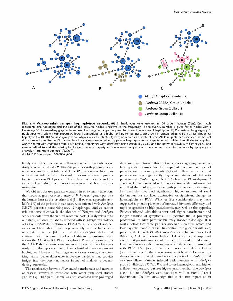
PknbpxbThe three gaps observed in five Pknbpxb reference sequences
were conserved in all patient isolates (n = 134) in the study relative
to the published sequence Pknbpxb EU86772. Alignment of the
134 Pknbpxb 879 bp sequences identified 46 polymorphic sites
(Table 3) and 25 of the sites were parsimony informative. Fifty-one
(51) Pknbpxb haplotypes were resolved with haplotype diversity
= 0.922 (SD60.014). A minimum spanning haplotype network
shows some dimorphic characteristics at the Pknbpxb locus,
Figure 4A. Pknbpxb amino acid changes relative to the published
reference sequence ACJ54536 are given in table S7b.
Genetic markers of disease progressionTwelve Pknbpxa and two Pknbpxb haplotype groups were
resolved, each with two or three allelic forms. The alleles were
added to the patient dataset and analysed for significant
differences in clinical and laboratory markers of disease severity
(Table S4). Pknbpxa alleles that segregated with markers of disease
severity were within the KH195 dimorphism. For example
patients infected with parasites with Pknbpxa group 6, 913C
allele iii were restricted to the KH195 dimorphism and had; higher
parasitaemia (p = 0.02), WBC’s (p = 0.02), blood urea (p = 0.003),
serum creatinine (p = 0.0057), plasma lactate (p = 0.003), IL-10
(p = 0.003), lower platelets (p = 0.001), systolic blood pressure
(p = 0.005) and prolonged duration of fever (p = 0.036) (Figure 3B
and Figure S6 a-j). When isolates with the 913C polymorphism
were overlaid on the haplotype network a degree of haplotype
clustering was observed although the allele appeared in two
distinct nodes (Figure 3B). Pknbpxa haplotype group 8 describes a
complex polymorphic site (982T/G/C). Parasites in the KH273
dimorphism all had 982T allele i (n = 61). The KH195 dimor-
phism had two alleles, allele ii, 982G (n = 51) and allele iii, 982C
(n = 26). Alleles ii and iii had some association with markers of
disease progression. Patients infected with parasites with 982G
allele ii had higher plasma lactate (p = 0.006), higher IL-10
(p = 0.03) and lower haemoglobin (p = 0.034) and those with 982C
allele iii had higher AST (p = 0.033), (Figure 3C and Figure S7 a-
f). When the 982C and 982G, group 8 alleles ii and iii were
mapped onto the Pknbpxa haplotype network they formed
clusters. However five haplotypes with group 8 allele iii appeared
on the edges of the group 8 allele ii cluster (Figure 3C). There was
some overlap between Pknbpxa groups 6 and 8 (Figures 3C boxed
area).
Two Pknbpxb allelic groups were resolved. Pknbpxb group 1
had two alleles, 2637A:2638C and 2637C:2638A. Patients infected
with parasites with the 2637C:2638A allele had lower haemoglo-
bin (p = 0.004) and higher axillary temperature (p = 0.001)
(Figure 4B and Figure S8a and S8b). Isolates with 2637C:2638A
appeared as two clusters at the edges of the Pknbpxb haplotype
network (Figure 4B). Pknbpxb group 2 was more complex with
three major alleles (i, ii, iii) involving 6 polymorphic sites (Table
S4). Four patient isolates were not easily grouped and excluded
from the Pknbpxb group 2 analysis. Patients infected with parasites
with Pknbpxb group 2 allele iii had increased parasitaemia
(p = 0.016), plasma lactate (p = 0.004), total bilirubin (p = 0.047)
and AST (p = 0.002), Figure 4B and Figure S9a-f. Haplotype
clustering was observed when Pknbpxb group 2 alleles i, ii and iii
Ta
ble
3.
Pkn
bp
xaan
dP
knb
pxb
refe
ren
cean
dh
aplo
typ
ing
seq
ue
nce
div
ers
ity.
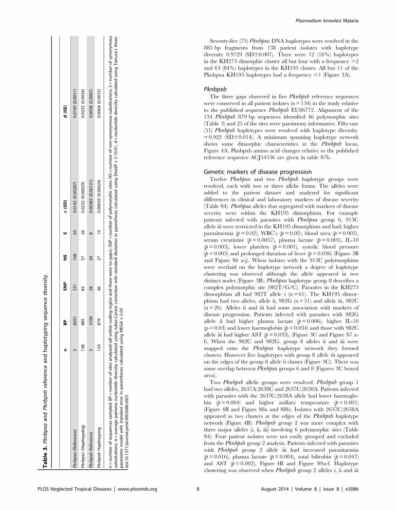
nB
PS
NP
NS
Sp
(SD
)d
(SE
)
Pkn
bp
xa(R
efe
ren
ce)
58
50
12
31
16
86
30
.01
42
(0.0
02
87
)0
.01
45
(0.0
01
1)
Pkn
bp
xa(H
aplo
typ
ing
)1
38
88
58
35
72
60
.02
32
(0.0
00
29
)0
.02
33
(0.0
03
4)
Pkn
bp
xbR
efe
ren
ce5
31
00
28
20
80
.00
38
2(0
.00
12
1)
0.0
03
8(0
.00
07
)
Pkn
bp
xbH
aplo
typ
ing
13
48
79
46
27
19
0.0
06
39
(0.0
00
24
)0
.00
64
(0.0
01
5)
n=
nu
mb
er
of
seq
ue
nce
ssa
mp
led
;BP
=n
um
be
ro
fsi
tes
anal
yze
d(a
llw
ith
inco
din
gre
gio
nan
dth
ere
we
ren
og
aps)
;SN
P=
nu
mb
er
of
po
lym
orp
hic
site
s;N
S=
nu
mb
er
of
no
n-s
yno
nym
ou
ssu
bst
itu
tio
ns;
S=
nu
mb
er
of
syn
on
ymo
us
sub
stit
uti
on
s;p
=av
era
ge
pai
rwis
en
ucl
eo
tid
ed
ive
rsit
yca
lcu
late
du
sin
gJu
kes-
Can
tor
corr
ect
ion
wit
hst
and
ard
de
viat
ion
inp
are
nth
esi
sca
lcu
late
du
sin
gD
naS
Pv
5.1
0.0
1;
d=
nu
cle
oti
de
div
ers
ity
calc
ula
ted
usi
ng
Tam
ura
’sth
ree
-p
aram
ete
rm
od
el
wit
hst
and
ard
err
or
inp
are
nth
ese
sca
lcu
late
du
sin
gM
EGA
v5
.05
do
i:10
.13
71
/jo
urn
al.p
ntd
.00
03
08
6.t
00
3
Plasmodium knowlesi Malaria
PLOS Neglected Tropical Diseases | www.plosntds.org 8 August 2014 | Volume 8 | Issue 8 | e3086

were mapped onto the Pknbpxb haplotype network (Figure 4C).
Pknbpxb group 2 allelic cluster i was not associated with markers of
disease progression and formed a discrete cluster in the Pknbpxbnetwork. Group 2 allele ii also formed a discrete cluster and allele
iii appeared as 2 clusters. In addition there was allelic sharing
between Pknbpxb haplotype group 1 allele ii and Group 2 allele iii
(Figure 4C boxed).
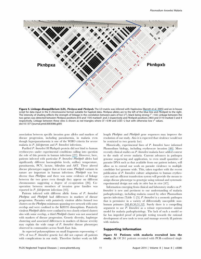
Linkage disequilibrium within and between Pknbpxa andPknbpxb sites
A strict 29 bp haplotype defines the Pknbpxa KH195 - KH273
dimorphism in the Pknbpxa haplotyping sequence (885 bp). The
intensity of r2 indicates that several Pknbpxa alleles, including
those defining the dimorphism are in strong LD r2.0.8 forming
linked blocks in the matrix (Figure 5). The r2 values for linked sites
between the two genes were weak. However, there was some
between gene linkage with high D’ values (.0.99) and LOD.2
(Figure 5). For example Pknbpxa position 810 and Pknbpxaposition 1105 (Figure 5, positions marked 1 and 2. Also Pknbpxbpositions 2403 and 3110 with the main blocks defining the
Pknbpxa dimorphism (Figure 5 positions marked 3 and 4).
Discussion
Patients with P. knowlesi malaria in Sarawak are infected with
diverse parasites at the Pknbpxa and Pknbpxb loci. Some of the
diversity clusters with increased parasitaemia and measures of
disease severity. To our knowledge this is the first time that
particular alleles of members of the Plasmodium RBP gene family
have been linked to increased parasitaemia and disease in patients
with malaria.
Members of the RBP’s are represented in all Plasmodium
species studied so far [17]. They are diverse in nature and thought
to be responsible for erythrocyte selection during merozoite
invasion of host erythrocytes. Even though humans are not the
natural hosts of P. knowlesi we did not find evidence of human
host selection, represented by clonality at these important loci. In
agreement with P. falciparum (Pf) Rh1, PfRh2a, PfRh2b and P.vivax RBP-2 non-synonymous substitutions were predominant at
each P. knowlesi RBP locus [37]. The 5’ end of Pknbpxa,
corresponding to the putative erythrocyte-binding site, was
particularly polymorphic although polymorphic sites occurred
along the entire 8105 bp fragment, similar to P. vivax RBP-2[37].
Diversity involving non-synonymous substitutions near func-
tional sites on Plasmodium merozoite surface proteins is not
unusual and a particular problem in vaccine design [38,39]. Non-
synonymous diversity is most often attributed to immune evasion
rather than altered function. Although a single amino acid change
in experimental lines of P. falciparum reticulocyte binding-like
orthologue PfRh5 changed function and conferred infectivity to
Aotus monkey erythrocytes [40]. Therefore, assigning loss of host
erythrocyte restriction to a single amino acid suggests that non-
synonymous substitutions on other members of this invasion gene
Figure 3. Pknbpxa minimum spanning haplotype network. (A) 75 haplotypes were resolved in 138 patient isolates coloured nodes. Isolates inthe KH273 dimorphic group are in green and those in the KH195 dimorphism in blue. Each node represents one haplotype and the size of thecoloured nodes is relative to the frequency. The frequency number is given for all nodes with a frequency .1. Intermediary gray nodes representmissing haplotypes required to connect two different haplotypes. (B) Haplotypes with Pkbnpxa group 6 allele iii (913C) that had increased markers ofdisease severity are shown in yellow. P. knowlesi isolates with this mutation appear in 2 clusters within the KH195 dimorphism. (C) Haplotypes withPknbpxa group 8 982 alleles are shown: 982T allele i (KH273 green); 982G allele ii (KH195 blue); 982C allele iii (KH195 pink). Group 8 alleles ii and iii hadincreased markers of disease severity when compared with allele i. There is one main cluster of 982C (pink) haplotypes with 5 additional andapparently un-connected to the main cluster that appear on the edges of the network. 982C (pink) haplotypes all occur in the KH195 dimorphicgroup (4a blue). Note that the boxed nodes also contain Pknbpxa 913C (4b). Haplotypes were generated using Arlequin v3.5.1.2 and the networkdrawn with Gephi v0.8.2 with manual editing to add the missing haplotypes. Haplotype groups were mapped onto the minimum spanning networkby applying the analysis of molecular variance (AMOVA).doi:10.1371/journal.pntd.0003086.g003
Plasmodium knowlesi Malaria
PLOS Neglected Tropical Diseases | www.plosntds.org 9 August 2014 | Volume 8 | Issue 8 | e3086
family may alter function as well as antigenicity. Patients in our
study were infected with P. knowlesi parasites with predominantly
non-synonymous substitutions at the RBP invasion gene loci. This
observation will be taken forward to examine altered protein
function between Pknbpxa and Pknbpxb protein variants and the
impact of variability on parasite virulence and host invasion
restriction.
We did not observe parasite clonality in P. knowlesi infections
that would suggest restricted entry of P. knowlesi genotypes into
the human host at this or other loci [1]. However, approximately
half (44%) of the patients in our study were infected with PknbpxaKH273 parasites, comprising only 12 haplotypes, and we cannot
rule out some selection in the absence of Pknbpxa and Pknbpxbsequence data from the natural macaque hosts. Highly relevant to
our study, children in Ghana infected with P. falciparum isolates
with the CAMP dimorphism of EBA-175, a member of another
important Plasmodium invasion gene family, were at higher risk
of a fatal outcome [41]. In our study Pknbpxa alleles that
clustered with increased markers of disease progression were
within the Pknbpxa KH195 dimorphism. Polymorphisms within
the CAMP dimorphism were not interrogated in the Ghanaian
study and this approach may have identified putative virulent
haplotypes. However, taken together with our results, character-
ising within species differences in parasite virulence may provide
insight into the potential health impact of malaria, especially
during outbreaks.
The relationship between P. knowlesi parasitaemia and markers
of disease severity is consistent with other published studies
[4,5,42,43]. High parasitaemia was not associated with prolonged
duration of symptoms in this or other studies suggesting parasite or
host specific reasons for the apparent increase in rate of
parasitaemia in some patients [5,42,44]. Here we show that
parasitaemia was significantly higher in patients infected with
parasites with Pknbpxa group 6, 913C allele iii or Pknbpxb group 2
allele iii. Patients infected with the Pknbpxa allele had some but
not all of the markers associated with parasitaemia in this study.
For example, they had significantly higher markers of renal
dysfunction but not liver dysfunction or significant changes in
haemoglobin or PCV. What at first consideration may have
suggested a phenotypic effect of increased invasion efficiency and
rapid progression to high parasitaemia may well be the opposite.
Patients infected with this variant had higher parasitaemia and
longer duration of symptoms. It is possible that a prolonged
progression to high parasitaemia may impact pathology. It is
worth noting that these patients also had increased lactate and
lower systolic blood pressure. In addition to higher parasitaemia,
patients infected with Pknbpxb group 2 allele iii had increased total
Bilirubin, AST and plasma lactate. Taken within the important
caveat that parasitaemia is central to our study and in multivariate
linear regression models parasitaemia is independently associated
with PCV, AST (transformed data), urea and plasma lactate
(transformed data), there was some stratification between the
disease markers that clustered with the particular Pknbpxa and
Pknbpxb alleles. Patients infected with parasites with Pknbpxbgroup 1 allele ii, 2637C:2638A had lower haemoglobin and higher
axilliary temperature but not higher parasitaemia. The Pknbpxaalleles but not Pknbpxb were associated with markers of renal
dysfunction. To our knowledge this is the first report of an
Figure 4. Pknbpxb (A) 51 haplotypes were resolved in 134 patient isolates (Blue). Each noderepresents one haplotype and the size of the coloured nodes is relative to the frequency. The frequency number is given for all nodes with afrequency .1. Intermediary gray nodes represent missing haplotypes required to connect two different haplotypes. (B) Pknbpxb haplotype group 1.Haplotypes with allele ii Pkbnpxb2638A, lower haemoglobin and higher axillary temperature, are shown in brown radiating from a high frequencyhaplotype (f = 18). (C) Pknbpxb group 2 haplotypes, alleles i (blue), ii (green) appeared as discrete clusters Allele iii (pink) had increased markers ofdisease severity and formed 2 clusters. Four isolates were excluded and appear as larger grey nodes. Haplotypes with alleles ii and iii cluster together.Alleles shared with Pknbpxb group 1 are boxed. Haplotypes were generated using Arlequin v3.5.1.2 and the network drawn with Gephi v0.8.2 andmanual edited to add the missing haplotypes markers. Haplotype groups were mapped onto the minimum spanning network by applying theanalysis of molecular variance (AMOVA).doi:10.1371/journal.pntd.0003086.g004
Plasmodium knowlesi Malaria
PLOS Neglected Tropical Diseases | www.plosntds.org 10 August 2014 | Volume 8 | Issue 8 | e3086
minimum spanning haplotype network.
association between specific invasion gene alleles and markers of
disease progression, including parasitaemia, in malaria even
though hyperparasitaemia is one of the WHO criteria for severe
malaria in P. falciparum and P. knowlesi infections.
Purified P. knowlesi H Pknbpxb protein did not bind to human
erythrocytes under experimental conditions calling into question
the role of this protein in human infections [27]. However, here,
patients infected with particular P. knowlesi Pknbpxb alleles had
significantly different haemoglobin levels, axillary temperature,
parasitaemia, PCV, lactate, bilirubin and AST. These altered
disease phenotypes suggest that at least some Pknbpxb variants in
nature are important in human infections. Pknbpxb was less
diverse than Pknbpxa and there was some evidence of linkage
between the two genes even though they appear on different
chromosomes suggesting a degree of co-operation [26]. Co-
operation between members of invasion gene families was
reported in P. falciparum infections [45].
Patients infected with different allelic forms of P. knowlesiPknbpxa and Pknbpxb had differences in markers of disease
progression. Parasites with putatively virulent alleles formed two
clusters on the Pknbpxa minimum spanning tree network with some
overlap and were confined to the KH195 dimorphism. Putatively
virulent Pknbpxb alleles were confined to two closely related clusters
also with some overlap, a third Pknbpxb cluster was not associated
with markers of disease progression. Genetic diversity, haplotype
clustering and associated differences in markers of disease severity
may explain the wide range of P. knowlesi disease phenotypes
observed in communities across South East Asia.
As expected polymorphisms on small fragments representing ,
10% of two P. knowlesi genetic loci did not capture all patients
with complications in our study. Therefore further work on full-
length Pknbpxa and Pknbpxb gene sequences may improve the
resolution of our study. Also it is expected that virulence would not
be restricted to two genetic loci.
Historically, experimental lines of P. knowlesi have informed
Plasmodium biology, including erythrocyte invasion [46]. More
recently clinical studies on P. knowlesi malaria have added context
to the study of severe malaria. Current advances in pathogen
genome sequencing and application, to even small quantities of
parasite DNA such as that available from our patient isolates, will
allow us to extend our work on parasite virulence to multiple
candidate loci genome wide. This, taken together with the recent
publication of P. knowlesi culture adaptation to human erythro-
cytes and an efficient transfection system will provide the means to
assign disease phenotype to genotype using rational and systematic
experimental design not only in vitro but in vivo [47].
Information emerging from clinical and laboratory studies on P.knowlesi is new and pertinent to our understanding of malaria
pathophysiology, including malaria caused by other Plasmodium
species infections (Table 1) [5]. P. knowlesi is a zoonotic pathogen
that is permissive in a variety of differentially susceptible non-
human primates [48,49,50,51,52]. Surely there is a compelling
argument to use P. knowlesi as a robust representative animal
model for malaria pathophysiology. The lack of such a model so
far has impeded proof of principle testing towards the rational
development of new tools to treat and manage severely ill patients
with malaria.
Supporting Information
Figure S1 Patients with malaria recruited into thestudy. (A) Of 261 patients recruited with PCR-confirmed single
Figure 5. Linkage disequilibrium (LD), Pknbpxa and Pknbpxb. The LD matrix was inferred with Haploview (Barrett et al, 2005) and an in-housescript for data input in the X chromosome format suitable for haploid data. Pknbpxa alleles are to the left of the blue line and Pknbpxb to the right.The intensity of shading reflects the strength of linkage in the correlation between pairs of loci (r2), black being strong r2 .0.8. Linkage between thetwo genes was detected between Pknbpxa positions 810 and 1105 marked1 and 2 respectively and Pknbpxb positions 2403 and 3110 marked 3 and 4respectively. Linkage between these sites is shown as red triangles where D’.0.99 and LOD.2 but with otherwise low r2 values.doi:10.1371/journal.pntd.0003086.g005
Plasmodium knowlesi Malaria
PLOS Neglected Tropical Diseases | www.plosntds.org 11 August 2014 | Volume 8 | Issue 8 | e3086
species P. knowlesi (Pk) infections five were repeat recruitment of
patients during the same clinical episode when referred from
Hospital Sarikei to Hospital Sibu (’B’ samples). Twenty-four of the
remaining patients did not fulfil the study criteria as follows: Three
patients were under 15 year, three patients were either pregnant or
with a co-morbidity and 17 had received antimalarial treatment
prior to recruitment. Therefore 232 patients with single species
PCR-confirmed P. knowlesi infections fulfilled the study criteria.
Of these 161 (69%) of P. knowlesi patients were recruited in
Hospital Sarikei (including one patient from Kapit) and 71 (31%)
in Hospital Sibu. Of the 147 group 99 were recruited in Sarikei
and one in Kapit grouped to 100 (68%)and 47 (32%) in Sibu. (B)
Of 85 patients with non-P. knowlesi malaria recruited into the
study, six patients with P. falciparum malaria had received
antimalarial treatment prior to recruitment. Four patients with P.vivax had received antimalarial treatment two were pregnant and
one had missing laboratory results.
(PDF)
Figure S2 Differences between men and women with P.knowlesi malaria. Significant differences in Hemoglobin, Pulse
and PVC between men and women were observed when outliers
were removed from the data (SPSS and Prism 4 for Macintosh,
GraphPad Software, Inc).
(PDF)
Figure S3 Clinical and laboratory measures of diseaseprogression that associate with P. knowlesi parasitae-mia in the unselected patient group (n = 232). Prism 4 for
Macintosh, GraphPad Software, Inc.
(PDF)
Figure S4 Evidence of Pknbpxa dimorphism. Neighbor-
Joining tree inferred from five Pknpxa reference DNA sequences
(8501 bp), P. knowlesi published sequence EU867191 with P.falciparum Rh2a (XM001350047.1) as the out group. The
percentage of replicate trees in which the associated taxa clustered
together in the bootstrap test (1000 replicates) are shown next to
the branches The evolutionary distances were computed using the
Jukes-Cantor method. All positions containing gaps and missing
data were eliminated. There were a total of 8414 positions in the
final dataset. Evolutionary analyses were conducted in MEGA5
(Tamura et al. 2011. Molecular Biology and Evolution, 28 (10)
pp2731-2739).
(PDF)
Figure S5 Pkbnpxb nucleotide deletions in patientisolates. Three nucleotide deletions were detected in five P.knowlesi reference isolates from patients compared with P.knowlesi H-strain Pknbpxb published sequence EU867792.
Nucleic acid sequences and amino acid translations are shown.
The conserved amino acid motif (ENL) in patient isolates and the
corresponding GKIY motif in the published Pknbpxb amino acid
sequence ACJ54536 are shown. Image generated using Geneious
6.0.4
(PDF)
Figure S6 Differences in markers of disease progres-sion in patients infected with Pknbpxa group 6 alleles.Significant differences in patents infected with Pknbpxa group 6
allele iii 913 C (yellow) compared with the 913T allele (green
when in the KH273 dimorphism and blue when in the KH195
dimorphism) are shown. P values were calculated using the
unpaired t test except for Serum creatinine. *Serum creatinine
levels were not normally distributed and the Mann-Whitney U
test was used. Prism 4 for Macintosh, GraphPad Software, Inc.
Note that all of the HK273 dimorphic form had the 913T
allele.
(PDF)
Figure S7 Differences in markers of disease progres-sion in patients infected with Pknbpxa group 8 alleles.Significant differences between patents infected with Pknbpxaalleles of the complex non-synonymous polymorphic site 982 (T/
G/C). T is the only allele within the KH 273 dimorphism (n-61,
green) and this dimorphism clustered with less severe markers of
disease progression. The KH195 dimorphism has either 982G
(n = 51, blue) or 982C (n = 26, pink) at this position each with
some changes in Haemoglobin, Plasma Lactate, AST and IL-10. P
values are shown for significant differences and determined using
the unpaired t test Prism 4 for Macintosh, GraphPad Software,
Inc.
(PDF)
Figure S8 Differences in markers of disease progres-sion in patents infected with Pknbpxb group 1 alleles(2638 A/C polymorphism). The 2638A allele ii, n = 20 (green)
grouped with low haemoglobin and high axillary temperature.
Tests for significant differences between groups: unpaired t test for
haemoglobin and the Mann- Whitney U test for axillary
temperature, Prism 4 for Macintosh, GraphPad Software, Inc.
(PDF)
Figure S9 Significant differences in markers of diseaseprogression in patents infected with Pknbpxb group 2alleles. Group 2 alleles comprised SNP sites
2117,2740,2757,2802,2834 and 3115 (Table S4). Allele i, n = 51,
blue. Allele ii) n = 49, green. Allele iii) n = 31, pink. Unpaired t test
for significant between group differences, Prism 4 for Macintosh,
GraphPad Software, Inc.
(PDF)
Table S1 Summary of publications on reticulocytebinding-like proteins in different Plasmodium species.
(PDF)
Table S2 Summary of direct PCR sequencing patientisolates for haplotyping. a Pknbpxa and b Pknbpxb gene
fragments.
(PDF)
Table S3 Normality tests for analysis of genetic mark-ers of disease progression.
(PDF)
Table S4 Pknbpxa and Pknbpxb allelic groups andsignificant differences between alleles in markers ofdisease severity.
(PDF)
Table S5 Reference isolates used for generating full-length (8501 bp) Pknbpxa and (3506 bp) Pknbpxb genesequences.
(PDF)
Table S6 P. knowlesi polymorphisms in the haplotyp-ing fragment (885 bp) making up the Pknbpxa dimor-phism.
(PDF)
Table S7 Non-synonymous sites and amino acid chang-es in P. knowlesi Pknbpxa and Pknbpxb haplotypingfragments. a Pknbpxa 138 patient isolates, b Pknbpxb 134
patient isolates.
(PDF)
Plasmodium knowlesi Malaria
PLOS Neglected Tropical Diseases | www.plosntds.org 12 August 2014 | Volume 8 | Issue 8 | e3086
Text S1 Additional information on patient recruitment,blood sample processing and the study cohort.(DOCX)
Acknowledgments
We would like to thank the following: Professor Stephen Gillespie and Dr.
Geoff Butcher for helpful comments on the manuscript. Datu Dr. Zulkifli
bin Jantan and Dr. Junaidi bin Diki of the Sarawak Health Department for
facilitating the practical implementation of this study; All staff and
Directors who supported the study in Sibu and Sarikei Hospitals,
particularly Dr. Tey Siew Chang (Director Sarikei Hospital), Mr. Wong
Ching Toh and Mr. Pek Peng Chin; staff in the male and female medical
wards and the Accident and Emergency Departments; Madam Fauziah
Wahet Medical Records Office Hospital Sarikei and Mr. Zul Azman b.
Sheblee and Mr. Michael Tira Medical Records Office Hospital Sibu.
Author Contributions
Conceived and designed the experiments: JCS SK. Performed the
experiments: AMA PCD SL MW. Analyzed the data: JCS AMA MMP
RZ. Contributed reagents/materials/analysis tools: MMP SBM. Wrote the
paper: JCS AMA. Patient recruitment: ITW CWL. Field work: AMA JCS
BS AS. Data extraction and entry: SKSK.
References
1. Lee KS, Divis PC, Zakaria SK, Matusop A, Julin RA, et al. (2011) Plasmodiumknowlesi: Reservoir Hosts and Tracking the Emergence in Humans and
Macaques. PLoS Pathog 7: e1002015.
2. Singh B, Daneshvar C (2013) Human Infections and Detection of Plasmodiumknowlesi. Clin Microbiol Rev 26: 165–184.
3. Marchand RP, Culleton R, Maeno Y, Quang NT, Nakazawa S Co-infections of
Plasmodium knowlesi, P. falciparum, and P. vivax among Humans andAnopheles dirus Mosquitoes, Southern Vietnam. Emerg Infect Dis 17: 1232–
1239.
4. Barber BE, William T, Grigg MJ, Menon J, Auburn S, et al. (2012) Aprospective comparative study of knowlesi, falciparum and vivax malaria in
Sabah, Malaysia: high proportion with severe disease from Plasmodium knowlesiand P. vivax but no mortality with early referral and artesunate therapy. Clin
Infect Dis 56:383–397
5. Daneshvar C, Davis TM, Cox-Singh J, Rafa’ee MZ, Zakaria SK, et al. (2009)
Clinical and laboratory features of human Plasmodium knowlesi infection. Clin
Infect Dis 49: 852–860.
6. Sallum MAM, Peyton EL, Harrison BA, Wilkerson RC (2005) Revision of the
Leucosphyrus group of Anopheles (Cellia) (Diptera Culicidae). Revisita Brasileira49: 1–52.
7. Yakob L, Bonsall MB, Yan G Modelling knowlesi malaria transmission in
humans: vector preference and host competence. Malar J 9: 329.
8. Nakazawa S, Marchand RP, Quang NT, Culleton R, Manh ND, et al. (2009)
Anopheles dirus co-infection with human and monkey malaria parasites inVietnam. Int J Parasitol 39: 1533–1537.
9. Berry A, Iriart X, Wilhelm N, Valentin A, Cassaing S, et al. (2011) ImportedPlasmodium knowlesi malaria in a French tourist returning from Thailand.
Am J Trop Med Hyg 84: 535–538.
10. Cox-Singh J (2012) Zoonotic malaria: Plasmodium knowlesi, an emerging
pathogen. Curr Opin Infect Dis 25: 530–536.
11. Barber BE, William T, Jikal M, Jilip J, Dhararaj P, et al. (2011) Plasmodiumknowlesi Malaria in Children. Emerg Infect Dis 17: 814–820.
12. Garnham PCC (1966) Malaria Parasites and other Haemosporidia. Oxford:Blackwell Scientific Publications Ltd.
13. Willmann M, Ahmed A, Siner A, Wong IT, Woon LC, et al. (2012) Laboratorymarkers of disease severity in Plasmodium knowlesi infection: a case control
study. Malar J 11: 363.
14. Miller LH, Ackerman HC, Su XZ, Wellems TE (2013) Malaria biology anddisease pathogenesis: insights for new treatments. Nature Med 19: 156–167.
15. Rayner JC (2009) The merozoite has landed: reticulocyte-binding-like ligandsand the specificity of erythrocyte recognition. Trends Parasitol 25: 104–106.
16. Tham WH, Healer J, Cowman AF (2012) Erythrocyte and reticulocyte binding-like proteins of Plasmodium falciparum. Trends Parasitol 28: 23–30.
17. Gunalan K, Gao X, Yap SS, Huang X, Preiser PR (2013) The role of the
reticulocyte-binding-like protein homologues of Plasmodium in erythrocytesensing and invasion. Cell Microbiol 15: 35–44.
18. Galinski MR, Medina CC, Ingravallo P, Barnwell JW (1992) A reticulocyte-binding protein complex of Plasmodium vivax merozoites. Cell 69: 1213–1226.
19. Galinski MR, Xu M, Barnwell JW (2000) Plasmodium vivax reticulocyte bindingprotein-2 (PvRBP-2) shares structural features with PvRBP-1 and the
Plasmodium yoelii 235 kDa rhoptry protein family. Mol Biochem Parasitol
108: 257–262.
20. Li J, Han ET (2012) Dissection of the Plasmodium vivax reticulocyte binding-
like proteins (PvRBPs). Biochem Biophys Res Commun 426: 1–6.
21. DeSimone TM, Jennings CV, Bei AK, Comeaux C, Coleman BI, et al. (2009)
Cooperativity between Plasmodium falciparum adhesive proteins for invasioninto erythrocytes. Mol Microbiol 72: 578–589.
22. Jennings CV, Ahouidi AD, Zilversmit M, Bei AK, Rayner J, et al. (2007)
Molecular analysis of erythrocyte invasion in Plasmodium falciparum isolatesfrom Senegal. Infect Immun 75: 3531–3538.
23. Bei AK, Membi CD, Rayner JC, Mubi M, Ngasala B, et al. (2007) Variantmerozoite protein expression is associated with erythrocyte invasion phenotypes in
Plasmodium falciparum isolates from Tanzania. Mol Biochem Parasitol 153: 66–71.
24. Gomez-Escobar N, Amambua-Ngwa A, Walther M, Okebe J, Ebonyi A, et al.
(2010) Erythrocyte invasion and merozoite ligand gene expression in severe and
mild Plasmodium falciparum malaria. J Infect Dis 201: 444–452.
25. Meyer EV, Semenya AA, Okenu DM, Dluzewski AR, Bannister LH, et al.
(2009) The reticulocyte binding-like proteins of P. knowlesi locate to the
micronemes of merozoites and define two new members of this invasion ligand
family. Mol Biochem Parasitol 165: 111–121.
26. Aurrecoechea C, Brestelli J, Brunk BP, Dommer J, Fischer S, et al. (2009)
PlasmoDB: a functional genomic database for malaria parasites. Nucleic Acids
Res 37: D539–543.
27. Semenya AA, Tran TM, Meyer EV, Barnwell JW, Galinski MR (2012) Two
functional reticulocyte binding-like (RBL) invasion ligands of zoonotic
Plasmodium knowlesi exhibit differential adhesion to monkey and human
erythrocytes. Malar J 11: 228.
28. Cox-Singh J, Mahayet S, Abdullah MS, Singh B (1997) Increased sensitivity of
malaria detection by nested polymerase chain reaction using simple sampling
and DNA extraction. Int J Parasitol 27: 1575–1577.
29. Snounou G, Viriyakosol S, Jarra W, Thaithong S, Brown KN (1993)
Identification of the four human malaria parasite species in field samples by
the polymerase chain reaction and detection of a high prevalence of mixed
infections. Mol Biochem Parasitol 58: 283–292.
30. Singh B, Kim Sung L, Matusop A, Radhakrishnan A, Shamsul SS, et al. (2004)
A large focus of naturally acquired Plasmodium knowlesi infections in human
beings. Lancet 363: 1017–1024.
31. World Health Organization: Management of Severe malaria - A Practical
Handbook (2013). Third edition ed. Geneva: World Health Organisation.
http://www.who.int/malaria/publications/atoz/9789241548526/en/
32. Kumar S, Nei M, Dudley J, Tamura K (2008) MEGA: a biologist-centric
software for evolutionary analysis of DNA and protein sequences. Brief
Bioinform 9: 299–306.
33. Librado P, Rozas J (2009) DnaSP v5: a software for comprehensive analysis of
DNA polymorphism data. Bioinformatics 25: 1451–1452.
34. Excoffier L, Lischer HE (2010) Arlequin suite ver 3.5: a new series of programs
to perform population genetics analyses under Linux and Windows. Mol Ecol
Resour 10: 564–567.
35. Bastian MH, S Jacomy, M. (2009) Gephi: an open source software for exploring
and manipulating networks. International AAAI Conference on Weblogs and
Social Media. http://aaai.org/ocs/index.php/ICWSM/09/paper/view/154
36. Barrett JC, Fry B, Maller J, Daly MJ (2005) Haploview: analysis and
visualization of LD and haplotype maps. Bioinformatics 21: 263–265.
37. Rayner JC, Tran TM, Corredor V, Huber CS, Barnwell JW, et al. (2005)
Dramatic difference in diversity between Plasmodium falciparum and Plasmo-dium vivax reticulocyte binding-like genes. Am J Trop Med Hyg 72: 666–
674.
38. Rayner J (2005) Getting down to malarial nuts and bolts: the interaction between
Plasmodium vivax merozoites and their host erythrocytes. Mol Micro 55: 1297–
1299.
39. Takala SL, Coulibaly D, Thera MA, Batchelor AH, Cummings MP, et al. (2009)
Extreme polymorphism in a vaccine antigen and risk of clinical malaria:
implications for vaccine development. Science Translat Med 1: 2ra5.
40. Hayton K, Gaur D, Liu A, Takahashi J, Henschen B, et al. (2008) Erythrocyte
binding protein PfRH5 polymorphisms determine species-specific pathways of
Plasmodium falciparum invasion. Cell Host Microbe 4: 40–51.
41. Cramer JP, Mockenhaupt FP, Mohl I, Dittrich S, Dietz E, et al. (2004) Allelic
dimorphism of the erythrocyte binding antigen-175 (eba-175) gene of
Plasmodium falciparum and severe malaria: Significant association of the C-
segment with fatal outcome in Ghanaian children. Malar J 3: 11.
42. Cox-Singh J, Davis TM, Lee KS, Shamsul SS, Matusop A, et al. (2008)
Plasmodium knowlesi malaria in humans is widely distributed and potentially life
threatening. Clin Infect Dis 46: 165–171.
43. William T, Menon J, Rajahram G, Chan L, Ma G, et al. (2011) Severe
Plasmodium knowlesi Malaria in a Tertiary Care Hospital, Sabah, Malaysia.
Emerg Infect Dis 17: 1248–1255.
44. Cox-Singh J, Hiu J, Lucas SB, Divis PC, Zulkarnaen M, et al. (2010) Severe
malaria - a case of fatal Plasmodium knowlesi infection with post-mortem
findings: a case report. Malar J 9: 10.
45. Lopaticki S, Maier AG, Thompson J, Wilson DW, Tham WH, et al. (2011)
Reticulocyte and erythrocyte binding-like proteins function cooperatively in
Plasmodium knowlesi Malaria
PLOS Neglected Tropical Diseases | www.plosntds.org 13 August 2014 | Volume 8 | Issue 8 | e3086

invasion of human erythrocytes by malaria parasites. Infect Immun 79: 1107–
1117.46. Bannister LH, Butcher GA, Mitchell GH (1977) Recent advances in
understanding the invasion of erythrocytes by merozoites of Plasmodiumknowlesi. Bull World Health Organ 55: 163–169.
47. Moon RW, Hall J, Rangkuti F, Ho YS, Almond N, et al. (2013) Adaptation
of the genetically tractable malaria pathogen Plasmodium knowlesi tocontinuous culture in human erythrocytes. Proc Nat Acad Sci U S A 110:
531–536.
48. Barnwell JW, Howard RJ, Miller LH (1982) Altered expression of Plasmodiumknowlesi variant antigen on the erythrocyte membrane in splenectomized rhesus
monkeys. J Immunol 128: 224–226.
49. Butcher G, Cohen S (1970) Schizogony of Plasmodium knowlesi in the presence
of normal and immune sera. Trans R Soc Trop Med Hyg 64: 470.
50. Langhorne J, Cohen S (1979) Plasmodium knowlesi in the marmoset (Callithrix
jacchus). Parasitol 78: 67–76.
51. Miller LH, Fremount HN, Luse SA (1971) Deep vascular schizogony of
Plasmodium knowlesi in Macaca mulatta. Distribution in organs and
ultrastructure of parasitized red cells. Am J Trop Med Hyg 20: 816–824.
52. Ozwara H, Langermans JA, Maamun J, Farah IO, Yole DS, et al. (2003)
Experimental infection of the olive baboon (Paplio anubis) with Plasmodiumknowlesi: severe disease accompanied by cerebral involvement. Am J Trop Med
Hyg 69: 188–194.
Plasmodium knowlesi Malaria
PLOS Neglected Tropical Diseases | www.plosntds.org 14 August 2014 | Volume 8 | Issue 8 | e3086
Related Documents


