DISEASES OF AQUATIC ORGANISMS Dis Aquat Org Vol. 99: 57–78, 2012 doi: 10.3354/dao02450 Published May 15 © Inter-Research 2012 · www.int-res.com *Email: [email protected] Pseudocollinia brintoni gen. nov., sp. nov. (Apostomatida: Colliniidae), a parasitoid ciliate infecting the euphausiid Nyctiphanes simplex J. Gómez-Gutiérrez 1, *, M. C. Strüder-Kypke 2 , D. H. Lynn 2,6 , T. C. Shaw 3 , M. J. Aguilar-Méndez 4,7 , A. López-Cortés 4 , S. Martínez-Gómez 1 , C. J. Robinson 5 1 Departamento de Plancton y Ecología Marina, Centro Interdisciplinario de Ciencias Marinas, Av. IPN s/n, A.P. 592, La Paz, Baja California Sur 23096, Mexico 2 Department of Integrative Biology, University of Guelph, Guelph, Ontario N1G 2W1, Canada 3 Cooperative Institute for Marine Resources and NOAA/NMFS, Hatfield Marine Science Center, Newport, Oregon 97365, USA 4 Centro de Investigaciones Biológicas del Noroeste (CIBNOR), La Paz, Baja California Sur 23090, Mexico 5 Laboratorio de Ecología de Pesquerías, Instituto de Ciencias del Mar y Limnología, Universidad Nacional Autónoma de México, A.P. 70-305, Ciudad de Mexico, D.F. 04510, Mexico 6 Present address: Department of Zoology, University of British Columbia, Vancouver, British Columbia V6T 1Z4, Canada 7 Present address: Instituto Politécnico Nacional (IPN), Unidad Profesional Interdisciplinaria de Ingeniería Campus Guanajuato (UPIIG), Silao de la Victoria, Guanajuato 36275, Mexico ABSTRACT: A novel parasitoid ciliate, Pseudocollinia brintoni gen. nov., sp. nov. was discov- ered infecting the subtropical sac-spawning euphausiid Nyctiphanes simplex off both coasts of the Baja California peninsula, Mexico. We used microscopic, and genetic information to describe this species throughout most of its life cycle. Pseudocollinia is distinguished from other Colliniidae genera because it exclusively infects euphausiids, has a polymorphic life cycle, and has a small cone-shaped oral cavity whose left wall has a field of ciliated kineto- somes and whose opening is surrounded on the left and right by 2 ‘oral’ kineties (or ciliary rows) that terminate at its anterior border. Two related species that infect different euphausiid species from higher latitudes in the northeastern Pacific Ocean, Collinia beringensis Capriulo and Small, 1986, briefly redescribed herein, and Collinia oregonensis Gómez-Gutiérrez, Peter- son, and Morado, 2006, are transferred to the genus Pseudocollinia. P. brintoni has between 12 and 18 somatic kineties, and its oral cavity has only 2 oral kineties, while P. beringensis comb. nov. has more somatic kineties, including 3 oral kineties. P. oregonensis comb. nov. has an intermediate number of somatic kineties. P. beringensis comb. nov. also infects Thysanoessa raschi (a new host species). SSU rRNA and cox1 gene sequences demonstrated that Pseudo- collinia ciliates are apostome ciliates and that P. brintoni is different from P. beringensis comb. nov. High densities of rod-shaped bacteria (1.7 μm length, 0.2 to 0.5 μm diameter) were associ- ated with P. brintoni. After euphausiid rupture, high concentrations of P. brintoni and bacteria cluster to form 3 to 6 cm long filaments where tomites encyst and transform to the phoront stage; this is a novel place for encystation. P. brintoni may complete its life cycle when the euphausiids feed on these filaments. KEY WORDS: Euphausiids · Ciliates · Apostomatidae · Collinia · cox1 · SSU rRNA · Mexico Resale or republication not permitted without written consent of the publisher

Welcome message from author
This document is posted to help you gain knowledge. Please leave a comment to let me know what you think about it! Share it to your friends and learn new things together.
Transcript
DISEASES OF AQUATIC ORGANISMSDis Aquat Org
Vol. 99: 57–78, 2012doi: 10.3354/dao02450
Published May 15
© Inter-Research 2012 · www.int-res.com*Email: [email protected]
Pseudocollinia brintoni gen. nov., sp. nov.(Apostomatida: Colliniidae), a parasitoid ciliateinfecting the euphausiid Nyctiphanes simplex
J. Gómez-Gutiérrez1,*, M. C. Strüder-Kypke2, D. H. Lynn2,6, T. C. Shaw3, M. J. Aguilar-Méndez4,7, A. López-Cortés4, S. Martínez-Gómez1, C. J. Robinson5
1Departamento de Plancton y Ecología Marina, Centro Interdisciplinario de Ciencias Marinas, Av. IPN s/n, A.P. 592, La Paz, Baja California Sur 23096, Mexico
2Department of Integrative Biology, University of Guelph, Guelph, Ontario N1G 2W1, Canada3Cooperative Institute for Marine Resources and NOAA/NMFS, Hatfield Marine Science Center, Newport, Oregon 97365, USA
4Centro de Investigaciones Biológicas del Noroeste (CIBNOR), La Paz, Baja California Sur 23090, Mexico5Laboratorio de Ecología de Pesquerías, Instituto de Ciencias del Mar y Limnología,
Universidad Nacional Autónoma de México, A.P. 70-305, Ciudad de Mexico, D.F. 04510, Mexico
6Present address: Department of Zoology, University of British Columbia, Vancouver, British Columbia V6T 1Z4, Canada
7Present address: Instituto Politécnico Nacional (IPN), Unidad Profesional Interdisciplinaria de IngenieríaCampus Guanajuato (UPIIG), Silao de la Victoria, Guanajuato 36275, Mexico
ABSTRACT: A novel parasitoid ciliate, Pseudocollinia brintoni gen. nov., sp. nov. was discov-ered infecting the subtropical sac-spawning euphausiid Nyctiphanes simplex off both coasts ofthe Baja California peninsula, Mexico. We used microscopic, and genetic information todescribe this species throughout most of its life cycle. Pseudocollinia is distinguished fromother Colliniidae genera because it exclusively infects euphausiids, has a polymorphic lifecycle, and has a small cone-shaped oral cavity whose left wall has a field of ciliated kineto-somes and whose opening is surrounded on the left and right by 2 ‘oral’ kineties (or ciliaryrows) that terminate at its anterior border. Two related species that infect different euphausiidspecies from higher latitudes in the northeastern Pacific Ocean, Collinia beringensis Capriuloand Small, 1986, briefly redescribed herein, and Collinia oregonensis Gómez-Gutiérrez, Peter-son, and Morado, 2006, are transferred to the genus Pseudocollinia. P. brintoni has between 12and 18 somatic kineties, and its oral cavity has only 2 oral kineties, while P. beringensis comb.nov. has more somatic kineties, including 3 oral kineties. P. oregonensis comb. nov. has anintermediate number of somatic kineties. P. beringensis comb. nov. also infects Thysanoessaraschi (a new host species). SSU rRNA and cox1 gene sequences demonstrated that Pseudo-collinia ciliates are apostome ciliates and that P. brintoni is different from P. beringensis comb.nov. High densities of rod-shaped bacteria (1.7 µm length, 0.2 to 0.5 µm diameter) were associ-ated with P. brintoni. After euphausiid rupture, high concentrations of P. brintoni and bacteriacluster to form 3 to 6 cm long filaments where tomites encyst and transform to the phorontstage; this is a novel place for encystation. P. brintoni may complete its life cycle when theeuphausiids feed on these filaments.
KEY WORDS: Euphausiids · Ciliates · Apostomatidae · Collinia · cox1 · SSU rRNA · Mexico
Resale or republication not permitted without written consent of the publisher
Dis Aquat Org 99: 57–78, 2012
INTRODUCTION
Parasites regularly cause a gradient of damageto their hosts, ranging from almost innocuous tolethal effects (Bradbury 1994, Morado & Small 1995,Gómez-Gutiérrez et al. 2003, 2006, 2009, 2010a). Par-asitoids are categorized as lethal when they must killtheir host to complete their life cycle, with an ecolog-ical function analogous to predators (Lafferty & Kuris2002).
Capriulo & Small (1986) described an endopara-sitic ciliate of krill infecting the eu phausiid Thysa-noessa inermis Krøyer, 1846 in the Bering Sea,naming the species Collinia beringensis and as -signing it to the family Colliniidae. A related spe-cies, named Collinia oregonensis Gómez-Gutiérrez,Peterson & Morado, 2006, was responsible for amass mortality of the temperate euphausiid Eu -phausia pacifica Hansen, 1911, in Astoria Canyon,Oregon, USA, and was recognized as a parasitoidciliate of euphausiids (Gómez-Gutiérrez et al.2003). Collinia oregonensis also infects at least 2other USA west coast eu phausiid species, Thysa-noessa spinifera Holmes, 1900, and Thysanoessagregaria G. O. Sars, 1883 (Gómez-Gutiérrez et al.2003, 2006).
In the southern part of the California CurrentSystem and Gulf of California, there is a diverseeuphausiid assemblage, among which Nyctiphanessimplex Hansen, 1911 accounts for >90% of theeuphausiid biomass and abundance in the neriticregions (Brinton 1962, 1981, Brinton & Townsend1980, Lavaniegos 1994, Gómez-Gutiérrez 1995,Gómez-Gutiérrez et al. 1995). During a study ofthe fecundity and molting rates of N. simplex car-ried out along the southwest coast of the Baja Cali-fornia peninsula and Gulf of California, Mexico,Gómez-Gutiérrez et al. (2010b,c) discovered sev-eral adults infected with a novel apostome speciessimilar to those previously assigned to the genusCollinia (Capriulo & Small 1986, Capriulo et al.1991, Gómez-Gutiérrez et al. 2003, 2006, 2010a).Here, we formally establish the new genus Pseudo-collinia gen. nov. and describe its type species asPseudocollinia brintoni sp. nov., based on its mor-phology, its nuclear small subunit (SSU) ribosomalribonucleic acid (rRNA) and mitochondrial cyto -chrome c oxidase subunit I (cox1) gene sequences.We provide the general symptoms of infection ofthis parasitoid, and we propose a hypothetical lifecycle based on our field observational and experi-mental data.
MATERIALS AND METHODS
Euphausiid collection
Infected Nyctiphanes simplex euphausiids werecollected during 3 oceanographic cruises carried outalong the continental shelf and the middle part ofMagdalena Bay, Baja California Sur, Mexico (BAMAcruises, March, July, and November 2004, 24° 30’ N,112° 30’ W) (Fig. 1A,B) and during 4 oceanographiccruises carried out along the Gulf of California(GOLCA cruises, November 2005, January and July2007, and March 2010, 24−30° N, 109−113.5° W)(Fig. 1C−E). In the September to October 2010 andNovember 2011 cruises in the same region, wheneuphausiids had unusually low abundance, noPseudocollinia-infected euphausiids were collected(n = 18 and 13 zooplankton samples, respectively)(Tables 1 & 2). In the Bering Sea, additional infectedThysanoessa inermis and Thysanoessa raschi M.Sars, 1864 (new host species) euphausiids were col-lected during an oceanographic cruise carried outfrom 18 June to 10 July 2010.
Euphausiids were collected mostly at night using adrifting black 300 µm mesh net with a 1 m diametermouth, 5 m length, and a PVC cod-end of 0.25 mdiameter and 0.70 m length. In Mexico, this zoo-plankton net was equipped with an underwater lamp(Ikelite Pro-video-lite system, 50 W) to attract theeuphausiids. Live zooplankton samples were ob -tained by lowering the net to the depth where thesplit-beam echosounder (SIMRAD EY-60, 120 kHz
58
Oceanographic cruise Sampling stationsn n with infected krill (%)
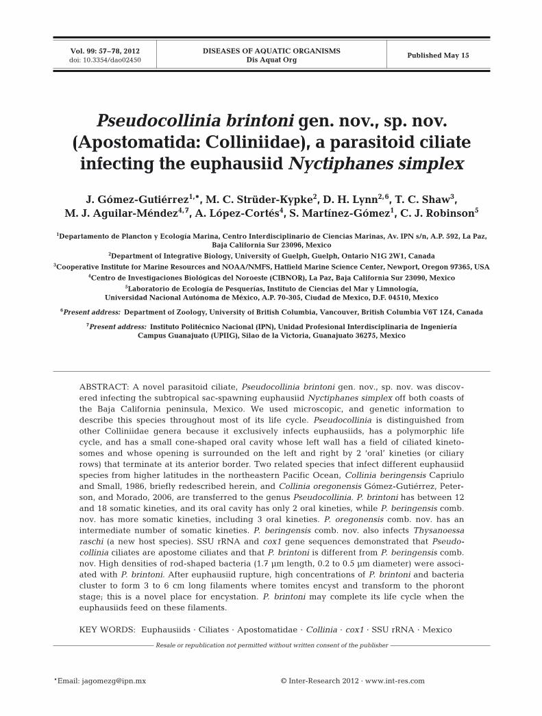
BAMA Mar 2004 32 3 (9.4)BAMA Jul 2004 16 2 (12.5)BAMA Dec 2004 35 6 (17.1)
GOLCA Nov 2005 26 1 (3.8)GOLCA Jan 2007 18 4 (22.2)GOLCA Jul 2007 26 1 (3.8)GOLCA Mar 2010 16 2 (12.5)GOLCA Sep−Oct 2010 18 0 (0)GOLCA Nov 2011 13 0 (0)
Total (mean) 200 19 (9.5)
Table 1. Total number of zooplankton samples collected dur-ing several oceanographic cruises using a drifting net, number of oceanographic stations where the euphausiidNyctiphanes simplex were infected with the endoparasitoidciliate Pseudocollinia brintoni gen. nov., sp. nov., and proportion (%) of oceanographic stations with infectedkrill. BAMA: Magdalena Bay; GOLCA: Gulf of California
oceanographic cruises
Gómez-Gutiérrez et al.: Parasitoid ciliate infecting Nyctiphanes simplex
frequency) and a submarine video camera (Deep SeaPower & Light) showed euphausiid aggregations.Sampling was conducted for 10 min while the shipwas drifting. This methodology ensures negligiblephysical damage to the euphausiids.
Incubation of the infected euphausiid withendoparasitoid ciliates
Live, infected Nyctiphanes simplex, identified byhaving a swollen yellow-beige or orange cephalotho-rax, were transferred into 1 l bottles of surface seawa-ter and incubated on board inside a cold room at 16 ±0.5°C until they died due to ciliate infection. This tem-perature is near the mean temperature of the seawa-
ter where N. simplex is typically collected off bothcoasts of the Baja California peninsula (Table 2). Oneof the original research goals was to estimate the eggproduction and molting rates of N. simplex at eachsampling station. Thus, we incubated >30 N. simplexadults with a healthy appearance individually in 1 lbottles for at least 48 h, plus ~200 adults in groups of10 animals per 1 l bottle filled with seawater from 4 mdepth sieved through a 20 µm mesh filter. After sev-eral hours of incubation in these bottles, some appar-ently healthy N. simplex specimens, based on obser-vations at the beginning of the experiment, changedto a typically yellow-beige or orange infected col-oration. Each infected specimen was isolated into a 1 lbottle for further observation. All of the infected eu-phausiids were monitored every 2 to 4 h during the
59
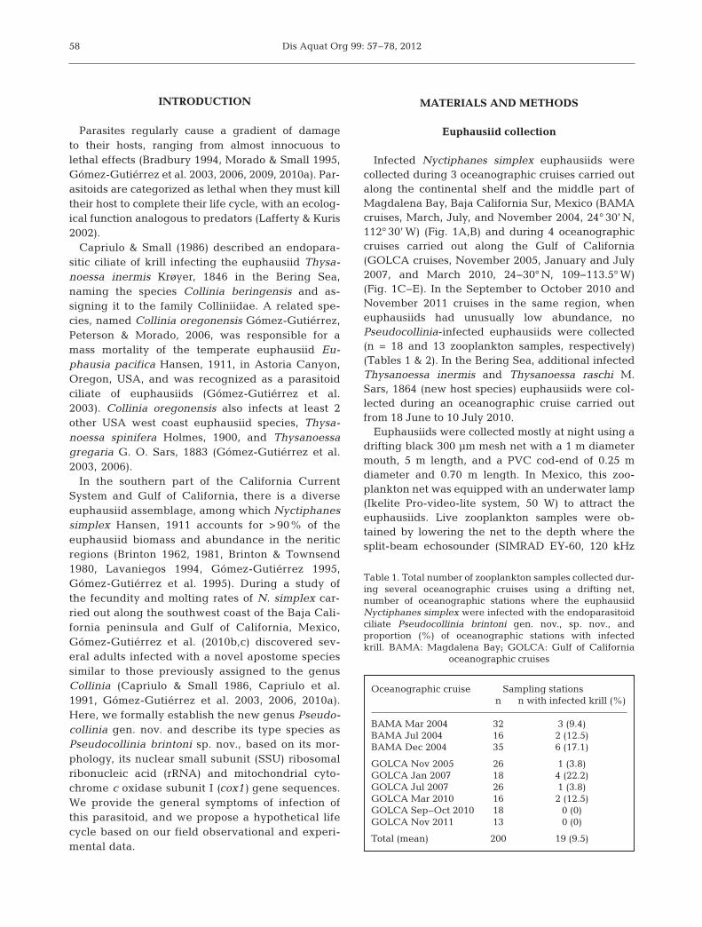
Fig. 1. Sampling stations (d) for krill in northwestern Mexico. (A) Areas of study and oceanographic stations along (B) thesouthwest coast of Baja California Peninsula (Magdalena Bay; Mar, Jul, and Dec 2004) and (C−E) Gulf of California during (C)Nov 2005, (D) Jan 2007 and (E) Jul 2007 where healthy (contour maps of abundance of krill >8 mm total length) and infected
Nyctiphanes simplex with Pseudocollinia brintoni gen. nov., sp. nov. (s) were collected
Dis Aquat Org 99: 57–78, 2012
night and every 6 h during the day to observe theirbehavior and the progression of infection and to de-tect when and how the euphausiids died. Euphausiidswere sexed, and their total length (mm) wasmeasured from the forward rim of the cephalothorax,located behind the base of the eyestalk, to the tip ofthe telson. Sequential photographs of the external ap-pearance of the hosts were taken using an OlympusCamedia 3040 digital camera at 3.3 × 106 pixel resolu-tion. We preserved the host and its ciliates with 4%(v/v) formaldehyde, or 2% (v/v) glutaraldehyde, formorphological analyses and 96% ethanol for geneticanalyses, as the infection progressed through the fol-lowing stages: (1) early infection stage (yellow-beigeeuphausiid carapace); (2) late infection stage (orangeand swollen euphausiid carapace); (3) when the ani-mal exploded and died; and (4) several hours after thehost died and the ciliates formed <6 cm long stickyfilaments of ciliate clusters. We used quantitative pro-targol staining on the ethanol-fixed ciliates (Mon-tagnes & Lynn 1993) and scanning electron mi-croscopy (SEM, Hitachi S-3000N) of sagittal cutsof the formaldehyde-preserved euphausiids to de-
scribe the different life-stages of these apostome ciliates inside N. simplex hosts.
When it was discovered through genetic analysis(see below) that this parasitoid ciliate from Mexicanwaters was indeed an apostome (order Apostom-atida), samples of Collinia beringensis were alsoobtained from the type host Thysanoessa inermis(23.1 mm total length, Stn NP13, 23 June 2010) and anew host T. raschi (23.3 mm total length, Stn 29,10 July 2010, at 70 m depth) during cruise KN195-10(18 June to 10 July 2010) in the Bering Sea. Both hostspecies were collected using a MOCNESS systemand infected specimens (recognizable by theirswollen orange cepha lothorax) were sorted out aliveand immediately preserved in 96% ethanol for ciliateprotargol staining and genetic analyses.
DNA sequence analysis of ciliate mitochondrialcox1 gene and SSU rDNA
Ciliates were obtained for genetic sequencing from1 Nyctiphanes simplex specimen collected during the
60
Cruise Date Time Latitude Longitude No. of infected No. of incubated Prevalence SST(N) (W) specimens euphausiids (%) (°C)
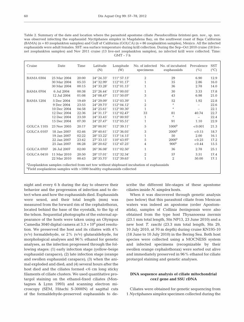
BAMA 0304 25 Mar 2004 20:00 24° 24.33’ 111° 57.13’ 2 29 6.90 12.930 Mar 2004 05:33 24° 32.99’ 112° 01.17’ 1 35 2.86 16.030 Mar 2004 00:15 24° 33.28’ 112° 01.15’ 1 36 2.78 14.0
BAMA 0704 6 Jul 2004 00:38 25° 26.44’ 113° 00.05’ 1 30 3.33 17.812 Jul 2004 01:08 24° 08.47’ 111° 50.07’ 3 43 6.98 21.0
BAMA 1204 5 Dec 2004 19:49 24° 29.09’ 112° 03.39’ 1 52 1.92 22.89 Dec 2004 23:55 24° 29.73’ 112° 04.12’ 2 a – 22.610 Dec 2004 04:58 24° 25.61’ 112° 00.30’ 1 a – 22.112 Dec 2004 22:36 24° 31.17’ 112° 02.47’ 33 81 40.74 22.712 Dec 2004 23:59 24° 33.45’ 112° 00.93’ 1 a – 22.415 Dec 2004 07:30 24° 27.47’ 112° 05.51’ 1 91 1.10 22.9
GOLCA 1105 23 Nov 2005 20:17 28° 00.91’ 112° 39.11’ 5 1000b <0.001 21.3
GOLCA 0107 18 Jan 2007 02:46 29° 40.61’ 112° 36.05’ 3 2000b <0.15 18.719 Jan 2007 02:22 28° 53.22’ 113° 14.15’ 1 50 2.00 16.122 Jan 2007 23:42 27° 33.15’ 110° 43.97’ 5 2000b <0.25 17.225 Jan 2007 06:28 28° 20.62’ 112° 47.25’ 4 900b <0.44 15.5
GOLCA 0707 26 Jul 2007 02:00 26° 36.08’ 111° 02.50’ 1 36 2.78 25.1
GOLCA 0410 11 Mar 2010 20:50 28° 17.01’ 112° 32.34’ 2 57 3.51 17.422 Mar 2010 00:43 28° 35.75’ 112° 39.65’ 1 2 50.00 17.1
aZooplankton samples collected from net tow without shipboard incubation of euphausiidsbField zooplankton samples with >1000 healthy euphausiids collected
Table 2. Summary of the date and location where the parasitoid apostome ciliate Pseudocollinia brintoni gen. nov., sp. nov.was observed infecting the euphausiid Nyctiphanes simplex in Magdalena Bay, on the southwest coast of Baja California(BAMA) (n = 83 zooplankton samples) and Gulf of California (GOLCA) (n = 86 zooplankton samples), Mexico. All the infectedeuphausiids were adult females. SST: sea surface temperature during krill collection. During the Sep−Oct 2010 cruise (18 live-net zooplankton samples) and Nov 2011 cruise (13 live-net zooplankton samples), no infected krill were collected. Time:
GMT – 7 h
Gómez-Gutiérrez et al.: Parasitoid ciliate infecting Nyctiphanes simplex
BAMA 1204 cruise (12 December 2004) and from 2specimens collected during the GOLCA 0410 cruise(11 March 2010). An infected Thysanoessa inermisand a T. raschi specimen collected from the BeringSea (KN 195-10 cruise, 18 June to 10 July 2010) werealso used for genetic sequencing. The cephalothoraxof the ethanol-fixed krill was opened, and chunks ofthe infecting ciliate cells were transferred into 1.5 mlmicrocentrifuge tubes. The microcentrifuge tubeswere left open for 30 min to let the ethanol evaporate.Subsequent DNA extraction was done using theDNEasy Blood & Tissue Kit (Qiagen), following themanufacturer’s protocol, with the exception that thesamples were incubated for 60 to 90 min and only100 µl of AE buffer were used for elution.
PCR amplifications of the SSU rDNA and the cox1gene were performed in an ABI 2720 thermocycler(Applied Biosystems). The forward primers for theSSU rDNA were the universal eukaryote primer A (5’-AAC CTG GTT GAT CCT GCC AGT-3’) (Medlin etal. 1988) and the internal primer 300F (5’-AGG GTTCGA TTC CGG AG-3’) (Elwood et al. 1985). The uni-versal eukaryote primer B (5’-TGA TCC TTC TGCAGG TTC ACC TAC-3’) (Medlin et al. 1988) and theprimers C (5’-TTG GTC CGT GTT TCA AGA C-3’)(Jerome et al. 1996) and LSUR (5’-GTT AGT TTC TTTTCC TCC GC-3’) (Bourland & Strüder-Kypke 2010)were used as reverse primers. The cox1 gene was am-plified using the forward primers F388dT (5’-TGTAAA ACG ACG GCC AGT GGW KCB AAA GATGTW GC-3’) for Pseudocollinia brintoni and F298dT(5’-TGT AAA ACG ACG GCC AGT GCN CAY GGTYTA ATN ATG GT-3’) for Collinia beringensis and thereverse primer R1184dT (5’-CAG GAA ACA GCTATG ACT ADA CYT CAG GGT GAC CRA AAA ATCA-3’) (Strüder-Kypke & Lynn 2010). The PCR productswere purified with a MinElute gel purification kit (Qi-agen) and se quenced in both directions with a 3730DNA Analyzer (Applied Biosystems), using ABI PrismBigDye Terminator (ver. 3.1) and a Cycle SequencingReady Reaction kit. The sequences were importedinto Sequencher ver. 4.0.5 (Gene Codes), trimmed atthe ends, assembled into contigs, and checked for se-quencing errors.
The cox1 sequences were imported into MEGAver. 4.1 (Kumar et al. 2008) and aligned based on theamino acid sequence. The SSU rDNA sequenceswere imported into an existing dedicated compara-tive sequence editor (De Rijk & De Wachter 1993)database and automatically aligned to other apos-tome ciliate sequences. Based on the secondarystructure of the SSU rRNA molecule, we furtherrefined the alignment.
Four different phylogenetic analyses were per-formed on the alignments of both genes to constructthe trees: maximum likelihood (ML; PhyML) (Guin-don et al. 2005), Bayesian inference (BI; MrBayes)(Ronquist & Huelsenbeck 2003), maximum parsi-mony (MP; PAUP ver. 4.10) (Swofford 2002), andneighbor joining (NJ; PHYLIP ver. 3.67) (Felsenstein2005). Modeltest ver. 3.0 (Posada & Crandall 1998)was employed to find the model of DNA substitutionthat best fit our data. The general-time-reversible(GTR) model for nucleotide substitution, with gamma-distributed substitution rates and invariable sites,was determined as the best model for both of thegenes. We included these parameters in PhyML andMrBayes. DNADIST was employed to calculate thegenetic distances with the Kimura 2-parametermodel (Kimura 1980), and the distance trees wereconstructed with NEIGHBOR (NJ) (Saitou & Nei1987). The data were re-sampled 500 (PhyML) and1000 (MP, NJ) times, respectively. The MP analysiswas performed with a random addition (n = 5) of thespecies and the tree bisection-reconnection (TBR)branch-swapping algorithm in effect.
Experimental infection mechanism
In March and July 2004, individual live infectedNyctiphanes simplex collected in Magdalena Baywere transferred to a 0.25 l bottle with water previ-ously filtered with 0.07 µm pore glass fiber filters(GF/F) to avoid plankton contamination. To determinethe effect of just-released parasitoid ciliates onhealthy euphausiids under laboratory conditions, sev-eral healthy N. simplex females were placed into the0.25 l bottle and exposed to a recently exploded eu-phausiid that had released the tomite stage of the cili-ate into the incubation bottle. The healthy euphausiidswere monitored almost continuously until they died.The animals were preserved and dissected in the lab-oratory to search for ciliates inside the cephalothorax.In December 2004, we conducted similar experimentsadding healthy specimens of N. simplex, an unidenti-fied mysid species and the small planktonic decapodProcessa pippinae Wickstein & Méndez, 1985 to pre-viously filtered seawater with recently formed bacte-ria-ciliate filaments to test whether this ciliate speciesis specific to euphausiids or if it could infect otherholoplanktonic crustaceans collected with the samegear and in the same areas where the infected N. sim-plex were collected. In December 2005, we made acomplete set of experiments in the Gulf of Californiawith healthy, mature N. simplex females exposed
61
Dis Aquat Org 99: 57–78, 2012
initially to free-swimming tomite cells and also to bacteria-ciliate filaments.
RESULTS
From a total of 200 zooplankton samples collectedon both coasts of the Baja California peninsula, Mexico, we found Nyctiphanes simplex infected withliving ciliates in only 19 oceanographic stations(Table 1, Fig. 1B−E). The overall proportion ofoceanographic stations with infected euphausiidsvaried among oceanographic cruises from 0 to 22.2%(mean = 9.5%) (Table 1). There was no significantdifference in the prevalence of infected euphau -siids among the oceanographic cruises duringwhich infections were found (χ2 = 0.046, p = 0.25,n = 7 cruises).
Prevalence of Pseudocollinia brintonigen. nov., sp. nov.
During the BAMA cruises, the prevalence of in-fected euphausiids per station ranged between 1.1and 40.7% (mean ± SE = 8.3 ± 8.4%, 95% CI =0.00−31.5%, df = 10). From 83 oceanographic stations,47 infected and 397 healthy euphausiids were col-lected for shipboard incubation (Tables 1 & 2).Infected animals were collected in waters with seasurface temperatures (SST) ranging from 12.9 to22.9°C, indicating that infections occur during all sea-sons and at a wide range of SST (Table 2). Theinfected animals were usually collected at night in themouth of Magdalena Bay at ~40 m depth near theseafloor and over the continental shelf (Fig. 1B).
During the GOLCA cruises, the prevalence ofinfected euphausiids per station ranged between<0.001 and 50% (mean ± SE = 7.4 ± 6.1%, 95% CI =0.00− 21.8%, df = 7). From 86 oceanographic stations,22 infected and ~6000 healthy euphausiids were col-lected for shipboard incubation (Tables 1 & 2).Infected animals were collected in waters with SSTranging from 15.5 to 25.1°C (Table 2). The infectedanimals were typically collected where Nyctiphanessimplex had the highest abundances, along the eastcoast of the Gulf of California during November 2005and July 2007 and along the west coast of the gulf inJanuary 2007 and March 2010 (Fig. 1C−E). In theSeptember to October 2010 and November 2011oceanographic cruises, euphausiids were not abun-dant in night samples, and no infected N. simplexwere collected in the drifting net.
Symptoms of infection by Pseudocollinia brintonigen. nov., sp. nov.
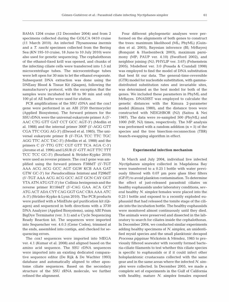
Healthy euphausiids are typically transparent, andgonads are easily observed through the carapace(Fig. 2A,B). Infection appeared only in female adultsof Nyctiphanes simplex, ranging from 9 to 19 mmTL (n = 69) with a yellow-beige to bright orange coloration and swollen cephalothorax (Fig. 2C,E,Table 2). At the station with the largest number ofinfected euphausiids in the southwest coast of theBaja California peninsula in December 2004 (33infected out of 81 collected specimens, prevalence41%), 3 animals with an initially healthy appearance(i.e. semi-transparent carapace and abdomen), incu-bated for egg production and molting rates, becameorange (i.e. infected). The initial infection stage (i.e.yellow-beige carapace) developed in <12 h, theadvanced infection stage (i.e. orange and swollencarapace) developed in <31 h, and death of these 3euphausiids (i.e. exploding) occurred in <33 h.
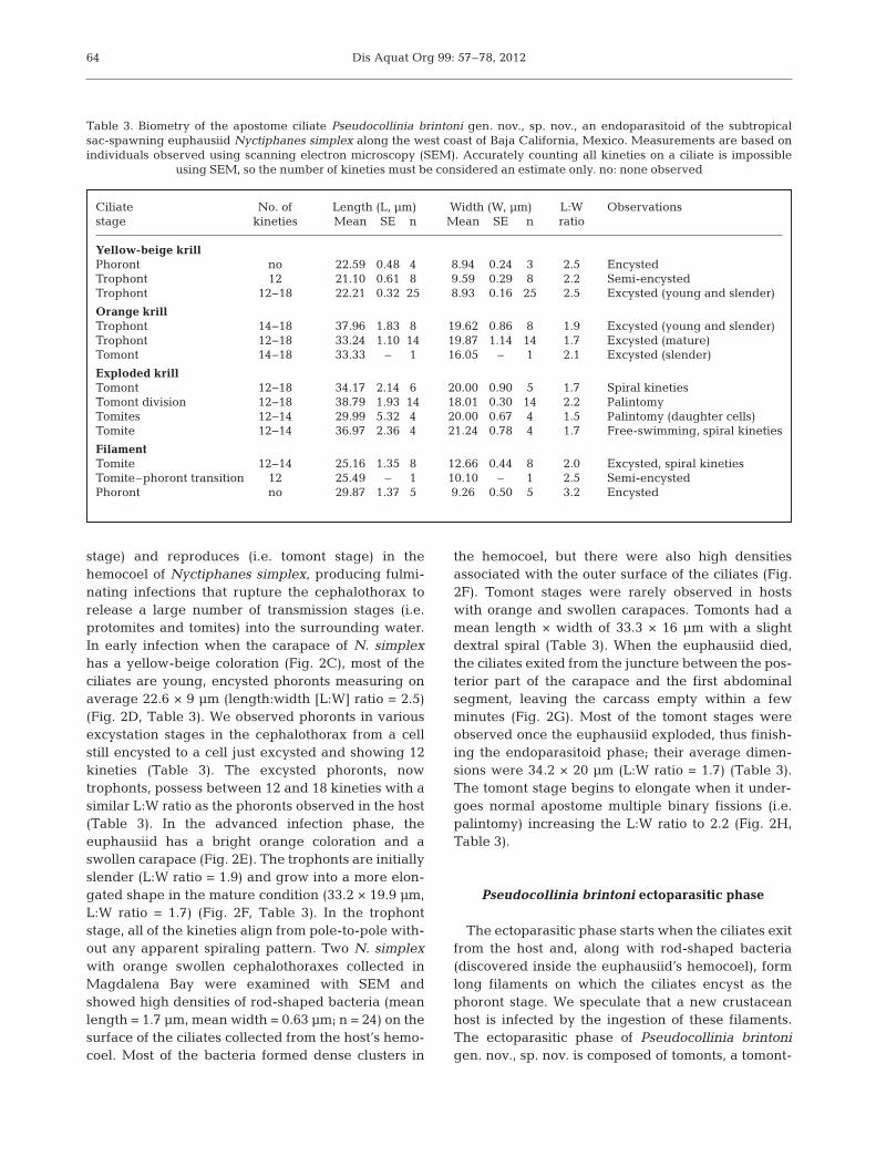
The apostome endoparasitoid ciliate recorded inthe hemocoel and abdomen of Nyctiphanes simplexshowed all of the ciliate life-cycle stages known forapostome ciliates (i.e. phoront, trophont, tomont, andtomite in the host; and tomont, tomite, and phoront out-side the host) with transitional stages between the ex-cysting of the phoront–trophont, beginning of palin-tomy of the tomont (i.e. with a fission furrow in thecenter), nearing the end of the division of tomites (i.e.figure-8-shaped or an advanced fission furrow), andtomite–phoront transition during encystation (Table 3,Figs. 2 & 3). All of the excysted developmental stages(i.e. trophont, tomont, and tomite) had 12 to 18 kineties(ciliary rows) when observed using SEM. However, be-cause one cannot count all of the kineties on each cell inSEM images, these numbers are only estimates(Table 3). The phoront stage, by definition encysted,did not have visible kineties. The life stages were se-quentially separated according to different externalhost conditions and the degree of morphological vari-ability observed within each ciliate developmentalstage: a tomite semi-encysted, a phoront completelyencysted, and a phoront starting excystation as atrophont — this last stage is already inside thecephalothorax of the krill host, in the hemocoel(Fig. 2C−H).
Pseudocollinia brintoni endoparasitoid phase
In the endoparasitoid phase, Pseudocollinia brin-toni gen. nov., sp. nov. feeds, grows (i.e. trophont
62
Gómez-Gutiérrez et al.: Parasitoid ciliate infecting Nyctiphanes simplex 63
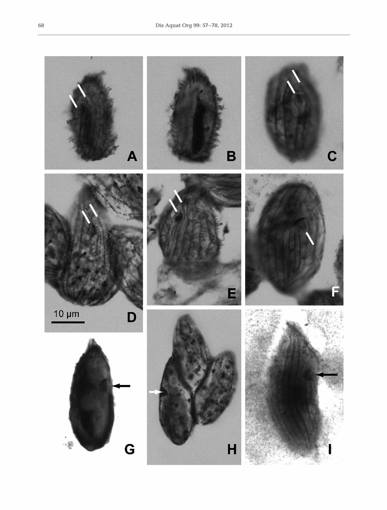
Fig. 2. (A,B) Healthy females of Nyctiphanes simplex with (A) transparent previtellogenic gonad and (B) pink gravid gonad.(C−H) Endoparasitoid phase of Pseudocollinia brintoni gen. nov., sp. nov. and chronological sequence of infection of N. sim-plex. (C) Female in early infection stage with yellow-beige opaque cephalothorax, ~12 h after the female had healthy appear-ance. (D) Trophont stage in early infection presumably just after phoront excystation. (E) Advanced stage of infection withbright orange cephalothorax, ~19 h after the carapace had yellow-beige coloration. (F) Mature trophont stage after feeding:cells appear almost spherical. In several host specimens, high abundances of rod-shaped bacteria (arrows; mean length1.7 µm, width 0.63 µm) were observed on the ciliate’s surface or in colonies on tissues in the euphausiid’s hemocoel. (G) A eu-phausiid exploded after the ciliates had digested its internal organs, ~33 h after the animal had healthy appearance. (H) Re-productive ciliate or tomont stage starting multiple cell divisions without intervening growth (i.e. palintomy) to produce the
dispersal stage (tomite)
Dis Aquat Org 99: 57–78, 2012
stage) and reproduces (i.e. tomont stage) in thehemocoel of Nyctiphanes simplex, producing fulmi-nating infections that rupture the cephalothorax torelease a large number of transmission stages (i.e.protomites and tomites) into the surrounding water.In early infection when the carapace of N. simplexhas a yellow-beige coloration (Fig. 2C), most of theciliates are young, encysted phoronts measuring onaverage 22.6 × 9 µm (length:width [L:W] ratio = 2.5)(Fig. 2D, Table 3). We observed phoronts in variousexcystation stages in the cephalothorax from a cellstill encysted to a cell just excysted and showing 12kineties (Table 3). The excysted phoronts, nowtrophonts, possess between 12 and 18 kineties with asimilar L:W ratio as the phoronts observed in the host(Table 3). In the advanced infection phase, theeuphausiid has a bright orange coloration and aswollen carapace (Fig. 2E). The trophonts are initiallyslender (L:W ratio = 1.9) and grow into a more elon-gated shape in the mature condition (33.2 × 19.9 µm,L:W ratio = 1.7) (Fig. 2F, Table 3). In the trophontstage, all of the kineties align from pole-to-pole with-out any apparent spiraling pattern. Two N. simplexwith orange swollen cephalothoraxes collected inMagdalena Bay were examined with SEM andshowed high densities of rod-shaped bacteria (meanlength = 1.7 µm, mean width = 0.63 µm; n = 24) on thesurface of the ciliates collected from the host’s hemo-coel. Most of the bacteria formed dense clusters in
the hemocoel, but there were also high densitiesassociated with the outer surface of the ciliates (Fig.2F). Tomont stages were rarely observed in hostswith orange and swollen carapaces. Tomonts had amean length × width of 33.3 × 16 µm with a slightdextral spiral (Table 3). When the euphausiid died,the ciliates exited from the juncture between the pos-terior part of the carapace and the first abdominalsegment, leaving the carcass empty within a fewminutes (Fig. 2G). Most of the tomont stages wereobserved once the euphausiid exploded, thus finish-ing the endoparasitoid phase; their average dimen-sions were 34.2 × 20 µm (L:W ratio = 1.7) (Table 3).The tomont stage begins to elongate when it under-goes normal apostome multiple binary fissions (i.e.palintomy) increasing the L:W ratio to 2.2 (Fig. 2H,Table 3).
Pseudocollinia brintoni ectoparasitic phase
The ectoparasitic phase starts when the ciliates exitfrom the host and, along with rod-shaped bacteria(discovered inside the euphausiid’s hemocoel), formlong filaments on which the ciliates encyst as thephoront stage. We speculate that a new crustaceanhost is infected by the ingestion of these filaments.The ectoparasitic phase of Pseudocollinia brintonigen. nov., sp. nov. is composed of tomonts, a tomont-
64
Ciliate No. of Length (L, µm) Width (W, µm) L:W Observationsstage kineties Mean SE n Mean SE n ratio
Yellow-beige krillPhoront no 22.59 0.48 4 8.94 0.24 3 2.5 EncystedTrophont 12 21.10 0.61 8 9.59 0.29 8 2.2 Semi-encystedTrophont 12−18 22.21 0.32 25 8.93 0.16 25 2.5 Excysted (young and slender)
Orange krillTrophont 14−18 37.96 1.83 8 19.62 0.86 8 1.9 Excysted (young and slender)Trophont 12−18 33.24 1.10 14 19.87 1.14 14 1.7 Excysted (mature)Tomont 14−18 33.33 – 1 16.05 – 1 2.1 Excysted (slender)
Exploded krillTomont 12−18 34.17 2.14 6 20.00 0.90 5 1.7 Spiral kinetiesTomont division 12−18 38.79 1.93 14 18.01 0.30 14 2.2 PalintomyTomites 12−14 29.99 5.32 4 20.00 0.67 4 1.5 Palintomy (daughter cells)Tomite 12−14 36.97 2.36 4 21.24 0.78 4 1.7 Free-swimming, spiral kineties
FilamentTomite 12−14 25.16 1.35 8 12.66 0.44 8 2.0 Excysted, spiral kinetiesTomite–phoront transition 12 25.49 – 1 10.10 – 1 2.5 Semi-encystedPhoront no 29.87 1.37 5 9.26 0.50 5 3.2 Encysted
Table 3. Biometry of the apostome ciliate Pseudocollinia brintoni gen. nov., sp. nov., an endoparasitoid of the subtropicalsac-spawning euphausiid Nyctiphanes simplex along the west coast of Baja California, Mexico. Measurements are based onindividuals observed using scanning electron microscopy (SEM). Accurately counting all kineties on a ciliate is impossible
using SEM, so the number of kineties must be considered an estimate only. no: none observed
Gómez-Gutiérrez et al.: Parasitoid ciliate infecting Nyctiphanes simplex
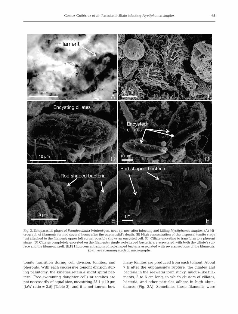
tomite transition during cell division, tomites, andphoronts. With each successive tomont division dur-ing palintomy, the kineties retain a slight spiral pat-tern. Free-swimming daughter cells or tomites arenot necessarily of equal size, measuring 25.1 × 10 µm(L:W ratio = 2.5) (Table 3), and it is not known how
many tomites are produced from each tomont. About7 h after the euphausiid’s rupture, the ciliates andbacteria in the seawater form sticky, mucus-like fila-ments, 3 to 6 cm long, to which clusters of ciliates,bacteria, and other particles adhere in high abun-dances (Fig. 3A). Sometimes these filaments were
65
Fig. 3. Ectoparasitic phase of Pseudocollinia brintoni gen. nov., sp. nov. after infecting and killing Nyctiphanes simplex. (A) Mi-crograph of filaments formed several hours after the euphausiid’s death. (B) High concentration of the dispersal tomite stagejust attached to the filament; upper left corner possibly shows an encysted cell. (C) Ciliate encysting to transform to a phorontstage. (D) Ciliates completely encysted on the filaments; single rod-shaped bacteria are associated with both the ciliate’s sur-face and the filament itself. (E,F) High concentrations of rod-shaped bacteria associated with several sections of the filaments.
(B−F) are scanning electron micrographs
Dis Aquat Org 99: 57–78, 2012
attached to the krill carcass. When the tomites settleon the filament, the kineties still show a perceptiblespiral pattern (Fig. 3B). The tomites beginning toencyst on the filaments (i.e. the tomite–phorontstage) have a L:W ratio of 2.5 (Fig. 3C). As encyst-ment is completed, the cell shape elongates consider-ably, increasing the L:W ratio to 3.2 (Fig. 3D). Thebacteria are also present in high densities on thenewly formed filaments (Fig. 3E), where they activelyreproduce likely by binary division (Fig. 3F). Thebacteria from the filament are smaller than the bacte-ria found in the host (Table 4). It is unknown whetherthe ciliate, the bacteria or both form the filament.
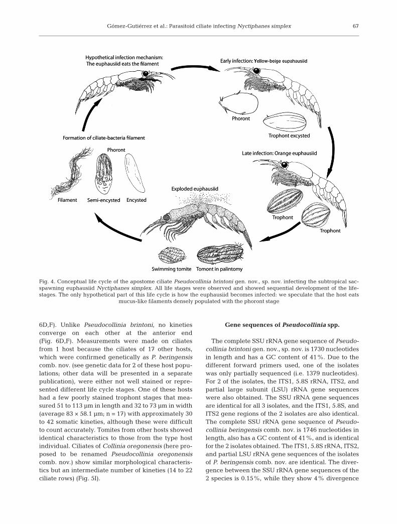
Based on these observations, our proposed lifecycle for Pseudocollinia brintoni gen. nov., sp. nov.infecting this sac-spawning euphausiid Nyctiphanessimplex is conceptually represented in Fig. 4. We stilldo not know how this species infects new hosts, butwe speculate that it is by ingestion of filaments popu-lated with parasitoid ciliates.
Tomite morphology of Pseudocollinia brintonigen. nov., sp. nov.
Protargol staining of ethanol-fixed Pseudocolliniabrintoni ciliates infecting Nyctiphanes simplex re -vealed mostly protomite–tomite stages (Figs. 5A−C,G & 6A−C). The cells were 28 to 35 µm long and 13 to21 µm wide (average 31.1 × 16.6 µm; n = 33). Theypossessed a long and narrow macronucleus thatstretched almost the length of the cell (18 to 28 µmlong by 2 to 11 µm wide, average 22.8 × 5.1 µm, n =32) (Figs. 5B & 6B); a micronucleus was not observed.There were on average 16 kineties (14 to 18, n = 33)that extended from pole to pole, except for 3 kineties(Figs. 5A,C & 6A,C). Two of these kineties curvearound the oral cavity opening and terminate (here-after referred to as ‘oral kineties’) and were num-bered Kinety 1 (K1) and Kinety n (Kn) (Figs. 5A,C &6A,C). The third kinety, Kinety n–1, curves anteriorlyand right above the oral cavity before terminating
in a non-ciliated region anterior to the oral cavity(Figs. 5A,C & 6A,C). The oral cavity, which is aboutone-third of the cell’s length from the anterior end,appears cone-shaped and lined on its left wall with adense field of ciliated kinetosomes or basal bodies(Figs. 5C,G & 6A,C). Kinety n–2 and occasionallyKinety n–3 terminate by abutting at the anteriorend of Kinety 2 (Fig. 6A,C). Measurements weremade on ciliates from a single N. simplex hostbecause the ciliates from 2 other N. simplex hosts,which were confirmed genetically as Pseudocolliniabrintoni, were not well stained. Nevertheless, a fewindividuals from these other 2 hosts showed identicalcharacteristics.
Tomite morphology of Pseudocollinia beringensiscomb. nov.
Protargol staining of ethanol-fixed Collinia berin-gensis (here proposed to be renamed Pseudocolliniaberingensis comb. nov.) infecting Thysanoessa iner-mis and T. raschi euphausiids revealed protomite–tomite stages (Figs. 5D−F,H & 6D−F). The cells were30 to 46 µm long and 16 to 27 µm wide (average 37.1× 21.9 µm; n = 33). The cells possessed a long andwider macronucleus that stretched to almost thelength of the cell (22 to 36 µm long by 5 to 14 µmwide, average 27.4 × 9.9 µm, n = 27) (Fig. 6E); amicronucleus was not observed. There were on aver-age 19 kineties (16 to 20, n = 33) that extended frompole to pole, except for 4 kineties (Figs. 5D,E & 6D,F).Three oral kineties, numbered Kinety 1 (K1), Kinetyn (Kn), and Kinety n–1, curve around the oral cavityopening and terminate (Figs. 5D,E & 6D,F). Thefourth kinety, Kinety n–2, curves anteriorly and rightabove the oral cavity before terminating in a non-cil-iated region anterior to the oral cavity (Figs. 5D,E &6D,F). The oral cavity, which is about one-third of thecell’s length from the anterior end, appears cone-shaped and lined on its left wall with a dense field ofciliated kinetosomes or basal bodies (Figs. 5D−F,H &
66
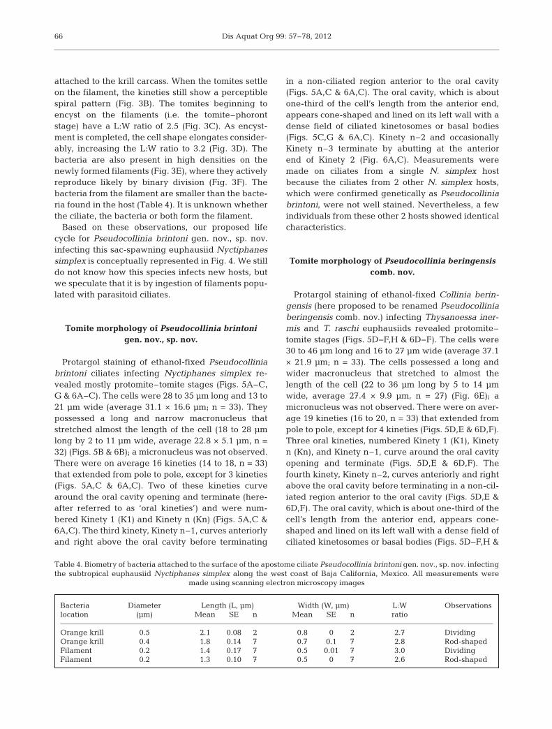
Bacteria Diameter Length (L, µm) Width (W, µm) L:W Observationslocation (µm) Mean SE n Mean SE n ratio
Orange krill 0.5 2.1 0.08 2 0.8 0 2 2.7 DividingOrange krill 0.4 1.8 0.14 7 0.7 0.1 7 2.8 Rod-shapedFilament 0.2 1.4 0.17 7 0.5 0.01 7 3.0 DividingFilament 0.2 1.3 0.10 7 0.5 0 7 2.6 Rod-shaped
Table 4. Biometry of bacteria attached to the surface of the apostome ciliate Pseudocollinia brintoni gen. nov., sp. nov. infectingthe subtropical euphausiid Nyctiphanes simplex along the west coast of Baja California, Mexico. All measurements were
made using scanning electron microscopy images
Gómez-Gutiérrez et al.: Parasitoid ciliate infecting Nyctiphanes simplex
6D,F). Unlike Pseu docollinia brintoni, no kinetiesconverge on each other at the anterior end(Fig. 6D,F). Measurements were made on ciliatesfrom 1 host because the ciliates of 17 other hosts,which were confirmed genetically as P. beringensiscomb. nov. (see genetic data for 2 of these host popu-lations; other data will be presented in a separatepublication), were either not well stained or repre-sented different life cycle stages. One of these hostshad a few poorly stained trophont stages that mea-sured 51 to 113 µm in length and 32 to 73 µm in width(average 83 × 58.1 µm; n = 17) with approximately 30to 42 somatic kineties, although these were difficultto count accurately. Tomites from other hosts showedidentical characteristics to those from the type hostindividual. Ciliates of Collinia oregonensis (here pro-posed to be renamed Pseudocollinia oregonensiscomb. nov.) show similar morphological characteris-tics but an intermediate number of kineties (14 to 22ciliate rows) (Fig. 5I).
Gene sequences of Pseudocollinia spp.
The complete SSU rRNA gene sequence of Pseudo-collinia brintoni gen. nov., sp. nov. is 1730 nucleotidesin length and has a GC content of 41%. Due to thedifferent forward primers used, one of the isolateswas only partially sequenced (i.e. 1379 nucleotides).For 2 of the isolates, the ITS1, 5.8S rRNA, ITS2, andpartial large subunit (LSU) rRNA gene sequenceswere also obtained. The SSU rRNA gene sequencesare identical for all 3 isolates, and the ITS1, 5.8S, andITS2 gene regions of the 2 isolates are also identical.The complete SSU rRNA gene sequence of Pseudo-collinia beringensis comb. nov. is 1746 nucleotides inlength, also has a GC content of 41%, and is identicalfor the 2 isolates obtained. The ITS1, 5.8S rRNA, ITS2,and partial LSU rRNA gene sequences of the isolatesof P. beringensis comb. nov. are identical. The diver-gence between the SSU rRNA gene sequences of the2 species is 0.15%, while they show 4% divergence
67
Fig. 4. Conceptual life cycle of the apostome ciliate Pseudocollinia brintoni gen. nov., sp. nov. infecting the subtropical sac-spawning euphausiid Nyctiphanes simplex. All life stages were observed and showed sequential development of the life-stages. The only hypothetical part of this life cycle is how the euphausiid becomes infected: we speculate that the host eats
mucus-like filaments densely populated with the phoront stage
Dis Aquat Org 99: 57–78, 201268
Gómez-Gutiérrez et al.: Parasitoid ciliate infecting Nyctiphanes simplex
in their ITS and 5.8S gene regions. Allof the sequences have been submittedto GenBank under the accession numbers HQ591468 to HQ591470,HQ591483, and HQ59 1484.
The mitochondrial cox1 genes ofPseudocollinia brintoni sp. nov. andPseudocollinia beringensis comb. nov.include inserts that are 440 and 473 nu-cleotides long, respectively. The insertsare typical for ciliates, but they are con-siderably longer than those of other cil-iate species analyzed to date (Strüder-Kypke & Lynn 2010). The partial cox1gene sequences are 873 to 966 nu-cleotides in length due to the differentprimers used. As in all ciliates, the GCcontent is low, at 28.1 to 28.6%. The 3isolates of P. brintoni were identical,while the 2 isolates of P. beringensiscomb. nov. showed 0.59% divergence.Isolates of the 2 species were 47% di-vergent, mainly due to considerabledifferences in the extremely variableinsert regions. The sequences havebeen submitted to GenBank underthe accession numbers HQ591489 toHQ591491, HQ591496, and HQ591498.The assigned protein_id numbers areAER59686 to AER59688, AER59693,and AER59695.
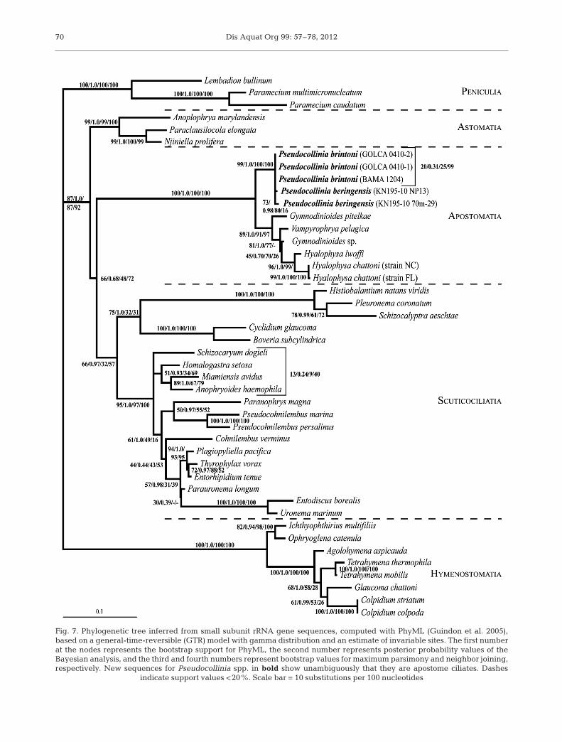
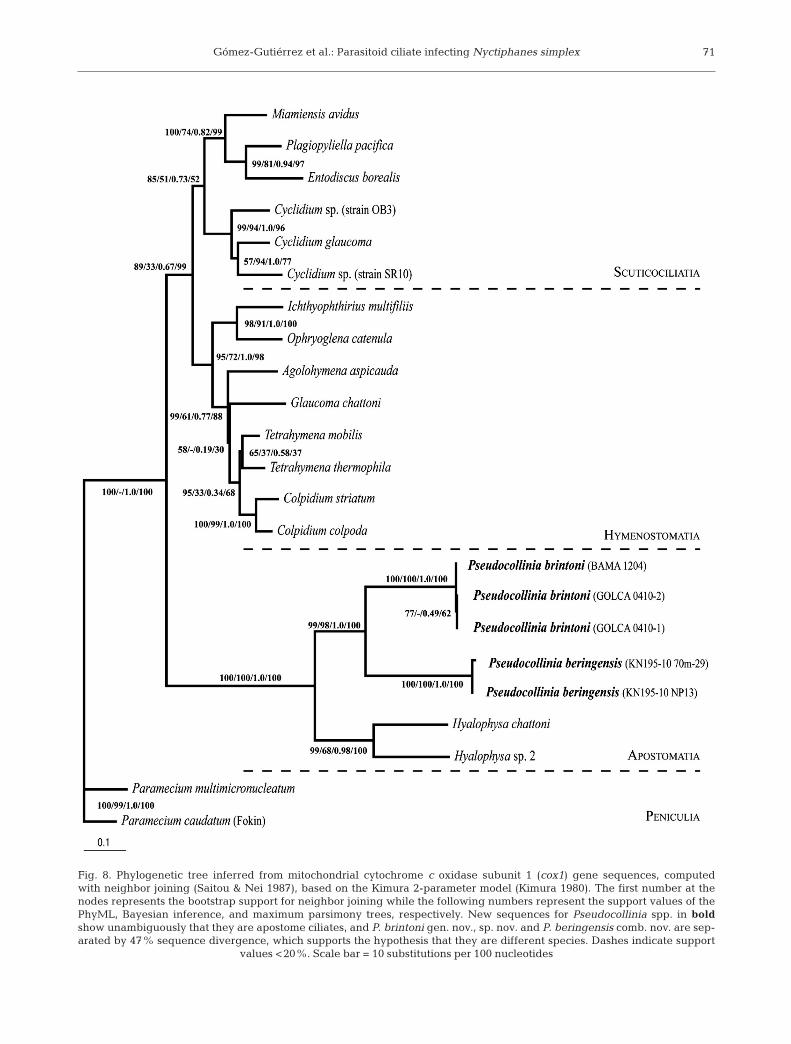
The phylogenetic analyses placePseudocollinia in a well-supportedmonophyletic clade with other apos-tome ciliates (Figs. 7 & 8). The 2 spe-cies are not well separated in the treeinferred from SSU rRNA gene se -quences (Fig. 7). However, a distinctgenetic distance of 0.47 separatesthem in the tree inferred from cox1gene sequences (Fig. 8).
69
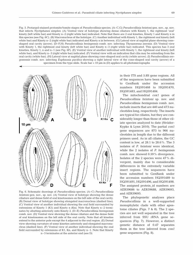
Fig. 5. Protargol-stained protomite/tomite stages of Pseudocollinia species. (A−C,G) Pseudocollinia brintoni gen. nov., sp. nov.that infects Nyctiphanes simplex. (A) Ventral view of holotype showing dense ciliature with Kinety 1, the rightmost ‘oral’kinety (left white bar) and Kinety n–2 (right white bar) indicated. Note that there are 2 oral kineties, Kinety 1 and Kinety n inthis species (see Fig. 6C). (B) Macronucleus of the holotype. (C) Another individual with Kinety 1, the rightmost oral kinety (leftwhite bar) and Kinety n–2 (right white bar) indicated and Kinety n in between. (G) Lateral view at sagittal plane showing cone-shaped oral cavity (arrow). (D−F,H) Pseudocollinia beringensis comb. nov. infecting Thysanoessa inermis. (D) Ventral viewwith Kinety 1, the rightmost oral kinety (left white bar) and Kinety n–3 (right white bar) indicated. This species has 3 oralkineties, Kinety 1, n and n–1 (see Fig. 6F). (E) Ventral view of another individual with Kinety 1, the rightmost oral kinety (leftwhite bar), and Kinety n–3 (right white bar) indicated. (F) Ventral view with an indication that cilia may be extending from theoral cavity (white bar). (H) Lateral view at sagittal plane showing cone-shaped oral cavity (white arrow). (I) Pseudocollinia ore-gonensis comb. nov. infecting Euphausia pacifica showing a right lateral view of the cone-shaped oral cavity (arrow) of a
specimen from the type slide. Scale bar = 10 µm in (D) applies to all photomicrographs
Fig. 6. Schematic drawings of Pseudocollinia species. (A−C) Pseudocolliniabrintoni gen. nov., sp. nov. (A) Ventral view of holotype showing the denseciliature and dense field of oral kinetosomes on the left side of the oral cavity.(B) Dorsal view of holotype showing elongated macronucleus (dashed line).(C) Ventral view of another individual showing the oral field surrounded byextensions of Kinety 1 (K1) and Kinety n (Kn). Note that Kinety n–2 termi-nates by abutting anteriorly onto Kinety 2. (D−E) Pseudocollinia beringensiscomb. nov. (D) Ventral view showing the dense ciliature and the dense fieldof oral kinetosomes on the left side of the oral cavity. Note that all kinetiesextend to the anterior pole except the 4 adjacent to the oral region. (E) Dorsalview showing curvature of some kineties anteriorly and elongated macronu-cleus (dashed line). (F) Ventral view of another individual showing the oralfield surrounded by extensions of K1, Kn, and Kinety n–1. Note that Kinety
n–3 terminates at the anterior end (see D)
Dis Aquat Org 99: 57–78, 201270
Fig. 7. Phylogenetic tree inferred from small subunit rRNA gene sequences, computed with PhyML (Guindon et al. 2005),based on a general-time-reversible (GTR) model with gamma distribution and an estimate of invariable sites. The first numberat the nodes represents the bootstrap support for PhyML, the second number represents posterior probability values of theBayesian analysis, and the third and fourth numbers represent bootstrap values for maximum parsimony and neighbor joining,respectively. New sequences for Pseudocollinia spp. in bold show unambiguously that they are apostome ciliates. Dashes
indicate support values <20%. Scale bar = 10 substitutions per 100 nucleotides
Gómez-Gutiérrez et al.: Parasitoid ciliate infecting Nyctiphanes simplex 71
Fig. 8. Phylogenetic tree inferred from mitochondrial cytochrome c oxidase subunit 1 (cox1) gene sequences, computedwith neighbor joining (Saitou & Nei 1987), based on the Kimura 2-parameter model (Kimura 1980). The first number at thenodes represents the bootstrap support for neighbor joining while the following numbers represent the support values of thePhyML, Bayesian inference, and maximum parsimony trees, respectively. New sequences for Pseudocollinia spp. in boldshow unambiguously that they are apostome ciliates, and P. brintoni gen. nov., sp. nov. and P. beringensis comb. nov. are sep-arated by 47% sequence divergence, which supports the hypothesis that they are different species. Dashes indicate support
values <20%. Scale bar = 10 substitutions per 100 nucleotides
Dis Aquat Org 99: 57–78, 2012
Pseudocollinia brintoni experimental infection
In March and July 2004, we incubated Pseudo-collinia brintoni gen. nov., sp. nov. ciliates after theykilled their euphausiid hosts. The ciliate and/or rod-shaped bacteria formed long filaments ~7 h after therupture of the host at 16°C. The mechanism of forma-tion of the filaments is unknown; perhaps they areformed from the host’s digested material. To investi-gate the infection mechanism of P. brintoni, we ex-posed several healthy Nyctiphanes simplex femalesin individual jars in a 0.25 l bottle filled with ciliate-bacteria filaments. However, these potential hostsdied without first showing symptoms of infection, ap-pearing to have been semi-digested while still alive.Our interpretation is that the digestive enzymes (pro-duced by ciliates or bacteria to feed on the euphausiid)become highly concentrated (not quantified), killingthe euphausiids in the laboratory setting. In nature,the enzyme concentration is likely to be significantlylower, and the filament may be an evolutionary strat-egy to maintain high ciliate concentrations to producea massive infection in a new euphausiid host.
During December 2004, we exposed healthyeuphausiids (males, immature females, and gravidfemales) to ciliate-bacterial filaments in water previ-ously filtered with GF/F filters. The euphausiidsremained alive for a longer time, and any of the ani-mals exposed to the filaments ate them as weexpected. However, we also observed that when theeuphausiid contacted the filaments, the filamentsattached to the euphausiid body. The euphausiideventually died in a very atypical way with the fila-ments attached to the krill’s body, in contrast to thetypical Pseudocollinia endoparasitoid infection inwhich the external body covering is apparentlyhealthy. Identical experiments were made withfemales (n = 2) of the decapod Processa pippinae,some of which were ovigerous, and ovigerous femalemysids (n = 3) of an unidentified species: they did noteat the filaments, and when we intentionally put themin physical contact with the filaments, they were com-pletely unaffected, remaining alive after 8 d of incu-bation (until the oceanographic cruise ended).
DISCUSSION
Proposal of Pseudocollinia gen. nov. andmorphological comparisons
Capriulo & Small (1986) first described apostomeciliates infecting the hemocoel of krill and assigned
these ciliates to the genus Collinia because ciliates inthis genus and family are characterized as parasitiz-ing the hemocoel of crustaceans (Chatton & Lwoff1935, Jankowski 2007, Lynn 2008). They included itin the genus Collinia using the following criteria: (1)the presence of a rosette structure and special kinetaly segment; (2) hemocoel-dwelling forms in crusta -ceans; (3) the unknown (at the time) complete lifehistory of the forms; (4) major stages in which themechanism of food ingestion is pinocytosis ratherthan phagocytosis; and (5) the characteristic of formsas apparently functional astomes with minimal apos-tome traits for the life history stages known then(Capriulo & Small 1986). Regrettably, there is no typeslide for Collinia beringensis, the ciliate that Capriulo& Small (1986) described from Thysanoessa inermisin the Bering Sea (>55° N), so only the photomicro-graphs and drawings (Figs. 1 & 2 by Capriulo & Small1986) can be used. A second species of krill-infectingciliate, Collinia oregonensis, was also assigned to thisgenus by Gómez-Gutiérrez et al. (2006), who submit-ted 3 slides to the Smithsonian Institution (holotypeUSNM 1084004, paratype USNM 1084005, and non-type USNM 1084006). Although the specimens onthese slides were not well stained, some do show acone-shaped oral cavity (Fig. 5I), as reported abovefor P. brintoni and P. beringensis comb. nov.
Collinia species have been redescribed using mod-ern cytological techniques (de Puytorac & Lom 1962,de Puytorac & Grain 1975). Based on this informationand the earlier literature, Jankowski (2007) suggestedthat the genus must be split into 3 genera: (1) ColliniaCépède, 1910, whose type species Collinia circulans(Balbiani, 1885) Cépède, 1910, has large, astomatoustrophonts with 10 spiral kineties, has a smaller tomitewith short x, y and z kineties, and infects the fresh -water isopod Asellus aquaticus Linnaeus, 1758; (2)Paracollinia Jankowski, 1980, whose type speciesParacollinia branchiarum (Stein, 1852) Jankowski,1980, has large astomatous trophonts with 55 to 60straight kineties, has smaller flattened tomites with 9kineties and short x, y, and z kineties, and infects thefreshwater amphipod Gammarus pulex Linnaeus,1758; and (3) Metacollinia Jankowski, 1980, whosetype species Metacollinia luciensis (Poisson, 1921)Jankowski, 1980, has large astomatous trophontswith 65 to 70 slightly spiral ing kineties with a non-cil-iated dorsal stripe, has a smaller protomite-tomitewith 9 kineties, short x, y, and z kineties, and a falci-form field, and infects the marine amphipodOrchestia littorea, accepted as Orchestia gammarel-lus (Pallas, 1766), in Europe. Jankowski (2007) be-lieved that Collinia orchestiae Summers & Kidder,
72
Gómez-Gutiérrez et al.: Parasitoid ciliate infecting Nyctiphanes simplex
1936 redescribed by de Puytorac & Grain (1975) islikely conspecific with the type species for the genusMetacollinia (i.e. M. luciensis). This represents the di -versity within the family Colliniidae, blood-parasitesof crustaceans, when one excludes those infecting theblood of krill. Because none of the above has been re-ported to infect krill or to have a cone-shaped oralcavity covered on its left wall by a field of ciliatedkinetosomes, we believe that the blood-parasites ofkrill represent a new genus. Because Capriulo &Small (1986) believed them close to Collinia, we sug-gest the name Pseudocollinia gen. nov.
Our results demonstrated that the ciliates foundinfecting krill in waters around Baja California andthose infecting krill in the Bering Sea, the type local-ity for the first apostome ciliate species recorded toinfect krill, share morphological and genetic similari-ties. Examination of the type slide of Collinia orego-nensis (Gómez-Gutiérrez et al. 2006) convinces usthat this krill-infecting species is also closely relatedto P. brintoni and P. beringensis comb. nov. There-fore, these 3 species should be placed in the newgenus Pseudocollinia gen. nov.
The ciliates Pseudocollinia brintoni gen. nov. sp.nov. infecting Nyctiphanes simplex along the westcoast of Baja California Sur (23°N) and Gulf of Cali-fornia have considerably fewer somatic kineties andsmaller inter-stage variability in the number ofkineties (12 to 18 kineties) than the other 2 apostomeciliates that infect euphausiids. Pseudocollinia berin-gensis comb. nov., which infects Thysanoessa iner-mis and Thysanoessa raschi in the Bering Sea, pos-sesses 24 to 80 kineties with large inter-stagevariability in the number of kineties (i.e. trophont =80, tomont = 34, protomite I = 28, and protomite II =24) (Capriulo & Small 1986). Pseudocollinia orego-nensis infects 3 euphausiid species (Euphausia paci-fica, Thysanoessa spinifera, and Thysanoessa gre-garia) off the Oregon–Washington coast (42−46° N)and has 14 to 22 kineties and a moderate inter-stagevariability in the number of kineties (i.e. trophont =18−22; tomont = 16−20, protomite = 18−20, andtomite = 14−16) (Gómez-Gutiérrez et al. 2003, 2006).There is a clear latitudinal cline in the number ofkineties of apostome ciliates along the Bering Seaand the California Current System, consistentlydecreasing from north to south.
Given the differences between these isolates, wehereby establish a new genus and species (i.e.Pseudocollinia brintoni gen. nov., sp. nov.) for the cil-iates infecting the krill Nyctiphanes simplex inwaters around Baja California. This species is char-acterized as follows: (1) this ciliate has a smaller
number of kineties than the 2 previously describedspecies that infect euphausiids (Capriulo & Small1986, Gómez-Gutiérrez et al. 2006); (2) this ciliate hasonly 2 oral kineties surrounding the oral cavity, whilePseudocollinia beringensis comb. nov. has at least 3oral kineties; (3) to date, this is the only apostome cil-iate species that infects a euphausiid with a sac-spawning strategy; (4) this ciliate infects a krill spe-cies with a subtropical zoogeographic pattern, whileother species infect euphausiids with arctic and sub-arctic distributions (Brinton 1962, 1981, Brinton et al.2000); and (5) the genetic differences are sufficient tosupport a differentiation at the species level betweenP. beringensis comb. nov. and P. brintoni. Therefore,we propose to name this ciliate Pseudocollinia brin-toni sp. nov. and designate it as the type species forthe new genus. Given the morphological and geneticsimilarities of Collinia beringensis Capriulo & Small,1986, with P. brintoni gen. nov. sp. nov., we herebytransfer the former species, making the new combi-nation Pseudocollinia beringensis (Capriulo & Small,1986) comb. nov. On the same grounds, we herebytransfer C. oregonensis Gómez-Gutiérrez et al., 2006,making the new combination Pseudocollinia orego-nensis comb. nov. We are currently undertaking mor-phological and genetic research on P. oregonensis toprovide details of its infraciliature and genetics todetermine how closely it is related to P. brintoni andP. beringensis comb. nov. Given that our research onkrill-infecting apostomes is relatively new, it is highlylikely that these Pseudocollinia species will be foundinfecting other krill species and that more Pseudo-collinia species may soon be discovered. Future SSUrRNA and cox1 sequences for any other Collinia spe-cies infecting other crustaceans will confirm the pro-posed designation of Pseudocollinia as a new genusof parasitoid ciliates in the family Colliniidae.
Life cycle of endoparasitoid apostome ciliates
The endoparasitic infection of Nyctiphanes simplexby Pseudocollinia brintoni gen. nov., sp. nov. showedmany similarities with the ciliate infections of the eu-phausiids along the Oregon coast (Gómez-Gutiérrezet al. 2003, 2006). In both regions, the trophont stageexclusively infects, in high numbers, adult femalespecimens. Larvae and juveniles seem to be inade-quate hosts for this type of endoparasite, possibly be-cause they have low biomass, low lipid content, and arelatively short developmental duration (Gómez-Gutiérrez et al. 2010c). The hypothesis that larvae andjuveniles are infected with parasitoid cells but the
73
Dis Aquat Org 99: 57–78, 2012
symptoms manifest once the host reaches a criticalbody-weight threshold, although unlikely, should betested with experimental and observational work. As-suming that the life span of N. simplex is ~245 d (i.e.17 mm total length, estimated using the Von Berta-lanffy model K = 0.010 d−1 and asymptotic length =18.6 mm; 1 yr old individuals should be consideredexceptionally long-lived) (Lavaniegos 1992) and thatthe age of first maturity of this species is ~66 d (~9 mmtotal length) (Lavaniegos 1992, 1995, Gómez-Gutiér-rez 1995), the period within the life span that is un-likely to be infected is short (<66 d; ~24% of the com-plete life span). This finding suggests that P. brintonican infect N. simplex adults over more than two-thirds of their life span. The endoparasitoid phase of P.brintoni is on the order of days, considering that thetime from healthy host appearance to host death is<33 h and the quick formation of filaments with en-cysted phoronts in an ectoparasitic phase is < 7 h. Therelatively short infection-to-death period for theseparasitoid ciliates (33 to 72 h) potentially enablesthem to infect and kill large numbers of euphausiidsof the same swarm. It is obvious that either sex or anylife stage of N. simplex could potentially ingest P.brintoni, but to date, only N. simplex females havebeen found to be infected. N. simplex producesovigerous sacs every 7 to 15 d (Gómez-Gutiérrez &Robinson 2005, Gómez-Gutiérrez et al. 2010c). Thus,the endoparasitoid phase of P. brintoni should beshorter than the observed krill interbrood period.
The tomont stage of Pseudocollinia brintoni repro-duces dramatically inside the host, producing thou-sands of transmission stages (i.e. tomites). Thisintense infection kills and ruptures the host, releasingthe actively swimming ciliates. The exoparasiticphase of Pseudocollinia spp. is considerably lessknown (Gómez-Gutiérrez et al. 2006). Originally, itwas thought that tomites of the genus Pseudocolliniainfected the euphausiids by attaching to the externalappendages because the phoront stages of otherapostomes commonly attach to euphausiid appen -dages (Lindley 1978, Landers et al. 2006, 2007) andbecause it is the most common life-cycle mode ofother well-studied apostome ectoparasites of crus-taceans (Bradbury 1966, 1994, Bradbury & Clamp1973, Bradbury & Goyal 1976). However, Landers etal. (2006, 2007) discovered that the commonly en-cysted phoronts attached to euphausiid appendagesare actually epibiotic exuviotrophic ciliates of thegenus Gymnodinoides that infest >70% of the krillpopulation, rather than Pseudocollinia species.
Gómez-Gutiérrez et al. (2006) suggested thatCollinia species (now Pseudocollinia) must be eaten
to explain the large number of ciliates observed inearly infection stages. However, our experimentallaboratory efforts provide no conclusive evidence toshow how euphausiids could filter such high densi-ties of ciliates. Once Pseudocollinia brintoni escapefrom the infected euphausiids, they swim for a whileand aggregate, forming filaments, several centi -meters long, to which the tomite stages attach andencyst as phoront stages. Similar filament formationwas also observed in infected euphausiids from theOregon region, but at that time, the filaments werenot observed using SEM techniques (Gómez-Gutiér-rez et al. 2003, 2006). Rod-shaped bacteria alsoadhere in high densities on the body surfaces of P.brintoni and these filaments. Because bacteria arenot always observed associated with the ciliates andbecause we have invariably observed filament for-mation, we strongly believe that the ciliates form thestructural base of the filament for cell attachment,perhaps through exocytosis of extrusomes. We ob -served incompletely encysted ciliates (i.e. a tomite–phoront transition) on just-formed filaments, and wealso observed completely encysted phoront stagesfrom recently infected Nyctiphanes simplex (krillwith a yellow-beige-colored carapace) and also cili-ates incompletely excysted (i.e. phoront–trophont)inside the carapace. Thus, we assume that the morelikely and parsimonious link between both events isthat the euphausiids eat the filaments bearing highciliate densities. Although we performed severalshipboard laboratory incubations to test this hypoth-esis, we were unable to obtain direct evidence aboutthis part of the life cycle of P. brintoni.
Can euphausiids actually eat drifting filaments innature? Euphausia pacifica eats marine snow particles>0.5 mm diameter, even when other sources of foodare provided (Dilling et al. 1998, Dilling & Brzezinski2004). Thus, it is likely that euphausiids can ingest fil-aments with high numbers of encysted Pseudocolliniaand bacteria in the field. E. pacifica is also able tofragment a single aggregate of marine snow into mul-tiple, smaller aggregates by the fluid stress createdaround the appendages during swimming (Dilling &Alldredge 2000, Goldthwait et al. 2004). Thus, asingle ciliate-bacterial filament may be fragmentedand potentially infect multiple euphausiids within theswarm. This infection mechanism of feeding on cili-ate-bacteria filaments could be morphologically anal-ogous to feeding on marine snow, explaining the ini-tially high density of trophont stages in the earlystages of infection. We demonstrated that apostomeendoparasitic ciliates are capable of forming filaments,and they may mimic marine snow. Gómez-Gutiérrez
74
Gómez-Gutiérrez et al.: Parasitoid ciliate infecting Nyctiphanes simplex
et al. (2006) discussed why Collinia (now Pseudo-collinia) infection on krill cannot be explained by in-fection mechanisms previously described for otherapostome ciliates as follows. (1) Other apostome spe-cies perforate the cuticle of the gills with a secretionproduced by the ciliate, thus dissolving a passagethrough the cuticle (Bradbury 1994, Bradbury &Goyal 1976); however, close examination of live in-fected krill did not reveal perforations in the cuticle.(2) Other apostome species penetrate the exoskeletonwhen it is soft after molting (Grimes & Bradbury1992); however, none of the krill infected by P. brin -toni had molted prior to infection. (3) Other apostomespecies infect their host after it has suffered a preda-tory attack by another metazoan predator (Grimes &Bradbury 1992); however, we can definitively statethat infected krill observed by us showed no evidenceof a previous predatory attack.
Crustaceans eat ciliates in nature as part of theirdiet (Zeldis et al. 2002), and some of these ciliatescould be prey, endosymbionts, parasites, and/or para -sitoids. Kucera (1992) suggested a passive transmis-sion of virus-like particles from shrimp-to-shrimpwhen the hosts consume the exuviotrophic apostomeciliate Hyalophysa chattoni, which possesses thevirus in their contractile vacuole. We were unable toinfect live healthy euphausiids exposed to free-swim-ming and filament-attached ciliates. Induced infec-tion of healthy Acartia tonsa by viruses has provedhighly complex and has also been unsuccessfulunder laboratory conditions (Drake & Dobbs 2005).This suggests that infection by Pseudocollinia mayrequire a combination of favorable conditions notcompletely met under our laboratory conditions orthat the parasite/parasitoids have complex infectionmechanisms and perhaps intermediate hosts notfacilitated or explored under laboratory conditions.
It was notable that several healthy, mature femalesof Nyctiphanes simplex, introduced individually inthe high-density ciliate seawater just after the origi-nal N. simplex exploded in a 0.25 l container, werekilled and semi-digested a couple of hours afterbeing exposed. This exogenous digestion of eu -phausiids must be a laboratory artifact because suchhigh tomite ciliate concentrations are unlikely tooccur in nature. However, this experiment suggeststhat ciliates can secrete high concentrations ofenzymes that digest even live euphausiids. Experi-ments introducing healthy but starved females topreviously filtered sea water with bacteria-ciliate fil-aments never showed a yellow-beige or orangecephalothorax (infection symptom), but the filamentbecame attached to the surface of the euphausiids,
and they eventually died. Local zooplanktonic fe -male decapods and mysids undergoing similar ex -periments with filaments were not affected, suggest-ing highly specific infection with euphausiids. So far,no other euphausiid species from either coast of theBaja peninsula have been observed infected withPseudocollinia (Gómez-Gutiérrez et al. 2010a).
Ecological impact of endoparasitoid ciliates
Because Nyctiphanes simplex accounts for >90%of the euphausiid standing stock from southern Cali-fornia (31° N) to Cape San Lucas (21° N) and theGulf of California, it plays a significant role in thefood web of this region (Brinton 1962, Brinton &Townsend 1980, Lavaniegos 1995, Gómez-Gutiérrezet al. 1995). Several top predators, such as the bluewhale Balaenoptera musculus, fin whale Balaen -optera physalus, whale shark Rhincodon typus,manta rays Mobula japonica and Mobula thurstoni,and juveniles of the jumbo squid Dosidicus gigasfeed almost exclusively on euphausiid swarms(Notarbartolo-di-Sciara 1988, Gendron 1992). Thislethal ciliate may have a significant impact on thepopulation structure, distribution, and secondaryproductivity of this euphausiid species, virtually com-peting with their better known predators. Infectedeuphausiids were recorded in ~9.5% (range 4 to22%) of the sampled oceanographic stations. Thoseprevalence rates were similar to the proportionrecorded along the Bering Sea and Oregon–Califor-nia coast for other species of Pseudocollinia (Capriuloet al. 1991, Gómez-Gutiérrez et al. 2003, 2006).
To date, apostome ciliate infection has been dis -covered in 6 out of 86 euphausiid species currentlydescribed: Thysanoessa inermis (Capriulo & Small1986, Capriulo et al. 1991) and Thysanoessa raschi inthe Bering Sea (present study), Euphausia pacifica,Thysanoessa spinifera, and Thysanoessa gregaria inthe Oregon-California coast (Gómez-Gutiérrez et al.2003, 2006), and Nyctiphanes simplex in the north-west region of Mexico (present study). It is likely thatother euphausiid species distributed around theworld have similar ciliate parasitoid–host associa-tions. In such serendipitous events, we recommendpreserving the krill hosts in >80% ethanol, if possiblewith the cuticle perforated with a needle, so that theciliates may be studied for both morphology andgenetics. Formalin (4 to 5%) with sodium boratepreservation is also adequate for ciliate morphologi-cal analysis. These infections may deserve more ofour attention because parasitoids are thought to play
75
Dis Aquat Org 99: 57–78, 2012
a significant regulatory role in the population dy -namics of their hosts (Anderson & May 1978, Brad-bury 1994, Wearing et al. 2004). The study of themetabolism of this ciliate (i.e. feeding and growthrates) and the detection of the exoparasitic phase ofPseudocollinia spp. in situ and the infection mecha-nism are the main scientific challenges for futurestudies.
Taxonomic summary
Subclass Apostomatia Chatton & Lwoff, 1928Order Apostomatida Chatton & Lwoff, 1928Family Colliniidae Cépède, 1910
Pseudocollinia Gómez-Gutiérrez, Strüder-Kypke,Lynn, Shaw, Aguilar-Méndez, López-Cortés, Martí -
nez-Gómez, Robinson, gen. nov.
Diagnosis: Blood-infecting parasitoid ciliates ofkrill with a polymorphic life cycle, includingtrophont, tomont, protomite, tomite, and phorontstages. Ciliates contain a cone-shaped oral cavity inthe anterior one-third of body with the left wall of theoral cavity covered by a field of ciliated kinetosomesand the oral cavity opening bordered by at least 2oral kineties.
Type species: Pseudocollinia brintoni sp. nov.Etymology: The genus name is given due to the
presumed similarities to apostome ciliates of thegenus Collinia, hence ‘pseudo’ from pseudes (Gr.,false). Gender: female.
Pseudocollinia brintoni Gómez-Gutiérrez, Strüder-Kypke, Lynn, Shaw, Aguilar-Méndez, López-Cortés,
Martínez-Gómez, Robinson, sp. nov.
Diagnosis: All life cycle stages possess 12 to 18somatic kineties, with at least the anterior end ofKinety n–2 abutting on Kinety 2; the oral cavity isbordered by 2 oral kineties; the ciliate infects a sac-spawning euphausiid with subtropical and neriticdistribution.
Type host: Nyctiphanes simplex Hansen, 1911Type location: Northeast Pacific, southwest coast
of Baja California peninsula, México, at the con -tinental shelf off Magdalena Bay (24° 24.33’ N,111° 57.13’ W) collected on 25 March 2004 (Table 3).
Endoparasitoid stage: In the hemocoel and insidethe abdomen and appendages of the host krill.
Ectoparasitic stage: Forming clusters of phorontson filaments, frequently associated with bacterialcolonies.
Type material: A female Nyctiphanes simplex(11.2 mm total length) infected with trophont andtomont stages of Pseudocollinia brintoni gen. nov.,sp. nov. located inside the cephalothorax and ab -domen (paratype USNM 1154442) and a protargol-stained slide of P. brintoni gen. nov., sp. nov. cells inthe tomite stage (holotype USNM 1156976) weredeposited in the International Protozoan Type SlideCollection of the Department of Invertebrate Zoologyof the National Museum of Natural History, Smith-sonian Institution. The holotype (Figs. 5A,B & 6A,B)is circled in black on the underside of the slide.
Gene sequences: Gene sequences of P. brintonigen. nov., sp. nov. were deposited in GenBank withthe accession numbers HQ591468 to HQ591470 (SSUrRNA) and HQ591489 to HQ591491 (cox1).
Etymology: The species name is given in memoryand honor of Professor Edward Brinton (1924−2010),Scripps Institution of Oceanography (San Diego,USA) who was an outstanding expert on the taxon-omy, ecology, population biology, zoogeography, andevolution of Euphausiacea.
Acknowledgements. J.G.G. dedicates this publication toProfessor E. Brinton for his invaluable friendship, guidance,and mentoring from 1990 until his death. Professor Brintonmaintained an excellent quality in euphausiid investiga-tions worldwide, and he was an outstanding example ofendurance, a kind person, and a dearest friend. We willnever forget his words, “We will never fully understand thevariability of life in the oceans. But please don’t fault us fortrying”. We thank A. Arturo Cruz-Villacorta (CIBNOR,Mexico) for his SEM assistance. We are in debt to the RV ‘ElPuma’ crew (UNAM) for their co-operation at sea. We aregrateful to D. Tikhonenkov for translations of Russianresearch. This research was partially supported by the Cen-tro Interdisciplinario de Ciencias Marinas from InstitutoPolitécnico Nacional (IPN-CGPI 2004-2011), CONACyT-2004-C01-00144, SAGARPA-CONACyT S007-2005-1-11717, and the Universidad Nacional Autónoma de México(PAPIIT IN219502 and IN210622). M.C.S.K. and D.H.L. weresupported by a NSERC Canada Discovery Grant. M.J.A.M.and A.L.C. were supported by CIBNOR PC4.2, SEP-CONA-CYT 45914/A-1 grants. J.G.G., A.L.C., and C.J.R. are sup-ported by an SNI fellowship, and J.G.G. was supported byCOFAA−IPN and EDI-IPN grants.
LITERATURE CITED
Anderson RM, May RM (1978) Regulation and stability ofhost–parasite population interactions. I. Regulatory pro-cesses. J Anim Ecol 47: 219−247
Balbiani EG (1885) Sur un infusoire cilie parasite du sang del’Asselle aquatique (Anoplophyra circulans). Zool Suisse2: 277−305
76
Gómez-Gutiérrez et al.: Parasitoid ciliate infecting Nyctiphanes simplex
Bourland WA, Strüder-Kypke MC (2010) Agolohymenaaspidocauda nov. gen., nov. spec., a histophagous fresh-water tetrahymenid ciliate in the family Deltopylidae(Ciliophora, Hymenostomatia), from Idaho (northwestU.S.A.): morphology, ontogenesis and molecular phylo -geny. Eur J Protistol 46: 221−242
Bradbury PC (1966) The life cycle and morphology of theapostome ciliate, Hyalophysa chattoni n. g., n. sp. J Pro-tozool 13: 209−225
Bradbury PC (1994) Parasitic protozoa of mollusks and crus-tacea. In: Kreier JP (ed) Parasitic protozoa, 2nd edn. Aca-demic Press, San Diego, CA, p 139−263
Bradbury PC, Clamp JC (1973) Hyalophysa lwoffi sp. n. fromthe fresh-water shrimp Palaemonetes paludosus andrevision of the genus Hyalophysa. J Protozool 20: 210−213
Bradbury PC, Goyal V (1976) The fine structure of a parasiticciliate Terebrospira during ingestion of the exoskeletonof a shrimp Palaemonetes. Tissue Cell 8: 573−582
Brinton E (1962) The distribution of Pacific euphausiids. BullScripps Inst Oceanogr Univ Calif 8: 51−270
Brinton E (1981) Euphausiid distributions in the CaliforniaCurrent during the warm winter-spring 1977-1978, in thecontext of a 1949-1966 time series. Calif Coop OceanFish Invest Rep 22: 135−154
Brinton E, Townsend AW (1980) Euphausiids in the Gulf ofCalifornia—the 1957 cruises. Calif Coop Ocean FishInvest Rep 21: 211−236
Brinton E, Ohman MD, Townsend AW, Knight MD, Bridge-man AL (2000) Euphausiids of the world ocean. WorldBiodiversity Database CD-ROM Series for [Microsoft]Windows version 1.0. Expert Center for Taxonomic Iden-tification, Amsterdam
Capriulo GM, Small EB (1986) Discovery of an apostome cil-iate (Collinia beringensis n. sp.) endoparasitic in theBering Sea euphausiid Thysanoessa inermis. Dis AquatOrg 1: 141−146
Capriulo GM, Pedone MJ, Small EB (1991) High apostomeciliate endoparasite infection rates found in the BeringSea euphausiid Thysanoessa inermis. Mar Ecol Prog Ser72: 203−204
Chatton E, Lwoff A (1935) Les ciliés apostomes. 1. Aperçuhistorique et general étude monographique des genreset des espèces. Arch Zool Exp Gen 77: 1−453
de Puytorac P, Grain J (1975) Étude de la tomitogenèse et del’ultrastructure de Collinia orchestiae, cilié apostomesanguicole, endoparasite du crustacé Orchestia gam-marella Pallas. Protistologica 11: 61−74
de Puytorac P, Lom J (1962) La tomitogenèse des Collinia cil-iés apostomes sanguicoles endoparasites des crustacés.Ann Parasitol Hum Comp 37: 195−209
De Rijk P, De Wachter R (1993) DCSE, an interactive tool forsequence alignment and secondary structure research.Comput Appl Biosci 9: 735−740
Dilling L, Alldredge AL (2000) Fragmentation of marinesnow by swimming macrozooplankton: a new processimpacting carbon cycling in the sea. Deep-Sea Res I 47: 1227−1245
Dilling L, Brzezinski MA (2004) Quantifying marine snow asa food choice for zooplankton using stable silicon isotopetracers. J Plankton Res 26: 1105−1114
Dilling L, Wilson J, Steinberg D, Alldredge A (1998) Feedingby the euphausiid Euphausia pacifica and the copepodCalanus pacificus on marine snow. Mar Ecol Prog Ser170: 189−201
Drake LA, Dobbs FC (2005) Do viruses affect fecundity andsurvival of the copepod Acartia tonsa Dana? J PlanktonRes 27: 167−174
Elwood HJ, Olsen GJ, Sogin ML (1985) The small-subunitribosomal RNA gene sequences from the hypotrichousciliates Oxytricha nova and Stylonychia pustulata. MolBiol Evol 2: 399−410
Felsenstein J (2005) PHYLIP (Phylogeny Interference Package) version 3.6a2. Distributed by the author,Department of Genome Sciences, University of Washing-ton, Seattle, WA. Available at http://evolution.genetics.washington.edu/phylip.html
Gendron DL (1992) Population structure of daytime surfaceswarms of Nyctiphanes simplex (Crustacea: Euphausi-acea) in the Gulf of California, Mexico. Mar Ecol ProgSer 87: 1−6
Goldthwait S, Yen J, Brown J, Alldredge A (2004) Quantifi-cation of marine snow fragmentation by swimmingeuphausiids. Limnol Oceanogr 49: 940−952
Gómez JG (1995) Distribution patterns, abundance and pop-ulation dynamics of the euphausiids Nyctiphanes sim-plex and Euphausia eximia off the west coast of Baja Cal-ifornia, Mexico. Mar Ecol Prog Ser 119: 63−76
Gómez-Gutiérrez J, Robinson CJ (2005) Embryonic, earlylarval development time, hatching mechanism and inter-brood period of the sac-spawning euphausiid Nycti -phanes simplex Hansen. J Plankton Res 27: 279−295
Gómez-Gutiérrez J, Palomares-García R, Gendron D (1995)Community structure of the euphausiid populationsalong the west coast of Baja California, Mexico, duringthe weak ENSO 1986-87. Mar Ecol Prog Ser 120: 41−51
Gómez-Gutiérrez J, Peterson WT, De Robertis A, BrodeurRD (2003) Mass mortality of krill caused by parasitoid cil-iates. Science 301: 339
Gómez-Gutiérrez J, Peterson WT, Morado FJ (2006) Discov-ery of a ciliate parasitoid of euphausiids off Oregon,USA: Collinia oregonensis n. sp. (Apostomatida: Collini-idae). Dis Aquat Org 71: 33−49
Gómez-Gutiérrez J, Kawaguchi S, Nicol S (2009) Epibioticsuctorians and enigmatic ecto- and endoparasitoiddinoflagellates of euphausiid eggs (Euphausiacea) offOregon, USA. J Plankton Res 31: 777−786
Gómez-Gutiérrez J, Robinson CJ, Kawaguchi S, Nicol S(2010a) Parasite diversity of Nyctiphanes simplex andNematoscelis difficilis (Crustacea: Euphausiacea) alongthe northwestern coast of Mexico. Dis Aquat Org 88: 249−266
Gómez-Gutiérrez J, Tremblay N, Martínez-Gómez S, Robin-son CJ, Del Ángel-Rodríguez J, Rodríguez-Jaramillo C,Zavala-Hernández C (2010b) Biology of the subtropicalsac-spawning euphausiid Nyctiphanes simplex in thenorthwestern seas of Mexico: vertical and horizontal dis-tribution patterns and seasonal variability of brood size.Deep-Sea Res II 57: 606−615
Gómez-Gutiérrez J, Rodríguez-Jaramillo C, Del Ángel-Rodríguez J, Robinson CJ, Zavala-Hernández C, Trem-blay N, Martínez-Gómez S (2010c) Biology of the sub-tropical sac-spawning euphausiid Nyctiphanes simplexin the northwestern seas of Mexico: interbrood period,gonad development and lipid content. Deep-Sea Res II57: 616−630
Grimes BH, Bradbury PC (1992) The biology of Vampy-rophrya pelagica (Chatton and Lwoff, 1930), ahistophagous apostome ciliate associate with marinecalanoid copepods. J Protozool 39: 65−79
77
Dis Aquat Org 99: 57–78, 2012
Guindon S, Lethiec F, Duroux P, Gascuel O (2005) PhyML On-line—a web server for fast maximum likelihood-basedphylogenetic inference. Nucleic Acids Res 33: W557−W559
Jankowski AW (1980) Conspectus of a new system of thephylum Ciliophora. Trudy Zool Inst Akad Nauk SSSR 94: 103−121
Jankowski AW (2007) Phylum Ciliophora Doflein, 1901. In: Alimov AF (ed) Protista, Part 2, Handbook on Zoology.Russian Academy of Sciences, Zoological Institute, St.Petersburg, p 415−993 (in Russian with English sum-mary)
Jerome CA, Simon EM, Lynn DH (1996) Description ofTetrahymena empidokyrea n. sp., a new species in theTetrahymena pyriformis sibling species complex (Cilio-phora, Oligohymenophorea), and an assessment of itsphylogenetic position using small-subunit rRNA se -quences. Can J Zool 74: 1898−1906
Kimura M (1980) A simple model for estimating evolutionaryrates of base substitutions through comparative studiesof nucleotide sequences. J Mol Evol 16: 111−120
Kucera FP (1992) Virus-like particles associated with theapostome ciliate Hyalophysa chattoni. Dis Aquat Org 12: 151−153
Kumar S, Nei M, Dudley J, Tamura K (2008) MEGA: a biol-ogist-centric software for evolutionary analysis of DNAand protein sequences. Brief Bioinform 9: 299−306
Lafferty KD, Kuris AM (2002) Trophic strategies, animaldiversity and body size. Trends Ecol Evol 17: 507−511
Landers SC, Gómez-Gutiérrez J, Peterson WT (2006)Gymnodinioides pacifica, n. sp., an exuviotrophic ciliatedprotozoan (Ciliophora, Apostomatida) from the euphausi-ids of the North Pacific. Eur J Protistol 42: 97−106
Landers SC, Gómez-Gutiérrez J, Peterson WT (2007) Thefine structure of the phoront of Gymnodinioides pacifica,a ciliated protozoan (Ciliophora, Apostomatida) fromeuphausiids of the Northeastern Pacific. Eur J Protistol43: 239−249
Lavaniegos EB (1992) Growth and larval development ofNyctiphanes simplex in laboratory conditions. Calif CoopOcean Fish Invest Rep 33: 162−171
Lavaniegos EB (1994) Dispersion and development patternsin larvae of Nyctiphanes simplex (Euphausiacea) in theupwelling region off Baja California. Mar Ecol Prog Ser106: 207−225
Lavaniegos EB (1995) Production of the euphausiid Nyc-tiphanes simplex in Vizcaíno Bay, western Baja Califor-nia. J Crustac Biol 15: 444−453
Lindley A (1978) Continuous plankton records: The occur-rence of apostome ciliates (Protozoa) on Euphausiacea inthe North Atlantic Ocean and North Sea. Mar Biol 46: 131−136
Lynn DH (2008) The ciliated protozoa: characterization,classification, and guide to the literature, 2nd printing[2010]. Springer, Dordrecht
Medlin L, Elwood HJ, Stickel S, Sogin ML (1988) The char-acterization of enzymatically amplified eukaryotic 16S-like rRNA-coding regions. Gene 71: 491−499
Montagnes DJS, Lynn DH (1993) A quantitative protargolstain (QPS) for ciliates and other protists. In: Kemp PF,Sherr BF, Sherr EB, Cole JJ (eds) Handbook of methodsin aquatic microbial ecology. Lewis Publishers, BocaRaton, FL, p 229−240
Morado JF, Small EB (1995) Ciliate parasites and related dis-eases of Crustacea: a review. Rev Fish Sci 3: 275−354
Notarbartolo-di-Sciara G (1988) Natural history of the raysof the genus Mobula in the Gulf of California. Fish Bull86: 45−66
Poisson R (1921) Sur un infusoire du genre Balantidium, par-asite du tube digestif d’Orchestia littorea. CR SeancesSoc Biol Paris 84: 333−335
Posada D, Crandall KA (1998) MODELTEST: testing themodel of DNA substitution. Bioinformatics 14: 817−818
Ronquist F, Huelsenbeck J (2003) MrBayes 3: Bayesian phy-logenetic inference under mixed models. Bioinformatics19: 1572−1574
Saitou N, Nei M (1987) The neighbor-joining method: a newmethod for reconstructing phylogenetic trees. Mol BiolEvol 4: 406−425
Stein F (1852) Neue Beiträge zur Kenntniss der Entwick-lungsgeschichte und des feineren Baues der Infusions-thiere. Z Wiss Zool 3: 475−509
Strüder-Kypke MC, Lynn DH (2010) Comparative analysisof the mitochondrial cytochrome c oxidase subunit I(COI) gene in ciliates (Alveolata, Ciliophora) and evalu-ation of its suitability as a biodiversity marker. Syst Bio-divers 8: 131−148
Swofford DL (2002) PAUP*: phylogenetic analysis using par-simony, 4.0b10 edn. Sinauer, Sunderland, MA
Wearing HJ, Sait SM, Cameron TC, Rohani P (2004) Stage-structured competition and the cyclic dynamics of host-parasitoid populations. J Anim Ecol 73: 706−722
Zeldis J, James MR, Grieve J, Richards L (2002) Omnivoryby copepods in the New Zealand subtropical frontalzone. J Plankton Res 24: 9−23
78
Editorial responsibility: Sven Klimpel,Frankfurt am Main, Germany
Submitted: February 9, 2011; Accepted: February 2, 2012Proofs received from author(s): April 24, 2012
Related Documents